Embed Size (px)
Citation preview
Are PVCs in patients without structural heart disease really safe?Ventricular Arrhythmias in Patients
With Normal Hearts (Ventricular Arrhythmias in the
Absence of Structural Heart Disease)
Prof. Samir Morcos Rafla, FACC, FESC, FHRSAlexandria Univ.
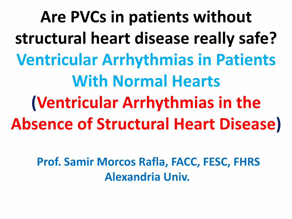
Ventricular tachycardia in patients without apparent structural heart disease
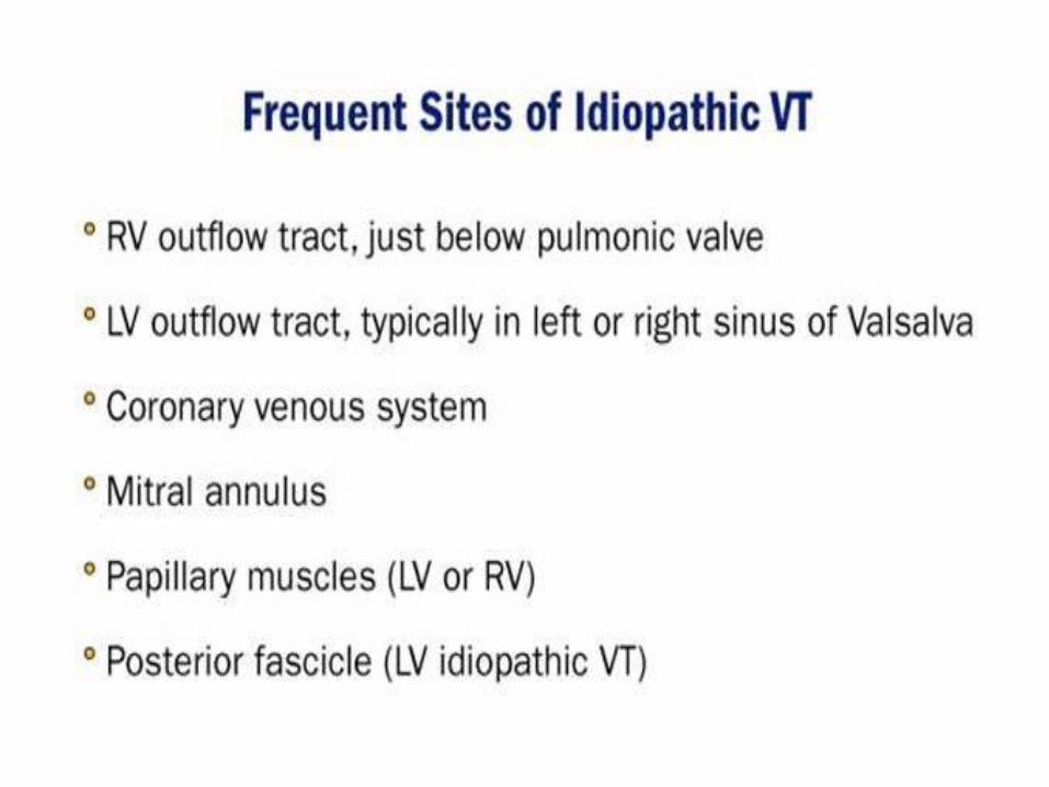
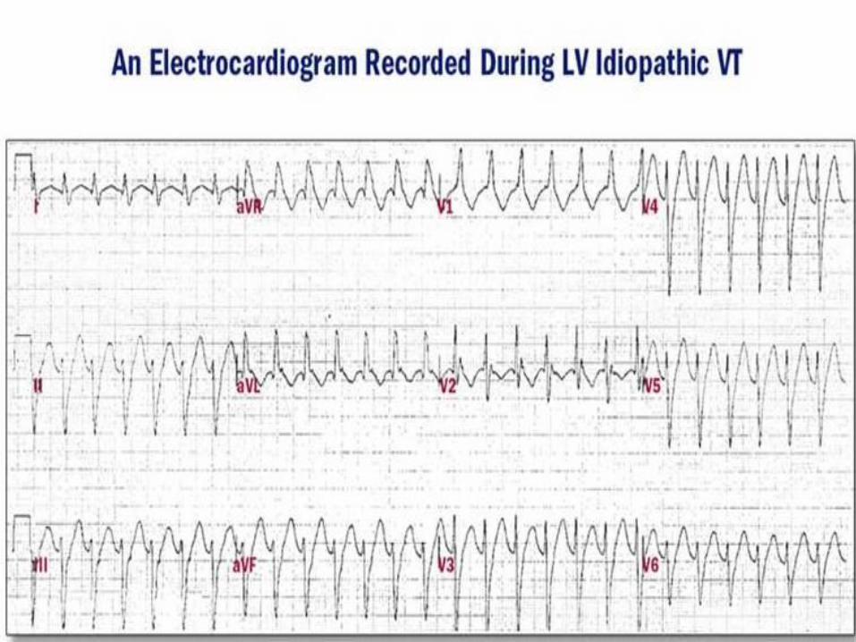
• Idiopathic ventricular tachycardia (IVT), a term that has been used for ventricular tachycardia (VT) in the absence of clinically apparent structural heart disease , accounts for around 10% of all VTs. Several types have been reported according to their clinical presentation, ventricular origin, response to drugs, electrocardiographic pattern, among others. The most common type is the so called ventricular outflow tract (VOT-T) or adenosine-sensitive tachycardia while other monomorphic forms of IVT, include intrafascicular verapamil-sensitive reentrant tachycardia and ventricular tachycardia in patients with structural heart disease.
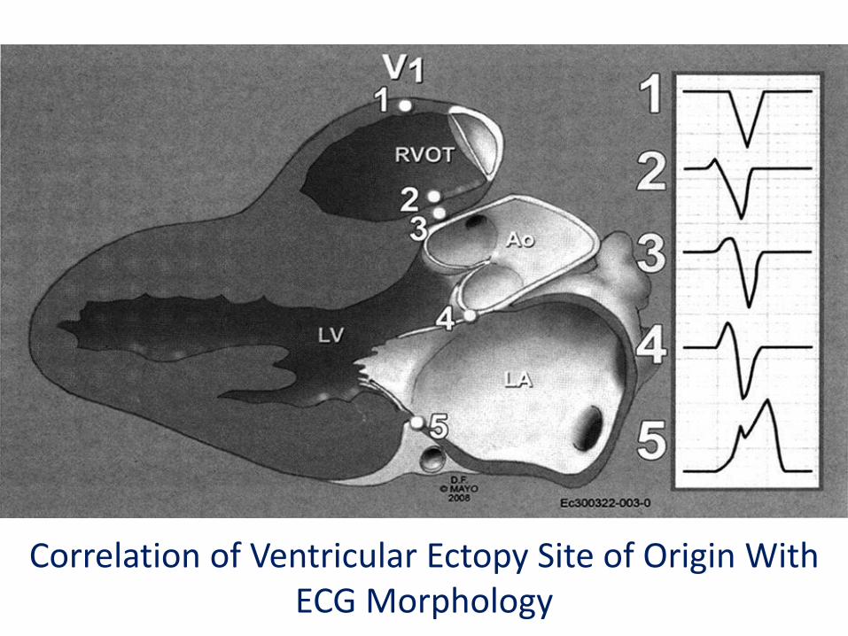
Correlation of Ventricular Ectopy Site of Origin With ECG Morphology
• Correlation of the site of origin of ventricular ectopy with the electrocardiogram (ECG) morphology in V1. The anatomy of the outflow tract region is such that areas on the right and left sides of the heart can be in close proximity to each other. This can give similar ECG patterns in several leads. However, note that in V1, there is a gradual increase in the amplitude of the r-wave as the site of origin of the ventricular ectopy moves leftward. Ao = aorta; LA = left atrium; LV = left ventricle; RVOT = right ventricular outflow tract.
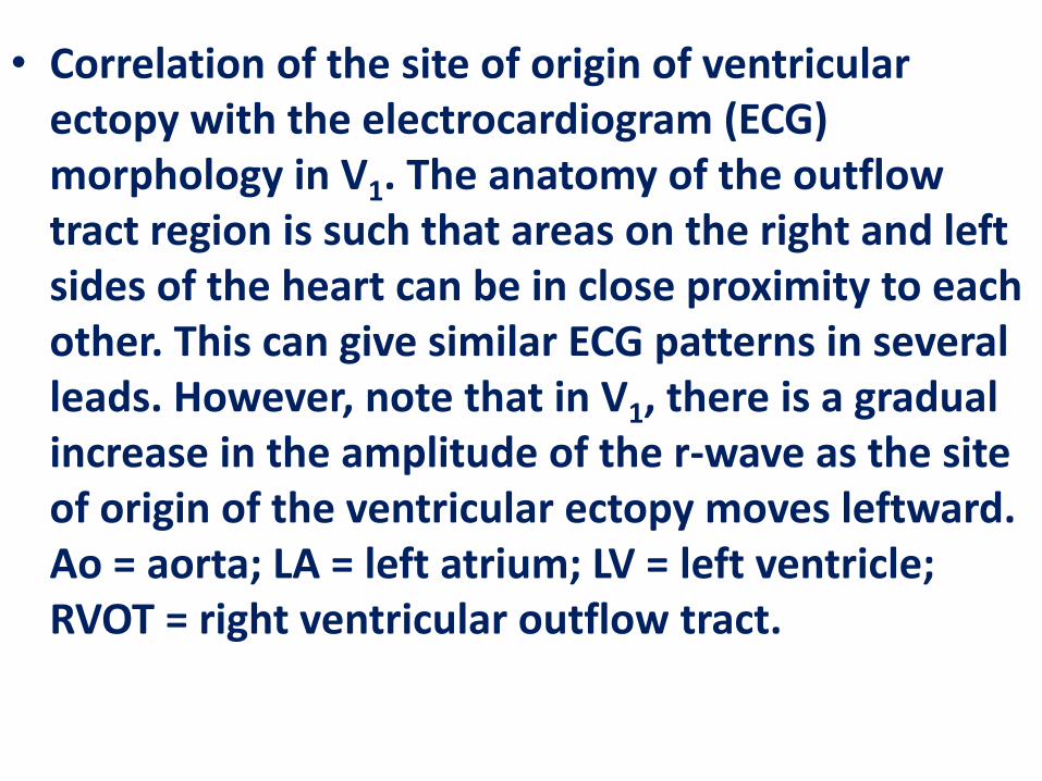
Typical electrocardiographical pattern of ventricular outflow tract tachycardia
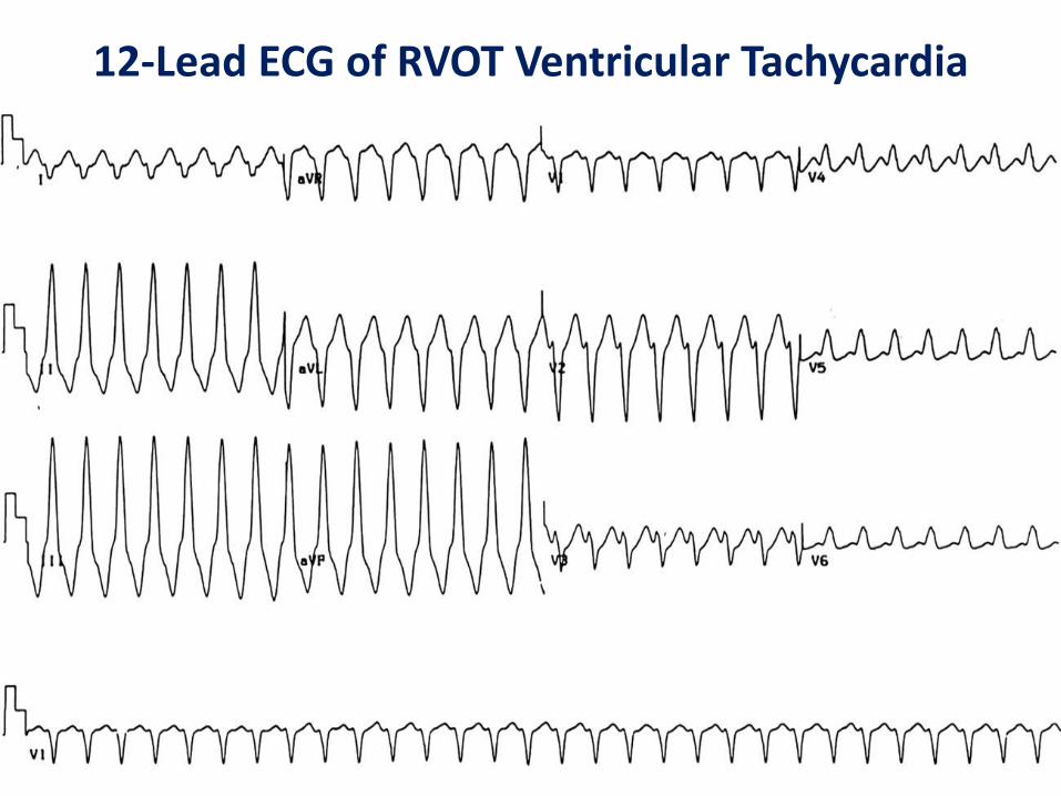
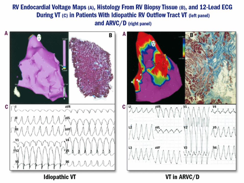
12-Lead ECG of RVOT Ventricular Tachycardia
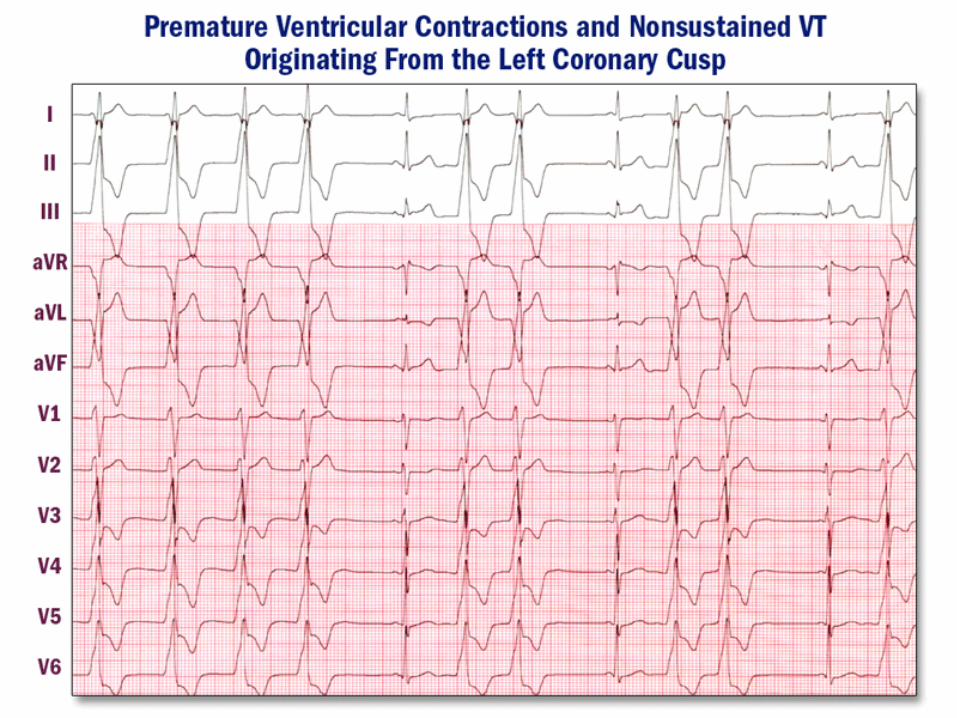
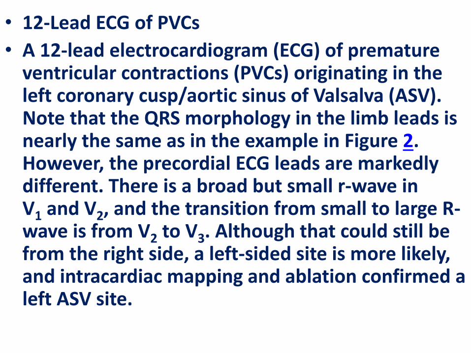
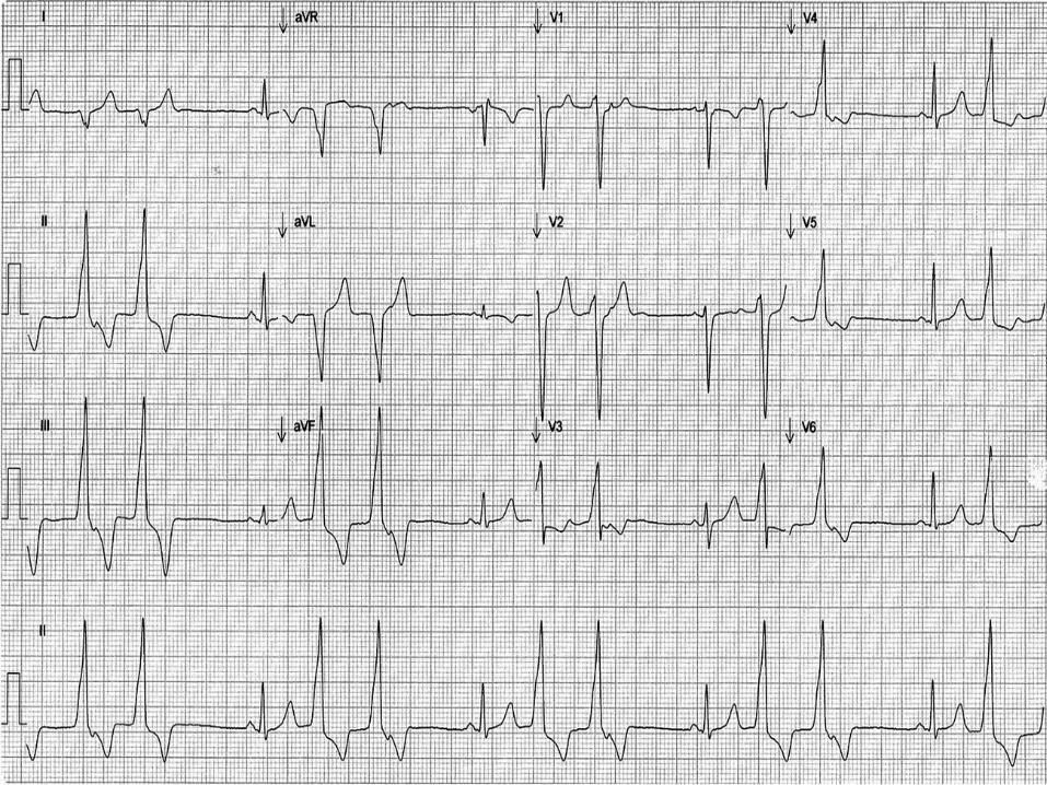
• 12-Lead ECG of PVCs
• A 12-lead electrocardiogram (ECG) of premature ventricular contractions (PVCs) originating in the left coronary cusp/aortic sinus of Valsalva (ASV). Note that the QRS morphology in the limb leads is nearly the same as in the example in Figure 2. However, the precordial ECG leads are markedly different. There is a broad but small r-wave in V1 and V2, and the transition from small to large R-wave is from V2 to V3. Although that could still be from the right side, a left-sided site is more likely, and intracardiac mapping and ablation confirmed a left ASV site.
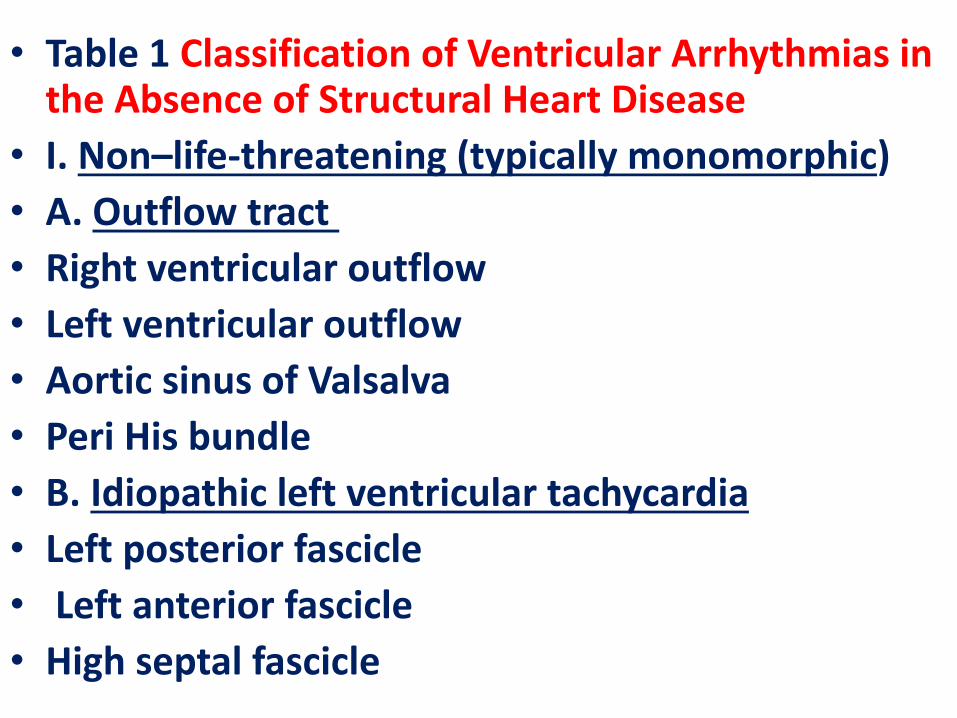
• Table 1 Classification of Ventricular Arrhythmias in the Absence of Structural Heart Disease
• I. Non–life-threatening (typically monomorphic)
• A. Outflow tract
• Right ventricular outflow
• Left ventricular outflow
• Aortic sinus of Valsalva
• Peri His bundle
• B. Idiopathic left ventricular tachycardia
• Left posterior fascicle
• Left anterior fascicle
• High septal fascicle
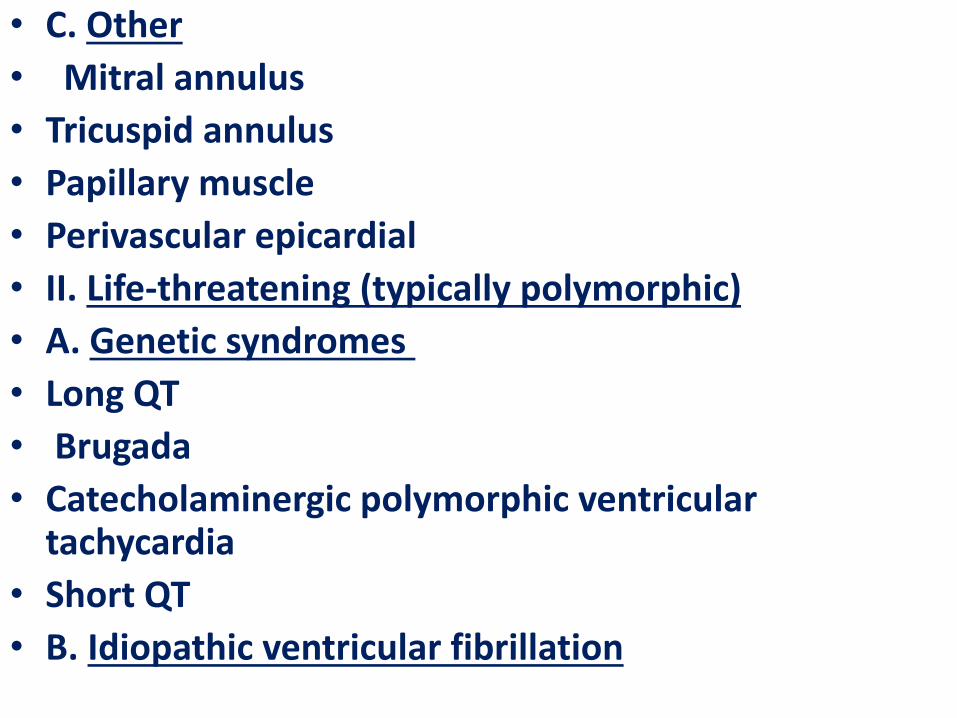
• C. Other
• Mitral annulus
• Tricuspid annulus
• Papillary muscle
• Perivascular epicardial
• II. Life-threatening (typically polymorphic)
• A. Genetic syndromes
• Long QT
• Brugada
• Catecholaminergic polymorphic ventricular tachycardia
• Short QT
• B. Idiopathic ventricular fibrillation
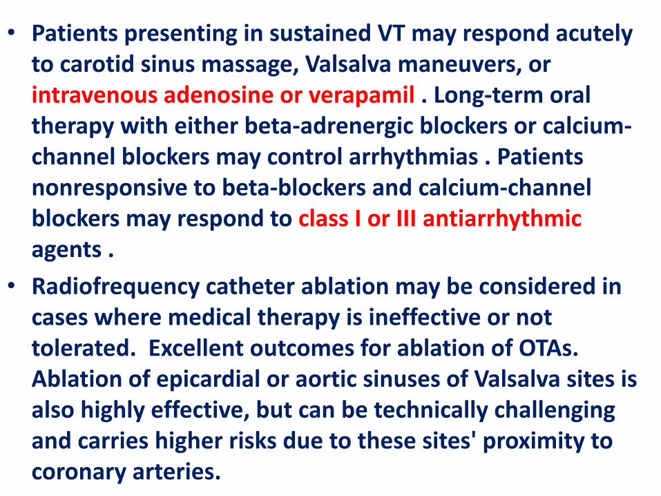
• Patients presenting in sustained VT may respond acutely to carotid sinus massage, Valsalva maneuvers, or intravenous adenosine or verapamil . Long-term oral therapy with either beta-adrenergic blockers or calcium-channel blockers may control arrhythmias . Patients nonresponsive to beta-blockers and calcium-channel blockers may respond to class I or III antiarrhythmic agents .
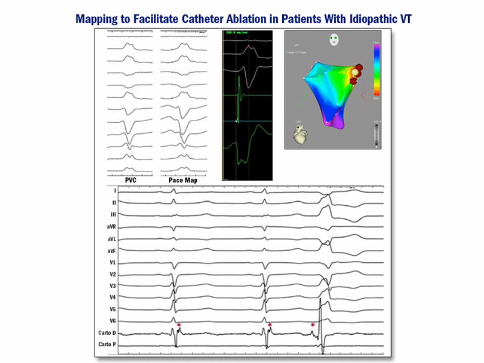
• Radiofrequency catheter ablation may be considered in cases where medical therapy is ineffective or not tolerated. Excellent outcomes for ablation of OTAs. Ablation of epicardial or aortic sinuses of Valsalva sites is also highly effective, but can be technically challenging and carries higher risks due to these sites' proximity to coronary arteries.
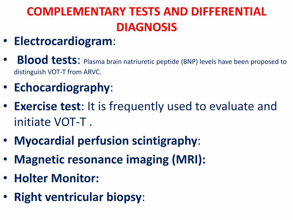
COMPLEMENTARY TESTS AND DIFFERENTIAL DIAGNOSIS
• Electrocardiogram:
• Blood tests: Plasma brain natriuretic peptide (BNP) levels have been proposed to
distinguish VOT-T from ARVC.
• Echocardiography:
• Exercise test: It is frequently used to evaluate and initiate VOT-T .
• Myocardial perfusion scintigraphy:
• Magnetic resonance imaging (MRI):
• Holter Monitor:
• Right ventricular biopsy:
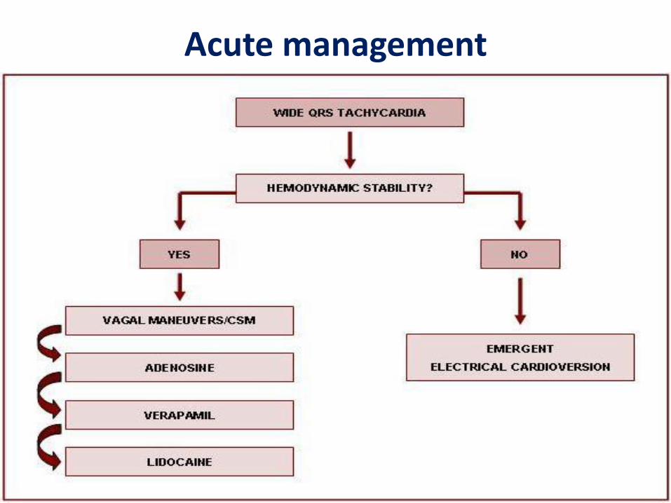
Acute management
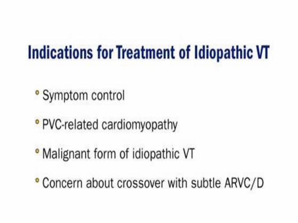
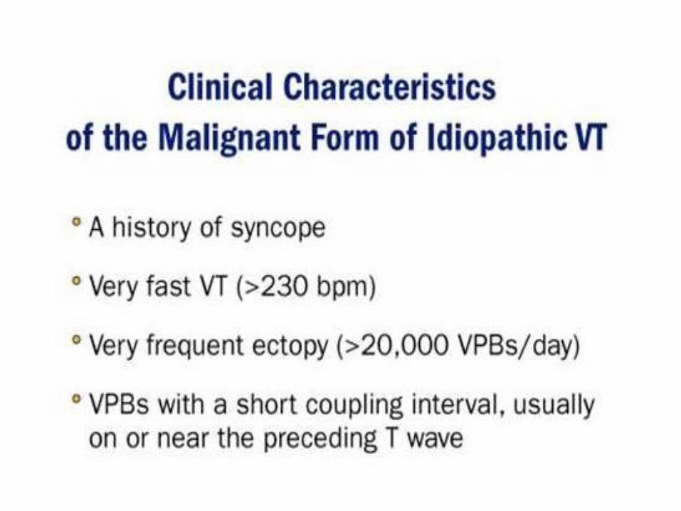
Key Points• Although most patients with idiopathic ventricular
arrhythmias do not require treatment, some patients will experience severe symptoms. In addition, treatment is warranted for patients with PVC-related myopathy and the malignant form of idiopathic VT, which can trigger episodes of PMVT.
• Treatment for idiopathic VT includes pharmacologic agents (i.e., beta-blockers, calcium channel antagonists, class IC antiarrhythmic drugs) and catheter ablation.
• Inherited arrhythmogenic diseases constitute an expanding field with a wide spectrum of electrocardiographic manifestations.
• Mutations in the same gene may cause different phenotypes according to the functional consequences of mutations and are often associated with variable penetrance and incomplete expressivity.
• Both tachyarrhythmias and bradyarrhythmias in the structurally normal heart may be genetically determined.
• Gene-specific electrocardiographic features have been identified in LQTS and BrS. In LQTS, the underlying genotype modulates both the clinical outcome and the response to therapy.
Key Points
Amiodarone is the major antiarrhythmic drug option for symptomatic ventricular arrhythmias in patients with depressed ventricular function, but the drug has significant toxicities.
Sustained polymorphic VT is usually due to an acute coronary syndrome or QT prolonging factors causing torsade de pointes.
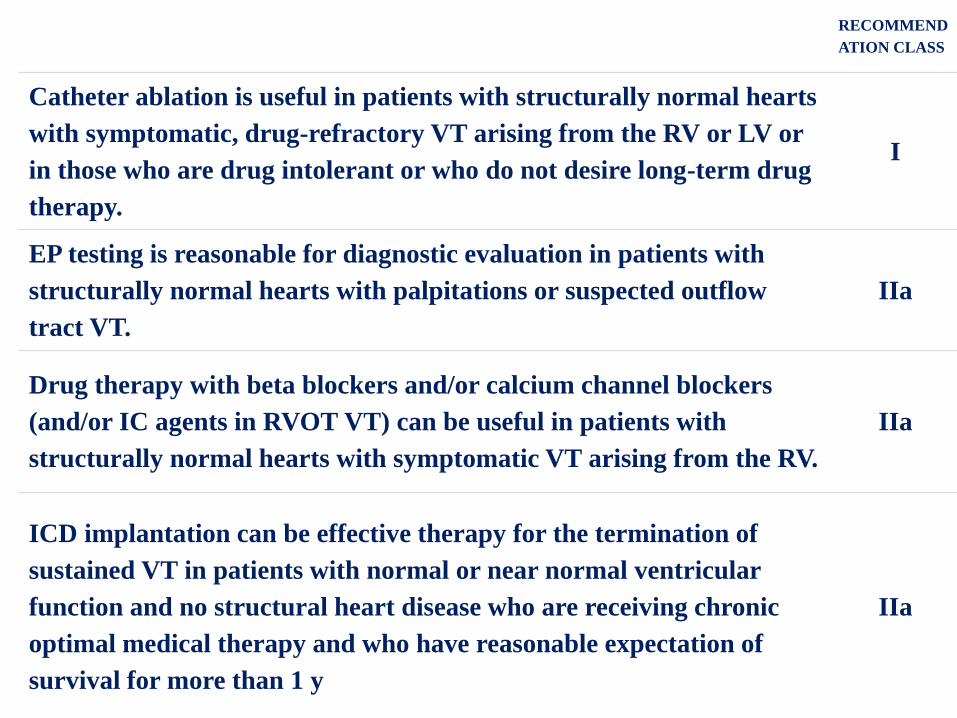
RECOMMEND
ATION CLASS
Catheter ablation is useful in patients with structurally normal hearts
with symptomatic, drug-refractory VT arising from the RV or LV or
in those who are drug intolerant or who do not desire long-term drug
therapy.
I
EP testing is reasonable for diagnostic evaluation in patients with
structurally normal hearts with palpitations or suspected outflow
tract VT.
IIa
Drug therapy with beta blockers and/or calcium channel blockers
(and/or IC agents in RVOT VT) can be useful in patients with
structurally normal hearts with symptomatic VT arising from the RV.
IIa
ICD implantation can be effective therapy for the termination of
sustained VT in patients with normal or near normal ventricular
function and no structural heart disease who are receiving chronic
optimal medical therapy and who have reasonable expectation of
survival for more than 1 y
IIa