Embed Size (px)
DESCRIPTION
Citation preview
Ankle and Foot Pain
Enas Mohamed Shahine; MDAss. Prof. Physical Medicine, Rheumatology &
Rehabilitation Faculty of Medicine,
Alexandria University, Egypt
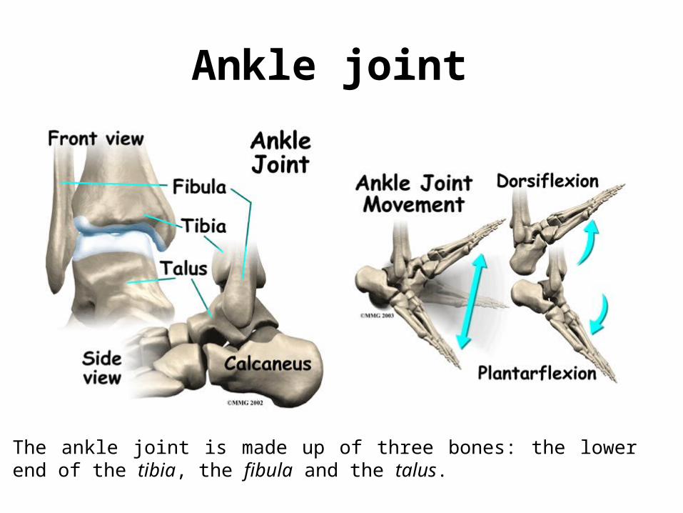
Ankle joint
The ankle joint is made up of three bones: the lower end of the tibia, the fibula and the talus.
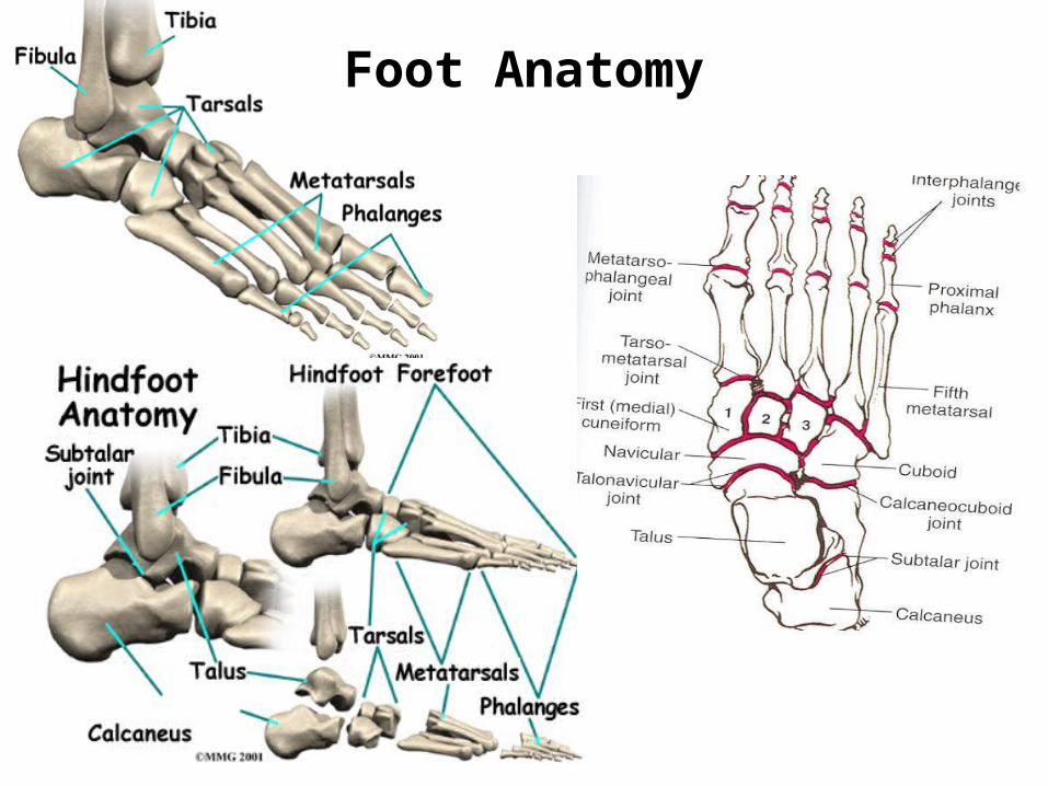
Foot Anatomy
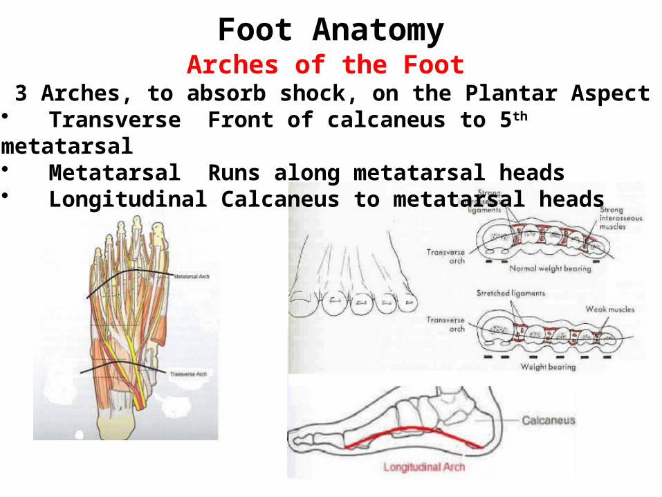
Foot AnatomyArches of the Foot
3 Arches, to absorb shock, on the Plantar Aspect • Transverse Front of calcaneus to 5th metatarsal• Metatarsal Runs along metatarsal heads• Longitudinal Calcaneus to metatarsal heads
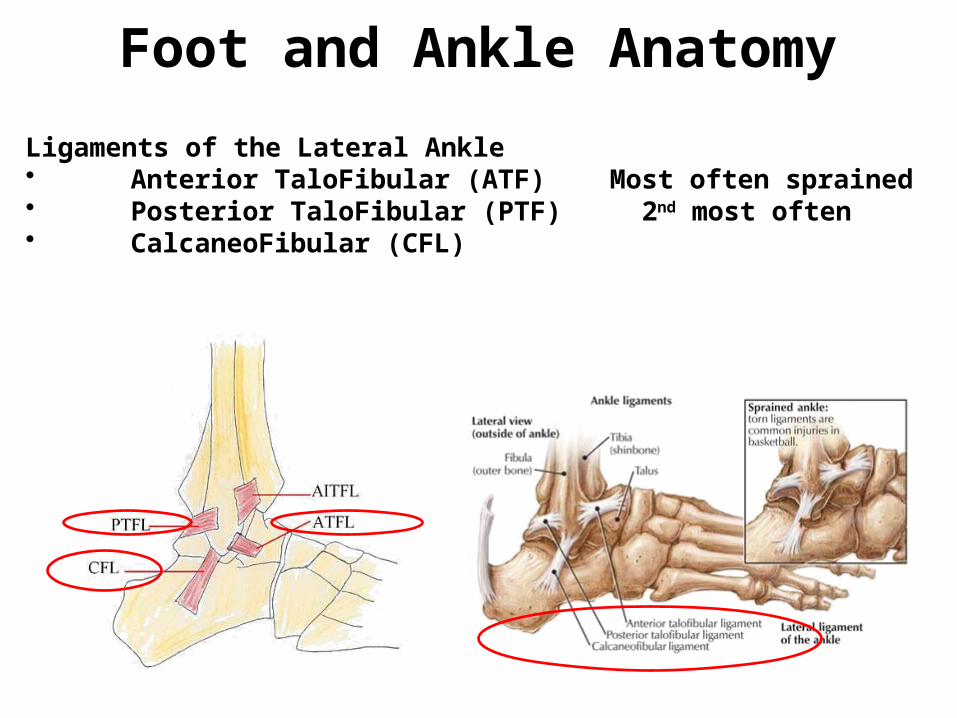
Foot and Ankle Anatomy
Ligaments of the Lateral Ankle• Anterior TaloFibular (ATF) Most often sprained• Posterior TaloFibular (PTF) 2nd most often• CalcaneoFibular (CFL)
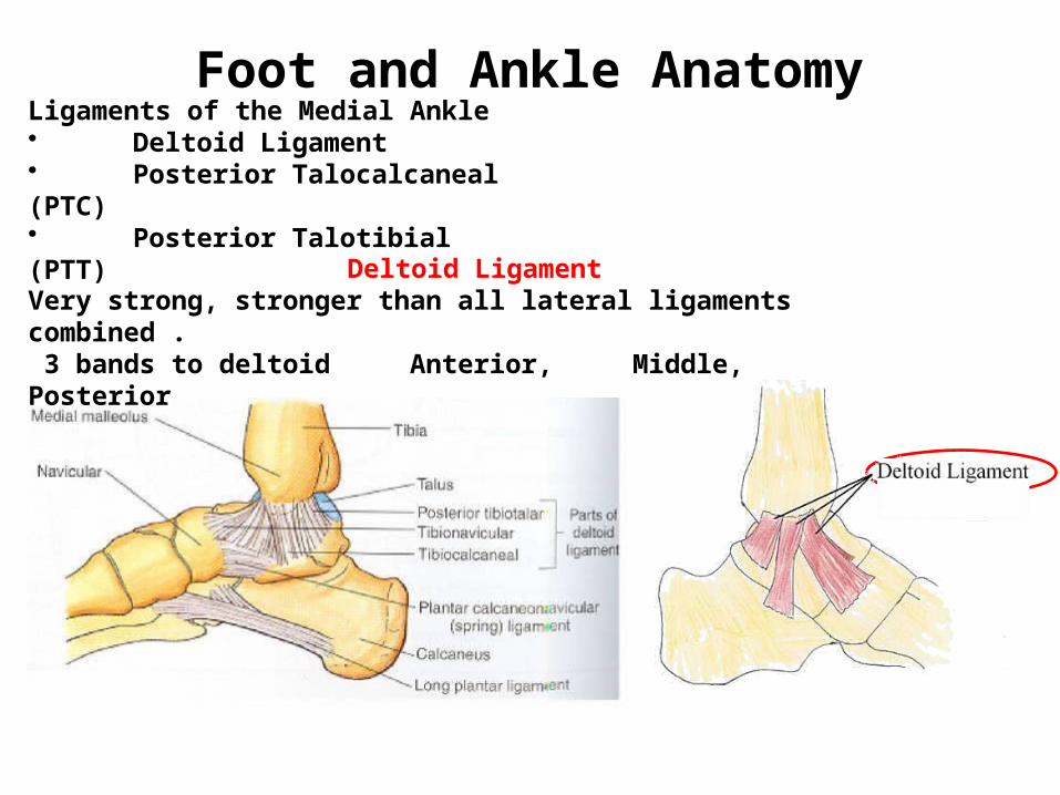
Deltoid Ligament Very strong, stronger than all lateral ligaments combined . 3 bands to deltoid Anterior, Middle, Posterior
Foot and Ankle AnatomyLigaments of the Medial Ankle• Deltoid Ligament • Posterior Talocalcaneal (PTC)• Posterior Talotibial (PTT)
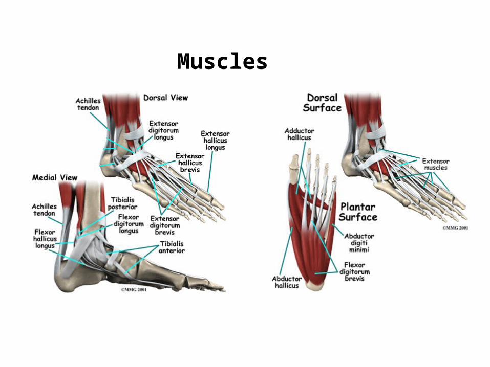
Muscles
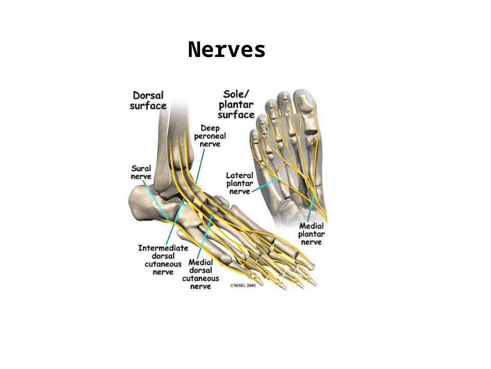
Nerves
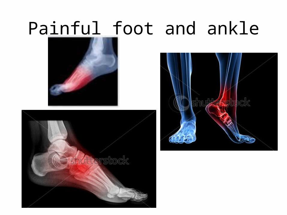
Painful foot and ankle
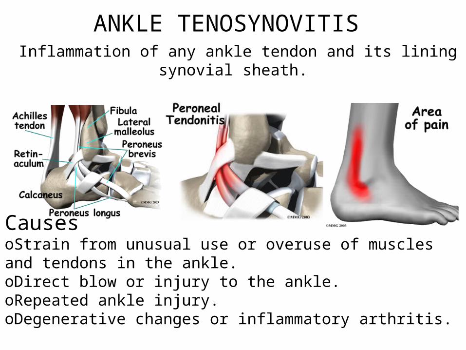
ANKLE TENOSYNOVITIS Inflammation of any ankle tendon and its lining synovial
sheath.
CausesoStrain from unusual use or overuse of muscles and tendons in the ankle. oDirect blow or injury to the ankle. oRepeated ankle injury.oDegenerative changes or inflammatory arthritis.
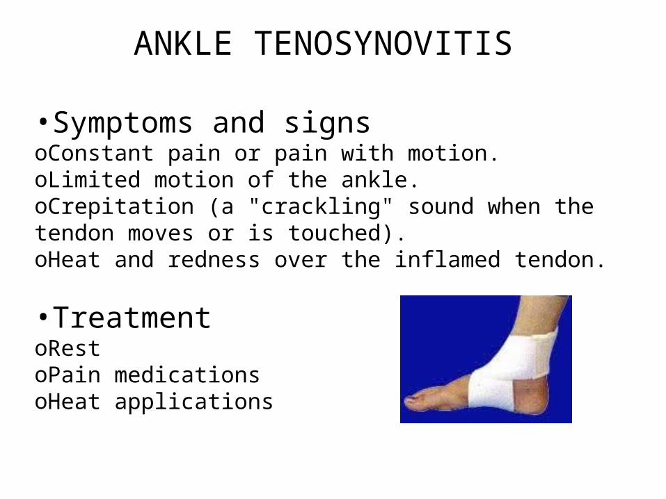
ANKLE TENOSYNOVITIS
•Symptoms and signsoConstant pain or pain with motion. oLimited motion of the ankle. oCrepitation (a "crackling" sound when the tendon moves or is touched). oHeat and redness over the inflamed tendon.
•Treatment oRestoPain medicationsoHeat applications
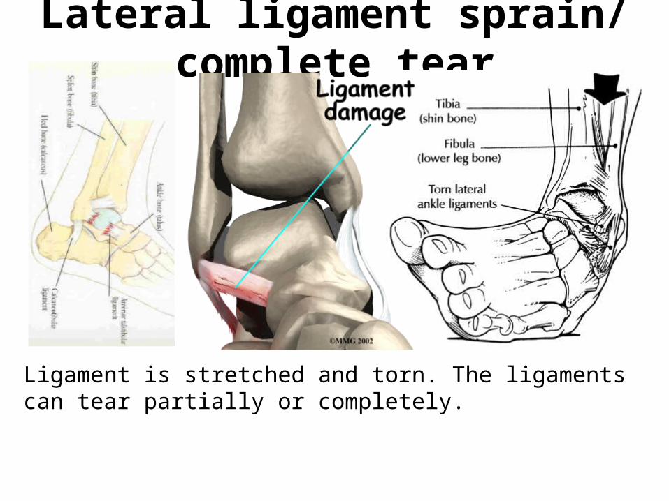
Lateral ligament sprain/ complete tear
Ligament is stretched and torn. The ligaments can tear partially or completely.
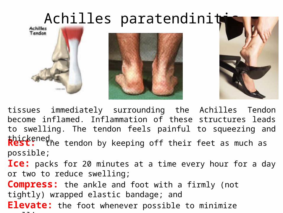
Achilles paratendinitis
Rest: the tendon by keeping off their feet as much as possible; Ice: packs for 20 minutes at a time every hour for a day or two to reduce swelling; Compress: the ankle and foot with a firmly (not tightly) wrapped elastic bandage; and Elevate: the foot whenever possible to minimize swelling.
tissues immediately surrounding the Achilles Tendon become inflamed. Inflammation of these structures leads to swelling. The tendon feels painful to squeezing and thickened.
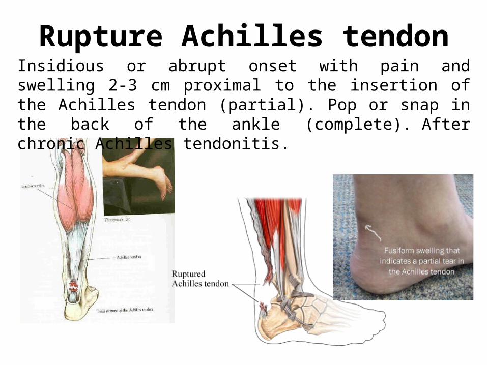
Rupture Achilles tendonInsidious or abrupt onset with pain and swelling 2-3 cm proximal to the insertion of the Achilles tendon (partial). Pop or snap in the back of the ankle (complete). After chronic Achilles tendonitis.
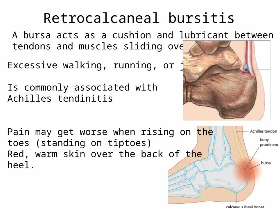
Retrocalcaneal bursitisA bursa acts as a cushion and lubricant between tendons and muscles sliding over bone.
Excessive walking, running, or jumping.
Is commonly associated with Achilles tendinitis
Pain may get worse when rising on the toes (standing on tiptoes) Red, warm skin over the back of the heel.
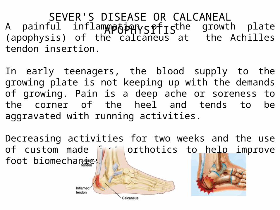
SEVER'S DISEASE OR CALCANEAL APOPHYSITIS
A painful inflammation of the growth plate (apophysis) of the calcaneus at the Achilles tendon insertion.
In early teenagers, the blood supply to the growing plate is not keeping up with the demands of growing. Pain is a deep ache or soreness to the corner of the heel and tends to be aggravated with running activities.
Decreasing activities for two weeks and the use of custom made foot orthotics to help improve foot biomechanics.
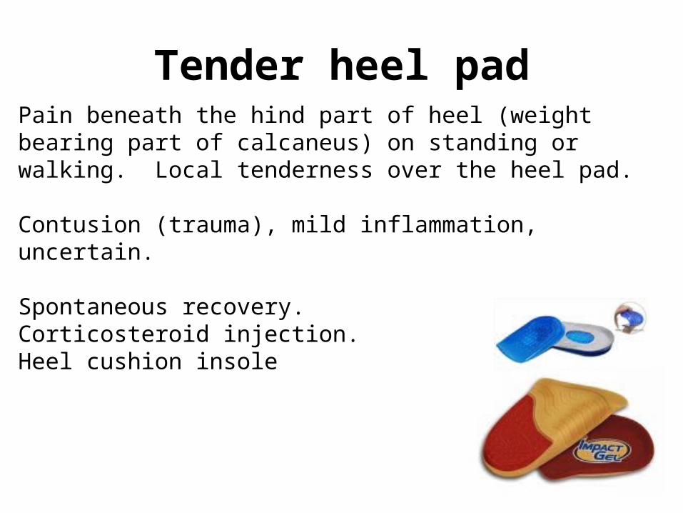
Tender heel padPain beneath the hind part of heel (weight bearing part of calcaneus) on standing or walking. Local tenderness over the heel pad.
Contusion (trauma), mild inflammation, uncertain.
Spontaneous recovery.Corticosteroid injection.Heel cushion insole
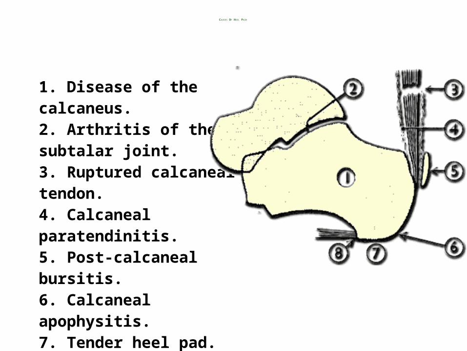
CAUSES OF HEEL PAIN
1. Disease of the calcaneus.2. Arthritis of the subtalar joint.3. Ruptured calcaneal tendon.4. Calcaneal paratendinitis.5. Post-calcaneal bursitis.6. Calcaneal apophysitis.7. Tender heel pad.8. Plantar fascitis.
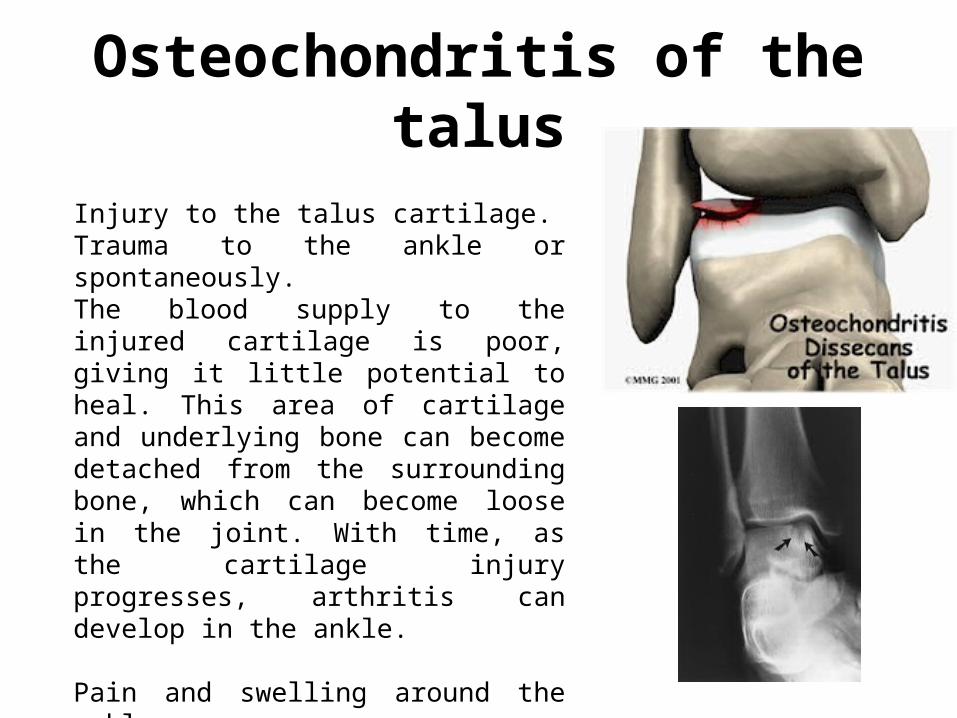
Osteochondritis of the talus
Injury to the talus cartilage.Trauma to the ankle or spontaneously.The blood supply to the injured cartilage is poor, giving it little potential to heal. This area of cartilage and underlying bone can become detached from the surrounding bone, which can become loose in the joint. With time, as the cartilage injury progresses, arthritis can develop in the ankle.
Pain and swelling around the ankle.
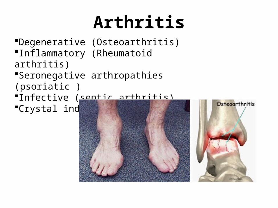
ArthritisDegenerative (Osteoarthritis)Inflammatory (Rheumatoid arthritis)Seronegative arthropathies (psoriatic )Infective (septic arthritis)Crystal induced (Gouty arthritis)
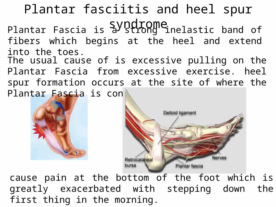
Plantar fasciitis and heel spur syndrome
Plantar Fascia is a strong inelastic band of fibers which begins at the heel and extend into the toes.
The usual cause of is excessive pulling on the Plantar Fascia from excessive exercise. heel spur formation occurs at the site of where the Plantar Fascia is connected to the heel.
cause pain at the bottom of the foot which is greatly exacerbated with stepping down the first thing in the morning.
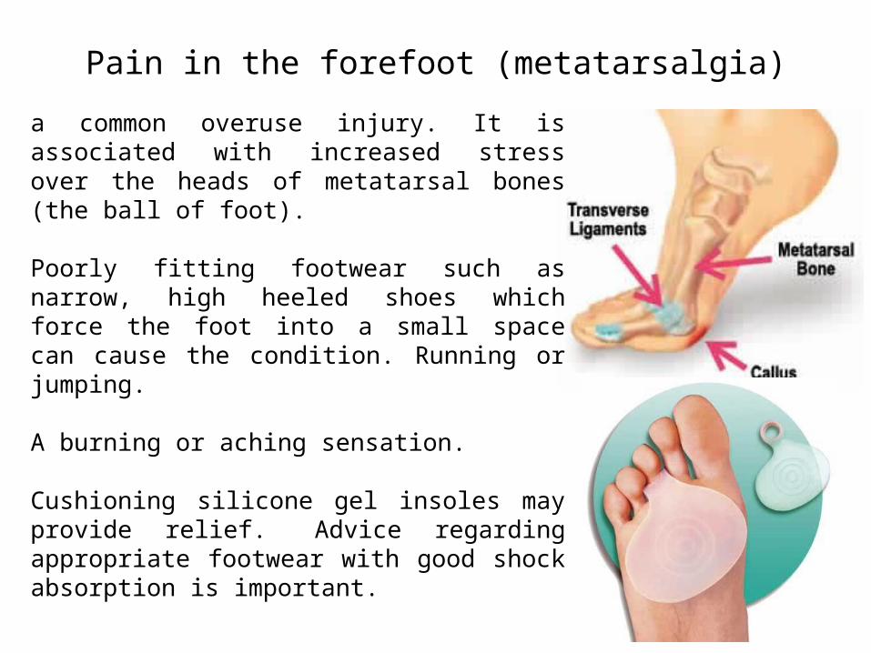
Pain in the forefoot (metatarsalgia)
a common overuse injury. It is associated with increased stress over the heads of metatarsal bones (the ball of foot).
Poorly fitting footwear such as narrow, high heeled shoes which force the foot into a small space can cause the condition. Running or jumping.
A burning or aching sensation.
Cushioning silicone gel insoles may provide relief. Advice regarding appropriate footwear with good shock absorption is important.
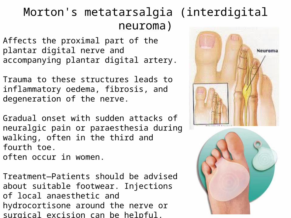
Morton's metatarsalgia (interdigital neuroma)
Affects the proximal part of the plantar digital nerve and accompanying plantar digital artery.
Trauma to these structures leads to inflammatory oedema, fibrosis, and degeneration of the nerve.
Gradual onset with sudden attacks of neuralgic pain or paraesthesia during walking, often in the third and fourth toe. often occur in women.
Treatment—Patients should be advised about suitable footwear. Injections of local anaesthetic and hydrocortisone around the nerve or surgical excision can be helpful.
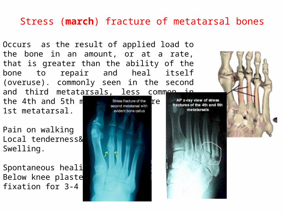
Stress (march) fracture of metatarsal bones
Occurs as the result of applied load to the bone in an amount, or at a rate, that is greater than the ability of the bone to repair and heal itself (overuse). commonly seen in the second and third metatarsals, less common in the 4th and 5th metatarsals. Rare in the 1st metatarsal.
Pain on walkingLocal tenderness& Swelling.
Spontaneous healing.Below knee plasterfixation for 3-4 weeks
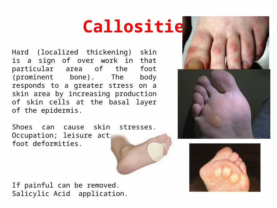
CallositiesHard (localized thickening) skin is a sign of over work in that particular area of the foot (prominent bone). The body responds to a greater stress on a skin area by increasing production of skin cells at the basal layer of the epidermis.
Shoes can cause skin stresses. Occupation; leisure activities and foot deformities.
If painful can be removed.Salicylic Acid application.
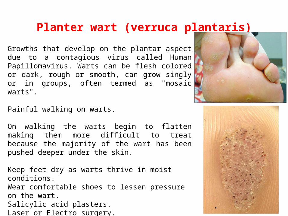
Planter wart (verruca plantaris)
Growths that develop on the plantar aspect due to a contagious virus called Human Papillomavirus. Warts can be flesh colored or dark, rough or smooth, can grow singly or in groups, often termed as "mosaic warts".
Painful walking on warts.
On walking the warts begin to flatten making them more difficult to treat because the majority of the wart has been pushed deeper under the skin.
Keep feet dry as warts thrive in moist conditions.Wear comfortable shoes to lessen pressure on the wart.Salicylic acid plasters.Laser or Electro surgery. Cutting. Freezing methods.
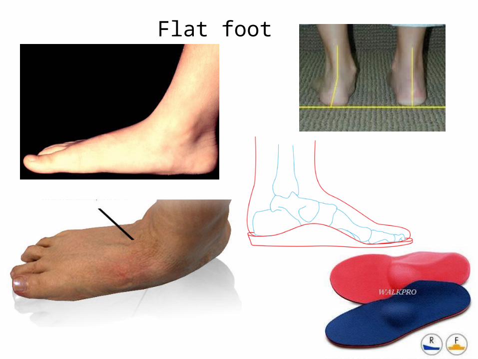
Flat foot
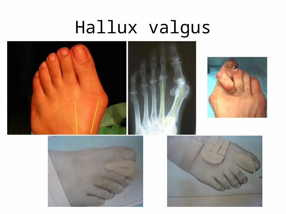
Hallux valgus
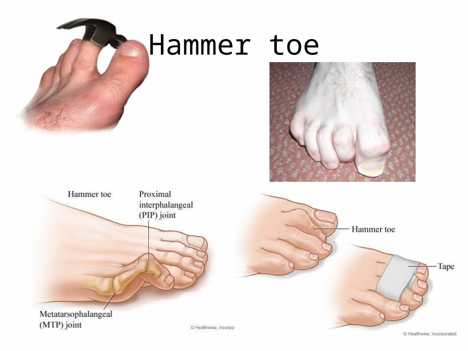
Hammer toe
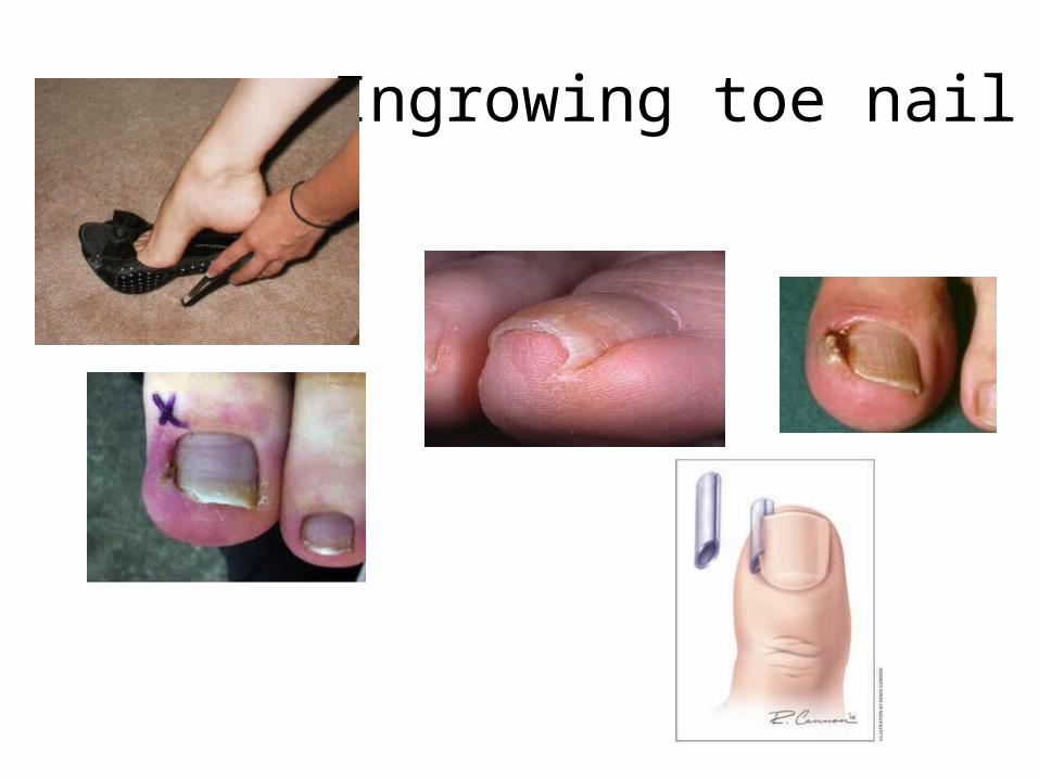
Ingrowing toe nail