Embed Size (px)
Citation preview
FOOT AND ANKLE INJURIES 0278-5919/94 S0.00 + 20
CHRONIC LEG PAIN IN THE ATHLETE
Thomas O. Clanton, MD, and Barry W. Solcher, MD
Chronic leg pain can be a source of great frustration for the athlete. Complaints of leg pain were often labeled as shin splints in the past and accepted as a natural part of athletic participation. Through a combination of factors including technological advancements, development of sports medicine as a subspecialty, and increasing performance demands by athletes and coaches, the term shin splints has become more precisely identifiable. The current goals of the physician caring for an athlete with leg complaints are accurate diagnosis, specific treatment to ensure adequate healing, and the earliest safe return to activity.
SOURCES OF LEG PAIN IN ATHLETES
Sources of leg pain are plentiful. Each tissue, including bone, muscle, tendons, ligaments, fascia, arteries, veins, and skin, can elicit a pain syndrome. The differential diagnosis when evaluating chronic leg pain in athletes should include chronic compartment syndrome, medial tibial stress syndrome, stress fractures, gastrocnemius strain, nerve entrapment syndromes, venous disease, arterial occlusion, fascial herniations, tendonitis, and radiculopathies. These conditions can be distinguished by a carefully directed history and physical examination followed by specialized diagnostic tests.
Several reviews have examined the relative incidence of these conditions and found significant numbers of affected athletes, especially runners. "• "• **•!t
The authors' review of 150 patients with exercise-induced leg pain found 33% of patients with chronic compartment syndromes, 25% with stress fractures, 14% with muscle strains, 13% with medial tibial stress syndrome, 10% with nerve entrapments, 4% with venous pathology, and one patient with spinal stenosis.7
Styf* has reported a similar breakdown in his review of 98 patients with recurrent anterior leg pain. In Styf's series, 25% of patients had anterior chronic
From the Department of Orthopaedic Surgery, University of Texas Medical School at Houston, Texas (TOC, BWS); and Hermann Hospital (TOC), Houston, Texas
C L INKS IN SPORTS MEDICINE
VOLUME 13 • NUMBER 4 • OCTOBER 1W4 743
744 i 1 V s h ' \ ft SOU I Ilk
compartment syndrome, 25% had periostitis of the anterior tibia with an associated medial tibial stress syndrome, 13% had superficial peroneal nerve entrapment, 7% had sequelae of previous fractures, and 5",, had muscle herniations without chronic compartment syndrome or nerve entrapment. In addition, one patient had a herniated nucleus pulposa, one patient had deep venous insufficiency, and three patients had muscular hypertension syndrome.
In another report, Styl"'1 identified periostitis in 40% of patients with anterior leg pain and in 30% to 50% of patients with chronic anterior compartment syndrome. Patients with muscle strains, myositis, tendinitis, compartment syndrome, and stress fractures may have coexisting periostitis. Finally, according to Styf, medial tibial syndrome was the most common cause of posterior medial leg pain. This occurred concurrently in 25% of patients with chronic anterior compartment syndrome.
Chronic Compartment Syndrome
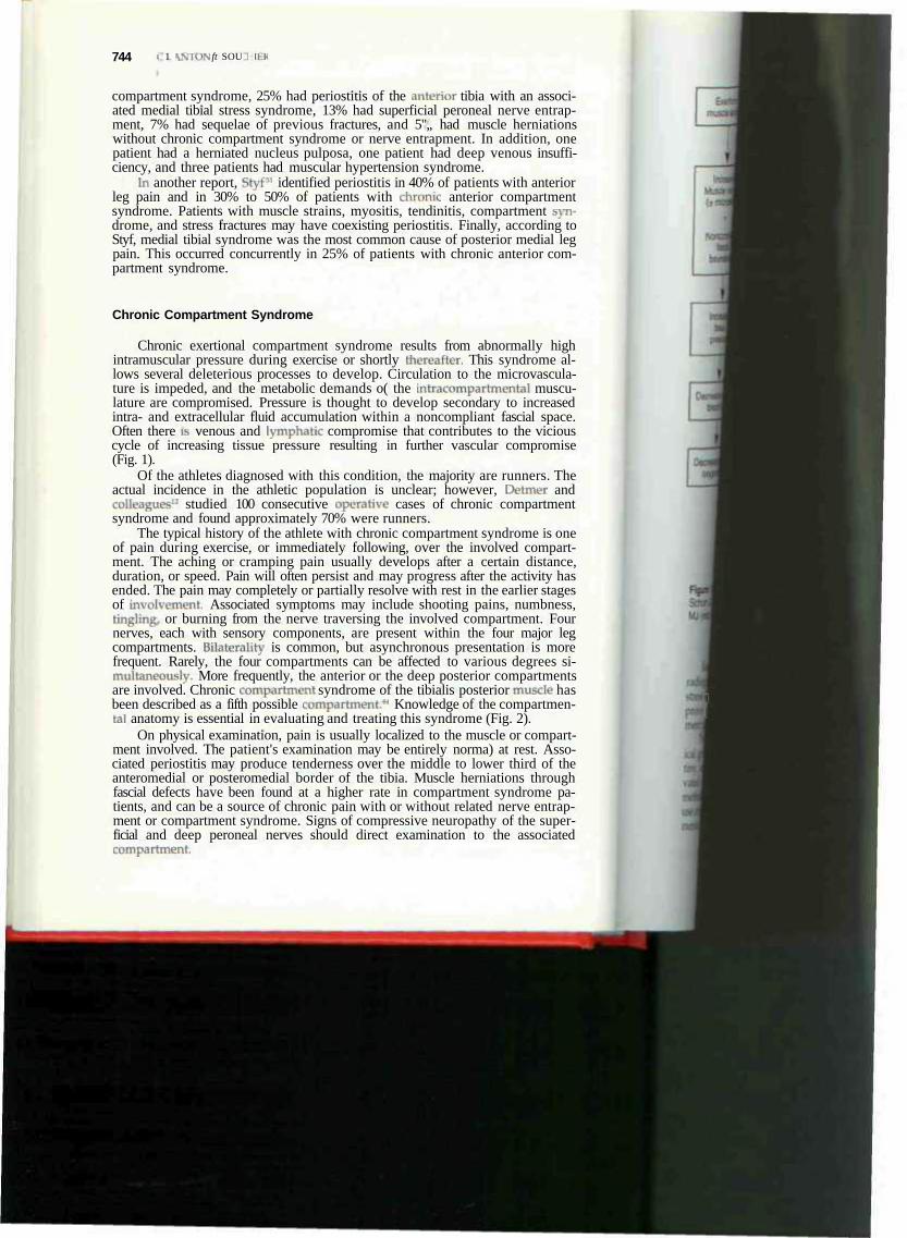
Chronic exertional compartment syndrome results from abnormally high intramuscular pressure during exercise or shortly thereafter. This syndrome allows several deleterious processes to develop. Circulation to the microvascula-ture is impeded, and the metabolic demands o( the intracompartmental musculature are compromised. Pressure is thought to develop secondary to increased intra- and extracellular fluid accumulation within a noncompliant fascial space. Often there is venous and lymphatic compromise that contributes to the vicious cycle of increasing tissue pressure resulting in further vascular compromise (Fig. 1).
Of the athletes diagnosed with this condition, the majority are runners. The actual incidence in the athletic population is unclear; however, Detmer and colleagues12 studied 100 consecutive operative cases of chronic compartment syndrome and found approximately 70% were runners.
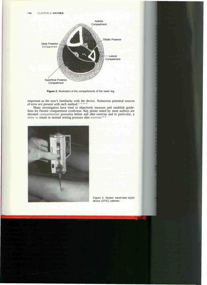
The typical history of the athlete with chronic compartment syndrome is one of pain during exercise, or immediately following, over the involved compartment. The aching or cramping pain usually develops after a certain distance, duration, or speed. Pain will often persist and may progress after the activity has ended. The pain may completely or partially resolve with rest in the earlier stages of involvement. Associated symptoms may include shooting pains, numbness, tingling, or burning from the nerve traversing the involved compartment. Four nerves, each with sensory components, are present within the four major leg compartments. Bilaterality is common, but asynchronous presentation is more frequent. Rarely, the four compartments can be affected to various degrees simultaneously- More frequently, the anterior or the deep posterior compartments are involved. Chronic compartment syndrome of the tibialis posterior muscle has been described as a fifth possible compartment.*4 Knowledge of the compartmen-tal anatomy is essential in evaluating and treating this syndrome (Fig. 2).
On physical examination, pain is usually localized to the muscle or compartment involved. The patient's examination may be entirely norma) at rest. Associated periostitis may produce tenderness over the middle to lower third of the anteromedial or posteromedial border of the tibia. Muscle herniations through fascial defects have been found at a higher rate in compartment syndrome patients, and can be a source of chronic pain with or without related nerve entrapment or compartment syndrome. Signs of compressive neuropathy of the superficial and deep peroneal nerves should direct examination to the associated compartment.
t
I
Exertional muscle activity
y {
CHRONIC LEG TAIN IN THE ATHLETE 745
Increased ; ( incased intra- and extracellular fluid) Muscle volume , ~""" ( i microtears) •• '
4
Nonce mpliant fascial
boundaries
1 Increased
bssue pressure
I
I
4 ^ -^^^
?JF Increased capillary
permeability
/
Increased rrptabortc
f
Decreased tissue blood flow
\ f
Decreased tissue oxygenation
\ byproducts
s
1
Ischemia
n w Pain Impaired Impaired
muscle nerve function function
Figure 1. Row chart depicting how compartment syndrome develops. [From Clanton TO, Schon LC: Athletic injuries to the soft tissues of the loot and ankle. In Mann HA, Coughlin MJ (eds): Surgery ot the Foot and Ankle, ed 6. St. Louis. CV Mosby. 1993; with permission.)
Rad io log ic eva lua t i on is i m p o r t a n t to ru le o u t associated c o n d i t i o n s . P la in rad iographs , bone scan, and c o m p u t e d t o m o g r a p h y (CT) scan s h o u l d revea l stress f ractures, per ios t i t i s , o r occul t bone t u m o r s as causes. MR i m a g i n g m a y p r o v e t o be an ef fect ive non invas i ve m e t h o d o f d e t e r m i n i n g a b n o r m a l c o m p a r t -m e n t pressures.
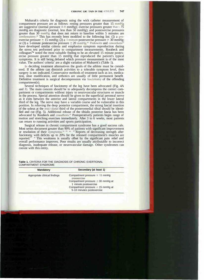
The d iagnos is o f ch ron ic c o m p a r t m e n t s y n d r o m e i s p r i m a r i l y m a d e on c l i n ical g r o u n d s ; howeve r , in a hea l th care reform e n v i r o n m e n t of increas ing sc ru -t i nv , object ive d o c u m e n t a t i o n has become m o r e c ruc ia l . D o c u m e n t a t i o n o f elevated c o m p a r t m e n t pressure has become inc reas ing ly ach ievab le . Several me thods are ava i lab le to ob ta in accurate i n t r a c o m p a r t m e n t a l pressures i n c l u d i n g use of a w i c k catheter, a s l i t catheter, a n d the so l id state t ransducer i n t r acompa r t men ta l (STIC) catheter (Fig. 3). The t ype of dev i ce one uses is no t nea r l y as
assssssssssMasH as^asssssH k P U T . wm.* /u r. '
^ p i > ^ V ^ - r - " - , • -
: * 3 P I , "u__ __
. - a - 1-aVaaa. ^ ' " i - .
- - , f a U s f P " - i - • P '
' ^ " ^ P 1 ^ .J "^, 1 'iZTp 1 M ™ _ r > . * JB JT-1 — • • • • I I n B =- - 1 - ,i _ _ -J . ,
1 sssesl • ea -
7 4 6 CI.ANTON & SOUTHER
Deep Posterior Compartment
Superficial Posterior Compartment
Anterior Compartment
Tibialis Posterior
Lateral Compartment
Figure 2. Illustration ot the compartments of the lower leg.
important as the user's familiarity with the device. Numerous potential sources of error are present with each method
Many investigators have tried to objectively measure and establish guidelines for chronic compartment syndrome. Key points noted by most authors are elevated compartmenta! pressures before and after exercise and in particular, a delay in return to normal resting pressure after exercise,414<i
Figure 3. Stryker hand-held device (STIC) catheter.
CHRONIC LBC TAIN IN THE ATHI III 747
Mubarak's criteria for diagnosis using the wick catheter measurement of compartment pressure are as follows: resting pressures greater than 15 mmHg are suggestive (normal pressure 0-8 mmHg); exercise pressures greater thai I 75 mmHg are diagnostic (normal, less than 50 mmHg); and postexercise pressures greater than 30 mmHg that does not return to baseline within 5 minutes are confirmatory.4" This has recently been modified to the following list: (]) a pre-exercise pressure > 15 mmHg; (2) a 1-minute postexercise pressure 2: 30 mmHg; or (3) a 5-minute postexercise pressure > 20 mmHi;.'1 Pedowit/ and coworkers42
have developed similar criteria and emphasize symptom reproduction during the stress test performed prior to compartment measurements. Rorabeck and colleagues4* noted the most valuable finding to be an elevated 15 minute postexercise pressure greater than 15 mmHg that reproduced the patient's typical symptoms. It is still being debated which pressure measurement is of the most value. The authors' criteria7 are a slight variation of Mubarek's (Table 1).
In deciding treatment alternatives the goals of the athlete must be considered. If the athlete can diminish activities to a tolerable symptom level, then surgery is not indicated. Conservative methods of treatment such as ice, medication, shoe modification, and orthotics are usually of little permanent benefit Definitive treatment is surgical decompression via fasciotomy of the offending compartment(s).
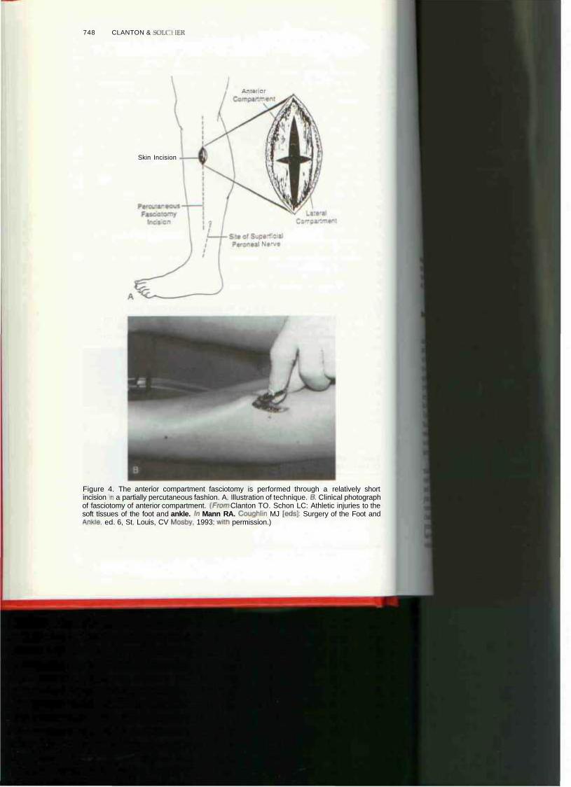
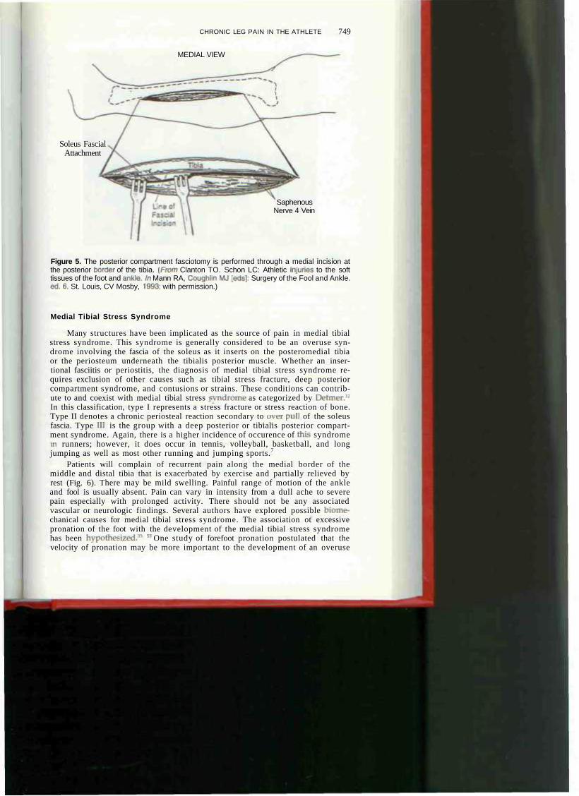
Several techniques of fasciotomy of the leg have been advocated (Fig. 4A and B). The main concern should be to adequately decompress the correct compartment or compartments without injury to neurovascular structures or muscle in the process. Special attention should be given to the superficial peroneal nerve as it exits between the anterior and lateral compartments in the lower lateral third of the leg. The nerve may have a variable course and be vulnerable in this position. In relieving the deep posterior compartment, the strong fascial insertion of the soleus at the mid/distal third of the posteromedial tibial should be identified and cut (Fig. 5). Additional release of the tibialis posterior fascia has been advocated by Rorabeck and coauthors.'" Postoperatively patients begin range of motion and stretching exercises immediately. After 3 to 6 weeks, most patients can return to running activities and sports participation.
Surgical release in chronic compartment syndrome has a good success rale. Most series document greater than 90% of patients with significant improvement or resolution of their symptoms.1*-*5 ** E Reports of decreasing strength after fasciotomy with deficits up to 20% for the released compartment's muscles are reported.17 * This weakness is usually offset by the significant pain relief and overall performance improves. Poor results are usually attributable to incorrect diagnosis, inadequate release, or neurovascular damage. Other syndromes can coexist with this entity.
Table 1. CRITERIA FOR THE DIAGNOSIS OF CHRONIC EXERTIONAL COMPARTMENT SYNDROME
Mandatory Secondary (at least 1)
Appropriate clinical findings Compartment pressure 2 15 mmHg preexercise
Compartment pressure > 30 mmHg at 1 minute postexercise
Compartment pressure 2 15 mmHg at 5-10 minutes postexercise
748 CLANTON & SOU. I UK
A.-:;- •:•
Skin Incision
Figure 4. The anterior compartment fasciotomy is performed through a relatively short incision in a partially percutaneous fashion. A. Illustration of technique. B, Clinical photograph of fasciotomy of anterior compartment. (From Clanton TO. Schon LC: Athletic injuries to the soft tissues of the foot and ankle. In Mann RA. Coughlin MJ [eds): Surgery of the Foot and Ankle, ed. 6, St. Louis, CV Mosby. 1993: with permission.)
CHRONIC LEG PAIN IN THE ATHLETE
MEDIAL VIEW
749
Soleus Fascial Attachment
Saphenous Nerve 4 Vein
Figure 5. The posterior compartment fasciotomy is performed through a medial incision at the postenor tjorder of the tibia. [From Clanton TO. Schon LC: Athletic injunes to the soft tissues of the foot and ankle. In Mann RA, Coughlin MJ [eds): Surgery of the Fool and Ankle. ed. 6. St. Louis, CV Mosby, 1993; with permission.)
Medial Tibial Stress Syndrome
Many structures have been implicated as the source of pain in medial tibial stress syndrome. This syndrome is generally considered to be an overuse syndrome involving the fascia of the soleus as it inserts on the posteromedial tibia or the periosteum underneath the tibialis posterior muscle. Whether an inser-tional fasciitis or periostitis, the diagnosis of medial tibial stress syndrome requires exclusion of other causes such as tibial stress fracture, deep posterior compartment syndrome, and contusions or strains. These conditions can contribute to and coexist with medial tibial stress -vndrome as categorized by Detmer.12
In this classification, type I represents a stress fracture or stress reaction of bone. Type II denotes a chronic periosteal reaction secondary to over pull of the soleus fascia. Type HI is the group with a deep posterior or tibialis posterior compartment syndrome. Again, there is a higher incidence of occurence of thus syndrome in runners; however, it does occur in tennis, volleyball, basketball, and long jumping as well as most other running and jumping sports.7
Patients will complain of recurrent pain along the medial border of the middle and distal tibia that is exacerbated by exercise and partially relieved by rest (Fig. 6). There may be mild swelling. Painful range of motion of the ankle and fool is usually absent. Pain can vary in intensity from a dull ache to severe pain especially with prolonged activity. There should not be any associated vascular or neurologic findings. Several authors have explored possible biome-chanical causes for medial tibial stress syndrome. The association ot excessive pronation of the foot with the development of the medial tibial stress syndrome has been hypothesized." " One study of forefoot pronation postulated that the velocity of pronation may be more important to the development of an overuse

750 CLANTON & SOLCHES
Figure 6. Distance runner depicting location of pain along medial border of the middle and distal tibia in medial tibial stress syndrome.
m
m syndrome than the actual amount of pronation measured.'4 In another study comparing normal male athletes with 35 affected male athletes, excessive angular displacement in the subtalar joint or the Achilles tendon angle was more common in the symptomatic group."
Routine radiographs of the leg often are negative, and should exclude other sources of tibial pain. The bone scan can be a valuable aid in differentiating this syndrome. Although it may be normal, it is more likely to show a moderate increase of linear activity along the posteromedial border of the tibia involving as much as one third of the length of the tibia.
Although Detmer subclassifies medial tibial stress syndrome, the authors believe this is only a clinical stratagem, and more specific testing should clarify the diagnosis. Radiographs and bone scintigraphy help differentiate stress fractures. Compartment pressure measurements in this syndrome assist in identifying the presence of a deep posterior or posterior tibialis compartment syndrome. Furthermore, measurements are important to rule out concurrent anterior compartment syndrome. Following appropriate testing of athletes diagnosed clinically with medial tibial stress syndrome, one should be able to distinguish those who fall into each of Detmer's categories. The authors believe that the type II subgroup related to pull of the soleus fascial sling at the posteromedial border of the tibia is the essential lesion in medial tibial stress syndrome.
The majority of patients with medial tibial stress syndrome can be treated conservatively with success. This plan relies heavily on resl followed by gradual resumption of activity. Rest can be considered a relative decrease in the offending activity to a level which is comfortable. Other treatment methods such as stretching, use of moist heat, bracing, local steroid injection, and taping are of temporary symptomatic value and do not change the overall course. They may be useful to buy time before rest can commence. Custom orthotic devices should be pre-
(8
l>ult
ai
mt< inv
wh
CHRONIC LEC PAIN IN THE ATHLETE 751
scribed to assist athletes with excessive pronation or abnormal subtalar mobility. With a short period of rest or cross braining, runners can usually return to a restricted program on soft surfaces. Gradual increase in distance and speed is emphasized to prevent recurrence.
Surgical treatment is reserved for patients in whom conservative modalities fail. When fasciotomy is required, a posteromedial lasciotomy with release ot the medial soleus fascial bridge and the deep posterior compartment fascia is performed. Favorable results can be obtained with this release and proper rehabilitation.
Stress Fractures of the Tibia and Fibula
Most stress or fatigue fractures in athletes are associated with running/4 • ** The tibia is the most common bone involved.1 :4 • The injur)' develops from abnormal repetitive loads on the bone that cause an imbalance of bony resorption over formation. The bone structurally weakens to the point of failure and pain develops. Other sports such as ballet, basketball, soccer, and aerobics have been reported to produce a significant incidence of stress fractures.13 Dancers and basketball players are particularly vulnerable to anterior midtibia stress fractures that have been shown to heal poorly.*- *• *' Associated menstrual irregularities or amenorrhoea combined with smaller bones has been postulated as being responsible for the increased risk of stress fractures in women."
Classically, the athlete reports a recent increase or change in their workout. This is reflected as an increased duration of running, different shoes, altered running surface, or change of speed. There is usually a gradual onsel of pain over a period of several weeks. The pain begins during or following stressful activity, and then progresses to activities of daily living.
The clinical examination reveals a well-localized area of tenderness over a confined area on the tibia or fibula There may be increased warmth, swelling, or erythema present. One-legged hopping and percussion of the bone often elicit pain. Ultrasound treatment of the lesion frequently seems to aggravate the symptoms.
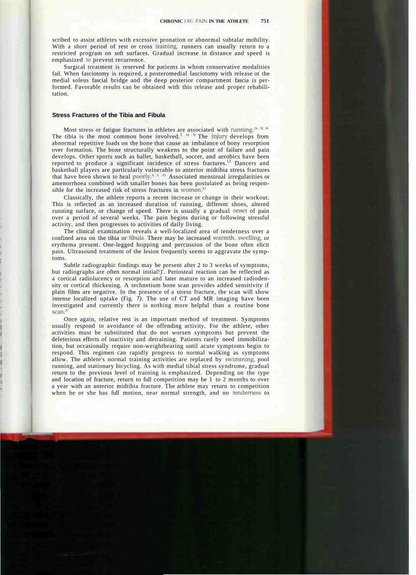
Subtle radiographic findings may be present after 2 to 3 weeks of symptoms, but radiographs are often normal initial!)'. Periosteal reaction can be reflected as a cortical radiolucency or resorption and later mature to an increased radioden-sity or cortical thickening. A technetium bone scan provides added sensitivity if plain films are negative. In the presence of a stress fracture, the scan will show intense localized uptake (Fig. 7). The use of CT and MR imaging have been investigated and currently there is nothing more helpful than a routine bone scan.27
Once again, relative rest is an important method of treatment. Symptoms usually respond to avoidance of the offending activity. For the athlete, other activities must be substituted that do not worsen symptoms but prevent the deleterious effects of inactivity and detraining. Patients rarely need immobilization, but occasionally require non-weightbearing until acute symptoms begin to respond. This regimen can rapidly progress to normal walking as symptoms allow. The athlete's normal training activities are replaced by swimming, pool running, and stationary bicycling. As with medial tibial stress syndrome, gradual return to the previous level of training is emphasized. Depending on the type and location of fracture, return to full competition may be 1 to 2 months to over a year with an anterior midtibia fracture. The athlete may return to competition when he or she has full motion, near normal strength, and no tenderness to
752 . ION & SOLCHER
I
Figure 7. The bone scan in a stress fracture of the libia stress fracture of the tibia shows a well-localized area of increased uptake.
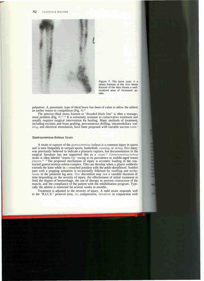
palpation. A pneumatic type of tibial brace has been of value to allow the athlete an earlier return to competition (Fig. 8).*
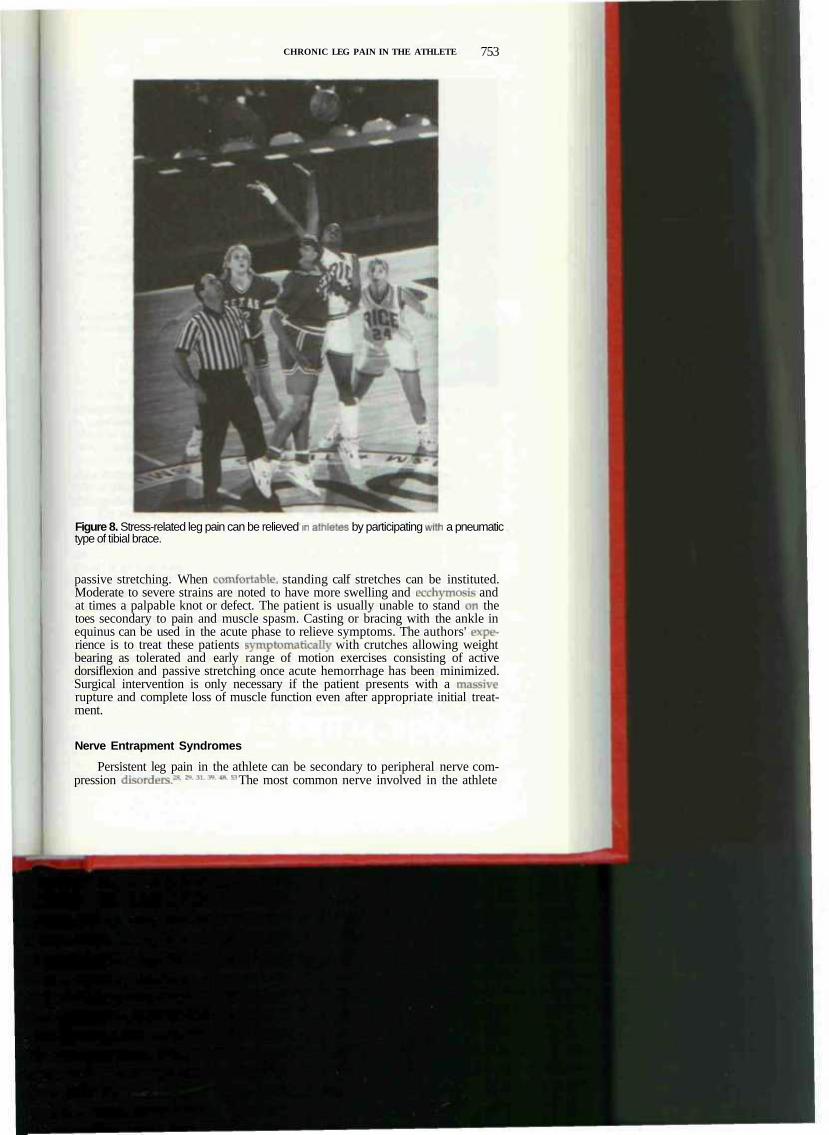
The anterior tibial stress fracture or "dreaded black line" is often a management problem (Fig. 9).1"4' It is extremely resistant to conservative treatment and usually requires surgical intervention for healing. Many methods of treatment, including excision and bone grafting, percutaneous drilling, intramedullary rod-ding, and electrical stimulation, have been proposed with variable success rates.2
Gastrocnemius-Soleus Strain
A strain or rupture of the gastrocnemius /soleus is a common injury in sports and is seen frequently in racquet sports, basketball, running, or skiing, this injury was previously believed to indicate a plantaris rupture, but documentation in the surgical literature has not supported this as a cause.*'' Gastrocnemius-soleus strain is often labeled "tennis leg" owing to its prevalence in middle-aged tennis players.1" p The proposed mechanism of injury is eccentric loading of the contracted gastrocnemius-soleus complex. This can develop when a player suddenly extends the knee while in a crouched position with the ankle dorsiflexed. Sudden pain with a popping sensation is occasionally followed by swelling and ecchy-mosis in the posterior leg area. ITiis discomfort may lasl a variable duration of time depending on the severity of injury, the effectiveness of initial treatment to limit the degree of hemorrhage, the use of therapy to prevent contracture of the muscle, and the compliance of the patient with the rehabilitation program. Typically the athlete is restricted for several weeks to months.
Treatment is adjusted to the severity of injury. A mild strain responds well to the "R.I.C.E." protocol (rest, fee, compression, elevation) in conjunction with
CHRONIC LEG PAIN IN THE ATHLETE 753
Figure 8. Stress-related leg pain can be relieved in athletes by participating with a pneumatic type of tibial brace.
passive stretching. When comfortable, standing calf stretches can be instituted. Moderate to severe strains are noted to have more swelling and ecchymosis and at times a palpable knot or defect. The patient is usually unable to stand tin the toes secondary to pain and muscle spasm. Casting or bracing with the ankle in equinus can be used in the acute phase to relieve symptoms. The authors' experience is to treat these patients symptomatically with crutches allowing weight bearing as tolerated and early range of motion exercises consisting of active dorsiflexion and passive stretching once acute hemorrhage has been minimized. Surgical intervention is only necessary if the patient presents with a maaaive rupture and complete loss of muscle function even after appropriate initial treatment.
Nerve Entrapment Syndromes
Persistent leg pain in the athlete can be secondary to peripheral nerve compression disorders.2* w " - w • * ° The most common nerve involved in the athlete
754 CLANTON * SCH.CHER
Popliteal Vain
Popliteal Artery
Medial Head of Gastrocnemius
Sciatic Nerve
Plantaris
Lateral Head of Gastrocnemius
Figure 9. The transverse anterior tibial stress fracture is an ominous finding.
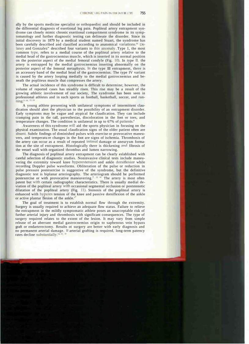
Figure 10. Diagram depicting the medial course of the popliteal artery relative to the medial head of the gastrocnemius muscle.
is the superficial peroneal nerve. Other entrapment syndromes include a high tarsal tunnel with entrapment of the posterior tibial nerve, entrapment of the common peroneal nerve at the neck of the fibula, saphenous nerve entrapment as it pierces Hunter's canal, and sural nerve entrapment in the posterior calf
Patients typically have a subacute onset of a neuritic type pain such as burning, tingling, or radiation. Careful investigation of the involved area can assist in locating the affected nerve. Palpating tenderness of the nerve at the zone of compression is a characteristic of the physical examination. Eliciting a Tinel's sign or pain with prolonged compression of the area can be diagnostic. The workup must include evaluation for an associated chronic compartment syndrome or a more proximal nerve entrapment (double crush syndrome). Electromyographic and nerve conduction studies should be considered along with selected nerve blocks.
Conservative treatment is initiated in the patients with recent onset of symptoms. These modalities include rest, injections, massage, thermogesic or counter-irritant creams, nonsteroidal anti-inflammatory medicines, amirriptyline, and shoe modification. U these fail or the patient has established symptoms, surgical treatment is warranted. Release of the nerve with localized fasciotomy is usually necessary for improvement.
Popliteal Artery Entrapment Syndrome
Popliteal artery entrapment syndrome is a relatively rare entity that causes calf pain in young athletes This syndrome may be seen and evaluated occasion-
no i
then
dUi enh or i
I hei lurthei surga
graft i no j rates(
( HRQNK LH.I 'AIN l \ 11II All II I 1! 755
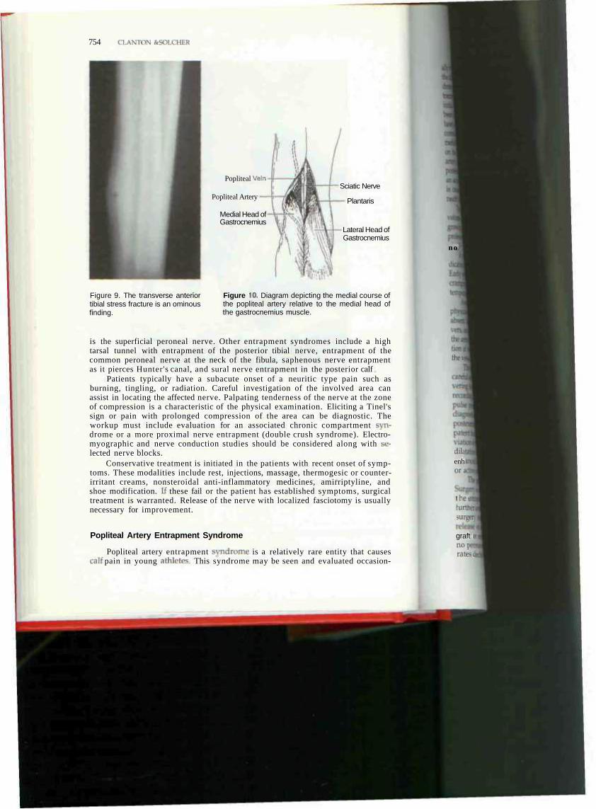
ally by the sports medicine specialist or orthopaedist and should be included in the differential diagnosis of exertional leg pain. Popliteal artery entrapment syndrome can closely mimic chronic exertional compartment syndrome in its symptomatology and further diagnostic testing can delineate the disorder. Since its initial discovery in 1879 by a medical student named Stuart, the syndrome has been carefully described and classified according to anatomical variations.v De-laney and Gonzalez" described four variants to this anomaly Type 1, the most common type, refers to a medial course of the popliteal artery relative to the medial head of the gastrocnemius muscle, which is inserted in its normal position on the posterior aspect of the medial femoral condyle (Fig. 10). In type II, the artery is entrapped by the medial gastrocnemius inserting abnormally on the posterior aspect of the femoral metaphysis. In the type III entrapment, there is an accessory band of the medial head of the gastrocnemius. The type IV variant is caused by the artery looping medially to the medial gastrocnemius and beneath the popliteus muscle that compresses the artery.
The actual incidence of this syndrome is difficult to determine; however, the volume of reported cases has steadily risen. This rise may be a result of the growing athletic involvement of our society, The syndrome has been seen in professional athletes and in such sports as football, basketball, soccer, and run-ning."4 , ' - r '
A young athlete presenting with unilateral symptoms of intermittent claudication should alert the physician to the possibility of an entrapment disorder. Early symptoms may be vague and atypical for claudication. They can include cramping pain in the calf, paresthesias, discoloration in the foot or toes, and temperature changes. The condition is unilateral in up to 67% of patients.*
Awareness of this syndrome will aid the sports physician in focusing on the physical examination. The usual claudication signs of the older patient often are absent. Subtle findings of diminished pulses with exercise or provocative maneuvers, and temperatu re changes in the foot are signs of ischemia. Thrombosis of the artery can occur as a result of repeated intimal damage or aneurysm formation at the site of entrapment. Histologically there is thickening ami fibrosis of the vessel wall with organized thrombus and lumen narrowing.
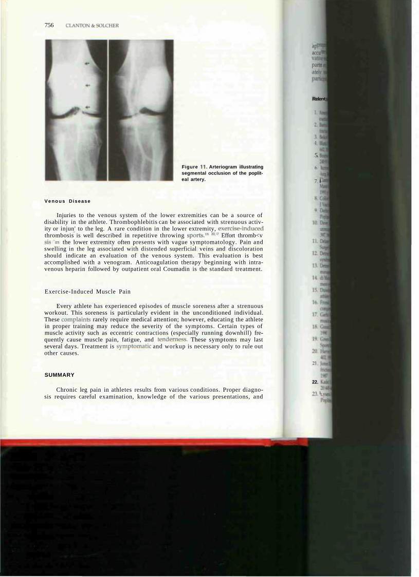
The diagnosis of popliteal artery entrapment can be clearly eslablished with careful selection of diagnostic studies. Noninvasive clinical tests include maneuvering the extremity toward knee hyperextension and ankle dorsiflexion while recording Doppler pulse waveforms. Obliteration of the pulse or reduction in pulse pressure postexercise is suggestive of the syndrome, but the definitive diagnostic test is biplanar arteriography. The arteriogram should be performed postexercise or with provocative maneuvering.1 * 4" The artery is most often patent but with certain radiographic characteristics. There is usually medial deviation of the popliteal artery with occasional segmental occlusion or poststenotic dilatation of the popliteal artery (Fig. 11). Stenosis of the popliteal artery is enhanced with hyperex tension of the knee and passive dorsiflexion of the ankle or active plantar flexion of the ankle.'7
The goal of treatment is to establish normal flow through the extremity. Surgery is usually required to achieve an adequate flow status. Failure to relieve the entrapment in the mildly symptomatic athlete poses an unacceptable risk of further arterial injury and thrombosis with significant consequences. The type of surgery required relates to the extent of the lesion. It may vary from simple release of an aberrant medial gastrocnemius origin to saphenous vein bypass graft or endarterectomy. Results ot surgery are better with early diagnosis and no permanent arterial damage. If arterial grafting is required, long-term patency rates decline substantially.14'"' •
756 CLANTONfcSOLCHER
Figure 11 . Arteriogram illustrating segmental occlusion of the popliteal artery.
V e n o u s Disease
Injuries to the venous system of the lower extremities can be a source of disability in the athlete. Thrombophlebitis can be associated with strenuous activity or injun' to the leg. A rare condition in the lower extremity, exercise-induced thrombosis is well described in repetitive throwing sports." * p Effort thromb<v Bis m the lower extremity often presents with vague symptomatology. Pain and swelling in the leg associated with distended superficial veins and discoloration should indicate an evaluation of the venous system. This evaluation is best accomplished with a venogram. Anticoagulation therapy beginning with intravenous heparin followed by outpatient oral Coumadin is the standard treatment.
Exercise-Induced Muscle Pain
Every athlete has experienced episodes of muscle soreness after a strenuous workout. This soreness is particularly evident in the unconditioned individual. These complaint* rarely require medical attention; however, educating the athlete in proper training may reduce the severity of the symptoms. Certain types of muscle activity such as eccentric contractions (especially running downhill) frequently cause muscle pain, fatigue, and tenderness. These symptoms may last several days. Treatment is symptomatic and workup is necessary only to rule out other causes.
SUMMARY
Chronic leg pain in athletes results from various conditions. Proper diagnosis requires careful examination, knowledge of the various presentations, and
aj> add
partrti •Itl'lv
Relent
i
5.
h. I
i
7. i
] |
22.
T5 1 .
CHRONIC LEG PAIN IN THE ATI ILL IT 757
appropriate use of diagnostic studies. These conditions can often coexist, making accurate diagnosis difficult, Most exercise-induced leg pain responds to conservative nonsurgical treatment; however, certain syndromes such as chronic compartment syndrome or popliteal artery entrapment syndrome are more appropriately treated surgically to improve the athlete's ability to return to full participation.
References
1. Amendola A, Rorabeck CH, Bellett D, et at; The use of magnetic resonance imaging in exertional compartment syndromes. Am ) Sports Med 18:29->4, 19411
2 Barrick EF. Jackson CB: Prophylactic intramedullary nailing- of the tibia for stress fracture in a professional .llhlete. I Orthop Trauma 6:241-244, 1993
3. Belkin SC: Stress fractures in athletes Orthop Clin North Am 11:735, 1980 4. Blank S: Transverse tibial stress fractures. A Special Problem. Am ) Sports Med 15:597-
602. 1987 5. Bourne RB, Rorabeck CH: Compartment svndromes of the lower leg Clin Orthop
240:97-104,1989 6. Burrows HJ: Fatigue infraction of the middle of the tibia in ballet dancers. J Bone Joinl
Surg 38B:83-94.1956 7. Clanton TO, Schon LC: Athletic injuries to the soft tissues of the foot and ankle. In
Mann RA, Coughlin MJ (eds): Surgery of the Foot and Ankle, ed 6. St Louis, CV Mosby. 1993, p 1105
8. Collins PS, McDonald FT, Lim RC: Popliteal artery entrapment: An evolving svndrome J Vase Surg 10:484-490, 1989
9. Darling RS, Buckley C], Abbott WM, et at Intermittent claudication in young athletes: Popliteal artery entrapment syndrome. J Trauma 14:343-552, 1974
10. Davey JR, Rorabeck CH, Fowler PJ: The tibialis posterior muscle compartment. An unrecognized cause of exertional compartment syndrome. Am J Sports Med 12:391-397, 1984
11. Deiancv TA, Gonzalez LL: Occlusion of popliteal artery due to muscular entrapmait. Surgery 69^7-101,1971
12. Detmer DE: Chronic shin splints: Classification and management of medial tibial stress syndrome. Sports Med 3:436-446, 1986
13. Detmer DE, Sharpe K, Sufit RL, et al: Chronic compartment svndrome: Diagnosis, management, and outcomes. Am J Sports Med 13:162-170. 1985
14. di Marzo L, Cavallaro A, Sciacca V, et al: Surgical treatment of popliteal artery entrapment syndrome: A ten-year experience. Eur j Vase Surg 5:51-64, 19° I
15. Dim elms I'l. Kelbel JM, Jardon OM, et al Popliteal artery entrapment in a high school athlete: A case report. Am J Sports Med 15:371-373,1987
16. Fronek J, Mubarak 5, Hargens A, el al: Management of chronic exertional anterior iipartment syndrome of the lower extremity Clin Orthop 220:217-227. 1987
17. Carfin SR, Tipton CM, Mubarak SJ, el al: "Ihe role of fascia in the maintenance of muscle tension and pressure. J Appl Physiol 51:317-320,1981
18. Conard DA: Effort thrombosis in an American football plaver. Br J Sports Med 24:15, 1990
19. Green NE, Rogers RA, Lipscomb AB: Nonunions of stress fractures of the tibia. Am J Sports Med 13:171-176, 1985
20. Harvev IS ]r. Effort thrombosis of lower extremity of a runner. Am J Sports Med 6:400-402,1978
21 Jones DC. James SL Overuse injuries of the lower extremity: Shin splints, iliotibial band friction svndrome and exertional compartment svndromes. Clin Sports Med 6:273-290, 1987
22 Kadel NJ. Teitz CC, Krommal RA: Stress fractures in ballet dancers. Am J Sports Med 20:445-449, 1992
23. Lysens RJ, Renson LM, Ostyn MS, et al: Intermittent claudication in young athletes: Popliteal artery entrapment syndrome. Am J Sports Med 11:177-179, 1983
758 CLANTON & SOLCIIKR
24. Markey KL: Stress fractures. Clin Sports Med 6:405, 1987 25. Martens MA, Backaert M, Vermaut G, et at: Chronic leg pain in athletes due to a
recurrent compartment syndrome. Am j Sports Med 12:148-151, 1984 26. Marti B, Vadi-r IT, Minder CE. et al: On the epidemiology of running injuries: The Bern
Gran-Prix study Am J Sports Med 16:285-294. 1988 .rtin SD, Henley fH, Horowitz S: Stress fracture MRI. Orthopedics 16:75-78, 1993
28. Martti V: Decompression for peroneal nerve entrapment. Acta Orthop Scand 57-551-551, 1986
29. Massey EW, Pleet AB: Neuropathy in joggers. Am J Sports Med 6:209-211. 1978 30. Matheson GO, Clement DB, McKenzie DC, et al: Stress fractures in athletes: A study of
300 cases. Am J Sports Med 15:46, 1987 31. McAulifre TB, Fiddian NJ, Browett JP: Entrapment neuropathy of the superficial pero
neal nerve: A bilateral case. J Bone Joint Surg 67:62-63, 1985 32. McBryde AM: Stres- fractures in runners. Clin Sports Med 4:737, 1985 33. McDonald IT, Easterbrook JA, Rick NM, et al: Popliteal artery entrapment syndrome.
Clinical, noninvasive and angiographic diagnosis. Am ) Surg 39:318-325, 1980 34. McKenzie DC, Clement DB. Taunton JE: Running shoes, orthotics and injuries. Sports
Med 2:334-347,1985 35 Michael RH, Holder L£: The soleus syndrome. A cause of medial tibial stress (shin
splintsl Am J SporLs Med 13:87-94, 1985 36. Millar AP: Strains of the posterior calf musculature ("tennis leg"). Am J Sports Med
7:172-174, 1979 37. Miller WA: Rupture of the musculotendinous juncture of the medial head of the
gastrocnemius muscle. Am J Sports Med 5:191-193, 1977 38. Mozan LC, Keagy RD Muscle relationship in functional fascia. Clin Orthop 67:225-
230, 1969 39. Mozes M. Ouaknine G, Nathan H: Saphenous nerve entrapment simulating vascular
disorder. Surgery 77:299-303.1975 40. Mubarak SJ: Exertional compartment syndromes. In Mubarak SJ, I largens AR (eds):
Compartment Syndromes and Volkmass's Contracture. Philadelphia, WB Saunders, 1981, pp 209-226
41. Nussbaum AR, Treves ST, Micheli L Bone stress lesions in ballet dancers: Scintigraphic assessment. Am J Roent 150:851-855,1988
42. Pedowitz RA, Hargens AR, Mubarak SJ, et al: Modified criteria for the objective diagnosis of chronic compartment syndrome of the leg. Am J Sports Med 18:35-40, 1990
43. Rettig AC, Shelboume KD, McCarroll JR, et al: lhe natural history and treatment of delaved union stress fractures of the anterior cortex of the tibia. Am J Sports Med 16:250-255. 1988
44. Rorabeck CH: Exertional tibialis posterior compartment syndrome in athletes. Clin Orthop 208:61-64, 1986
45. Rorabeck CH, Bourne RB, Fowler PJ. et al: The role of tissue pressure measurement in diagnosing chronic anterior compartment syndrome. Am J Sports Med 16:143-146,1988
46. Rorabeck CH, Fowler PJ, Nott L: The results of lasaotomy in the management of chronic exertional compartment syndrome. Am J Sports Med 16:224-227,1988
47. Rudo ND, Noble HB, Conn J Jr, et al: Popliteal arterv entrapment syndrome in athletes. Physician Sports Med 10:105-114, May 1982
48. Sdion LC, Baxter DE: Neuropathies of the foot and ankle in athletes. Clin Sports Med 9:489-509, 1990
49. Sev erance HW, Bassett FH: Rupture of the plantaris: Does it exist? J Bone Joint Surg 64A:1387-1388,1982
50. Stuart TP: Note on a variation in the course ol the popliteal arterv. J Anat Phvsiol 13:162, 1879
51. Styf J: Chronic exercise-induced pain in the anterior aspect of the lower leg: An overview of diagnosis. Sports Med 7331-339. 1989
52. Styf J: Diagnosis of exercise-induced pain in the anterior aspect of the lower leg. Am J Sports Med 16:165-169,1988
53. Styf J: Entrapment of the superficial peroneal nerve: Diagnosis and results of decompression. J Bone Joint Surg 71:131-135, 1989
54. Sullivan D, Warren RF, Pavlow H: Stress fractures in 51 Runners. Clin Orthop 187:188, 1984

CHRONIC l-EG PAIN IN THE ATHLETE 759
• iitasalo JT, Kvist M: Some biomechanical aspects of the foot and ankle in athletes with and without shin splints. Am j Sports Med 11:125-130, 1983
56 Whitelow GP, Wetzler MJ, Levy AS, et al: A pneumatic leg brace for the treatment of tibial stress fractures. Clin Orthop 270.301, 1991
57. Zigun JR, Schneider SM: "Effort" thrombosis (Paget Schroetter's syndrome) secondarv to martial arts training. Am J Sports Med 16:189-190, 1988
Addresi reprint reqiu
Thomas O. Clantcm, MD 6410 Fannin, Suite 1100
I louston, TX 77030