Embed Size (px)
Citation preview


(skin & its appendages)The skin covers the entire surface of the
human body. In an adult, the skin has a surface area of about 1.8 square meters

Principal functions:
Protection, water retention, thermoregulation, vitamin D synthesis, sensation, nonverbal communication and has also limited excretory and absorbing powers.
The skin (integument) is the largest organ of the body, and together with its accessory organs (hair, glands, and nails), it constitutes the integumentary system

In certain areas of the body, it has adaptive modifications that accommodate protective or metabolic functions.
In its role as a dynamic interface between the continually changing external environment and the body’s internal environment, the skin helps maintain homeostasis.

Structure of the Skin
consists of two regions:
the epidermis
the dermis.
The hypodermis;
a subcutaneous tissue, is found between the skin and any underlying structures, such as muscle.

But where no muscles are present, the hypodermis attaches directly to bone or underlying deep fascia, e.g. there are flexion creases where the skin attaches directly to the joints

The epidermis is the outer and thinner region of the skin.
It is made up of stratified squamous epithelium divided into several layers;
the deepest layer is the stratum basale,
the most superficial layer is the stratum corneum.
Epidermis

DermisThe dermis, is a deeper and thicker region
than the epidermis, is composed of dense irregular connective tissue.
The upper layer of the dermis has fingerlike projections called dermal papillae.
Dermal papillae project into and anchor the epidermis.

In the overlying epidermis,dermal papillae
cause ridges, resulting in spiral patterns
commonly known as “fingerprints’.
The function of the epidermal ridges is to
Increase friction to provide a better ‘grip’.
Because they are unique to each person,
fingerprint and footprints can be used for
identification purposes.

The dermis contains :
collagenous and elastic fibers.
The collagenous fibers are flexible but offer great resistance to overstretching;
So they prevent the skin from being torn.
elastic fibers stretch to allow movement
of underlying muscles and joints, but they
maintain normal skin tension.

The dermis also contains blood vessels that nourish the skin.
Sometimes, blood flow to a particular area is restricted in bedridden patients, and consequently they develop decubitus ulcers (bedsores).
There are also numerous sensory nerve fibers in the dermis.


The Appendages of the Skin:The appendages of the skin are:
the nails
the hairs
the sudoriferous (sweat glands) , and
sebaceous glands with their ducts.
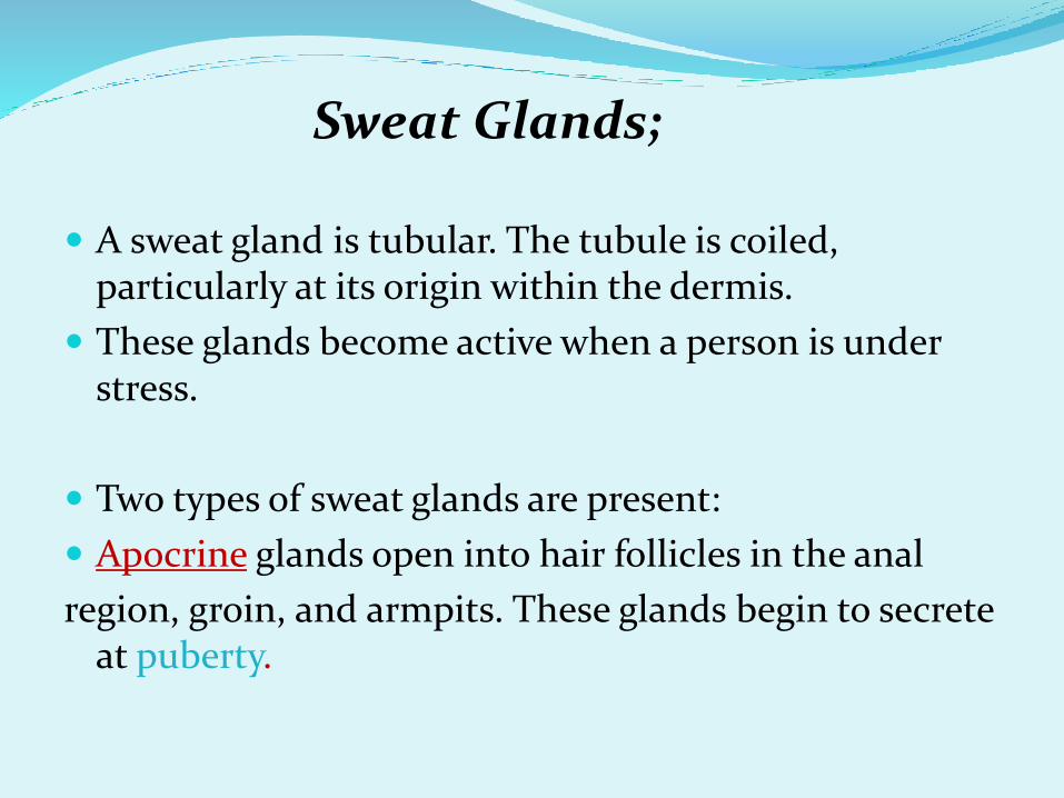
Sweat Glands;
A sweat gland is tubular. The tubule is coiled, particularly at its origin within the dermis.
These glands become active when a person is under stress.
Two types of sweat glands are present:
Apocrine glands open into hair follicles in the anal
region, groin, and armpits. These glands begin to secrete at puberty.

The anatomical position is described as follows:
Eccrine glands open onto the surface of the skin.
They become active when a person is hot, helping
to lower body temperature as sweat evaporates.
The sweat (perspiration) produced by these glands
is mostly water, but it also contains salts and some
urea. Therefore, sweat is a form of excretion.
Ears contain modified sweat glands, called Ceruminous
glands, which produce cerumen (earwax).

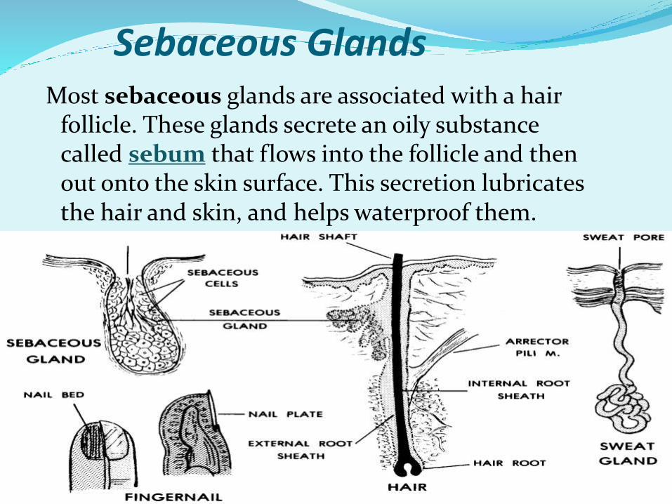
Sebaceous GlandsMost sebaceous glands are associated with a hair
follicle. These glands secrete an oily substance called sebum that flows into the follicle and then out onto the skin surface. This secretion lubricates the hair and skin, and helps waterproof them.

Particularly on the face and back, the sebaceous glands may fail to discharge sebum, and the secretions collect, forming whiteheads or blackheads.
If pus-inducing bacteria are also present, a boil or pimple may result.
Acne vulgaris is the most common form of acne, is an inflammation of the sebaceous glands that most often occurs during adolescence.
Hormonal changes during puberty cause the sebaceous glands to become more active at this time.

Hair and Nails
Hair is found on all body parts except the
palms, soles, lips, nipples, and portions of the
external reproductive organs.
Most of this hair is fine and downy, but the
hair on the head includes stronger types as
well.

After puberty, when sex hormones are made in an enough quantity, there is noticeable hair in the axillary and pelvic regions of both sexes.
In the male, a beard develops, and other parts of the body may also become quite hairy.
When women produce more male sex hormone than usual, they can develop hirsutism.
In males, baldness occurs when the hair on the head fails to regrow.
Alopecia, meaning hair loss, can have many causes.
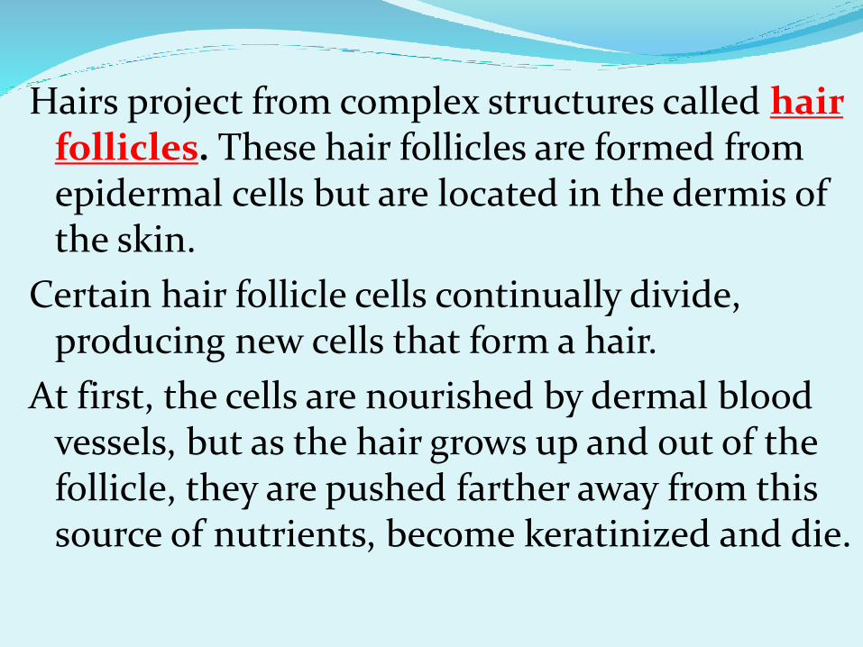
Hairs project from complex structures called hair follicles. These hair follicles are formed from epidermal cells but are located in the dermis of the skin.
Certain hair follicle cells continually divide, producing new cells that form a hair.
At first, the cells are nourished by dermal blood vessels, but as the hair grows up and out of the follicle, they are pushed farther away from this source of nutrients, become keratinized and die.


The portion of a hair within the follicle is called the root,
and the portion that extends beyond the skin is called the shaft.

Each hair has one or more sebaceous, glands,
whose ducts empty sebum into the follicle (oil).
A smooth muscle, the arrector pili, attaches to the follicle in such a way that contraction of the muscle causes the hair to stand on end. If a personhas had a scare or is cold, “goose bumps” develop due to contraction of these muscles (gooseflesh).

NailsNails grow from special epithelial cells
at the base of the nail in a region called the nail root
(a portion where a nail is implanted to the skin),
These cells become keratinized as they grow out over the nail bed.
The visible portion of thenail is called the nail body,
the distal extremitythe free edge.
The part beneath the body androot of the nail is called the nail matrix
or .

NAMESThe cuticle is a fold of skin hiding
the nail root. Ordinarily, nails grow only about 1 millimeter
per week (fingernails faster than toenails).
The pink color of a nail is due to the vascular dermal tissue beneath it.
The lunula is the whitish color, half-moon shaped base of the nail results from the thicker germinal layer in this area.

Lines of tension:
Lines of tension are
caused by the pull of
elastic and
collagenous fibers
within the dermis of
the skin.
Surgical incisions
made parallel to the
lines of tension heal
more rapidly and
create less scar
tissue than those
made across the lines
of tension.

THE END


![Skin anatomy chc training 2012 [compatibility mode] [repaired]](https://img.pdfslide.us/doc/110x75/54580319af795963388b680f/skin-anatomy-chc-training-2012-compatibility-mode-repaired.jpg)