Embed Size (px)
Citation preview

Anatomy of Chest wall,Lungs and Mediastinum
Dr R S Dhaliwal MBBS,MS,DNB(Surgery),M.Ch,DNB(CTVSurgery),
FACS,FCCP,FICA,FNCCP,FIACSProf. of CTV Surgery, UCMS
Former, Prof & HOD, CTV Surgery, PGIMER,Chandigarh

Chest wall
• The thorax is upper part of trunk bounded by 12 ribs, vertebrae and sternum.It contains trachea , two lungs , heart and great vessels, esophagus and lymph nodes.
• There are two thoracic apertures or openings . Superior thoracic aperture also called as thoracic inlet and inferior thoracic aperture . Head and neck and upper limbs communicate with thoracic cavity through thoracic inlet .It is bounded by body of first thoracic vertebra, Inferior thoracic aperture opens into peritoneal cavity

Inferior thoracic aperture
The posterior aspect of the thorax. (Inferior thoracic aperture visible at bottom.)
Latin apertura thoracis inferior
[

Chest wall• Skelton- The bony framework is bounded by the 12 thoracic
vertebrae postly, from which 12 sets of bony ribs articulate and curve around, connecting with the manubrium, sternum, and costal cartilage anteriorly
• The central anterior chest is defined by the sternum, which consists of 3 separate bones , manubrium first,then sternal body, and then xiphoid process. These are flat polygonal bones .The manubrium is located at the level of the T3 and T4 vertebra and is widest and thickest of 3 sternal bones. A palpable landmark on manubrium is jugular or suprasternal notch, which is bounded on either side by the medial ends of the clavicles. The sterno manubrial junction(Angle of Louis ) is projected . The body of sternum is located at the level of T5-T9 vertebrae. The xiphoid process is the smallest and thinnest bone. It often has a point, it can be blunt, bifid, or curved. The xiphoid is cartilaginous in younger people but is nearly ossified by age 40.

Chest wall• The first 7 ribs, called true ribs, are attached to the sternum
and manubrium directly. The 8th-10th ribs called False ribs are attached via the costal cartilages, while the 11th and 12th ribs remain unattached anteriorly. The 8th-10th ribs are known as false ribs because they lack direct attachment to the sternum, and the 11th-12th are referred to as floating ribs. Posteriorly, the ribs articulate with costal facets of 2 adjacent vertebrae. An articular capsule surrounds the head of each rib, which is secured by attachments of the radiate ligament. Each rib also articulates with the transverse process of its adjacent vertebrae through a costotransverse joint. The bony ribs then curve around anteriorly, where the next major junction is the costochondral joint.

Chest wall- bones

Chest wall• There are hyaline cartilage joints, and the rib and
cartilage are firmly attached through the continuity of overlying periosteum and perichondrium. The ribs/costal cartilages have various attachments to the sternum. The first pair of ribs articulates through cartilaginous joints or synchondroses and is relatively immobile. The second through seventh pairs of costal cartilages articulate with the sternum at synovial joints that move during respiration. These articular capsules are reinforced by sternocostal ligaments. This bony architecture provides attachments for muscles to the neck, thorax, upper limbs, abdomen, and back.

Chest wall -anterior

Chest wall posterior

Chest wall -Muscles• Muscles - 1.Intercostals
2. Pectoralis major/minor 3. Serratus anterior 4 Rectus abdominis
5. External & Internal oblique 6 Transversus thoracis and abdominis

Chest wall Muscles

Chest wall muscles

Pleura• The pleural cavity is the potential space between the two pleura (visceral and
parietal) of the lungs. The pleura is a serous membrane which folds back onto itself to form a two-layered membrane structure. The thin space between the two pleural layers is known as the pleural cavity and normally contains a small amount of pleural fluid. The outer pleura (parietal pleura) is attached to the chest wall. The inner pleura (visceral pleura) covers the lungs and adjoining structures, via blood vessels, bronchi and nerves.
• The parietal pleura is highly sensitive to pain, while the visceral pleura is not, due to its lack of sensory innervation
• In humans, there is no anatomical connection between the left and right pleural cavities. Therefore, in cases of pneumothorax, the other lung will still function normally unless there is a tension pneumothorax or simultaneous bilateral pneumothorax, which may collapse the contralateral parenchyma, blood vessels and bronchi.
• The pleural cavity, with its associated pleurae, aids optimal functioning of the lungs during respiration. The pleural cavity also contains pleural fluid, which allows the pleurae to slide effortlessly against each other during ventilation. Surface tension of the pleural fluid also leads to close apposition of the lung surfaces with the chest wall.

Pleurae

Pleura - anatomy

Arteries of Thorax

Lungs • Two lungs – Right and
Left .Right bigger in size• -3 Lobes in right lung –
Upper, Middle, Lower
.2 Lobes in left lung - Upper and Lower
Upper divided into Upper and Lingula

Lungs• Bronchopulmonary segments – Total 18
RT lung having 10 segments- Upper Lobe -Apical ,Anterior and Posterior Middle lobe – Medial and Lateral Lower Lobe - Superior, Medial ,Anterior and Posterior basal
• Left lung – 8 bronchopulmonary segments Upper Lobe- Apico Posterior Anterior , Lingula - Superior, Inferior Lower lobe – Superior, Anteromedial basal Lateral and Posterior basal

Lung segments

Lung segments

Lung Hilum

Mediastinum
• It is space in thorax located between thoracic inlet superiorly, inferiorly by diaphragm, antly by sternum , postly by spine and laterally by pleural spaces
• Classically it is divided into four compartments superior,anterior,middle and posterior. Another popular division is into three parts – Anterosuperior, middle and posterior
• Shields classified into three compartments- Anterior, Middle or visceral and Posterior or para ventral sulcus

Mediastinum

Subdivision of mediastinum as seen on cross section
• anterior mediastinum (1)
• middle mediastinum (2)
• posterior mediastinum (3)

Boundaries Of Mediatinum
• Anterior - sternum• Posterior - Vertebral Column• Superior - Thoracic inlet• Inferior - Diaphragm• ** Mediastinum is connected to neck &
retroperitoneum allowing spread of air & infection

Mediastinum

Mediastinum
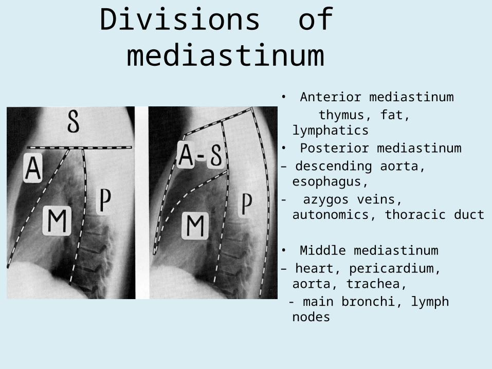
Divisions of mediastinum
• Anterior mediastinum thymus, fat, lymphatics• Posterior mediastinum– descending aorta, esophagus,- azygos veins, autonomics,
thoracic duct
• Middle mediastinum– heart, pericardium, aorta,
trachea, - main bronchi, lymph nodes

Mediastinal contents• Anterior Superior Mediast • Thymus Gland• Aortic Arch• SVC Superior Vena Cava• Lymph Node• Parathyroid Gland • Ectopic Thyroid Tissue
• Posterior Mediastinum • Esophagus• Vagus nerves• Sympathetic Chain• Thoracic duct• Thoracic desending aorta
• Middle Mediastinum • Pericardium• Heart• Great Vessels• Trachea• Trachea Bifurcation• Main Bronchi• Phrenic Nerves• Hilar Lymph Nodes
• Azygos vein• Hemiazygosvein• Paravertebral Lymphnodes

Mediastinal Structures

Influencing the life styles even after 2500 years
Thank you