Embed Size (px)
Citation preview
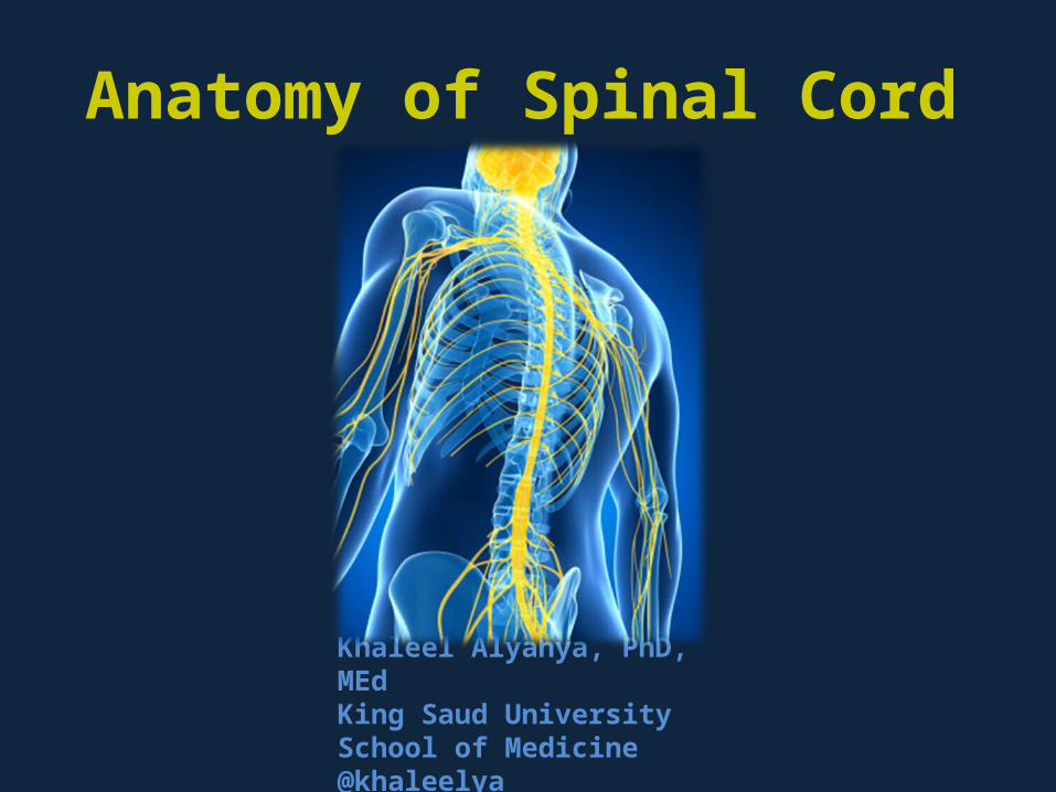
Khaleel Alyahya, PhD, MEdKing Saud UniversitySchool of Medicine@khaleelya
Anatomy of Spinal Cord

Tweet of Today

OBJECTIVES
At the end of the lecture, students should be able to: Describe the external anatomy of the spinal cord. Describe the internal anatomy of the spinal cord. Describe the spinal nerves: formation, branches and distribution via plexuses. Define Dermatome and describe its significance. Describe the meninges of the spinal cord. Define a reflex and reflex arc, and describe the components of the reflex arc.

Resources Human Anatomy & Physiology
Elaine MariebIntroduction to Human Body
Gerard TortoraHuman Brain
John NolteGoogle
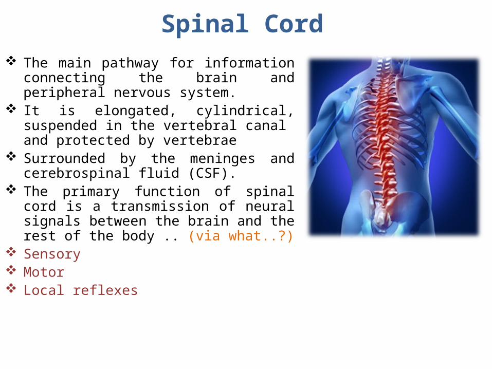
Spinal Cord The main pathway for information connecting
the brain and peripheral nervous system. It is elongated, cylindrical, suspended in the
vertebral canal and protected by vertebrae Surrounded by the meninges and cerebrospinal
fluid (CSF). The primary function of spinal cord is a
transmission of neural signals between the brain and the rest of the body .. (via what..?)
Sensory Motor Local reflexes

Spinal Cord Extends from foramen magnum to second
lumbar vertebra. Continuous above with the medulla oblongata. The tapered inferior end forms conus medullaris. It is connected to the coccyx by a non-neuronal
cord called Filum Terminale.

Spinal Cord Gives rise to 31 pairs of spinal nerves The bundle of spinal nerves extending inferiorly
from lumbosacral enlargement and conus medullaris surround the filum terminale and form cauda equina
Segmented 8 Cervical 12 Thoracic 5 Lumbar 5 Sacral 1 Coccygeal
Has two enlargements: Cervical Enlargement: supplies upper limbs. Lumbosacral Enlargement: supplies lower
limbs.

NucleusA group of neurons
within the CNS
Ganglion A group of neurons
outside the CNS
Tract A group of nerve fibers (axons) within the CNS
Nerve A group of nerve fibers (axons) outside the CNS

Cross Section of Spinal Cord
The spinal cord is incompletely divided into two equal parts, anteriorly by a short, shallow median fissure and posteriorly by a deep narrow septum, the posterior median sulcus.
Composed of grey matter in the center surrounded by white matter supported by neuroglia.
Commissures: connections between left and right halves Gray with central canal in the center White
Roots: spinal nerves arise as rootlets then combine to form roots Dorsal (posterior) root has a ganglion Ventral (anterior) Two roots merge laterally and form
the spinal nerve

The arrangement of grey matter in the spinal cord resembles the shape of the letter H, having: two posterior two anterior two lateral horns/columns.
Consists of: Nerve cell bodies and their
processes Neuroglia (supportive cells) Blood vessels
Grey Matter

The nerve cells are multipolar, and are of three main categories:
Sensory neurons (Tract cells) receive impulses from the periphery
of the body and whose axons constitute the ascending fasciculi of the white matter.
located in the dorsal horns. Lower motor neurons
transmit impulses to the skeletal muscles.
located in the ventral horns similar neurons in the lateral
horn are the preganglionic neurons of the autonomic system.
Interneurons (connector neurons) linking sensory and motor neurons, at
the same or different levels, which form spinal reflex arcs.
Grey Matter

Neuronal Architecture of Spinal Grey Matter
Cells of the same type are clustered into groups, which occur in long columns
In transverse section, these columns appear as layers, especially within the dorsal horn
These layers are called the laminae of Rexed (a Swedish neuroscientist, 1950), that are numbered by Roman numerals, starting from the tip of the dorsal horn and moving ventrally into the ventral horn

Nerve Cell Groups in Dorsal Horn
4 main groups1. Substantia Gelatinosa2. Nucleus Proprius3. Nucleus Dorsalis
Clark’s column Nucleus thoracis)
4. Visceral Afferent Nucleus

Substantia Gelatinosa• Rexed Laminae II• Located at the apex of the horn• Composed of large neurons• Extends throughout the length of spinal
cord• Afferents: dorsal root fibers concerned
with pain, temperature and touch
Nucleus Proprius• Rexed Lamina IV• Located anterior to substantia gelatinosa• Composed of large neurons• Extends throughout the length of spinal
cord• Afferents: dorsal root fibers concerned
with senses of position & movement (proprioception)

Nucleus Dorsalis (Clark’s column, Nucleus thoracis)
• Rexed Lamina VII• Located at the base of dorsal horn• Composed mostly of large neurons• Extends from C8 to L3-4 segments• Associated with proprioceptive endings• Afferents: dorsal root fibers concerned with
information from muscle spindles and tendon organs.
Visceral Afferent Nucleus• Rexed Lamina VII• Located lateral to nucleus dorsalis• Composed mostly of medium size neurons• Extends from T1 to L3 segments• Afferents: Visceral afferents
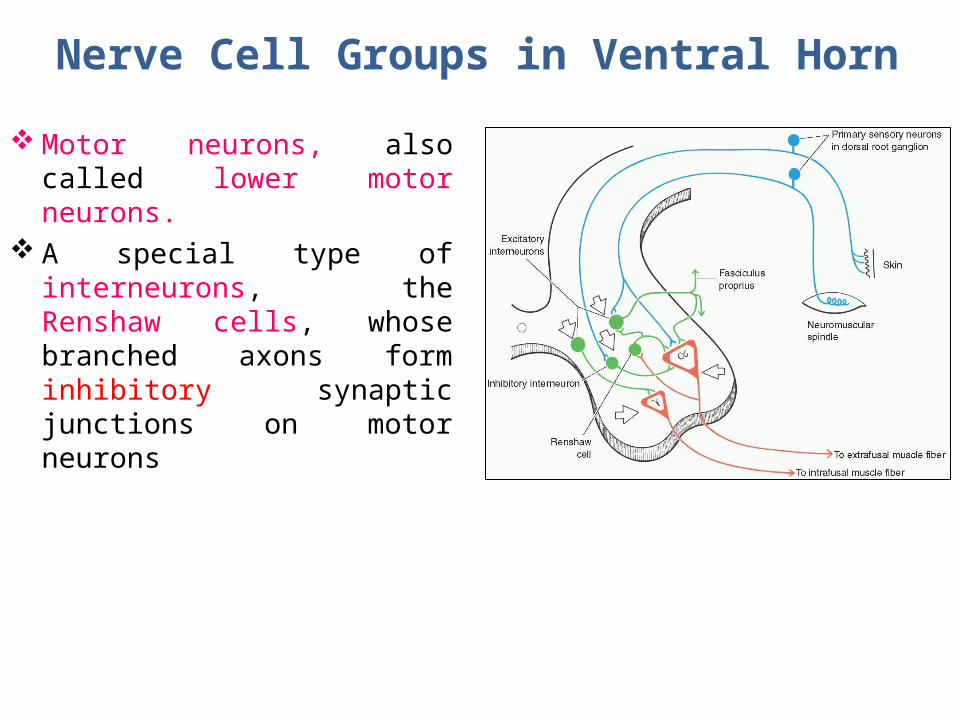
Nerve Cell Groups in Ventral Horn Motor neurons, also called
lower motor neurons. A special type of interneurons,
the Renshaw cells, whose branched axons form inhibitory synaptic junctions on motor neurons

Motor Neurons in Ventral Horn Are of Two types Large multipolar cells
whose axons pass out in the ventral roots of spinal nerves as alpha efferents which innervate extrafusal muscle fibers of skeletal muscles.
Less numerous smaller multipolar cells whose axons pass out in the
ventral roots of spinal nerves as gamma efferents which innervate intrafusal muscle fibers of neuromuscular spindles
Both alpha and gamma motor neurons are under the
influence of descending pathways from brain

Motor neurons are organized in 3 groups:
Medial: Present in most segments,
innervates muscles of neck and trunk (including intercostal and abdominal muscles)
Central: Smallest, present in some
cervical (phrenic C3-5, spinal accessory C1-6) and lumbosacral (L2-S1) segments
Lateral: Present in cervical and
lumbosacral segments, innervates muscles of the limbs
Neurons supplying flexor muscles are located dorsal
to neurons for extensor muscles
Motor Neurons in Ventral Horn

Nerve Cell Groups in Lateral Horn
Small Column composed of small neurons Extends from T1 to L2-3
segments: Give rise to preganglionic
sympathetic fibers Extends from S2-4 segments:
Give rise to preganglionic parasympathetic fibers

White Matter Consists of mixture of nerve fibers,
neuroglia and blood vessels. White color is due to high proportion of
myelinated nerve fibers The white matter of the spinal cord is
arranged in columns/funiculi; anterior, posterior and lateral.
The nerve fibers are arranged as bundles, running vertically through the cord.
A group of nerve fibers (axons) that share a common origin, termination and function form a tract or fasciculus
These tracts are formed by sensory nerve fibers ascending to the brain, motor nerve fibers descending from the brain and fibers of connector neurons.
Tracts are often named according to their points of origin and destination, e.g. spinothalamic, corticospinal.
Depending on their function, the spinal tracts are divided into ascending
and descending tracts


Commissures of the Spinal Cord
Grey Commissure: Transverse bridge of grey matter
connecting the anterior and posterior gray horns on each side
Is pierced by the central canal that divides it into anterior and posterior parts
White Commissure: Lies ventral to the gray commissure Mainly contains decussating nerve
fibers

Central Canal The cerebrospinal-filled space that runs
longitudinally through the entire length of the spinal cord.
Lined by ependyma (ciliated columnar epithelium)
Continuous with the ventricular system of the brain
Superiorly opens into the 4th ventricle Inferiorly in the conus medullaris, it
expands into the fusiform terminal ventricle and terminates below at the root of filum terminale

Regional Differences Although the general pattern of gray
matter is the same throughout spinal cord, regional differences are apparent in transverse sections
The amount of white matter increases in a caudal-to-cranial direction because fibers are added to ascending tracts and fibers leave descending tracts
The gray matter is in increased volume in cervical & lumbosacral enlargements for innervation of upper & lower limbs
The lateral horn is characteristics of thoracic and upper lumbar segments
Cervical
Thoracic
Lumbar
Sacral

Spinal Nerves Thirty-one pairs of spinal nerves First pair exit vertebral column between skull
and atlas, last four pairs exit via the sacral foramina and others exit through intervertebral foramina
Eight pair cervical, twelve pair thoracic, five pair lumbar, five pair sacral, one pair coccygeal
Each spinal nerve arises as rootlets which then combine to form dorsal (posterior) & ventral (anterior) roots.
Two roots merge laterally and form the spinal nerve.
Dorsal (posterior) root has a ganglion (dorsal root/sensory ganglion) that contains the cell bodies of the sensory neurons
Each spinal nerve then divides into a smaller dorsal and a larger ventral ramus
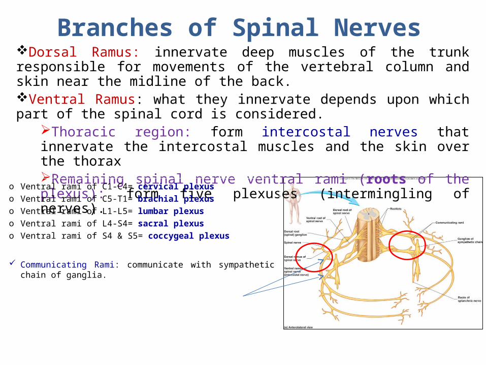
Branches of Spinal NervesDorsal Ramus: innervate deep muscles of the trunk responsible for movements of the vertebral column and skin near the midline of the back.Ventral Ramus: what they innervate depends upon which part of the spinal cord is considered.
Thoracic region: form intercostal nerves that innervate the intercostal muscles and the skin over the thoraxRemaining spinal nerve ventral rami (roots of the plexus): form five plexuses (intermingling of nerves).
o Ventral rami of C1-C4= cervical plexuso Ventral rami of C5-T1= brachial plexuso Ventral rami of L1-L5= lumbar plexuso Ventral rami of L4-S4= sacral plexuso Ventral rami of S4 & S5= coccygeal plexus
Communicating Rami: communicate with sympathetic chain of ganglia.
Dermatomes
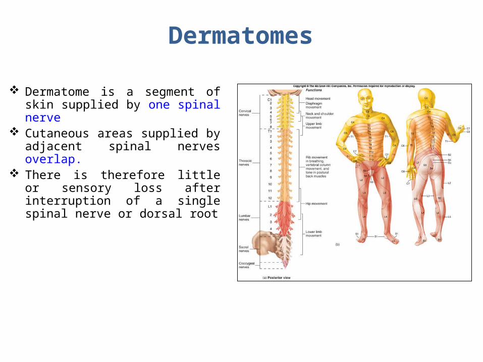
Dermatome is a segment of skin supplied by one spinal nerve
Cutaneous areas supplied by adjacent spinal nerves overlap.
There is therefore little or sensory loss after interruption of a single spinal nerve or dorsal root

Spinal Meninges Connective tissue membranes surrounding
spinal cord and brain Dura mater: continuous with epineurium
of the spinal nerves Arachnoid mater: thin and wispy Pia mater: bound tightly to surface of brain
and spinal cord. Forms the filum terminale, which anchors spinal
cord to coccyx and the denticulate ligaments that attach the spinal cord to the dura mater
Spaces Epidural: Contains blood vessels,
connective tissue and fat. Subdural: Contains serous fluid Subarachnoid: Contains CSF and blood
vessels within web-like strands of arachnoid tissue

Reflex & Reflex ArcA reflex is a rapid, involuntary, stereotyped pattern of response brought by a sensory stimulus
A neural pathway mediating the reflex actions is called reflex arc.

Components of a Reflex ArcAction potentials produced in
Sensory neuronSensory receptor
InterneuronMotor neuronEffector organ which responds with a reflex

Questions!