Embed Size (px)
Citation preview

Spinal Cord, Human Reflex
1

Gross Anatomy of the Adult Spinal Cord
2

Extends from the foramen magnum to L1 or L2Conus medularis - it is the end of the spinal
cord Denticulate ligaments – pia mater attaching the
spinal cord t the vertebral wallFilum terminale – pia mater extension from the
conus medularis to the coccix
Anatomy of the Spinal Cord
3

Lumbar tap – removal of CSF from the vertebral canal bellow L3
Cervical and lumbar enlargements31 pairs of spinal nerves leave the spinal cordCauda equina – collection of spinal nerves at
the end of the spinal cord
Anatomy of the Spinal Cord
4

The Spinal Cord and Spinal Meninges
5

Spinal meninges
Three layersDura mater
Single layerEpidural space – between the vertebra
and dura. Filled with fat and blood vessels
ArachnoidSubarachnoid space – between
arachnoid and pia. Filled with CSFPia mater
6

The Spinal Cord and Spinal Meninges
7

Sectional Organization of the Spinal Cord
8

Structures of the spinal cord
Gray Matter Cell bodies, unmyelinated axons,
dendrites and neurogliaPosterior or dorsal horns – interneurons
and sensory fibers coming from the dorsal root ganglion
Anterior or ventral horns – cell bodies of the somatic motor neurons that send their axons to the ventral root
9

Lateral gray horns contain visceral motor neurons
Gray commissures contain axons that cross from one side to the other
Central canal – filled with CSF
Gray matter
10

White matter
Divided into six columns (funiculi) containing tracts
Posterior funiculiPosterior median sulcus
Anterior funiculiAnterior median fissure
Lateral funiculi
11

Spinal Nerves
12

8 pairs of cervicalAbove C7 all nerves emerge above the
corresponding vertebraC8 emerge between C7 and T1
12 pairs of thoracic5 pairs of lumbar5 pairs of sacralI pair of coccygeal
31 pairs of spinal nerves (mixed)
13
OriginDorsal root and dorsal root ganglionVentral root
Dorsal ramus sensory and motor innervation to the skin
and muscles of the back
Spinal nerves
14

Spinal nerves
Ventral ramus sensory and motor to the ventrolateral
body surface, body wall and limbsT2 – T12 form intercostal nervesAll other will form plexuses
15
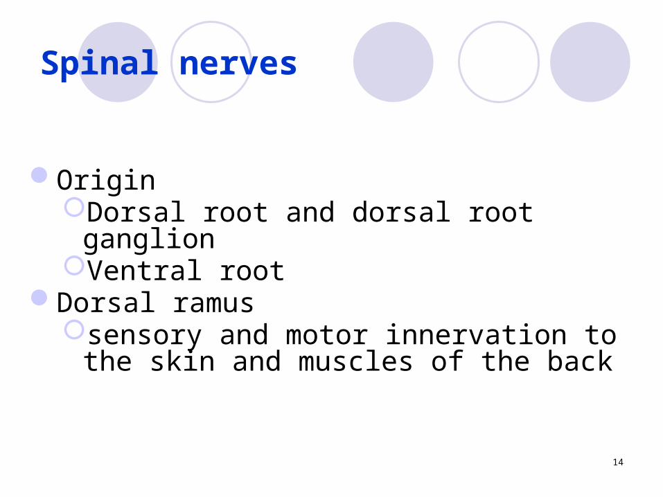
Complex interwoven network of nervesFour large plexuses
Cervical plexusBrachial plexusLumbar plexusSacral plexus
Nerve plexus
16

Peripheral Nerves and Nerve Plexus
17

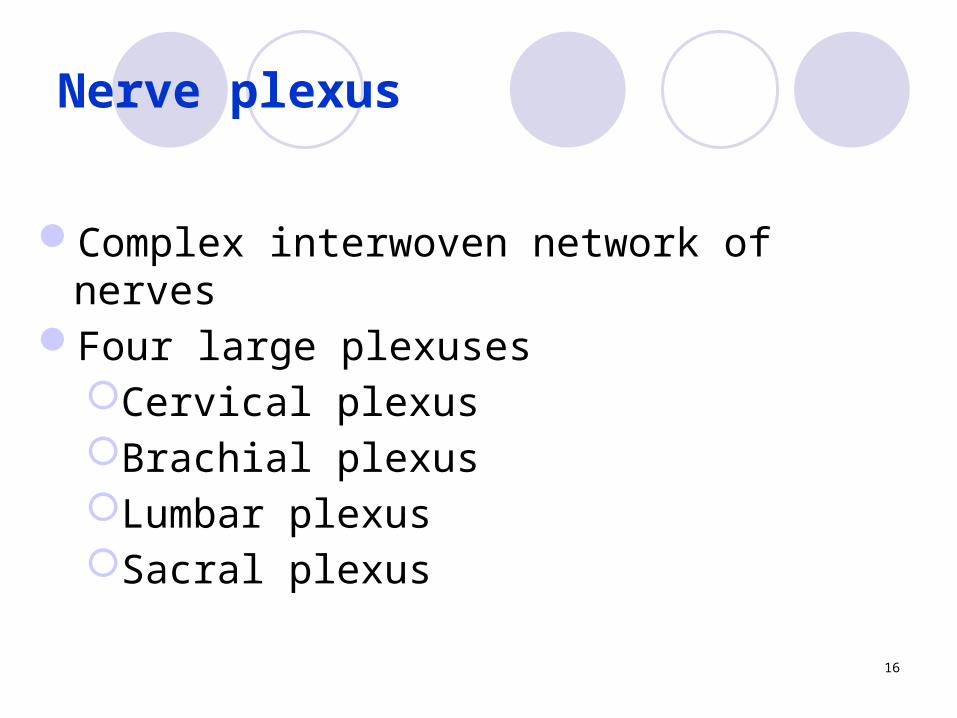
The Cervical Plexus
Ventral rami of spinal nerves C1 to C5Innervate muscles of the neckInnervate the diaphragm
Phrenic nerve (C3, C4,C5)
18
Cervical Plexus
19

Brachial Plexus
20

Brachial Plexus
21

The Brachial Plexus
Axillary nerveShoulder
Musculocutaneous nerve Anterior brachial area
22

The Brachial PlexusRadial nerve
Posterior arm and hand
Median nerve Anterior antebrachial area
23

The Brachial Plexus
Ulnar nerve Anterior antebrachial area
24
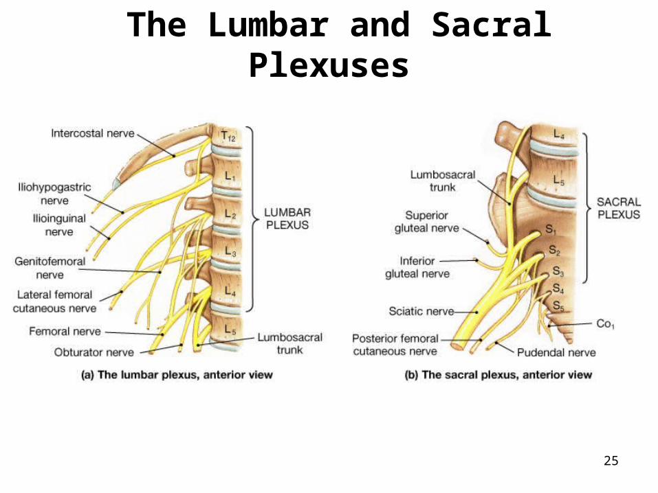
The Lumbar and Sacral Plexuses
25

Lumbar Plexus
26

The Lumbar Plexus
Lower abdominopelvic region and the anterior thigh
L1 – L4Femoral Nerve
Anterior muscles of femoral areaObturator Nerve
Adductors of leg
27

The Sacral Plexus
Posterior thigh, gluteal region, leg and footL4 – S4Sciatic nerve
Tibial nerveCommon fibular nerve
Pudendal nerveMuscles and skin of perineum
28

Reflexes are rapid and predictable automatic responses to stimuli
Neural reflex involves sensory fibers to CNS and motor fibers to effectors
Spinal Reflexes
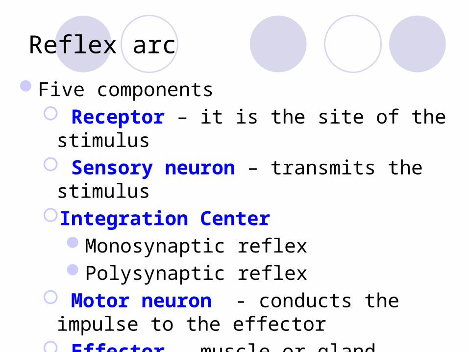
Five components Receptor – it is the site of the stimulus Sensory neuron – transmits the stimulusIntegration Center
Monosynaptic reflexPolysynaptic reflex
Motor neuron - conducts the impulse to the effector
Effector – muscle or gland
Reflex arc

Arc Reflex Components

Somatic ReflexesSpinal Reflexes
Patellar (knee-jerk)Achilles Triceps Biceps etc.
Reflex classification

The Babinski Reflexes

Reflex classification
Superficial reflexes Plantar reflex
• Normal response – curling the toe• Abnormal response – Babisnki’s sign
Corneal reflex - Cranial nerve VGag reflex
• Cranial nerves IX and X

Autonomic ReflexesPupillary reflex
Ipsilateral constriction of the pupilConsensual reflex
Contralateral constriction of the pupilCiliospinal reflex
Pupil dilation when pinching the neckSalivary reflex
Reflex classification