Embed Size (px)
Citation preview
An imprint of Elsevier Inc
© 2007, Elsevier Inc. All rights reserved.
Chapter 12 figures © BodyAesthetic Plastic Surgery & Skincare Center
No part of this publication may be reproduced, stored in a retrieval system, or
transmitted in any form or by any means, electronic, mechanical, photocopying,
recording or otherwise, without the prior permission of the Publishers. Permissions may
be sought directly from Elsevier’s Health Sciences Rights Department, 1600 John F.
Kennedy Boulevard, Suite 1800, Philadelphia, PA 19103-2899, USA: phone: (+1) 215
239 3804; fax: (+1) 215 239 3805; or, e-mail: [email protected]. You
may also complete your request on-line via the Elsevier homepage
(http://www.elsevier.com), by selecting ‘Support and contact’ and then ‘Copyright and
Permission’.
ISBN-13: 978-1-4160-2952-6
ISBN-10: 1-4160-2952-4
British Library Cataloguing in Publication Data
A catalogue record for this book is available from the British Library
Library of Congress Cataloging in Publication Data
A catalog record for this book is available from the Library of Congress
NoticeMedical knowledge is constantly changing. Standard safety precautions must be
followed, but as new research and clinical experience broaden our knowledge, changes
in treatment and drug therapy may become necessary or appropriate. Readers are
advised to check the most current product information provided by the manufacturer
of each drug to be administered to verify the recommended dose, the method and
duration of administration, and contraindications. It is the responsibility of the
practitioner, relying on experience and knowledge of the patient, to determine dosages
and the best treatment for each individual patient. Neither the Publisher nor the author
assume any liability for any injury and/or damage to persons or property arising from
this publication.
The Publisher
Printed in China
Last digit is the print number: 9 8 7 6 5 4 3 2 1
Siamak Agha-Mohammadi MD PhD
Clinical Assistant Professor of Surgery (Plastic)
Division of Plastic Surgery
University of Pittsburgh
Pittsburgh, PA, USA
Al S. Aly MD FACS
Plastic Surgeon
Iowa City Plastic Surgery
Coralville, IA, USA
Loren J. Borud MD
Plastic Surgeon
Beth Israel Deaconess Medical Center;
Harvard Medical School
Boston, MA, USA
Stacy A. Brethauer MD
Fellow, Advanced Laparoscopic and Bariatric Surgery
Cleveland Clinic
Cleveland, OH, USA
Joseph F. Capella MD
Plastic Surgeon
Surgical Weight Reduction and Body Contouring
Ramsey, NJ, USA
Robert F. Centeno MD
Plastic Surgeon
Body Aesthetic Plastic Surgery and Skincare Center
St Louis, MO, USA
Susan E. Downey MD FACS
Clinical Associate Professor of Plastic Surgery
Keck School of Medicine
University of Southern California
Los Angeles, CA, USA
Felmont F. Eaves III MD
Attending Surgeon
Charlotte Plastic Surgery
Charlotte, NC, USA
David T. Greenspun MD MSc
Plastic Surgeon
Private Practice
New York, NY, USA
Dennis J. Hurwitz MD FACS
Clinical Professor of Surgery (Plastic)
University of Pittsburgh Medical Center
Pittsburgh, PA, USA
Alan Matarasso MD
Clinical Professor of Plastic Surgery
Albert Einstein College of Medicine
New York, NY, USA
James P. O’Toole MD
Body Contouring Fellow
Division of Plastic Surgery
University of Pittsburgh Medical Center
Pittsburgh, PA, USA
Ivo Pitanguy MD
Head Professor
Department of Plastic Surgery
Pontifical Catholic University of Rio de Janeiro;
Carlos Chagas Post-Graduate Medical Institute;
Director
Clinica Ivo Pitanguy
Rio de Janeiro, Brazil
Henrique N. Radwanski MD
Assistant Professor of Plastic Surgery
Pontifical Catholic University of Rio de Janeiro;
Carlos Chagas Post-Graduate Medical Institute
Rio de Janeiro, Brazil
J. Peter Rubin MD
Director, Life After Weight Loss Program;
Assistant Professor of Plastic Surgery
Department of Surgery
University of Pittsburgh
Pittsburgh, PA, USA
vii
CONTRIBUTORS

Philip R. Schauer MD
Professor of Surgery
Cleveland Clinic Lerner School of Medicine;
Director, Advanced Laparoscopic and Bariatric Surgery
Bariatric and Metabolic Institute (BMI)
The Cleveland Clinic
Cleveland, OH, USA
Berish Strauch MD
Professor and Chair
Department of Plastic and Reconstructive Surgery
Albert Einstein College of Medicine and Montefiore Medical Center
Bronx, NY, USA
V. Leroy Young MD
Plastic Surgeon
BodyAesthetic Plastic Surgery and Skincare Center
St Louis, MO, USA
Contributors
viii
The historian Arnold J. Toynbee explained the rise of
civilization in terms of challenge and response. He could have
been describing the history of plastic surgery. Our specialty
began because of a need, perhaps the first being to rebuild the
nose. Plastic surgery has continued, even flourished, because of
its ability to recognize and respond successfully, although not
always optimally, to the changing requirements of patients, as
this well written, carefully edited and admirably illustrated
book testifies.
That human beings have eating disorders, ranging from
anorexia to obesity, is a fact and that the United States has an
astonishing and disproportionate incidence of the enormously
overweight is also a fact. Until recently, weight loss centers,
psychotherapists, and questionably effective and frequently
dangerous medications, were the usual recourse. Surgery for
massive obesity was once considered farfetched, prohibitively
dangerous, and even indulgent. Toward these patients our
society has had, and to a lessor degree still has, a punitive
attitude: “They should be able to work it out themselves
through diet and restraint. Why should we devote our resources
to their problem?” The reality is that their personal problem is
our society’s problem, now a healthcare crisis.
With the increasing numbers of the very obese, the
realization of their compromised quality and length of life, with
better education and more public understanding, as well as
improvement in safety and success of bariatric surgery,
operative treatment of this condition has not only been accepted
by, but also welcomed by, the medical and surgical profession,
and certainly by patients and their families.
As the editors, Dr Rubin and Dr Matarasso have so well
documented in this book, Aesthetic Surgery After Massive
Weight Loss, the combined best of our aesthetic as well as our
reconstructive skills. The surgical demands are difficult, and
not to be undertaken casually by someone inexperienced who
has not seriously studied, and hopefully observed, surgeons
who have learned how best to minimize complications and to
secure results beyond merely satisfactory. For anyone
contemplating doing these operations, whether plastic surgeon
or general surgeon, and to anyone interested in this area of
medicine, this book is important and essential. It is not just
informative and helpful but honest, born of extensive
experience on the part of the contributors, as well as the editors.
They have been more than willing to share their mistakes in
judgment, their errors of execution, and their ways of dealing
with undesirable outcomes.
Bariatric surgery, in joining together with various specialties,
including psychotherapy, internal medicine, general surgery,
anesthesiology and plastic surgery, has been good for our
specialty. It has returned us again to the mainstream where we
belong and where we can interact and learn from colleagues in
other fields who also can learn from us – all to the benefit of
the patient who is and must always be our primary focus.
The bariatric surgeon now realizes, and certainly the patient
has long known, that losing weight through an operation is not
the end of the treatment. The long, painful journey for the
patient is not over but the destination is in sight. That person
still confronts physical deformity, emotional distress and
additional operations because of excess tissue in numerous
areas of the body. The patient, who has already endured so
much, wants finally to look and be normal, a desire which is
shared by most who seek plastic surgery.
My congratulations to the editors, the contributors, and the
publishers for bringing this fine book to fruition.
Robert M. Goldwyn MD
Clinical Professor of Surgery
Harvard Medical School;
Editor Emeritus
Plastic and Reconstructive Surgery
Journal of the American Society of Plastic Surgeons
ix
FOREWORD
x
Obesity is a rapidly growing disease that has spread widely in
the western world and presents as an emerging issue in
developing countries. The increase of the obese population has
popularized the demand for bariatric surgery, and it is estimated
that more than 70% of the patients who undergo such surgery
state that, due to skin laxity and ptosis of certain anatomical
areas, significant weight loss causes an unacceptable worsening
of their body image. This becomes more relevant in our beauty-
centered global society, where life is fast-paced and people are
rapidly judged with regards to their appearance. It has therefore
become more common for the patient who has undergone a
great amount of weight reduction to present to the plastic
surgeon requesting the removal of excess skin, from one or,
more typically, many regions of the body.
In this timely book, Aesthetic Surgery After Massive Weight
Loss, the various body contour deformities are addressed.
Several authors, from many different medical specialties, and
some who are well known for their work in aesthetic plastic
surgery, present their experience in the treatment of the patient
following great weight loss. Under the careful and competent
supervision of Drs. Rubin and Matarasso, the medical issues
pertaining to these patients and the complexity of the different
deformities are focused in separate chapters, but with a clear
editorial guidance. The editors and authors are to be
commended for their contribution to this fascinating subject
that is proving to be a new specialty in medicine and,
particularly, in aesthetic plastic surgery.
Ivo Pitanguy MD FACS FICS
Professor of the Post-Graduate Courses in
Plastic Surgery of the Pontifical Catholic University of
Rio de Janeiro and the Carlos Chagas Post-Graduate Medical
Institute. Member of the Brazilian Society of Plastic Surgery,
the Brazilian National Academy of Medicine,
and the Brazilian Academy of Letters.
FOREWORD
This book is dedicated to my wife Julie, whose partnership,
patience, and constant support of my academic interests have
enabled me to pursue this project. To my children, Eliana and
Liviya, who inspire me to be more curious every day. And to
the memory of my father, Leonard R. Rubin MD, who never
stopped searching for new ideas.
J. Peter Rubin MD
Dedicated to:
Daniel MATARASSO ben
Hamaskil Albert MATARASSO
Alan Matarasso MD
DEDICATION
Each decade has witnessed major advances in our specialty
leading to the establishment of new arenas of plastic surgery.
Bariatric plastic surgery represents the next dimension in the
evolution of our specialty and holds with it the promise and
hope of helping many patients.
The editors are extremely grateful to the many experts who
contributed to this text. It was only through their commitment
of valuable time and energy that such a comprehensive
textbook could be produced around an evolving field of plastic
surgery. These are skillful surgeons who have focused their
creativity on helping the massive weight loss patient achieve
their ultimate goals. We recognize the sacrifice that academic
contributions entail and appreciate how generous each of the
contributors has been in sharing their surgical expertise. Indeed,
their diverse perspectives and approaches make this book a
valuable resource for all plastic surgeons.
We also wish to thank the editorial team at Elsevier. Their
commitment to this project enabled us to invite the top experts
in post-bariatric surgery as contributors, and allowed for the
highest quality of production.
J. Peter Rubin MD
Alan Matarasso MD
xi
ACKNOWLEDGMENTS
OBESITY
Obesity is defined as the accumulation of excess body fat that
leads to pathology. This disease can lead to an extensive list of
comorbid conditions, the most serious of which are:
• hypertension,
• diabetes,
• heart disease,
• stroke,
• obstructive sleep apnea, and
• degenerative joint disease.
Body mass index (BMI = weight (kg)/height (m)2) is the
primary measurement used to categorize obese patients. In
1991, the National Institutes of Health (NIH) defined morbid
obesity as a BMI of 35 kg/m2 or greater with severe obesity-
related comorbidity, or a BMI of 40 kg/m2 or greater without
comorbidity.1 Patients with a BMI of 50 kg/m2 or greater are
often referred to as superobese or massively obese.
There has been increasing interest in obesity and major
advances in bariatric surgery over the past 15 years as the
problems associated with morbid obesity and the benefits of
surgical treatment for this disease have become more clearly
defined.
Epidemiology and risk factorsObesity is a major public health problem in the USA that has
significantly worsened over the past four decades and has
now reached epidemic proportions. The National Center for
Health Statistics has conducted periodic National Health and
Nutrition Examination Surveys (NHANES) since 1960 to de-
termine the prevalence of obesity.2 According to this continu-
ous study, 65% of US adults are overweight (BMI > 25 kg/m2)
or obese (BMI > 30 kg/m2). These studies have shown an
increase in the prevalence of obesity from 15% in 1980 to
30% in 2002. Additionally, 5% of Americans 20 years of age
or older currently have a BMI > 40 kg/m2. Children and older
Americans are increasingly becoming obese as well. Thirty-
one percent of children aged 6–19 are at risk for overweight
(BMI for age > 85th percentile) or overweight (BMI for age
> 95th percentile), and 16% are overweight. Thirty-three per-
cent of Americans over the age of 60 are obese. These increases
have occurred despite expenditures of over $45 billion annually
on weight loss products.3
Obesity and morbid obesity affect women and minorities
(particularly middle-aged black and Mexican American women)
more than white males. However, in almost every age and ethnic
group examined by NHANES, the prevalence of overweight
or obesity exceeds 50%.2
EtiologyThe etiology of obesity is not as straightforward as once
thought. It is not simply an excess of caloric intake in relation
to caloric expenditure, but a complex interaction of excessive
intake, inefficient calorie utilization, reduced metabolic activity,
a reduction in the thermogenic response to meals, and an ab-
normally high set-point for body weight. Genetic, environmen-
tal, and psychosocial factors all contribute to this problem.
Children of obese parents have an 80–90% chance of develop-
ing obesity by adulthood, while only 10% of children of
normal-weight parents will become obese. The high-fat and
high-calorie American diet in conjunction with a sedentary
lifestyle contributes significantly to this problem.
OVERVIEW OF BARIATRIC SURGERY
This section provides an overview of the different weight loss
procedures and their physiologic effects.
1
WEIGHT LOSS SURGERY: STATE OFTHE ART 1Philip R. Schauer and Stacy A. Brethauer
Key Points• Patients with a BMI of 40 kg/m2, or 35 kg/m2 with severe comorbidi-
ties of obesity, qualify for weight loss surgery.
• The type of weight loss procedure performed can have differential
effects on weight loss and on long-term nutritional status.
• Most medical comorbidities associated with obesity improve after
surgically induced weight loss.
• The most commonly performed procedure is Roux-en-Y gastric bypass.
• Laparoscopic approaches are becoming increasingly common.
Goals of surgery and mechanism of actionThe goal of bariatric surgery is to improve the health of mor-
bidly obese patients by reducing or eliminating their comorbid
conditions. This is achieved by long-term weight loss that in-
volves a significant reduction in caloric intake or absorption.
Bariatric operations that are currently performed involve:
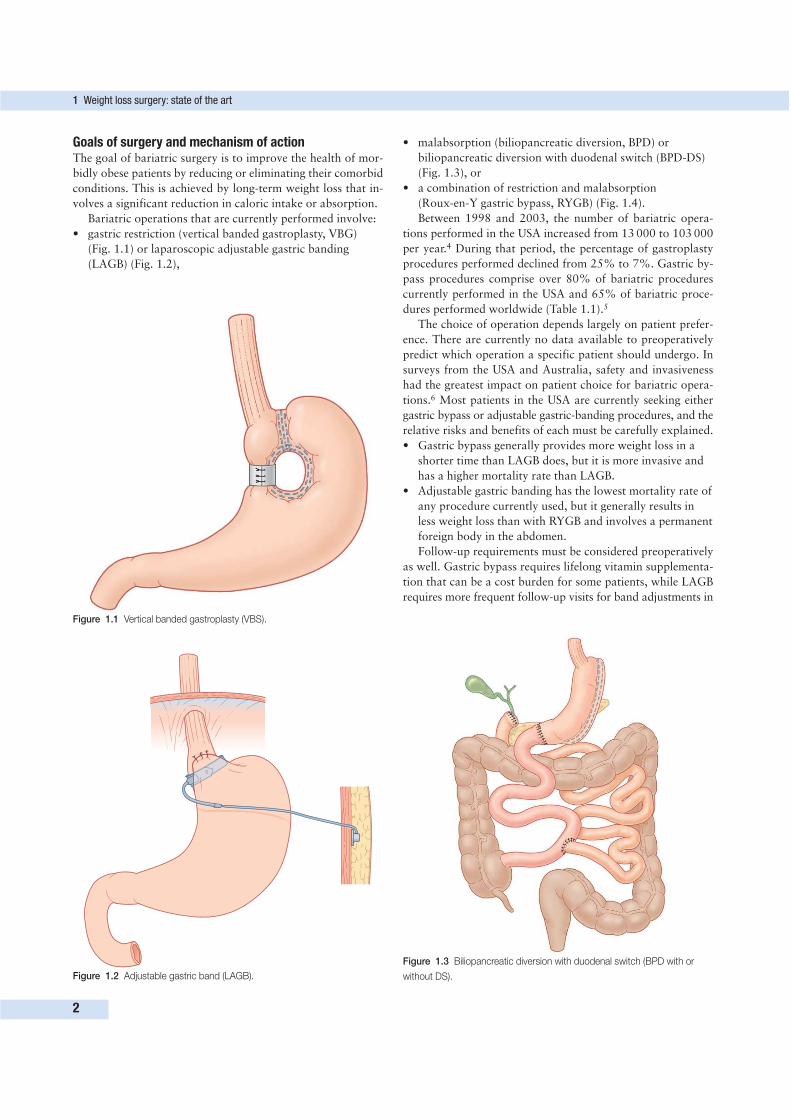
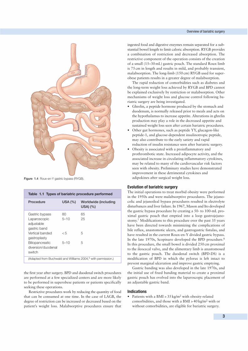
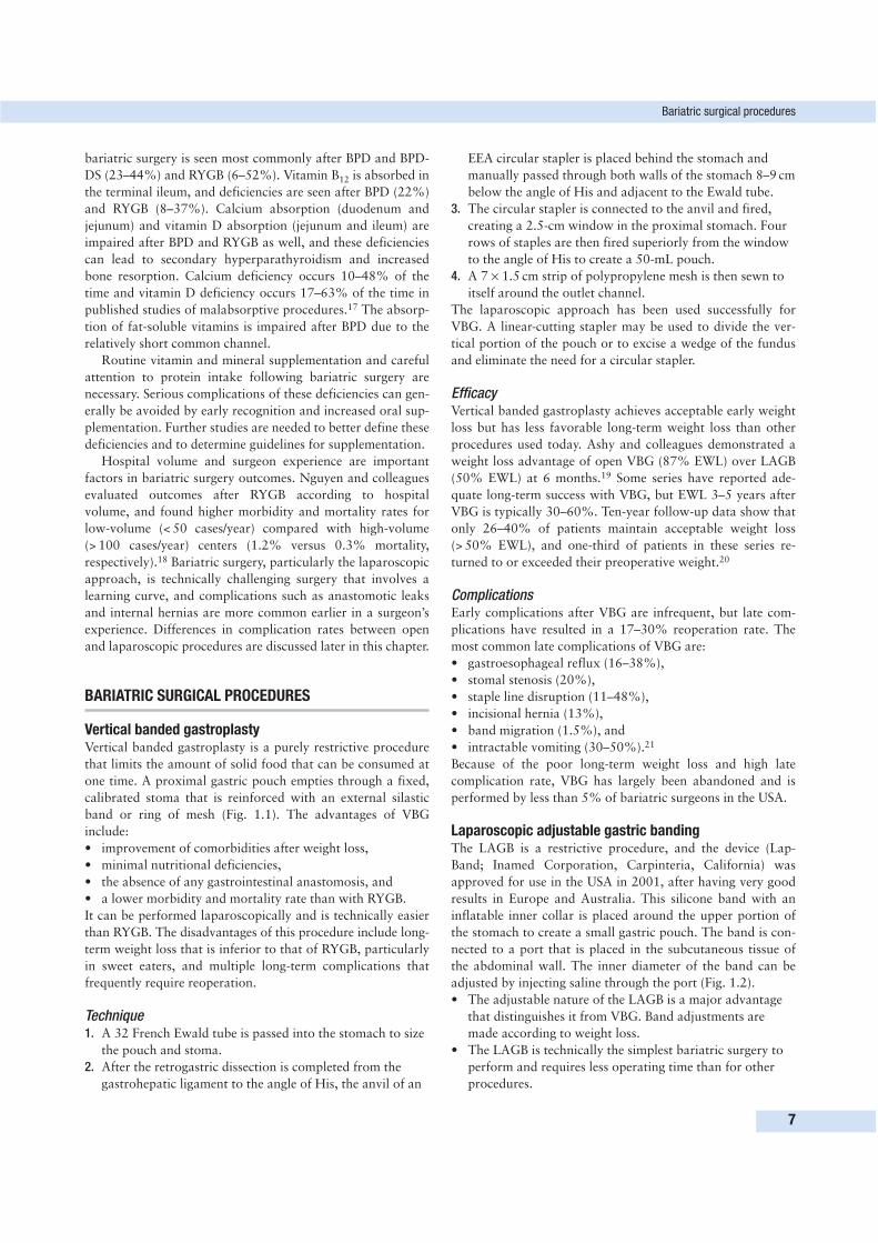
• gastric restriction (vertical banded gastroplasty, VBG)
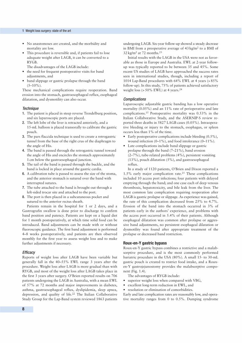
(Fig. 1.1) or laparoscopic adjustable gastric banding
(LAGB) (Fig. 1.2),
1 Weight loss surgery: state of the art
2
Figure 1.1 Vertical banded gastroplasty (VBS).
Figure 1.2 Adjustable gastric band (LAGB).
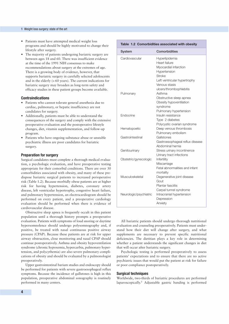
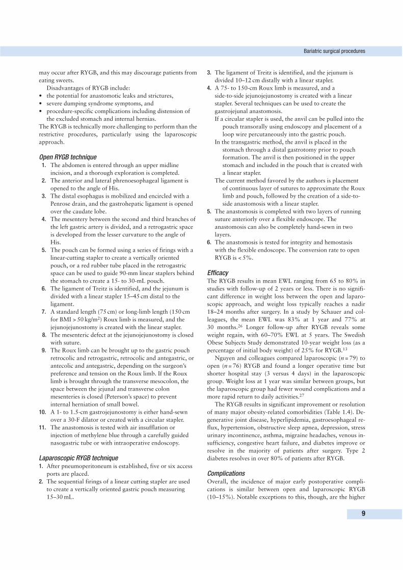
Figure 1.3 Biliopancreatic diversion with duodenal switch (BPD with or
without DS).
• malabsorption (biliopancreatic diversion, BPD) or
biliopancreatic diversion with duodenal switch (BPD-DS)
(Fig. 1.3), or
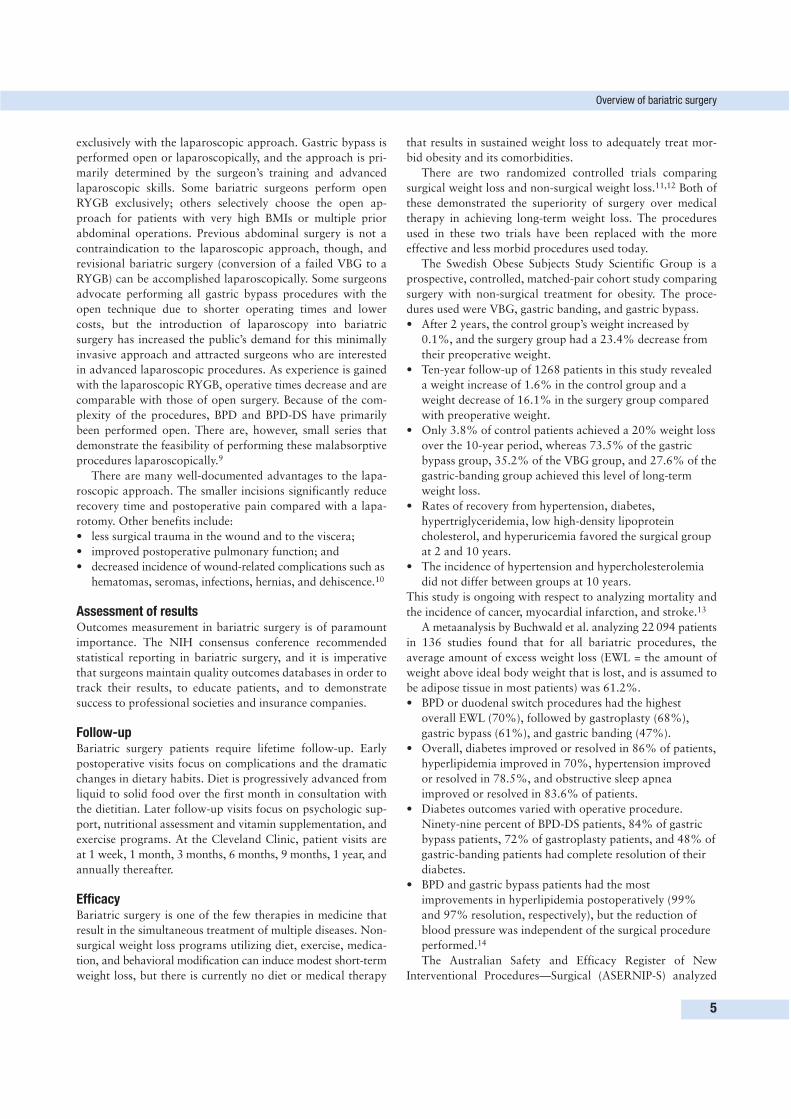
• a combination of restriction and malabsorption
(Roux-en-Y gastric bypass, RYGB) (Fig. 1.4).
Between 1998 and 2003, the number of bariatric opera-
tions performed in the USA increased from 13 000 to 103 000
per year.4 During that period, the percentage of gastroplasty
procedures performed declined from 25% to 7%. Gastric by-
pass procedures comprise over 80% of bariatric procedures
currently performed in the USA and 65% of bariatric proce-
dures performed worldwide (Table 1.1).5
The choice of operation depends largely on patient prefer-
ence. There are currently no data available to preoperatively
predict which operation a specific patient should undergo. In
surveys from the USA and Australia, safety and invasiveness
had the greatest impact on patient choice for bariatric opera-
tions.6 Most patients in the USA are currently seeking either
gastric bypass or adjustable gastric-banding procedures, and the
relative risks and benefits of each must be carefully explained.
• Gastric bypass generally provides more weight loss in a
shorter time than LAGB does, but it is more invasive and
has a higher mortality rate than LAGB.
• Adjustable gastric banding has the lowest mortality rate of
any procedure currently used, but it generally results in
less weight loss than with RYGB and involves a permanent
foreign body in the abdomen.
Follow-up requirements must be considered preoperatively
as well. Gastric bypass requires lifelong vitamin supplementa-
tion that can be a cost burden for some patients, while LAGB
requires more frequent follow-up visits for band adjustments in
the first year after surgery. BPD and duodenal switch procedures
are performed at a few specialized centers and are more likely
to be performed in superobese patients or patients specifically
seeking these operations.
Restrictive procedures work by reducing the quantity of food
that can be consumed at one time. In the case of LAGB, the
degree of restriction can be increased or decreased based on the
patient’s weight loss. Malabsorptive procedures ensure that
ingested food and digestive enzymes remain separated for a sub-
stantial bowel length to limit caloric absorption. RYGB provides
a combination of restriction and decreased absorption. The
restrictive component of the operation consists of the creation
of a small (15–30 mL) gastric pouch. The standard Roux limb
is 75 cm in length and results in mild, and probably transient,
malabsorption. The long-limb (150 cm) RYGB used for super-
obese patients results in a greater degree of malabsorption.
The rapid reduction of comorbidities such as diabetes and
the long-term weight loss achieved by RYGB and BPD cannot
be explained exclusively by restriction or malabsorption. Other
mechanisms of weight loss and glucose control following ba-
riatric surgery are being investigated.
• Ghrelin, a peptide hormone produced by the stomach and
duodenum, is normally released prior to meals and acts on
the hypothalamus to increase appetite. Alterations in ghrelin
production may play a role in the decreased appetite and
sustained weight loss seen after certain bariatric procedures.
• Other gut hormones, such as peptide YY, glucagon-like
peptide-1, and glucose-dependent insulinotropic peptide,
may also contribute to the early satiety and rapid
reduction of insulin resistance seen after bariatric surgery.
• Obesity is associated with a proinflammatory and
prothrombotic state. Increased adipocyte activity, and the
associated increase in circulating inflammatory cytokines,
may be related to many of the cardiovascular risk factors
seen with obesity. Preliminary studies have demonstrated
improvement in these detrimental cytokines and
adipokines after surgical weight loss.
Evolution of bariatric surgeryThe initial operations to treat morbid obesity were performed
in the 1950s and were malabsorptive procedures. The jejuno-
colic and jejunoileal bypass procedures resulted in electrolyte
disturbances and liver failure. In 1967, Mason and Ito developed
the gastric bypass procedure by creating a 50- to 100-mL pro-
ximal gastric pouch that emptied into a loop gastrojejuno-
stomy.7 Modifications to this procedure over the past 35 years
have been directed towards minimizing the complications of
bile reflux, anastomotic ulcers, and gastrogastric fistulas, and
have resulted in the current Roux-en-Y divided gastric bypass.
In the late 1970s, Scopinaro developed the BPD procedure.8
In this procedure, the small bowel is divided 250 cm proximal
to the ileocecal valve, and the alimentary limb is anastomosed
to the gastric pouch. The duodenal switch (BPD-DS) is a
modification of BPD in which the pylorus is left intact to
prevent marginal ulceration and improve gastric emptying.
Gastric banding was also developed in the late 1970s, and
the initial use of fixed banding material to create a proximal
gastric pouch has evolved into the laparoscopic placement of
an adjustable gastric band.
Indications• Patients with a BMI > 35 kg/m2 with obesity-related
comorbidities, and those with a BMI > 40 kg/m2 with or
without comorbidities, are eligible for bariatric surgery.
Overview of bariatric surgery
3
Table 1.1 Types of bariatric procedure performed
Procedure USA (%) Worldwide (including
USA) (%)
Gastric bypass 80 65
Laparoscopic 5–10 25
adjustable
gastric band
Vertical banded < 5 5
gastroplasty
Biliopancreatic 5–10 5
diversion/duodenal
switch
(Adapted from Buchwald and Williams 2004,5 with permission.)
Figure 1.4 Roux-en-Y gastric bypass (RYGB).
• Patients must have attempted medical weight loss
programs and should be highly motivated to change their
lifestyle after surgery.
• The majority of patients undergoing bariatric surgery are
between ages 18 and 60. There was insufficient evidence
at the time of the 1991 NIH consensus to make
recommendations about surgery at the extremes of age.
There is a growing body of evidence, however, that
supports bariatric surgery in carefully selected adolescents
and in the elderly (> 60 years). The current indications for
bariatric surgery may broaden as long-term safety and
efficacy studies in these patient groups become available.
Contraindications• Patients who cannot tolerate general anesthesia due to
cardiac, pulmonary, or hepatic insufficiency are not
candidates for surgery.
• Additionally, patients must be able to understand the
consequences of the surgery and comply with the extensive
preoperative evaluation and the postoperative lifestyle
changes, diet, vitamin supplementation, and follow-up
program.
• Patients who have ongoing substance abuse or unstable
psychiatric illness are poor candidates for bariatric
surgery.
Preparation for surgerySurgical candidates must complete a thorough medical evalua-
tion, a psychologic evaluation, and have preoperative testing
appropriate for their comorbid conditions. There are over 30
comorbidities associated with obesity, and many of these pre-
dispose bariatric surgical patients to increased perioperative
risk (Table 1.2). Because morbidly obese patients are at higher
risk for having hypertension, diabetes, coronary artery
disease, left ventricular hypertrophy, congestive heart failure,
and pulmonary hypertension, an electrocardiogram should be
performed on every patient, and a preoperative cardiology
evaluation should be performed when there is evidence of
cardiovascular disease.
Obstructive sleep apnea is frequently occult in this patient
population until a thorough history prompts a preoperative
evaluation. Patients with symptoms of loud snoring or daytime
hypersomnolence should undergo polysomnography and, if
positive, be treated with nasal continuous positive airway
pressure (CPAP). Because these patients are at risk for upper
airway obstruction, close monitoring and nasal CPAP should
continue postoperatively. Asthma and obesity hypoventilation
syndrome (chronic hypoxemia, hypercarbia, pulmonary hyper-
tension, and polycythemia) are also severe pulmonary compli-
cations of obesity and should be evaluated by a pulmonologist
preoperatively.
Upper gastrointestinal barium studies and endoscopy should
be performed for patients with severe gastroesophageal reflux
symptoms. Because the incidence of gallstones is high in this
population, preoperative abdominal sonography is routinely
performed in many centers.
All bariatric patients should undergo thorough nutritional
evaluation and counseling preoperatively. Patients must under-
stand how their diet will change after surgery, and what
supplements are necessary to prevent specific nutritional
deficiencies. The dietitian plays a key role in determining
whether a patient understands the significant changes in diet
that will occur after bariatric surgery.
Psychologic testing is performed preoperatively to assess
patients’ expectations and to ensure that there are no active
psychiatric issues that would put the patient at risk for failure
or poor compliance postoperatively.
Surgical techniquesWorldwide, two-thirds of bariatric procedures are performed
laparoscopically.5 Adjustable gastric banding is performed
1 Weight loss surgery: state of the art
4
Table 1.2 Comorbidities associated with obesity
System Comorbidities
Cardiovascular Hyperlipidemia
Heart failure
Myocardial infarction
Hypertension
Stroke
Left ventricular hypertrophy
Venous stasis
ulcers/thrombophlebitis
Pulmonary Asthma
Obstructive sleep apnea
Obesity hypoventilation
syndrome
Pulmonary hypertension
Endocrine Insulin resistance
Type 2 diabetes
Polycystic ovarian syndrome
Hematopoetic Deep venous thrombosis
Pulmonary embolism
Gastrointestinal Gallstones
Gastroesophageal reflux disease
Abdominal hernia
Genitourinary Stress urinary incontinence
Urinary tract infections
Obstetric/gynecologic Infertility
Miscarriage
Fetal abnormalities and infant
mortality
Musculoskeletal Degenerative joint disease
Gout
Plantar fasciitis
Carpal tunnel syndrome
Neurologic/psychiatric Intracranial hypertension
Depression
Anxiety
exclusively with the laparoscopic approach. Gastric bypass is
performed open or laparoscopically, and the approach is pri-
marily determined by the surgeon’s training and advanced
laparoscopic skills. Some bariatric surgeons perform open
RYGB exclusively; others selectively choose the open ap-
proach for patients with very high BMIs or multiple prior
abdominal operations. Previous abdominal surgery is not a
contraindication to the laparoscopic approach, though, and
revisional bariatric surgery (conversion of a failed VBG to a
RYGB) can be accomplished laparoscopically. Some surgeons
advocate performing all gastric bypass procedures with the
open technique due to shorter operating times and lower
costs, but the introduction of laparoscopy into bariatric
surgery has increased the public’s demand for this minimally
invasive approach and attracted surgeons who are interested
in advanced laparoscopic procedures. As experience is gained
with the laparoscopic RYGB, operative times decrease and are
comparable with those of open surgery. Because of the com-
plexity of the procedures, BPD and BPD-DS have primarily
been performed open. There are, however, small series that
demonstrate the feasibility of performing these malabsorptive
procedures laparoscopically.9
There are many well-documented advantages to the lapa-
roscopic approach. The smaller incisions significantly reduce
recovery time and postoperative pain compared with a lapa-
rotomy. Other benefits include:
• less surgical trauma in the wound and to the viscera;
• improved postoperative pulmonary function; and
• decreased incidence of wound-related complications such as
hematomas, seromas, infections, hernias, and dehiscence.10
Assessment of resultsOutcomes measurement in bariatric surgery is of paramount
importance. The NIH consensus conference recommended
statistical reporting in bariatric surgery, and it is imperative
that surgeons maintain quality outcomes databases in order to
track their results, to educate patients, and to demonstrate
success to professional societies and insurance companies.
Follow-upBariatric surgery patients require lifetime follow-up. Early
postoperative visits focus on complications and the dramatic
changes in dietary habits. Diet is progressively advanced from
liquid to solid food over the first month in consultation with
the dietitian. Later follow-up visits focus on psychologic sup-
port, nutritional assessment and vitamin supplementation, and
exercise programs. At the Cleveland Clinic, patient visits are
at 1 week, 1 month, 3 months, 6 months, 9 months, 1 year, and
annually thereafter.
EfficacyBariatric surgery is one of the few therapies in medicine that
result in the simultaneous treatment of multiple diseases. Non-
surgical weight loss programs utilizing diet, exercise, medica-
tion, and behavioral modification can induce modest short-term
weight loss, but there is currently no diet or medical therapy
that results in sustained weight loss to adequately treat mor-
bid obesity and its comorbidities.
There are two randomized controlled trials comparing
surgical weight loss and non-surgical weight loss.11,12 Both of
these demonstrated the superiority of surgery over medical
therapy in achieving long-term weight loss. The procedures
used in these two trials have been replaced with the more
effective and less morbid procedures used today.
The Swedish Obese Subjects Study Scientific Group is a
prospective, controlled, matched-pair cohort study comparing
surgery with non-surgical treatment for obesity. The proce-
dures used were VBG, gastric banding, and gastric bypass.
• After 2 years, the control group’s weight increased by
0.1%, and the surgery group had a 23.4% decrease from
their preoperative weight.
• Ten-year follow-up of 1268 patients in this study revealed
a weight increase of 1.6% in the control group and a
weight decrease of 16.1% in the surgery group compared
with preoperative weight.
• Only 3.8% of control patients achieved a 20% weight loss
over the 10-year period, whereas 73.5% of the gastric
bypass group, 35.2% of the VBG group, and 27.6% of the
gastric-banding group achieved this level of long-term
weight loss.
• Rates of recovery from hypertension, diabetes,
hypertriglyceridemia, low high-density lipoprotein
cholesterol, and hyperuricemia favored the surgical group
at 2 and 10 years.
• The incidence of hypertension and hypercholesterolemia
did not differ between groups at 10 years.
This study is ongoing with respect to analyzing mortality and
the incidence of cancer, myocardial infarction, and stroke.13
A metaanalysis by Buchwald et al. analyzing 22 094 patients
in 136 studies found that for all bariatric procedures, the
average amount of excess weight loss (EWL = the amount of
weight above ideal body weight that is lost, and is assumed to
be adipose tissue in most patients) was 61.2%.
• BPD or duodenal switch procedures had the highest
overall EWL (70%), followed by gastroplasty (68%),
gastric bypass (61%), and gastric banding (47%).
• Overall, diabetes improved or resolved in 86% of patients,
hyperlipidemia improved in 70%, hypertension improved
or resolved in 78.5%, and obstructive sleep apnea
improved or resolved in 83.6% of patients.
• Diabetes outcomes varied with operative procedure.
Ninety-nine percent of BPD-DS patients, 84% of gastric
bypass patients, 72% of gastroplasty patients, and 48% of
gastric-banding patients had complete resolution of their
diabetes.
• BPD and gastric bypass patients had the most
improvements in hyperlipidemia postoperatively (99%
and 97% resolution, respectively), but the reduction of
blood pressure was independent of the surgical procedure
performed.14
The Australian Safety and Efficacy Register of New
Interventional Procedures—Surgical (ASERNIP-S) analyzed
Overview of bariatric surgery
5
international data regarding LAGB and 55 papers evaluating
VBG and RYGB.15 The reported 56% EWL at 4-year follow-
up after LAGB was comparable with the long-term weight
loss achieved with RYGB.
In an observational cohort study, Christou and associates
evaluated long-term morbidity and mortality in morbidly
obese patients. They compared 1035 patients who underwent
RYGB to 5746 age- and gender-matched morbidly obese
controls who had non-surgical management of their weight.
• The surgery group had a mean EWL of 67% at 5-year
follow-up; > 60% EWL at 16 years (72% follow-up); and
significantly reduced risk of developing cardiovascular
disease, cancer, infectious diseases, and endocrinologic,
musculoskeletal, and respiratory disorders.
• Five-year mortality in the bariatric surgery group was
0.68%, compared with 6.17% in the control group (89%
relative risk reduction).16
ComplicationsThe risks of bariatric surgery have decreased with increasing
experience and technical refinements. The operative mortality
for restrictive procedures, gastric bypass, and BPD are 0.1%,
0.5%, and 1.1%, respectively. In the ASERNIP-S review,
LAGB had an early mortality of 0.05%. Mortality after
bariatric surgery is primarily due to pulmonary embolism and
anastomotic leak. Early postoperative complications, parti-
cularly septic complications, are less common after restrictive
procedures such as VBG and LAGB.
Vertical banded gastroplasty has largely been abandoned
due to poor long-term weight loss and the late complications
of gastroesophageal reflux, stomal stenosis, staple line dehi-
scence, and intractable vomiting. Patients with these com-
plications frequently require conversion to a RYGB.
Biliopancreatic diversion and duodenal switch procedures
have excellent results in terms of short- and long-term weight
loss and resolution of comorbidities, but these procedures
have a higher mortality rate than other bariatric procedures
and a higher incidence of metabolic and nutritional problems.
Operative mortality for BPD ranges from 0.5 to 1.3%. Early
postoperative complications include intraperitoneal bleeding,
wound dehiscence, wound infection, anastomotic leak, and
gastric perforation. Nutritional deficiencies can occur after
bariatric procedures that bypass segments of the small bowel
(BPD, duodenal switch, and RYGB). Table 1.3 summarizes
the data from a review of nutritional deficiencies after baria-
tric procedures.17
Protein malnutrition is characterized clinically by hypo-
albuminemia (< 3.5 g/dL), anemia, edema, and alopecia, and
occurs 3–18% of the time after BPD or BPD-DS. These
patients may require total parenteral nutrition, and 6% will
have a revision to lengthen their common channel. Protein
malnutrition is seen less frequently after standard RYGB
(0–1.4%), but long-limb (> 150 cm) RYGB for superobese
patients can result in protein deficiency 3–13% of the time and
typically occurs within 2 years of surgery. Iron is absorbed in
the duodenum and proximal jejunum, and iron deficiency after
1 Weight loss surgery: state of the art
6
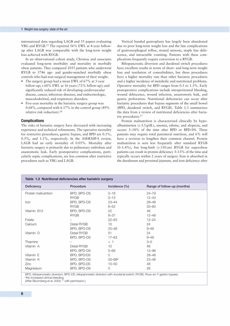
Table 1.3 Nutritional deficiencies after bariatric surgery
Deficiency Procedure Incidence (%) Range of follow-up (months)
Protein malnutrition BPD, BPD-DS 0–18 24–79
RYGB 0–13 12–43
Iron BPD, BPD-DS 23–44 28–48
RYGB 6–52 20–60
Vitamin B12 BPD, BPD-DS 22 48
RYGB 8–37 12–48
Folate – 22–63 12–24
Calcium Distal RYGB 10 24
BPD, BPD-DS 25–48 9–48
Vitamin D Distal RYGB 51 24
BPD, BPD-DS 17–63 9–48
Thiamine – < 1 3–5
Vitamin A Distal RYGB 10 48
BPD, BPD-DS 5–69 12–96
Vitamin E BPD, BPD/DS 5 28–48
Vitamin K BPD, BPD-DS 50–68a 23–48
Zinc BPD, BPD,DS 10–50 48
Magnesium BPD, BPD-DS 5 28
BPD, biliopancreatic diversion; BPD-DS, biliopancreatic diversion with duodenal switch; RYGB, Roux-en-Y gastric bypass.aNo increased clinical bleeding.(After Bloomberg et al. 2005,17 with permission.)
bariatric surgery is seen most commonly after BPD and BPD-
DS (23–44%) and RYGB (6–52%). Vitamin B12 is absorbed in
the terminal ileum, and deficiencies are seen after BPD (22%)
and RYGB (8–37%). Calcium absorption (duodenum and
jejunum) and vitamin D absorption (jejunum and ileum) are
impaired after BPD and RYGB as well, and these deficiencies
can lead to secondary hyperparathyroidism and increased
bone resorption. Calcium deficiency occurs 10–48% of the
time and vitamin D deficiency occurs 17–63% of the time in
published studies of malabsorptive procedures.17 The absorp-
tion of fat-soluble vitamins is impaired after BPD due to the
relatively short common channel.
Routine vitamin and mineral supplementation and careful
attention to protein intake following bariatric surgery are
necessary. Serious complications of these deficiencies can gen-
erally be avoided by early recognition and increased oral sup-
plementation. Further studies are needed to better define these
deficiencies and to determine guidelines for supplementation.
Hospital volume and surgeon experience are important
factors in bariatric surgery outcomes. Nguyen and colleagues
evaluated outcomes after RYGB according to hospital
volume, and found higher morbidity and mortality rates for
low-volume (< 50 cases/year) compared with high-volume
(> 100 cases/year) centers (1.2% versus 0.3% mortality,
respectively).18 Bariatric surgery, particularly the laparoscopic
approach, is technically challenging surgery that involves a
learning curve, and complications such as anastomotic leaks
and internal hernias are more common earlier in a surgeon’s
experience. Differences in complication rates between open
and laparoscopic procedures are discussed later in this chapter.
BARIATRIC SURGICAL PROCEDURES
Vertical banded gastroplastyVertical banded gastroplasty is a purely restrictive procedure
that limits the amount of solid food that can be consumed at
one time. A proximal gastric pouch empties through a fixed,
calibrated stoma that is reinforced with an external silastic
band or ring of mesh (Fig. 1.1). The advantages of VBG
include:
• improvement of comorbidities after weight loss,
• minimal nutritional deficiencies,
• the absence of any gastrointestinal anastomosis, and
• a lower morbidity and mortality rate than with RYGB.
It can be performed laparoscopically and is technically easier
than RYGB. The disadvantages of this procedure include long-
term weight loss that is inferior to that of RYGB, particularly
in sweet eaters, and multiple long-term complications that
frequently require reoperation.
Technique1. A 32 French Ewald tube is passed into the stomach to size
the pouch and stoma.
2. After the retrogastric dissection is completed from the
gastrohepatic ligament to the angle of His, the anvil of an
EEA circular stapler is placed behind the stomach and
manually passed through both walls of the stomach 8–9 cm
below the angle of His and adjacent to the Ewald tube.
3. The circular stapler is connected to the anvil and fired,
creating a 2.5-cm window in the proximal stomach. Four
rows of staples are then fired superiorly from the window
to the angle of His to create a 50-mL pouch.
4. A 7 × 1.5 cm strip of polypropylene mesh is then sewn to
itself around the outlet channel.
The laparoscopic approach has been used successfully for
VBG. A linear-cutting stapler may be used to divide the ver-
tical portion of the pouch or to excise a wedge of the fundus
and eliminate the need for a circular stapler.
EfficacyVertical banded gastroplasty achieves acceptable early weight
loss but has less favorable long-term weight loss than other
procedures used today. Ashy and colleagues demonstrated a
weight loss advantage of open VBG (87% EWL) over LAGB
(50% EWL) at 6 months.19 Some series have reported ade-
quate long-term success with VBG, but EWL 3–5 years after
VBG is typically 30–60%. Ten-year follow-up data show that
only 26–40% of patients maintain acceptable weight loss
(> 50% EWL), and one-third of patients in these series re-
turned to or exceeded their preoperative weight.20
ComplicationsEarly complications after VBG are infrequent, but late com-
plications have resulted in a 17–30% reoperation rate. The
most common late complications of VBG are:
• gastroesophageal reflux (16–38%),
• stomal stenosis (20%),
• staple line disruption (11–48%),
• incisional hernia (13%),
• band migration (1.5%), and
• intractable vomiting (30–50%).21
Because of the poor long-term weight loss and high late
complication rate, VBG has largely been abandoned and is
performed by less than 5% of bariatric surgeons in the USA.
Laparoscopic adjustable gastric bandingThe LAGB is a restrictive procedure, and the device (Lap-
Band; Inamed Corporation, Carpinteria, California) was
approved for use in the USA in 2001, after having very good
results in Europe and Australia. This silicone band with an
inflatable inner collar is placed around the upper portion of
the stomach to create a small gastric pouch. The band is con-
nected to a port that is placed in the subcutaneous tissue of
the abdominal wall. The inner diameter of the band can be
adjusted by injecting saline through the port (Fig. 1.2).
• The adjustable nature of the LAGB is a major advantage
that distinguishes it from VBG. Band adjustments are
made according to weight loss.
• The LAGB is technically the simplest bariatric surgery to
perform and requires less operating time than for other
procedures.
Bariatric surgical procedures
7
• No anastomoses are created, and the morbidity and
mortality are low.
• This procedure is reversible and, if patients fail to lose
adequate weight after LAGB, it can be converted to a
RYGB.
The disadvantages of the LAGB include:
• the need for frequent postoperative visits for band
adjustments, and
• band slippage or gastric prolapse through the band
(5–10%).
These mechanical complications require reoperation. Band
erosion into the stomach, gastroesophageal reflux, esophageal
dilatation, and dysmotility can also occur.
Technique1. The patient is placed in steep reverse Trendelburg position,
and six laparoscopic ports are placed.
2. The left lobe of the liver is retracted anteriorly, and a
15-mL balloon is placed transorally to calibrate the gastric
pouch.
3. The pars flaccida technique is used to create a retrogastric
tunnel from the base of the right crus of the diaphragm to
the angle of His.
4. The band is passed through the retrogastric tunnel toward
the angle of His and encircles the stomach approximately
1 cm below the gastroesophageal junction.
5. The tail of the band is passed through the buckle, and the
band is locked in place around the gastric cardia.
6. A calibration tube is passed to assess the size of the stoma,
and the anterior stomach is sutured over the band with
interrupted sutures.
7. The tube attached to the band is brought out through a
left-sided trocar site and attached to the port.
8. The port is then placed in a subcutaneous pocket and
sutured to the anterior rectus sheath.
Patients remain in the hospital for 1 or 2 days, and a
Gastrografin swallow is done prior to discharge to confirm
band position and patency. Patients are kept on a liquid diet
for 1 month postoperatively, at which time solid food can be
introduced. Band adjustments can be made with or without
fluoroscopic guidance. The first band adjustment is performed
4–8 weeks postoperatively, and patients are then observed
monthly for the first year to assess weight loss and to make
further adjustments if necessary.
EfficacyReports of weight loss after LAGB have been variable but
generally fall in the 40–55% EWL range 3 years after the
procedure. Weight loss after LAGB is more gradual than with
RYGB, and most of the weight loss after LAGB takes place in
the first 3 years after surgery. O’Brien reported results on 706
patients undergoing the LAGB in Australia, with a mean EWL
of 57% at 72 months and major improvements in diabetes,
asthma, gastroesophageal reflux, dyslipidemia, sleep apnea,
depression, and quality of life.22 The Italian Collaborative
Study Group for the Lap-Band system reviewed 1863 patients
undergoing LAGB. Six-year follow-up showed a steady decrease
in BMI from a preoperative average of 43 kg/m2 to a BMI of
32 kg/m2 at 72 months.23
Initial results with the LAGB in the USA were not as favor-
able as those in Europe and Australia. EWL at 2-year follow-
up was typically reported to be between 35 and 45%. Some
recent US studies of LAGB have approached the success rates
seen in international studies, though, including a report of
1014 Lap-Band procedures with 64% EWL at 4 years (> 85%
follow-up). In this study, 75% of patients achieved satisfactory
weight loss (> 50% EWL) at 4 years.24
ComplicationsLaparoscopic adjustable gastric banding has a low operative
mortality (0.05%) and an 11% rate of perioperative and late
complications.15 Postoperative mortality was 0.53% in the
Italian Collaborative Study, and the ASERNIP-S review re-
ported three deaths in 5827 LAGB cases (0.05%). Intraopera-
tive bleeding or injury to the stomach, esophagus, or spleen
occurs less than 1% of the time.
• Early postoperative complications include bleeding (0.5%),
wound infection (0–1%), and food intolerance (0–11%).
• Late complications include band slippage or gastric
prolapse through the band (7–21%), band erosion
(2–7%), tube-related problems (4%), persistent vomiting
(13%), pouch dilatation (5%), and gastroesophageal
reflux.
In a study of 1120 patients, O’Brien and Dixon reported a
1.5% early major complication rate.25 These complications
included 10 access port infections; four patients with delayed
emptying through the band; and one case each of deep venous
thrombosis, hepatotoxicity, and bile leak from the liver. The
most common late complication requiring reoperation after
LAGB is gastric prolapse or slippage. As experience was gained,
the rate of this complication decreased from 25% to 4.7%.
Erosion of the band into the stomach occurred in 3% of
patients early in the authors’ experience, and problems with
the access port occurred in 5.4% of their patients. Although
esophageal dilatation was common after prolapse or aggres-
sive band adjustments, no persistent esophageal dilatation or
dysmotility was found after appropriate treatment of the
prolapse or decreased band restriction.
Roux-en-Y gastric bypassRoux-en-Y gastric bypass combines a restrictive and a malab-
sorptive procedure, and is the most commonly performed
bariatric procedure in the USA (80%). A small 15- to 30-mL
gastric pouch is created to restrict food intake, and a Roux-
en-Y gastrojejunostomy provides the malabsorptive compo-
nent (Fig. 1.4).
The advantages of RYGB include:
• superior weight loss when compared with VBG,
• excellent long-term reduction in EWL, and
• resolution or elimination of comorbidities.
Early and late complication rates are reasonably low, and opera-
tive mortality ranges from 0 to 0.5%. Dumping syndrome
1 Weight loss surgery: state of the art
8
may occur after RYGB, and this may discourage patients from
eating sweets.
Disadvantages of RYGB include:
• the potential for anastomotic leaks and strictures,
• severe dumping syndrome symptoms, and
• procedure-specific complications including distension of
the excluded stomach and internal hernias.
The RYGB is technically more challenging to perform than the
restrictive procedures, particularly using the laparoscopic
approach.
Open RYGB technique1. The abdomen is entered through an upper midline
incision, and a thorough exploration is completed.
2. The anterior and lateral phrenoesophageal ligament is
opened to the angle of His.
3. The distal esophagus is mobilized and encircled with a
Penrose drain, and the gastrohepatic ligament is opened
over the caudate lobe.
4. The mesentery between the second and third branches of
the left gastric artery is divided, and a retrogastric space
is developed from the lesser curvature to the angle of
His.
5. The pouch can be formed using a series of firings with a
linear-cutting stapler to create a vertically oriented
pouch, or a red rubber tube placed in the retrogastric
space can be used to guide 90-mm linear staplers behind
the stomach to create a 15- to 30-mL pouch.
6. The ligament of Treitz is identified, and the jejunum is
divided with a linear stapler 15–45 cm distal to the
ligament.
7. A standard length (75 cm) or long-limb length (150 cm
for BMI > 50 kg/m2) Roux limb is measured, and the
jejunojejunostomy is created with the linear stapler.
8. The mesenteric defect at the jejunojejunostomy is closed
with suture.
9. The Roux limb can be brought up to the gastric pouch
retrocolic and retrogastric, retrocolic and antegastric, or
antecolic and antegastric, depending on the surgeon’s
preference and tension on the Roux limb. If the Roux
limb is brought through the transverse mesocolon, the
space between the jejunal and transverse colon
mesenteries is closed (Peterson’s space) to prevent
internal herniation of small bowel.
10. A 1- to 1.5-cm gastrojejunostomy is either hand-sewn
over a 30-F dilator or created with a circular stapler.
11. The anastomosis is tested with air insufflation or
injection of methylene blue through a carefully guided
nasogastric tube or with intraoperative endoscopy.
Laparoscopic RYGB technique1. After pneumoperitoneum is established, five or six access
ports are placed.
2. The sequential firings of a linear cutting stapler are used
to create a vertically oriented gastric pouch measuring
15–30 mL.
3. The ligament of Treitz is identified, and the jejunum is
divided 10–12 cm distally with a linear stapler.
4. A 75- to 150-cm Roux limb is measured, and a
side-to-side jejunojejunostomy is created with a linear
stapler. Several techniques can be used to create the
gastrojejunal anastomosis.
If a circular stapler is used, the anvil can be pulled into the
pouch transorally using endoscopy and placement of a
loop wire percutaneously into the gastric pouch.
In the transgastric method, the anvil is placed in the
stomach through a distal gastrotomy prior to pouch
formation. The anvil is then positioned in the upper
stomach and included in the pouch that is created with
a linear stapler.
The current method favored by the authors is placement
of continuous layer of sutures to approximate the Roux
limb and pouch, followed by the creation of a side-to-
side anastomosis with a linear stapler.
5. The anastomosis is completed with two layers of running
suture anteriorly over a flexible endoscope. The
anastomosis can also be completely hand-sewn in two
layers.
6. The anastomosis is tested for integrity and hemostasis
with the flexible endoscope. The conversion rate to open
RYGB is < 5%.
EfficacyThe RYGB results in mean EWL ranging from 65 to 80% in
studies with follow-up of 2 years or less. There is no signifi-
cant difference in weight loss between the open and laparo-
scopic approach, and weight loss typically reaches a nadir
18–24 months after surgery. In a study by Schauer and col-
leagues, the mean EWL was 83% at 1 year and 77% at
30 months.26 Longer follow-up after RYGB reveals some
weight regain, with 60–70% EWL at 5 years. The Swedish
Obese Subjects Study demonstrated 10-year weight loss (as a
percentage of initial body weight) of 25% for RYGB.13
Nguyen and colleagues compared laparoscopic (n = 79) to
open (n = 76) RYGB and found a longer operative time but
shorter hospital stay (3 versus 4 days) in the laparoscopic
group. Weight loss at 1 year was similar between groups, but
the laparoscopic group had fewer wound complications and a
more rapid return to daily activities.27
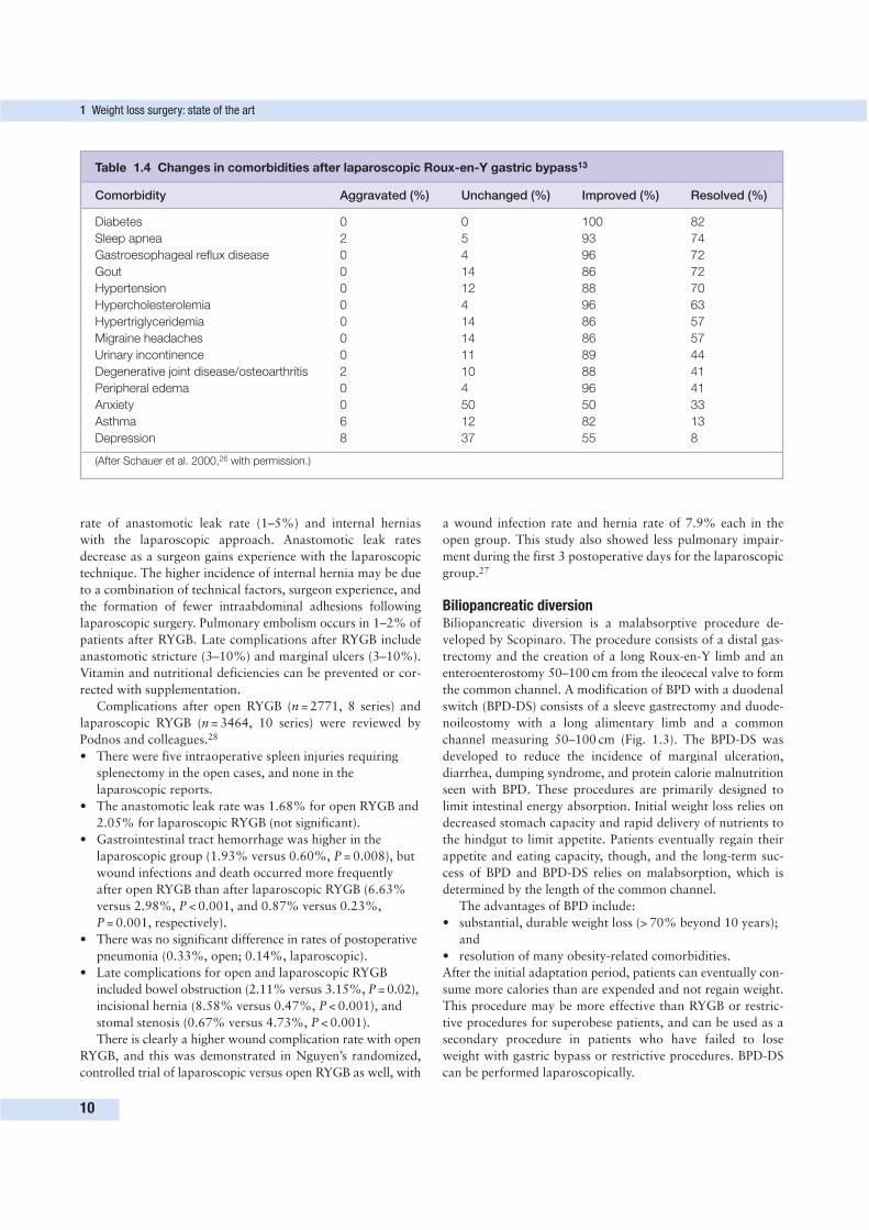
The RYGB results in significant improvement or resolution
of many major obesity-related comorbidities (Table 1.4). De-
generative joint disease, hyperlipidemia, gastroesophageal re-
flux, hypertension, obstructive sleep apnea, depression, stress
urinary incontinence, asthma, migraine headaches, venous in-
sufficiency, congestive heart failure, and diabetes improve or
resolve in the majority of patients after surgery. Type 2
diabetes resolves in over 80% of patients after RYGB.
ComplicationsOverall, the incidence of major early postoperative compli-
cations is similar between open and laparoscopic RYGB
(10–15%). Notable exceptions to this, though, are the higher
Bariatric surgical procedures
9
rate of anastomotic leak rate (1–5%) and internal hernias
with the laparoscopic approach. Anastomotic leak rates
decrease as a surgeon gains experience with the laparoscopic
technique. The higher incidence of internal hernia may be due
to a combination of technical factors, surgeon experience, and
the formation of fewer intraabdominal adhesions following
laparoscopic surgery. Pulmonary embolism occurs in 1–2% of
patients after RYGB. Late complications after RYGB include
anastomotic stricture (3–10%) and marginal ulcers (3–10%).
Vitamin and nutritional deficiencies can be prevented or cor-
rected with supplementation.
Complications after open RYGB (n = 2771, 8 series) and
laparoscopic RYGB (n = 3464, 10 series) were reviewed by
Podnos and colleagues.28
• There were five intraoperative spleen injuries requiring
splenectomy in the open cases, and none in the
laparoscopic reports.
• The anastomotic leak rate was 1.68% for open RYGB and
2.05% for laparoscopic RYGB (not significant).
• Gastrointestinal tract hemorrhage was higher in the
laparoscopic group (1.93% versus 0.60%, P = 0.008), but
wound infections and death occurred more frequently
after open RYGB than after laparoscopic RYGB (6.63%
versus 2.98%, P < 0.001, and 0.87% versus 0.23%,
P = 0.001, respectively).
• There was no significant difference in rates of postoperative
pneumonia (0.33%, open; 0.14%, laparoscopic).
• Late complications for open and laparoscopic RYGB
included bowel obstruction (2.11% versus 3.15%, P = 0.02),
incisional hernia (8.58% versus 0.47%, P < 0.001), and
stomal stenosis (0.67% versus 4.73%, P < 0.001).
There is clearly a higher wound complication rate with open
RYGB, and this was demonstrated in Nguyen’s randomized,
controlled trial of laparoscopic versus open RYGB as well, with
a wound infection rate and hernia rate of 7.9% each in the
open group. This study also showed less pulmonary impair-
ment during the first 3 postoperative days for the laparoscopic
group.27
Biliopancreatic diversionBiliopancreatic diversion is a malabsorptive procedure de-
veloped by Scopinaro. The procedure consists of a distal gas-
trectomy and the creation of a long Roux-en-Y limb and an
enteroenterostomy 50–100 cm from the ileocecal valve to form
the common channel. A modification of BPD with a duodenal
switch (BPD-DS) consists of a sleeve gastrectomy and duode-
noileostomy with a long alimentary limb and a common
channel measuring 50–100 cm (Fig. 1.3). The BPD-DS was
developed to reduce the incidence of marginal ulceration,
diarrhea, dumping syndrome, and protein calorie malnutrition
seen with BPD. These procedures are primarily designed to
limit intestinal energy absorption. Initial weight loss relies on
decreased stomach capacity and rapid delivery of nutrients to
the hindgut to limit appetite. Patients eventually regain their
appetite and eating capacity, though, and the long-term suc-
cess of BPD and BPD-DS relies on malabsorption, which is
determined by the length of the common channel.
The advantages of BPD include:
• substantial, durable weight loss (> 70% beyond 10 years);
and
• resolution of many obesity-related comorbidities.
After the initial adaptation period, patients can eventually con-
sume more calories than are expended and not regain weight.
This procedure may be more effective than RYGB or restric-
tive procedures for superobese patients, and can be used as a
secondary procedure in patients who have failed to lose
weight with gastric bypass or restrictive procedures. BPD-DS
can be performed laparoscopically.
1 Weight loss surgery: state of the art
10
Table 1.4 Changes in comorbidities after laparoscopic Roux-en-Y gastric bypass13
Comorbidity Aggravated (%) Unchanged (%) Improved (%) Resolved (%)
Diabetes 0 0 100 82
Sleep apnea 2 5 93 74
Gastroesophageal reflux disease 0 4 96 72
Gout 0 14 86 72
Hypertension 0 12 88 70
Hypercholesterolemia 0 4 96 63
Hypertriglyceridemia 0 14 86 57
Migraine headaches 0 14 86 57
Urinary incontinence 0 11 89 44
Degenerative joint disease/osteoarthritis 2 10 88 41
Peripheral edema 0 4 96 41
Anxiety 0 50 50 33
Asthma 6 12 82 13
Depression 8 37 55 8
(After Schauer et al. 2000,26 with permission.)
Disadvantages include:
• a higher operative mortality rate (1.1%) than with other
bariatric procedures; and
• metabolic complications including vitamin, mineral, and
protein deficiencies that occasionally require reoperation
to lengthen the common channel.
Liver disease and diarrhea occur with BPD and BPD-DS, al-
though less frequently than was seen with jejunoileal bypass.
After surgery, patients typically have four to six foul-smelling
stools per day and flatulence as a result of fat malabsorption.
Inability or unwillingness to comply with a strict nutritional
supplementation regiment postoperatively is a contraindica-
tion to performing this procedure. BPD and BPD-DS, parti-
cularly if done laparoscopically, are technically challenging
operations performed routinely only at specialized centers.
TechniqueBiliopancreatic diversion
Biliopancreatic diversion consists of a subtotal gastrectomy
leaving a proximal 200- or 400-mL pouch. The smaller pouch
is used for superobese patients.
1. The small bowel is divided 250 cm from the ileocecal
valve, and the distal end is anastomosed to the gastric
pouch with a 2- to 3-cm stoma.
2. A common channel is formed by completing the Roux-en-
Y enteroenterostomy 50–100 cm from the ileocecal valve.
If present, the gallbladder is routinely removed at the time of
BPD due to the high incidence of postoperative cholelithiasis.
Duodenal switch
The duodenal switch consists of a greater curvature sleeve
gastrectomy, leaving the antrum, the pylorus, and the first
portion of the duodenum in continuity. The remaining gastric
reservoir is 150–200 mL.
1. The proximal duodenum is divided, and a
duodenoileostomy is created using a 250 cm long
alimentary limb.
2. A Roux-en-Y anastomosis is then created to form a
100 cm long common channel.
EfficacyWeight loss after BPD is excellent, and the results are durable.
A recent metaanalysis demonstrated that BPD had a higher
percentage of EWL (70%) than other bariatric procedures.14
Scopinaro reported overall EWL of 74% at 8 years and 77%
at 18 years. There was no difference in long-term EWL
between morbidly obese and superobese (> 120% ideal body
weight) subjects.29 Ren and colleagues performed 40 laparo-
scopic BPD-DS procedures and reported EWL of 58% at
9 months. Operative time and perioperative morbidity were
higher in patients with BMI > 65 kg/m2.9
ComplicationsPostoperative complication rates for BPD are relatively high,
and postoperative mortality ranges from 0.4 to 1.3%. Mar-
ginal ulceration can occur up to 10% of the time, but this can
be reduced to 1–3% with the duodenal switch and acid sup-
pression therapy. Other complications include:
• dumping syndrome;
• protein calorie malnutrition and anemia in up to 12% and
40% of patients, respectively;
• vitamin B12 deficiency;
• hypocalcemia;
• fat-soluble vitamin deficiency; and
• bone demineralization (6%).
Failure to screen for such problems can lead to an unfavorable
wound healing after body-contouring surgery. The plastic
surgeon reading this chapter should also be cognizant of the
expected outcomes from these procedures in terms of magni-
tude of weight loss and effect on medical problems. A basic
appreciation of how the specific procedures impact nutri-
tional status is crucial.
In Scopinaro’s series of over 1700 BPD patients, the overall
rate of early major surgical complications (intraperitoneal
bleeding, wound dehiscence, wound infection, anastomotic
leak, and gastric perforation) decreased from 2.7% in his first
738 cases to 1.4% in his last 500 cases. Late complications of
BPD included iron deficiency anemia, which was decreased to
less than 5% with supplementation. Other late complications
included stomal ulcer in 3% of patients, incisional hernia
(8.7%), and protein malnutrition (7%). Four percent of patients
required elongation of the common channel or reversal of BPD.
In Ren’s laparoscopic series, there was one death (2.5%).
Postoperative complications included anastomotic leak (2.5%),
venous thrombosis (2.5%), subphrenic abscess (2.5%), and
staple line hemorrhage (10%), with an overall major morbi-
dity rate of 15%.
CONCLUSION
Obesity is a major public health problem in developed coun-
tries worldwide. Currently, the only treatment for this disease
that provides long-term weight loss is surgery. Restrictive, mal-
absorptive, and combination procedures have been developed,
and each has its merits and unique set of risks and compli-
cations. Weight loss after bariatric surgery is accompanied by
improvement or resolution of obesity-related comorbidities
and improved life expectancy.
Careful patient selection for bariatric surgery and selection of
the appropriate procedure for each patient are keys to success
when performing these operations. Close monitoring for nutri-
tional deficiencies and short- and long-term complications is
required to completely assess outcomes after these procedures.
REFERENCES
1. National Institutes of Health Conference. Gastrointestinal surgery
for severe obesity. Ann Intern Med 1991; 115:956–961.
2. Hedley AA, Odgen CL, Johnson CL, et al. Overweight and obesity
among US children, adolescents, and adults, 1999–2002. JAMA
2004; 291:2847–2850.
References
11
3. Wolf AM, Colditz GA. The costs of obesity: the US perspective.
Pharmacoeconomics 1994; 5:34–37.
4. Santry HP, Gillen DL, Lauderdale DS. Trends in bariatric surgical
procedures. JAMA 2005; 294(15):1909–1917.
5. Buchwald H, Williams SE. Bariatric surgery worldwide 2003. Obes
Surg 2004; 14(9):1157–1164.
6. Ren CJ, Cabrera I, Rajaram K, et al. Factors influencing patient
choice for bariatric operation. Obes Surg 2005; 15(2):202–206.
7. Mason EE, Ito C. Gastric bypass. Ann Surg 1969; 170:329–339.
8. Scopinaro N, Adami FG, Marinari GM, et al. Biliopancreatic
diversion. World J Surg 1998; 22:936–946.
9. Ren CJ, Patterson E, Gagner M. Early results of laparoscopic bilio-
pancreatic diversion with duodenal switch: a case series of 40 con-
secutive patients. Obes Surg 2000; 10(6):514–523; discussion 524.
10. Cottam DR, Mattar SG, Schauer PR. Laparoscopic era of opera-
tions for morbid obesity. Arch Surg 2003; 138(4):367–375.
11. [Anonymous]. Randomised trial of jejunoileal bypass versus
medical treatment in morbid obesity. The Danish Obesity Project.
Lancet 1979; 2:1255–1258.
12. Anderson T, Backer OG, Stokholm KH, et al. Randomized trial of
diet and gastroplasty compared with diet alone in morbid obesity.
N Engl J Med 1984; 310:352–356.
13. Sjostrom L, Lindroos AK, Peltonen M, et al. Lifestyle, diabetes, and
cardiovascular risk factors 10 years after bariatric surgery. N Engl J
Med 2004; 351(26):2683–2693.
14. Buchwald H, Avidor Y, Braunwald E, et al. Bariatric surgery. A
systematic review and meta-analysis. JAMA 2004;
292(14):1727–1737.
15. Chapman A, Kiroff G, Game P, et al. Systematic review of laparo-
scopic adjustable gastric banding in the treatment of obesity
(ASERNIP-S report no. 31). Adelaide: Australian Safety and
Efficacy Register of New Interventional Procedures—Surgical;
2002:18–48.
16. Christou NV, Sampalis JS, Liberman M, et al. Surgery decreases
long-term mortality, morbidity, and health care use in morbidly
obese patients. Ann Surg 2004; 240(3):416–424.
17. Bloomberg RD, Fleishman A, Nalle JE, et al. Nutritional deficien-
cies following bariatric surgery: what have we learned? Obes Surg
2005; 15:145–154.
18. Nguyen NT, Paya M, Stevens M, et al. The relationship between
hospital volume and outcome in bariatric surgery at academic
medical centers. Ann Surg 2004; 240(4):586–594.
19. Ashy AR, Merdad AA. A prospective study comparing vertical
banded gastroplasty versus laparoscopic adjustable gastric banding
in the treatment of morbid and superobesity. Int Surg 1998;
83:108–110.
20. Ramsey-Stewart G. Vertical banded gastroplasty for morbid obe-
sity: weight loss at short and long-term follow up. Aust N Z J Surg
1995; 65:4–7.
21. DeMaria EJ, Jamal MK. Surgical options for obesity. Gastroenterol
Clin North Am 2005; 34:127–142.
22. O’Brien PE, Brown WA, Smith A, et al. Prospective study of a
laparoscopically placed, adjustable gastric band in the treatment of
morbid obesity. Br J Surg 1999; 86:113–118.
23. Angrisani L, Furbetta F, Doldi B, et al. Lap-Band adjustable gastric
banding system: the Italian experience with 1863 patients operated
on 6 years. Surg Endosc 2003; 17:409–412.
24. Ponce J, Dixon JB. 2004 ASBS Consensus Conference. Laparoscopic
adjustable gastric banding. Surg Obes Relat Dis 2005; 1:310–316.
25. O’Brien PE, Dixon JB. Weight loss and early and late complica-
tions—the international experience. Am J Surg 2002; 184:42S–45S.
26. Schauer PR, Ikramuddin S, Gourash W, et al. Outcomes after
laparoscopic Roux-en-Y gastric bypass for morbid obesity. Ann
Surg 2000; 232(4):515–529.
27. Nguyen NT, Goldman C, Rosenquist J, et al. Laparoscopic versus
open gastric bypass: a randomized study of outcomes, quality of
life, and costs. Ann Surg 2001; 234(3):279–291.
28. Podnos YD, Jiminez JC, Wilson SF, et al. Complications after
laparoscopic gastric bypass: a review of 3464 cases. Arch Surg
2003; 138:957–961.
29. Scopinaro N, Gianetta E, Adami GF, et al. Biliopancreatic diversion
for obesity at eighteen years. Surgery 1996; 119:261–268.
1 Weight loss surgery: state of the art
12
With the universal increase in morbid obesity and the con-
comitant development of advanced laparoscopic techniques, a
large number of patients are opting for surgical therapy to
reduce excess body weight and ameliorate the myriad of asso-
ciated medical problems. The US Centers for Disease Control
and Prevention estimate that in excess of 64% of the US
population is either overweight or obese.1 On a global scale,
the International Obesity Task Force estimates that more than
1 billion individuals are overweight.2 The American Society for
Bariatric Surgery estimated that greater than 150 000 weight
loss procedures would be performed in the USA alone in the
year 2005.3 As surgical techniques have evolved, and weight
loss surgery has been performed with greater frequency, the
tremendous health benefits have been noted in many studies.4–13
However, the enormous benefits that the patients receive also
come at the cost of redundant, loose, hanging rolls of skin and
fat. Nearly every region of the body can be affected. This has
fueled a rapid increase in the number of patients presenting to
the plastic surgeon’s office for body-contouring procedures. It
is essential that the plastic surgeon approach these patients in
a concise, well-thought-out fashion with safety as the primary
concern.
PATIENT INTERVIEW
The individuals who seek the advice and expertise of a plastic
surgeon regarding the removal of excess skin after massive
weight loss have undergone a major life-altering event. While
their overall body shape has changed dramatically, they retain a
daily reminder of their obese state in the form of loose, hanging
skin. It is important for the clinician to realize this, and to re-
cognize that patients may still view themselves as ‘fat’ and
‘different’. Despite successful weight loss, self-esteem may be
low. These patients often state that they feel triply stigmatized:
• first for being morbidly obese,
• second for choosing surgical therapy to lose weight (the
‘easy way out’), and
• third for being considered vain and seeking the help of a
plastic surgeon.
Patients will be looking for a specialist who understands the
emotional as well as the physical needs of the postbariatric
patient, and their comfort with you will be influenced by your
sensitivity to self-esteem issues. We often start the interview
by congratulating patients on the progress they have made in
the process of weight loss and for taking steps to reclaim their
lives. Key historical components specific to the weight loss
patient are described in detail below, and provide the basis for
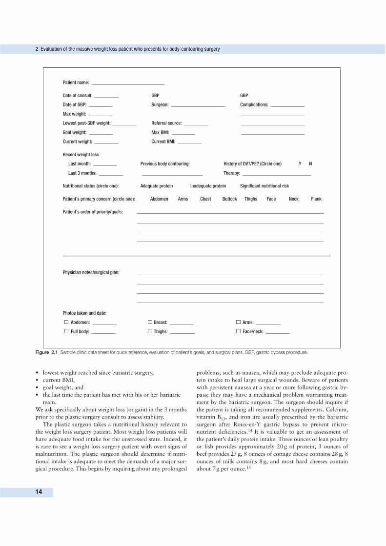
a thoughtful assessment. Figure 2.1 shows an office data col-
lection sheet that we use in our center to summarize some of
the important data points.
Weight loss history and nutritional assessmentWhile the initial interview is an excellent time to establish a
rapport with your patients, it is also an opportunity to elicit a
detailed history of their weight loss surgery and compliance with
the nutritional regimen after weight loss. The surgeon should
know what type of procedure the patient had, as different
operations will have varying potential to cause nutritional
deficits. Other important data points include:
• the timing of the weight loss surgery relative to the plastic
surgery consult,
• Body Mass Index (BMI) prior to surgery,
13
EVALUATION OF THE MASSIVEWEIGHT LOSS PATIENT WHOPRESENTS FOR BODY-CONTOURINGSURGERY
2James P. O’Toole and J. Peter Rubin
Key PointsProper evaluation of the weight loss patient includes the following key
components.
• Calculating BMI at time of presentation and assessing stability of weight.
• Screening for residual medical problems associated with obesity and
gastric bypass.
• Elucidating relevant psychosocial issues.
• Diagnosing the deformities that result from massive weight loss.
• Understanding the patient’s goals and expectations.
• Formulating a safe treatment plan.
• lowest weight reached since bariatric surgery,
• current BMI,
• goal weight, and
• the last time the patient has met with his or her bariatric
team.
We ask specifically about weight loss (or gain) in the 3 months
prior to the plastic surgery consult to assess stability.
The plastic surgeon takes a nutritional history relevant to
the weight loss surgery patient. Most weight loss patients will
have adequate food intake for the unstressed state. Indeed, it
is rare to see a weight loss surgery patient with overt signs of
malnutrition. The plastic surgeon should determine if nutri-
tional intake is adequate to meet the demands of a major sur-
gical procedure. This begins by inquiring about any prolonged
problems, such as nausea, which may preclude adequate pro-
tein intake to heal large surgical wounds. Beware of patients
with persistent nausea at a year or more following gastric by-
pass; they may have a mechanical problem warranting treat-
ment by the bariatric surgeon. The surgeon should inquire if
the patient is taking all recommended supplements. Calcium,
vitamin B12, and iron are usually prescribed by the bariatric
surgeon after Roux-en-Y gastric bypass to prevent micro-
nutrient deficiencies.14 It is valuable to get an assessment of
the patient’s daily protein intake. Three ounces of lean poultry
or fish provides approximately 20 g of protein, 3 ounces of
beef provides 25 g, 8 ounces of cottage cheese contains 28 g, 8
ounces of milk contains 8 g, and most hard cheeses contain
about 7 g per ounce.15
2 Evaluation of the massive weight loss patient who presents for body-contouring surgery
14
Patient name:
Date of consult: GBP GBP
Date of GBP: Surgeon: Complications:
Max weight:
Lowest post-GBP weight: Referral source:
Goal weight: Max BMI:
Current weight: Current BMI:
Recent weight loss
Last month: Previous body contouring: History of DVT/PE? (Circle one) Y N
Last 3 months: Therapy:
Nutritional status (circle one): Adequate protein Inadequate protein Significant nutritional risk
Patient’s primary concern (circle one): Abdomen Arms Chest Buttock Thighs Face Neck Flank
Patient’s order of priority/goals:
Physician notes/surgical plan:
Photos taken and date:
Abdomen: Breast: Arms:
Full body: Thighs: Face/neck:
Figure 2.1 Sample clinic data sheet for quick reference, evaluation of patient’s goals, and surgical plans. GBP, gastric bypass procedure.
Ask about any food aversions. Many patients will struggle
with concentrated animal protein after gastric bypass and may
have a difficult time maintaining a high protein intake.16 In
our center, we require patients to take at least 50–70 g of pro-
tein per day before elective body-contouring surgery. A referral
for formal nutritional evaluation and counseling, followed by
dietary modification and repeat assessment, would be re-
commended if protein intake is poor. Even patients with food
aversions can find protein sources that they can tolerate well if
they are coached through the process. It is essential for the
surgeon to understand that a weight loss patient with a favor-
able BMI does not necessarily represent a good surgical
candidate. Major surgery can increase the body’s nutritional
requirements by 25%, and many weight loss patients may
have to adjust their oral intake.17
Screening for medical problemsThe initial patient interview also provides the clinician with the
first opportunity to appreciate any medical issues that may in-
crease the risk of surgery. While body-contouring surgery after
massive weight loss may make a patient look and feel better, it
does not have the same level of overall health benefit as gastric
bypass does.18 The key focus is patient safety, and a history of
significant medical problems, including hypertension, ischemic
cardiac disease, sleep apnea, and diabetes, must be fully delin-
eated and addressed before body-contouring surgery. While most
medical comorbidities of obesity are significantly improved, if
not resolved, following weight loss, the plastic surgeon must
search for residual disease. Exercise tolerance is a useful indi-
cator of surgical risk. Patients who routinely do 45 min of vigo-
rous exercise without shortness of breath or other symptoms
will likely tolerate the stress of surgery. However, beware of the
inactive patient. These patients may have cardiac disease that
will be unmasked by a major surgical procedure. We advise
liberal use of medical consultants, as warranted, for preoperative
evaluation and recommendations for managing chronic disease
states. Patients who smoke are encouraged to take responsibility
for stopping in order to decrease their perioperative risk.
Psychosocial and lifestyle issuesPermanent lifestyle modifications are essential to long-term
weight loss success for patients after bariatric surgery. Do they
have a definitive exercise regimen? Do they have an exercise
‘buddy’ or at least a source of encouragement from friends
and family? Does the patient attend support group meetings?
Delineate the follow-up routine the patient has with their ba-
riatric surgeon. The majority of trained weight loss surgeons
have well-developed postoperative routines and support groups.
If your patient has gone to such a surgeon, and has not been
faithful with the postoperative regimen, explore the reasons.
Issues with compliance may be elucidated. These queries give
a reasonable assessment of how invested the patient is in her
or his own care. We find that the more motivated patient
generally represents a better candidate for elective body-
contouring surgery. We look for patients who understand that
it is not just the gastric bypass surgery that made them lose
weight, but rather their own personal commitment and res-
ponsibility to the process.
Weight loss can often be accompanied by major changes in
interpersonal relationships. Relationships may be strengthened
as family and friends rally behind the successful bariatric patient.
However, the radical change in appearance and lifestyle of the
patient also has the potential to evoke feelings of envy,
jealousy, and abandonment in people close to them. Turmoil
may ensue. While patients may be reluctant to discuss these
issues, it is vital to understand the stability of their support
network and the stressors that may be active before adding
the additional burden of recovering from surgery. Our ap-
proach is to ask patients about their personal lives, their
marriages, their living arrangements, their level of content-
ment with their lives personally and professionally, and their
support network. Example questions include the following.
• ‘Who lives at home with you, and are they able and
willing to help?’
• ‘Who are the other people available to help you in the first
few days to weeks?’
• ‘Who can drive you to post op visits?’
Observe the affect of the patient during the interview.
Individuals who have triumphed over the problems associated
with obesity can reasonably be expected to be proud of their
accomplishments. Be cautious of the patient who gives elusive
or vague answers to questions about their social situation.
The withdrawn individual should prompt further questioning
about symptoms of depression. While it is common to see
patients treated with antidepressants after a gastric bypass
procedure, simple depression is not a contraindication to sur-
gery. Inquire about general mood and any depressive episodes
during the past year. Patients with poorly treated (or untreated)
depression should be referred for psychiatric clearance. Addi-
tionally, any patients with bipolar disorder or schizophrenia
should also have formal psychiatric clearance.
PHYSICAL EXAMINATION
All aspects of a thorough physical examination should be
included in the initial patient evaluation in order to fully
appreciate the deformities and screen for residual medical
problems. The massive weight loss patient will present with a
wide range of physical anomalies. BMI, overall body type
(truncal versus peripheral), remaining adipose tissue, and rolls
and folds should be noted. Body fat distribution will vary
greatly in this patient population and will influence surgical
options. Attention should be given to the patient’s skin tone
and elasticity, as well as regional variations in skin elasticity.
On the abdominal examination, make note of:
• thickness of the subcutaneous tissue,
• presence of any hernias,
• degree of diastasis, and
• overall laxity of the abdominal wall.
Physical examination
15
To facilitate analysis of deformities in each anatomical region
of the body, a four-point rating scale can be applied. Table 2.1
shows the Pittsburgh Weight Loss Deformity Scale, which serves
as a tool to delineate the severity of deformities.19 During the
examination, consideration may be given to the number of
procedures required, the interactions of each procedure, and
whether staging would be appropriate. Look for stigmata of
nutritional depletion, including thin hair, brittle nails, and
BMI < 23 kg/m2 (it is rare for patients to reach this level). Be
observant for any physical limitations that will make the
recovery period too physically demanding or be aggravated by
surgical trauma. For example, a patient with chronic shoulder
pain that limits range of motion may have a difficult time
recovering from a brachioplasty.
MANAGING PATIENT EXPECTATIONS
Our approach is to ask patients to list the regions of their
bodies that they would like to correct in order of priority. We
then discuss surgical options that would effect changes in these
regions, including the location of the scars and the extent of
recovery. We emphasize the concept of trading excess skin for
scar, and assess the patient’s willingness to accept these scars.
We also emphasize the concept that, in general, body-contouring
procedures are major surgical procedures. Having adequate
time available to recover from the procedure is something that
should be addressed before surgery; this will allow patients to
make arrangements with their employer or, if necessary, delay
surgery until a more suitable time. Patients are also informed
that skin relaxation (relapse of skin laxity) is unpredictable
and can be severe enough to lead to operative revision. We
recommend advising patients about any office policies regarding
fees associated with revision surgery.
We find it useful to stand patients in front of a mirror and
review how areas of skin laxity might be improved on their
body, including a demonstration of how the surgeon pulls on
the skin to estimate the amount of resection and the resultant
impact on contour. During this part of the examination, limi-
tations of the procedures, given the patient’s body type, are
discussed. This often includes an explanation of which ana-
tomical regions can be changed with a given procedure and,
importantly, which adjacent regions will not be impacted.
How existing scars will be handled, and the effect of the pro-
cedure on stretch marks inside and outside the area of planned
resection, is explained. The quality of previous scars is noted
and used as a guideline to predict how future scars may appear.
To further emphasize the issue of surgical scars, a skin marker
is often used to draw the location of the scars directly on the
patient’s body and photographs are taken. This also helps the
patient review scar location with their spouses or significant
others after the consultation.
Patients who comprehend these issues and whose priorities
are addressed first are likely to be satisfied with the procedures
performed. If the points outlined in this section are thoroughly
conveyed by the surgeon, unrealistic expectations on the part of
the patient will emerge during the discussion. If these expec-
tations cannot be balanced, an unsatisfactory result is likely.
PATIENT SELECTION
Patient selection must be focused on maximizing safety. With
that goal in mind, the following key principles should be
applied.
• The patient should be weight-stable.
• BMI should be favorable.
• Nutrition must be adequate.
• Medical and psychosocial issues should be stable.
• The patient should have reasonable goals and expectations
considering their age, health, and body habitus.
It is also desirable for the patient to be on a definitive exercise
regimen. One may be lured into operating on a patient whose
anatomical deformities are easy to correct. However, under-
appreciated nutritional, medical, and psychosocial issues may
lead to an unfavorable outcome. Any issue that may influence
the safety of the planned procedure must be remedied prior to
operative intervention. If surgery is not to be offered at the
initial consultation, remain the patient’s advocate and encour-
age his or her continued progress. Inform patients that you
respect all that they have accomplished. We emphasize that
there is a correct time for elective surgery, and that this may
not be the best time. While they may be disappointed, they
will understand and appreciate that you are keeping their best
interests in mind. It is a common practice in our center to have
patients work on problematic nutritional or medical issues after
the initial consultation and follow-up for another evaluation
in 1–3 months. Figure 2.2 shows a checklist of the important
components to consider.
All patients considered candidates for body-contouring sur-
gery must be weight-stable for 3 months (this usually occurs
between 12 and 18 months after a gastric bypass procedure).
This is important for several reasons.
• For large surgical wounds, nutritional homeostasis and a
positive nitrogen balance are necessary to facilitate the
healing process.20
• A more predictable outcome can be achieved when the
patient is not actively losing weight.
• A high BMI is associated with increased wound-healing
complications.21,22
The BMI at presentation is an important factor. As the
patient’s BMI decreases, we are able to offer more safe sur-
gical options and expect better aesthetic outcomes. The best
candidates have a BMI of 28 kg/m2 or less. We are more cau-
tious in our level of aggressiveness with patients who have a
BMI between 29 kg/m2 and 32 kg/m2. Patients whose BMI is
between 32 and 35 kg/m2 should be selected with great care,
and procedures may be more limited than for patients with a
lower BMI. If a patient in this BMI range desires significant
contouring, we recommend delaying the operation until further
weight loss can be achieved. The technical challenge and sub-
sequent outcome are impacted by body fat distribution.
2 Evaluation of the massive weight loss patient who presents for body-contouring surgery
16
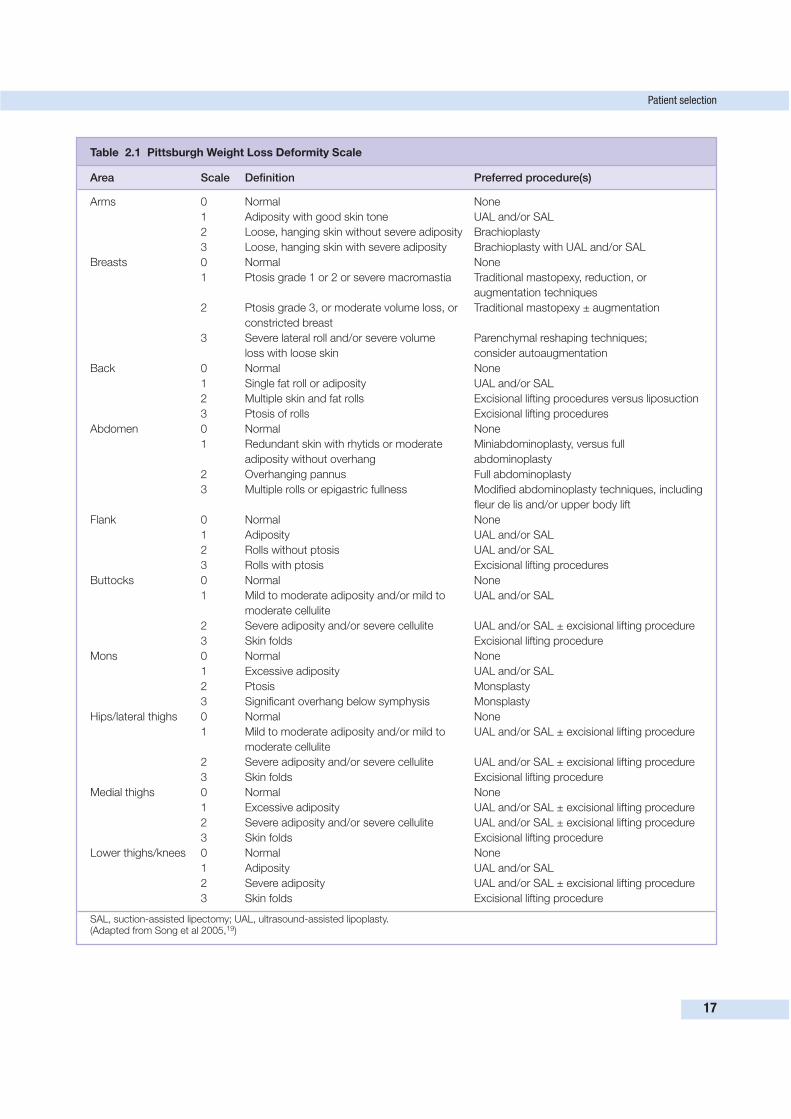
Patient selection
17
Table 2.1 Pittsburgh Weight Loss Deformity Scale
Area Scale Definition Preferred procedure(s)
Arms 0 Normal None
1 Adiposity with good skin tone UAL and/or SAL
2 Loose, hanging skin without severe adiposity Brachioplasty
3 Loose, hanging skin with severe adiposity Brachioplasty with UAL and/or SAL
Breasts 0 Normal None
1 Ptosis grade 1 or 2 or severe macromastia Traditional mastopexy, reduction, or
augmentation techniques
2 Ptosis grade 3, or moderate volume loss, or Traditional mastopexy ± augmentation
constricted breast
3 Severe lateral roll and/or severe volume Parenchymal reshaping techniques;
loss with loose skin consider autoaugmentation
Back 0 Normal None
1 Single fat roll or adiposity UAL and/or SAL
2 Multiple skin and fat rolls Excisional lifting procedures versus liposuction
3 Ptosis of rolls Excisional lifting procedures
Abdomen 0 Normal None
1 Redundant skin with rhytids or moderate Miniabdominoplasty, versus full
adiposity without overhang abdominoplasty
2 Overhanging pannus Full abdominoplasty
3 Multiple rolls or epigastric fullness Modified abdominoplasty techniques, including
fleur de lis and/or upper body lift
Flank 0 Normal None
1 Adiposity UAL and/or SAL
2 Rolls without ptosis UAL and/or SAL
3 Rolls with ptosis Excisional lifting procedures
Buttocks 0 Normal None
1 Mild to moderate adiposity and/or mild to UAL and/or SAL
moderate cellulite
2 Severe adiposity and/or severe cellulite UAL and/or SAL ± excisional lifting procedure
3 Skin folds Excisional lifting procedure
Mons 0 Normal None
1 Excessive adiposity UAL and/or SAL
2 Ptosis Monsplasty
3 Significant overhang below symphysis Monsplasty
Hips/lateral thighs 0 Normal None
1 Mild to moderate adiposity and/or mild to UAL and/or SAL ± excisional lifting procedure
moderate cellulite
2 Severe adiposity and/or severe cellulite UAL and/or SAL ± excisional lifting procedure
3 Skin folds Excisional lifting procedure
Medial thighs 0 Normal None
1 Excessive adiposity UAL and/or SAL ± excisional lifting procedure
2 Severe adiposity and/or severe cellulite UAL and/or SAL ± excisional lifting procedure
3 Skin folds Excisional lifting procedure
Lower thighs/knees 0 Normal None
1 Adiposity UAL and/or SAL
2 Severe adiposity UAL and/or SAL ± excisional lifting procedure
3 Skin folds Excisional lifting procedure
SAL, suction-assisted lipectomy; UAL, ultrasound-assisted lipoplasty.(Adapted from Song et al 2005,19)

The patient should be counseled that additional weight loss
allows for a safer operation with better aesthetic outcomes.
Work on a weight loss plan with the patient and nutritionist,
and schedule a 2- to 3-month follow-up appointment. This
way, the patient will remain under your care and not feel
abandoned; moreover, you are able to serve as a motivating
source. Some patients in this BMI range may benefit from a
first-stage breast reduction or simple panniculectomy if such a
procedure would improve their ability to exercise and pro-
gress with further weight loss. For patients with a BMI greater
than 35 kg/m2, our practice is, in most cases, to avoid opera-
tions because of increased risk of complications and less po-
tential for satisfying aesthetic results.22,23 Patients in this BMI
range would generally be offered only a truly functional
panniculectomy, with strict indications of severe panniculitis
or a profoundly disabling pannus.
The importance of the nutritional status of the postbariatric
patient cannot be overstressed.24–27 If patients have symptoms
consistent with a physical impedance to eating, have them see
their bariatric surgeon to rule out stricture. Because gastric by-
pass patients have altered gastrointestinal physiology, and sub-
sequent dietary issues are to be expected, nutritional issues
should be revisited in the postoperative period if any wound-
healing complications arise.28 As mentioned earlier, our prac-
tice is to require at least 50–70 g of protein intake per day
before surgery will be offered. A patient who is incapable of
50 g per day does not represent a surgical candidate, and
dietary modification is essential.
Medical and psychosocial issues must also be stable prior
to any operation. Patients with significant medical comorbidi-
ties are routinely sent to an appropriate medical specialist for
further evaluation and clearance. An adequate support network
should be in place. Active smokers are encouraged to stop at
least 1 month prior to surgery. If this is not possible, then the
extent of the procedure performed, especially the amount of
tissue undermining, is limited. Similar caution is exercised
with diabetic patients and those treated with steroids.
The final component is a reasonable set of goals and expec-
tations. Patients should be willing to accept extensive scars in
exchange for loose skin, understand both the power and
limitations of the intended procedures, and appreciate which
areas of the body will not be affected by the planned surgery.
This last point is important because improving one area of the
body may highlight deformities in adjacent areas.
COMBINATION PROCEDURES, STAGING, AND DEALINGWITH ABDOMINAL HERNIAS
Performing body-contouring procedures in two or more stages
should be considered if the patient has goals of reshaping
multiple regions. The advantages of staging are:
• less anesthetic time,
• less blood loss,
• less surgeon fatigue,
• avoidance of opposing vectors of pull on regions of skin,
and
• the ability to have a second chance to correct any
contour irregularities or skin relaxation seen after the
first stage.
Disadvantages of staging include:
• multiple anesthetics,
• increased time off work, and
• increased expense for the patient.
2 Evaluation of the massive weight loss patient who presents for body-contouring surgery
18
Evaluation/screening checklist
What is the current BMI?
Has the patient's weight been stable for at least 3 months?
Active nausea or vomiting? If yes, immediate referral to gastric bypass surgeon.
Would the patient benefit from further weight loss? If yes, return in 2–3 months for weight check.
Is the patient's nutrition adequate? If no, comprehensive nutritional evaluation.
Is the psychosocial situation stable and adequate?
Are there medical issues that preclude safe surgery and/or require further evaluation?
Is the patient willing to accept visible scars?
Does the patient understand the magnitude of the planned procedure?
Does the patient appreciate the recovery involved and have an adequate support network?
Are expectations reasonable?
Figure 2.2 Screening and evaluation checklist.
While it may be feasible to do two or three procedures in a
single stage, the surgeon should be guided by his or her level
of experience, experience of the operating room team, and
treatment setting. Individual procedures may be performed
safely at a fully equipped surgery center, assuming that ade-
quate personnel are available for recovery and that adequate
arrangements are in place should extended recovery be neces-
sary. Great caution should be exercised in the surgery center
setting if combined procedures are considered. Multiple (more
than two) procedures performed in a single anesthetic should
take place in a hospital setting.
It is not uncommon for the plastic surgeon to encounter a
massive weight loss patient with an incisional hernia. When
approaching these patients, we first consider whether there has
been sufficient weight loss to avoid excessive pressure on the
repair exerted by a still obese intraabdominal compartment. It
is reasonable to recommend further weight loss and use of an
abdominal binder for comfort before performing surgery on a
large asymptomatic hernia, if necessary. If the patient has
reached an appropriate body weight for hernia repair, consi-
deration is then given to the extent of the procedure. For small
or moderate-sized hernias, we will combine the repair with
major body-contouring procedures (e.g. lower body lift). Very
large hernias may require extensive lysis of adhesions and/or
separation of the abdominal wall components to achieve clo-
sure. When such an abdominal wall reconstruction is antici-
pated, we limit the body-contouring procedures to a concurrent
panniculectomy and stage any other desired surgeries. We
routinely bowel-prepare patients with hernias, and seek re-
commendation from the patient’s bariatric surgeon regarding
the preferred method. Bariatric surgeons may be dogmatic
about which gastrointestinal medications are prescribed for
their patients. Moreover, the referring weight loss surgeon
may want to be involved with these cases in a team approach.
CONCLUSION
Body contouring is a wonderful adjunct to bariatric surgery
and completes the weight loss process for many patients. Any
plastic surgeon who evaluates patients after massive weight
loss will see the full spectrum of patient subtypes. The majo-
rity of patients who present to the office for contouring sur-
gery will be well adjusted and have undertaken great measures
to reclaim their lives. However, there will be individuals who
are not quite prepared for surgery. A thoughtful and orga-
nized approach to the massive weight loss patient will identify
the individuals who represent good surgical candidates. Care-
fully devised operations for the appropriate patient at the
right time have the potential to provide a tremendously re-
warding experience for the patient and surgeon. As the sur-
geon, you have the capability to eradicate the last reminders
of the obesity that these patients have labored so long to be
rid of.
REFERENCES
1. National Center for Health Statistics. National Health and Nutrition
Examination Survey. Online. Available: http://www.cdc.gov/nchs/
nhanes.htm 2006.
2. International Obesity Task Force. About obesity. Online. Available:
http://www.iotf.org 2006.
3. American Society for Bariatric Surgery. Online. Available:
http://www.asbs.org/ 2006.
4. Dixon JB, O’Brien PE. Changes in co-morbidities and improvements
in quality of life after LAP-BAND placement. Am J Surg 2002;
184:51S–54S.
5. Dhabuwala A, Cannan RJ, Stubbs RS. Improvement in co-
morbidities following weight loss from gastric bypass. Obes Surg
2000; 10:428–435.
6. Choban PS, Onyejekwe J, Burge JC, et al. A health status assess-
ment of the effect of weight loss following Roux-en-Y gastric bypass
for clinical obesity. J Am Coll Surg 1999; 188:491–497.
7. Vidal J. Updated review on the benefits of weight loss. Int J Obes
2002; 26:25S.
8. Dietel M. How much weight loss is sufficient to overcome major
co-morbidities? Obes Surg 2001; 11:659.
9. Goldstein DJ. Beneficial health effects of modest weight loss. Int J
Obes 1991; 16:397.
10. Carson JL, Ruddy ME, Duff AE, et al. The effect of gastric bypass
surgery on hypertension in morbidly obese patients. Arch Int Med
1994; 154:193–200.
11. Pories WJ, Swanson MS, MacDonald KG, et al. Who would have
thought it? An operation proves to be the most effective therapy for
adult-onset diabetes mellitus. Ann Surg 1995; 222:339–341.
12. Sugerman JH, Baron PL, Fairman RP, et al. Hemodynamic dys-
function in obesity hypoventilation syndrome and the effects of
treatment with surgically induced weight loss. Ann Surg 1998;
207:603–605.
13. Frezza EE, Ikramuddin S, Gourash W, et al. Symptomatic improve-
ment in gastroesophageal reflux disease (GERD) following laparo-
scopic Roux-en-Y gastric bypass. Surg Endosc 2002; 16:1027–1031.
14. Rubin JP, Nguyen V, Schwentker A. Perioperative management of
the post–gastric-bypass patient presenting for body contour surgery.
Clin Plast Surg 2004; 31(4):601–610.
15. US Department of Agriculture. USDA National Nutrient Database
for Standard Reference, release 17. Washington: USDA; 2004.
16. Brown EK, Settle EA, Van Rij AM. Food intake patterns of gastric
bypass patients. J Am Diabet Assoc 1982; 80(5):437–443.
17. Van Way CW. Nutritional support in the injured patient. Surg Clin
North Am 1991; 71:537–548.
18. Gleysteen JJ, Barboriak JJ. Improvement in heart disease risk
factors after gastric bypass. Arch Surg 1983; 118:681–682.
19. Song AY, Jean RD, Hurwitz DJ, et al. A classification of weight loss
deformities: the Pittsburgh Rating Scale. Plast Reconstr Surg 2005;
116:1535–1554.
20. Halverson JD. Micronutrient deficiencies after gastric bypass for
morbid obesity. Am Surg 1986; 52(11):594–598.
21. Matory WE, O’Sullivan J, Fudem G, et al. Abdominal surgery in
patients with severe morbid obesity. Plast Reconstr Surg 1994;
94:976–987.
22. Vastine VL, Morgan RF, Williams GS. Wound complications of
abdominoplasty in obese patients. Ann Plast Surg 1999;
42:33–35.
23. Choban PS, Flancbaum L. The impact of obesity on surgical
outcomes: a review. J Am Coll Surg 1997; 185:592–593.
References
19
24. Charles P. Calcium absorption and calcium bioavailability. J Int
Med 1992; 231(2):161–168.
25. Rhode BM, Arseneau P, Cooper BA, et al. Vitamin B-12 deficiency
after gastric surgery for obesity. Am J Clin Nutr 1996;
63(1):103–109.
26. Lash A, Saleem A. Iron metabolism: a comprehensive review. Ann
Clin Lab Sci 1995; 25(1):20–30.
27. Kushner R. Managing the obese patient after bariatric surgery: a
case report of severe malnutrition and review of the literature.
JPEN: J Parenter Enteral Nutr 2000; 24(2):126–132.
28. Halverson JD. Metabolic risk of obesity surgery and long-term
follow-up. Am J Clin Nutr 1992; 55(2 suppl):602S–605S.
2 Evaluation of the massive weight loss patient who presents for body-contouring surgery
20
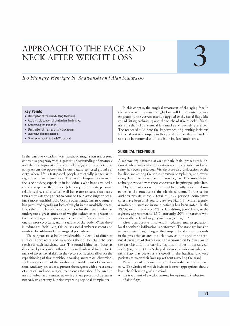
In the past few decades, facial aesthetic surgery has undergone
enormous progress, with a greater understanding of anatomy
and the development of newer technology and products that
complement the operation. In our beauty-centered global so-
ciety, where life is fast-paced, people are rapidly judged with
regards to their appearance. The face is frequently the main
focus of anxiety, especially in individuals who have attained a
certain stage in their lives. Job competition, interpersonal
relationships, and physical well-being are reasons that many
times motivate the patient to come to the plastic surgeon seek-
ing a more youthful look. On the other hand, bariatric surgery
has permitted significant loss of weight in the morbidly obese.
It has therefore become more common for the patient who has
undergone a great amount of weight reduction to present to
the plastic surgeon requesting the removal of excess skin from
one or, more typically, many regions of the body. When there
is redundant facial skin, this causes social embarrassment and
needs to be addressed by a surgical procedure.
The surgeon must be knowledgeable in details of different
surgical approaches and variations thereof to attain the best
result for each individual case. The round-lifting technique, as
described by the senior author, is very well indicated for the treat-
ment of excess facial skin, as the vectors of traction allow for the
repositioning of tissues without causing anatomical distortion,
such as dislocation of the hairline and visible signs of skin trac-
tion. Ancillary procedures present the surgeon with a vast array
of surgical and non-surgical techniques that should be used in
an individualized manner, as each patient presents differences
not only in anatomy but also regarding regional complaints.
In this chapter, the surgical treatment of the aging face in
the patient with massive weight loss will be presented, giving
emphasis to the correct traction applied to the facial flaps (the
round-lifting technique) and the forehead (the ‘block’ lifting),
assuring that all anatomical landmarks are precisely preserved.
The reader should note the importance of planning incisions
for facial aesthetic surgery in this population, so that redundant
skin can be removed without distorting key landmarks.
SURGICAL TECHNIQUE
A satisfactory outcome of an aesthetic facial procedure is ob-
tained when signs of an operation are undetectable and ana-
tomy has been preserved. Visible scars and dislocation of the
hairline are among the most common complaints, and every-
thing should be done to avoid these stigmas. The round-lifting
technique evolved with these concerns as its principal guidelines.
Rhytidoplasty is one of the most frequently performed sur-
geries in the practice of the plastic surgeon. In the senior
author’s private clinic, a total of 7927 personal consecutive
cases have been analyzed to date (see Fig. 3.1). More recently,
a noticeable increase in male patients has been noted. In the
1970s, men represented 6% of face-lifting procedures; in the
eighties, approximately 15%; currently, 20% of patients who
seek aesthetic facial surgery are men (see Fig. 3.2).
After appropriate intravenous sedation and preparation,
local anesthetic infiltration is performed. The standard incision
is demarcated, beginning in the temporal scalp, and proceeds
in the preauricular area in such a way as to respect the anato-
mical curvature of this region. The incision then follows around
the earlobe and, in a curving fashion, finishes in the cervical
scalp (Fig. 3.3). (This S-shaped incision creates an advance-
ment flap that prevents a step-off in the hairline, allowing
patients to wear their hair up without revealing the scar.)
Variations of this incision are chosen depending on each
case. The choice of which incision is most appropriate should
have the following goals in mind:
• the treatment of specific regions for optimal distribution
of skin flaps,
21
APPROACH TO THE FACE ANDNECK AFTER WEIGHT LOSS 3Ivo Pitanguy, Henrique N. Radwanski and Alan Matarasso
Key Points• Description of the round-lifting technique.
• Avoiding dislocation of anatomical landmarks.
• Addressing the forehead.
• Description of main ancillary procedures.
• Overview of complications.
• Short scar facelift in the MWL patient.
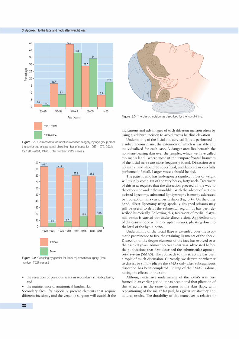
indications and advantages of each different incision often by
using a sideburn incision to avoid excess hairline elevation.
Undermining of the facial and cervical flaps is performed in
a subcutaneous plane, the extension of which is variable and
individualized for each case. A danger area lies beneath the
non–hair-bearing skin over the temples, which we have called
‘no man’s land’, where most of the temporofrontal branches
of the facial nerve are more frequently found. Dissection over
no man’s land should be superficial, and hemostasis carefully
performed, if at all. Larger vessels should be tied.
The patient who has undergone a significant loss of weight
will usually complain of the very heavy, fatty neck. Treatment
of this area requires that the dissection proceed all the way to
the other side under the mandible. With the advent of suction-
assisted lipectomy, submental lipodystrophy is mostly addressed
by liposuction, in a crisscross fashion (Fig. 3.4). On the other
hand, direct lipectomy using specially designed scissors may
still be useful to defat the submental region, as has been de-
scribed historically. Following this, treatment of medial platys-
mal bands is carried out under direct vision. Approximation
of diastasis is done with interrupted sutures, plicating down to
the level of the hyoid bone.
Undermining of the facial flaps is extended over the zygo-
matic prominence to free the retaining ligaments of the cheek.
Dissection of the deeper elements of the face has evolved over
the past 20 years. Almost no treatment was advocated before
the publications that first described the submuscular aponeu-
rotic system (SMAS). The approach to this structure has been
a topic of much discussion. Currently, we determine whether
to dissect or simply plicate the SMAS only after subcutaneous
dissection has been completed. Pulling of the SMAS is done,
noting the effects on the skin.
Although extensive undermining of the SMAS was per-
formed in an earlier period, it has been noted that plication of
this structure in the same direction as the skin flaps, with
repositioning of the malar fat pad, has given satisfactory and
natural results. The durability of this maneuver is relative to
3 Approach to the face and neck after weight loss
22
45
40
35
30
25
20
15
10
5
0
Per
cent
age
20–29 30–39 40–49 50–59 > 60
Age (years)
2.41.5
16.7
9.1
43.9
38
28.7
34
8.3
17.7
1957–1979
1980–2004
Figure 3.1 Collated data for facial rejuvenation surgery, by age group, from
the senior author’s personal clinic. Number of cases for 1957–1979, 2934;
for 1980–2004, 4993. (Total number: 7927 cases.)
40
50
60
70
80
90
100
30
20
10
0
1970–1974 1975–1980 1981–1985 1986–2004
93.7
6.3
91.6
8.4
83.2
16.8
81.4
18.6
Female
Male
Figure 3.2 Grouping by gender for facial rejuvenation surgery. (Total
number: 7927 cases.)
Figure 3.3 The classic incision, as described for the round-lifting.
• the resection of previous scars in secondary rhytidoplasty,
and
• the maintenance of anatomical landmarks.
Secondary face-lifts especially present elements that require
different incisions, and the versatile surgeon will establish the
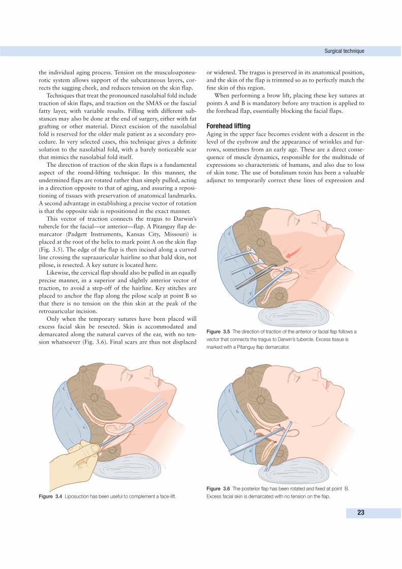
the individual aging process. Tension on the musculoaponeu-
rotic system allows support of the subcutaneous layers, cor-
rects the sagging cheek, and reduces tension on the skin flap.
Techniques that treat the pronounced nasolabial fold include
traction of skin flaps, and traction on the SMAS or the fascial
fatty layer, with variable results. Filling with different sub-
stances may also be done at the end of surgery, either with fat
grafting or other material. Direct excision of the nasolabial
fold is reserved for the older male patient as a secondary pro-
cedure. In very selected cases, this technique gives a definite
solution to the nasolabial fold, with a barely noticeable scar
that mimics the nasolabial fold itself.
The direction of traction of the skin flaps is a fundamental
aspect of the round-lifting technique. In this manner, the
undermined flaps are rotated rather than simply pulled, acting
in a direction opposite to that of aging, and assuring a reposi-
tioning of tissues with preservation of anatomical landmarks.
A second advantage in establishing a precise vector of rotation
is that the opposite side is repositioned in the exact manner.
This vector of traction connects the tragus to Darwin’s
tubercle for the facial—or anterior—flap. A Pitanguy flap de-
marcator (Padgett Instruments, Kansas City, Missouri) is
placed at the root of the helix to mark point A on the skin flap
(Fig. 3.5). The edge of the flap is then incised along a curved
line crossing the supraauricular hairline so that bald skin, not
pilose, is resected. A key suture is located here.
Likewise, the cervical flap should also be pulled in an equally
precise manner, in a superior and slightly anterior vector of
traction, to avoid a step-off of the hairline. Key stitches are
placed to anchor the flap along the pilose scalp at point B so
that there is no tension on the thin skin at the peak of the
retroauricular incision.
Only when the temporary sutures have been placed will
excess facial skin be resected. Skin is accommodated and
demarcated along the natural curves of the ear, with no ten-
sion whatsoever (Fig. 3.6). Final scars are thus not displaced
or widened. The tragus is preserved in its anatomical position,
and the skin of the flap is trimmed so as to perfectly match the
fine skin of this region.
When performing a brow lift, placing these key sutures at
points A and B is mandatory before any traction is applied to
the forehead flap, essentially blocking the facial flaps.
Forehead liftingAging in the upper face becomes evident with a descent in the
level of the eyebrow and the appearance of wrinkles and fur-
rows, sometimes from an early age. These are a direct conse-
quence of muscle dynamics, responsible for the multitude of
expressions so characteristic of humans, and also due to loss
of skin tone. The use of botulinum toxin has been a valuable
adjunct to temporarily correct these lines of expression and
Surgical technique
23
Figure 3.4 Liposuction has been useful to complement a face-lift.
Figure 3.5 The direction of traction of the anterior or facial flap follows a
vector that connects the tragus to Darwin’s tubercle. Excess tissue is
marked with a Pitanguy flap demarcator.
Figure 3.6 The posterior flap has been rotated and fixed at point B.
Excess facial skin is demarcated with no tension on the flap.
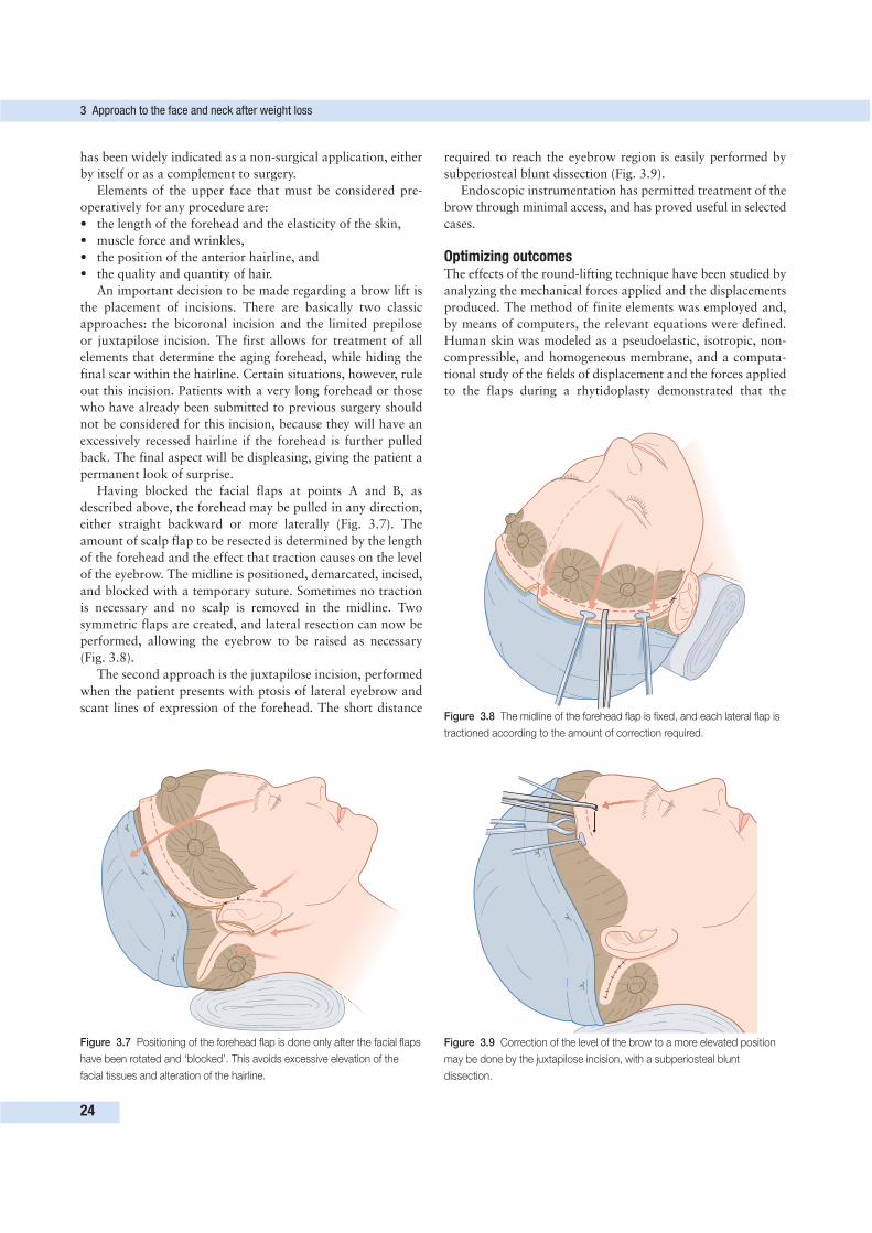
has been widely indicated as a non-surgical application, either
by itself or as a complement to surgery.
Elements of the upper face that must be considered pre-
operatively for any procedure are:
• the length of the forehead and the elasticity of the skin,
• muscle force and wrinkles,
• the position of the anterior hairline, and
• the quality and quantity of hair.
An important decision to be made regarding a brow lift is
the placement of incisions. There are basically two classic
approaches: the bicoronal incision and the limited prepilose
or juxtapilose incision. The first allows for treatment of all
elements that determine the aging forehead, while hiding the
final scar within the hairline. Certain situations, however, rule
out this incision. Patients with a very long forehead or those
who have already been submitted to previous surgery should
not be considered for this incision, because they will have an
excessively recessed hairline if the forehead is further pulled
back. The final aspect will be displeasing, giving the patient a
permanent look of surprise.
Having blocked the facial flaps at points A and B, as
described above, the forehead may be pulled in any direction,
either straight backward or more laterally (Fig. 3.7). The
amount of scalp flap to be resected is determined by the length
of the forehead and the effect that traction causes on the level
of the eyebrow. The midline is positioned, demarcated, incised,
and blocked with a temporary suture. Sometimes no traction
is necessary and no scalp is removed in the midline. Two
symmetric flaps are created, and lateral resection can now be
performed, allowing the eyebrow to be raised as necessary
(Fig. 3.8).
The second approach is the juxtapilose incision, performed
when the patient presents with ptosis of lateral eyebrow and
scant lines of expression of the forehead. The short distance
3 Approach to the face and neck after weight loss
24
Figure 3.7 Positioning of the forehead flap is done only after the facial flaps
have been rotated and ‘blocked’. This avoids excessive elevation of the
facial tissues and alteration of the hairline.
Figure 3.8 The midline of the forehead flap is fixed, and each lateral flap is
tractioned according to the amount of correction required.
Figure 3.9 Correction of the level of the brow to a more elevated position
may be done by the juxtapilose incision, with a subperiosteal blunt
dissection.
required to reach the eyebrow region is easily performed by
subperiosteal blunt dissection (Fig. 3.9).
Endoscopic instrumentation has permitted treatment of the
brow through minimal access, and has proved useful in selected
cases.
Optimizing outcomesThe effects of the round-lifting technique have been studied by
analyzing the mechanical forces applied and the displacements
produced. The method of finite elements was employed and,
by means of computers, the relevant equations were defined.
Human skin was modeled as a pseudoelastic, isotropic, non-
compressible, and homogeneous membrane, and a computa-
tional study of the fields of displacement and the forces applied
to the flaps during a rhytidoplasty demonstrated that the
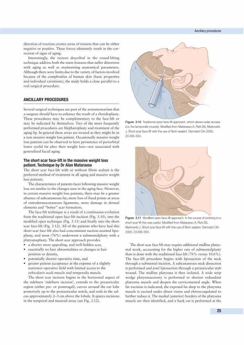
direction of traction creates areas of tension that can be either
negative or positive. These forces ultimately result in the cor-
rection of signs of aging.
Interestingly, the vectors described in the round-lifting
technique address both the main features that suffer distortion
with aging as well as maintaining anatomical parameters.
Although there were limits due to the variety of factors involved
because of the complexities of human skin (basic properties
and individual variations), the study holds a close parallel to a
real surgical procedure.
ANCILLARY PROCEDURES
Several surgical techniques are part of the armamentarium that
a surgeon should have to enhance the result of a rhytidoplasty.
These procedures may be complementary to the face-lift or
may be indicated by themselves. Two of the more frequently
performed procedures are blepharoplasty and treatment of the
aging lip. In general these areas are treated as they might be in
a non massive weight loss patient. Occasionally massive weight
loss patients can be observed to have persistence of periorbital
lower eyelid fat after their weight loss—not associated with
generalized facial aging.
The short scar face-lift in the massive weight losspatient. Technique by Dr Alan MatarassoThe short scar face-lift with or without fibrin sealant is the
preferred method of treatment in all aging and massive weight
loss patients.
The characteristics of patients faces following massive weight
loss are similar to the changes seen in the aging face. However,
in certain massive weight loss patients, there may be a greater
absence of subcutaneous fat, more loss of fixed points at areas
of osteodermocutaneous ligaments, more damage in dermal
elements and “better” scar formation.
The face-lift technique is a result of a continuous evolution
from the traditional open face-lift incision (Fig. 3.10), into the
modified open technique (Fig. 3.11) and finally into the short
scar face-lift (Fig. 3.12). All of the patients who have had this
short scar face-lift also had concomitant suction-assisted lipo-
plasty, and most (76%) underwent a submentalplasty with a
platysmaplasty. The short scar approach provides
• a shorter more appealing, and well-hidden scar,
• essentially no hair abnormalities or changes in hair
position or density,
• potentially shorter operative time, and
• greater patient acceptance at the expense of a slightly
narrower operative field with limited access to the
orbicularis oculi muscle and temporalis muscle.
The short scar incision begins in the horizontal aspect of
the sideburn ‘sideburn incision’, extends to the preauricular
region (either pre- or posttragal), curves around the ear lobe
posteriorly up to the postauricular notch, and ends in the sul-
cus approximately 2–3 cm above the lobule. It spares incisions
in the temporal and mastoid areas (see Fig. 3.12).
The short scar face-lift may require additional midline platys-
mal work, accounting for the higher rate of submentalplasty
than is done with the traditional face-lift (76% versus 10.6%).
The face-lift procedure begins with liposuction of the neck
through a submental incision. A subcutaneous neck dissection
is performed and jowl liposuction through a preauricular stab
wound. The midline platysma is then isolated. A wide strip
wedge platysmaectomy is performed to shorten redundant
platysma muscle and deepen the cervicomental angle. When
fat excision is indicated, the exposed fat deep to the platysma
muscle is excised under direct vision and eletrocoagulated to
further reduce it. The medial (anterior) borders of the platysma
muscle are then identified, and a back cut is performed at the
Ancillary procedures
25
Figure 3.10 Traditional open face-lift approach, which allows wider access
(i.e. the temporalis muscle). Modified from Matarasso A, Rizk SS, Markowitz
J. Short scar face-lift with the use of fibrin sealant. Dermatol Clin 2005;
23:495–504.
Figure 3.11 Modified open face-lift approach. In the course of evolving to a
short scar lift this was useful. Modified from Matarasso A, Rizk SS,
Markowitz J. Short scar face-lift with the use of fibrin sealant. Dermatol Clin
2005; 23:495–504.
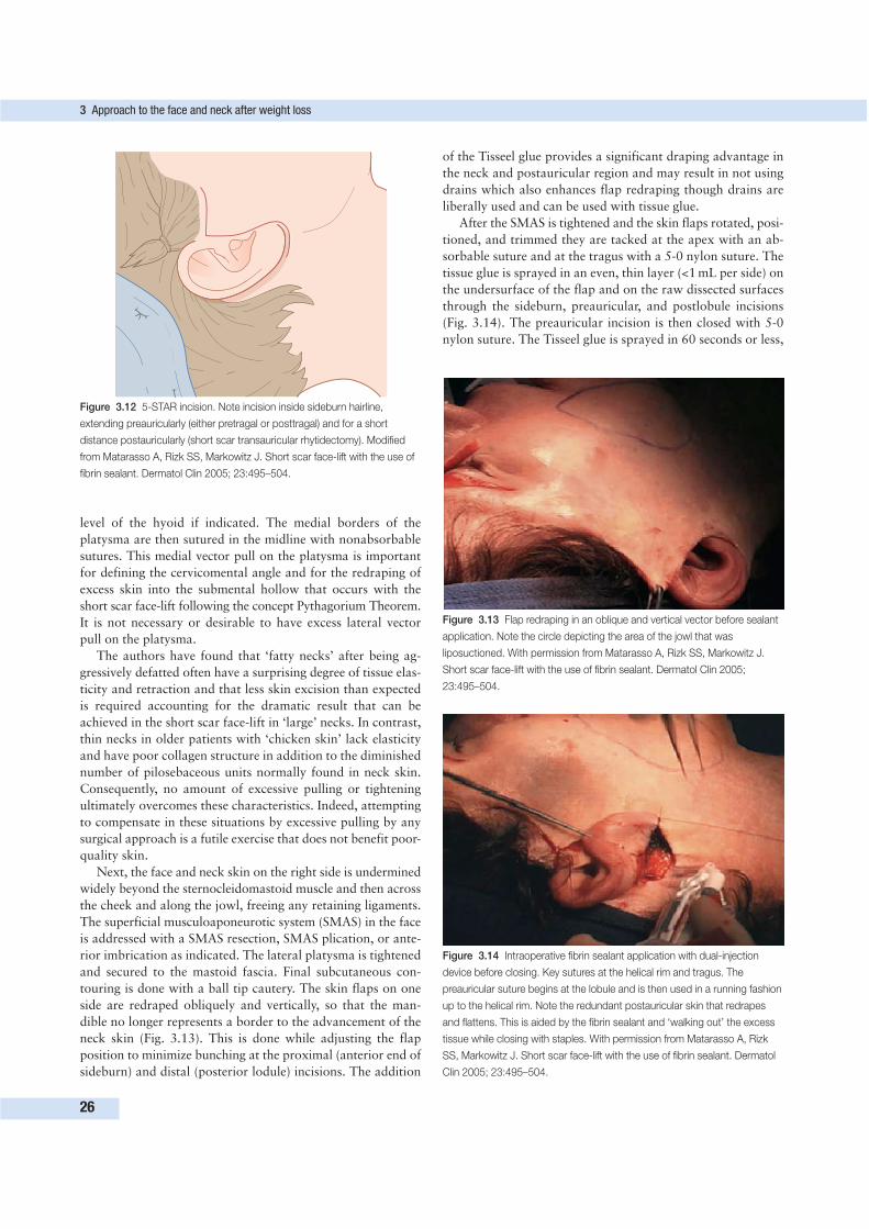
level of the hyoid if indicated. The medial borders of the
platysma are then sutured in the midline with nonabsorbable
sutures. This medial vector pull on the platysma is important
for defining the cervicomental angle and for the redraping of
excess skin into the submental hollow that occurs with the
short scar face-lift following the concept Pythagorium Theorem.
It is not necessary or desirable to have excess lateral vector
pull on the platysma.
The authors have found that ‘fatty necks’ after being ag-
gressively defatted often have a surprising degree of tissue elas-
ticity and retraction and that less skin excision than expected
is required accounting for the dramatic result that can be
achieved in the short scar face-lift in ‘large’ necks. In contrast,
thin necks in older patients with ‘chicken skin’ lack elasticity
and have poor collagen structure in addition to the diminished
number of pilosebaceous units normally found in neck skin.
Consequently, no amount of excessive pulling or tightening
ultimately overcomes these characteristics. Indeed, attempting
to compensate in these situations by excessive pulling by any
surgical approach is a futile exercise that does not benefit poor-
quality skin.
Next, the face and neck skin on the right side is undermined
widely beyond the sternocleidomastoid muscle and then across
the cheek and along the jowl, freeing any retaining ligaments.
The superficial musculoaponeurotic system (SMAS) in the face
is addressed with a SMAS resection, SMAS plication, or ante-
rior imbrication as indicated. The lateral platysma is tightened
and secured to the mastoid fascia. Final subcutaneous con-
touring is done with a ball tip cautery. The skin flaps on one
side are redraped obliquely and vertically, so that the man-
dible no longer represents a border to the advancement of the
neck skin (Fig. 3.13). This is done while adjusting the flap
position to minimize bunching at the proximal (anterior end of
sideburn) and distal (posterior lodule) incisions. The addition
of the Tisseel glue provides a significant draping advantage in
the neck and postauricular region and may result in not using
drains which also enhances flap redraping though drains are
liberally used and can be used with tissue glue.
After the SMAS is tightened and the skin flaps rotated, posi-
tioned, and trimmed they are tacked at the apex with an ab-
sorbable suture and at the tragus with a 5-0 nylon suture. The
tissue glue is sprayed in an even, thin layer (<1 mL per side) on
the undersurface of the flap and on the raw dissected surfaces
through the sideburn, preauricular, and postlobule incisions
(Fig. 3.14). The preauricular incision is then closed with 5-0
nylon suture. The Tisseel glue is sprayed in 60 seconds or less,
3 Approach to the face and neck after weight loss
26
Figure 3.12 5-STAR incision. Note incision inside sideburn hairline,
extending preauricularly (either pretragal or posttragal) and for a short
distance postauricularly (short scar transauricular rhytidectomy). Modified
from Matarasso A, Rizk SS, Markowitz J. Short scar face-lift with the use of
fibrin sealant. Dermatol Clin 2005; 23:495–504.
Figure 3.13 Flap redraping in an oblique and vertical vector before sealant
application. Note the circle depicting the area of the jowl that was
liposuctioned. With permission from Matarasso A, Rizk SS, Markowitz J.
Short scar face-lift with the use of fibrin sealant. Dermatol Clin 2005;
23:495–504.
Figure 3.14 Intraoperative fibrin sealant application with dual-injection
device before closing. Key sutures at the helical rim and tragus. The
preauricular suture begins at the lobule and is then used in a running fashion
up to the helical rim. Note the redundant postauricular skin that redrapes
and flattens. This is aided by the fibrin sealant and ‘walking out’ the excess
tissue while closing with staples. With permission from Matarasso A, Rizk
SS, Markowitz J. Short scar face-lift with the use of fibrin sealant. Dermatol
Clin 2005; 23:495–504.
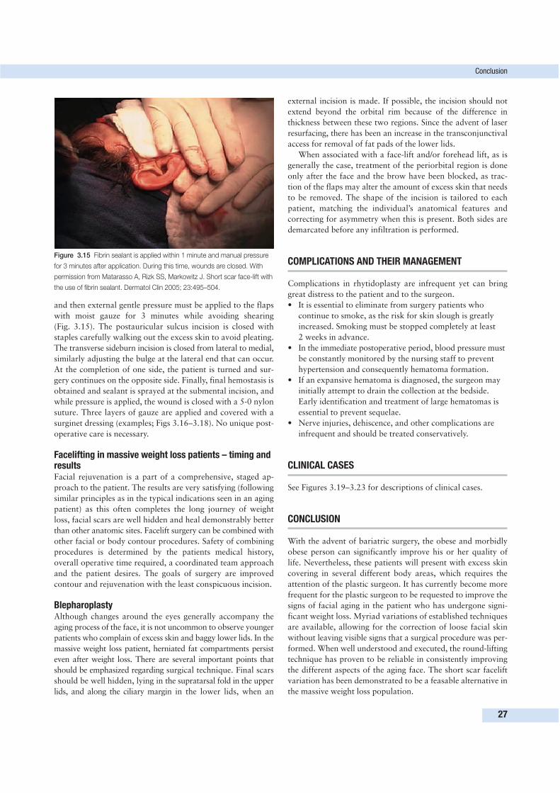
and then external gentle pressure must be applied to the flaps
with moist gauze for 3 minutes while avoiding shearing
(Fig. 3.15). The postauricular sulcus incision is closed with
staples carefully walking out the excess skin to avoid pleating.
The transverse sideburn incision is closed from lateral to medial,
similarly adjusting the bulge at the lateral end that can occur.
At the completion of one side, the patient is turned and sur-
gery continues on the opposite side. Finally, final hemostasis is
obtained and sealant is sprayed at the submental incision, and
while pressure is applied, the wound is closed with a 5-0 nylon
suture. Three layers of gauze are applied and covered with a
surginet dressing (examples; Figs 3.16–3.18). No unique post-
operative care is necessary.
Facelifting in massive weight loss patients – timing andresultsFacial rejuvenation is a part of a comprehensive, staged ap-
proach to the patient. The results are very satisfying (following
similar principles as in the typical indications seen in an aging
patient) as this often completes the long journey of weight
loss, facial scars are well hidden and heal demonstrably better
than other anatomic sites. Facelift surgery can be combined with
other facial or body contour procedures. Safety of combining
procedures is determined by the patients medical history,
overall operative time required, a coordinated team approach
and the patient desires. The goals of surgery are improved
contour and rejuvenation with the least conspicuous incision.
BlepharoplastyAlthough changes around the eyes generally accompany the
aging process of the face, it is not uncommon to observe younger
patients who complain of excess skin and baggy lower lids. In the
massive weight loss patient, herniated fat compartments persist
even after weight loss. There are several important points that
should be emphasized regarding surgical technique. Final scars
should be well hidden, lying in the supratarsal fold in the upper
lids, and along the ciliary margin in the lower lids, when an
external incision is made. If possible, the incision should not
extend beyond the orbital rim because of the difference in
thickness between these two regions. Since the advent of laser
resurfacing, there has been an increase in the transconjunctival
access for removal of fat pads of the lower lids.
When associated with a face-lift and/or forehead lift, as is
generally the case, treatment of the periorbital region is done
only after the face and the brow have been blocked, as trac-
tion of the flaps may alter the amount of excess skin that needs
to be removed. The shape of the incision is tailored to each
patient, matching the individual’s anatomical features and
correcting for asymmetry when this is present. Both sides are
demarcated before any infiltration is performed.
COMPLICATIONS AND THEIR MANAGEMENT
Complications in rhytidoplasty are infrequent yet can bring
great distress to the patient and to the surgeon.
• It is essential to eliminate from surgery patients who
continue to smoke, as the risk for skin slough is greatly
increased. Smoking must be stopped completely at least
2 weeks in advance.
• In the immediate postoperative period, blood pressure must
be constantly monitored by the nursing staff to prevent
hypertension and consequently hematoma formation.
• If an expansive hematoma is diagnosed, the surgeon may
initially attempt to drain the collection at the bedside.
Early identification and treatment of large hematomas is
essential to prevent sequelae.
• Nerve injuries, dehiscence, and other complications are
infrequent and should be treated conservatively.
CLINICAL CASES
See Figures 3.19–3.23 for descriptions of clinical cases.
CONCLUSION
With the advent of bariatric surgery, the obese and morbidly
obese person can significantly improve his or her quality of
life. Nevertheless, these patients will present with excess skin
covering in several different body areas, which requires the
attention of the plastic surgeon. It has currently become more
frequent for the plastic surgeon to be requested to improve the
signs of facial aging in the patient who has undergone signi-
ficant weight loss. Myriad variations of established techniques
are available, allowing for the correction of loose facial skin
without leaving visible signs that a surgical procedure was per-
formed. When well understood and executed, the round-lifting
technique has proven to be reliable in consistently improving
the different aspects of the aging face. The short scar facelift
variation has been demonstrated to be a feasable alternative in
the massive weight loss population.
Conclusion
27
Figure 3.15 Fibrin sealant is applied within 1 minute and manual pressure
for 3 minutes after application. During this time, wounds are closed. With
permission from Matarasso A, Rizk SS, Markowitz J. Short scar face-lift with
the use of fibrin sealant. Dermatol Clin 2005; 23:495–504.
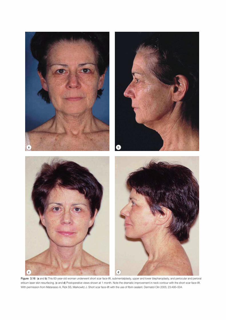
d
a b
c
Figure 3.16 (a and b) This 60-year-old woman underwent short scar face-lift, submentalplasty, upper and lower blepharoplasty, and periocular and perioral
erbium laser skin resurfacing. (c and d) Postoperative views shown at 1 month. Note the dramatic improvement in neck contour with the short scar face-lift.
With permission from Matarasso A, Rizk SS, Markowitz J. Short scar face-lift with the use of fibrin sealant. Dermatol Clin 2005; 23:495–504.
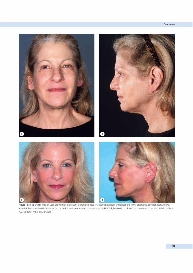
Conclusion
29
d
a b
c
Figure 3.17 (a and b) This 64-year-old woman underwent a short scar face-lift, submentalplasty, and upper and lower blepharoplasty (transconjunctival).
(c and d) Postoperative views shown at 2 months. With permission from Matarasso A, Rizk SS, Markowitz J. Short scar face-lift with the use of fibrin sealant.
Dermatol Clin 2005; 23:495–504.
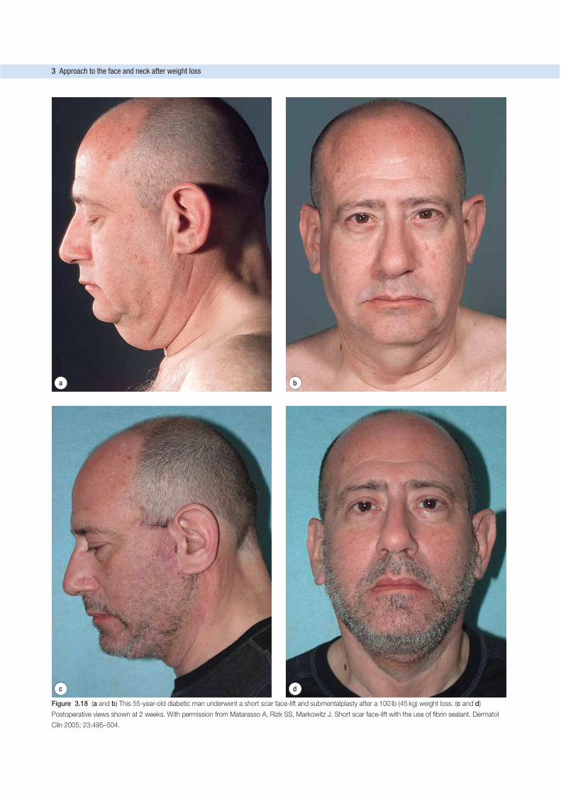
d
a b
c
Figure 3.18 (a and b) This 55-year-old diabetic man underwent a short scar face-lift and submentalplasty after a 100 lb (45 kg) weight loss. (c and d)
Postoperative views shown at 2 weeks. With permission from Matarasso A, Rizk SS, Markowitz J. Short scar face-lift with the use of fibrin sealant. Dermatol
Clin 2005; 23:495–504.
3 Approach to the face and neck after weight loss
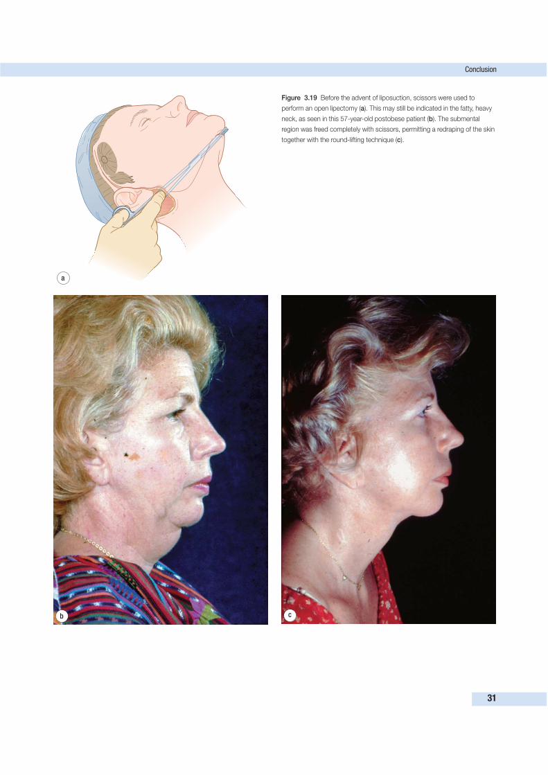
Conclusion
31
a
b c
Figure 3.19 Before the advent of liposuction, scissors were used to
perform an open lipectomy (a). This may still be indicated in the fatty, heavy
neck, as seen in this 57-year-old postobese patient (b). The submental
region was freed completely with scissors, permitting a redraping of the skin
together with the round-lifting technique (c).
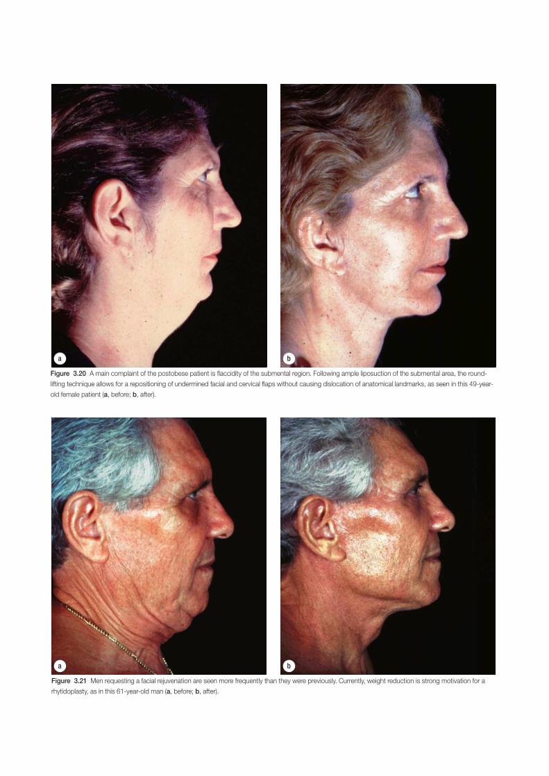
a b
Figure 3.20 A main complaint of the postobese patient is flaccidity of the submental region. Following ample liposuction of the submental area, the round-
lifting technique allows for a repositioning of undermined facial and cervical flaps without causing dislocation of anatomical landmarks, as seen in this 49-year-
old female patient (a, before; b, after).
a b
Figure 3.21 Men requesting a facial rejuvenation are seen more frequently than they were previously. Currently, weight reduction is strong motivation for a
rhytidoplasty, as in this 61-year-old man (a, before; b, after).
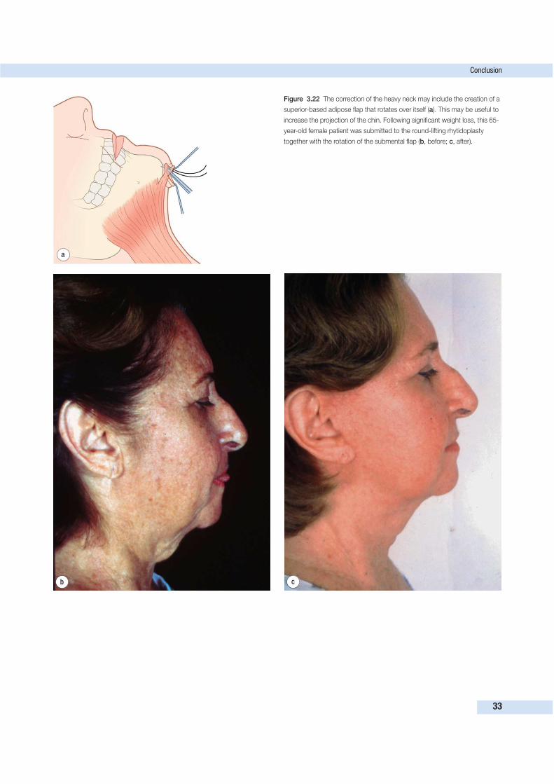
Conclusion
33
a
b c
Figure 3.22 The correction of the heavy neck may include the creation of a
superior-based adipose flap that rotates over itself (a). This may be useful to
increase the projection of the chin. Following significant weight loss, this 65-
year-old female patient was submitted to the round-lifting rhytidoplasty
together with the rotation of the submental flap (b, before; c, after).
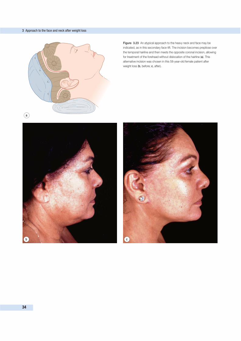
3 Approach to the face and neck after weight loss
34
a
b c
Figure 3.23 An atypical approach to the heavy neck and face may be
indicated, as in this secondary face-lift. The incision becomes prepilose over
the temporal hairline and then meets the opposite coronal incision, allowing
for treatment of the forehead without dislocation of the hairline (a). This
alternative incision was chosen in this 58-year-old female patient after
weight loss (b, before; c, after).
Conclusion
35
Finally, the plastic surgeon should be assured that the
patient understands that the purpose of any procedure for the
aging face is to help the individual cross with enhanced self-
confidence the sometimes difficult path to a mature age, and
not to return the patient to an earlier stage of life. Experience
is necessary to investigate and appreciate these subjective moti-
vations. This evaluation requires both empathy and openness
toward the patient.
AcknowledgmentThe authors are grateful to Natale Gontijo do Amorim, M.D.,
for her close collaboration in the preparation of this chapter.
FURTHER READING
Matarasso A. Botox injections for facial rejuvenation. In: Nahai, F. The
art of aesthetic surgery: Principles and technique. St Louis: Quality
Medical Publishing; 2005:195–221.
Matarasso A. Botulinum toxin. In: McCarthy J, Galiano R, Boutros S.
Current therapy in plastic surgery. Philadelphia: Saunders;
2005:324–325.
Matarasso A, Elkwood A, Rankin M, Elkowitz M. National plastic sur-
gery survey: face-lift techniques and complications. Plast Reconstr
Surg 2000; 106:1185–1195.
Matarasso A. Elkwood AI, Rankin M, et al. National plastic surgery:
Brow lifting techniques and complications. Plast Reconstr Surg
2001; 108(7):2143–2153.
Matarasso A, Rizk SS, Markowitz J. Short scar face-lift with the use of
fibrin sealant. Dermatol Clin 2005; 23:495–504.
Matarasso A, Wallach SG, DiFrancesco L, Rankin M. Age-based com-
parisons of patients undergoing secondary rhytidectomy. Aesth Surg
J 2002; 22:526–530.
Pitanguy I, Amorim NFG. Forehead lifting: the juxtapilose subperios-
teal approach. Aesthetic Plast Surg 2003; 27:58–62.
Pitanguy I, Amorim NFG. Treatment of the nasolabial fold. Rev Bras
Cir 1997; 87:231–242.
Pitanguy I, Brentano JMS, Salgado F, et al. Incisions in primary and
secondary rhytidoplasties. Rev Bras Cir 1995; 85:165–176.
Pitanguy I, Ceravolo M. Hematoma post-rhytidectomy: how we treat it.
Plast Reconstr Surg 1981; 67:526–528.
Pitanguy I, Ceravolo MP, Dègand M. Nerve injuries during rhytidec-
tomy: considerations after 3,203 cases. Aesthetic Plast Surg 1980;
4:257–265.
Pitanguy I, Pamplona DC, Giuntini ME, et al. Computational simulation
of rhytidectomy by the ‘round-lifting’ technique. Rev Bras Cir 1995;
85:213–218.
Pitanguy I, Pamplona DC, Weber HI, et al. Numerical modeling of the
aging face. Plast Reconstr Surg 1998; 102:200–204.
Pitanguy I, Radwanski HN, Amorim NFG. Treatment of the aging face
using the ‘round lifting’ technique. Aesth Surg J 1999; 19:216–222.
Pitanguy I, Radwanski HN. Rejuvenation of the brow. Matarasso SL,
Matarasso A, eds. Dermatology clinics, vol 15. Philadelphia: Saunders;
1998:623–635.
Pitanguy I, Ramos A. The frontal branch of the facial nerve: the import-
ance of its variations in face-lifting. Plast Reconstr Surg 1966;
38:352–356.
Pitanguy I, Salgado F, Radwanski HN. Submental liposuction as an
ancillary procedure in face-lifting. Face 1995; 4(1):1–13.
Pitanguy I, Soares G, Machado BH, et al. CO2 laser associated with the
‘round-lifting’ technique. J Cutan Laser Ther 1999; 1:145–152.
Pitanguy I. Ancillary procedures in face-lifting. Clin Plast Surg 1978;
5:51–69.
Pitanguy I. Facial cosmetic surgery: a 30-year perspective. Plast Reconstr
Surg 2000; 105:1517–1529.
Pitanguy I. Forehead lifting. In: Pitanguy I. Aesthetic surgery of head
and body. Berlin: Springer Verlag; 1984:202–214.
Pitanguy I. Frontalis–procerus–corrugator apponeurosis in the cor-
rection of frontal and glabellar wrinkles. Ann Plast Surg 1979;
2:422–427.
Pitanguy I. Indication for and treatment of frontal and glabellar wrinkles
in an analysis of 3,404 consecutive cases of rhytidectomy. Plast
Reconstr Surg 1981; 67:157–166.
Pitanguy I. Les chemins de la beauté. Un maitre de la chirurgie plastique
témoigne. Paris: JC Lattes; 1983.
Pitanguy I. The aging face. In: Carlsen L, Slatt B. The naked face.
Ontario: General Publishing; 1979:27.
Pitanguy I. The face. In: Pitanguy I. Aesthetic surgery of head and body.
Berlin: Springer Verlag; 1984:165–200.
Pitanguy I. The round-lifting technique. Facial Plast Surg 2000;
16(3):255–267.
INTRODUCTION
The nature of breast deformities after weight lossPostbariatric patients manifest severe breast deformities that
are very different from those seen in the traditional mastopexy
candidate. Severe volume deflation with distortion of shape
and inelastic skin is common. There are four problems.
1. There is a tendency toward significant and sometimes
asymmetric breast volume loss with a deflated and
flattened appearance.
2. There tends to be dramatic loss of skin elasticity, as well as
tremendous skin excess relative to the parenchymal
volume.
3. The nipples are usually too medial in position.
4. A final peculiarity, fairly unique to this population, is the
presence of prominent axillary skin, or in many cases a
fatty roll. This blurs the border between the lateral breast
and chest wall, sometimes forming one continuous roll of
tissue (Fig. 4.1).
The role of short scar techniquesTo achieve an aesthetically pleasing breast in the setting of
these deformities, there must be reshaping of the deflated breast
parenchyma and augmentation with autologous tissue to re-
store superior fullness and projection. The skin envelope must
be reduced and prominent axillary skin rolls eliminated. It is
the authors’ view that short scar techniques are inadequate in
handling the redundant inelastic skin envelope in these patients.
Moreover, short scar techniques cannot properly address the
lateral skin excess.
Approach used by the authorsThe authors have developed and refined a technique using the
principles of dermal suspension and total parenchymal re-
shaping. An extended Wise pattern encompasses and eliminates
lateral skin rolls, while at the same time providing additional
tissue that may be used as necessary for volume augmen-
tation. Deepithelialization of the entire Wise pattern creates a
broad dermal surface area that can be plicated to precisely
control breast shape and can be suspended to the chest wall.
BackgroundThe technique developed by the authors for the weight loss
patient is based on lessons learned from the historical develop-
ment of breast-reshaping methods. Schwarzmann’s early con-
tribution demonstrating the importance of dermal blood supply
was essential.1 Beisenberger’s conceptual revolution of total
dissociation of the skin envelope from the glandular tissue
was invaluable in the development of this and many other
procedures.2 While the Beisenberger technique had great sup-
port and longevity, surgeons continued to produce technical
refinements. Thorek is credited with introducing the free nipple
graft in the 1920s,3 and this method provides a valuable
lifeboat for breast surgeons who note poor nipple perfusion in
the operating room. The 1950s saw Wise describe a technique
to control the skin envelope in a manner that accentuates breast
shape.4 In 1960, Strombeck described a horizontal bipedicled
procedure with enhanced nipple vascularity.5 A significant
contribution came from McKissock’s vertical bipedicled flap,
which facilitated the creation of a more natural-appearing
breast.6 In 1963, Skoog produced work supporting the trans-
position of the nipple areolar complex (NAC) on a unilateral
vascular pedicle.7 Eventually, Rubiero described,8 and Courtiss
and Goldwyn championed the inferior pedicle with the Wise
pattern of scars.9 The various approaches applied in the
37
APPROACH TO THE BREAST AFTERWEIGHT LOSS 4J. Peter Rubin, James O’Toole and Siamak Agha-Mohammadi
Key Points• Carefully assess parenchymal volume, amount of redundant skin
envelope, and extent of lateral skin/fat roll.
• Consider order of breast reshaping in association with other planned
body-contouring procedures.
• Plan Wise pattern marking to encompass lateral chest wall tissue in
order to eliminate skin/fat roll and also allow for autologous volume
augmentation.
• Deepithelialization of entire Wise pattern and complete degloving of
parenchyma preserves breast volume and provides broad dermal
surface area.
• Permanent suspension sutures secure dermis to rib periosteum, and
multiple plication sutures in dermis allow precise control of breast
shape.
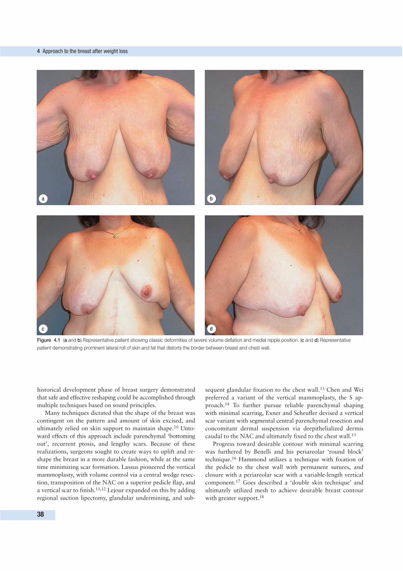
historical development phase of breast surgery demonstrated
that safe and effective reshaping could be accomplished through
multiple techniques based on sound principles.
Many techniques dictated that the shape of the breast was
contingent on the pattern and amount of skin excised, and
ultimately relied on skin support to maintain shape.10 Unto-
ward effects of this approach include parenchymal ‘bottoming
out’, recurrent ptosis, and lengthy scars. Because of these
realizations, surgeons sought to create ways to uplift and re-
shape the breast in a more durable fashion, while at the same
time minimizing scar formation. Lassus pioneered the vertical
mammoplasty, with volume control via a central wedge resec-
tion, transposition of the NAC on a superior pedicle flap, and
a vertical scar to finish.11,12 Lejour expanded on this by adding
regional suction lipectomy, glandular undermining, and sub-
sequent glandular fixation to the chest wall.13 Chen and Wei
preferred a variant of the vertical mammoplasty, the S ap-
proach.14 To further pursue reliable parenchymal shaping
with minimal scarring, Exner and Scheufler devised a vertical
scar variant with segmental central parenchymal resection and
concomitant dermal suspension via deepithelialized dermis
caudal to the NAC and ultimately fixed to the chest wall.15
Progress toward desirable contour with minimal scarring
was furthered by Benelli and his periareolar ‘round block’
technique.16 Hammond utilizes a technique with fixation of
the pedicle to the chest wall with permanent sutures, and
closure with a periareolar scar with a variable-length vertical
component.17 Goes described a ‘double skin technique’ and
ultimately utilized mesh to achieve desirable breast contour
with greater support.18
4 Approach to the breast after weight loss
38
b
c d
a
Figure 4.1 (a and b) Representative patient showing classic deformities of severe volume deflation and medial nipple position. (c and d) Representative
patient demonstrating prominent lateral roll of skin and fat that distorts the border between breast and chest wall.
Many surgeons focused on strategies to improve and main-
tain upper pole fullness, and these techniques often involved
fixation of breast tissue to adjacent structures. Pitanguy re-
stricted resection to only the inferior pole, and utilized a
‘straight resection’ or ‘inverted keel’ for firmer breast tissue.
Closure of medial and lateral pillars of parenchyma and an
inverted T incision finished his procedure.19 Cerqueira’s ap-
proach was to create a superior pedicle, resect a central block
of parenchyma, and subsequently secure the dermoglandular
pedicle under the pectoralis.20 Frey’s contribution allowed for
parenchymal contouring and suspension via a dermal brassiere
fixated to the anterior thoracic wall with non-absorbable
suture, and complete elimination of the medial component of
the scar.21 Building upon the concept of a dermal bra, Qiao et
al. devised an approach that resected a crescent of glandular
tissue superolaterally, with dermal fixation to the pectoralis
fascia.22 Gulyas’s periareolar techniques also relied on mani-
pulation of the ‘dermal cloak’ to support and shape the
breast.23 Graf and Biggs created an inferior dermoglandular
pedicle that they passed under a loop of pectoralis and secured
to the pectoralis fascia. The NAC is carried on the elevated
breast, and the inferior flap is fixed to the pectoralis fascia in
the upper pole to ensure upper pole fullness with closure of
medial and lateral pillars behind the flap.10 Lockwood achieved
his results via a modification of the Wise pattern, with the pri-
mary supportive element being non-absorbable sutures in the
superficial fascial system to decrease dermal tension and sub-
sequent scarring.24
Many important principles are embodied in the techniques
described. However, when considering the complex deformity
seen in the massive weight loss patient, none of the above
procedures seem to be ideal. Moreover, it becomes obvious
that short scar techniques are of limited value in this patient
population. What is required is a technique that allows for:
• precise and symmetric NAC positioning,
• precise control of parenchymal breast shape and contour,
• possible autoaugmentation in the volume-deficient patient,
and
• control of the remaining skin envelope.
In our technique, we make use of a well-vascularized central
dermoglandular pedicle.2,16 A modification of the traditional
Wise pattern allows for precise control of the skin envelope
and NAC position.4 The dermal suspension techniques of Qiao,
Frey, Cerqueira, and others prompted our use of parenchymal
suspension and extensive sculpting via dermal plication and
fixation to the chest wall.15,18,20–23 Holmstrom’s lateral thora-
codorsal transposition flap for breast reconstruction after
mastectomy facilitated the notion of autoaugmentation via
recruitment of redundant axillary tissue.25 Medial fullness is
assured via the elevation and manipulation of a medial breast
flap.
The technique described below has the advantages of cor-
recting, with a low complication rate, the severe breast defor-
mities associated with weight loss. Notably, the deformity of a
lateral axillary roll can be eliminated and used to augment
breast volume. The disadvantages of this technique include:
• a lengthy scar,
• considerable time in the operating room for the extensive
deepithelialization, and
• a high degree of ‘intraoperative tailoring’ that cannot be
premarked.
Despite the disadvantages, this technique is safe and reliable for
restoring a youthful breast shape in the massive weight loss
patient. Great control over both skin envelope and parenchymal
shape may be gained with this procedure.
PREOPERATIVE EVALUATION
Patients with mild breast deformities following weight loss
should be considered for traditional mastopexy techniques,
including short scar approaches. However, existing mastopexy
techniques are not always adequate to achieve a good aesthetic
result with these deformities when faced with the following
clinical findings.
• Profound breast volume loss with flattening of the
parenchyma against the chest wall.
• A redundant, inelastic skin envelope.
• Grade 3 nipple ptosis.
• Medialization of the NAC.
• The presence of a prominent axillary roll of skin that
extends from the lateral breast.
We have identified few contraindications for the use of this
technique. Because of the extensive flap dissection, we have
avoided performing this procedure on active tobacco users. As
with all breast reshaping patients, we perform a thorough his-
tory and physical examination for breast disease, as well as
require mammography imaging consistent with the American
Cancer Society screening guidelines. Scars from previous breast
surgery may present a relative contraindication if they pose a
risk to perfusion of undermined tissues. Careful evaluation for
parenchymal volume is undertaken, as well as asymmetry. The
lateral breast region is inspected for a significant skin roll, and
an assessment is made regarding the amount of tissue that
may be mobilized from the lateral chest wall for autologous
breast augmentation. In the case of significant asymmetry, we
will either selectively augment the smaller breast using lateral
chest wall tissue or, if this is not possible, reduce the larger
breast to match the smaller one.
The surgical goals for breast reshaping in the face of these
deformities are to:
• use all available breast tissue, and also have the ability to
recruit additional autologous tissue;
• address the nipple position;
• restore superior pole projection;
• reshape the skin envelope without relying on it for support;
• eliminate the lateral skin roll; and
• create a discrete ‘lateral sweep’ to the breast shape.
The technique we describe, using the principles of controlled
parenchymal reshaping and dermal suspension, will meet
these goals. This safe and reproducible technique yields a
youthful breast shape in a very challenging population.
Preoperative evaluation
39
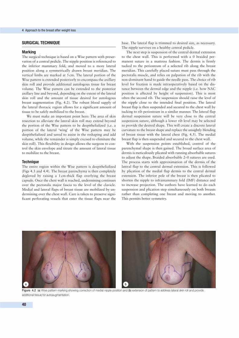
SURGICAL TECHNIQUE
MarkingThe surgical technique is based on a Wise pattern with preser-
vation of a central pedicle. The nipple position is referenced to
the inferior mammary fold, and moved to a more lateral
position along a symmetrically drawn breast meridian. The
vertical limbs are marked at 5 cm. The lateral portion of the
Wise pattern is extended posteriorly to encompass the axillary
skin roll and provide additional autologous tissue for breast
volume. The Wise pattern can be extended to the posterior
axillary line and beyond, depending on the extent of the lateral
skin roll and the amount of tissue desired for autologous
breast augmentation (Fig. 4.2). The robust blood supply of
the lateral thoracic region allows for a significant amount of
tissue to be safely mobilized to the breast.
We must make an important point here: The area of skin
resection to alleviate the lateral skin roll may extend beyond
the portion of the Wise pattern to be deepithelialized (i.e. a
portion of the lateral ‘wing’ of the Wise pattern may be
deepithelialized and saved to assist in the reshaping and add
volume, while the remainder is simply excised to eliminate the
skin roll). This flexibility in design allows the surgeon to con-
trol the skin envelope and titrate the amount of lateral tissue
to mobilize to the breast.
TechniqueThe entire region within the Wise pattern is deepithelialized
(Figs 4.3 and 4.4). The breast parenchyma is then completely
degloved by raising a 1 cm-thick flap overlying the breast
capsule. Once the chest wall is reached, undermining continues
over the pectoralis major fascia to the level of the clavicle.
Medial and lateral flaps of breast tissue are mobilized by un-
dermining over the chest wall. Care is taken to preserve signi-
ficant perforating vessels that enter the tissue flaps near the
base. The lateral flap is trimmed to desired size, as necessary.
The nipple survives on a healthy central pedicle.
The next step is suspension of the central dermal extension
to the chest wall. This is performed with a 0 braided per-
manent suture in a mattress fashion. The dermis is firmly
tacked to the periosteum of a selected rib along the breast
meridian. This carefully placed suture must pass through the
pectoralis muscle, and relies on palpation of the rib with the
non-dominant hand to guide the needle pass. The choice of rib
level for fixation is made intraoperatively based on the dis-
tance between the dermal edge and the nipple (i.e. how NAC
position is affected by height of suspension). This is most
often the second rib. The suspension should raise the level of
the nipple close to the intended final position. The lateral
breast flap is then suspended and secured to the chest wall by
tacking to rib periosteum in a similar manner. The lateral flap
dermal suspension suture will be very close to the central
suspension suture, although a lower rib level may be selected
to provide the desired shape. This will create a discrete lateral
curvature to the breast shape and replace the unsightly blending
of breast tissue with the lateral chest (Fig. 4.5). The medial
breast flap is then suspended and secured to the chest wall.
With the suspension points established, control of the
parenchymal shape is then gained. The broad surface area of
dermis is meticulously plicated with running absorbable sutures
to adjust the shape. Braided absorbable 2–0 sutures are used.
The process starts with approximation of the dermis of the
lateral flap to the central dermal extension. This is followed
by plication of the medial flap dermis to the central dermal
extension. The inferior pole of the breast is then plicated to
shorten the nipple to inframammary fold (IMF) distance and
to increase projection. The authors have learned to do each
suspension and plication step simultaneously on both breasts
rather than completing one breast and moving to another.
This permits better symmetry.
4 Approach to the breast after weight loss
40
a b
Figure 4.2 (a) Wise pattern marking showing correction of medial nipple position and (b) extension of pattern to address lateral skin roll and provide
additional tissue for autoaugmentation.
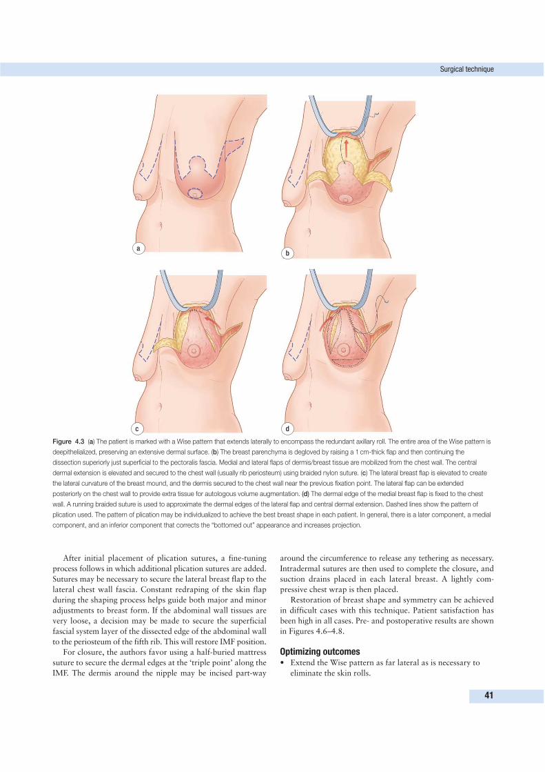
After initial placement of plication sutures, a fine-tuning
process follows in which additional plication sutures are added.
Sutures may be necessary to secure the lateral breast flap to the
lateral chest wall fascia. Constant redraping of the skin flap
during the shaping process helps guide both major and minor
adjustments to breast form. If the abdominal wall tissues are
very loose, a decision may be made to secure the superficial
fascial system layer of the dissected edge of the abdominal wall
to the periosteum of the fifth rib. This will restore IMF position.
For closure, the authors favor using a half-buried mattress
suture to secure the dermal edges at the ‘triple point’ along the
IMF. The dermis around the nipple may be incised part-way
around the circumference to release any tethering as necessary.
Intradermal sutures are then used to complete the closure, and
suction drains placed in each lateral breast. A lightly com-
pressive chest wrap is then placed.
Restoration of breast shape and symmetry can be achieved
in difficult cases with this technique. Patient satisfaction has
been high in all cases. Pre- and postoperative results are shown
in Figures 4.6–4.8.
Optimizing outcomes• Extend the Wise pattern as far lateral as is necessary to
eliminate the skin rolls.
Surgical technique
41
ab
c d
Figure 4.3 (a) The patient is marked with a Wise pattern that extends laterally to encompass the redundant axillary roll. The entire area of the Wise pattern is
deepithelialized, preserving an extensive dermal surface. (b) The breast parenchyma is degloved by raising a 1 cm-thick flap and then continuing the
dissection superiorly just superficial to the pectoralis fascia. Medial and lateral flaps of dermis/breast tissue are mobilized from the chest wall. The central
dermal extension is elevated and secured to the chest wall (usually rib periosteum) using braided nylon suture. (c) The lateral breast flap is elevated to create
the lateral curvature of the breast mound, and the dermis secured to the chest wall near the previous fixation point. The lateral flap can be extended
posteriorly on the chest wall to provide extra tissue for autologous volume augmentation. (d) The dermal edge of the medial breast flap is fixed to the chest
wall. A running braided suture is used to approximate the dermal edges of the lateral flap and central dermal extension. Dashed lines show the pattern of
plication used. The pattern of plication may be individualized to achieve the best breast shape in each patient. In general, there is a later component, a medial
component, and an inferior component that corrects the “bottomed out” appearance and increases projection.
• The entire lateral wing of the Wise pattern may be
deepithelialized and preserved to add volume to the breast,
as needed. Conversely, a smaller portion may be preserved
and the remainder excised.
• Keep the breast flap approximately 1 cm thick (or greater),
and once at the level of the pectoralis fascia, continue
undermining superiorly above the level of the second rib.
• Avoid performing this operation on smokers because of
the risk of flap necrosis.
• Plication of the dermis is most effective on the lateral and
inferior aspects of the breast, where it serves to increase
projection and create a distinct lateral curvature to the
breast mound.
• If the nipple is tethered, the surrounding dermis may be
partially incised to release it. A robust central pedicle
supports the nipple and allows this to be done safely.
Postoperative care and course• The authors use a lightly compressive breast dressing for
the first 5 days, and then ask the patient to wear a sports
bra with no wires for the next month.
• Drains are maintained for the first 48 h and then
discontinued if the output is decreasing.
• Heavy lifting and exercise is prohibited until 4 weeks after
surgery.
4 Approach to the breast after weight loss
42
a
b
c d
Figure 4.4 (a) The dermis of the medial breast flap is approximated to the central dermal extension using a running suture. The dermis on the inferior pole of
the breast is plicated with a running suture to shorten the distance between areola and inferior mammary fold to approximately 5 cm. (b) The dermis along the
lateral breast is secured to the lateral chest fascia (not rib pereostium) with permanent sutures to increase projection and accentuate the lateral curve of the
breast. The breast parenchyma is now firmly secured to the chest wall, and the shape has been adjusted using the plication sutures. (c and d) The breast skin
flap is redraped and closed with absorbable intradermal sutures over a drain. If the nipple is tethered and pointing in an inappropriate direction, the dermis
adjacent to the nipple is scored to release the tension. Because of the robust pedicle, scoring of the dermis can be safely performed along part of the
circumference, if necessary.
Surgical technique
43
d
a b
c
Figure 4.5 (a) Intraoperative photographs showing extensive de-epithelialization. (b) Suspension of the central dermal extension bilaterally. (c) Plication
sutures in place. (d) Redraping of skin flap. Pre- and postoperative photographs of this patient are shown in Figure 4.6.
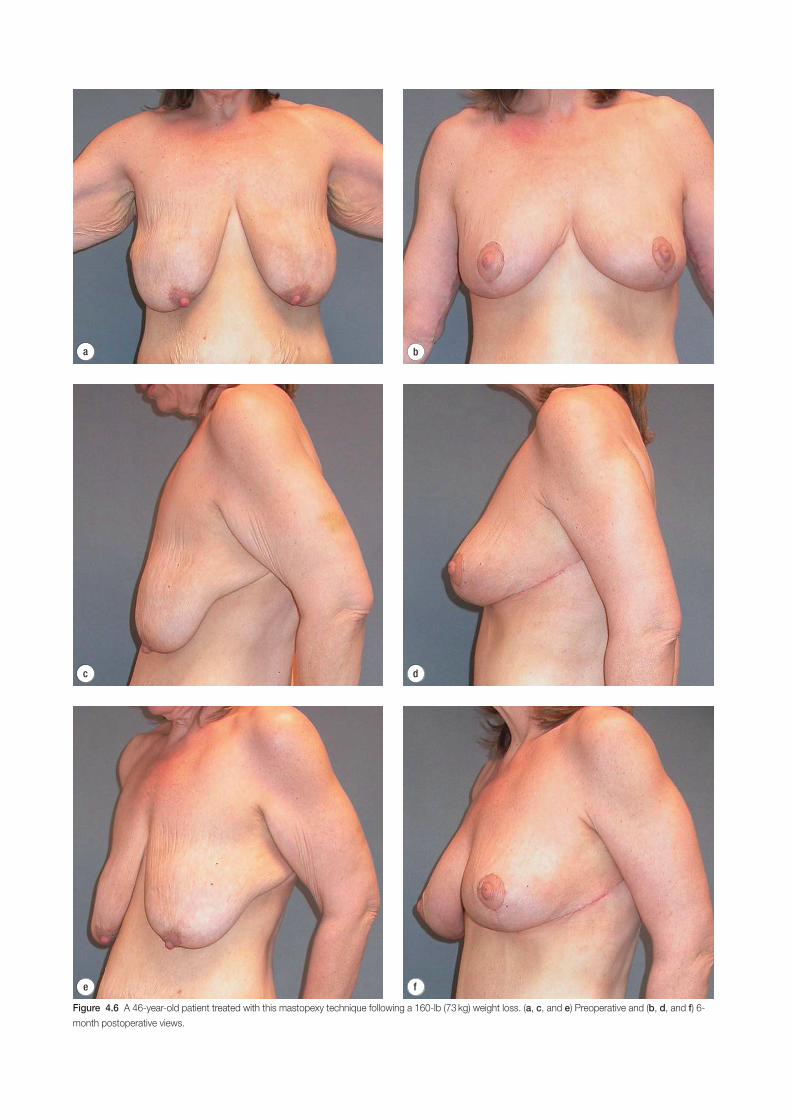
d
e f
a b
c
Figure 4.6 A 46-year-old patient treated with this mastopexy technique following a 160-lb (73 kg) weight loss. (a, c, and e) Preoperative and (b, d, and f) 6-
month postoperative views.
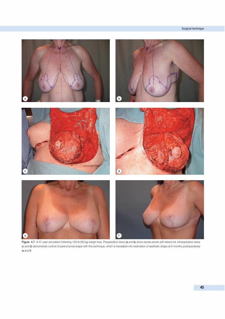
Surgical technique
45
d
e f
a b
c
Figure 4.7 A 57-year-old patient following 130-lb (60 kg) weight loss. Preoperative views (a and b) show severe ptosis with lateral roll. Intraoperative views
(c and d) demonstrate control of parenchymal shape with this technique, which is translated into restoration of aesthetic shape at 6 months postoperatively
(e and f).
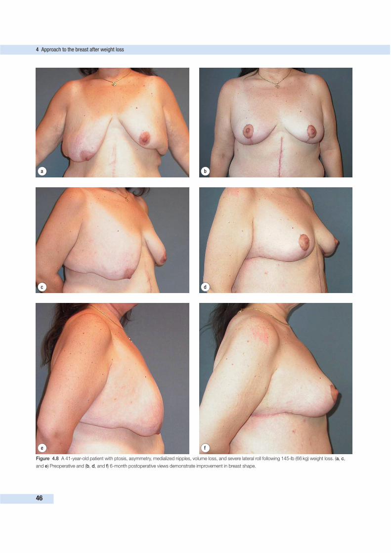
4 Approach to the breast after weight loss
46
d
e f
a b
c
Figure 4.8 A 41-year-old patient with ptosis, asymmetry, medialized nipples, volume loss, and severe lateral roll following 145-lb (66 kg) weight loss. (a, c,
and e) Preoperative and (b, d, and f) 6-month postoperative views demonstrate improvement in breast shape.
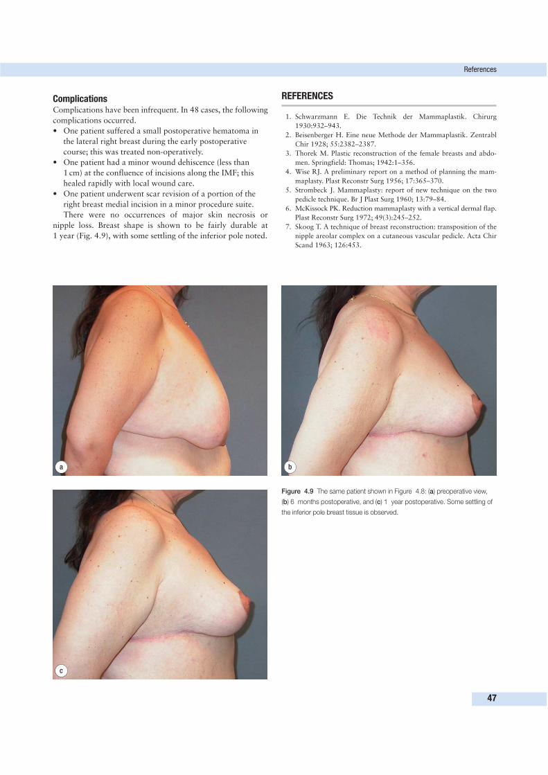
ComplicationsComplications have been infrequent. In 48 cases, the following
complications occurred.
• One patient suffered a small postoperative hematoma in
the lateral right breast during the early postoperative
course; this was treated non-operatively.
• One patient had a minor wound dehiscence (less than
1 cm) at the confluence of incisions along the IMF; this
healed rapidly with local wound care.
• One patient underwent scar revision of a portion of the
right breast medial incision in a minor procedure suite.
There were no occurrences of major skin necrosis or
nipple loss. Breast shape is shown to be fairly durable at
1 year (Fig. 4.9), with some settling of the inferior pole noted.
REFERENCES
1. Schwarzmann E. Die Technik der Mammaplastik. Chirurg
1930:932–943.
2. Beisenberger H. Eine neue Methode der Mammaplastik. Zentrabl
Chir 1928; 55:2382–2387.
3. Thorek M. Plastic reconstruction of the female breasts and abdo-
men. Springfield: Thomas; 1942:1–356.
4. Wise RJ. A preliminary report on a method of planning the mam-
maplasty. Plast Reconstr Surg 1956; 17:365–370.
5. Strombeck J. Mammaplasty: report of new technique on the two
pedicle technique. Br J Plast Surg 1960; 13:79–84.
6. McKissock PK. Reduction mammaplasty with a vertical dermal flap.
Plast Reconstr Surg 1972; 49(3):245–252.
7. Skoog T. A technique of breast reconstruction: transposition of the
nipple areolar complex on a cutaneous vascular pedicle. Acta Chir
Scand 1963; 126:453.
References
47
a b
c
Figure 4.9 The same patient shown in Figure 4.8: (a) preoperative view,
(b) 6 months postoperative, and (c) 1 year postoperative. Some settling of
the inferior pole breast tissue is observed.
8. Rubiero L. A new technique for reduction mammaplasty. Plast
Reconstr Surg 1975; 55:330–334.
9. Courtiss EH, Goldwyn RM. Reduction mammaplasty by the
inferior pedicle technique. Plast Reconstr Surg 1977; 59:64–67.
10. Graf R, Biggs TM. In search of better shape in mastopexy and re-
duction mammoplasty. Plast Reconstr Surg 2002; 110(1):309–317.
11. Lassus C. A 30 year experience with vertical mammaplasty. Plast
Reconstr Surg 1996; 97:373–380.
12. Lassus C. A technique for breast reduction. Int Surg 1970; 53:69–72.
13. Lejour M. Vertical mammaplasty without inframammary scar and
with breast liposuction. Perspect Plast Surg 1990; 4:64–67.
14. Chen T, Wei F. Evolution of the vertical reduction mammaplasty:
the S approach. Aesthetic Plast Surg 1997; 21:97–104.
15. Exner K, Scheufler O. Dermal suspension flap in vertical-scar re-
duction mammaplasty. Plast Reconstr Surg 2002; 109:2289–2300.
16. Benelli L. A new peri-areolar mammaplasty: the ‘round block’
technique. Aesthetic Plast Surg 1990; 14:93.
17. Hammond D. Short scar peri-areolar inferior pedicle reduction
(SPAIR) mammaplasty. Plast Reconstr Surg 1999; 103:890–901.
18. Goes J. Periareolar mammaplasty with mixed mesh support: the
double skin technique. Oper Tech Plast Reconstr Surg 1996;
3:197–199.
19. Pitanguy I. Evaluation of body contouring surgery today: a 30 year
perspective. Plast Reconstr Surg 2000; 105:1499–1514.
20. Cerqueira A. Mammaplasty: breast fixation with dermoglandular
mono upper pedicle flap under the pectoralis muscle. Aesthetic
Plast Surg 1998; 22:276–283.
21. Frey M. A new technique of reduction mammaplasty: dermis
suspension and elimination of medial scars. Br J Plast Surg 1999;
52:45–51.
22. Qiao Q, et al. Reduction mammaplasty and correction of ptosis:
dermal bra technique. Plast Reconstr Surg 2003; 111:122–1130.
23. Gulyas G. Mammaplasty with a periareolar dermal cloak for
glandular support. Aesthetic Plast Surg 1999; 23:164–169.
24. Lockwood T. Reduction mammaplasty and mastopexy with SFS
suspension. Plast Reconstr Surg 1990; 5:1411–1420.
25. Holmstrom H. The lateral thoracodorsal flap in breast reconstruc-
tion. Plast Reconstr Surg 1986; 77:933–943.
4 Approach to the breast after weight loss
48
As early as 1899, the term abdominal lipectomy was devised by
Kelly to describe a transverse resection of a large pendulous
abdomen.1 In 1910, Dr. Kelly described his experience with
eight patients.2 Thorek in 1939 described his technique, which
he called ‘plastic adipectomy’ for resecting ‘fat aprons’.3 These
early operations were designed to relieve the functional pro-
blems associated with large fat aprons. However, early on the
cosmetic benefits were noted. Kelly stated in 1910 that ‘quite
apart, however, from the tremendous physical and, in some
cases psychical benefit, I personally recommend and would do
the operation in extreme cases for the cosmetic benefit’.2
From these early efforts have come the techniques known
as abdominoplasty. Although abdominoplasty is a procedure
well known to plastic surgeons, the management of the post–
massive weight loss abdomen is much more complicated.
Although variation can be seen in the traditional abdomino-
plasty patient, the post–massive weight loss patient presents
with a wider range of anatomical variables as well as a higher
rate of complications.
As patients lose weight following bariatric surgery, they
begin to develop loose and overhanging skin in many areas.
Universally, the abdomen is a prime focal area of concern in
post–massive weight loss patients. Various techniques have
been described. The goals of all these techniques are to:
• allow excision of excess skin and fat, and
• tighten the diastasis recti and/or repair hernias if present.
In traditional abdominoplasty patients, the third goal is to
have minimum scarring.4 This is not the case for the massive
weight loss patient. Contour is a more important goal than
minimum scarring in this population, and several scars may
be necessary to give the patient the desired contour.
Panniculectomy and abdominoplasty have been used inter-
changeably to describe surgical procedures to remove excess
skin and fat of the abdominal wall. Panniculectomy describes
procedures removing only skin and fat—i.e. a functional
operation that removes a symptomatic apron of skin—while
abdominoplasty refers to not only the removal of skin and fat
but also the tightening up of the muscles of the abdominal
wall (it is a term that connotes aesthetic goals). Often, the ab-
dominoplasty may be considered a cosmetic procedure while
a panniculectomy refers to a more reconstructive type of
operation. A panniculcetomy may be done in patients who
have not yet begun their weight loss to remove a large apron,
or in patients who have an extremely large overhanging apron
after massive weight loss and have interference with activities
of daily life or a history of recurrent rashes. For the massive
weight loss patient, an abdominoplasty is commonly done after
weight loss is complete, and is performed to recontour the
abdominal wall with removal of excess skin and fat as well as
tightening up of the muscles underneath.
As a general rule, more attention can be safely given to
aesthetic goals as the BMI of the patient decreases. Wound
complications tend to be higher when contouring operations
are performed in patients who are still obese, and a more
49
APPROACH TO THE ABDOMENAFTER WEIGHT LOSS 5Susan E. Downey
Key PointsA lower abdominal incision may not adequately address the redundancy of
the abdomen in a post–massive weight loss patient; vertical or lateral
abdominal incisions may need to be utilized.
• Contouring of the mons should be considered in most weight loss
patients.
• Postoperative seromas are an increased risk in this population, and
intraoperative techniques may need to be altered to minimize this
occurrence.
• Hernias may be addressed safely at the time of panniculectomy.
DEFINITIONS• Abdominoplasty. Removal of skin and fat of the abdominal wall
with tightening of the underlying musculature. In general, this is
considered a cosmetic procedure.
• Belt lipectomy. A method designed to circumferentially reduce
truncal excess combining an abdominoplasty, lateral thigh lift,
buttocks lift, and sometimes liposuction of select areas.
• Lower body lift. Described initially by Lockwood and refers to a
combined transverse thigh/buttock lift with a high-tension
abdominoplasty.
• Panniculectomy. Removal of skin and fat of the abdominal wall.
In general, this is considered a reconstructive procedure.
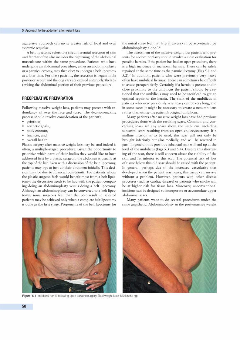
aggressive approach can invite greater risk of local and even
systemic sequelae.
A belt lipectomy refers to a circumferential resection of skin
and fat that often also includes the tightening of the abdominal
musculature within the same procedure. Patients who have
undergone an abdominal procedure, either an abdominoplasty
or a panniculectomy, may then elect to undergo a belt lipectomy
at a later time. For these patients, the resection is begun in the
posterior aspect and the dog ears are excised anteriorly, thereby
revising the abdominal portion of their previous procedure.
PREOPERATIVE PREPARATION
Following massive weight loss, patients may present with re-
dundancy all over the face and torso. The decision-making
process should involve consideration of the patient’s:
• priorities,
• aesthetic goals,
• body contour,
• finances, and
• overall health.
Plastic surgery after massive weight loss may be, and indeed is
often, a multiple-staged procedure. Given the opportunity to
prioritize which parts of their bodies they would like to have
addressed first by a plastic surgeon, the abdomen is usually at
the top of the list. Even with a discussion of the belt lipectomy,
patients may opt to just do their abdomen initially. This deci-
sion may be due to financial constraints. For patients whom
the plastic surgeon feels would benefit most from a belt lipec-
tomy, the discussion needs to be had with the patient compar-
ing doing an abdominoplasty versus doing a belt lipectomy.
Although an abdominoplasty can be converted to a belt lipec-
tomy, some surgeons feel that the best result in selected
patients may be achieved only when a complete belt lipectomy
is done as the first stage. Proponents of the belt lipectomy for
the initial stage feel that lateral excess can be accentuated by
abdominoplasty alone.5,6
The assessment of the massive weight loss patient who pre-
sents for abdominoplasty should involve a close evaluation for
possible hernias. If the patient has had an open procedure, there
is a high incidence of incisional hernias. These can be safely
repaired at the same time as the panniculectomy (Figs 5.1 and
5.2).7 In addition, patients who were previously very heavy
often have umbilical hernias. These can sometimes be difficult
to assess preoperatively. Certainly, if a hernia is present and in
close proximity to the umbilicus the patient should be cau-
tioned that the umbilicus may need to be sacrificed to get an
optimal repair of the hernia. The stalk of the umbilicus in
patients who were previously very heavy can be very long, and
in some cases it might be necessary to create a neoumbilicus
rather than utilize the patient’s original umbilicus.
Many patients after massive weight loss have had previous
procedures done with the resulting scars. Common and con-
cerning scars are any scars above the umbilicus, including
subcostal scars resulting from an open cholecystectomy. If a
midline incision is to be used, this scar will not only be
brought inferiorly but also medially, and will be resected in
part. In general, this previous subcostal scar will end up at the
level of the umbilicus (Figs 5.3 and 5.4). Despite this shorten-
ing of the scar, there is still concern about the viability of the
skin and fat inferior to this scar. The potential risk of loss
of tissue below this old scar should be raised with the patient.
In general, perhaps due to the increased vascularity that
developed when the patient was heavy, this tissue can survive
without a problem. However, patients with other disease
processes (such as cardiac disease) or patients who smoke will
be at higher risk for tissue loss. Moreover, unconventional
incisions can be designed to incorporate or accomodate upper
abdominal scars.
Many patients want to do several procedures under the
same anesthetic. Abdominoplasty in the post–massive weight
5 Approach to the abdomen after weight loss
50
Figure 5.1 Incisional hernia following open bariatric surgery. Total weight loss: 120 lbs (54 kg).
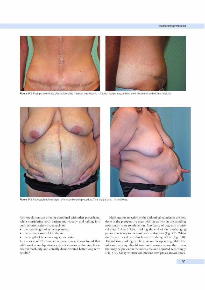
loss population can often be combined with other procedures,
while considering each patient individually and taking into
consideration safety issues such as:
• the total length of surgery planned,
• the patient’s overall health, and
• the length of time the surgery will take.
In a review of 73 consecutive procedures, it was found that
additional dermolipectomies do not increase abdominoplasty-
related morbidity and actually demonstrated better long-term
results.8
Markings for resection of the abdominal panniculus are best
done in the preoperative area with the patient in the standing
position or prior to admission. Avoidance of dog ears is criti-
cal (Figs 5.5 and 5.6); marking the end of the overhanging
panniculus is key to the avoidance of dog ears (Fig. 5.7). When
the patient lies down, this lateral overhang is lost (Fig. 5.8).
The inferior marking can be done on the operating table. The
inferior marking should take into consideration the excess
that may be present in the mons area and adjusted accordingly
(Fig. 5.9). Many women will present with ptosis and/or exces-
Preoperative preparation
51
Figure 5.2 Postoperative views after incisional hernia repair and resection of abdominal pannus, utilizing lower abdominal and midline incisions.
Figure 5.3 Subcostal midline incision after open bariatric procedure. Total weight loss: 111 lbs (50 kg).
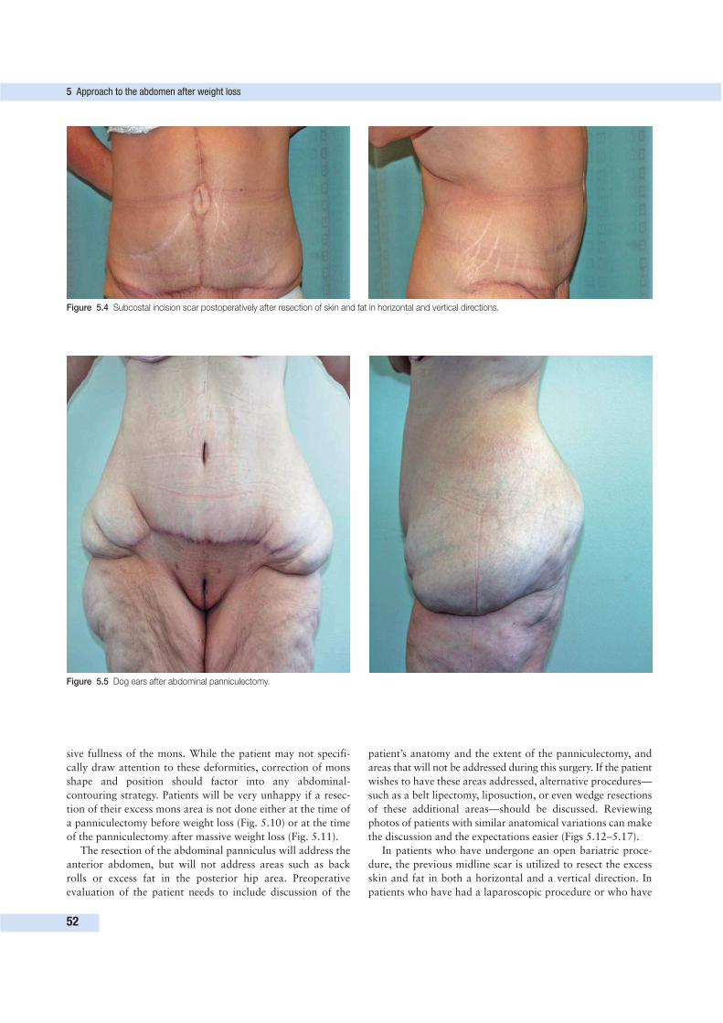
sive fullness of the mons. While the patient may not specifi-
cally draw attention to these deformities, correction of mons
shape and position should factor into any abdominal-
contouring strategy. Patients will be very unhappy if a resec-
tion of their excess mons area is not done either at the time of
a panniculectomy before weight loss (Fig. 5.10) or at the time
of the panniculectomy after massive weight loss (Fig. 5.11).
The resection of the abdominal panniculus will address the
anterior abdomen, but will not address areas such as back
rolls or excess fat in the posterior hip area. Preoperative
evaluation of the patient needs to include discussion of the
patient’s anatomy and the extent of the panniculectomy, and
areas that will not be addressed during this surgery. If the patient
wishes to have these areas addressed, alternative procedures—
such as a belt lipectomy, liposuction, or even wedge resections
of these additional areas—should be discussed. Reviewing
photos of patients with similar anatomical variations can make
the discussion and the expectations easier (Figs 5.12–5.17).
In patients who have undergone an open bariatric proce-
dure, the previous midline scar is utilized to resect the excess
skin and fat in both a horizontal and a vertical direction. In
patients who have had a laparoscopic procedure or who have
5 Approach to the abdomen after weight loss
52
Figure 5.4 Subcostal incision scar postoperatively after resection of skin and fat in horizontal and vertical directions.
Figure 5.5 Dog ears after abdominal panniculectomy.
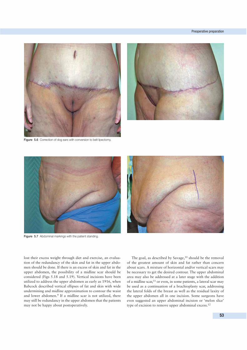
lost their excess weight through diet and exercise, an evalua-
tion of the redundancy of the skin and fat in the upper abdo-
men should be done. If there is an excess of skin and fat in the
upper abdomen, the possibility of a midline scar should be
considered (Figs 5.18 and 5.19). Vertical incisions have been
utilized to address the upper abdomen as early as 1916, when
Babcock described vertical ellipses of fat and skin with wide
undermining and midline approximation to contour the waist
and lower abdomen.9 If a midline scar is not utilized, there
may still be redundancy in the upper abdomen that the patients
may not be happy about postoperatively.
The goal, as described by Savage,10 should be the removal
of the greatest amount of skin and fat rather than concern
about scars. A mixture of horizontal and/or vertical scars may
be necessary to get the desired contour. The upper abdominal
area may also be addressed at a later stage with the addition
of a midline scar,11 or even, in some patients, a lateral scar may
be used as a continuation of a brachioplasty scar, addressing
the lateral folds of the breast as well as the residual laxity of
the upper abdomen all in one incision. Some surgeons have
even suggested an upper abdominal incision or ‘melon slice’
type of excision to remove upper abdominal excess.12
Preoperative preparation
53
Figure 5.6 Correction of dog ears with conversion to belt lipectomy.
Figure 5.7 Abdominal markings with the patient standing.
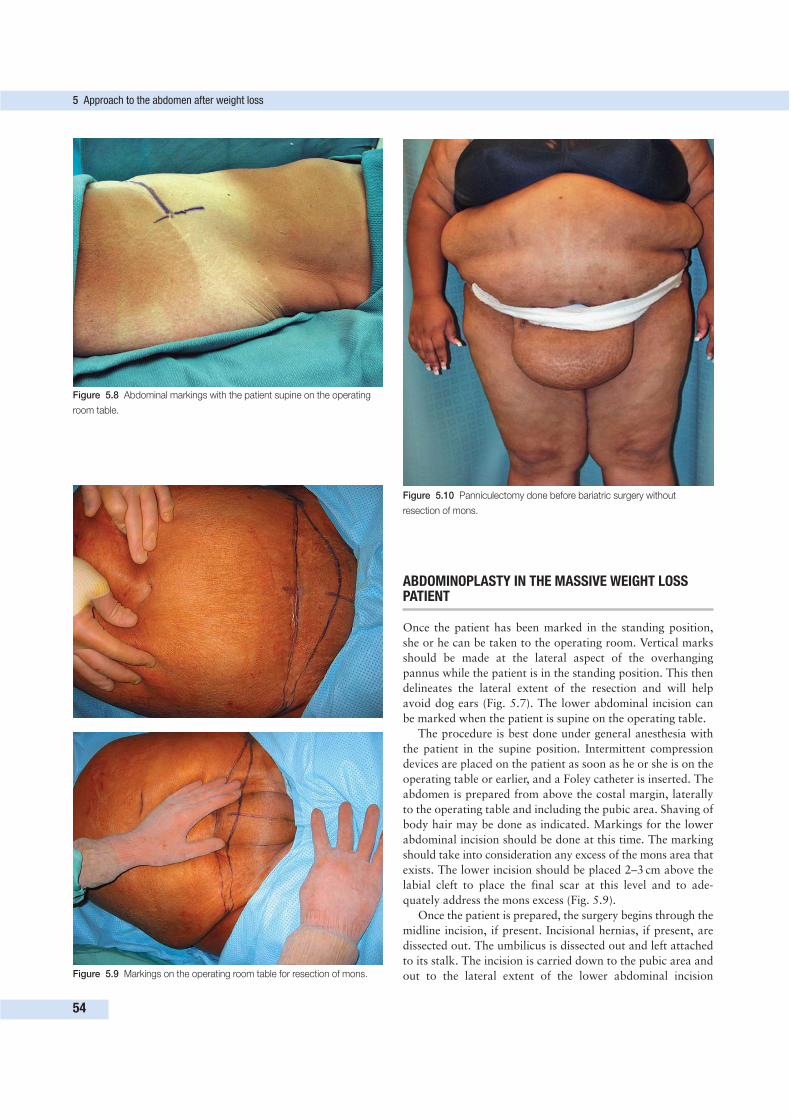
ABDOMINOPLASTY IN THE MASSIVE WEIGHT LOSSPATIENT
Once the patient has been marked in the standing position,
she or he can be taken to the operating room. Vertical marks
should be made at the lateral aspect of the overhanging
pannus while the patient is in the standing position. This then
delineates the lateral extent of the resection and will help
avoid dog ears (Fig. 5.7). The lower abdominal incision can
be marked when the patient is supine on the operating table.
The procedure is best done under general anesthesia with
the patient in the supine position. Intermittent compression
devices are placed on the patient as soon as he or she is on the
operating table or earlier, and a Foley catheter is inserted. The
abdomen is prepared from above the costal margin, laterally
to the operating table and including the pubic area. Shaving of
body hair may be done as indicated. Markings for the lower
abdominal incision should be done at this time. The marking
should take into consideration any excess of the mons area that
exists. The lower incision should be placed 2–3 cm above the
labial cleft to place the final scar at this level and to ade-
quately address the mons excess (Fig. 5.9).
Once the patient is prepared, the surgery begins through the
midline incision, if present. Incisional hernias, if present, are
dissected out. The umbilicus is dissected out and left attached
to its stalk. The incision is carried down to the pubic area and
out to the lateral extent of the lower abdominal incision
5 Approach to the abdomen after weight loss
54
Figure 5.8 Abdominal markings with the patient supine on the operating
room table.
Figure 5.9 Markings on the operating room table for resection of mons.
Figure 5.10 Panniculectomy done before bariatric surgery without
resection of mons.
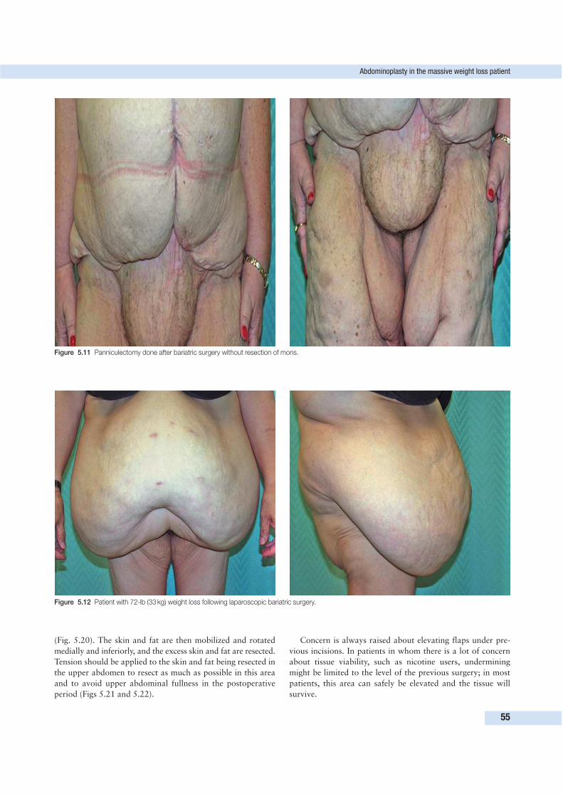
Abdominoplasty in the massive weight loss patient
55
Figure 5.11 Panniculectomy done after bariatric surgery without resection of mons.
Figure 5.12 Patient with 72-lb (33 kg) weight loss following laparoscopic bariatric surgery.
(Fig. 5.20). The skin and fat are then mobilized and rotated
medially and inferiorly, and the excess skin and fat are resected.
Tension should be applied to the skin and fat being resected in
the upper abdomen to resect as much as possible in this area
and to avoid upper abdominal fullness in the postoperative
period (Figs 5.21 and 5.22).
Concern is always raised about elevating flaps under pre-
vious incisions. In patients in whom there is a lot of concern
about tissue viability, such as nicotine users, undermining
might be limited to the level of the previous surgery; in most
patients, this area can safely be elevated and the tissue will
survive.
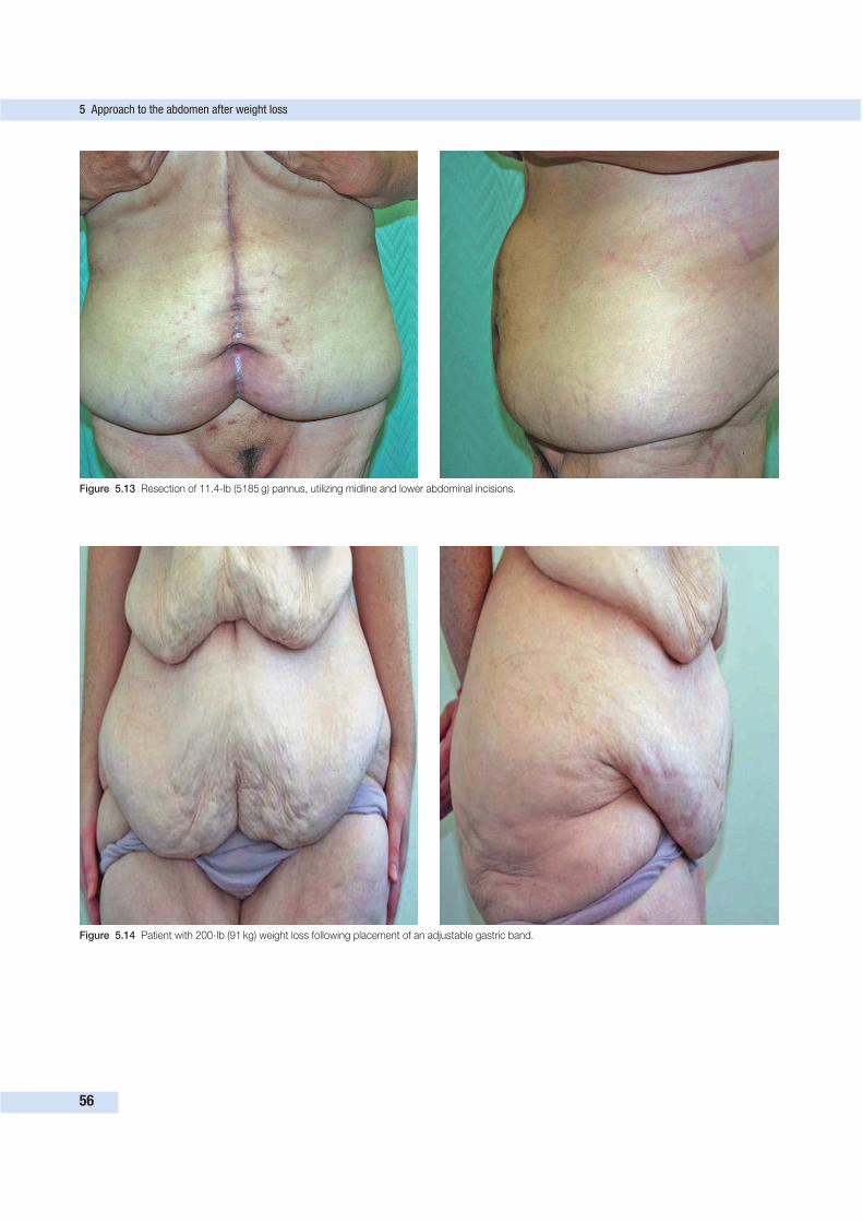
5 Approach to the abdomen after weight loss
56
Figure 5.13 Resection of 11.4-lb (5185 g) pannus, utilizing midline and lower abdominal incisions.
Figure 5.14 Patient with 200-lb (91 kg) weight loss following placement of an adjustable gastric band.
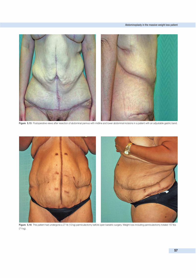
Abdominoplasty in the massive weight loss patient
57
Figure 5.15 Postoperative views after resection of abdominal pannus with midline and lower abdominal incisions in a patient with an adjustable gastric band.
Figure 5.16 This patient had undergone a 27-lb (12 kg) panniculectomy before open bariatric surgery. Weight loss including panniculectomy totaled 157 lbs
(71 kg).
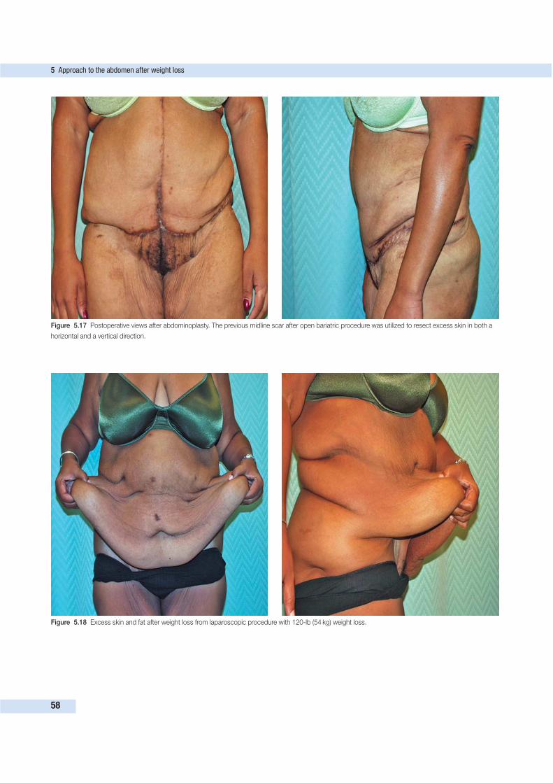
5 Approach to the abdomen after weight loss
58
Figure 5.17 Postoperative views after abdominoplasty. The previous midline scar after open bariatric procedure was utilized to resect excess skin in both a
horizontal and a vertical direction.
Figure 5.18 Excess skin and fat after weight loss from laparoscopic procedure with 120-lb (54 kg) weight loss.
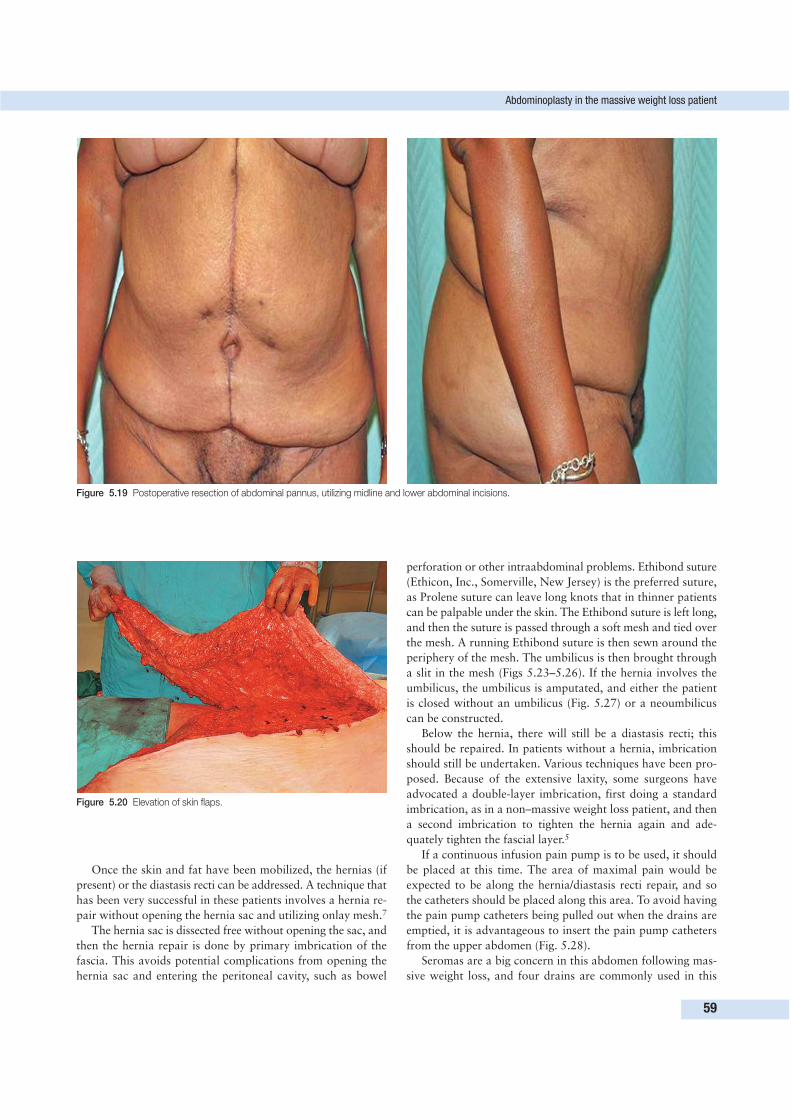
Abdominoplasty in the massive weight loss patient
59
Figure 5.19 Postoperative resection of abdominal pannus, utilizing midline and lower abdominal incisions.
Figure 5.20 Elevation of skin flaps.
Once the skin and fat have been mobilized, the hernias (if
present) or the diastasis recti can be addressed. A technique that
has been very successful in these patients involves a hernia re-
pair without opening the hernia sac and utilizing onlay mesh.7
The hernia sac is dissected free without opening the sac, and
then the hernia repair is done by primary imbrication of the
fascia. This avoids potential complications from opening the
hernia sac and entering the peritoneal cavity, such as bowel
perforation or other intraabdominal problems. Ethibond suture
(Ethicon, Inc., Somerville, New Jersey) is the preferred suture,
as Prolene suture can leave long knots that in thinner patients
can be palpable under the skin. The Ethibond suture is left long,
and then the suture is passed through a soft mesh and tied over
the mesh. A running Ethibond suture is then sewn around the
periphery of the mesh. The umbilicus is then brought through
a slit in the mesh (Figs 5.23–5.26). If the hernia involves the
umbilicus, the umbilicus is amputated, and either the patient
is closed without an umbilicus (Fig. 5.27) or a neoumbilicus
can be constructed.
Below the hernia, there will still be a diastasis recti; this
should be repaired. In patients without a hernia, imbrication
should still be undertaken. Various techniques have been pro-
posed. Because of the extensive laxity, some surgeons have
advocated a double-layer imbrication, first doing a standard
imbrication, as in a non–massive weight loss patient, and then
a second imbrication to tighten the hernia again and ade-
quately tighten the fascial layer.5
If a continuous infusion pain pump is to be used, it should
be placed at this time. The area of maximal pain would be
expected to be along the hernia/diastasis recti repair, and so
the catheters should be placed along this area. To avoid having
the pain pump catheters being pulled out when the drains are
emptied, it is advantageous to insert the pain pump catheters
from the upper abdomen (Fig. 5.28).
Seromas are a big concern in this abdomen following mas-
sive weight loss, and four drains are commonly used in this
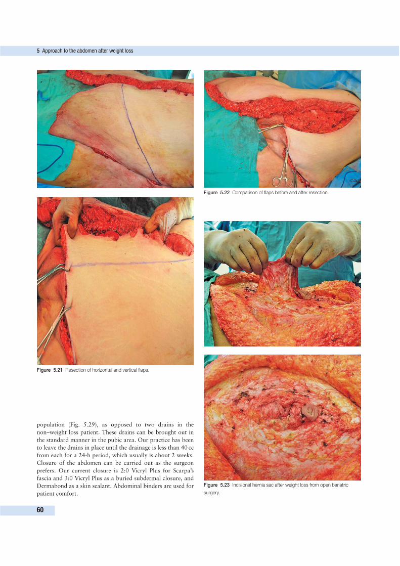
5 Approach to the abdomen after weight loss
60
Figure 5.21 Resection of horizontal and vertical flaps.
Figure 5.22 Comparison of flaps before and after resection.
Figure 5.23 Incisional hernia sac after weight loss from open bariatric
surgery.
population (Fig. 5.29), as opposed to two drains in the
non–weight loss patient. These drains can be brought out in
the standard manner in the pubic area. Our practice has been
to leave the drains in place until the drainage is less than 40 cc
from each for a 24-h period, which usually is about 2 weeks.
Closure of the abdomen can be carried out as the surgeon
prefers. Our current closure is 2:0 Vicryl Plus for Scarpa’s
fascia and 3:0 Vicryl Plus as a buried subdermal closure, and
Dermabond as a skin sealant. Abdominal binders are used for
patient comfort.
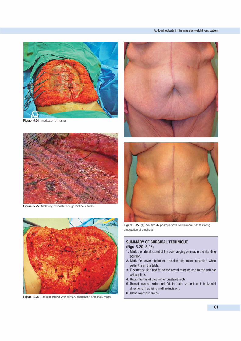
Abdominoplasty in the massive weight loss patient
61
Figure 5.24 Imbrication of hernia.
Figure 5.25 Anchoring of mesh through midline sutures.
Figure 5.26 Repaired hernia with primary imbrication and onlay mesh.
Figure 5.27 (a) Pre- and (b) postoperative hernia repair necessitating
amputation of umbilicus.
SUMMARY OF SURGICAL TECHNIQUE (Figs 5.20–5.26)1. Mark the lateral extent of the overhanging pannus in the standing
position.
2. Mark for lower abdominal incision and mons resection when
patient is on the table.
3. Elevate the skin and fat to the costal margins and to the anterior
axillary line.
4. Repair hernia (if present) or diastasis recti.
5. Resect excess skin and fat in both vertical and horizontal
directions (if utilizing midline incision).
6. Close over four drains.
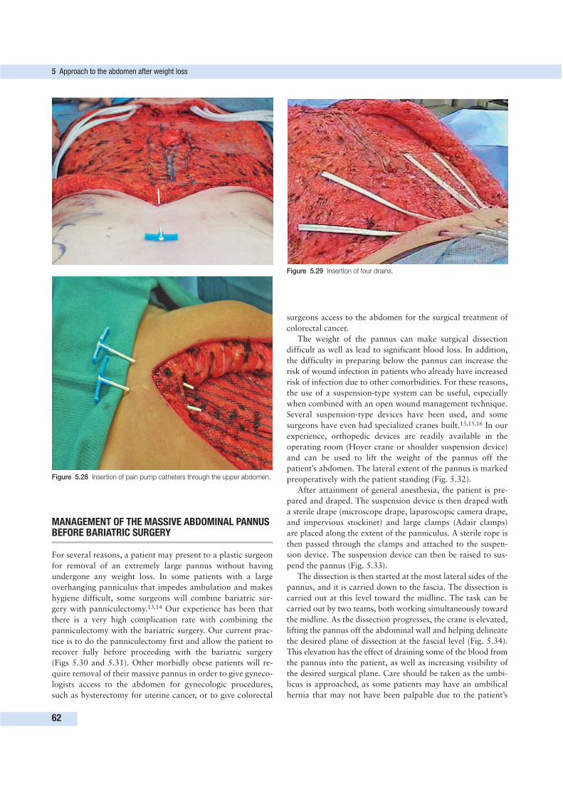
5 Approach to the abdomen after weight loss
62
Figure 5.28 Insertion of pain pump catheters through the upper abdomen.
Figure 5.29 Insertion of four drains.
MANAGEMENT OF THE MASSIVE ABDOMINAL PANNUSBEFORE BARIATRIC SURGERY
For several reasons, a patient may present to a plastic surgeon
for removal of an extremely large pannus without having
undergone any weight loss. In some patients with a large
overhanging panniculus that impedes ambulation and makes
hygiene difficult, some surgeons will combine bariatric sur-
gery with panniculectomy.13,14 Our experience has been that
there is a very high complication rate with combining the
panniculectomy with the bariatric surgery. Our current prac-
tice is to do the panniculectomy first and allow the patient to
recover fully before proceeding with the bariatric surgery
(Figs 5.30 and 5.31). Other morbidly obese patients will re-
quire removal of their massive pannus in order to give gyneco-
logists access to the abdomen for gynecologic procedures,
such as hysterectomy for uterine cancer, or to give colorectal
surgeons access to the abdomen for the surgical treatment of
colorectal cancer.
The weight of the pannus can make surgical dissection
difficult as well as lead to significant blood loss. In addition,
the difficulty in preparing below the pannus can increase the
risk of wound infection in patients who already have increased
risk of infection due to other comorbidities. For these reasons,
the use of a suspension-type system can be useful, especially
when combined with an open wound management technique.
Several suspension-type devices have been used, and some
surgeons have even had specialized cranes built.13,15,16 In our
experience, orthopedic devices are readily available in the
operating room (Hoyer crane or shoulder suspension device)
and can be used to lift the weight of the pannus off the
patient’s abdomen. The lateral extent of the pannus is marked
preoperatively with the patient standing (Fig. 5.32).
After attainment of general anesthesia, the patient is pre-
pared and draped. The suspension device is then draped with
a sterile drape (microscope drape, laparoscopic camera drape,
and impervious stockinet) and large clamps (Adair clamps)
are placed along the extent of the panniculus. A sterile rope is
then passed through the clamps and attached to the suspen-
sion device. The suspension device can then be raised to sus-
pend the pannus (Fig. 5.33).
The dissection is then started at the most lateral sides of the
pannus, and it is carried down to the fascia. The dissection is
carried out at this level toward the midline. The task can be
carried out by two teams, both working simultaneously toward
the midline. As the dissection progresses, the crane is elevated,
lifting the pannus off the abdominal wall and helping delineate
the desired plane of dissection at the fascial level (Fig. 5.34).
This elevation has the effect of draining some of the blood from
the pannus into the patient, as well as increasing visibility of
the desired surgical plane. Care should be taken as the umbi-
licus is approached, as some patients may have an umbilical
hernia that may not have been palpable due to the patient’s
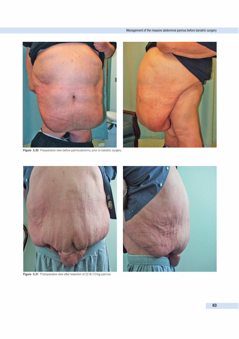
Management of the massive abdominal pannus before bariatric surgery
63
Figure 5.30 Preoperative view before panniculectomy, prior to bariatric surgery.
Figure 5.31 Postoperative view after resection of 22-lb (10 kg) pannus.
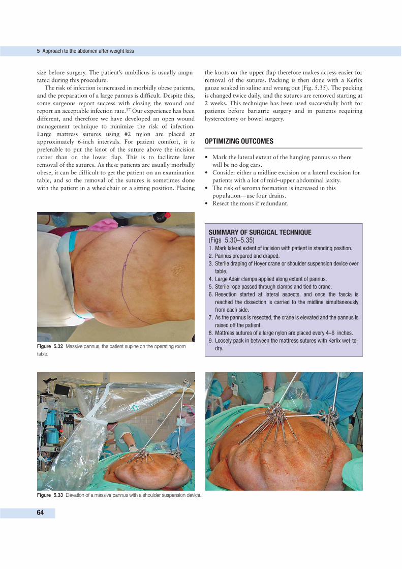
5 Approach to the abdomen after weight loss
64
Figure 5.32 Massive pannus, the patient supine on the operating room
table.
Figure 5.33 Elevation of a massive pannus with a shoulder suspension device.
size before surgery. The patient’s umbilicus is usually ampu-
tated during this procedure.
The risk of infection is increased in morbidly obese patients,
and the preparation of a large pannus is difficult. Despite this,
some surgeons report success with closing the wound and
report an acceptable infection rate.17 Our experience has been
different, and therefore we have developed an open wound
management technique to minimize the risk of infection.
Large mattress sutures using #2 nylon are placed at
approximately 6-inch intervals. For patient comfort, it is
preferable to put the knot of the suture above the incision
rather than on the lower flap. This is to facilitate later
removal of the sutures. As these patients are usually morbidly
obese, it can be difficult to get the patient on an examination
table, and so the removal of the sutures is sometimes done
with the patient in a wheelchair or a sitting position. Placing
the knots on the upper flap therefore makes access easier for
removal of the sutures. Packing is then done with a Kerlix
gauze soaked in saline and wrung out (Fig. 5.35). The packing
is changed twice daily, and the sutures are removed starting at
2 weeks. This technique has been used successfully both for
patients before bariatric surgery and in patients requiring
hysterectomy or bowel surgery.
OPTIMIZING OUTCOMES
• Mark the lateral extent of the hanging pannus so there
will be no dog ears.
• Consider either a midline excision or a lateral excision for
patients with a lot of mid–upper abdominal laxity.
• The risk of seroma formation is increased in this
population—use four drains.
• Resect the mons if redundant.
SUMMARY OF SURGICAL TECHNIQUE (Figs 5.30–5.35)1. Mark lateral extent of incision with patient in standing position.
2. Pannus prepared and draped.
3. Sterile draping of Hoyer crane or shoulder suspension device over
table.
4. Large Adair clamps applied along extent of pannus.
5. Sterile rope passed through clamps and tied to crane.
6. Resection started at lateral aspects, and once the fascia is
reached the dissection is carried to the midline simultaneously
from each side.
7. As the pannus is resected, the crane is elevated and the pannus is
raised off the patient.
8. Mattress sutures of a large nylon are placed every 4–6 inches.
9. Loosely pack in between the mattress sutures with Kerlix wet-to-
dry.
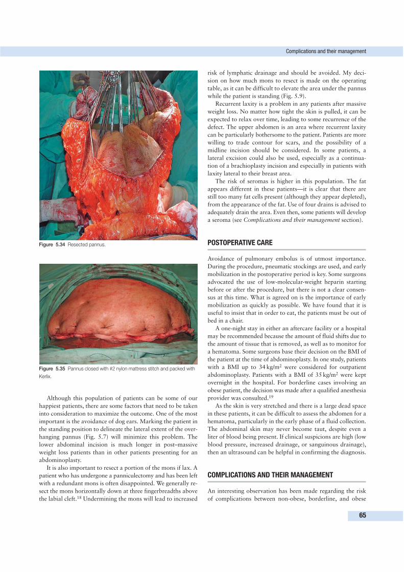
Although this population of patients can be some of our
happiest patients, there are some factors that need to be taken
into consideration to maximize the outcome. One of the most
important is the avoidance of dog ears. Marking the patient in
the standing position to delineate the lateral extent of the over-
hanging pannus (Fig. 5.7) will minimize this problem. The
lower abdominal incision is much longer in post–massive
weight loss patients than in other patients presenting for an
abdominoplasty.
It is also important to resect a portion of the mons if lax. A
patient who has undergone a panniculectomy and has been left
with a redundant mons is often disappointed. We generally re-
sect the mons horizontally down at three fingerbreadths above
the labial cleft.18 Undermining the mons will lead to increased
risk of lymphatic drainage and should be avoided. My deci-
sion on how much mons to resect is made on the operating
table, as it can be difficult to elevate the area under the pannus
while the patient is standing (Fig. 5.9).
Recurrent laxity is a problem in any patients after massive
weight loss. No matter how tight the skin is pulled, it can be
expected to relax over time, leading to some recurrence of the
defect. The upper abdomen is an area where recurrent laxity
can be particularly bothersome to the patient. Patients are more
willing to trade contour for scars, and the possibility of a
midline incision should be considered. In some patients, a
lateral excision could also be used, especially as a continua-
tion of a brachioplasty incision and especially in patients with
laxity lateral to their breast area.
The risk of seromas is higher in this population. The fat
appears different in these patients—it is clear that there are
still too many fat cells present (although they appear depleted),
from the appearance of the fat. Use of four drains is advised to
adequately drain the area. Even then, some patients will develop
a seroma (see Complications and their management section).
POSTOPERATIVE CARE
Avoidance of pulmonary embolus is of utmost importance.
During the procedure, pneumatic stockings are used, and early
mobilization in the postoperative period is key. Some surgeons
advocated the use of low-molecular-weight heparin starting
before or after the procedure, but there is not a clear consen-
sus at this time. What is agreed on is the importance of early
mobilization as quickly as possible. We have found that it is
useful to insist that in order to eat, the patients must be out of
bed in a chair.
A one-night stay in either an aftercare facility or a hospital
may be recommended because the amount of fluid shifts due to
the amount of tissue that is removed, as well as to monitor for
a hematoma. Some surgeons base their decision on the BMI of
the patient at the time of abdominoplasty. In one study, patients
with a BMI up to 34 kg/m2 were considered for outpatient
abdominoplasty. Patients with a BMI of 35 kg/m2 were kept
overnight in the hospital. For borderline cases involving an
obese patient, the decision was made after a qualified anesthesia
provider was consulted.19
As the skin is very stretched and there is a large dead space
in these patients, it can be difficult to assess the abdomen for a
hematoma, particularly in the early phase of a fluid collection.
The abdominal skin may never become taut, despite even a
liter of blood being present. If clinical suspicions are high (low
blood pressure, increased drainage, or sanguinous drainage),
then an ultrasound can be helpful in confirming the diagnosis.
COMPLICATIONS AND THEIR MANAGEMENT
An interesting observation has been made regarding the risk
of complications between non-obese, borderline, and obese
Complications and their management
65
Figure 5.34 Resected pannus.
Figure 5.35 Pannus closed with #2 nylon mattress stitch and packed with
Kerlix.
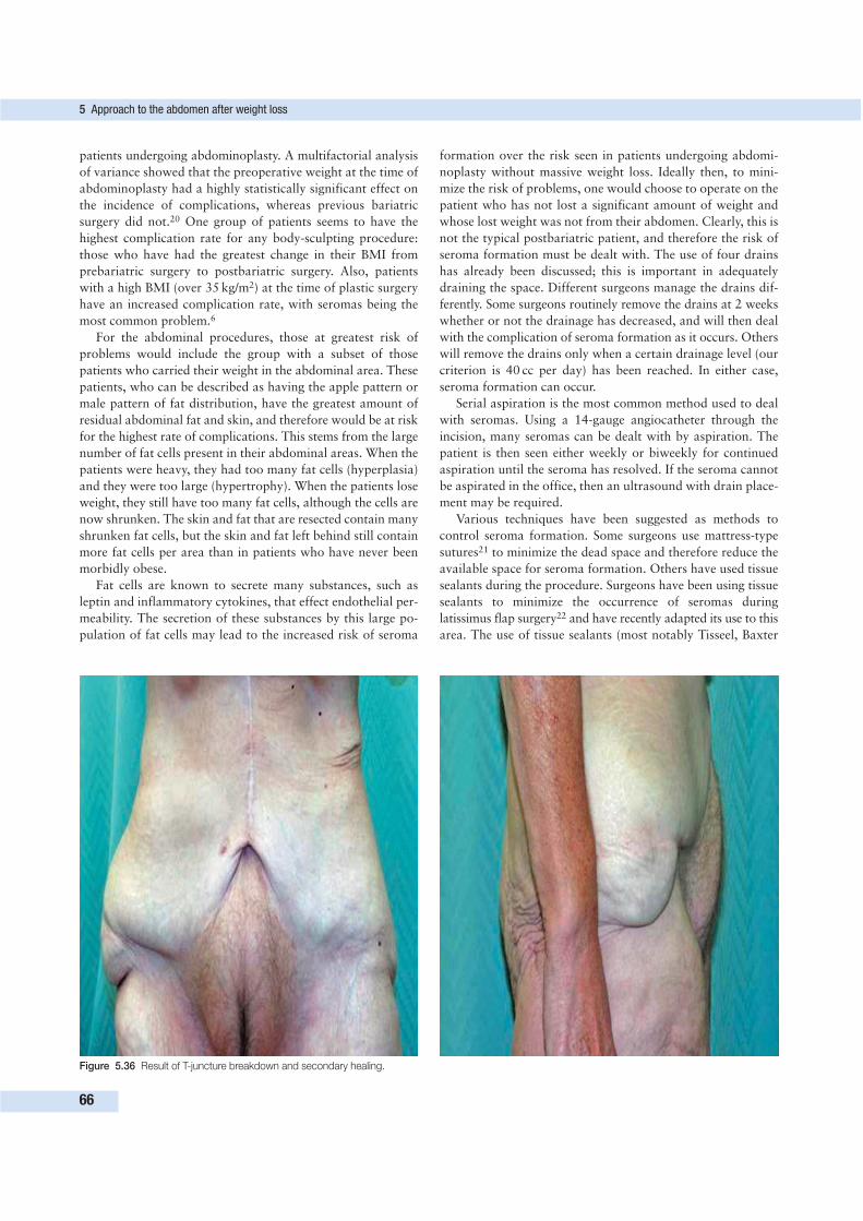
patients undergoing abdominoplasty. A multifactorial analysis
of variance showed that the preoperative weight at the time of
abdominoplasty had a highly statistically significant effect on
the incidence of complications, whereas previous bariatric
surgery did not.20 One group of patients seems to have the
highest complication rate for any body-sculpting procedure:
those who have had the greatest change in their BMI from
prebariatric surgery to postbariatric surgery. Also, patients
with a high BMI (over 35 kg/m2) at the time of plastic surgery
have an increased complication rate, with seromas being the
most common problem.6
For the abdominal procedures, those at greatest risk of
problems would include the group with a subset of those
patients who carried their weight in the abdominal area. These
patients, who can be described as having the apple pattern or
male pattern of fat distribution, have the greatest amount of
residual abdominal fat and skin, and therefore would be at risk
for the highest rate of complications. This stems from the large
number of fat cells present in their abdominal areas. When the
patients were heavy, they had too many fat cells (hyperplasia)
and they were too large (hypertrophy). When the patients lose
weight, they still have too many fat cells, although the cells are
now shrunken. The skin and fat that are resected contain many
shrunken fat cells, but the skin and fat left behind still contain
more fat cells per area than in patients who have never been
morbidly obese.
Fat cells are known to secrete many substances, such as
leptin and inflammatory cytokines, that effect endothelial per-
meability. The secretion of these substances by this large po-
pulation of fat cells may lead to the increased risk of seroma
formation over the risk seen in patients undergoing abdomi-
noplasty without massive weight loss. Ideally then, to mini-
mize the risk of problems, one would choose to operate on the
patient who has not lost a significant amount of weight and
whose lost weight was not from their abdomen. Clearly, this is
not the typical postbariatric patient, and therefore the risk of
seroma formation must be dealt with. The use of four drains
has already been discussed; this is important in adequately
draining the space. Different surgeons manage the drains dif-
ferently. Some surgeons routinely remove the drains at 2 weeks
whether or not the drainage has decreased, and will then deal
with the complication of seroma formation as it occurs. Others
will remove the drains only when a certain drainage level (our
criterion is 40 cc per day) has been reached. In either case,
seroma formation can occur.
Serial aspiration is the most common method used to deal
with seromas. Using a 14-gauge angiocatheter through the
incision, many seromas can be dealt with by aspiration. The
patient is then seen either weekly or biweekly for continued
aspiration until the seroma has resolved. If the seroma cannot
be aspirated in the office, then an ultrasound with drain place-
ment may be required.
Various techniques have been suggested as methods to
control seroma formation. Some surgeons use mattress-type
sutures21 to minimize the dead space and therefore reduce the
available space for seroma formation. Others have used tissue
sealants during the procedure. Surgeons have been using tissue
sealants to minimize the occurrence of seromas during
latissimus flap surgery22 and have recently adapted its use to this
area. The use of tissue sealants (most notably Tisseel, Baxter
5 Approach to the abdomen after weight loss
66
Figure 5.36 Result of T-juncture breakdown and secondary healing.
Corp., Deerfield, Illinois) for reducing the risk of seromas is
an off-label use of the product. The use of Tisseel seems to
reduce the number of seromas that occur and, when seromas
do occur, their size is diminished.23
When drainage is persistent, some surgeons have been using
doxycycline in the drains. Similarly to the use of doxycycline in
thoracic surgery to decrease pleural effusions, the doxycycline
is diluted (100 mg in 5 cc of saline) and injected into the drain.
The drain is then left unclamped for 4 h and then suction is
again applied. Some patients may complain of a temporary
burning sensation, but most do not report any symptoms. The
burning sensation, if felt, seems to be more common in patients
who are less than 2 weeks out from their procedure. Anecdotal
evidence shows that, for some patients, this method is
effective in expediting the resolution of the seroma.
The most common site of wound breakdown is at the T
juncture where the vertical and horizontal incisions come
together. Debridement and packing will usually allow this area
to heal, but patients may require a scar revision (Fig. 5.36).
Infections are not that common but, when they do occur,
can be troublesome to manage. If a patient presents with an
infection, it is important to recall which bariatric procedure
the patient had undergone. Patients who have undergone a
malabsorptive procedure, especially a duodenal switch, may
not absorb adequate antibiotics and so may require intra-
venous therapy. We have handled this situation by admitting
the patients, having a peripherally inserted central catheter line
placed, and then continuing the intravenous antibiotics at home.
CONCLUSION
The post–massive weight loss patient is both challenging and
rewarding. Although the surgery may be more difficult, in re-
quiring different incisions or even a staged approach, the out-
come may be life-changing for the patient. Careful planning
and discussions with the patient, as well as some different
intraoperative routines, can minimize the complications as
well as undesirable outcomes.
REFERENCES
1. Kelly HA. Report of gynecological cases. John Hopkins Med J
1899; 10:197.
2. Kelly HA. Excision of the fat of the abdominal wall lipectomy. Surg
Gynecol Obstet 1910; 10:299.
3. Thorek M. Plastic reconstruction of the female breast and abdomen.
Am J Surg 1939; 43:268.
4. Matarasso A, Swift RW, Rankin M. Abdominoplasty and abdominal
contour surgery: a national plastic surgery survey. Plast Reconstr
Surg 2006; 117:1797–1808.
5. Aly AS, Cram AE, Heddens C. Truncal body contouring surgery
in the massive weight loss patient. Clin Plast Surg 2004;
31:611–624.
6. Aly AS, Cram AE, Chao M, et al. Belt lipectomy for circumferential
truncal excess: the University of Iowa experience. Plast Reconstr
Surg 2003; 111(1):398–413.
7. Downey SE, Kelso R, Anthone G, et al. Review of technique for
combined closed incisional repair and panniculectomy status post
bariatric surgery. Surg Obes Relat Dis (in press).
8. Gmur RU, Banic A, Erni D. Is it safe to combine abdominoplasty
with other dermolipectomy procedures to correct skin excess after
weight loss? Ann Plast Surg 2003; 51(4):353–357.
9. Babcock W. The correction of the obese and relaxed abdominal wall
with especial reference to the use of buried silver chain. Phila Obstet
Soc 1916; May 4.
10. Savage RC. Abdominoplasty following gastrointestinal bypass sur-
gery. Plast Reconstr Surg 1983; 71(4):500–507.
11. Rosenfield LK. Comprehensive abdominoplasty approaches using
complementary techniques. In: Nahai F. The art of aesthetic surgery.
St. Louis: Quality Medical Publishing; 2005.
12. Ward DJ, Wilson JSP. Abdominal reduction following jejunoileal
bypass for morbid obesity. Br J Plast Surg 1989; 42:586–590.
13. Jensen PL, Sanger JR, Matloub HS, et al. Use of a portable floor
crane as an aid to resection of the massive panniculus. Ann Plast
Surg 1990; 25:234–235.
14. Acarturk TO, Wachtman G, Heil B, et al. Panniculectomy as an
adjuvant to bariatric surgery. Ann Plast Surg 2004; 53(4):360–366.
15. Matory WE, O’Sullivan J, Fudem G, et al. Abdominal surgery in
patients with severe morbid obesity. Plast Reconstr Surg 1994;
94(7):976–987.
16. Richard EF. A mechanical aid for abdominal panniculectomy. Br J
Plast Surg 1965; 18:336–337.
17. Hopkins MP, Shriner AM, Parker MG, et al. Panniculectomy at the
time of gynecologic surgery in morbidly obese patients. Am J Obstet
Gynecol 2000; 182:1502–1505.
18. Matarasso A, Wallach S. Abdominal contour surgery: treating all
aesthetic units, including the mons pubis. Aesthetic Surg J 2001;
21(2):111–119.
19. Williams TC, Hardaway M, Altuna B. Ambulatory abdominoplasty
tailored to patients with an appropriate body mass index. Aesthetic
Surg J 2005; March–April:132–137.
20. Vastine VL, Morgan RF, Williams GS, et al. Wound complications of
abdominoplasty in obese patients. Ann Plast Surg 1999; 42(1):34–39.
21. Pollock H, Pollack T. Progressive tension sutures: a technique to
reduce local complications in abdominoplasty. Plast Reconstr Surg
2000; 105(7):2583–2586.
22. Weinrach JC, Cronin ED, Smith BK, et al. Preventing seroma in the
latissimus dorsi flap donor site with fibrin sealant. Ann Plast Surg
2004; 53(1):12–16.
23. Downey SE, Morales CL. The use of fibrin sealant in the preven-
tion of seromas in the massive weight loss patient. Poster presen-
tation at the Annual ASPS Meeting, September 2005, Chicago,
Illinois.
ADDITIONAL READING
Al-Basti HB, El-Khatib HA, Taha A, et al. Intraabdominal pressure after
full abdominoplasty in obese multiparous patients. Plast Reconstr
Surg 2004; 113(7):2145–2150.
Baroudi R, Ferreira C. Seroma: how to avoid it and how to treat it.
Aesthetic Surg J 1999; 18:439.
Belin RP, Stone NH, Fischer RP, et al. Improved technique of pannicu-
lectomy. Surgery 1966; 59(2):222–225.
Blomfield PI, Le T, Allen DG, et al. Panniculectomy: a useful technique
for the obese patient undergoing gynecological surgery. Gynecol
Oncol 1998; 70:80–86.
Bolton MA, Pruzinsky T, Cash TF, et al. Measuring outcomes in plastic
surgery: body image and quality of life in abdominoplasty patients.
Plast Reconstr Surg 2003; 112(2):619–625.
Additional reading
67
Carwell GR, Horton CE Sr. Circumferential torsoplasty. Ann Plast Surg
1997; 38(3):213–216.
Cosin JA, Powell JL, Donovan JT, et al. The safety and efficacy of
extensive abdominal panniculectomy at the time of pelvic surgery.
Gynecol Oncol 1994; 55:36–40.
Da Costa LF, Landecker A, Manta AM. Optimizing body contour in
massive weight loss patients: the modified vertical abominoplasty.
Plast Reconstr Surg 2004; 114(7):1917–1923.
Dardour JC, Vilain R. Alternatives to the classic abdominoplasty. Ann
Plast Surg 1986; 17(3):247–258.
Daw JL, Mustoe TA. Use of a tourniquet in panniculus resection. Plast
Reconstr Surg 1997; 99(7):2082–2084.
Desjardin A. Lipectomy for extreme obesity. Paris Chir 1911; 3:466.
El-Khatib HA, Bener A. Abdominal dermolipectomy in an abdomen with
pre-existing scars: a different concept. Plast Reconstr Surg 2004;
114(4):992–997.
Goessl A, Redl H. Optimized thrombin dilution protocol for a slowly
setting fibrin sealant in surgery. Eur Surg 2005; 37(1):43–51.
Grazer FM, Goldwyn RM. Abdominoplasty assessed by survey, with em-
phasis on complications. Plast Reconstr Surg 1997; 59(4):513–517.
Hagerty RF, Hawk JC Jr, Boniface K, et al. Resection of massive abdo-
minal panniculus adiposus. South Med J 1974; 67(8):984–989.
Hensel JM, Lehman JA Jr, Tantri MP, et al. An outcomes analysis and
satisfaction survey of 199 consecutive abdominoplasties. Ann Plast
Surg 2001; 46(4):357–363.
Hester TR Jr, Baird W, Bostwick J III, et al. Abdominoplasty combined
with other major surgical procedures: safe or sorry? Plast Reconstr
Surg 1989; 83(6):997–1004.
Hunstad JP. Body contouring in the obese patient. Clin Plast Surg 1996;
23(4):647–670.
Kamper MJ, Galloway DV, Ashley F. Abdominal panniculectomy after
massive weight loss. Plast Reconstr Surg 1972; 50(5):441–446.
Krueger JK, Rohrich RJ. Clearing the smoke. Scientific rationale for
tobacco abstention with plastic surgery. Plast Reconstr Surg 2001;
108(4):1063–1073.
Kulber DA, Bacilious N, Peters ED, et al. The use of fibrin sealant in the
prevention of seromas. Plast Reconstr Surg 1997; 99(3):842–849.
Lockwood T. High–lateral-tension abdominoplasty with superficial fas-
cial system suspension. Plast Reconstr Surg 1995; 96(3):603–615.
Lockwood T. Lower body lift with superficial fascial system suspension.
Plast Reconstr Surg 1993; 92(6):1112–1122.
Lockwood T. Superficial fascial system (SFS) of the trunk and extre-
mities: a new concept. Plast Reconstr Surg 1991; 87:1009–1018.
Lockwood T. Transverse flank-thigh-buttock lift with superficial fascial
suspension. Plast Reconstr Surg 1991; 87(6):1019–1027.
Lockwood TE. Lower-body lift. Aesthetic Surg J 2001; 21:335.
Matarasso A. Abdominoplasty. In: Achauer BM, Eriksson E, Guyuron
B, et al, eds. Plastic surgery: indications, operations and outcomes,
vol 5. Aesthetic surgery. St. Louis: Mosby; 2000:2783–2821.
Matarasso A. Liposuction as an adjunct to a full abdominoplasty re-
visted. Plast Reconstr Surg 2000; 106(5):1197–1202.
Matarasso A. The male abdominoplasty. Clin Plast Surg 2004;
31(4):555–569.
McCabe WP, Kelly AP Jr, Frame B. Panniculectomy following intestinal
bypass. Br J Plast Surg 1974; 27:346–351.
McGraw LH. Surgical rehabilitation after massive weight reduction:
case report. Annual Meeting of the American Society for Aesthetic
Plastic Surgery, March 12, 1973, California.
Meyerowitz BR, Gruber RP, Laub DR. Massive abdominal panniculec-
tomy. JAMA 1973; 225(4):408–409.
Micha JP, Rettenmaier MA, Francis L, et al. ‘Medically necessary’ pan-
niculectomy to facilitate gynecologic cancer surgery in morbidly
obese patients. Gynecol Oncol 1998; 69:237–242.
Oguz AT, Wachtman G, Heil B, et al. Panniculectomy as an adjuvant to
bariatric surgery. Ann Plast Surg 2004; 53(4):360–366.
Petty P, Manson PN, Black R, et al. Panniculus morbidus. Ann Plast
Surg 1992; 28(5):442–452.
Powell JL, Kasparek DK, Connor GP. Panniculectomy to facilitate gyne-
cologic surgery in morbidly obese women. Obstet Gynecol 1999;
94(4):528–531.
Rubin JP, Nguyen V, Schwentker A. Perioperative management of the
post-gastric-bypass patient presenting for body contour surgery.
Clin Plast Surg 2004; 31:601–610.
Soundararajan V, Hart NB, Royston CMS. Abdominoplasty following
vertical banded gastroplasty for morbid obesity. Br J Plast Surg
1995; 48:423–427.
Stanhope CR, Winburn KA, Silberman MB. Indicated noncosmetic pan-
niculectomy in gynecologic surgery. J Pelvic Surg 2002; 8:197–201.
Van Geertruyden JP, Vandeweyer E, de Fontaine S, et al. Circumferen-
tial torsoplasty. Br J Plast Surg 1999; 52(8):623–628.
Van Uchelen JH, Werker PM, Kon M. Complications of abdomino-
plasty in 86 patients. Plast Reconstr Surg 2001; 107(7):1869–1873.
Young SC, Freiberg A. A critical look at abdominal lipectomy following
morbid obesity surgery. Aesthetic Plast Surg 1991; 15:81–84.
Zook EG. Abdominoplasty following gastrointestinal bypass surgery.
Plast Reconstr Surg 1983; 4:508–509.
Zook EG. Massive weight loss patient. Clin Plast Surg 1975; 2(3):457.
5 Approach to the abdomen after weight loss
68
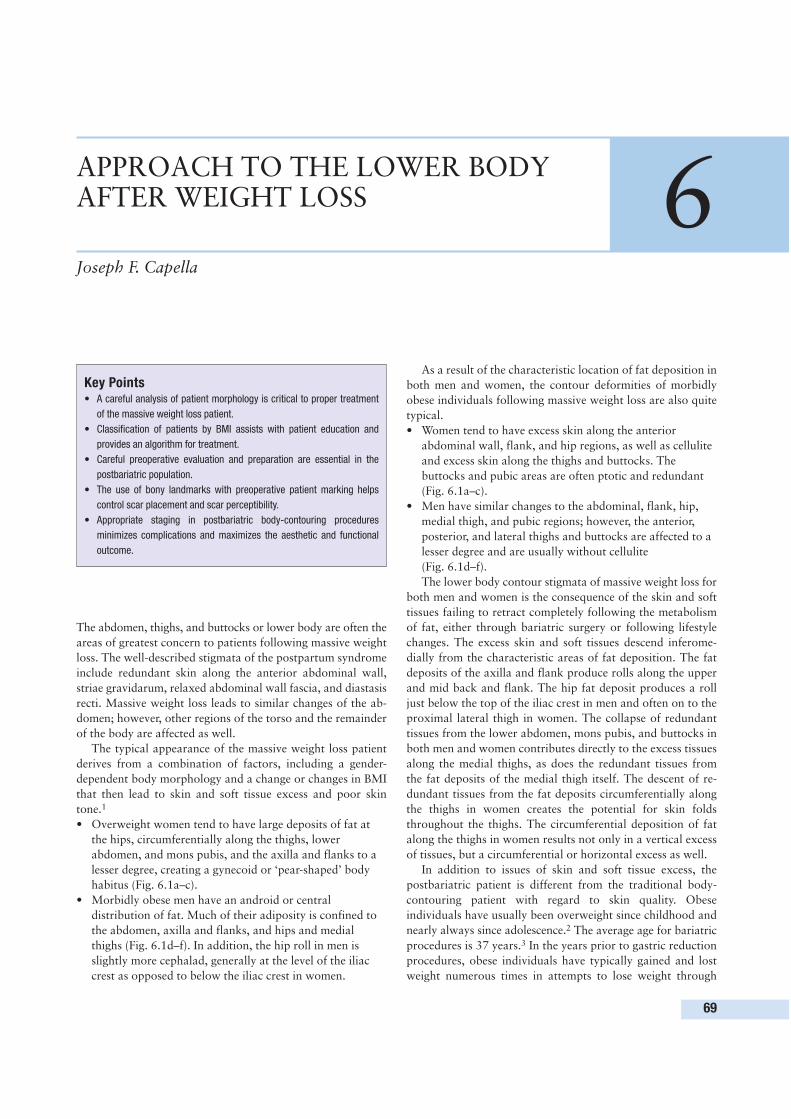
The abdomen, thighs, and buttocks or lower body are often the
areas of greatest concern to patients following massive weight
loss. The well-described stigmata of the postpartum syndrome
include redundant skin along the anterior abdominal wall,
striae gravidarum, relaxed abdominal wall fascia, and diastasis
recti. Massive weight loss leads to similar changes of the ab-
domen; however, other regions of the torso and the remainder
of the body are affected as well.
The typical appearance of the massive weight loss patient
derives from a combination of factors, including a gender-
dependent body morphology and a change or changes in BMI
that then lead to skin and soft tissue excess and poor skin
tone.1
• Overweight women tend to have large deposits of fat at
the hips, circumferentially along the thighs, lower
abdomen, and mons pubis, and the axilla and flanks to a
lesser degree, creating a gynecoid or ‘pear-shaped’ body
habitus (Fig. 6.1a–c).
• Morbidly obese men have an android or central
distribution of fat. Much of their adiposity is confined to
the abdomen, axilla and flanks, and hips and medial
thighs (Fig. 6.1d–f). In addition, the hip roll in men is
slightly more cephalad, generally at the level of the iliac
crest as opposed to below the iliac crest in women.
As a result of the characteristic location of fat deposition in
both men and women, the contour deformities of morbidly
obese individuals following massive weight loss are also quite
typical.
• Women tend to have excess skin along the anterior
abdominal wall, flank, and hip regions, as well as cellulite
and excess skin along the thighs and buttocks. The
buttocks and pubic areas are often ptotic and redundant
(Fig. 6.1a–c).
• Men have similar changes to the abdominal, flank, hip,
medial thigh, and pubic regions; however, the anterior,
posterior, and lateral thighs and buttocks are affected to a
lesser degree and are usually without cellulite
(Fig. 6.1d–f).
The lower body contour stigmata of massive weight loss for
both men and women is the consequence of the skin and soft
tissues failing to retract completely following the metabolism
of fat, either through bariatric surgery or following lifestyle
changes. The excess skin and soft tissues descend inferome-
dially from the characteristic areas of fat deposition. The fat
deposits of the axilla and flank produce rolls along the upper
and mid back and flank. The hip fat deposit produces a roll
just below the top of the iliac crest in men and often on to the
proximal lateral thigh in women. The collapse of redundant
tissues from the lower abdomen, mons pubis, and buttocks in
both men and women contributes directly to the excess tissues
along the medial thighs, as does the redundant tissues from
the fat deposits of the medial thigh itself. The descent of re-
dundant tissues from the fat deposits circumferentially along
the thighs in women creates the potential for skin folds
throughout the thighs. The circumferential deposition of fat
along the thighs in women results not only in a vertical excess
of tissues, but a circumferential or horizontal excess as well.
In addition to issues of skin and soft tissue excess, the
postbariatric patient is different from the traditional body-
contouring patient with regard to skin quality. Obese
individuals have usually been overweight since childhood and
nearly always since adolescence.2 The average age for bariatric
procedures is 37 years.3 In the years prior to gastric reduction
procedures, obese individuals have typically gained and lost
weight numerous times in attempts to lose weight through
69
APPROACH TO THE LOWER BODYAFTER WEIGHT LOSS 6Joseph F. Capella
Key Points• A careful analysis of patient morphology is critical to proper treatment
of the massive weight loss patient.
• Classification of patients by BMI assists with patient education and
provides an algorithm for treatment.
• Careful preoperative evaluation and preparation are essential in the
postbariatric population.
• The use of bony landmarks with preoperative patient marking helps
control scar placement and scar perceptibility.
• Appropriate staging in postbariatric body-contouring procedures
minimizes complications and maximizes the aesthetic and functional
outcome.
dieting or behavioral modification. The prolonged period of
skin under tension and the frequent history of ‘yo-yo’ dieting
lead to poor skin tone following massive weight loss. Striae
and cellulite are common throughout the torso, particularly in
women. The extreme body contour deformities that dis-
tinguish the routine patient from the massive weight loss
patient have led to the development of operative techniques
specific to these individuals.
The ideal lower body–contouring procedure for the massive
weight loss patient should effectively address all or as many of
the characteristic stigmata as possible in a safe, efficient, and
consistent manner. Various techniques have been described to
treat the lower body postbariatric condition; these include
body lift, belt lipectomy, lower body lift, and circumferential
torsoplasty.1,4–6 While having different names, each in this
group involves a simultaneous abdominoplasty, and thigh and
buttock lift. The goal of all these procedures is to reverse or
derotate the inferomedial collapse of the skin and soft tissues
of the lower body (Fig. 6.2). Aside from the obvious advan-
tage of addressing the thighs and buttocks as well as the abdo-
men in one stage, a simultaneous circumferential procedure
offers another very important advantage: a standing cone is
not a concern. In any procedure that is limited by the length of
a scar, some graduation in the amount of skin traction that
can be applied must exist to prevent skin redundancy along the
lateral extent of the scar. Circumferential procedures allow for
much higher levels of tension to be applied without this
concern. This is particularly important for the body lift where
the distal thigh and upper abdomen are being addressed from
the waistline.
The surge in bariatric procedures in the USA and abroad
over the past 5 years has led to increasing patient requests for
body-contouring procedures.7 To treat the postbariatric con-
dition, some plastic surgeons are implementing traditional
6 Approach to the lower body after weight loss
70
a b c
d e f
Figure 6.1 Type 3 patients. (a–c) A 40-year-old woman 40 months following gastric bypass surgery and weight loss of 269 lbs (122 kg). Current weight and
BMI: 254 lbs (115 kg) and 41 kg/m2, respectively. Highest weight and BMI: 522 lbs (237 kg), 84 kg/m2. (d-f) A 39-year-old man 16 months following gastric
bypass surgery and weight loss of 209 lbs (95 kg). Current weight and BMI: 229 lbs (104 kg) and 37 kg/m2, respectively. Highest weight and BMI: 439 lbs
(199 kg), 71 kg/m2.
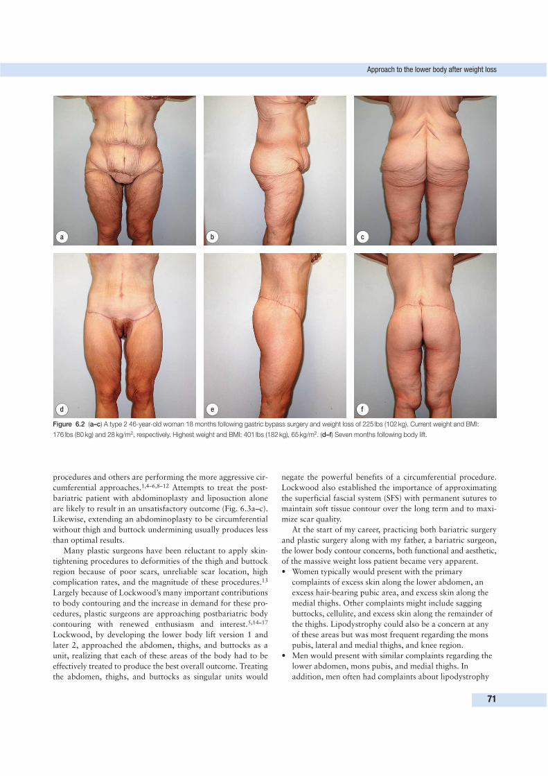
procedures and others are performing the more aggressive cir-
cumferential approaches.1,4–6,8–12 Attempts to treat the post-
bariatric patient with abdominoplasty and liposuction alone
are likely to result in an unsatisfactory outcome (Fig. 6.3a–c).
Likewise, extending an abdominoplasty to be circumferential
without thigh and buttock undermining usually produces less
than optimal results.
Many plastic surgeons have been reluctant to apply skin-
tightening procedures to deformities of the thigh and buttock
region because of poor scars, unreliable scar location, high
complication rates, and the magnitude of these procedures.13
Largely because of Lockwood’s many important contributions
to body contouring and the increase in demand for these pro-
cedures, plastic surgeons are approaching postbariatric body
contouring with renewed enthusiasm and interest.5,14–17
Lockwood, by developing the lower body lift version 1 and
later 2, approached the abdomen, thighs, and buttocks as a
unit, realizing that each of these areas of the body had to be
effectively treated to produce the best overall outcome. Treating
the abdomen, thighs, and buttocks as singular units would
negate the powerful benefits of a circumferential procedure.
Lockwood also established the importance of approximating
the superficial fascial system (SFS) with permanent sutures to
maintain soft tissue contour over the long term and to maxi-
mize scar quality.
At the start of my career, practicing both bariatric surgery
and plastic surgery along with my father, a bariatric surgeon,
the lower body contour concerns, both functional and aesthetic,
of the massive weight loss patient became very apparent.
• Women typically would present with the primary
complaints of excess skin along the lower abdomen, an
excess hair-bearing pubic area, and excess skin along the
medial thighs. Other complaints might include sagging
buttocks, cellulite, and excess skin along the remainder of
the thighs. Lipodystrophy could also be a concern at any
of these areas but was most frequent regarding the mons
pubis, lateral and medial thighs, and knee region.
• Men would present with similar complaints regarding the
lower abdomen, mons pubis, and medial thighs. In
addition, men often had complaints about lipodystrophy
Approach to the lower body after weight loss
71
d e f
a b c
Figure 6.2 (a–c) A type 2 46-year-old woman 18 months following gastric bypass surgery and weight loss of 225 lbs (102 kg). Current weight and BMI:
176 lbs (80 kg) and 28 kg/m2, respectively. Highest weight and BMI: 401 lbs (182 kg), 65 kg/m2. (d–f) Seven months following body lift.
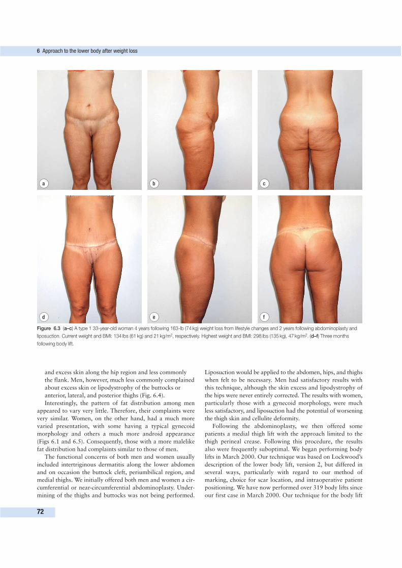
and excess skin along the hip region and less commonly
the flank. Men, however, much less commonly complained
about excess skin or lipodystrophy of the buttocks or
anterior, lateral, and posterior thighs (Fig. 6.4).
Interestingly, the pattern of fat distribution among men
appeared to vary very little. Therefore, their complaints were
very similar. Women, on the other hand, had a much more
varied presentation, with some having a typical gynecoid
morphology and others a much more android appearance
(Figs 6.1 and 6.5). Consequently, those with a more malelike
fat distribution had complaints similar to those of men.
The functional concerns of both men and women usually
included intertriginous dermatitis along the lower abdomen
and on occasion the buttock cleft, periumbilical region, and
medial thighs. We initially offered both men and women a cir-
cumferential or near-circumferential abdominoplasty. Under-
mining of the thighs and buttocks was not being performed.
Liposuction would be applied to the abdomen, hips, and thighs
when felt to be necessary. Men had satisfactory results with
this technique, although the skin excess and lipodystrophy of
the hips were never entirely corrected. The results with women,
particularly those with a gynecoid morphology, were much
less satisfactory, and liposuction had the potential of worsening
the thigh skin and cellulite deformity.
Following the abdominoplasty, we then offered some
patients a medial thigh lift with the approach limited to the
thigh perineal crease. Following this procedure, the results
also were frequently suboptimal. We began performing body
lifts in March 2000. Our technique was based on Lockwood’s
description of the lower body lift, version 2, but differed in
several ways, particularly with regard to our method of
marking, choice for scar location, and intraoperative patient
positioning. We have now performed over 319 body lifts since
our first case in March 2000. Our technique for the body lift
6 Approach to the lower body after weight loss
72
d e f
a b c
Figure 6.3 (a–c) A type 1 33-year-old woman 4 years following 163-lb (74 kg) weight loss from lifestyle changes and 2 years following abdominoplasty and
liposuction. Current weight and BMI: 134 lbs (61 kg) and 21 kg/m2, respectively. Highest weight and BMI: 298 lbs (135 kg), 47 kg/m2. (d–f) Three months
following body lift.
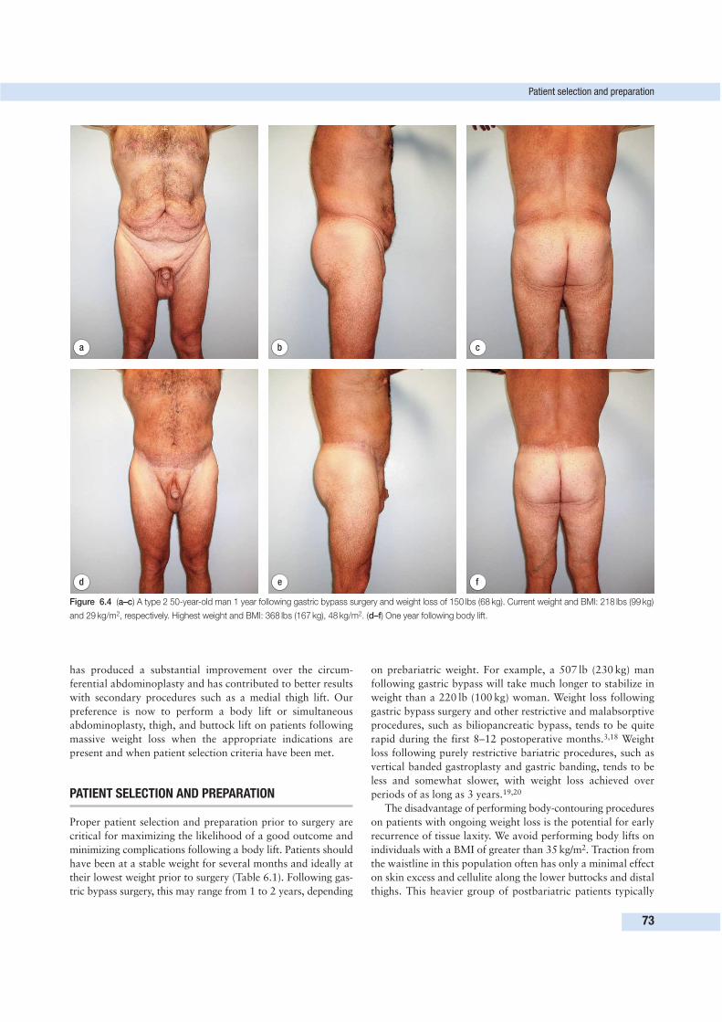
has produced a substantial improvement over the circum-
ferential abdominoplasty and has contributed to better results
with secondary procedures such as a medial thigh lift. Our
preference is now to perform a body lift or simultaneous
abdominoplasty, thigh, and buttock lift on patients following
massive weight loss when the appropriate indications are
present and when patient selection criteria have been met.
PATIENT SELECTION AND PREPARATION
Proper patient selection and preparation prior to surgery are
critical for maximizing the likelihood of a good outcome and
minimizing complications following a body lift. Patients should
have been at a stable weight for several months and ideally at
their lowest weight prior to surgery (Table 6.1). Following gas-
tric bypass surgery, this may range from 1 to 2 years, depending
on prebariatric weight. For example, a 507 lb (230 kg) man
following gastric bypass will take much longer to stabilize in
weight than a 220 lb (100 kg) woman. Weight loss following
gastric bypass surgery and other restrictive and malabsorptive
procedures, such as biliopancreatic bypass, tends to be quite
rapid during the first 8–12 postoperative months.3,18 Weight
loss following purely restrictive bariatric procedures, such as
vertical banded gastroplasty and gastric banding, tends to be
less and somewhat slower, with weight loss achieved over
periods of as long as 3 years.19,20
The disadvantage of performing body-contouring procedures
on patients with ongoing weight loss is the potential for early
recurrence of tissue laxity. We avoid performing body lifts on
individuals with a BMI of greater than 35 kg/m2. Traction from
the waistline in this population often has only a minimal effect
on skin excess and cellulite along the lower buttocks and distal
thighs. This heavier group of postbariatric patients typically
Patient selection and preparation
73
d e f
a b c
Figure 6.4 (a–c) A type 2 50-year-old man 1 year following gastric bypass surgery and weight loss of 150 lbs (68 kg). Current weight and BMI: 218 lbs (99 kg)
and 29 kg/m2, respectively. Highest weight and BMI: 368 lbs (167 kg), 48 kg/m2. (d–f) One year following body lift.
has a large pannus present along the lower abdomen, extending
to the hips and tapering over the buttocks. Difficulty with acti-
vities, severe intertriginous dermatitis, and back discomfort are
usually their biggest complaints. We offer these patients a near
circumferential abdominoplasty, a far less complex procedure.
We do on occasion offer body lifts to this heavier group,
particularly for patients less than 35 years of age, and usually
men, but also women with a more central fat distribution.
We avoid performing body lifts on postbariatric patients
greater than 55 years of age. Morbidly obese individuals who
have sought bariatric surgery in the fifth and sixth decades of
life have often developed degenerative arthritis, and in many
instances have undergone joint replacement. We find the
recovery from body lifts in patients with ongoing arthritis and
following joint replacement to be difficult and protracted. We
usually offer this group an abdominoplasty or an abdomino-
plasty to be followed in 6 months by a thigh and buttock lift.
Postbariatric patients, particularly menstruating women
and those who have had malabsorptive procedures, i.e. gastric
bypass and biliopancreatic bypass, are often anemic.21 These
anemias tend to be secondary to the poor absorption of both
iron and folate. Patients considering a body lift are encour-
aged to take both an iron supplement and daily multivitamins.
Severely anemic patients are referred to a hematologist. We
prefer a baseline hemoglobin of 12 g/dL. All postbariatric sur-
gery patients are encouraged to continue follow-up with their
bariatric surgeon.
SURGICAL TECHNIQUE
The challenge of performing a consistently effective circum-
ferential lower body-contouring procedure in the massive weight
loss population relates directly to the properties inherent in
this patient population and the objectives to be achieved.
Common to the lower body of virtually all postbariatric
patients is skin and soft tissue excess and a high degree of skin
and soft tissue mobility. Attempting to affect change to the
upper abdomen or distal thighs from the waistline, the usual
location for circumferential procedures, requires a significant
degree of traction. The combination of these patient pro-
perties with high levels of traction leads to the potential for
inconsistent results with regard to scar location, scar quality,
and overall outcome. Careful patient marking prior to a body
lift is essential for an optimal outcome.
Circumferential body-contouring procedures have the com-
mon goal of minimizing scar perceptibility by placing the scar
along the waistline. An analysis of where both men and women
wear their pants, undergarments, bathing suits, bikinis, thongs,
etc. reveals that the superior portion of most garments in the
hip region lies at the level of the anterior superior iliac spine
(ASIS) or approximately 6–7 cm below the superior edge of
the iliac crest. Posteriorly, garments traverse horizontally along
the lower back and above the buttocks, also at the level of the
ASIS. Anteriorly, virtually all undergarments cover the interface
between the hair-bearing pubic area and the hypogastrium
(Fig. 6.6). Ideally, the scar for the body lift should be at the
level of the ASIS along the hip and lower back, and gradually
descend to the interface between the hair-bearing pubic area
and hypogastrium anteriorly (Figs 6.6 and 6.7). An effective
technique for marking the body lift should produce a scar that
reliably lies along the waistline, despite the extreme tissue
mobility of the massive weight loss patient and the high level
of traction required to affect significant change from the
waistline. To do so requires a marking technique that uses
bony landmarks such as the ASIS to control scar placement.
Preoperative marking1. With the patient standing, an area above the buttock cleft
is marked first. This point (A), with downward traction to
the skin, should be horizontal to the ASIS (Fig. 6.7). The
ASIS is often difficult to palpate but is usually at a level
approximately three fingerbreadths below the iliac crest
(6–7 cm). With strong downward traction to the skin
along the right anterior iliac region, a point (B) along the
anterior axillary line should be marked that is horizontal
to point A under downward traction (Fig. 6.7). A dotted
line is drawn from A to B with downward traction over
the right thigh and buttock. The dotted line with
downward traction should be aligned with the patient’s
waistline.
2. Sitting in front of the patient, the surgeon identifies a
symmetric point (C) along the left anterior axillary line. A
dotted line is similarly drawn from C to A with downward
traction to the left thigh and buttock. With downward
traction to the right and left buttocks and thigh areas, a
straight dotted line should result from point B on the right
to point C on the left, passing through point A over the
buttock cleft (Fig. 6.7). If the line is found to be straight,
the dots are connected.
3. Point B′ is identified inferior to point B by the pinch
technique. The two points, when approximated, eliminate
cellulite along the anterior and lateral thigh. A similar
procedure is performed on the left side and at the buttock
cleft from point A. The redundant skin of the left and right
buttocks is estimated with the pinch technique. Points B
and B′ and C and C′ are called points of commitment,
because the surgeon does not remeasure the distance
between these points during surgery and commits to
removing this skin. The remaining lower set of lines and
point A′ are estimates only (Fig. 6.7).
6 Approach to the lower body after weight loss
74
Table 6.1 Patient selection criteria
Feature Criterion
Weight Stable
BMI (kg/m2) < 35
Age (years) < 55
Hemoglobin (g/dL) ≥ 12
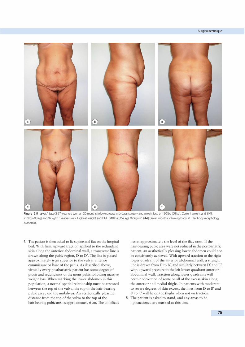
4. The patient is then asked to lie supine and flat on the hospital
bed. With firm, upward traction applied to the redundant
skin along the anterior abdominal wall, a transverse line is
drawn along the pubic region, D to D′. The line is placed
approximately 6 cm superior to the vulvar anterior
commissure or base of the penis. As described above,
virtually every postbariatric patient has some degree of
ptosis and redundancy of the mons pubis following massive
weight loss. When marking the lower abdomen in this
population, a normal spatial relationship must be restored
between the top of the vulva, the top of the hair-bearing
pubic area, and the umbilicus. An aesthetically pleasing
distance from the top of the vulva to the top of the
hair-bearing pubic area is approximately 6 cm. The umbilicus
lies at approximately the level of the iliac crest. If the
hair-bearing pubic area were not reduced in the postbariatric
patient, an aesthetically pleasing lower abdomen could not
be consistently achieved. With upward traction to the right
lower quadrant of the anterior abdominal wall, a straight
line is drawn from D to B′, and similarly between D′ and C′
with upward pressure to the left lower quadrant anterior
abdominal wall. Traction along lower quadrants will
permit correction of some or all of the excess skin along
the anterior and medial thighs. In patients with moderate
to severe degrees of skin excess, the lines from D to B′ and
D to C′ will lie on the thighs when not on traction.
5. The patient is asked to stand, and any areas to be
liposuctioned are marked at this time.
Surgical technique
75
d e f
a b c
Figure 6.5 (a–c) A type 3 27-year-old woman 20 months following gastric bypass surgery and weight loss of 130 lbs (59 kg). Current weight and BMI:
216 lbs (98 kg) and 32 kg/m2, respectively. Highest weight and BMI: 346 lbs (157 kg), 32 kg/m2. (d–f) Seven months following body lift. Her body morphology
is android.
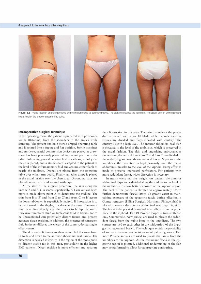
Intraoperative surgical techniqueIn the operating room, the patient is prepared with povidone–
iodine (Betadine) from the shoulders to the ankles while
standing. The patient sits on a sterile draped operating table
and is rotated into a supine and flat position. Sterile stockings
and sterile sequential compression devices are placed. A draw-
sheet has been previously placed along the midportion of the
table. Following general endotracheal anesthesia, a Foley ca-
theter is placed, and a sterile sheet is stapled to the patient at
the level of the inframammary fold and around either flank to
nearly the midback. Drapes are placed from the operating
table over either arm board. Finally, an ether drape is placed
in the usual fashion over the chest area. Grounding pads are
placed on each arm and secured with tape.
At the start of the surgical procedure, the skin along the
lines A–B and A–C is scored superficially. A 1-cm vertical hatch
mark is made above point A to demarcate the midline. The
skin from B to B′ and from C to C′ and from C′ to B′ across
the lower abdomen is superficially incised. If liposuction is to
be performed to the thighs, it is done at this time. Tumescent
fluid is infiltrated only into the tissues to be liposuctioned.
Excessive tumescent fluid or tumescent fluid in tissues not to
be liposuctioned can potentially distort tissues and prevent
accurate tissue excision. In addition, the presence of tumescent
fluid in tissues diffuses the energy of the cautery, decreasing its
effectiveness.
The skin and soft tissues are then incised full thickness from
C′ to B′ and down to the anterior abdominal wall fascia. The
dissection is beveled inferiorly in the region of the mons pubis
to directly excise fat in this area, particularly in the higher
BMI patients. Direct excision is more efficient and accurate
than liposuction in this area. The skin throughout the proce-
dure is incised with a no. 10 blade while the subcutaneous
tissues are divided and flaps elevated with cautery. The
cautery is set to a high level. The anterior abdominal wall flap
is elevated to the level of the umbilicus, which is preserved in
the usual fashion. The skin and underlying subcutaneous
tissue along the vertical lines C to C′ and B to B′ are divided to
the underlying anterior abdominal wall fascia. Superior to the
umbilicus, the dissection is kept primarily over the rectus
abdominus muscles to the level of the xiphoid. Every effort is
made to preserve intercostal perforators. For patients with
more redundant fascia, wider dissection is necessary.
In nearly every massive weight loss patient, the anterior
abdominal flap can be divided along the midline to the level of
the umbilicus to allow better exposure of the xiphoid region.
The back of the patient is elevated to approximately 35° to
further demonstrate fascial laxity. To greatly assist in main-
taining exposure of the epigastric fascia during plication, a
Gomez retractor (Pilling Surgical, Horsham, Philadelphia) is
placed to elevate the anterior abdominal wall flap (Fig. 6.9).
The fascia to be plicated is marked as an ellipse from the pubic
bone to the xiphoid. Two #1 Prolene looped sutures (Ethicon
Inc., Sommerville, New Jersey) are used to plicate the redun-
dant fascia from the pubic bone to the umbilicus. The two
sutures are tied to each other in the midportion of the hypo-
gastric region and buried. The technique avoids the possibility
of suture extrusion near incisions or of palpating knots. Two
more Prolene sutures are used to plicate the fascia from the
umbilicus to the xiphoid. As the redundant fascia in the epi-
gastric region is plicated, additional undermining of the flap
may be performed to allow for appropriate contouring.
6 Approach to the lower body after weight loss
76
Figure 6.6 Typical location of undergarments and their relationship to bony landmarks. The dark line outlines the iliac crest. The upper portion of the garment
lies at level of the anterior superior iliac spine.
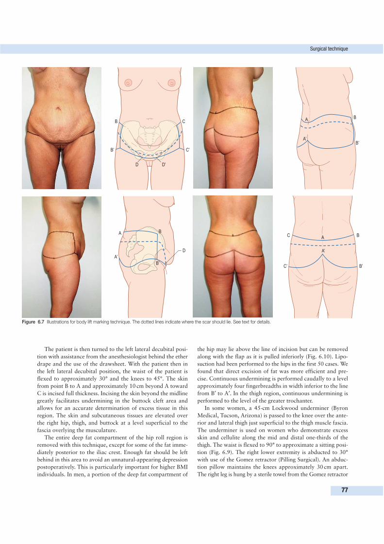
Surgical technique
77
B
B’
C
C’
D D’
A
A’
B
B’
D
A
A’
B
B’
C
C’
B
B’
A
A’
Figure 6.7 Illustrations for body lift marking technique. The dotted lines indicate where the scar should lie. See text for details.
The patient is then turned to the left lateral decubital posi-
tion with assistance from the anesthesiologist behind the ether
drape and the use of the drawsheet. With the patient then in
the left lateral decubital position, the waist of the patient is
flexed to approximately 30° and the knees to 45°. The skin
from point B to A and approximately 10 cm beyond A toward
C is incised full thickness. Incising the skin beyond the midline
greatly facilitates undermining in the buttock cleft area and
allows for an accurate determination of excess tissue in this
region. The skin and subcutaneous tissues are elevated over
the right hip, thigh, and buttock at a level superficial to the
fascia overlying the musculature.
The entire deep fat compartment of the hip roll region is
removed with this technique, except for some of the fat imme-
diately posterior to the iliac crest. Enough fat should be left
behind in this area to avoid an unnatural-appearing depression
postoperatively. This is particularly important for higher BMI
individuals. In men, a portion of the deep fat compartment of
the hip may lie above the line of incision but can be removed
along with the flap as it is pulled inferiorly (Fig. 6.10). Lipo-
suction had been performed to the hips in the first 50 cases. We
found that direct excision of fat was more efficient and pre-
cise. Continuous undermining is performed caudally to a level
approximately four fingerbreadths in width inferior to the line
from B′ to A′. In the thigh region, continuous undermining is
performed to the level of the greater trochanter.
In some women, a 45-cm Lockwood underminer (Byron
Medical, Tucson, Arizona) is passed to the knee over the ante-
rior and lateral thigh just superficial to the thigh muscle fascia.
The underminer is used on women who demonstrate excess
skin and cellulite along the mid and distal one-thirds of the
thigh. The waist is flexed to 90° to approximate a sitting posi-
tion (Fig. 6.9). The right lower extremity is abducted to 30°
with use of the Gomez retractor (Pilling Surgical). An abduc-
tion pillow maintains the knees approximately 30 cm apart.
The right leg is hung by a sterile towel from the Gomez retractor
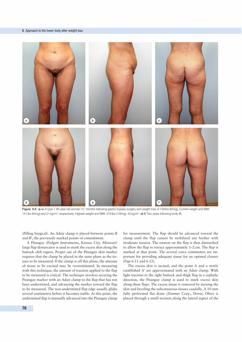
(Pilling Surgical). An Adair clamp is placed between points B
and B′, the previously marked points of commitment.
A Pitanguy (Padgett Instruments, Kansas City, Missouri)
large flap demarcator is used to mark the excess skin along the
buttock cleft region. Proper use of the Pitanguy skin marker
requires that the clamp be placed in the same plane as the tis-
sues to be measured. If the clamp is off this plane, the amount
of tissue to be excised may be overestimated. In measuring
with this technique, the amount of traction applied to the flap
to be measured is critical. The technique involves securing the
Pitanguy marker with an Adair clamp to the flap that has not
been undermined, and advancing the marker toward the flap
to be measured. The non-undermined flap edge usually glides
several centimeters before it becomes stable. At this point, the
undermined flap is manually advanced into the Pitanguy clamp
for measurement. The flap should be advanced toward the
clamp until the flap cannot be mobilized any further with
moderate tension. The tension on the flap is then diminished
to allow the flap to retract approximately 1–2 cm. The flap is
marked at that point. The several extra centimeters are im-
portant for providing adequate tissue for an optimal closure
(Figs 6.11 and 6.12).
The excess skin is incised, and the point A and a newly
established A′ are approximated with an Adair clamp. With
light traction to the right buttock and thigh flap in a cephalic
direction, the Pitanguy clamp is used to mark excess skin
along these flaps. The excess tissue is removed by incising the
skin and beveling the subcutaneous tissues caudally. A 10-mm
fully perforated flat drain (Zimmer Corp., Dover, Ohio) is
placed through a small incision along the lateral aspect of the
6 Approach to the lower body after weight loss
78
d e f
a b c
Figure 6.8 (a–c) A type 1 46-year-old woman 15 months following gastric bypass surgery and weight loss of 139 lbs (63 kg). Current weight and BMI:
141 lbs (64 kg) and 21 kg/m2, respectively. Highest weight and BMI: 278 lbs (126 kg), 42 kg/m2. (d–f) Two years following body lift.
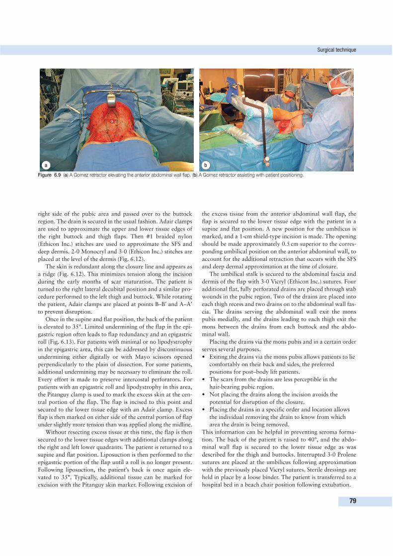
right side of the pubic area and passed over to the buttock
region. The drain is secured in the usual fashion. Adair clamps
are used to approximate the upper and lower tissue edges of
the right buttock and thigh flaps. Then #1 braided nylon
(Ethicon Inc.) stitches are used to approximate the SFS and
deep dermis. 2-0 Monocryl and 3-0 (Ethicon Inc.) stitches are
placed at the level of the dermis (Fig. 6.12).
The skin is redundant along the closure line and appears as
a ridge (Fig. 6.12). This minimizes tension along the incision
during the early months of scar maturation. The patient is
turned to the right lateral decubital position and a similar pro-
cedure performed to the left thigh and buttock. While rotating
the patient, Adair clamps are placed at points B–B′ and A–A′
to prevent disruption.
Once in the supine and flat position, the back of the patient
is elevated to 35°. Limited undermining of the flap in the epi-
gastric region often leads to flap redundancy and an epigastric
roll (Fig. 6.13). For patients with minimal or no lipodystrophy
in the epigastric area, this can be addressed by discontinuous
undermining either digitally or with Mayo scissors opened
perpendicularly to the plain of dissection. For some patients,
additional undermining may be necessary to eliminate the roll.
Every effort is made to preserve intercostal perforators. For
patients with an epigastric roll and lipodystrophy in this area,
the Pitanguy clamp is used to mark the excess skin at the cen-
tral portion of the flap. The flap is incised to this point and
secured to the lower tissue edge with an Adair clamp. Excess
flap is then marked on either side of the central portion of flap
under slightly more tension than was applied along the midline.
Without resecting excess tissue at this time, the flap is then
secured to the lower tissue edges with additional clamps along
the right and left lower quadrants. The patient is returned to a
supine and flat position. Liposuction is then performed to the
epigastric portion of the flap until a roll is no longer present.
Following liposuction, the patient’s back is once again ele-
vated to 35°. Typically, additional tissue can be marked for
excision with the Pitanguy skin marker. Following excision of
the excess tissue from the anterior abdominal wall flap, the
flap is secured to the lower tissue edge with the patient in a
supine and flat position. A new position for the umbilicus is
marked, and a 1-cm shield-type incision is made. The opening
should be made approximately 0.5 cm superior to the corres-
ponding umbilical position on the anterior abdominal wall, to
account for the additional retraction that occurs with the SFS
and deep dermal approximation at the time of closure.
The umbilical stalk is secured to the abdominal fascia and
dermis of the flap with 3-0 Vicryl (Ethicon Inc.) sutures. Four
additional flat, fully perforated drains are placed through stab
wounds in the pubic region. Two of the drains are placed into
each thigh recess and two drains on to the abdominal wall fas-
cia. The drains serving the abdominal wall exit the mons
pubis medially, and the drains leading to each thigh exit the
mons between the drains from each buttock and the abdo-
minal wall.
Placing the drains via the mons pubis and in a certain order
serves several purposes.
• Exiting the drains via the mons pubis allows patients to lie
comfortably on their back and sides, the preferred
positions for post–body lift patients.
• The scars from the drains are less perceptible in the
hair-bearing pubic region.
• Not placing the drains along the incision avoids the
potential for disruption of the closure.
• Placing the drains in a specific order and location allows
the individual removing the drain to know from which
area the drain is being removed.
This information can be helpful in preventing seroma forma-
tion. The back of the patient is raised to 40°, and the abdo-
minal wall flap is secured to the lower tissue edge as was
described for the thigh and buttocks. Interrupted 3-0 Prolene
sutures are placed at the umbilicus following approximation
with the previously placed Vicryl sutures. Sterile dressings are
held in place by a loose binder. The patient is transferred to a
hospital bed in a beach chair position following extubation.
Surgical technique
79
a b
Figure 6.9 (a) A Gomez retractor elevating the anterior abdominal wall flap. (b) A Gomez retractor assisting with patient positioning.
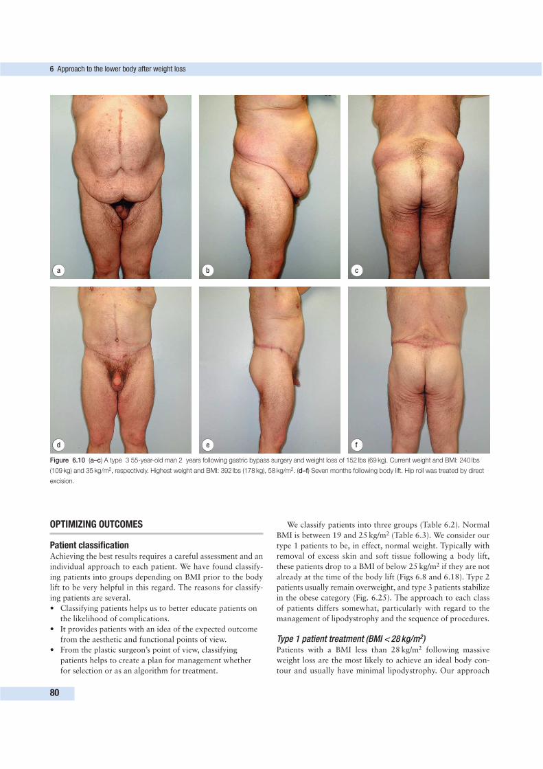
OPTIMIZING OUTCOMES
Patient classificationAchieving the best results requires a careful assessment and an
individual approach to each patient. We have found classify-
ing patients into groups depending on BMI prior to the body
lift to be very helpful in this regard. The reasons for classify-
ing patients are several.
• Classifying patients helps us to better educate patients on
the likelihood of complications.
• It provides patients with an idea of the expected outcome
from the aesthetic and functional points of view.
• From the plastic surgeon’s point of view, classifying
patients helps to create a plan for management whether
for selection or as an algorithm for treatment.
We classify patients into three groups (Table 6.2). Normal
BMI is between 19 and 25 kg/m2 (Table 6.3). We consider our
type 1 patients to be, in effect, normal weight. Typically with
removal of excess skin and soft tissue following a body lift,
these patients drop to a BMI of below 25 kg/m2 if they are not
already at the time of the body lift (Figs 6.8 and 6.18). Type 2
patients usually remain overweight, and type 3 patients stabilize
in the obese category (Fig. 6.25). The approach to each class
of patients differs somewhat, particularly with regard to the
management of lipodystrophy and the sequence of procedures.
Type 1 patient treatment (BMI < 28 kg/m2)Patients with a BMI less than 28 kg/m2 following massive
weight loss are the most likely to achieve an ideal body con-
tour and usually have minimal lipodystrophy. Our approach
6 Approach to the lower body after weight loss
80
d e f
a b c
Figure 6.10 (a–c) A type 3 55-year-old man 2 years following gastric bypass surgery and weight loss of 152 lbs (69 kg). Current weight and BMI: 240 lbs
(109 kg) and 35 kg/m2, respectively. Highest weight and BMI: 392 lbs (178 kg), 58 kg/m2. (d–f) Seven months following body lift. Hip roll was treated by direct
excision.
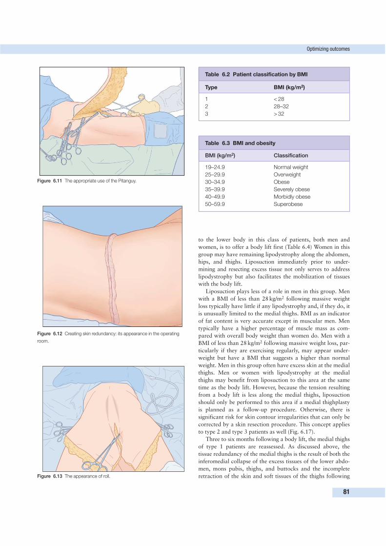
to the lower body in this class of patients, both men and
women, is to offer a body lift first (Table 6.4) Women in this
group may have remaining lipodystrophy along the abdomen,
hips, and thighs. Liposuction immediately prior to under-
mining and resecting excess tissue not only serves to address
lipodystrophy but also facilitates the mobilization of tissues
with the body lift.
Liposuction plays less of a role in men in this group. Men
with a BMI of less than 28 kg/m2 following massive weight
loss typically have little if any lipodystrophy and, if they do, it
is unusually limited to the medial thighs. BMI as an indicator
of fat content is very accurate except in muscular men. Men
typically have a higher percentage of muscle mass as com-
pared with overall body weight than women do. Men with a
BMI of less than 28 kg/m2 following massive weight loss, par-
ticularly if they are exercising regularly, may appear under-
weight but have a BMI that suggests a higher than normal
weight. Men in this group often have excess skin at the medial
thighs. Men or women with lipodystrophy at the medial
thighs may benefit from liposuction to this area at the same
time as the body lift. However, because the tension resulting
from a body lift is less along the medial thighs, liposuction
should only be performed to this area if a medial thighplasty
is planned as a follow-up procedure. Otherwise, there is
significant risk for skin contour irregularities that can only be
corrected by a skin resection procedure. This concept applies
to type 2 and type 3 patients as well (Fig. 6.17).
Three to six months following a body lift, the medial thighs
of type 1 patients are reassessed. As discussed above, the
tissue redundancy of the medial thighs is the result of both the
inferomedial collapse of the excess tissues of the lower abdo-
men, mons pubis, thighs, and buttocks and the incomplete
retraction of the skin and soft tissues of the thighs following
Optimizing outcomes
81
Table 6.2 Patient classification by BMI
Type BMI (kg/m2)
1 < 28
2 28–32
3 > 32
Table 6.3 BMI and obesity
BMI (kg/m2) Classification
19–24.9 Normal weight
25–29.9 Overweight
30–34.9 Obese
35–39.9 Severely obese
40–49.9 Morbidly obese
50–59.9 Superobese
Figure 6.11 The appropriate use of the Pitanguy.
Figure 6.12 Creating skin redundancy: its appearance in the operating
room.
Figure 6.13 The appearance of roll.
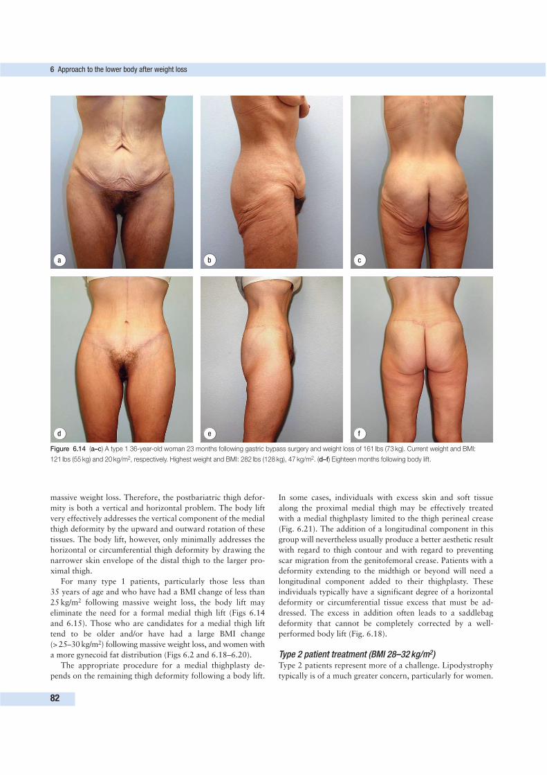
6 Approach to the lower body after weight loss
82
d e f
a b c
Figure 6.14 (a–c) A type 1 36-year-old woman 23 months following gastric bypass surgery and weight loss of 161 lbs (73 kg). Current weight and BMI:
121 lbs (55 kg) and 20 kg/m2, respectively. Highest weight and BMI: 282 lbs (128 kg), 47 kg/m2. (d–f) Eighteen months following body lift.
massive weight loss. Therefore, the postbariatric thigh defor-
mity is both a vertical and horizontal problem. The body lift
very effectively addresses the vertical component of the medial
thigh deformity by the upward and outward rotation of these
tissues. The body lift, however, only minimally addresses the
horizontal or circumferential thigh deformity by drawing the
narrower skin envelope of the distal thigh to the larger pro-
ximal thigh.
For many type 1 patients, particularly those less than
35 years of age and who have had a BMI change of less than
25 kg/m2 following massive weight loss, the body lift may
eliminate the need for a formal medial thigh lift (Figs 6.14
and 6.15). Those who are candidates for a medial thigh lift
tend to be older and/or have had a large BMI change
(> 25–30 kg/m2) following massive weight loss, and women with
a more gynecoid fat distribution (Figs 6.2 and 6.18–6.20).
The appropriate procedure for a medial thighplasty de-
pends on the remaining thigh deformity following a body lift.
In some cases, individuals with excess skin and soft tissue
along the proximal medial thigh may be effectively treated
with a medial thighplasty limited to the thigh perineal crease
(Fig. 6.21). The addition of a longitudinal component in this
group will nevertheless usually produce a better aesthetic result
with regard to thigh contour and with regard to preventing
scar migration from the genitofemoral crease. Patients with a
deformity extending to the midthigh or beyond will need a
longitudinal component added to their thighplasty. These
individuals typically have a significant degree of a horizontal
deformity or circumferential tissue excess that must be ad-
dressed. The excess in addition often leads to a saddlebag
deformity that cannot be completely corrected by a well-
performed body lift (Fig. 6.18).
Type 2 patient treatment (BMI 28–32 kg/m2)Type 2 patients represent more of a challenge. Lipodystrophy
typically is of a much greater concern, particularly for women.
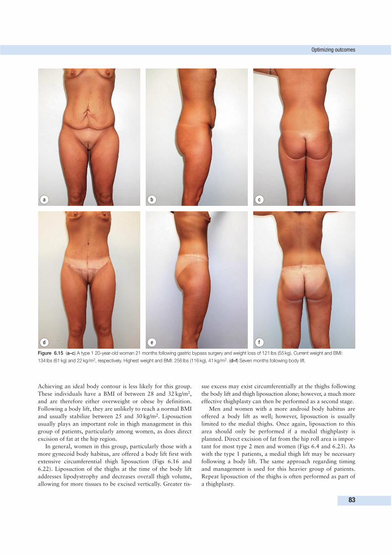
Optimizing outcomes
83
Achieving an ideal body contour is less likely for this group.
These individuals have a BMI of between 28 and 32 kg/m2,
and are therefore either overweight or obese by definition.
Following a body lift, they are unlikely to reach a normal BMI
and usually stabilize between 25 and 30 kg/m2. Liposuction
usually plays an important role in thigh management in this
group of patients, particularly among women, as does direct
excision of fat at the hip region.
In general, women in this group, particularly those with a
more gynecoid body habitus, are offered a body lift first with
extensive circumferential thigh liposuction (Figs 6.16 and
6.22). Liposuction of the thighs at the time of the body lift
addresses lipodystrophy and decreases overall thigh volume,
allowing for more tissues to be excised vertically. Greater tis-
sue excess may exist circumferentially at the thighs following
the body lift and thigh liposuction alone; however, a much more
effective thighplasty can then be performed as a second stage.
Men and women with a more android body habitus are
offered a body lift as well; however, liposuction is usually
limited to the medial thighs. Once again, liposuction to this
area should only be performed if a medial thighplasty is
planned. Direct excision of fat from the hip roll area is impor-
tant for most type 2 men and women (Figs 6.4 and 6.23). As
with the type 1 patients, a medial thigh lift may be necessary
following a body lift. The same approach regarding timing
and management is used for this heavier group of patients.
Repeat liposuction of the thighs is often performed as part of
a thighplasty.
d e f
a b c
Figure 6.15 (a–c) A type 1 20-year-old woman 21 months following gastric bypass surgery and weight loss of 121 lbs (55 kg). Current weight and BMI:
134 lbs (61 kg) and 22 kg/m2, respectively. Highest weight and BMI: 256 lbs (116 kg), 41 kg/m2. (d–f) Seven months following body lift.
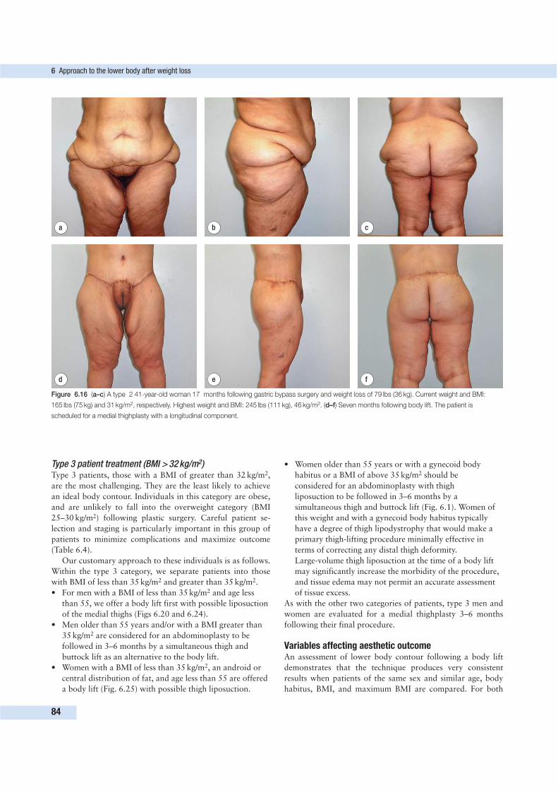
Type 3 patient treatment (BMI > 32 kg/m2)Type 3 patients, those with a BMI of greater than 32 kg/m2,
are the most challenging. They are the least likely to achieve
an ideal body contour. Individuals in this category are obese,
and are unlikely to fall into the overweight category (BMI
25–30 kg/m2) following plastic surgery. Careful patient se-
lection and staging is particularly important in this group of
patients to minimize complications and maximize outcome
(Table 6.4).
Our customary approach to these individuals is as follows.
Within the type 3 category, we separate patients into those
with BMI of less than 35 kg/m2 and greater than 35 kg/m2.
• For men with a BMI of less than 35 kg/m2 and age less
than 55, we offer a body lift first with possible liposuction
of the medial thighs (Figs 6.20 and 6.24).
• Men older than 55 years and/or with a BMI greater than
35 kg/m2 are considered for an abdominoplasty to be
followed in 3–6 months by a simultaneous thigh and
buttock lift as an alternative to the body lift.
• Women with a BMI of less than 35 kg/m2, an android or
central distribution of fat, and age less than 55 are offered
a body lift (Fig. 6.25) with possible thigh liposuction.
• Women older than 55 years or with a gynecoid body
habitus or a BMI of above 35 kg/m2 should be
considered for an abdominoplasty with thigh
liposuction to be followed in 3–6 months by a
simultaneous thigh and buttock lift (Fig. 6.1). Women of
this weight and with a gynecoid body habitus typically
have a degree of thigh lipodystrophy that would make a
primary thigh-lifting procedure minimally effective in
terms of correcting any distal thigh deformity.
Large-volume thigh liposuction at the time of a body lift
may significantly increase the morbidity of the procedure,
and tissue edema may not permit an accurate assessment
of tissue excess.
As with the other two categories of patients, type 3 men and
women are evaluated for a medial thighplasty 3–6 months
following their final procedure.
Variables affecting aesthetic outcomeAn assessment of lower body contour following a body lift
demonstrates that the technique produces very consistent
results when patients of the same sex and similar age, body
habitus, BMI, and maximum BMI are compared. For both
6 Approach to the lower body after weight loss
84
d e f
a b c
Figure 6.16 (a–c) A type 2 41-year-old woman 17 months following gastric bypass surgery and weight loss of 79 lbs (36 kg). Current weight and BMI:
165 lbs (75 kg) and 31 kg/m2, respectively. Highest weight and BMI: 245 lbs (111 kg), 46 kg/m2. (d–f) Seven months following body lift. The patient is
scheduled for a medial thighplasty with a longitudinal component.
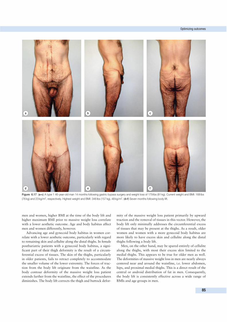
men and women, higher BMI at the time of the body lift and
higher maximum BMI prior to massive weight loss correlate
with a lower aesthetic outcome. Age and body habitus affect
men and women differently, however.
Advancing age and gynecoid body habitus in women cor-
relate with a lower aesthetic outcome, particularly with regard
to remaining skin and cellulite along the distal thighs. In female
postbariatric patients with a gynecoid body habitus, a signi-
ficant part of their thigh deformity is the result of a circum-
ferential excess of tissues. The skin of the thighs, particularly
in older patients, fails to retract completely to accommodate
the smaller volume of the lower extremity. The forces of trac-
tion from the body lift originate from the waistline. As the
body contour deformity of the massive weight loss patient
extends farther from the waistline, the effect of the procedures
diminishes. The body lift corrects the thigh and buttock defor-
mity of the massive weight loss patient primarily by upward
traction and the removal of tissues in this vector. However, the
body lift only minimally addresses the circumferential excess
of tissues that may be present at the thighs. As a result, older
women and women with a more gynecoid body habitus are
more likely to have excess skin and cellulite along the distal
thighs following a body lift.
Men, on the other hand, may be spared entirely of cellulite
along the thighs, with most their excess skin limited to the
medial thighs. This appears to be true for older men as well.
The deformities of massive weight loss in men are nearly always
centered near and around the waistline, i.e. lower abdomen,
hips, and proximal medial thighs. This is a direct result of the
central or android distribution of fat in men. Consequently,
the body lift is consistently effective across a wide range of
BMIs and age groups in men.
Optimizing outcomes
85
d e f
a b c
Figure 6.17 (a–c) A type 1 46-year-old man 14 months following gastric bypass surgery and weight loss of 179 lbs (81 kg). Current weight and BMI: 168 lbs
(76 kg) and 23 kg/m2, respectively. Highest weight and BMI: 346 lbs (157 kg), 48 kg/m2. (d–f) Seven months following body lift.
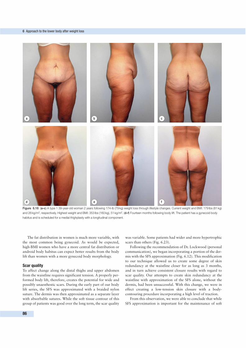
The fat distribution in women is much more variable, with
the most common being gynecoid. As would be expected,
high-BMI women who have a more central fat distribution or
android body habitus can expect better results from the body
lift than women with a more gynecoid body morphology.
Scar qualityTo affect change along the distal thighs and upper abdomen
from the waistline requires significant tension. A properly per-
formed body lift, therefore, creates the potential for wide and
possibly unaesthestic scars. During the early part of our body
lift series, the SFS was approximated with a braided nylon
suture. The dermis was then approximated as a separate layer
with absorbable sutures. While the soft tissue contour of this
group of patients was good over the long term, the scar quality
was variable. Some patients had wider and more hypertrophic
scars than others (Fig. 6.23).
Following the recommendation of Dr. Lockwood (personal
communication), we began incorporating a portion of the der-
mis with the SFS approximation (Fig. 6.12). This modification
to our technique allowed us to create some degree of skin
redundancy at the waistline closer for as long as 3 months,
and in turn achieve consistent closure results with regard to
scar quality. Our attempts to create skin redundancy at the
waistline with approximation of the SFS alone, without the
dermis, had been unsuccessful. With this change, we were in
effect creating a low-tension skin closure with a body-
contouring procedure incorporating a high level of traction.
From this observation, we were able to conclude that while
SFS approximation is important for the maintenance of soft
6 Approach to the lower body after weight loss
86
d e f
a b c
Figure 6.18 (a–c) A type 1 39-year-old woman 2 years following 174-lb (79 kg) weight loss through lifestyle changes. Current weight and BMI: 179 lbs (81 kg)
and 26 kg/m2, respectively. Highest weight and BMI: 353 lbs (160 kg), 51 kg/m2. (d–f) Fourteen months following body lift. The patient has a gynecoid body
habitus and is scheduled for a medial thighplasty with a longitudinal component.
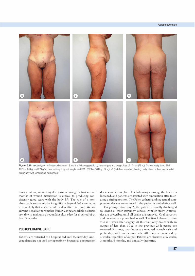
tissue contour, minimizing skin tension during the first several
months of wound maturation is critical to producing con-
sistently good scars with the body lift. The role of a non-
absorbable suture may be insignificant beyond 3–6 months, as
it is unlikely that a scar would widen after that time. We are
currently evaluating whether longer lasting absorbable sutures
are able to maintain a redundant skin edge for a period of at
least 3 months.
POSTOPERATIVE CARE
Patients are restricted to a hospital bed until the next day. Anti-
coagulants are not used perioperatively. Sequential compression
devices are left in place. The following morning, the binder is
loosened, and patients are assisted with ambulation after toler-
ating a sitting position. The Foley catheter and sequential com-
pression devices are removed if the patient is ambulating well.
On postoperative day 2, the patient is usually discharged
following a lower extremity venous Doppler study. Antibio-
tics are prescribed until all drains are removed. Oral narcotics
and laxatives are prescribed as well. The first follow-up office
visit is 1 week after surgery. At this visit, only drains with an
output of less than 30 cc in the previous 24-h period are
removed. At most, two drains are removed at each visit and
preferably not from the same side. All drains are removed by
5 weeks, regardless of output. Patients are observed at 6 weeks,
3 months, 6 months, and annually thereafter.
Postoperative care
87
d e f
a b c
Figure 6.19 (a–c) A type 1 40-year-old woman 13 months following gastric bypass surgery and weight loss of 174 lbs (79 kg). Current weight and BMI:
187 lbs (85 kg) and 27 kg/m2, respectively. Highest weight and BMI: 362 lbs (164 kg), 52 kg/m2. (d–f) Four months following body lift and subsequent medial
thighplasty with longitudinal component.
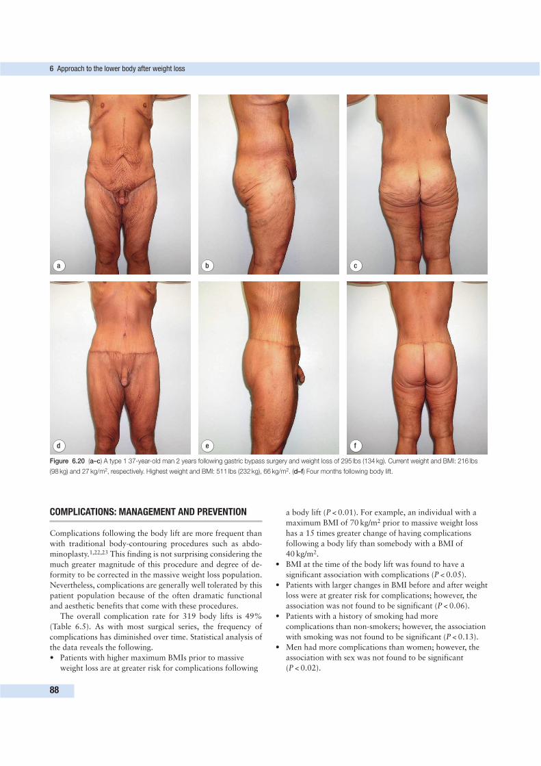
COMPLICATIONS: MANAGEMENT AND PREVENTION
Complications following the body lift are more frequent than
with traditional body-contouring procedures such as abdo-
minoplasty.1,22,23 This finding is not surprising considering the
much greater magnitude of this procedure and degree of de-
formity to be corrected in the massive weight loss population.
Nevertheless, complications are generally well tolerated by this
patient population because of the often dramatic functional
and aesthetic benefits that come with these procedures.
The overall complication rate for 319 body lifts is 49%
(Table 6.5). As with most surgical series, the frequency of
complications has diminished over time. Statistical analysis of
the data reveals the following.
• Patients with higher maximum BMIs prior to massive
weight loss are at greater risk for complications following
a body lift (P < 0.01). For example, an individual with a
maximum BMI of 70 kg/m2 prior to massive weight loss
has a 15 times greater change of having complications
following a body lify than somebody with a BMI of
40 kg/m2.
• BMI at the time of the body lift was found to have a
significant association with complications (P < 0.05).
• Patients with larger changes in BMI before and after weight
loss were at greater risk for complications; however, the
association was not found to be significant (P < 0.06).
• Patients with a history of smoking had more
complications than non-smokers; however, the association
with smoking was not found to be significant (P < 0.13).
• Men had more complications than women; however, the
association with sex was not found to be significant
(P < 0.02).
6 Approach to the lower body after weight loss
88
d e f
a b c
Figure 6.20 (a–c) A type 1 37-year-old man 2 years following gastric bypass surgery and weight loss of 295 lbs (134 kg). Current weight and BMI: 216 lbs
(98 kg) and 27 kg/m2, respectively. Highest weight and BMI: 511 lbs (232 kg), 66 kg/m2. (d–f) Four months following body lift.

• Age at the tome of the body lift was also not found to be
significantly correlated with complications.
Skin dehiscenceSkin dehiscence is our most frequent complication following a
body lift (Table 6.5). This can be attributed to the facts that
the procedure is circumferential, and that a high degree of
traction is needed to produce an ideal outcome. Nevertheless,
the frequency and severity of this complication has continued
to diminish. Skin dehiscence in the vast majority of instances
in our series has occurred at the buttock cleft and hips, the
two areas of greatest tension following this procedure.
In the early part of the series, the skin to be removed at the
buttock cleft was measured with the patient standing prior to
surgery. During surgery, the waist was not completely flexed
into a sitting position, and the previously marked skin to be
removed appeared to be appropriate. When assuming a sitting
position, patients place tremendous tension on this minimally
mobile part of the lower back. In addition, the relatively greater
period of time in bed in the early postoperative period may
lead to some degree of ischemia over the sacrum and coccyx,
likely contributing to poor healing in this area. Measuring the
tissue to be removed intraoperatively, with the patient flexed
into a sitting position, has greatly decreased the frequency and
severity of this problem.
The hip had been another problem area for skin dehiscence
in the early part of our series. Approximating the SFS along
with a small dermal component with a permanent stitch, as
suggested by Lockwood, allowed us to create some degree of
tissue redundancy along the closure for several months. We
Complications: management and prevention
89
d e f
a b c
Figure 6.21 (a–c) A type 1 53-year-old woman 14 months following gastric bypass surgery and weight loss of 117 lbs (53 kg). Current weight and BMI:
137 lbs (62 kg) and 21 kg/m2, respectively. Highest weight and BMI: 254 lbs (115 kg), 39 kg/m2. (d–f) Twenty-four months following body lift and subsequent
medial thighplasty with approach limited to the thigh perineal crease.
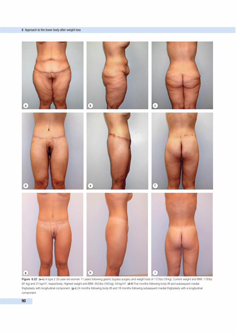
6 Approach to the lower body after weight loss
90
d e f
a b c
g h i
Figure 6.22 (a–c) A type 2 33-year-old woman 11 years following gastric bypass surgery and weight loss of 172 lbs (78 kg). Current weight and BMI: 179 lbs
(81 kg) and 31 kg/m2, respectively. Highest weight and BMI: 353 lbs (160 kg), 54 kg/m2. (d–f) Five months following body lift and subsequent medial
thighplasty with longitudinal component. (g–i) 24 months following body lift and 18 months following subsequent medial thighplasty with a longitudinal
component.
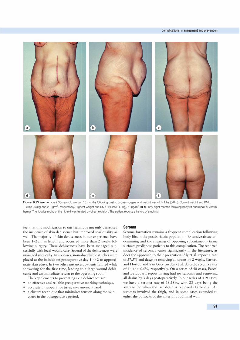
feel that this modification to our technique not only decreased
the incidence of skin dehiscence but improved scar quality as
well. The majority of skin dehiscences in our experience have
been 1–2 cm in length and occurred more than 2 weeks fol-
lowing surgery. These dehiscences have been managed suc-
cessfully with local wound care. Several of the dehiscences were
managed surgically. In six cases, non-absorbable stitches were
placed at the bedside on postoperative day 1 or 2 to approxi-
mate skin edges. In two other instances, patients fainted while
showering for the first time, leading to a large wound dehis-
cence and an immediate return to the operating room.
The key elements to preventing skin dehiscence are:
• an effective and reliable preoperative marking technique,
• accurate intraoperative tissue measurement, and
• a closure technique that minimizes tension along the skin
edges in the postoperative period.
SeromaSeroma formation remains a frequent complication following
body lifts in the postbariatric population. Extensive tissue un-
dermining and the shearing of opposing subcutaneous tissue
surfaces predispose patients to this complication. The reported
incidence of seromas varies significantly in the literature, as
does the approach to their prevention. Aly et al. report a rate
of 37.5% and describe removing all drains by 2 weeks. Carwell
and Horton and Van Geertruyden et al. describe seroma rates
of 14 and 6.6%, respectively. On a series of 40 cases, Pascal
and Le Louarn report having had no seromas and removing
all drains by 3 days postoperatively. In our series of 319 cases,
we have a seroma rate of 18.18%, with 23 days being the
average for when the last drain is removed (Table 6.5). All
seromas involved the thigh, and in some cases extended to
either the buttocks or the anterior abdominal wall.
Complications: management and prevention
91
d e f
a b c
Figure 6.23 (a–c) A type 2 35-year-old woman 13 months following gastric bypass surgery and weight loss of 141 lbs (64 kg). Current weight and BMI:
183 lbs (83 kg) and 29 kg/m2, respectively. Highest weight and BMI: 324 lbs (147 kg), 51 kg/m2. (d–f) Forty-eight months following body lift and repair of ventral
hernia. The lipodystrophy of the hip roll was treated by direct excision. The patient reports a history of smoking.
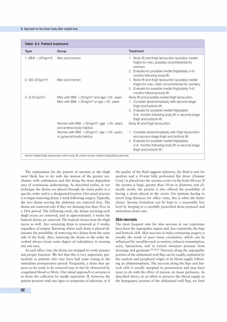
The explanation for the pattern of seromas at the thigh
most likely has to do with the motion of the greater tro-
chanter with ambulation and this being the most dependent
area of continuous undermining. As described earlier, in our
technique the drains are placed through the mons pubis in a
specific order and to a designated location. Our usual practice
is to begin removing drains 1 week following surgery. Typically,
the two drains serving the abdomen are removed first. The
drains are removed only if they are draining less than 30 cc in
a 24-h period. The following week, the drains servicing each
thigh recess are removed, and at approximately 3 weeks the
buttock drains are removed. The buttock drains treat the thigh
recess as well. Any remaining drain is removed at 5 weeks,
regardless of output. Knowing where each drain is placed eli-
minates the possibility of removing two drains from the same
side of the body. Also, removing the drains in the order de-
scribed always forms some degree of redundancy in treating
any one area.
At each office visit, the drains are stripped to verify patency
and proper function. We feel that this is very important, par-
ticularly in patients who may have had some oozing in the
immediate postoperative period. Frequently, a drain that ap-
pears to be ready to be removed may in fact be obstructed by
coagulated blood or fibrin. Our initial approach to seromas is
to drain the collection by needle aspiration. If, however, the
patient presents with any signs or symptoms of infection, or if
the quality of the fluid suggests infection, the fluid is sent for
analysis and a 10-mm fully perforated flat drain (Zimmer
Corp.) is placed into the seroma cavity via the body lift scar. If
the seroma is large, greater than 10 cm in diameter, and cli-
nically sterile, the patient is also offered the possibility of
having a drain placed in the cavity. For patients having to
travel long distances for office visits, this is often the better
choice. Seroma formation can be kept to a reasonably low
level by keeping to a carefully prescribed drain protocol and
meticulous drain care.
Skin necrosisThe most frequent sites for skin necrosis in our experience
have been the suprapubic region and, less commonly, the hips
and buttock cleft. Skin necrosis in body-contouring surgery is
usually the result of poor tissue circulation, which can be
influenced by variables such as tension, tobacco consumption,
scars, liposuction, and in certain instances pressure from
dressings and garments.22,24,25 Necrosis along the suprapubic
portion of the abdominal wall flap can be readily explained by
the random and peripheral origin of its blood supply follow-
ing an abdominoplasty. The necrosis along the hips and but-
tock cleft is usually marginal in presentation and may have
more to do with the effect of tension on tissue perfusion. As
described above, in an effort to preserve the blood supply to
the hypogastric portion of the abdominal wall flap, we limit
6 Approach to the lower body after weight loss
92
Table 6.4 Patient treatment
Type Group Treatment
1 (BMI < 28 kg/m2) Men and women 1. Body lift and thigh liposuction (possibly medial
thighs for men, possibly circumferential for
women).
2. Evaluate for possible medial thighplasty 3–6
months following body lift.
2 (28–32 kg/m2) Men and women 1. Body lift and thigh liposuction (possibly medial
thighs for men, often circumferential for women).
2. Evaluate for possible medial thighplasty 3–6
months following body lift.
3 (≥ 32 kg/m2) Men with BMI < 35 kg/m2 and age < 55 years Body lift and possible medial thigh liposuction.
Men with BMI > 35 kg/m2 or age > 55 years 1. Consider abdominoplasty with second-stage
thigh and buttock lift.
2. Evaluate for possible medial thighplasty
3–6 months following body lift or second-stage
thigh and buttock lift.
Women with BMI < 35 kg/m2, age < 55 years, Body lift and thigh liposuction.
and android body habitus
Women with BMI >35kg/m2, age > 55 years, 1. Consider abdominoplasty with thigh liposuction
or gynecoid body habitus 1. and second-stage thigh and buttock lift.
2. Evaluate for possible medial thighplasty
3–6 months following body lift or second-stage
thigh and buttock lift.
Avoid medial thigh liposuction with body lift unless future medial thighplasty planned
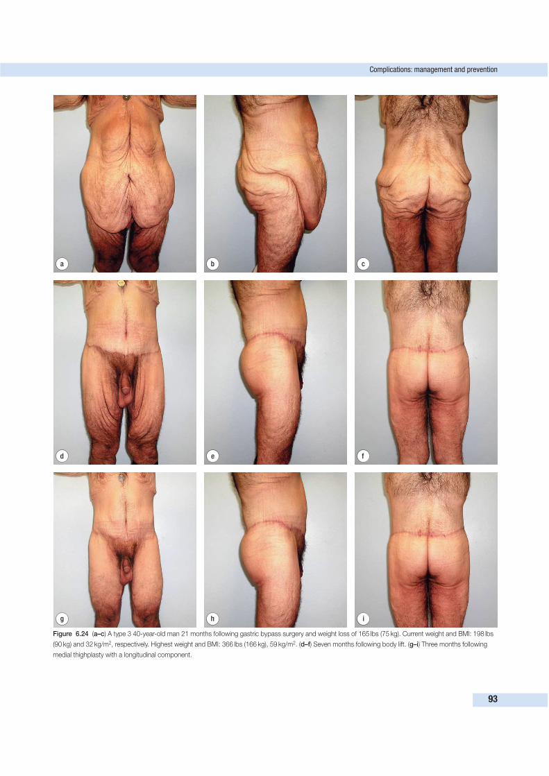
Complications: management and prevention
93
d e f
a b c
g h i
Figure 6.24 (a–c) A type 3 40-year-old man 21 months following gastric bypass surgery and weight loss of 165 lbs (75 kg). Current weight and BMI: 198 lbs
(90 kg) and 32 kg/m2, respectively. Highest weight and BMI: 366 lbs (166 kg), 59 kg/m2. (d–f) Seven months following body lift. (g–i) Three months following
medial thighplasty with a longitudinal component.
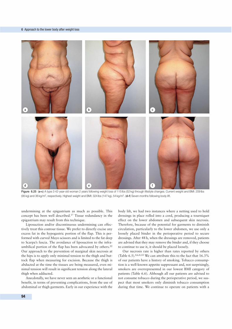
undermining at the epigastrium as much as possible. This
concept has been well described.17 Tissue redundancy in the
epigastrium may result from this technique.
Liposuction and/or discontinuous undermining can effec-
tively treat this contour tissue. We prefer to directly excise any
excess fat in the hypogastric portion of the flap. This is per-
formed with curved Mayo scissors and is limited to the fat deep
to Scarpa’s fascia. The avoidance of liposuction to the infra-
umbilical portion of the flap has been advocated by others.22
Our approach to the prevention of marginal skin necrosis at
the hips is to apply only minimal tension to the thigh and but-
tock flap when measuring for excision. Because the thigh is
abducted at the time the tissues are being measured, even mi-
nimal tension will result in significant tension along the lateral
thigh when adducted.
Anecdotally, we have never seen an aesthetic or a functional
benefit, in terms of preventing complications, from the use of
abdominal or thigh garments. Early in our experience with the
body lift, we had two instances where a netting used to hold
dressings in place rolled into a cord, producing a tourniquet
effect on the lower abdomen and subsequent skin necrosis.
Therefore, because of the potential for garments to diminish
circulation, particularly to the lower abdomen, we use only a
loosely placed binder in the perioperative period to secure
dressings. After 48 h, when the dressings are removed, patients
are advised that they may remove the binder and, if they choose
to continue to use it, it should be placed loosely.
Our necrosis rate is higher than rates reported by others
(Table 6.5).4,6,8,10 We can attribute this to the fact that 16.3%
of our patients have a history of smoking. Tobacco consump-
tion is a well-known appetite suppressant and, not surprisingly,
smokers are overrepresented in our lowest BMI category of
patients (Table 6.6). Although all our patients are advised to
not consume tobacco during the perioperative period, we sus-
pect that most smokers only diminish tobacco consumption
during that time. We continue to operate on patients with a
6 Approach to the lower body after weight loss
94
d e f
a b c
Figure 6.25 (a–c) A type 3 42-year-old woman 2 years following weight loss of 115 lbs (52 kg) through lifestyle changes. Current weight and BMI: 209 lbs
(95 kg) and 36 kg/m2, respectively. Highest weight and BMI: 324 lbs (147 kg), 54 kg/m2. (d–f) Seven months following body lift.
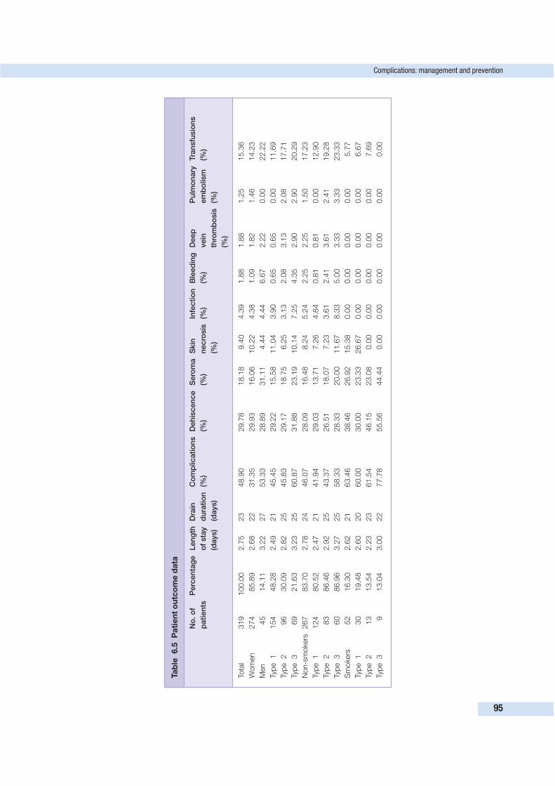
Complications: management and prevention
95
Ta
ble
6.5
Pa
tie
nt
ou
tco
me
da
ta
No
. o
f P
erc
en
tag
eL
en
gth
D
rain
C
om
plicati
on
s
Deh
iscen
ce
Sero
ma
Skin
In
fecti
on
B
leed
ing
D
eep
P
ulm
on
ary
Tra
nsfu
sio
ns
pati
en
tso
f sta
yd
ura
tio
n
(%)
(%)
(%)
necro
sis
(%
)(%
)vein
em
bo
lism
(%
)
(days)
(days)
(%)
thro
mb
osis
(%
)
(%)
Tota
l319
100.0
02.7
523
48.9
029.7
818.1
89.4
04.3
91.8
81.8
81.2
515.3
6
Wom
en
274
85.8
92.6
822
31.3
529.9
316.0
610.2
24.3
81.0
91.8
21.4
614.2
3
Men
45
14.1
13.2
227
53.3
328.8
931.1
14.4
44.4
46.6
72.2
20.0
022.2
2
Typ
e1
154
48.2
82.4
921
45.4
529.2
215.5
811.0
43.9
00.6
50.6
50.0
011.6
9
Typ
e2
96
30.0
92.8
225
45.8
329.1
718.7
56.2
53.1
32.0
83.1
32.0
817.7
1
Typ
e3
69
21.6
33.2
325
60.8
731.8
823.1
910.1
47.2
54.3
52.9
02.9
020.2
9
Non-s
mokers
267
83.7
02.7
824
46.0
728.0
916.4
88.2
45.2
42.2
52.2
51.5
017.2
3
Typ
e1
124
80.5
22.4
721
41.9
429.0
313.7
17.2
64.8
40.8
10.8
10.0
012.9
0
Typ
e2
83
86.4
62.9
225
43.3
726.5
118.0
77.2
33.6
12.4
13.6
12.4
119.2
8
Typ
e3
60
86.9
63.2
725
58.3
328.3
320.0
011.6
78.3
35.0
03.3
33.3
323.3
3
Sm
okers
52
16.3
02.6
221
63.4
638.4
626.9
215.3
80.0
00.0
00.0
00.0
05.7
7
Typ
e1
30
19.4
82.6
020
60.0
030.0
023.3
326.6
70.0
00.0
00.0
00.0
06.6
7
Typ
e2
13
13.5
42.2
323
61.5
446.1
523.0
80.0
00.0
00.0
00.0
00.0
07.6
9
Typ
e3
913.0
43.0
022
77.7
855.5
644.4
40.0
00.0
00.0
00.0
00.0
00.0
0
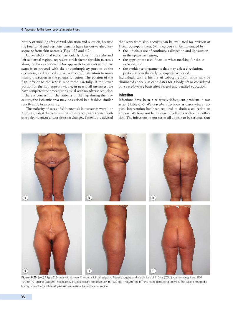
history of smoking after careful education and selection, because
the functional and aesthetic benefits have far outweighed any
sequelae from skin necrosis (Figs 6.23 and 6.26).
Upper abdominal scars, particularly those in the right and
left subcostal region, represent a risk factor for skin necrosis
along the lower abdomen. Our approach to patients with these
scars is to proceed with the abdominoplasty portion of the
operation, as described above, with careful attention to mini-
mizing dissection in the epigastric region. The portion of the
flap inferior to the scar is monitored carefully. If the lower
portion of the flap appears viable, in nearly all instances, we
have completed the procedure as usual with no adverse sequelae.
If there is concern for the viability of the flap during the pro-
cedure, the ischemic area may be excised in a fashion similar
to a fleur de lis procedure.
The majority of cases of skin necrosis in our series were 1 or
2 cm at greatest diameter, and in all instances were treated with
sharp debridement and/or dressing changes. Patients are advised
that scars from skin necrosis can be evaluated for revision at
1 year postoperatively. Skin necrosis can be minimized by:
• the judicious use of continuous dissection and liposuction
in the epigastric region;
• the appropriate use of tension when marking for tissue
excision; and
• the avoidance of garments that may affect circulation,
particularly in the early postoperative period.
Individuals with a history of tobacco consumption may be
eliminated entirely as candidates for a body lift or considered
on a case-by-case basis after careful and detailed education.
InfectionInfections have been a relatively infrequent problem in our
series (Table 6.5). We describe infections as cases where sur-
gical intervention has been required to drain a collection or
abscess. We have not had a case of cellulitis without a collec-
tion. The infections in our series all appear to be seromas that
6 Approach to the lower body after weight loss
96
d e f
a b c
Figure 6.26 (a–c) A type 2 24-year-old woman 11 months following gastric bypass surgery and weight loss of 115 lbs (52 kg). Current weight and BMI:
170 lbs (77 kg) and 28 kg/m2, respectively. Highest weight and BMI: 287 lbs (130 kg), 47 kg/m2. (d–f) Thirty months following body lift. The patient reported a
history of smoking and developed skin necrosis in the suprapubic region.
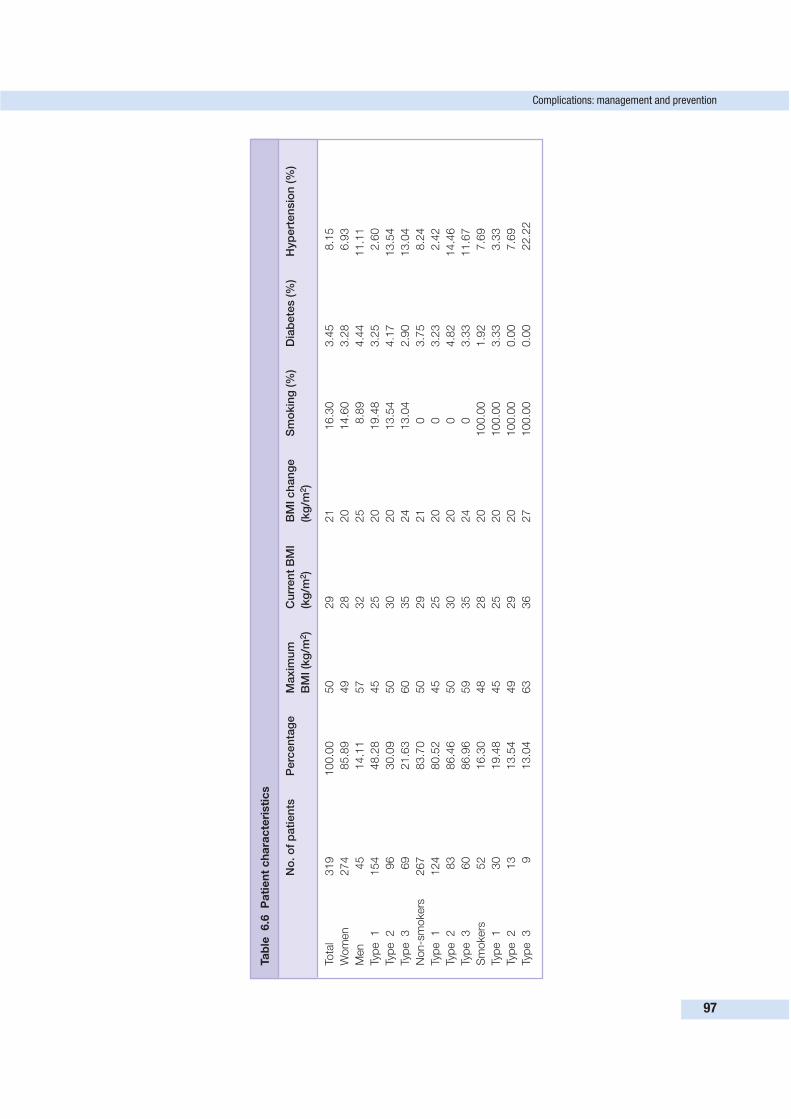
Complications: management and prevention
97
Ta
ble
6.6
Pa
tie
nt
ch
ara
cte
risti
cs
No
. o
f p
ati
en
tsP
erc
en
tag
eM
axim
um
C
urr
en
t B
MI
BM
I ch
an
ge
Sm
okin
g (%
)D
iab
ete
s (%
)H
yp
ert
en
sio
n (%
)
BM
I (k
g/m
2)
(kg
/m2)
(kg
/m2)
Tota
l319
100.0
050
29
21
16.3
03.4
58.1
5
Wom
en
274
85.8
949
28
20
14.6
03.2
86.9
3
Men
45
14.1
157
32
25
8.8
94.4
411.1
1
Typ
e1
154
48.2
845
25
20
19.4
83.2
52.6
0
Typ
e2
96
30.0
950
30
20
13.5
44.1
713.5
4
Typ
e3
69
21.6
360
35
24
13.0
42.9
013.0
4
Non-s
mokers
267
83.7
050
29
21
03.7
58.2
4
Typ
e1
124
80.5
245
25
20
03.2
32.4
2
Typ
e2
83
86.4
650
30
20
04.8
214.4
6
Typ
e3
60
86.9
659
35
24
03.3
311.6
7
Sm
okers
52
16.3
048
28
20
100.0
01.9
27.6
9
Typ
e1
30
19.4
845
25
20
100.0
03.3
33.3
3
Typ
e2
13
13.5
449
29
20
100.0
00.0
07.6
9
Typ
e3
913.0
463
36
27
100.0
00.0
022.2
2
were either clinically evident or undiagnosed and that became
infected. All patients were treated with open drainage or open
drainage with replacement of a 10-mm fully perforated flat
drain (Zimmer Corp.) in the collection cavity. The drainage
was sent for analysis, and the patients were placed on either
oral or intravenous antibiotics. No return to the operating
room was required.
The pathogenesis of infected seromas is unclear. A possi-
bility includes bacteria tracking from the skin on drains and
infecting devitalized tissue, probably fat. Drains kept for long
periods of time may create a risk factor for this problem. Our
current protocol is to keep patients on antibiotics until the last
drain is removed. This extended antibiotic regimen may pre-
dispose patients to infections with more resistant organisms.
We are currently reassessing our protocol regarding this matter.
Hematoma/bleedingBleeding and blood loss during and following body lifts are a
major concern. Many aspects of these procedures predispose
patients to a risk for blood loss. To effectively treat the lower
body contour deformity of the massive weight loss patient
requires extensive tissue undermining, and with that the need
to either ligate or cauterize a multitude of blood vessels.
Meticulous hemostasis is critical throughout these procedures.
We have found cautery set to a high level to be very helpful in
this regard. Heavier patients, men, and those with larger BMI
changes are at greater risk for significant blood loss. We avoid
the routine use of anticoagulants in the perioperative period
because of the concern for bleeding.
Menstruating women following malapsorptive bariatric
procedures often present with significant degrees of anemia.
All postbariatric patients are advised to take iron supplements
when considering body-contouring surgery, and those with
more severe cases of anemia are referred to a hematologist.
We avoid having an already anemic patient bank autologous
blood in the 1-month period prior to a body lift. Rather, we
prefer to transfuse non-autologous blood if it becomes neces-
sary. Our transfusion rate has decreased slightly over the course
of the series.
Our hematoma rate has remained relatively low at 1.88%
(Table 6.5). We defined a hematoma as a collection of blood
that required surgery for evacuation. We presume that there
may be other, smaller hematomas that go unnoticed and/or are
evacuated by the drains themselves.
Deep vein thrombosis and pulmonary embolismDeep vein thrombosis and pulmonary embolism represent the
most serious risks for body lift patients. Several recognized
risk factors for deep vein thrombosis are fundamental to these
procedures.26 The population of patients on average are over-
weight (Table 6.6), and the body lift is a lengthy procedure,
routinely over 4 h. To complicate matters further, early ambu-
lation can be difficult, and the early, routine use of anticoagu-
lants may create a significant risk for bleeding.
Our approach to the avoidance of deep vein thrombosis is to
provide the continual use of mechanical anticoagulation until
the patient is ambulatory. Patients are kept on bed rest until
the day following surgery. A lower extremity venous Doppler is
then obtained on the day of discharge. Our deep vein throm-
bosis rate is 1.88%. We would expect this number to be signi-
ficantly lower if all our patients were not routinely studied.
Pulmonary embolism remains relatively rare in our series.
The management of this life-threatening complication in the
post–body lift patient presents special challenges. Hepariniza-
tion of the early postoperative patient may lead to significant
bleeding. The timing and dosing of heparin must be evaluated
carefully, as should the possible need for a vena caval filter.
SEQUENCE AND COMBINATIONS OF PROCEDURES
Massive weight loss individuals are often candidates for and
are eager to have multiple procedures. Younger patients tend
to present initially with more concerns about their torso and
breasts, while older patients often have issue with their face
and arms. The medial thighs and flanks can be of primary
concern for both groups. Our preference regarding the torso is
to perform a body lift first, as a single procedure. As we dis-
cussed before, the body lift may eliminate the need for a
formal medial thigh lift in many patients, particularly those
less than 35 years of age and who have had a BMI change of
less than 20–25 kg/m2 prior to the body lift. Furthermore, a
more effective medial thigh lift can be performed in a patient
following a body lift.
The body lift can often have a significant effect on the upper
body, i.e. breasts, flanks, and back (Figs 6.20, 6.24 and 6.26).
In men, it may eliminate the need for upper body-contouring
surgery or reduce the magnitude of the procedure required. In
women, while the body lift can positively impact the back and
flanks, it can also cause significant downward migration of the
inframammary fold. For this reason, ideally we prefer not to per-
form breast surgery prior to or concomitantly with a body lift.
Following a body lift, other body-contouring procedures we
commonly perform are combination brachioplasty and mam-
moplasty, thighplasty alone, or thighplasty with brachioplasty.
CONCLUSION
The lower body in the massive weight loss patient presents an
extreme form of traditional aesthetic and functional body
contour concerns. Routine body-contouring procedures usually
produce only suboptimal results in this patient population.
The body lift described above is an excellent alternative to
treat the lower body deformity of the postbariatric patient. As
with every technique, careful patient selection, education, and
preparation are critical to minimizing complications and opti-
mizing outcome.
6 Approach to the lower body after weight loss
98
REFERENCES
1. Capella JF, Oliak DA, Nemerofsky RB. Body lift: an account of 200
consecutive cases in the massive weight loss patient. Plast Reconstr
Surg 2006; 117(2):414–430.
2. Capella JF, Capella RF. Bariatric surgery in adolescence. Is this the
best time to operate? Obes Surg 2003; 13:826–832.
3. Capella JF, Capella RF. An assessment of vertical banded
gastroplasty—Roux-en-Y gastric bypass for the treatment of
morbid obesity. Am J Surg 2002; 183:117–123.
4. Aly AS, Cram AE, Chao M, et al. Belt lipectomy for circumferential
truncal excess: the University of Iowa experience. Plast Reconstr
Surg 2003; 111:398–413.
5. Lockwood TE. Lower-body lift. Aesthetic Surg J 2001; 21:355–370.
6. Van Geertruyden JP, Vandeweyer E, de Fontaine S, et al. Circum-
ferential torsoplasty. Br J Plast Surg 1999; 52:623–628.
7. Mallory GN. American Society for Bariatric Surgery membership
survey. Gainesville: ASBS; 2004.
8. Carwell GR, Horton CE. Circumferential torsoplasty. Ann Plast
Surg 1997; 38:213–216.
9. Hurwitz DJ. Single-staged total body lift after massive weight loss.
Ann Plast Surg 2004; 52:435–441.
10. Pascal JF, Le Louarn C. Remodeling body lift with high lateral
tension. Aesthetic Plast Surg 2002; 26:223–230.
11. Hamra ST. Circumferential body lift. Aesthetic Surg J 1999;
19:244–251.
12. Morales Gracia HJ. Circular lipectomy with lateral thigh–buttock
lift. Aesthetic Plast Surg 2003; 27(1):50–57.
13. Regnault P, Daniel R. Secondary thigh–buttock deformities after
classical techniques: prevention and treatment. Clin Plast Surg
1984; 11:505–516.
14. Lockwood TE. Fascial anchoring technique in medial thigh lifts.
Plast Reconstr Surg 1988; 82:299–304.
15. Lockwood T. Superficial fascial system (SFS) of the trunk and ex-
tremities: a new concept. Plast Reconstr Surg 1991; 87:1009–1027.
16. Lockwood T. Transverse flank–thigh–buttock lift with superficial
fascial suspension. Plast Reconstr Surg 1993; 92:1112–1122.
17. Lockwood T. High–lateral-tension abdominoplasty with superficial
fascial suspension. Plast Reconstr Surg 1995; 96:603–615.
18. Marceau S, Hould FS, Simard S, et al. Biliopancreatic diversion
with duodenal switch. World J Surg 1998; 22:947–954.
19. Chapman AE, Kiroff G, Game P, et al. Laparoscopic adjustable
gastric banding in the treatment of obesity: a systematic literature
review. Surgery 2004; 135:326–351.
20. Capella JF, Capella RF. The weight reduction operation of choice:
vertical banded gastroplasty or gastric bypass? Am J Surg 1996;
171:74–79.
21. Brolin RE. Metabolic deficiencies and supplements following ba-
riatric operations. In: Martin L, ed. Obesity surgery. New York:
McGraw-Hill; 2004:275–300.
22. Matarasso A. Liposuction as an adjunct to a full abdominoplasty.
Plast Reconstr Surg 1995; 95:829–836.
23. Chaouat M, Levan P, Lalanne B, et al. Abdominal dermolipectomies:
early postoperative complications and long-term unfavorable results.
Plast Reconstr Surg 2000; 106:1614–1623.
24. El-khatib HA, Bener A. Abdominal dermolipectomy in an abdo-
men with pre-existing scars: a different concept. Plast Reconstr
Surg 2004; 114:992–997.
25. Manassa EH, Hertl CH, Olbrisch R. Wound healing problems in
smokers and nonsmokers after 132 abdominoplasties. Plast Reconstr
Surg 2003; 111:2082–2087.
26. Geerts WH, Heit JA, Clagett GP, et al. Prevention of venous
thromboembolism. Chest 2001; 119:132S–175S.
References
99
7
A subset of patients will present with significant rolls of skin
along the upper back and lateral chest. This chapter describes
technical approaches for correcting these deformities. There is
little historical basis for these procedures; rather, they represent
an early step in the evolution of approaches for contouring
regions that have not traditionally been the focus of plastic
surgeons.
DEFORMITIES OF THE UPPER TRUNK
As with any problem faced in plastic surgery, an accurate
assessment of the deformity and how it is formed is needed. A
surgical plan can then be devised based on this analysis. The
thoracic region will often undergo dramatic changes during
the process of massive weight gain and subsequent loss. The
soft tissue envelope develops varying degrees of laxity in both
the horizontal and the vertical directions. Zones of adherence,
located over the sternum and spine, restrict movement of the
overlying skin and act like hooks that tissues drape off,
leading to both anterior and posterior inverted V deformities
(see Fig. 7.1).
The lateral thoracic soft tissues descend inferiorly to varying
degrees, causing a ‘dropout’ of the lateral inframammary
crease. Some patients will experience no descent of the lateral
inframammary crease, while others will drop significantly, mani-
festing this change into lateral breast rolls. The lateral breast
rolls become upper back rolls as they traverse posteriorly.
Although many patients will develop the full extent of defor-
mities described here, there are others whose fat deposition
pattern may lead to less severe deformities. If the lateral chest
rolls dissipate in the region of the posterior axillary line, their
correction may be incorporated into an extended mastopexy
or gynecomastia correction. If the back rolls extend further, a
decision must be made about the suitability of liposuction.
Rolls with ptosis and poor skin tone will likely require exci-
sion for adequate treatment. Additionally, the position of the
lateral inframammary crease is important. If it has dropped
out laterally, then some form of an upper body lift will usually
be required.
Three surgical approaches are demonstrated.
1. Transverse excision of back rolls combined with
mastopexy and brachioplasty.
2. Transverse excision of back rolls combined with mastopexy.
3. Lateral excision of trunk tissue combined with mastopexy.
APPROACH 1: TRANSVERSE EXCISION OF BACK ROLLSCOMBINED WITH MASTOPEXY AND BRACHIOPLASTY
Three goals are accomplished with this upper body lift approach.
1. Elimination of horizontal excess through an extension of
the brachioplasty procedure on to the lateral chest wall.
2. Elimination of vertical excess by elevating the lateral
inframammary crease to its correct position and excising
lateral breast/upper back rolls.
3. Building the breast based on a repositioned inframammary
crease.
MarkingsThe patient is marked in the sitting position (see Fig. 7.2).
The arms are marked utilizing a double-ellipse technique.
The ellipses cross the axilla onto the lateral chest wall, with
their widths and lengths of the lateral chest wall extension
based on the amount of excess that the particular patient pre-
sents with. The outer ellipse of the double-ellipse technique is
based on the estimation of the pinch of redundant tissue just
inferior to the underlying musculoskeletal core. Because the
pinch technique does not take into account the amount of
101
APPROACHES TO UPPER BODYROLLS
J. Peter Rubin, Al S. Aly and Felmont F. Eaves III
Key Points• An upper body lift is defined as correction of upper back or flank rolls by
excision of tissue on the upper torso.
• Excision of upper back rolls can be accomplished with a transverse scar
on the upper back or with bilateral lateral or oblique scars.
• Excision of upper back rolls may be combined with breast reshaping or
gynecomastia correction.
• A circumferential approach or near-circumferential approach may be
employed.
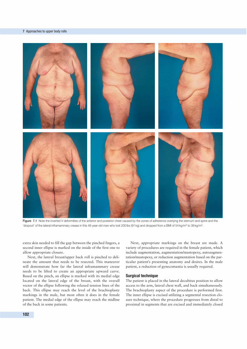
extra skin needed to fill the gap between the pinched fingers, a
second inner ellipse is marked on the inside of the first one to
allow appropriate closure.
Next, the lateral breast/upper back roll is pinched to deli-
neate the amount that needs to be resected. This maneuver
will demonstrate how far the lateral inframammary crease
needs to be lifted to create an appropriate upward curve.
Based on the pinch, an ellipse is marked with its medial edge
located on the lateral edge of the breast, with the overall
vector of the ellipse following the relaxed tension lines of the
back. This ellipse may reach the level of the brachioplasty
markings in the male, but most often it does in the female
patient. The medial edge of the ellipse may reach the midline
of the back in some patients.
Next, appropriate markings on the breast are made. A
variety of procedures are required in the female patient, which
include augmentation, augmentation/mastopexy, autoaugmen-
tation/mastopexy, or reduction augmentation based on the par-
ticular patient’s presenting anatomy and desires. In the male
patient, a reduction of gynecomastia is usually required.
Surgical techniqueThe patient is placed in the lateral decubitus position to allow
access to the arm, lateral chest wall, and back simultaneously.
The brachioplasty aspect of the procedure is performed first.
The inner ellipse is excised utilizing a segmental resection clo-
sure technique, where the procedure progresses from distal to
proximal in segments that are excised and immediately closed
7 Approaches to upper body rolls
102
Figure 7.1 Note the inverted V deformities of the anterior and posterior chest caused by the zones of adherence overlying the sternum and spine and the
‘dropout’ of the lateral inframammary crease in this 48-year-old man who lost 200 lbs (91 kg) and dropped from a BMI of 54 kg/m2 to 38 kg/m2.
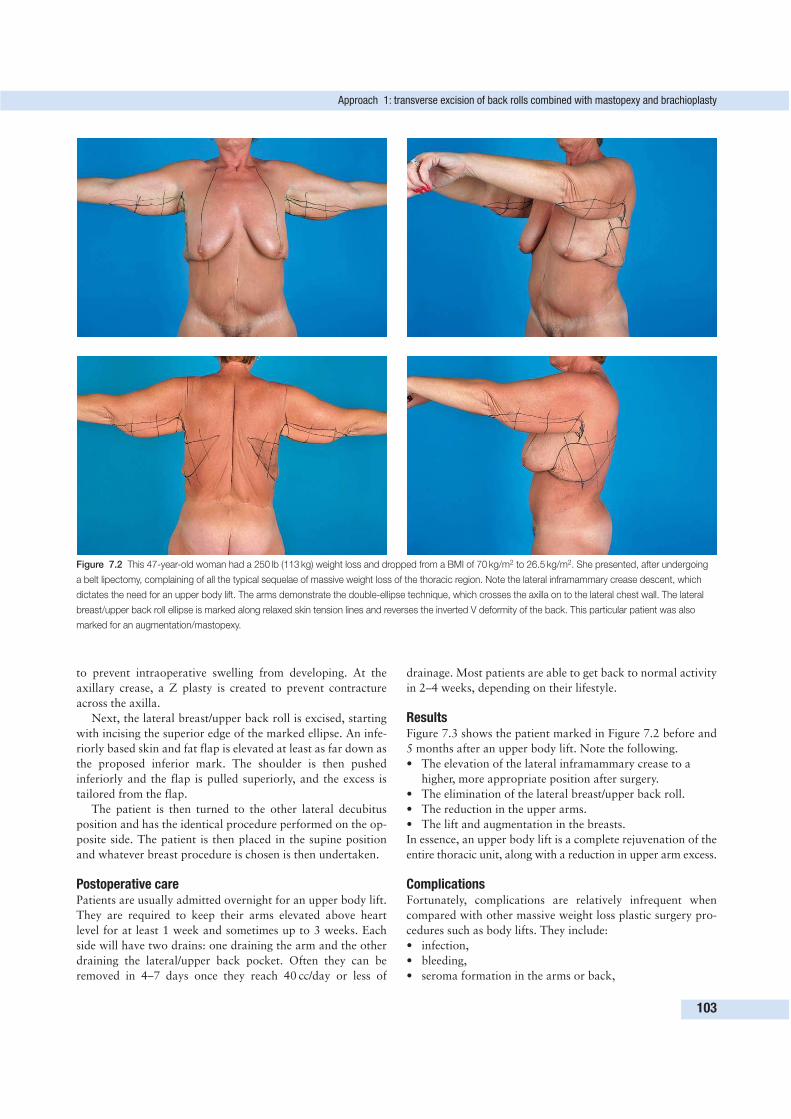
to prevent intraoperative swelling from developing. At the
axillary crease, a Z plasty is created to prevent contracture
across the axilla.
Next, the lateral breast/upper back roll is excised, starting
with incising the superior edge of the marked ellipse. An infe-
riorly based skin and fat flap is elevated at least as far down as
the proposed inferior mark. The shoulder is then pushed
inferiorly and the flap is pulled superiorly, and the excess is
tailored from the flap.
The patient is then turned to the other lateral decubitus
position and has the identical procedure performed on the op-
posite side. The patient is then placed in the supine position
and whatever breast procedure is chosen is then undertaken.
Postoperative carePatients are usually admitted overnight for an upper body lift.
They are required to keep their arms elevated above heart
level for at least 1 week and sometimes up to 3 weeks. Each
side will have two drains: one draining the arm and the other
draining the lateral/upper back pocket. Often they can be
removed in 4–7 days once they reach 40 cc/day or less of
drainage. Most patients are able to get back to normal activity
in 2–4 weeks, depending on their lifestyle.
ResultsFigure 7.3 shows the patient marked in Figure 7.2 before and
5 months after an upper body lift. Note the following.
• The elevation of the lateral inframammary crease to a
higher, more appropriate position after surgery.
• The elimination of the lateral breast/upper back roll.
• The reduction in the upper arms.
• The lift and augmentation in the breasts.
In essence, an upper body lift is a complete rejuvenation of the
entire thoracic unit, along with a reduction in upper arm excess.
ComplicationsFortunately, complications are relatively infrequent when
compared with other massive weight loss plastic surgery pro-
cedures such as body lifts. They include:
• infection,
• bleeding,
• seroma formation in the arms or back,
Approach 1: transverse excision of back rolls combined with mastopexy and brachioplasty
103
Figure 7.2 This 47-year-old woman had a 250 lb (113 kg) weight loss and dropped from a BMI of 70 kg/m2 to 26.5 kg/m2. She presented, after undergoing
a belt lipectomy, complaining of all the typical sequelae of massive weight loss of the thoracic region. Note the lateral inframammary crease descent, which
dictates the need for an upper body lift. The arms demonstrate the double-ellipse technique, which crosses the axilla on to the lateral chest wall. The lateral
breast/upper back roll ellipse is marked along relaxed skin tension lines and reverses the inverted V deformity of the back. This particular patient was also
marked for an augmentation/mastopexy.
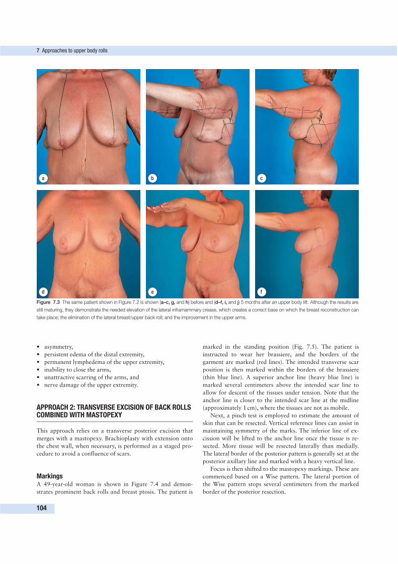
• asymmetry,
• persistent edema of the distal extremity,
• permanent lymphedema of the upper extremity,
• inability to close the arms,
• unattractive scarring of the arms, and
• nerve damage of the upper extremity.
APPROACH 2: TRANSVERSE EXCISION OF BACK ROLLSCOMBINED WITH MASTOPEXY
This approach relies on a transverse posterior excision that
merges with a mastopexy. Brachioplasty with extension onto
the chest wall, when necessary, is performed as a staged pro-
cedure to avoid a confluence of scars.
MarkingsA 49-year-old woman is shown in Figure 7.4 and demon-
strates prominent back rolls and breast ptosis. The patient is
marked in the standing position (Fig. 7.5). The patient is
instructed to wear her brassiere, and the borders of the
garment are marked (red lines). The intended transverse scar
position is then marked within the borders of the brassiere
(thin blue line). A superior anchor line (heavy blue line) is
marked several centimeters above the intended scar line to
allow for descent of the tissues under tension. Note that the
anchor line is closer to the intended scar line at the midline
(approximately 1 cm), where the tissues are not as mobile.
Next, a pinch test is employed to estimate the amount of
skin that can be resected. Vertical reference lines can assist in
maintaining symmetry of the marks. The inferior line of ex-
cission will be lifted to the anchor line once the tissue is re-
sected. More tissue will be resected laterally than medially.
The lateral border of the posterior pattern is generally set at the
posterior axillary line and marked with a heavy vertical line.
Focus is then shifted to the mastopexy markings. These are
commenced based on a Wise pattern. The lateral portion of
the Wise pattern stops several centimeters from the marked
border of the posterior resection.
7 Approaches to upper body rolls
104
d e f
a b c
Figure 7.3 The same patient shown in Figure 7.2 is shown (a–c, g, and h) before and (d–f, i, and j) 5 months after an upper body lift. Although the results are
still maturing, they demonstrate the needed elevation of the lateral inframammary crease, which creates a correct base on which the breast reconstruction can
take place; the elimination of the lateral breast/upper back roll; and the improvement in the upper arms.
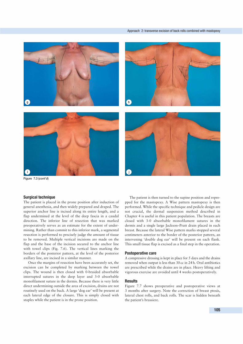
Surgical techniqueThe patient is placed in the prone position after induction of
general anesthesia, and then widely prepared and draped. The
superior anchor line is incised along its entire length, and a
flap undermined at the level of the deep fascia in a caudal
direction. The inferior line of resection that was marked
preoperatively serves as an estimate for the extent of under-
mining. Rather than commit to this inferior mark, a segmental
resection is performed to precisely judge the amount of tissue
to be removed. Multiple vertical incisions are made on the
flap and the base of the incision secured to the anchor line
with towel clips (Fig. 7.6). The vertical lines marking the
borders of the posterior pattern, at the level of the posterior
axillary line, are incised in a similar manner.
Once the margins of resection have been accurately set, the
excision can be completed by marking between the towel
clips. The wound is then closed with 0-braided absorbable
interrupted sutures in the deep layer and 3-0 absorbable
monofilament suture in the dermis. Because there is very little
direct undermining outside the area of excision, drains are not
routinely used on the back. A large ‘dog ear’ will be present at
each lateral edge of the closure. This is simply closed with
staples while the patient is in the prone position.
The patient is then turned to the supine position and repre-
pped for the mastopexy. A Wise pattern mastopexy is then
performed. While the specific technique and pedicle design are
not crucial, the dermal suspension method described in
Chapter 4 is useful in this patient population. The breasts are
closed with 3-0 absorbable monofilament sutures in the
dermis and a single large Jackson–Pratt drain placed in each
breast. Because the lateral Wise pattern marks stopped several
centimeters anterior to the border of the posterior pattern, an
intervening ‘double dog ear’ will be present on each flank.
This small tissue flap is excised as a final step in the operation.
Postoperative careA compressive dressing is kept in place for 5 days and the drains
removed when output is less than 30 cc in 24 h. Oral antibiotics
are prescribed while the drains are in place. Heavy lifting and
vigorous exercise are avoided until 4 weeks postoperatively.
ResultsFigure 7.7 shows preoperative and postoperative views at
3 months after surgery. Note the correction of breast ptosis,
lateral chest rolls, and back rolls. The scar is hidden beneath
the patient’s brassiere.
Approach 2: transverse excision of back rolls combined with mastopexy
105
Figure 7.3 (cont’d)
g h
i j
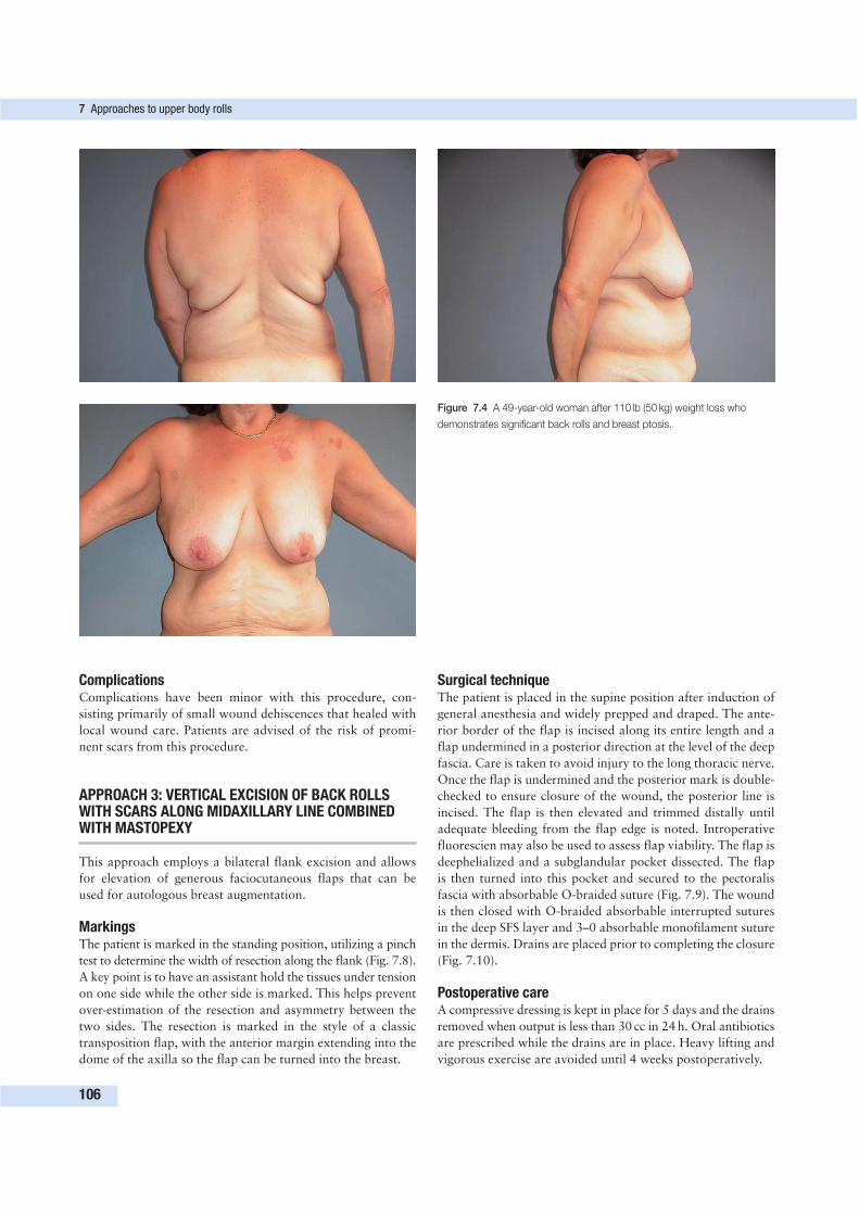
ComplicationsComplications have been minor with this procedure, con-
sisting primarily of small wound dehiscences that healed with
local wound care. Patients are advised of the risk of promi-
nent scars from this procedure.
APPROACH 3: VERTICAL EXCISION OF BACK ROLLSWITH SCARS ALONG MIDAXILLARY LINE COMBINEDWITH MASTOPEXY
This approach employs a bilateral flank excision and allows
for elevation of generous faciocutaneous flaps that can be
used for autologous breast augmentation.
MarkingsThe patient is marked in the standing position, utilizing a pinch
test to determine the width of resection along the flank (Fig. 7.8).
A key point is to have an assistant hold the tissues under tension
on one side while the other side is marked. This helps prevent
over-estimation of the resection and asymmetry between the
two sides. The resection is marked in the style of a classic
transposition flap, with the anterior margin extending into the
dome of the axilla so the flap can be turned into the breast.
Surgical techniqueThe patient is placed in the supine position after induction of
general anesthesia and widely prepped and draped. The ante-
rior border of the flap is incised along its entire length and a
flap undermined in a posterior direction at the level of the deep
fascia. Care is taken to avoid injury to the long thoracic nerve.
Once the flap is undermined and the posterior mark is double-
checked to ensure closure of the wound, the posterior line is
incised. The flap is then elevated and trimmed distally until
adequate bleeding from the flap edge is noted. Introperative
fluorescien may also be used to assess flap viability. The flap is
deephelialized and a subglandular pocket dissected. The flap
is then turned into this pocket and secured to the pectoralis
fascia with absorbable O-braided suture (Fig. 7.9). The wound
is then closed with O-braided absorbable interrupted sutures
in the deep SFS layer and 3–0 absorbable monofilament suture
in the dermis. Drains are placed prior to completing the closure
(Fig. 7.10).
Postoperative careA compressive dressing is kept in place for 5 days and the drains
removed when output is less than 30 cc in 24 h. Oral antibiotics
are prescribed while the drains are in place. Heavy lifting and
vigorous exercise are avoided until 4 weeks postoperatively.
7 Approaches to upper body rolls
106
Figure 7.4 A 49-year-old woman after 110 lb (50 kg) weight loss who
demonstrates significant back rolls and breast ptosis.
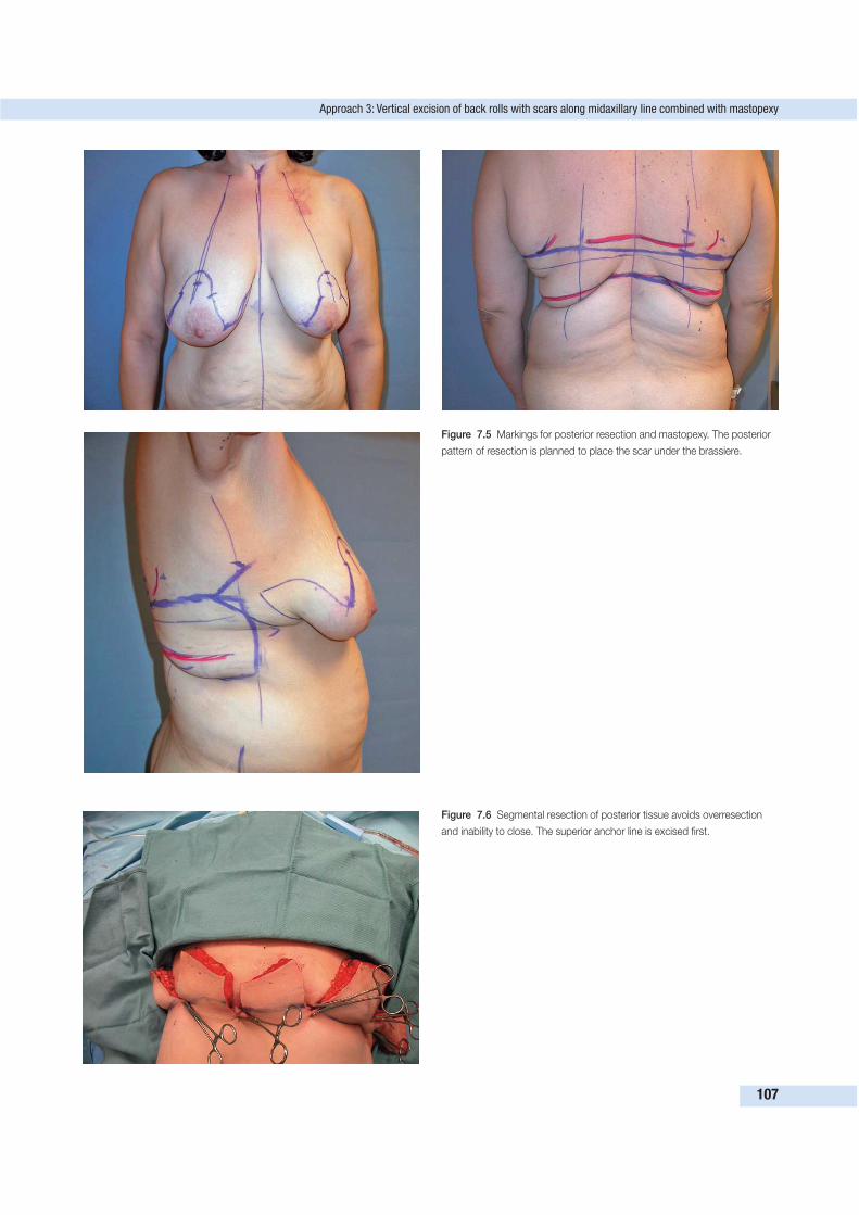
Approach 3: Vertical excision of back rolls with scars along midaxillary line combined with mastopexy
107
Figure 7.5 Markings for posterior resection and mastopexy. The posterior
pattern of resection is planned to place the scar under the brassiere.
Figure 7.6 Segmental resection of posterior tissue avoids overresection
and inability to close. The superior anchor line is excised first.
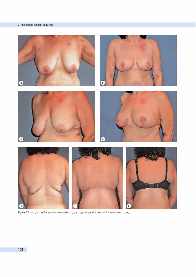
7 Approaches to upper body rolls
108
e f g
Figure 7.7 (a, c, and e) Preoperative views and (b, d, f, and g) postoperative views at 3 months after surgery.
d
a b
c
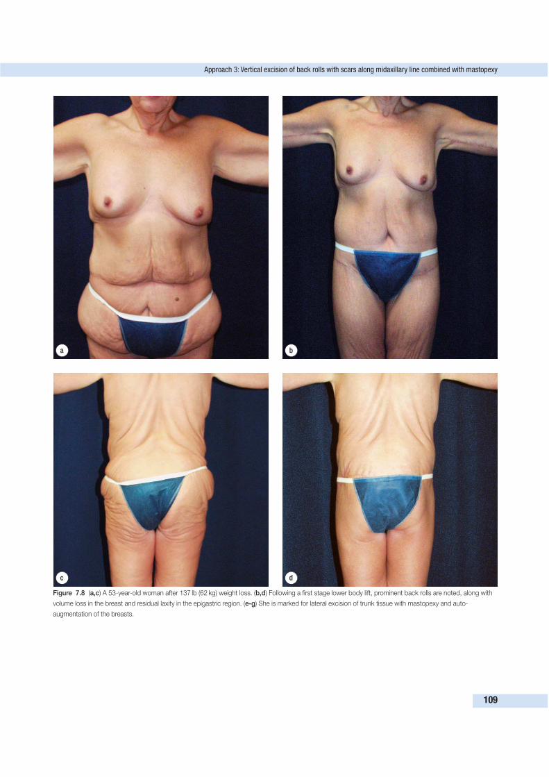
Approach 3: Vertical excision of back rolls with scars along midaxillary line combined with mastopexy
109
d
a b
c
Figure 7.8 (a,c) A 53-year-old woman after 137 lb (62 kg) weight loss. (b,d) Following a first stage lower body lift, prominent back rolls are noted, along with
volume loss in the breast and residual laxity in the epigastric region. (e-g) She is marked for lateral excision of trunk tissue with mastopexy and auto-
augmentation of the breasts.
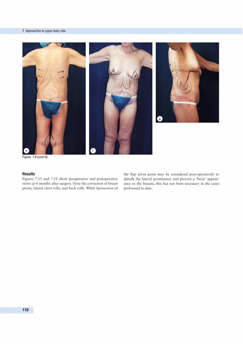
ResultsFigures 7.11 and 7.12 show preoperative and postoperative
views at 6 months after surgery. Note the correction of breast
ptosis, lateral chest rolls, and back rolls. While liposuction of
the flap pivot point may be considered post-operatively to
debulk the lateral prominence and prevent a ‘boxy’ appear-
ance to the breasts, this has not been necessary in the cases
performed to date.
7 Approaches to upper body rolls
110
e f
g
Figure 7.8 (cont’d)

Approach 3: Vertical excision of back rolls with scars along midaxillary line combined with mastopexy
111
Figure 7.9 Intraoperative views showing elevation of tissue flap along flank, deepithelialization of flap, and transposition of flap into subglandular breast
pocket.
Figure 7.10 Intraoperative views demonstrating lateral scar and increased breast volume from autologous augmentation.
d ea b c
Figure 7.11 (a) Preoperative view. (b,d) Postoperative view at 2 months and (c,e) 2 years showing maturation of lateral scar and maintenance of breast
shape.
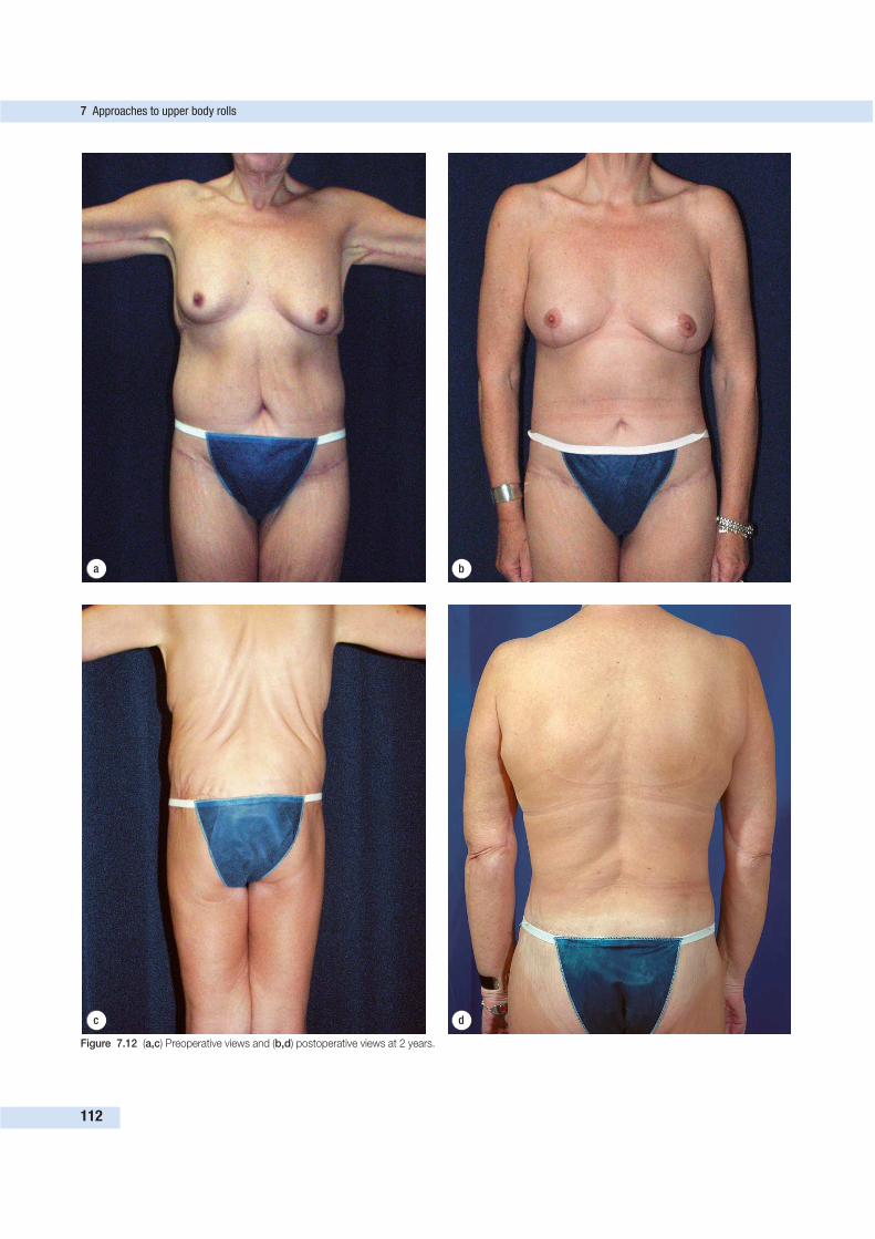
7 Approaches to upper body rolls
112
d
a b
c
Figure 7.12 (a,c) Preoperative views and (b,d) postoperative views at 2 years.
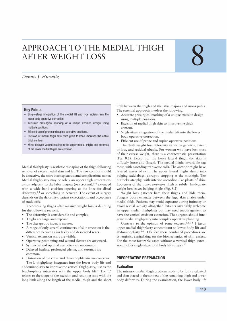
Medial thighplasty is aesthetic reshaping of the thigh following
removal of excess medial skin and fat. The new contour should
be attractive, the scars inconspicuous, and complications minor.
Medial thighplasty may be solely an upper thigh crescent ex-
cision adjacent to the labia majora (or scrotum),1–4 extended
with a wide band excision tapering at the knee for distal
deformity,3,5 or something in between. The extent of surgery
depends on the deformity, patient expectations, and acceptance
of trade-offs.
Recontouring thighs after massive weight loss is daunting
for the following reasons.
• The deformity is considerable and complex.
• Thighs are large and exposed.
• The therapeutic index is narrow.
• A range of only several centimeters of skin resection is the
difference between skin laxity and descended scars.
• Vertical extension scars are visible.
• Operative positioning and wound closure are awkward.
• Symmetry and optimal aesthetics are uncommon.
• Delayed healing, prolonged edema, and seromas are
common.
• Distortion of the vulva and thrombophlebitis are concerns.
The L thighplasty integrates into the lower body lift and
abdominoplasty to improve the vertical thighplasty, just as the
brachioplasty integrates with the upper body lift.5 The ‘L’
relates to the shape of the excision and resulting scar, with the
long limb along the length of the medial thigh and the short
limb between the thigh and the labia majora and mons pubis.
The essential approach involves the following.
• Accurate presurgical marking of a unique excision design
using multiple positions.
• Excision of medial thigh skin to improve the thigh
contour.
• Single-stage integration of the medial lift into the lower
body operative correction.
• Efficient use of prone and supine operative positions.
The thigh weight loss deformity varies by genetics, extent
of loss, and residual obesity. For women who have lost most
of their excess weight, there is a characteristic presentation
(Fig. 8.1). Except for the lower lateral thigh, the skin is
diffusely loose and flaccid. The medial thighs invariable sag
most, with cascading transverse rolls. The anterior thighs have
layered waves of skin. The upper lateral thighs slump into
bulging saddlebags, abruptly stopping at the midthigh. The
buttocks atrophy, with inferior accordion-like pleats of skin.
Looseness of the upper posterior thigh is subtle. Inadequate
weight loss leaves bulging thighs (Fig. 8.2).
Weight loss patients hate their thighs and hide them.
Pungent odors emanate between the legs. Skin chafes under
medial folds. Patients may avoid exposure during intimacy or
avoid sexual activity altogether. Patients invariably welcome
an upper medial thighplasty but may need encouragement to
have the vertical excision extension. The surgeon should inte-
grate medial thighplasty into complex operative planning.
Contrary to the opinion of some experts,1,2,6–8 I favor
upper medial thighplasty concomitant to lower body lift and
abdominoplasty.9–11 I believe these combined procedures are
synergistic, capitalizing on the biomechanics of skin excess.
For the most favorable cases without a vertical thigh exten-
sion, I offer single-stage total body lift surgery.10
PREOPERATIVE PREPARATION
EvaluationThe intrinsic medial thigh problem needs to be fully evaluated
and then placed in the context of the remaining thigh and lower
body deformity. During the examination, the lower body lift
113
APPROACH TO THE MEDIAL THIGHAFTER WEIGHT LOSS 8Dennis J. Hurwitz
Key Points• Single-stage integration of the medial lift and type incision into the
lower body operative correction.
• Accurate presurgical marking of a unique excision design using
multiple positions.
• Efficient use of prone and supine operative positions.
• Excision of medial thigh skin from groin to knee improves the entire
thigh contour.
• Minor delayed wound healing in the upper medial thighs and seromas
of the lower medial thighs are common.
d
e f
a b
c
Figure 8.1 Multiple views of the thighs of a
49-year-old, 5’ 7” (1.70 m), 157-lb (71 kg) woman
(a, c, e, g, i) before and (b, d, f, h, j) 5 months
after an L thighplasty with an abdominoplasty
and lower body lift reported in the Aesthetic
Surgery Journal.5 She had lost 230 lbs (104 kg)
subsequent to a gastric restrictive procedure and
hated her thighs. Her rolls of redundant skin were
worst medial, and least upper anterior and lower
lateral thigh. The medial thighs had cascading
transverse loose rolls of skin. The middle anterior
thighs had stacked layers of skin like melted
candle wax. Loose skin hung from the hips to the
midlateral thigh. The buttocks had inferior
accordion-like pleats. Except for the distal thigh,
the postoperative views show these deformities
corrected by a single complex 10-h operation, as
described. The scars are level, symmetric, and
narrow. There are long but inconspicuous scars
running down the medial posterior thighs,
between the labia and thigh, and in a beltlike
manner around the lower torso. The buttock
curvature is full due to the adipose flap
reconstruction. There is some residual looseness
below the buttocks and about the knees, which
will be corrected secondarily.
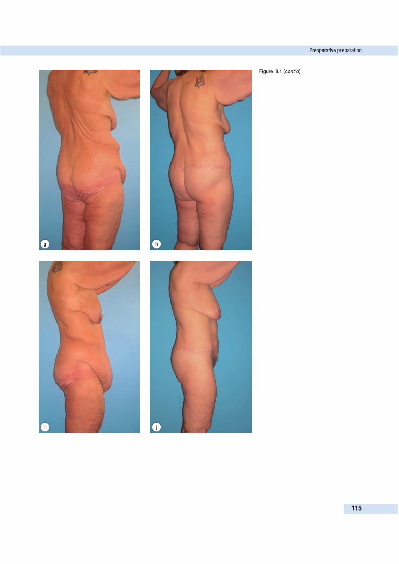
Preoperative preparation
115
g h
i j
Figure 8.1 (cont’d)
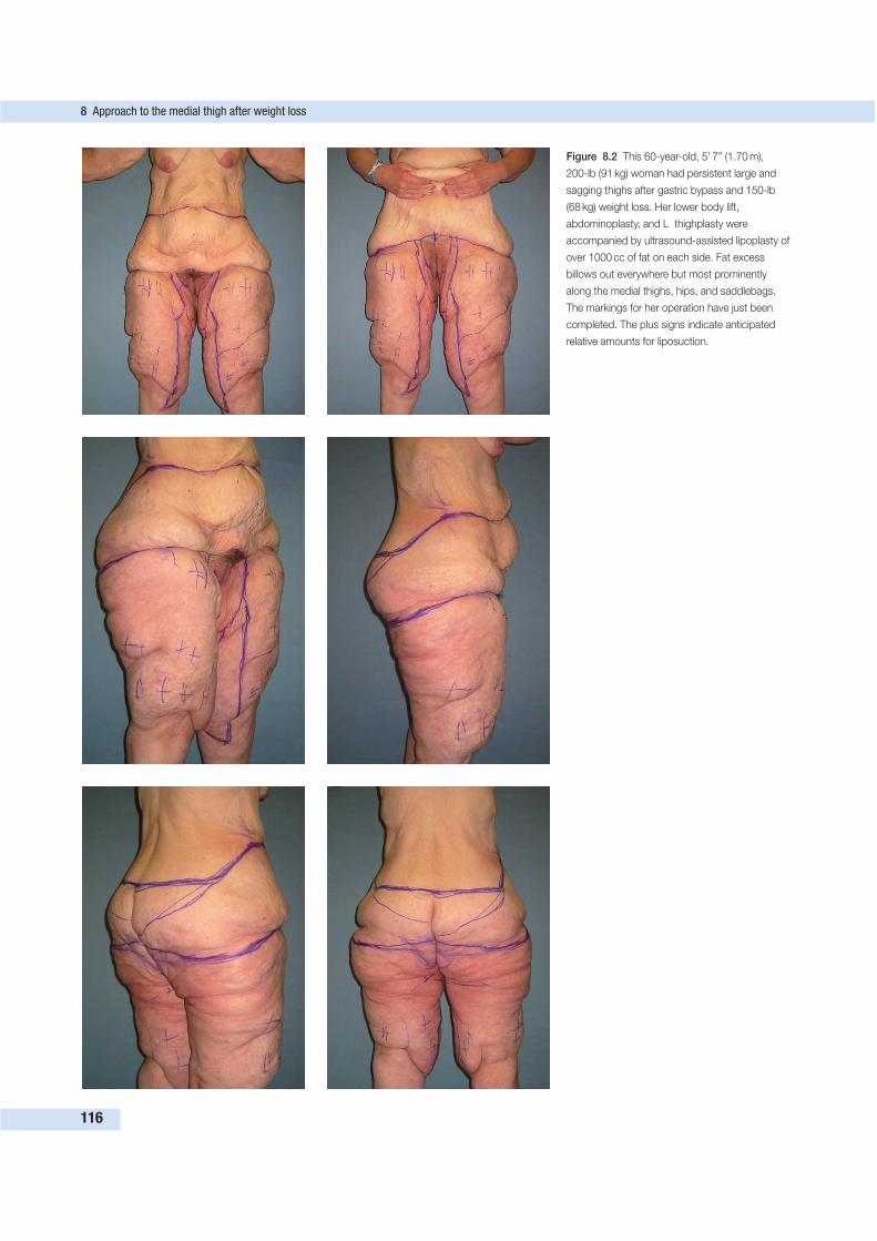
8 Approach to the medial thigh after weight loss
116
Figure 8.2 This 60-year-old, 5’ 7” (1.70 m),
200-lb (91 kg) woman had persistent large and
sagging thighs after gastric bypass and 150-lb
(68 kg) weight loss. Her lower body lift,
abdominoplasty, and L thighplasty were
accompanied by ultrasound-assisted lipoplasty of
over 1000 cc of fat on each side. Fat excess
billows out everywhere but most prominently
along the medial thighs, hips, and saddlebags.
The markings for her operation have just been
completed. The plus signs indicate anticipated
relative amounts for liposuction.
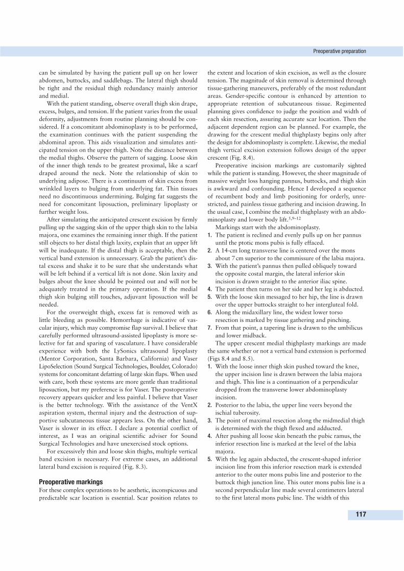
can be simulated by having the patient pull up on her lower
abdomen, buttocks, and saddlebags. The lateral thigh should
be tight and the residual thigh redundancy mainly anterior
and medial.
With the patient standing, observe overall thigh skin drape,
excess, bulges, and tension. If the patient varies from the usual
deformity, adjustments from routine planning should be con-
sidered. If a concomitant abdominoplasty is to be performed,
the examination continues with the patient suspending the
abdominal apron. This aids visualization and simulates anti-
cipated tension on the upper thigh. Note the distance between
the medial thighs. Observe the pattern of sagging. Loose skin
of the inner thigh tends to be greatest proximal, like a scarf
draped around the neck. Note the relationship of skin to
underlying adipose. There is a continuum of skin excess from
wrinkled layers to bulging from underlying fat. Thin tissues
need no discontinuous undermining. Bulging fat suggests the
need for concomitant liposuction, preliminary lipoplasty or
further weight loss.
After simulating the anticipated crescent excision by firmly
pulling up the sagging skin of the upper thigh skin to the labia
majora, one examines the remaining inner thigh. If the patient
still objects to her distal thigh laxity, explain that an upper lift
will be inadequate. If the distal thigh is acceptable, then the
vertical band extension is unnecessary. Grab the patient’s dis-
tal excess and shake it to be sure that she understands what
will be left behind if a vertical lift is not done. Skin laxity and
bulges about the knee should be pointed out and will not be
adequately treated in the primary operation. If the medial
thigh skin bulging still touches, adjuvant liposuction will be
needed.
For the overweight thigh, excess fat is removed with as
little bleeding as possible. Hemorrhage is indicative of vas-
cular injury, which may compromise flap survival. I believe that
carefully performed ultrasound-assisted lipoplasty is more se-
lective for fat and sparing of vasculature. I have considerable
experience with both the LySonics ultrasound lipoplasty
(Mentor Corporation, Santa Barbara, California) and Vaser
LipoSelection (Sound Surgical Technologies, Boulder, Colorado)
systems for concomitant defatting of large skin flaps. When used
with care, both these systems are more gentle than traditional
liposuction, but my preference is for Vaser. The postoperative
recovery appears quicker and less painful. I believe that Vaser
is the better technology. With the assistance of the VentX
aspiration system, thermal injury and the destruction of sup-
portive subcutaneous tissue appears less. On the other hand,
Vaser is slower in its effect. I declare a potential conflict of
interest, as I was an original scientific adviser for Sound
Surgical Technologies and have unexercised stock options.
For excessively thin and loose skin thighs, multiple vertical
band excision is necessary. For extreme cases, an additional
lateral band excision is required (Fig. 8.3).
Preoperative markingsFor these complex operations to be aesthetic, inconspicuous and
predictable scar location is essential. Scar position relates to
the extent and location of skin excision, as well as the closure
tension. The magnitude of skin removal is determined through
tissue-gathering maneuvers, preferably of the most redundant
areas. Gender-specific contour is enhanced by attention to
appropriate retention of subcutaneous tissue. Regimented
planning gives confidence to judge the position and width of
each skin resection, assuring accurate scar location. Then the
adjacent dependent region can be planned. For example, the
drawing for the crescent medial thighplasty begins only after
the design for abdominoplasty is complete. Likewise, the medial
thigh vertical excision extension follows design of the upper
crescent (Fig. 8.4).
Preoperative incision markings are customarily sighted
while the patient is standing. However, the sheer magnitude of
massive weight loss hanging pannus, buttocks, and thigh skin
is awkward and confounding. Hence I developed a sequence
of recumbent body and limb positioning for orderly, unre-
stricted, and painless tissue gathering and incision drawing. In
the usual case, I combine the medial thighplasty with an abdo-
minoplasty and lower body lift.5,9–12
Markings start with the abdominoplasty.
1. The patient is reclined and evenly pulls up on her pannus
until the ptotic mons pubis is fully effaced.
2. A 14-cm long transverse line is centered over the mons
about 7 cm superior to the commissure of the labia majora.
3. With the patient’s pannus then pulled obliquely toward
the opposite costal margin, the lateral inferior skin
incision is drawn straight to the anterior iliac spine.
4. The patient then turns on her side and her leg is abducted.
5. With the loose skin messaged to her hip, the line is drawn
over the upper buttocks straight to her intergluteal fold.
6. Along the midaxillary line, the widest lower torso
resection is marked by tissue gathering and pinching.
7. From that point, a tapering line is drawn to the umbilicus
and lower midback.
The upper crescent medial thighplasty markings are made
the same whether or not a vertical band extension is performed
(Figs 8.4 and 8.5).
1. With the loose inner thigh skin pushed toward the knee,
the upper incision line is drawn between the labia majora
and thigh. This line is a continuation of a perpendicular
dropped from the transverse lower abdominoplasty
incision.
2. Posterior to the labia, the upper line veers beyond the
ischial tuberosity.
3. The point of maximal resection along the midmedial thigh
is determined with the thigh flexed and adducted.
4. After pushing all loose skin beneath the pubic ramus, the
inferior resection line is marked at the level of the labia
majora.
5. With the leg again abducted, the crescent-shaped inferior
incision line from this inferior resection mark is extended
anterior to the outer mons pubis line and posterior to the
buttock thigh junction line. This outer mons pubis line is a
second perpendicular line made several centimeters lateral
to the first lateral mons pubic line. The width of this
Preoperative preparation
117
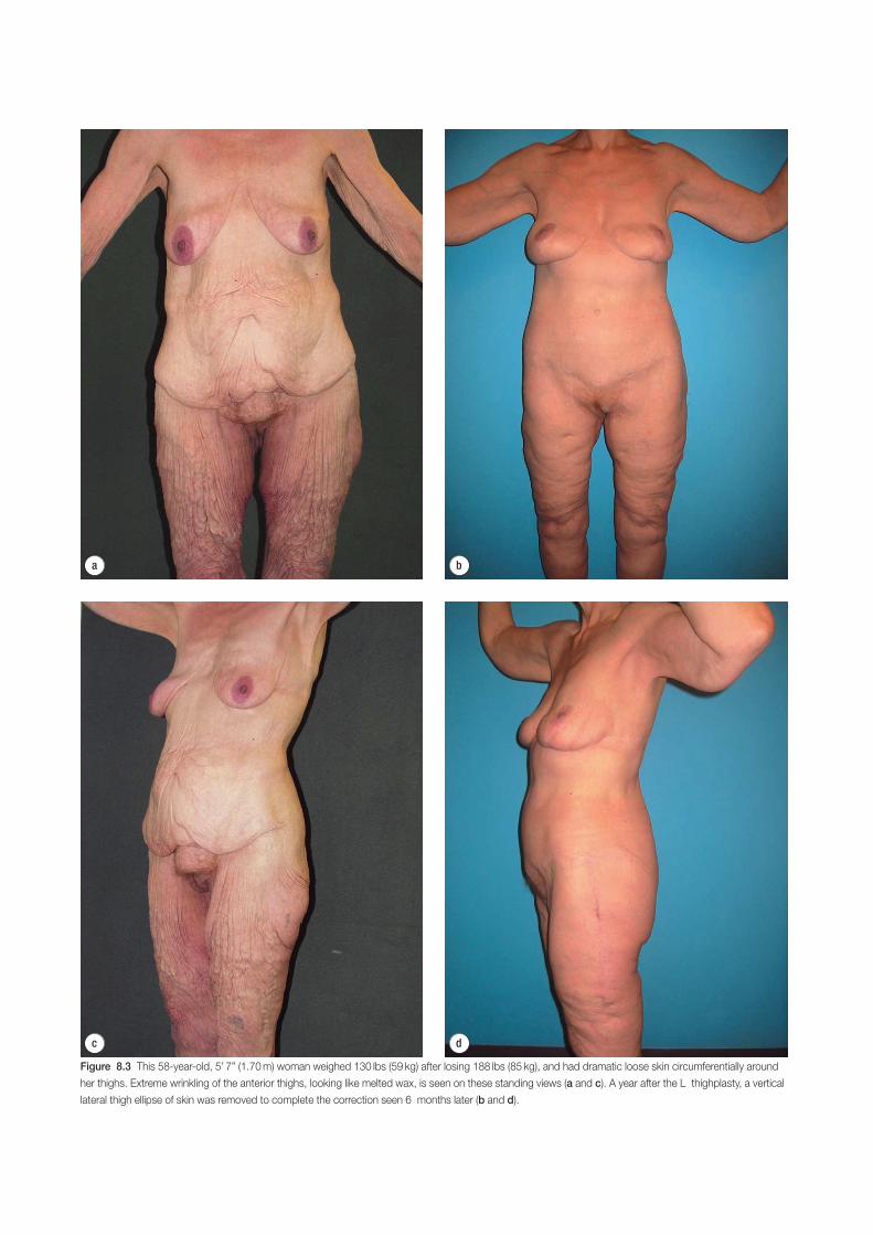
d
a b
c
Figure 8.3 This 58-year-old, 5’ 7” (1.70 m) woman weighed 130 lbs (59 kg) after losing 188 lbs (85 kg), and had dramatic loose skin circumferentially around
her thighs. Extreme wrinkling of the anterior thighs, looking like melted wax, is seen on these standing views (a and c). A year after the L thighplasty, a vertical
lateral thigh ellipse of skin was removed to complete the correction seen 6 months later (b and d).
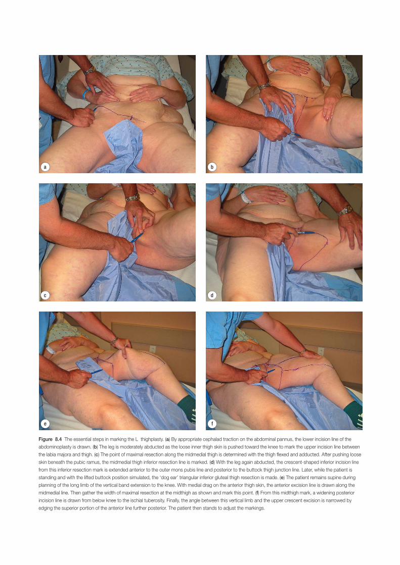
d
e f
a b
c
Figure 8.4 The essential steps in marking the L thighplasty. (a) By appropriate cephalad traction on the abdominal pannus, the lower incision line of the
abdominoplasty is drawn. (b) The leg is moderately abducted as the loose inner thigh skin is pushed toward the knee to mark the upper incision line between
the labia majora and thigh. (c) The point of maximal resection along the midmedial thigh is determined with the thigh flexed and adducted. After pushing loose
skin beneath the pubic ramus, the midmedial thigh inferior resection line is marked. (d) With the leg again abducted, the crescent-shaped inferior incision line
from this inferior resection mark is extended anterior to the outer mons pubis line and posterior to the buttock thigh junction line. Later, while the patient is
standing and with the lifted buttock position simulated, the ‘dog ear’ triangular inferior gluteal thigh resection is made. (e) The patient remains supine during
planning of the long limb of the vertical band extension to the knee. With medial drag on the anterior thigh skin, the anterior excision line is drawn along the
midmedial line. Then gather the width of maximal resection at the midthigh as shown and mark this point. (f) From this midthigh mark, a widening posterior
incision line is drawn from below knee to the ischial tuberosity. Finally, the angle between this vertical limb and the upper crescent excision is narrowed by
edging the superior portion of the anterior line further posterior. The patient then stands to adjust the markings.
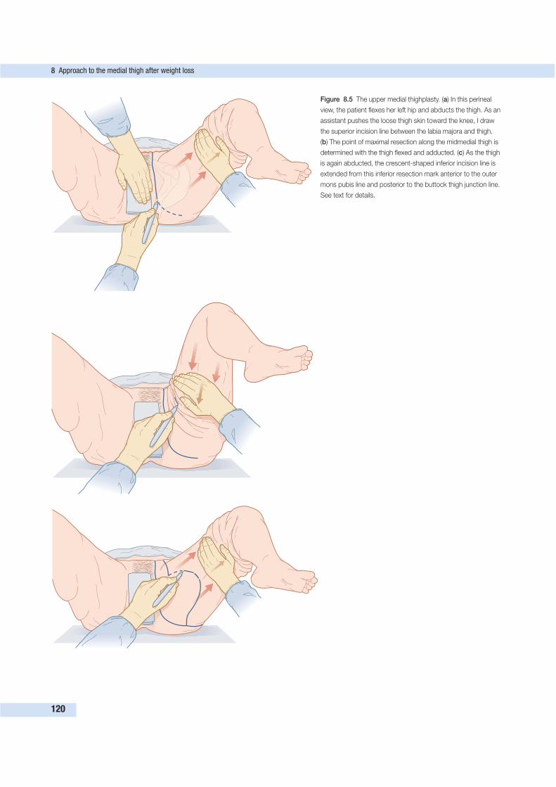
8 Approach to the medial thigh after weight loss
120
Figure 8.5 The upper medial thighplasty. (a) In this perineal
view, the patient flexes her left hip and abducts the thigh. As an
assistant pushes the loose thigh skin toward the knee, I draw
the superior incision line between the labia majora and thigh.
(b) The point of maximal resection along the midmedial thigh is
determined with the thigh flexed and adducted. (c) As the thigh
is again abducted, the crescent-shaped inferior incision line is
extended from this inferior resection mark anterior to the outer
mons pubis line and posterior to the buttock thigh junction line.
See text for details.
resection of paramedian pubic skin is just enough to efface
the mons pubis.
6. While the patient is standing and the lifted buttock
position simulated, the ‘dog ear’ triangular gluteal thigh
resection is marked.
The vertical excision extended medial thighplasty is called
an L thighplasty because the resections and subsequent scar
form the letter ‘L’ from pubis to knees.
1. The short limb of the L plasty (crescent upper thigh
excision) is planned first, with the patient supine and the
thigh flexed and abducted as just described.
2. The long limb of the L (vertical band extension to the
knee) is also planned supine (Figs 8.4 and 8.6). With the
leg on the bed, and superior and medial drag on the
anterior thigh skin, the anterior excision line is drawn
from medial knee up the thigh to the apex of the crescent
excision line.
3. Then gather the width of maximal resection at the
midthigh and mark this point.
4. From this midthigh mark, a widening posterior incision
line is drawn from below knee to the ischial tuberosity.
5. Finally, the angle between this vertical limb and the upper
crescent excision is narrowed by edging the superior
portion of the anterior line further posterior. This change
in position moves the scar slightly posterior, which creates
an L shape.
6. For symmetry, the lines are emphasized and then the
thighs are rubbed together to imprint one on to the other.
7. The accuracy is confirmed by tissue gathering.
8. The patient then stands to adjust the markings as needed
(Fig. 8.7).
SURGICAL TECHNIQUE
The thighplasty begins with the lower body lift. The surgeon
stands to the right side of the prone patient, facing the but-
tocks. Along the suture lines and the anticipated planes of dis-
section, she or he liberally infuses dilute vasoconstrictor and
anesthetic (1 mg of adrenaline [epinephrine] and 20 cc of 1%
lidocaine [Xylocaine] per liter of saline). In three or four
swipes, the inferior posterior incision is made down to mus-
cular fascia with a scalpel from anterior superior iliac spine
(ASIS) across the buttocks, the lumbar spine, and the opposite
buttocks to the opposite ASIS. Electrocautery cutting is avoided
because thermal injury may reduce the suture holding of the
subsequent tightly closed subcutaneous fascia. The buttock
incision stops at the gluteus maximus muscle and continues
laterally to the fascia lata. Scattered fascial adherences from
the fascia lata to the lateral thigh deep dermis are released to
beyond the palpable lateral trochanter.
Ultrasound-assisted lipoplasty of the lateral thighs debulks
overly full subcutaneous tissue. Discontinuous undermining is
provided as needed by forceful thrusts of Lockwood dissec-
tors (Padgett Instruments, Kansas City, Missouri) over the fas-
cia lata to nearly the knee. After mobilizing the lateral thigh
skin, the superior incision line is confirmed. There needs to be
enough mobilization of the lateral thigh so that the skin, not
the underlying fascial extensions, is limiting cephalad advan-
cement. The previously marked superior incision along the
lower back is now incised to lumbodorsal fascia and external
oblique muscles.
The skin and adipose between the superior and inferior
incisions is resected at the desired depth. Usually, most of the
large globular lumbar fat is preserved. If fat flap buttock
augmentation is planned, then only a beltlike band of skin is
removed (Fig. 8.8). The buttock skin is elevated off the upper
two-thirds of the gluteus maximus muscle for a space for the
adipose flap. The retained lower back mobile pad of adipose
can be advanced and sutured inferiorly to augment the but-
tocks (Fig. 8.9). The lower buttock skin flap is then sutured to
the lower back superior incision.
The lower torso midlateral wide resection with tight clo-
sure effaces the saddlebag deformity. In order to close the gap
under the least tension, the leg is abducted on a wide arm
board rotated out about 45°. Large, deeply placed absorbable
sutures secure the lateral thigh deep dermis to the fascia lata
of the thigh. The beltlike excision is closed with very large,
absorbable braided sutures in the subcutaneous fascia, fol-
lowed by an intradermal closure with long-lasting monofila-
ment absorbable sutures.
While assistants close the lower body lift, the surgeon
removes the anticipated infragluteal dog ears of the medial
thighplasties under the buttock folds. In the unusual situation,
when the posterior thigh is very loose, this excision can be as
wide as 8 cm. The infragluteal excision cannot be made until
the buttock lift is completed. The width of the triangular
excision is adjusted inferiorly as needed. One should rely on
the premarked superior incision line, which appears to curve
superiorly. The depth of resection of this posterior dog ear is
superficial to the facial lata, lateral to the ischial tuberosity, to
avoid injury to buttock sensory inferior cluneal nerves and
nutrient vasculature. If there is a vertical band excision and it is
wide, then the posterior limbs are now incised through deep
subcutaneous fascia. The terminal incision is more superficial
to avoid injury to major lymphatics and may fishtail anterior
and inferior to the knee or posterior toward the popliteal
fossa. Medial to the ischial tuberosity, the posterior thigh skin
and fascia lata is anchored to the bony prominence periosteum
with two to three braided sutures. Then the triangular infra-
gluteal wound wedge is closed in two layers of absorbable
sutures. Prior to turning the patient supine, the posterior ver-
tical thigh incision is temporarily approximated with staples.
The patient is wrapped into a surgeon’s gown and turned
supine. Larger patients are rolled over on to a gurney. Then the
gown and patient are dragged back on to the operating room
table. To relieve tension on lower abdominal skin, the patient
is frog-legged. After a second antiseptic preparation, dilute
anesthetic and vasoconstricting fluid is again injected into an-
ticipated incisions and areas for liposuction and undermining.
The abdominoplasty is resumed with the inferior incision from
ASIS across the groins through the mons pubis, and completed
Surgical technique
121
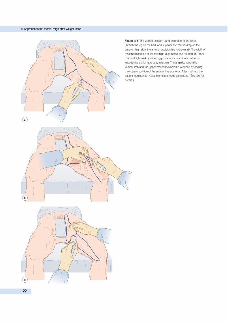
8 Approach to the medial thigh after weight lossc
122
a
b
c
Figure 8.6 The vertical excision band extension to the knee.
(a) With the leg on the bed, and superior and medial drag on the
anterior thigh skin, the anterior excision line is drawn. (b) The width of
maximal resection at the midthigh is gathered and marked. (c) From
this midthigh mark, a widening posterior incision line from below
knee to the ischial tuberosity is drawn. The angle between this
vertical limb and the upper crescent excision is widened by edging
the superior portion of the anterior line posterior. After marking, the
patient then stands. Adjustments are made as needed. (See text for
details.)
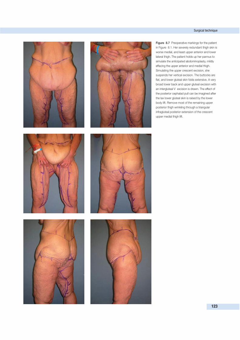
Surgical technique
123
Figure 8.7 Preoperative markings for the patient
in Figure 8.1. Her severely redundant thigh skin is
worse medial, and least upper anterior and lower
lateral thigh. The patient holds up her pannus to
simulate the anticipated abdominoplasty, mildly
effacing the upper anterior and medial thigh.
Simulating the upper crescent excision, she
suspends her vertical excision. The buttocks are
flat, and lower gluteal skin folds extensive. A very
broad lower back and upper gluteal excision with
an intergluteal V excision is drawn. The effect of
the posterior cephalad pull can be imagined after
the lax lower gluteal skin is raised by the lower
body lift. Remove most of the remaining upper
posterior thigh wrinkling through a triangular
infragluteal posterior extension of the crescent
upper medial thigh lift.
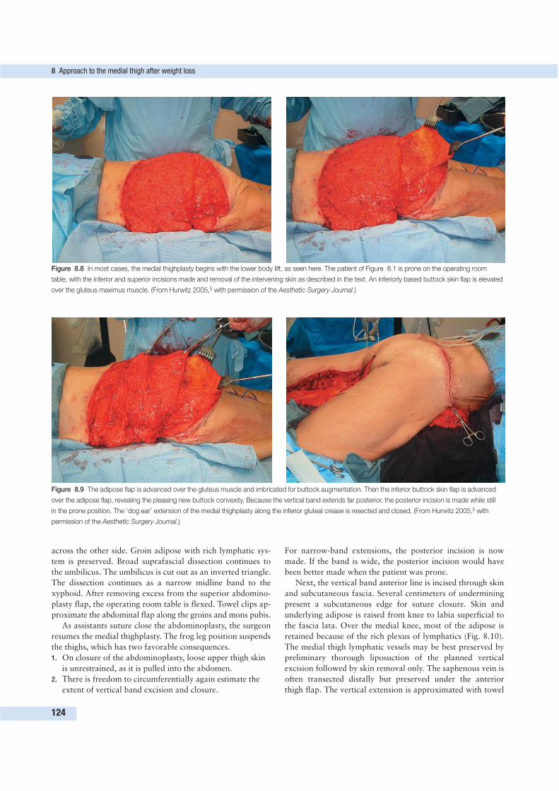
across the other side. Groin adipose with rich lymphatic sys-
tem is preserved. Broad suprafascial dissection continues to
the umbilicus. The umbilicus is cut out as an inverted triangle.
The dissection continues as a narrow midline band to the
xyphoid. After removing excess from the superior abdomino-
plasty flap, the operating room table is flexed. Towel clips ap-
proximate the abdominal flap along the groins and mons pubis.
As assistants suture close the abdominoplasty, the surgeon
resumes the medial thighplasty. The frog leg position suspends
the thighs, which has two favorable consequences.
1. On closure of the abdominoplasty, loose upper thigh skin
is unrestrained, as it is pulled into the abdomen.
2. There is freedom to circumferentially again estimate the
extent of vertical band excision and closure.
For narrow-band extensions, the posterior incision is now
made. If the band is wide, the posterior incision would have
been better made when the patient was prone.
Next, the vertical band anterior line is incised through skin
and subcutaneous fascia. Several centimeters of undermining
present a subcutaneous edge for suture closure. Skin and
underlying adipose is raised from knee to labia superficial to
the fascia lata. Over the medial knee, most of the adipose is
retained because of the rich plexus of lymphatics (Fig. 8.10).
The medial thigh lymphatic vessels may be best preserved by
preliminary thorough liposuction of the planned vertical
excision followed by skin removal only. The saphenous vein is
often transected distally but preserved under the anterior
thigh flap. The vertical extension is approximated with towel
8 Approach to the medial thigh after weight loss
124
Figure 8.8 In most cases, the medial thighplasty begins with the lower body lift, as seen here. The patient of Figure 8.1 is prone on the operating room
table, with the inferior and superior incisions made and removal of the intervening skin as described in the text. An inferiorly based buttock skin flap is elevated
over the gluteus maximus muscle. (From Hurwitz 2005,5 with permission of the Aesthetic Surgery Journal.)
Figure 8.9 The adipose flap is advanced over the gluteus muscle and imbricated for buttock augmentation. Then the inferior buttock skin flap is advanced
over the adipose flap, revealing the pleasing new buttock convexity. Because the vertical band extends far posterior, the posterior incision is made while still
in the prone position. The ‘dog ear’ extension of the medial thighplasty along the inferior gluteal crease is resected and closed. (From Hurwitz 2005,5 with
permission of the Aesthetic Surgery Journal.)
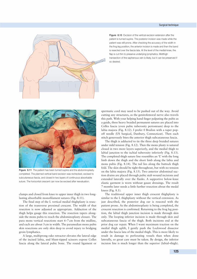
clamps and closed from knee to upper inner thigh in two long-
lasting absorbable monofilament sutures (Fig. 8.11).
The final step of the L vertical medial thighplasty is resec-
tion of the transverse proximal crescent. The width of that
resection is now adjusted as appropriate. Adduction of the
thigh helps gauge this resection. The resection tapers along-
side the mons pubis to reach the abdominoplasty closure. The
para mons vertical resections start 6–7 cm from the midline,
and each are about 3 cm in width. The paramedian mons pubis
skin resections are only skin deep to avoid injury to bridging
groin lymphatics.
A large, multiprong rake retractor elevates the lateral edge
of the incised labia, and blunt-tipped scissors expose Colles
fascia along the lateral pubic bone. The round ligament or
spermatic cord may need to be pushed out of the way. Avoid
cutting any structures, as the genitofemoral nerve also travels
this path. With your helping hand finger palpating the pubis as
a guide, three heavy braided permanent sutures are placed into
Colles fascia (even pubic tuberosity periosteum) deep to the
labia majora (Fig. 8.12). I prefer 0 Brailon with a taper pop-
off needle (US Surgical, Danbury, Connecticut). Then each
stitch generously bites the anterior thigh subcutaneous fascia.
The thigh is adducted to tie the three deep braided sutures
under mild tension (Fig. 8.12). Then the mons plasty is sutured
closed in two more layers superiorly, and the medial thigh to
labial junction to the ischial tuberosity inferiorly (Fig. 8.13).
The completed thigh suture line resembles an ‘L’ with the long
limb down the thigh and the short limb along the labia and
mons pubis (Fig. 8.14). The tail lies along the buttock thigh
fold. The skin should be tight throughout, but with no tension
on the labia majora (Fig. 8.15). Two anterior abdominal suc-
tion drains are placed through pubic stab wound incisions and
extended laterally over the flanks. A supportive below-knee
elastic garment is worn without gauze dressings. The result
7 months later needs a little further resection about the medal
knees (Fig. 8.1).
The traditional upper inner thigh crescent thighplasty is
similar to the L thighplasty without the vertical extension. As
just described, the posterior dog ear is resected with the
patient prone. As the abdominoplasty is being completed, the
crescent resection is confirmed. Returning to the frog leg posi-
tion, the labial thigh junction incision is made through skin
only. The looping inferior incision is made through skin and
subcutaneous fascia of the thigh. Both incisions end at the
prior dog ear repair. When I want maximum traction on the
medial thigh uplift, I gently push the Lockwood dissector
under the fascia lata of the medial thigh. This is more likely to
result in damage to perforating vessels than when done
laterally, so great care must be taken. By design, the inferior
incision line is much longer than the superior (labial–thigh).
Surgical technique
125
Figure 8.10 Excision of the vertical excision extension after the
patient is turned supine. The posterior incision was made while the
patient was still prone. After checking the accuracy of the width in
the frog leg position, the anterior incision is made and then the band
is resected over the fascia lata. At the level of the medial knee, the
flap is cut thin to preserve underlying lymphatics. Midthigh
transection of the saphenous vein is likely, but it can be preserved if
so desired.
Figure 8.11 The patient has been turned supine and the abdominoplasty
completed. The planned vertical band excision was rechecked, excised to
subcutaneous fascia, and closed in two layers of continuous absorbable
suture. The horizontal crescent can now be excised after reevaluation.
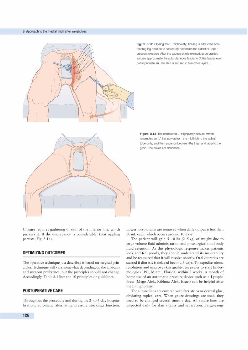
Closure requires gathering of skin of the inferior line, which
puckers it. If the discrepancy is considerable, then rippling
persists (Fig. 8.14).
OPTIMIZING OUTCOMES
The operative technique just described is based on surgical prin-
ciples. Technique will vary somewhat depending on the anatomy
and surgeon preference, but the principles should not change.
Accordingly, Table 8.1 lists the 10 principles or guidelines.
POSTOPERATIVE CARE
Throughout the procedure and during the 2- to 4-day hospita-
lization, automatic alternating pressure stockings function.
Lower torso drains are removed when daily output is less than
50 mL each, which occurs around 10 days.
The patient will gain 5–10 lbs (2–5 kg) of weight due to
large-volume fluid administration and postsurgical total body
fluid retention. As this physiologic response makes patients
look and feel poorly, they should understand its inevitability
and be reassured that it will resolve shortly. Oral diuretics are
started if diuresis is delayed beyond 3 days. To expedite edema
resolution and improve skin quality, we prefer to start Ender-
mologie (LPG, Miami, Florida) within 2 weeks. A month of
home use of an automatic pressure device such as a Lympha
Press (Mego Afek, Kibhutz Afek, Israel) can be helpful after
the L thighplasty.
The suture lines are covered with Steristrips or dermal glue,
obviating topical care. When gauze dressings are used, they
need to be changed several times a day. All suture lines are
inspected daily for skin vitality and separation. Large-gauge
8 Approach to the medial thigh after weight loss
126
Figure 8.12 Closing the L thighplasty. The leg is adducted from
the frog leg position to accurately determine the extent of upper
crescent excision. After the excess skin is excised, large braided
sutures approximate the subcutaneous fascia to Colles fascia, even
pubic periosteum. The skin is sutured in two more layers.
Figure 8.13 The completed L thighplasty closure, which
resembles an ‘L’ that curves from the midthigh to the ischial
tuberosity, and then ascends between the thigh and labia to the
groin. The drains are abdominal.
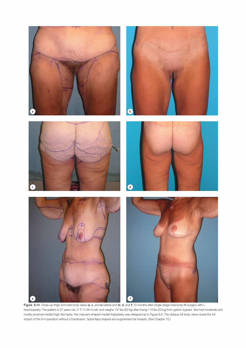
d
e f
a b
c
Figure 8.14 Close-up thigh and total body views (a, c, and e) before and (b, d, and f) 10 months after single-stage total body lift surgery with L
brachioplasty. The patient is 37 years old, 5’ 5” (1.65 m) tall, and weighs 137 lbs (62 kg) after losing 115 lbs (52 kg) from gastric bypass. She had moderate and
mostly proximal medial thigh skin laxity. Her crescent-shaped medial thighplasty was designed as in Figure 8.6. The oblique full body views reveal the full
impact of the 8-h operation without a transfusion. Spiral flaps shaped and augmented her breasts. (See Chapter 10.)
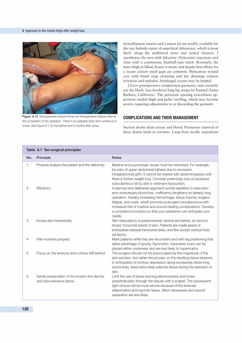
monofilament sutures and a suture kit are readily available for
the rare bedside repair of superficial dehiscence, which is most
likely along the midlateral torso and ischial closures. I
anesthetize the area with lidocaine (Xylocaine) injections and
close with a continuous, baseball-type stitch. Routinely, the
inner thigh to labial closure is moist, and despite best efforts for
a secure closure small gaps are common. Meticulous wound
care with bland soap cleansing and dry dressings reduces
irritation and malodor. Antifungal creams may be helpful.
I favor postoperative compression garments, and currently
use the black, lace-bordered long leg wraps by Inamed (Santa
Barbara, California). The perineum opening exacerbates up-
permost medial thigh and pubic swelling, which may become
severe, requiring adjustments to or discarding the garment.
COMPLICATIONS AND THEIR MANAGEMENT
Suction drains drain serum and blood. Premature removal of
these drains leads to seromas. Large-bore needle aspirations
8 Approach to the medial thigh after weight loss
128
Figure 8.15 Intraoperative closure shows an intraoperative oblique view at
the completion of the operation. There is no palpable laxity from umbilicus to
knees. See Figure 8.1 for the before and 5 months after views.
Table 8.1 Ten surgical principles
No. Principle Notes
1 Properly analyze the patient and the deformity Medical and psychologic issues must be minimized. For example,
be wary of upper abdominal fullness due to excessive
intraabdominal girth. It cannot be treated with abdominoplasty until
there is further weight loss. Consider preliminary loss of excessive
subcutaneous fat by diet or extensive liposuction.
2 Efficiency A planned and deliberate approach avoids repetition in execution
and unnecessary blood loss. Inefficiency lengthens an already long
operation, thereby increasing hemorrhage, tissue trauma, surgeon
fatigue, and costs, which promote prolonged convalescence with
increased risk of medical and wound-healing complications. Develop
a consistent procedure so that your assistants can anticipate your
needs.
3 Excise skin transversely Skin redundancy is predominantly vertical and lateral, so remove
broad, horizontal bands of skin. Patients are made aware of
anticipated residual transverse laxity, and few accept vertical torso
excisions.
4 Plan incisions properly Mark patients while they are recumbent and with leg positioning that
takes advantage of gravity. Symmetric, transverse scars can be
placed within underwear and are less likely to hypertrophy.
5 Focus on the tensions and contour left behind The surgeon should not be preoccupied by the magnitude of the
skin excision, but rather should plan on the resulting tissue tensions.
In anticipation of contour depression along excessively tense long
suture lines, leave extra deep adipose tissue during the resection of
skin.
6 Gentle preservation of the incision line dermis Limit the use of tissue-burning electrocautery and incise
and subcutaneous fascia perpendicularly through the tissues with a scalpel. The subsequent
tight closure will be more secure because of the reduced
inflammation and necrotic tissue. Stitch abscesses and wound
separation are less likely.
Conclusion
129
Table 8.1 (cont’d)
No. Principle Notes
7 Limit liposuction of flaps, and keep it as gentle This means prior generous saline infiltration of lidocaine (Xylocaine)
as possible and adrenaline (epinephrine), and a limited course with ultrasound
probe before vented liposuction. Stop suction on the onset of
bleeding.
8 High-tension, two-layer skin flap closure High-tension, two-layer skin flap closure due to the poor skin
elasticity, expedited by relieving the tension during closure by
preliminary approximation of skin edges with towel clips and most
favorable repositioning of limbs or body.
9 Close wounds as expeditiously as possible over This is to reduce swelling, infection rate, phlebitis, and seroma.
long-dwelling suction catheters; respect larger Preliminary liposuction of the medial vertical band excisions with
lymphatics and use strategic quilting sutures skin only removal pressures lymphatics. A secure two-layer closure
is optimal. Elasticized garments with minimal pressure over the lower
abdomen are comfortable and reassuring.
10 Continuously analyze aesthetic results Systematically compare standard before and after photos and solicit
standardized patient comments. At the University of Pittsburgh, we
have developed a standardized deformity and outcome grading
scale.
are both diagnostic and therapeutic. Local compression with a
sponge and elastic wrap is tried for about a week. If serum
reaccumulates, then aspiration is repeated or preferably a
percutaneous drainage catheter is inserted, sutured in place,
and connected to a suction bulb. It is removed 7–10 days later.
These catheters can initiate serious infections, so meticulous
care is essential. On rare occasions, a drain is reinserted several
times. Once a scarred seroma cavity is formed, compete reso-
lution may require injection of sclerosing agents or surgical
excision with quilting suture closure.
Several weeks after surgery, a firm, deep, slightly tender
mass may be palpable above the medial knee. On aspiration,
this invariably yields straw-colored, watery fluid, which refills
to firmness within a day, suggestive of a lymphocele. Prolonged
closed suction drainage usually resolves the problem. A small
residual mass is left alone, as it tends to resolve by fibrosis.
Delayed distal medial thigh abscess has required incision
and debridement in four limbs over the past 5 years. All healed
secondarily. A recent patient had sepsis from a Streptococcus
viridans abscess of the proximal thigh 1 week after her total
body lift with L thighplasty and extensive Vaser® LipoSelection®.
With the onset of high fever and obtundation, immediate opera-
tive drainage and intravenous antibiotics restored her health.
Inadequate care and excessive activity can lead to trouble-
some thigh swelling. Skin edge necrosis will be followed by
suture line dehiscence. Because of the tightness of the closure
and persistent swelling, a conservative wound care approach
is taken. There may be a long line of necrotic and inflamma-
tory tissue. Thorough debridement is performed. Topical
papain-urea agents such as Accuzyme followed by Panafil are
applied. Be vigilant for undrained areas that may lead to ab-
scesses. Increasing redness and fever require investigation. Once
a granulating bed is cultivated, the wounds tend to contract
and epithelialize within weeks. Attention to meticulous hygiene,
clipping of irritating hairs, and offending sutures are essential.
Descent of the labial thigh scars and distortion of the labia
are recognized long-term complications. With the introduc-
tion of the Colles fascia stitch, I believe that this problem has
become uncommon.4 Nevertheless, overresection of medial
thigh skin cannot be overcome by those sutures. Skin grafts
are the most expedient means to correct the labial deformity,
but they may be rejected as unsightly by the patient. Theo-
retically, tissue expansion, although awkward in this location,
should yield more skin. If there is residual transverse laxity of
thigh skin, then a limited vertical band excision can raise the
scar and take distorting tension off the labia majora.
There is no operative solution to excessively heavy, thick
thighs, as they are prone to abscess infections and pulling
through of sutures. Further weight loss or preliminary lipo-
plasty is indicated. Some thighs appear too heavy but are ac-
tually primarily sheets of sagging skin. Pull the skin superiorly
and palpate the thickness. If it is not too thick, proceed with
thighplasty but plan for an exceptionally broad resection of
skin (Fig. 8.3).
Weight loss patients with the following are not candidates
for this surgery:
• unstable chronic illnesses,
• cardiovascular disease,
• postphlebtic syndrome, and
• lymphedema.
Also, patients with unresolved depression or unrealistic expec-
tations should be avoided.
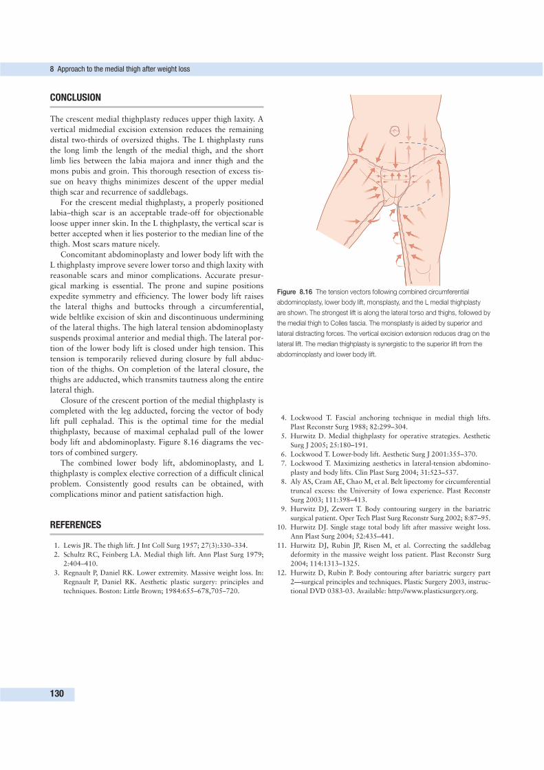
CONCLUSION
The crescent medial thighplasty reduces upper thigh laxity. A
vertical midmedial excision extension reduces the remaining
distal two-thirds of oversized thighs. The L thighplasty runs
the long limb the length of the medial thigh, and the short
limb lies between the labia majora and inner thigh and the
mons pubis and groin. This thorough resection of excess tis-
sue on heavy thighs minimizes descent of the upper medial
thigh scar and recurrence of saddlebags.
For the crescent medial thighplasty, a properly positioned
labia–thigh scar is an acceptable trade-off for objectionable
loose upper inner skin. In the L thighplasty, the vertical scar is
better accepted when it lies posterior to the median line of the
thigh. Most scars mature nicely.
Concomitant abdominoplasty and lower body lift with the
L thighplasty improve severe lower torso and thigh laxity with
reasonable scars and minor complications. Accurate presur-
gical marking is essential. The prone and supine positions
expedite symmetry and efficiency. The lower body lift raises
the lateral thighs and buttocks through a circumferential,
wide beltlike excision of skin and discontinuous undermining
of the lateral thighs. The high lateral tension abdominoplasty
suspends proximal anterior and medial thigh. The lateral por-
tion of the lower body lift is closed under high tension. This
tension is temporarily relieved during closure by full abduc-
tion of the thighs. On completion of the lateral closure, the
thighs are adducted, which transmits tautness along the entire
lateral thigh.
Closure of the crescent portion of the medial thighplasty is
completed with the leg adducted, forcing the vector of body
lift pull cephalad. This is the optimal time for the medial
thighplasty, because of maximal cephalad pull of the lower
body lift and abdominoplasty. Figure 8.16 diagrams the vec-
tors of combined surgery.
The combined lower body lift, abdominoplasty, and L
thighplasty is complex elective correction of a difficult clinical
problem. Consistently good results can be obtained, with
complications minor and patient satisfaction high.
REFERENCES
1. Lewis JR. The thigh lift. J Int Coll Surg 1957; 27(3):330–334.
2. Schultz RC, Feinberg LA. Medial thigh lift. Ann Plast Surg 1979;
2:404–410.
3. Regnault P, Daniel RK. Lower extremity. Massive weight loss. In:
Regnault P, Daniel RK. Aesthetic plastic surgery: principles and
techniques. Boston: Little Brown; 1984:655–678,705–720.
4. Lockwood T. Fascial anchoring technique in medial thigh lifts.
Plast Reconstr Surg 1988; 82:299–304.
5. Hurwitz D. Medial thighplasty for operative strategies. Aesthetic
Surg J 2005; 25:180–191.
6. Lockwood T. Lower-body lift. Aesthetic Surg J 2001:355–370.
7. Lockwood T. Maximizing aesthetics in lateral-tension abdomino-
plasty and body lifts. Clin Plast Surg 2004; 31:523–537.
8. Aly AS, Cram AE, Chao M, et al. Belt lipectomy for circumferential
truncal excess: the University of Iowa experience. Plast Reconstr
Surg 2003; 111:398–413.
9. Hurwitz DJ, Zewert T. Body contouring surgery in the bariatric
surgical patient. Oper Tech Plast Surg Reconstr Surg 2002; 8:87–95.
10. Hurwitz DJ. Single stage total body lift after massive weight loss.
Ann Plast Surg 2004; 52:435–441.
11. Hurwitz DJ, Rubin JP, Risen M, et al. Correcting the saddlebag
deformity in the massive weight loss patient. Plast Reconstr Surg
2004; 114:1313–1325.
12. Hurwitz D, Rubin P. Body contouring after bariatric surgery part
2—surgical principles and techniques. Plastic Surgery 2003, instruc-
tional DVD 0383-03. Available: http://www.plasticsurgery.org.
8 Approach to the medial thigh after weight loss
130
Figure 8.16 The tension vectors following combined circumferential
abdominoplasty, lower body lift, monsplasty, and the L medial thighplasty
are shown. The strongest lift is along the lateral torso and thighs, followed by
the medial thigh to Colles fascia. The monsplasty is aided by superior and
lateral distracting forces. The vertical excision extension reduces drag on the
lateral lift. The median thighplasty is synergistic to the superior lift from the
abdominoplasty and lower body lift.
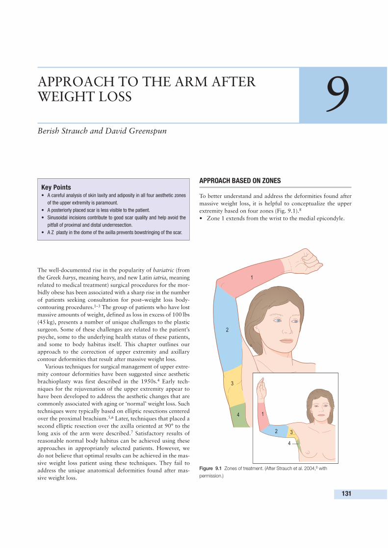
The well-documented rise in the popularity of bariatric (from
the Greek barys, meaning heavy, and new Latin iatria, meaning
related to medical treatment) surgical procedures for the mor-
bidly obese has been associated with a sharp rise in the number
of patients seeking consultation for post–weight loss body-
contouring procedures.1–3 The group of patients who have lost
massive amounts of weight, defined as loss in excess of 100 lbs
(45 kg), presents a number of unique challenges to the plastic
surgeon. Some of these challenges are related to the patient’s
psyche, some to the underlying health status of these patients,
and some to body habitus itself. This chapter outlines our
approach to the correction of upper extremity and axillary
contour deformities that result after massive weight loss.
Various techniques for surgical management of upper extre-
mity contour deformities have been suggested since aesthetic
brachioplasty was first described in the 1950s.4 Early tech-
niques for the rejuvenation of the upper extremity appear to
have been developed to address the aesthetic changes that are
commonly associated with aging or ‘normal’ weight loss. Such
techniques were typically based on elliptic resections centered
over the proximal brachium.5,6 Later, techniques that placed a
second elliptic resection over the axilla oriented at 90° to the
long axis of the arm were described.7 Satisfactory results of
reasonable normal body habitus can be achieved using these
approaches in appropriately selected patients. However, we
do not believe that optimal results can be achieved in the mas-
sive weight loss patient using these techniques. They fail to
address the unique anatomical deformities found after mas-
sive weight loss.
APPROACH BASED ON ZONES
To better understand and address the deformities found after
massive weight loss, it is helpful to conceptualize the upper
extremity based on four zones (Fig. 9.1).8
• Zone 1 extends from the wrist to the medial epicondyle.
131
APPROACH TO THE ARM AFTERWEIGHT LOSS 9Berish Strauch and David Greenspun
Key Points• A careful analysis of skin laxity and adiposity in all four aesthetic zones
of the upper extremity is paramount.
• A posteriorly placed scar is less visible to the patient.
• Sinusoidal incisions contribute to good scar quality and help avoid the
pitfall of proximal and distal underresection.
• A Z plasty in the dome of the axilla prevents bowstringing of the scar.
1
1
2
2
3
3
4
4
Figure 9.1 Zones of treatment. (After Strauch et al. 2004,8 with
permission.)
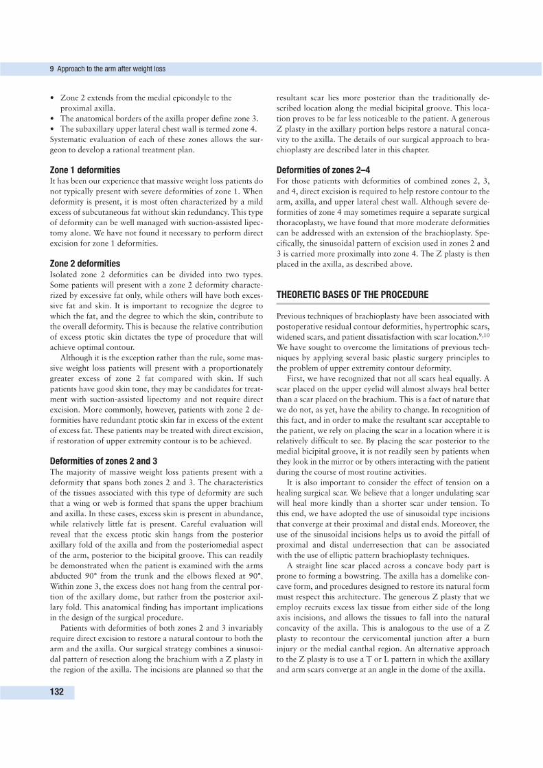
• Zone 2 extends from the medial epicondyle to the
proximal axilla.
• The anatomical borders of the axilla proper define zone 3.
• The subaxillary upper lateral chest wall is termed zone 4.
Systematic evaluation of each of these zones allows the sur-
geon to develop a rational treatment plan.
Zone 1 deformitiesIt has been our experience that massive weight loss patients do
not typically present with severe deformities of zone 1. When
deformity is present, it is most often characterized by a mild
excess of subcutaneous fat without skin redundancy. This type
of deformity can be well managed with suction-assisted lipec-
tomy alone. We have not found it necessary to perform direct
excision for zone 1 deformities.
Zone 2 deformitiesIsolated zone 2 deformities can be divided into two types.
Some patients will present with a zone 2 deformity characte-
rized by excessive fat only, while others will have both exces-
sive fat and skin. It is important to recognize the degree to
which the fat, and the degree to which the skin, contribute to
the overall deformity. This is because the relative contribution
of excess ptotic skin dictates the type of procedure that will
achieve optimal contour.
Although it is the exception rather than the rule, some mas-
sive weight loss patients will present with a proportionately
greater excess of zone 2 fat compared with skin. If such
patients have good skin tone, they may be candidates for treat-
ment with suction-assisted lipectomy and not require direct
excision. More commonly, however, patients with zone 2 de-
formities have redundant ptotic skin far in excess of the extent
of excess fat. These patients may be treated with direct excision,
if restoration of upper extremity contour is to be achieved.
Deformities of zones 2 and 3The majority of massive weight loss patients present with a
deformity that spans both zones 2 and 3. The characteristics
of the tissues associated with this type of deformity are such
that a wing or web is formed that spans the upper brachium
and axilla. In these cases, excess skin is present in abundance,
while relatively little fat is present. Careful evaluation will
reveal that the excess ptotic skin hangs from the posterior
axillary fold of the axilla and from the posteriomedial aspect
of the arm, posterior to the bicipital groove. This can readily
be demonstrated when the patient is examined with the arms
abducted 90° from the trunk and the elbows flexed at 90°.
Within zone 3, the excess does not hang from the central por-
tion of the axillary dome, but rather from the posterior axil-
lary fold. This anatomical finding has important implications
in the design of the surgical procedure.
Patients with deformities of both zones 2 and 3 invariably
require direct excision to restore a natural contour to both the
arm and the axilla. Our surgical strategy combines a sinusoi-
dal pattern of resection along the brachium with a Z plasty in
the region of the axilla. The incisions are planned so that the
resultant scar lies more posterior than the traditionally de-
scribed location along the medial bicipital groove. This loca-
tion proves to be far less noticeable to the patient. A generous
Z plasty in the axillary portion helps restore a natural conca-
vity to the axilla. The details of our surgical approach to bra-
chioplasty are described later in this chapter.
Deformities of zones 2–4For those patients with deformities of combined zones 2, 3,
and 4, direct excision is required to help restore contour to the
arm, axilla, and upper lateral chest wall. Although severe de-
formities of zone 4 may sometimes require a separate surgical
thoracoplasty, we have found that more moderate deformities
can be addressed with an extension of the brachioplasty. Spe-
cifically, the sinusoidal pattern of excision used in zones 2 and
3 is carried more proximally into zone 4. The Z plasty is then
placed in the axilla, as described above.
THEORETIC BASES OF THE PROCEDURE
Previous techniques of brachioplasty have been associated with
postoperative residual contour deformities, hypertrophic scars,
widened scars, and patient dissatisfaction with scar location.9,10
We have sought to overcome the limitations of previous tech-
niques by applying several basic plastic surgery principles to
the problem of upper extremity contour deformity.
First, we have recognized that not all scars heal equally. A
scar placed on the upper eyelid will almost always heal better
than a scar placed on the brachium. This is a fact of nature that
we do not, as yet, have the ability to change. In recognition of
this fact, and in order to make the resultant scar acceptable to
the patient, we rely on placing the scar in a location where it is
relatively difficult to see. By placing the scar posterior to the
medial bicipital groove, it is not readily seen by patients when
they look in the mirror or by others interacting with the patient
during the course of most routine activities.
It is also important to consider the effect of tension on a
healing surgical scar. We believe that a longer undulating scar
will heal more kindly than a shorter scar under tension. To
this end, we have adopted the use of sinusoidal type incisions
that converge at their proximal and distal ends. Moreover, the
use of the sinusoidal incisions helps us to avoid the pitfall of
proximal and distal underresection that can be associated
with the use of elliptic pattern brachioplasty techniques.
A straight line scar placed across a concave body part is
prone to forming a bowstring. The axilla has a domelike con-
cave form, and procedures designed to restore its natural form
must respect this architecture. The generous Z plasty that we
employ recruits excess lax tissue from either side of the long
axis incisions, and allows the tissues to fall into the natural
concavity of the axilla. This is analogous to the use of a Z
plasty to recontour the cervicomental junction after a burn
injury or the medial canthal region. An alternative approach
to the Z plasty is to use a T or L pattern in which the axillary
and arm scars converge at an angle in the dome of the axilla.
9 Approach to the arm after weight loss
132
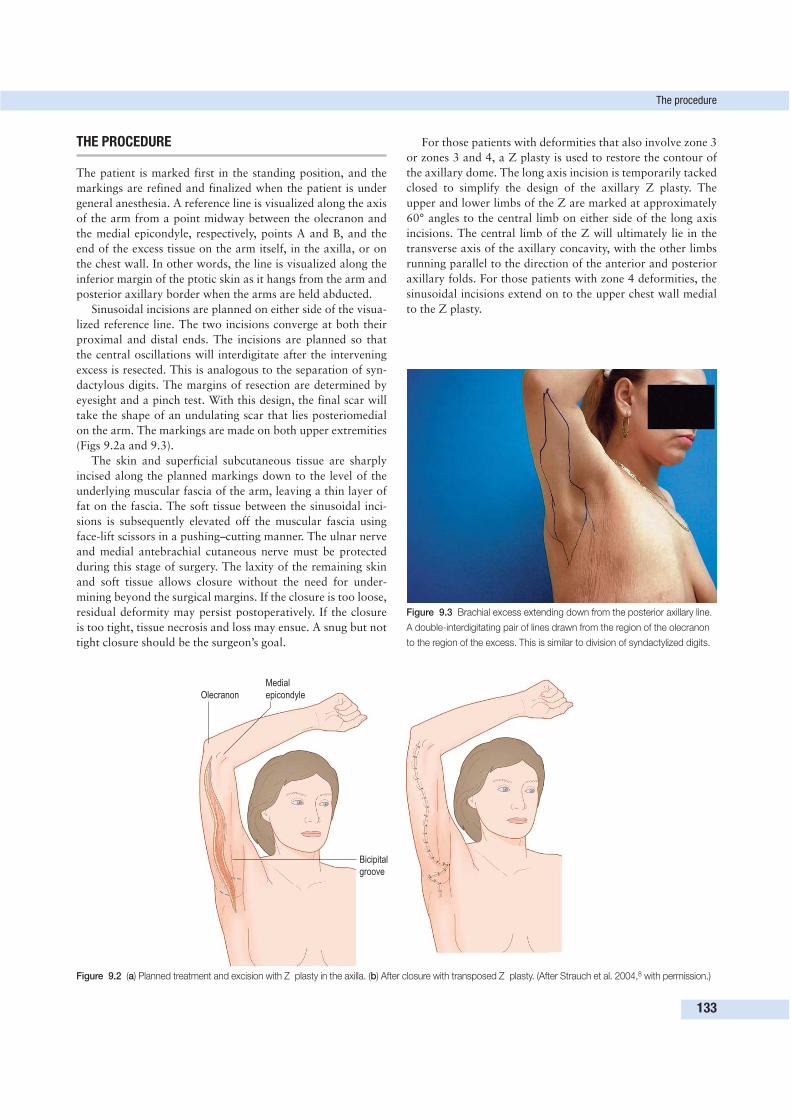
THE PROCEDURE
The patient is marked first in the standing position, and the
markings are refined and finalized when the patient is under
general anesthesia. A reference line is visualized along the axis
of the arm from a point midway between the olecranon and
the medial epicondyle, respectively, points A and B, and the
end of the excess tissue on the arm itself, in the axilla, or on
the chest wall. In other words, the line is visualized along the
inferior margin of the ptotic skin as it hangs from the arm and
posterior axillary border when the arms are held abducted.
Sinusoidal incisions are planned on either side of the visua-
lized reference line. The two incisions converge at both their
proximal and distal ends. The incisions are planned so that
the central oscillations will interdigitate after the intervening
excess is resected. This is analogous to the separation of syn-
dactylous digits. The margins of resection are determined by
eyesight and a pinch test. With this design, the final scar will
take the shape of an undulating scar that lies posteriomedial
on the arm. The markings are made on both upper extremities
(Figs 9.2a and 9.3).
The skin and superficial subcutaneous tissue are sharply
incised along the planned markings down to the level of the
underlying muscular fascia of the arm, leaving a thin layer of
fat on the fascia. The soft tissue between the sinusoidal inci-
sions is subsequently elevated off the muscular fascia using
face-lift scissors in a pushing–cutting manner. The ulnar nerve
and medial antebrachial cutaneous nerve must be protected
during this stage of surgery. The laxity of the remaining skin
and soft tissue allows closure without the need for under-
mining beyond the surgical margins. If the closure is too loose,
residual deformity may persist postoperatively. If the closure
is too tight, tissue necrosis and loss may ensue. A snug but not
tight closure should be the surgeon’s goal.
For those patients with deformities that also involve zone 3
or zones 3 and 4, a Z plasty is used to restore the contour of
the axillary dome. The long axis incision is temporarily tacked
closed to simplify the design of the axillary Z plasty. The
upper and lower limbs of the Z are marked at approximately
60° angles to the central limb on either side of the long axis
incisions. The central limb of the Z will ultimately lie in the
transverse axis of the axillary concavity, with the other limbs
running parallel to the direction of the anterior and posterior
axillary folds. For those patients with zone 4 deformities, the
sinusoidal incisions extend on to the upper chest wall medial
to the Z plasty.
The procedure
133
Medial
epicondyleOlecranon
Bicipital
groove
Figure 9.2 (a) Planned treatment and excision with Z plasty in the axilla. (b) After closure with transposed Z plasty. (After Strauch et al. 2004,8 with permission.)
Figure 9.3 Brachial excess extending down from the posterior axillary line.
A double-interdigitating pair of lines drawn from the region of the olecranon
to the region of the excess. This is similar to division of syndactylized digits.
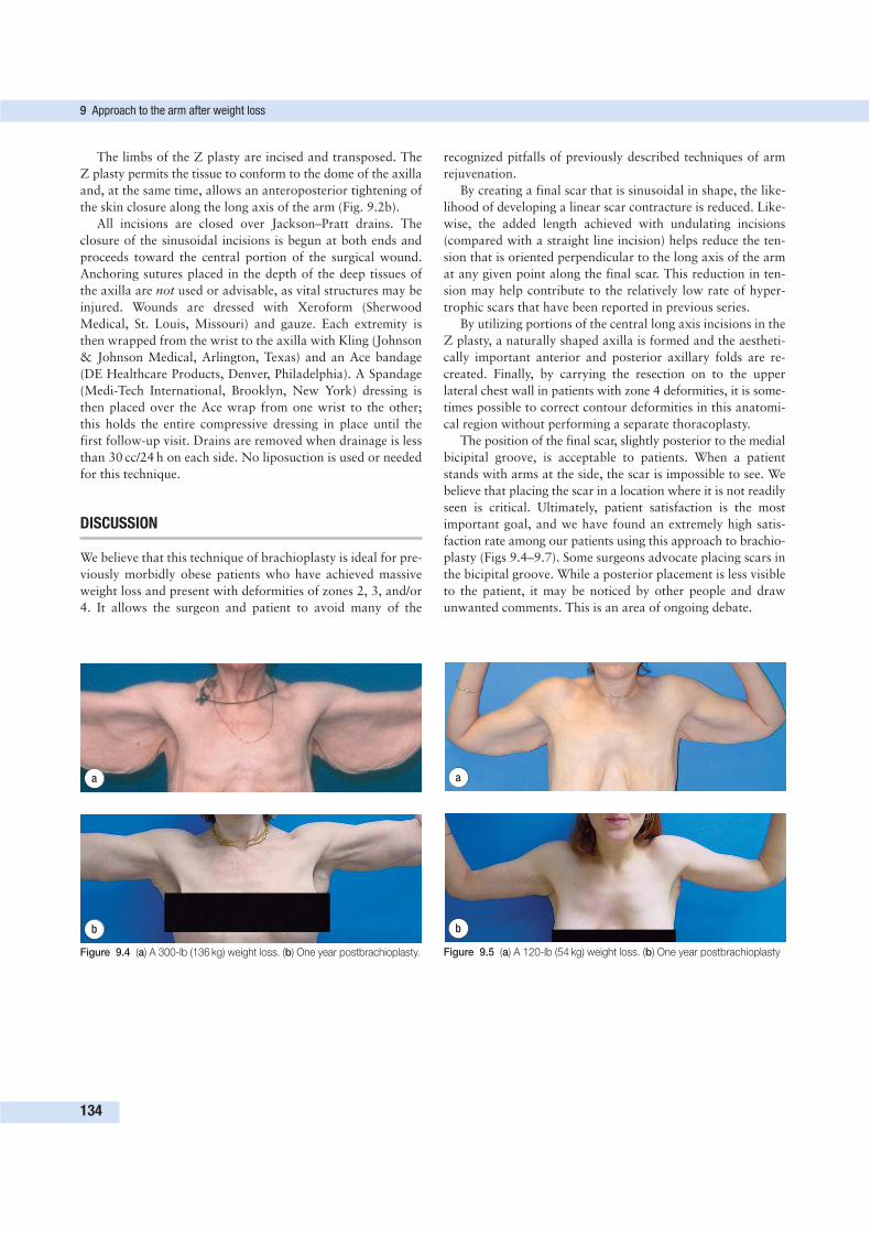
The limbs of the Z plasty are incised and transposed. The
Z plasty permits the tissue to conform to the dome of the axilla
and, at the same time, allows an anteroposterior tightening of
the skin closure along the long axis of the arm (Fig. 9.2b).
All incisions are closed over Jackson–Pratt drains. The
closure of the sinusoidal incisions is begun at both ends and
proceeds toward the central portion of the surgical wound.
Anchoring sutures placed in the depth of the deep tissues of
the axilla are not used or advisable, as vital structures may be
injured. Wounds are dressed with Xeroform (Sherwood
Medical, St. Louis, Missouri) and gauze. Each extremity is
then wrapped from the wrist to the axilla with Kling (Johnson
& Johnson Medical, Arlington, Texas) and an Ace bandage
(DE Healthcare Products, Denver, Philadelphia). A Spandage
(Medi-Tech International, Brooklyn, New York) dressing is
then placed over the Ace wrap from one wrist to the other;
this holds the entire compressive dressing in place until the
first follow-up visit. Drains are removed when drainage is less
than 30 cc/24 h on each side. No liposuction is used or needed
for this technique.
DISCUSSION
We believe that this technique of brachioplasty is ideal for pre-
viously morbidly obese patients who have achieved massive
weight loss and present with deformities of zones 2, 3, and/or
4. It allows the surgeon and patient to avoid many of the
recognized pitfalls of previously described techniques of arm
rejuvenation.
By creating a final scar that is sinusoidal in shape, the like-
lihood of developing a linear scar contracture is reduced. Like-
wise, the added length achieved with undulating incisions
(compared with a straight line incision) helps reduce the ten-
sion that is oriented perpendicular to the long axis of the arm
at any given point along the final scar. This reduction in ten-
sion may help contribute to the relatively low rate of hyper-
trophic scars that have been reported in previous series.
By utilizing portions of the central long axis incisions in the
Z plasty, a naturally shaped axilla is formed and the aestheti-
cally important anterior and posterior axillary folds are re-
created. Finally, by carrying the resection on to the upper
lateral chest wall in patients with zone 4 deformities, it is some-
times possible to correct contour deformities in this anatomi-
cal region without performing a separate thoracoplasty.
The position of the final scar, slightly posterior to the medial
bicipital groove, is acceptable to patients. When a patient
stands with arms at the side, the scar is impossible to see. We
believe that placing the scar in a location where it is not readily
seen is critical. Ultimately, patient satisfaction is the most
important goal, and we have found an extremely high satis-
faction rate among our patients using this approach to brachio-
plasty (Figs 9.4–9.7). Some surgeons advocate placing scars in
the bicipital groove. While a posterior placement is less visible
to the patient, it may be noticed by other people and draw
unwanted comments. This is an area of ongoing debate.
9 Approach to the arm after weight loss
134
a
b
Figure 9.4 (a) A 300-lb (136 kg) weight loss. (b) One year postbrachioplasty.
a
b
Figure 9.5 (a) A 120-lb (54 kg) weight loss. (b) One year postbrachioplasty
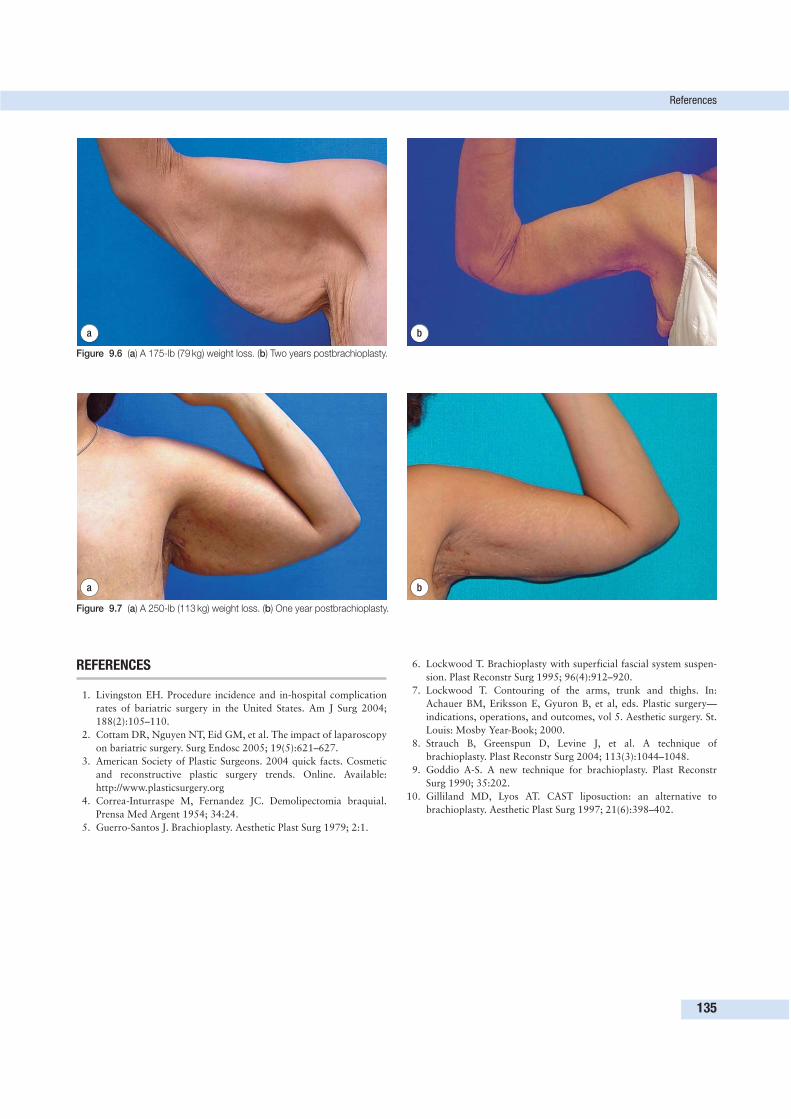
REFERENCES
1. Livingston EH. Procedure incidence and in-hospital complication
rates of bariatric surgery in the United States. Am J Surg 2004;
188(2):105–110.
2. Cottam DR, Nguyen NT, Eid GM, et al. The impact of laparoscopy
on bariatric surgery. Surg Endosc 2005; 19(5):621–627.
3. American Society of Plastic Surgeons. 2004 quick facts. Cosmetic
and reconstructive plastic surgery trends. Online. Available:
http://www.plasticsurgery.org
4. Correa-Inturraspe M, Fernandez JC. Demolipectomia braquial.
Prensa Med Argent 1954; 34:24.
5. Guerro-Santos J. Brachioplasty. Aesthetic Plast Surg 1979; 2:1.
6. Lockwood T. Brachioplasty with superficial fascial system suspen-
sion. Plast Reconstr Surg 1995; 96(4):912–920.
7. Lockwood T. Contouring of the arms, trunk and thighs. In:
Achauer BM, Eriksson E, Gyuron B, et al, eds. Plastic surgery—
indications, operations, and outcomes, vol 5. Aesthetic surgery. St.
Louis: Mosby Year-Book; 2000.
8. Strauch B, Greenspun D, Levine J, et al. A technique of
brachioplasty. Plast Reconstr Surg 2004; 113(3):1044–1048.
9. Goddio A-S. A new technique for brachioplasty. Plast Reconstr
Surg 1990; 35:202.
10. Gilliland MD, Lyos AT. CAST liposuction: an alternative to
brachioplasty. Aesthetic Plast Surg 1997; 21(6):398–402.
References
135
a b
Figure 9.6 (a) A 175-lb (79 kg) weight loss. (b) Two years postbrachioplasty.
a b
Figure 9.7 (a) A 250-lb (113 kg) weight loss. (b) One year postbrachioplasty.
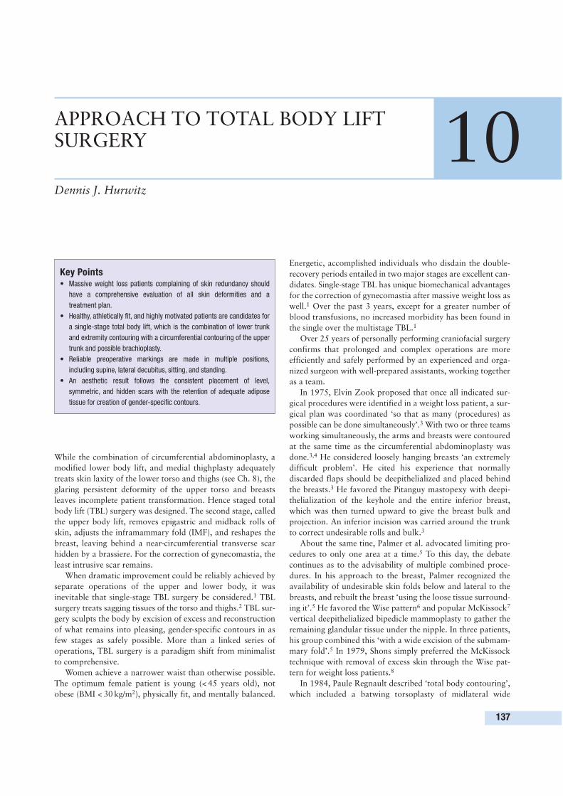
While the combination of circumferential abdominoplasty, a
modified lower body lift, and medial thighplasty adequately
treats skin laxity of the lower torso and thighs (see Ch. 8), the
glaring persistent deformity of the upper torso and breasts
leaves incomplete patient transformation. Hence staged total
body lift (TBL) surgery was designed. The second stage, called
the upper body lift, removes epigastric and midback rolls of
skin, adjusts the inframammary fold (IMF), and reshapes the
breast, leaving behind a near-circumferential transverse scar
hidden by a brassiere. For the correction of gynecomastia, the
least intrusive scar remains.
When dramatic improvement could be reliably achieved by
separate operations of the upper and lower body, it was
inevitable that single-stage TBL surgery be considered.1 TBL
surgery treats sagging tissues of the torso and thighs.2 TBL sur-
gery sculpts the body by excision of excess and reconstruction
of what remains into pleasing, gender-specific contours in as
few stages as safely possible. More than a linked series of
operations, TBL surgery is a paradigm shift from minimalist
to comprehensive.
Women achieve a narrower waist than otherwise possible.
The optimum female patient is young (< 45 years old), not
obese (BMI < 30 kg/m2), physically fit, and mentally balanced.
Energetic, accomplished individuals who disdain the double-
recovery periods entailed in two major stages are excellent can-
didates. Single-stage TBL has unique biomechanical advantages
for the correction of gynecomastia after massive weight loss as
well.1 Over the past 3 years, except for a greater number of
blood transfusions, no increased morbidity has been found in
the single over the multistage TBL.1
Over 25 years of personally performing craniofacial surgery
confirms that prolonged and complex operations are more
efficiently and safely performed by an experienced and orga-
nized surgeon with well-prepared assistants, working together
as a team.
In 1975, Elvin Zook proposed that once all indicated sur-
gical procedures were identified in a weight loss patient, a sur-
gical plan was coordinated ‘so that as many (procedures) as
possible can be done simultaneously’.3 With two or three teams
working simultaneously, the arms and breasts were contoured
at the same time as the circumferential abdominoplasty was
done.3,4 He considered loosely hanging breasts ‘an extremely
difficult problem’. He cited his experience that normally
discarded flaps should be deepithelialized and placed behind
the breasts.3 He favored the Pitanguy mastopexy with deepi-
thelialization of the keyhole and the entire inferior breast,
which was then turned upward to give the breast bulk and
projection. An inferior incision was carried around the trunk
to correct undesirable rolls and bulk.3
About the same tine, Palmer et al. advocated limiting pro-
cedures to only one area at a time.5 To this day, the debate
continues as to the advisability of multiple combined proce-
dures. In his approach to the breast, Palmer recognized the
availability of undesirable skin folds below and lateral to the
breasts, and rebuilt the breast ‘using the loose tissue surround-
ing it’.5 He favored the Wise pattern6 and popular McKissock7
vertical deepithelialized bipedicle mammoplasty to gather the
remaining glandular tissue under the nipple. In three patients,
his group combined this ‘with a wide excision of the submam-
mary fold’.5 In 1979, Shons simply preferred the McKissock
technique with removal of excess skin through the Wise pat-
tern for weight loss patients.8
In 1984, Paule Regnault described ‘total body contouring’,
which included a batwing torsoplasty of midlateral wide
137
APPROACH TO TOTAL BODY LIFTSURGERY 10Dennis J. Hurwitz
Key Points• Massive weight loss patients complaining of skin redundancy should
have a comprehensive evaluation of all skin deformities and a
treatment plan.
• Healthy, athletically fit, and highly motivated patients are candidates for
a single-stage total body lift, which is the combination of lower trunk
and extremity contouring with a circumferential contouring of the upper
trunk and possible brachioplasty.
• Reliable preoperative markings are made in multiple positions,
including supine, lateral decubitus, sitting, and standing.
• An aesthetic result follows the consistent placement of level,
symmetric, and hidden scars with the retention of adequate adipose
tissue for creation of gender-specific contours.
excisions of skin from the upper arms to the hips.9 Fred
Grazer described secondary correction of upper abdominal
skin laxity by reverse abdominoplasty along the IMFs.10
Zienowicz has championed using nearby excess tissue for cos-
metic breast enlargement by augmentation by reverse abdomi-
noplasty.11 The reverse abdominoplasty crosses the sternum
and is suspended by deepithelialized dermal tabs sutured to
chest fascia.12
THE TOTAL BODY LIFT
Fundamental to my TBL is Lockwood’s elucidation of the su-
perficial fascial system and securing this subcutaneous multi-
layer fascia for high-tension skin closure.13 For tightening the
loose IMF and improved breast projection, he fixes the IMF at
‘the appropriate elevated position by non-absorbable sutures
from the superficial fascial system of the inferior skin wound
edge to the underlying muscular fascia’.13
Most massive weight loss patients have bizarre midtorso rolls
of excess skin, flat drooping breasts, and oversized axillae that
lead into batwinged arms.14 There are four intertwined com-
ponents to an upper body lift:
1. reverse abdominoplasty,
2. positioning of a secure IMF,
3. removal of midtorso excess skin, and
4. reshaping and augmenting the breasts.
The upper body lift is optimally combined with the L bra-
chioplasty to reduce lateral chest and oversized axilla, and raise
the ptotic posterior axillary fold (described below).15
In the following sections, the aim of each component of an
upper body lift is elaborated.
Reverse abdominoplastyNumber 1, and fundamental, is the reverse abdominoplasty,
which removes residual excess skin of the upper abdomen.
When associated with a well-defined midtorso transverse roll,
standard abdominoplasty fails to efface loose epigastric skin.
Positioning of a secure IMFComponent no. 2 is upward repositioning and securing the
descended IMFs. The new IMF repositioning and the reverse
abdominoplasty are integral. A properly located and secure IMF
is essential to success. The reverse abdominoplasty remains
tight, and the breast is better situated and supported. In the
male patient, the goal is opposite. The IMF is obliterated. The
tightened upper abdomen is suspended by the upper chest
boomerang pattern excision and pulled down by the abdo-
minoplasty.1,2
Removal of midtorso excess skinComponent no. 3 is removal of the midtorso back skin rolls,
which is essentially a posterior continuation of the reverse
abdominoplasty. A lower body lift does not correct prominent
midback rolls unless the excision level is raised unacceptably
cephalad.
Reshaping and augmenting the breastsComponent no. 4 is reshaping the breasts. If the breasts have
adequate or excess volume, they are reshaped or reduced using
a Wise pattern and pedicle of choice. If the breasts are small
and misshapen, they may be reconstructed with implants and
mastopexy. Unfortunately, the reshaped breasts rarely conform
well to the implants. Over time, the larger implants sag and
ripple. These atrophied breasts are better rebuilt with a Wise
pattern mastopexy and a deepithelialized spiral flap.
In essence, excess skin and fat of the epigastrium and
midtorso back rolls is deepithelialized in continuity with the
central breast mount. The epigastric flap is flipped on to the
inferior breast, and the lateral extension is twisted around the
breast mound over the pectoralis major muscle. Created from
torso discard, the spiral flaps are mobile enough to permit
artistic creativity in shaping and augmentation. The breasts are
not only enlarged and well shaped, but are also soft and shift
naturally with change in body position. The constricted inferior
breast is filled and supported with redundant deepithelialized
epigastric tissue. Tapering of the lateral breast along the ante-
rior axillary line into the axilla is possible for the first time.
In men, the excess midtorso tissue is excised transversely
except at the nipple areolar complex (NAC). Here, two oblique
ellipses rise to meet over the descended NAC. A continuous
horizontal scar is avoided with accurate repositioning of the
NAC, removal of gynecomastia, smoothing out lower chest
and upper abdomen, and obliterating the IMF by ultrasound-
assisted lipoplasty (UAL).
PREOPERATIVE PREPARATION
Body contouring can start approximately 1 year after bariatric
surgery if weight loss has stabilized for 4 months. Rapid weight
loss of about 70% of excess weight is completed by 1 year
after a Roux-en-Y bypass. This is regularly followed by a 20%
weight gain over the next 3 years. Skin quality will not im-
prove by waiting longer, although patients should be warned
that body contouring followed by further weight loss may
result in undesirable skin sagging.
A compulsive review of recognized comorbidities of obesity
and their change after bariatric surgery may reveal unaccept-
able, inadequately or overly treated chronic medical conditions.
• Smoking and narcotic drug dependence are contraindications.
• Depression is ubiquitous in the obese and will be reduced
in 50% of the weight loss patients. Candidates with
persistent, disabling depression or personality disorders
should be rejected.
• Albumin levels should be checked in all candidates.
Protein deficiency should be suspected with selected
dietary limitations, a wide range of food allergies, and
recurrent vomiting. Hypoproteinemia leads to delayed
healing and chronic edema.
• Inadequate vitamin K absorption may follow intestinal
bypass, and supplemental treatment may improve blood
coagulation.
10 Approach to total body lift surgery
138
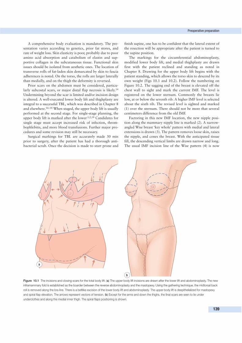
A comprehensive body evaluation is mandatory. The pre-
sentation varies according to genetics, prior fat stores, and
rate of weight loss. Skin elasticity is poor, probably due to poor
amino acid absorption and catabolism of elastin and sup-
portive collagen in the subcutaneous tissue. Functional skin
issues should be isolated from aesthetic ones. The location of
transverse rolls of fat-laden skin demarcated by skin to fascia
adherences is noted. On the torso, the rolls are larger laterally
than medially, and on the thigh the deformity is reversed.
Prior scars on the abdomen must be considered, particu-
larly subcostal scars, or major distal flap necrosis is likely.14
Undermining beyond the scar is limited and/or incision design
is altered. A well-executed lower body lift and thighplasty are
integral to a successful TBL, which was described in Chapter 8
and elsewhere.16–18 When staged, the upper body lift is usually
performed at the second stage. For single-stage planning, the
upper body lift is marked after the lower.1,2,18 Candidates for
single stage must accept increased risk of infection, throm-
bophlebitis, and more blood transfusions. Further major pro-
cedures and some revision may still be necessary.
Surgical markings for TBL are accurately made 30 min
prior to surgery, after the patient has had a thorough anti-
bacterial scrub. Once the decision is made to start prone and
finish supine, one has to be confident that the lateral extent of
the resection will be appropriate after the patient is turned to
the supine position.
The markings for the circumferential abdominoplasty,
modified lower body lift, and medial thighplasty are drawn
first with the patient reclined and standing as noted in
Chapter 8. Drawing for the upper body lift begins with the
patient standing, which allows the torso skin to descend by its
own weight (Figs 10.1 and 10.2). Follow the numbering on
Figure 10.2. The sagging end of the breast is elevated off the
chest wall to sight and mark the current IMF. The level is
registered on the lower sternum. Commonly the breasts lie
low, at or below the seventh rib. A higher IMF level is selected
about the sixth rib. The revised level is sighted and marked
(1) over the sternum. There should not be more that several
centimeters difference from the old IMF.
Factoring in this new IMF location, the new nipple posi-
tion along the mammary nipple line is marked (2). A narrow-
angled Wise breast ‘key whole’ pattern with medial and lateral
extensions is drawn (3). The pattern removes loose skin, raises
the nipple, and cones the breast. With the anticipated tissue
fill, the descending vertical limbs are drawn narrow and long.
The usual IMF incision line of the Wise pattern (4) is now
Preoperative preparation
139
a
b
Figure 10.1 The incisions and closing scars for the total body lift. (a) The upper body lift incisions are drawn after the lower lift and abdominoplasty. The new
inframammary fold is established as the boarder between the reverse abdominoplasty and the mastopexy. Using the gathering technique, the midtorsal back
roll is removed along the bra line. There is a beltlike excision of the lower body lift and abdominoplasty. The upper body lift is deepithelialized for mastopexy
and spiral flap elevation. The arrows represent vectors of tension. (b) Except for the arms and down the thighs, the final scars are seen to lie under
underclothes and along the medial inner thigh. The spiral flaps positioning is shown.
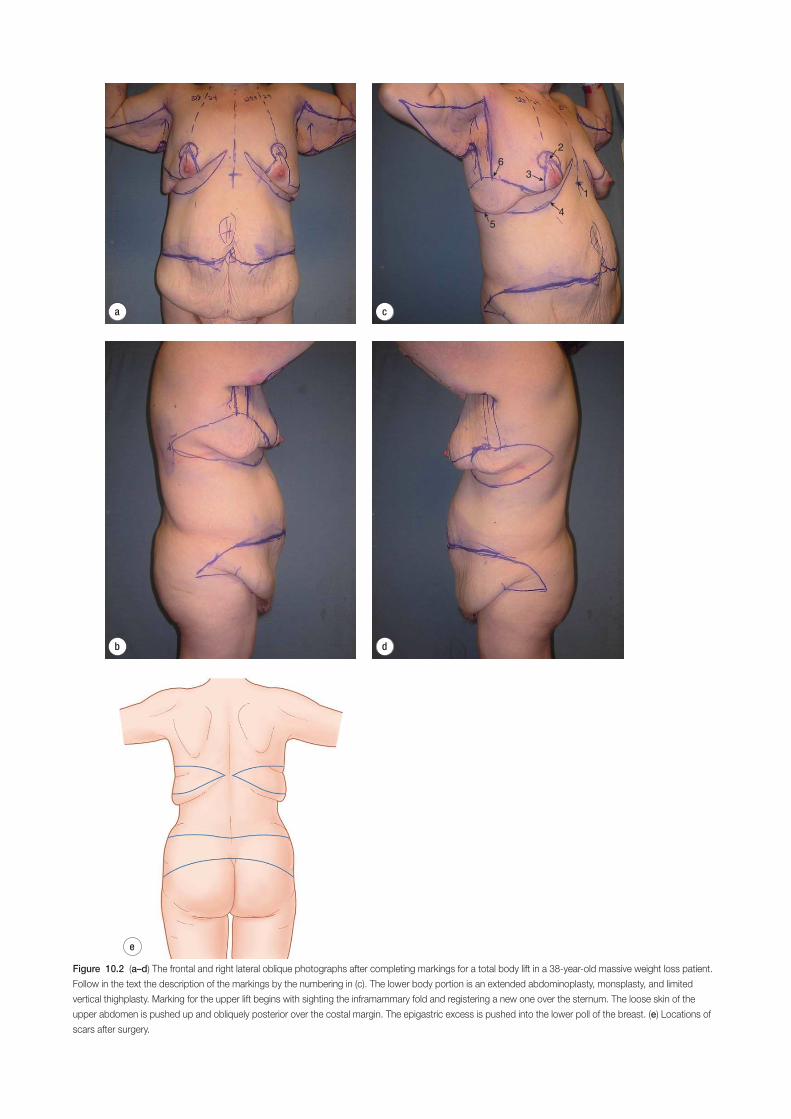
6
5
4
1
2
3
d
e
a
b
c
Figure 10.2 (a–d) The frontal and right lateral oblique photographs after completing markings for a total body lift in a 38-year-old massive weight loss patient.
Follow in the text the description of the markings by the numbering in (c). The lower body portion is an extended abdominoplasty, monsplasty, and limited
vertical thighplasty. Marking for the upper lift begins with sighting the inframammary fold and registering a new one over the sternum. The loose skin of the
upper abdomen is pushed up and obliquely posterior over the costal margin. The epigastric excess is pushed into the lower poll of the breast. (e) Locations of
scars after surgery.
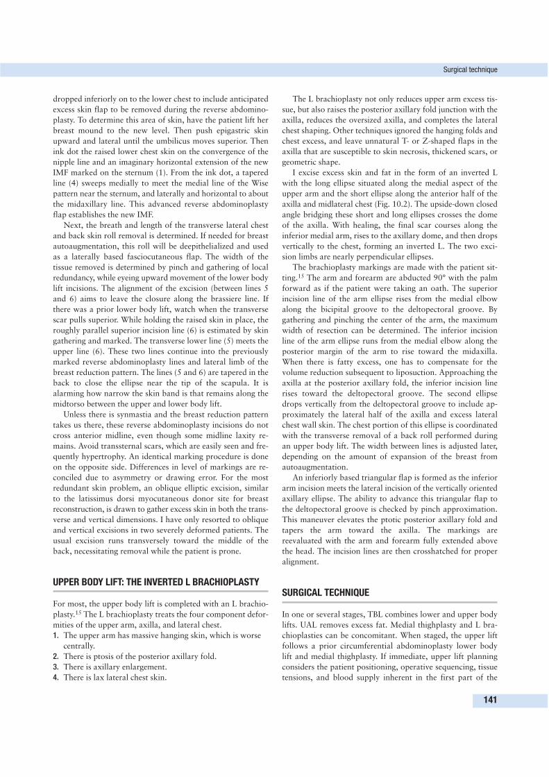
dropped inferiorly on to the lower chest to include anticipated
excess skin flap to be removed during the reverse abdomino-
plasty. To determine this area of skin, have the patient lift her
breast mound to the new level. Then push epigastric skin
upward and lateral until the umbilicus moves superior. Then
ink dot the raised lower chest skin on the convergence of the
nipple line and an imaginary horizontal extension of the new
IMF marked on the sternum (1). From the ink dot, a tapered
line (4) sweeps medially to meet the medial line of the Wise
pattern near the sternum, and laterally and horizontal to about
the midaxillary line. This advanced reverse abdominoplasty
flap establishes the new IMF.
Next, the breath and length of the transverse lateral chest
and back skin roll removal is determined. If needed for breast
autoaugmentation, this roll will be deepithelialized and used
as a laterally based fasciocutaneous flap. The width of the
tissue removed is determined by pinch and gathering of local
redundancy, while eyeing upward movement of the lower body
lift incisions. The alignment of the excision (between lines 5
and 6) aims to leave the closure along the brassiere line. If
there was a prior lower body lift, watch when the transverse
scar pulls superior. While holding the raised skin in place, the
roughly parallel superior incision line (6) is estimated by skin
gathering and marked. The transverse lower line (5) meets the
upper line (6). These two lines continue into the previously
marked reverse abdominoplasty lines and lateral limb of the
breast reduction pattern. The lines (5 and 6) are tapered in the
back to close the ellipse near the tip of the scapula. It is
alarming how narrow the skin band is that remains along the
midtorso between the upper and lower body lift.
Unless there is synmastia and the breast reduction pattern
takes us there, these reverse abdominoplasty incisions do not
cross anterior midline, even though some midline laxity re-
mains. Avoid transsternal scars, which are easily seen and fre-
quently hypertrophy. An identical marking procedure is done
on the opposite side. Differences in level of markings are re-
conciled due to asymmetry or drawing error. For the most
redundant skin problem, an oblique elliptic excision, similar
to the latissimus dorsi myocutaneous donor site for breast
reconstruction, is drawn to gather excess skin in both the trans-
verse and vertical dimensions. I have only resorted to oblique
and vertical excisions in two severely deformed patients. The
usual excision runs transversely toward the middle of the
back, necessitating removal while the patient is prone.
UPPER BODY LIFT: THE INVERTED L BRACHIOPLASTY
For most, the upper body lift is completed with an L brachio-
plasty.15 The L brachioplasty treats the four component defor-
mities of the upper arm, axilla, and lateral chest.
1. The upper arm has massive hanging skin, which is worse
centrally.
2. There is ptosis of the posterior axillary fold.
3. There is axillary enlargement.
4. There is lax lateral chest skin.
The L brachioplasty not only reduces upper arm excess tis-
sue, but also raises the posterior axillary fold junction with the
axilla, reduces the oversized axilla, and completes the lateral
chest shaping. Other techniques ignored the hanging folds and
chest excess, and leave unnatural T- or Z-shaped flaps in the
axilla that are susceptible to skin necrosis, thickened scars, or
geometric shape.
I excise excess skin and fat in the form of an inverted L
with the long ellipse situated along the medial aspect of the
upper arm and the short ellipse along the anterior half of the
axilla and midlateral chest (Fig. 10.2). The upside-down closed
angle bridging these short and long ellipses crosses the dome
of the axilla. With healing, the final scar courses along the
inferior medial arm, rises to the axillary dome, and then drops
vertically to the chest, forming an inverted L. The two exci-
sion limbs are nearly perpendicular ellipses.
The brachioplasty markings are made with the patient sit-
ting.15 The arm and forearm are abducted 90° with the palm
forward as if the patient were taking an oath. The superior
incision line of the arm ellipse rises from the medial elbow
along the bicipital groove to the deltopectoral groove. By
gathering and pinching the center of the arm, the maximum
width of resection can be determined. The inferior incision
line of the arm ellipse runs from the medial elbow along the
posterior margin of the arm to rise toward the midaxilla.
When there is fatty excess, one has to compensate for the
volume reduction subsequent to liposuction. Approaching the
axilla at the posterior axillary fold, the inferior incision line
rises toward the deltopectoral groove. The second ellipse
drops vertically from the deltopectoral groove to include ap-
proximately the lateral half of the axilla and excess lateral
chest wall skin. The chest portion of this ellipse is coordinated
with the transverse removal of a back roll performed during
an upper body lift. The width between lines is adjusted later,
depending on the amount of expansion of the breast from
autoaugmentation.
An inferiorly based triangular flap is formed as the inferior
arm incision meets the lateral incision of the vertically oriented
axillary ellipse. The ability to advance this triangular flap to
the deltopectoral groove is checked by pinch approximation.
This maneuver elevates the ptotic posterior axillary fold and
tapers the arm toward the axilla. The markings are
reevaluated with the arm and forearm fully extended above
the head. The incision lines are then crosshatched for proper
alignment.
SURGICAL TECHNIQUE
In one or several stages, TBL combines lower and upper body
lifts. UAL removes excess fat. Medial thighplasty and L bra-
chioplasties can be concomitant. When staged, the upper lift
follows a prior circumferential abdominoplasty lower body
lift and medial thighplasty. If immediate, upper lift planning
considers the patient positioning, operative sequencing, tissue
tensions, and blood supply inherent in the first part of the
Surgical technique
141
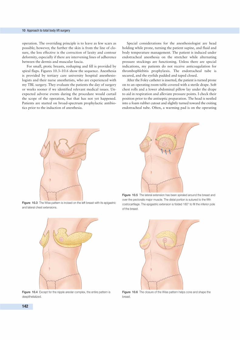
operation. The overriding principle is to leave as few scars as
possible; however, the further the skin is from the line of clo-
sure, the less effective is the correction of laxity and contour
deformity, especially if there are intervening lines of adherence
between the dermis and muscular fascia.
For small, ptotic breasts, reshaping and fill is provided by
spiral flaps. Figures 10.3–10.6 show the sequence. Anesthesia
is provided by tertiary care university hospital anesthesio-
logists and their nurse anesthetists, who are experienced with
my TBL surgery. They evaluate the patients the day of surgery
or weeks sooner if we identified relevant medical issues. Un-
expected adverse events during the procedure would curtail
the scope of the operation, but that has not yet happened.
Patients are started on broad-spectrum prophylactic antibio-
tics prior to the induction of anesthesia.
Special considerations for the anesthesiologist are head
holding while prone, turning the patient supine, and fluid and
body temperature management. The patient is induced under
endotracheal anesthesia on the stretcher while alternating
pressure stockings are functioning. Unless there are special
indications, my patients do not receive anticoagulation for
thrombophlebitis prophylaxis. The endotracheal tube is
secured, and the eyelids padded and taped closed.
After the Foley catheter is inserted, the patient is turned prone
on to an operating room table covered with a sterile drape. Soft
chest rolls and a lower abdominal pillow lay under the drape
to aid in respiration and alleviate pressure points. I check their
position prior to the antiseptic preparation. The head is nestled
into a foam rubber cutout and slightly turned toward the exiting
endotracheal tube. Often, a warming pad is on the operating
10 Approach to total body lift surgery
142
Figure 10.3 The Wise pattern is incised on the left breast with its epigastric
and lateral chest extensions.
Figure 10.4 Except for the nipple areolar complex, the entire pattern is
deepithelialized.
Figure 10.5 The lateral extension has been spiraled around the breast and
over the pectoralis major muscle. The distal portion is sutured to the fifth
costocartilage. The epigastric extension is folded 180° to fill the inferior pole
of the breast.
Figure 10.6 The closure of the Wise pattern helps cone and shape the
breast.
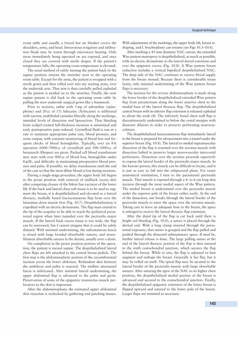
room table and usually a forced hot air blanket covers the
shoulders, arms, and head. Intravenous irrigation and infiltra-
tion fluids may be warm through microwave heating. Only
areas immediately being operated on are exposed, and once
closed they are covered with sterile drapes. If the patient’s
temperature falls, the operating room temperature is elevated.
The usual method of safely turning the patient back to the
supine position returns the stretcher next to the operating
room table. Except for the arms, the patient is wrapped with a
sterile gown and then rolled over into my waiting arms, over
the underside arm. That arm is then carefully pulled cephalad
as the patient is nestled on to the stretcher. Finally, the now
supine patient is slid back to the operating room table by
pulling the now underside surgical gown like a hammock.
Prior to incision, saline with 1 mg of adrenaline (epine-
phrine) and 20 cc of 1% lidocaine (Xylocaine) is infiltrated
with narrow, multiholed cannulas liberally along the markings,
intended levels of dissection and liposuction. Thus bleeding
from scalpel-created full-thickness incisions is minimized and
early postoperative pain reduced. Crystalloid fluid is run at a
rate to maintain appropriate pulse rate, blood pressure, and
urine output, with constant monitoring of blood loss and fre-
quent checks of blood hemoglobin. Typically, over an 8-h
operation 6000–7000 cc of crystalloid and 500–1000 cc of
hetastarch (Hespan) are given. Packed cell blood transfusions
may start with over 800 cc of blood loss, hemoglobin under
8 g/dL, and difficulty in maintaining preoperative blood pres-
sure and pulse. If possible, we delay transfusions until the end
of the case so that the most dilute blood is lost during incisions.
During a single-stage procedure, the upper body lift begins
in the prone position with removal of midback excess skin
after competing closure of the bikini line excision of the lower
lift. If the back and lateral chest soft tissue is to be used to aug-
ment the breast, it is deepithelialized and elevated as a lateral
thoracic, medially based fasciocutaneous flap from over the
latissimus dorsi muscle first (Fig. 10.7). Deepithelialization is
expedited with an electric dermatome. The flap must extend to
the tip of the scapular to be able to reach the ipsilateral paras-
ternal region when later tunneled over the pectoralis major
muscle. If the lateral back excess tissue is too wide, the flap
can be narrowed, but I cannot imagine that it could be safely
thinned. With minimal undermining, the subcutaneous fascia
is closed with large braided absorbable sutures, and mono-
filament absorbable sutures in the dermis, usually over a drain.
On completion in the prone position portion of the opera-
tion, the patient is turned supine. The deepithelialized lateral
chest flaps are left attached to the central breast pedicle. The
first step is the abdominoplasty portion of the circumferential
incision across the lower abdomen. Redundant skin between
the umbilicus and pubis is resected. The midline attenuated
fascia is imbricated. After minimal lateral undermining, the
upper abdominal flap is advanced to the pubis and groin.
Preservation of some of the epigastric transrectus muscle per-
forators to the skin is important.
After the abdominoplasty, the estimated upper abdominal
skin resection is rechecked by gathering and pinching tissues.
With adjustments of the markings, the upper body lift, breast re-
shaping. and L brachioplasty can resume (see Figs 10.3–10.6).
After marking a 45-mm diameter NAC cutout, the extended
Wise pattern mastopexy is deepithelialized, as much as possible,
with an electric dermatome to the lateral dorsal extension and
over the epigastric excess (Fig. 10.8). A Wise pattern breast
reduction includes a vertical bipedical deepithelialized NAC.
The deep side of the NAC continues to receive blood supply
from the breast mound. Because there is considerable tissue
laxity, only minimal undermining of the Wise pattern breast
flaps is necessary.
The incision for the reverse abdominoplasty is made along
the lower border of the deepithelialized extended Wise pattern
flap from parasternum along the lower anterior chest to the
medial base of the lateral thoracic flap. The deepithelialized
central breast with its inferior flap extension is released cephalad
to about the sixth rib. The inferiorly based chest wall flap is
discontinuously undermined to below the costal margins with
dissector dilators in order to preserve perforating neurovas-
culature.
The deepithelialized fasciocutaneous flap immediately lateral
to the breast is prepared for advancement into a tunnel under the
superior breast (Fig. 10.8). The lateral to medial supramuscular
dissection of the flap is resumed over the serratus muscle with
dissection halted to preserve larger neurovascular intercostal
perforators. Dissection over the serratus proceeds superiorly
to expose the lateral border of the pectoralis major muscle. In
the heavier person, this muscle can be difficult to locate, and it
is just as easy to fall into the subpectoral plane. For easier
anatomical orientation, I turn to the parasternal pectoralis
muscle. That muscle is exposed through a 4- to 6-cm long skin
incision through the most medial aspect of the Wise pattern.
The medial breast is undermined over the pectoralis muscle
under the superior pole of the breast rather easily. At the end
of the dissection, one breaks through the lateral border of the
pectoralis muscle to enter the space over the serratus muscle.
Taking care to leave an adequate base to the breast, the space
is enlarged to receive the lateral thoracic flap extension.
After the distal tip of the flap is cut back until there is
bright red bleeding (Fig. 10.8), a suture is placed through the
dermal end. With a long clamp inserted through the paras-
ternal exposure, that suture is grasped and the flap pulled and
pushed through the dissected submammary space. If need be,
further lateral release is done. The large pulling suture at the
end of the lateral thoracic portion of the flap is then sutured
to the sixth costochondral junction, which secures the flap
behind the breast. While in situ, the flap is adjusted to best
augment and reshape the breast. Generally it lies flat, but it
may be rolled on itself. The spiral flap may be secured to the
lateral border of the pectoralis muscle with large absorbable
sutures. After suturing the apex of the NAC to its higher chest
position, the deepithelialized medial portion of the breast is
advanced and secured to the costochondral junction. Finally,
the deepithelialized epigastric extension of the lower breast is
flipped upward and sutured to the lower pole of the breast.
Larger flaps are trimmed as necessary.
Surgical technique
143
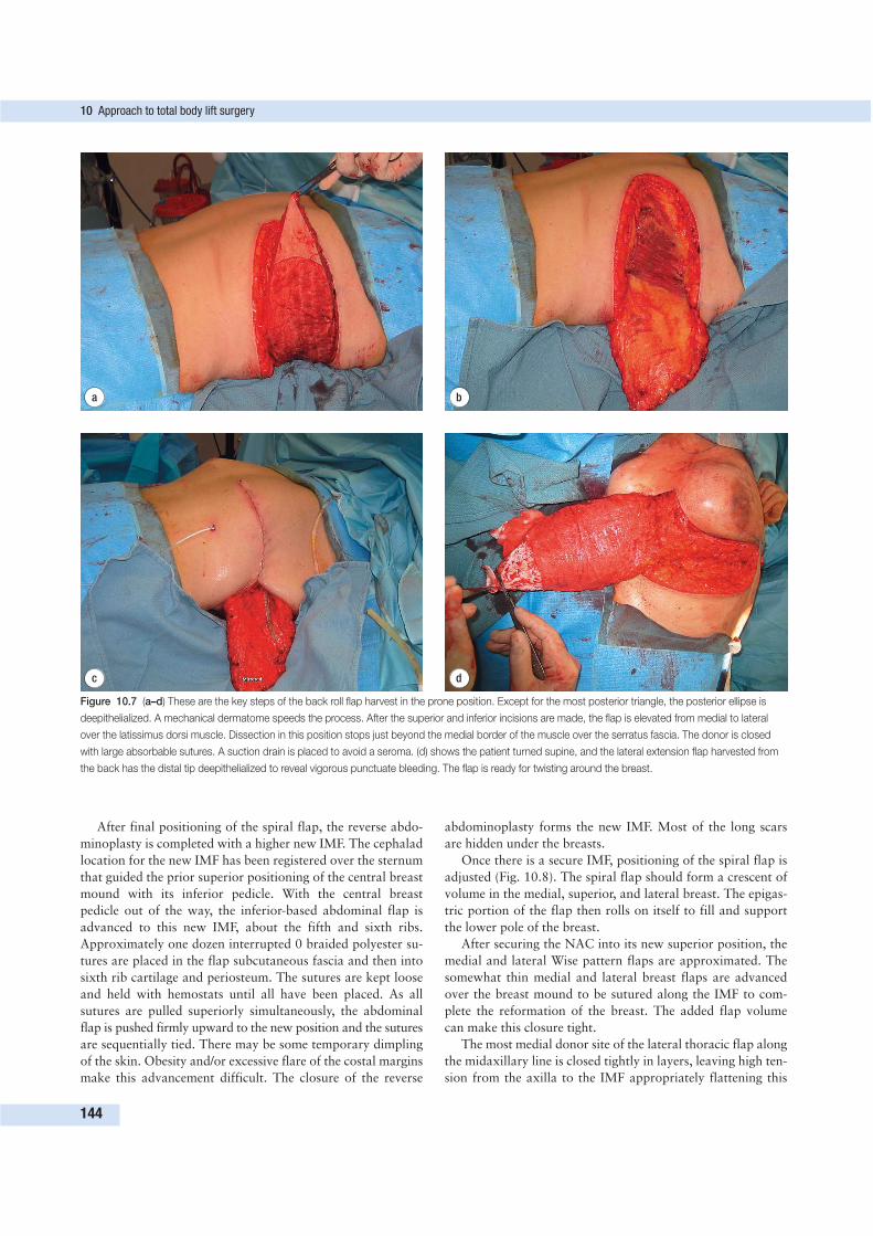
After final positioning of the spiral flap, the reverse abdo-
minoplasty is completed with a higher new IMF. The cephalad
location for the new IMF has been registered over the sternum
that guided the prior superior positioning of the central breast
mound with its inferior pedicle. With the central breast
pedicle out of the way, the inferior-based abdominal flap is
advanced to this new IMF, about the fifth and sixth ribs.
Approximately one dozen interrupted 0 braided polyester su-
tures are placed in the flap subcutaneous fascia and then into
sixth rib cartilage and periosteum. The sutures are kept loose
and held with hemostats until all have been placed. As all
sutures are pulled superiorly simultaneously, the abdominal
flap is pushed firmly upward to the new position and the sutures
are sequentially tied. There may be some temporary dimpling
of the skin. Obesity and/or excessive flare of the costal margins
make this advancement difficult. The closure of the reverse
abdominoplasty forms the new IMF. Most of the long scars
are hidden under the breasts.
Once there is a secure IMF, positioning of the spiral flap is
adjusted (Fig. 10.8). The spiral flap should form a crescent of
volume in the medial, superior, and lateral breast. The epigas-
tric portion of the flap then rolls on itself to fill and support
the lower pole of the breast.
After securing the NAC into its new superior position, the
medial and lateral Wise pattern flaps are approximated. The
somewhat thin medial and lateral breast flaps are advanced
over the breast mound to be sutured along the IMF to com-
plete the reformation of the breast. The added flap volume
can make this closure tight.
The most medial donor site of the lateral thoracic flap along
the midaxillary line is closed tightly in layers, leaving high ten-
sion from the axilla to the IMF appropriately flattening this
10 Approach to total body lift surgery
144
d
a b
c
Figure 10.7 (a–d) These are the key steps of the back roll flap harvest in the prone position. Except for the most posterior triangle, the posterior ellipse is
deepithelialized. A mechanical dermatome speeds the process. After the superior and inferior incisions are made, the flap is elevated from medial to lateral
over the latissimus dorsi muscle. Dissection in this position stops just beyond the medial border of the muscle over the serratus fascia. The donor is closed
with large absorbable sutures. A suction drain is placed to avoid a seroma. (d) shows the patient turned supine, and the lateral extension flap harvested from
the back has the distal tip deepithelialized to reveal vigorous punctuate bleeding. The flap is ready for twisting around the breast.
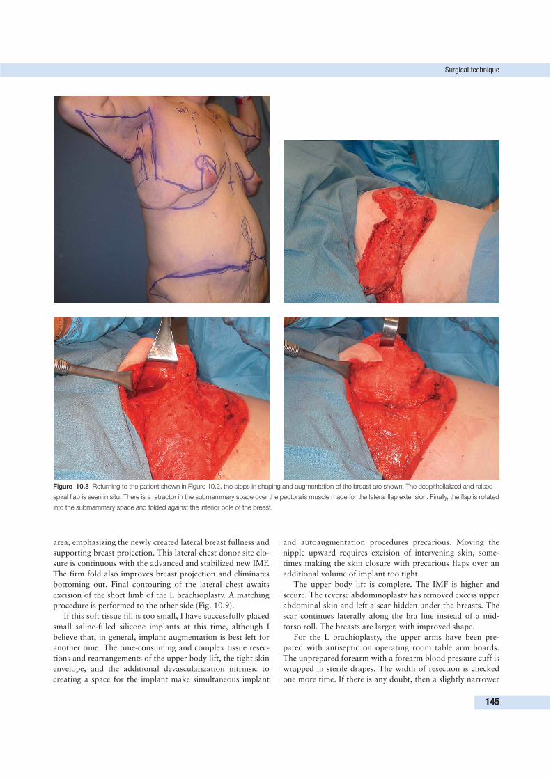
area, emphasizing the newly created lateral breast fullness and
supporting breast projection. This lateral chest donor site clo-
sure is continuous with the advanced and stabilized new IMF.
The firm fold also improves breast projection and eliminates
bottoming out. Final contouring of the lateral chest awaits
excision of the short limb of the L brachioplasty. A matching
procedure is performed to the other side (Fig. 10.9).
If this soft tissue fill is too small, I have successfully placed
small saline-filled silicone implants at this time, although I
believe that, in general, implant augmentation is best left for
another time. The time-consuming and complex tissue resec-
tions and rearrangements of the upper body lift, the tight skin
envelope, and the additional devascularization intrinsic to
creating a space for the implant make simultaneous implant
and autoaugmentation procedures precarious. Moving the
nipple upward requires excision of intervening skin, some-
times making the skin closure with precarious flaps over an
additional volume of implant too tight.
The upper body lift is complete. The IMF is higher and
secure. The reverse abdominoplasty has removed excess upper
abdominal skin and left a scar hidden under the breasts. The
scar continues laterally along the bra line instead of a mid-
torso roll. The breasts are larger, with improved shape.
For the L brachioplasty, the upper arms have been pre-
pared with antiseptic on operating room table arm boards.
The unprepared forearm with a forearm blood pressure cuff is
wrapped in sterile drapes. The width of resection is checked
one more time. If there is any doubt, then a slightly narrower
Surgical technique
145
Figure 10.8 Returning to the patient shown in Figure 10.2, the steps in shaping and augmentation of the breast are shown. The deepithelialized and raised
spiral flap is seen in situ. There is a retractor in the submammary space over the pectoralis muscle made for the lateral flap extension. Finally, the flap is rotated
into the submammary space and folded against the inferior pole of the breast.
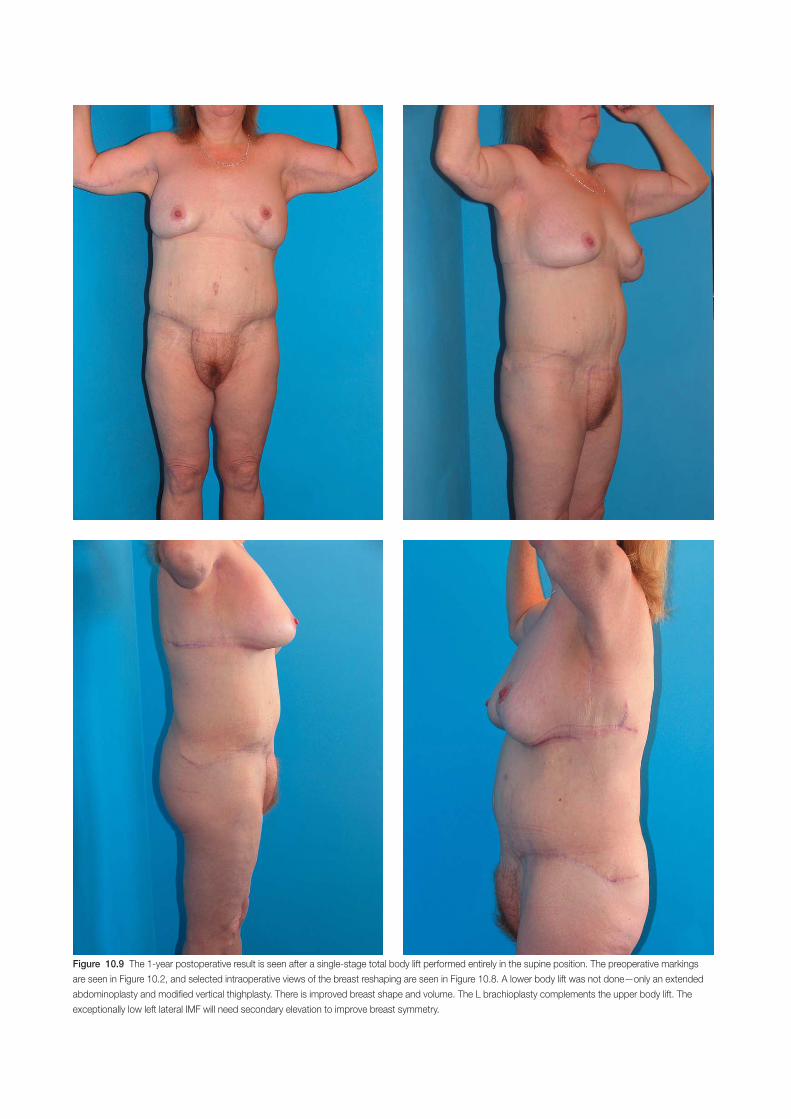
Figure 10.9 The 1-year postoperative result is seen after a single-stage total body lift performed entirely in the supine position. The preoperative markings
are seen in Figure 10.2, and selected intraoperative views of the breast reshaping are seen in Figure 10.8. A lower body lift was not done—only an extended
abdominoplasty and modified vertical thighplasty. There is improved breast shape and volume. The L brachioplasty complements the upper body lift. The
exceptionally low left lateral IMF will need secondary elevation to improve breast symmetry.
ellipse is removed. In the manner previously described, I infuse
several hundred cubic centimeters of saline with dilute adrena-
line (epinephrine) and lidocaine (Xylocaine). After allowing
10 min for vasoconstriction, UAL is performed as needed.
With the medial skin rolled superiorly, the inferior incision
is made to the level of the crural fascia enveloping the muscles.
About 1 cm of undermining is done. Then the arching superior
incision is made from the elbow to deltopectoral groove and
also minimally undermined. Hemostasis is again obtained. I
similarly incise the outline of the axillary chest ellipse, taking
care to go just deep to the dermis in the axilla. The triangle of
skin and fat at the elbow are grasped with the multitooth
clamp or rake. The instrument firmly distracts the ellipse
toward the chest so as remove the tissue, leaving a fine deep
layer of subcutaneous fascia and fat over the subcutaneous
nerves. Dissection stops to give electrocoagulation to patients
with greater bleeding. The excision courses subdermal through
the axilla, and then completes deeply over muscular fascia of
the lateral chest. The clavipectoral fascia of the axilla is seen
but not entered. Major veins and sensory nerves are not seen.
The final decision on the width of lateral chest excision is made
so as to remove all excess skin without lateralizing the breast.
Using the previously marked guidelines, the incisions are
aligned with towel clamps. A continuous running 2-0 long-
lasting but absorbable suture approximates the subcutaneous
fascia. When approaching a towel clamp, a second clamp
leapfrogs ahead before the first clamp is released. A second,
smaller caliber continuous intradermal closure follows. Stern
strips or dermal glue completes the operation. The arms are
wrapped by an Ace wrap over a large ABD pad.
As the skin tensions equilibrate, the scar courses from the
medial epicondyle to along the inferior medial arm, inferior to
the bicipital groove. It gently rises to the axillary dome and
then drops vertically to the chest, forming an inverted L. The
inferior contour of the arm drops slightly at the midhumerus
and then distinctly rises to a superiorly positioned posterior
axillary fold. The suspended posterior axillary fold skin
conforms well to the axillary hollow.
The breasts are placed in a surgical bra. No constricting
binder is placed across the midabdomen, although for the
lower body lift a long-leg lower body elastic garment is used.
When only an upper body lift is done, patients are admitted
for a single night’s observation and care. The arm wrap is
replaced with elastic sleeves several days later, taking care not
to put direct pressure on the delicate triangular flap crossing
the axilla. See Figures 10.10–10.12 for three cases of single-
stage TBLs with L brachioplasty.
UPPER BODY LIFT IN MEN
In men, the objective of the upper body lift is to obliterate the
IMF while correcting gynecomastia and redundant skin. Male
upper body lift has definite synergistic effect when combined
with the lower body lift and circumferential abdominoplasty.
The upper lift in men also has four components.
1. A unique reverse abdominoplasty.
2. Obliteration of the IMF.
3. Removal of the midtorso roll.
4. Correction of the gynecomastia.
Male massive weight loss patients have loose upper abdo-
minal skin, but too often a protuberant upper abdomen due to
persistent intraabdominal epigastric obesity, which has to be
considered in any reconstruction. A distinct IMF accentuates
their disdainfully enlarged breasts. The midtorso rolls are
lateral extensions of moderately ptotic gynecomastia. The
gynecomastia is not only severe but also has inelastic skin that
will not accommodate to a reduced volume.
The complete correction of weight loss grade 4 gynecomastia:
• properly positions NACs on pedicles;
• removes offending glands and skin, both vertically and
horizontally; and
• leaves inconspicuous, long, anteriolateral chest scars
(Fig. 10.13).
This is best accomplished with two elliptic excisions of skin
wrapped around the areola, which I call a boomerang pattern
excision correction of gynecomastia.
A common technique for loose skin gynecomastia is to
remove the ptotic nipple. The gynecomastia is cut out along a
long horizontal ellipse. Then the excised nipple is grafted on
to the chest in the proper location. The take is not assured, and
irregularity follows partial necrosis. But even with a 100%
take, the nipple graft often looks like a skin graft, unnaturally
flat and discolored. The long, straight scar is conspicuous,
with a distinctly postsurgical appearance.
I have recently described the boomerang excision correc-
tion of gynecomastia. This procedure is an improvement over
prior techniques because:
• the resection includes both vertical and horizontal excess;
• the NAC remains on a skin/glandular pedicle;
• the NAC is integrated into the upper body lift and TBL,
and the long scar changes direction as it wraps around the
repositioned areola.1
This gynecomastia correction considers biomechanical and
aesthetic issues. There is a full-thickness triangular flap to
support the nipple. That triangular base flap has excess fat
and breast. I emulsify the fat and obliterate the IMF with
UAL, followed by judicious liposuction. The resulting scar has
a short limb that starts near the lower sternum, rises to arch
the areola, and then descends toward the lower outer chest.
Because the areola acts to break up the scar, it appears as if
there were two smaller scars. The scar that wraps around the
areola is less conspicuous than a straight line scar. The exci-
sion pattern resembles a boomerang, hence the appellation. In
some cases, further reduction of the base was necessary at a
later procedure. The ideal patient has a hirsute chest, which
tends to be most dense around the areola and that obscures
the scar.
Preoperative marking of the boomerang correction starts
with sighting the new nipple position and registering it on the
sternum. The ptotic breast and NAC are raised until the NAC
falls in the correct position as agreed by the surgeon and
Upper body lift in men
147
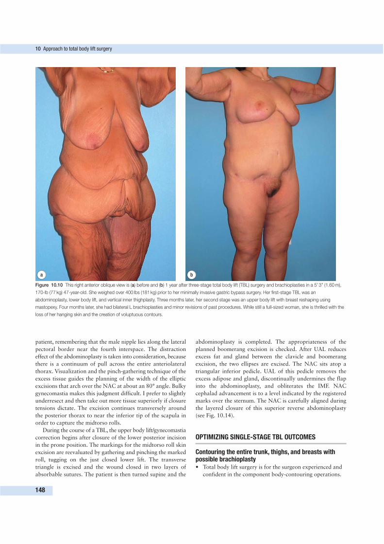
patient, remembering that the male nipple lies along the lateral
pectoral border near the fourth interspace. The distraction
effect of the abdominoplasty is taken into consideration, because
there is a continuum of pull across the entire anteriolateral
thorax. Visualization and the pinch-gathering technique of the
excess tissue guides the planning of the width of the elliptic
excisions that arch over the NAC at about an 80° angle. Bulky
gynecomastia makes this judgment difficult. I prefer to slightly
underresect and then take out more tissue superiorly if closure
tensions dictate. The excision continues transversely around
the posterior thorax to near the inferior tip of the scapula in
order to capture the midtorso rolls.
During the course of a TBL, the upper body lift/gynecomastia
correction begins after closure of the lower posterior incision
in the prone position. The markings for the midtorso roll skin
excision are reevaluated by gathering and pinching the marked
roll, tugging on the just closed lower lift. The transverse
triangle is excised and the wound closed in two layers of
absorbable sutures. The patient is then turned supine and the
abdominoplasty is completed. The appropriateness of the
planned boomerang excision is checked. After UAL reduces
excess fat and gland between the clavicle and boomerang
excision, the two ellipses are excised. The NAC sits atop a
triangular inferior pedicle. UAL of this pedicle removes the
excess adipose and gland, discontinually undermines the flap
into the abdominoplasty, and obliterates the IMF. NAC
cephalad advancement is to a level indicated by the registered
marks over the sternum. The NAC is carefully aligned during
the layered closure of this superior reverse abdominoplasty
(see Fig. 10.14).
OPTIMIZING SINGLE-STAGE TBL OUTCOMES
Contouring the entire trunk, thighs, and breasts withpossible brachioplasty• Total body lift surgery is for the surgeon experienced and
confident in the component body-contouring operations.
10 Approach to total body lift surgery
148
a b
Figure 10.10 This right anterior oblique view is (a) before and (b) 1 year after three-stage total body lift (TBL) surgery and brachioplasties in a 5’ 3” (1.60 m),
170-lb (77 kg) 47-year-old. She weighed over 400 lbs (181 kg) prior to her minimally invasive gastric bypass surgery. Her first-stage TBL was an
abdominoplasty, lower body lift, and vertical inner thighplasty. Three months later, her second stage was an upper body lift with breast reshaping using
mastopexy. Four months later, she had bilateral L brachioplasties and minor revisions of past procedures. While still a full-sized woman, she is thrilled with the
loss of her hanging skin and the creation of voluptuous contours.
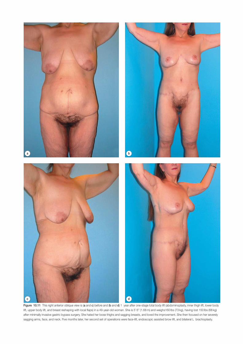
d
a b
c
Figure 10.11 This right anterior oblique view is (a and c) before and (b and d) 1 year after one-stage total body lift (abdominoplasty, inner thigh lift, lower body
lift, upper body lift, and breast reshaping with local flaps) in a 49-year-old woman. She is 5’ 6” (1.68 m) and weighs160 lbs (73 kg), having lost 150 lbs (68 kg)
after minimally invasive gastric bypass surgery. She hated her loose thighs and sagging breasts, and loved the improvement. She then focused on her severely
sagging arms, face, and neck. Five months later, her second set of operations were face-lift, endoscopic assisted brow lift, and bilateral L brachioplasty.
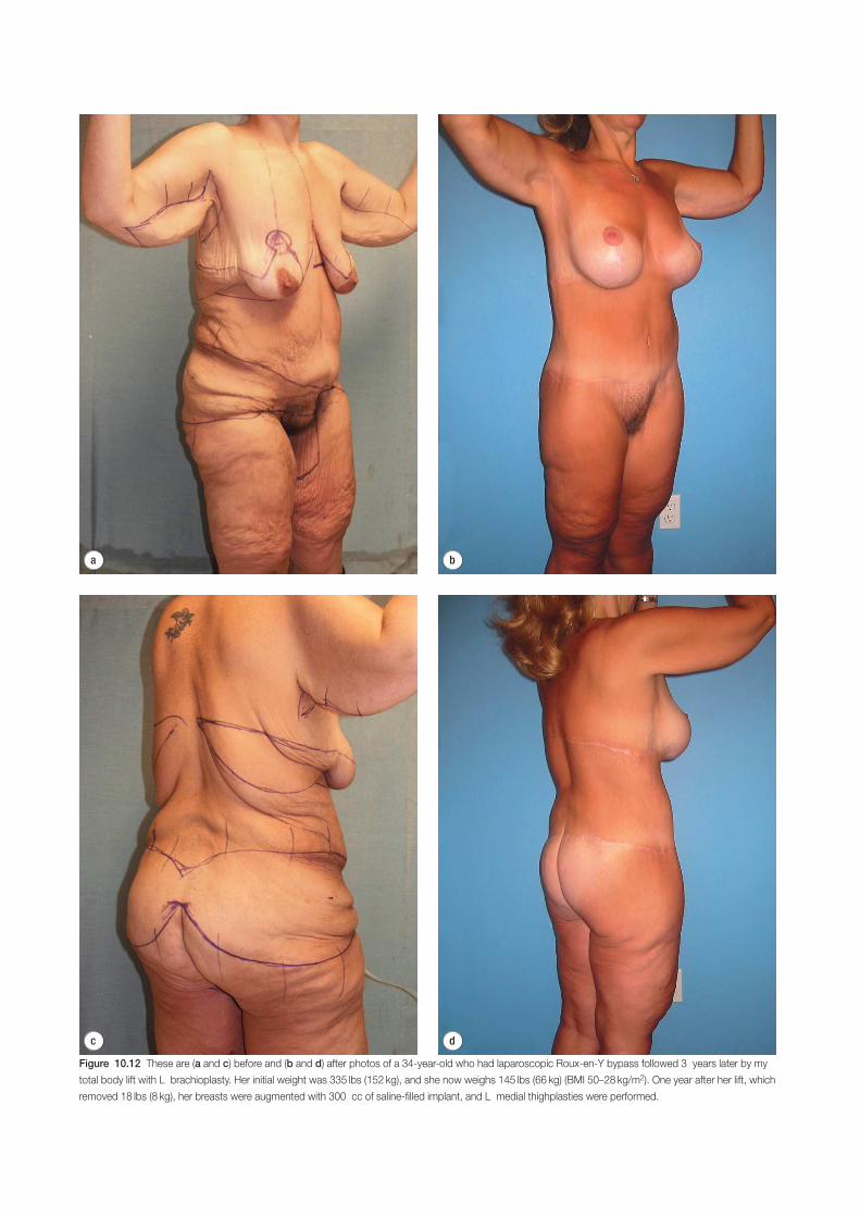
d
a b
c
Figure 10.12 These are (a and c) before and (b and d) after photos of a 34-year-old who had laparoscopic Roux-en-Y bypass followed 3 years later by my
total body lift with L brachioplasty. Her initial weight was 335 lbs (152 kg), and she now weighs 145 lbs (66 kg) (BMI 50–28 kg/m2). One year after her lift, which
removed 18 lbs (8 kg), her breasts were augmented with 300 cc of saline-filled implant, and L medial thighplasties were performed.
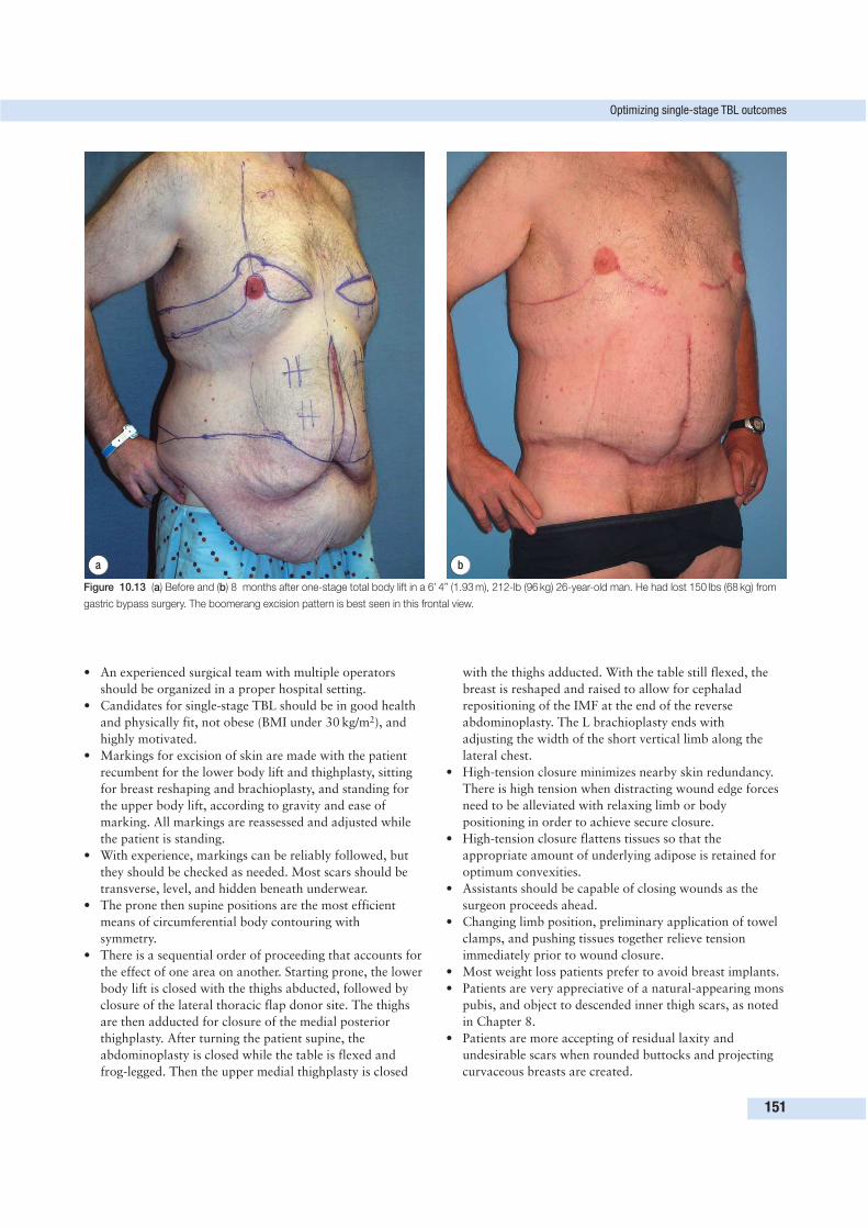
Optimizing single-stage TBL outcomes
151
a b
Figure 10.13 (a) Before and (b) 8 months after one-stage total body lift in a 6’ 4” (1.93 m), 212-lb (96 kg) 26-year-old man. He had lost 150 lbs (68 kg) from
gastric bypass surgery. The boomerang excision pattern is best seen in this frontal view.
• An experienced surgical team with multiple operators
should be organized in a proper hospital setting.
• Candidates for single-stage TBL should be in good health
and physically fit, not obese (BMI under 30 kg/m2), and
highly motivated.
• Markings for excision of skin are made with the patient
recumbent for the lower body lift and thighplasty, sitting
for breast reshaping and brachioplasty, and standing for
the upper body lift, according to gravity and ease of
marking. All markings are reassessed and adjusted while
the patient is standing.
• With experience, markings can be reliably followed, but
they should be checked as needed. Most scars should be
transverse, level, and hidden beneath underwear.
• The prone then supine positions are the most efficient
means of circumferential body contouring with
symmetry.
• There is a sequential order of proceeding that accounts for
the effect of one area on another. Starting prone, the lower
body lift is closed with the thighs abducted, followed by
closure of the lateral thoracic flap donor site. The thighs
are then adducted for closure of the medial posterior
thighplasty. After turning the patient supine, the
abdominoplasty is closed while the table is flexed and
frog-legged. Then the upper medial thighplasty is closed
with the thighs adducted. With the table still flexed, the
breast is reshaped and raised to allow for cephalad
repositioning of the IMF at the end of the reverse
abdominoplasty. The L brachioplasty ends with
adjusting the width of the short vertical limb along the
lateral chest.
• High-tension closure minimizes nearby skin redundancy.
There is high tension when distracting wound edge forces
need to be alleviated with relaxing limb or body
positioning in order to achieve secure closure.
• High-tension closure flattens tissues so that the
appropriate amount of underlying adipose is retained for
optimum convexities.
• Assistants should be capable of closing wounds as the
surgeon proceeds ahead.
• Changing limb position, preliminary application of towel
clamps, and pushing tissues together relieve tension
immediately prior to wound closure.
• Most weight loss patients prefer to avoid breast implants.
• Patients are very appreciative of a natural-appearing mons
pubis, and object to descended inner thigh scars, as noted
in Chapter 8.
• Patients are more accepting of residual laxity and
undesirable scars when rounded buttocks and projecting
curvaceous breasts are created.
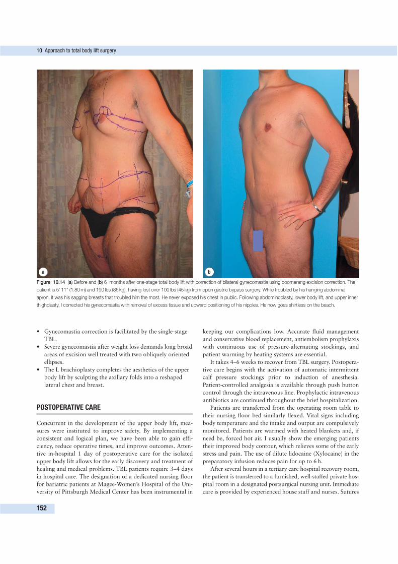
• Gynecomastia correction is facilitated by the single-stage
TBL.
• Severe gynecomastia after weight loss demands long broad
areas of excision well treated with two obliquely oriented
ellipses.
• The L brachioplasty completes the aesthetics of the upper
body lift by sculpting the axillary folds into a reshaped
lateral chest and breast.
POSTOPERATIVE CARE
Concurrent in the development of the upper body lift, mea-
sures were instituted to improve safety. By implementing a
consistent and logical plan, we have been able to gain effi-
ciency, reduce operative times, and improve outcomes. Atten-
tive in-hospital 1 day of postoperative care for the isolated
upper body lift allows for the early discovery and treatment of
healing and medical problems. TBL patients require 3–4 days
in hospital care. The designation of a dedicated nursing floor
for bariatric patients at Magee-Women’s Hospital of the Uni-
versity of Pittsburgh Medical Center has been instrumental in
keeping our complications low. Accurate fluid management
and conservative blood replacement, antiembolism prophylaxis
with continuous use of pressure-alternating stockings, and
patient warming by heating systems are essential.
It takes 4–6 weeks to recover from TBL surgery. Postopera-
tive care begins with the activation of automatic intermittent
calf pressure stockings prior to induction of anesthesia.
Patient-controlled analgesia is available through push button
control through the intravenous line. Prophylactic intravenous
antibiotics are continued throughout the brief hospitalization.
Patients are transferred from the operating room table to
their nursing floor bed similarly flexed. Vital signs including
body temperature and the intake and output are compulsively
monitored. Patients are warmed with heated blankets and, if
need be, forced hot air. I usually show the emerging patients
their improved body contour, which relieves some of the early
stress and pain. The use of dilute lidocaine (Xylocaine) in the
preparatory infusion reduces pain for up to 6 h.
After several hours in a tertiary care hospital recovery room,
the patient is transferred to a furnished, well-staffed private hos-
pital room in a designated postsurgical nursing unit. Immediate
care is provided by experienced house staff and nurses. Sutures
10 Approach to total body lift surgery
152
a b
Figure 10.14 (a) Before and (b) 6 months after one-stage total body lift with correction of bilateral gynecomastia using boomerang excision correction. The
patient is 5’ 11” (1.80 m) and 190 lbs (86 kg), having lost over 100 lbs (45 kg) from open gastric bypass surgery. While troubled by his hanging abdominal
apron, it was his sagging breasts that troubled him the most. He never exposed his chest in public. Following abdominoplasty, lower body lift, and upper inner
thighplasty, I corrected his gynecomastia with removal of excess tissue and upward positioning of his nipples. He now goes shirtless on the beach.
are available at the bedside to repair minor dehiscence. Patients
start using the incentive spirometer but do not ambulate until the
next morning. I insist on full return of sensorium before moving.
If a patient’s condition deteriorates, transfer to an intensive
care unit is immediate for continuous monitoring and care.
Strict monitoring of fluid intake and output through an
indwelling bladder catheter and suction drains is essential
throughout the stay. Hemoglobin and serum chemistries are
monitored daily, with appropriate treatment until stable. Fluid
retention due to traumatic swelling and stress hormone release
is expected over several weeks. Edema, particularly of the legs,
is common and is usually treated with diuretics, leg elevation,
and compression wrappings. Recently, we have initiated ex-
tremity suction/massage therapy prior to discharge with the
use of the Well Box (LPG, Miami, Florida) with success.
When the patients’ condition is stable and they are am-
bulating, the Foley catheter is removed. Prior to discharge, the
patient is showered and discharged in properly sized elastic
garments. After discharge, we encourage our patients to in-
crease progressively non-taxing light activity. Within 4 weeks,
most patients can resume daily functions such as driving and
desk work. Elastic garments are worn for 6 weeks to encour-
age proper healing and provide support for the incisions. The
first office visit is 10 days after surgery. The dramatic im-
provement in body contour becomes evident. Stitches around
the umbilicus are removed. I will remove suction drains with
output less than 50 cc per day. Many patients can resume vigo-
rous exercise after 6 weeks. Minor wound-healing problems,
especially along the medial thighs, are common and will re-
quire the patient to regularly change dressings.
COMPLICATIONS AND THEIR MANAGEMENT
Complex and lengthy surgery over a large portion of the body
understandably entails medical and surgical risks. TBL sur-
gery may be performed in several stages or in a single stage
depending on the patient presentation and desire. Optimal
candidates for single-stage TBL are physically and mentally
stable. Highly motivated patients are willing to accept theo-
retic greater chance of morbidity and mortality for the effi-
ciency and satisfaction of a single-stage operation. They accept
that revision surgery is possible. Refined metabolic and in-
flammatory tissue markers are being considered to identify
ideal candidates. Individuals having multiple stages did not
fulfill these criteria or were under treatment before the single
stage was regularly offered. Since regularly offering a single-
stage operation in 2002 to optimal candidates, 53% (38 of
72) of the patients having TBL had a one-stage procedure.
Regarding complications, points to note are as follow.
• High-risk patients have nutritional disorders, obesity,
undertreated or unstable chronic medical conditions,
coagulation issues, mental disorders, and unrealistic
expectations.
• Patients over 55 years of age are probably at higher risk of
medical complications.
• Patients with insulin-dependent (type 1) diabetes, poorly
controlled hypertension, unstable cardiac condition, and
arrhythmias, or who are chronic smokers, should be
avoided or have limited procedures.
Disregarding these admonitions may result in extensive wound-
healing problems, postoperative intensive care unit admissions,
prolonged or rehospitalization, and death.
After the first 72 patients with a single- and two-stage body
lift, there have been no cases of thrombophlebitis. There has
been one single-stage TBL patient with sepsis requiring read-
mission a week after her surgery. I emergently drained an
upper medial thigh abscess that grew Streptococcus viridans
and Haemophilus influenzae. A week of intravenous anti-
biotics and wound care cleared up the infection, and she was
discharged to home 1 week later; within 4 weeks, the thigh
incision wounds healed. She had 3000 cc of fat removed from
her thighs using UAL lipoplasty during her TBL. I suspect that
contamination must have been introduced at that time. Six
months later, she is troubled by recurrent stitch abscesses. In
two other patients, I have drained two midthigh abscesses
1 month and 3 months after their TBL.
The most common dilemma is the persistently overweight
patient, having a BMI from 31 to 35 kg/m2. The operations
are more bloody and lengthy. High-tension closure of heavier
tissues may dehisce or stretch out and depress, with loss of
carefully created contours. Fat necrosis, wound infections, and
suture abscesses are common. For these and general medical
issues, oversized patients are encouraged to lose weight. An
in-office nutritionist with an accepted rapid weight loss pro-
gram is helpful. Through the cooperation of Drs El Hassane
Tazi of Casablanca, Morocco, and Trudy Vogt of Zurich,
Switzerland, we have used the A.W. Simeon severe caloric re-
striction diet with low-dose, off-label, daily human chorionic
gonadotropin hormone (hCG) injections.19 Dozens of our
patients have lost from 15 to 30 lbs (7–14 kg) without suffer-
ing hunger in 6 weeks, making them better candidates for body-
contouring surgery. While this rigorous low-caloric/hCG
injection program has had high success without morbidity in
Switzerland and Morocco, it has not yet been submitted to
recent clinical trials in the USA. As such, the Simeon method
is considered investigational. Confident of its advantage in
preparing borderline patients for body contouring, I feel obli-
gated to implement it with the aid of my physician assistant.
For the still oversized, optimal body contouring includes
extensive liposuction, which is traumatic to the patient and
flaps. I believe UAL to be the least injurious. The greater the
amount of liposuction, the lesser should be the extent of exci-
sion surgery. Vacuum suction drainage is mandatory when
liposuction and flap elevation are extensive (Fig. 10.15).
Because of her excessive weight and an occult lateral thigh
seroma cavity, outpatient readvancement of the lateral hips
were needed in the patient in Figure 10.15. When there is
excessive fat deposition and limited skin laxity, then a preli-
minary staged liposuction may be indicated.
On the flip side is the dramatically thin patient with cir-
cumferential layers of hanging skin. On the torso, transverse
Complications and their management
153
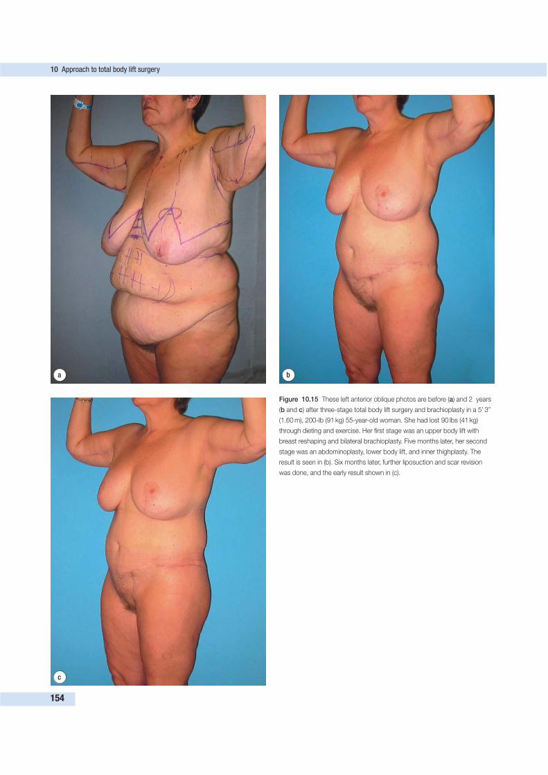
10 Approach to total body lift surgery
154
a b
c
Figure 10.15 These left anterior oblique photos are before (a) and 2 years
(b and c) after three-stage total body lift surgery and brachioplasty in a 5’ 3”
(1.60 m), 200-lb (91 kg) 55-year-old woman. She had lost 90 lbs (41 kg)
through dieting and exercise. Her first stage was an upper body lift with
breast reshaping and bilateral brachioplasty. Five months later, her second
stage was an abdominoplasty, lower body lift, and inner thighplasty. The
result is seen in (b). Six months later, further liposuction and scar revision
was done, and the early result shown in (c).
excision only will leave too much loose skin vertically. On the
thighs, the vertical extension excision needs to be precariously
broad, and even then, secondary strips of excision need to be
done.
All patients are informed of the inherent risks of TBL sur-
gery. Our written informed consent document is instructive
and covers the following major points:
• change in plans during the operation;
• bleeding;
• infection;
• thrombophlebitis and pulmonary embolism;
• change in nipple and skin sensation;
• long-term effects due to aging and weight change
unrelated to the surgery;
• chronic pain;
• suture spit;
• anesthesia risks;
• allergic reactions to tape, suture material, or topical
preparations;
• aesthetic shortcomings; and
• pregnancy and breast-feeding concerns.
SUMMARY AND CONCLUSION
Total body lift surgery is an original and boldly comprehen-
sive correction of skin sagging, demanding insight, artistry,
skill, stamina, and teamwork. TBL surgery was created to
meet the unique challenge of body contouring after massive
weight loss, and has been extended to treat the consequences
of pregnancy and aging.
The single-stage TBL is an artistic tour de force, made
possible by thoughtful surgical experience and innovation,
modern anesthesia, and widespread patient education.2 Effec-
tiveness and safety are intertwined and directly related to the
surgeon’s outlook, temperament, and experience. There is a
synergism at the midtorso level with improved narrowing of
the waist and better effacement of gynecomastia. With proper
organization, I believe that motivated plastic surgeons can
reliably and safely offer TBL surgery to their patients.
Total body lift surgery is analogous to craniofacial surgery.
Craniofacial surgery was introduced in the 1970s as a drama-
tic new discipline for the congenitally deformed. After 25 years
of practicing craniofacial surgery, I consider that field com-
plex and a dramatic, high-risk aesthetic facial reconstruction.
Before craniofacial surgery, corrective operations for the con-
genitally deformed were limited in scope. Neurosurgeons re-
shaped congenially deformed craniums. Later, plastic surgeons
advanced the jaws and bone grafted the midface and orbits.
As a boundary between the cranium and face, the orbits were
poorly treated. There was no comprehensive and coordinated
planning and treatment. With the advent of craniofacial sur-
gery, the entire deformity, including the orbits, could be ap-
proached in a coordinated single stage. Plastic surgeons,
uniquely experienced in body contouring, can organize a team
to treat the entire massive weight loss deformity.
As the craniofacial approach to the congenitally deformed
became routine, enormous progress was made in elective
aesthetic facial surgery. Similarly, once I developed a routine,
coordinated total body approach for the weight loss patient,
my aesthetic body contouring expanded and improved. As I
became confident in the essential elements of skin excision, I
could concentrate on the aesthetic details that make a differ-
ence. TBL surgery is as grand in scope as craniofacial surgery.
Total body lift surgery is a time-tested way to improve the
abdomen, thighs, buttocks, midback, and breasts. Commonly,
a first stage corrects the abdomen, thighs, and lower body. I
position the patient prone and remove a large beltlike segment
of skin above the buttocks, up to the lower back. On closure
of this broad wound, the thighs and buttocks are lifted. Then I
turn my patient supine to complete the anterior and medial
thighs and the abdomen.
If it is not done immediately, I will correct the upper body
deformity in stage 2 as early as 3 months after the first opera-
tion. By that time, all minor wound-healing issues, the threat
of thrombophlebitis, and chronic edema are resolved. The
patient should be on a healthy diet, restoring protein and
correcting anemia. The upper body lift consists of a reverse
abdominoplasty (from umbilicus to breasts), removal of mid-
back rolls, and reshaping of flattened and hanging breasts. If
the patient desires, the upper arms are included.
The upper body lift hides the upper scar under the breast
and along the bra line. The breasts are beautifully shaped as
the nipples are raised to the optimal position. A distinct new
fold is secured under the breast to help maintain breast shape
and a flat upper abdomen. Then I complement the upper body
lift with an L brachioplasty. I remove excess skin and fat of the
upper arm, axilla, and side of the chest roughly in the form of
an L. The scar may take many months to mature, leaving a
sweeping and as inconspicuous scar as possible because it lies
between the bicipital groove and the posterior margin of the
arm (see Fig. 10.16).
By coordinating several surgeons and skilled assistants, the
TBL takes approximately 8 h, with additional time needed for
larger patients. On average, three units of blood transfusion
are needed. There has been no recognized thrombophlebitis or
pulmonary embolism. Consistent with our initial report, there
have been no increased complications as compared with the
multistaged approach.1
The final contour relates to the deep fat, the extent of
undermining, the tension of the closure, and the elasticity of the
skin. In the massive weight loss patient, the skin is inelastic, so
that only in areas that it is pulled taut is there no looseness in
that direction. Transverse pull corrects vertical laxity only.
Nevertheless, I had hoped that the combined superior and
inferior tension at the bra and bikini line excisions would create
a Chinese finger trap effect, thereby narrowing the waist; this
is best seen in thinner patients.
By limiting the undermining and using gentle liposuction,
removal of skin from both the upper and lower ends of the
abdomen does not lead to flap edge ischemia. It is clear that
patients with prior abdominoplasty and considerable upper
Summary and conclusion
155
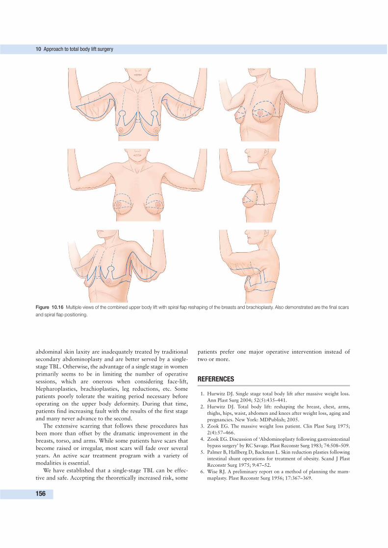
abdominal skin laxity are inadequately treated by traditional
secondary abdominoplasty and are better served by a single-
stage TBL. Otherwise, the advantage of a single stage in women
primarily seems to be in limiting the number of operative
sessions, which are onerous when considering face-lift,
blepharoplasties, brachioplasties, leg reductions, etc. Some
patients poorly tolerate the waiting period necessary before
operating on the upper body deformity. During that time,
patients find increasing fault with the results of the first stage
and many never advance to the second.
The extensive scarring that follows these procedures has
been more than offset by the dramatic improvement in the
breasts, torso, and arms. While some patients have scars that
become raised or irregular, most scars will fade over several
years. An active scar treatment program with a variety of
modalities is essential.
We have established that a single-stage TBL can be effec-
tive and safe. Accepting the theoretically increased risk, some
patients prefer one major operative intervention instead of
two or more.
REFERENCES
1. Hurwitz DJ. Single stage total body lift after massive weight loss.
Ann Plast Surg 2004; 52(5):435–441.
2. Hurwitz DJ. Total body lift: reshaping the breast, chest, arms,
thighs, hips, waist, abdomen and knees after weight loss, aging and
pregnancies. New York: MDPublish; 2005.
3. Zook EG. The massive weight loss patient. Clin Plast Surg 1975;
2(4):57–466.
4. Zook EG. Discussion of ‘Abdominoplasty following gastrointestinal
bypass surgery’ by RC Savage. Plast Reconstr Surg 1983; 74:508–509.
5. Palmer B, Hallberg D, Backman L. Skin reduction plasties following
intestinal shunt operations for treatment of obesity. Scand J Plast
Reconstr Surg 1975; 9:47–52.
6. Wise RJ. A preliminary report on a method of planning the mam-
maplasty. Plast Reconstr Surg 1956; 17:367–369.
10 Approach to total body lift surgery
156
Figure 10.16 Multiple views of the combined upper body lift with spiral flap reshaping of the breasts and brachioplasty. Also demonstrated are the final scars
and spiral flap positioning.
7. McKissock PK. Reduction mammoplasty with a vertical dermal
pedicle. Plast Reconstr Surg 1972; 49:245–252.
8. Shons AR. Plastic reconstruction after bypass surgery and massive
weight loss. Surg Clin North Am 1979; 59:1139–1152.
9. Regnault P, Daniel RK. Massive weight loss. In: Regnault P, Daniel
RK. Aesthetic plastic surgery: principles and techniques. Boston:
Little Brown; 1984:705–720.
10. Grazer FM. Abdominoplasty. In: McCarthy et al, eds. Plastic sur-
gery, vol. 6. The trunk and lower extremity. Philadelphia: Saunders;
1994:3929–3963.
11. Zienowicz RJ. Augmentation mammoplasty by reverse abdomino-
plasty. Presented at Emerging Technologies and Techniques in Plastic
Surgery, New York University Medical Center, May 20–21, 2005.
12. Lockwood TE. Superficial fascial system (SFS) of the trunk and ex-
tremities: a new concept. Plast Reconstr Surg 1991; 87:1009–1015.
13. Lockwood TE. Reduction mammaplasty and mastopexy with
superficial fascial system suspension. Plast Reconstr Surg 1999;
103:1411–1420.
14. Hurwitz DJ, Golla D. Breast reshaping after massive weight loss.
Semin Plast Surg 2004; 18:179–187.
15. Hurwitz DJ, Holland SW. The L brachioplasty: an innovative
approach to correct excess tissue of the upper arm, axilla and
lateral chest. Plast Reconstr Surg 2006; 117(2):403–411.
16. Hurwitz DJ, Zewert T. Body contouring surgery in the bariatric
surgical patient. Oper Tech Plast Surg 2002; 8:87–95.
17. Hurwitz DJ, Rubin JP, Risen M, et al. Correcting the saddlebag
deformity in the massive weight loss patient. Plast Reconstr Surg
2004; 114(5):1313–1325.
18. Hurwitz D. Medial thighplasty for operative strategies. Aesthetic
Soc J 2005; 25:180–191.
19. Vogt T, Belluscio D. Controversies in plastic surgery: suction-
assisted lipectomy (SAL) and the hCG (human chorionic gonado-
tropin) protocol for obesity treatment. Aesthetic Plast Surg 1987;
11(3):131–156.
References
157
You are ambitious, which, within reasonable bounds,
does good rather than harm.
Abraham Lincoln
Whether in philosophy, politics, business, love, war—or sur-
gery, examples abound of the conflict between the strategy of
the rapid, quick, decisive move versus the prudent, stepwise,
conservative process. In plastic surgery, this yin and yang is
nowhere more evident than in the massive weight loss (MWL)
patient undergoing body-contouring surgery. How much is too
much? Should one ‘get it over with’ in one or two long opera-
tions? Or is it safer to divide the job into multiple stages?
Advances in laparoscopic techniques, anesthetic manage-
ment, and establishment of comprehensive bariatric centers
have transformed bariatric surgery from an extreme, risky
treatment of last resort reserved for only the most morbidly
obese patients into a widespread, established series of
techniques applicable to vast numbers of patients in the USA
and across the world. In the past 10 years, the number of such
procedures performed in the USA has increased an
astonishing 644%.1 As recently as a decade ago, it was extra-
ordinary to encounter a patient who had lost 100 lbs (45 kg),
usually through diet and exercise. Now, plastic surgeons are
faced with these scenarios on a daily basis.
Such MWL is associated with multiple areas of substantial
skin excess that are of medical and aesthetic concern to most
patients. The MWL patient is frequently a candidate for mul-
tiple body-contouring procedures from head to toe, including:
• face/neck lift;
• mastopexy/breast augmentation or reduction;
• brachioplasty;
• panniculectomy/abdominoplasty;
• belt lipectomy/buttock lift;
• thigh lift; and
• various combinations and permutations of these, such as
lower body lift, total body lift, and other procedures.
Individually, the various body-contouring procedures can
be extensive, lengthy procedures. In no other realm of plastic
surgery are the surgeon and patient confronted with such
vexing questions of how such varied anatomical regions and
procedures should be combined and/or staged. Intense media
exposure in recent years has popularized the ‘extreme make-
over’ mentality. While some patients are well-informed and
extremely sophisticated in terms of understanding the risks of
prolonged surgery, some other patients view body contouring
as merely an extended cosmetic makeover.
At this time, there is no generally accepted consensus on the
right or wrong ways of combining or staging body-contouring
procedures in the MWL patient. Any dogmatic formula or
policy for this complex problem is intrinsically flawed, because
it could not be applied to all patients, nor could it be useful to
a diverse group of surgeons with varied practice settings and
levels of experience. This chapter seeks instead to outline the
risks and benefits, the pros and the cons, of combining or
staging various combinations of body-contouring procedures.
159
COMBINED PROCEDURES ANDSTAGING 11Loren J. Borud
Key Points• There is no current consensus on an optimum strategy for combining and
staging body-contouring procedures in the massive weight loss patient.
• Advantages of combining procedures include patient satisfaction, finan-
cial savings, and reduction in total recovery time and time out of work.
• Disadvantages of combining procedures include lengthy operating time
and higher risks of blood transfusions. Potentially, risk of deep venous
thrombosis, pulmonary embolus, and other complications may be in-
creased. When procedures are staged, there is generally less pain from
each stage, and thus patients are more mobile in the postoperative
period. Staged procedures allow built-in opportunities to revise unpre-
dictable skin relaxation in previously operated areas. Finally, some pro-
cedures, such as upper body lift and lower body lift, have vectors of pull
in opposite directions and may interfere with each other if performed
simultaneously.
• An individualized approach for each patient is advocated, with assess-
ment of patient priorities, general medical risk, and patient work and
lifestyle considerations.
• Surgeons are encouraged to develop their own individualized approach
based on experience, availability of personnel, and level of assistance,
tracking recent operative times for component procedures, and estimated
total operating time and transfusion risks for proposed combinations of
procedures.
It is designed to assist plastic surgeons in formulating their own
optimum strategy for treating individual patients.
PREOPERATIVE PREPARATION
Evaluating surgeon experience and practice setting:expected operating room timeBody-contouring operations in MWL patients can generally
be described as lengthy, complicated, technically demanding,
and time-intensive versions of the standard body-contouring
procedures familiar to most plastic surgeons. They require spe-
cialized knowledge and expertise, as well as an appropriately
trained surgical team of assistants, nurses, and anesthesiolo-
gists. Even prior to evaluating the patient, careful surgeons
will evaluate:
• their own level of experience with these procedures,
• the availability of appropriate first or second assistants, and
• the availability of efficient and experienced nursing and
anesthesia team members.
Surgeons should be able to estimate fairly accurately, based on
their own practice situation and carefully maintained records
from recent body-contouring cases, factors such as the ex-
pected duration and blood loss for the various proposed com-
binations of body-contouring procedures for a particular
patient. As outlined below, there is evidence that the risk of
the most substantial complications is related to the total time
under general anesthesia. Therefore the expected operating
room time should include the surgery time plus the typical
anesthesia induction, preparation, and emergence time in the
surgeon’s practice setting.
Evaluation of the MWL patientEvaluation of the MWL patient is discussed in greater detail
elsewhere in this text. A detailed history, physical examina-
tion, and photographs form the foundation of this evaluation.
A thorough discussion of the various body areas that could be
treated follows. The surgeon’s most important task at this
time is to provide a detailed discussion of the various proce-
dures and to ensure that the patient gains an understanding of
realistic expectations of each procedure. The anticipated degree
of skin resection, the location of incisions, and the expected
appearance of the resulting scars and contour are discussed.
The duration of hospitalization, potential for blood transfu-
sion, and expected duration of recovery should be emphasized,
as well as the possibilities of:
• deep venous thrombosis,
• pulmonary embolus,
• hematoma,
• seroma,
• need for return to the operating room,
• pneumonia,
• fat necrosis,
• cellulitis,
• lymphocele, and
• lymphatic injury leading to lymphedema.
The informed consent should potentially include a rough
estimate of the duration of the procedure.
It is vital to focus on the chief complaint and, after detailed
discussion of each possible component procedure, the patient
and surgeon should make a written list of the patient’s prio-
rities. The most common areas treated and their associated
procedures are summarized in Table 11.1. Of course, the
amount of surgery involved in a given procedure can vary
tremendously from patient to patient, because there is a broad
spectrum of skin excess within the MWL patient population.
Our practice is to classify patients into three broad categories,
summarized in Table 11.2, based on their skin excess, which is
the difference between the body surface area (BSA) at
maximum weight minus their expected BSA at their current
weight. The Mosteller formula shown below is the most com-
monly used formula for BSA,2 and easy-to-use calculators are
readily available on the Internet:
BSA (m2) = (height [inches] × weight [lbs])/31311/2This classification is helpful in estimating the degree of the
procedure and in determining the various staging options.
Finally, the surgeon must take special note of any other addi-
tional procedures that must be done at the time of body con-
touring, such as repair of a large ventral hernia, and any
medical conditions that present an increased anesthetic risk to
the patient.
11 Combined procedures and staging
160
Table 11.1 Body-contouring procedures
Body area Procedure
Face/neck Rhytidectomy
Breast Breast reduction
Mastopexy
Mastopexy and augmentation
Arm Brachioplasty
Trunk/back Panniculectomy
Abdominoplasty
Belt lipectomy
Lower body lift
Upper body lift
Buttock/thighs Thigh lift
Lower body lift
Buttock lift
Total body lift (all areas)
Table 11.2 Classification of skin excess in the
massive weight loss patient
Class Skin excess Excess surface area (m2)
1 Moderate < 0.4
2 Large 0.4–0.7
3 Extreme > 0.7
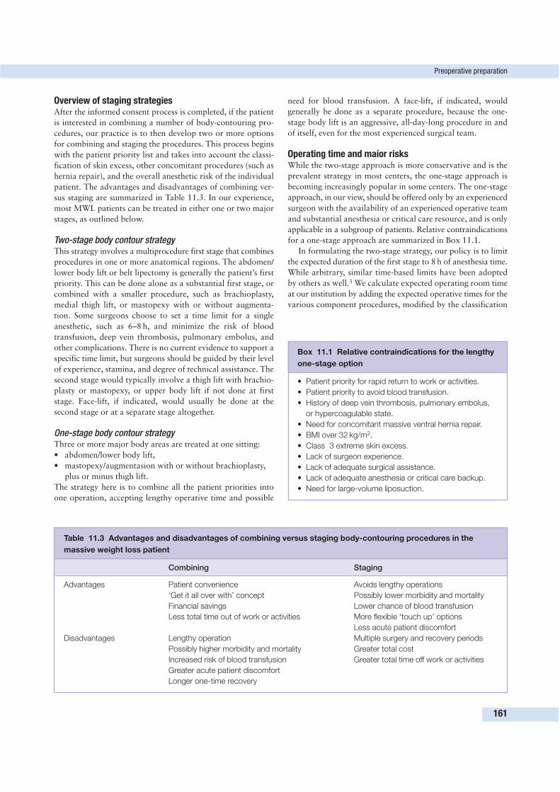
Overview of staging strategiesAfter the informed consent process is completed, if the patient
is interested in combining a number of body-contouring pro-
cedures, our practice is to then develop two or more options
for combining and staging the procedures. This process begins
with the patient priority list and takes into account the classi-
fication of skin excess, other concomitant procedures (such as
hernia repair), and the overall anesthetic risk of the individual
patient. The advantages and disadvantages of combining ver-
sus staging are summarized in Table 11.3. In our experience,
most MWL patients can be treated in either one or two major
stages, as outlined below.
Two-stage body contour strategyThis strategy involves a multiprocedure first stage that combines
procedures in one or more anatomical regions. The abdomen/
lower body lift or belt lipectomy is generally the patient’s first
priority. This can be done alone as a substantial first stage, or
combined with a smaller procedure, such as brachioplasty,
medial thigh lift, or mastopexy with or without augmenta-
tion. Some surgeons choose to set a time limit for a single
anesthetic, such as 6–8 h, and minimize the risk of blood
transfusion, deep vein thrombosis, pulmonary embolus, and
other complications. There is no current evidence to support a
specific time limit, but surgeons should be guided by their level
of experience, stamina, and degree of technical assistance. The
second stage would typically involve a thigh lift with brachio-
plasty or mastopexy, or upper body lift if not done at first
stage. Face-lift, if indicated, would usually be done at the
second stage or at a separate stage altogether.
One-stage body contour strategyThree or more major body areas are treated at one sitting:
• abdomen/lower body lift,
• mastopexy/augmentasion with or without brachioplasty,
plus or minus thigh lift.
The strategy here is to combine all the patient priorities into
one operation, accepting lengthy operative time and possible
need for blood transfusion. A face-lift, if indicated, would
generally be done as a separate procedure, because the one-
stage body lift is an aggressive, all-day-long procedure in and
of itself, even for the most experienced surgical team.
Operating time and maíor risksWhile the two-stage approach is more conservative and is the
prevalent strategy in most centers, the one-stage approach is
becoming increasingly popular in some centers. The one-stage
approach, in our view, should be offered only by an experienced
surgeon with the availability of an experienced operative team
and substantial anesthesia or critical care resource, and is only
applicable in a subgroup of patients. Relative contraindications
for a one-stage approach are summarized in Box 11.1.
In formulating the two-stage strategy, our policy is to limit
the expected duration of the first stage to 8 h of anesthesia time.
While arbitrary, similar time-based limits have been adopted
by others as well.3 We calculate expected operating room time
at our institution by adding the expected operative times for the
various component procedures, modified by the classification
Preoperative preparation
161
Table 11.3 Advantages and disadvantages of combining versus staging body-contouring procedures in the
massive weight loss patient
Combining Staging
Advantages Patient convenience Avoids lengthy operations
‘Get it all over with’ concept Possibly lower morbidity and mortality
Financial savings Lower chance of blood transfusion
Less total time out of work or activities More flexible ‘touch up’ options
Less acute patient discomfort
Disadvantages Lengthy operation Multiple surgery and recovery periods
Possibly higher morbidity and mortality Greater total cost
Increased risk of blood transfusion Greater total time off work or activities
Greater acute patient discomfort
Longer one-time recovery
Box 11.1 Relative contraindications for the lengthy
one-stage option
• Patient priority for rapid return to work or activities.
• Patient priority to avoid blood transfusion.
• History of deep vein thrombosis, pulmonary embolus,
or hypercoagulable state.
• Need for concomitant massive ventral hernia repair.
• BMI over 32 kg/m2.
• Class 3 extreme skin excess.
• Lack of surgeon experience.
• Lack of adequate surgical assistance.
• Lack of adequate anesthesia or critical care backup.
• Need for large-volume liposuction.
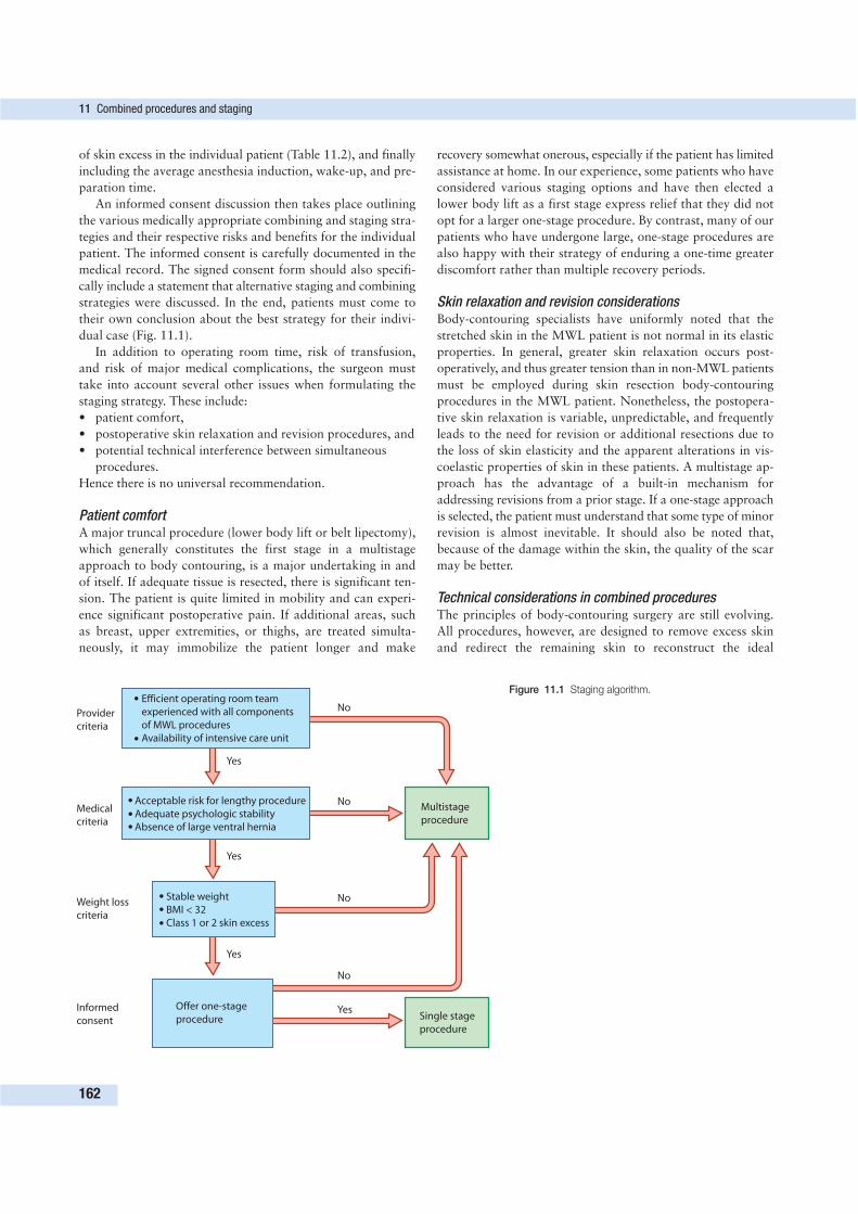
of skin excess in the individual patient (Table 11.2), and finally
including the average anesthesia induction, wake-up, and pre-
paration time.
An informed consent discussion then takes place outlining
the various medically appropriate combining and staging stra-
tegies and their respective risks and benefits for the individual
patient. The informed consent is carefully documented in the
medical record. The signed consent form should also specifi-
cally include a statement that alternative staging and combining
strategies were discussed. In the end, patients must come to
their own conclusion about the best strategy for their indivi-
dual case (Fig. 11.1).
In addition to operating room time, risk of transfusion,
and risk of major medical complications, the surgeon must
take into account several other issues when formulating the
staging strategy. These include:
• patient comfort,
• postoperative skin relaxation and revision procedures, and
• potential technical interference between simultaneous
procedures.
Hence there is no universal recommendation.
Patient comfortA major truncal procedure (lower body lift or belt lipectomy),
which generally constitutes the first stage in a multistage
approach to body contouring, is a major undertaking in and
of itself. If adequate tissue is resected, there is significant ten-
sion. The patient is quite limited in mobility and can experi-
ence significant postoperative pain. If additional areas, such
as breast, upper extremities, or thighs, are treated simulta-
neously, it may immobilize the patient longer and make
recovery somewhat onerous, especially if the patient has limited
assistance at home. In our experience, some patients who have
considered various staging options and have then elected a
lower body lift as a first stage express relief that they did not
opt for a larger one-stage procedure. By contrast, many of our
patients who have undergone large, one-stage procedures are
also happy with their strategy of enduring a one-time greater
discomfort rather than multiple recovery periods.
Skin relaxation and revision considerationsBody-contouring specialists have uniformly noted that the
stretched skin in the MWL patient is not normal in its elastic
properties. In general, greater skin relaxation occurs post-
operatively, and thus greater tension than in non-MWL patients
must be employed during skin resection body-contouring
procedures in the MWL patient. Nonetheless, the postopera-
tive skin relaxation is variable, unpredictable, and frequently
leads to the need for revision or additional resections due to
the loss of skin elasticity and the apparent alterations in vis-
coelastic properties of skin in these patients. A multistage ap-
proach has the advantage of a built-in mechanism for
addressing revisions from a prior stage. If a one-stage approach
is selected, the patient must understand that some type of minor
revision is almost inevitable. It should also be noted that,
because of the damage within the skin, the quality of the scar
may be better.
Technical considerations in combined proceduresThe principles of body-contouring surgery are still evolving.
All procedures, however, are designed to remove excess skin
and redirect the remaining skin to reconstruct the ideal
11 Combined procedures and staging
162
Efficient operating room team
experienced with all components
of MWL procedures
Availability of intensive care unit
Acceptable risk for lengthy procedure
Adequate psychologic stability
Absence of large ventral hernia
Stable weight
BMI < 32
Class 1 or 2 skin excess
Offer one-stage
procedure Single stage
procedure
Multistage
procedure
No
No
No
No
Yes
Yes
Yes
Yes
Provider
criteria
Medical
criteria
Weight loss
criteria
Informed
consent
Figure 11.1 Staging algorithm.
anatomical form. Because of skin relaxation concerns, the
vectors of pull in many of these procedures are substantial. In
certain permutations and combinations of procedures, the
surgeon may find that vectors of pull in various operative
fields are counterbalancing, influencing, or complicating each
other. In a lower body lift or belt lipectomy, for example, the
abdominoplasty flap in the upper abdomen and flank is pulled
inferiorly and laterally with great tension to meet the lower
flap from the groin and hips. This may place some downward
tension on the inframammary fold area and create some in-
ferior displacement of the fold. If an upper body lift is per-
formed simultaneously, the key principle of restoring the
inframammary fold and its lateral extension to the correct
position results in an opposite, superiorly directed vector on
the very same upper abdominal and flank tissue. At a mini-
mum, this may lead to increased technical difficulty during an
already complex procedure.
It is possible that conflicting vectors of pull from simulta-
neous procedures may also lead to suboptimal results, asym-
metries, or wound dehiscence. The surgeon must individually
consider the vectors of pull of proposed combined procedures
to ensure that the combination will not create technical prob-
lems or confounding conditions.
SURGICAL TECHNIQUE AND OUTCOMES
Detailed descriptions of techniques and outcomes for the va-
rious procedures are outlined elsewhere in this text. If multiple
procedures are performed at one sitting, the usual precautions
for lengthy procedures must be taken. These include:
• placement of a urinary catheter,
• sequential compression devices, and
• appropriate padding and checking of pressure points.
We do not routinely use prophylactic anticoagulants.
Procedures that involve multiple position changes, such as
lower body lift or belt lipectomy, should be performed first.
Currently, the most common positioning strategies are prone–
supine and supine–lateral–lateral, although supine–lateral–
lateral is also used by some surgeons. Our preferred sequence
is to begin prone, performing the posterior body lift, the but-
tock autoaugmentation, the posterior thigh resection, and/or
the posterior upper body lift resection. The legs are abducted
and adducted at appropriate points in the procedure. Because
the abdominal closure is the tightest, it is performed last, so
that additional position changes are not required after com-
pletion of that component of the surgery.
Following the prone phase of the procedure, the patient is
placed in the supine position for the remaining elements. We
have found it useful to roll the patient to the supine position
on an adjacent stretcher, and then move directly back to the
operating room table. The remaining procedure is then
completed, such as the anterior portion of the body lift, the
anterior element of the thigh lift, brachioplasty, and/or breast
surgery.
COMPLICATIONS AND THEIR MANAGEMENT
Most complications of combined procedures relate to an indi-
vidual component procedure and are discussed in the appro-
priate section of the text. There is no evidence that seromas,
wound dehiscence, and other common complications of indi-
vidual procedures are increased in incidence when procedures
are combined. In this chapter, discussion will be limited to
those complications that are of particular concern in com-
bined procedures. As outlined above, the major concerns about
combining multiple procedures are complications that are
associated with lengthy operative time. The most important
and life-threatening of these is venous thromboembolism.
Death from pulmonary embolus is fortunately an extremely
rare complication of body-contouring surgery. When it occurs,
especially in the setting of aesthetic surgery, it is a devastating
complication. In their recent review of thromboembolism in
plastic surgery, Most et al. described a death from pulmonary
embolus in an MWL patient following hernia repair, abdomi-
noplasty, and thigh lift, despite the use of all appropriate
perioperative precautions.3 Abdominoplasty alone carries a
reported incidence of 0.8% for pulmonary embolus.4 When
combined with other intraabdominal or aesthetic procedures,
the incidence is higher, from 1.1% to 6.6%.5,6 In other cos-
metic procedures, such as rhytidectomy, deep vein thrombosis
and pulmonary embolus were more likely if the procedure
was performed under general anesthesia, according to results
of a survey by Reinisch et al.7
A task force from the American Society of Plastic Surgeons
stratified risk in office-based procedures.8 Because all body-
contouring procedures in the MWL patient require over
30 min of general anesthesia, all such patients fall into the
‘moderate’ or ‘high’ risk category established by the task force.
Moderate-risk patients require comfortable positioning and
sequential compression stockings. High-risk patients, including
those with malignancy, obesity, or hypercoagulable state, are
advised to use the same precautions as those for the moderate-
risk patients, plus a hematology consultation and possible use
of low-molecular-weight heparin before the procedure and
daily in the postoperative period until ambulatory.
Several preparations of low-molecular-weight heparin exist.
A common regimen for use of one of these agents is to
administer dalteparin 2500 IU 1–2 h before surgery and then
2500 IU every day for 5–10 days after surgery. But to date
there is no clear-cut evidence that low-molecular-weight heparin
offers a distinct advantage over intermittent pneumatic com-
pression stockings in this patient population, nor is there
evidence that the marginal addition of low-molecular-weight
heparin in addition to intermittent pneumatic compression
stockings provides a distinct benefit in body-contouring
surgery.
When deep vein thrombosis is suspected, it should be
promptly and aggressively evaluated, initially with Doppler
examination of the venous system. If a deep vein thrombosis is
confirmed, treatment should begin immediately, and further
Complication and Their Management
163
evaluation for pulmonary embolus should be performed,
including spiral computerized tomography scan.
Many reports involving combined body-contouring proce-
dures appropriately focused on description of the techniques,
and lack sufficient numbers to determine the incidence of low-
probability events such as pulmonary embolus.9 The term belt
lipectomy was used originally by Gonzalez-Ulloa,10 and was
modified by Baroudi.11 Currently, this term is generally applied
to circumferential resections centered above the hips and along
the waistline. Most early discussions of combined procedures
were prior to the popularization of bariatric surgery.12–15
Lockwood’s seminal work involved description of the super-
ficial fascial system and the pioneering design of many com-
bined procedures in the MWL patient.16–18
In one of the first large series of body contouring in post–
weight loss patients, presented by Dardour in 1986,19 the
single reported mortality in 300 patients was due to a pulmo-
nary embolus. In 30 patients who underwent circumferential
torsoplasty by Van Geertruyden,20 one pulmonary embolus
was noted. In Hamra’s report of a series of 40 body lift
patients,21 no major complications were reported. Da Costa
recently published the results for a series of 48 patients who
underwent modified abdominoplasty after MWL.22 These
were limited procedures, averaging 180 min of total operative
time, and there were no instances of pulmonary embolus.
Recent reports on combined body-contouring procedures
in the MWL patient, performed by recognized experts at
renowned centers of excellence, show a high incidence of
pulmonary embolus. In a series of 32 patients who underwent
belt lipectomy, which combines abdominoplasty with a cir-
cumferential trunk excision, Aly reported a 9.3% pulmonary
embolus rate.23 This series included some patients who were
still overweight, but contained a group of 21 patients with
MWL (average 187-lb [85 kg] preoperative weight loss). Their
average operative time was 5.75 h, ranging from 4.86 to 6.93 h,
and the average tissue resection was 10 lbs (4.5 kg). There was
no mortality, and all patients recovered fully.
In Ellabban’s series of 14 MWL patients who underwent
abdominoplasty combined with medial thigh lift, all patients
were given perioperative low-molecular-weight heparin as well
as intraoperative sequential compression devices.24 Operative
times were remarkably low, with a mean time of 2 h, and the
average mass of removed tissue was 70 oz (1995 g). No pul-
monary embolus was noted. It is important to note that these
combined procedures did not include circumferential resection.
Pascal described a series of 40 lower body lifts that com-
bine high lateral tension abdominoplasty with circumferential
skin resection and buttock lift. The incisions for the lower
body lift are generally lower than for the related procedure of
belt lipectomy. His group used low-molecular-weight heparin
and sequential compression devices. There was no mention of
average operative time or mass of resected tissue. No pulmo-
nary embolus was noted.
Hurwitz reported eight cases of what may be considered
the ultimate in combined body-contouring procedure: the total
body lift.25 This includes:
• circumferential abdominoplasty,
• lower body lift, and
• medial thighplasty.
It may also include brachioplasty and/or mastopexy and aug-
mentation. Operative time ranged from 7–12 h, and trans-
fusions ranged from 0 to 4 units. No pulmonary embolus
occurred in these eight patients. One patient suffered from
generalized edema and required readmission. These results are
possible only with a very experienced team, and occasional
use of the two-team approach with simultaneous surgery in
two areas was noted. Even so, Hurwitz states that ‘only the
smaller and healthy weight loss patients should be offered
these 1-stage procedures’.
CONCLUSION
The explosive popularity of bariatric surgery has created
demand for a new genre of body-contouring surgery. Plastic
surgeons performing these procedures on the MWL patient
need to constantly examine their own practice and experience,
as well as the needs and priorities of the individual patient, to
make sound recommendations about how multiple procedures
should be combined or staged.
• In the healthy MWL patient who is a candidate for
treatment of numerous body areas, one-stage and
two-stage approaches are medically appropriate options,
with informed consent about the risks.
• Multiprocedure one-stage combinations should be
performed only in appropriate patients by experienced
surgical teams. Two-stage approaches are currently more
common in most centers.
When undertaking lengthy combined procedures, careful
medical evaluation and perioperative prophylaxis against
deep venous thrombosis and other risks are essential.
REFERENCES
1. Steinbrook R. Surgery for severe obesity. N Engl J Med 2004;
350(11):1075–1079.
2. Mosteller RD. Simplified calculation of body surface area. N Engl J
Med 1987; 317(17):1098.
3. Most D, Kozlow J, Heller J, et al. Thromboembolism in plastic
surgery. Plast Reconstr Surg 2004; 115(2):20e–30e.
4. Grazer FM, Goldwyn RM. Abdominoplasty assessed by survey,
with emphasis on complications. Plast Reconstr Surg 1977;
59(4):513–517.
5. Voss SC, Sharp HC, Scott JR. Abdominoplasty combined with gyne-
cologic surgical procedures. Obstet Gynecol 1986; 67(2):181–185.
6. Hester RT Jr, Baird W, Bostwick J III, et al. Abdominoplasty
combined with other surgical procedures: safe or sorry? Plast
Reconstr Surg 1989; 83(6):997–1004.
7. Reinisch JF, Bresnick SD, Walker JWT, et al. Deep venous throm-
bosis and pulmonary embolus after face lift: a study of incidence
and prophylaxis. Plast Reconstr Surg 2001; 107(6):1570–1575.
8. Iverson RE, ASPS Task Force on Patient Safety in Office-based
Surgery Facilities. Patient safety in office-based surgery facilities: I.
11 Combined procedures and staging
164
Procedures in the office-based surgery setting. Plast Reconstr Surg
2002; 110(5):1337–1342.
9. Gonzalez M, Guerrero-Santos J. Deep planed torso-abdomino-
plasty combined with buttocks pexy. Aesthetic Plast Surg 1997;
21(4):245–253.
10. Gonzalez-Ulloa M. Belt lipectomy. Br J Plast Surg 1961; 13:179.
11. Baroudi R. Body contouring surgery in the 90s. In: Advances in
Plastic and Reconstructive Surgery, vol 9. St. Louis: Mosby Year-
Book; 1992:1–37.
12. Barrett BM, Kelly MV. Combined abdominoplasty and augmenta-
tion mammaplasty through a transverse suprapublic incision. Ann
Plast Surg 1980; 4(4):286–291.
13. Pitanguy I, Ceravolo MP. Our experience with combined procedures
in aesthetic plastic surgery. Plast Reconstr Surg 1983; 71(1):56–65.
14. Hallock GG, Altobelli JA. Simultaneous brachioplasty, thoracoplasty,
and mammaplasty. Aesthetic Plast Surg 1985; 9(3):233–235.
15. Hauben DJ, Benmeir P, Charuzi I. One-stage body contouring. Ann
Plast Surg 1988; 21(5):472–479.
16. Lockwood TE. Superficial fascial system (SFS) of the trunk and extre-
mities: a new concept. Plast Reconstr Surg 1991; 87(6):1009–1018.
17. Lockwood TE. Lower body lift with superficial fascial system
suspension. Plast Reconstr Surg 1993; 92(6):1112–1122.
18. Lockwood TE. Lower-body lift. Aesthetic Surg J 2001; 21:355.
19. Dardour JC, Vilain R. Alternatives to the classic abdominoplasty.
Ann Plast Surg 1986; 17(3):247–258.
20. Van Geertruyden J, Vandeweyer E, de Fontaine S, et al. Circum-
ferential torsoplasty. Br J Plast Surg 1999; 52(8):623–628.
21. Hamra ST. Circumferential body lift. Aesthetic Surg J 1999; 19:244.
22. Da Costa LF, Landecker A, Manta, AM. Optimizing body contour
in massive weight loss patients: the modified vertical abdomino-
plasty. Plast Reconstr Surg 2004; 114(7):1917–1923.
23. Aly A, Cram A, Chao M, et al. Belt lipectomy for circumferential
truncal excess: the University of Iowa experience. Plast Reconstr
Surg 2003; 111(1):398–413.
24. Ellabban MG, Hart NB. Body contouring by combined abdomino-
plasty and medial vertical thigh reduction: experience of 14 cases.
Br J Plast Surg 2004; 57(3):222–227.
25. Hurwitz DJ. Single stage total body lift after massive weight loss.
Ann Plast Surg 2004; 52(5):435–441.
FURTHER READING
Matarasso A. Discussion. Is it safe to combine abdominoplasty with
elective breast surgery? A review of ISI consecutive cases. Plast
Reconstr Surg 2006; 118(1):213–4.
Further reading
165
167
THE ROLE OF LARGE-VOLUMELIPOSUCTION AND OTHERADJUNCTIVE PROCEDURES 12V. Leory Young and Robert F. Centeno
Key PointsLiposuction
• If the patient needs debulking of subcutaneous fat in several areas,
including the trunk, large-volume liposuction (LVL) may be an appro-
priate first stage of body contouring, especially if LVL will improve the
aesthetic outcome of later staged excisions. This most often applies to
patients with a BMI higher than 30 kg/m2.
• If the patient chooses a major excisional procedure first, such as a
circumferential body lift (CBL), identify remote areas (e.g. upper back,
arms, thighs, or neck) that will benefit most from liposuction during the
same surgery.
• Liposuction is useful for refining contour or removing residual subcuta-
neous fat several months after excisional procedure wounds have healed.
• Know your vascular anatomy, and be extremely cautious if performing
liposuction near an area that will be excised in the same surgery to
prevent disruption of a flap’s vascular supply.
• If lipoplasty and an excisional procedure are performed during a single
surgery, patient safety issues become more complex. Surgeons must be
mindful of potential complications arising from both excision and lipo-
suction and treat patients accordingly.
Mons reduction
• Improving the mons and genital area will improve function, hygiene,
appearance, and patient satisfaction.
• Mons reduction can be safely combined with a CBL.
• Keep mons undermining to a minimum.
• Inform patients about temporarily decreased skin sensation, clitoral
hypersensitivity in female patients, and prolonged edema and hypere-
mia following mons reduction.
Intergluteal reduction
• The skin length discrepancy and deforming effect of the posterior
portion of a CBL can create a secondary deformity of the buttock. Inter-
gluteal reduction or a V-shaped inverted dart incision in the intergluteal
cleft helps minimize this deformity.
• The traditional posterior portion of a CBL incision is higher than is
aesthetically ideal.
• To improve gluteal aesthetics, preserve the sacral triangle by lowering
the central portion of the posterior body lift incision. Keep the incision at
or below the level of the posterior iliac crest.
• Consult with patients about preferred underwear and bathing suit styles
when designing incisions, but remind patients that fashion trends
change. The goal of incision design should be the optimal aesthetic in
the nude.
Autologous gluteal augmentation
• Thoracic spine/postural changes and anterior-inferior pelvic rotation
associated with morbid obesity persist after massive weight loss and
contribute to severe platypygia.
• The posterior component of a CBL causes flattening of the buttock.
• Autologous tissue of the lower back that would normally be discarded
can be safely used to preserve or enhance projection in the gluteal
region.
• Paucity of tissue overlying the coccyx and sacrum can be symptomatic,
so preserving tissue in this area is important.
Axilloplasty
• Reducing the skin excess of the lateral chest wall/axilla can be safely
combined with a brachioplasty, mastopexy or autologous breast augmen-
tation, upper body lift, or CBL.
• Addressing this skin excess and recreating the lateral inframammary
crease enhances the aesthetic results of breast procedures.
• Tissue that is normally discarded can be used for breast autoaug-
mentation as a well-perfused, well-described flap with known cir-
culation.
• Preservation of critical axillary structures—including the brachial plexus,
intercostobrachial nerve, lymphatics, and axillary fascia—will decrease
morbidity.
Autologous breast augmentation
• The use of autologous axillary or lateral chest wall tissue to increase
breast volume represents a good option for patients who do not want
augmentation with an implant.
• Adding autologous tissue to the breast area provides increased volume
and/or padding to prevent implant wrinkling and palpability if augmen-
tation mammaplasty and/or mastopexy are planned.
• The normally discarded axillary tissue forms a lateral thoracoepigastric
flap that is characterized by reliable perfusion and known anatomy.
• The use of the lateral thoracoepigastric flap is flexible enough to ac-
commodate virtually all pedicle, skin excision, and breast pocket
designs.
Liposuction plays an important role in body contouring of
massive weight loss (MWL) patients and can be used to con-
tour any body area that has excess fat. Suction-assisted (SAL),
ultrasound-assisted (UAL), and power-assisted lipoplasty
(PAL)—or their combination—are useful in the following
contexts.
• Patients who need debulking of widespread subcutaneous
fat prior to a staged excisional procedure.
• Patients who want or need additional contouring or
removal of residual excess subcutaneous fat following an
excisional procedure.
• Patients who have lipodystrophy in areas such as the
upper back, thighs, or arms that may be improved with
liposuction rather than excision.
Regardless of whether a patient has lost weight following
gastric bypass surgery or through rigorous diet and exercise,
weight loss will not be evenly distributed throughout all ana-
tomical regions. Most patients lose visceral fat, which corre-
lates with the reduction of their medical comorbidities, but
significant subcutaneous fat may remain even after weight
loss has stabilized. In most cases, areas of localized lipodys-
trophy are produced.
The volume of subcutaneous tissue plays an important role
in the decision-making process when considering which pro-
cedures to undertake and in what order. As an example, loss
of subcutaneous fat in the lower abdomen may be greater
than in the upper abdomen. If a circumferential body lift (CBL)
or panniculectomy is performed, patients may still have a large
excess of subcutaneous fat in the epigastric region. Debulking
this area with liposuction can simplify excisional procedures
and produce a better aesthetic outcome. Liposuction is espe-
cially effective for removing excess fat in the back that is
difficult to treat with a CBL. Another area that benefits from
debulking prior to excision is the arms, which may retain
significant excess fat even after patients have plateaued in
their weight loss. If the arms are debulked with liposuction
first, an excisional brachioplasty performed 3–6 months
later—after the tissues have softened and vascularity has
improved—will produce much better results. The thighs also
benefit from debulking liposuction, as long as drains are used
to prevent chronic seroma formation and infection.
For patients with significant subcutaneous volume, staged
debulking liposuction can be safely performed before or after
excisional procedures. For some patients, large-volume lipo-
suction (LVL) as the first stage of body contouring may permit
use of less extensive excisions or fewer staged procedures, as
illustrated by the patient shown in Figure 12.1. The improve-
ment in this patient’s body contour would not have been
possible without LVL, which prompted her weight loss. Limited
liposuction combined with excisional procedures has been
performed for years. Established combinations include:
• lower flank liposuction with abdominoplasty,
• submental liposuction with facialplasty,
• reduction mammaplasty with axillary lipoplasty, and
• CBL with thigh liposuction (Figure 12.2).
All these combinations share a focused use of liposuction based
on known vascular anatomy and accumulated experience.
As the natural tendency toward innovation continues in
plastic surgery, the literature increasingly reports on exci-
sional procedures—such as abdominoplasty, thighplasty, and
brachioplasty—combined in a single surgery with lipoplasty
in areas that share a vascular supply.1–7 Proponents believe
that liposuction performed on or adjacent to flaps allows
smaller excisions and improves aesthetic outcomes. Reports
published thus far are interesting and suggest that less flap
undermining is required if liposuction and excision are com-
bined. However, more safety data are needed before we know
whether the risk associated with these combinations is accept-
able, and Matarasso advises that extensive liposuction with a
full abdominoplasty is ill advised.8 Patients must be properly
informed about the potentially increased risks of delayed
wound healing, infection, flap necrosis, or unfavorable scarring
if excision and lipoplasty are combined. Above all, know your
vascular anatomy before attempting to perform liposuction in
or near an excision site. When in doubt, take a conservative
approach rather than risk serious complications such as flap
necrosis or delayed healing.
Issues of patient selection and informed consent have been
covered elsewhere in this book. If liposuction is to be included
in the body-contouring process, additional patient assessment
must be done and consent obtained. By its nature, liposuction
induces what may be considered blunt trauma injury. In addi-
tion, LVL may be associated with large fluid shifts that are
dangerous—even fatal—if not handled appropriately. Patients
should understand that they will have some excess skin and
contour irregularities such as lumps, depressions, and wrinkles
after LVL. The duration of recovery for LVL patients is appro-
ximately 3 weeks, but persistent swelling may last up to
6 months. Impressive skin retraction often occurs, especially
after LVL, but final results will not be known for 3–6 months.
Excision may be performed then if excessive skin laxity or
contour irregularities remain.
PREOPERATIVE PREPARATION
The length of surgery and health history of MWL patients
demand that multiple factors be addressed during the month
or so prior to surgery, regardless of whether the planned surgery
is LVL alone or excision plus liposuction. Some guidelines
follow.
• Obtain clearance from MWL patients’ internists or
primary care physicians to ensure that they can safely
undergo a large and lengthy operation. If patients do not
have a physician, refer them to an internist.
• Pay special attention to cardiac health in patients
undergoing LVL, because high-dose adrenaline
(epinephrine) increases the risk for arrhythmias, fatal
asystole, and myocardial infarction during surgery.
Therefore hyperthyroidism, severe hypertension, cardiac
12 The role of large-volume liposuction and other adjunctive procedures
168
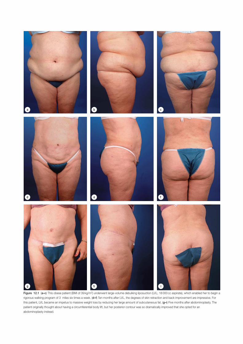
d e f
a b c
g h i
Figure 12.1 (a–c) This obese patient (BMI of 39 kg/m2) underwent large-volume debulking liposuction (LVL; 18 000 cc aspirate), which enabled her to begin a
rigorous walking program of 3 miles six times a week. (d–f) Ten months after LVL, the degrees of skin retraction and back improvement are impressive. For
this patient, LVL became an impetus to massive weight loss by reducing her large amount of subcutaneous fat. (g–i) Five months after abdominoplasty. The
patient originally thought about having a circumferential body lift, but her posterior contour was so dramatically improved that she opted for an
abdominoplasty instead.
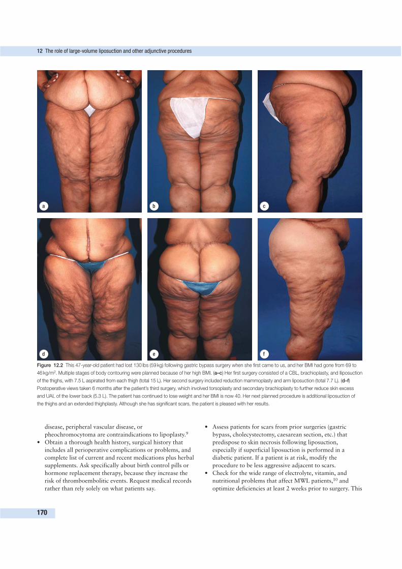
disease, peripheral vascular disease, or
pheochromocytoma are contraindications to lipoplasty.9
• Obtain a thorough health history, surgical history that
includes all perioperative complications or problems, and
complete list of current and recent medications plus herbal
supplements. Ask specifically about birth control pills or
hormone replacement therapy, because they increase the
risk of thromboembolitic events. Request medical records
rather than rely solely on what patients say.
• Assess patients for scars from prior surgeries (gastric
bypass, cholecystectomy, caesarean section, etc.) that
predispose to skin necrosis following liposuction,
especially if superficial liposuction is performed in a
diabetic patient. If a patient is at risk, modify the
procedure to be less aggressive adjacent to scars.
• Check for the wide range of electrolyte, vitamin, and
nutritional problems that affect MWL patients,10 and
optimize deficiencies at least 2 weeks prior to surgery. This
12 The role of large-volume liposuction and other adjunctive procedures
170
d e f
a b c
Figure 12.2 This 47-year-old patient had lost 130 lbs (59 kg) following gastric bypass surgery when she first came to us, and her BMI had gone from 69 to
46 kg/m2. Multiple stages of body contouring were planned because of her high BMI. (a–c) Her first surgery consisted of a CBL, brachioplasty, and lliposuction
of the thighs, with 7.5 L aspirated from each thigh (total 15 L). Her second surgery included reduction mammoplasty and arm liposuction (total 7.7 L). (d–f)
Postoperative views taken 6 months after the patient’s third surgery, which involved torsoplasty and secondary brachioplasty to further reduce skin excess
and UAL of the lower back (5.3 L). The patient has continued to lose weight and her BMI is now 40. Her next planned procedure is additional liposuction of
the thighs and an extended thighplasty. Although she has significant scars, the patient is pleased with her results.
may involve intensive vitamin supplementation, protein
supplementation, and nutritional counseling for at least a
month before surgery.
• Carefully evaluate hematologic parameters, because low
hemoglobin levels are frequent among MWL patients.
Some may require recombinant erythropoietin to raise the
hematocrit before surgery,11 but this therapy carries an
increased risk of hypercoagulability, requires intravenous
iron therapy, and is costly.
• Type and cross-match patients in anticipation of the need
for transfusion, a possibility that must be explained.
Autologous blood donation should be discouraged, but
directed donorship by family members can be arranged.
• Arrange for smoking cessation counseling to prevent
wound-healing problems in smokers. To measure
compliance with smoking cessation, perform continine
testing 2 weeks prior to surgery, on the morning of
surgery, and 2 weeks after surgery. A positive test before
surgery should result in delaying the procedure until the
patient stops smoking.
MarkingPatients undergoing liposuction alone should be marked in
the standing position before receiving any sedative medica-
tions. They may be marked in the preoperative area, but we
prefer to mark patients who will have excisional procedures
(with or without liposuction) a day or two before surgery.
Marking with indelible markers is best done in an unhurried
and private environment to enhance accuracy and improve
the patient experience. Preoperative marking takes time if
done properly, because it demands careful measurements and
double-checking.
• Make bilateral markings as symmetric as possible, and
note any preexisting asymmetries.
• Delineate prominent areas such as folds or bulges to be
liposuctioned, because they will be less apparent when the
patient lies down.
• Border areas where liposculpture feathering is anticipated
should also be identified.
Using differently colored markers facilitates color coding and
indicates areas to be treated differently.
Prophylactic measures 30–60 min before surgeryHypothermia prophylaxisBecause procedures are lengthy and large body areas are
exposed, body-contouring patients are highly susceptible to
inadvertent hypothermia, which is defined as a core body
temperature below 36°C. Hypothermia has been found to
increase the incidence of postoperative wound infections and
inhibit tissue oxygen delivery and coagulation functions,
thereby raising the risk of bleeding-related complications.12
Begin warming the patient in the preoperative area with either
heated cotton blankets or forced air blankets (such as a Bear
Hugger) at least 30 min prior to surgery. Cotton blankets
quickly lose their heat so must be continuously renewed. The
objective of prewarming is to increase the heat content of the
extremities so that heat will not be transferred out of the core
during surgery.
Raise the operating room temperature to 73°F (23°C),
which is the upper limit recommended by health-related govern-
ment agencies.13 Infection risk increases when temperature rises
above 73°F and humidity is outside the range of 30–60%.
Intravenous fluids, as well as liposuction infiltration fluids,
should be warmed between 37 and 42°C with a fluid warmer
to help maintain normothermia.12 This includes the fluids
begun in the preoperative area to replace deficits caused by
overnight fasting. All fluids administered throughout the sur-
gery and recovery room should be warmed. Do not heat fluids
to temperatures higher than 42°C or burns may result.
Warming the infiltration fluids is probably not necessary in
UAL because the ultrasonic energy raises the temperature of
tissues and fluids.
Thromboembolism prophylaxisIn 2004, the American College of Chest Physicians identified
the following to be among the major risk factors for venous
thromboembolitic events (VTEs) such as deep vein thrombo-
sis (DVT) and pulmonary embolism (PE):14
• prolonged surgical time (more than 1 h),
• general anesthesia,
• patient age of 40 or more, and
• obesity.
By these criteria, essentially all MWL patients undergoing body
contouring have a moderate to high risk for VTEs. PE usually
arises from DVT in the legs at or proximal to the popliteal
veins, with above-knee DVTs most often being the culprit. The
frequency of DVT is between 15 and 40% of general surgery
patients if no prophylaxis is given,14 and 30–50% of patients
with undiagnosed and untreated DVT progress to PE.15 Even
when prophylactic measures are taken, the risk of DVT lasts
for at least 4 weeks after surgery.14 Consequently, attention to
VTE prevention must be a priority long after patients have
gone home.
Mechanical prophylactic methods include compression
stockings and intermittent pneumatic compression devices or
venous foot pumps. Intermittent pneumatic compression devices
or venous foot pumps are recommended for any plastic sur-
gery procedure that lasts more than 1 h and for all patients
undergoing general anesthesia.16 The use of intermittent
pneumatic compression devices or venous foot pumps should
begin approximately 30–60 min prior to surgery.
Anticoagulant therapy is the most effective method of
DVT/PE prevention and the only real option for patients with
a prior history of DVT/PE or a hypercoagulability disorder.
Anticoagulant choices include:
• low-molecular-weight heparin (LMWH);
• low-dose unfractionated heparin; and
• the recently approved drug called fondaparinux (Arixtra),
which specifically inhibits the activation of coagulation
factor X.
Preoperative preparation
171
Clinical trials suggest that fondaparinux may be twice as
effective as LMWH in preventing postoperative DVT, and its
use requires no routine coagulation monitoring.17 Adequate
prophylaxis can be achieved by administering either LMWH
or fondaparinux the morning after surgery, or at least 12 h
following surgery completion. VTE prophylaxis should be
continued until patients are fully ambulatory. For high- and
very high-risk patients, continue chemoprophylaxis at home
for 2 weeks, or longer if warranted by risk factors.
None of these anticoagulants has been found to increase
clinically significant bleeding, and although postoperative hema-
tomas are possible, they are uncommon. To help put bleeding
risks in context, remember that acute adverse events occur in
less than 1% of patients receiving transfusion18 versus the
15–40% of general surgery patients who develop DVT. Con-
cerns about bleeding during liposuction are probably justified
because sites of bleeding cannot be visualized and addressed,
as is the case with excision. However, we have not had adverse
bleeding in LVL patients given postoperative chemoprophylaxis.
Antibiotic prophylaxis• For patients not allergic to penicillin, begin administration
of 1 g of cefazolin (Ancef) 30 min before surgery.
• Patients with a penicillin allergy are given 500 mg of
clindamycin intravenously infused over 1 h immediately
prior to surgery.
• Diflucan should be given to patients with yeast infections.
• In cases that take longer than 6 h, repeat antibiotics during
surgery.
DrapingPlace forced warm air blankets beneath the patient on the
operating table and also cover patient areas outside the
operating field, such as the head and extremities. The key to
draping is to allow easy access for infusion and aspiration of the
wetting solution. Areas wider than those to be suctioned are
exposed so that the area being contoured can be blended into
the non-contoured area. Drapes should not distort the body
contours with their weight. After completing work on an area
(or two symmetric areas), redrape the patient to retain heat.
SURGICAL TECHNIQUE
AnesthesiaLarge-volume liposuction (5000 cc of aspirate or greater) and
other body-contouring procedures in MWL patients are best
performed using general anesthesia with endotracheal intuba-
tion. Because these patients typically must be repositioned
during surgery, intubation assures maintenance of the airway.
In addition, patients are more comfortable, oxygenation is
ensured, and monitoring can be done to detect any problems.
When anesthesia is induced, a Foley catheter is inserted to aid
with fluid monitoring. We advise a distal esophageal probe or
tympanic membrane device for constant monitoring of core
body temperature.
OxygenationMost anesthesiologists administer oxygen at an FiO2 of
30–50% during general anesthesia. However, a large rando-
mized and blinded study of intraabdominal surgery patients
found that an FiO2 of 80% during surgery and for 2 h after-
ward reduced the incidence of wound infections by more than
half when compared with the use of 30% FiO2 (5.2% versus
11.2%).12 The use of 80% FiO2 may be especially important
in lipoplasty patients who have received intentional vasocon-
striction by adrenaline (epinephrine). Another benefit of using
80% FiO2 is that the incidence of postoperative nausea and
vomiting is markedly reduced (approximately 50%) when
compared with 30% for FiO2.12
PositioningPosition is dictated by the areas being treated with liposuction
and same-surgery excisional procedures. The arms, flanks, back,
hips, and outer thighs are most accessible to liposuction in the
lateral decubitus position. The outer thigh offers a good exam-
ple of the effect that supine or prone versus lateral position can
have. In the supine or prone position, the weight of the body
distorts the area and access is limited, in contrast to the lateral
position that offers easy access and minimizes distortion. It is
also much easier to evaluate results with inspection and palpa-
tion. The symmetry of areas can be assessed and refined in supine
or prone positions. The abdomen, breasts, submental area, mons
pubis, anterior and inner thigh, and knees are best treated in
the supine position. When the patient is in a supine position,
place a pillow under the knees to promote venous return flow
through the popliteal area and thereby help prevent DVT.
Whatever position is chosen, it should allow easy access to
the areas being treated and minimize the risk of distortion
caused by position or pressure. A roll (folded/rolled linen)
under the patient’s chest or pelvis as indicated when in the
supine position is used to prevent pressure or allow thoracic
excision. Padding pressure points (i.e. joints) is important.
The legs can be widely abducted to allow access, and in order
to do so the ankles are positioned on padded arm shields.
Fluid managementFluid management is always a challenge in LVL because of the
risks of hypovolemia or fluid overload. Consequently, patients
undergoing LVL require a rigorous fluid management regimen.
The superwet technique is recommended to keep fluid infiltra-
tion and aspiration as close as possible to a 1:1 ratio (1 mL in
and 1 mL out). The tumescent technique relies on larger amounts
of infiltrate, with ratios as high as 3:1 to 7:1, and is therefore
more likely to cause fluid overload. When managing fluids,
remember that approximately 70% of the infiltrated wetting
solution is not aspirated but remains in the subcutaneous tissues
until slowly absorbed into the intravascular space.19,20 Thus the
majority of material in the aspirate is fat, not wetting solution.21
Use a data sheet to record the actual measurements of the
amounts of fluid going in and coming out. The ‘in’ half of the
1:1 ratio includes the subcutaneous infiltrate plus any supple-
mental fluids given intravenously. The ‘out’ consists of 30%
12 The role of large-volume liposuction and other adjunctive procedures
172
of the suctioned aspirate (the other 70% of infused fluid is not
aspirated), blood loss, urine output, and drainage through
drains. Subcutaneous infiltration solutions are usually mixed
in 1- or 3-L plastic bags with graduated markers of volume.
However, measuring by volume markers is very inaccurate.
Instead, measure the weight (in grams) dispersed from the bag.
When using the 1:1 ratio of infiltration and aspiration, the
volume of replacement fluids should be reduced to avoid the
danger of fluid overload. The suggested amount for LVL is
0.25 cc of crystalloid for each cc aspirated over 5000 cc.16,19
This is in addition to crystalloid intravenous maintenance
fluid administered at a rate of 1.5–2.0 cc/kg per h. The amount
of maintenance and replacement fluids should be monitored
and adjusted to vital signs and urine output.
Along with keeping meticulous records of fluid amounts
going in and coming out, a patient’s heart rate, blood pres-
sure, and urine output give important clues to the fluid status.
The patient is hemodynamically stable if:
• the systolic blood pressure is over 100 mmHg,
• the heart rate is under 100 bpm, and
• the urine output is 0.5–1.0 cc/kg per h or greater.
Urine output is perhaps the best indicator of the need for sup-
plemental fluids. The first sign of hypovolemia is usually tachy-
cardia or a heart rate greater than 100 bpm. Young, healthy
patients can often compensate by maintaining their blood
pressure, but they tend to become tachycardic eventually.
Blood lossDuring lipoplasty, the infiltrated wetting solution contains 1 cc
of adrenaline (epinephrine) 1:1000 per liter of lactated Ringer’s
solution (for a final concentration of 1:1 000 000 per liter) to
achieve vasoconstriction. Before adrenaline became part of the
liposuction wetting solution, the estimated blood loss was as
high as 45%. Some studies have determined that blood loss re-
presents about 1% of the aspirate when adrenaline is added.3,21
Karmo et al. compared hemoglobin levels before and 7 days
after surgery, and found a mean decrease in hemoglobin (g/dL)
of 0.93 ± 0.92 in SAL and 1 ± 0.64 in UAL for aspirate volumes
up to 6000 mL. However, Cárdenas-Camarena and colleagues
also evaluated the aspirate of patients undergoing LVL
(5–22.3 L) and determined blood loss to be more in the range
of 10% of the aspirate and higher after the seventh or eighth
liter was aspirated.22 The mean reduction of hemoglobin 1 week
after surgery was 3.8 g compared to presurgical levels.
Transfusion is always a possibility with LVL or liposuction
combined with excision. The guidelines for blood transfusion
are a hematocrit below 23% or symptoms such as orthostatic
hypotension and tachycardia. Patients with coronary or cen-
tral nervous system atherosclerosis should be treated more
aggressively. Hematocrit can be easily checked during surgery
to assess patient blood loss, but results may not be entirely
reliable for several days, until hematocrit equilibrium is
achieved following final resolution of fluid shifts.9,21 Healthy
young individuals with normal preoperative hematocrits of
approximately 40% can tolerate larger volumes of liposuc-
tion. Even though we have aspirated up to 34 L without giving
transfusion to a morbidly obese patient, it is not uncommon
to transfuse 2 units of packed red blood cells for aspirates
over 20 L. Safety should be the first concern, and either the
volume aspirated should be limited to an amount that main-
tains hemodynamic stability or transfusion should be avail-
able based on hematocrit and symptoms.
Fluid infusionSurgeons should use the technologies and materials with which
they are most comfortable. Neither LVL nor liposuction com-
bined with excisional procedures should be attempted by the
inexperienced because of the complex fluid management issues.
Some general guidelines follow.
• Consider not including lidocaine when liposuction is
performed under general anesthesia (as it usually is in
MWL patients).
• Add 1 cc of adrenaline (epinephrine) (1:1000) for
hemostasis per liter of Ringer’s lactate (for a final solution
of 1:1 000 000).
• Warm infused fluids to a temperature between 37 and
42°C for SAL.
• Keep in mind the 1:1 infiltration to aspiration ratio when
infiltrating wetting solution.
• Infuse wetting solution with a blunt needle that connects
the wetting solution tubing and pump. Klein needles are
available in numerous lengths and diameters to address a
wide variety of areas treated.
• Use small puncture wounds for infusion to minimize fluid
loss through the incision.
• Place incisions in locations that can be used for aspiration.
• Infiltrate the wetting solution in all fat layers until the area
to be aspirated and the tissues at its periphery are
uniformly turgid or firm to palpation.
• Use a pump and tubing capable of very high flow rates.
• Wait 12–15 min following infiltration before aspiration.
Vasoconstriction from adrenaline (epinephrine) is
sufficient when the skin appears blanched.
• Perform sequential infiltrations and aspirations rather
than infusing wetting solution in all areas to be treated
before aspiration begins. If multiple areas will be
suctioned, you can usually start aspirating the first infused
area as soon as the next area to be treated is infiltrated.
• Limit epinephrine dosing to 10 mg/3 hr period. This dose
may be repeated after 3 hrs.23
Application of ultrasoundUltrasound-assisted lipoplasty is especially effective for treating
fibrous or dense areas such as the back, flanks, and upper
abdomen, as well as areas that received previous liposuction.
UAL is less appropriate for superficial sculpting and refine-
ments. We avoid using it in curved body areas because the
cannula or probe lacks the flexibility needed to follow curves.
UAL is applied as an intermediate step between infiltration
and aspiration, with the ultrasonic probe being turned on for
a minute or two after infiltration to emulsify fat, which is then
aspirated in the standard suction-assisted manner. The length
Surgical technique
173
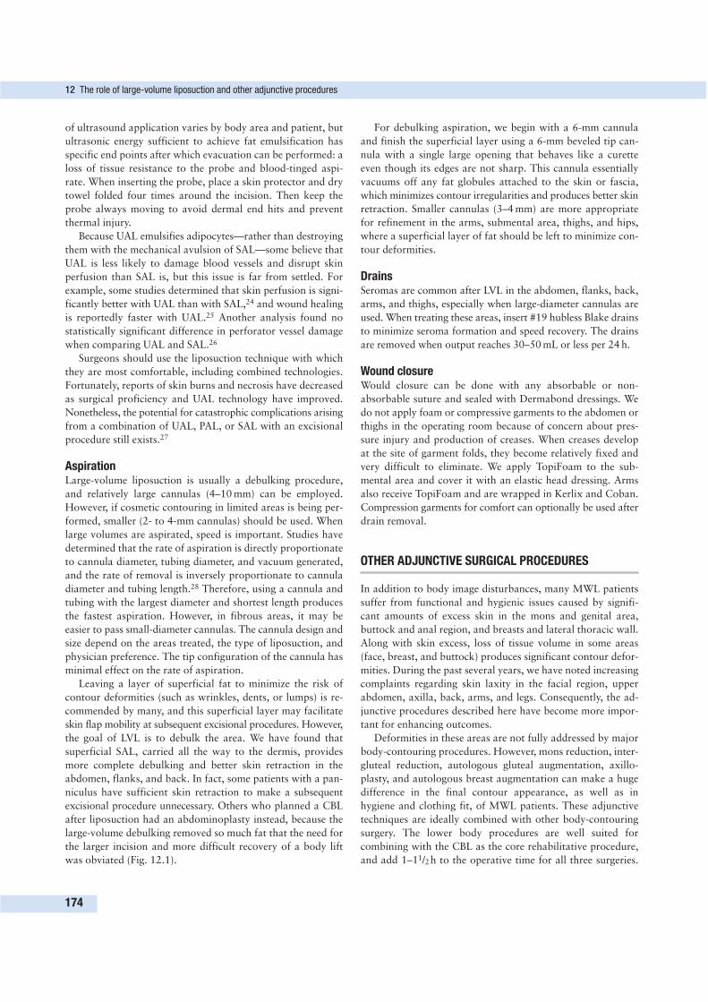
of ultrasound application varies by body area and patient, but
ultrasonic energy sufficient to achieve fat emulsification has
specific end points after which evacuation can be performed: a
loss of tissue resistance to the probe and blood-tinged aspi-
rate. When inserting the probe, place a skin protector and dry
towel folded four times around the incision. Then keep the
probe always moving to avoid dermal end hits and prevent
thermal injury.
Because UAL emulsifies adipocytes—rather than destroying
them with the mechanical avulsion of SAL—some believe that
UAL is less likely to damage blood vessels and disrupt skin
perfusion than SAL is, but this issue is far from settled. For
example, some studies determined that skin perfusion is signi-
ficantly better with UAL than with SAL,24 and wound healing
is reportedly faster with UAL.25 Another analysis found no
statistically significant difference in perforator vessel damage
when comparing UAL and SAL.26
Surgeons should use the liposuction technique with which
they are most comfortable, including combined technologies.
Fortunately, reports of skin burns and necrosis have decreased
as surgical proficiency and UAL technology have improved.
Nonetheless, the potential for catastrophic complications arising
from a combination of UAL, PAL, or SAL with an excisional
procedure still exists.27
AspirationLarge-volume liposuction is usually a debulking procedure,
and relatively large cannulas (4–10 mm) can be employed.
However, if cosmetic contouring in limited areas is being per-
formed, smaller (2- to 4-mm cannulas) should be used. When
large volumes are aspirated, speed is important. Studies have
determined that the rate of aspiration is directly proportionate
to cannula diameter, tubing diameter, and vacuum generated,
and the rate of removal is inversely proportionate to cannula
diameter and tubing length.28 Therefore, using a cannula and
tubing with the largest diameter and shortest length produces
the fastest aspiration. However, in fibrous areas, it may be
easier to pass small-diameter cannulas. The cannula design and
size depend on the areas treated, the type of liposuction, and
physician preference. The tip configuration of the cannula has
minimal effect on the rate of aspiration.
Leaving a layer of superficial fat to minimize the risk of
contour deformities (such as wrinkles, dents, or lumps) is re-
commended by many, and this superficial layer may facilitate
skin flap mobility at subsequent excisional procedures. However,
the goal of LVL is to debulk the area. We have found that
superficial SAL, carried all the way to the dermis, provides
more complete debulking and better skin retraction in the
abdomen, flanks, and back. In fact, some patients with a pan-
niculus have sufficient skin retraction to make a subsequent
excisional procedure unnecessary. Others who planned a CBL
after liposuction had an abdominoplasty instead, because the
large-volume debulking removed so much fat that the need for
the larger incision and more difficult recovery of a body lift
was obviated (Fig. 12.1).
For debulking aspiration, we begin with a 6-mm cannula
and finish the superficial layer using a 6-mm beveled tip can-
nula with a single large opening that behaves like a curette
even though its edges are not sharp. This cannula essentially
vacuums off any fat globules attached to the skin or fascia,
which minimizes contour irregularities and produces better skin
retraction. Smaller cannulas (3–4 mm) are more appropriate
for refinement in the arms, submental area, thighs, and hips,
where a superficial layer of fat should be left to minimize con-
tour deformities.
DrainsSeromas are common after LVL in the abdomen, flanks, back,
arms, and thighs, especially when large-diameter cannulas are
used. When treating these areas, insert #19 hubless Blake drains
to minimize seroma formation and speed recovery. The drains
are removed when output reaches 30–50 mL or less per 24 h.
Wound closureWould closure can be done with any absorbable or non-
absorbable suture and sealed with Dermabond dressings. We
do not apply foam or compressive garments to the abdomen or
thighs in the operating room because of concern about pres-
sure injury and production of creases. When creases develop
at the site of garment folds, they become relatively fixed and
very difficult to eliminate. We apply TopiFoam to the sub-
mental area and cover it with an elastic head dressing. Arms
also receive TopiFoam and are wrapped in Kerlix and Coban.
Compression garments for comfort can optionally be used after
drain removal.
OTHER ADJUNCTIVE SURGICAL PROCEDURES
In addition to body image disturbances, many MWL patients
suffer from functional and hygienic issues caused by signifi-
cant amounts of excess skin in the mons and genital area,
buttock and anal region, and breasts and lateral thoracic wall.
Along with skin excess, loss of tissue volume in some areas
(face, breast, and buttock) produces significant contour defor-
mities. During the past several years, we have noted increasing
complaints regarding skin laxity in the facial region, upper
abdomen, axilla, back, arms, and legs. Consequently, the ad-
junctive procedures described here have become more impor-
tant for enhancing outcomes.
Deformities in these areas are not fully addressed by major
body-contouring procedures. However, mons reduction, inter-
gluteal reduction, autologous gluteal augmentation, axillo-
plasty, and autologous breast augmentation can make a huge
difference in the final contour appearance, as well as in
hygiene and clothing fit, of MWL patients. These adjunctive
techniques are ideally combined with other body-contouring
surgery. The lower body procedures are well suited for
combining with the CBL as the core rehabilitative procedure,
and add 1–11/2h to the operative time for all three surgeries.
12 The role of large-volume liposuction and other adjunctive procedures
174
An axilloplasty (~1 h) and breast autoaugmentation (2–3 h)
work well when combined with upper body procedures. Alter-
natively, adjunctive surgery can be performed in separate stages
of rehabilitation if combined procedures are not feasible.
These smaller procedures are not metabolically demanding or
lengthy, and may be done on an outpatient basis. When ad-
junctive procedures are combined together or performed in
conjunction with a larger surgery such as a CBL or LVL,
admission to the hospital for postoperative observation is
advised.
Mons reductionThe suprapubic and genital regions are typically involved to a
similar extent in MWL patients. Failure to contour these
regions results in a suboptimal aesthetic outcome to the CBL,
such as that seen in Figure 12.3, and decreased patient satis-
faction, partly because problems with genital hygiene and
function are not solved.
• In mild cases of suprapubic skin excess and lipodystrophy,
standard liposuction of the mons will suffice.29
• In moderate cases of skin excess and lipodystrophy, excise
an inverted triangular wedge of skin and tissue without
undermining. Secure the superficial fascial system (SFS) of
the mons to the anterior rectus fascia with ‘1’ Ethibond or
Vicryl Plus to prevent excess superior displacement. Then
close in layers with 3-0 Monocryl (Fig. 12.4).
• A deep tacking suture at the lateral aspects of the mons
“triangle” helps to restore a more normal contour after
mons reduction.
Other adjunctive surgical procedures
175
Figure 12.3 Early in our experience with circumferential body lift, we did
not understand the importance of mons reduction. This patient illustrates the
deformity that can result if mons reduction is not performed in conjunction
with a lower body lift.
Figure 12.4 Perform mons reduction before closing circumferential body lift incisions.
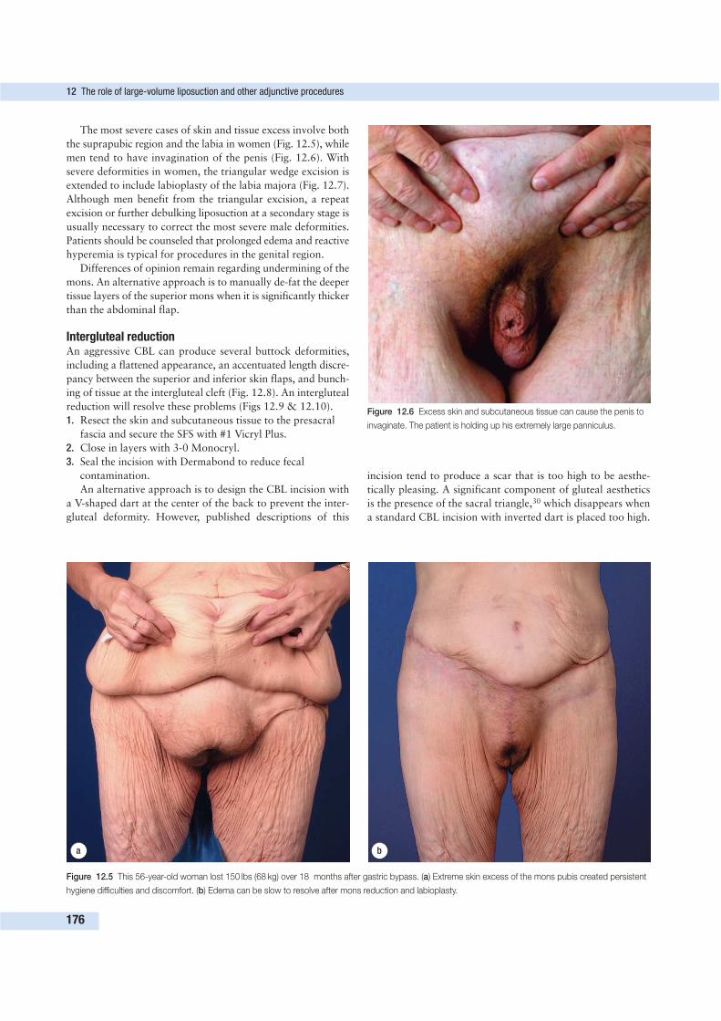
The most severe cases of skin and tissue excess involve both
the suprapubic region and the labia in women (Fig. 12.5), while
men tend to have invagination of the penis (Fig. 12.6). With
severe deformities in women, the triangular wedge excision is
extended to include labioplasty of the labia majora (Fig. 12.7).
Although men benefit from the triangular excision, a repeat
excision or further debulking liposuction at a secondary stage is
usually necessary to correct the most severe male deformities.
Patients should be counseled that prolonged edema and reactive
hyperemia is typical for procedures in the genital region.
Differences of opinion remain regarding undermining of the
mons. An alternative approach is to manually de-fat the deeper
tissue layers of the superior mons when it is significantly thicker
than the abdominal flap.
Intergluteal reductionAn aggressive CBL can produce several buttock deformities,
including a flattened appearance, an accentuated length discre-
pancy between the superior and inferior skin flaps, and bunch-
ing of tissue at the intergluteal cleft (Fig. 12.8). An intergluteal
reduction will resolve these problems (Figs 12.9 & 12.10).
1. Resect the skin and subcutaneous tissue to the presacral
fascia and secure the SFS with #1 Vicryl Plus.
2. Close in layers with 3-0 Monocryl.
3. Seal the incision with Dermabond to reduce fecal
contamination.
An alternative approach is to design the CBL incision with
a V-shaped dart at the center of the back to prevent the inter-
gluteal deformity. However, published descriptions of this
incision tend to produce a scar that is too high to be aesthe-
tically pleasing. A significant component of gluteal aesthetics
is the presence of the sacral triangle,30 which disappears when
a standard CBL incision with inverted dart is placed too high.
12 The role of large-volume liposuction and other adjunctive procedures
176
a b
Figure 12.5 This 56-year-old woman lost 150 lbs (68 kg) over 18 months after gastric bypass. (a) Extreme skin excess of the mons pubis created persistent
hygiene difficulties and discomfort. (b) Edema can be slow to resolve after mons reduction and labioplasty.
Figure 12.6 Excess skin and subcutaneous tissue can cause the penis to
invaginate. The patient is holding up his extremely large panniculus.
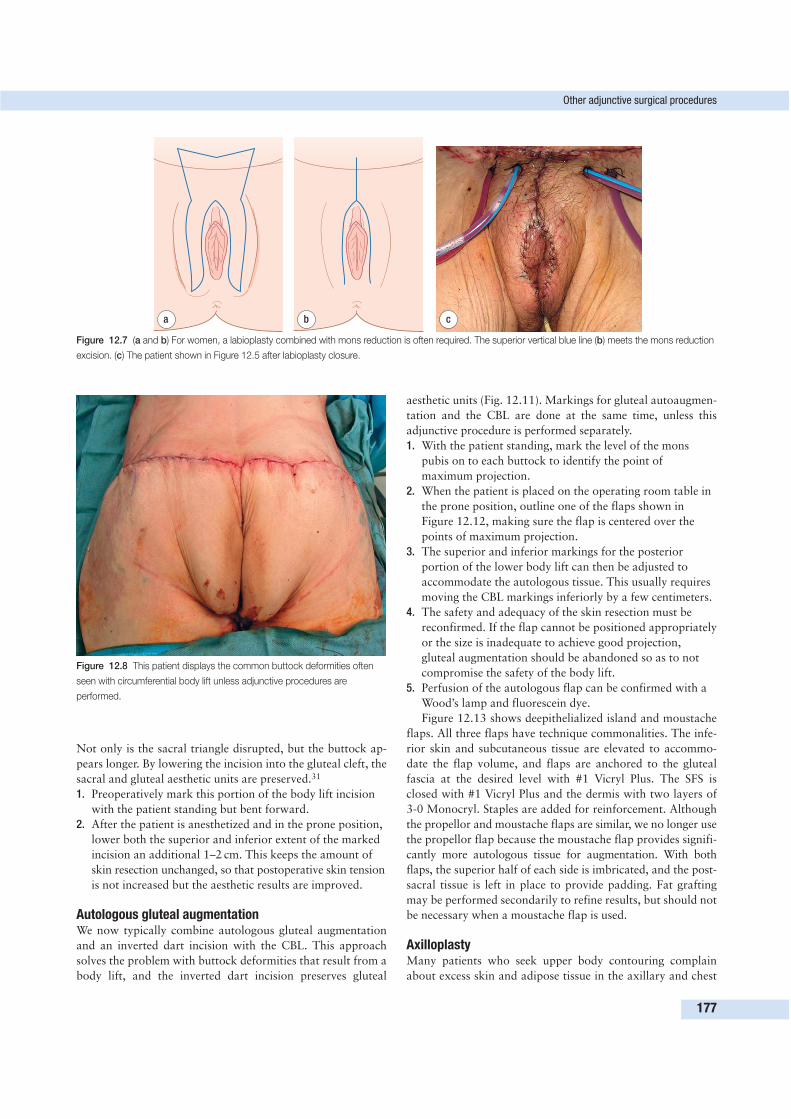
Not only is the sacral triangle disrupted, but the buttock ap-
pears longer. By lowering the incision into the gluteal cleft, the
sacral and gluteal aesthetic units are preserved.31
1. Preoperatively mark this portion of the body lift incision
with the patient standing but bent forward.
2. After the patient is anesthetized and in the prone position,
lower both the superior and inferior extent of the marked
incision an additional 1–2 cm. This keeps the amount of
skin resection unchanged, so that postoperative skin tension
is not increased but the aesthetic results are improved.
Autologous gluteal augmentationWe now typically combine autologous gluteal augmentation
and an inverted dart incision with the CBL. This approach
solves the problem with buttock deformities that result from a
body lift, and the inverted dart incision preserves gluteal
aesthetic units (Fig. 12.11). Markings for gluteal autoaugmen-
tation and the CBL are done at the same time, unless this
adjunctive procedure is performed separately.
1. With the patient standing, mark the level of the mons
pubis on to each buttock to identify the point of
maximum projection.
2. When the patient is placed on the operating room table in
the prone position, outline one of the flaps shown in
Figure 12.12, making sure the flap is centered over the
points of maximum projection.
3. The superior and inferior markings for the posterior
portion of the lower body lift can then be adjusted to
accommodate the autologous tissue. This usually requires
moving the CBL markings inferiorly by a few centimeters.
4. The safety and adequacy of the skin resection must be
reconfirmed. If the flap cannot be positioned appropriately
or the size is inadequate to achieve good projection,
gluteal augmentation should be abandoned so as to not
compromise the safety of the body lift.
5. Perfusion of the autologous flap can be confirmed with a
Wood’s lamp and fluorescein dye.
Figure 12.13 shows deepithelialized island and moustache
flaps. All three flaps have technique commonalities. The infe-
rior skin and subcutaneous tissue are elevated to accommo-
date the flap volume, and flaps are anchored to the gluteal
fascia at the desired level with #1 Vicryl Plus. The SFS is
closed with #1 Vicryl Plus and the dermis with two layers of
3-0 Monocryl. Staples are added for reinforcement. Although
the propellor and moustache flaps are similar, we no longer use
the propellor flap because the moustache flap provides signifi-
cantly more autologous tissue for augmentation. With both
flaps, the superior half of each side is imbricated, and the post-
sacral tissue is left in place to provide padding. Fat grafting
may be performed secondarily to refine results, but should not
be necessary when a moustache flap is used.
AxilloplastyMany patients who seek upper body contouring complain
about excess skin and adipose tissue in the axillary and chest
Other adjunctive surgical procedures
177
a b c
Figure 12.7 (a and b) For women, a labioplasty combined with mons reduction is often required. The superior vertical blue line (b) meets the mons reduction
excision. (c) The patient shown in Figure 12.5 after labioplasty closure.
Figure 12.8 This patient displays the common buttock deformities often
seen with circumferential body lift unless adjunctive procedures are
performed.
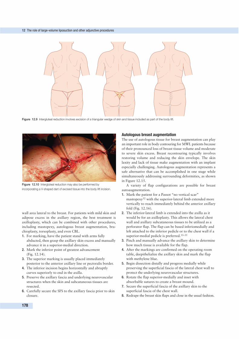
wall area lateral to the breast. For patients with mild skin and
adipose excess in the axillary region, the best treatment is
axilloplasty, which can be combined with other procedures,
including mastopexy, autologous breast augmentation, bra-
chioplasty, torsoplasty, and even CBL.
1. For marking, have the patient stand with arms fully
abducted, then grasp the axillary skin excess and manually
advance it in a superior-medial direction.
2. Mark the inferior point of greatest advancement
(Fig. 12.14).
3. The superior marking is usually placed immediately
posterior to the anterior axillary line or pectoralis border.
4. The inferior incision begins horizontally and abruptly
curves superiorly to end in the axilla.
5. Preserve the axillary fascia and underlying neurovascular
structures when the skin and subcutaneous tissues are
resected.
6. Carefully secure the SFS to the axillary fascia prior to skin
closure.
Autologous breast augmentationThe use of autologous tissue for breast augmentation can play
an important role in body contouring for MWL patients because
of their pronounced loss of breast tissue volume and moderate
to severe skin excess. Breast recontouring typically involves
restoring volume and reducing the skin envelope. The skin
laxity and lack of tissue make augmentation with an implant
especially challenging. Autologous augmentation represents a
safe alternative that can be accomplished in one stage while
simultaneously addressing surrounding deformities, as shown
in Figure 12.15.
A variety of flap configurations are possible for breast
autoaugmentation.
1. Mark the patient for a Passot “no vertical scar”
mastopexy32 with the superior-lateral limb extended more
vertically to reach immediately behind the anterior axillary
fold (Fig. 12.16).
2. The inferior-lateral limb is extended into the axilla as it
would be for an axilloplasty. This allows the lateral chest
wall and axillary subcutaneous tissues to be utilized as a
perforator flap. The flap can be based inferiomedially and
left attached to the inferior pedicle or to the chest wall if a
superior-medial pedicle is preferred.33–35
3. Pinch and manually advance the axillary skin to determine
how much tissue is available for the flap.
4. After the markings are confirmed on the operating room
table, deepithelialize the axillary skin and mark the flap
with methylene blue.
5. Begin dissection distally and progress medially while
preserving the superficial fascia of the lateral chest wall to
protect the underlying neurovascular structures.
6. Rotate the flap superior-medially and inset with
absorbable sutures to create a breast mound.
7. Secure the superficial fascia of the axillary skin to the
superficial fascia of the chest wall.
8. Redrape the breast skin flaps and close in the usual fashion.
12 The role of large-volume liposuction and other adjunctive procedures
178
Figure 12.9 Intergluteal reduction involves excision of a triangular wedge of skin and tissue included as part of the body lift.
Figure 12.10 Intergluteal reduction may also be performed by
incorporating a V-shaped dart of excised tissue into the body lift incision.
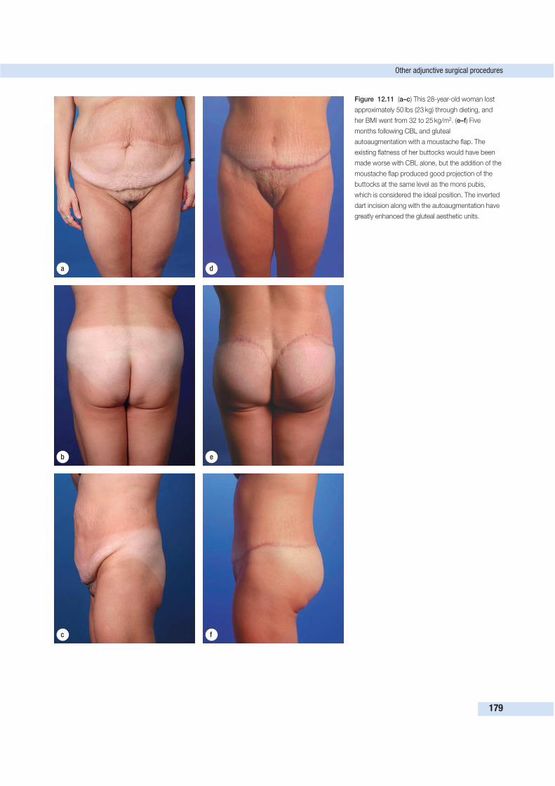
Other adjunctive surgical procedures
179
d
e
f
a
b
c
Figure 12.11 (a–c) This 28-year-old woman lost
approximately 50 lbs (23 kg) through dieting, and
her BMI went from 32 to 25 kg/m2. (e–f) Five
months following CBL and gluteal
autoaugmentation with a moustache flap. The
existing flatness of her buttocks would have been
made worse with CBL alone, but the addition of the
moustache flap produced good projection of the
buttocks at the same level as the mons pubis,
which is considered the ideal position. The inverted
dart incision along with the autoaugmentation have
greatly enhanced the gluteal aesthetic units.
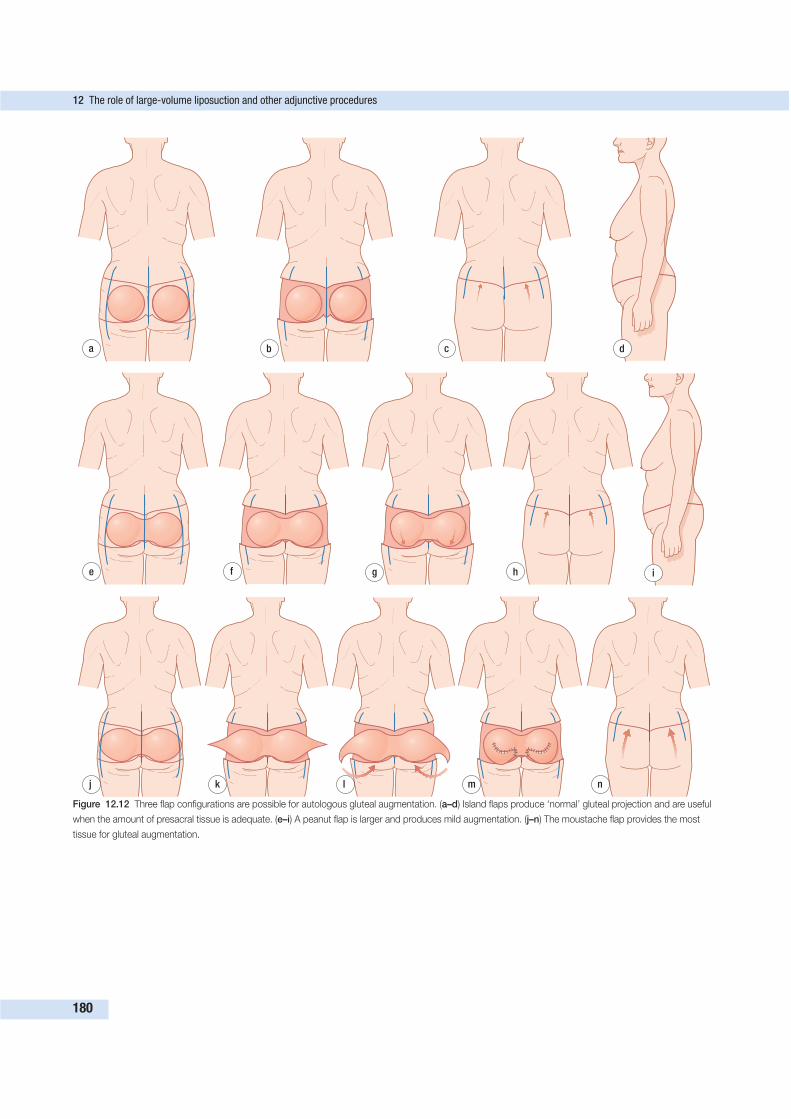
12 The role of large-volume liposuction and other adjunctive procedures
180
d
e f
a b c
g h i
j k l m n
Figure 12.12 Three flap configurations are possible for autologous gluteal augmentation. (a–d) Island flaps produce ‘normal’ gluteal projection and are useful
when the amount of presacral tissue is adequate. (e–i) A peanut flap is larger and produces mild augmentation. (j–n) The moustache flap provides the most
tissue for gluteal augmentation.
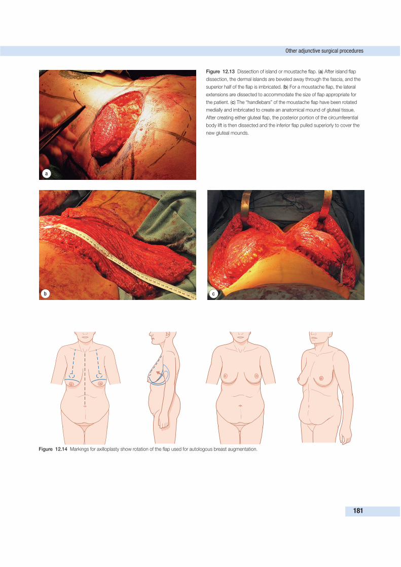
Other adjunctive surgical procedures
181
a
b c
Figure 12.13 Dissection of island or moustache flap. (a) After island flap
dissection, the dermal islands are beveled away through the fascia, and the
superior half of the flap is imbricated. (b) For a moustache flap, the lateral
extensions are dissected to accommodate the size of flap appropriate for
the patient. (c) The “handlebars” of the moustache flap have been rotated
medially and imbricated to create an anatomical mound of gluteal tissue.
After creating either gluteal flap, the posterior portion of the circumferential
body lift is then dissected and the inferior flap pulled superiorly to cover the
new gluteal mounds.
Figure 12.14 Markings for axilloplasty show rotation of the flap used for autologous breast augmentation.
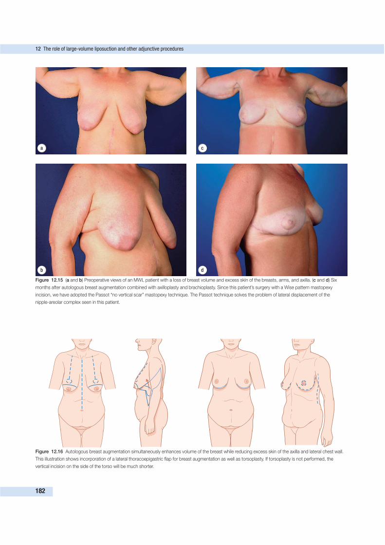
12 The role of large-volume liposuction and other adjunctive procedures
182
d
a
b
c
Figure 12.15 (a and b) Preoperative views of an MWL patient with a loss of breast volume and excess skin of the breasts, arms, and axilla. (c and d) Six
months after autologous breast augmentation combined with axilloplasty and brachioplasty. Since this patient’s surgery with a Wise pattern mastopexy
incision, we have adopted the Passot “no vertical scar” mastopexy technique. The Passot technique solves the problem of lateral displacement of the
nipple-areolar complex seen in this patient.
Figure 12.16 Autologous breast augmentation simultaneously enhances volume of the breast while reducing excess skin of the axilla and lateral chest wall.
This illustration shows incorporation of a lateral thoracoepigastric flap for breast augmentation as well as torsoplasty. If torsoplasty is not performed, the
vertical incision on the side of the torso will be much shorter.
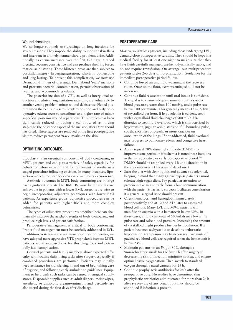
Wound dressingsWe no longer routinely use dressings on long incisions for
several reasons. They impede the ability to monitor skin flaps
and intervene in a timely manner should problems arise. Addi-
tionally, as edema increases over the first 1–3 days, a taped
dressing becomes constrictive and can produce shearing forces
that cause blistering. These blistered areas are then subject to
postinflammatory hyperpigmentation, which is bothersome
and long-lasting. To prevent this complication, we now use
Dermabond in lieu of dressings. Dermabond ‘seals’ incisions
and prevents bacterial contamination, permits observation of
healing, and accommodates edema.
The posterior incision of a CBL, as well as intergluteal re-
duction and gluteal augmentation incisions, are vulnerable to
another vexing problem: minor wound dehiscence. Flexed pos-
ture when the bed is in a semi-Fowler’s position and early post-
operative edema seem to contribute to a higher rate of minor
superficial posterior wound separations. This problem has been
significantly reduced by adding a scant row of reinforcing
staples to the posterior aspect of the incision after Dermabond
has dried. These staples are removed at the first postoperative
visit to reduce permanent ‘track’ marks on the skin.
OPTIMIZING OUTCOMES
Lipoplasty is an essential component of body contouring in
MWL patients and can play a variety of roles, especially for
debulking before excision and for refinement of results in a
staged procedure following excision. In many instances, lipo-
suction reduces the need for excision or minimizes excision size.
Aesthetic outcomes in MWL body contouring are in large
part significantly related to BMI. Because better results are
achievable in patients with a lower BMI, surgeons are wise to
begin incorporating adjunctive techniques with lower BMI
patients. As experience grows, adjunctive procedures can be
added for patients with higher BMIs and more complex
deformities.
The types of adjunctive procedures described here can dra-
matically improve the aesthetic results of body contouring and
produce high levels of patient satisfaction.
Perioperative management is critical in body contouring.
Proper fluid management must be carefully addressed in LVL.
In addition to stressing the maintenance of normothermia, we
have adopted more aggressive VTE prophylaxis because MWL
patients are at increased risk for this dangerous and poten-
tially fatal complication.
Counsel patients and family members about expected diffi-
culty with routine daily living tasks after surgery, especially if
combined procedures are performed. Patients may initially
need assistance for transferring in and out of bed, taking care
of hygiene, and following early ambulation guidelines. Equip-
ment to help with such tasks can be rented at surgical supply
stores. Disposable supplies such as adult diapers, moist wipes,
anesthetic or antibiotic creams/ointment, and peroxide are
also useful during the first days after discharge.
POSTOPERATIVE CARE
Massive weight loss patients, including those undergoing LVL,
demand close postoperative scrutiny. They should be kept in a
medical facility for at least one night to make sure that they
have fluids carefully managed, are hemodynamically stable, and
do not require transfusion. On average, our multiprocedure
patients prefer 2–3 days of hospitalization. Guidelines for the
immediate postoperative period follow.
• Continue forced air and fluid warming in the recovery
room. Once on the floor, extra warming should not be
necessary.
• Continue fluid resuscitation until oral intake is sufficient.
The goal is to ensure adequate urine output, a systolic
blood pressure greater than 100 mmHg, and a pulse rate
below 100 per minute. This generally means 125–150 mL
of crystalloid per hour. If hypovolemia is evident, treat
with a crystalloid fluid challenge of 500 mL/h. Use
diuretics to treat fluid overload, which is characterized by
hypertension, jugular vein distension, full bounding pulse,
cough, shortness of breath, or moist crackles on
auscultation of the lungs. If not addressed, fluid overload
may progress to pulmonary edema and congestive heart
failure.
• Apply topical 70% dimethyl sulfoxide (DMSO) to
improve tissue perfusion if ischemia is noted near incisions
in the intraoperative or early postoperative period.36
DMSO should be reapplied every 4 h until circulation in
the area improves. (This is an off-label use.)
• Start the diet with clear liquids and advance as tolerated,
keeping in mind that many gastric bypass patients cannot
tolerate high-sugar diets. Pay particular attention to
protein intake in a suitable form. Close communication
with the patient’s bariatric surgeon facilitates consultation
if a general surgical issue should present.
• Check hematocrit and hemoglobin immediately
postoperatively and at 12 and 24 h later to assess red
blood cell loss. Many LVL and MWL patients will
manifest an anemia with a hematocrit below 30%. In
these cases, a fluid challenge of 500 mL/h may lower the
pulse rate and raise blood pressure. Increasing the amount
of crystalloid might produce further hemodilution. If a
patient becomes tachycardic or develops orthostatic
hypotension, transfusion may be necessary. Two units of
packed red blood cells are required when the hematocrit is
below 23%.
• Maintain patients on an FiO2 of 80% through a
‘non-rebreather’ mask for the first 2 h after surgery to
decrease the risk of infection, minimize nausea, and ensure
optimal tissue oxygenation. Then switch to standard
oxygen through a nasal cannula for 24 h.
• Continue prophylactic antibiotics for 24 h after the
preoperative dose. No studies have determined that
prophylactic antibiotics administrated for more than 24 h
after surgery are of any benefit, but they should be
continued if infection is present.
Postoperative care
183
• Continue DVT prophylaxis with intermittent pneumatic
compression devices and stress early mobilization.
Intermittent pneumatic compression devices should be
removed and replaced after walking until the patient is
discharged. Encourage patients to begin ambulation the
day after surgery. If appropriate, continue
anticoagulation prophylaxis with LMWH or
fondaparinux for 1–4 weeks after surgery or until fully
ambulatory.
• Manage pain with morphine or meperidine (Demerol)
patient-controlled analgesia and/or oral narcotics as
needed. Gradually wean patients to non-narcotic pain
relievers. Some body-contouring patients report chronic
pain after surgery that may result from nerve injury.
Gabapentin (Neurontin) and/or amitriptyline (Elavil) are
sometimes effective for treating the type of burning pain
patients describe.
• Discontinue the Foley catheter early on the first
postoperative day to encourage ambulation.
• Order a complete blood count and basic metabolic
panel for the morning after surgery. Glucose monitoring
may also be warranted. Common electrolyte
abnormalities that follow LVL include lowered sodium,
potassium, and blood urea nitrogen levels in the early
postoperative period.34 Liver enzyme testing has revealed
significantly lowered levels of albumin and protein that
are consistent with hemodilution and lowered blood
viscosity. In addition, levels of plasma aminotransferases
significantly increased in LVL patients, a possible
indication of injury to adipocytes or skeletal muscles or
hepatocellular damage.37 Creatine kinase levels also may
be elevated.
• Leave drains in place until output is in the range of
30–50 cc in 24 h. If drainage is prolonged, perform
sclerosis with a high-concentration doxycycline solution
(100 mg per 10 cc of 0.9% saline solution) infused through
the drain. Prior to sclerosis, infuse with 0.5% marcaine for
anesthesia. Clamp the drain for 15 min and then return to
suction. Because the doxycycline concentration is higher
than recommended for infusion, it must be specially
ordered for off-label use. Infusion can sometimes be
painful, and analgesics are recommended. Injection into a
seroma cavity can be performed, but it must not be into
the subcutaneous tissue because doxycycline can cause fat
and skin necrosis.
• Occasional massage therapy is often useful to help speed
the resolution of edema following liposuction.
• Compressive binders and garments should not be used
routinely in the immediate postoperative period, because
they may interfere with already-challenged perfusion of
skin and/or flaps and impair the ability to monitor blood
flow. Drains inadvertently placed beneath a binder can
cause pressure necrosis. Later in the postoperative course,
it may be prudent to add a compression garment to reduce
swelling, dead space, and discomfort associated with
ambulation.
COMPLICATIONS AND THEIR MANAGEMENT
LiposuctionRecent statistics place the rate of significant complications
secondary to lipoplasty in the range of 0.3%16 to 1.8%.38,39
Major complications include:
• hemorrhage (usually resulting from visceral perforation),
• hematoma (particularly in the retroperitoneal space if the
fascia is penetrated),
• skin or fat necrosis (major) or skin slough,
• infection,
• necrotizing fasciitis,
• pulmonary edema (resulting from fluid overload),
• lidocaine toxicity,
• DVT,
• PE,
• fat embolus,
• cardiac arrhythmia, and
• death.
Minor complications are:
• contour irregularities,
• scarring,
• prolonged edema,
• paresthesias,
• anemia,
• hypovolemia,
• hemodilution that requires blood transfusion, and
• thermal injury from ultrasonic energy.
Seroma is perhaps the most common complication of liposuc-
tion, but its frequency can be greatly reduced with drains.
There is no evidence of increased complication rates when
aspirate volumes of ≥ 5000 cc are compared with volumes
< 5000 cc.16,40
Massive weight loss patients who undergo debulking lipo-
suction with or without excisional procedures have the poten-
tial to develop the typical complications of liposuction plus
some additional risks. Contour irregularities such as wrinkles,
lumps, or dents occur in almost all MWL patients, but they
are generally tolerant of such irregularities if the possibility has
been discussed preoperatively. If excessive skin laxity remains
after liposuction—and it usually does—staged excisional pro-
cedures are the only option for correction. Some patients, how-
ever, accept the skin excess if the fat debulking is sufficient to
make them more physically comfortable.
The risk of lidocaine toxicity becomes real if the total
delivered dose exceeds 35 mg/kg. Lidocaine toxicity can be
completely avoided by omitting it from the infusion solution.
Kenkel and colleagues determined that only about 10% of
infiltrated lidocaine is aspirated, and lidocaine toxicity may
not manifest for 8–16 h after surgery.41,42 The time to peak for
the lidocaine metabolite monoethylglycinexylidide may be even
longer, up to 28 h. (Because lidocaine is metabolized in the
liver, it should not be used in patients with liver dysfunction.)
Therefore the period of potential lidocaine toxicity lasts longer
than is commonly believed. However, the analgesic effect of
lidocaine is not long-lasting. Kenkel et al. found that even
12 The role of large-volume liposuction and other adjunctive procedures
184
though lidocaine is present in blood for up to 18 h, it does not
remain at a therapeutic dose in local tissues for more than
4–8 h. Most surgeries performed in MWL patients require
general anesthesia because procedures are lengthy and rigo-
rous monitoring is essential. Patients receiving LVL or lipo-
suction plus excision are going to require opiate analgesia
postoperatively, as well as hospitalization. Therefore the need
for lidocaine is non-existent in these patients.
Fat embolism has been reported with liposuction, although
its frequency is unknown. Estimates place this complication in
the range of 1:100 000 to 1:300 000.43 Fat embolization occurs
when small globules of fat migrate through the venous circu-
lation to the lungs. It usually does not produce significant
symptoms unless there is a large amount of embolization, but
symptoms may include tachycardia, tachypnea, elevated tem-
perature, hypoxemia, hypocapnia, or thrombocytopenia. In
contrast, fat embolism syndrome is an inflammatory and bio-
chemical condition associated with free fatty acids released
into the blood that produce a syndrome of petechial rash, res-
piratory distress, and cerebral dysfunction approximately
24–72 h after surgery. A suggestion for preventing fat accumu-
lation and emboli is continuation of intravenous fluids for
24 h after surgery to flush fatty material through the circu-
latory system.40
Blindness has been recently reported in patients undergoing
liposuction who develop a significant anemia and decreased
retinal circulation.44 This makes it very important to monitor
the hematocrit in these patients and keep them well hydrated
and volume expanded to avoid hypotension.
Skin necrosis is uncommon in liposuction, except in diabetic
patients and people who have scars from previous procedures.
Because many MWL patients meet these criteria, they should
be warned in advance of the necrosis risk.
Mons reductionIf undermining can be avoided, postoperative complications
such as skin necrosis and delayed wound healing are uncom-
mon because tissues in this area are very well vascularized.
However, lymphatic drainage is compromised when mons
reduction is combined with a CBL or thigh lift. This results in
prolonged postoperative lymphedema and hyperemia that can
resemble cellulitis. Empiric antibiotic therapy can be used but
is often unnecessary. Sensation is temporarily altered but usually
resolves. Hypersensitivity of the clitoris in women can be a
problem if aggressive lifting and reduction of the mons are
performed. It may improve over time but can lead to perma-
nent discomfort. Should this be a problem, desensitization
creams can be helpful.
Intergluteal reductionThe most significant complication associated with intergluteal
reduction is delayed wound healing. This region is a ‘watershed’
of blood supply that may become compromised by overresec-
tion and undue tension on the closure. Having the patient bend
over when marking the central posterior incision adds an
additional safety margin. Closure of ‘dead space’ with a layered
closure helps prevent seromas that could lead to wound sepa-
ration. Covering the anal region with a povidone–iodine
(Betadine)-soaked towel prevents contamination of the sutures
during closure, and sealing the incision with Dermabond
reduces fecal contamination. Careful attention to cutting the
deep SFS sutures close to the knot helps lessen suture burden,
extrusion (spitting), and potential infection. This procedure
can be eliminated by incorporating an inverted dart incision
into the CBL and/or gluteal augmentation.
Autologous gluteal augmentationComplications directly related to autologous gluteal augmen-
tation are relatively uncommon in our practice. The robustness
of vascularization in the area produces good flap viability,
which can be confirmed with a Wood’s lamp and fluorescein
dye. Small areas of fat necrosis are typically allowed to resorb
on their own. Seromas due to large dead spaces can be avoided
by putting drains in the most dependent portion of the gluteal
pocket. If seroma does occur, management is important because
it can precipitate wound dehiscence. (Sclerosis with doxycyc-
line was described earlier.) We do not routinely use quilting
sutures in this area, but they may be helpful.
Delayed wound-healing rates for our CBL patients with and
without gluteal augmentation do not appear to be significantly
different. Nonetheless, inferior flap undermining and tension
on the closure increases when gluteal augmentation is added,
and this can lead to wound-healing problems plus anorectal
hypersensitivity and maceration due to overexposure of the
anus. Maceration is usually self-limited and can be managed by
topical anesthetics such as hydrocortisone (Anusol), a ‘dough-
nut’ cushion for sitting, frequent positional changes, high-fiber
diet, sitz baths, or baby wipes for cleansing.
Until gaining experience with gluteal autoaugmentation, we
advise careful preoperative planning and conservatively sized
island flaps to avoid overresection that may lead to wound-
healing problems, skin necrosis, and dehiscence. Although this
may limit the quality of initial results, aesthetic outcomes will
significantly improve with experience. Secondary excisional
touch-up procedures such as adjunctive flank liposuction and
infragluteal fold excisions can further refine aesthetic outcomes.
AxilloplastyThe critical neurovascular structures of the axilla are less
likely to be injured if surgical dissection remains above the
axillary fascia. Inevitably, the fascia will be violated from time
to time. The structures most likely to be injured are the inter-
costobrachial nerve, the lower roots of the brachial plexus,
and the axillary lymphatics. Injury to the intercostobrachial
nerve can be treated by neurorrhaphy or proximal transposi-
tion. Because brachial plexus injury is more problematic, it is
best avoided; if injury does occur, prompt consultation with a
peripheral nerve specialist is recommended.
Inadvertent excision or transection of lymphatics results in
lymphorrhea and lymphoceles, but these can be prevented by
tying off the afferent channels if nodes are involved in the tis-
sue to be resected. If problems occur, distally inject lymphazurin
Complications and their management
185
blue and surgically localize the involved afferent channels with
ligation.45 Sclerosis of a lymphocele with high-dose doxycyc-
line (100 mg per 10 cc of 0.9% normal saline) is sometimes
helpful. Wound dehiscence in the axilla results from undue
tension caused by overexcision. Anchoring the SFS to the axil-
lary fascia with #1 Vicryl Plus should help reduce tension on
the skin closure.
Autologous breast augmentationComplications from autologous breast augmentation utilizing
a lateral thoracoepigastric flap in conjunction with axilloplasty
and mastopexy can largely be avoided with careful preopera-
tive planning. Skin excision with a Passot “no vertical scar”
technique makes redistribution of the axillary skin and lateral
breast flap easier than when a Wise pattern excision is used. It
also reduces the problem of lateral displacement of the nipple-
areolar complex. It is often helpful to mark the lateral breast
flap immediately posterior to the anterior axillary line or the
pectoralis major muscle border. Doing so leaves a small margin
of extra lateral breast flap skin that helps prevent overresection.
In addition, careful dissection and leaving a layer of adipose
tissue over the lateral chest wall prevents injury to the fourth
and fifth intercostal nerves. Once the autologous tissue is
added to the breast mound, tension on the breast skin can be
significant. Meticulous pedicle dissection avoids compromising
the circulation of the nipple areolar complex.
CONCLUSION
Almost all MWL patients will benefit greatly from liposuction
added as part of the staged procedures often required to
achieve optimal aesthetic results. Circumferential debulking
liposuction is especially useful for patients who have excess
subcutaneous fat, particularly if it is distributed throughout
the body, as is typical in patients with a BMI of 30 kg/m2 or
higher. In this context, LVL can have a major impact on final
body contour if performed as the first stage. Other patients
have localized lipodystrophies that are easily treated with
liposuction. For patients who prefer not to undergo multiple
staged excisional surgeries, liposuction offers an alternative
with few risks and quick recovery time. If too much excess
skin remains after liposuction, an excisional procedure can be
scheduled.
The complexity of deformities after MWL is unprecedented
in plastic surgery. Body contouring in this population challenges
our ingenuity, creativity, and surgical skills on a regular basis.
The adjunctive techniques described here have enabled us to
improve clinical outcomes and enhance satisfaction among our
patients.
REFERENCES
1. Brauman D. Liposuction abdominoplasty: an evolving concept.
Plast Reconstr Surg 2003; 112:288–301.
2. Cárdenas-Camarena L, González LE. Large-volume liposuction
and extensive abdominoplasty: a feasible alternative for improving
body shape. Plast Reconstr Surg 1998; 102:1698–1707.
3. Hunstad JP. Body contouring in the obese patient. Clin Plast Surg
1996; 23:647–670.
4. Vogt PA. Brachial suction-assisted lipoplasty and brachioplasty.
Aesthetic Surg J 2001; 21:164–167.
5. Abramson DL. Ultrasound-assisted abdominoplasty: combining
modalities in a safe and effective technique. Plast Reconstr Surg
2003; 112:898–902.
6. Avelar JM. Abdominoplasty without panniculus undermining and
resection: analysis and 3-year follow-up of 97 consecutive cases.
Aesthetic Surg J 2002; 22:16–25.
7. Saldanha OR, de Souza Pinto EB, Matos WN, et al. Lipoabdomi-
noplasty without undermining. Aesthetic Surg J 2001; 21:518–526.
8. Matarasso A. Liposuction as an adjunct to a full abdominoplasty
revisited. Plast Reconstr Surg 2000; 106:1197–1202.
9. Iverson RE, Lynch DJ, ASPS Committee on Patient Safety. Practice
advisory on liposuction. Plast Reconstr Surg 2004; 113:1478–1490.
10. Alvarez-Leite JI. Nutrient deficiencies secondary to bariatric surgery.
Curr Opin Clin Nutr Metab Care 2004; 7:569–575.
11. Goldberg MA. Perioperative epoetin alfa increases red blood cell
mass and reduces exposure to transfusions: results of randomized
clinical trials. Semin Hematol 1997; 34:41–47.
12. Sessler DI, Akça O. Nonpharmacological prevention of surgical
wound infections. Healthc Epidemiol 2002: 35:1397–1404.
13. Mangram AJ, Horan TC, Pearson ML, et al. Guideline for preven-
tion of surgical site infection, 1999. Hospital Infection Control
Practices Advisory Committee. Infect Control Hosp Epidemiol
1999; 20(4):250–278.
14. Geerts WH, Pineo GF, Heit JA, et al. Prevention of venous throm-
boembolism: the Seventh ACCP Conference on Antithrombotic
and Thrombolytic Therapy. Chest 2004; 126:338S–400S.
15. Young VL, Watson ME. The need for venous thromboembolism
(VTE) prophylaxis in plastic surgery. Aesthetic Surg J 2006;
26:157–175.
16. Rohrich RJ, Beran SJ. Is liposuction safe? Plast Reconstr Surg
1999; 104:819–822.
17. Nijkeuter M, Huisman MV. Pentasaccharides in the prophylaxis
and treatment of venous thromboembolism: a systematic review.
Curr Opin Pulm Med 2004; 10:338–344.
18. Sazama K, DeChristopher PJ, Dodd R, et al. Practice parameter for
the recognition, management, and prevention of adverse consequences
of blood transfusion. Arch Pathol Lab Med 2000; 124:61–70.
19. Matarasso A. Superwet anesthesia redefines large volume liposuc-
tion. Aesthetic Surg J 1997; 17(6):358–364.
20. Trott SA, Beran SJ, Rohrich R, et al. Safety considerations and fluid
resuscitation in liposuction: an analysis of 53 consecutive patients.
Plast Reconstr Surg 1998; 102:2220–2229.
21. Karmo FR, Milan MF, Silbergleit A. Blood loss in major liposuc-
tion procedures: a comparison study using suction-assisted versus
ultrasonically-assisted lipoplasty. Plast Reconstr Surg 2001;
108:241–247.
22. Cárdenas-Camarena L, Tobar-Losada A, Lacouture AM. Large-
volume circumferential liposuction with tumescent technique: a
sure and viable procedure. Plast Reconstr Surg 1999;
104:1887–1899.
23. Brown SA, Lipschitz AH, Kenkel JM, et al. Pharmacokinetics and
safety of epinephrine use in liposuction. Plast Reconstr Surg 2004;
114:756–763.
24. Gupta SC, Khiabani KT, Stephenson LL, et al. Effect of liposuction
on skin perfusion. Plast Reconstr Surg 2002; 110:1748–1751.
25. Hasen KV, Greve S, Casas LA. Differences in wound healing between
ultrasound-assisted lipoplasty and suction-assisted lipoplasty in in-
12 The role of large-volume liposuction and other adjunctive procedures
186
verted T-pattern breast reduction surgery and chest wall contouring.
Aesthetic Surg J 2001; 21:27–31.
26. Blondeel PN, Derks D, Roche N, et al. The effect of ultrasound-
assisted liposuction and conventional liposuction on the perforator
vessels in the lower abdominal wall. Br J Plast Surg 2003;
56:266–271.
27. Cedidi CC, Berger A. Severe abdominal wall necrosis after ultrasound-
assisted liposuction. Aesthetic Plast Surg 2002; 26:20–22.
28. Young VL, Brandon HJ. The physics of suction-assisted lipoplasty.
Aesthetic Surg J 2004; 24:206–210.
29. Matarasso A, Wallach S. Abdominal contour surgery: treating all
aesthetic units, including the mons pubis. Aesthetic Surg J 2001;
21(2):111–119.
30. Cuenca-Guerra R, Quezada J. What makes buttocks beautiful? A
review and classification of the determinants of gluteal beauty and
the surgical techniques to achieve them. Aesthetic Plast Surg 2004;
28:340–347.
31. Centeno RF. Gluteal aesthetic unit classification: a tool to improve
outcomes in body contouring. Aesthetic Surg J 2006; 26:200–208.
32. Lalonde DH, Lalonde J, French R. The no vertical scar breast re-
duction: a minor variation that allows you to remove vertical scar
portion of the inferior pedicle Wise pattern T scar. Aesth Plast Surg
2003; 27:335–344.
33. Hurwitz DJ, Golla D. Breast reshaping after massive weight loss.
Semin Plast Surg 2004; 18:179–187.
34. Van Landuyt K, Hamdi M, Blondeel P, et al. Autologous breast
augmentation by pedicled perforator flaps. Ann Plast Surg 2004;
53:322–327.
35. Lossing C, Elander A, Gewalli F, et al. The lateral thoracodorsal
flap in breast reconstruction: a long term follow up study. Scand J
Plast Reconstr Surg 2001; 35:183–192.
36. Young VL, Boswell CB, Centeno RF, et al. DMSO: applications in
plastic surgery. Aesthetic Surg J 2005; 25:201–209.
37. Lipschitz AH, Kenkel JM, Luby M, et al. Electrolyte and plasma
enzyme analyses during large-volume liposuction. Plast Reconstr
Surg 2004; 114:766–775.
38. Gilliland MD, Commons GW, Halperin B. Safety issues in ultrasound-
assisted large-volume lipoplasty. Clin Plast Surg 1999; 26:317–335.
39. Matarasso A, Hutchinson O. Liposuction. JAMA 2001;
285(3):266–268.
40. Cárdenas-Camarena L. Lipoaspiration and its complications: a safe
operation. Plast Reconstr Surg 2003; 112:1435–1441.
41. Kenkel JM, Lipschitz AH, Shepherd G, et al. Pharmacokinetics and
safety of lidocaine and monoethylglycinexylidide in liposuction: a
microdialysis study. Plast Reconstr Surg 2004; 114:516–524.
42. Matarasso A. Lidocaine in ultrasonic-assisted lipoplasty. Clin Plast
Surg 1999; 26(3):431–439.
43. Grazer FM, de Jong RH. Fatal outcomes from liposuction: census
survey of cosmetic surgeons. Plast Reconstr Surg 2000; 105:436–446.
44. Foroozan R, Varon J. Bilateral anterior ischemic optic neuropathy
after liposuction. J Neuroophthalmol 2004; 24:211–213.
45. Stadelmann WK. Intraoperative lymphatic mapping to treat groin
lymphorrhea complicating an elective medial thigh lift. Ann Plast
Surg 2002; 48:205–208.
References
187
AAbdomen, 49–67
combined procedures, 161complications, 65–66development of surgical procedures,
49massive pannus management before
bariatric surgery, 62–64postbariatric condition, 69
gender-related differences, 71–72postoperative care, 65
drains, 65, 66, 67postoperative wound breakdown, 67preoperative evaluation, 15, 50–53
extent of procedures, 52preoperative marking, 51–52, 54, 62,
65recurrent laxity, 65scarring, 49, 52–53
from previous procedures, 50seroma management, 66, 67surgical goals, 49, 53
Abdominal hernias, 18–19Abdominal lipectomy, 49Abdominoplasty, 49, 54–55, 59–60, 71,
72, 137, 159belt lipectomy following, 50closure, 60, 124combined procedures, 50–51complications, 65–66
pulmonary embolus, 163, 164seroma, 59–60, 65
with diastasis recti repair, 59drains placement, 59–60, 65with hernia repair, 59incision, 54with lower flank liposuction, 168with medial thighplasty, 113
patient evaluation, 117surgical technique, 121, 124
outcome optimization, 64–65patient selection, 74preoperative evaluation, 50preoperative marking, 62, 65summary of technique, 61
surgical goals, 49with thigh/buttock lift, 70, 72, 73, 74,
84total body lift (reverse
abdominoplasty), 15, 137, 138,141, 143, 144
in men, 147Abscess
complicating medial thighplasty, 129complicating total body lift, 153
Adair clamp, 78, 79Adenaline (epinephrine)
vasoconstriction, 172, 173Adjunctive procedures, 167, 174–186
outcome optimization, 183Adolescents, indications for weight loss
surgery, 4Amitryptiline, 184Anastomotic leaks, 6, 7, 9, 10Android body habitus, 69
body lift aesthetic outcome, 86Anemia, 171
following malabsorptive procedures,74, 98
liposuction postoperative care, 183,185
Anesthesia, 172duration, 160, 161
Antibioticspostoperative
abdominal procedures, 67back rolls excision, 105, 106body lift, 88, 98
prophylacticliposuction, 172, 183total body lift, 142, 152
Anticoagulants, thromboprophylaxis,171
Antidepressants, 15Appetite, ghrelin effects, 3Arms, 131–135
body-contouring procedures, 160liposuction, 168see also Upper extremity
deformities
Asthma, 4Australian Safety and Efficacy Register
of New InterventionalProcedures–Surgical (ASERNIP-S), 5–6, 8
Axillary contour deformities, 37, 69,131, 132
inverted L brachioplasty with totalbody lift, 141
lipoplasty with reductionmammoplasty, 168
Axillary Z plasty, 132preoperative marking, 133surgical procedure, 133–134
Axilloplasty, 167, 174, 175, 177–178complications, 185–186surgical technique, 178
BBack
body-contouring procedures, 160liposuction, 168preprocedural discomfort, 74
Back rolls excisionwith mastopexy and brachioplasty
(upper body lift), 101–104brachioplasty, 102–103complications, 103–104drains placement, 103lateral breast/upper back roll
excision, 103markings, 101–102postoperative care, 103results, 103surgical goals, 101surgical technique, 102–103
total body lift, 138, 141, 143, 147transverse with mastopexy,
104–106complications, 106markings, 104postoperative care, 105results, 105scar placement, 104, 106surgical technique, 105
189
INDEX
Back rolls excision (cont’d)vertical with scars along midaxillary
line and mastopexy, 106, 110drains placement, 106markings, 106postoperative care, 106results, 110surgical technique, 106
Barium studies, preoperativee, 4Belt lipectomy, 49, 50, 70, 159, 162,
164combined procedures, 161
technical considerations, 163indications, 50preoperative evaluation, 52pulmonary embolsim complicating,
164Biliopancreatic diversion, 2, 3, 10–11
advantages/disadvantages, 10, 11anaemia following, 74complications, 6, 7, 11efficacy, 11historical background, 3non-surgical treatment comparison, 5open approach, 5operative mortality, 6postoperative nutritional deficiency,
6, 7technique, 11weight stabilization following, 73see also Duodenal
switch/biliopancreatic diversionBipolar disorder, 15Blepharoplasty, 25
surgical technique, 27‘Block’ forehead lift, 21Blood loss, 161back rolls excision with mastopexy and
brachioplasty, 103body lift, 98liposuction, 173, 184preoperative estimation, 160
Blood transfusion, 161, 171liposuction, 173
Body lift, 70, 72–73with autologous gluteal
augmentation, 177complications, 88–89
body mass index relationship, 89deep vein thrombosis/pulmonary
embolism, 98haematoma/bleeding, 98infection, 98seroma, 92, 94skin dehiscence, 89, 91–92skin necrosis, 94, 96, 98
effects on upper body, 98with intergluteal reduction, 176–177with medial thigh lift, 73mons reduction, 175
outcome data, 95outcome optimization, 80–87patient classification by body mass
index, 80–85type 1 (normal weight), 80–83, 92type 2 (overweight), 80, 83–84, 92type 3 (obese), 80, 84–85, 92
patient selection, 73, 97body mass index, 73–74
postoperative care, 87–88drains removal, 88, 92
preoperative marking, 74–75, 77points of commitment, 74
preoperative preparation, 73scars
placement, 74quality, 86–87
surgical technique, 74–79drain placement, 78–79epigastric roll elimination, 79intraoperative procedure, 76–79seroma formation prevention, 79superficial fascial system suturing,
71, 79, 86–87, 91thigh liposuction, 81, 83–84, 85, 168thigh/buttock deformity correction,
85–86variables affecting aesthetic outcome,
85–86Body mass index, 1
body lift patient classification, 80–85
body-contouring surgery patients, 13,14, 16, 18
total body lift patient selection, 153weight loss surgery indications, 3
Body surface area estimation, 160Body temperature maintenance
liposuction, 171total body lift, 142–143, 152
Body-contouring procedures, 160patient evaluation, 13–19
Bone metabolism/demineralization, 7,11
Botulinum toxin, forehead linescorrection, 23–24
Brachioplasty, 99, 131, 132, 159with axillary Z plasty, 132, 133–134combined procedures, 161drains placement, 134extension for chest wall deformity
management, 132, 133following liposuction, 168inverted L with total body lift, 141,
143, 152technique, 146, 147
postoperative care, 134preoperative marking, 133, 141scars, 132
placement, 132, 133, 134, 147
sinusoidal incision, 132surgical principles, 132surgical procedure, 133–134with upper back rolls excision and
mastopexy see Back rollsexcision
Breast augmentation, 159autologous, 167, 174, 175, 178–179
complications, 186surgical technique, 178
combined procedures, 161Breast deformities after weight loss, 37,
38, 39Breast implants, placement during total
body lift, 138, 145, 151Breast procedures, 37–47, 98, 99, 159
axillary skin prominence, 37elimination, 39use to augment breast volume, 39,
40dermal suspension with total
parenchymal reshapingtechnique, 37, 39
advantages/disadvantages, 39closure, 41complications, 47follow-up, 47indications, 39outcome optimization, 41–42postoperative care, 42preoperative evaluation, 39results, 44–47technique, 40–41, 43Wise pattern marking, 40
development of approaches, 37–39short scar techniques, 37, 39surgical goals, 39total body lift, 137, 138, 141, 142,
143–145traditional mastopexy techniques, 39with upper back rolls excision, 102see also Breast augmentation; Breast
reduction; MastopexyBreast reduction, 18
with axillary lipoplasty, 168Brow lift, 23–24Buttock lift, 159
with abdominoplasty/thigh lift, 70,73, 74, 84, 121
problems, 71see also Gluteal augmentation,
autologousButtocks
autologous gluteal augmentation,176–177
body-contouring procedures, 160intergluteal reduction, 176–177lower body lift approach, 71postbariatric condition, 69, 113
gender-related differences, 71, 72
Index
190
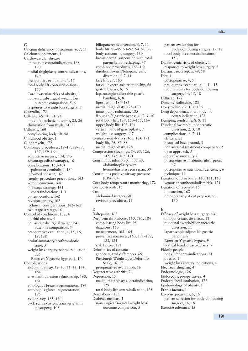
CCalcium deficiency, postoperative, 7, 11Calcium supplements, 14Cardiovascular disease
liposuction contraindications, 168, 170
medial thighplasty contraindications,129
preoperative evaluation, 4, 15total body lift contraindications,
153Cardiovascular risks of obesity, 1non-surgical/surgical weight loss
outcome comparison, 5, 6responses to weight loss surgery, 3
Cefazolin, 172Cellulite, 69, 70, 71, 72
body lift aesthetic outcome, 85, 86elimination from thigh, 74, 77
Cellulitis, 160complicating body lift, 98
Childhood obesity, 1Clindamycin, 172Combined procedures, 18–19, 98–99,
137, 159–164adjunctive surgery, 174, 175advantages/disadvantages, 161complications, 163–164
pulmonary embolism, 164informed consent, 162lengthy procedure precautions, 163with liposuction, 168one-stage strategy, 161
contraindications, 161patient comfort, 162revision surgery, 162technical considerations, 162–163two-stage strategy, 161
Comorbid conditions, 1, 2, 4morbid obesity, 4non-surgical/surgical weight loss
outcome comparison, 5preoperative evaluation, 4, 15, 16,
18, 138proinflammatory/prothrombotic
state, 3weight loss surgery-related reduction,
3, 5Roux-en-Y gastric bypass, 9, 10
Complicationsabdominoplasty, 59–60, 65–66, 163,
164anesthesia duration relationship, 160,
161autologous breast augmentation, 186autologous gluteal augmentation,
185axilloplasty, 185–186back rolls excision, transverse with
mastopexy, 106
biliopancreatic diversion, 6, 7, 11body lift, 88–89, 91–92, 94, 96, 98body-contouring surgery, 160breast dermal suspension with total
parenchymal reshaping, 47combined procedures, 163–164duodenal switch/biliopancreatic
diversion, 6, 7, 11face lift, 27, 163fat cell hyperplasia relationship, 66gastric bypass, 6, 15laparoscopic adjustable gastric
banding, 6, 8liposuction, 184–185medial thighplasty, 128–130mons pubis reduction, 185Roux-en-Y gastric bypass, 6, 7, 9–10total body lift, 139, 153–155, 164upper body lift, 103–104vertical banded gastroplasty, 7weight loss surgery, 6–7
Compression devices, 163, 164, 171body lift, 76, 87, 88medial thighplasty, 128
Compression stockings, 54, 65, 126,142, 152, 163, 171
Continuous infusion pain pump,abdominoplasty withhernia/diastasis recti repair, 59
Continuous positive airway pressure(CPAP), 4
Core body temperature monitoring, 172Corticosteroids, 18Costs
abdominal surgery, 50revision procedures, 16
DDalteparin, 163Deep vein thrombosis, 160, 161, 184
complicating body lift, 98diagnosis, 163management, 163–164preventive measures, 163, 171–172,
183, 184risk factors, 171
Deformities of contourgender-related differences, 69Pittsburgh Weight Loss Deformity
Scale, 16, 17preoperativee evaluation, 16
Degenerative arthritis, 74Depression, 15
medial thighplasty contraindication,129
total body lift contraindication, 138Dermabond, 183Diabetes mellitus, 1
non-surgical/surgical weight lossoutcome comparison, 5
patient evaluation for body-contouring surgery, 15, 18
total body lift contraindications, 153
Diabetogenic risks of obesity, 1responses to weight loss surgery, 3
Diastasis recti repair, 49, 59Diet, 1
postoperative, 4, 5preoperative evaluation, 4, 14–15requirements for body-contouring
surgery, 14, 15, 18Diflucan, 172Dimethyl sulfoxide, 183Doxycycline, 67, 184, 186Drug dependence, total body lift
contraindication, 138Dumping syndrome, 8, 9, 11Duodenal switch/biliopancreatic
diversion, 2, 3, 10complications, 6, 7, 11efficacy, 11historical background, 3non-surgical treatment comparison, 5open approach, 5operative mortality, 6postoperative antibiotics absorption,
67postoperative nutritional deficiency, 6technique, 11
Duration of procedure, 160, 161, 163venous thromboembolism risk, 171
Duration of recovery, 16liposuction, 168preoperative patient preparation,
160
EEfficacy of weight loss surgery, 5–6
biliopancreatic diversion, 11duodenal switch/biliopancreatic
diversion, 11laparoscopic adjustable gastric
banding, 8Roux-en-Y gastric bypass, 9vertical banded gastroplasty, 7
Elderly peoplebody lift contraindications, 74obesity, 1weight loss surgery indications, 4
Electrocardiogarm, 4Endermologie, 126Endoscopy, preoperativee, 4Endotracheal intubation, 172Epidemiology of obesity, 1Ethnic factors, 1Exercise programs, 5, 15
patient selection for body-contouringsurgery, 16, 18
Exercise tolerance, 15
Index
191
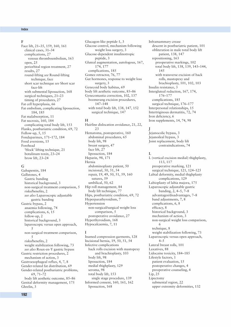
FFace lift, 21–35, 159, 160, 161
clinical cases, 31–34complications, 27
venous thromboembolism, 163open, 25periorbital region treatment, 27results, 27round-lifting see Round-lifting
technique, faceshort scar technique see Short scar
face-liftwith submental liposuction, 168surgical techniques, 21–23timing of procedures, 27
Fat cell hyperplasia, 66Fat embolism, complicating liposuction,
184, 185Fat malabsorption, 11Fat necrosis, 160, 184
complicating total body lift, 153Flanks, postbariatric condition, 69, 72Follow-up, 5, 15Fondaparinux, 171–172, 184Food aversions, 15Forehead
‘block’ lifting technique, 21botulinum toxin, 23–24brow lift, 23–24
GGabapentin, 184Gallstones, 4
Gastric bandinghistorical background, 3non-surgical treatment comparison, 5risks/benefits, 2see also Laparoscopic adjustable
gastric bandingGastric bypass, 2
anaemia following, 74complications, 6, 15follow-up, 2historical background, 3laparoscopic versus open approach,
5non-surgical treatment comparison,
5risks/benefits, 2weight stabilization following, 73see also Roux-en-Y gastric bypass
Gastric restriction procedures, 2mechanism of action, 3
Gastroesophageal reflux, 4, 7, 8Gender-related fat distribution, 69Gender-related postbariatric problems,
69, 71–72body lift aesthetic outcome, 85–86
Genital deformity management, 175Ghrelin, 3
Glucagon-like peptide-1, 3Glucose control, mechanism following
weight loss surgery, 3Glucose-dependent insulinotropic
peptide, 3Gluteal augmentation, autologous, 167,
174, 177complications, 185
Gomez retractor, 76, 77Gut hormones, response to weight loss
surgery, 3Gynecoid body habitus, 69body lift aesthetic outcome, 85–86Gynecomastia correction, 102, 137
boomerang excision procedures,147–148
with total body lift, 138, 147, 152surgical technique, 147
HHairline dislocation avoidance, 21, 22,
23Hematoma, postoperative, 160
abdominal procedures, 65body lift, 98breast surgery, 47face lift, 27liposuction, 184
Heparin, 98, 171Hernia
abdominoplasty patient, 50incisional, 50, 51, 54repair, 19, 49, 50, 51, 59, 160
sutures, 59umbilical, 50, 62
Hip roll management, 84body lift technique, 77
Hips, postbariatric condition, 69, 72Hyperparathyroidism, 7Hypertension
non-surgical/surgical weight losscomparison, 5
postoperative avoidance, 27Hyperthyroidism, 168Hypocalcaemia, 7, 11
IInamed compression garments, 128Incisional hernia, 19, 50, 51, 54Infective complications
back rolls excision with mastopexyand brachioplasty, 103
body lift, 98liposuction, 184medial thighplasty, 129seroma, 98total body lift, 153
single stage procedure, 139Informed consent, 160, 161, 162
liposuction, 168
Inframammary creasedescent in postbariatric patient, 101obliteration in male total body lift
patient, 138, 147repositioning, 163
preoperative markings, 102total body lift, 138, 139, 143–144,
145with transverse excision of back
rolls, mastopexy andbrachioplasty, 101, 102, 103
Insulin resistance, 3Intergluteal reduction, 167, 174,
176–177complications, 185surgical technique, 176–177
Interpersonal relationships, 15Intertriginous dermatitis, 72, 74Iron deficiency, 6Iron supplements, 14, 74, 98
JJejunocolic bypass, 3Jejunoileal bypass, 3Joint replacement, body lift
contraindications, 74
LL (vertical excision medial) thighplasty,
113, 117preoperative marking, 121surgical technique, 121, 124–125
Labial deformity, medial thighplastycomplications, 129
Labioplasty of labia majora, 175Laparoscopic adjustable gastric
banding, 2, 4–5, 7–8advantages/disadvantages, 7–8band adjustments, 7, 8complications, 6, 8efficacy, 8historical background, 3mechanism of action, 3non-surgical weight loss comparison,
6technique, 8weight stabilization following, 73
Laparoscopic versus open approach,4–5
Lateral breast rolls, 101Laxatives, 88Lidocaine toxicity, 184–185Lifestyle factors, 1
patient evaluation, 15postoperative changes, 4preoperative counseling, 4
Lip, 25Lipectomy
submental region, 22upper extremity deformities, 132
Index
192
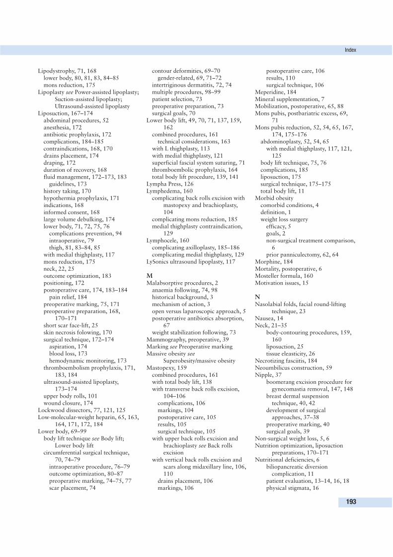
Lipodystrophy, 71, 168lower body, 80, 81, 83, 84–85mons reduction, 175
Lipoplasty see Power-assisted lipoplasty;Suction-assisted lipoplasty;Ultrasound-assisted lipoplasty
Liposuction, 167–174abdominal procedures, 52anesthesia, 172antibiotic prophylaxis, 172complications, 184–185contraindications, 168, 170drains placement, 174draping, 172duration of recovery, 168fluid management, 172–173, 183
guidelines, 173history taking, 170hypothermia prophylaxis, 171indications, 168informed consent, 168large volume debulking, 174lower body, 71, 72, 75, 76
complications prevention, 94intraoperative, 79thigh, 81, 83–84, 85
with medial thighplasty, 117mons reduction, 175neck, 22, 25outcome optimization, 183positioning, 172postoperative care, 174, 183–184
pain relief, 184preoperative marking, 75, 171preoperative preparation, 168,
170–171short scar face-lift, 25skin necrosis folowing, 170surgical technique, 172–174
aspiration, 174blood loss, 173hemodynamic monitoring, 173
thromboembolism prophylaxis, 171,183, 184
ultrasound-assisted lipoplasty,173–174
upper body rolls, 101wound closure, 174
Lockwood dissectors, 77, 121, 125Low-molecular-weight heparin, 65, 163,
164, 171, 172, 184Lower body, 69–99
body lift technique see Body lift;Lower body lift
circumferential surgical technique,70, 74–79
intraoperative procedure, 76–79outcome optimization, 80–87preoperative marking, 74–75, 77scar placement, 74
contour deformities, 69–70gender-related, 69, 71–72
intertriginous dermatitis, 72, 74multiple procedures, 98–99patient selection, 73preoperative preparation, 73surgical goals, 70
Lower body lift, 49, 70, 71, 137, 159,162
combined procedures, 161technical considerations, 163
with L thighplasty, 113with medial thighplasty, 121superficial fascial system suturing, 71thromboembolic prophylaxis, 164total body lift procedure, 139, 141
Lympha Press, 126Lymphedema, 160
complicating back rolls excision withmastopexy and brachioplasty,104
complicating mons reduction, 185medial thighplasty contraindication,
129Lymphocele, 160
complicating axilloplasty, 185–186complicating medial thighplasty, 129
LySonics ultrasound lipoplasty, 117
MMalabsorptive procedures, 2
anaemia following, 74, 98historical background, 3mechanism of action, 3open versus laparoscopic approach, 5postoperative antibiotics absorption,
67weight stabilization following, 73
Mammography, preoperative, 39Marking see Preoperative markingMassive obesity see
Superobesity/massive obesityMastopexy, 159
combined procedures, 161with total body lift, 138with transverse back rolls excision,
104–106complications, 106markings, 104postoperative care, 105results, 105surgical technique, 105
with upper back rolls excision andbrachioplasty see Back rollsexcision
with vertical back rolls excision andscars along midaxillary line, 106,110
drains placement, 106markings, 106
postoperative care, 106results, 110surgical technique, 106
Meperidine, 184Mineral supplementation, 7Mobilization, postoperative, 65, 88Mons pubis, postbariatric excess, 69,
71Mons pubis reduction, 52, 54, 65, 167,
174, 175–176abdominoplasty, 52, 54, 65
with medial thighplasty, 117, 121,125
body lift technique, 75, 76complications, 185liposuction, 175surgical technique, 175–175total body lift, 11
Morbid obesitycomorbid conditions, 4definition, 1weight loss surgery
efficacy, 5goals, 2non-surgical treatment comparison,
6prior panniculectomy, 62, 64
Morphine, 184Mortality, postoperative, 6Mosteller formula, 160Motivation issues, 15
NNasolabial folds, facial round-lifting
technique, 23Nausea, 14Neck, 21–35
body-contouring procedures, 159,160
liposuction, 25tissue eleasticity, 26
Necrotizing fasciitis, 184Neoumbilicus construction, 59Nipple, 37
boomerang excision procedure forgynecomastia removal, 147, 148
breast dermal suspensiontechnique, 40, 42
development of surgicalapproaches, 37–38
preoperative marking, 40surgical goals, 39
Non-surgical weight loss, 5, 6Nutrition optimization, liposuction
preparations, 170–171Nutritional deficiencies, 6
biliopancreatic diversioncomplication, 11
patient evaluation, 13–14, 16, 18physical stigmata, 16
Index
193
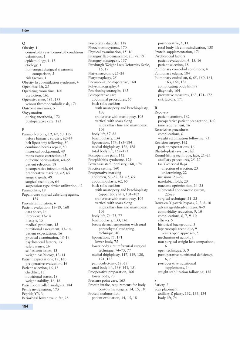
OObesity, 1
comorbidity see Comorbid conditionsdefinitions, 1epidemiology, 1, 13etiology, 1non-surgical/surgical treatment
comparison, 5risk factors, 1
Obesity hypoventilation syndrome, 4Open face-lift, 25Operating room time, 160
prediction, 161Operative time, 161, 163
venous thromboembolis risk, 171Outcome measures, 5Oxygenation
during anesthesia, 172postoperative care, 183
PPanniculectomy, 19, 49, 50, 159
before bariatric surgery, 62–64belt lipectomy following, 50combined hernia repair, 50historical background, 49mons excess correction, 65outcome optimization, 64–65patient selection, 18postoperative infection risk, 64preoperative marking, 62, 65surgical goals, 49surgical technique, 64suspension-type device utilization, 62
Panniculitis, 18Papain-urea topical debriding agents,
129Parenteral nutrition, 6Patient evaluation, 13–19, 160
data sheet, 14interview, 13–14lifestyle, 15medical problems, 15nutritional assessment, 13–14patient expectations, 16physical examination, 15–16psychosocial factors, 15safety issues, 16self esteem issues, 13weight loss history, 13–14
Patient expectations, 18, 160preoperative evaluation, 16
Patient selection, 16, 18checklist, 18nutritional status, 18weight stability, 16, 18
Patient-controlled analgesia, 184Penile invagination, 175Peptide YY, 3Periorbital lower eyelid fat, 25
Personality disorder, 138Pheochromocytoma, 170Physical examination, 15–16Pitanguy flap demarcator, 23, 78, 79Pitanguy mastopexy, 137Pittsburgh Weight Loss Deformity Scale,
16, 17Platysmaectomy, 25–26Platysmaplasty, 25Pneumonia, postoperative, 160Polysomnography, 4Positioning strategies, 163Postoperative care
abdominal procedures, 65back rolls excision
with mastopexy and brachioplasty,103
transverse with mastopexy, 105vertical with scars along
midaxillary line and mastopexy,106
body lift, 87–88brachioplasty, 134liposuction, 174, 183–184medial thighplasty, 126, 128total body lift, 152–153
Postoperative pain, 162Postphlebitis syndrome, 129Power-assisted lipoplasty, 168, 174Practice setting, 160Preoperative marking
abdomen, 51–52, 54, 62, 65abdominoplasty, 62, 65back rolls excision
with mastopexy and brachioplasty(upper body lift), 101–102
transverse with mastopexy, 104vertical with scars along
midaxillary line and mastopexy,106
body lift, 74–75, 77brachioplasty, 133, 141breast dermal suspension with total
parenchymal reshapingtechnique, 40
liposuction, 75, 171lower body, 75
lower body circumferential surgicaltechnique, 74–75, 77
medial thighplasty, 117, 119, 120,121, 123
panniculectomy, 62, 65total body lift, 139–141, 151
Preoperative preparation, 160lower body, 73
Pressure point care, 163Protein intake, requirements for body-
contouring surgery, 14, 15, 18Protein malnutrition
patient evaluation, 14, 15, 18
postoperative, 6, 11total body lift contraindication, 138
Protein supplementation, 171Psychosocial factors
patient evaluation, 4, 15, 16patient selection, 18
Pulmonary comorbid conditions, 4Pulmonary edema, 184Pulmonary embolism, 6, 65, 160, 161,
163, 164, 184complicating body lift, 98diagnosis, 164preventive measures, 163, 171–172risk factors, 171
RRecovery
patient comfort, 162preoperative patient preparation, 160time requirement, 16
Restrictive procedurescomplications, 6weight stabilization following, 73
Revision surgery, 162patient expectations, 16
Rhytidoplasty see Face liftRound-lifting technique, face, 21–25
ancillary procedures, 25–27facial/cervical flaps
direction of traction, 23undermining, 22
incisions, 21–22nasolabial folds, 23outcome optimization, 24–25submental aponeurotic system,
22–23surgical technique, 21–23
Roux-en-Y gastric bypass, 2, 3, 8–10advantages/disadvantages, 8–9comorbidity reduction, 9, 10complications, 6, 7, 9–10efficacy, 9historical background, 3laparoscopic technique, 9
versus open approach, 5mechanism of action, 3non-surgical weight loss comparison,
6open technique, 5, 9postoperative nutritional deficiency,
6, 7postoperative nutritional
supplements, 14weight stabilization following, 138
SSatiety, 3Scar placement
axillary Z plasty, 132, 133, 134body lift, 74
Index
194
Scar placement (cont’d)boomerang excision procedure for
gynecomastia removal, 147brachioplasty, 132, 133, 134, 147medial thighplasty, 117total body lift, 141, 142, 144, 145transverse back rolls excision with
mastopexy, 104, 106upper extremity deformities, 132, 133
Scarring, 18abdomen, 49, 52–53
from previous procedures, 50back rolls excision with mastopexy
and brachioplasty, 104body lift, 86–87brachioplasty, 132medial thighplasty, 113, 129one-stage versus multistage approach,
162patient expectations, 16preoperative evaluation, 138preoperative patient preparation, 160
Schizophrenia, 15Self esteem issues, 13Seroma, 160, 163
abdominoplasty complication, 59–60,65, 66
back rolls excision with mastopexyand brachioplasty complication,103
body lift complication, 92, 94infection, 98
liposuction complication, 174, 184management, 66–67, 92, 94medial thighplasty complication, 113,
128–129prevention of formation, 66–67, 79serial aspiration, 66
Short scar breast techniques, 37, 39Short scar face-lift, 25–27
clinical cases, 28–30closure, 27incision, 25neck liposuction, 25platysmaectomy, 25–26superficial musculoaponeurotic
system tightening, 26tissue glue application, 26–27
Simeon, A.W. severe caloric restrictiondiet, 153
Skin elasticity/tone, 69–70, 138postoperative relaxation, 162preoperative evaluatin, 15, 139
Skin excess classification, 160Skin necrosis
autologous gluteal augmentationcomplication, 185
body lift complication, 94, 96, 98liposuction complication, 170, 184,
185
management, 98medial thighplasty complication, 129prevention, 98
Skin wound dehiscence, 163autologous gluteal augmentation
complication, 185body lift complication, 89, 91–92medial thighplasty complication, 129prevention, 183transverse back rolls excision with
mastopexy complication, 106Sleep apnea, obstructive, 1
non-surgical/surgical weight lossoutcome comparison, 5
preoperative evaluation, 4, 15Smoking status
abdominal procedures, 50, 55body lift patients, postoperative
complications, 89, 96, 98breast surgery, 39, 42liposuction preparations, 171preoperative cessation, 15, 18, 27total body lift contraindications, 138,
153Staging, 18–19, 159–164
advantages/disadvantages, 18, 161algorithm, 162informed consent, 162patient comfort, 162revision surgery, 162
Stretch marks (striae), 70patient expectations, 16
Submental aponeurotic system, 22facial round-lifting technique, 22–23
Submental lipodystrophy, 22Submentoplasty, 25Suction-assisted lipoplasty, 25, 168, 174Superficial fascial system, 164
suturingbody lift, 71, 86–87, 91lower body lift, 71total body lift, 138
Superficial musculoaponeurotic system,short scar face-lift, 26
Superobesity/massive obesity, 1biliopancreatic diversion, 10, 11postoperative nutritional deficiency, 6weight loss procedures, 3
Support groups, 15Support networks, 15, 18Surgeon experience, 160, 161
one-stage approach, 161Swedish Obese Subjects Study Scientific
Group, 5
TThigh
body-contouring procedures, 160liposuction, 168lower body lift approach, 71, 72
postbariatric condition, 69, 81, 113contraindications to medial
thighplasty, 129evaluation, 113, 117gender-related differences, 71, 72
Thigh lift, 72, 159with abdominoplasty/buttock lift, 70,
73, 74, 84with body lift, 73
aesthetic outcome, 85, 86liposuction, 81, 83–84, 92seroma complicating, 92
medial see Thighplasty, medialproblems, 71
Thighplasty, medial, 81, 83, 84, 85, 98,99, 113–130, 137
combined procedures, 161complications, 128–130
infection/abscess, 129lymohocele, 129seroma, 128–129skin necrosis, 129superficial dehiscence, 128
contraindications, 129indications, 113with lower body lift/abdominoplasty,
113, 117, 121, 124outcome optimization (surgical
principles), 126, 128–129patient evaluation, 113, 117postoperative care, 126, 128
edema resolution, 126preoperative marking, 117, 119, 120,
121, 123preoperative preparation, 113, 117,
121scar placement, 117surgical technique, 121, 122,
124–125closure, 126L (vertical excision), 124, 125upper inner thigh crescent, 125
total body lift, 139, 141ultrasound-assisted lipoplasty, 117,
121vertical excision extension, 113, 117
Thoracic soft tissue deformities, 101Thromboembolism prophylaxis
liposuction, 171, 183, 184total body lift, 142, 152
Thrombophlebitis, complicating totalbody lift, 153
single stage procedure, 139Timing of surgery, 16Tissue sealants, 66, 67, 183
abdominoplasty closure, 60seroma formation prevention, 66–67short scar face-lift, 26, 27
Total body lift, 137–156, 159anesthesia, 142
Index
195
Total body lift (cont’d)antiembolic prophylaxis, 142, 152breast reshaping/augmentation, 138,
141complications, 139, 153–155, 164
informed consent form, 155components of procedure, 138historical background, 137–138inframammary crease positioning,
138, 143–144, 145selection of new location, 139, 141
midtorso back skin rolls removal,138, 141
multiple stages, 137, 139, 153, 155,156
combined procedures, 137patient satisfaction, 151patient selection, 137, 164
body mass index, 153postoperative care, 146, 152–153
edema management, 153preoperative markings, 139–141,
151preoperative preparation, 138–141prophylactic antibiotics, 142, 152reverse abdominoplasty, 138, 141scar placement, 141, 142, 144, 145single stage, 137, 139, 143, 153, 155,
156optimizing outcomes, 148,
151–152patient characteristics, 153
superficial fascial system suturing,138
surgical goals, 137surgical technique, 141–147
abdominoplasty, 143, 144, 145blood transfusion/fluid
replacement, 143, 152breasts, 142, 143, 144–145, 151closure, 151L brachioplasty, 143, 145, 147,
151, 152patient body temperature
maintenance, 142–143, 152upper body, 143–145
with ultrasound-assisted liposoplasty,153
upper body lift, 141gynecomastia correction, 147–148
UUltrasound
abdominal haematoma detection, 65preoperative gallstones detection, 4seroma management with drain
placement, 66Ultrasound-assisted lipoplasty, 168,
173–174male intramammary fold obliteration,
138thighs, 117, 121total body lift, 141, 153
gynecomastia correction, 147, 148,152
Umbilical hernia, 50, 62Upper body lift, 137, 161
total body lift, 139, 141, 143–145inverted L brachioplasty, 141in men (gynecomastia correction),
147–148see also Back rolls excision, with
mastopexy and brachioplastyUpper body rolls, 101–112
back see Back rolls excisionsurgical approaches, 101
Upper extremity deformities, 131–135scar placement, 132surgical procedure, 133surgical strategies, 132
total body lift, 137treatment zones, 131–132
Upper lateral chest wall deformities,132
Upper trunk deformities, 101Urinary catheterization, 163
VVaser LipoSelection, 117, 129Venous foot pumps, 171Venous thromboembolism, 163
risk factors, 171
see also Deep vein thrombosis;Pulmonary embolism
Vertical banded gastroplasty, 2, 7advantages/disadvantages, 7complications, 6, 7efficacy, 7non-surgical weight loss comparison,
6technique, 7weight stabilization following, 73
Vitamin B12 deficiency, 7, 11Vitamin B12 supplements, 14Vitamin D deficiency, 7Vitamin K supplements, 138Vitamin supplementation, 2, 5, 7, 74,
171
WWeight loss history, patient evaluation,
13–14Weight loss surgery, 1–11
complications, 6–7surgeon experience/hospital volume
impact, 7contraindications, 4efficacy, 5–6follow-up, 5goals, 2gut hormone responses, 3historical background, 3indications, 3–4laparoscopic versus open approach,
4–5mechanisms of action, 3non-surgical treatment comparison, 5postoperative mortality, 6preparations, 4procedures, 1–3, 7–11
selection, 2–3results assessment, 5
Well Box, 153Wound dehiscence see Skin wound
dehiscenceWound dressings, 183
Index
196