Embed Size (px)
Citation preview
Anatomy and physiology of the peripheral and central auditory system
Lecture 2(a)
Dr. Ghulam SaqulainM.B.B.S., D.L.O, F.C.P.S
Head of Department of E.N.TCapital Hospital

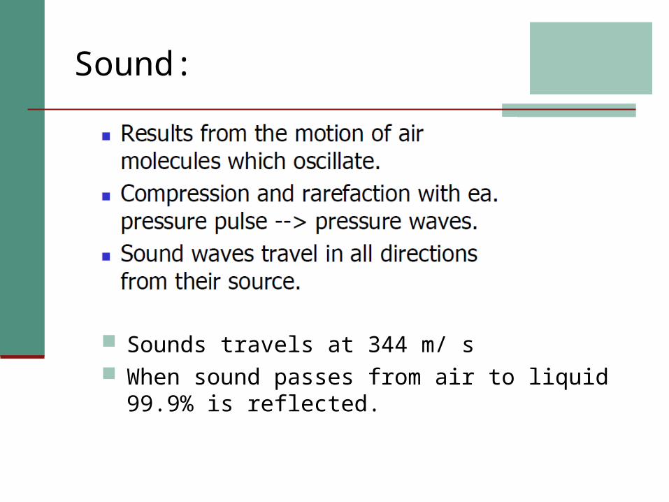
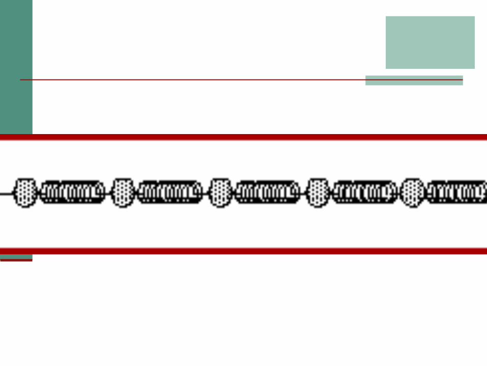
Sound:
Sounds travels at 344 m/ s When sound passes from air to liquid 99.9% is
reflected.


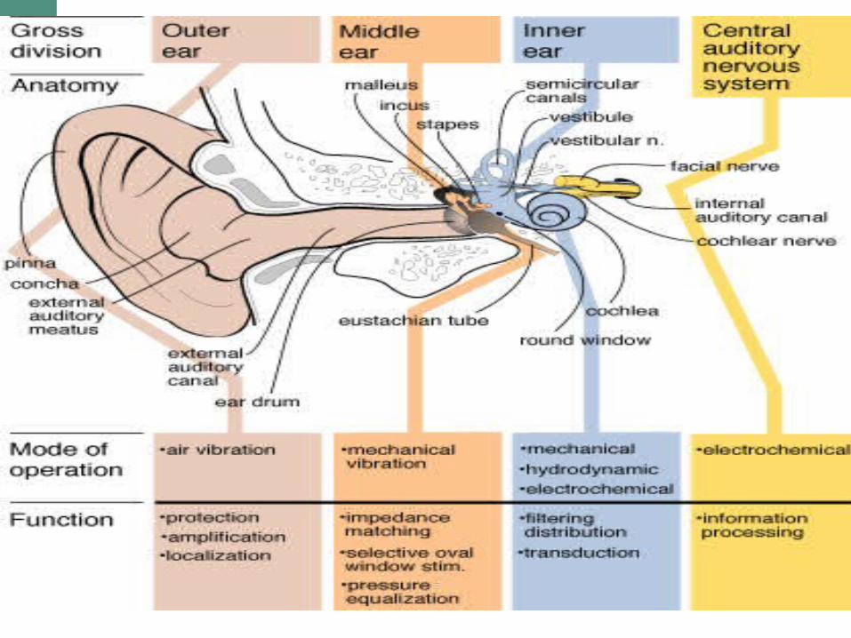
Ear comprises of three main parts playing their own vital role:
Outer Ear Middle Ear Inner Ear.

EXTERNAL EAR
MIDDLE EAR
INTERNAL EAR

OUTER EAR
Pinna and External Auditory Canal Pinna:
elastic cartilage muscles thin skin.

External auditory Canal
Curved tube Covered by skin S shaped in adult Inner2/3-bone
outer1/3-cartilage..
ceruminous gland in outer 1/3


Function:

EAC Channel the auditory stimuli along the meatus to the tympanic membrane
Gain in sound pressure at T.M Pinna-concha system 15-20 db
Sound Localization Monoaural Binaural

Thin fibrous membrane
Obliquely set facing d/w,f/w & lat. Divides Ext.ear fr. Middle ear
note; Medial surface
handle of Malleus ..
Ear drum/ Tympanic Membrane

Sensory nerve supply
1.Great auricular -Cranial aricular surface
2.Auriculo temporal -lateral surface of auricle -ext.audit.meatus -ext.surf. tympan.memb.
3.Auricular br. of Vagus - floor xt.aud.meatus& adjoin.tympanic memb.

Appearance of Tympanic membrane under otoscopic examination
Tymp.membrane.is concave laterally Umbo -depression at the
deepest part produced by tip of handle of Malleus
Cone of light is produced at antero inferior quadrant of membr, *Not visible in middle ear
infections .
Umbo
Cone of light
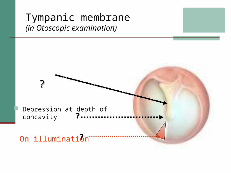
Tympanic membrane (in Otoscopic examination)
Depression at depth of concavity
??
On illumination?
?

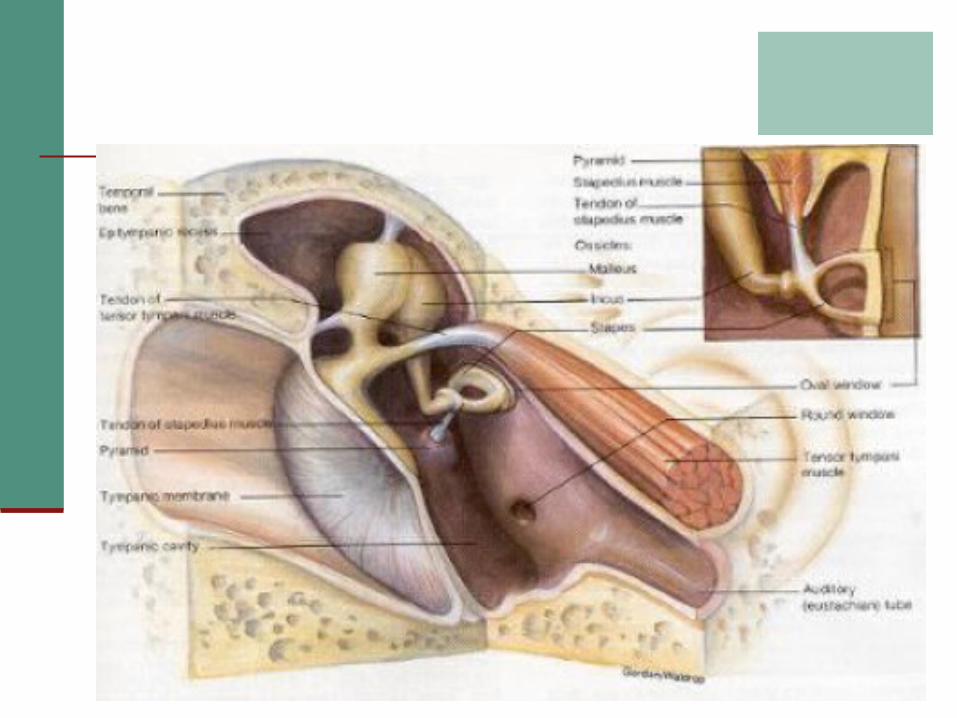
MIDDLE EAR
It is an AIR filled space. Part of the ear between the tympanic membrane and the oval window
Contents: Air Ossicles: Malleus, Incus and
Stapes Muscles: Tensor tympani,
Stapedius
The TYMPANIC MEMBRANE (ear drum) vibrates in response to sound.
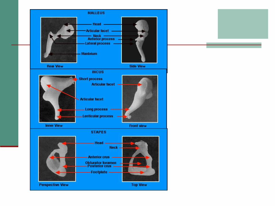
Attached to it are 3 bones: The MALLEUS (hammer), INCUS (anvil), and the STAPES (stirrup) are the smallest bones in the body.
Their function is to amplify sound vibrations. The malleus vibrates the incus, which vibrates the stapes.

The middle ear is open to the nasopharynx by way of the AUDITORY TUBE
If this tube is closed, the ears feel plugged up.
The function of the auditory
tube is to equalize the pressure of the middle ear and the outside air so the ear bones can vibrate.
3 Ossicles
2 Muscles Tensor tympani Stapedius
3 Nerves Chorda tympani Tympanic plexus Lesser petrosal
Main Contents:

Walls & boundaries of Middle ear:
It hasIt has RoofRoof FloorFloor Anterior wallAnterior wall Post. wallPost. wall Lat. Wall Lat. Wall .. Medial wallMedial wall

Roof,Floor & their relations:
RoofRoof Tegmen tympaniTegmen tympani Petrous temporalPetrous temporal --Temporal lobeTemporal lobe
FloorFloor Thin plate of boneThin plate of bone Internal jugular veinInternal jugular vein....
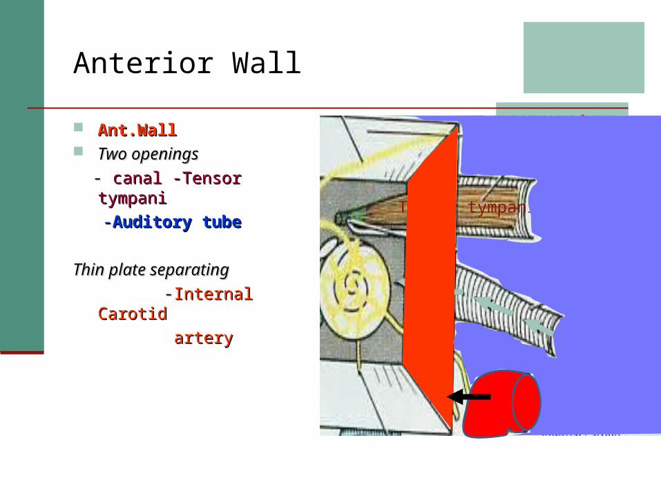
Anterior Wall
Ant.WallAnt.Wall Two openingsTwo openings - - canal -Tensor tympani canal -Tensor tympani --Auditory tubeAuditory tube
Thin plate separatingThin plate separating--Internal CarotidInternal Carotid
arteryartery
Tensor tympani Audit.tube
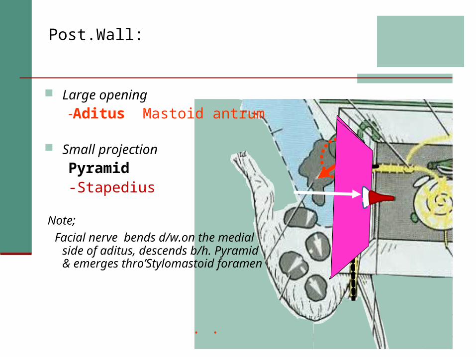
Post.Wall:
. .
Large opening-Aditus Mastoid antrum
Small projection
Pyramid-Stapedius
Note; Facial nerve bends d/w.on the medial
side of aditus, descends b/h. Pyramid & emerges thro’Stylomastoid foramen
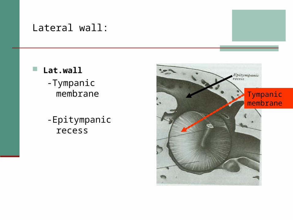
Lateral wall:
Lat.wall-Tympanic membrane
-Epitympanic recess
Tympanic membrane
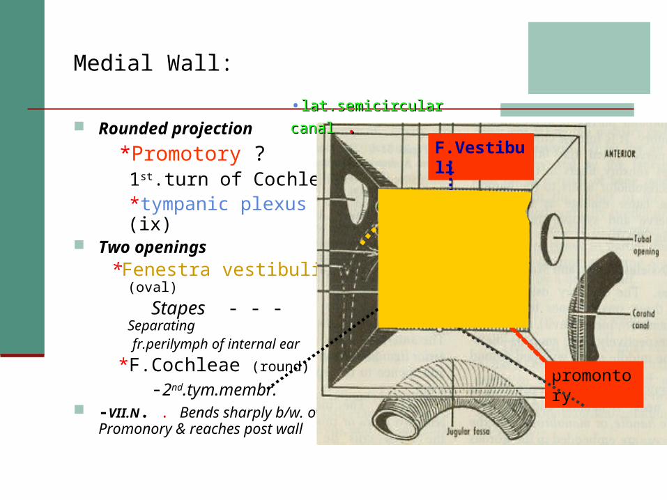
Medial Wall:
Rounded projection *Promotory ? 1st.turn of Cochlea *tympanic plexus (ix)
Two openings *Fenestra vestibuli (oval)
Stapes - - -Separating fr.perilymph of internal ear *F.Cochleae (round)
-2nd.tym.membr. -VII.N. . Bends sharply b/w. over
Promonory & reaches post wall promontory
F.Vestibuli
•lat.semicircular canallat.semicircular canal ..

Functions: It couples sound energy to the cochlea Physical protection of cochlea Matches the impedance of air --cochlear fluids Apply sound preferentially to one window
Impedance Transformer MechanismLow pressure High Displacement – High pressure Low Displacement

This occurs as follows: Liver action of ossicles: 1.3:1 Mechanical advantage 1.3 Hydraulic Action of T.M 14:1 Curved Membrane effect Natural Resonance of Ext. and Middle ear


Physical Protection of Cochlea: