Embed Size (px)
Citation preview
OTOLOGY
2009
Physiology of auditory system
Surgeon’s perspective drtbalu
W W W . D R T B A L U . C O M
Physiology of auditory
system
By
Dr. T. Balasubramanian M.S. D.L.O.
Introduction: Before dwelling into the exact physiology of hearing it will be
better to refresh the physics of sound. Understanding the basic physical
properties of sound is a prerequisite for better understanding of the
physiology.
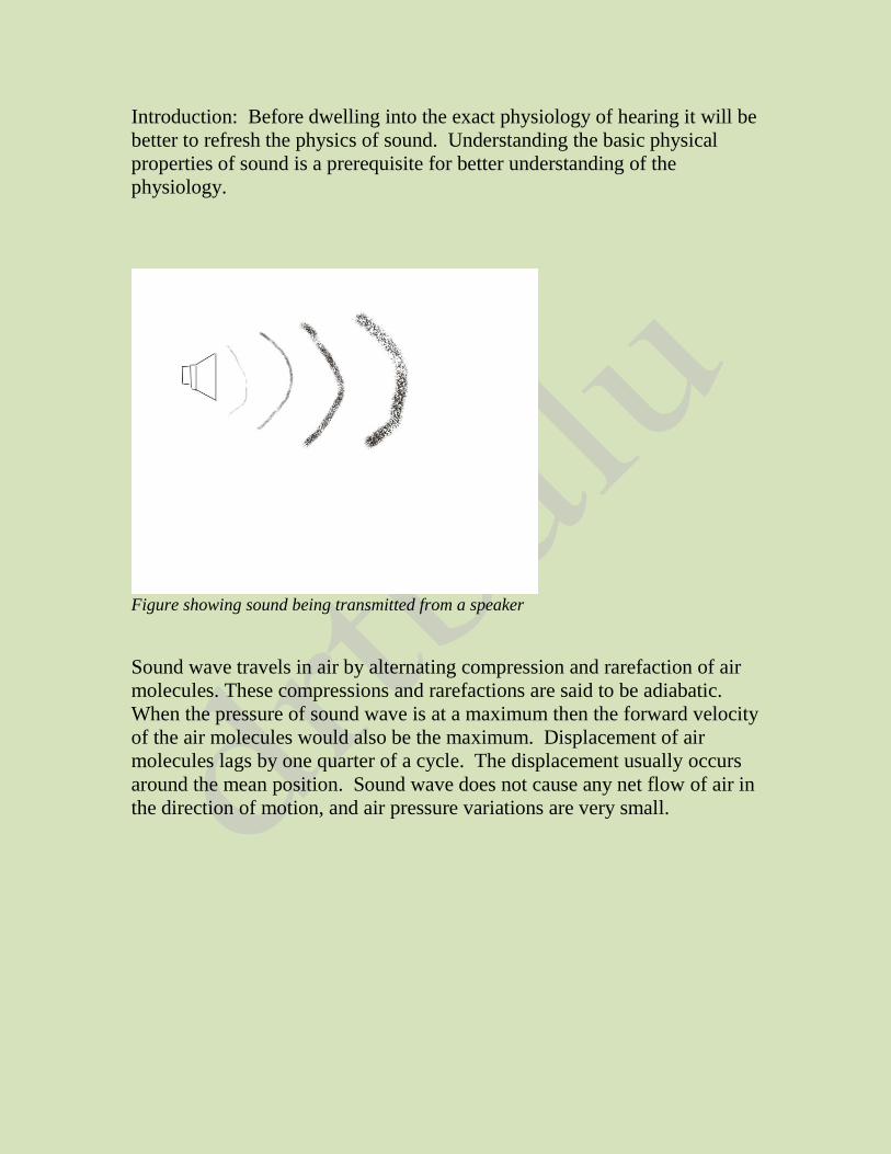
Figure showing sound being transmitted from a speaker
Sound wave travels in air by alternating compression and rarefaction of air
molecules. These compressions and rarefactions are said to be adiabatic.
When the pressure of sound wave is at a maximum then the forward velocity
of the air molecules would also be the maximum. Displacement of air
molecules lags by one quarter of a cycle. The displacement usually occurs
around the mean position. Sound wave does not cause any net flow of air in
the direction of motion, and air pressure variations are very small.
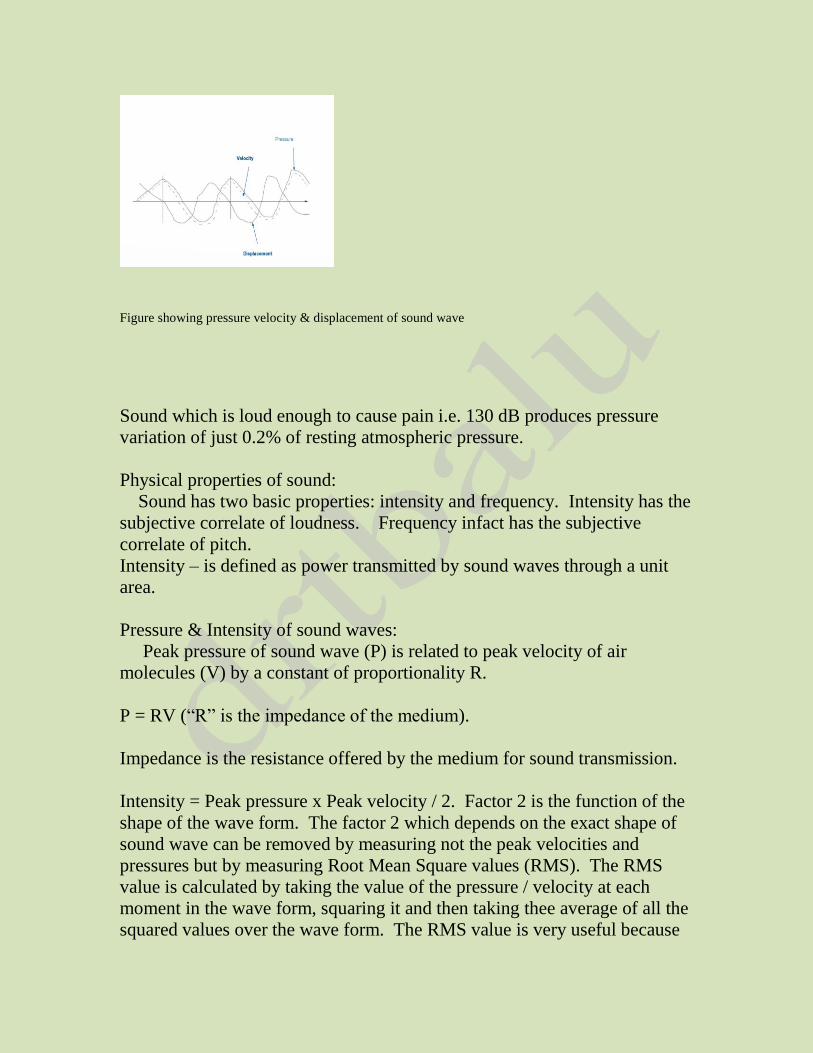
Figure showing pressure velocity & displacement of sound wave
Sound which is loud enough to cause pain i.e. 130 dB produces pressure
variation of just 0.2% of resting atmospheric pressure.
Physical properties of sound:
Sound has two basic properties: intensity and frequency. Intensity has the
subjective correlate of loudness. Frequency infact has the subjective
correlate of pitch.
Intensity – is defined as power transmitted by sound waves through a unit
area.
Pressure & Intensity of sound waves:
Peak pressure of sound wave (P) is related to peak velocity of air
molecules (V) by a constant of proportionality R.
P = RV (“R” is the impedance of the medium).
Impedance is the resistance offered by the medium for sound transmission.
Intensity = Peak pressure x Peak velocity / 2. Factor 2 is the function of the
shape of the wave form. The factor 2 which depends on the exact shape of
sound wave can be removed by measuring not the peak velocities and
pressures but by measuring Root Mean Square values (RMS). The RMS
value is calculated by taking the value of the pressure / velocity at each
moment in the wave form, squaring it and then taking thee average of all the
squared values over the wave form. The RMS value is very useful because
the same relation between intensity, pressure and velocity holds good for all
shapes of sound wave forms.
If intensity of the sound wave is constant, the peak velocity and peak
pressure are also constant. This relationship does not depend on the
frequency of the sound wave; on the contrary displacements produced by
sound waves do vary with frequency if the sound intensity is constant.
Displacements usually vary in inverse proportion to the frequency. For
example for a constant intensity, low frequency vibrations produce greater
displacements.
Decibel:
Sound pressure levels are usually measured in decibels. This is the
logarithmic unit of measurement that expresses the magnitude of the
physical quality of sound, relative to a reference level.
A difference of 1 dB is the minimum perceptible change in the volume of
sound.
Properties of sound:
i. Frequency / wavelength / velocity of sound:
Velocity of sound waves in air is independent of its frequency. At sea level
the speed of sound in air is about 330m/s. This means that if the frequency
of a wave is f cycles / sec (Hz) then f waves must pass any point in one
second.
Length of one wave is 330/f metres. Wave lengths become shorter with
increasing sound frequency.
ii. Sound gets attenuated by distance. In an ideal situation, the sound wave
front will not lose / gain energy with time. The energy in each unit area of
wave front decreases with distance. Another factor that plays a role in sound
attenuation with distance is that when air is compressed at the peak of sound
pressure wave, its temperature raises, and some of the energy of sound wave
is stored as heat. The process gets reversed, and the energy is passed back to
the wave during the trough phase. This passage of heat from the warmer to
the cooler regions of sound wave can cause some loss of energy in the wave.
This particular factor is very important for high frequency waves, where the
short wavelength allows flow of heat between the peaks and troughs of the
wave.
iii. Sound transmission in different media: Sound transmission in different
media depends on the impedance values of the media concerned. When
sound traverses through media of differing impedance values, majority of
sound waves get reflected from the interface between the two media.
Fourier analysis:
Analysis of a complex sound into its constituent sinusoids is known as
Fourier’s analysis. This analysis makes study of sound waves really simple
as mathematical tools can be easily applied. A sinusoidal wave behaves in a
simple manner in complex environments like the reflecting environment of
external auditory canal, or in complex mechanical environments like the
middle ear. Cochlea uses Fourier analysis of sound in order to process it.
These sinusoidal waves can easily be projected in an oscilloscope.
Role of external ear in sound transmission mechanism:
Figure showing the external ear
The external ear components that play an important role in sound
transmission are:
a. Pinna
b. Concha
c. External auditory meatus
External ear has two main influences on the incoming sound:
1. It increases the pressure at the ear drum level in a frequency sensitive
way by acting as a resonator.
2. It increases the pressure in a way that depends on the direction of the
sound source. This particular feature plays an important role in sound
localization function of the ear. It also plays a very important role in
sound localization in difficult situations, especially front to back and
high to low distinctions, where interaural time differences donot help.
3. The Pinna Concha system acts like a trumpet, catching sound over a
large area and concentrating it in the smaller area of external meatus.
This increases the total energy available to the ear drum.
Resonating function of external ear: Resonance of sound at the external
auditory canal changes the sound pressure at the ear drum level in a
frequency selective way.
Role of external ear in sound localization:
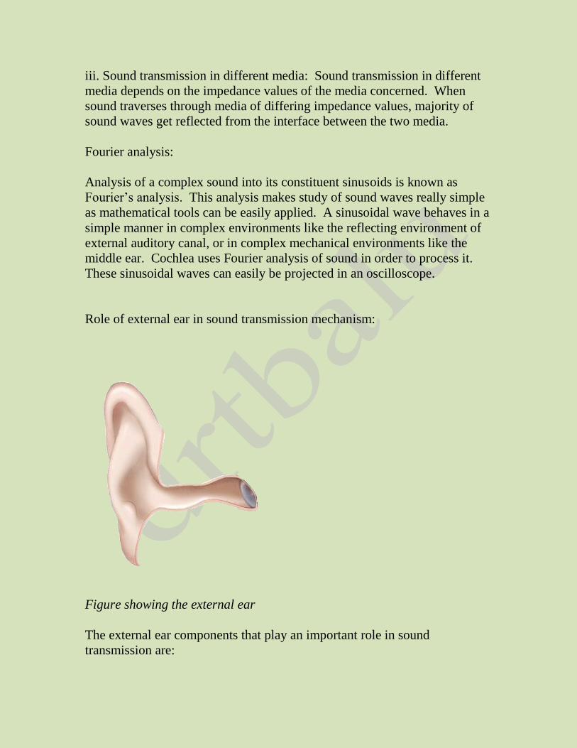
Figure showing a tube of quarter wave length
If a tube of quarter wavelength long, blocked at one end and open at the
other is placed in a sound field, the pressure will be high at the closed end
and low at the open end. The tube also adds resonance to the incident sound
making the sound frequencies of 3 kHz stronger. This phenomenon is seen
in the human external ear. This particular resonance is known as “Quarter
wave resonance”. The meatal quarter wave resonance can develop only if
the meatus is terminated by a boundary with higher impedance than the air
in the canal. This feature adds to the clarity of hearing to spoken sounds.
The external ear by dampening low frequency sounds serves to block out the
endogenous noises produced by the body (air inside the lungs, blood flowing
inside blood vessels etc).
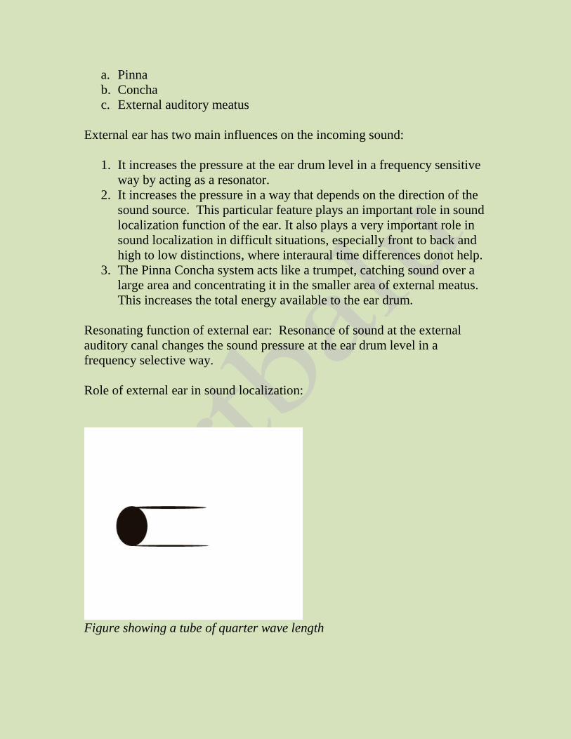
Figure showing the effects of quarter wave resonance
Another important resonance which is present in the external is termed as
the “Broad resonance”. This particular resonance adds about 10dB for
sound frequencies around 5 kHz. These two resonances are complimentary
as they increase the sound pressure relatively uniformly over a frequency
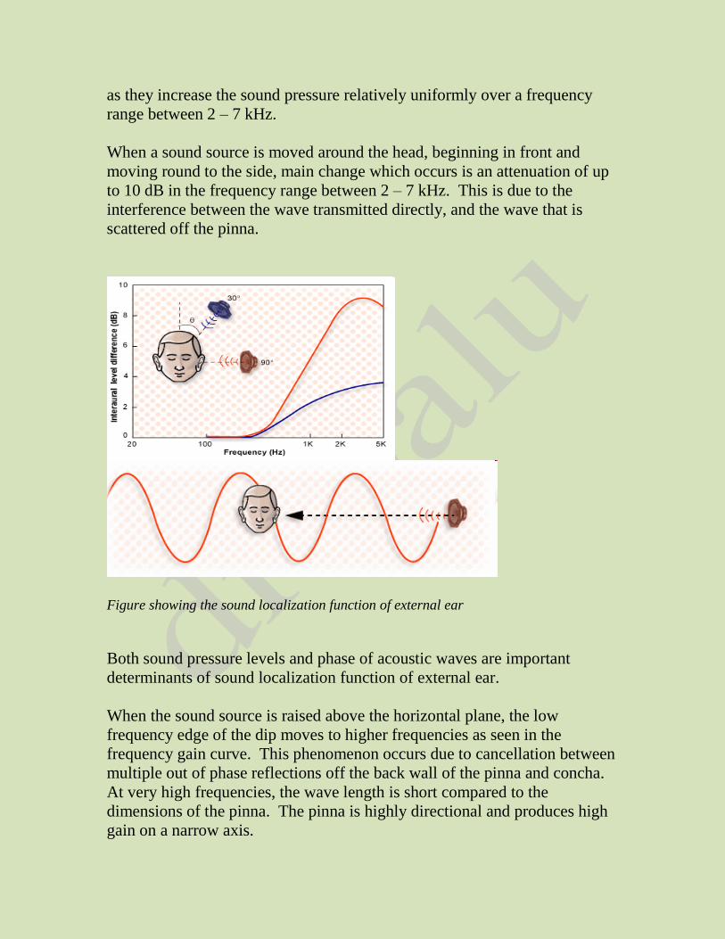
range between 2 – 7 kHz.
When a sound source is moved around the head, beginning in front and
moving round to the side, main change which occurs is an attenuation of up
to 10 dB in the frequency range between 2 – 7 kHz. This is due to the
interference between the wave transmitted directly, and the wave that is
scattered off the pinna.
Figure showing the sound localization function of external ear
Both sound pressure levels and phase of acoustic waves are important
determinants of sound localization function of external ear.
When the sound source is raised above the horizontal plane, the low
frequency edge of the dip moves to higher frequencies as seen in the
frequency gain curve. This phenomenon occurs due to cancellation between
multiple out of phase reflections off the back wall of the pinna and concha.
At very high frequencies, the wave length is short compared to the
dimensions of the pinna. The pinna is highly directional and produces high
gain on a narrow axis.
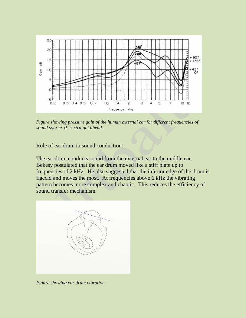
Figure showing pressure gain of the human external ear for different frequencies of
sound source. 0º is straight ahead.
Role of ear drum in sound conduction:
The ear drum conducts sound from the external ear to the middle ear.
Bekesy postulated that the ear drum moved like a stiff plate up to
frequencies of 2 kHz. He also suggested that the inferior edge of the drum is
flaccid and moves the most. At frequencies above 6 kHz the vibrating
pattern becomes more complex and chaotic. This reduces the efficiency of
sound transfer mechanism.
Figure showing ear drum vibration
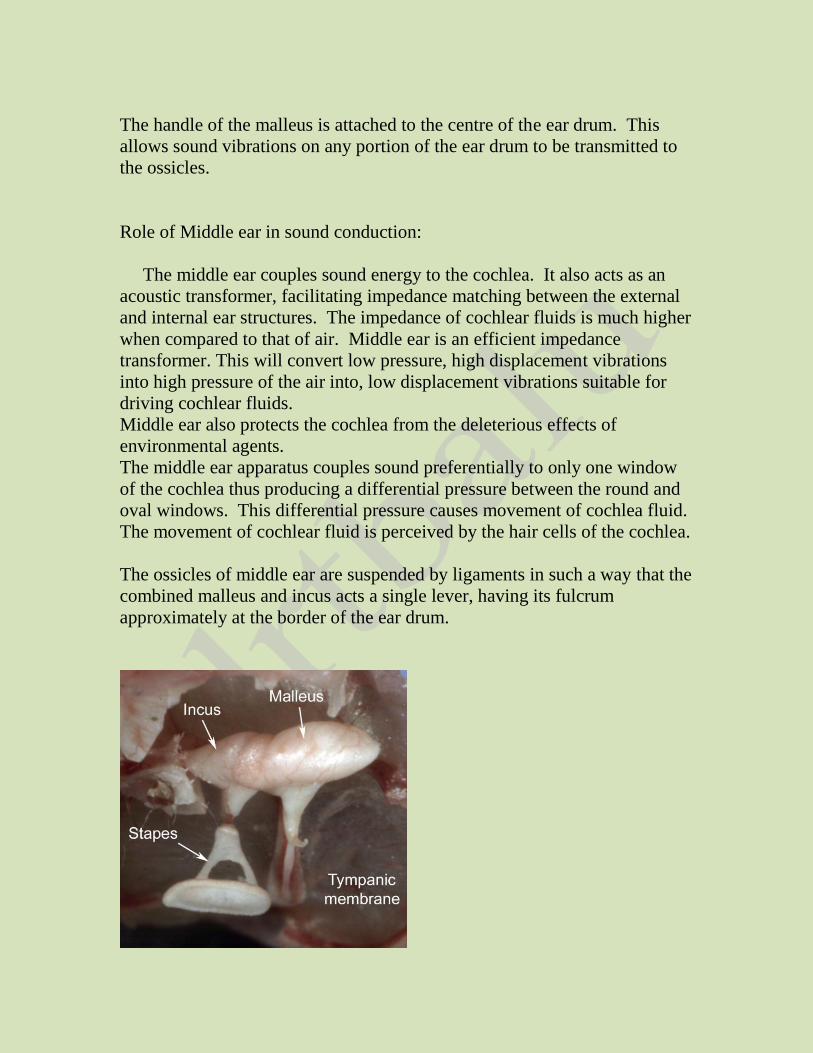
The handle of the malleus is attached to the centre of the ear drum. This
allows sound vibrations on any portion of the ear drum to be transmitted to
the ossicles.
Role of Middle ear in sound conduction:
The middle ear couples sound energy to the cochlea. It also acts as an
acoustic transformer, facilitating impedance matching between the external
and internal ear structures. The impedance of cochlear fluids is much higher
when compared to that of air. Middle ear is an efficient impedance
transformer. This will convert low pressure, high displacement vibrations
into high pressure of the air into, low displacement vibrations suitable for
driving cochlear fluids.
Middle ear also protects the cochlea from the deleterious effects of
environmental agents.
The middle ear apparatus couples sound preferentially to only one window
of the cochlea thus producing a differential pressure between the round and
oval windows. This differential pressure causes movement of cochlea fluid.
The movement of cochlear fluid is perceived by the hair cells of the cochlea.
The ossicles of middle ear are suspended by ligaments in such a way that the
combined malleus and incus acts a single lever, having its fulcrum
approximately at the border of the ear drum.
Picture showing middle ear structures
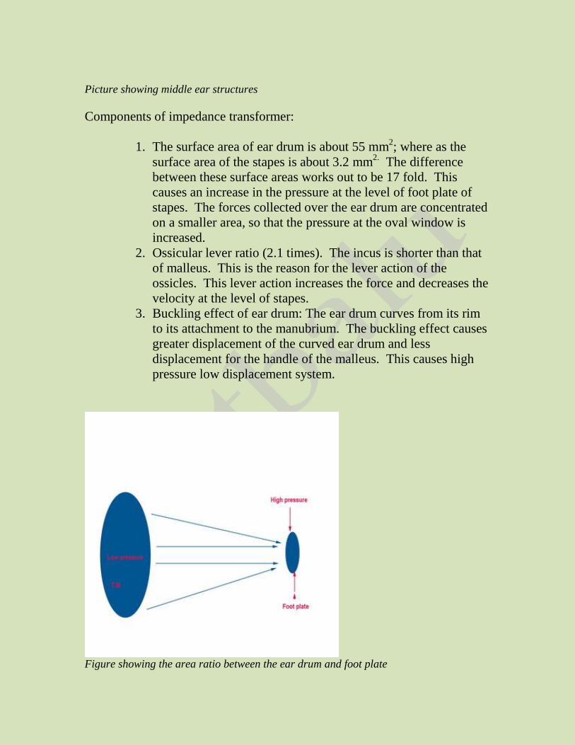
Components of impedance transformer:
1. The surface area of ear drum is about 55 mm2; where as the
surface area of the stapes is about 3.2 mm2. The difference
between these surface areas works out to be 17 fold. This
causes an increase in the pressure at the level of foot plate of
stapes. The forces collected over the ear drum are concentrated
on a smaller area, so that the pressure at the oval window is
increased.
2. Ossicular lever ratio (2.1 times). The incus is shorter than that
of malleus. This is the reason for the lever action of the
ossicles. This lever action increases the force and decreases the
velocity at the level of stapes.
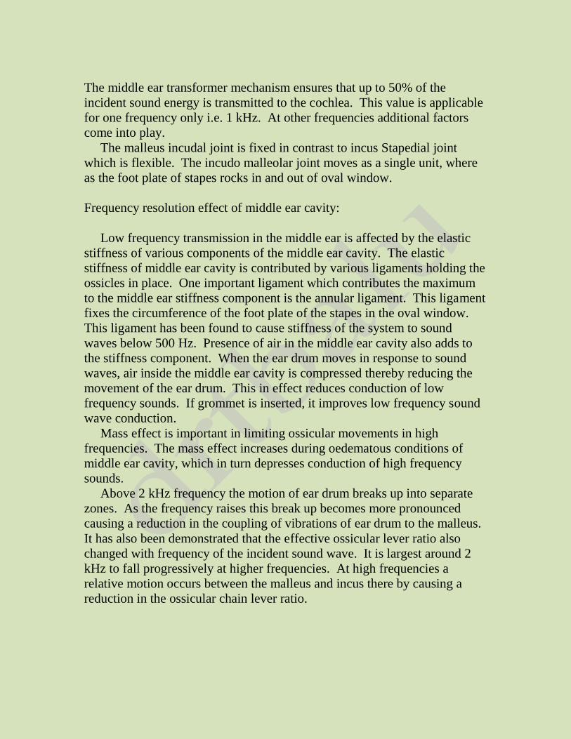
3. Buckling effect of ear drum: The ear drum curves from its rim
to its attachment to the manubrium. The buckling effect causes
greater displacement of the curved ear drum and less
displacement for the handle of the malleus. This causes high
pressure low displacement system.
Figure showing the area ratio between the ear drum and foot plate
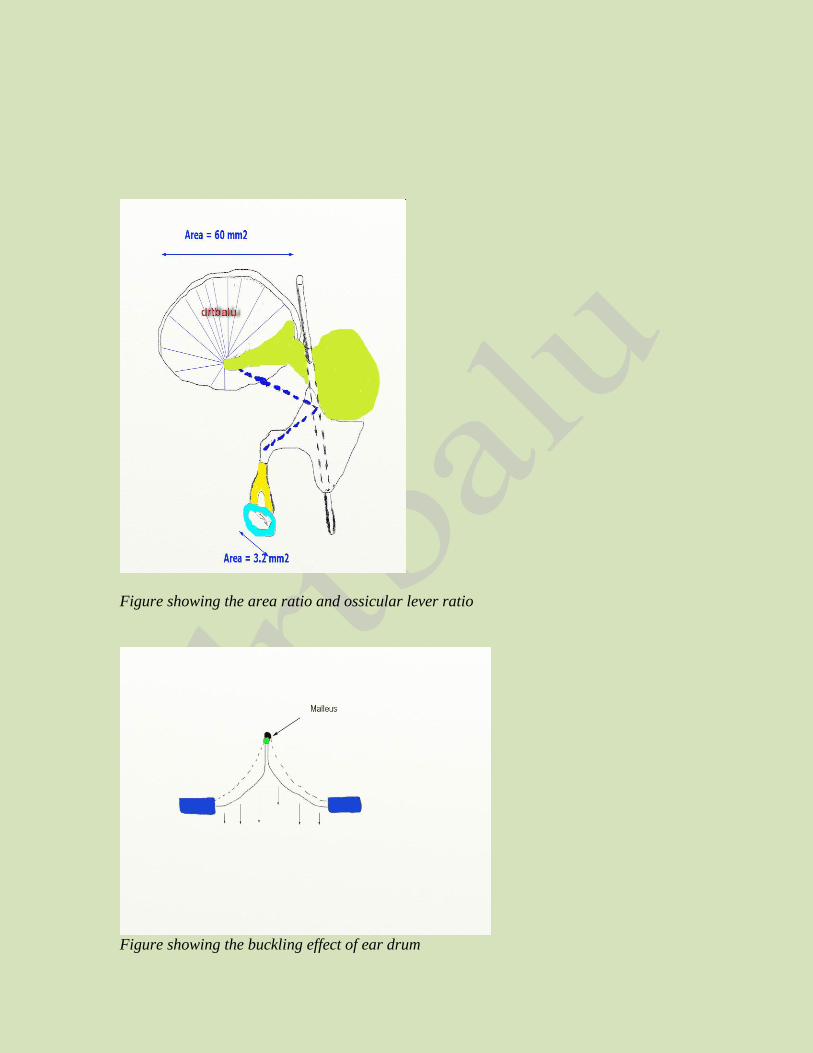
Figure showing the area ratio and ossicular lever ratio
Figure showing the buckling effect of ear drum
The middle ear transformer mechanism ensures that up to 50% of the
incident sound energy is transmitted to the cochlea. This value is applicable
for one frequency only i.e. 1 kHz. At other frequencies additional factors
come into play.
The malleus incudal joint is fixed in contrast to incus Stapedial joint
which is flexible. The incudo malleolar joint moves as a single unit, where
as the foot plate of stapes rocks in and out of oval window.
Frequency resolution effect of middle ear cavity:
Low frequency transmission in the middle ear is affected by the elastic
stiffness of various components of the middle ear cavity. The elastic
stiffness of middle ear cavity is contributed by various ligaments holding the
ossicles in place. One important ligament which contributes the maximum
to the middle ear stiffness component is the annular ligament. This ligament
fixes the circumference of the foot plate of the stapes in the oval window.
This ligament has been found to cause stiffness of the system to sound
waves below 500 Hz. Presence of air in the middle ear cavity also adds to
the stiffness component. When the ear drum moves in response to sound
waves, air inside the middle ear cavity is compressed thereby reducing the
movement of the ear drum. This in effect reduces conduction of low
frequency sounds. If grommet is inserted, it improves low frequency sound
wave conduction.
Mass effect is important in limiting ossicular movements in high
frequencies. The mass effect increases during oedematous conditions of
middle ear cavity, which in turn depresses conduction of high frequency
sounds.
Above 2 kHz frequency the motion of ear drum breaks up into separate
zones. As the frequency raises this break up becomes more pronounced
causing a reduction in the coupling of vibrations of ear drum to the malleus.
It has also been demonstrated that the effective ossicular lever ratio also
changed with frequency of the incident sound wave. It is largest around 2
kHz to fall progressively at higher frequencies. At high frequencies a
relative motion occurs between the malleus and incus there by causing a
reduction in the ossicular chain lever ratio.
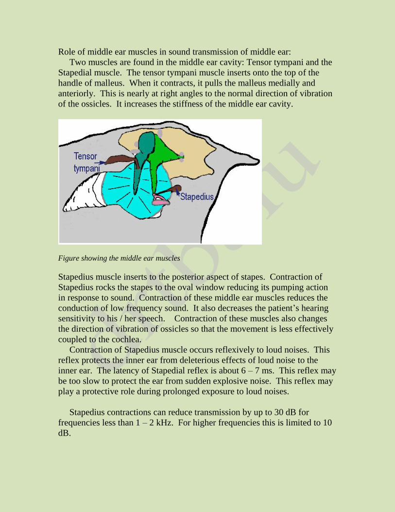
Role of middle ear muscles in sound transmission of middle ear:
Two muscles are found in the middle ear cavity: Tensor tympani and the
Stapedial muscle. The tensor tympani muscle inserts onto the top of the
handle of malleus. When it contracts, it pulls the malleus medially and
anteriorly. This is nearly at right angles to the normal direction of vibration
of the ossicles. It increases the stiffness of the middle ear cavity.
Figure showing the middle ear muscles
Stapedius muscle inserts to the posterior aspect of stapes. Contraction of
Stapedius rocks the stapes to the oval window reducing its pumping action
in response to sound. Contraction of these middle ear muscles reduces the
conduction of low frequency sound. It also decreases the patient’s hearing
sensitivity to his / her speech. Contraction of these muscles also changes
the direction of vibration of ossicles so that the movement is less effectively
coupled to the cochlea.
Contraction of Stapedius muscle occurs reflexively to loud noises. This
reflex protects the inner ear from deleterious effects of loud noise to the
inner ear. The latency of Stapedial reflex is about 6 – 7 ms. This reflex may
be too slow to protect the ear from sudden explosive noise. This reflex may
play a protective role during prolonged exposure to loud noises.
Stapedius contractions can reduce transmission by up to 30 dB for
frequencies less than 1 – 2 kHz. For higher frequencies this is limited to 10
dB.
Sound transmission through damaged middle ear:
Middle ear damage can lead to sound transmission problems due to:
1. Inadequate coupling to the tympanic membrane
2. Loss of impedance transformer
3. Reduction of the ability of the ossicles to move
4. Differential sound pressure levels at the oval and round windows may
be affected (Round window baffle)
The loss of middle ear transformer mechanism alone can lead to an increase
of 15 dB in auditory threshold levels of mid range frequencies. In patients
with no middle ear apparatus, the pressure level delivered by sound is more
or less equal in both round and oval windows. Pressure differential between
both these windows is very important to set the cochlear fluids into motion.
Only the moving cochlear fluid can stimulate the hair cells of the cochlea.
Tondroff by his classic experiments was able to demonstrate that sound
transmission is still possible without any pressure differential between the
two windows. He postulated that pressure release through cochlear aqueduct
could cause pressure differential. Other factors that could facilitate sound
conduction in the absence of middle ear are:
1. Lower compliance of annular ligament of stapes, in comparison to
that of round window membrane. This will facilitate differential
movement of cochlear fluids
2. Scala vestibuli is more yielding than Scala tympani. This could
facilitate differential movement of cochlear fluids.
Physiology of bone conduction:
Sound conduction via bone is one important way in which one hears
his/her voice. It also maintains some level of residual hearing in patients
with conductive hearing losses. Bone conducted sound can be used for
diagnostic purposes (Bone conduction audiometry).
Cochlea is embedded in a bony cavity in the temporal bone. Vibrations
of the entire skull can cause fluid vibrations within the cochlea itself.
Vibrations of the skull could vibrate the CSF. Cochlear fluids are in
continuity with the CSF via the cochlear aqueduct. Vibrations from CSF can
be transmitted via the cochlear aqueduct causing movement of cochlear
fluids. Bone vibrations may also be transmitted by the air inside the external
canal which in turn could cause vibrations of the ear drum.
Direct vibration of osseous spiral lamina can excite the hair cells of the
cochlea.
The centre of inertia of middle ear ossicles does not coincide with their
attachment points. Translational vibrations of skull will cause rotational
vibration of bones, which can be coupled to the internal ear.
The middle ear acts like a broadly tuned filter with peak transmission of
sound waves of frequencies 1 – 2 kHz. If the resonance of middle ear is
hampered by fixation of foot plate of stapes, main loss would be seen in this
frequency. This explains the presence of Carhart’s notch in patients with
mild stapes fixation. This dip appears around 2 kHz in bone conduction
curve.
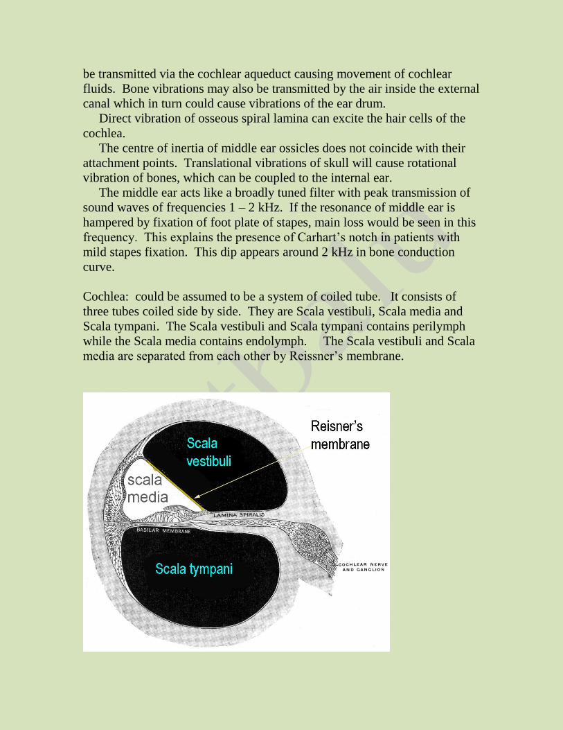
Cochlea: could be assumed to be a system of coiled tube. It consists of
three tubes coiled side by side. They are Scala vestibuli, Scala media and
Scala tympani. The Scala vestibuli and Scala tympani contains perilymph
while the Scala media contains endolymph. The Scala vestibuli and Scala
media are separated from each other by Reissner’s membrane.
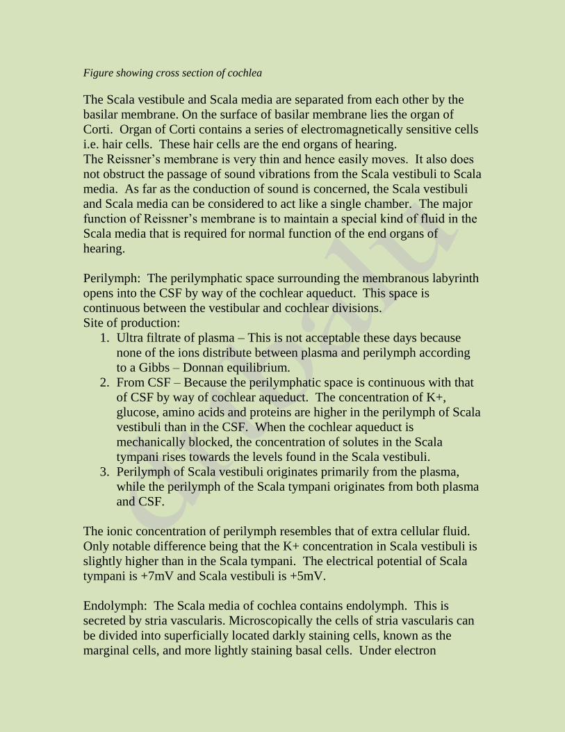
Figure showing cross section of cochlea
The Scala vestibule and Scala media are separated from each other by the
basilar membrane. On the surface of basilar membrane lies the organ of
Corti. Organ of Corti contains a series of electromagnetically sensitive cells
i.e. hair cells. These hair cells are the end organs of hearing.
The Reissner’s membrane is very thin and hence easily moves. It also does
not obstruct the passage of sound vibrations from the Scala vestibuli to Scala
media. As far as the conduction of sound is concerned, the Scala vestibuli
and Scala media can be considered to act like a single chamber. The major
function of Reissner’s membrane is to maintain a special kind of fluid in the
Scala media that is required for normal function of the end organs of
hearing.
Perilymph: The perilymphatic space surrounding the membranous labyrinth
opens into the CSF by way of the cochlear aqueduct. This space is
continuous between the vestibular and cochlear divisions.
Site of production:
1. Ultra filtrate of plasma – This is not acceptable these days because
none of the ions distribute between plasma and perilymph according
to a Gibbs – Donnan equilibrium.
2. From CSF – Because the perilymphatic space is continuous with that
of CSF by way of cochlear aqueduct. The concentration of K+,
glucose, amino acids and proteins are higher in the perilymph of Scala
vestibuli than in the CSF. When the cochlear aqueduct is
mechanically blocked, the concentration of solutes in the Scala
tympani rises towards the levels found in the Scala vestibuli.
3. Perilymph of Scala vestibuli originates primarily from the plasma,
while the perilymph of the Scala tympani originates from both plasma
and CSF.
The ionic concentration of perilymph resembles that of extra cellular fluid.
Only notable difference being that the K+ concentration in Scala vestibuli is
slightly higher than in the Scala tympani. The electrical potential of Scala
tympani is +7mV and Scala vestibuli is +5mV.
Endolymph: The Scala media of cochlea contains endolymph. This is
secreted by stria vascularis. Microscopically the cells of stria vascularis can
be divided into superficially located darkly staining cells, known as the
marginal cells, and more lightly staining basal cells. Under electron
microscope the marginal cells have long infoldings on their basal edge.
These infoldings contain mitochondria. The endolymphatic fluid
component is joined to the endolymphatic sac by means of endolymphatic
duct.
Figure showing stria vascularis
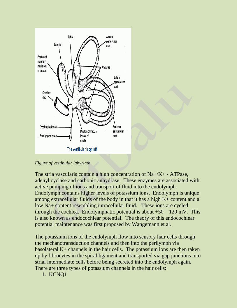
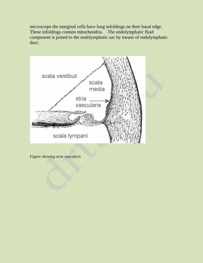
Figure of vestibular labyrinth
The stria vascularis contain a high concentration of Na+/K+ - ATPase,
adenyl cyclase and carbonic anhydrase. These enzymes are associated with
active pumping of ions and transport of fluid into the endolymph.
Endolymph contains higher levels of potassium ions. Endolymph is unique
among extracellular fluids of the body in that it has a high K+ content and a
low Na+ content resembling intracellular fluid. These ions are cycled
through the cochlea. Endolymphatic potential is about +50 – 120 mV. This
is also known as endocochlear potential. The theory of this endocochlear
potential maintenance was first proposed by Wangemann et al.
The potassium ions of the endolymph flow into sensory hair cells through
the mechanotransduction channels and then into the perilymph via
basolateral K+ channels in the hair cells. The potassium ions are then taken
up by fibrocytes in the spiral ligament and transported via gap junctions into
strial intermediate cells before being secreted into the endolymph again.
There are three types of potassium channels in the hair cells:
1. KCNQ1
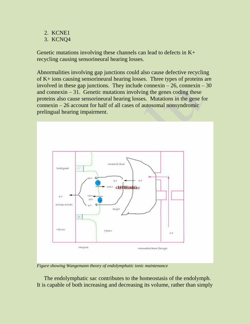
2. KCNE1
3. KCNQ4
Genetic mutations involving these channels can lead to defects in K+
recycling causing sensorineural hearing losses.
Abnormalities involving gap junctions could also cause defective recycling
of K+ ions causing sensorineural hearing losses. Three types of proteins are
involved in these gap junctions. They include connexin – 26, connexin – 30
and connexin – 31. Genetic mutations involving the genes coding these
proteins also cause sensorineural hearing losses. Mutations in the gene for
connexin – 26 account for half of all cases of autosomal nonsyndromic
prelingual hearing impairment.
Figure showing Wangemann theory of endolymphatic ionic maintenance
The endolymphatic sac contributes to the homeostasis of the endolymph.
It is capable of both increasing and decreasing its volume, rather than simply
absorbing endolymph. Obliteration of the endolymphatic sac and duct
causes endolymphatic hydrops.
Basilar membrane: The basilar membrane is a fibrous membrane that
separates the Scala media from the Scala tympani. This membrane contains
20,000 – 30,000 basilar fibers that project from the bony center of the
cochlea (modiolus) towards the outer wall. These fibers are stiff, elastic and
reed like structures that are fixed at their basal ends in the central bony
structure (modiolus) of the cochlea. They are not fixed at their distal ends.
The distal ends are embedded in the loose basilar membrane. Since these
fibers are stiff and free at one end, they can vibrate like the reeds of a
harmonica.
The lengths of the basilar fibers increase progressively beginning at the
oval window and going from the base of the cochlea to the apex, increasing
from a length of about 0.004 mm near the oval and round windows to 0.5
mm at the tip of the cochlea (Helicotrema). This constitutes a 12 fold
increase in length.
The diameters of these fibers however decrease from the oval window to
the Helicotrema, so that their overall stiffness decreases more than 100 fold.
The stiff, short fibers near the oval window vibrate best at very high
frequency, while the long limber fibers near the tip of the cochlea vibrate
best at a low frequency.
The high frequency resonance of the basilar membrane occurs near the base,
where the sound waves enter the cochlea through the oval window. Low
frequency resonance occurs near the Helicotrema, mainly because of the less
stiff fibers.
Cochlear mechanics:
The mechanical travelling wave in the cochlea is the basis of frequency
selectivity. A normal travelling wave is fundamental to normal auditory
function, while a pathological wave, which occurs in cases of cochlear
sensorineural hearing loss, can cause severe sensori neural deficit.
The cochlear travelling wave was first described by Bekesy.
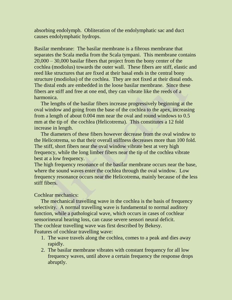
Features of cochlear travelling wave:
1. The wave travels along the cochlea, comes to a peak and dies away
rapidly.
2. The basilar membrane vibrates with constant frequency for all low
frequency waves, until above a certain frequency the response drops
abruptly.
3. According to Bekesy, the capacity of the basilar membrane for
frequency filtering is very poor indeed.
4. As the travelling wave moves up the cochlea towards its peak, it
reaches a region which is mechanically active. In this region the
membrane starts putting more energy in to the wave. The amplitude
hence rises rapidly and falls rapidly.
5. The travelling wave has two components. The first component is a
broadly tuned small component. This component depends on the
mechanical properties of the membrane. The second component is
caused by active input from the cochlea. This component is
prominent for low intensity sounds.
Figure showing travelling wave of Bekesy. Base of cochlea is to the left, and the apex
towards right. Solid lines show displacements at four successive instants. Dotted lines
show the envelope which is static
It has been shown that the relative amplitude of basilar membrane vibration
is the largest for low intensity stimulus. As the stimulus amplitude is
increased, the relative response becomes smaller and smaller.
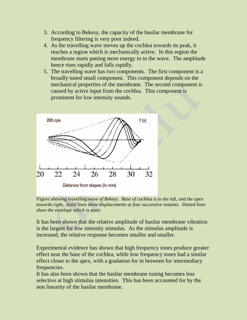
Experimental evidence has shown that high frequency tones produce greater
effect near the base of the cochlea, while low frequency tones had a similar
effect closer to the apex, with a gradation for in between for intermediary
frequencies.
It has also been shown that the basilar membrane tuning becomes less
selective at high stimulus intensities. This has been accounted for by the
non linearity of the basilar membrane.
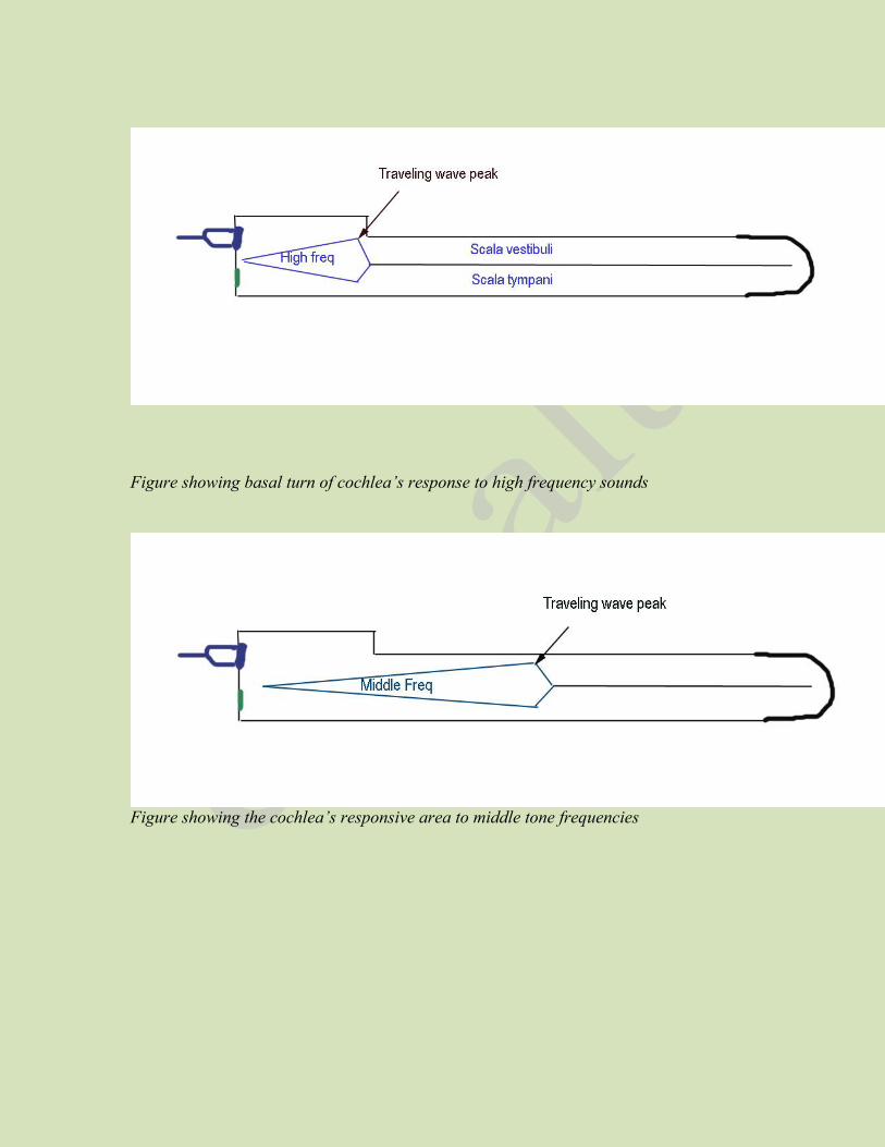
Figure showing basal turn of cochlea’s response to high frequency sounds
Figure showing the cochlea’s responsive area to middle tone frequencies
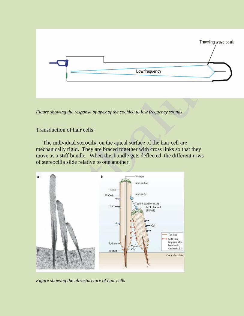
Figure showing the response of apex of the cochlea to low frequency sounds
Transduction of hair cells:
The individual sterocilia on the apical surface of the hair cell are
mechanically rigid. They are braced together with cross links so that they
move as a stiff bundle. When this bundle gets deflected, the different rows
of stereocilia slide relative to one another.
Figure showing the ultrasturcture of hair cells
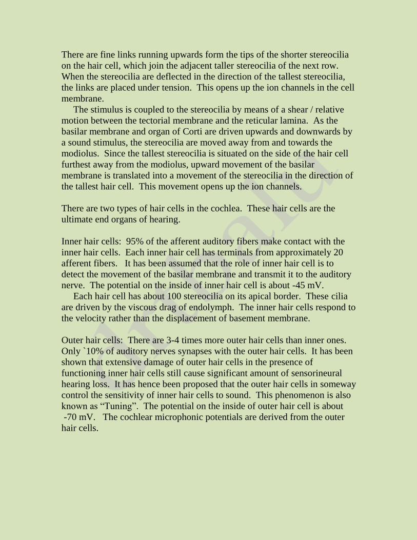
There are fine links running upwards form the tips of the shorter stereocilia
on the hair cell, which join the adjacent taller stereocilia of the next row.
When the stereocilia are deflected in the direction of the tallest stereocilia,
the links are placed under tension. This opens up the ion channels in the cell
membrane.
The stimulus is coupled to the stereocilia by means of a shear / relative
motion between the tectorial membrane and the reticular lamina. As the
basilar membrane and organ of Corti are driven upwards and downwards by
a sound stimulus, the stereocilia are moved away from and towards the
modiolus. Since the tallest stereocilia is situated on the side of the hair cell
furthest away from the modiolus, upward movement of the basilar
membrane is translated into a movement of the stereocilia in the direction of
the tallest hair cell. This movement opens up the ion channels.
There are two types of hair cells in the cochlea. These hair cells are the
ultimate end organs of hearing.
Inner hair cells: 95% of the afferent auditory fibers make contact with the
inner hair cells. Each inner hair cell has terminals from approximately 20
afferent fibers. It has been assumed that the role of inner hair cell is to
detect the movement of the basilar membrane and transmit it to the auditory
nerve. The potential on the inside of inner hair cell is about -45 mV.
Each hair cell has about 100 stereocilia on its apical border. These cilia
are driven by the viscous drag of endolymph. The inner hair cells respond to
the velocity rather than the displacement of basement membrane.
Outer hair cells: There are 3-4 times more outer hair cells than inner ones.
Only `10% of auditory nerves synapses with the outer hair cells. It has been
shown that extensive damage of outer hair cells in the presence of
functioning inner hair cells still cause significant amount of sensorineural
hearing loss. It has hence been proposed that the outer hair cells in someway
control the sensitivity of inner hair cells to sound. This phenomenon is also
known as “Tuning”. The potential on the inside of outer hair cell is about
-70 mV. The cochlear microphonic potentials are derived from the outer
hair cells.
Electrical potentials of cochlea:
By placing electrodes in the vicinity of cochlea electrical potentials
generated by it in response to sound waves can be recorded. These
potentials can be divided into three components. They are:
1. Cochlear microphonics – A/C potential
2. Summating potential – D/C potential
3. Negative neural potentials – N1 & N2.
The cochlear microphonics is derived almost exclusively from outer hair
cells. This helps in the magnification of the traveling wave. This is an A/C
response following the waveform of the stimulus. Superimposed on this is a
D/C shift in the baseline. This is known as the summating potential. The
summating potential could either be negative / positive depending on the
stimulus conditions. The summating potential takes sometime to reach the
maximum after the onset of stimulus. It is mostly generated as a distortion
component of the outer hair cell response, with perhaps a small contribution
from the inner hair cells. At the beginning of sometimes at the end, a series
of deflections in the negative direction can be seen. These are also known as
neural potentials. The first one is known as N1, and the second smaller one
which is not always visible is known as N2 potential.
Determination of loudness of sound:
Loudness is determined by the auditory system in the following three ways:
1. As the sound becomes louder, the amplitude of vibration of the basilar
membrane and hair cells also increases, so that the hair cells excite the
nerve endings at more rapid rates.
2. As the amplitude off vibration increases, it causes more and more of
the hair cells on the fringes of the resonating portion of the basilar
membrane to become stimulated, causing spatial summation of
impulses i.e. transmission through many nerve fibers rather than
through only a few.
3. The outer hair cells do not become stimulated significantly until
vibration of the basilar membrane reaches high intensity and
stimulation of these cells appraises the nervous system that the sound
is loud.
Auditory nerve fibers response to stimuli:
In response to stimulus, neurotransmitter is released in the synapses at the
base of inner hair cells. This gives rise to action potentials in the auditory
nerve fibers. Single auditory nerve stimuli are always excitatory, and never
inhibitory. Transmitter release occurs due to depolarization of inner hair
cells. For low frequency stimuli, the transmitter will be released in packets
concentrated during the depolarization phases of the hair cell response.
These events take place in synchrony with the sound stimulus, transmitter
release and action potential generation is also in synchrony with individual
cycles of the stimulus. This is known as phase locking. Phase locking takes
place only at low frequencies. Phase locking is one way in which
information about the sound stimulus is transmitted via the auditory nerve.
For stimulus frequencies below 5 kHz, the timing of action potentials play a
vital role in frequency discrimination. This is known as temporal coding of
the stimuli. Frequency specificity is another method by which frequency
discrimination occur at the level of auditory nerve. This is also known as
“Place coding”. Fibers responding best to different frequencies arise from
different places in the cochlea.
Theories of hearing:
Various theories of hearing have been proposed. They are:
1. Place theory of Helmholtz
2. Telephone theory of Rutherford
3. Volley theory of Weaver
4. Place volley theory of Lawrence
5. Travelling wave theory of Bekesy
Place theory of Helmholtz: Also known as resonance theory. It was first
proposed by Helmholtz. He proposed that the basilar membrane was
constructed of different segments that resonated in response to different
frequencies. These segments are arranged according to the location along
the length of basement membrane. For this tuning process to occur the
segments in different locations would have to be under different degrees of
tension. According to this theory, a sound entering the cochlea causes the
vibration of the segments that are tuned to resonate at that frequency.
Pitfalls: This theory has various pitfalls. Sharply tuned resonators dampen
rather slowly. This could lead to constant after ringing long after the
stimulus has ceased. This theory also fails to explain why a stream of clicks
of frequencies ranging from 1220, 1300 and 1400 Hz is heard as 100 Hz
pitch.
Telephone theory of Rutherford: According to this theory proposed by
Rutherford the entire cochlea responds as a whole to all frequencies instead
of being activated on a plate by place basis. Here the sounds of all
frequencies are transmitted as in a telephone cable and frequency analysis is
performed at a higher level (brain).
Pitfalls: Damage to certain portions of cochlea can cause preferential loss of
hearing certain frequencies i.e. like damage to the basal turn of cochlea
causing inability to hear high frequency sounds. This cannot be explained by
telephone theory.
Temporal theory supposes that the auditory nerve conducts sounds
containing the whole range of frequencies and discrimination takes place at
higher centers.
The neurons respond to stimulus obeying the all or none law. They either
fire or stop firing. They should be provided with a latent interval of atleast 1
millisecond before the next stimulus is presented. The auditory nerves will
have no problems conducting sounds up to 900 Hz. Conduction of sounds
above 1000 Hz will become a problem and this theory doesn’t explain how
this frequency gets conducted and perceived by the brain.
Volley theory of Wever: Wever proposed that several neurons acting as a
group can fire in response to high frequency sound, even though none of
them could do it individually. This could be possible if one neuron could
fire in response to one cycle and another neuron fires in response to the next
cycle, while the first neuron is still in its refractory period.
Place volley theory of Lawrence: Lawrence combined both volley and place
theory to explain sound transmission and perception.
Travelling wave theory of Bekesy: Bekesy proposed that frequency coding
took place at the level of cochlea. Contrary to resonance theory, Bekesy
found that basilar membrane is not under tension and elasticity is uniform
throughout. Anatomically the basilar membrane gets wider towards the top
resulting in a gradation of stiffness along its length, going from stiffest at the
base, close to the stapes and least stiff at the apex (helicotrema). As a result
of this stiffness gradient sound transmitted to the cochlea develop a special
kind of wave pattern which travels from base towards the apex.
This wave is known as the travelling wave. The travelling wave involves a
displacement pattern that gradually increases in amplitude as it moves up the
basilar membrane; it reaches a peak at a certain location, decays in
amplitude rather quickly beyond the peak. The location of the traveling
wave peak along the basilar membrane depends on the frequency of the
sound. In other words traveling wave is the mechanism that translates signal
frequency into place of stimulation along the basilar membrane. High
frequencies are represented towards the base of the cochlea, and
successively lower frequencies are represented closer and closer to the apex.
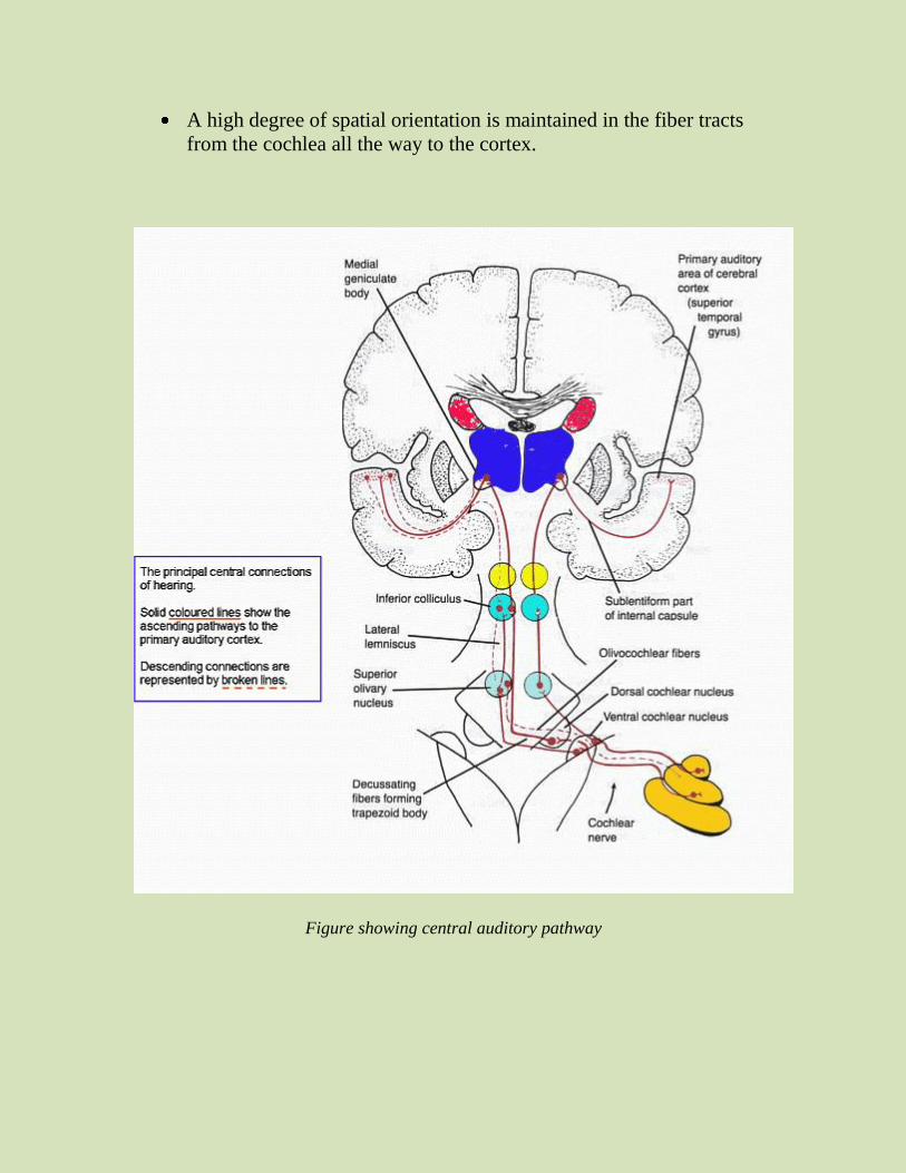
Central auditory pathways: Nerve fibers from the spiral ganglion of corti
enter the dorsal and ventral cochlear nuclei. These nuclei are located in the
upper portion of the medulla. Here all the fibers synapse, and the second
order neurons pass mainly to the opposite side of brain stem to terminate in
the superior olivary nucleus. A few second order neuron fibers also pass to
the superior olivary nucleus on the same side. From the superior olivary
nucleus, the auditory pathway passes upwards through the lateral lemniscus.
Some of these fibers terminate in the nucleus of lateral lemniscus, but many
fibers bypass this nucleus and travel to the inferior colliculus, where almost
all of the auditory fibers synapse. From here, almost all the fibers synapse at
the medial geniculate nucleus. From here auditory fibers pass via auditory
radiation to the auditory cortex located in the superior gyrus of the temporal
lobe.
Signals from both ears are transmitted through the pathways of both
sides of the brain, with a preponderance of transmission in the
contralateral pathway
Crossing over occurs in three places in the brain stem. Usually in the
trapezoid body, in the commissure between the two nuclei of the
lateral lemnisci and in the commissure connecting the two inferior
colliculi.
Many collateral fibers from the auditory tract passes directly into the
reticular activating system of the brain stem
Other collaterals go to the vermis of the cerebellum.
A high degree of spatial orientation is maintained in the fiber tracts
from the cochlea all the way to the cortex.
Figure showing central auditory pathway
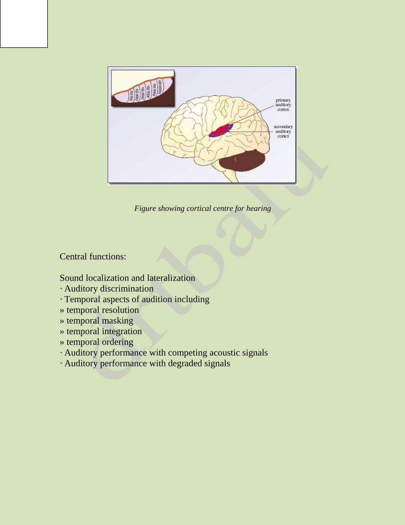
Figure showing cortical centre for hearing
Central functions:
Sound localization and lateralization
· Auditory discrimination
· Temporal aspects of audition including
» temporal resolution
» temporal masking
» temporal integration
» temporal ordering
· Auditory performance with competing acoustic signals
· Auditory performance with degraded signals