Embed Size (px)
Citation preview

Basic Molecular Genetic Basic Molecular Genetic Studies Studies
in Atherosclerosisin Atherosclerosis
Methodology, Applications, Methodology, Applications, and “Future Horizons”and “Future Horizons”
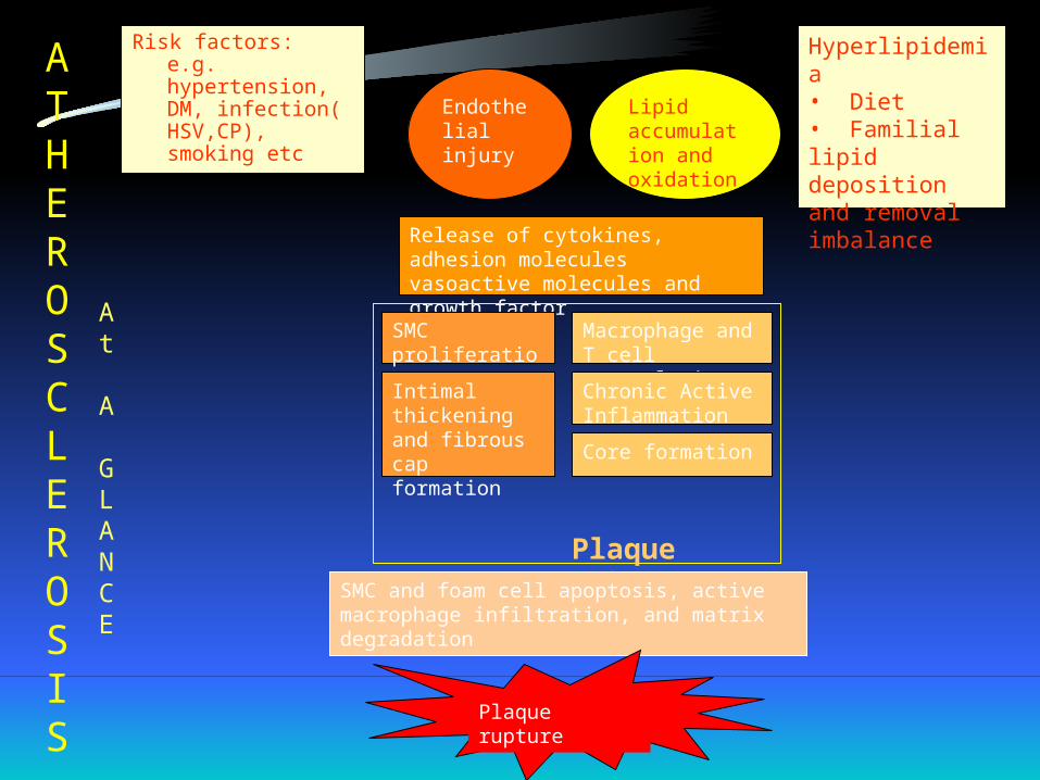
Risk factors: e.g. hypertension, DM, infection( HSV,CP), smoking etc
Hyperlipidemia• Diet• Familial lipid deposition and removal imbalance
Endothelial injury
Release of cytokines, adhesion molecules vasoactive molecules and growth factor
Lipidaccumulation and oxidation
Macrophage and T cell accumulation
Chronic Active Inflammation
SMC proliferation
Intimal thickening and fibrous cap formation Core formation
Plaque formationSMC and foam cell apoptosis, active macrophage infiltration, and matrix degradation
Plaque rupture
ATHEROSCLEROSIS
At
A
GLANCE

Molecular genetics
Brief overview of methods to analyse and study genetics
Polymerase chain reaction-PCR
• This is achieved by repeated rounds of three different steps: heat denaturation of template DNA,
• Annealing of two convergent oligonucleotide primers to the opposite strands of the DNA template
• 5’-3’ extension from each of the annealed primers using a thermosable DNA polymerase
Chain Termination
• This is done to obtain DNA fragments of different lengths
• For this reaction to occur four standard dNTPs (dATP, dCTP, DGTP, dTTP) are included in the reaction mixtures

Gel-Electrophoresis and Autoradiography
• After the chain terminated strands are obtained, they are run through an agarose gel and usually stained with Ethidium bromide and visualize the bands on a u.v transilluminator.
• Electrophoresis makes use of the fact that molecules like DNA migrate in an electric field inversely proportional to its molecular weight towards the positive pole.
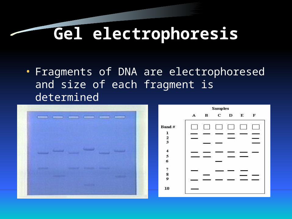
Gel electrophoresis
• Fragments of DNA are electrophoresed and size of each fragment is determined
Southern Blot for DNA analysis
• The restriction fragments obtained by gel-electrophoresis are subject to denaturation with alkali and transferred to a nitrocellulose filter or nylon membrane by blotting.
• The filter is them incubated under hybridization conditions with a specific radiolabeled probe usually generated from a cloned restriction fragment.
• The DNA restriction fragment that is complementary to the probe hybridizes, and its location on the filter can be revealed by autoradiography.
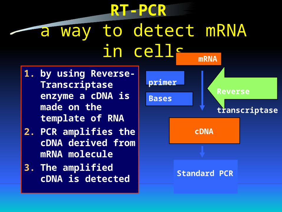
RT-PCR a way to detect mRNA in cells
1. by using Reverse-Transcriptase enzyme a cDNA is made on the template of RNA
2. PCR amplifies the cDNA derived from mRNA molecule
3. The amplified cDNA is detected
mRNA
Standard PCR
primer
Bases
cDNA
Reverse transcriptase
Methods to detect Sequence Variations
• Use of silver stainings to detect Nucleic acids• Nonradioactive Method for the Detection of
Single-strand Conformational Polymorphisms.• Temperature Gradient Gel Electrophoresis(TGGE)
for the detection of polymorphic DNA and RNA.• TGGE in Quanttitative PCR of DNA and RNA• Direct sequencing of PCR products

Analysis of Specific Nucleic Acids in Complex Mixtures
• Southern Blotting Detects specific DNA fragments
• Northern Blotting Detects Specific RNAs• Nuclease Protection is used to Quantitate Specific
RNAs and Map the DNA Regions encoding them
Probes
• A probe is a relatively small piece of DNA that is used to find another piece of DNA.
• In nucleic acid hybridization a DNA probe, labeled radioactively or non-radioactively, seeks out and finds complementary DNA In the target DNA.
• The shortest useful probe is about 20 bases long and is known as oligonucleotide probe.
• RNA molecules may also be used as probes and are termed “riboprobes”.
Nucleic acid probes are segments of DNA(/RNA) that :
1.1. have been labeledhave been labeled (with enzymes antigeneic substrate , chemiluminescent moieties or radiolabeled) and can bind with an bind with high specificityhigh specificity to DNA/ RNA targets
2. probes can be 2 to 1000 bases
3. early DNA probes were labeled with 32 P, now in most kits radioisotopes are replaced by enzymes, affinity labeles and chemiluminescent molecules. ( Enzymes like alkaline phosphatase , horseradish peroxidase , affinity labeles like biotin, digoxigenin)
Hybridization
• The process of forming a double stranded DNA molecule between a probe and a target.
• DNA is double stranded and can be made single stranded.
• If a large excess of DNA, called a probe, that has complementarity to a particular sequence of interest is added to the target, they form a double stranded structure and this is termed hybridization.

Nucleic Acid Immunocytochemistry
This technique uses nucleic acid-antibody complexes as probes and nucleic acids as targets. The aspects of a nucleic acid immunocytochemistry are as follows:
• Target is DNA or mRNA localized within a cell• The recognition and detection of target nucleic
acid relies on base pairing(hybridization) of complementary bases of the target nucleic acid
• The recognition and detection of target nucleic acid relies on base pairing(hybridization) of complementary bases of the target nucleic acid.
• The antibody moiety complexed to the nucleic acid portion of the probe serves only as a signal-generating system reporting that a target has been found and hybridization has occurred.

Fluorescence In Situ Hybridization (FISH)
• FISH is widely used to determine the chromosomal map location and the relative order of genes and DNA sequences within a chromosomal band.

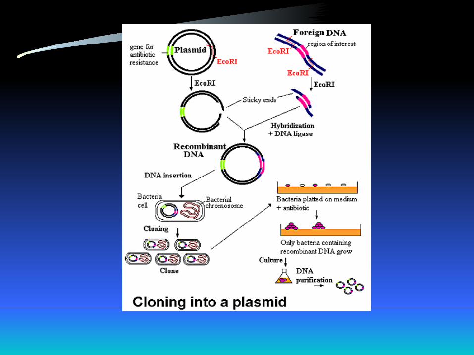
Recombinant DNA Technology
• Recombinant DNA is artificially created DNA for the purpose of genetic engineering which can be introduced into appropriate cells to form a clone of such cells.
• It makes use of two enzymes:• Restriction enzymes: which cut the DNA from any
organism at specific sequences.• Ligases: which can insert DNA restriction fragments into
replicating DNA molecules producing recombinant DNA.
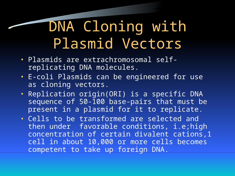
DNA Cloning with Plasmid Vectors
• Plasmids are extrachromosomal self-replicating DNA molecules.
• E-coli Plasmids can be engineered for use as cloning vectors.
• Replication origin(ORI) is a specific DNA sequence of 50-100 base-pairs that must be present in a plasmid for it to replicate.
• Cells to be transformed are selected and then under favorable conditions, i.e;high concentration of certain divalent cations,1 cell in about 10,000 or more cells becomes competent to take up foreign DNA.
Commercial Potential of Recombinant DNA
• Genetic Engineering as a man-made entity has been refined over the 25 years of its existence to the point where medically and pharmaceutically important reagents ( for example, insulin, and interferon) as well as certain vaccines are now routinely being made on a manufacturing scale.
• Genetic Engineering has been also used in brewing, fermenting, wine making and other fields.
Genomic analysis
1. DNA sequencing2. Single molecule sequencing3. SNP( single nucleotide polymorphism)
4. DNA array technology (Functional Genomic analysis)
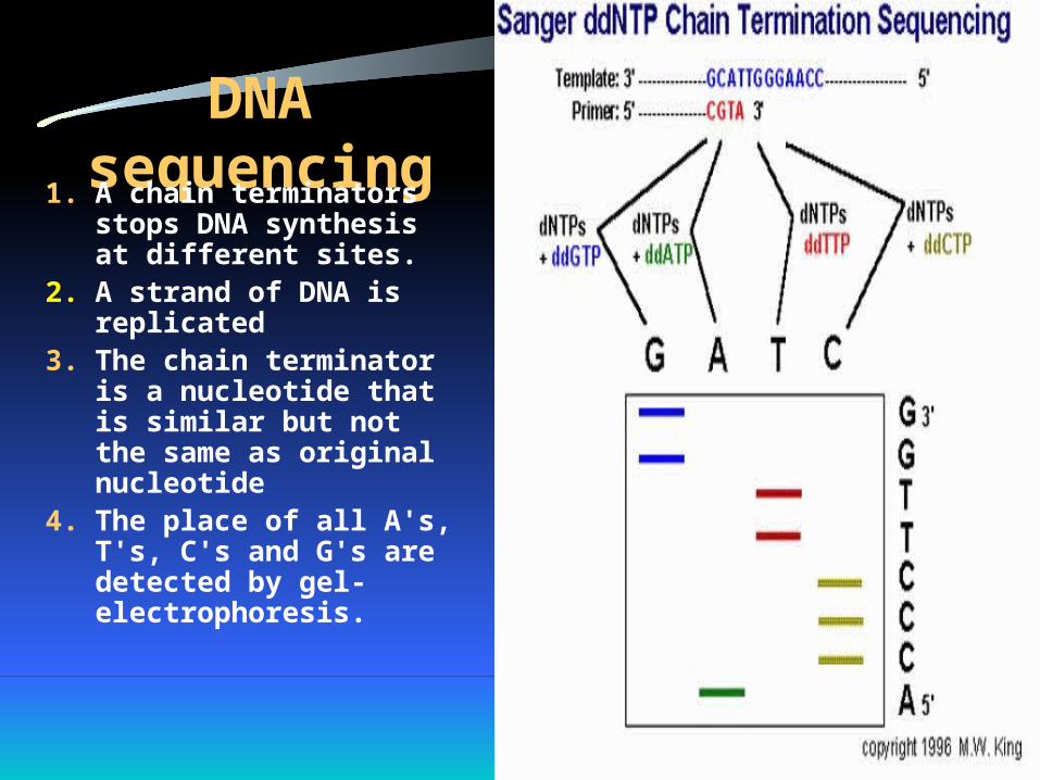
DNA sequencing1. A chain terminators
stops DNA synthesis at different sites.
2. A strand of DNA is replicated
3. The chain terminator is a nucleotide that is similar but not the same as original nucleotide
4. The place of all A's, T's, C's and G's are detected by gel-electrophoresis.
Single molecule sequencing
• Uses exonucleases to degrade DNA by cleaving individual nucleotides from 5’-end of a DNA molecule and detecting them
• The most common methods use DNA segments made from nucleotides where four bases have been differentially tagged with fluorescent labels
• When the tagged nucleotide is clipped, it flows past a laser-based fluorescence detector
Single nucloetide polymorphism• The most common type of stable genetic variations,
these point mutations can produce different phenotypes and can be contributory factors for human disease.
• SNP detections methods are diverse and beyond the scope of this review.
• One notable approach uses matrix-assisted laser desorption mass spectroscopy(MALDI-MS) on a silicon chip for SNP detection; another system employs electrical circury on siliccon microchips to produce a fluorescent signal
Expressed Sequence Tags
• ESTs are identified using reverse transcriptase (RT) to create cDNA sequnces from mRNA present in a cell, allowing genes expressed in different tissues or environmental conditions to be easily amplified by PCR for further study
• Because ESTs might be only gene fragments(typically 3’ or 5’0, they are more easily generated than entire sequence information
Active and In-active mRNA• Protein expression levels are not correlated with mRNA
expression levels• Because protein rather than mRNA levels determine
phenotype, there are efforts underway to investigate this difference by analysing the translation state of mRNA
• Active and inactive ribosomes can be readily separated using sucrose gradient centrifugation; the fractions can then be identified using labeled cDNA probes and used to interpret data based on mRNA expression to estimate protein levels
DNA arrays
• DNA arrays consists of large number of DNA molecules spotted in a systematic order on a solid substrate( such as nylon membrane, glass slide, or silicon chip).
• Depending on the size of each DNA spot on the array, DNA arrays can be categorized as microarrays(each DNA spot has a diameter of <250 microns) and macroarrays( spot diameter of>300 microns).
• When the solid substrate used is small in size, arrays are also referred to as DNA chips
Microarray assays• Traditional hybridization assays developed in the 1970s
utilize flexible membranes such as nitrocellulose and nylon, radioactivity, and autoradiography.
• By contrast, microarray or biochip assays utilize solid surfaces such as glass with fluorescent labelling and detection, this miniaturized biochip format has revolutionized biological analysis.
• Advantages of solid surface are it being non-porous, allows using of small sample volumes, rapid hybridization kinetics, uniform attachement surface with increased quality of the array elements.
Microarray Fabrication
• There are three primary technologies used presently in microarray manufacture include photolithography, ink-jetting, mechanical microspotting, and derivatives thereof.
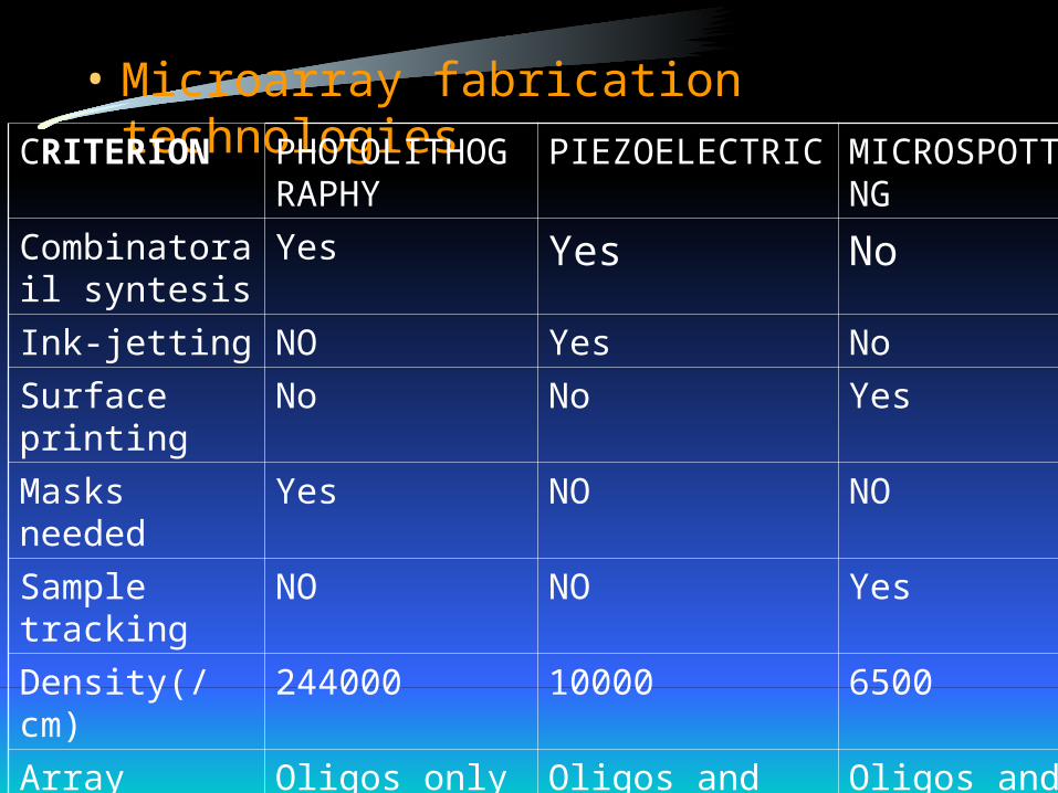
• Microarray fabrication technologiesCRITERION PHOTOLITHOG
RAPHYPIEZOELECTRIC MICROSPOTTI
NGCombinatorail syntesis
Yes Yes No
Ink-jetting NO Yes NoSurface printing No No Yes
Masks needed Yes NO NOSample tracking NO NO Yes
Density(/cm) 244000 10000 6500Array elements Oligos only Oligos and cDNAs Oligos and
cDNAsPrototyping cost High Moderate Low
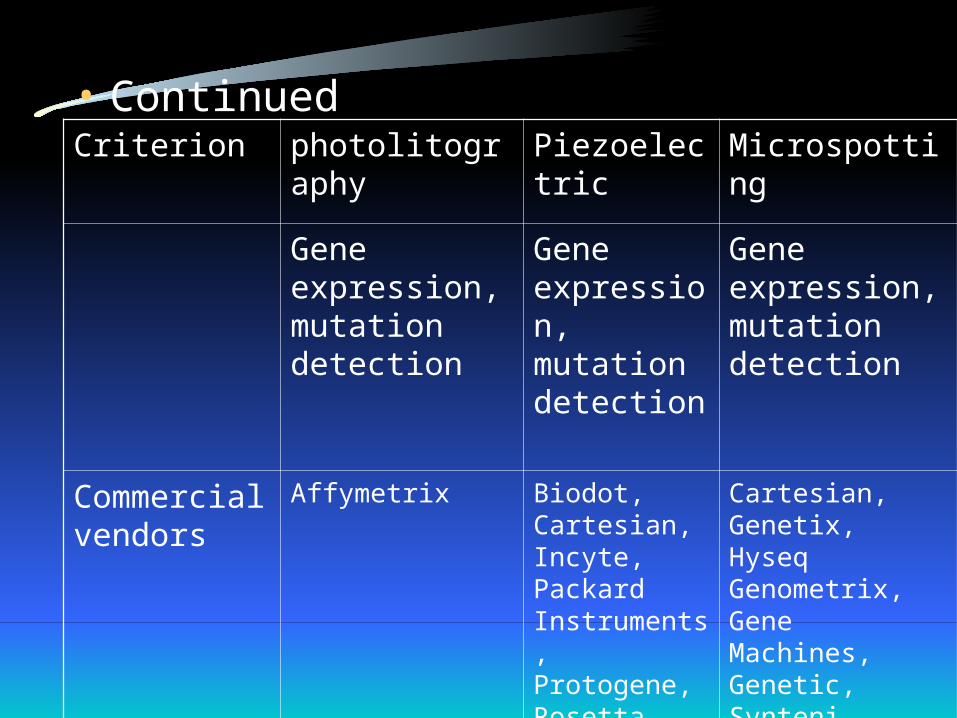
• ContinuedCriterion photolitography Piezoelectric Microspotting
Gene expression, mutation detection
Gene expression, mutation detection
Gene expression, mutation detection
Commercial vendors
Affymetrix Biodot, Cartesian, Incyte, Packard Instruments, Protogene, Rosetta
Cartesian, Genetix, Hyseq Genometrix, Gene Machines, Genetic, SynteniMicrosystems, Norgen Systems, Telechem, Molecular Dynamics

Applications of DNA array
• Gene expression profiling• De novo gene sequencing• Gene mutation analysis(SNP)• Gene mapping and genotyping
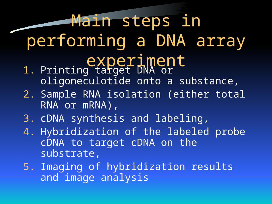
Main steps in performing a DNA array experiment
1. Printing target DNA or oligoneculotide onto a substance,
2. Sample RNA isolation (either total RNA or mRNA),
3. cDNA synthesis and labeling,4. Hybridization of the labeled probe cDNA to
target cDNA on the substrate,5. Imaging of hybridization results and image
analysis
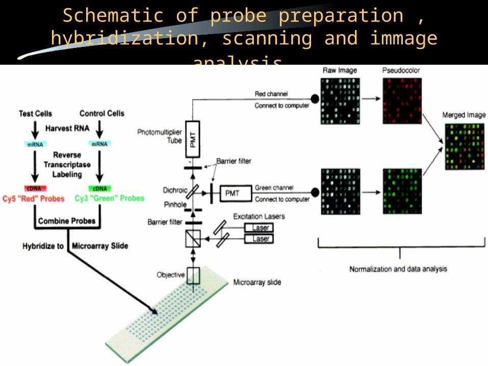
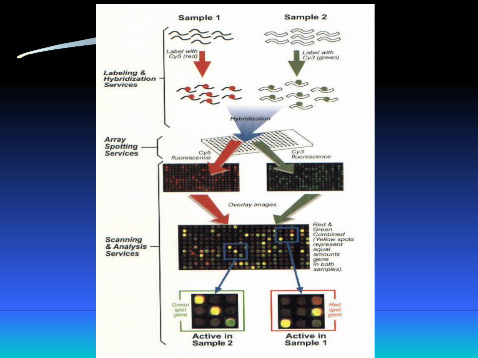
Schematic of probe preparation , hybridization, scanning and immage analysis
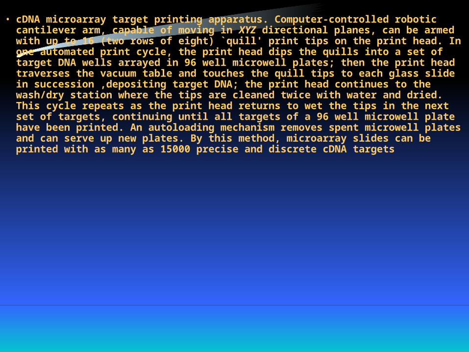
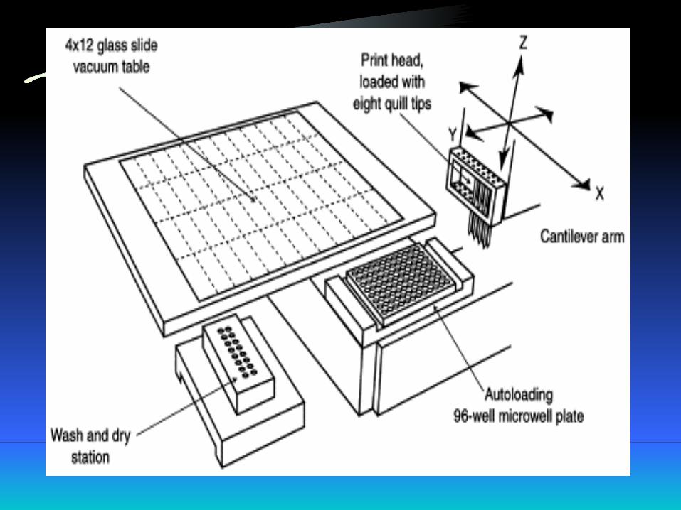
• cDNA microarray target printing apparatus. Computer-controlled robotic cantilever arm, capable of moving in XYZ directional planes, can be armed with up to 16 (two rows of eight) `quill' print tips on the print head. In one automated print cycle, the print head dips the quills into a set of target DNA wells arrayed in 96 well microwell plates; then the print head traverses the vacuum table and touches the quill tips to each glass slide in succession ,depositing target DNA; the print head continues to the wash/dry station where the tips are cleaned twice with water and dried. This cycle repeats as the print head returns to wet the tips in the next set of targets, continuing until all targets of a 96 well microwell plate have been printed. An autoloading mechanism removes spent microwell plates and can serve up new plates. By this method, microarray slides can be printed with as many as 15000 precise and discrete cDNA targets
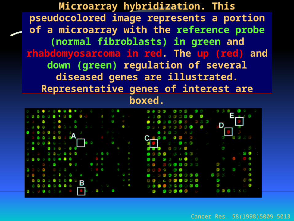
Microarray hybridization. This pseudocolored image represents a portion of a microarray with the reference
probe (normal fibroblasts) in green and rhabdomyosarcoma in red. The up (red) and down
(green) regulation of several diseased genes are illustrated. Representative genes of interest are boxed.
Cancer Res. 58(1998)5009-5013
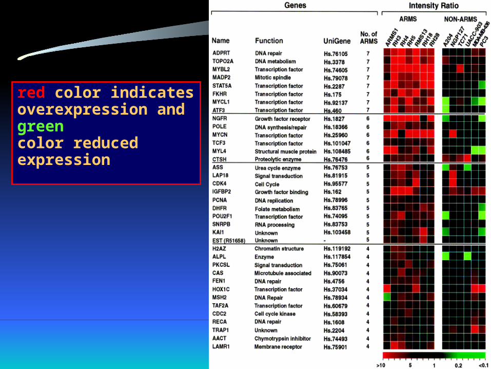
red color indicatesoverexpression andgreencolor reducedexpression
• The impact of molecular biology and genetic research on the discovery of the root causes of a wide variety of hereditary and acquired diseases has long been self-evident.
• Now growing research in this field has shown that conditions like hypertension, arryhtmias, certain cardiomyopathies, Hyperlipidemia, aortic aneurysms, myocardial infarction are all genetically determined.
Molecular Genetics and Its Application to Understanding Cardiovascular Disease

Gene Therapy• Gene therapy, i.e; to introduce genes in selected cells to
treat genetic or acquired diseases .Recombinant DNA technology has revolutionized this field.
• Clinical trials are underway to treat a variety of conditions like cancer, AIDS, familial hypercholesterolemia, cystic fibrosis, and many other diseases with this therapy.
• The growing acceptance of gene therapy for cardiovascular diseases is due to its potential to treat common , multifactorial disorders like cancer, atherogenesis, and inflammation.

Our Aim• We intend to induce rupture of atherosclerotic plaque in
apo-e mice by injecting several drugs, which we hope will cause rupture of the the hemodynamic system and raising the plaque by altering oxidative stress and alter the composition of the plaque.
• After achieving this goal we will perform gene- array study on these specimens and will try to find out which genes are expressed more in plaques which are ruptured or are prone to rupture, by comparing our results at the histopathology lab which will define the structural details of such vulnerable plaques.
• Drugs being used to induce rupture of atherosclerotic plaque in apo-e mice
Drugs being used
• LNAME• Adrenalin• Cocaine• Xanthine• Xanthine Oxidase• Interleukin-1 beta• Methionine• Buthionine Sulfoximine

L-NAME
• L-NAME (N-nitro-L-arginine methyl ester), is a negative effector of nitric oxide synthase.
• Action:By virtue of this action, it blocks the action of nitric oxide and interferes with the arterial dilatation, causing more load on the heart and the circulation
• The production of NO is a major contributor to the endothelium-dependent relaxation in large isolated arteries, including coronary, mesenteric, pulmonary, systemic, and cerebral arteries.

• All the actions of nitric-oxide are inhibited by L-NAME, as well as it promotes adhesion of platelets and leukocytes to the vascular lumen..
• Also the inhibitory effect of NO towards inhibiting platelet aggregation action of prostacyclin and inhibition of growth of vascular smooth cells is abolished by L-NAME.
Interleukin-1 beta
• Class: Cytokine• These cytokines are polypeptides produced
by many cell types(but principally activated lymphocytes and macrophages).
• Their secretion can be stimulated by endotoxin, immune complexes, toxins, physical injury, and a variety of inflammatory processes.
• Monocytes are the main source of secreted IL-1. They express predominantly IL-1-beta while human keratinocytes express large amounts of IL-1-alpha . Murine macrophages display a transition from IL-1-beta to IL-1-alpha production during maturation of monocytes into inflammatory macrophages .

Action
• Their most important actions in inflammation are the local effects on endothelium, the systemic acute-phase reactions, and the effect on fibroblasts.
• In particular they induce the synthesis and surface expression of the endothelial adhesion molecules that mediate leukocyte sticking and increase surface thrombogenicity of the endothelium.
Adrenalin
• Class: Sympathomimetic• Mech: At low doses, beta effects on the vascular
system predominate, whereas at high doses, alpha effects are strongest.
• Action: Major actions are on the cardiovascular system, causes tachycardia, increases myocardium’s oxygen demand, constricts arterioles in the skin, mucous membrane and viscera.

• Also causes broncodilation, hyperglycemia, lipolysis.
• Adverse effects: causes CNS disturbances including tremor, anxiety, and marked elevation of blood pressure, and arrythmias.
Methionine
• Class: Amino acid• Metabolism of the amino acid methionine, a
limiting amino acid in the synthesis of many proteins, affects several biochemical pathways involving the production of nutrients which are essential to the optimal functioning of the cardiovascular, skeletal, and nervous system Homocysteine is an intermediate product of methionine metabolism.

Action• Homocysteine has direct cytotoxic effects on endothelial
cells in vitro • Homocysteine has a direct procoagulant effect on factors V
and X and inhibits protein C activation. • Additionally, there is exciting preliminary data linking a flux
in the homocysteine pathway with liver disease, leukemia, psoriasis and other relatively common disorders. Research is being conducted currently in many of these areas.

• In the past 20+ years, research has shown that elevated homocysteine, or hyperhomocysteinaemia, is linked to atherosclerosis, pregnancies complicated by neural tube defects, early pregnancy loss and venous thrombosis.
• Patients with chronic renal disease have a two to three fold increase in homocysteine levels .

Buthionine Sulfoximine
• Class: selective glutathione (GSH) synthase inhibitor.
• Due to their action causing depletion of GSH, which is a necessary component of the natural antioxidant system, BSO causes oxidative stress, which might cause hypertension by inactivation and sequestration of NO(mediated by ROS).
Cocaine
• Class: Sympathomimetic• Mech: Blocks neuronal re-uptake of
norepinephrine, serotonin, and dopamine.• Action: Stimulates CNS, causes euphoria,
increases motor activity.• Potentiates the action of cathecholamines,i.e;
tachycardia, hypertension, pupillary dilation and peripheral vasoconstriction.
• Adverse effects:• Anxiety, causing increased blood pressure and
heart rate, sweating.• Depression, Like all stimulant drugs, cocaine
stimulation of CNS is followed by a period of mental depression.
Xanthine, Xanthine-Oxidase
• Xanthine oxidase (XO) is a complex enzyme containing flavins, molybdenum, iron and sulfide cofactors. The reaction catalyzed by Xanthine oxidase is shown below:
• XO• Xanthine + O2 + H2O ------> Uric Acid + H2O2 • XO is thought to be the principal source of free
radical generation via degradation of nucleotides to the end product, uric acid.

• Xanthine can exert lethal effects by generating free radicals, this has been proved by several studies, and the effcet of these free radicals is to depress myocardial contractility, by virtue of robbing the myocytes of high energy phospahtes, also it causes the LDLs to be more prone to be oxidized.
Doses of drugs used
Based on references from articles from Pub-Med
• . Substance: LNAMERoute of Administration: IP Dose: 20 mg/kg/day for 7 days
Frequency: 7 References: Nitric oxide 2000Apr;4(2):85-93 Life Sci 1995;57(21):1949-61, Br J Pharmacology:1994oct;113(2):345-8
• 2. Substance: adrenalin Route of Administration: IP Dose: 1 microgram/kg Frequency: 7 References: Auton Pharmacol 1998 jun;18(3):149-
155 Life Sci 1982 Apr 26;30(170:1465-72
Can J Cardiol 1990 Mar;6(2):71-4
.

• Substance: cocaine Route of Administration: IP Dose: 10 mg/kg/day Frequency: 7 References: Nihon Arukoru Yakubutsu Igakkai Zasshi
1997 Apr;32(2):122-38J Appl Physiol 1991 Mar;70(3):1323-7Eur J Invest 1997 Feb;27(2):116-20
• 4. Substance: xanthineRoute of Administration: IP
Dose: 0.0012 mM/kg/day for 7 days Frequency: 7
References: Naunyn Schmiedebergs Arch Pharmacol 1994 Sp;350 (3):227-83 Naunyn Schmiedebergs Arch Pharmacol 1992 Oct;346(4):457-61
• 5. Substance: xanthine oxidaseRoute of Administration: IP
Dose: 0.035 U/ml/day for 7 days Frequency: 7 References: Cardiovasc Res 1986 Aug;20(8):597-603
Mol Cell Biochem 1995 Apr 26;145(2):103-10 Naunyn Schmiedebergs Arch Pharmacol 1994 Sp;350 (3):227-83 Naunyn Schmiedebergs Arch Pharmacol 1992 Oct;346(4):457-61

• 6. Substance: IL-1 betaRoute of Administration: IP
Dose: 1ug/mouse/day for 7 days Frequency: 7Reference: Cancer Biother Radiopharm 1997 Apr:12(2):101-9
Am J Physiol 1995 Apr;268(4 Pt 1):E551-7Brain Res 1999 Jan 9;815(2):337-48Pediatr Res 1995 Jun;37(6):714-9J Neuroendocrinol 1994 Apr;6(2):217-24
• Substance: methionineRoute of Administration: IP
Dose: 1280 mg/kg/day for 7 days Frequency: 7 Reference: Pharmacol Biochem Behav 1999
Sep;64(1):89-93
• Substance: Buthionine sulfoximine Route of Administration: IP Dose:1 mmol/kg Frequency: 7References:In previous studies doses of 2.5mmol, 7.2mmolI/V,And 5mmol/kg, and 3mmol/kghave been used, the dose we are going to use
is much lower than that.Hepatology 2000 Aug;32(2):321-33J Pharmacol Exp Ther 1998Nov;287(2):515-20

Taking Samples and performing the study
12/5/00 42
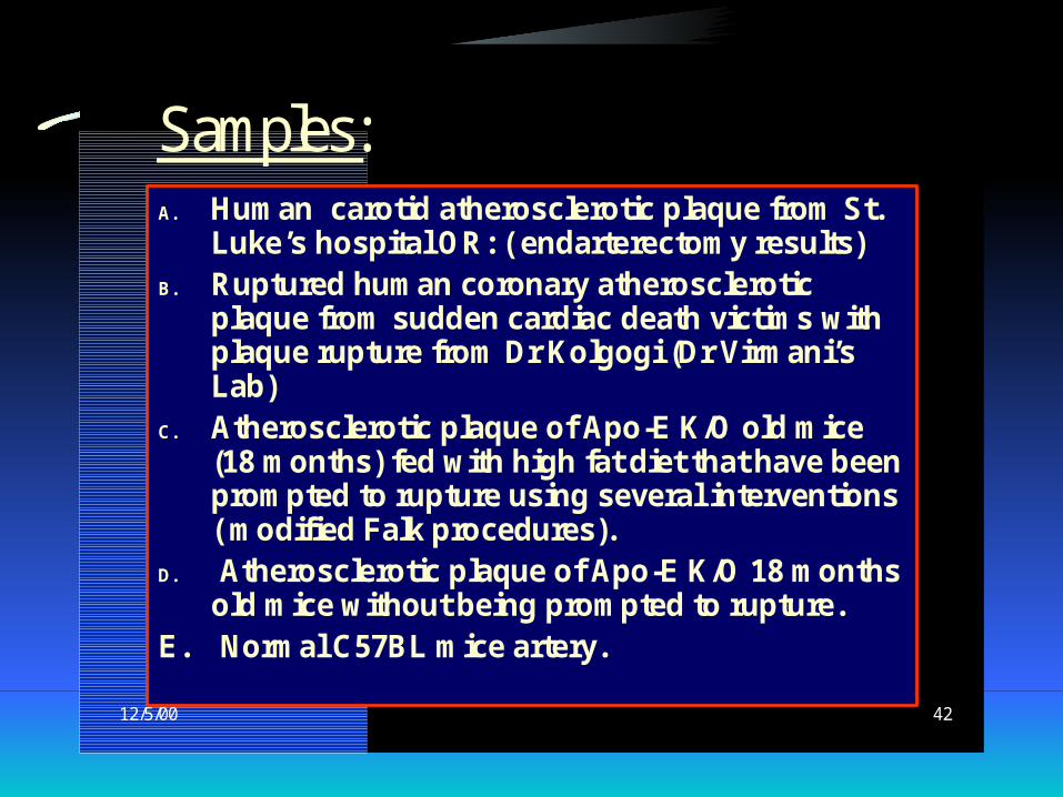
Samples:A. Human carotid atherosclerotic plaque from St.
Luke’s hospital OR: ( endarterectomy results) B. Ruptured human coronary atherosclerotic
plaque from sudden cardiac death victims with plaque rupture from Dr Kolgogi (Dr Virmani’s Lab)
C. Atherosclerotic plaque of Apo-E K/O old mice (18 months) fed with high fat diet that have been prompted to rupture using several interventions ( modified Falk procedures).
D. Atherosclerotic plaque of Apo-E K/O 18 months old mice without being prompted to rupture.
E. Normal C57BL mice artery.
Results
• We will perform gene-array on the RNAs obtained form the ruptures palque to see which genes are more expressed in the palque
• We will also compare our results in the histochemical laboratory
9/10/00 43
Gene array method
We will use mRNA for probe. RNA will be extracted according the protocol and then cDNA is made by RT-PCR. Hybridize with specific probes and scanned.
We will use Affymetrix gene array materials. The chips for samples As & Bs contain 12000
human genes. The chips for samples Cs and Ds contain 19000
mouse genes. We will scan the chips with Affymetrix scanner.

If we do not find anything
Very pleasant, at least we shalll find something new