Embed Size (px)
Citation preview
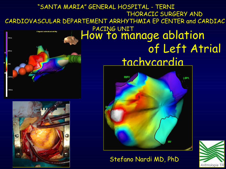
“ “SANTA MARIA” GENERAL HOSPITAL - TERNISANTA MARIA” GENERAL HOSPITAL - TERNI THORACIC SURGERY AND THORACIC SURGERY AND
CARDIOVASCULAR DEPARTEMENT ARRHYTHMIA EP CENTER and CARDIAC CARDIOVASCULAR DEPARTEMENT ARRHYTHMIA EP CENTER and CARDIAC PACING UNIT PACING UNIT
Stefano Nardi MD, PhD
How to manage ablation of Left Atrial
tachycardia
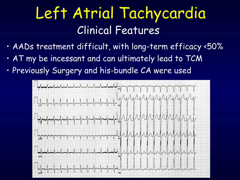
• AADs treatment difficult, with long-term efficacy <50%
Clinical FeaturesLeft Atrial Tachycardia
• AT my be incessant and can ultimately lead to TCM • Previously Surgery and his-bundle CA were used
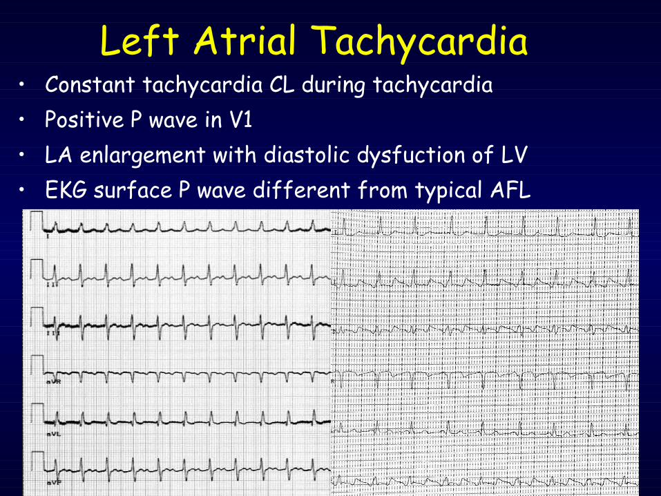
• Constant tachycardia CL during tachycardia• Positive P wave in V1• LA enlargement with diastolic dysfuction of LV• EKG surface P wave different from typical AFL
Left Atrial Tachycardia
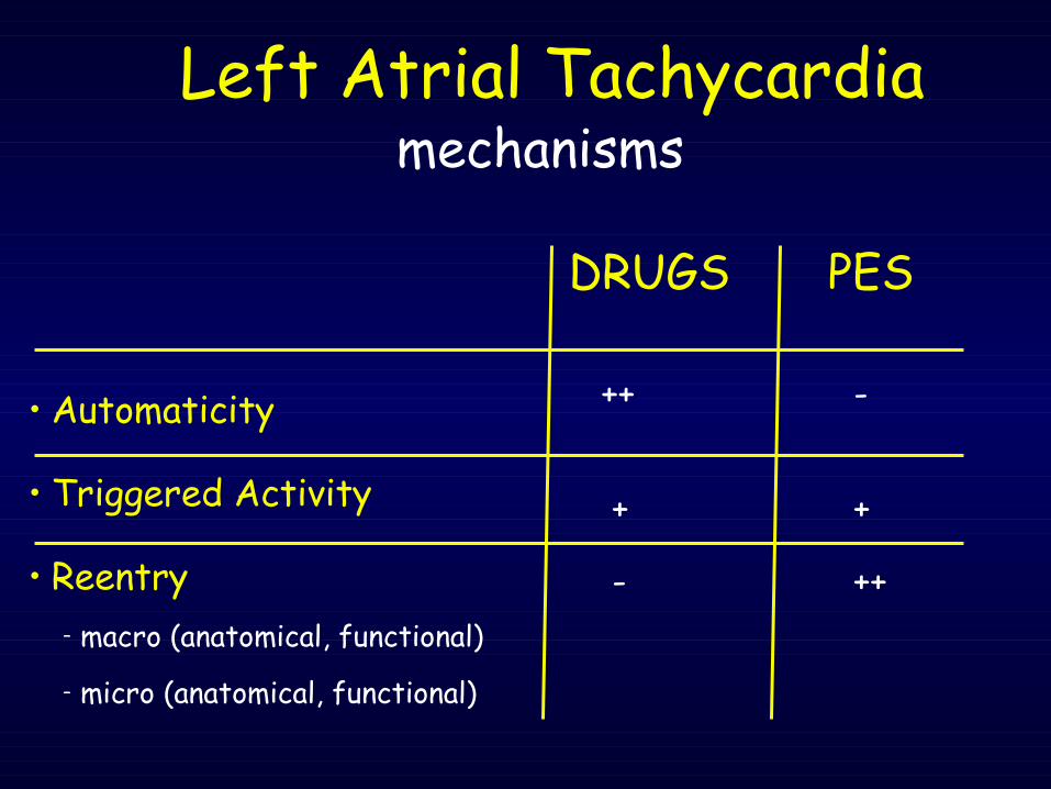
• Automaticity
• Triggered Activity
• Reentry– macro (anatomical, functional)
– micro (anatomical, functional)
mechanismsLeft Atrial Tachycardia
DRUGS PES
++ -
+ +
- ++
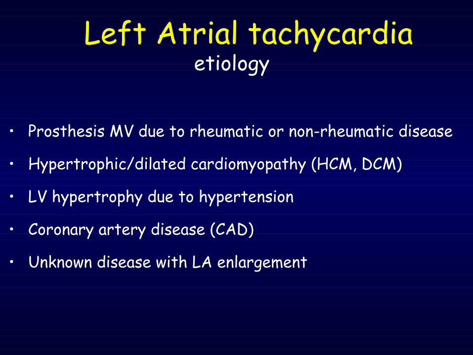
• Prosthesis MV due to rheumatic or non-rheumatic disease
• Hypertrophic/dilated cardiomyopathy (HCM, DCM)
• LV hypertrophy due to hypertension
• Coronary artery disease (CAD)
• Unknown disease with LA enlargement
etiologyLeft Atrial tachycardia
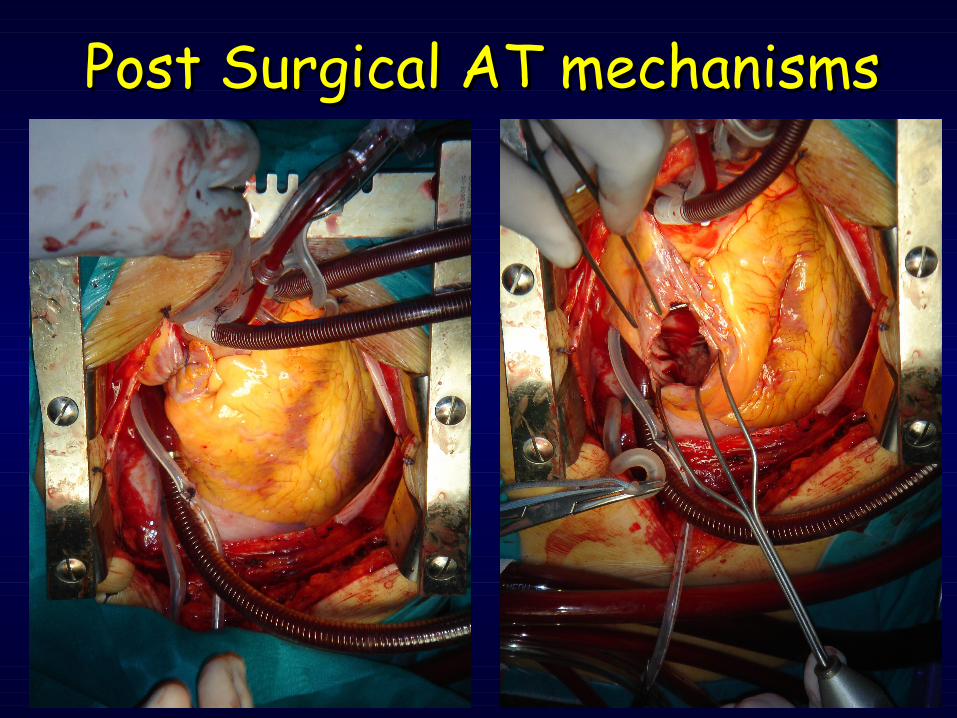
Post Surgical AT mechanismsPost Surgical AT mechanisms
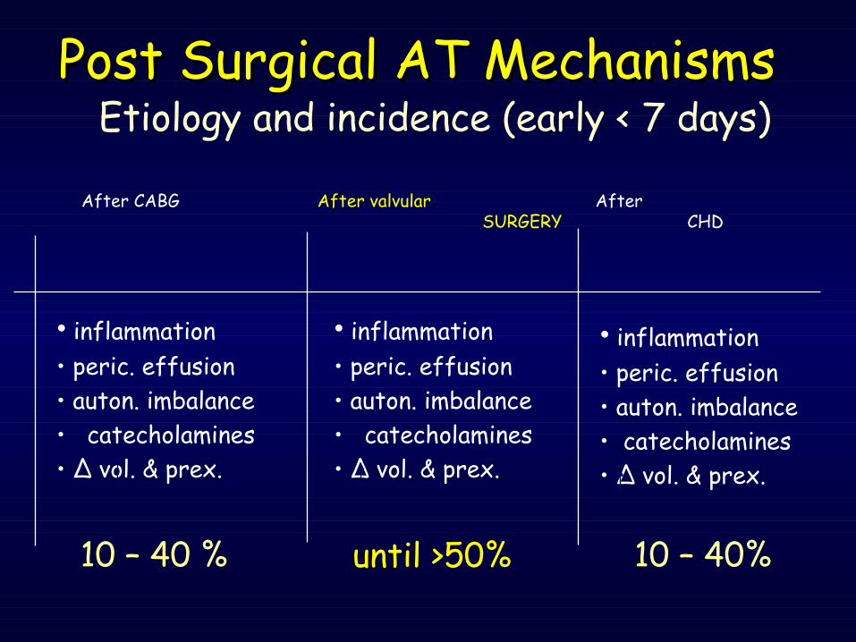
• inflammation• peric. effusion• auton. imbalance• catecholamines• Δ vol. & prex.
• inflammation• peric. effusion• auton. imbalance• catecholamines• Δ vol. & prex.
• inflammation• peric. effusion• auton. imbalance• catecholamines• Δ vol. & prex.
Post Surgical AT MechanismsPost Surgical AT MechanismsEtiology and incidence (early < 7 days)
After CABG
After valvular SURGERY
After CHD
10 – 40 % until >50% 10 – 40%
• Scar• Atriotomy (RA)
• Scar• Atriotomy (RA-LA)• Post-inflammatory (LA)
• Scar• Atriotomy (RA)• Post-inflammatory (LA)
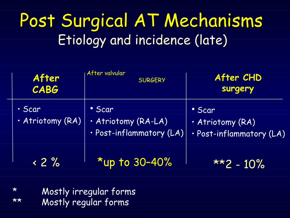
Post Surgical AT MechanismsPost Surgical AT MechanismsEtiology and incidence (late)
After CHD surgery
After CABG
After valvular SURGERY
< 2 % *up to 30–40% **2 - 10%
* Mostly irregular forms** Mostly regular forms
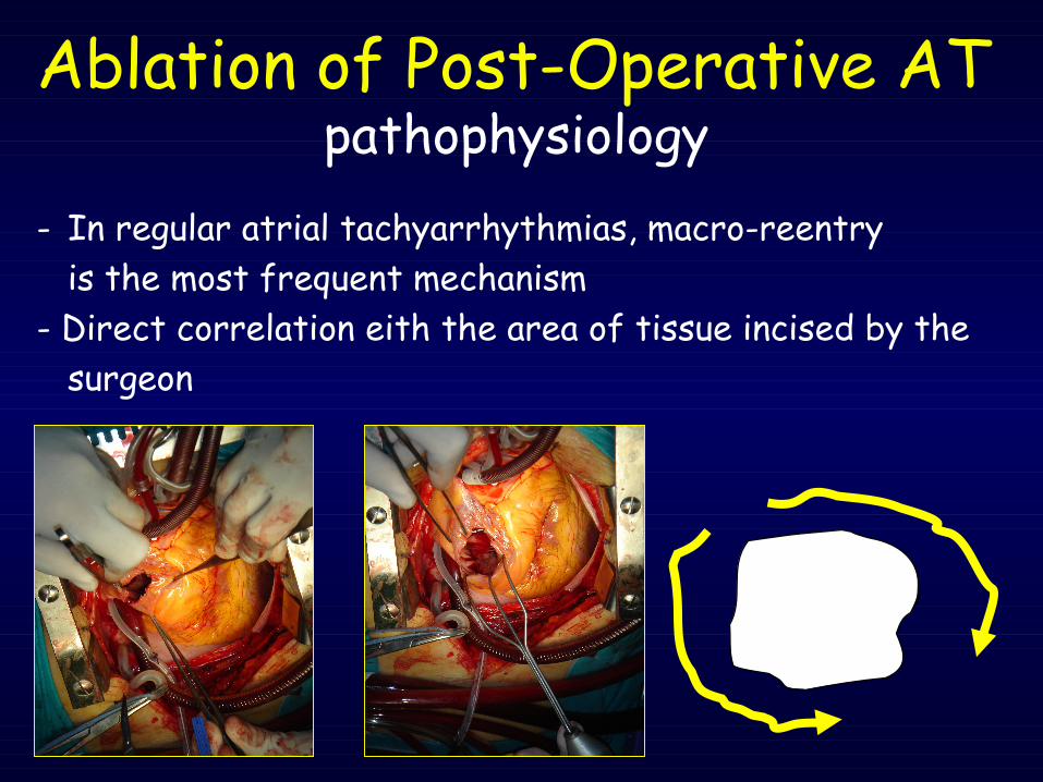
Ablation of Post-Operative ATpathophysiology
- In regular atrial tachyarrhythmias, macro-reentry is the most frequent mechanism- Direct correlation eith the area of tissue incised by the
surgeon
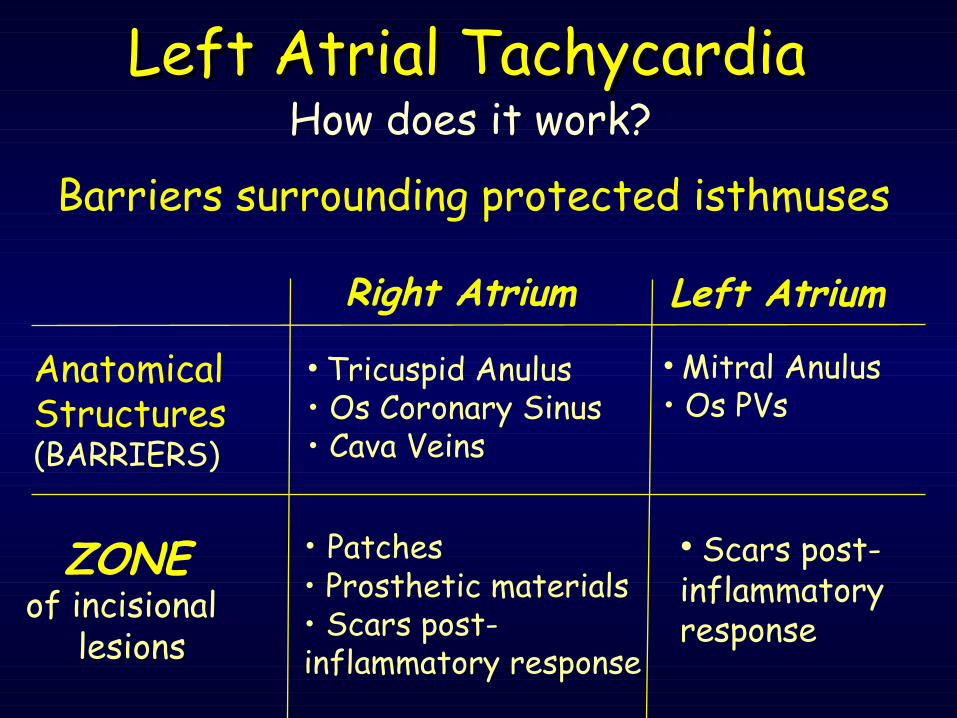
Anatomical Structures (BARRIERS)
• Tricuspid Anulus• Os Coronary Sinus• Cava Veins
• Mitral Anulus• Os PVs
Right Atrium Left Atrium
ZONE of incisional
lesions
• Patches • Prosthetic materials• Scars post-inflammatory response
• Scars post-inflammatory response
Left Atrial TachycardiaLeft Atrial TachycardiaHow does it work?
Barriers surrounding protected isthmuses
It’s really important to use the appropriate technique for Atrial
Tachycardia management
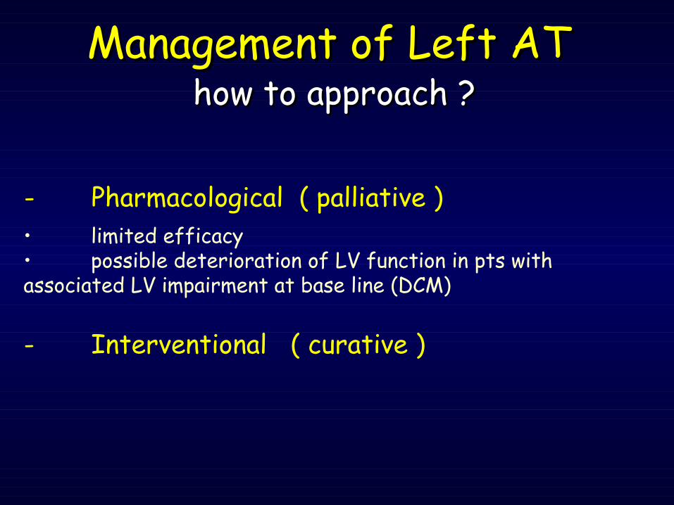
- Pharmacological ( palliative )
Management of Left ATManagement of Left AT
• limited efficacy• possible deterioration of LV function in pts with associated LV impairment at base line (DCM)
- Interventional ( curative )
how to approach ?how to approach ?
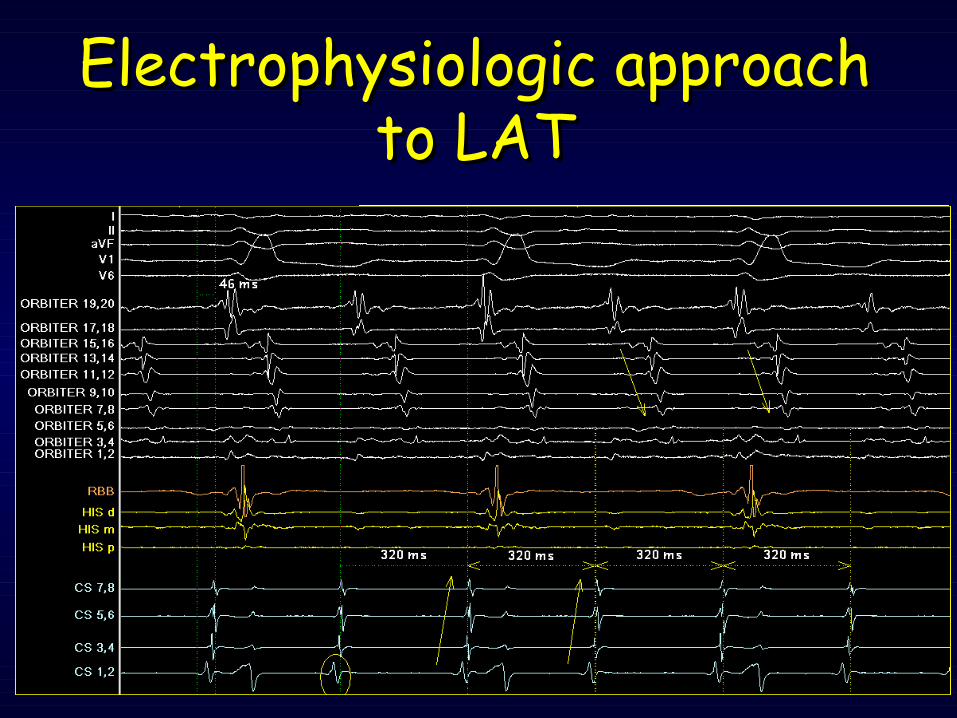
Electrophysiologic approach Electrophysiologic approach to LATto LAT
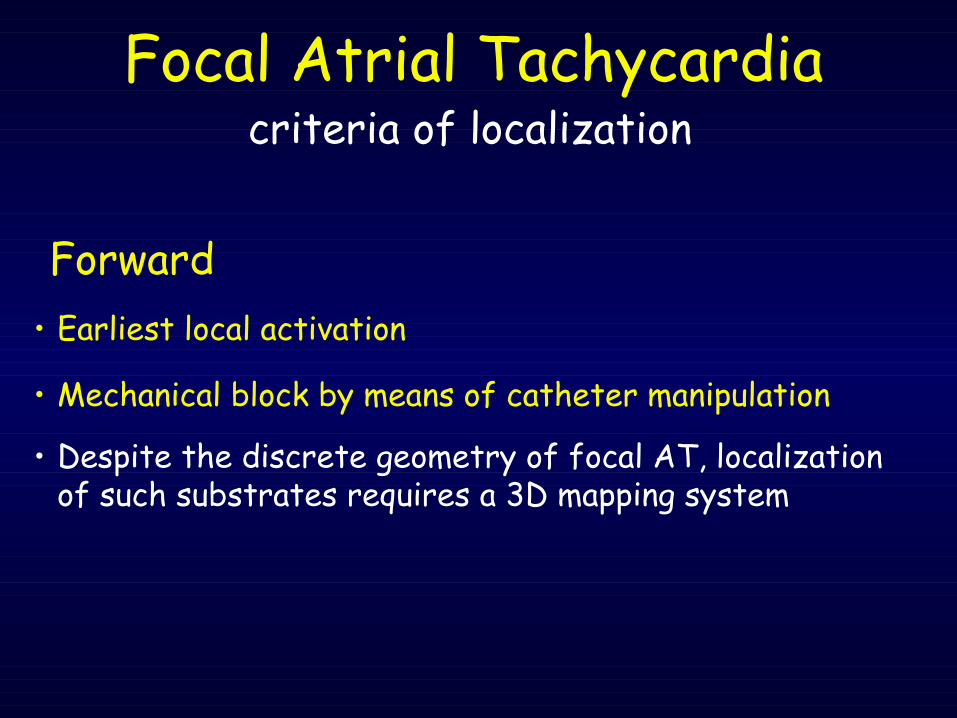
• Despite the discrete geometry of focal AT, localization of such substrates requires a 3D mapping system
Forward
criteria of localizationFocal Atrial Tachycardia
• Earliest local activation
• Mechanical block by means of catheter manipulation
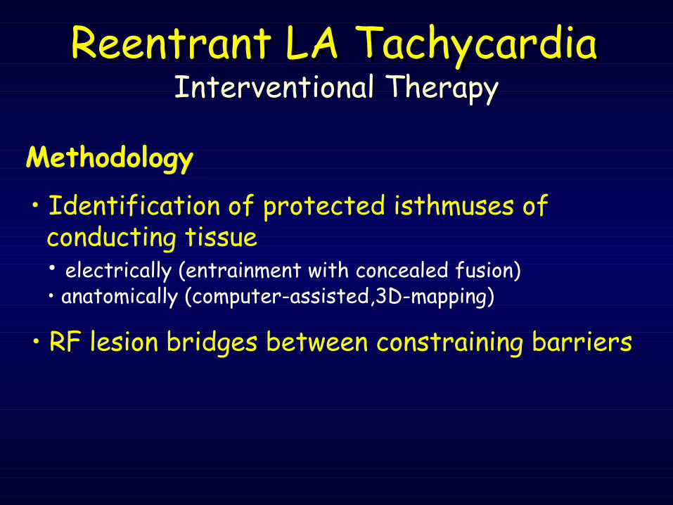
Methodology • Identification of protected isthmuses of conducting tissue
• electrically (entrainment with concealed fusion)• anatomically (computer-assisted,3D-mapping)
• RF lesion bridges between constraining barriers
Reentrant LA TachycardiaReentrant LA TachycardiaInterventional Therapy
Use of Entrainment
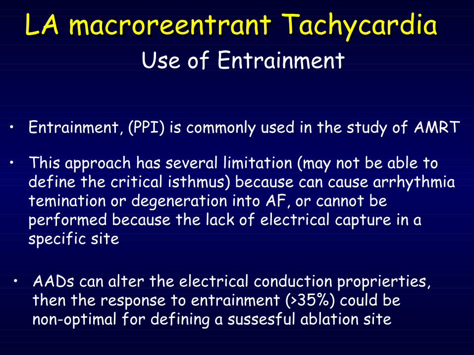
• Entrainment, (PPI) is commonly used in the study of AMRT
• AADs can alter the electrical conduction proprierties, then the response to entrainment (>35%) could be non-optimal for defining a sussesful ablation site
• This approach has several limitation (may not be able to define the critical isthmus) because can cause arrhythmia temination or degeneration into AF, or cannot be performed because the lack of electrical capture in a specific site
LA macroreentrant TachycardiaLA macroreentrant Tachycardia
Left Atrial TachycardiaLeft Atrial TachycardiaDrawbacks of
Electrophysiologic Approach
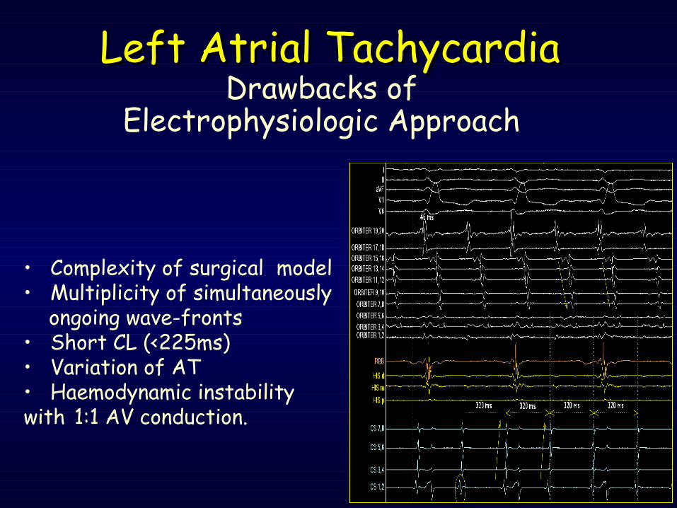
• Complexity of surgical model• Multiplicity of simultaneously
ongoing wave-fronts• Short CL (<225ms)• Variation of AT• Haemodynamic instability with 1:1 AV conduction.

Which is the impact of the new
technologies ?
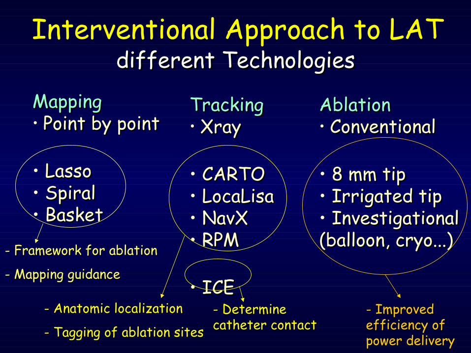
different Technologiesdifferent Technologies
MappingMapping• Point by pointPoint by point
• LassoLasso• SpiralSpiral• BasketBasket
TrackingTracking• XrayXray
• CARTOCARTO• LocaLisaLocaLisa• NavXNavX• RPMRPM
• ICEICE
AblationAblation• ConventionalConventional
• 8 mm tip8 mm tip• Irrigated tipIrrigated tip• InvestigationalInvestigational(balloon, cryo...)(balloon, cryo...)- Framework for ablationFramework for ablation
- Mapping guidanceMapping guidance
- Anatomic localizationAnatomic localization
- Tagging of ablation sites- Tagging of ablation sites- Determine Determine catheter contactcatheter contact
- Improved Improved efficiency of efficiency of power deliverypower delivery
Interventional Approach to LAT
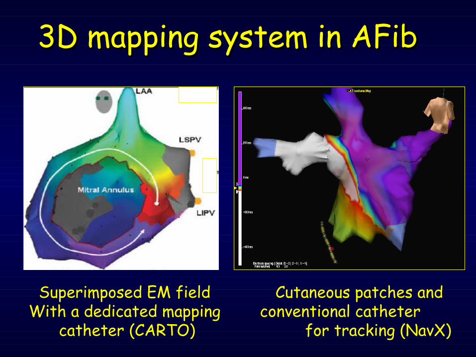
3D mapping system in AFib 3D mapping system in AFib
Cutaneous patches and conventional catheter for tracking (NavX)
Superimposed EM field With a dedicated mapping
catheter (CARTO)
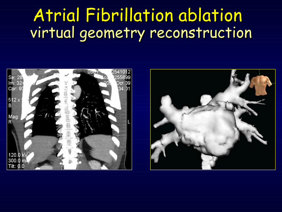
Atrial Fibrillation ablationAtrial Fibrillation ablationvirtual geometry reconstructionvirtual geometry reconstruction
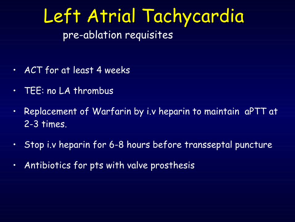
• ACT for at least 4 weeks
• TEE: no LA thrombus
• Replacement of Warfarin by i.v heparin to maintain aPTT at 2-3 times.
• Stop i.v heparin for 6-8 hours before transseptal puncture
• Antibiotics for pts with valve prosthesis
pre-ablation requisitesLeft Atrial TachycardiaLeft Atrial Tachycardia
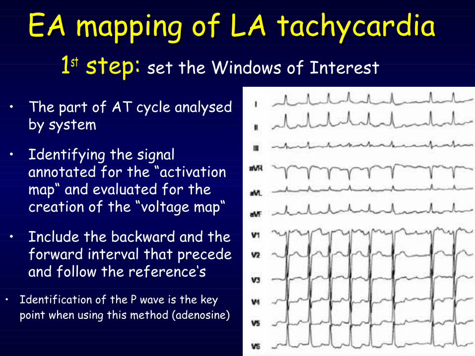
• The part of AT cycle analysed by system
• Identifying the signal annotated for the “activation map“ and evaluated for the creation of the “voltage map“
• Include the backward and the forward interval that precede and follow the reference‘s
EA mapping of LA tachycardiaEA mapping of LA tachycardia1st step: set the Windows of Interest
• Identification of the P wave is the key point when using this method (adenosine)
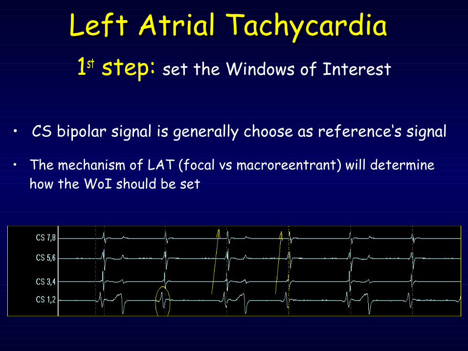
1st step: set the Windows of Interest
• CS bipolar signal is generally choose as reference‘s signal
Left Atrial TachycardiaLeft Atrial Tachycardia
• The mechanism of LAT (focal vs macroreentrant) will determine how the WoI should be set
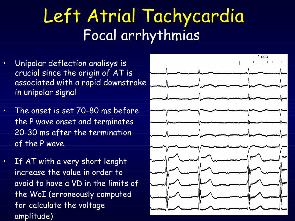
• The onset is set 70-80 ms before the P wave onset and terminates 20-30 ms after the termination of the P wave.
• If AT with a very short lenght increase the value in order to avoid to have a VD in the limits of the WoI (erroneously computed for calculate the voltage amplitude)
Left Atrial TachycardiaLeft Atrial TachycardiaFocal arrhythmias
• Unipolar deflection analisys is crucial since the origin of AT is associated with a rapid downstroke in unipolar signal
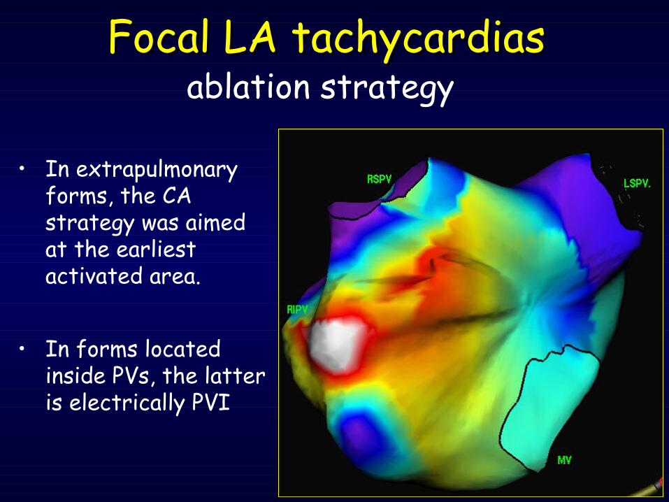
ablation strategy
• In extrapulmonary forms, the CA strategy was aimed at the earliest activated area.
• In forms located inside PVs, the latter is electrically PVI
Focal LA tachycardiasFocal LA tachycardias
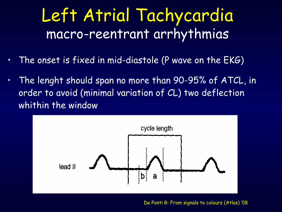
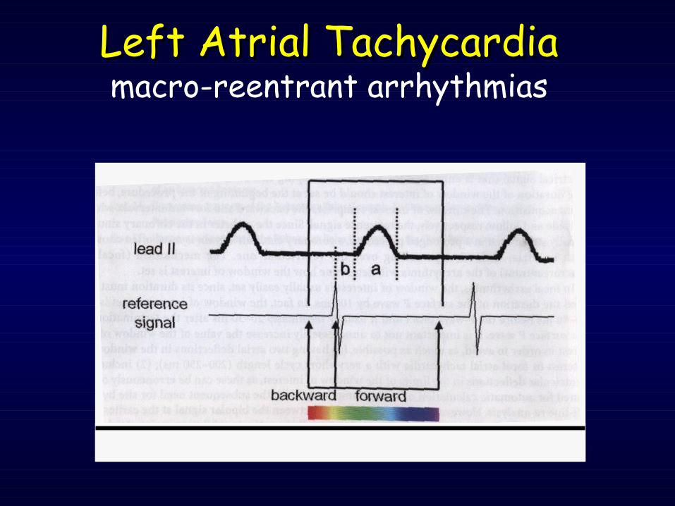
• The onset is fixed in mid-diastole (P wave on the EKG)
• The lenght should span no more than 90-95% of ATCL, in order to avoid (minimal variation of CL) two deflection whithin the window
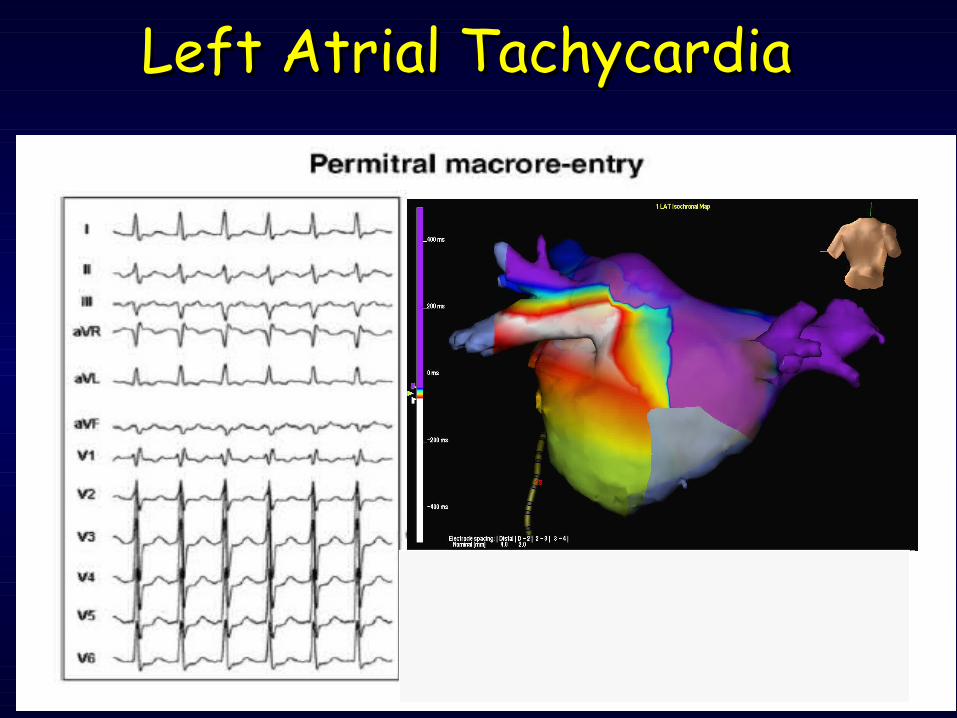
Left Atrial TachycardiaLeft Atrial Tachycardiamacro-reentrant arrhythmias
De Ponti R: From signals to colours (Atlas) ‘08
Left Atrial TachycardiaLeft Atrial Tachycardiamacro-reentrant arrhythmias
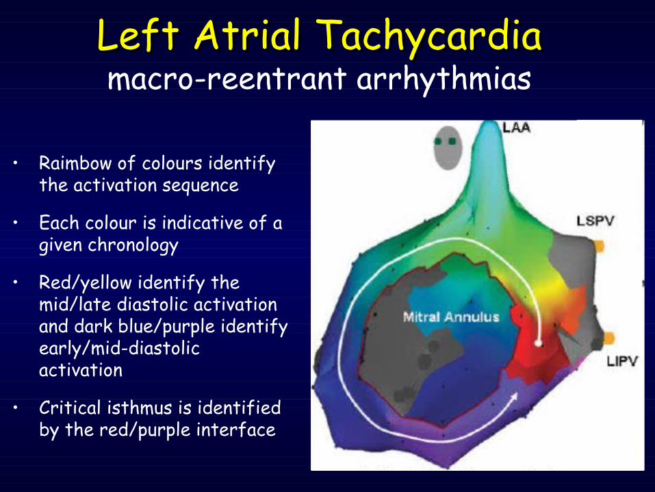
• Raimbow of colours identify the activation sequence
• Each colour is indicative of a given chronology
• Red/yellow identify the mid/late diastolic activation and dark blue/purple identify early/mid-diastolic activation
• Critical isthmus is identified by the red/purple interface
Left Atrial TachycardiaLeft Atrial Tachycardiamacro-reentrant arrhythmias
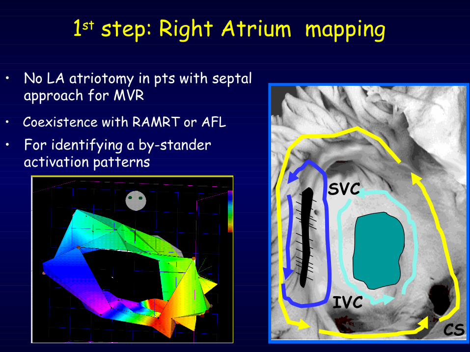
• No LA atriotomy in pts with septal approach for MVR
CSIVC
SVC
• For identifying a by-stander activation patterns
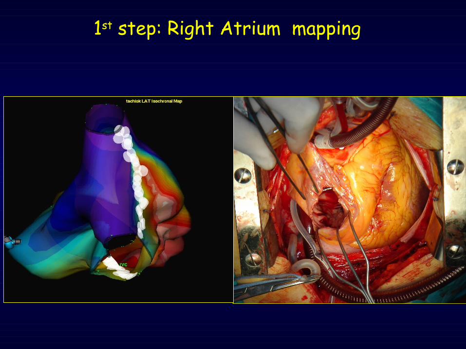
1st step: Right Atrium mapping
• Coexistence with RAMRT or AFL
1st step: Right Atrium mapping
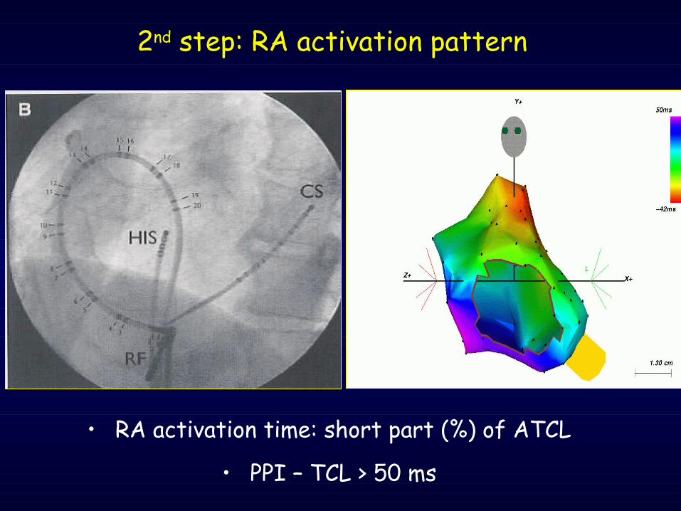
• RA activation time: short part (%) of ATCL
• PPI – TCL > 50 ms
2nd step: RA activation pattern
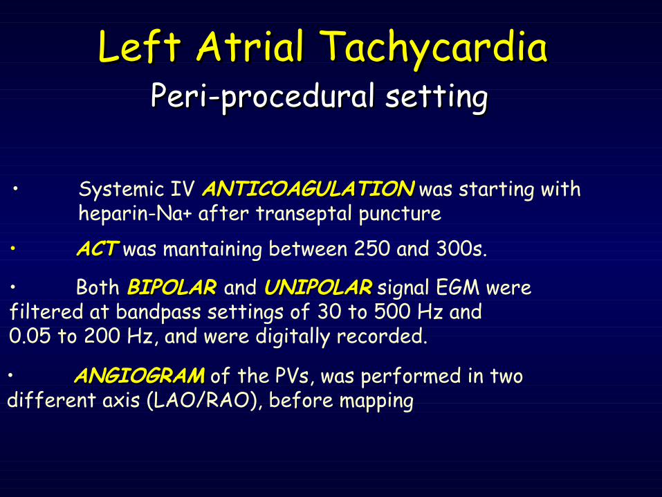
• Both BIPOLAR BIPOLAR and UNIPOLARUNIPOLAR signal EGM were filtered at bandpass settings of 30 to 500 Hz and 0.05 to 200 Hz, and were digitally recorded.
• Systemic IV ANTICOAGULATIONANTICOAGULATION was starting with heparin-Na+ after transeptal puncture
• ANGIOGRAMANGIOGRAM of the PVs, was performed in two different axis (LAO/RAO), before mapping
• ACTACT was mantaining between 250 and 300s.
Peri-procedural settingPeri-procedural settingLeft Atrial TachycardiaLeft Atrial Tachycardia

• Bipolar signal amplitude <0,05 mV (not distinguishable from the baseline noise) are defined as electrically silent areas (grey dot)
• Electrical signal in sites with minimal but still-present a bipolar deflection.
• If multi-component/fragmented potential, annote the 1st deflection
Left Atrial TachycardiaLeft Atrial Tachycardia
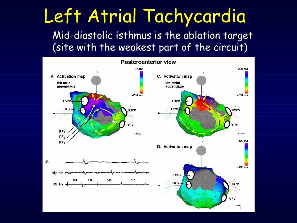
Left Atrial TachycardiaLeft Atrial TachycardiaMid-diastolic isthmus is the ablation target (site with the weakest part of the circuit)
Left Atrial TachycardiaLeft Atrial Tachycardia
Left Atrial TachycardiaLeft Atrial Tachycardia
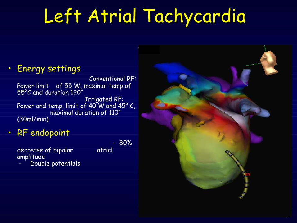
• Energy settings Conventional RF: Power limit of 55 W, maximal temp of 55°C and duration 120“ Irrigated RF: Power and temp. limit of 40 W and 45° C, maximal duration of 110“ (30ml/min)
• RF endopoint - 80% decrease of bipolar atrial amplitude - Double potentials
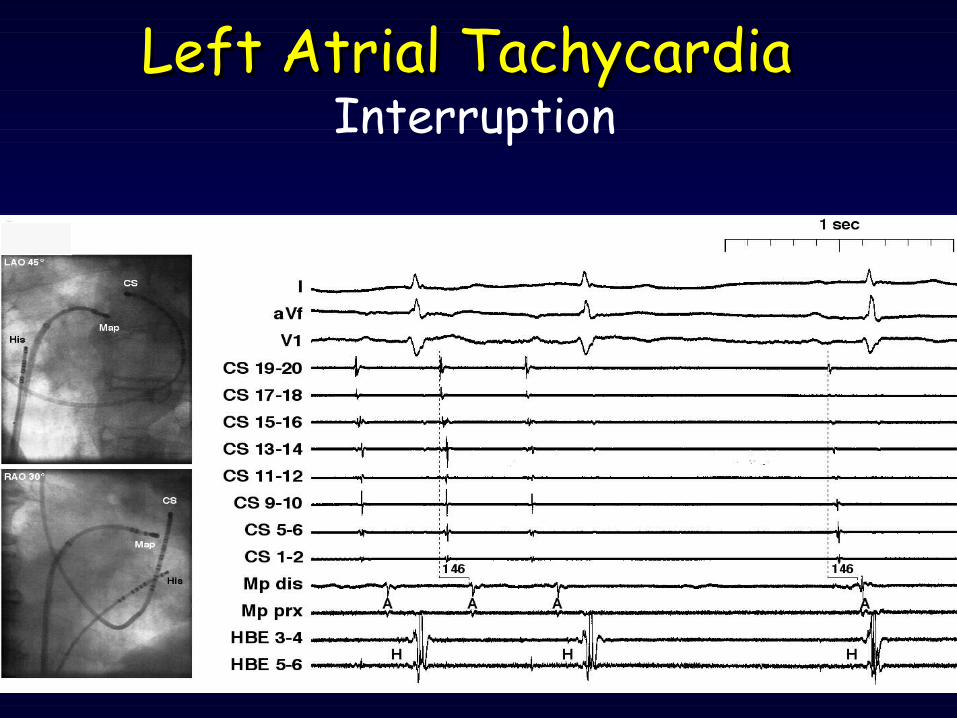
Left Atrial TachycardiaLeft Atrial TachycardiaInterruption
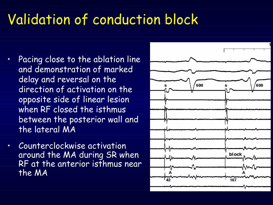
Validation of conduction block
• Pacing close to the ablation line and demonstration of marked delay and reversal on the direction of activation on the opposite side of linear lesion when RF closed the isthmus between the posterior wall and the lateral MA
• Counterclockwise activation around the MA during SR when RF at the anterior isthmus near the MA
• ECHOCARDIOGRAPHYECHOCARDIOGRAPHY after ablation
Post-ablation managementPost-ablation management
• SYSTEMIC ANTICOAGULATIONSYSTEMIC ANTICOAGULATION was starting with heparin-Na+ six hours after the end of the procedure
• ORAL ANTICOAGULATIONORAL ANTICOAGULATION 24 hs later
Left Atrial TachycardiaLeft Atrial Tachycardia
• After LAT has been interrupted, and ablation completed, induction of Arrhythmia by PES with multiple extrastimuli and burst is attempted
• Unusual geometry of target tisue• Complexity of the surgical model• Multiplicity of simultaneously ongoing wavefronts
Inability to identify protected isthmuses
Ablation of Reentrant LATcauses of future recurrence
Inability to bridge protected isthmuses
• Thickness of atrial wall• Inadequate temperature at intramyocardial/epicardial depth (a poor cooling by reduced blood flow)• Multiplicity of active isthmuses
• Conventional EP mapping is not always a really appropriate strategies for left AT’s ablation because it provides very limited understanding of these complex arrhythmias which are highly variable from one pt to the other.
• The main drawback of a pure EP approach is that the identification of all putative “end-point” could be
extremely difficult to achieve.
Ablation of LA TachycardiaAblation of LA Tachycardiaconclusionsconclusions
• Success of CA is limited by a number of factors, including the inability to identify or severe the active protected isthmuses sustaining macro-reentry
• In pts with ventricular dysfunction, elimination of AT leads to immediate relief of symptoms, followed by progressive improvement of LV function
• Efficacy of CA differ between right-sided and left-sided ATs
Ablation of LA TachycardiaAblation of LA Tachycardiaconclusionsconclusions
• Success of CA is limited by a number of factors, including the inability to identify or severe the active protected isthmuses sustaining macro-reentry
• The implemented use of virtual geometry and 3D mapping system W or w/o a merge integration could fulfill some important clinical demand for detailed anatomic guidance, especially in case of abnormal anatomy, condition that can increase the risk of damage if not adequately realized.
Ablation of LA TachycardiaAblation of LA Tachycardiaconclusionsconclusions
• Inherent EA limitations can lead to a potential source of error, however we believe that represents a
significant improvement respect to previous only EP criteria
• This approach may be USEFUL USEFUL in the treatment of pts with cardiac arrhythmias where ablation therapy is primarily ANATOMICALLY BASEDANATOMICALLY BASED