Embed Size (px)
Citation preview


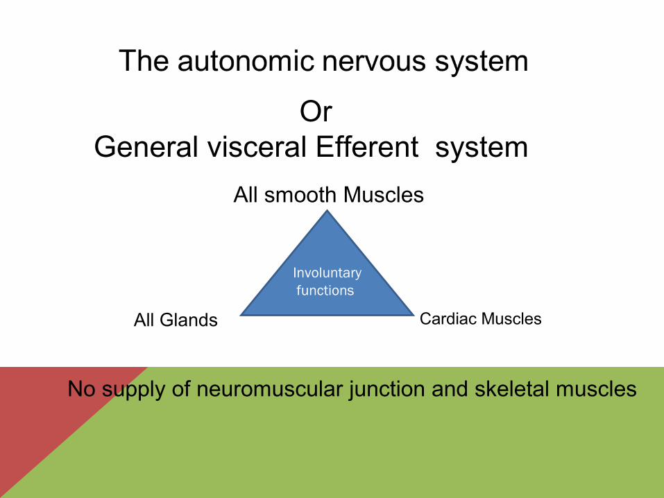
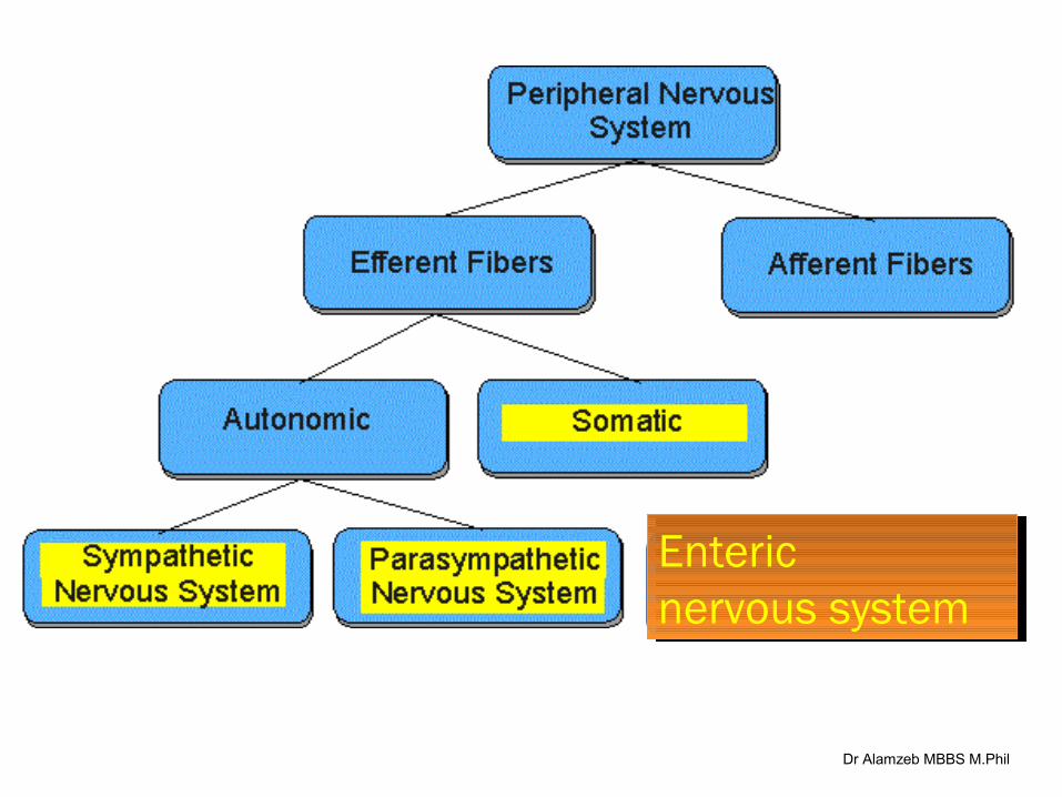
OrGeneral visceral Efferent system
Involuntary functions
All smooth Muscles
Cardiac Muscles All Glands
No supply of neuromuscular junction and skeletal muscles
Dr Alamzeb MBBS M.Phil
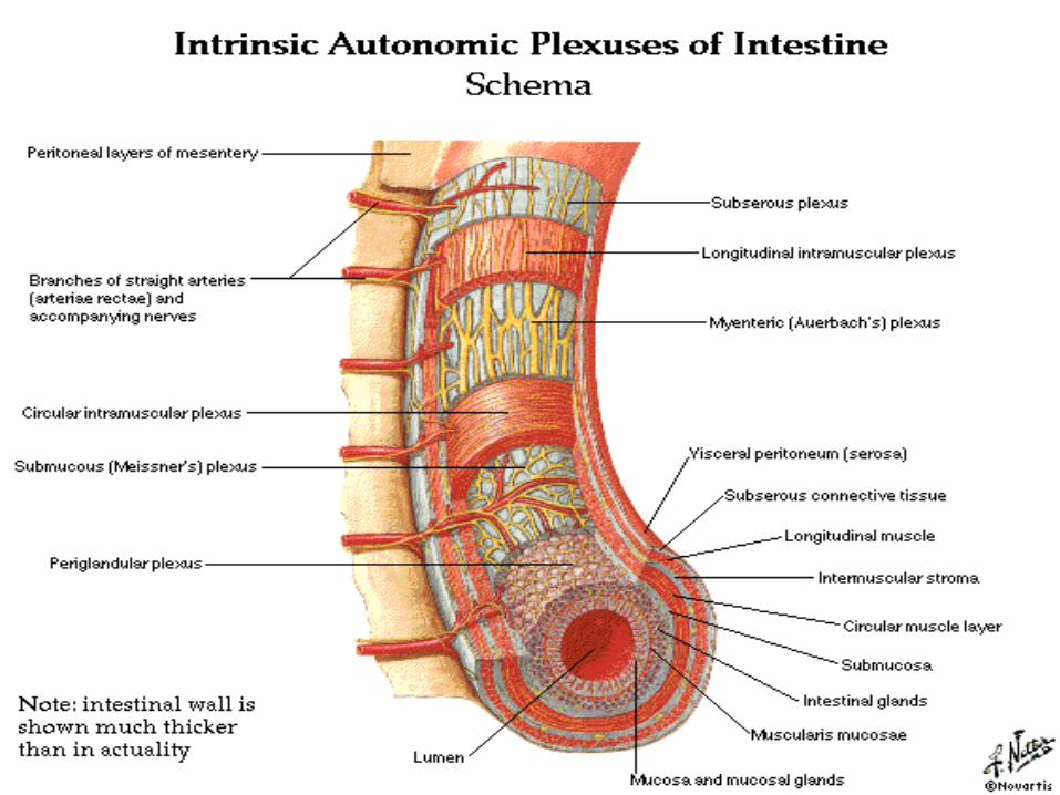
Enteric nervous systemEnteric nervous system
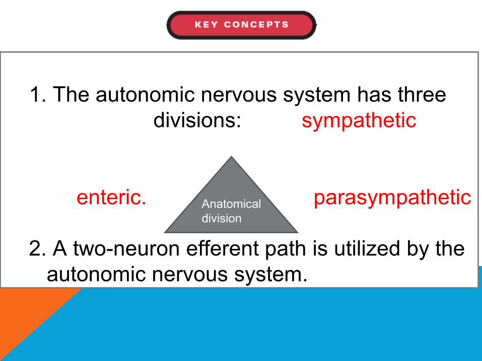
1. The autonomic nervous system has three divisions: sympathetic
enteric. parasympathetic
2. A two-neuron efferent path is utilized by the autonomic nervous system.
Anatomical division
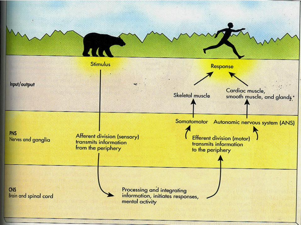
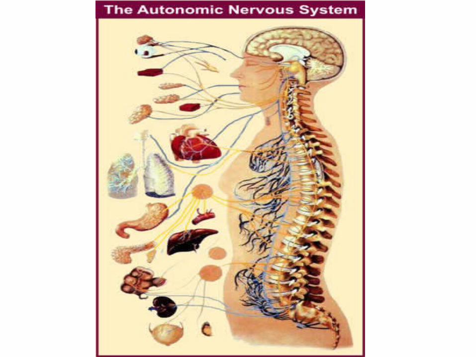
AUTONOMIC NERVOUS SYSTEM.
The portion of the nervous system that controls most visceral functions of the body that are generally not under conscious control is called the autonomic nervous system.
This system helps to control arterial pressure, Gastrointestinal motility, Gastrointestinal secretion, Urinary bladder emptying, Sweating, Body temperature,
Dr Alamzeb MBBS M.Phil
4. The three divisions have neurochemical differences.
5. The sympathetic and parasympathetic divisions differ in anatomic origin and function.
6. The central nervous system controls autonomic function through a hierarchy of reflexes and integrative centers.
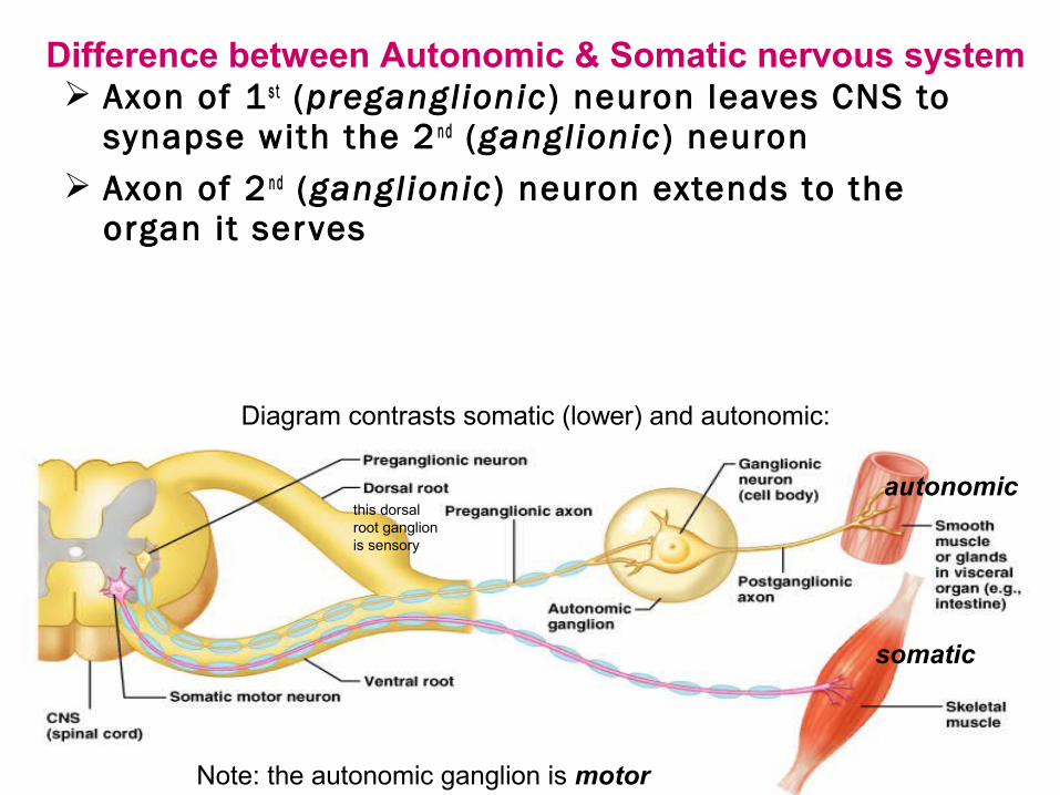
Axon of 1 s t (preganglionic ) neuron leaves CNS to synapse with the 2 n d (ganglionic ) neuron
Axon of 2 n d (ganglionic ) neuron extends to the organ it serves
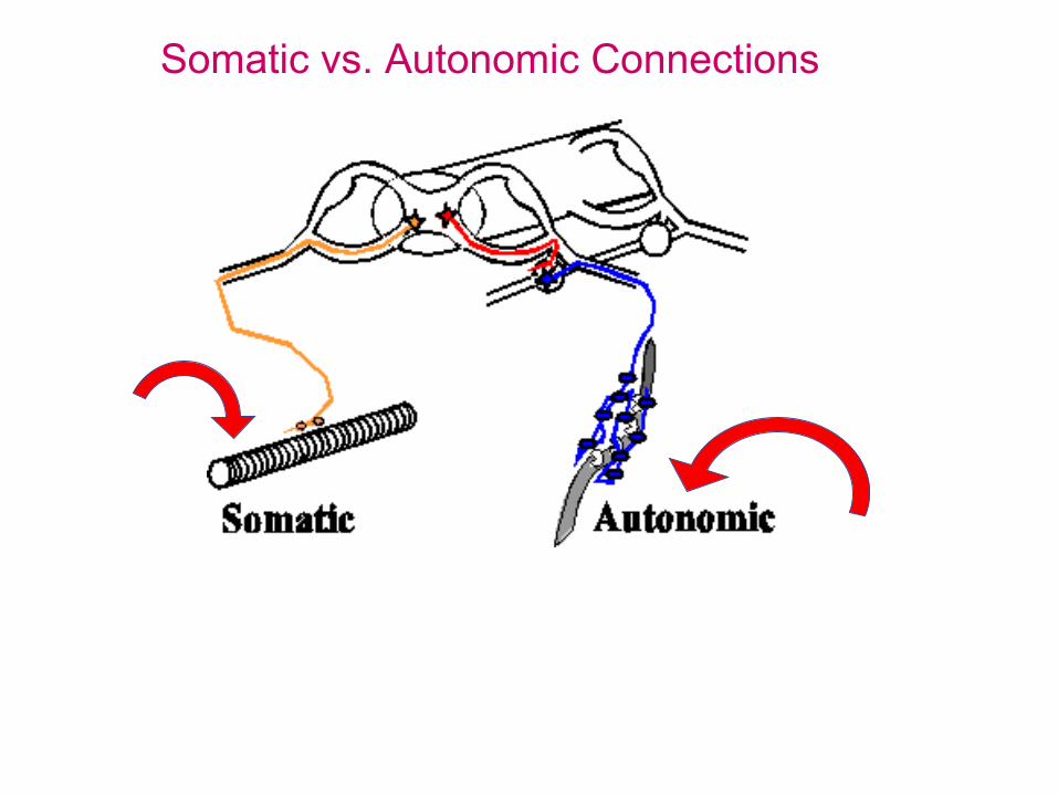
Diagram contrasts somatic (lower) and autonomic:
autonomic
somatic
Note: the autonomic ganglion is motor
this dorsal root ganglion is sensory
Difference between Autonomic & Somatic nervous system
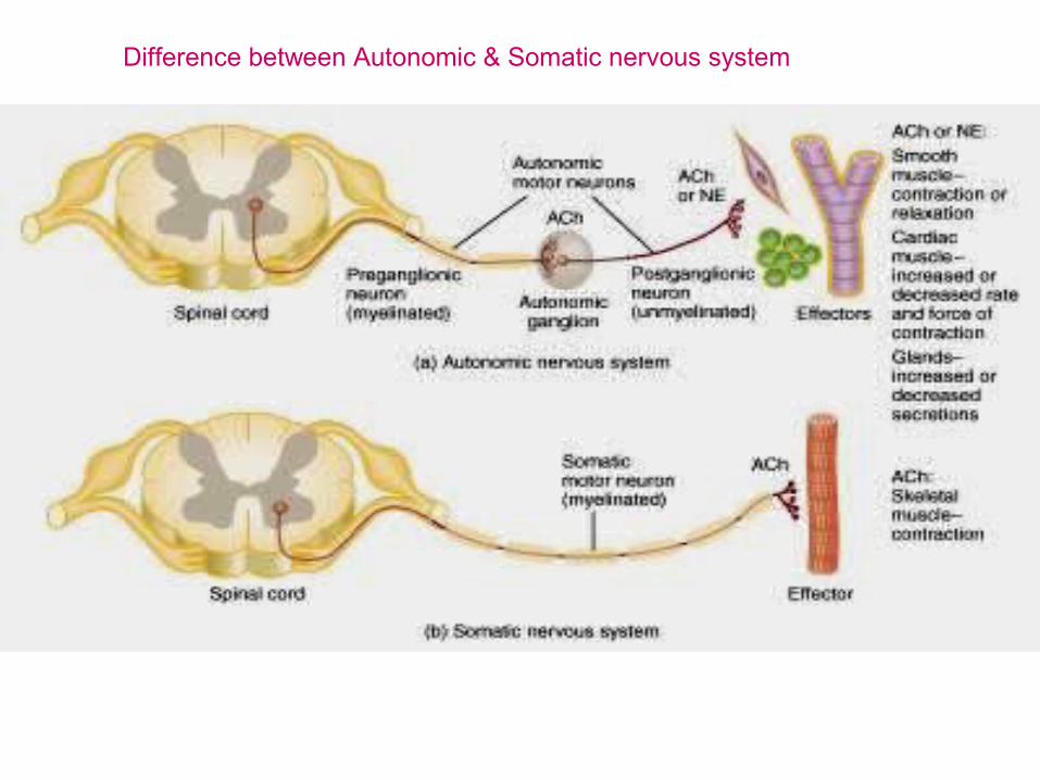
Difference between Autonomic & Somatic nervous system
Dr Alamzeb MBBS M.Phil
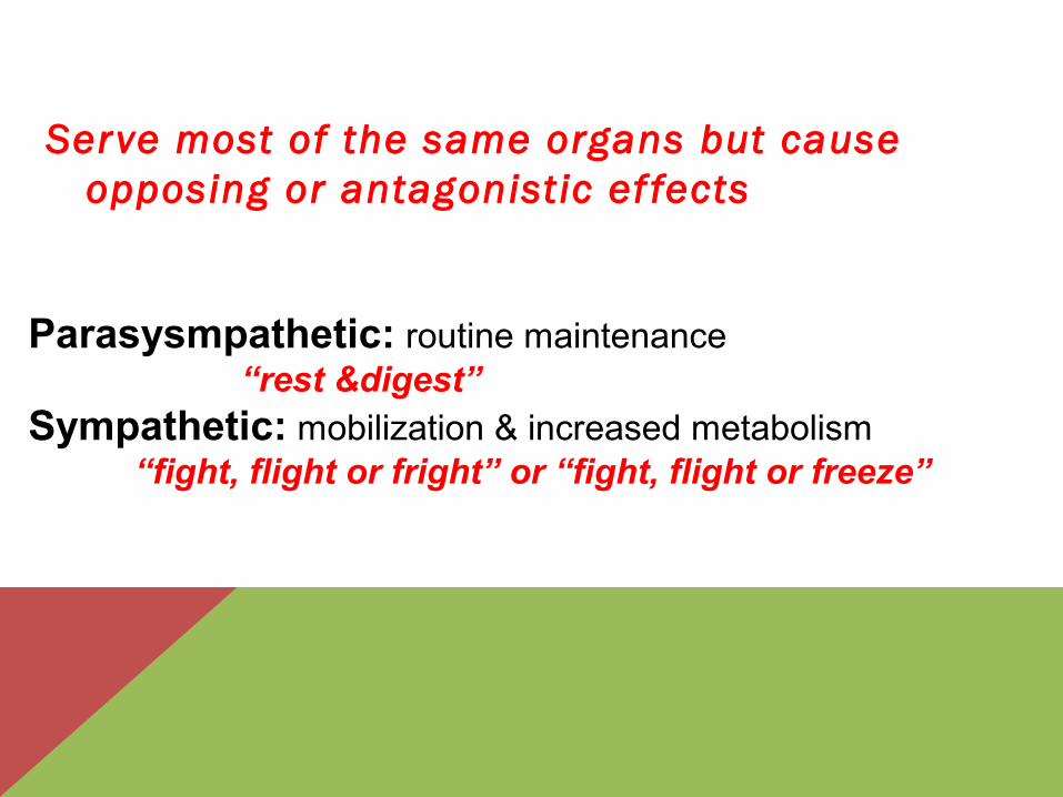
Serve most of the same organs but cause opposing or antagonistic ef fects
Parasysmpathetic: routine maintenance“rest &digest”
Sympathetic: mobilization & increased metabolism “fight, flight or fright” or “fight, flight or freeze”
CHARACTERISTICS
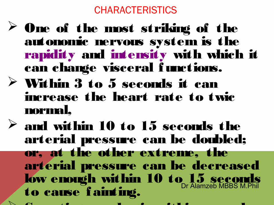
One of the most striking of the autonomic nervous system is the rapidity and intensity with which it can change visceral functions.
Within 3 to 5 seconds it can increase the heart rate to twic normal,
and within 10 to 15 seconds the arterial pressure can be doubled; or, at the other extreme, the arterial pressure can be decreased low enough within 10 to 15 seconds to cause fainting.
Sweating can begin within seconds, and the urinary bladder may empty involuntarily, also within seconds.
Dr Alamzeb MBBS M.Phil
GENERAL ORGANIZATION OF THE
AUTONOMIC NERVOUS SYSTEM
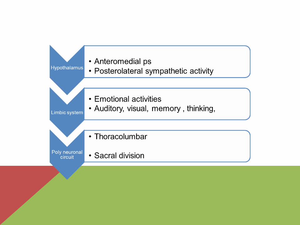
The autonomic nervous system is activated mainly by centers located in the spinal cord , brain stem , and hypothalamus .
por tions of the cerebral cor tex, especially of the l imbic cor tex, can transmit signals to the lower centers and in this way influence autonomic control.
Dr Alamzeb MBBS M.Phil03/0
2/15 14
PHYSIOLOGIC ANATOMY OF THE SYMPATHETIC NERVOUS SYSTEM
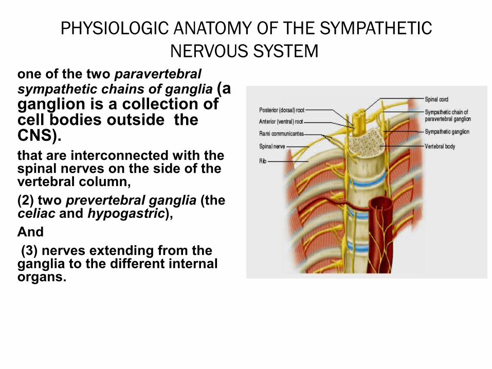
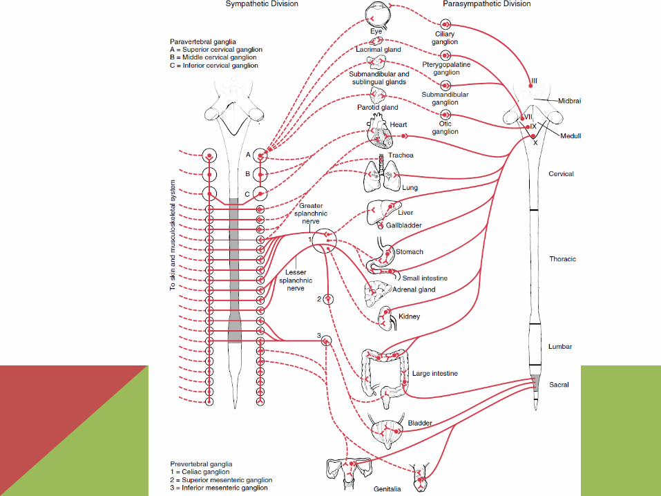
one of the two paravertebral sympathetic chains of ganglia (a ganglion is a collection of cell bodies outside the CNS).that are interconnected with the spinal nerves on the side of the vertebral column, (2) two prevertebral ganglia (the celiac and hypogastric), And (3) nerves extending from the ganglia to the different internal organs.
ORGANIZATION
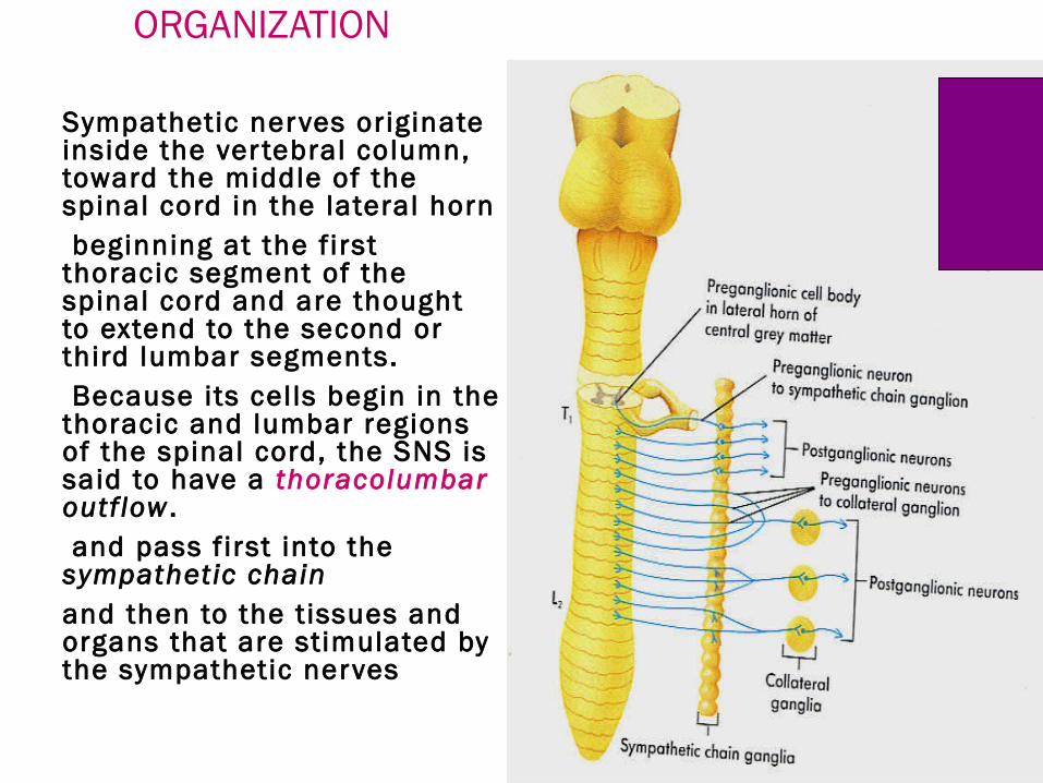
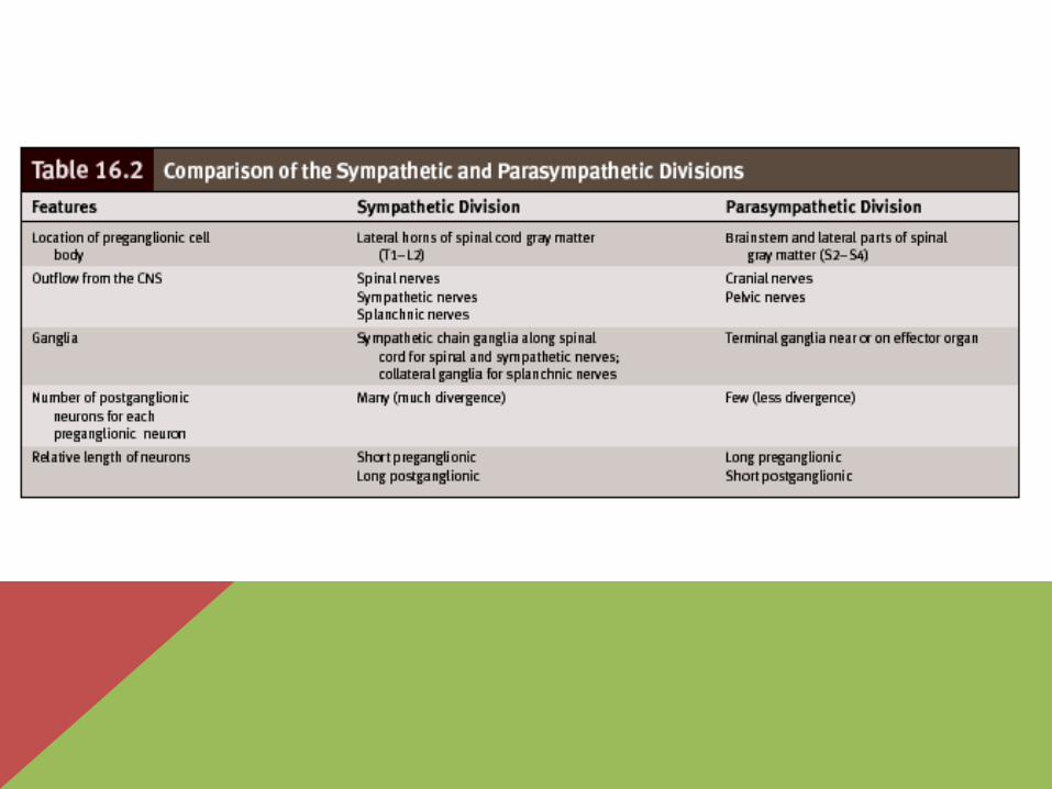
Sympathetic nerves originate inside the ver tebral column, toward the middle of the spinal cord in the lateral horn beginning at the f irst thoracic segment of the spinal cord and are thought to extend to the second or third lumbar segments. Because its cells begin in the thoracic and lumbar regions of the spinal cord, the SNS is said to have a thoracolumbar outflow . and pass f irst into the sympathetic chain and then to the t issues and organs that are stimulated by the sympathetic nerves
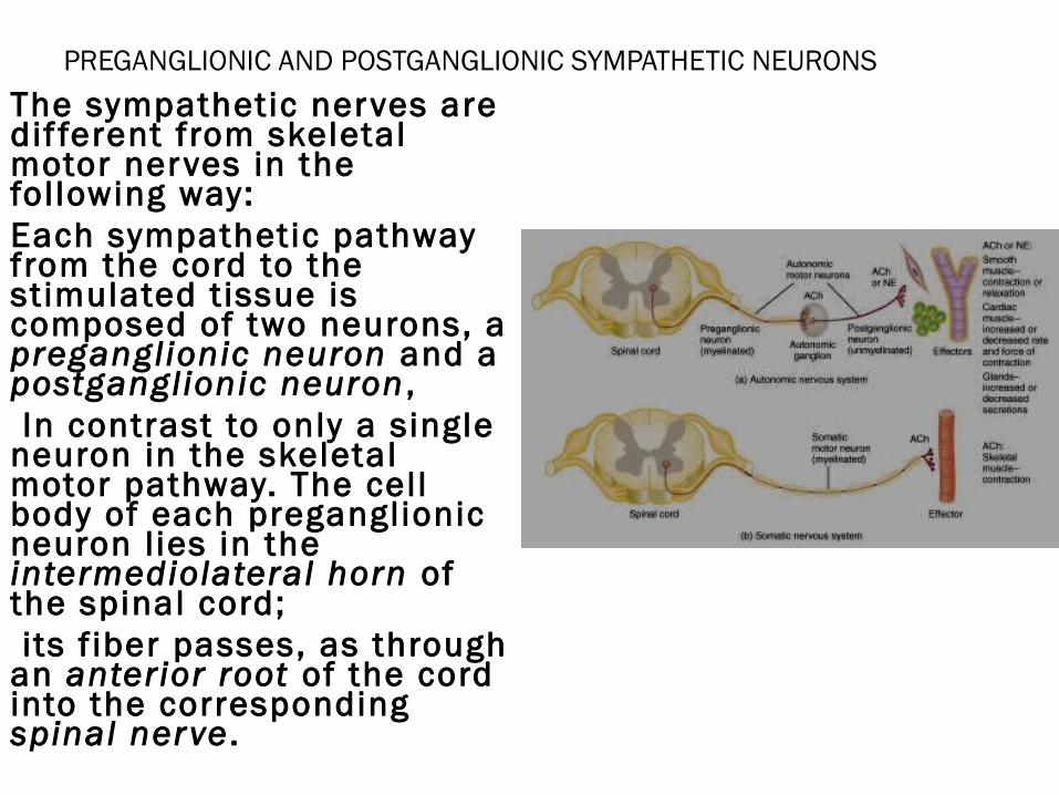
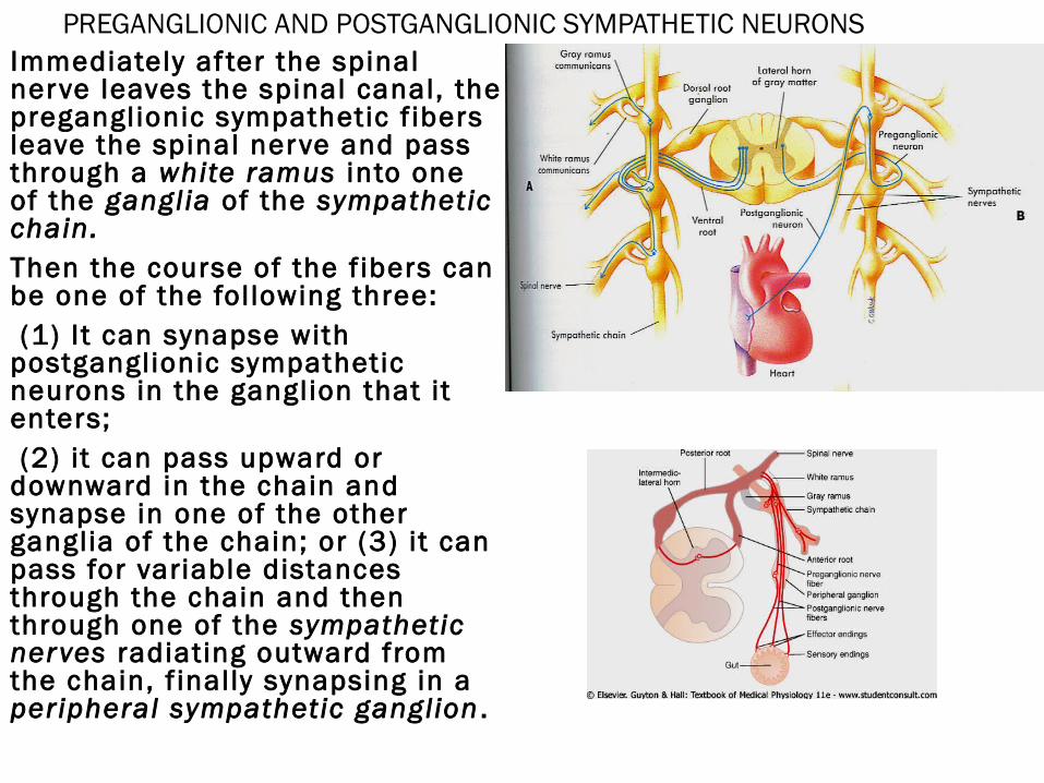
PREGANGLIONIC AND POSTGANGLIONIC SYMPATHETIC NEURONS The sympathetic nerves are dif ferent from skeletal motor nerves in the following way: Each sympathetic pathway from the cord to the stimulated t issue is composed of two neurons, a preganglionic neuron and a postganglionic neuron , In contrast to only a single neuron in the skeletal motor pathway. The cell body of each preganglionic neuron l ies in the intermediolateral horn of the spinal cord; its f iber passes, as through an anterior root of the cord into the corresponding spinal nerve .
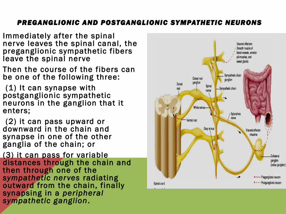
PREGANGLIONIC AND POSTGANGLIONIC SYMPATHETIC NEURONS
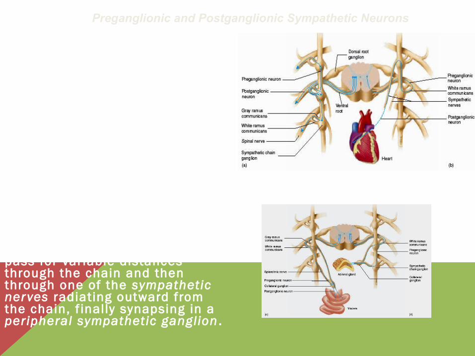
Immediately af ter the spinal nerve leaves the spinal canal, the preganglionic sympathetic f ibers leave the spinal nerve Then the course of the f ibers can be one of the fol lowing three: (1) It can synapse with postganglionic sympathetic neurons in the ganglion that it enters; (2) it can pass upward or downward in the chain and synapse in one of the other ganglia of the chain; or (3) it can pass for variable distances through the chain and then through one of the sympathetic nerves radiating outward from the chain, f inally synapsing in a peripheral sympathetic ganglion .
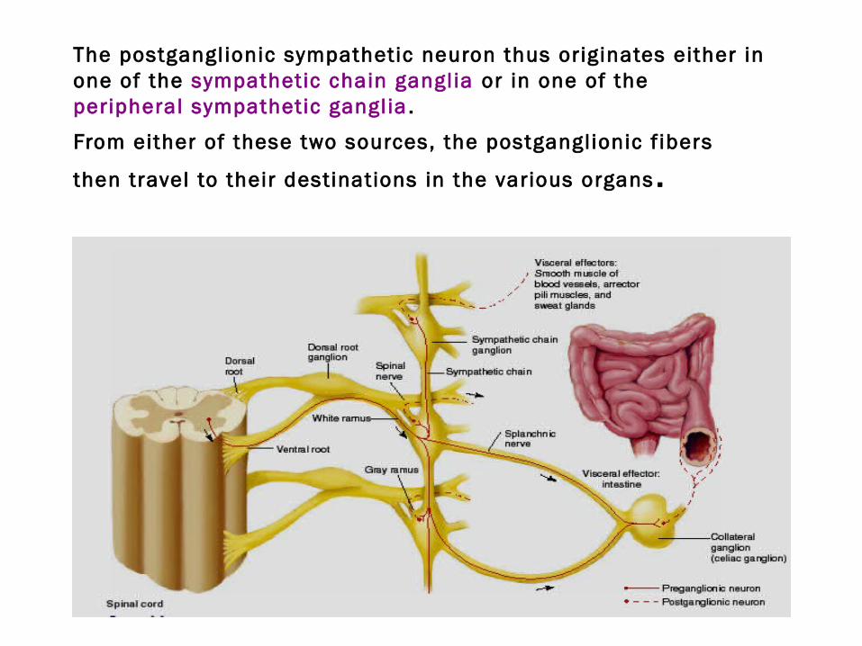
The postgangl ionic sympathetic neuron thus originates either in one of the sympathetic chain gangl ia or in one of the peripheral sympathetic gangl ia .
From either of these two sources, the postganglionic f ibers
then travel to their destinations in the various organs .
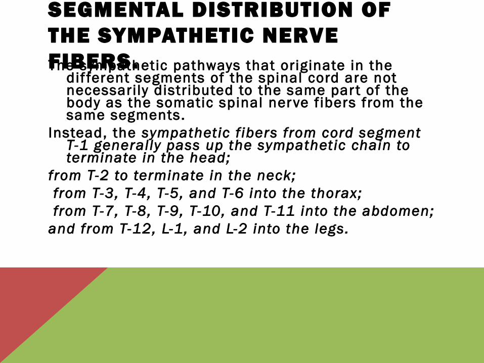
SEGMENTAL DISTRIBUTION OF THE SYMPATHETIC NERVE FIBERS.The sympathetic pathways that originate in the
dif ferent segments of the spinal cord are not necessarily distributed to the same par t of the body as the somatic spinal nerve f ibers from the same segments.
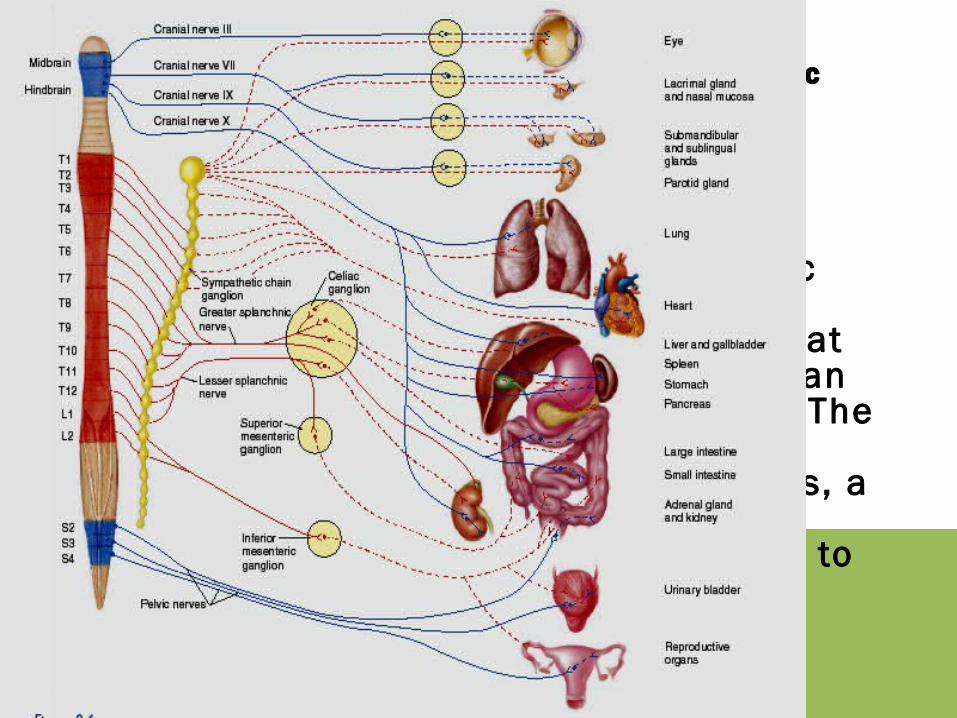
Instead, the sympathetic f ibers from cord segment T-1 generally pass up the sympathetic chain to terminate in the head;
from T-2 to terminate in the neck; from T-3, T-4, T-5, and T-6 into the thorax; from T-7, T-8, T-9, T-10, and T-11 into the abdomen; and from T-12, L-1, and L-2 into the legs.
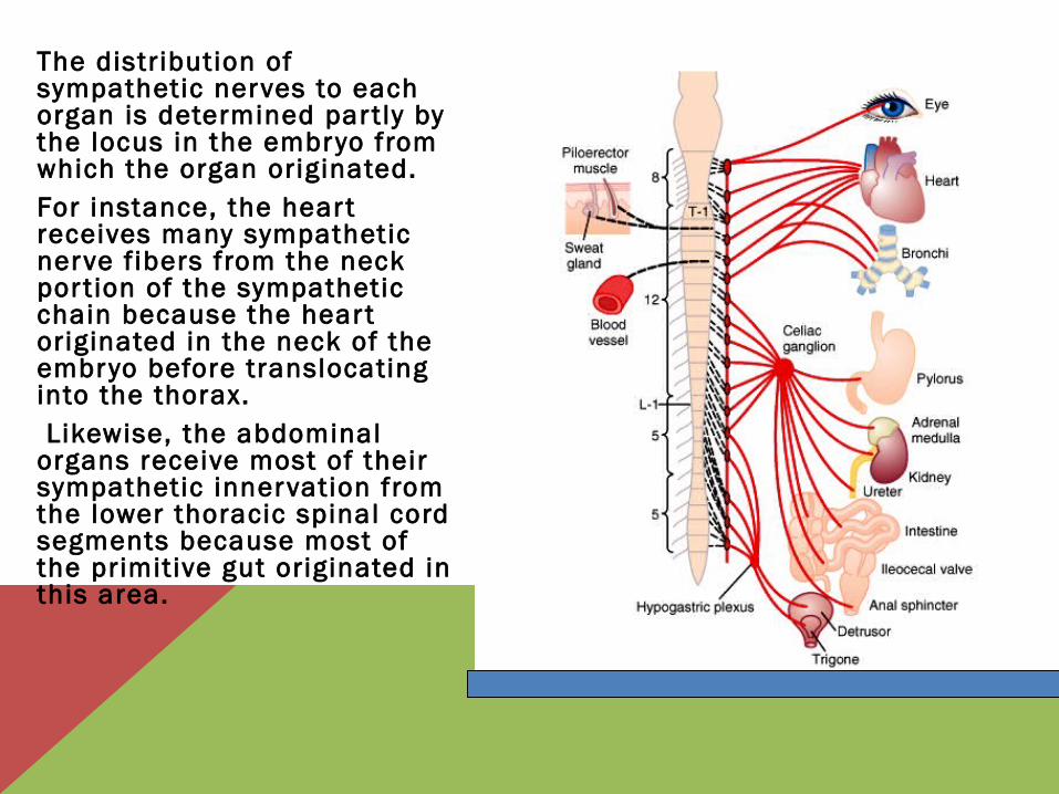
The distribution of sympathetic nerves to each organ is determined partly by the locus in the embryo from which the organ originated. For instance, the hear t receives many sympathetic nerve f ibers from the neck por tion of the sympathetic chain because the hear t originated in the neck of the embryo before translocating into the thorax. Likewise, the abdominal organs receive most of their sympathetic innervation from the lower thoracic spinal cord segments because most of the primitive gut originated in this area.
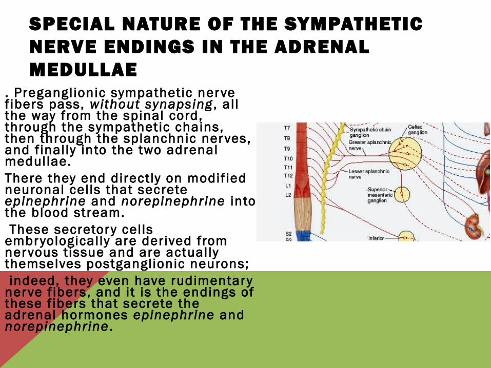
SPECIAL NATURE OF THE SYMPATHETIC NERVE ENDINGS IN THE ADRENAL MEDULLAE
. Preganglionic sympathetic nerve f ibers pass, without synapsing , all the way from the spinal cord, through the sympathetic chains, then through the splanchnic nerves, and f inally into the two adrenal medullae. There they end directly on modif ied neuronal cells that secrete epinephrine and norepinephrine into the blood stream. These secretory cells embryologically are derived from nervous tissue and are actually themselves postganglionic neurons; indeed, they even have rudimentary nerve f ibers, and it is the endings of these f ibers that secrete the adrenal hormones epinephrine and norepinephrine .

PHYSIOLOGIC ANATOMY OF THE PARASYMPATHETIC NERVOUS SYSTEM
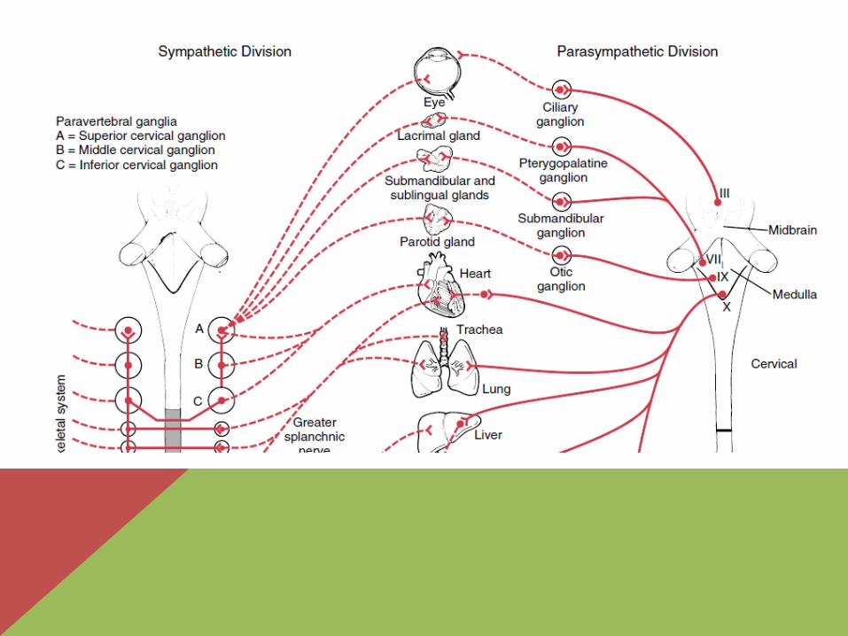
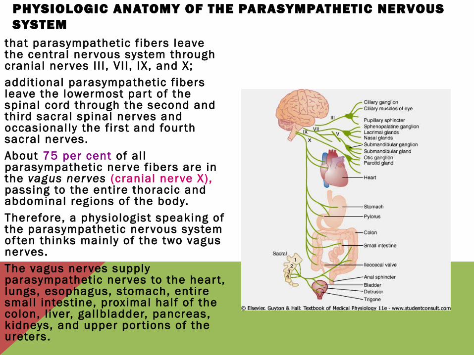
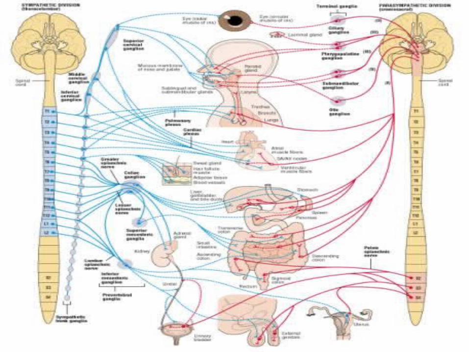
that parasympathetic f ibers leave the central nervous system through cranial nerves I I I , VI I , IX, and X; addit ional parasympathetic f ibers leave the lowermost par t of the spinal cord through the second and third sacral spinal nerves and occasional ly the f irst and four th sacral nerves. About 75 per cent of al l parasympathetic nerve f ibers are in the vagus nerves (cranial nerve X), passing to the entire thoracic and abdominal regions of the body. Therefore, a physiologist speaking of the parasympathetic nervous system of ten thinks mainly of the two vagus nerves. The vagus nerves supply parasympathetic nerves to the hear t, lungs, esophagus, stomach, entire small intestine, proximal half of the colon, l iver, gal lbladder, pancreas, kidneys, and upper por tions of the ureters.
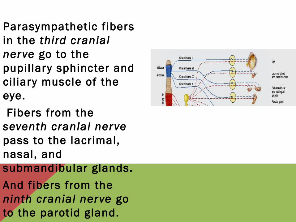
Parasympathetic f ibers in the third cranial nerve go to the pupil lary sphincter and cil iary muscle of the eye.
Fibers from the seventh cranial nerve pass to the lacrimal, nasal, and submandibular glands.
And f ibers from the ninth cranial nerve go to the parotid gland.
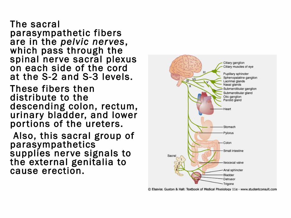
The sacral parasympathetic f ibers are in the pelvic nerves , which pass through the spinal nerve sacral plexus on each side of the cord at the S-2 and S-3 levels. These f ibers then distr ibute to the descending colon, rectum, urinary bladder, and lower por t ions of the ureters. Also, this sacral group of parasympathetics supplies nerve signals to the external genitalia to cause erection.
PREGANGLIONIC AND POSTGANGLIONIC PARASYMPATHETIC NEURONS
. The parasympathetic system, l ike the sympathetic, has both preganglionic and postganglionic neurons. parasympathetic nerves, the preganglionic f ibers pass uninterrupted all the way to the organ that is to be controlled. In the wall of the organ are located the postganglionic neurons. The preganglionic f ibers synapse with these, and very, very shor t postganglionic f ibers, a fraction of a mil l imeter to several centimeters in length, leave the neurons to innervate the tissues of the organ. This location of the parasympathetic
PREGANGLIONIC AND POSTGANGLIONIC SYMPATHETIC NEURONSImmediately af ter the spinal nerve leaves the spinal canal, the preganglionic sympathetic f ibers leave the spinal nerve and pass through a white ramus into one of the ganglia of the sympathetic chain. Then the course of the f ibers can be one of the fol lowing three: (1) It can synapse with postganglionic sympathetic neurons in the ganglion that it enters; (2) it can pass upward or downward in the chain and synapse in one of the other ganglia of the chain; or (3) it can pass for variable distances through the chain and then through one of the sympathetic nerves radiating outward from the chain, f inally synapsing in a peripheral sympathetic ganglion .
Immediately af ter the spinal nerve leaves the spinal canal, the preganglionic sympathetic f ibers leave the spinal nerve and pass through a white ramus into one of the ganglia of the sympathetic chain. Then the course of the f ibers can be one of the fol lowing three: (1) It can synapse with postganglionic sympathetic neurons in the ganglion that it enters; (2) it can pass upward or downward in the chain and synapse in one of the other ganglia of the chain; or (3) it can pass for variable distances through the chain and then through one of the sympathetic nerves radiating outward from the chain, f inally synapsing in a peripheral sympathetic ganglion .
Preganglionic and Postganglionic Sympathetic Neurons
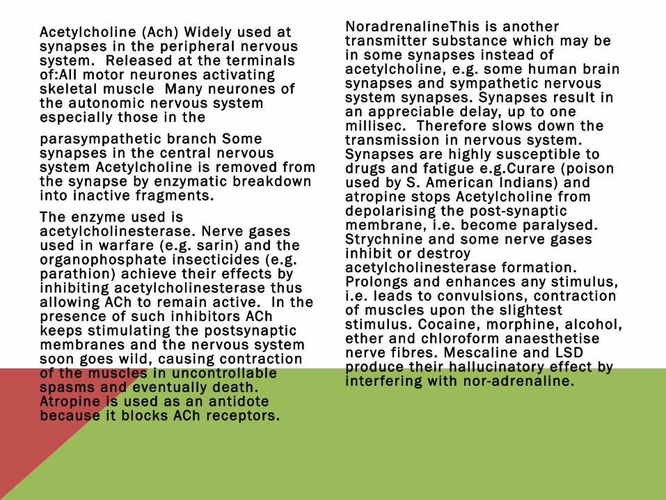
Acetylcholine (Ach) Widely used at synapses in the peripheral nervous system. Released at the terminals of:Al l motor neurones activating skeletal muscle Many neurones of the autonomic nervous system especial ly those in the
parasympathetic branch Some synapses in the central nervous system Acetylcholine is removed from the synapse by enzymatic breakdown into inactive fragments.
The enzyme used is acetylcholinesterase. Nerve gases used in war fare (e.g. sarin) and the organophosphate insecticides (e.g. parathion) achieve their ef fects by inhibiting acetylcholinesterase thus al lowing ACh to remain active. In the presence of such inhibitors ACh keeps stimulating the postsynaptic membranes and the nervous system soon goes wild, causing contraction of the muscles in uncontrollable spasms and eventually death. Atropine is used as an antidote because it blocks ACh receptors.
NoradrenalineThis is another transmitter substance which may be in some synapses instead of acetylcholine, e.g. some human brain synapses and sympathetic nervous system synapses. Synapses result in an appreciable delay, up to one mil l isec. Therefore slows down the transmission in nervous system. Synapses are highly susceptible to drugs and fatigue e.g.Curare (poison used by S. American Indians) and atropine stops Acetylcholine from depolarising the post-synaptic membrane, i .e. become paralysed. Strychnine and some nerve gases inhibit or destroy acetylcholinesterase formation. Prolongs and enhances any stimulus, i .e. leads to convulsions, contraction of muscles upon the sl ightest stimulus. Cocaine, morphine, alcohol, ether and chloroform anaesthetise nerve f ibres. Mescaline and LSD produce their hallucinatory ef fect by inter fering with nor-adrenaline.
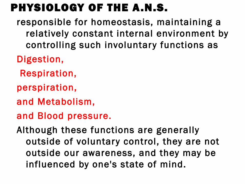
PHYSIOLOGY OF THE A.N.S. responsible for homeostasis, maintaining a
relatively constant internal environment by controll ing such involuntary functions as
Digestion,
Respiration,
perspiration,
and Metabolism,
and Blood pressure.
Although these functions are generally outside of voluntary control, they are not outside our awareness, and they may be influenced by one's state of mind.
SYMPATHETIC AND PARASYMPATHETIC NERVOUS SYSTEM
The autonomic nervous system is divided into two subsystems,
sympathetic nervous system
parasympathetic, nervous system
Somatic vs. Autonomic Connections
SYMPATHETIC
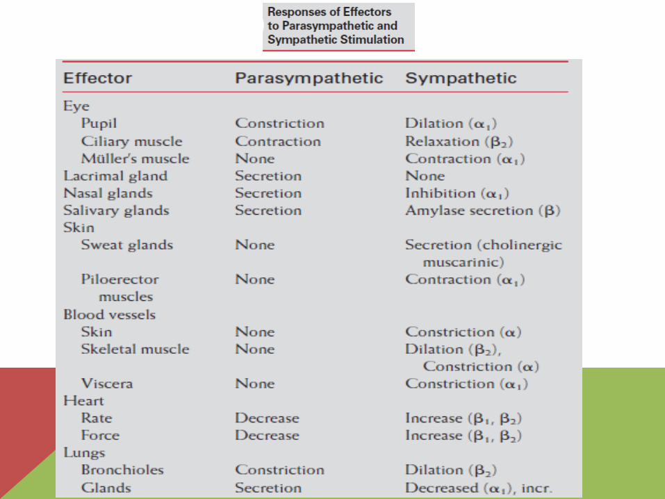
• The sympathetic system is responsible for providing responses and energy needed to cope with stressful situations such as fear or extremes of physical activity.
• In response to such stress, the sympathetic system: raises blood pressure, hear t rate,
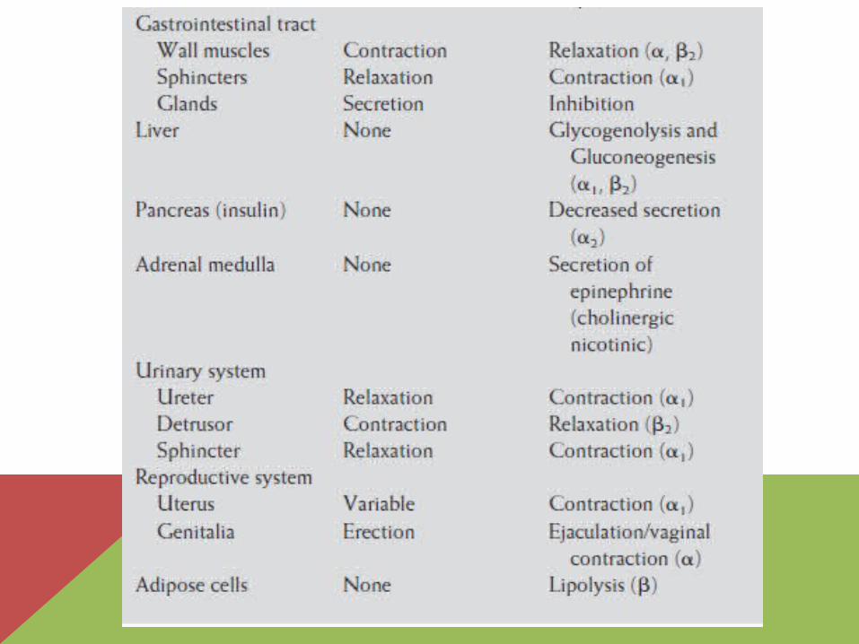
• and the blood supply to the skeletal muscles at the expense of the gastrointestinal tract, kidneys, and skin;
• dilates both the pupils and the bronchioles, providing improved vision and oxygenation;
• and generates needed energy by st imulating glycogenolysis in the l iver and l ipolysis in adipose t issue.
• In general, i t serves to st imulate organs and to mobil ize energy.
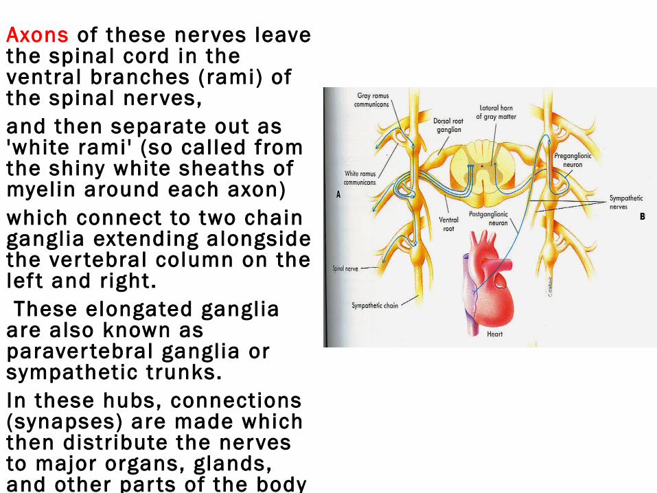
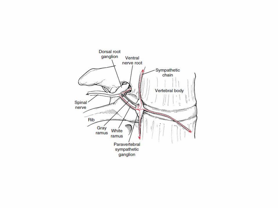
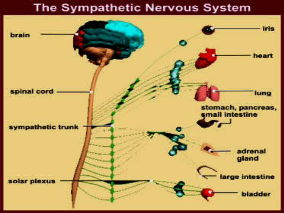
Axons of these nerves leave the spinal cord in the ventral branches (rami) of the spinal nerves, and then separate out as 'white rami' (so cal led from the shiny white sheaths of myelin around each axon) which connect to two chain ganglia extending alongside the ver tebral column on the lef t and right. These elongated ganglia are also known as paraver tebral ganglia or sympathetic trunks. In these hubs, connections (synapses) are made which then distribute the nerves to major organs, glands, and other par ts of the body
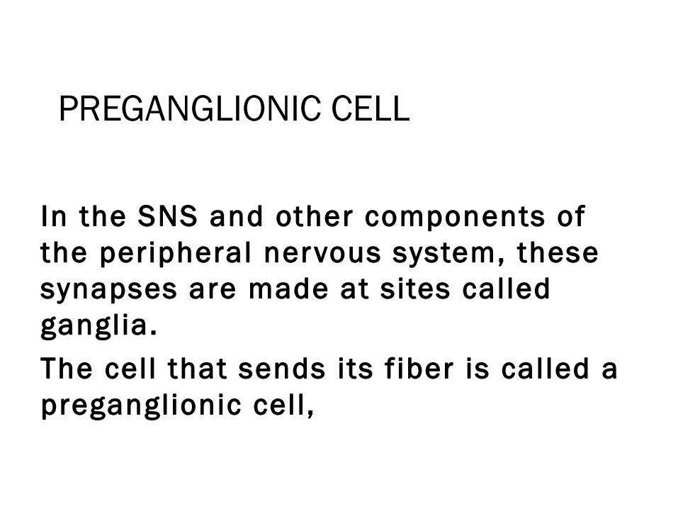
PREGANGLIONIC CELL
In the SNS and other components of the peripheral nervous system, these synapses are made at sites called ganglia.
The cell that sends its f iber is called a preganglionic cell,
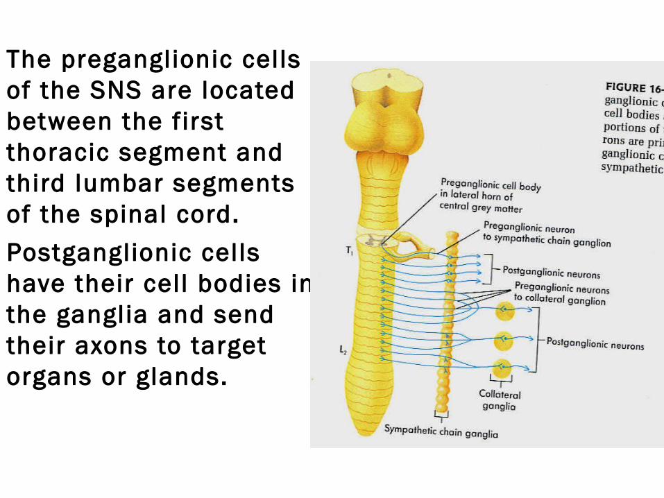
The preganglionic cells of the SNS are located between the f irst thoracic segment and third lumbar segments of the spinal cord.
Postganglionic cells have their cell bodies in the ganglia and send their axons to target organs or glands.
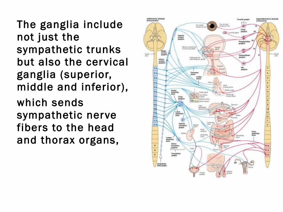
The ganglia include not just the sympathetic trunks but also the cervical ganglia (superior, middle and inferior),
which sends sympathetic nerve f ibers to the head and thorax organs,
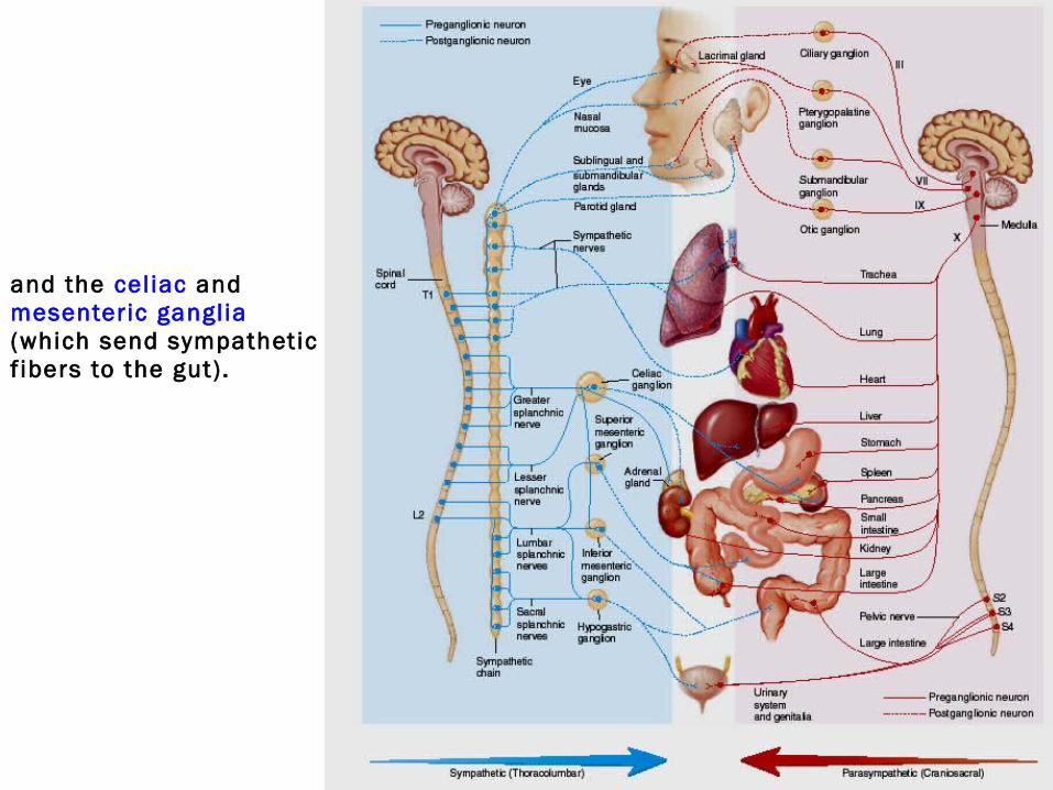
and the cel iac and mesenteric ganglia (which send sympathetic f ibers to the gut).
INFORMATION TRANSMISSION
Messages travel through the SNS in a bidirectional f low.
EFFERENTEfferent messages can trigger changes in
dif ferent par ts of the body simultaneously. For example, the sympathetic nervous system can
accelerate hear t rate; widen bronchial passages; decrease motil ity (movement) of the large
intestine; constrict blood vessels; increase peristalsis in the
esophagus; cause pupil dilation, piloerection (goose bumps) perspiration (sweating); and raise blood pressure.
Af ferent messages carry sensations such as
heat,
cold,
or pain.
The f irst synapse (in the sympathetic chain) is mediated by nicotinic receptors physiologically activated by acetylcholine,
and the target synapse is mediated by adrenergic receptors physiologically activated by either noradrenaline or adrenaline.
An exception is with sweat glands which receive sympathetic innervation but have muscarinic acetylcholine receptors which are normally characteristic of PNS.
Another exception is with cer tain deep muscle blood vessels, which have acetylcholine receptors and which dilate (rather than constrict) with an increase in sympathetic tone.
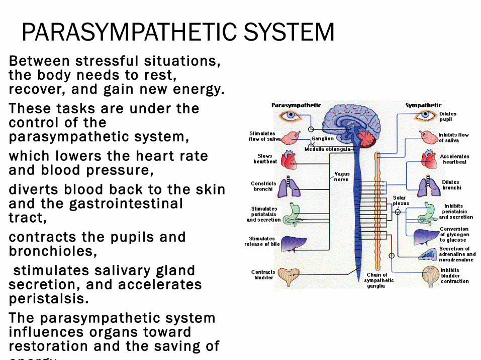
PARASYMPATHETIC SYSTEMBetween stressful situations, the body needs to rest, recover, and gain new energy. These tasks are under the control of the parasympathetic system, which lowers the hear t rate and blood pressure, diver ts blood back to the skin and the gastrointestinal tract, contracts the pupils and bronchioles, stimulates salivary gland secretion, and accelerates peristalsis. The parasympathetic system influences organs toward restoration and the saving of energy.
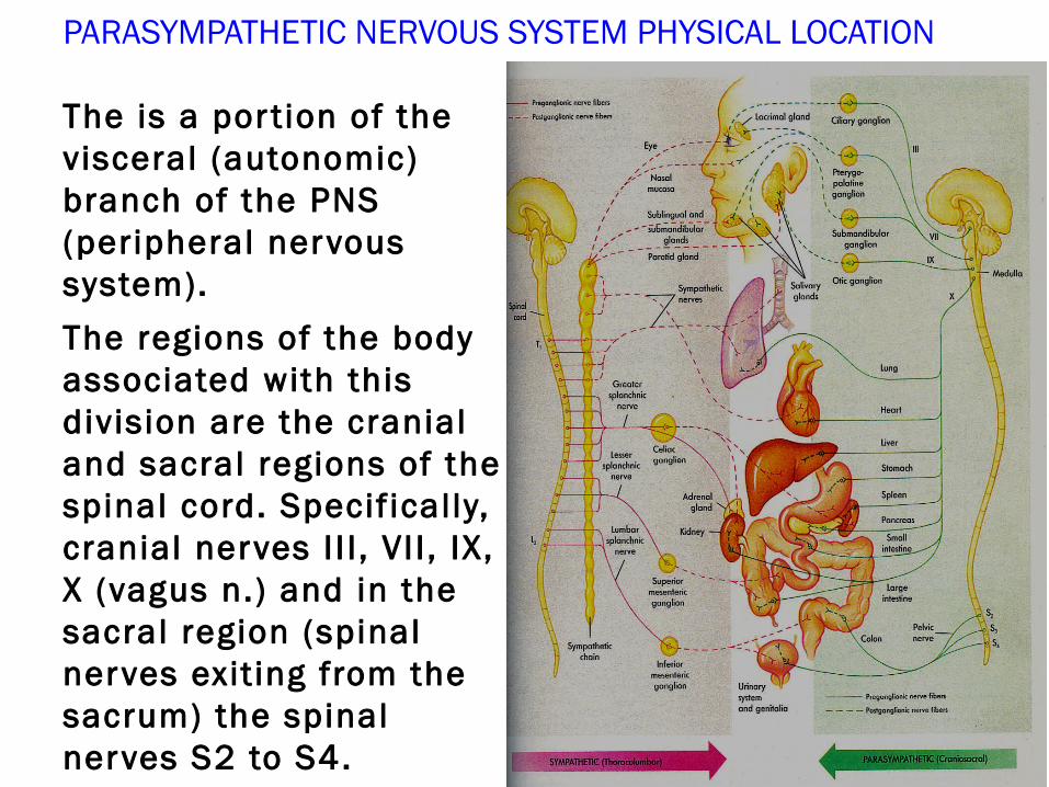
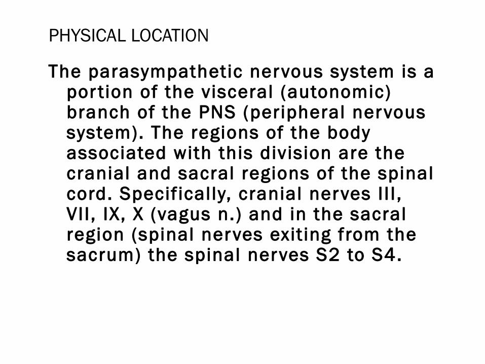
PARASYMPATHETIC NERVOUS SYSTEM PHYSICAL LOCATION
The is a por t ion of the visceral (autonomic) branch of the PNS (peripheral nervous system).
The regions of the body associated with this division are the cranial and sacral regions of the spinal cord. Specif ically, cranial nerves I I I , VI I , IX, X (vagus n.) and in the sacral region (spinal nerves exit ing from the sacrum) the spinal nerves S2 to S4.
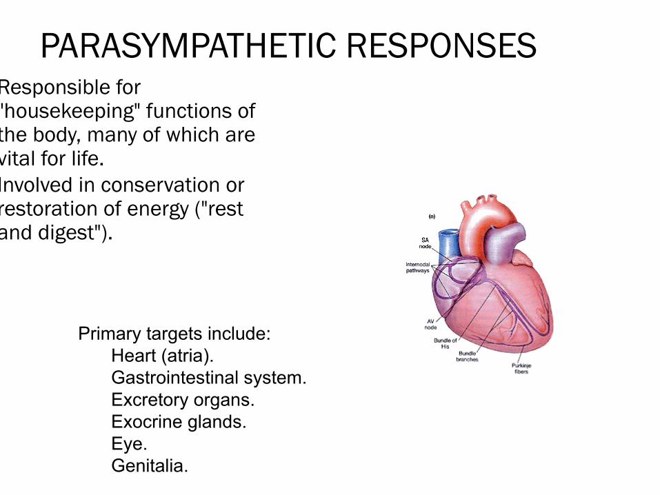
PARASYMPATHETIC RESPONSESResponsible for
"housekeeping" functions of the body, many of which are vital for life.
Involved in conservation or restoration of energy ("rest and digest").
Primary targets include: Heart (atria). Gastrointestinal system. Excretory organs. Exocrine glands. Eye. Genitalia.
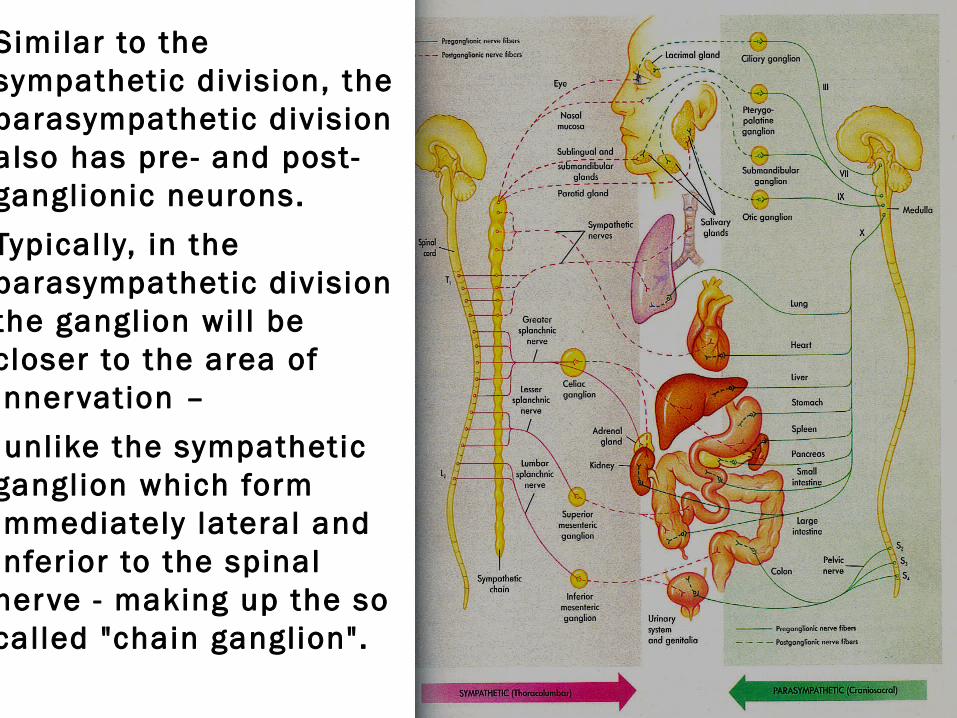
Similar to the sympathetic division, the parasympathetic division also has pre- and post- ganglionic neurons.
Typical ly, in the parasympathetic division the ganglion wil l be closer to the area of innervation –
unlike the sympathetic ganglion which form immediately lateral and inferior to the spinal nerve - making up the so called "chain ganglion".
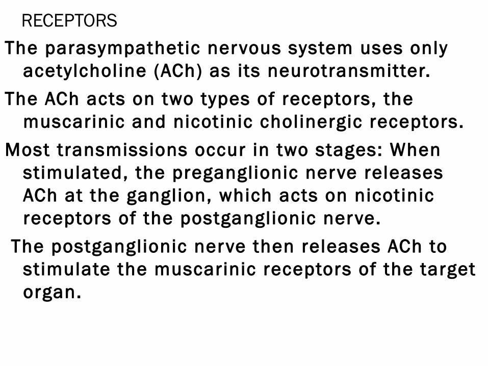
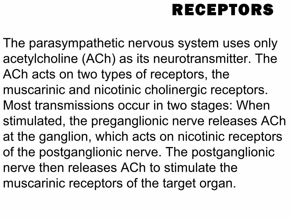
RECEPTORSThe parasympathetic nervous system uses only
acetylcholine (ACh) as its neurotransmitter.
The ACh acts on two types of receptors, the muscarinic and nicotinic cholinergic receptors.
Most transmissions occur in two stages: When stimulated, the preganglionic nerve releases ACh at the ganglion, which acts on nicotinic receptors of the postganglionic nerve.
The postganglionic nerve then releases ACh to stimulate the muscarinic receptors of the target organ.
Types of muscarinic receptors• The three main types of muscarinic receptors that are well
characterised • The M1 muscarinic receptors (CHRM1) are located in the neural
system. • The M2 muscarinic receptors (CHRM2) are located in the heart,
and act to bring the heart back to normal after the actions of the sympathetic nervous system: slowing down the heart rate, reducing contractile forces of the atrial cardiac muscle, and reducing conduction velocity of the sinoatrial node (SA node) and atrioventricular node (AV node). Note, they have no effect on the contractile forces of the ventricular muscle.
• The M3 muscarinic receptors (CHRM3) are located at many places in the body, such as the smooth muscles of the blood vessels, as well as the lungs, which means that they cause vasoconstriction and bronchoconstriction. They are also in the smooth muscles of the gastrointestinal tract (GIT), which help in increasing intestinal motility and dilating sphincters. The M3 receptors are also located in many glands that help to stimulate secretion in salivary glands and other glands of the body.
• The M4 muscarnic receptors: Postganglionic cholinergic nerves, possible CNS effects
• The M5 muscarnic receptors: Possible effects on the CNS
EPINEPHRINE• Epinephrine or adrenaline is a hormone.
• It is a catecholamine, a sympathomimetic monoamine derived from the amino acids phenylalanine and tyrosine.
• The Latin roots ad-+renes and the Greek roots epi-+nephros both l iterally mean "on/to the kidney" (referring to the adrenal gland, which sits atop the kidneys and secretes epinephrine).

ACTIONS IN THE BODY
Epinephrine is a "f ight or f l ight" hormone which is released from the adrenal glands when danger threatens.
When secreted into the bloodstream, it rapidly prepares the body for action in emergency situations.
The hormone boosts the supply of oxygen and energy-giving glucose to the brain and muscles;
Epinephrine plays a central role in the shor t-term stress reaction—the physiological response to threatening, excit ing, or environmental stressor conditions such as high noise levels or bright l ight.
I t is secreted by the adrenal medulla. When released into the bloodstream, epinephrine
binds to mult iple receptors and has numerous ef fects throughout the body.
It increases hear t rate and stroke volume, dilates the pupils, and constricts ar terioles in the skin and gut while dilating ar terioles in leg muscles.
It elevates the blood sugar level by increasing catalysis of glycogen to glucose in the l iver, and at the same time begins the breakdown of l ipids in fat cells.
Like some other stress hormones, epinephrine has a suppressive ef fect on the immune system.
ADRENAL MEDULLA
The adrenal medulla is par t of the adrenal gland. It is located at the center of the gland, being surrounded by the adrenal cor tex.
FUNCTIONSComposed mainly of hormone-producing
chromaffin cells, the adrenal medulla is the principal site of the conversion of the amino acid tyrosine into the catecholamines adrenaline (epinephrine) and noradrenaline (norepinephrine).
In response to stressors such as exercise or imminent danger, medullary cells release catecholamines into the blood in a 85:15 ratio of adrenaline to noradrenaline.
Notable ef fects of adrenaline and noradrenaline include increased hear t rate, blood vessel constriction, bronchiole dilation, and increased metabolism, al l of which are characteristic of the f ight-or-f l ight response.
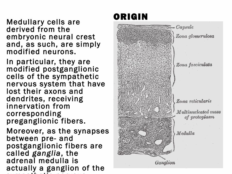
ORIGINMedullary cel ls are derived from the embryonic neural crest and, as such, are simply modif ied neurons.In par t icular, they are modif ied postganglionic cel ls of the sympathetic nervous system that have lost their axons and dendrites, receiving innervation from corresponding preganglionic f ibers.Moreover, as the synapses between pre- and postganglionic f ibers are cal led ganglia , the adrenal medulla is actually a ganglion of the sympathetic nervous system.
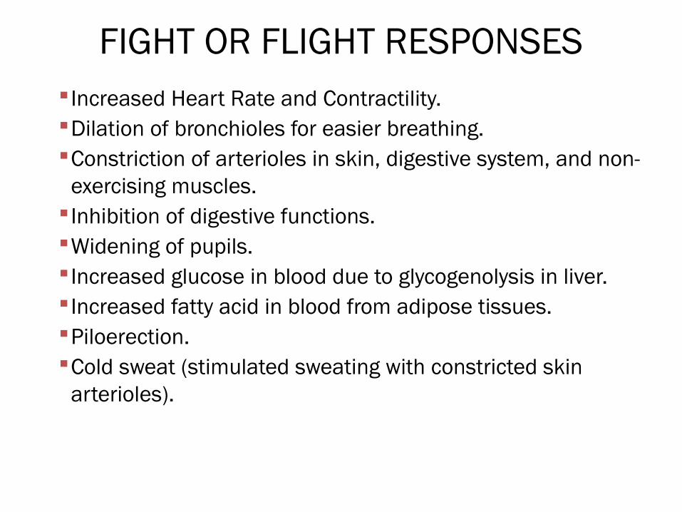
Fight or Flight ResponsesIncreased Heart Rate and Contractility. Dilation of bronchioles for easier breathing. Constriction of arterioles in skin, digestive system, and non-exercising muscles. •Inhibition of digestive functions. Widening of pupils. Increased glucose in blood due to glycogenolysis in liver. Increased fatty acid in blood from adipose tissues. Piloerection. Cold sweat (stimulated sweating with constricted skin arterioles).
Fight or Flight TargetsHeart. Nearly all blood vessels. Lungs. Smooth muscle in nearly all organs. Some glands (sweat, salivary, and digestive). Metabolic tissue (fat cells, liver).
OTHER TISSUES (EYES, SKIN, KIDNEYS).
FIGHT OR FLIGHT RESPONSES Increased Heart Rate and Contractility. Dilation of bronchioles for easier breathing. Constriction of arterioles in skin, digestive system, and non-
exercising muscles. Inhibition of digestive functions. Widening of pupils. Increased glucose in blood due to glycogenolysis in liver. Increased fatty acid in blood from adipose tissues. Piloerection. Cold sweat (stimulated sweating with constricted skin
arterioles).
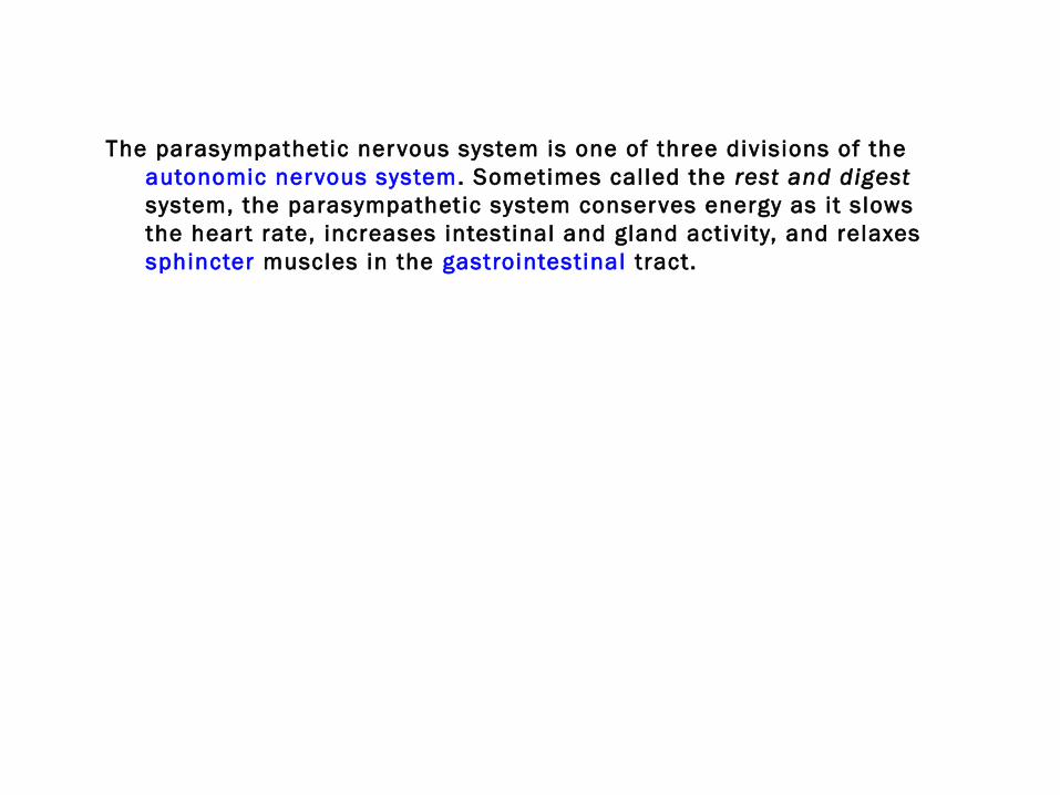
The parasympathetic nervous system is one of three divisions of the autonomic nervous system. Sometimes called the rest and digest system, the parasympathetic system conserves energy as it slows the hear t rate, increases intestinal and gland activity, and relaxes sphincter muscles in the gastrointestinal tract.
PARASYMPATHETIC NERVOUS SYSTEM
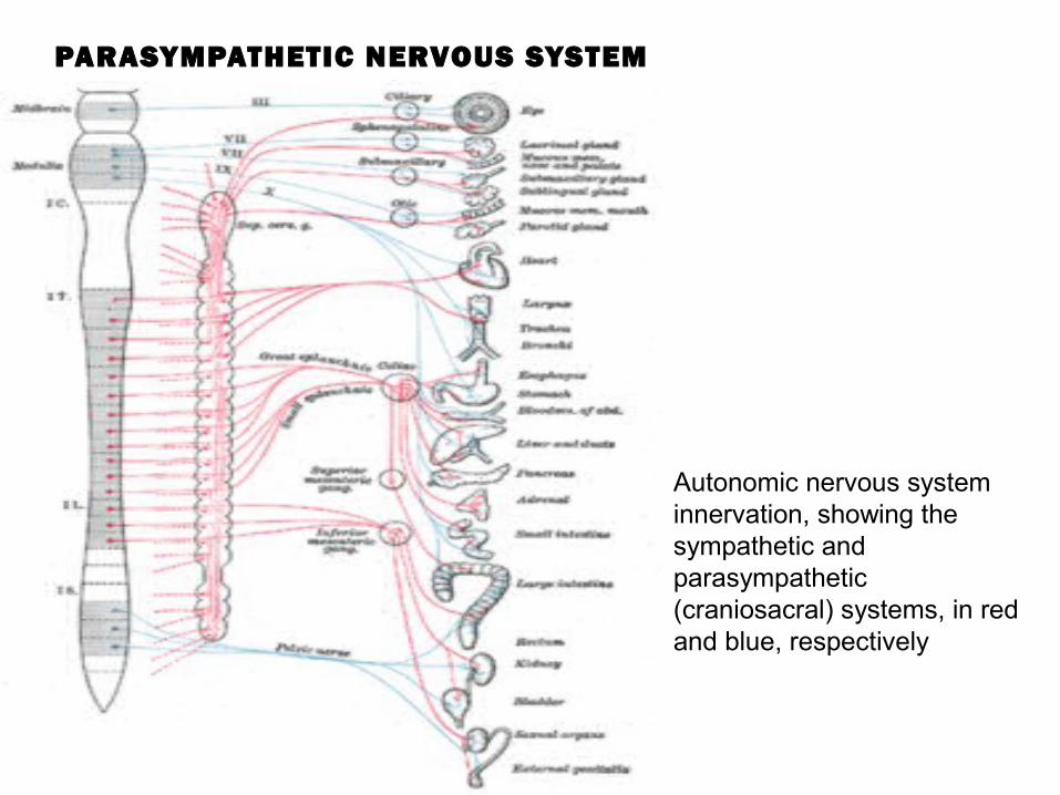
Autonomic nervous system innervation, showing the sympathetic and parasympathetic (craniosacral) systems, in red and blue, respectively
PHYSICAL LOCATION
The parasympathetic nervous system is a por tion of the visceral (autonomic) branch of the PNS (peripheral nervous system). The regions of the body associated with this division are the cranial and sacral regions of the spinal cord. Specif ically, cranial nerves II I , VII, IX, X (vagus n.) and in the sacral region (spinal nerves exit ing from the sacrum) the spinal nerves S2 to S4.
RECEPTORS
The parasympathetic nervous system uses only acetylcholine (ACh) as its neurotransmitter. The ACh acts on two types of receptors, the muscarinic and nicotinic cholinergic receptors. Most transmissions occur in two stages: When stimulated, the preganglionic nerve releases ACh at the ganglion, which acts on nicotinic receptors of the postganglionic nerve. The postganglionic nerve then releases ACh to stimulate the muscarinic receptors of the target organ.
The autonomic nerve f ibers form a subsidiary system that regulates the ir is of the eye and the smooth-muscle action of the hear t, blood vessels, glands, lungs, stomach, colon, bladder, and other visceral organs not subject to wil lful control. Although the autonomic nervous system's impulses originate in the central nervous system, it per forms the most basic human functions more or less automatically, without conscious intervention of higher brain centers. Because it is l inked to those centers, however, the autonomic system is inf luenced by the emotions; for example, anger can increase the rate of hear tbeat. Al l of the f ibers of the autonomic nervous system are motor channels, and their impulses arise from the nerve t issue itself, so that the organs they innervate per form more or less involuntari ly and do not require st imulation to function.
Autonomic nerve f ibers exit from the central nervous system as par t of other peripheral nerves but branch from them to form two more subsystems: the sympathetic and parasympathetic nervous systems, the actions of which usually oppose each other. For example, sympathetic nerves cause ar teries to contract while parasympathetic nerves cause them to di late. Sympathetic impulses are conducted to the organs by two or more neurons. The cel l body of the f irst l ies within the central nervous system and that of the second in an external ganglion. Eighteen pairs of such ganglia interconnect by nerve f ibers to form a double chain just outside the spine and running paral lel to it . Parasympathetic impulses are also relayed by at least two neurons, but the cell body of the second generally l ies near or within the target organ .
Axons of these nerves leave the spinal cord in the ventral branches (rami) of the spinal nerves,
and then separate out as 'white rami' (so called from the shiny white sheaths of myelin around each axon)
0which connect to two chain ganglia extending alongside the ver tebral column on the lef t and right.
These elongated ganglia are also known as paraver tebral ganglia or sympathetic trunks.
In these hubs, connections (synapses) are made which then distribute the nerves to major organs, glands, and other par ts of the body.
ORGANIZATION
Sympathetic nerves originate inside the ver tebral column, toward the middle of the spinal cord in the intermediolateral cell column (or lateral horn),
beginning at the f irst thoracic segment of the spinal cord and are thought to extend to the second or third lumbar segments.
Because its cells begin in the thoracic and lumbar regions of the spinal cord, the SNS is said to have a thoracolumbar outflow .
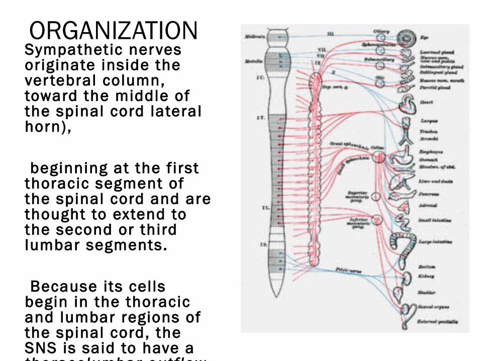
ORGANIZATIONSympathetic nerves originate inside the ver tebral column, toward the middle of the spinal cord lateral horn),
beginning at the f irst thoracic segment of the spinal cord and are thought to extend to the second or third lumbar segments.
Because its cel ls begin in the thoracic and lumbar regions of the spinal cord, the SNS is said to have a thoracolumbar outf low .
Axons of these nerves leave the spinal cord in the ventral branches (rami) of the spinal nerves,
and then separate out as 'white rami' (so called from the shiny white sheaths of myelin around each axon)
0which connect to two chain ganglia extending alongside the ver tebral column on the lef t and right.
These elongated ganglia are also known as paraver tebral ganglia or sympathetic trunks.
In these hubs, connections (synapses) are made which then distribute the nerves to major organs, glands, and other par ts of the body.
FUNCTION The sympathetic nervous system activates what is
of ten termed the f ight or f l ight response . This response is also known as sympatho-adrenal
response of the body, as the pre-ganglionic sympathetic f ibers that end in the adrenal medulla (but also all other sympathetic f ibers) secrete acetylcholine,which activates the secretion of adrenaline (epinephrine) and to a lesser extent noradrenaline (norepinephrine) from it.
Therefore, this response that acts primarily on the cardiovascular system is mediated directly via impulses transmitted through the sympathetic nervous system
and indirectly via catecholamines secreted from the adrenal medulla.
Science typically looks at the SNS as an automatic regulation system, that is, one that operates without the intervention of conscious thought. as the sympathetic nervous system is responsible for priming the body for action. One example of this priming is in the moments before waking, in which sympathetic outflow spontaneously increases in preparation for action.

Thank You
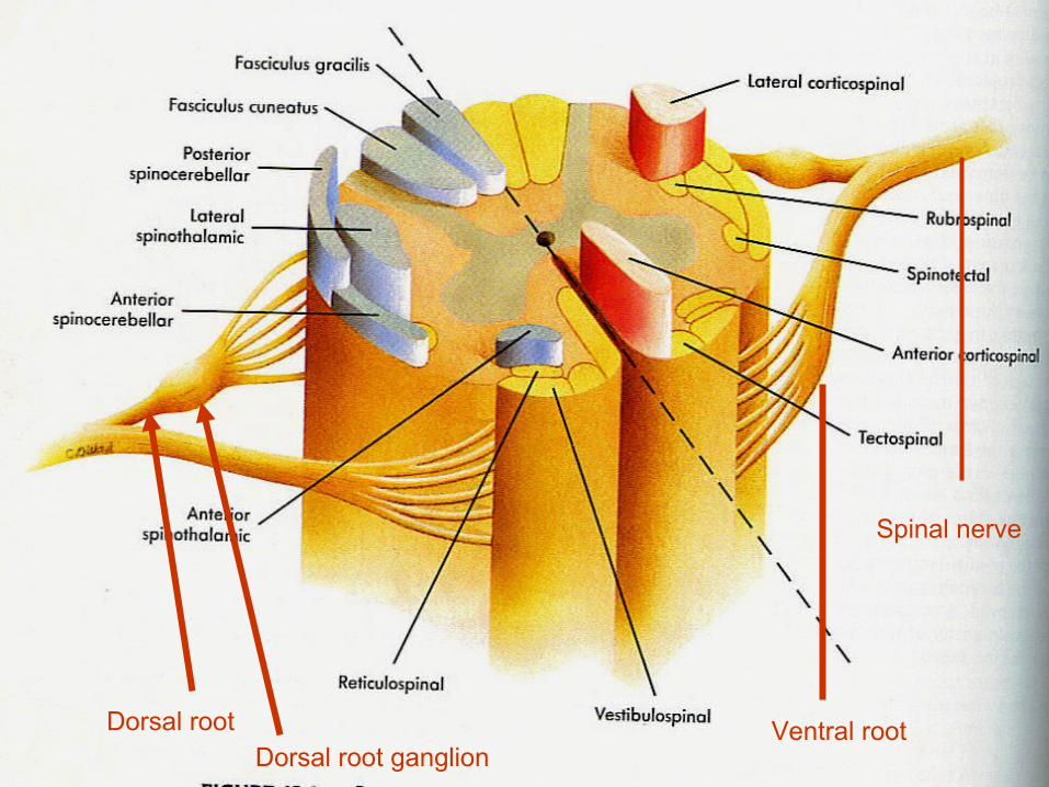
Dorsal root Ventral rootDorsal root ganglion
Spinal nerve