Embed Size (px)
Citation preview
PRIMARY PULMONARY TUBERCULOSIS
Presented by :Arif Khan
5th Year
4th Group
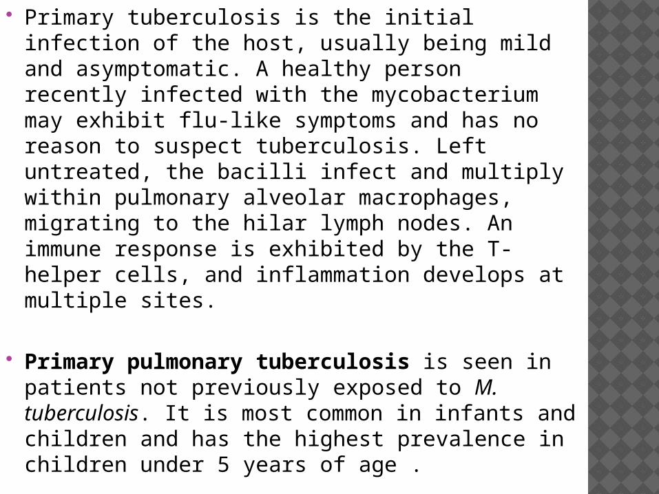
Primary tuberculosis is the initial infection of the host, usually being mild and asymptomatic. A healthy person recently infected with the mycobacterium may exhibit flu-like symptoms and has no reason to suspect tuberculosis. Left untreated, the bacilli infect and multiply within pulmonary alveolar macrophages, migrating to the hilar lymph nodes. An immune response is exhibited by the T-helper cells, and inflammation develops at multiple sites.
Primary pulmonary tuberculosis is seen in patients not previously exposed to M. tuberculosis. It is most common in infants and children and has the highest prevalence in children under 5 years of age .
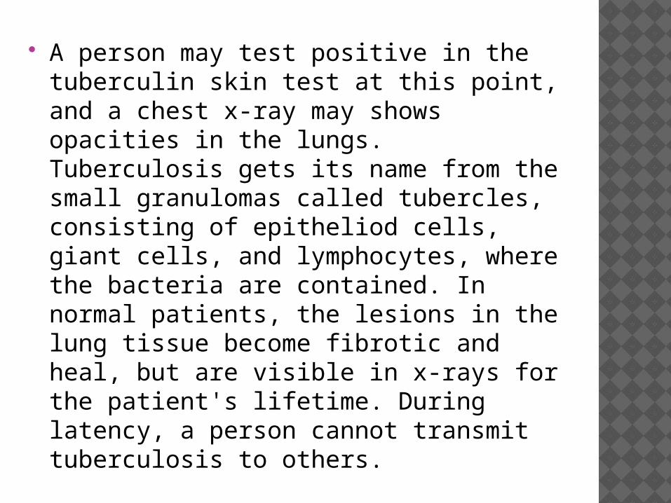
A person may test positive in the tuberculin skin test at this point, and a chest x-ray may shows opacities in the lungs. Tuberculosis gets its name from the small granulomas called tubercles, consisting of epitheliod cells, giant cells, and lymphocytes, where the bacteria are contained. In normal patients, the lesions in the lung tissue become fibrotic and heal, but are visible in x-rays for the patient's lifetime. During latency, a person cannot transmit tuberculosis to others.

PRIMARY PULMONARY TUBERCULOSIS MANIFESTS AS FOUR MAIN ENTITIES:
parenchymal disease: usually manifests as dense, homogeneous parenchymal consolidation in any lobe; however, predominance in the lower and middle lobes (subpleural sites) is suggestive of the disease, especially in adults 1
lymphadenopathy miliary opacities pleural effusion
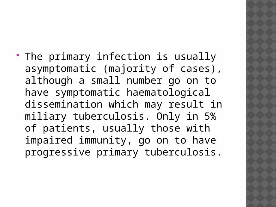
The primary infection is usually asymptomatic (majority of cases), although a small number go on to have symptomatic haematological dissemination which may result in miliary tuberculosis. Only in 5% of patients, usually those with impaired immunity, go on to have progressive primary tuberculosis.
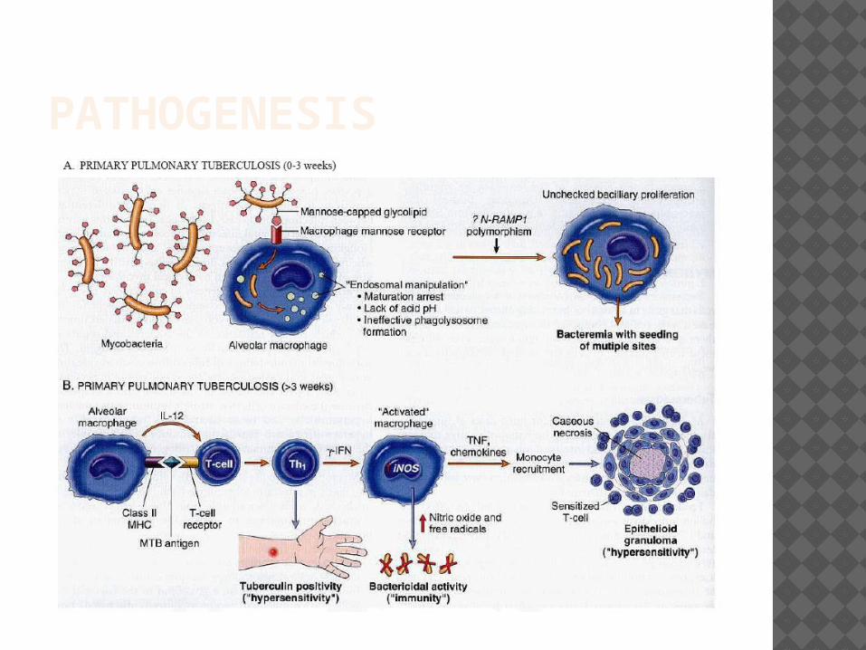
PATHOGENESIS
PATHOGENESISPrimary tuberculosis is always result of
exogenous infection. The infection penetrates into
organism by: - aerogenic (the most often way of
penetration) - alimentary; - contact way.
CLINICAL MANIFESTATIONS Primary TB infection may be asymptomatic,cause
fevers and pleuritic pain or, rarely, progress to life threatening disease. Dur ing the primary pulmonary infection, symptoms may occur as the burden of bacilli increases and the host mounts a systemic immune response. Fever is the most common symptom.
On examination, a patient with primary pulmonary TB may have erythema nodosum, bluish red tender subcutaneous nodules several millimetres to several centimetres in diameter appearing on the legs, and phlyctenular conjunctivitis, hard raised red 1 to 3 mm nodules accompanied by a zone of hyperaemia located near the limbus on the bulbar conjunctiva of the eye.
PERCUSSION: dullness over lung component with a
big size. Weakend breathing with streached
exhale.
Hemogram: Leucocytosis 10-13 T/l, insignificant shift to the left, lymphopenia, monocytosis, ESR 20-25 mm/h
X-RAY DIAGNOSTICS:Phases: 1) infiltrative or pneumonic; 2)
resorbtion(suction,bipolarities); 3) scarring 4) calcification.

PRIMARY TUBERCULOUS COMPLEX, INFILTRATION PHASE
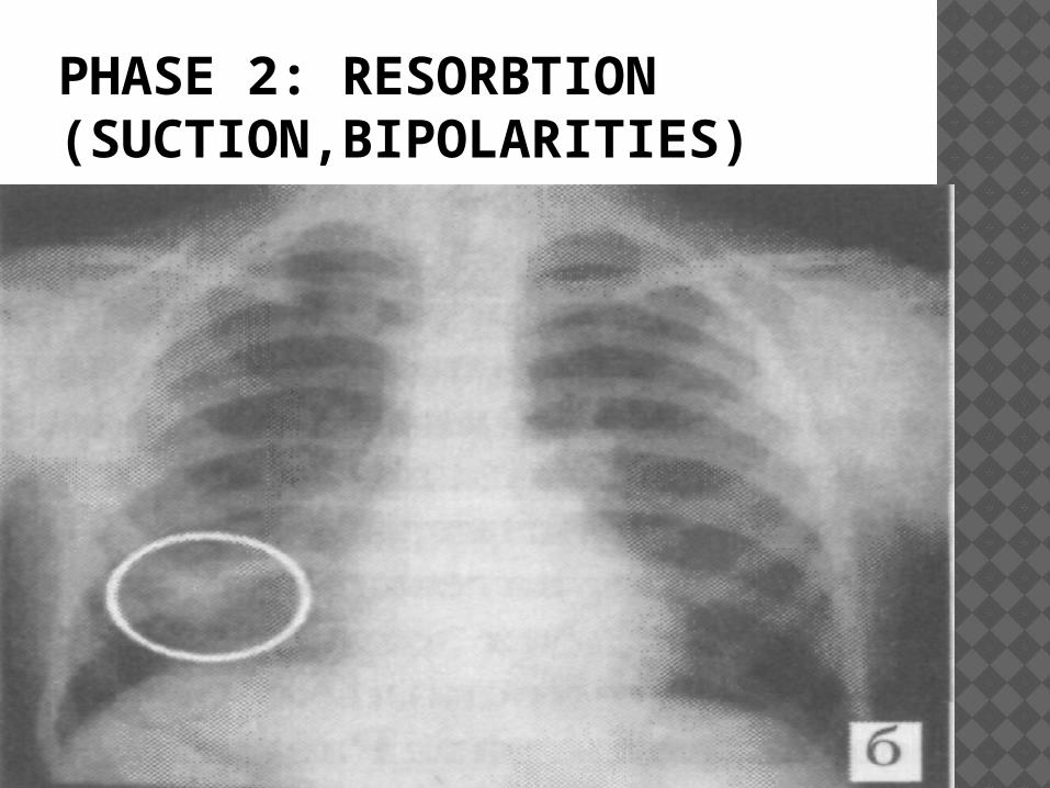
PHASE 2: RESORBTION (SUCTION,BIPOLARITIES)
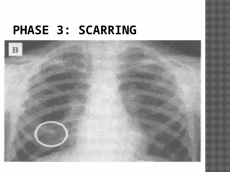
PHASE 3: SCARRING
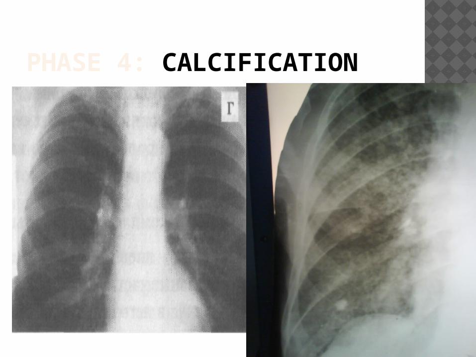
PHASE 4: CALCIFICATION
RADIOGRAPHIC FEATURES
In primary pulmonary tuberculosis, the initial focus of infection can be located anywhere within the lung and has non-specific appearances ranging from too small to be detectable, to patchy areas or consolidation or even lobar consolidation.
Radiographic evidence of parenchymal infection is seen in 70% of children and 90% of adults .
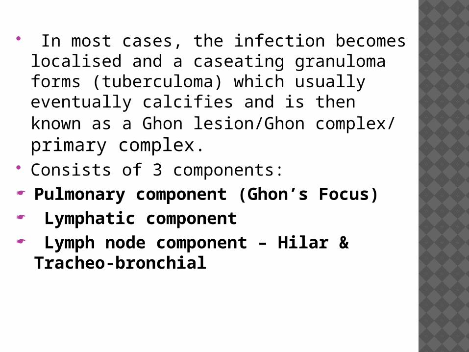
In most cases, the infection becomes localised and a caseating granuloma forms (tuberculoma) which usually eventually calcifies and is then known as a Ghon lesion/Ghon complex/ primary complex.
Consists of 3 components: Pulmonary component (Ghon’s Focus) Lymphatic component Lymph node component – Hilar &
Tracheo-bronchial
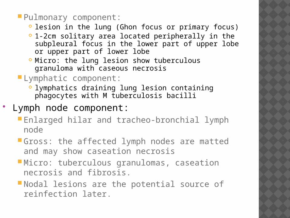
Pulmonary component: lesion in the lung (Ghon focus or primary focus) 1-2cm solitary area located peripherally in the
subpleural focus in the lower part of upper lobe or upper part of lower lobe
Micro: the lung lesion show tuberculous granuloma with caseous necrosis
Lymphatic component: lymphatics draining lung lesion containing
phagocytes with M tuberculosis bacilli
Lymph node component: Enlarged hilar and tracheo-bronchial lymph node Gross: the affected lymph nodes are matted and
may show caseation necrosis Micro: tuberculous granulomas, caseation
necrosis and fibrosis. Nodal lesions are the potential source of
reinfection later.
COMPLICATIONSComplications connected with regional
lymphadenitis:- hematogenic dissemination- lymphogenic dissemination- pleuritis- extending of specific process from lymphatic
node It’s results: a) formation of fistula b) dispersion of caseous masses,
bronchogenic dessemination, bronchi tuberculosis c) disorder of bronchial permeability,
atelectasis
DIAGNOSIS OF TB The doctor or nurse will perform a physical exam. This may
show: Clubbing of the fingers or toes (in people with advanced
disease) Swollen or tender lymph nodes in the neck or other areas Fluid around a lung (pleural effusion) Unusual breath sounds (crackles) Tests may include: Biopsy of the affected tissue (rare) Bronchoscopy Chest CT scan Chest x-ray Interferon-gamma release blood test such as the QFT-Gold test
to test for TB infection Sputum examination and cultures Thoracentesis Tuberculin skin test (also called a PPD test)
TREATMENTThe treatment of tuberculosis (TB) must satisfy
the following basic therapeutic principles: Any regimen must use multiple drugs to
which Mycobacterium tuberculosis is susceptible The medications must be taken regularly The therapy must continue for a period sufficient to
resolve the illness New cases are initially treated with four drugs:
isoniazid, rifampin, pyrazinamide, and either ethambutol or streptomycin. After 2 months, they are then treated with a continuation phase of 4 months with isoniazid and rifampin. Patients requiring retreatment should initially receive at least 5 drugs, including isoniazid, rifampin, pyrazinamide, and at least 2 (preferably 3) new drugs to which the patient has not been exposed.