Embed Size (px)
Citation preview
David L. Nelson and Michael M. Cox
Lehninger Principles of
David L. Nelson and Michael M. Cox
Lehninger Principles of BiochemistryFourth EditionFourth Edition
Chapter 7:Chapter 7:
Carbohydrates and Glycobiology
Copyright © 2004 by W. H. Freeman & Company
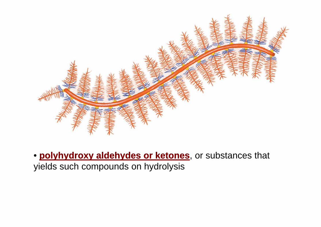
• polyhydroxy aldehydes or ketonespolyhydroxy aldehydes or ketones, or substances that yields such compounds on hydrolysisyields such compounds on hydrolysis
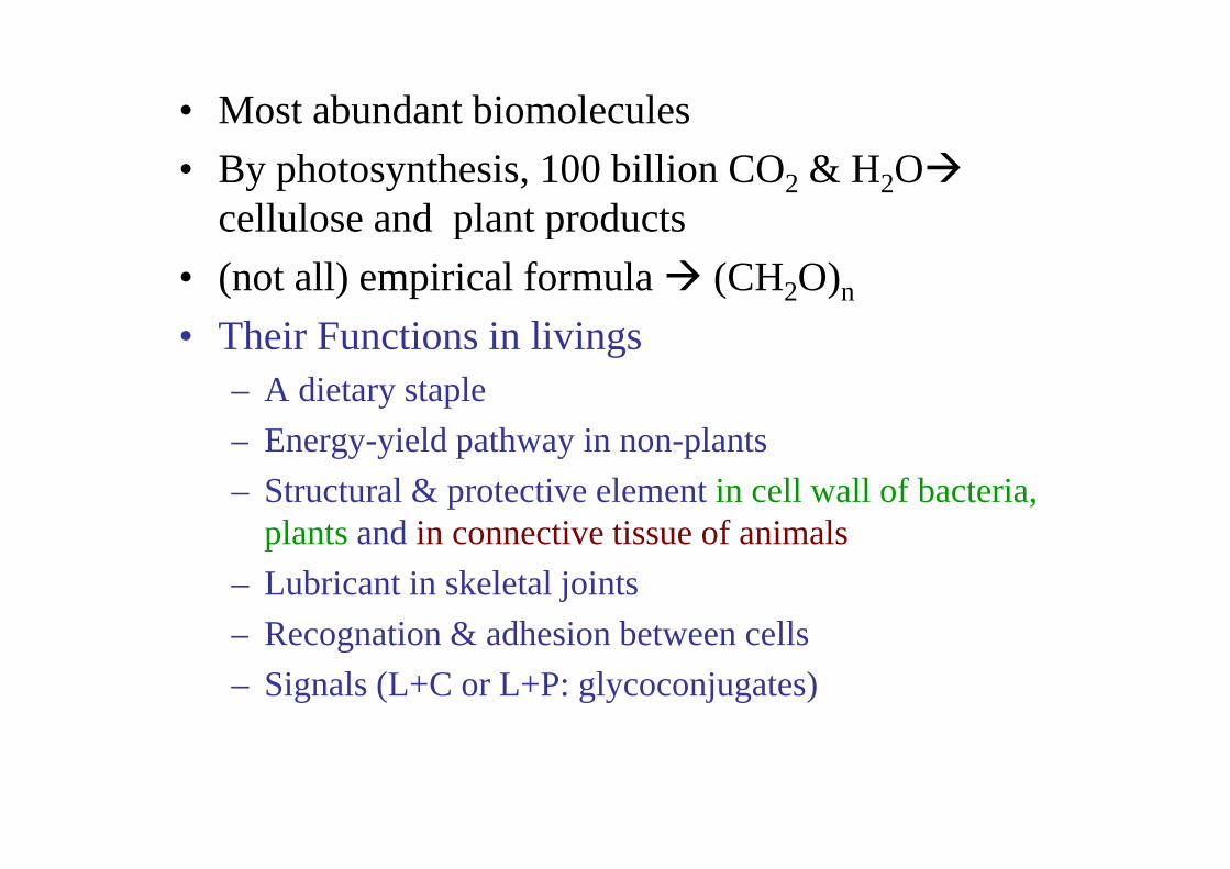
• Most abundant biomolecules
• By photosynthesis, 100 billion CO2 & H2O�
cellulose and plant productscellulose and plant products
• (not all) empirical formula � (CH2O)n• Their Functions in livings• Their Functions in livings
– A dietary staple
– Energy-yield pathway in non-plants– Energy-yield pathway in non-plants
– Structural & protective element in cell wall of bacteria, plantsand in connective tissue of animalsplantsand in connective tissue of animals
– Lubricant in skeletal joints
– Recognation & adhesion between cells– Recognation & adhesion between cells
– Signals (L+C or L+P: glycoconjugates)
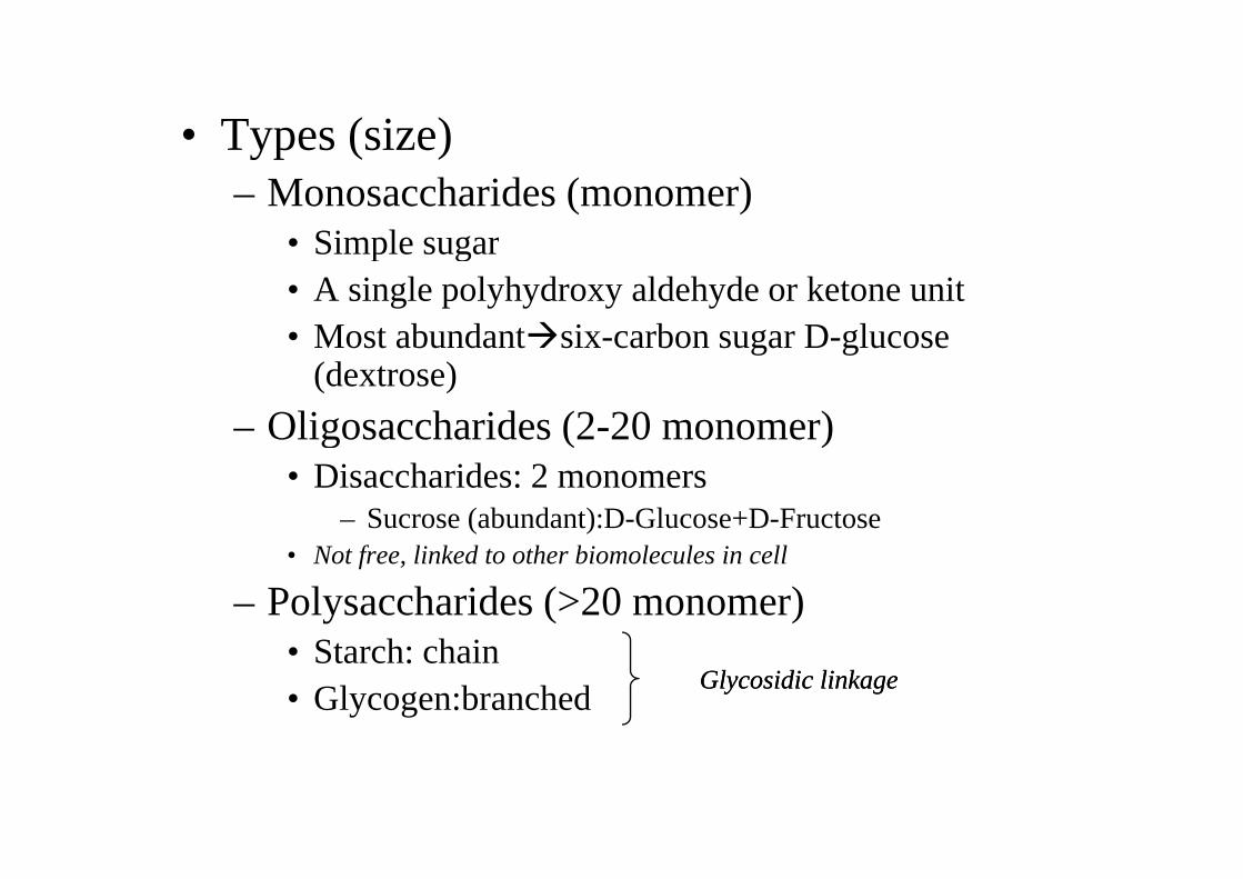
• Types (size)• Types (size)– Monosaccharides (monomer)
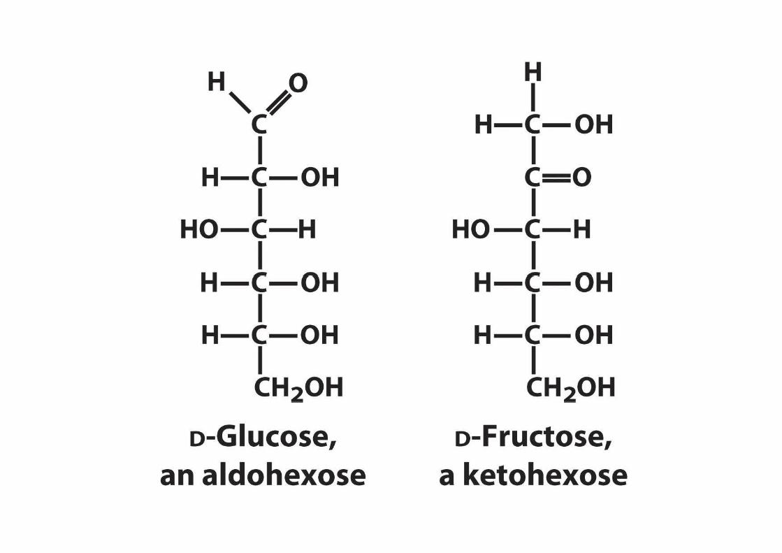
• Simple sugar• Simple sugar• A single polyhydroxy aldehyde or ketone unit• Most abundant�six-carbon sugar D-glucose • Most abundant�six-carbon sugar D-glucose
(dextrose)
– Oligosaccharides (2-20 monomer)– Oligosaccharides (2-20 monomer)• Disaccharides: 2 monomers
– Sucrose (abundant):D-Glucose+D-Fructose• Not free, linked to other biomolecules in cell
– Polysaccharides (>20 monomer)• Starch: chain• Starch: chain• Glycogen:branched
Glycosidic linkageGlycosidic linkage
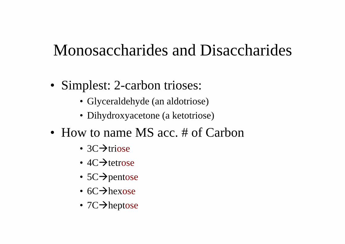
Monosaccharides and Disaccharides
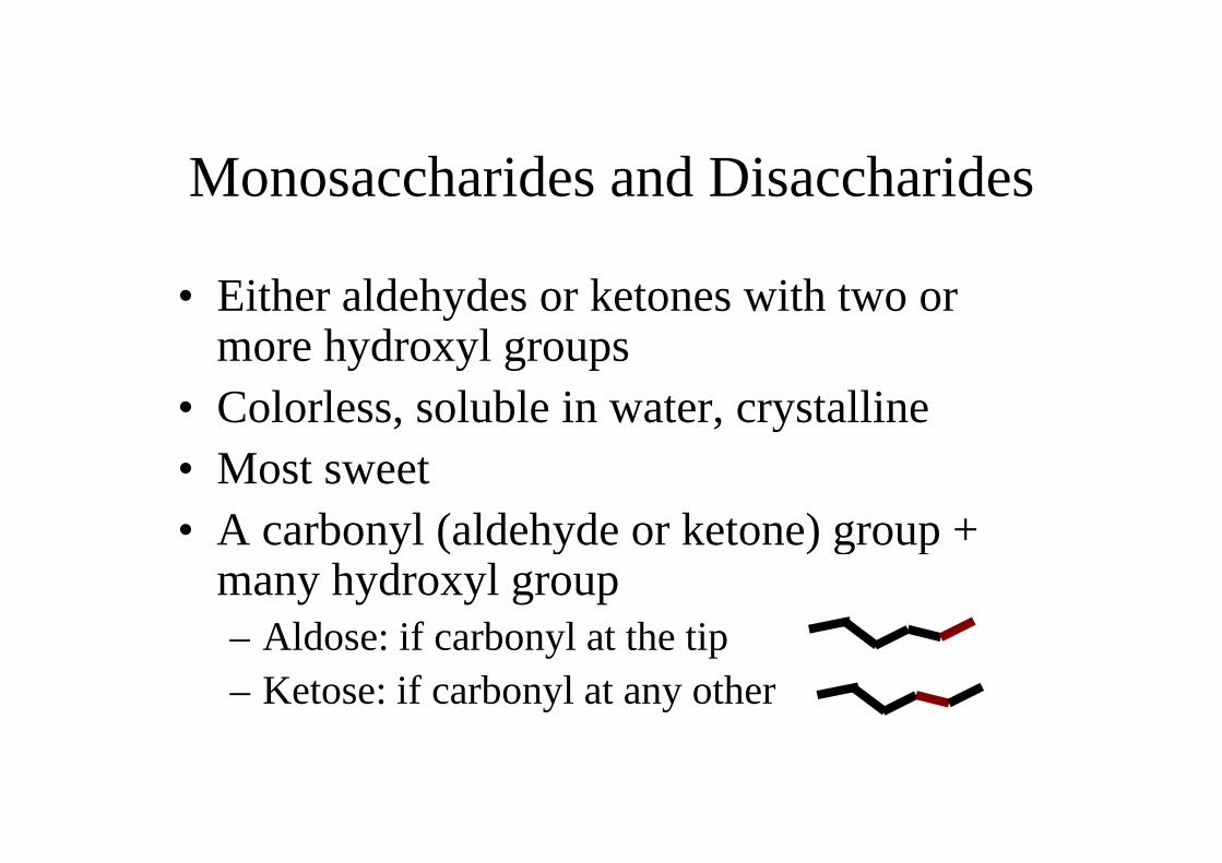
• Either aldehydes or ketones with two or more hydroxyl groupsmore hydroxyl groups
• Colorless, soluble in water, crystalline• Colorless, soluble in water, crystalline• Most sweet• A carbonyl (aldehyde or ketone) group + • A carbonyl (aldehyde or ketone) group +
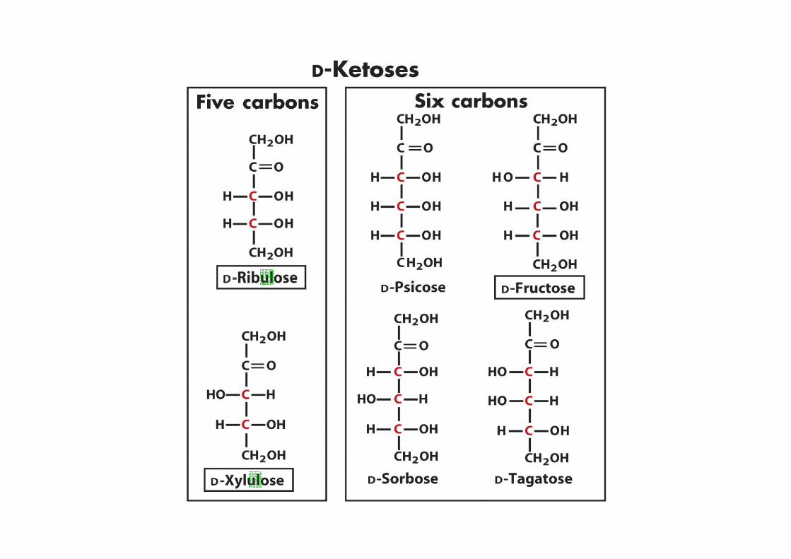
many hydroxyl group– Aldose: if carbonyl at the tip– Aldose: if carbonyl at the tip– Ketose: if carbonyl at any other
Monosaccharides and Disaccharides
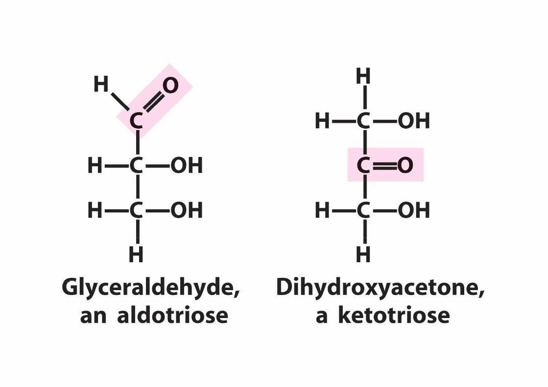
• Simplest: 2-carbon trioses:• Glyceraldehyde (an aldotriose)
• Dihydroxyacetone (a ketotriose)
• How to name MS acc. # of Carbon• 3C�triose• 3C�triose
• 4C�tetrose
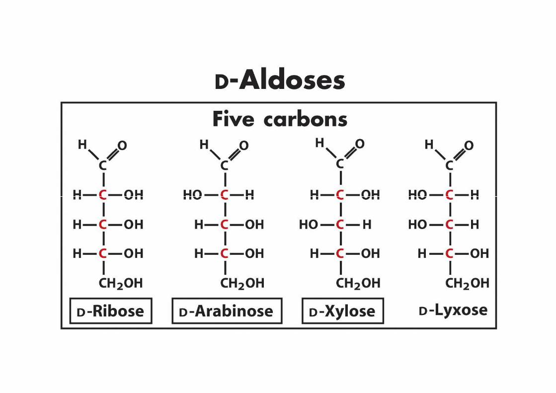
• 5C�pentose• 5C�pentose
• 6C�hexose
• 7C�heptose• 7C�heptose
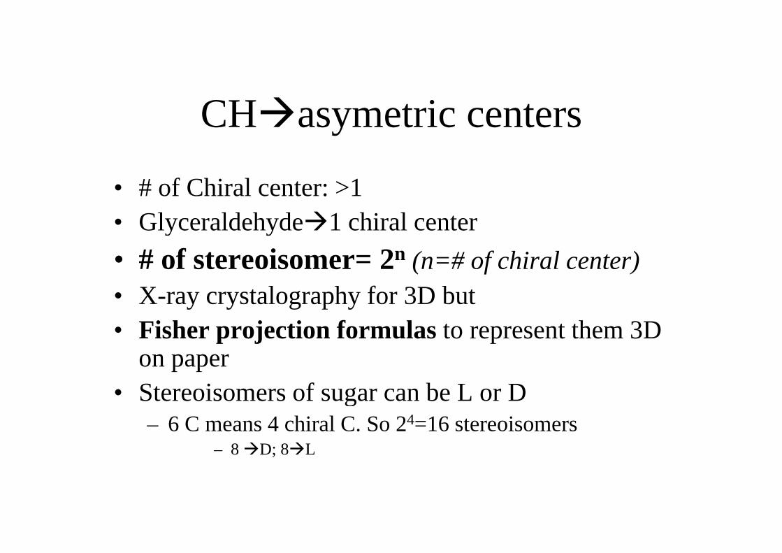
CH�asymetric centers
• # of Chiral center: >1 • Glyceraldehyde�1 chiral center• Glyceraldehyde�1 chiral center
• # of stereoisomer= 2n (n=# of chiral center)• X-ray crystalography for 3D but • Fisher projection formulas to represent them 3D • Fisher projection formulas to represent them 3D
on paper• Stereoisomers of sugar can be L or D• Stereoisomers of sugar can be L or D
– 6 C means 4 chiral C. So 24=16 stereoisomers– 8 �D; 8�L
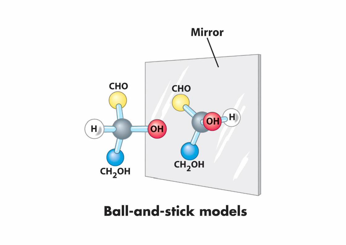
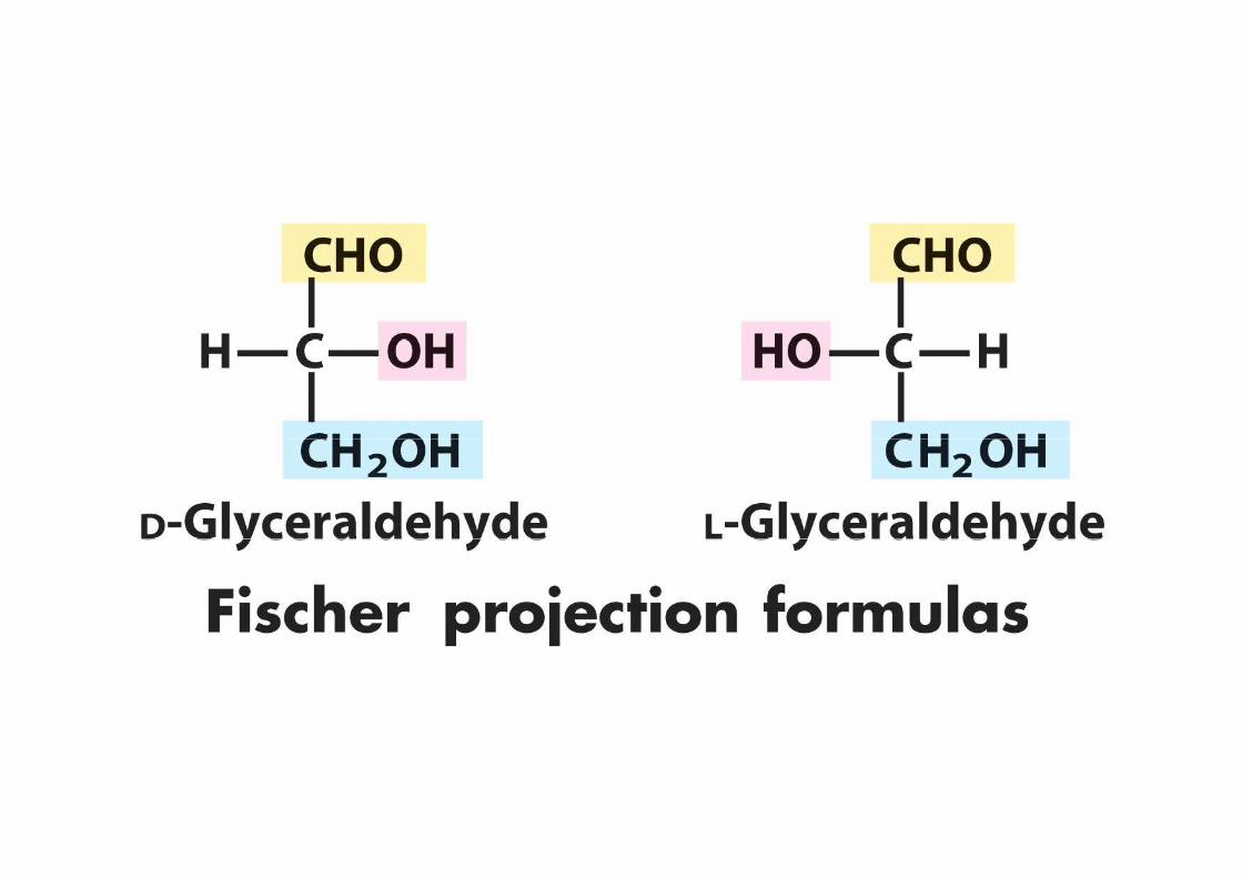
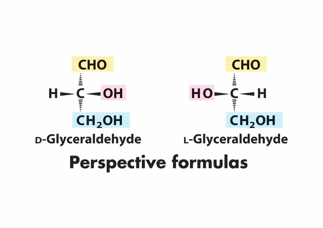
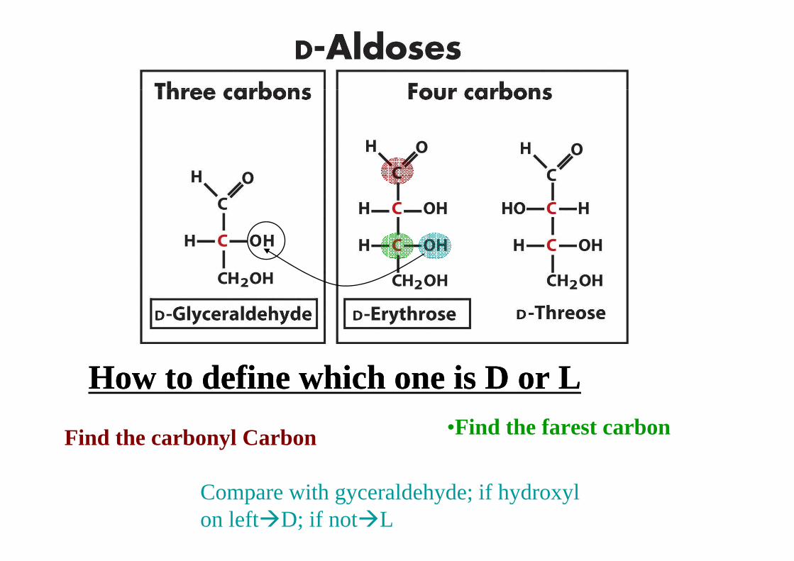
How to define which one is D or LHow to define which one is D or L
Find the carbonyl Carbon •Find the farest carbon
Compare with gyceraldehyde; if hydroxyl on left�D; if not�L
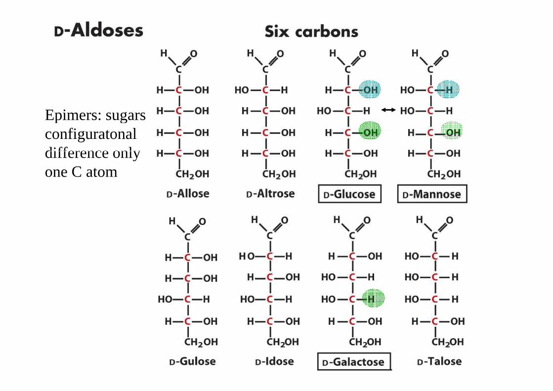
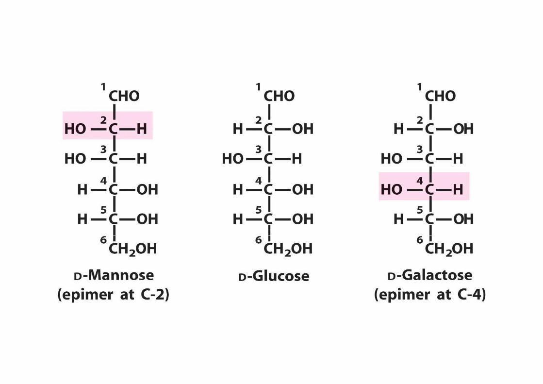
Epimers: sugars Epimers: sugars configuratonal difference only difference only one C atom
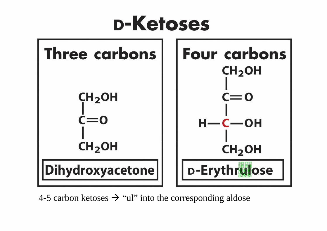
4-5 carbon ketoses � “ul” into the corresponding aldose4-5 carbon ketoses � “ul” into the corresponding aldose
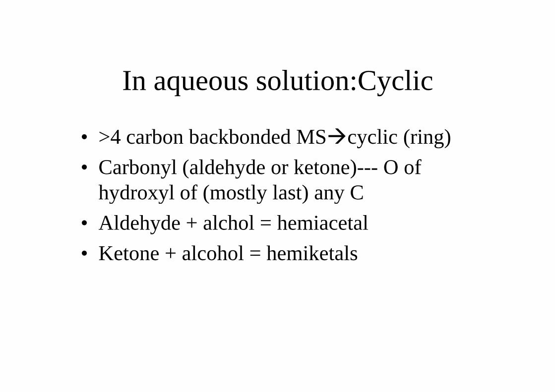
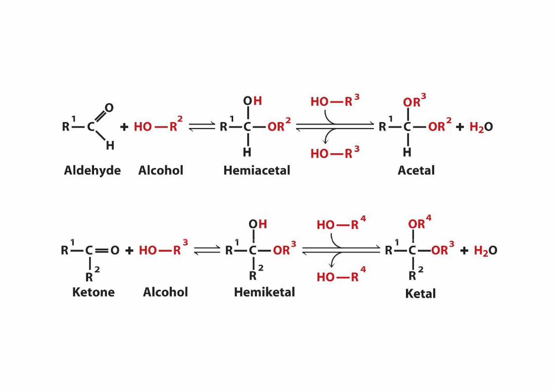
In aqueous solution:Cyclic
• >4 carbon backbonded MS�cyclic (ring)
• Carbonyl (aldehyde or ketone)--- O of hydroxyl of (mostly last) any Chydroxyl of (mostly last) any C
• Aldehyde + alchol = hemiacetal
• Ketone + alcohol = hemiketals
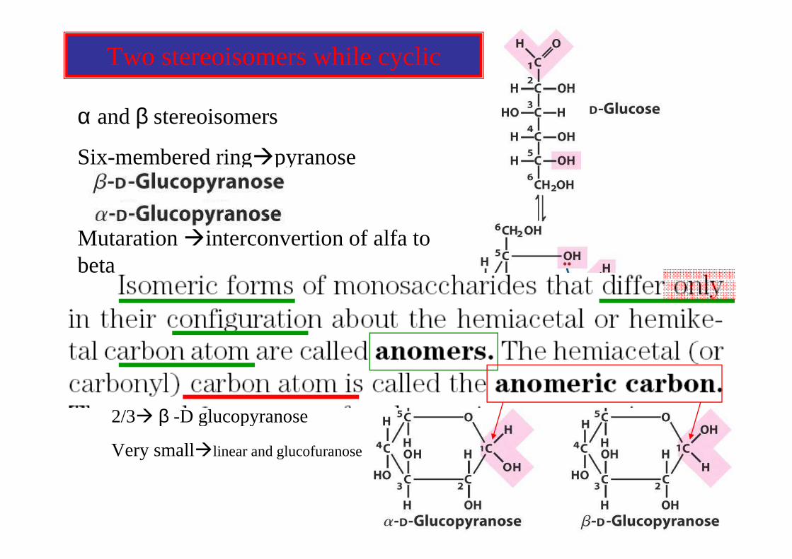
Two stereoisomers while cyclic
α and β stereoisomers
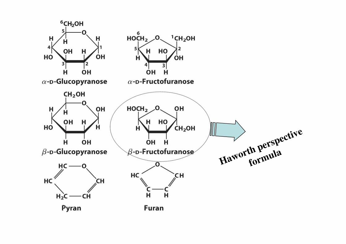
Six-membered ring�pyranoseSix-membered ring�pyranose
Mutaration �interconvertion of alfa to beta
Five-membered� furanose
In normal aquoes soltionIn normal aquoes soltion
1/3� α-D glucopyranose
2/3� β -D glucopyranose2/3� β -D glucopyranose
Very small�linear and glucofuranose
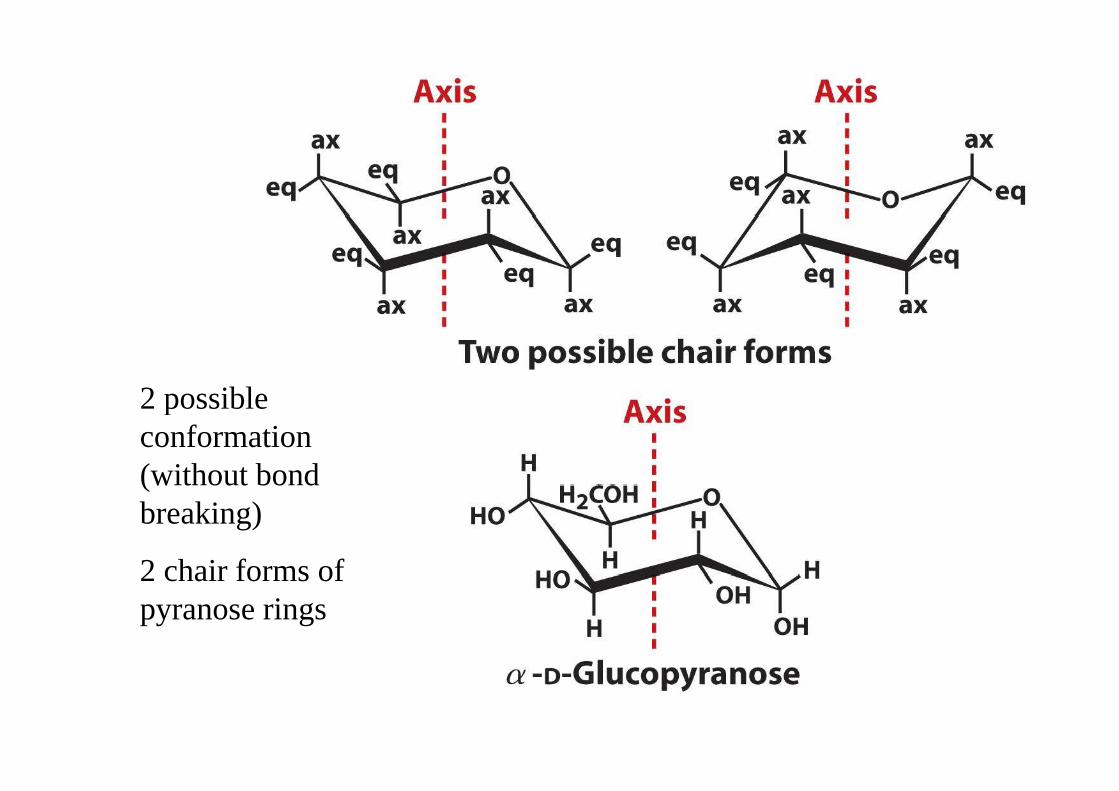
2 possible 2 possible conformation (without bond (without bond breaking)
2 chair forms of 2 chair forms of pyranose rings
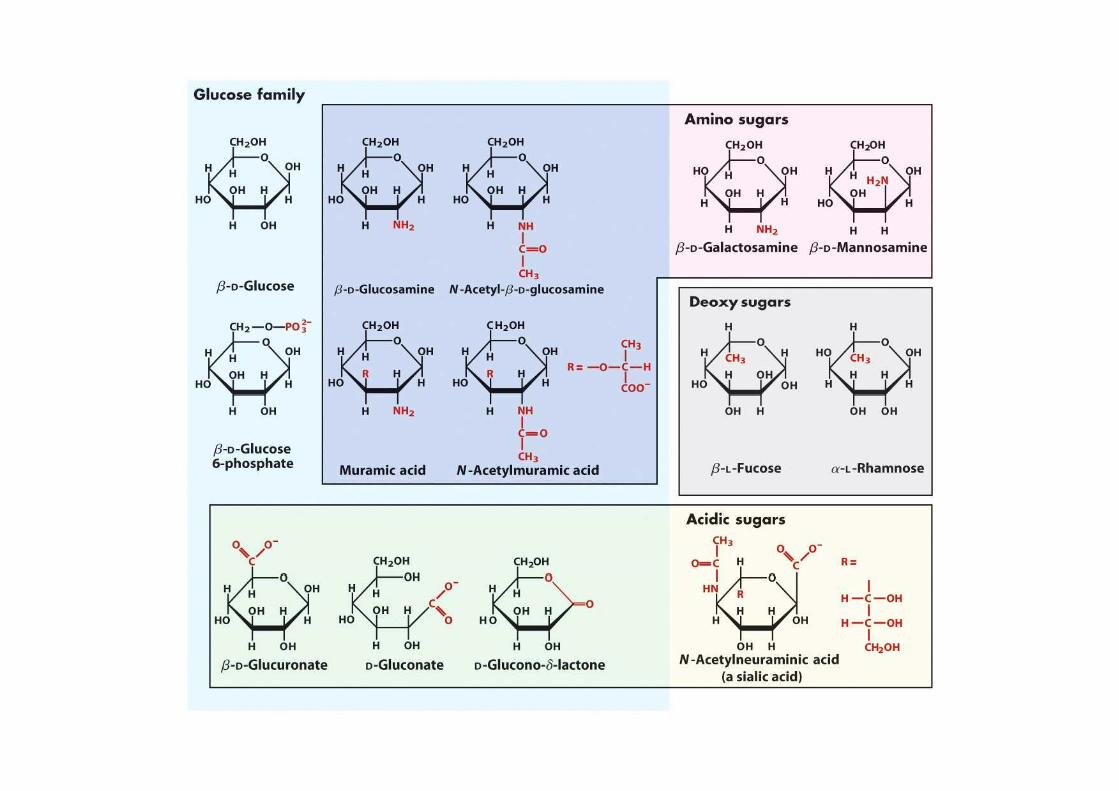
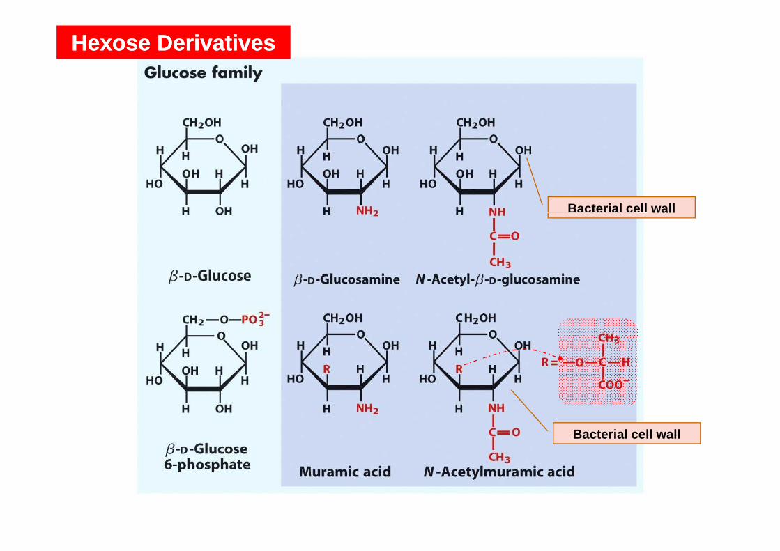
Hexose DerivativesHexose Derivatives
Bacterial cell wallBacterial cell wall
Bacterial cell wall
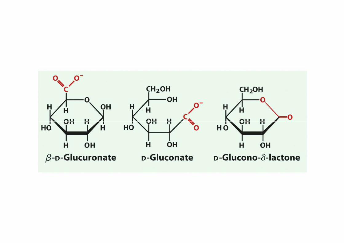
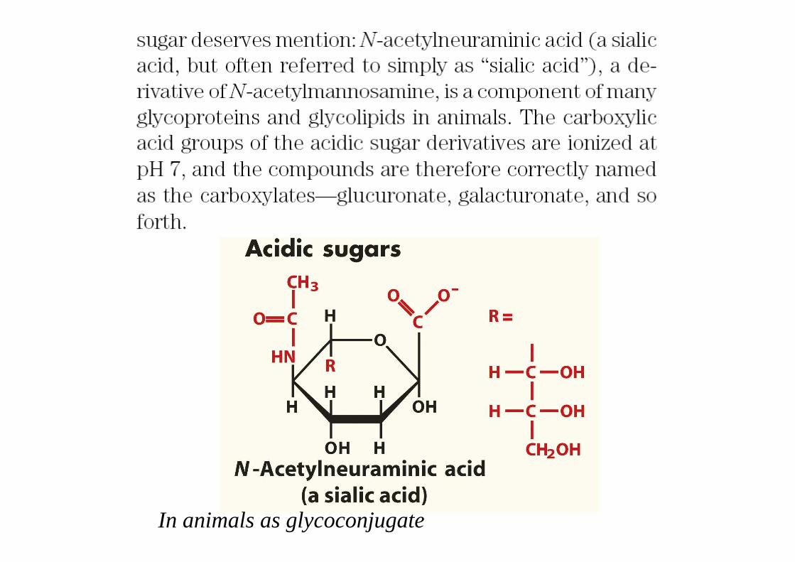
In animals as glycoconjugate
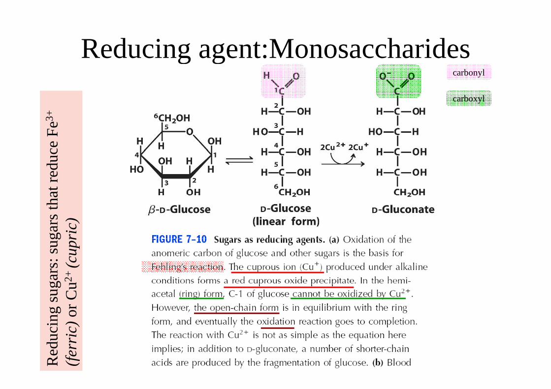
Reducing agent:MonosaccharidesReducing agent:Monosaccharidescarbonyl
carboxyl
Red
ucin
g su
gars
: sug
ars
that
red
uce
Fe
3+
R
educ
ing
suga
rs: s
ugar
s th
at r
educ
e F
eR
educ
ing
suga
rs: s
ugar
s th
at r
educ
e F
e(c
upri
c)R
educ
ing
suga
rs: s
ugar
s th
at r
educ
e F
eor
Cu2
+ (c
upri
c)R
educ
ing
suga
rs: s
ugar
s th
at r
educ
e F
e(f
erri
c)or
Cu
Red
ucin
g su
gars
: sug
ars
that
red
uce
Fe
(fer
ric)
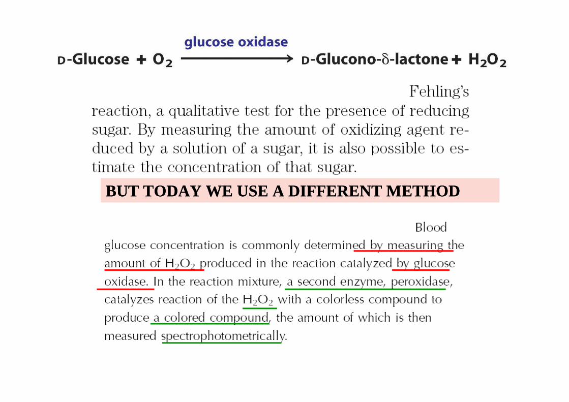
BUT TODAY WE USE A DIFFERENT METHODBUT TODAY WE USE A DIFFERENT METHOD
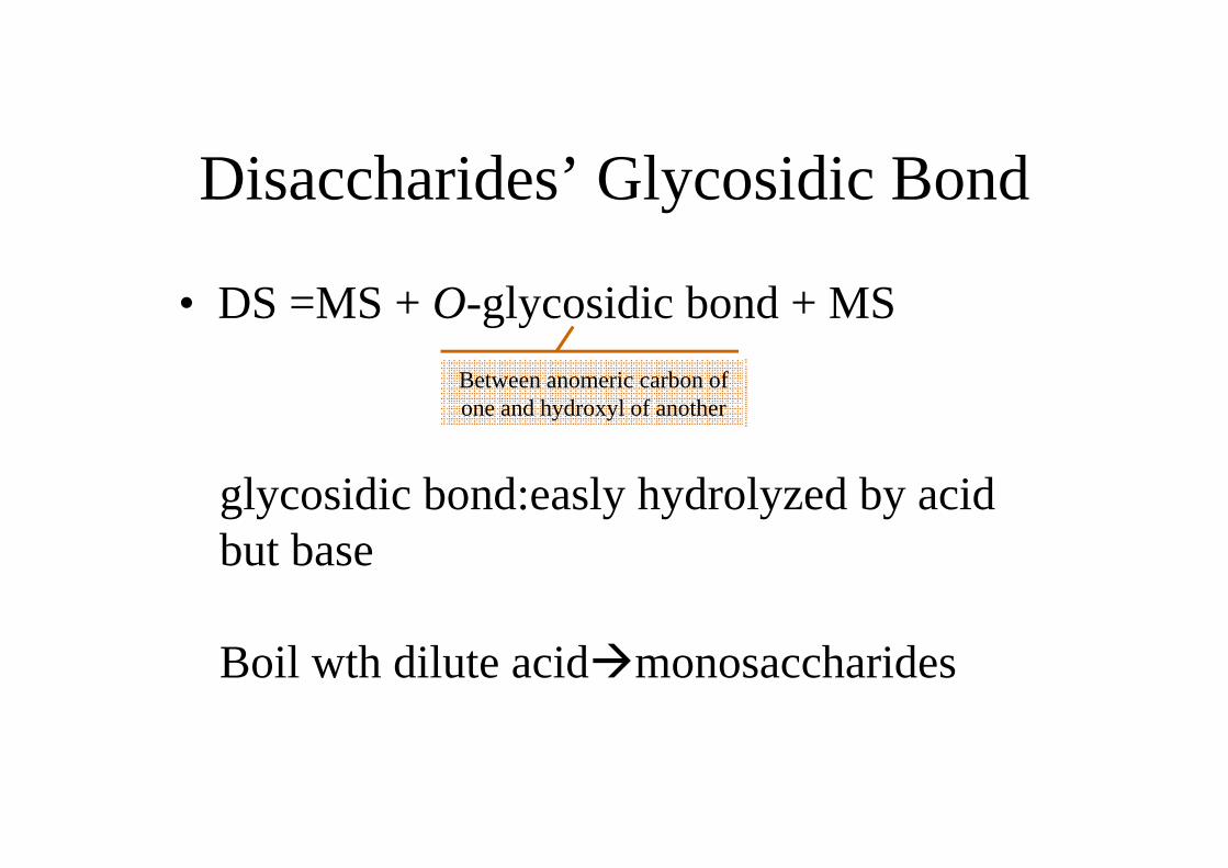
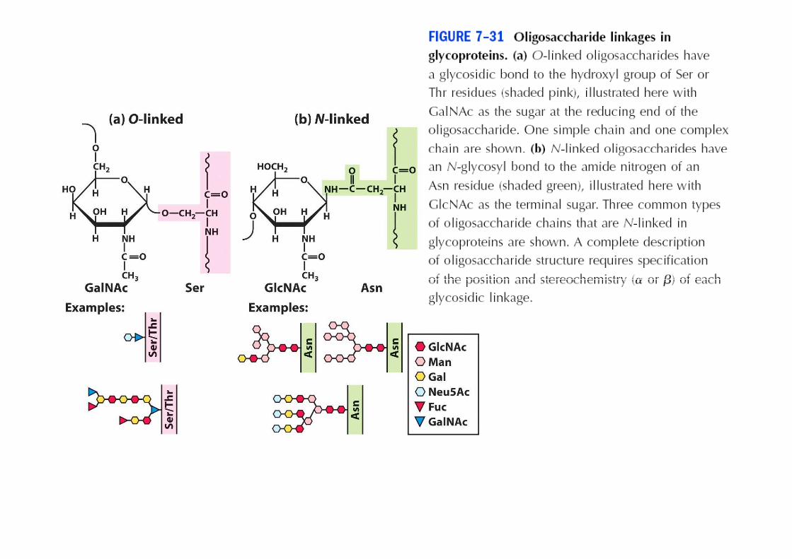
Disaccharides’ Glycosidic Bond
• DS =MS + O-glycosidic bond + MS
Between anomeric carbon of one and hydroxyl of another
glycosidic bond:easly hydrolyzed by acid but basebut base
Boil wth dilute acid�monosaccharides
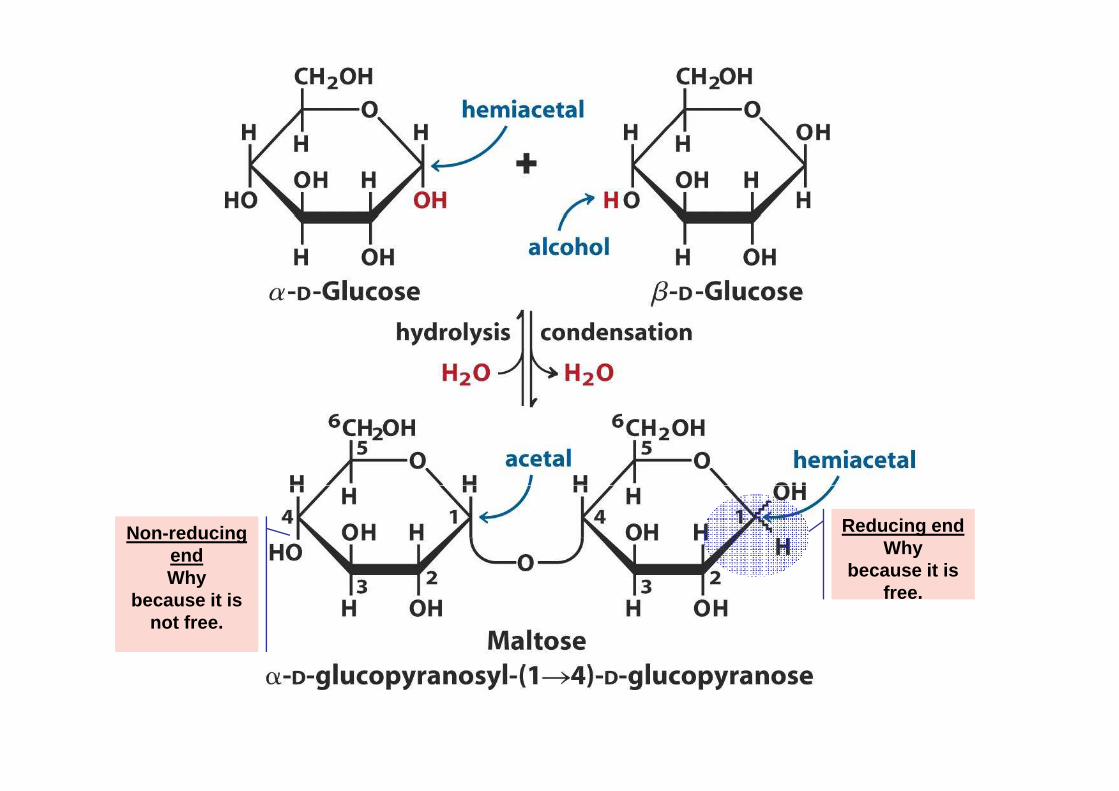
Reducing endWhy
because it is
Non-reducing endWhy because it is
free.Why
because it is not free.
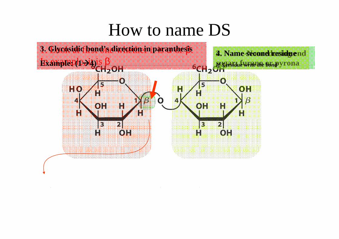
How to name DSHow to name DS1. Look at first one whether it is α or β. In example, it is β
2. Name-Nonreducing end sugar; furano or pyrona
3. Glycosidic bond’s direction in paranthesis
Example: (1����4)4. Name second residueIf 3.residue write the bondIn example, it is β sugar; furano or pyrona Example: (1����4) If 3.residue write the bond
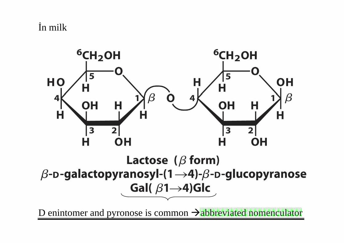
İn milk
D enintomer and pyronose is common �abbreviated nomenculator
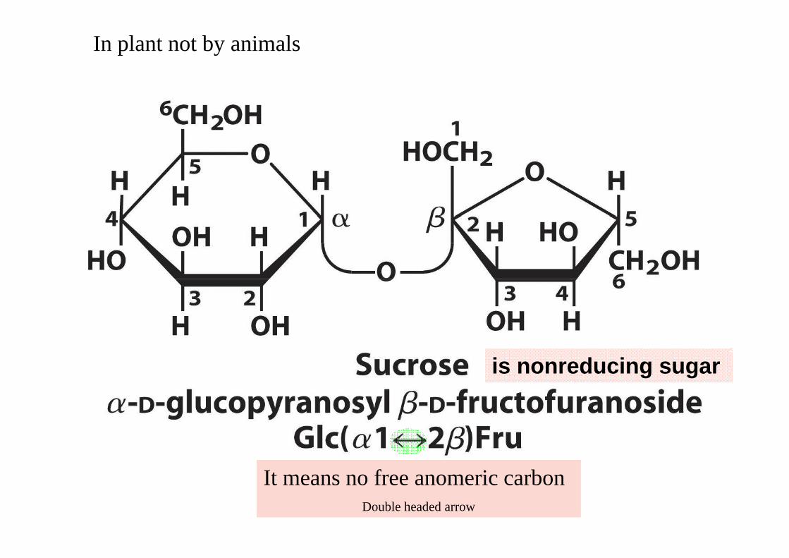
In plant not by animals
is nonreducing sugar
It means no free anomeric carbonIt means no free anomeric carbonDouble headed arrow
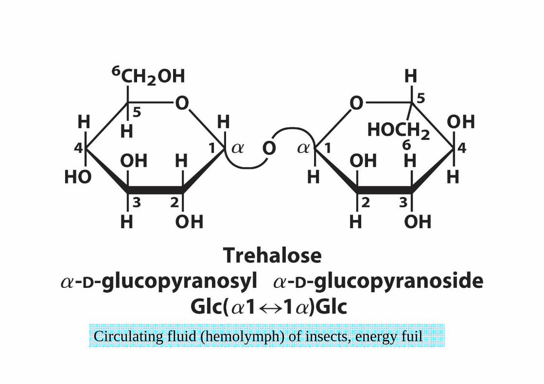
Circulating fluid (hemolymph) of insects, energy fuil
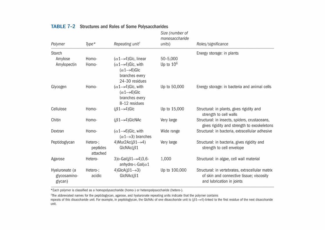
Polysaccharides (glycans)
• Classification– identity of their recurring MS
– Size– Size
– Branch
– Types of bonds– Types of bonds
Polysaccharides (glycans)
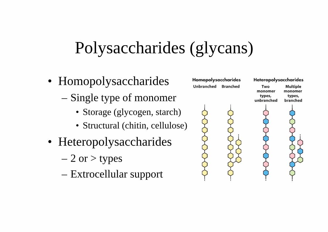
• Homopolysaccharides– Single type of monomer
• Storage (glycogen, starch)• Storage (glycogen, starch)
• Structural (chitin, cellulose)
• Heteropolysaccharides• Heteropolysaccharides– 2 or > types
– Extrocellular support– Extrocellular support
Homopolysaccharides as energy sourceHomopolysaccharides as energy source
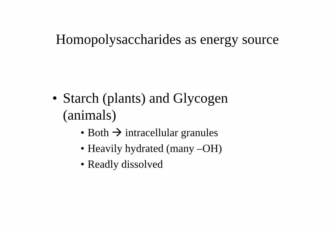
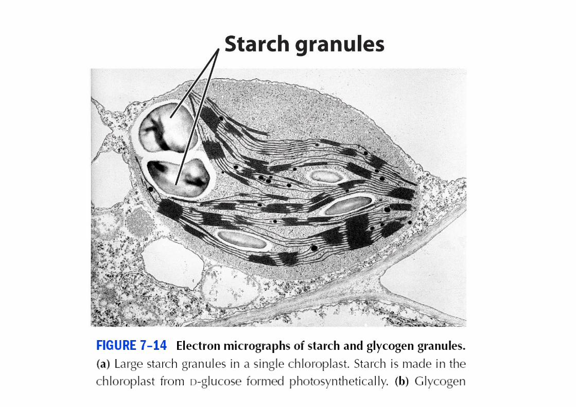
• Starch (plants) and Glycogen • Starch (plants) and Glycogen (animals)(animals)
• Both � intracellular granules
• Heavily hydrated (many –OH)• Heavily hydrated (many –OH)
• Readly dissolved• Readly dissolved
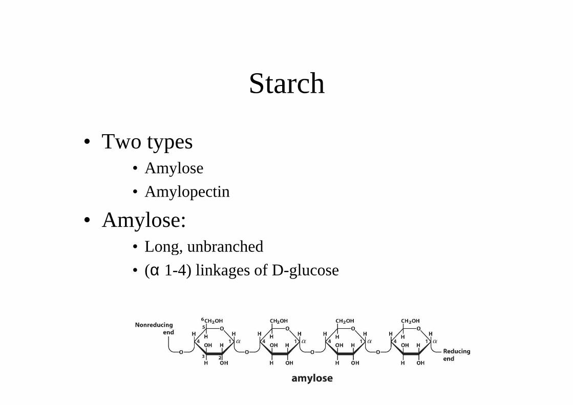
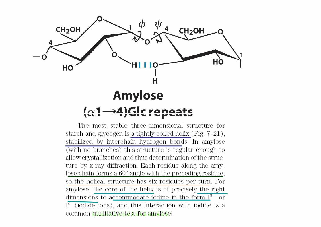
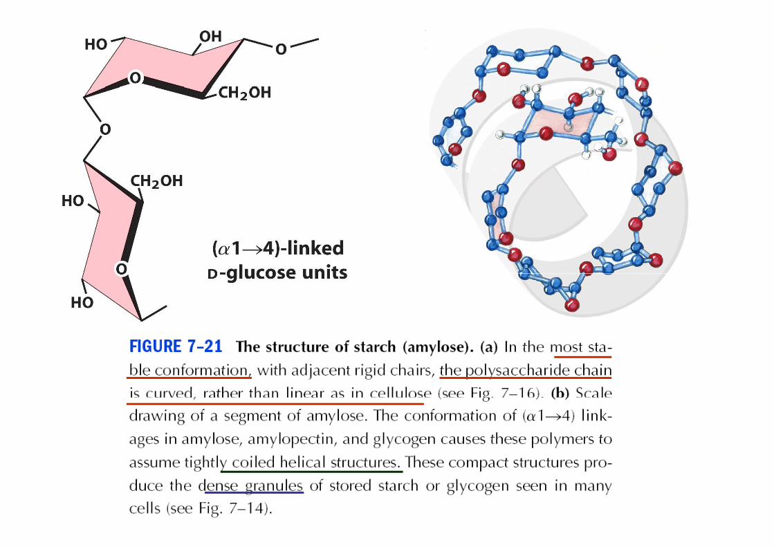
Starch
• Two types• Amylose
• Amylopectin
• Amylose:• Long, unbranched• Long, unbranched
• (α 1-4) linkages of D-glucose
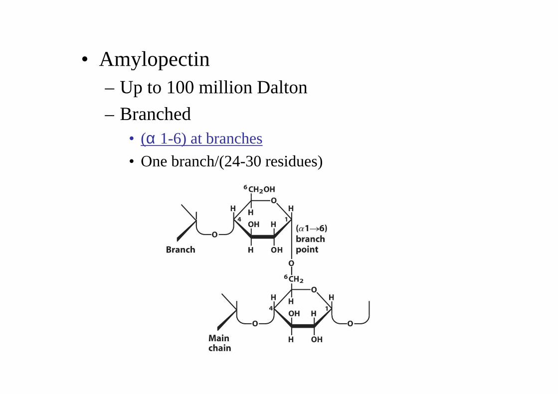
• Amylopectin• Amylopectin– Up to 100 million Dalton
– Branched– Branched• (α 1-6) at branches
• One branch/(24-30 residues)• One branch/(24-30 residues)
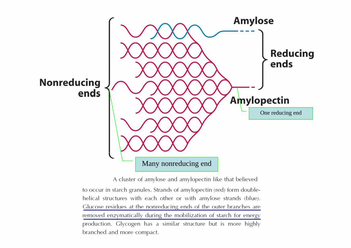
One reducing end
Many nonreducing end
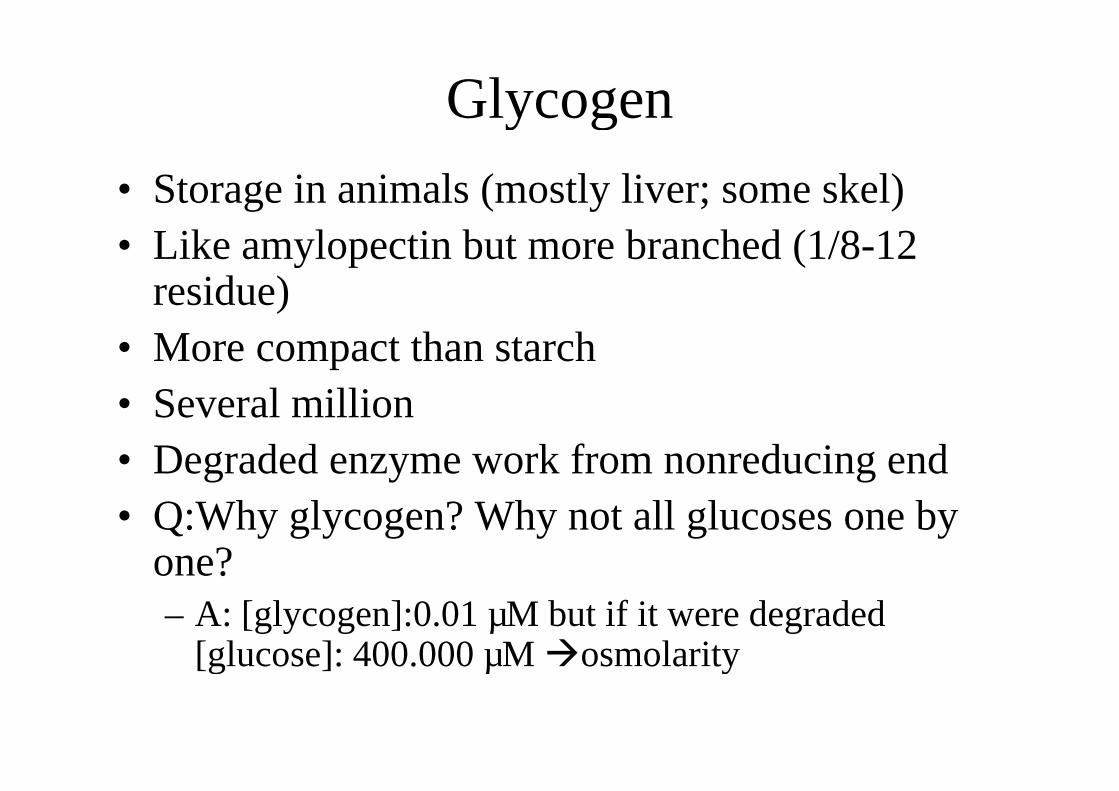
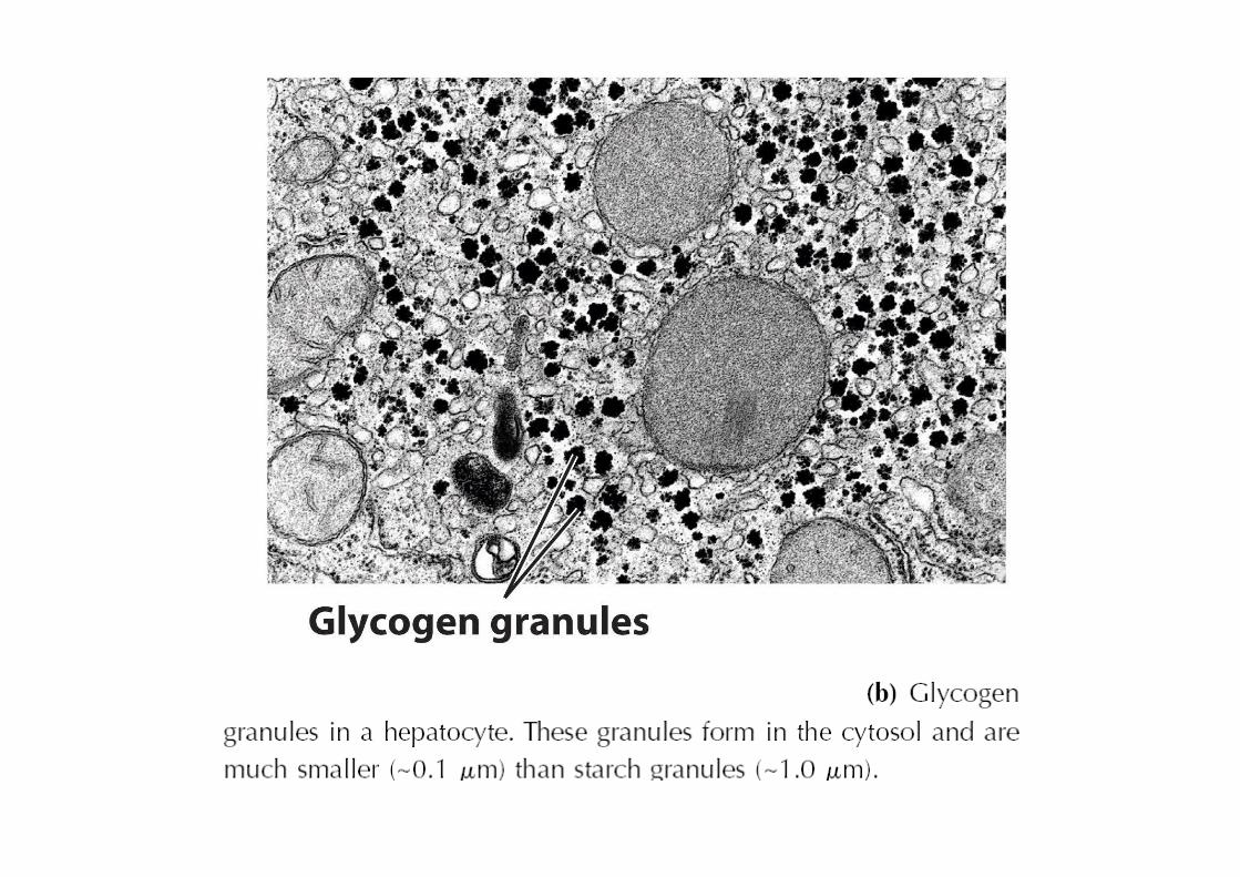
GlycogenGlycogen
• Storage in animals (mostly liver; some skel)• Like amylopectin but more branched (1/8-12
residue)residue)• More compact than starch• Several million• Several million• Degraded enzyme work from nonreducing end• Q:Why glycogen? Why not all glucoses one by
one?one?– A: [glycogen]:0.01 µM but if it were degraded
[glucose]: 400.000 µM �osmolarity[glucose]: 400.000 µM �osmolarity
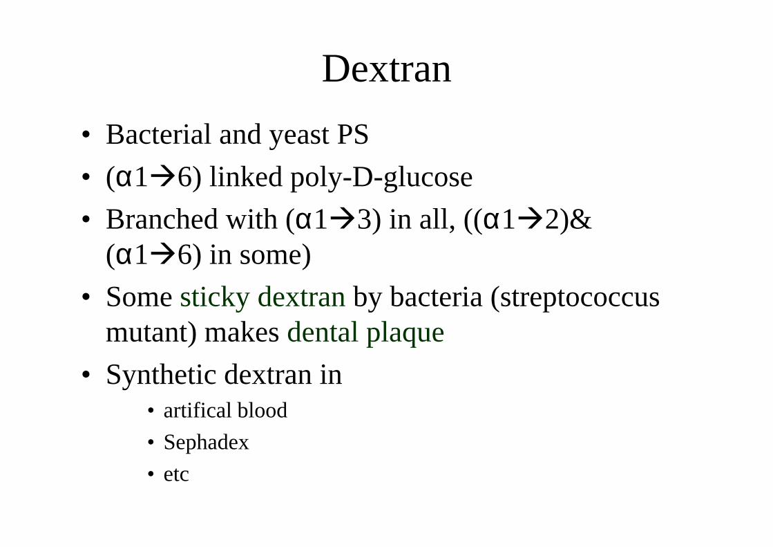
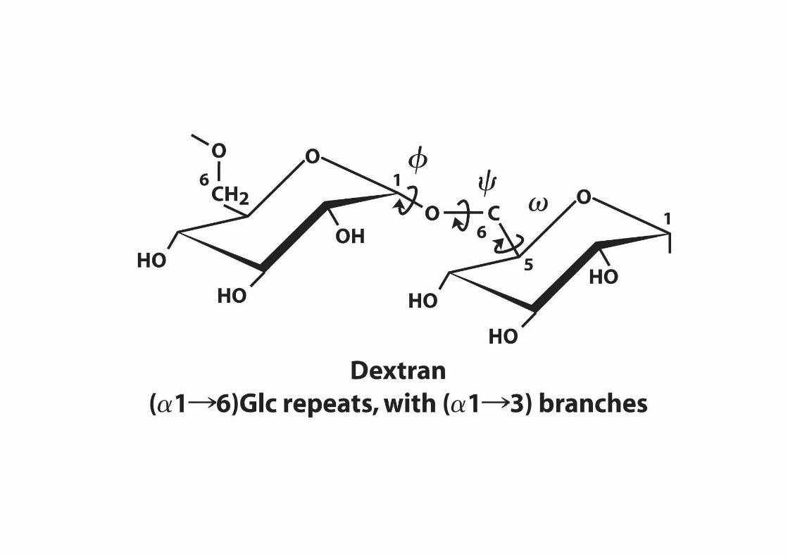
DextranDextran
• Bacterial and yeast PS• Bacterial and yeast PS
• (α1�6) linked poly-D-glucose
• Branched with (α1�3) in all, ((α1�2)& • Branched with (α1�3) in all, ((α1�2)& (α1�6) in some)
• Some sticky dextranby bacteria (streptococcus mutant) makes dental plaquemutant) makes dental plaque
• Synthetic dextran in • artifical blood• artifical blood
• Sephadex
• etc
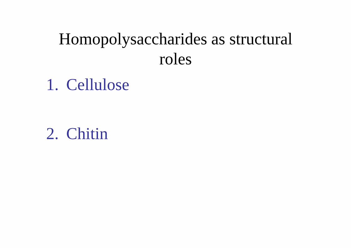
Homopolysaccharides as structural Homopolysaccharides as structural rolesroles
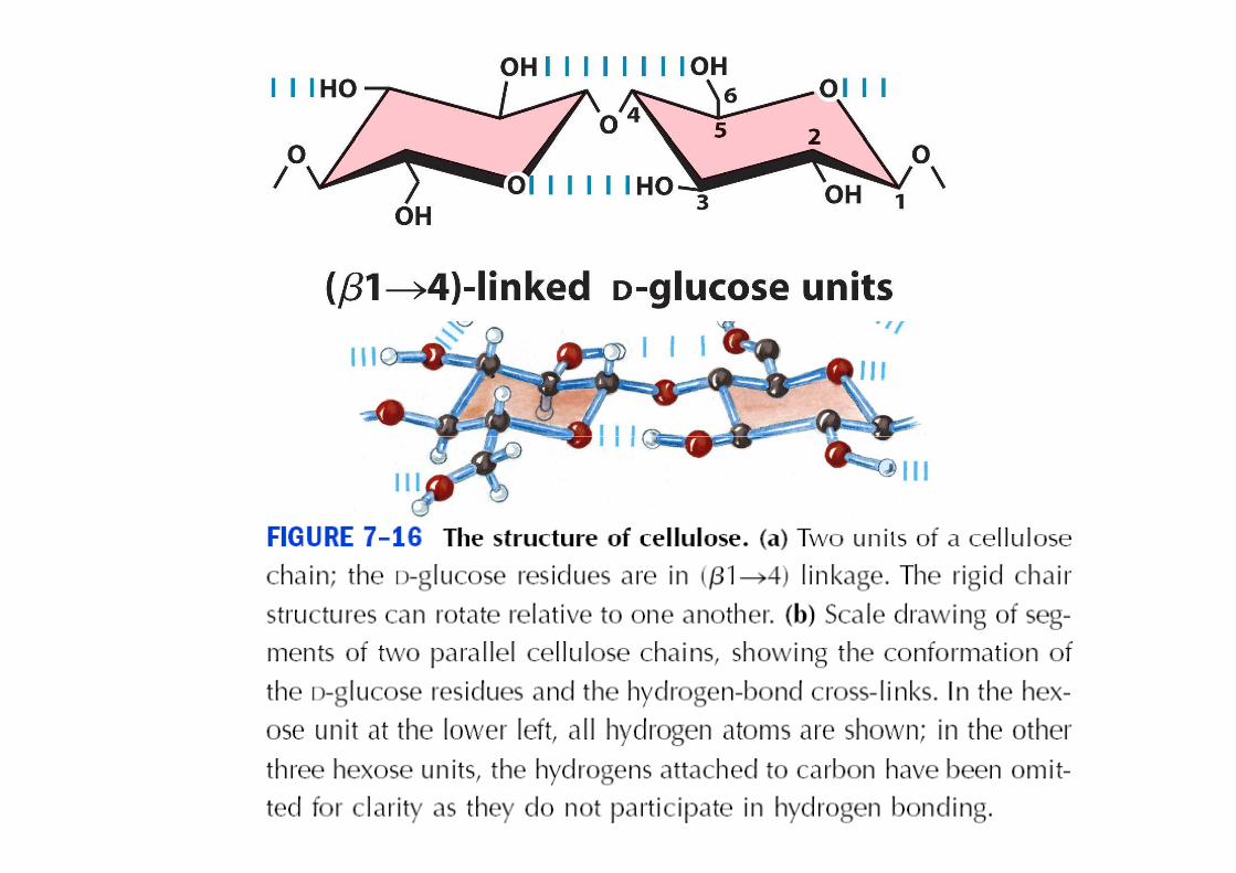
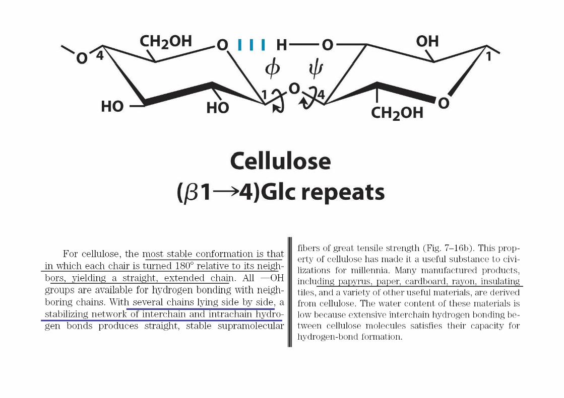
1. Cellulose
2. Chitin
1. Cellulose
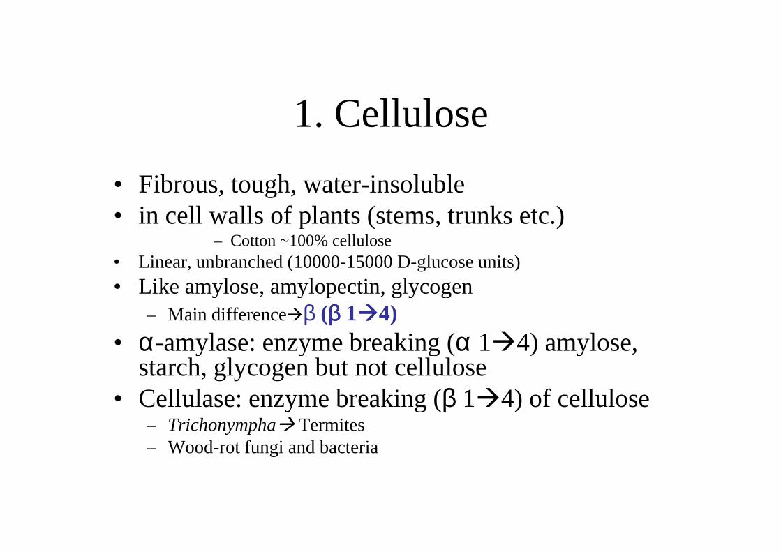
• Fibrous, tough, water-insoluble• in cell walls of plants (stems, trunks etc.)• in cell walls of plants (stems, trunks etc.)
– Cotton ~100% cellulose• Linear, unbranched (10000-15000 D-glucose units)• Like amylose, amylopectin, glycogen• Like amylose, amylopectin, glycogen
– Main difference�β (ββββ 1����4)• α-amylase: enzyme breaking (α 1�4) amylose, • α-amylase: enzyme breaking (α 1�4) amylose,
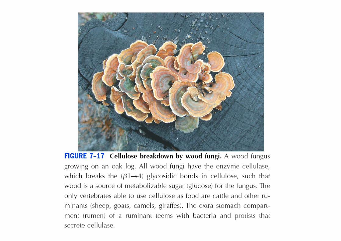
starch, glycogen but not cellulose• Cellulase: enzyme breaking (β 1�4) of cellulose• Cellulase: enzyme breaking (β 1�4) of cellulose
– Trichonympha� Termites– Wood-rot fungi and bacteria
2. Chitin
• Linear with N-acetylglucosamine in β-linkagelinkage
• Exoskeloton of nearly a million species of • Exoskeloton of nearly a million species of arthropodsinsects, lobsters, crabs etc
• Most abondunt PS after cellulose in nature• Most abondunt PS after cellulose in nature
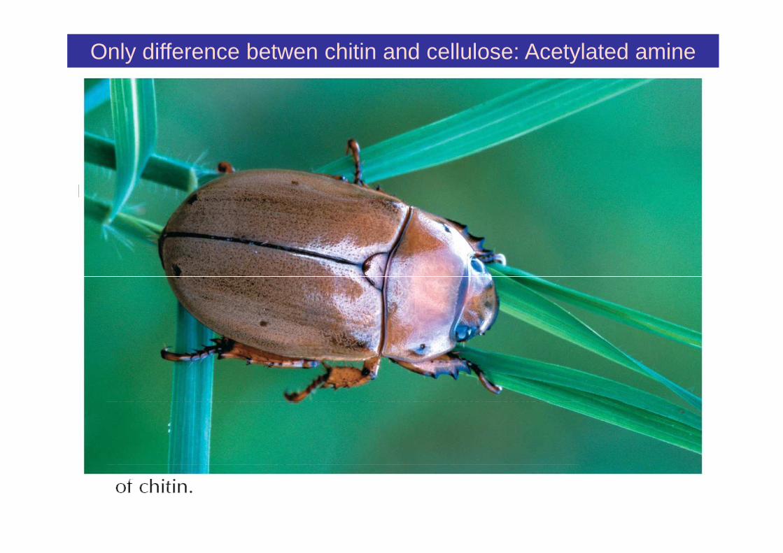
Only difference betwen chitin and cellulose: Acetylated amine
CelluloseCellulose
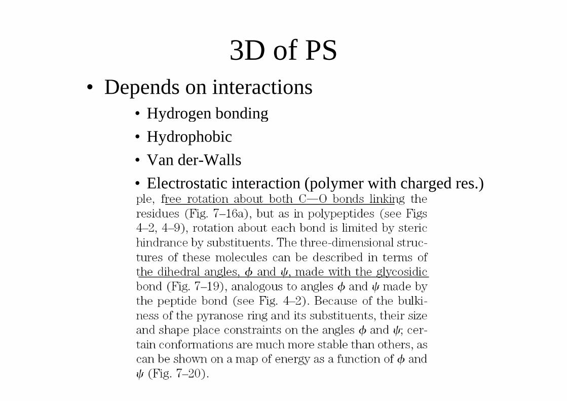
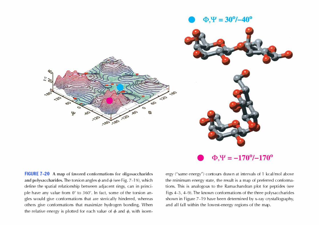
3D of PS3D of PS• Depends on interactions
• Hydrogen bonding• Hydrogen bonding
• Hydrophobic
• Van der-Walls• Van der-Walls
• Electrostatic interaction (polymer with charged res.)
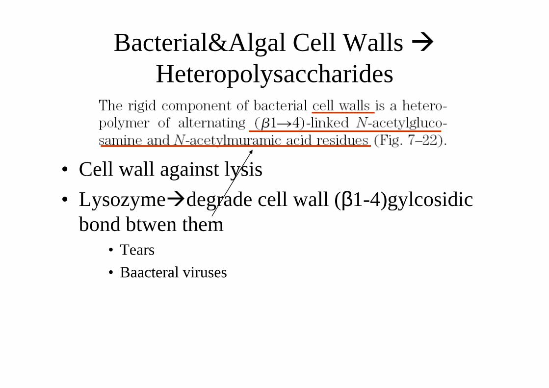
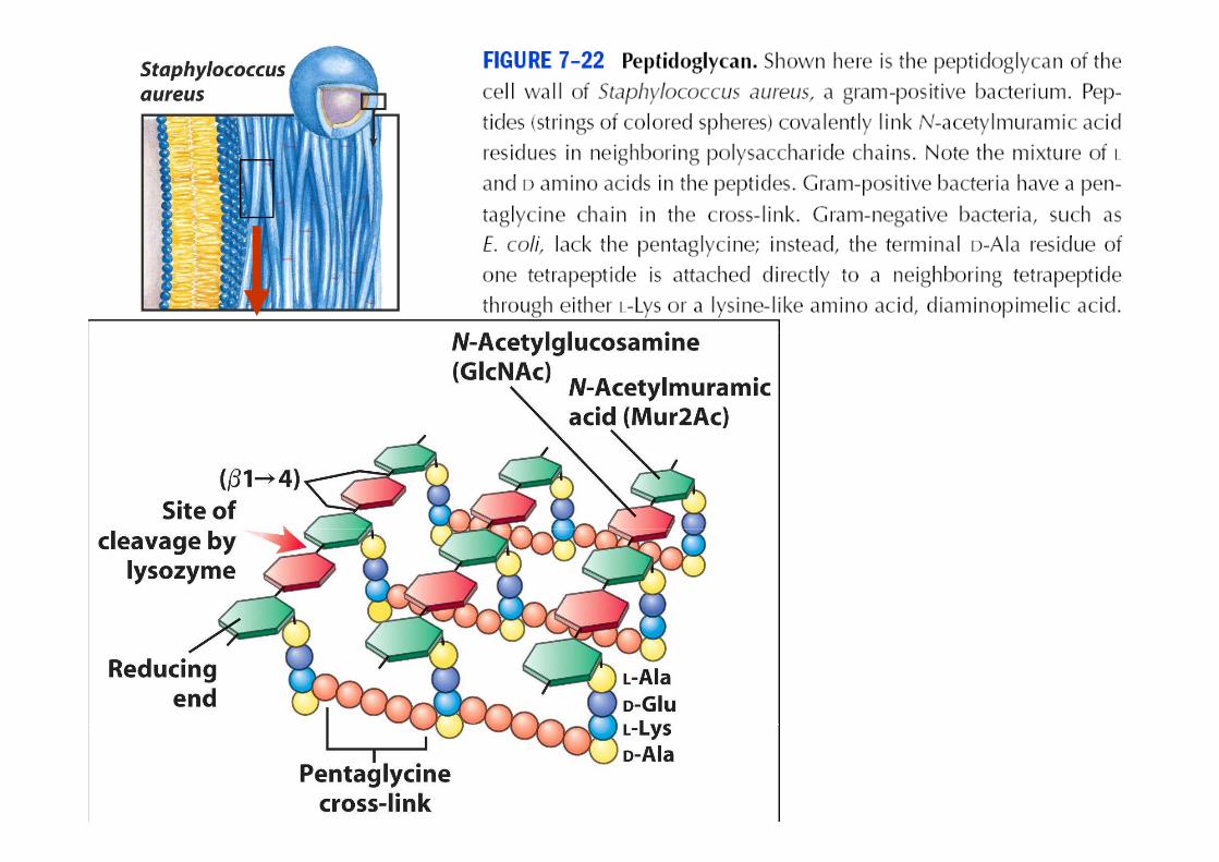
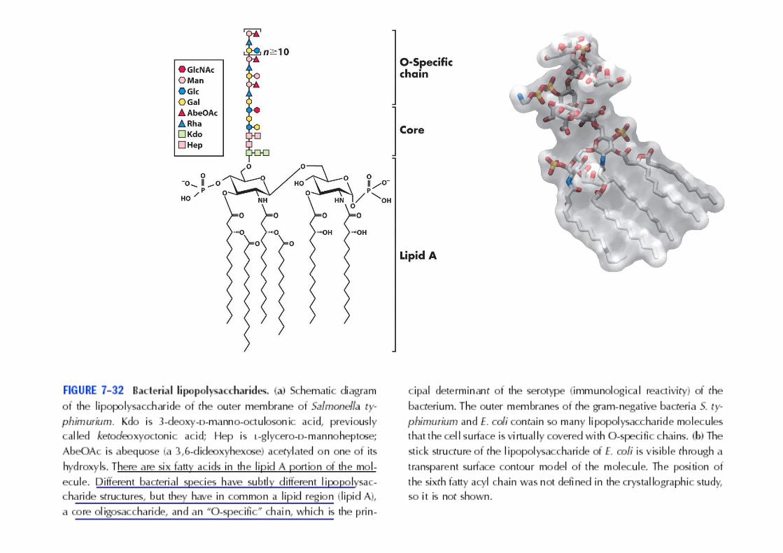
Bacterial&Algal Cell Walls �HeteropolysaccharidesHeteropolysaccharides
• Cell wall against lysis
• Lysozyme�degrade cell wall (β1-4)gylcosidic • Lysozyme�degrade cell wall (β1-4)gylcosidic bond btwen them
• Tears
• Baacteral viruses
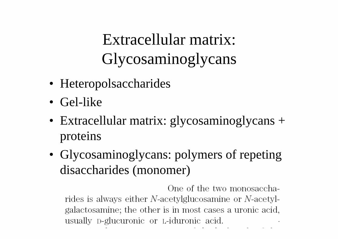
Extracellular matrix: Extracellular matrix: GlycosaminoglycansGlycosaminoglycans
• Heteropolsaccharides
• Gel-like
• Extracellular matrix: glycosaminoglycans + • Extracellular matrix: glycosaminoglycans + proteins
• Glycosaminoglycans: polymers of repeting disaccharides (monomer)disaccharides (monomer)
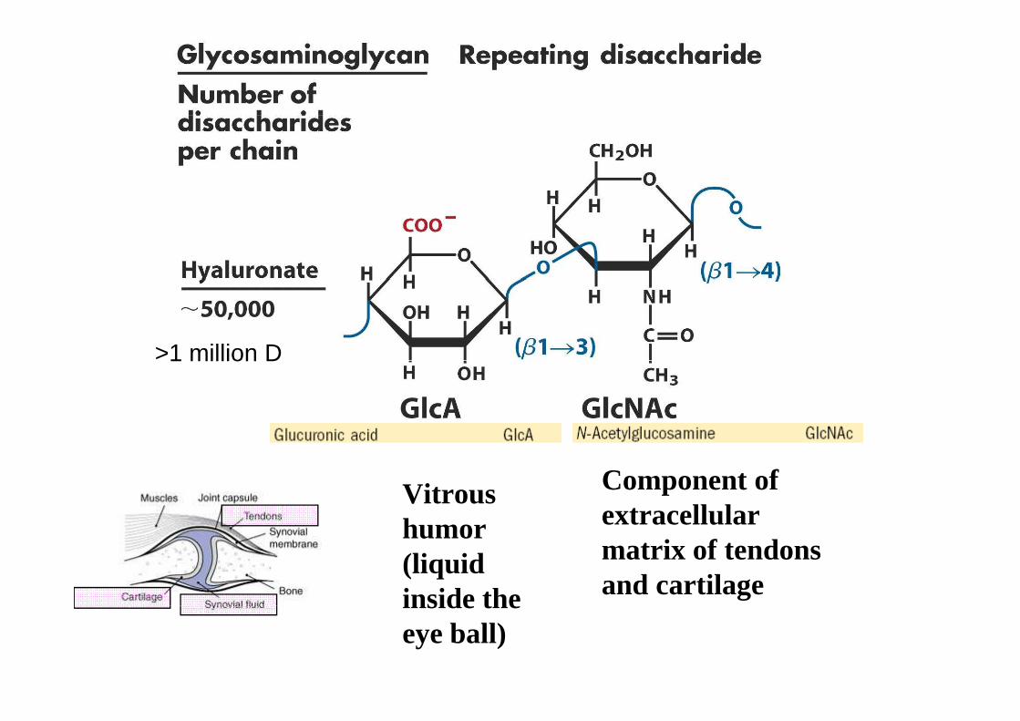
>1 million D>1 million D
Vitrous humor
Component of extracellular humor
(liquid inside the
extracellular matrix of tendons and cartilageinside the
eye ball)
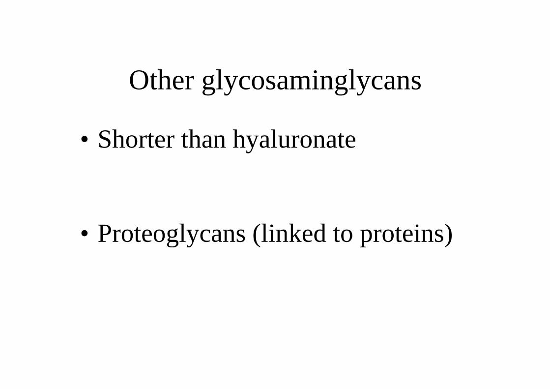
Other glycosaminglycans
• Shorter than hyaluronate• Shorter than hyaluronate
• Proteoglycans (linked to proteins)• Proteoglycans (linked to proteins)
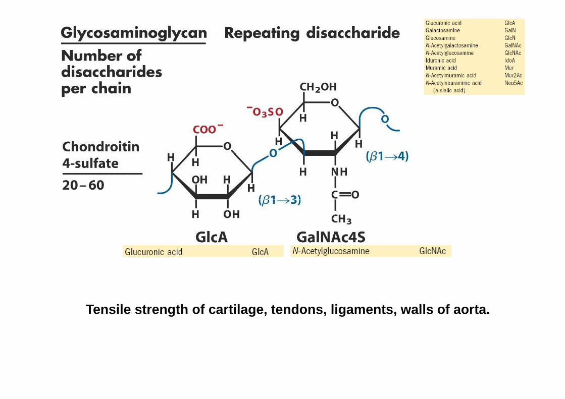
Tensile strength of cartilage, tendons, ligaments, walls of aorta.
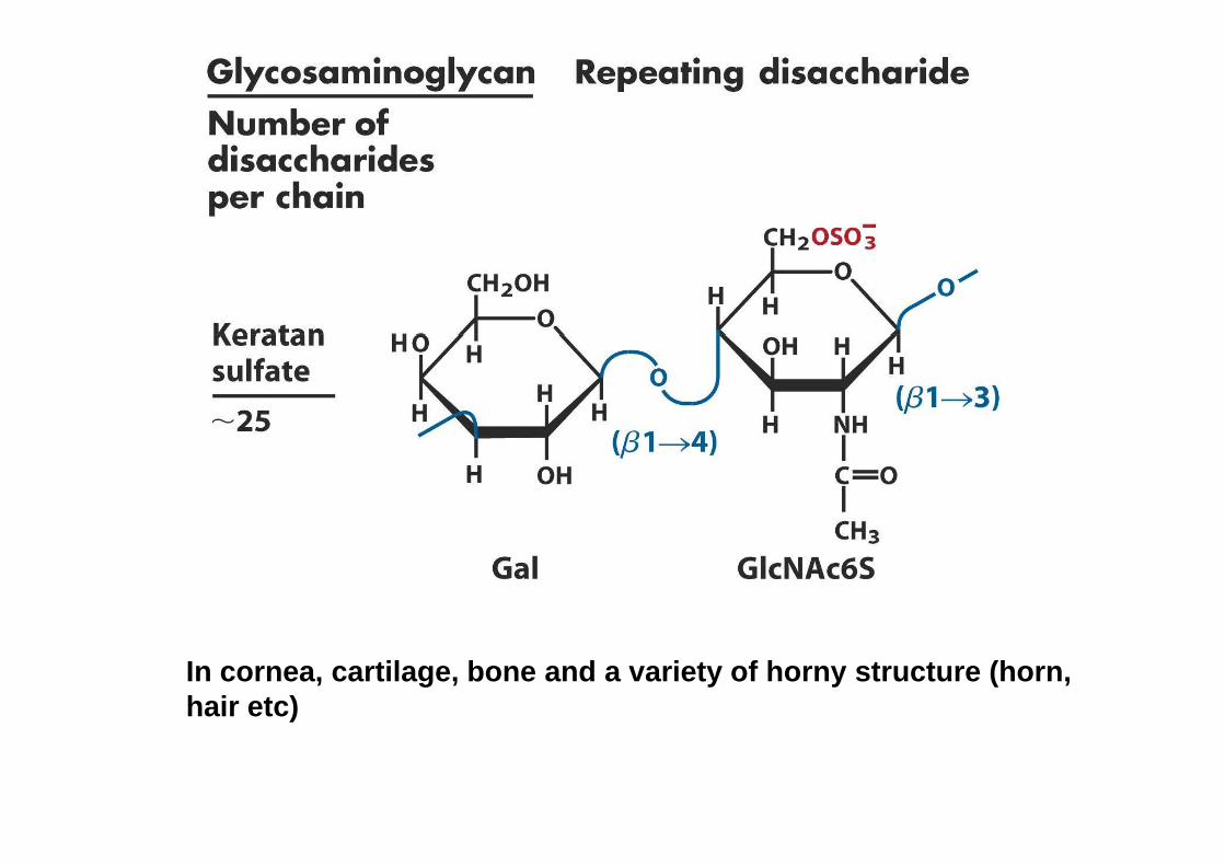
In cornea, cartilage, bone and a variety of horny structure (horn, hair etc)
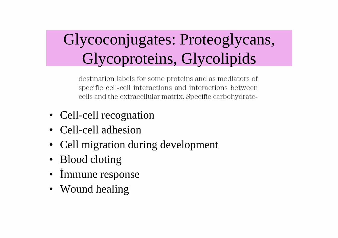
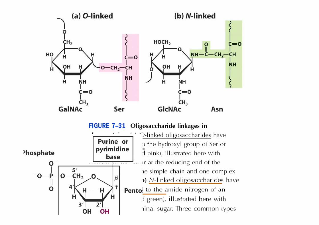
Glycoconjugates: Proteoglycans, Glycoconjugates: Proteoglycans, Glycoproteins, GlycolipidsGlycoproteins, Glycolipids
• Cell-cell recognation• Cell-cell recognation• Cell-cell adhesion• Cell migration during development• Cell migration during development• Blood cloting• İmmune response• İmmune response• Wound healing
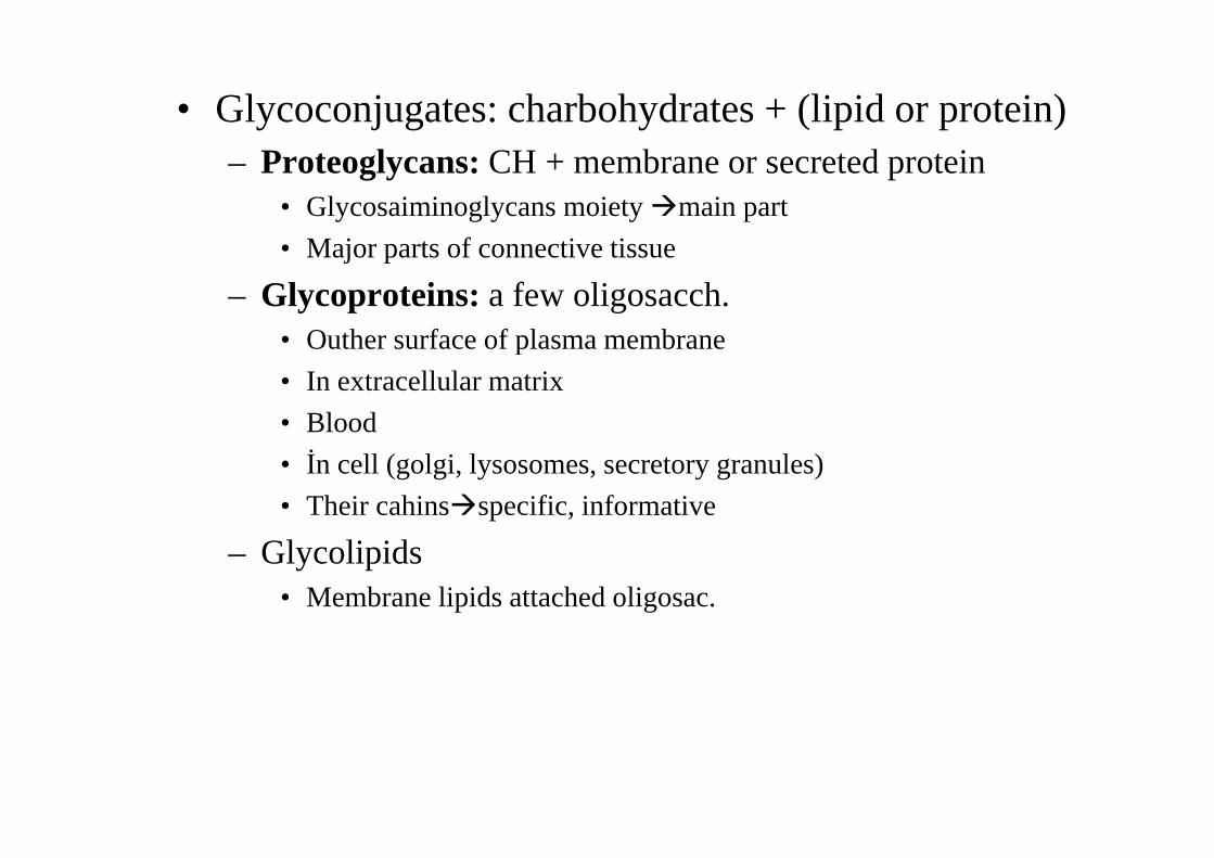
• Glycoconjugates: charbohydrates + (lipid or protein)– Proteoglycans:CH + membrane or secreted protein
• Glycosaiminoglycans moiety �main part
• Major parts of connective tissue• Major parts of connective tissue
– Glycoproteins: a few oligosacch.• Outher surface of plasma membrane• Outher surface of plasma membrane
• In extracellular matrix
• Blood• Blood
• İn cell (golgi, lysosomes, secretory granules)
• Their cahins�specific, informative
– Glycolipids• Membrane lipids attached oligosac.
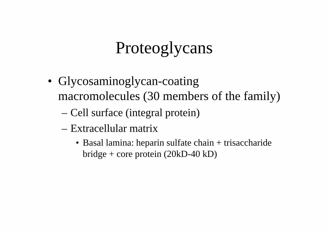
Proteoglycans
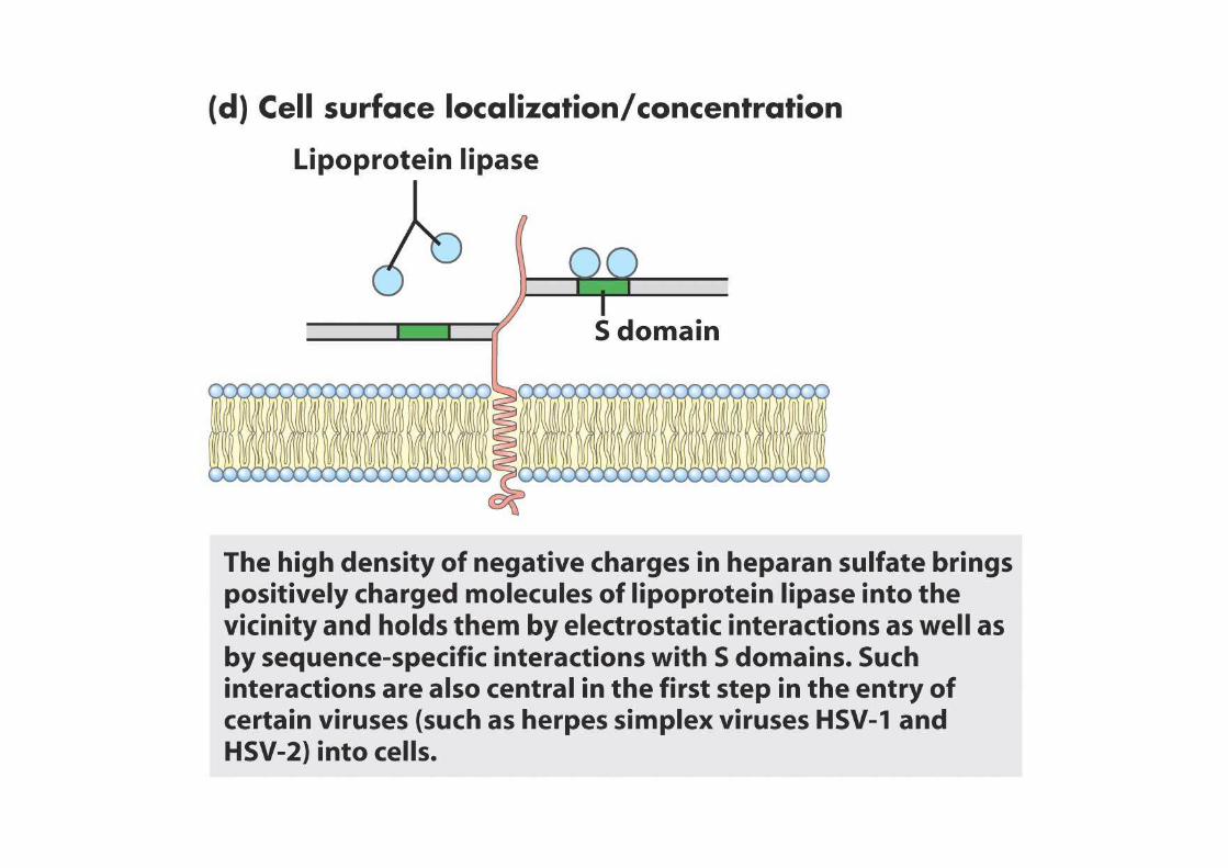
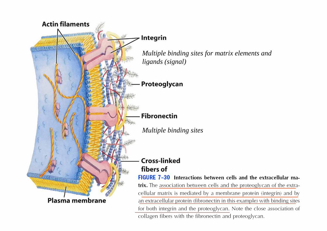
• Glycosaminoglycan-coating macromolecules (30 members of the family)macromolecules (30 members of the family)– Cell surface (integral protein)– Cell surface (integral protein)
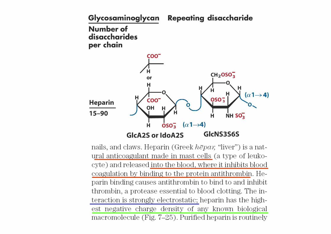
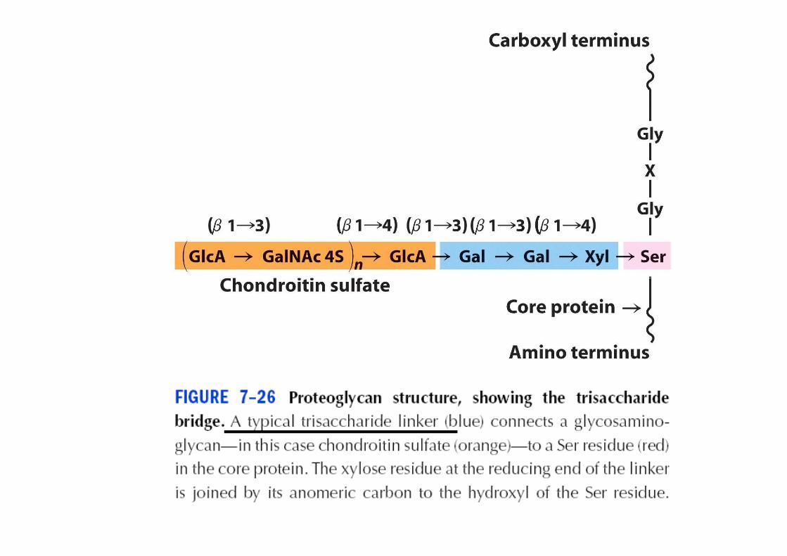
– Extracellular matrix• Basal lamina: heparin sulfate chain + trisaccharide • Basal lamina: heparin sulfate chain + trisaccharide
bridge + core protein (20kD-40 kD)
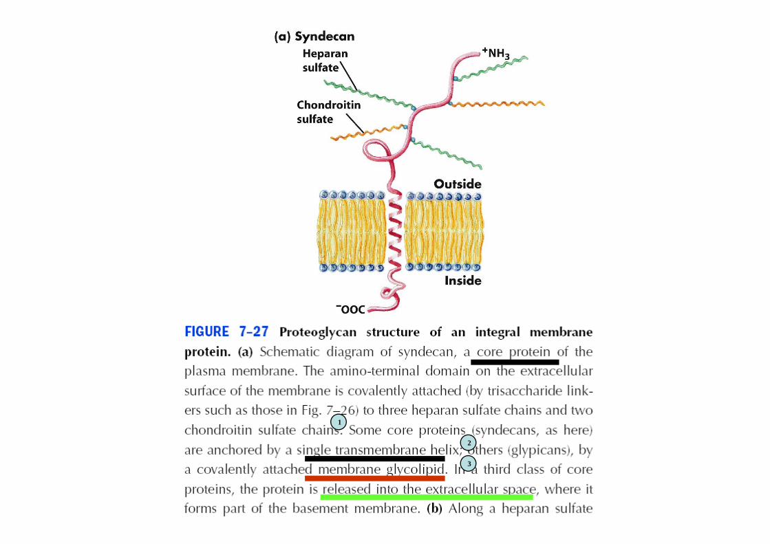
2
1
3

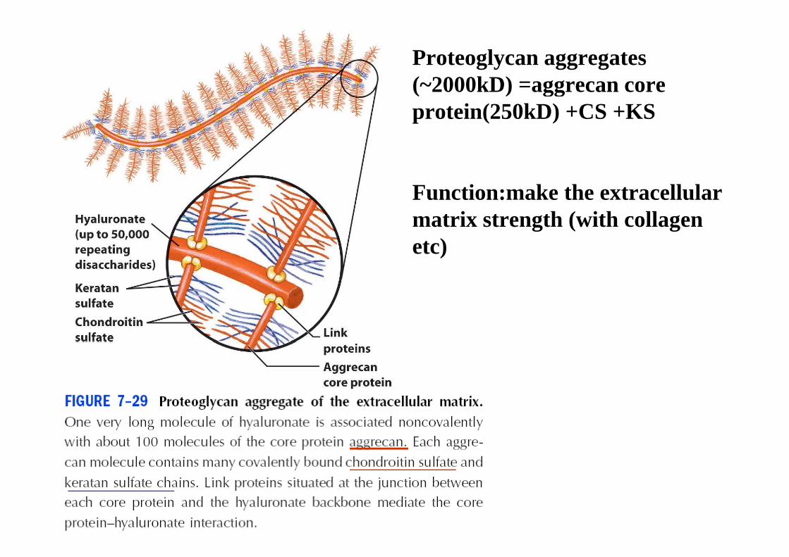
Proteoglycan aggregates (~2000kD) =aggrecan core (~2000kD) =aggrecan core protein(250kD) +CS +KS
Function:make the extracellular matrix strength (with collagen matrix strength (with collagen etc)
Multiple binding sites for matrix elements and ligands (signal)ligands (signal)
Multiple binding sites
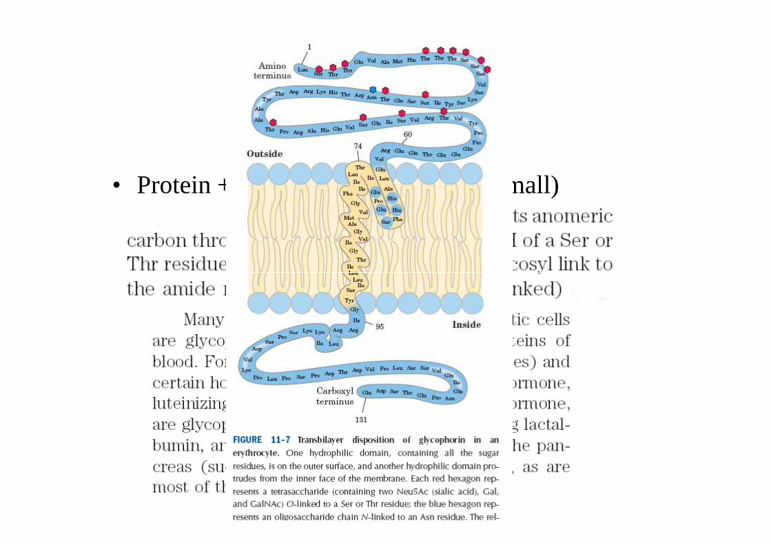
Glycoproteins
• Protein + carbohydrate (its moiety small)
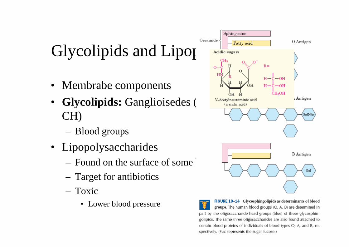
Glycolipids and Lipopolysaccharides
• Membrabe components
• Glycolipids: Ganglioisedes (membrane lipid + • Glycolipids: Ganglioisedes (membrane lipid + CH)– Blood groups
• Lipopolysaccharides• Lipopolysaccharides– Found on the surface of some bacteria
– Target for antibiotics– Target for antibiotics
– Toxic• Lower blood pressure• Lower blood pressure
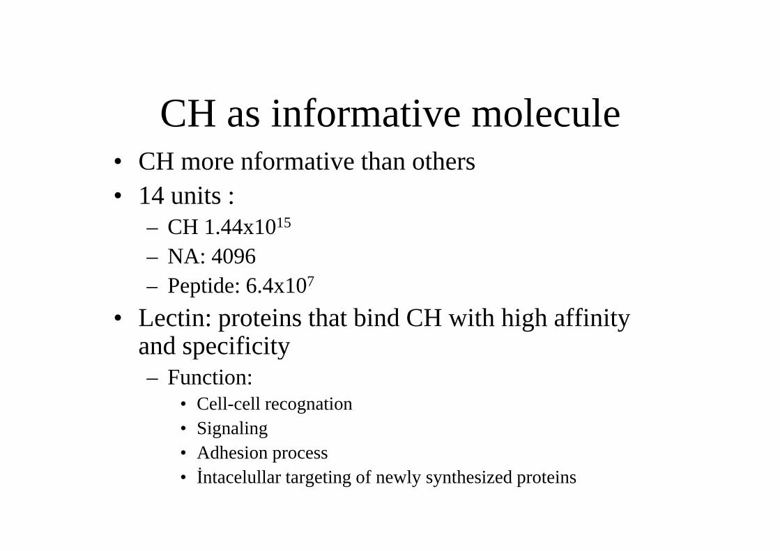
CH as informative molecule• CH more nformative than others• CH more nformative than others• 14 units :
– CH 1.44x10¹⁵
– NA: 4096– Peptide: 6.4x10⁷– Peptide: 6.4x10⁷
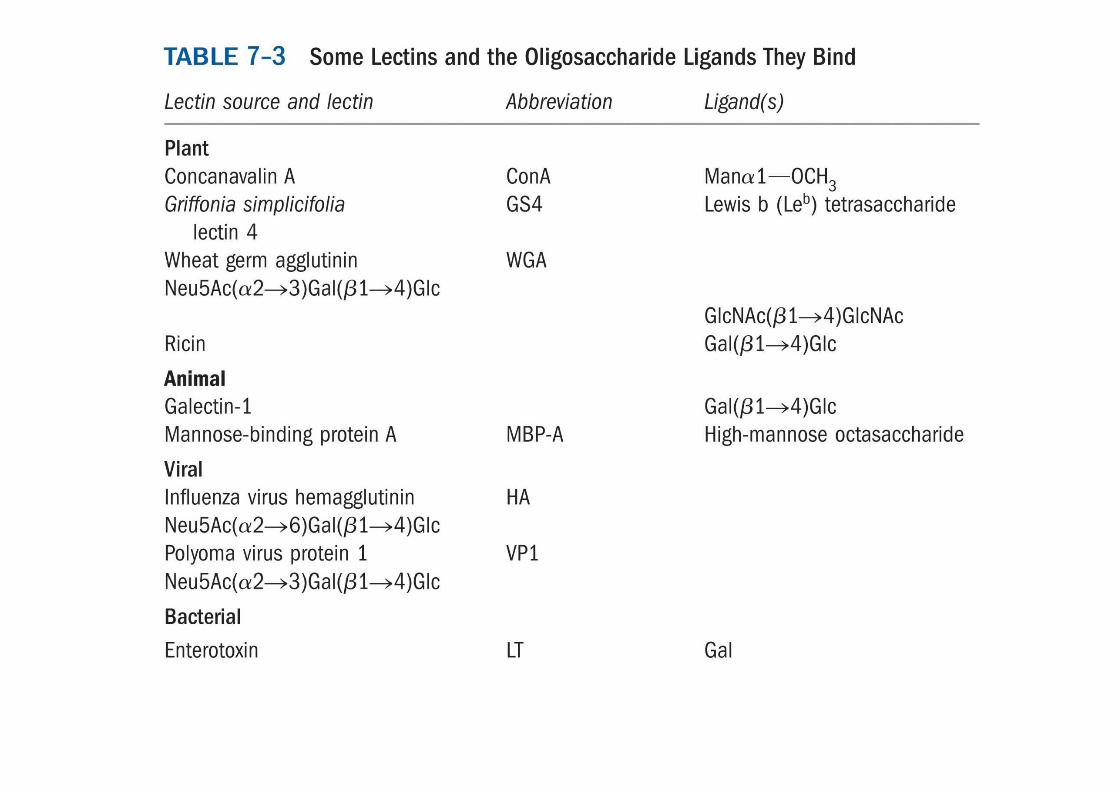
• Lectin: proteins that bind CH with high affinity and specificityand specificity– Function:
• Cell-cell recognation• Cell-cell recognation• Signaling• Adhesion process• İntacelullar targeting of newly synthesized proteins
• Some hormones and RBC are marked with Oligos. MoiteiesOligos. Moiteies– Funciton: affect their half-life: if you remove, – Funciton: affect their half-life: if you remove,
half-life decrease
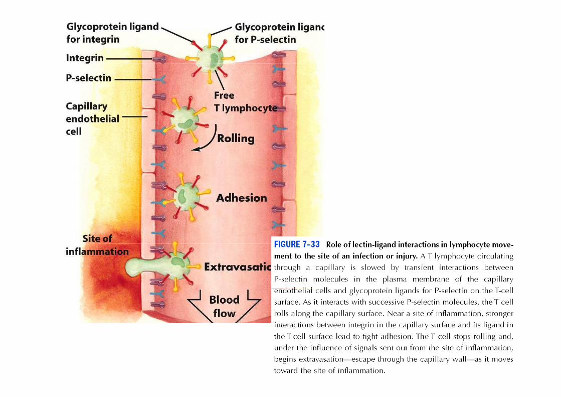
Selectins: a family of plasma membrane lectinslectins
• F: cell adhesion
• Cell-cell recognation
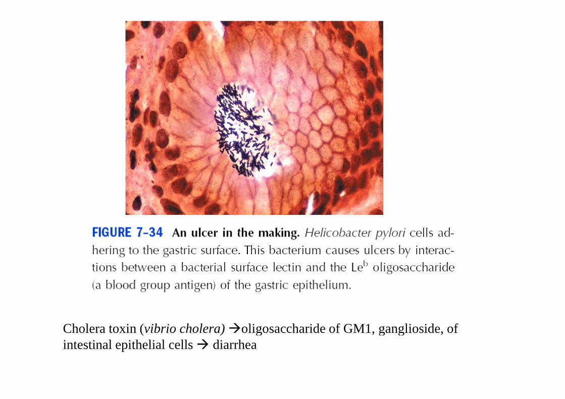
Cholera toxin (vibrio cholera) �oligosaccharide of GM1, ganglioside, of Cholera toxin (vibrio cholera) �oligosaccharide of GM1, ganglioside, of intestinal epithelial cells � diarrhea
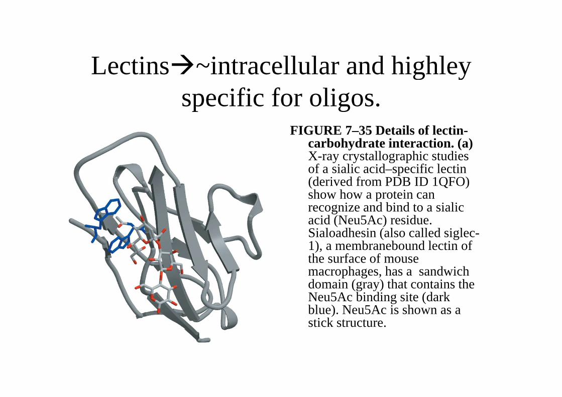
Lectins�~intracellular and highley Lectins�~intracellular and highley specific for oligos.specific for oligos.
FIGURE 7–35 Details of lectin-carbohydrate interaction. (a) X-raycrystallographic studies X-raycrystallographic studies of a sialic acid–specific lectin (derived from PDB ID 1QFO) show how a protein can show how a protein can recognize and bind to a sialicacid (Neu5Ac) residue. Sialoadhesin (also called siglec-1), a membraneboundlectin of 1), a membraneboundlectin of the surface of mouse macrophages, has a sandwichdomain (gray) that contains the domain (gray) that contains the Neu5Ac binding site (dark blue). Neu5Ac is shown as a stick structure.
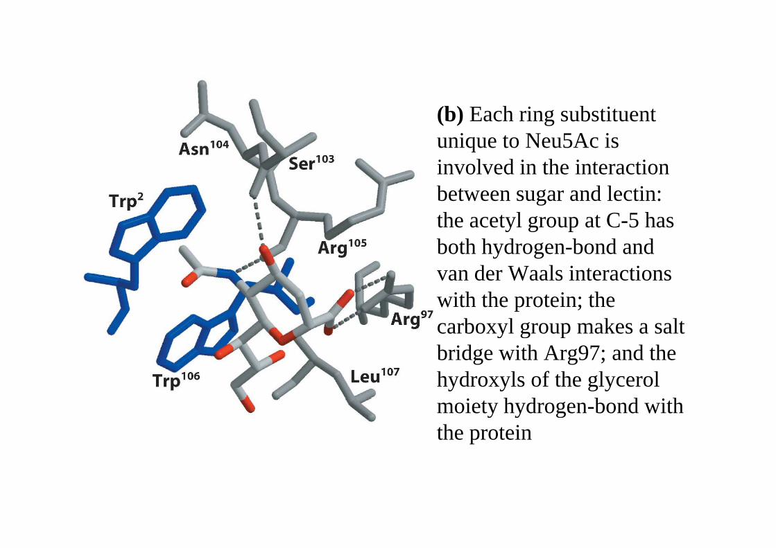
(b) Each ring substituent uniqueto Neu5Ac is uniqueto Neu5Ac is involved in the interaction between sugar and lectin: theacetyl group at C-5 has theacetyl group at C-5 has both hydrogen-bond and van der Waals interactionsvan der Waals interactionswith the protein; thecarboxyl group makes a salt carboxyl group makes a salt bridge with Arg97; and the hydroxyls of the glycerol moiety hydrogen-bond with moiety hydrogen-bond with the protein
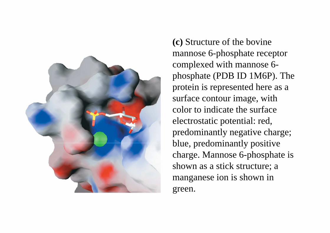
(c) Structure of the bovine (c) Structure of the bovine mannose 6-phosphate receptorcomplexed with mannose 6-complexed with mannose 6-phosphate (PDB ID 1M6P). The protein isrepresented here as a protein isrepresented here as a surface contour image, with color to indicate the surface electrostatic potential: red, electrostatic potential: red, predominantly negative charge; blue,predominantly positive blue,predominantly positive charge. Mannose 6-phosphate is shown as astick structure; a shown as astick structure; a manganese ion is shown in green.
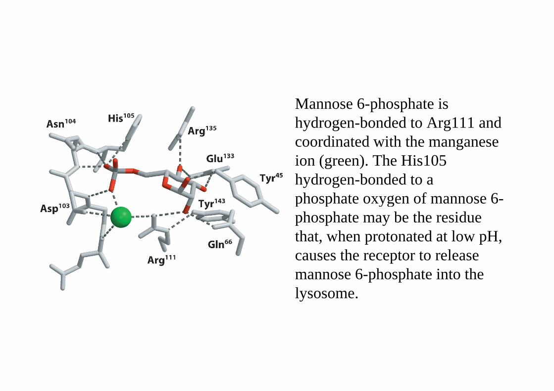
Mannose 6-phosphate is Mannose 6-phosphate is hydrogen-bonded to Arg111 and coordinated with the manganese ion (green). The His105 hydrogen-bonded to aphosphate oxygen of mannose 6-phosphate oxygen of mannose 6-phosphate may be the residue that,when protonated at low pH, that,when protonated at low pH, causes the receptor to release mannose 6-phosphate into the lysosome.lysosome.
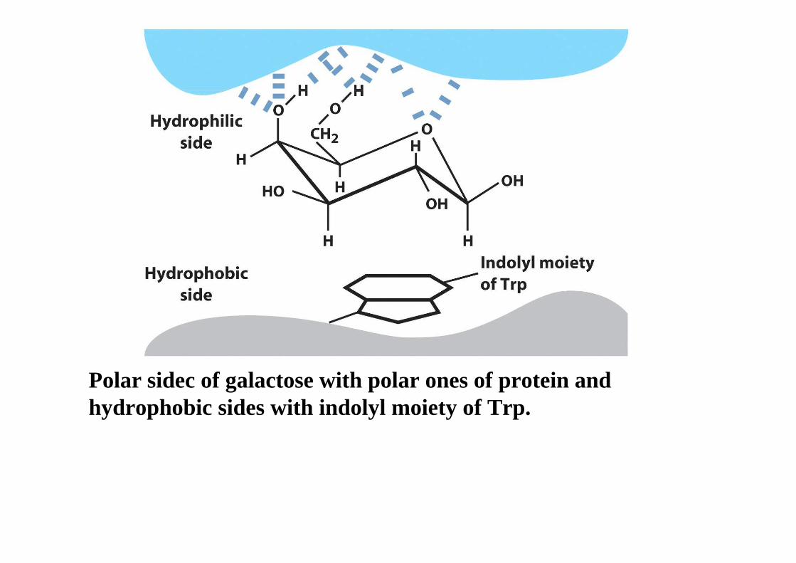
Polar sidec of galactose with polar ones of protein and hydrophobic sides with indolyl moiety of Trp.hydrophobic sides with indolyl moiety of Trp.
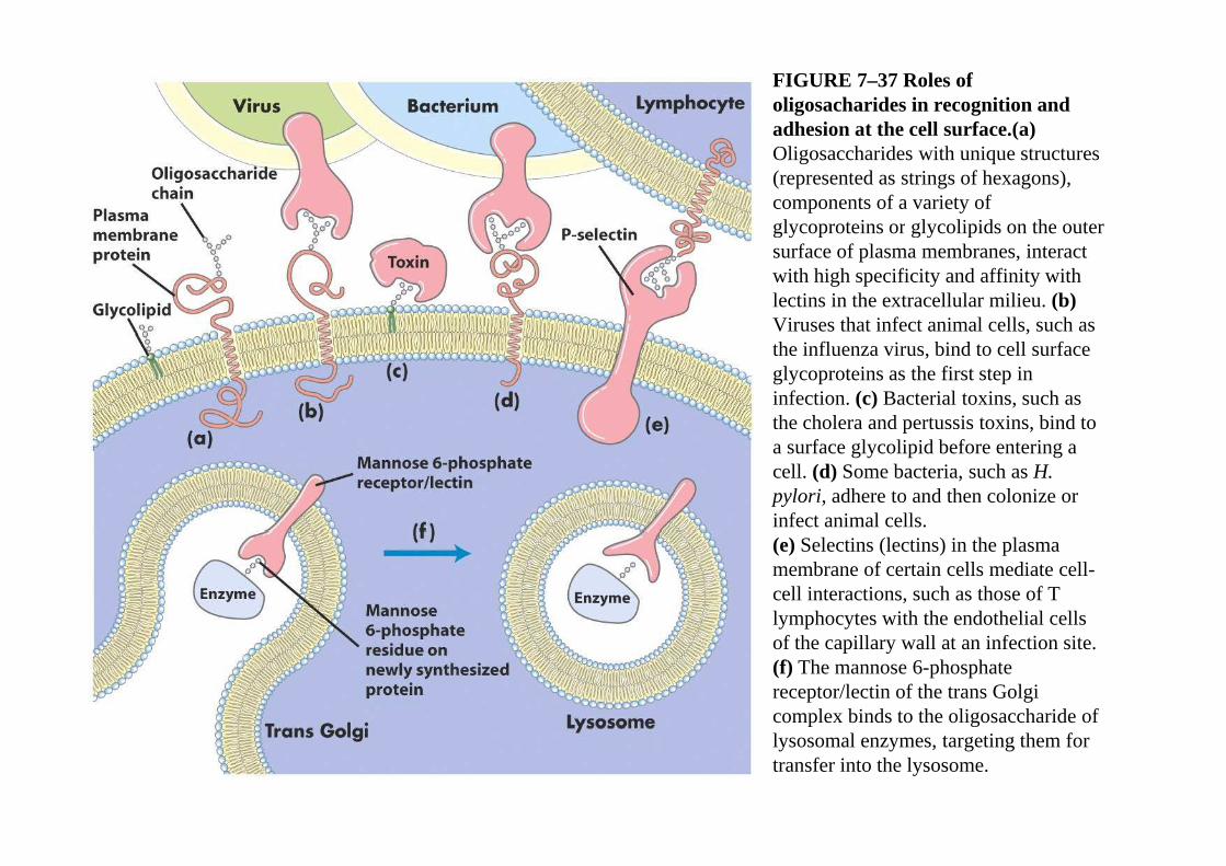
FIGURE 7–37 Roles of oligosacharides in recognition and adhesion at the cell surface.(a) adhesion at the cell surface.(a) Oligosaccharides with unique structures(represented as strings of hexagons),components of a variety of glycoproteins orglycolipids on the outer glycoproteins orglycolipids on the outer surface of plasma membranes, interact with high specificity and affinity with lectins in the extracellular milieu. (b) Viruses that infect animal cells,such as Viruses that infect animal cells,such as the influenza virus, bind to cell surface glycoproteins as the first step ininfection. (c) Bacterial toxins, such as thecholera and pertussis toxins, bind to thecholera and pertussis toxins, bind to a surface glycolipid before entering a cell. (d) Some bacteria, such as H. pylori, adhere to and then colonize or infect animal cells.(e) Selectins (lectins) in the plasmamembrane of certain cells mediate cell-cell interactions, such as those of T lymphocytes with the endothelial cells of the capillary wall at an infection site. (f) The mannose 6-phosphate receptor/lectin of the trans Golgicomplex binds to the oligosaccharide oflysosomal enzymes, targeting them fortransfer into the lysosome.