Embed Size (px)
Citation preview
UK Journal of Pharmaceutical and Biosciences Vol. 3(2), 46-59, 2015 RESEARCH ARTICLE
Evaluation of Salicin Isolated from Salix subserrata as a Radioprotector Against
Gamma Irradiation Induced Ultrastructural and Electrophoretic Changes in Spleen
Tissue in Rats
Monira Abdel-latif Abdel-karim, Ibrahim Abulyazid, Mohga Shafik Abdalla, Hayat Mohamed Sharada,
Wael Mahmoud Kamel*
Genetic Engineering and Biotechnology Division, National Research Centre, Dokki, Giza, Egypt-12622
Article Information
Received 16 Feburary 2015
Received in revised form 29 April 2015
Accepted 30 April 2015
Abstract
The aim of this study was to investigate the radioprotective effect of salicin against irradiation
effect on spleen tissue in male rats. Lipid peroxidation product (MDA) level was measured as
thiobarbituric acid reactive substance. Ultrastructural examination was carried out in spleen
tissue by scanning electron microscope (SEM). The polyacrylamide gel electrophoresis for native
protein, lipoprotein and zymogram were carried out in spleen homogenate. As expected, salicin
resisted the irradiation effect and declined the MDA level in spleen homogenate of all treated
groups. The alterations which were occurred as a result of irradiation in the spleen tissue could
not be detected microscopically but they were detected electrophoertically at levels of protein and
isozymes. Salicin prevented the qualitative mutagenic effect of irradiation on the electrophoretic
protein pattern in the irradiated salicin simultaneous treated group (SI = 0.73). It showed the
highest protective effect against qualitative mutagenic irradiation effect in catalase pattern in
irradiated salicin pre-treated group (SI = 0.80). It could not prevent the abnormalities occurred
qualitatively and quantitatively as a result of irradiation in peroxidase pattern in all irradiated
salicin treated groups. While the esterase pattern showed the same electrophoretic pattern in the
all irradiated salicin treated groups. The results suggested the radioprotective ability of salicin
against gamma irradiation effect on various ultrastructural and electrophoretic patterns in spleen
tissue of male rats.
Keywords:
Gamma irradiation,
Salicin,
Spleen,
Protein electrophoresis,
Isozymes
*Corresponding Author:
E-mail: [email protected]
Tel.: 00201126682686
1 Introduction
Gamma rays are a packet of pure electromagnetic rays which are
photons of high frequency and high energy and hence short wave
length1. They can penetrate into living tissues or cells and result in
transduction of radiation energy to biological materials. The
absorbed energy of ionizing radiation can break chemical bonds and
cause ionization of different molecules including water and different
biological essential macromolecules as DNA2, membrane lipids and
proteins3.
It has been reported that whole-body gamma irradiation induces
oxidative stress. The most important consequences of oxidative
stress are lipid peroxidation, protein oxidation and depletion of
antioxidants4,5
. It was found that irradiation decreases tissue
concentrations of natural nonenzymatic antioxidants6,7
and causes
induction of lipid peroxidation as evidenced by increased
malondialdhyde (MDA)8.
Spleen plays an important role in immune functions by proliferating
lymphocytes. The integrity of the immune system depends upon the
normal functioning of the lymphoid organs so that the alterations in
the homeostasis of spleen tissues will affect immune responses9. It
was demonstrated that spleen was the most biosensitive organs to
low doses of irradiation in rats10
. Irradiation caused alterations in
spleen tissue and it caused induction of DNA affecting the
radiosensitive gene in the spleen of rats11,12
. Ezz,13
showed that
spleen taken to study the ameliorative effect of radioprotector against
irradiation induced oxidative stress and immune responses in rats.
UK Journal of Pharmaceutical and Biosciences
Available at www.ukjpb.com ISSN: 2347-9442
Kamel et al. Evaluation of Salicin Isolated from Salix subserrata as a Radioprotector
UK J Pharm & Biosci, 2015: 3(2); 47
Proteins are the most complex compounds and at the same time the
most characteristic of living matter. They are present in all viable
cells; they are the compounds which, as nucleoproteins, are
essential for cell division and, as enzymes and hormones, control
many chemical reactions in the metabolism of cells. Thus, the
separation and characterization of the individual proteins facilitate
the study of the chemical nature and physiological function of each
protein14
. They are major targets for oxidative damage due to their
abundance and rapid rates of reaction with a wide range of radicals
and excited state species15
. Changes in the protein patterns of the
tissues may reflect specialization and adaptation in the organisms. It
is worthy to note that each protein is considered as reflect to the
activity of specific gene through the production of enzyme, which act
as catalyst to produce the demanded protein; this type of produced
protein is responsible for a specific biological character16
. The
radiation-induced alteration of the protein structure was observed by
measuring the changes in the molecular properties of the proteins17
.
Recently, it was found that irradiation showed significant increase in
protein carbonyls by 73%18
.
Antioxidants enzymes as catalase (CAT) and peroxidase (GPx) are
important in the elimination of free radicals19,20
. They are involved to
counteract the toxicity of ROS21
. These enzymes are the first line of
defense against oxidative injury. Superoxide dismutase is the
primary step of the defense mechanism in the antioxidant system
against oxidative stress by catalyzing the dismutation of 2
superoxide radicals (O2-) into molecular oxygen (O2) and hydrogen
peroxide (H2O2)22
. H2O2 can synthesize a highly reactive OH, is
neutralized by the combined action of CAT and GPx in all
vertebrates23,24
. These enzymes act in coordination and the cells
may be pushed to oxidative stress state if any change occurs in the
levels of enzymes21
.
Irradiation can exert a significant inflammatory response in cells. So,
it is essential to develop methods to target the radiosensitive organs
and / or to protect the normal tissues. Antioxidants eliminate the free
radicals and neutralize reactive oxygen species (ROS) before they
can do their damage. However, much remains unknown about
mechanisms of radio-protection. Development of protective agents
presented new solutions for recovery of undesired tissue damage
induced by irradiation25,26
.
The discovery of radioprotectors for the first time seemed to be very
promising and has attracted the interest of a number of
radiobiologists. Although synthetic radioprotectors such as the
aminothiols have yielded the highest protective factors; typically they
are more toxic than naturally occurring protectors27
.
Salicin (C13H18O7) is a natural product extracted from several species
of Salix (willow) and Populus (poplar), and was also found in
Gaultheria procumbens (wintergreen) and in Betula lenta (sweet
birch), the volatile oils of which consist almost entirely of methyl
salicylate28
. Salicin is considered as natural aspirin. It is very possible
to be digested without side effects in the stomach and kidneys, while
acetylsalicylic acid is known to upset the stomach and in some cases
damage kidneys. Scientists believe that this is because salicin is
converted to acetylsalicylic acid after the stomach has absorbed it29
.
It is a pro-drug that is gradually transported to the lower part of the
intestine, hydrolysed to saligenin by intestinal bacteria, and
converted to salicylic acid after absorption. It thus produces an
antipyretic action without causing gastric injury30
.
It belonged to the phenolic compounds which are believed to work
synergistically to promote healthy conditions through a variety of
different mechanisms, such as enhancing antioxidant activity,
impacting cellular processes associated with apoptosis, platelet
aggregation, blood vessel dilation, and enzyme activities associated
with carcinogen activation and detoxification31,32
.
The present main objective is to optimize salicin as a radioprotector
against effect of gamma irradiation on the spleen tissue in the hope
that this compound may be further explored as novel antioxidative
radioprotector.
2 Materials and Methods
2.1 Salicin isolation
Salicin was extracted and isolated from fresh young leaves of willow
trees (Salix subserrata, Salix safsaf) according to method suggested
by Mabry et al.[33]
and purified according to method described by
Kur'yanov et al.34
then identified qualitatively by advanced
chromatographic techniques.
2.2 Acute toxicity test
The safety of salicin orally was evaluated by determination it’s LD50.
Forty eight adult female albino mice weighing 20-25 g was used to
study acute toxicity. It was divided into 6 groups each of 8 mice. The
groups were treated orally with rising doses of 500, 1000, 2000,
3000, 4000 and 5000 mg/kg body weight of aqueous solution of
salicin solution. Mortality was recorded 24 hrs post treatment. The
LD50 was calculated according to the equation suggested by Paget
and Barnes35
.
2.3 Animals
Seven groups of male rats weighing between 150-200 gm per one
obtained from the animal house laboratory of national research
centre. Ten rats in each group. All the animals were kept under
normal environmental and nutritional conditions. The animal groups
were divided into Rats were non-irradiated and non-treated with
salicin representing Control group; Rats were non-irradiated but
Kamel et al. Evaluation of Salicin Isolated from Salix subserrata as a Radioprotector
UK J Pharm & Biosci, 2015: 3(2); 48
treated with the safe dose of salicin (was about 150 mg / Kg) taking
in the consideration weight of each rat representing Salicin treated
group; Rats were irradiated at the dose 7 Gy and non-treated with
salicin representing Irradiated group; Rats were treated with salicin
for 15 days followed by irradiation at the 15th day representing
Irradiated salicin pre-treated group; Rats were treated with salicin for
15 days followed by irradiation at the 15th day then the treatment was
continued daily for another 15 days representing Irradiated salicin
prepost-treated group; Rats were irradiated and treated with salicin
at the same time of irradiation and continue daily for 15 days
representing Irradiated salicin simultaneous treated group; and Rats
were irradiated at the same gamma dose then left without treatment
for 15 days. At the 15th day, the rats were treated with salicin for
another 15 days representing Irradiated salicin post-treated group.
2.4 Irradiation
Whole body of the animals was exposed to an acute single dose of 7
Gy delivered at a dose rate of 1.167 Rad/Sec. using cobalt-60 (Co60
)
from the biological irradiator gamma cell source belonging to Middle
Eastern Regional Radioisotopes Center for Arab Countries, Dokki,
Cairo, Egypt.
2.5 Lipid peroxidation measurement
Lipid peroxidation level was measured as thiobarbituric acid reactive
substance in spleen homogenate according to method of Ohkawa et
al.36
.
2.6 Scanning Microscopic examination (SEM).
This examination was carried out in piece of spleen tissue using
SEM. The tissue was preserved in gluteraldhyde purchased from
Gpr Chemicals Co. It was prepared according to the method
suggested by Tánaka37
who reported that the specimen showed be
passed through series of the dehydration steps by placing it in ethyl
alcohol then incubated for 15 min. then coated with the golden atoms
to be ready for the electron microscopic examination.
2.7 Electrophoretic protein and lipoprotein patterns
Total protein was determined in spleen homogenate according to
Bradford38
. The sample was mixed with the sample buffer. The
protein concentration in each well must be about 70 μg protein.
Proteins were separated through polyacrylamide gel electrophoresis
(PAGE). Electrode and gel buffer and polyacrylamide stock were
prepared according Laemmli39
. After electrophoretic separation, the
gel was gently removed from the apparatus and put into a staining
solution of coomasie brilliant blue for native protein pattern40
and
staining solution of sudan black B (SBB) for lipoprotein pattern41
.
2.8 Isozyme
Native protein gel was stained for peroxidase pattern using certain
stain prepared according to the method suggested by Rescigno et
al.42
, for catalase pattern according to method described by Siciliano
and Shaw43
and for esterase pattern according to method of Baker
and Manwell44
.
2.9 Data analysis
The polyacrylamide gel plate was photographed, scanned and then
analyzed using Phoretix 1D pro software (Version 12.3). The
similarity index (S.I.) compares patterns within, as well as, between
irradiated and non-irradtated samples. The similarity values were
converted into genetic distance (GD) according the method
suggested by Nei and Li45
.
3 Statistical Analysis
All the grouped data were statistically evaluated with SPSS/16.00
software. The results were expressed as mean ± SE of studied
groups using the analysis of variance test (one-way ANOVA)
followed by student’s t-test. P values of less than 0.05 were
considered to indicate statistical significance. The means of
irradiated groups and the salicin treated groups were individually
compared with those of control group. The irradiated group was
compared with irradiated salicin treated groups.
4 Results
4.1 Lipid peroxidation
As compared to control, irradiation caused significant (P < 0.05)
elevation in the MDA level in spleen tissue. Salicin administration
showed the ameliorative effect against irradiation by reducing MDA
level in all irradiated salicin treated rats. From the data compiled in
Table 1, it was found that salicin showed the most suitable
antagonistic effect against irradiation on spleen of irradiated salicin
pre-treated group as compared to irradiated group.
4.2 Spleen ultrastructure
The ultrastructural observations in spleen tissue of control rats
revealed normal tissue surface (Fig. 1a). Salicin administration
showed normal appearance, no ultrastructural changes and no
deviation from control (Fig. 1b). Irradiation caused no obvious
abnormalities on the spleen surface indicating to inability of this
radiation dose to cause any differences observed microscopically on
surface of the spleen tissue (Fig. 1c). In the irradiated salicin pre-
treated group, there was superfacial lesions with cellular loses (Fig.
1d).
Spleen tissue showed smooth appearance in irradiated salicin
simultaneous treated group with presence of blood aggregates on
the tissue surface (black arrow) (Fig. 1e). In the irradiated salicin
prepost-treated group, it was found that there was surface erosion
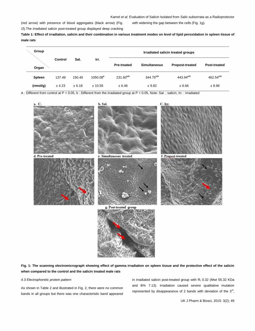
Kamel et al. Evaluation of Salicin Isolated from Salix subserrata as a Radioprotector
UK J Pharm & Biosci, 2015: 3(2); 49
(red arrow) with presence of blood aggregates (black arrow) (Fig.
1f).The irradiated salicin post-treated group displayed deep cracking
with widening the gap between the cells (Fig. 1g).
Table 1: Effect of irradiation, salicin and their combination in various treatment modes on level of lipid peroxidation in spleen tissue of
male rats
Group
Organ
Control Sal. Irr.
Irradiated salicin treated groups
Pre-treated Simultaneous Prepost-treated Post-treated
Spleen
(nmol/g)
137.49
± 4.23
150.40
± 6.18
1050.08a
± 10.55
231.60a,b
± 6.46
344.75a,b
± 9.82
443.94a,b
± 6.66
462.54a,b
± 8.96
A : Different from control at P < 0.05, b : Different from the irradiated group at P < 0.05, Note: Sal. ; salicin, Irr. : Irradiated
Fig. 1: The scanning electromicrograph showing effect of gamma irradiation on spleen tissue and the protective effect of the salicin
when compared to the control and the salicin treated male rats
4.3 Electrophoretic protein pattern
As shown in Table 2 and illustrated in Fig. 2, there were no common
bands in all groups but there was one characteristic band appeared
in irradiated salicin post-treated group with Rf 0.32 (Mwt 55.32 KDa
and B% 7.13). Irradiation caused severe qualitative mutation
represented by disappearance of 2 bands with deviation of the 3rd,
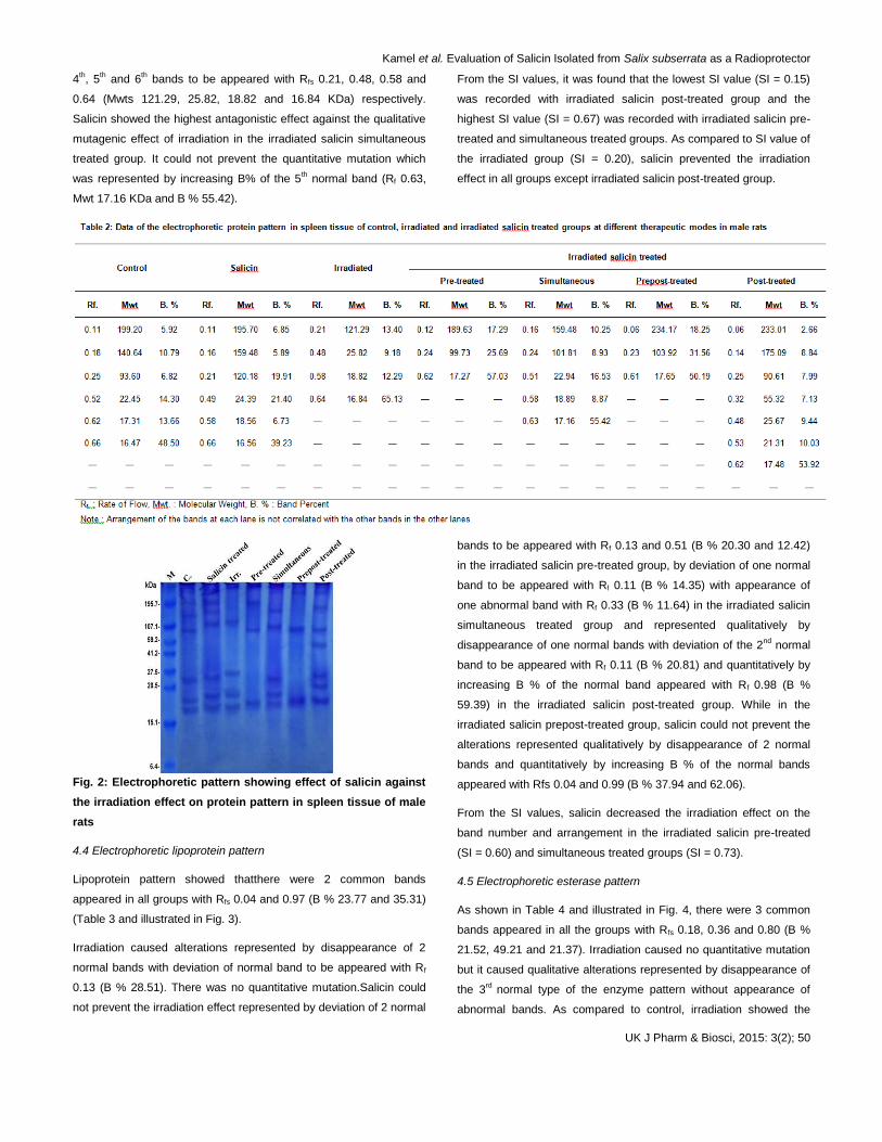
Kamel et al. Evaluation of Salicin Isolated from Salix subserrata as a Radioprotector
UK J Pharm & Biosci, 2015: 3(2); 50
4th, 5
th and 6
th bands to be appeared with Rfs 0.21, 0.48, 0.58 and
0.64 (Mwts 121.29, 25.82, 18.82 and 16.84 KDa) respectively.
Salicin showed the highest antagonistic effect against the qualitative
mutagenic effect of irradiation in the irradiated salicin simultaneous
treated group. It could not prevent the quantitative mutation which
was represented by increasing B% of the 5th normal band (Rf 0.63,
Mwt 17.16 KDa and B % 55.42).
From the SI values, it was found that the lowest SI value (SI = 0.15)
was recorded with irradiated salicin post-treated group and the
highest SI value (SI = 0.67) was recorded with irradiated salicin pre-
treated and simultaneous treated groups. As compared to SI value of
the irradiated group (SI = 0.20), salicin prevented the irradiation
effect in all groups except irradiated salicin post-treated group.
Fig. 2: Electrophoretic pattern showing effect of salicin against
the irradiation effect on protein pattern in spleen tissue of male
rats
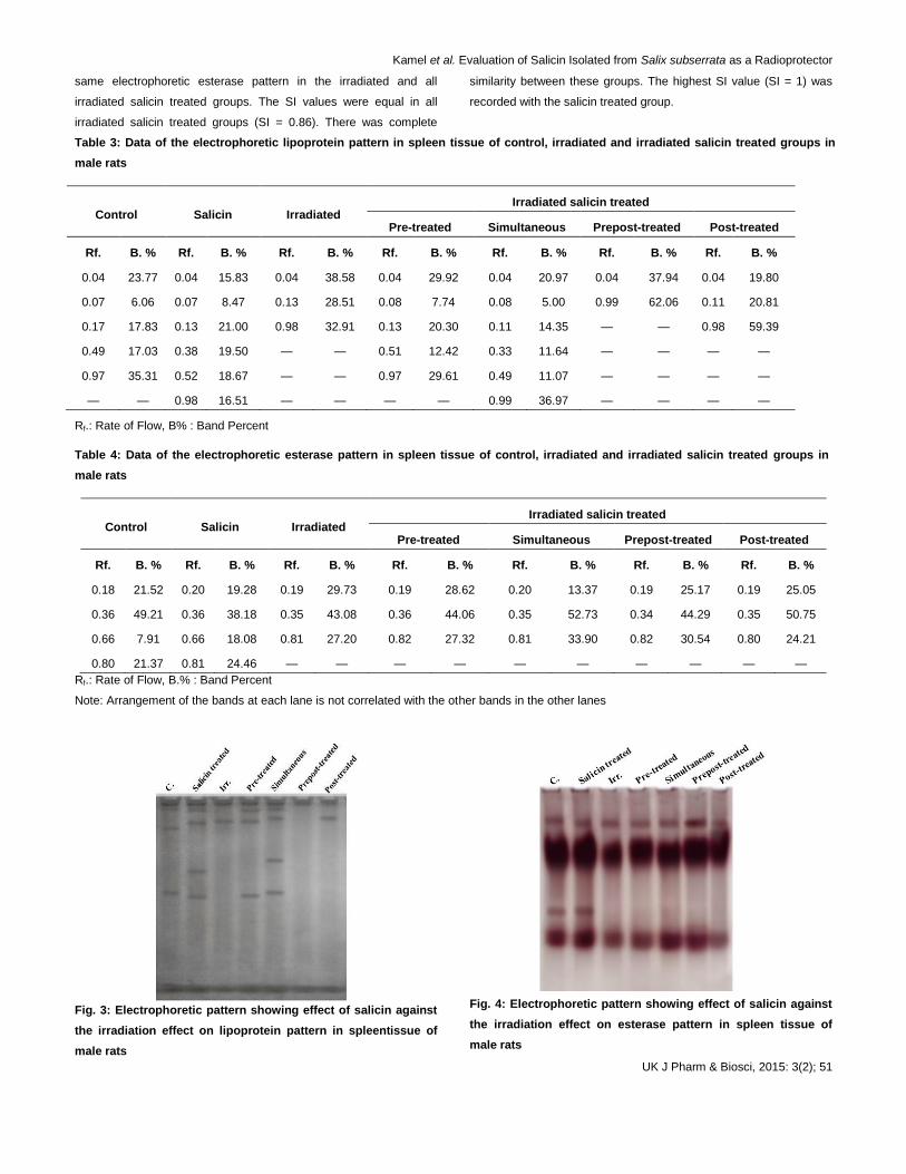
4.4 Electrophoretic lipoprotein pattern
Lipoprotein pattern showed thatthere were 2 common bands
appeared in all groups with Rfs 0.04 and 0.97 (B % 23.77 and 35.31)
(Table 3 and illustrated in Fig. 3).
Irradiation caused alterations represented by disappearance of 2
normal bands with deviation of normal band to be appeared with Rf
0.13 (B % 28.51). There was no quantitative mutation.Salicin could
not prevent the irradiation effect represented by deviation of 2 normal
bands to be appeared with Rf 0.13 and 0.51 (B % 20.30 and 12.42)
in the irradiated salicin pre-treated group, by deviation of one normal
band to be appeared with Rf 0.11 (B % 14.35) with appearance of
one abnormal band with Rf 0.33 (B % 11.64) in the irradiated salicin
simultaneous treated group and represented qualitatively by
disappearance of one normal bands with deviation of the 2nd
normal
band to be appeared with Rf 0.11 (B % 20.81) and quantitatively by
increasing B % of the normal band appeared with Rf 0.98 (B %
59.39) in the irradiated salicin post-treated group. While in the
irradiated salicin prepost-treated group, salicin could not prevent the
alterations represented qualitatively by disappearance of 2 normal
bands and quantitatively by increasing B % of the normal bands
appeared with Rfs 0.04 and 0.99 (B % 37.94 and 62.06).
From the SI values, salicin decreased the irradiation effect on the
band number and arrangement in the irradiated salicin pre-treated
(SI = 0.60) and simultaneous treated groups (SI = 0.73).
4.5 Electrophoretic esterase pattern
As shown in Table 4 and illustrated in Fig. 4, there were 3 common
bands appeared in all the groups with Rfs 0.18, 0.36 and 0.80 (B %
21.52, 49.21 and 21.37). Irradiation caused no quantitative mutation
but it caused qualitative alterations represented by disappearance of
the 3rd
normal type of the enzyme pattern without appearance of
abnormal bands. As compared to control, irradiation showed the
Kamel et al. Evaluation of Salicin Isolated from Salix subserrata as a Radioprotector
UK J Pharm & Biosci, 2015: 3(2); 51
same electrophoretic esterase pattern in the irradiated and all
irradiated salicin treated groups. The SI values were equal in all
irradiated salicin treated groups (SI = 0.86). There was complete
similarity between these groups. The highest SI value (SI = 1) was
recorded with the salicin treated group.
Table 3: Data of the electrophoretic lipoprotein pattern in spleen tissue of control, irradiated and irradiated salicin treated groups in
male rats
Control Salicin Irradiated Irradiated salicin treated
Pre-treated Simultaneous Prepost-treated Post-treated
Rf. B. % Rf. B. % Rf. B. % Rf. B. % Rf. B. % Rf. B. % Rf. B. %
0.04 23.77 0.04 15.83 0.04 38.58 0.04 29.92 0.04 20.97 0.04 37.94 0.04 19.80
0.07 6.06 0.07 8.47 0.13 28.51 0.08 7.74 0.08 5.00 0.99 62.06 0.11 20.81
0.17 17.83 0.13 21.00 0.98 32.91 0.13 20.30 0.11 14.35 — — 0.98 59.39
0.49 17.03 0.38 19.50 — — 0.51 12.42 0.33 11.64 — — — —
0.97 35.31 0.52 18.67 — — 0.97 29.61 0.49 11.07 — — — —
— — 0.98 16.51 — — — — 0.99 36.97 — — — —
Rf.: Rate of Flow, B% : Band Percent
Table 4: Data of the electrophoretic esterase pattern in spleen tissue of control, irradiated and irradiated salicin treated groups in
male rats
Control Salicin Irradiated Irradiated salicin treated
Pre-treated Simultaneous Prepost-treated Post-treated
Rf. B. % Rf. B. % Rf. B. % Rf. B. % Rf. B. % Rf. B. % Rf. B. %
0.18 21.52 0.20 19.28 0.19 29.73 0.19 28.62 0.20 13.37 0.19 25.17 0.19 25.05
0.36 49.21 0.36 38.18 0.35 43.08 0.36 44.06 0.35 52.73 0.34 44.29 0.35 50.75
0.66 7.91 0.66 18.08 0.81 27.20 0.82 27.32 0.81 33.90 0.82 30.54 0.80 24.21
0.80 21.37 0.81 24.46 — — — — — — — — — —
Rf.: Rate of Flow, B.% : Band Percent
Note: Arrangement of the bands at each lane is not correlated with the other bands in the other lanes
Fig. 3: Electrophoretic pattern showing effect of salicin against
the irradiation effect on lipoprotein pattern in spleentissue of
male rats
Fig. 4: Electrophoretic pattern showing effect of salicin against
the irradiation effect on esterase pattern in spleen tissue of
male rats
Kamel et al. Evaluation of Salicin Isolated from Salix subserrata as a Radioprotector
UK J Pharm & Biosci, 2015: 3(2); 52
4.6 Electrophoretic catalase pattern
The electrophoretic catalase pattern showed that there were no
common bands appeared in all groups (Table 5 and illustrated in Fig.
5).
Irradiation caused qualitative alteration represented by
disappearance of one normal type with appearance of one abnormal
band with Rf 0.28 (B % 69.42). Salicin administration showed the
highest protective effect against qualitative mutagenic effect of
irradiation in the irradiated salicin pre-treated group. It could not
prevent the irradiation effect which was represented qualitatively by
appearance 2 abnormal bands with Rfs 0.28 and 0.59 (B % 19.45
and 29.30) in the irradiated salicin simultaneous treated group, with
Rfs 0.16 and 0.27 (B % 34.62 and 25.57) in the irradiated salicin
prepost-treated group and Rfs 0.20 and 0.57 (B % 32.77 and 26.19)
in the irradiated salicin post-treated group. It could not prevent the
quantitative mutation which was represented by decreasing the B %
of the 1st type of the catalase enzyme in all irradiated salicin treated
groups.
The SI values showed that the lowest SI value (SI = 0.33) was
observed with irradiated salicin simultaneous group and the highest
value (SI = 0.8) observed with irradiated salicin pre-treated group. In
the irradiated salicin prepost-treated group, it was observed that all
the bands were not matched with all bands of the other groups.
Salicin treatment minimized the irradiation effect the irradiated salicin
pre-treated (SI = 0.80) and post-treated group.
Table 5: Data of the electrophoretic Catalase pattern in spleen tissue of control, irradiated and irradiated salicin treated groups in
male rats
Control Salicin Irradiated
Irradiated salicin treated
Pre-treated Simultaneous Prepost-treated Post-treated
Rf. B. % Rf. B. % Rf. B. % Rf. B. % Rf. B. % Rf. B. % Rf. B. %
0.06 73.44 0.06 22.25 0.06 73.44 0.06 22.25 0.06 73.44 0.06 22.25 0.06 73.44
0.69 26.56 0.16 37.46 0.69 26.56 0.16 37.46 0.69 26.56 0.16 37.46 0.69 26.56
— — 0.55 14.60 — — 0.55 14.60 — — 0.55 14.60 — —
— — 0.62 11.22 — — 0.62 11.22 — — 0.62 11.22 — —
— — 0.68 14.47 — — 0.68 14.47 — — 0.68 14.47 — —
Rf.: Rate of Flow, B.% : Band Percent.
Note: Arrangement of the bands at each lane is not correlated with the other bands in the other lanes
Fig. 5: Electrophoretic pattern showing effect of salicin against
the irradiation effect on catalase pattern in spleen tissue of male
rats
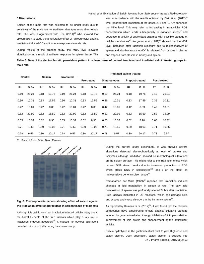
4.7 Electrophoretic peroxidase pattern
All the data were recorded in Table 6 and illustrated in Fig. 6. There
were no common bands in all groups. Irradiation caused qualitative
alterations represented by disappearance of the 1st and 6
th types with
appearance of one abnormal band with Rf 0.27 (B % 30.98) and
deviation of the 2nd
, 3rd and 5
th types to be appeared with Rfs 0.33,
0.40 and 0.62 (B % values 8.14, 9.02 and 18.89). Salicin
administration could not prevent the abnormalities occurred
qualitatively and represented by disappearance of normal bands,
appearance of abnormal bands and deviation of some normal bands
to be appeared with different data. It could not prevent the alterations
which were represented quantitatively in all irradiated salicin treated
groups by increasing B % of some normal bands.
The SI values showed that the lowest SI value (SI = 0.15) was
observed in irradiated group and the highest value (SI = 0.43)
noticed in the salicin treated group. In the irradiated salicin pre-
treated group, it was observed that all the bands were not matched
with all bands of the other groups.
Kamel et al. Evaluation of Salicin Isolated from Salix subserrata as a Radioprotector
UK J Pharm & Biosci, 2015: 3(2); 53
5 Discussions
Spleen of the male rats was selected to be under study due to
sensitivity of the male rats to irradiation damages more than female
rats. This was in agreement with Ezz, (2011)13
who showed that
spleen taken to study the ameliorative effect of radioprotector against
irradiation induced OS and immune responses in male rats.
During results of the present study, the MDA level elevated
significantly as a result of radiation exposure in spleen tissue. This
was in accordance with the results obtained by Dixit et al. (2012)46
who reported that irradiation at the doses 2, 6 and 10 Gy enhanced
the MDA level. This may refer to increasing in intracellular ROS
concentration which leads subsequently to oxidative stress47
and
decrease in activity of antioxidant enzymes with possible damage of
cellular membranes48
. Kergonou et al. (1981)49
showed that the MDA
level increased after radiation exposure due to radiosensitivity of
spleen and also because the MDA is released from tissues in plasma
and trapped from plasma in kidney and spleen.
Table 6: Data of the electrophoretic peroxidase pattern in spleen tissue of control, irradiated and irradiated salicin treated groups in
male rats
Control Salicin Irradiated
Irradiated salicin treated
Pre-treated Simultaneous Prepost-treated Post-treated
Rf. B. % Rf. B. % Rf. B. % Rf. B. % Rf. B. % Rf. B. % Rf. B. %
0.19 26.24 0.18 19.78 0.19 26.24 0.18 19.78 0.19 26.24 0.18 19.78 0.19 26.24
0.36 10.31 0.33 17.59 0.36 10.31 0.33 17.59 0.36 10.31 0.33 17.59 0.36 10.31
0.42 10.01 0.42 8.03 0.42 10.01 0.42 8.03 0.42 10.01 0.42 8.03 0.42 10.01
0.52 22.99 0.52 15.50 0.52 22.99 0.52 15.50 0.52 22.99 0.52 15.50 0.52 22.99
0.65 10.32 0.62 8.90 0.65 10.32 0.62 8.90 0.65 10.32 0.62 8.90 0.65 10.32
0.71 10.56 0.69 10.03 0.71 10.56 0.69 10.03 0.71 10.56 0.69 10.03 0.71 10.56
0.78 9.57 0.80 20.17 0.78 9.57 0.80 20.17 0.78 9.57 0.80 20.17 0.78 9.57
Rf.: Rate of Flow, B.% : Band Percent
Fig. 6: Electrophoretic pattern showing effect of salicin against
the irradiation effect on peroxidase in spleen tissue of male rats
Although it is well known that irradiation induced cellular injury due to
the harmful effects of the free radicals which play a key role in
irradiation induced apoptosis50
, it caused no obvious alterations
detected microscopically during the current study.
During the current study experiment, it was showed severe
alterations detected electrophoretically at level of protein and
isozymes although irradiation showed no morphological alterations
on the spleen surface. This might refer to the irradiation effect which
caused DNA strand breaks due to increased production of ROS
which attack DNA in splenocytes[51]
and / or the effect on
radiosensitive gene in spleen tissue12
.
Ramanathan and Misra (1979)52
reported that irradiation induced
changes in lipid metabolism in spleen of rats. The fatty acid
composition of spleen was profoundly altered 24 hrs after irradiation.
Free radicals implicated in OS reactions, which can damage cells
and tissues and cause disorders in the immune system53
.
As reported by Hamzaa et al. (2012)54
, it was found that the phenolic
compounds have ameliorating effects against oxidative damage
induced by gamma-irradiation through inhibition of lipid peroxidation,
improvement of lipid profile and enhancement of the antioxidant
activity.
Salicin hydrolyzes in the gastrointestinal tract to give D-glucose and
salicyl alcohol. Upon absorption, salicyl alcohol is oxidized into
Kamel et al. Evaluation of Salicin Isolated from Salix subserrata as a Radioprotector
UK J Pharm & Biosci, 2015: 3(2); 54
salicylic acid55
. Thus in the current study, the effect of salicin was
attributed to its hydrolysable form salicylic acid.
The effect of salicylic acid was compatible with an antioxidant profile:
it inhibited lipid peroxidation and increasedglutathione synthesis, but
did not modify the activitiesof glutathione-related enzymes56
. The
effect of the salicylic acid on lipid peroxidation may be explainable by
the ability of salicylic acid to absorb hydroxyl ions57
and thus impede
a main step in the process of membrane lipid peroxidation. Salicylic
acid might spare glutathione stores by avoiding factors that stimulate
glutathione depletion. Two observations support this notion: the
percentage of oxidized glutathione was reduced, and the activities of
enzymes associated with maintaining glutathione levels were not
modified substantially56
. Salicylic acid showed a direct effect on the
glutathione system. This effect may be related with the ability of both
to react with hydroxyl radicals57,58
.
On the other hand, Rebouch and Seim (1998)59
and Ibrahim et al.
(2007)60
recorded that salicin might induce elevation in activities of
the AOs as glutathione peroxidase in these tissues. It might act by
improving the turnover of fatty acids peroxidated by the free oxygen
radicals during normal metabolism. It might be added to category of
the natural products as olive oil, Nigella sativa oil and pomegranate
extract which play vital role in male fertility61
.
The present results showed that irradiation caused alterations in all
electrophoretic patterns in spleen tissue. This was in agreement with
results reported by many previous studies which suggested that
irradiation produces ROS that damage proteins, lipids and nucleic
acid62
.
The current experiment showed that irradiation decreased the
ordered structure of proteins. This was in agreement Moon and
Song, (2001)17
who suggested that radiation caused initial
fragmentation of polypeptide chains and, as result, subsequent
aggregation and degeneration of proteins by scavenging ROS
produced by irradiation. The difference in the protein fractions
separated electrophoretically after radiation exposure was in
agreement with Pleshakova et al., (1998)63
who reported that
irradiation caused a rise of protein carbonyl only in the cytoplasm
and mitochondria and this was followed by activation of histone –
specific proteases in nuclei of the irradiated rats. The proteins are
responsible for a specific biological process, so due to the difference
in protein bands between all the treated samples, the biological
processes may also be differed. The separation and characterization
of the individual proteins facilitate study of the chemical nature and
physiological function of each protein64
.
During the current study, irradiation caused alterations in the native
proteins detected electrophoretically. This was in accordance with
Davies and Delsignore (1987)65
who documented that irradiation
caused irreversible changes at the molecular level by breakage of
the covalent bonds of the polypeptide chains due to generation of the
hydroxy and superoxide anion radicals which modify the primary
structure of the proteins resulting in distortions of the secondary and
tertiary structures. The exposure of proteins to oxygen radicals
resulted in both non-random and random fragmentations66
. It was
reported that irradiation caused aggregation and cross-linking of
proteins. Covalent cross linkages are formed between free amino
acids and proteins, and between peptides and proteins in solution
after irradiation66,67
.
Data in the present study indicated that specific protein bands in
spleen tissue of irradiated rats differed (through disappearence in
some protein bands or appearance of new ones). Disappearance of
some protein bands in treated rats may be attributed to the effects of
irradiation which inhibits the synthesis and expression process of
these deleted proteins (qualitative effect). In addition, even the band
remained after irradiation, it usually differs in the amount of protein,
and this may be explained by that irradiation could not inhibit the
synthesis of this protein type, but it may be affected only on the
quantitative level.
Giometti et al. (1987)68
postulated that different mutations were
detected by the appearance of new proteins or by the quantitative
decrease in abundance of normally occurring proteins and the
electrophoresis can be used to detect the mutations reflected as
quantitative changes in the protein expression.
The difference in protein pattern may act as a tool to identify the
similarity index and genetic distance between the control and the
other treated samples. The chemical changes of the proteins that are
caused by irradiation are fragmentation, cross-linking, aggregation,
and oxidation by oxygen radicals that are generated in the radiolysis
of water69
.
Lipoproteins are lipid–protein complexes that contain large insoluble
glycerides and cholesterol with a superficial coating of phospholipids
and proteins synthesized in the liver70
. All lipoproteins carry all types
of lipid, but in different proportions, so that the density is directly
proportional to the protein content and inversely proportional to the
lipid content71
.
In the present study, irradiation caused alterations in the
electrophoretic lipoprotein pattern. This was in agreement with
Tsumura et al. (2001)72
who reported that the lipoproteins were more
susceptible to oxidative modifications resulting in small lipoproteins.
Bonnefont-Rousselot (2004)73
mentioned that the ROS can initiate
one-electron oxidation or one-electron reduction reactions on
numerous biological systems. The oxidative hypothesis classically
admits the involvement of the lipoproteins oxidation radiolytically.
Kamel et al. Evaluation of Salicin Isolated from Salix subserrata as a Radioprotector
UK J Pharm & Biosci, 2015: 3(2); 55
There was natural binding between protein and lipoproteins in the rat
tissues. These two tissues known to be involved in the processing of
the lipoproteins. The lipoproteins-binding protein has previously been
identified in adrenal cortical plasma membranes and concentration of
the binding protein was strongest in kidneys74
. So the alterations in
the protein pattern were associated with altering the lipoprotein
pattern in these tissues. The alterations in the lipoprotein pattern
may refer to the disturbances in the cholesteryl esterase required or
cholesterol hydrolysis75
.
Esterases are very large class of enzymes. They can break an ester
bond in the presence of water molecule76
. The esterase activity
stimulated breakdown of acetylcholine liberated during nervous
stimulation. They are very polymorphic, tissue-specific and variable
in populations of rats. Esterase zymograms showed that intensity
and number of the nonspecific esterase bands are very variable77
.
Esterases are found associated with membrane structures. There
was correlation between different esterases and the total esterase
activity in the different tissues78
.
According to results of the present study, irradiation caused
electrophoretic qualitative and quantitative alterations in the
electrophoretic esterase pattern in the spleen tissue. This may refer
to effect of irradiation on the protein pattern79
or the disturbances
occurred in the cholesterol metabolism as a result of radiation
exposure. The total esterase activities were correlated to the
cholesterol responses in rats80
.
As regards changes in electrophoretic mobility demonstrated in the
present study, it seemed that free radicals affect the integrity of the
polypeptide chain in the protein molecule causing fragmentation of
the polypeptide chain due to sulfhydral-mediated cross linking of the
labile amino acids as claimed by Bedwell et al. (1989)79
. The
changes in the fractional activity of different isoenzymes seemed to
be correlated with changes in the rate of protein expression
secondary to DNA damage initiated by free radicals81
.
During the current experiment, irradiation caused alterations in the
electrophoretic catalase and peroxidase patterns. This was in
agreement with Li et al. (2007)82
who showed that irradiation
decreased the peroxidase activity which may be due to that
irradiation-induced ROS markedly alters the physical, chemical and
immunologic properties of endogenetic antioxidant enzymes (CAT
and GPx), which further increase oxidative damage in cells.
The study showed that the decrease in CAT and GPx activity could
be attributed to the uncontrolled production of ROS and
accumulation of H2O2 whereby oxidative damage to enzymes can
cause a modification of their activity83,84
.
Bhatia and Manda (2004)85
reported that the electrophoretic
disturbances occurred as a result of irradiation in the peroxidase
pattern. This might be due to irradiation-induced depletion in the
level of reduced GSH, as well as GSH peroxidase. This leads to
elevation of the hydrogen peroxide and hence generation of the free
radicals86
. GPx utilizes GSH as a substrate to catalyse the reduction
of organic hydroperoxides and H2O287
.
Salicin and salicylic acid belonged to the phenolic compounds which
showed antioxidant activity due to their ability to scavenge free
radicals88
. The maintenance of normal protein levels after the
treatment with salicin may be due to trapping of these free radicals
by this compound, thus preventing DNA damage. Salicin was able to
overcome the disturbances in the protein pattern in the spleen tissue.
It showed protective effect against the irradiation due to its
antioxidative effect against attack of the free radicals. It prevented
the alterations in the proteins and hence the lipoproteins and
isozymes in the spleen tissue.
The current results are in line with that obtained by Cetin et al.
(2008)89
who suggested that salicin treatment considerably
increased the formation of antioxidant products in different tissues.
Salicin treatment minimized the irradiation effect and this may refer
to its effect on stimulation of activities of the different enzymes. The
mRNA expression levels of the enzymes increased after
administration of salicin whicjh may play role in regulation of these
enzymes on a transcriptional level90
.
6 Conclusions
The study concluded that salicin minimized the irradiation effect and
showed radioprotective effect against irradiation induced
ultrastructural and different electrophoretic changes in spleen tissue
of male rats.
7 Competing interest
The present study aimed to optimize salicin as a radioprotector
against effect of gamma irradiation on the spleen tissue in the hope
that this compound may be further explored as novel antioxidative
radioprotector.
8 Author’s contributions
MALAK and MSA carried out literature review and draft the
manuscript. HMS participated in collection of data and arranged in
tabular form. IA and WMK carried out the experimental work. All
authors read and approved the final manuscript.
9 References
1. Grupen C, Cowan G, Eidelman SD and Stroh T. Astroparticle
Physics. Springer-Verlag Berlinand aheidelberg GmbH &
Co.K. 2005; pp: 109.
Kamel et al. Evaluation of Salicin Isolated from Salix subserrata as a Radioprotector
UK J Pharm & Biosci, 2015: 3(2); 56
2. Lett JT. Damage to cellular DNA from particulate radiation,
the efficacy of its processing and the radiosensitivity of
mammalian cells. Emphasis on DNA strand breaks and
chromatin break. Radiat. Environ. Biophys. 1992; 31: 257-
277.
3. Daniniak N and Tann BJ. Utility of biological membranes as
indicators for radiation exposure: alterations in membrane
structure and function over time. Stem Cells. 1995; 13: 142-
152.
4. Spitz DR, Azzam EI, Li JJ and Gius D. Metabolic
oxidation/reduction reactions and cellular responses to
ionizing radiation: a unifying concept in stress response
biology. Cancer Metastasis Rev. 2004; 23 (3-4): 311-22.
5. Fedorova M, Kuleva N and Hoffmann R. Identification,
quantification, and functional aspects of skeletal muscle
protein-carbonylation in vivo during acute oxidative stress. J.
Proteome Res. 2010; 9 (5):2516 - 2526.
6. Umegaki K, Aoki S and Esashi T. Whole body X-ray
irradiation to mice decreases ascorbic acid concentrations in
bone marrow: comparison between ascorbic acid and vitamin
E. Free Radic. Biol. Med. 1995; 19 (4):493 - 7.
7. Koc M, Taysi S, Buyukokuroglu M and Bakan, N. The effect
of melatonin against oxidative damage during total-body
irradiation in rats. Radiat. Res. 2003; 160: 251-255.
8. Nwozo SO, Okameme PE and Oyinloye BE. Potential of
Piper guineense and Aframomum longiscapum to reduce
radiation induced hepatic damage in male Wistar rats.
Radiats Biol. Radioecol. 2012 ; 52(4): 363-369.
9. Witztum J. Splenic immunity and atherosclerosis: a glimpse
into a novel paradigm? J. Clin. Invest. 2002; 109: 721-724.
10. Hawas AM. The biosensitivity of certain organs in rats
exposed to low doses of γ-radiation. Journal
of Radiation Research and Applied Sciences. 2013; 6 (2) : 56
– 62.
11. Otsuka K and Sakai K. Effects of low dose-rate long-
term gamma-ray irradiation on DNA damage in
mouse spleen. International Congress Series. 2005; 1276 :
258-259.
12. Koo HJ, Jang S, Yang K, Kang SC, Namkoong S, Kim T,
Hang DTT and Sohn E. Effects of red ginseng on the
regulation of cyclooxygenase-2 of spleen cells in whole-
body gamma irradiated mice. Food and Chemical Toxicology.
2013; 62 : 839-846.
13. Ezz MK. The Ameliorative Effect of Echinacea Purpurea
Against Gamma Radiation Induced Oxidative Stress and
Immune Responses in Male Rats. Australian Journal of Basic
and Applied Sciences. 2011; 5(10): 506-512.
14. Mohamed MI. Sterility and some associated physiological
changes in the adult cowpea weevil, Callosobruchus
maculatus (F.). Ph. D. Thesis, Dept. Entomol., Fac, Sci. Ain
Shams Univ. 1990.
15. Hawkins CL, Morgan PE and Davies MJ. Quantification of
protein modification by oxidants. Free Radical Biology and
Medicine. 2009; 46: 965 - 988.
16. Hassan Heba A and AbdEl- Hafez Hanan F. The
comparison effects of two acetylcholine receptor modulator
on some biological aspects, protein pattern and detoxification
enzyme of the cotton leafworm, spodoptera littoralis. Egypt.
J. Agric. Res. 2009; 87 (2): 103-117.
17. Moon S and Song KB. Effect of gamma-irradiation on the
molecular properties of ovalbumin and ovomucoid and
protection by ascorbic acid. Food Chem. 2001; 74: 479-483.
18. Smutná M, Beňová K, Dvořák P, Nekvapil T, Kopřiva V and
Maté D. Protein carbonyls and traditional biomarkers in pigs
exposed to low-dose γ-radiation. Research in Veterinary
Science. 2013; 94 (2): 214-218.
19. Matés M. Effects of antioxidant enzymes in the molecular
control of reactive oxygen species toxicology. Toxicology.
2000; 153(1–3):83–104.
20. Halliwell B and Gutteridge JMC. Free radicals in biology and
medicine. 4th ed. New York: Oxford University Press. 2007.
21. Attia AA, ElMazoudy RH and El-Shenawy NS. Antioxidant
role of propolis extract against oxidative damage of testicular
tissue induced by insecticide chlorpyrifos in rats. Pesticide
Biochemistry and Physiology, 2012; 103: 87–93.
22. Gupta RC. Toxicology of organophosphates and carbamate
compounds, Elsevier Academic Press. 2006.
23. La Falci VS, Yrjö-Koskinen AE, Fazeli A, Holt WV and
Watson PF. Antioxidant combinations are no more beneficial
than individual components in combating ram sperm
oxidative stress during storage at 5 °C. Anim. Reprod. Sci.
2011; 129(3-4): 180-187.
24. Strzezek R, Koziorowska-Gilun M and Stawiszynska M.
Cryopreservation of canine semen: the effect of two extender
variants on the quality and antioxidant properties of
spermatozoa. Pol. J. Vet. Sci. 2012; 15(4): 721-726.
25. Kim SH, Kim HJ, Lee HO and Ryu SY. Apoptosis in growing
hair follicles following gamma irradiation and application for
the evaluation of radioprotective agents. In Vivo. 2003; 17 :
211 – 214.
26. Elshazly SA, Ahmed MM, Hassan HE and Ibrahim ZS.
Protective effect of L-carnitine against γ-rays irradiation-
induced tissue damage in mice. American Journal of
Biochemistry and Molecular Biology. 2012; 2 (3): 120 – 132.
Kamel et al. Evaluation of Salicin Isolated from Salix subserrata as a Radioprotector
UK J Pharm & Biosci, 2015: 3(2); 57
27. Weiss JF and Landauer MR. Protection against ionizing
radiation by antioxidant nutrients and phytochemicals.
Toxicology. 2003; 189 (1-2): 1–20.
28. Jourdier S. A Miracle Drug. 1999; http://www.chemsoc.org/
chembytes/ezine.
29. Vane JR, Flower RJ and Botting RM. History of aspirin and
its mechanism of action. Stroke. 1990; 21: IV12-23.
30. Akao T, Yoshino T, Kobashi K and Hattori M. Evaluation of
salicin as an antipyretic prodrug that does not cause gastric
injury. Planta. Med. 2002; 68: 714-718.
31. Singh BN, Singh BR, Singh RL, Prakash D, Dhakarey R,
Upadhyay G and Singh HB. Oxidative DNA damage
protective activity, antioxidant and anti-quorum sensing
potentials of Moringa oleifera. Food Chem. Toxicol. 2009; 47:
1109–1116.
32. Nzaramba MN, Reddivari L, Bamberg JB and Creighton MJ.
Antiproliferative activity and cytotoxicity of Solanum jamesii
tuber extracts on human colon and prostate cancer cells in
vitro. J. Agric. Food Chem. 2009; 57: 8308–8315.
33. Mabry TJ, Markham KR and Thomaas MB. The Systematic
Identification of flavonoids, Springer-Verlag, Berlin. 1970.
34. Kur'yanov AA, Bondarenko LT, Kurkin VA, Zapesochnaya
GG, Dubichev AA and Vorontsov ED. Determination of the
biologically active components of the rhizomes of Rhodiola
rosea. Translated from Khimiya Prirodnykh Soedinenii. 1991;
3:320-323.
35. Paget and Barnes. Evaluation of drug activities
pharmacometrics. Vol. (1), Edited by Laurence, D.R. and
Bacharach, A.L. Academic Press, London and New York.
1974; 135.
36. Ohkawa H, Ohishi N and Yagi K. Assay for lipid peroxides in
animal tissues by thiobarbituric acid reaction. Anal. Biochem.
1979; 95 : 351 – 358.
37. Tánaka K. High resolution scanning electron microscopy of
the cell. Biology of the Cell. 1989; 65: 89-98.
38. Bradford MM. A rapid and sensitive method for the
quantitation of microgram quantities of protein utilizing the
principle of protein-dye binding. Anal. Biochem. 1976; 72:
248-254.
39. Laemmli UK. Cleavage of structural proteins during the
assembly of the head of Bacteriophage T4. Nature. 1970 ;
227: 680-685.
40. Hames BD. One-dimensional polyacrylamide gel
electrophoresis. In: Gel electrophoresis of proteins: B.D.
Hames B.D. and Rickwood D., 2nd ed.. Oxford university
press, NY. 1990; 1-147.
41. Chippendale GM and Beak SD. Haemolymph proteins of
Osirinla nubilalis (Hubner): during diapauses prepupa
differentiation . J. Insect Physiolo. 1966; 12 : 1629-1638.
42. Rescigno A, Sanjust E, Montanari L, Sollai F, Soddu G,
Rinaldi AC, Oliva S and Rinaldi A. Detection of laccase,
peroxidase, and polyphenol oxidase on a single
polyacrylamide gel electrophoresis, Anal. Lett. 1997; 30 (12):
2211.
43. Siciliano MJ and Shaw CR. Separation and visualization of
enzymes on gels, in Chromatographic and Electrophoretic
Techniques, Vol. 2, Zone Electrophoresis, Smith, I., Ed.,
Heinemann, London. 1976; p. 185.
44. Baker CMA and Manwell C. Heterozygosity of the sheep:
Polymorphism of 'malic enzyme', isocitrate dehydrogenase
(NADP+), catalase and esterase. Aust. J. Biol. Sci. 1977; 30
(1-2) : 127-40.
45. Nei M and Li WS. Mathematical model for studing genetic
variation in terms of restriction endonuclease. Proc. Natl.
Acad. Sci., USA. 1979; 76 : 5269 – 5273.
46. Dixit AK, Bhatnagar D, Kumar V, Chawla D, Fakhruddin K
and Bhatnagar D. Antioxidant potential and radioprotective
effect of soy isoflavone against gamma irradiation-induced
oxidative stress. J. Funct. Foods. 2012; 4: 196–206.
47. Maurel A, Hernandez C and Kunduzova O. Age-dependent
increase in hydrogen peroxide production by cardiac
monoamine oxidase A in rats. Am. J. Physiol. Heart Circ.
Physiol. 2003; 284: H1460 – H1467.
48. El Habit OHM, Saada HN, Azab KS, Abdel Rahman M and El
Malah DF. The modifying effect of B-carotene on gamma
radiation-induced elevation of oxidative reactions and
genotoxicity in male rats. Mutation Research. 2000; 466:
179-186.
49. Kergonou J, Bernard P, Braquet M and Rocquet G. Effect of
whole-body gamma irradiation on lipid peroxidation in rat
tissues. Biochimie. (1981). 63 (6): 555-559.
50. Vijayalaxmi RJ, Reiter DX, Tan TS, Herman CR and Thomas
J. Melatonin as a radioprotective agent: a review, Int. J.
Radiat. Oncol. Biol. Phys. 2004; 59: 639–653.
51. Richi B, Kale RK and Tiku AB. Radio-modulatory effects of
Green Tea Catechin EGCG on pBR322 plasmid DNA and
murine splenocytes against gamma-radiation induced
damage. Mutation Research. 2012; 747: 62– 70.
52. Ramanathan R and Misra UK. Spleen lipids: effect of whole
body gamma irradiation and radioprotective chemicals.
Biochem. Exp. Biol. 1979; 15(4):361-9.
53. Shahwar D, Sana U and Ahmad N. Synthesis and evaluation
of acetylcholineesterase inhibitory potential and antioxidant
Kamel et al. Evaluation of Salicin Isolated from Salix subserrata as a Radioprotector
UK J Pharm & Biosci, 2015: 3(2); 58
activity of benzothiazine derivatives. Turkish Journal of
Chemistry. 2013; 37: 262 – 270.
54. Hamzaa RG, El Shahat AN and Mekawey HMS. The
Antioxidant Role of Mulberry (Morusalba L.) Fruits in
Ameliorating the Oxidative Stress Induced in γ-Irradiated
Male Rats. Biochem. Anal. Biochem. 2012; 1(8): 122.
55. Chrubasik S and Eisenberg E. Willow Bark. 2004;
<http://www.rzuser. uni-heidelberg.de/~cn6/iasp-sig-
rp/willow.html> (accessed 11.03.04).
56. De La Cruz JP, Guerrero A, Gonzalez-Correa JA, Arrebola
MM and Sanchez de la Cuesta F. Antioxidant Effect of
Acetylsalicylic and Salicylic Acid in Rat Brain Slices
Subjected to Hypoxia. Journal of Neuroscience Research.
2004; 75: 280–290.
57. Sagone AL and Husney RM. Oxidation of salicylates by
stimulated granulocytes: evidence that these drugs act as
free radical scavengers in biological systems. J. Immunol.
1987; 138: 2177–2183.
58. Li PA, Liu GJ, He QP, Floyd RA and Siesjo BK. Production of
hydroxyl free radicals by brain tissues in hyperglycemic rats
subjected to transient forebrain ischemia. Free Radic. Biol.
Med. 1999 ; 27:1033–1040.
59. Rebouche CJ and Seim H. Carnitine metabolism and its
regulation in microorganisms and mammals. Annu. Rev.
Nutr. 1998; 18:9-61.
60. Ibrahim K, Seyithan T, Mustafa E, Ihsan K, Akcahan G,
Orhan S and Korkmaz S. The effect of L- carnitine in the
prevention of ionizing radiation induced cataracts; a rat
model.Graefe _s. Archive Clinic. and Exp. Ophthalm. 2007;
245(4): 588- 594.
61. Aitken RJ, Smith TB, Lord T, Kuczera L, Koppers AJ and
Naumovski N. On methods for the detection of reactive
oxygen species generation by humanspermatozoa: analysis
of the cellular responses to catechol oestrogen, lipid
aldehyde, menadione and arachidonic acid. Andrology. 2013;
1(2): 192-205.
62. Nair CKK, Parida D and Nomura T. Radioprotectors in
radiotherapy. J. Rad. Res. 2001; 42:21-37.
63. Pleshakova OV, Kutsyi MP, Sukharev SA, Sadovnikov VB
and Gaziev AI. Study of protein carbonyls in subcellular
fractions isolated from liver andspleen of old and γ-
irradiated rats. Mechanisms of Ageing and Development.
1998; 103(1): 45-55.
64. Cheeseman K. In DNA and Free Radicals (Halliwell, B. and
Aruoma, O. I., eds.), 1993; pp. 109-144, Ellis Horwood,
Chichester.
65. Davies KJA and Delsignore ME. Protein damage and
degradation by oxygen radicals III. Modification of secondary
structure and tertiary structure. J. Biol. Chem. 1987; 262:
9908-9913.
66. Filali-Mouhim A, Audette M, St-Louis M, Thauvette L,
Denoroy L, Penin F, Chen X, Rouleau N, Le Caer JP,
Rossier J, Potier M and Le Maire M. Lysozyme fragmentation
induced by γ-radiolysis. Int. J. Radiat. Biol. 1997; 72(1): 63-
70.
67. Garrison WM. Reaction mechanisms in the radiolysis of
peptides, polypeptides, and proteins. Chem. Rev. 1987; 87:
381-398.
68. Giometti CS, Gemmell MA, Nance SL, Tollaksen SL and
Taylor J. Detection of heritable mutations as quantitative
changes in protein expression. J. Biol. Chem. 1987; 262:
12764 – 12767.
69. Cho Y and Song KB. Effect of g-irradiation on the molecular
properties of BSA and b-lactoglobulin. J. Biochem. Mol. Biol.
2000; 33: 133-137.
70. Havel Rj and Kane Jp. Structure and metabolism of plasma
lipoproteins. In: CR Scriver, AL Beaudet, WS Sly and D
Valle, eds. The metabolic and molecular basis of inherited
disease, 7th edition. McGraw- Hill, USA. 1995; 1841-1851.
71. Bass KM, Newschaffer CJ, Klag MJ and Bush TL. Plasma
lipoprotein levels as predictors of cardiovascular death in
women. Arch. Intern. Med. 1993; 153 (19): 2209-16.
72. Tsumura M, Kinouchi T, Ono S, Nakajima T and Komoda T.
Serum lipid metabolism abnormalities and change in
lipoprotein contents in patients with advanced-stage renal
disease. Clinica. Chimica. Acta. 2001; 314: 27 – 37.
73. Bonnefont-Rousselot D. Gamma radiolysis as a tool to study
lipoprotein oxidation mechanisms. Biochimie. 2004; 86: 903-
911.
74. Fidge NH. Partial purification of a high density lipoprotein-
binding protein from rat liver and kidney membranes.
Federation of European Biochemical Societies. 1986; 199:
265-268.
75. Satoh T. Toxicological implications of esterases—From
molecular structures to functions. Toxicology and Applied
Pharmacology. 2005; 207: S11 – S18.
76. Koitka M, Höchel J, Gieschen H and Borchert H. Improving
the ex vivo stability of drug ester compounds in rat and dog
serum: Inhibition of the specific esterases and implications on
their identity. Journal of Pharmaceutical and Biomedical
Analysis. 2010; 51: 664 - 678.
77. Verimli R, Yigit N, Çolak E and Sozen M. Nonspecific
Esterase Patterns of Rattusnorvegicus (Berkenhout, 1769) in
Western Turkey.Turk J. Biol. 2000 ; 24 : 825–831.
Kamel et al. Evaluation of Salicin Isolated from Salix subserrata as a Radioprotector
UK J Pharm & Biosci, 2015: 3(2); 59
78. Tegelstrom H and Ryttman H. Sex differences and
androgenic regulation of esterases in the house mouse.
Hereditas. 1981; 94: 189-201
79. Bedwell S, Dean RT and Jessup W. The action of defined
oxygen centered free radicals on human low-density
lipoprotein. Biochem. J. 1989; 262: 707-712.
80. Beynen AC, Boogaard A, Van Laack HLJM, Weinans GJB
and Katan MB. Abstr. Commun.15th FEBS Meet., Brussels.
1983; p. 173.
81. El-Zayat EM. Isoenzyme Pattern and Activity in Oxidative
Stress-Induced Hepatocarcinogenesis: The Protective Role
of Selenium and Vitamin E. Research Journal of Medicine
and Medical Sciences. 2007; 2(2): 62-71.
82. Li XL, Zhou AG and Li XM. Inhibition of Lycium barbarum
polysaccharides and Ganoderma lucidum polysaccharides
against oxidative injury induced by γ-irradiation in rat liver
mitochondria. Carbohydrate Polymers. 2007; 69: 172–178.
83. Kregel K and Zhang H. An integrated view of oxidative stress
in aging: basic mechanisms, functional effects, and
pathological considerations. Am. J. Physiol. Regul. Integr.
Comp. Physiol. 2007 ; 292: 18-36.
84. De Freitas RB, Augusti PR, De Andrade ER, Rother FC,
Rovani BT, Quatrin A, Alves NM, Emanuelli T and
Bauermann LF. J. Black Grape Juice Protects Spleen from
Lipid Oxidation Induced by Gamma Radiation in Rats. Food
Biochem. 2014; 38(1): 119-127.
85. Bhatia AL and Manda K. Study of pre-treatment of melatonin
against radiation-induced oxidative stress in mice. Environ.
Toxicol. Pharmacol. 2004; 18: 13 – 20.
86. Mills GC. Glutathione peroxidase and the destruction of
hydrogen peroxide in animal tissues. Archives of
Biochemistry and Biophysics. 1960; 86: 1-5.
87. Ray G and Husain SA. Oxidants, antioxidants and
carcinogenesis. Ind. J. Exp. Biol. 2002; 42: 1213-1232.
88. Madrigal-Carballo S, Rodriguez G, Krueger CG, Dreher M
and Reed JD. Pomegranate (Punica granatum L.)
supplements: authenticity, antioxidant and polyphenol
composition. J. Funct. Food. 2009; 1: 324 - 329.
89. Cetin A, Kaynar L, Kocyigit I, Hacioglu SK, Saraymen R,
Ozturk A, Orhan O and Sagdic O. The effect of grape seed
extract on radiation-induced oxidative stress in the rat liver.
Turk. J. Gastroenterol. 2008; 19(2): 92-98.
90. Yeh CT and Yen GC. Effects of phenolic acids on human
phenolsulfotransferase in relation to their antioxidant activity.
J. Agric. Food Chem. 2003; 51: 1474–9.


![Dr. Amol Kharatmcpledu.org/doc/ARK/TY Pcog/Glycosides [Compatibility Mode].pdf · for example: 1-salicin –salix-2-cascaroside _cascara 3-aloin- Aloe vera 4- sennoside – senna-5-frangulin](https://img.pdfslide.us/doc/110x75/5e76290bde555b109365b3e1/dr-amol-pcogglycosides-compatibility-modepdf-for-example-1-salicin-asalix-2-cascaroside.jpg)