Embed Size (px)
Citation preview
Avian Digestive System


Plan of Talk
Introduction Digestive system organs and their functions Accessory digestive glands Mechanism of enzymes production and activation Mechanism of hunger

Introduction
Introduction
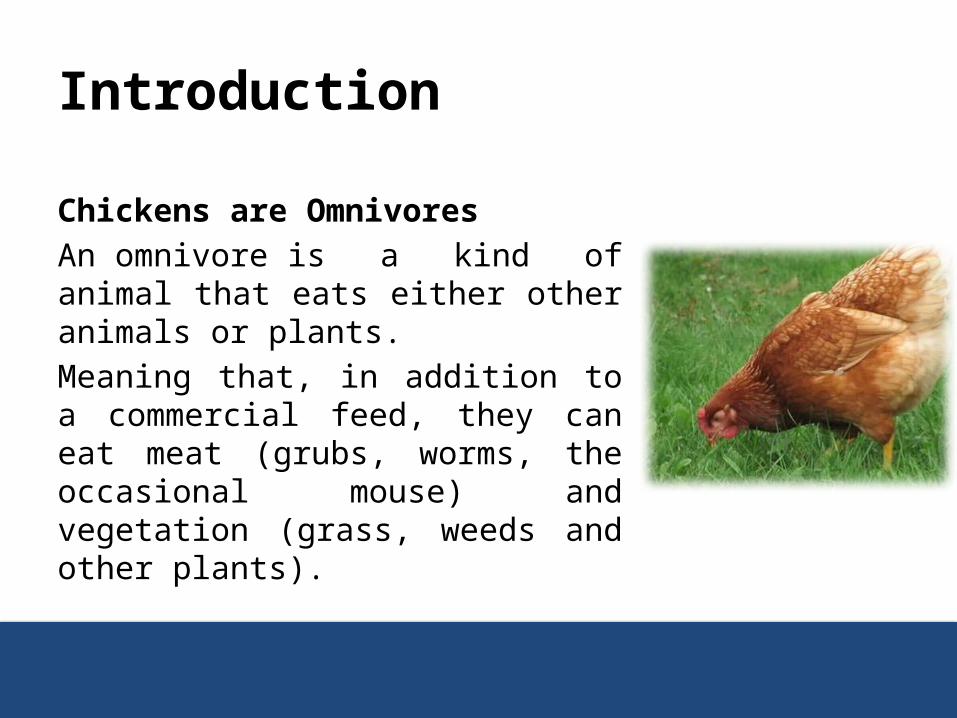
Chickens are Omnivores An omnivore is a kind of animal that eats either other animals or plants.Meaning that, in addition to a commercial feed, they can eat meat (grubs, worms, the occasional mouse) and vegetation (grass, weeds and other plants).
The Digestive System
The digestive system is responsible for the break down of complex non absorbable components like;
1. Carbohydrate2. Protein3. Fats
into relatively simplest and absorbable units like;4. Glucose5. Amino acid 6. Fatty acids


Digestion
Digestion is completed by the action of various enzymes secreted by different organs and accessory gland of the digestive system.

Digestive System OrgansDigestive system is divided into following parts;
1. Mouth2. Pharynx3. Esophagus/gullet4. Crop5. Proventriculus6. Gizzard7. Small intestine8. Caeca9. Large intestine10. Cloaca11. Vent
Digestive System Accessory Glands
The glands which aid in the process of digestion are known as accessory digestive glands, they are;
1. Salivary glands2. Liver3. Pancreas

Digestive System Organs And Their Functions
Organs and FunctionsThe Mouth
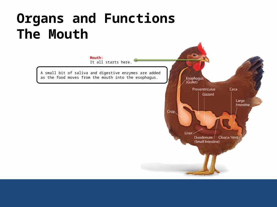
Mouth: It all starts here.
A small bit of saliva and digestive enzymes are added as the food moves from the mouth into the esophagus.
Organs and FunctionsThe Mouth
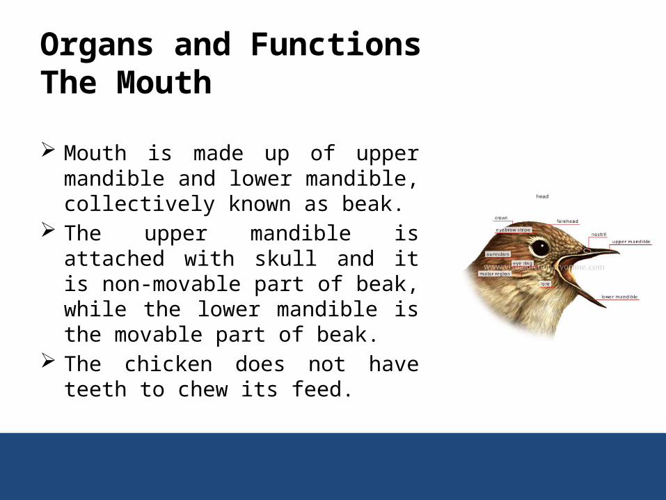
Mouth is made up of upper mandible and lower mandible, collectively known as beak.
The upper mandible is attached with skull and it is non-movable part of beak, while the lower mandible is the movable part of beak.
The chicken does not have teeth to chew its feed.

Organs and FunctionsThe Mouth
The roof of mouth is made up of hard palate that is divided by a long narrow slit in the center that is opened to the nasal passage.
The soft palate is absent in chicken.

Organs and FunctionsThe Mouth
The slit in the hard palate and the absence of soft palate make it impossible for the birds to create a vacuum to draw the water or feed into the mouth
Thus birds have to scoop up the water when drinking, elevates its head, and then let the water run down the gullet by the action of gravity.

Organs and FunctionsThe Mouth
The base of mouth is made up of tongue and it has rough surface at the beak to help force the feed into esophagus or gullet.
The mouth is also very sensitive to temperature differences. The base of the tongue has papilla, which contains very few
numbers of taste buds. The taste buds help to taste the feed
Organs and FunctionsThe Esophagus
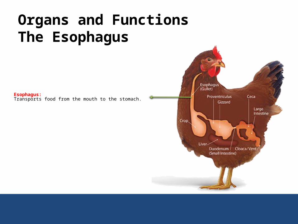
Esophagus: Transports food from the mouth to the stomach.
Organs and FunctionsThe Pharynx
Pharynx is a common passageway for feed and air, it is divided into two parts:
1. Esophagus2. Larynx

Organs and FunctionsThe Esophagus
DescriptionEsophagus is a tube like structure that extends from mouth to Proventriculus.
Functions1. It helps carry feed from mouth towards Proventriculus.2. Secrets mucous for lubrication.

Organs and FunctionsThe Crop
Crop: A pouch in the esophagus used to store food temporarily before moving it on to the stomach.
Feed can remain for up to 12 hours
Organs and FunctionsThe Crop
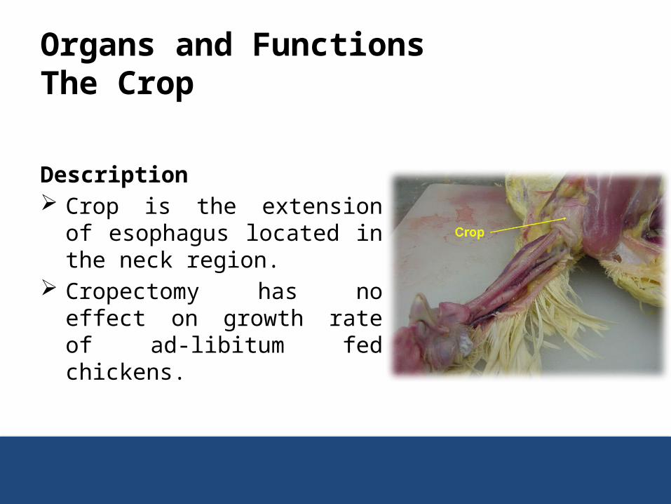
Description Crop is the extension of esophagus
located in the neck region. Cropectomy has no effect on
growth rate of ad-libitum fed chickens.

Organs and FunctionsThe Crop
Functions:1. Storage of feed, so, when the Proventriculus or gizzard is
empty the feed by pass the crop.2. Little digestion takes place with the action of salivary
amylase.1. Amylase activity at this site comes from either salivary secretions,
intestinal reflux, or plant and/or bacterial sources2. Starch is hydrolyzed within the crop where it can either be absorbed,
converted to alcohol, lactic or other acids

Organs and FunctionsProventriculus
Stomach:Principally the organ where food is broken into smaller units. It has two parts:1. Proventriculus
For storage. 2. Gizzard
Is a muscular part of the stomach that uses grit to grind grains and fiber into smaller particles.
Digestive enzymes are added to the mix and physical grinding of the food occurs.

Organs and FunctionsProventriculus
Description Also called glandular stomach or true stomach. It is a specialized enlargement of the gullet just before entry
into the gizzard.
Organs and FunctionsProventriculus
Functions: Production of gastric juice;
– Gastric juice is made up of the proenzyme known as pepsinogen and hydrochloric acid, both are produced by oxyntico-peptic cells.
– Gastric juice produced in response to protein content in diet.– Acid secretion of chickens is high relative to mammals.– Amylolysis occurs in the crop, it is not evident in the ventriculus.

Organs and FunctionsGizzard
Description Also called Muscular Stomach or Ventriculus. It is made up of two pairs of powerful muscles capable of
crushing and grinding the feed particle, which act as the bird’s teeth.
(The tunica muscularis of gizzard is made up of two layers of smooth muscles, inner circular & outer longitudinal)
Organs and FunctionsGizzard
Functions1. It performs powerful muscular contraction, which ultimately
leads to crushing and grinding of feed particles.2. This process is aided by the presence of grit or rocks present
in the gizzard.3. The gizzard performs 2-5 contractions per minute according
to the consistency of the feed particle!!!!!!!
Organs and Functions
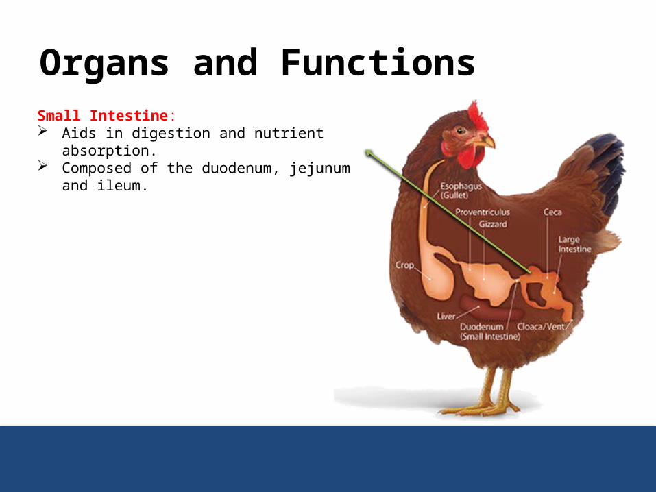
Small Intestine: Aids in digestion and nutrient absorption. Composed of the duodenum, jejunum and ileum.
Organs and FunctionsSmall Intestine
Small intestine is 1.5 meters long in the adult bird. It has three parts;
1. Duodenum2. Jejunum3. Ileum
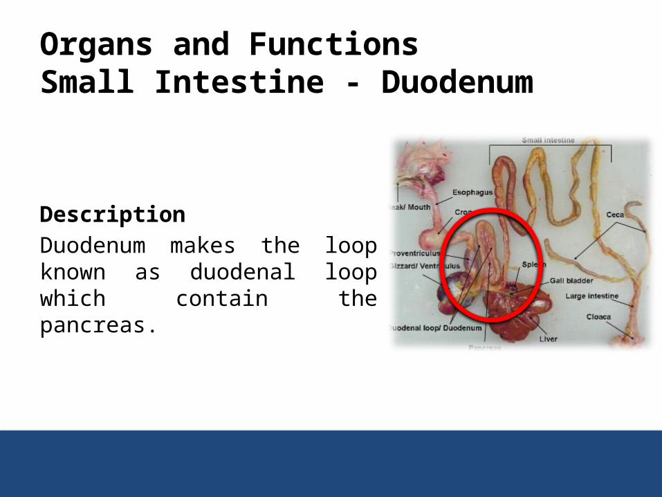
Organs and FunctionsSmall Intestine - Duodenum
DescriptionDuodenum makes the loop known as duodenal loop which contain the pancreas.

Organs and FunctionsSmall Intestine - Duodenum
Function Digestion of carbohydrates, protein, and fat take place in the
small intestine with the help of intestinal juice, pancreatic juice, and secretion of liver known as bile.

Organs and FunctionsSmall Intestine – Jejunum the Ileum
The jejunum and the ileum, together about 120 cm long.
Starts at the caudal end of the duodenum where the bile and the pancreatic duct papilla are located.
Ends at the ileo-caecal-colic junction (This junction is where the small intestine, the two caeca and the colon all meet)
Organs and FunctionsSmall Intestine – Jejunum the Ileum
This portion of the small intestine is similar in structure to the duodenum except that:1. It is suspended in the mesentery2. Villi are shorter3. There is less lymphoid tissue

Organs and FunctionsSmall Intestine – Jejunum the Ileum
Meckel’s Diverticulum is a constant feature about half way along the small intestine and appears as a small projection on the outer surface of the small intestine.
This projection is where the yolk sac was attached during the development of the embryo.
Organs and FunctionsSmall Intestine - Intestinal Juice
Intestinal juice contains variety of enzymes such as:1. Amylase, carbohydrates digestion.2. Invertase, carbohydrates digestion.3. Trypsin, proteins digestion.
Organs and FunctionsSmall Intestine - Pancreatic Juice
Similarly, pancreatic juice contain variety of enzymes that do take part in digestion of carbohydrates, protein and fat.

Organs and FunctionsSmall Intestine - Bile
The bile produced from the liver is responsible for emulsification of fat which is then digested by variety enzymes.

Organs and FunctionsSmall Intestine - Absorption
After completion of digestion, the end product of carbohydrate (glucose), protein (amino acid), fats (fatty acid) are absorbed by the finger like projections of small intestine known as villi.
The amino acid, fatty acids and glycerol are absorbed into the lymphatic vessels
These end products are ultimately reach the liver via portal vein.
Organs and Functions
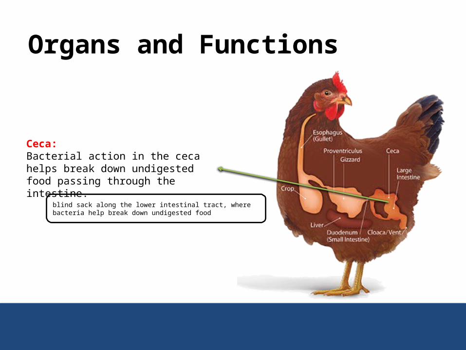
Ceca: Bacterial action in the ceca helps break down undigested food passing through the intestine.
blind sack along the lower intestinal tract, where bacteria help break down undigested food
Organs and FunctionsCaeca
Description These are two blind pouches located between the small
intestine and large intestine having a length of 1.5 cm. The ceca empty their contents two or three times a day. They produce pasty droppings that often smell worse than
regular droppings and often mustard to dark brown in color. The frequency of cecal droppings, as well as their appearance
among regular droppings, tells you the chicken’s digestive tract is functionally normally.
Organs and FunctionsCaecaFunction The function of Caeca is not clear. It is thought that it takes part in digestion of carbohydrate,
proteins, and crude fiber with the help of bacterial action. Re-absorption of water takes place in the ceca. Fermentation of coarse materials and production of the eight B
vitamins (Thiamine, riboflavin, niacin, pantothenic acid, pyridoxine, biotin, folic acid and vitamin B12) also occur in the ceca.
Because the ceca are located near the end of the digestive tract there is minimal absorption of any nutrients released.

Organs and Functions
Large Intestine: Functions primarily to absorb water, dry out indigestible foods and eliminate waste products.
absorbs water and dries out indigestible foods.
Organs and Functions Large Intestine
Description Large intestine is much shorter when compared to small
intestine and caecum The length of large intestine is 10 cm. The diameter is twice the diameter of small intestine. It extends from small intestine to cloaca.
Function It helps to maintain water balance by water absorption.

Organs and Functions
Cloaca: Where the digestive, urinary and reproductive systems meet. Vent: The external opening of the cloaca that passes waste to the outside.
Organs and FunctionsCloaca and Vent
Description It is the bulbous/enlarged area located at the end of large
intestine It is also known as common sewer because it receives the
openings from digestive system, reproductive system and urinary system
External opening of the cloaca is known as vent and its size is variable depending upon the productivity of the birds.
Accessory Digestive Glands
Accessory Digestive Glands
There are three accessory digestive glands which play a vital role in the process of digestion
1. Salivary Glands2. Pancreas3. Liver
Accessory Digestive GlandsSalivary Glands
It is responsible for production of saliva. Its secretions ranges from 7 to 30 ml per day.

Accessory Digestive GlandsSalivary GlandsThe salivary glands are:1. Maxillary – in the roof of the mouth.2. Palatine – on either side of the nasal opening in the roof of the mouth.3. Apheno-pteryoid glands – in the roof of the pharynx on each side of the
common opening for the eustachian tubes (the eustachian tubes connect the middle ear to the mouth and their function is to equalise the air pressure on each side of the tympanic membrane in the ear)
4. Anterior sub-mandible glands – in the angle formed by the union of the upper and lower beaks or mandibles.
5. Posterior sub-mandibular glands.6. Lingual glands – in the tongue.7. Crico-arytenoid glands – around the glottis.8. A small gland in the angle of the mouth.
Accessory Digestive GlandsSalivary GlandsThe saliva has following functions:1. Lubrication of the feed.2. Digestion, it contains salivary amylase which is responsible
for carbohydrates digestion. 3. Acts as a buffer, it contains bicarbonate and other salts.4. Helps tasting the feed.5. Protects the mucous membrane and keeps it moist.6. Helps regulate the body temperature.7. Contain enzyme known as muramidase which is bacteriocidal
in nature and thus it produces the local immunity.

Accessory Digestive GlandsPancreas
Pancreas produces a pancreatic juice. Its pH is 6.9 It is released in the distal end of the loop of duodenum. Pancreatic juice contains four kinds of enzymes;
1. Proteolytic Enzymes2. Lipolytic Enzyme3. Carbohydrate splitting Enzymes4. Nucleolytic Enzymes
Accessory Digestive GlandsPancreas
A- Proteolytic EnzymesThere are five different kinds of proteolytic enzymes
1. Trypsinogen2. Chymotrypsinogen A3. Chymotrypsinogen B4. Procarboxy peptidase A5. Procarboxy peptidase B
These enzymes are responsible for the break down of protein molecules into simpler units.

Accessory Digestive GlandsPancreas
B- Lipolytic EnzymeThere are three type of lipolytic enzymes which are produced by the pancreas;
1. Phospholipase, lipid breakdown.2. Pancreatic lipase, lipid breakdown.3. Cholesterol esterase, esterification of cholesterol.

Accessory Digestive GlandsPancreas
C- Carbohydrate splitting Enzymes These consist of:
1. Pancreatic amylase, acts on the starch and converts it into simpler units.
2. Invertase, acts on the sucrose and convert it into simpler sugar.
Accessory Digestive GlandsPancreas
D- Nucleolytic EnzymesThere are two kinds of nucleolytic enzymes:
1. Ribonuclease2. Deoxyribonuclease
Accessory Digestive GlandsPancreas
Besides enzymes, pancreatic juice also contains cations and anions.Cations: Na+, K+, Mg++, etc. These act as buffer, cofactors, and osmotic regulators.Anions: HCO3 These mainly act as buffer and osmotic regulators.
Organs and Functions
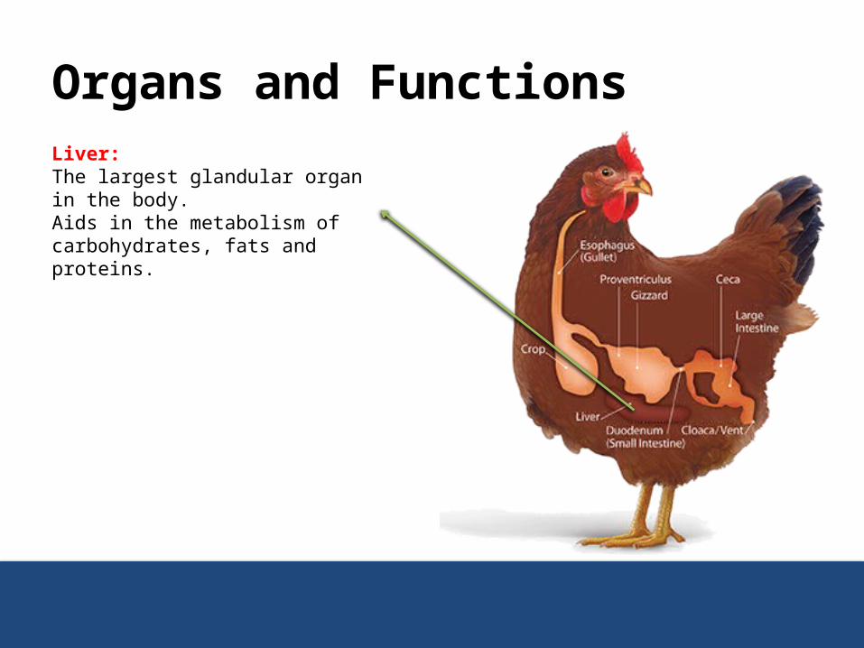
Liver: The largest glandular organ in the body. Aids in the metabolism of carbohydrates, fats and proteins.

Accessory Digestive GlandsLiver
Liver is a bilobed structure and it performs the following functions1. Detoxification.2. Store of vitamins and carbohydrates, carbohydrates are stored
in the form of glycogen.3. Formation plasma protein like albumin and globulin.4. It activates and inactivates the protein and peptide hormones.5. Liver is a site for the destruction of old RBCs.6. Formation of bile, which is responsible for the emulsification of
the fat.
Mechanism Of Enzyme Production And Activation
Mechanism Of Enzyme Production And Activation
The activities of gastrointestinal tract are controlled by:1. Nervous system2. Endocrine system

Mechanism Of Enzyme Production And Activation
The nervous system, in particular, the autonomic nervous is responsible for controlling the activity of gastrointestinal tract.
This system has two parts1. Parasympathetic nervous system2. Sympathetic nervous system
Mechanism Of Enzyme Production, Nervous system
The parasympathetic nervous system activates the gastrointestinal tract while sympathetic nervous system activates as well as deactivates the gastrointestinal tract
Mechanism Of Enzyme Production, Nervous system
1. Feed enter the oral cavity2. Visual stimuli3. Smell 4. Taste They stimulate the parasympathetic which to the produce saliva.
Mechanism Of Enzyme Production, Nervous system
Feed enters the Proventriculus and the walls are stretchedThis stimulate the release of Gastrin hormone which stimulates secretion of gastric juice.
Feed enters small intestine Duodenum produces secretin hormone which stimulate the pancreas to produce pancreatic juice.
Mechanism Of Enzyme Production, Nervous system
Fats in the duodenumDuodenum produces cholecystokinin hormone which stimulates gall bladder to release bile.

Mechanism Of Enzyme Activation, Endocrine system In the gastric juice Pepsinogen is activated into pepsin by HCl.
In the pancreatic juice Trypsinogen is activated by another enzyme known as
enterokinase, which is released from duodenum.
Carboxy peptidase ← Procarboxy Peptide
Chymotrypsin← Chymotrypsinogen
Mechanism of Hunger
Mechanism of Hunger
There are two systems or centers located in the brain or liver which controls the feeding behavior of animals
1. Satiety center2. Appetite center

Mechanism of Hunger
Satiety Center It is located in the liver of the chicken, while in other animals
it is located in the brain. This center is also known as glucostatiey Centre. Level of glucose in the blood activates and stimulates the
satiety center leading to cessation of feed in take.
Mechanism of Hunger
Appetite Center The stimulation of this Centre results in feed intake or hunger. This centre is stimulated by low concentration of glucose in
the blood. This is located in the brain.