Embed Size (px)
Citation preview
Chapter 19-Digestive System
Chapter 19.1-19.2: Digestive System Overview

Organ Systems
1) Gastointestinal (GI) Tract- Alimentary Canal: continuous tube extending from
mouth to anus- Mouth, pharynx, esophagus, stomach, sm.
intestines, lg. intestines- About 5-7 meters (16-23ft) long
2) Accessory Organs- Aid in the physical and chemical breakdown of food- Teeth, tongue, salivary glands, liver, gallbladder,
pancreas


Basic Functions
1) Ingestion: taking in food
2) Secretion: release ≈ 7 liters of water, acid, buffers, enzymes
3) Mixing and Propulsion: contraction/relaxation of smooth muscle pushes food/secretion mixture down GI tract

Basic Functions
4) Digestion: mechanical and chemical
5) Absorption: entrance of fluids, ions, and small molecules into the lining cells of GI Tract
6) Defecation: any non-absorbed material eliminated from body

Four Layers
1) Mucosa- Inner epithelium of GI tract
2) Submucosa- Connective tissue- Contains blood and nerve supply
3) Muscularis- Thick layer of muscle
4) Serosa - Outer layer- Simple squamous epithelium
SUBMUCOSA
MUSCULARIS
SEROSA
MUCOSA

Chapter 19.3-Mouth

Oral Cavity
- Formed by the cheeks, hard and soft palates, and tongue
- Hard palate: roof of the mouth- Soft palate: back of the mouth, including the uvula
- Tongue: accessory organ composed of skeletal muscle covered by a mucous membrane
- Lingual frenulum: a folded membrane in the midline of the undersurface that limits movement
- Papillae: small projections on the upper surface and sides
Lingual frenulum
Hard palate
Soft palateUvula

Salivary Glands
- Three pairs (accessory organs) that lie outside oral cavity- Release saliva into ducts emptying into oral cavity
1) Parotid glands2) Submandibular glands3) Sublingual glands
- Saliva = 99.5% water and 0.5% solutes- Lubricates food- Salivary amylase begins digestion of starches in mouth
Anatomy of Teeth
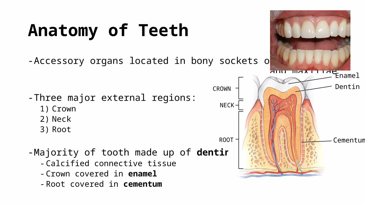
- Accessory organs located in bony sockets of mandible and maxillae
- Three major external regions:1) Crown2) Neck3) Root
- Majority of tooth made up of dentin- Calcified connective tissue- Crown covered in enamel- Root covered in cementum
CROWN
NECK
ROOT
Enamel
Dentin
Cementum

Anatomy of Teeth
- Dentin encloses the pulp cavity- Connective tissue containing blood/lymphatic
vessels and nerves- Extends into root canals CROWN
NECK
ROOT
Enamel
Dentin
Cementum
Pulp in pulp cavity
Root canal
Types of Teeth
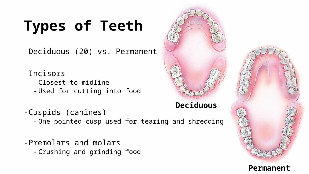
- Deciduous (20) vs. Permanent (32)
- Incisors- Closest to midline- Used for cutting into food
- Cuspids (canines)- One pointed cusp used for tearing and shredding
- Premolars and molars- Crushing and grinding food
Deciduous
Permanent

Digestion in the Mouth
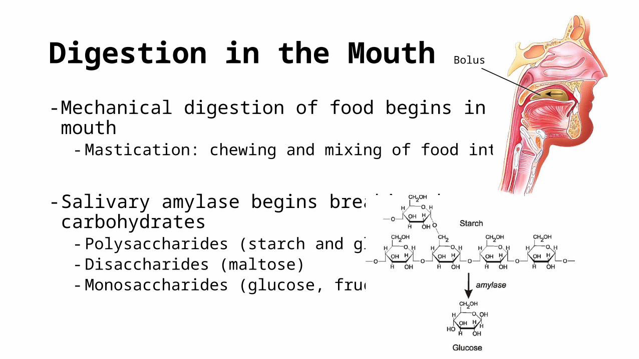
- Mechanical digestion of food begins in the mouth- Mastication: chewing and mixing of food into bolus
- Salivary amylase begins breaking down carbohydrates- Polysaccharides (starch and glycogen)- Disaccharides (maltose)- Monosaccharides (glucose, fructose)
Bolus

Chapter 19.4: Pharynx and Esophagus

Esophagus
Oropharynx
Laryngopharynx
Pharynx
- A funnel shaped tube composed of skeletal muscle and mucous membrane
- Food travels from mouth -> oropharynx -> laryngopharynx -> esophagus

Esophagus
- Muscular tube lined with stratified squamous epithelium
- Connects the laryngopharynx to the superior part of the stomach- Controls food transport using muscular contraction and sphincters
- Upper esophageal sphincter (UES)- Regulates movement of food into esophagus
- Lower esophageal sphincter (LES) - Regulates movement of food into stomach

Three Stages of Swallowing
1) Voluntary stage- Movement from mouth to pharynx
2) Pharyngeal stage- Esophageal sphincter relaxes and food moves into
esophagus- Breathing interrupted
3) Esophageal stage- Food pushed through esophagus: peristalsis