Embed Size (px)
Citation preview
44 Complex Pelvic Masses
CLINICAL IMAGAGINGAN ATLAS OF DIFFERENTIAL DAIGNOSIS
EISENBERG
DR. Muhammad Bin Zulfiqar PGR-FCPS III SIMS/SHL
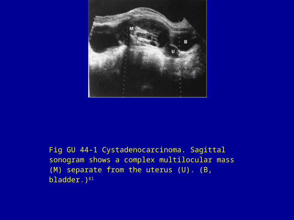
Fig GU 44-1 Cystadenocarcinoma. Sagittal sonogram shows a complex multilocular mass (M) separate from the uterus (U). (B, bladder.)61
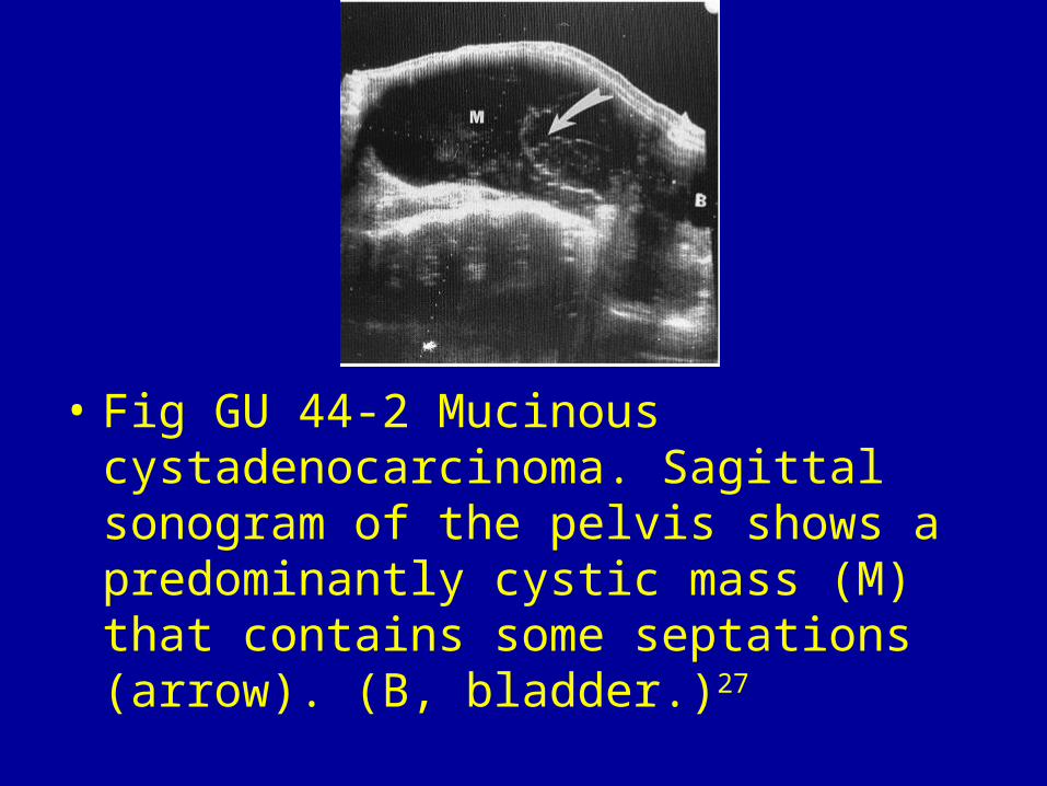
• Fig GU 44-2 Mucinous cystadenocarcinoma. Sagittal sonogram of the pelvis shows a predominantly cystic mass (M) that contains some septations (arrow). (B, bladder.)27
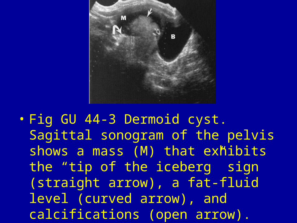
• Fig GU 44-3 Dermoid cyst. Sagittal sonogram of the pelvis shows a mass (M) that exhibits the “tip of the iceberg” sign (straight arrow), a fat-fluid level (curved arrow), and calcifications (open arrow). (B, bladder.)61
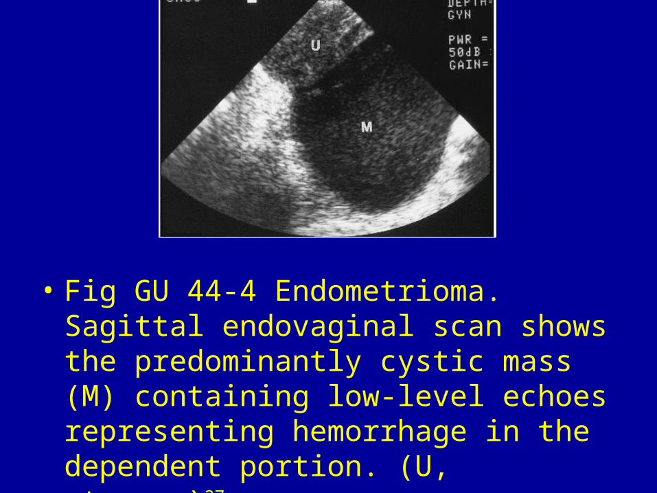
• Fig GU 44-4 Endometrioma. Sagittal endovaginal scan shows the predominantly cystic mass (M) containing low-level echoes representing hemorrhage in the dependent portion. (U, uterus.)27
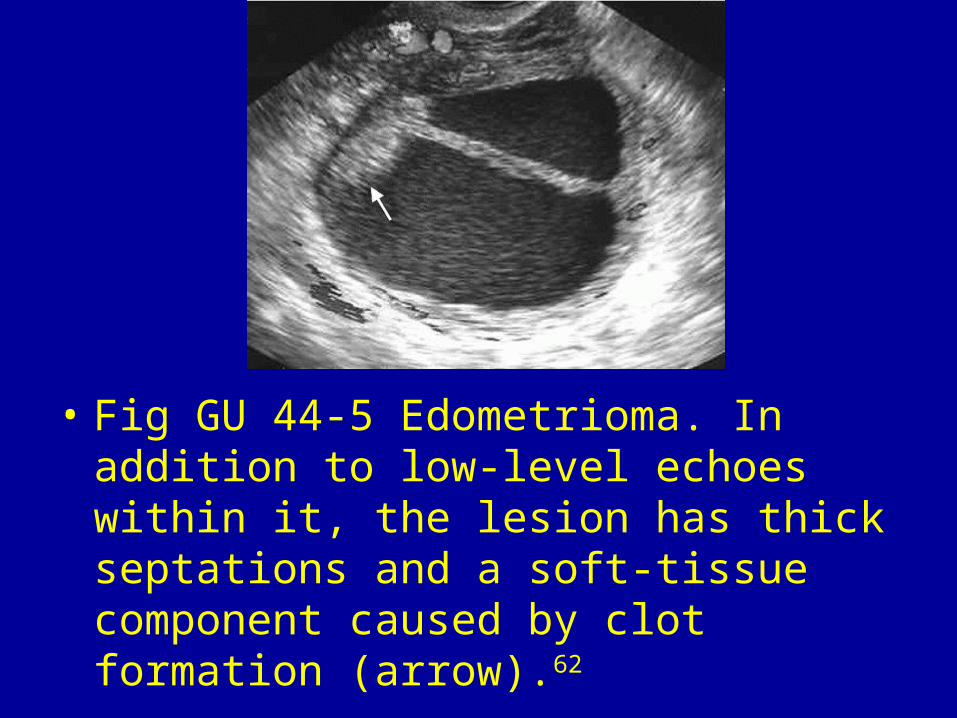
• Fig GU 44-5 Edometrioma. In addition to low-level echoes within it, the lesion has thick septations and a soft-tissue component caused by clot formation (arrow).62
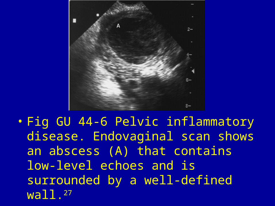
• Fig GU 44-6 Pelvic inflammatory disease. Endovaginal scan shows an abscess (A) that contains low-level echoes and is surrounded by a well-defined wall.27
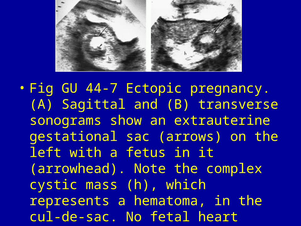
• Fig GU 44-7 Ectopic pregnancy. (A) Sagittal and (B) transverse sonograms show an extrauterine gestational sac (arrows) on the left with a fetus in it (arrowhead). Note the complex cystic mass (h), which represents a hematoma, in the cul-de-sac. No fetal heart activity was noted. (U, uterus.)63
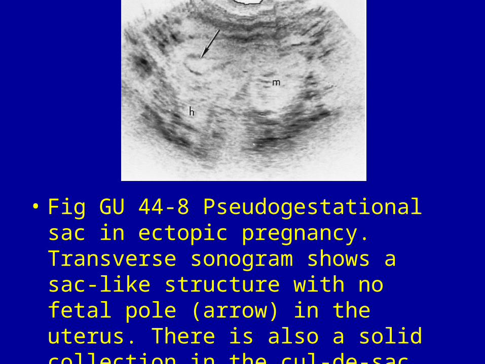
• Fig GU 44-8 Pseudogestational sac in ectopic pregnancy. Transverse sonogram shows a sac-like structure with no fetal pole (arrow) in the uterus. There is also a solid collection in the cul-de-sac (h) and a left adnexal mass (m).63
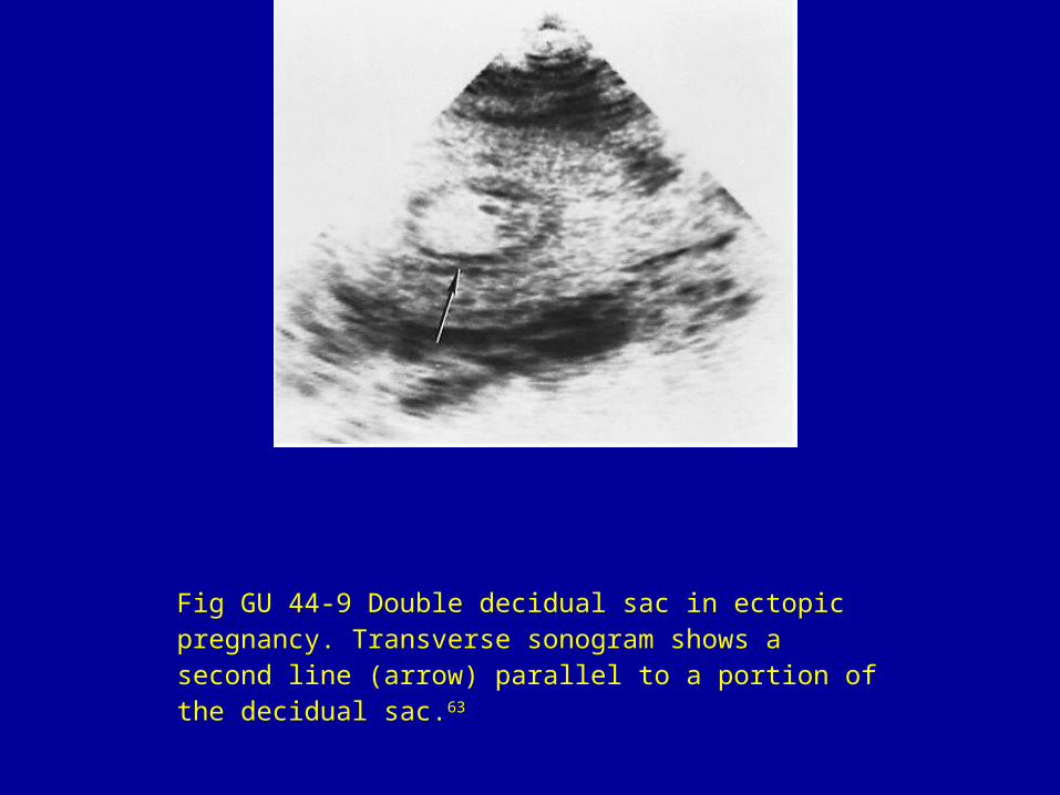
Fig GU 44-9 Double decidual sac in ectopic pregnancy. Transverse sonogram shows a second line (arrow) parallel to a portion of the decidual sac.63
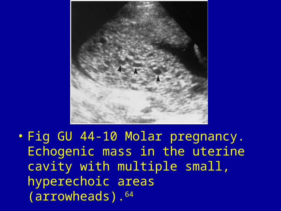
• Fig GU 44-10 Molar pregnancy. Echogenic mass in the uterine cavity with multiple small, hyperechoic areas (arrowheads).64

Fig GU 44-11 Hemorrhagic corpus luteum cyst. Sagittal scan of the ovary (O) shows a complex cystic mass containing internal low-level echoes (arrow).27
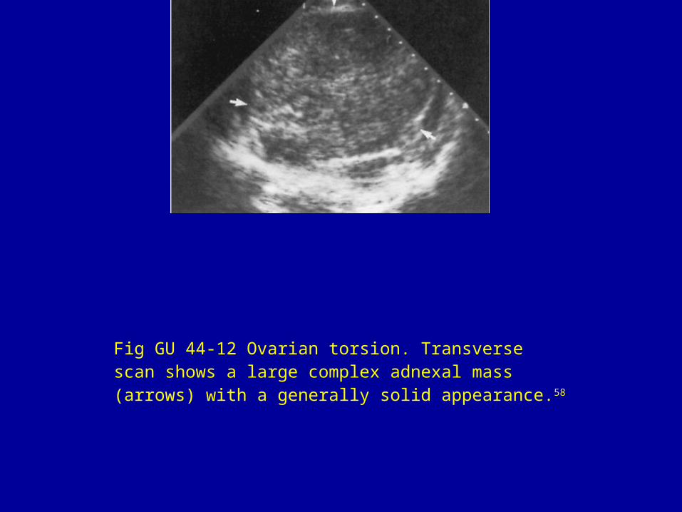
Fig GU 44-12 Ovarian torsion. Transverse scan shows a large complex adnexal mass (arrows) with a generally solid appearance.58