Embed Size (px)
Citation preview
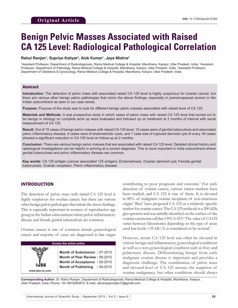
28International Journal of Scientifi c Study | September 2015 | Vol 3 | Issue 6
Benign Pelvic Masses Associated with Raised CA 125 Level: Radiological Pathological CorrelationRahul Ranjan1, Supriya Katiyar2, Alok Kumar1, Jaya Mishra3
1Assistant Professor, Department of Radiodiagnosis, Rama Medical College & Hospital, Mandhana, Kanpur, Uttar Pradesh, India, 2Assistant Professor, Department of Pathology, Rama Medical College & Hospital, Mandhana, Kanpur, Uttar Pradesh, India, 3Assistant Professor, Department of Obstetrics & Gynecology, Rama Medical College & Hospital, Mandhana, Kanpur, Uttar Pradesh, India
contributing to poor prognosis and outcome.1 For early detection of ovarian cancer, various tumor markers have been studied, and CA 125 is one of them. It is elevated in 80% of malignant ovarian neoplasm of non-mucinous origin.2 Bast3 have proposed CA 125 as a relatively specifi c marker for ovarian cancer. The CA 125 molecule is a 200-kDa glycoprotein and was initially identifi ed on the surface of the ovarian carcinoma cell line OVCA 433.4 The value of CA125 varies between laboratories depending on the type of assay used but levels <35 kIU/L is considered to be normal.5
However, serum CA 125 level can often be elevated in various benign and infl ammatory gynecological conditions as well as a non-gynecological condition such as liver and pulmonary disease. Differentiating benign from early malignant ovarian disease is important and provides a diagnostic challenge. The combination of pelvic mass and elevated level of CA 125 arouses the suspicion of ovarian malignancy, but other conditions should always
INTRODUCTION
The detection of pelvic mass with raised CA 125 level is highly suspicious for ovarian cancer, but there are various other benign pelvic pathologies that mimic the above fi ndings. This is especially important in women of reproductive age group in the Indian subcontinent where pelvic infl ammatory disease and female genital tuberculosis are common.
Ovarian cancer is one of common female gynecological cancer and majority of cases are diagnosed in late stages
Original Article
Abstract
Introduction: The detection of pelvic mass with associated raised CA 125 level is highly suspicious for ovarian cancer, but there are various other benign pelvic pathologies that mimic the above fi ndings, especially in premenopausal women in the Indian subcontinent as seen in our case series.
Purpose: Purpose of the study was to look for different benign pelvic masses associated with raised level of CA 125.
Materials and Methods: It was prospective study in which cases of pelvic mass with raised CA 125 level that turned out to be benign in etiology on complete work up were evaluated and followed up on treatment at 2 months of interval with serial measurement of CA 125.
Result: Out of 15 cases of benign pelvic masses with raised CA 125 level, 10 cases were of genital tuberculosis and associated pelvic infl ammatory disease, 4 cases were of endometriotic cysts, and 1 case was of ruptured dermoid cyst of ovary. All cases showed a signifi cant reduction in CA 125 level on follow-up at 2 months.
Conclusion: There are various benign pelvic masses that are associated with raised CA 125 level. Detailed clinical history and radiological investigations can be helpful in arriving at a correct diagnosis. This is more important in India subcontinent where genital tuberculosis and pelvic infl ammatory disease are common.
Key words: CA 125 antigen (cancer associated 125 antigen), Endometriosis, Ovarian dermoid cyst, Female genital tuberculosis, Ovarian neoplasm, Pelvic infl ammatory disease
Access this article online
www.ijss-sn.com
Month of Submission : 07-2015Month of Peer Review : 08-2015Month of Acceptance : 09-2015Month of Publishing : 09-2015
Corresponding Author: Dr. Rahul Ranjan, Department of Radiodiagnosis, Rama Medical College & Hospital, Mandhana, Kanpur, Uttar Pradesh, India. Phone: +91-9919293810. E-mail: [email protected]
DOI: 10.17354/ijss/2015/386
Ranjan, et al.: Our Experience of Benign Pelvic Masses with Raised Ca 125
29 International Journal of Scientifi c Study | September 2015 | Vol 3 | Issue 6
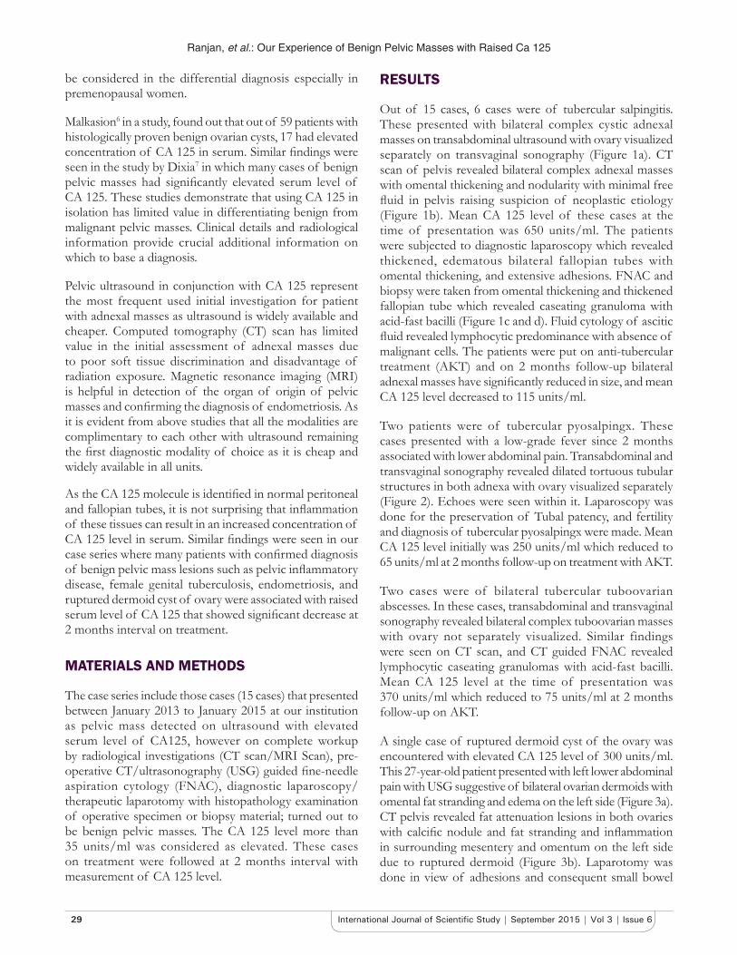
be considered in the differential diagnosis especially in premenopausal women.
Malkasion6 in a study, found out that out of 59 patients with histologically proven benign ovarian cysts, 17 had elevated concentration of CA 125 in serum. Similar fi ndings were seen in the study by Dixia7 in which many cases of benign pelvic masses had signifi cantly elevated serum level of CA 125. These studies demonstrate that using CA 125 in isolation has limited value in differentiating benign from malignant pelvic masses. Clinical details and radiological information provide crucial additional information on which to base a diagnosis.
Pelvic ultrasound in conjunction with CA 125 represent the most frequent used initial investigation for patient with adnexal masses as ultrasound is widely available and cheaper. Computed tomography (CT) scan has limited value in the initial assessment of adnexal masses due to poor soft tissue discrimination and disadvantage of radiation exposure. Magnetic resonance imaging (MRI) is helpful in detection of the organ of origin of pelvic masses and confi rming the diagnosis of endometriosis. As it is evident from above studies that all the modalities are complimentary to each other with ultrasound remaining the fi rst diagnostic modality of choice as it is cheap and widely available in all units.
As the CA 125 molecule is identifi ed in normal peritoneal and fallopian tubes, it is not surprising that infl ammation of these tissues can result in an increased concentration of CA 125 level in serum. Similar fi ndings were seen in our case series where many patients with confi rmed diagnosis of benign pelvic mass lesions such as pelvic infl ammatory disease, female genital tuberculosis, endometriosis, and ruptured dermoid cyst of ovary were associated with raised serum level of CA 125 that showed signifi cant decrease at 2 months interval on treatment.
MATERIALS AND METHODS
The case series include those cases (15 cases) that presented between January 2013 to January 2015 at our institution as pelvic mass detected on ultrasound with elevated serum level of CA125, however on complete workup by radiological investigations (CT scan/MRI Scan), pre-operative CT/ultrasonography (USG) guided fi ne-needle aspiration cytology (FNAC), diagnostic laparoscopy/therapeutic laparotomy with histopathology examination of operative specimen or biopsy material; turned out to be benign pelvic masses. The CA 125 level more than 35 units/ml was considered as elevated. These cases on treatment were followed at 2 months interval with measurement of CA 125 level.
RESULTS
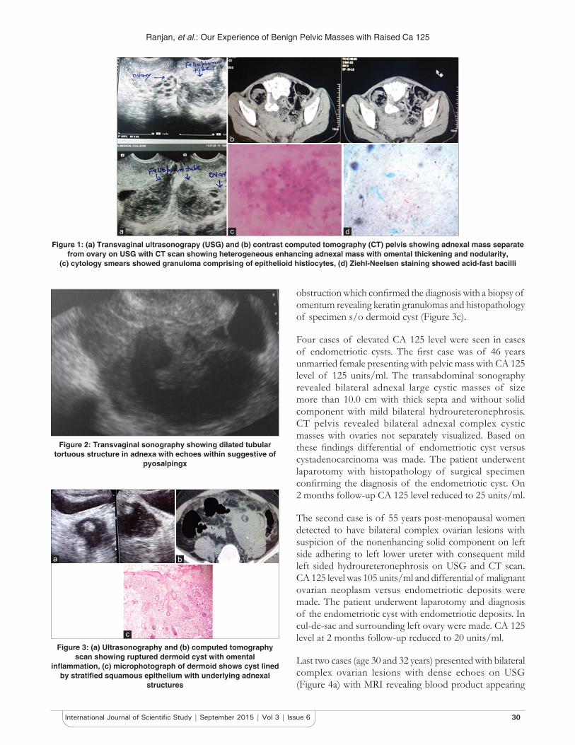
Out of 15 cases, 6 cases were of tubercular salpingitis. These presented with bilateral complex cystic adnexal masses on transabdominal ultrasound with ovary visualized separately on transvaginal sonography (Figure 1a). CT scan of pelvis revealed bilateral complex adnexal masses with omental thickening and nodularity with minimal free fl uid in pelvis raising suspicion of neoplastic etiology (Figure 1b). Mean CA 125 level of these cases at the time of presentation was 650 units/ml. The patients were subjected to diagnostic laparoscopy which revealed thickened, edematous bilateral fallopian tubes with omental thickening, and extensive adhesions. FNAC and biopsy were taken from omental thickening and thickened fallopian tube which revealed caseating granuloma with acid-fast bacilli (Figure 1c and d). Fluid cytology of ascitic fl uid revealed lymphocytic predominance with absence of malignant cells. The patients were put on anti-tubercular treatment (AKT) and on 2 months follow-up bilateral adnexal masses have signifi cantly reduced in size, and mean CA 125 level decreased to 115 units/ml.
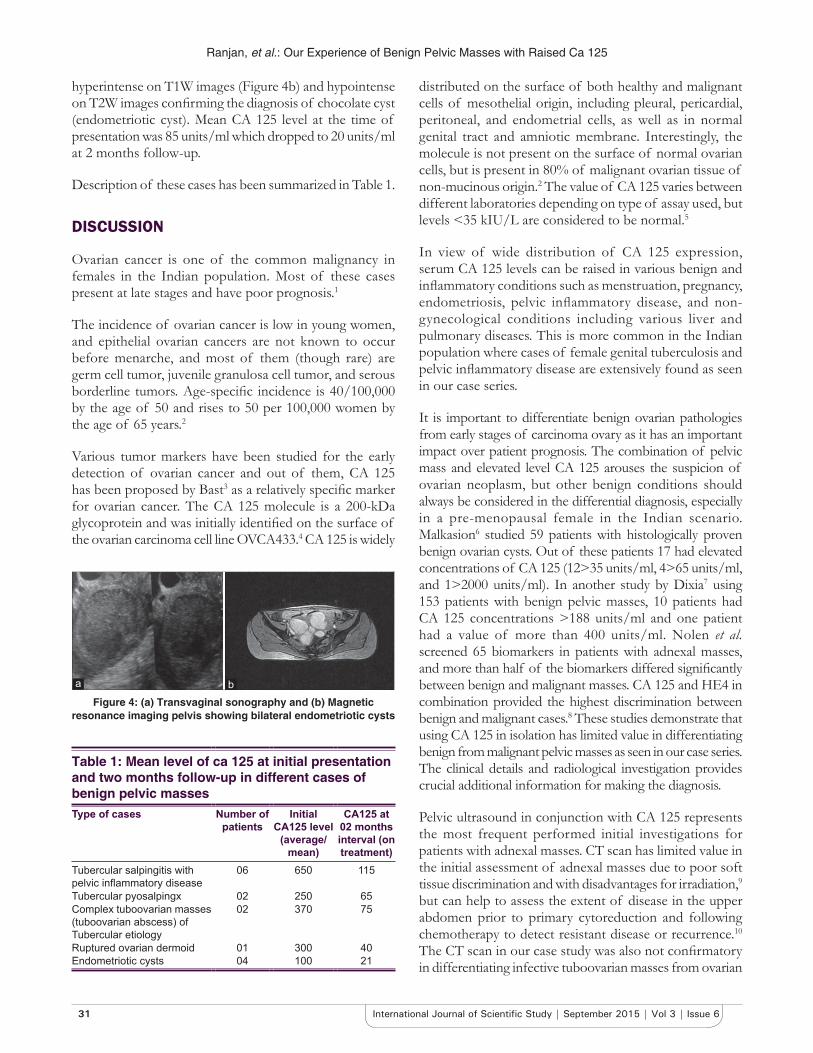
Two patients were of tubercular pyosalpingx. These cases presented with a low-grade fever since 2 months associated with lower abdominal pain. Transabdominal and transvaginal sonography revealed dilated tortuous tubular structures in both adnexa with ovary visualized separately (Figure 2). Echoes were seen within it. Laparoscopy was done for the preservation of Tubal patency, and fertility and diagnosis of tubercular pyosalpingx were made. Mean CA 125 level initially was 250 units/ml which reduced to 65 units/ml at 2 months follow-up on treatment with AKT.
Two cases were of bilateral tubercular tuboovarian abscesses. In these cases, transabdominal and transvaginal sonography revealed bilateral complex tuboovarian masses with ovary not separately visualized. Similar findings were seen on CT scan, and CT guided FNAC revealed lymphocytic caseating granulomas with acid-fast bacilli. Mean CA 125 level at the time of presentation was 370 units/ml which reduced to 75 units/ml at 2 months follow-up on AKT.
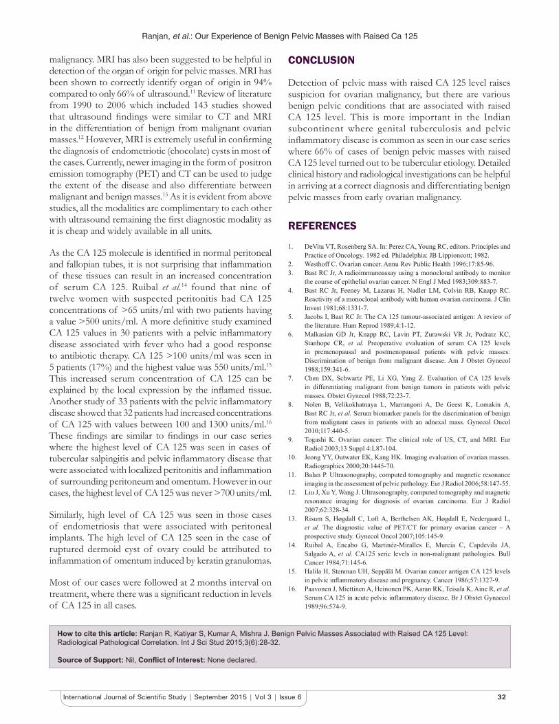
A single case of ruptured dermoid cyst of the ovary was encountered with elevated CA 125 level of 300 units/ml. This 27-year-old patient presented with left lower abdominal pain with USG suggestive of bilateral ovarian dermoids with omental fat stranding and edema on the left side (Figure 3a). CT pelvis revealed fat attenuation lesions in both ovaries with calcifi c nodule and fat stranding and infl ammation in surrounding mesentery and omentum on the left side due to ruptured dermoid (Figure 3b). Laparotomy was done in view of adhesions and consequent small bowel
Ranjan, et al.: Our Experience of Benign Pelvic Masses with Raised Ca 125
30International Journal of Scientifi c Study | September 2015 | Vol 3 | Issue 6
obstruction which confi rmed the diagnosis with a biopsy of omentum revealing keratin granulomas and histopathology of specimen s/o dermoid cyst (Figure 3c).
Four cases of elevated CA 125 level were seen in cases of endometriotic cysts. The fi rst case was of 46 years unmarried female presenting with pelvic mass with CA 125 level of 125 units/ml. The transabdominal sonography revealed bilateral adnexal large cystic masses of size more than 10.0 cm with thick septa and without solid component with mild bilateral hydroureteronephrosis. CT pelvis revealed bilateral adnexal complex cystic masses with ovaries not separately visualized. Based on these fi ndings differential of endometriotic cyst versus cystadenocarcinoma was made. The patient underwent laparotomy with histopathology of surgical specimen confi rming the diagnosis of the endometriotic cyst. On 2 months follow-up CA 125 level reduced to 25 units/ml.
The second case is of 55 years post-menopausal women detected to have bilateral complex ovarian lesions with suspicion of the nonenhancing solid component on left side adhering to left lower ureter with consequent mild left sided hydroureteronephrosis on USG and CT scan. CA 125 level was 105 units/ml and differential of malignant ovarian neoplasm versus endometriotic deposits were made. The patient underwent laparotomy and diagnosis of the endometriotic cyst with endometriotic deposits. In cul-de-sac and surrounding left ovary were made. CA 125 level at 2 months follow-up reduced to 20 units/ml.
Last two cases (age 30 and 32 years) presented with bilateral complex ovarian lesions with dense echoes on USG (Figure 4a) with MRI revealing blood product appearing
Figure 2: Transvaginal sonography showing dilated tubular tortuous structure in adnexa with echoes within suggestive of
pyosalpingx
Figure 1: (a) Transvaginal ultrasonograpy (USG) and (b) contrast computed tomography (CT) pelvis showing adnexal mass separate from ovary on USG with CT scan showing heterogeneous enhancing adnexal mass with omental thickening and nodularity,
(c) cytology smears showed granuloma comprising of epithelioid histiocytes, (d) Ziehl-Neelsen staining showed acid-fast bacilli
dc
b
a
Figure 3: (a) Ultrasonography and (b) computed tomography scan showing ruptured dermoid cyst with omental
infl ammation, (c) microphotograph of dermoid shows cyst lined by stratifi ed squamous epithelium with underlying adnexal
structures
c
ba
Ranjan, et al.: Our Experience of Benign Pelvic Masses with Raised Ca 125
31 International Journal of Scientifi c Study | September 2015 | Vol 3 | Issue 6
hyperintense on T1W images (Figure 4b) and hypointense on T2W images confi rming the diagnosis of chocolate cyst (endometriotic cyst). Mean CA 125 level at the time of presentation was 85 units/ml which dropped to 20 units/ml at 2 months follow-up.
Description of these cases has been summarized in Table 1.
DISCUSSION
Ovarian cancer is one of the common malignancy in females in the Indian population. Most of these cases present at late stages and have poor prognosis.1
The incidence of ovarian cancer is low in young women, and epithelial ovarian cancers are not known to occur before menarche, and most of them (though rare) are germ cell tumor, juvenile granulosa cell tumor, and serous borderline tumors. Age-specifi c incidence is 40/100,000 by the age of 50 and rises to 50 per 100,000 women by the age of 65 years .2
Various tumor markers have been studied for the early detection of ovarian cancer and out of them, CA 125 has been proposed by Bast3 as a relatively specifi c marker for ovarian cancer. The CA 125 molecule is a 200-kDa glycoprotein and was initially identifi ed on the surface of the ovarian carcinoma cell line OVCA433 .4 CA 125 is widely
distributed on the surface of both healthy and malignant cells of mesothelial origin, including pleural, pericardial, peritoneal, and endometrial cells, as well as in normal genital tract and amniotic membrane. Interestingly, the molecule is not present on the surface of normal ovarian cells, but is present in 80% of malignant ovarian tissue of non-mucinous origin.2 The value of CA 125 varies between different laboratories depending on type of assay used, but levels <35 kIU/L are considered to be normal .5
In view of wide distribution of CA 125 expression, serum CA 125 levels can be raised in various benign and infl ammatory conditions such as menstruation, pregnancy, endometriosis, pelvic infl ammatory disease, and non-gynecological conditions including various liver and pulmonary diseases. This is more common in the Indian population where cases of female genital tuberculosis and pelvic infl ammatory disease are extensively found as seen in our case series.
It is important to differentiate benign ovarian pathologies from early stages of carcinoma ovary as it has an important impact over patient prognosis. The combination of pelvic mass and elevated level CA 125 arouses the suspicion of ovarian neoplasm, but other benign conditions should always be considered in the differential diagnosis, especially in a pre-menopausal female in the Indian scenario. Malkasion 6 studied 59 patients with histologically proven benign ovarian cysts. Out of these patients 17 had elevated concentrations of CA 125 (12>35 units/ml, 4>65 units/ml, and 1>2000 units/ml). In another study by Dixia 7 using 153 patients with benign pelvic masses, 10 patients had CA 125 concentrations >188 units/ml and one patient had a value of more than 400 units/ml. Nolen et al. screened 65 biomarkers in patients with adnexal masses, and more than half of the biomarkers differed signifi cantly between benign and malignant masses. CA 125 and HE4 in combination provided the highest discrimination between benign and malignant cases.8 These studies demonstrate that using CA 125 in isolation has limited value in differentiating benign from malignant pelvic masses as seen in our case series. The clinical details and radiological investigation provides crucial additional information for making the diagnosis.
Pelvic ultrasound in conjunction with CA 125 represents the most frequent performed initial investigations for patients with adnexal masses. CT scan has limited value in the initial assessment of adnexal masses due to poor soft tissue discrimination and with disadvantages for irradiation,9 but can help to assess the extent of disease in the upper abdomen prior to primary cytoreduction and following chemotherapy to detect resistant disease or recurrence.10 The CT scan in our case study was also not confi rmatory in differentiating infective tuboovarian masses from ovarian
Figure 4: (a) Transvaginal sonography and (b) Magnetic resonance imaging pelvis showing bilateral endometriotic cysts
ba
Table 1: Mean level of ca 125 at initial presentation and two months follow-up in different cases of benign pelvic massesType of cases Number of
patientsInitial
CA125 level (average/
mean)
CA125 at 02 months interval (on treatment)
Tubercular salpingitis with pelvic infl ammatory disease
06 650 115
Tubercular pyosalpingx 02 250 65Complex tuboovarian masses (tuboovarian abscess) of Tubercular etiology
02 370 75
Ruptured ovarian dermoid 01 300 40Endometriotic cysts 04 100 21
Ranjan, et al.: Our Experience of Benign Pelvic Masses with Raised Ca 125
32International Journal of Scientifi c Study | September 2015 | Vol 3 | Issue 6
malignancy. MRI has also been suggested to be helpful in detection of the organ of origin for pelvic masses. MRI has been shown to correctly identify organ of origin in 94% compared to only 66% of ultrasound.11 Review of literature from 1990 to 2006 which included 143 studies showed that ultrasound fi ndings were similar to CT and MRI in the differentiation of benign from malignant ovarian masses.12 However, MRI is extremely useful in confi rming the diagnosis of endometriotic (chocolate) cysts in most of the cases. Currently, newer imaging in the form of positron emission tomography (PET) and CT can be used to judge the extent of the disease and also differentiate between malignant and benign masses.13 As it is evident from above studies, all the modalities are complimentary to each other with ultrasound remaining the fi rst diagnostic modality as it is cheap and widely available in all units.
As the CA 125 molecule is identifi ed in normal peritoneal and fallopian tubes, it is not surprising that infl ammation of these tissues can result in an increased concentration of serum CA 125. Ruibal et al.14 found that nine of twelve women with suspected peritonitis had CA 125 concentrations of >65 units/ml with two patients having a value >500 units/ml. A more defi nitive study examined CA 125 values in 30 patients with a pelvic infl ammatory disease associated with fever who had a good response to antibiotic therapy. CA 125 >100 units/ml was seen in 5 patients (17%) and the highest value was 550 units/ml .15 This increased serum concentration of CA 125 can be explained by the local expression by the infl amed tissue. Another study of 33 patients with the pelvic infl ammatory disease showed that 32 patients had increased concentrations of CA 125 with values between 100 and 1300 units/ml .16 These fi ndings are similar to fi ndings in our case series where the highest level of CA 125 was seen in cases of tubercular salpingitis and pelvic infl ammatory disease that were associated with localized peritonitis and infl ammation of surrounding peritoneum and omentum. However in our cases, the highest level of CA 125 was never >700 units/ml.
Similarly, high level of CA 125 was seen in those cases of endometriosis that were associated with peritoneal implants. The high level of CA 125 seen in the case of ruptured dermoid cyst of ovary could be attributed to infl ammation of omentum induced by keratin granulomas.
Most of our cases were followed at 2 months interval on treatment, where there was a signifi cant reduction in levels of CA 125 in all cases.
CONCLUSION
Detection of pelvic mass with raised CA 125 level raises suspicion for ovarian malignancy, but there are various benign pelvic conditions that are associated with raised CA 125 level. This is more important in the Indian subcontinent where genital tuberculosis and pelvic infl ammatory disease is common as seen in our case series where 66% of cases of benign pelvic masses with raised CA 125 level turned out to be tubercular etiology. Detailed clinical history and radiological investigations can be helpful in arriving at a correct diagnosis and differentiating benign pelvic masses from early ovarian malignancy.
REFERENCES
1. DeVita VT, Rosenberg SA. In: Perez CA, Young RC, editors. Principles and Practice of Oncology. 1982 ed. Philadelphia: JB Lippioncott; 1982.
2. Westhoff C. Ovarian cancer. Annu Rev Public Health 1996;17:85-96.3. Bast RC Jr, A radioimmunoassay using a monoclonal antibody to monitor
the course of epithelial ovarian cancer. N Engl J Med 1983;309:883-7.4. Bast RC Jr, Feeney M, Lazarus H, Nadler LM, Colvin RB, Knapp RC.
Reactivity of a monoclonal antibody with human ovarian carcinoma. J Clin Invest 1981;68:1331-7.
5. Jacobs I, Bast RC Jr. The CA 125 tumour-associated antigen: A review of the literature. Hum Reprod 1989;4:1-12.
6. Malkasian GD Jr, Knapp RC, Lavin PT, Zurawski VR Jr, Podratz KC, Stanhope CR, et al. Preoperative evaluation of serum CA 125 levels in premenopausal and postmenopausal patients with pelvic masses: Discrimination of benign from malignant disease. Am J Obstet Gynecol 1988;159:341-6.
7. Chen DX, Schwartz PE, Li XG, Yang Z. Evaluation of CA 125 levels in differentiating malignant from benign tumors in patients with pelvic masses. Obstet Gynecol 1988;72:23-7.
8. Nolen B, Velikokhatnaya L, Marrangoni A, De Geest K, Lomakin A, Bast RC Jr, et al. Serum biomarker panels for the discrimination of benign from malignant cases in patients with an adnexal mass. Gynecol Oncol 2010;117:440-5.
9. Togashi K. Ovarian cancer: The clinical role of US, CT, and MRI. Eur Radiol 2003;13 Suppl 4:L87-104.
10. Jeong YY, Outwater EK, Kang HK. Imaging evaluation of ovarian masses. Radiographics 2000;20:1445-70.
11. Balan P. Ultrasonography, computed tomography and magnetic resonance imaging in the assessment of pelvic pathology. Eur J Radiol 2006;58:147-55.
12. Liu J, Xu Y, Wang J. Ultrasonography, computed tomography and magnetic resonance imaging for diagnosis of ovarian carcinoma. Eur J Radiol 2007;62:328-34.
13. Risum S, Høgdall C, Loft A, Berthelsen AK, Høgdall E, Nedergaard L, et al. The diagnostic value of PET/CT for primary ovarian cancer – A prospective study. Gynecol Oncol 2007;105:145-9.
14. Ruibal A, Encabo G, Martinéz-Miralles E, Murcia C, Capdevila JA, Salgado A, et al. CA125 seric levels in non-malignant pathologies. Bull Cancer 1984;71:145-6.
15. Halila H, Stenman UH, Seppälä M. Ovarian cancer antigen CA 125 levels in pelvic infl ammatory disease and pregnancy. Cancer 1986;57:1327-9.
16. Paavonen J, Miettinen A, Heinonen PK, Aaran RK, Teisala K, Aine R, et al. Serum CA 125 in acute pelvic infl ammatory disease. Br J Obstet Gynaecol 1989;96:574-9.
How to cite this article: Ranjan R, Katiyar S, Kumar A, Mishra J. Benign Pelvic Masses Associated with Raised CA 125 Level: Radiological Pathological Correlation. Int J Sci Stud 2015;3(6):28-32.
Source of Support: Nil, Confl ict of Interest: None declared.