A DISSERTATION ON
CSF ANALYSIS IN THE DIFFERENTIAL
DIAGNOSIS OF ACUTE
ENCEPHALITIS SYNDROME
M.D (BRANCH VII)
PAEDIATRIC MEDICINE
MARCH 2010
THE TAMILNADU DR.MGR.MEDICAL UNIVERSITY
CHENNAI, TAMILNADU
CERTIFICATE
This is to certify that the dissertation titled “CSF ANALYSIS
IN THE DIFFERENTIAL DIAGNOSIS OF ACUTE
ENCEPHALITIS SYNDROME” submitted by Dr.S. DIVYA to
the Faculty of pediatrics, The Tamilnadu M.G.R.Medical
University,Chennai in partial fulfillment of the requirement for the
award of M.D.Degree (Pediatrics) is a bonafide research work
carried out by her under our direct supervision and guidance.
Dr.P.Amutha Rajeshwari,M.D.,DCH.,
Professor and Head of the Department, Institute of Child Health & Research Centre, Madurai Medical College, Madurai.
DECLARATION
I Dr. S. DIVYA, solemnly declare that the dissertation titled
“CSF ANALYSIS IN THE DIFFERENTIAL DIAGNOSIS OF
ACUTE ENCEPHALITIS SYNDROME” has been prepared by
me.
This is submitted to the Tamilnadu Dr.M.G.R.Medical
University, Chennai in partial fulfillment of the rules and
regulations for the M.D.Degree Examination in Paediatrics.
Place: Madurai
Date: Dr. S. DIVYA
ACKNOWLEDGEMENT
It is with immense pleasure and privilege that I express my
heartful gratitude, admiration and sincere thanks to my chief Prof.
Dr. P.Amutha Rajeswari, Professor and Head of the Department
of pediatrics, for her guidance and support during this study.
I express my sincere thanks and gratitude to Prof. Dr. M.L.
Vasantha kumari for her support and for her guidance, supervision,
constant encouragement and support throughout this study.
I wish to express my sincere thanks to my guide and teacher,
Asst professor Dr.S.Nataraja Rathinam for his valuable
suggestion, support and guidance at every stage of this study.
I would like to thank Prof. Dr.S. Rajasekaran,
Prof.Dr.G.Mathevan, Prof. Dr. Chitra Ayyappan , Prof. Dr. R.A
Sankarasubramaniam, and Prof. Dr.T. Nagarajan who guided
me to a great extent. I also thank all the members of the dissertation
committee for this valuable suggestions.
I would like to thank Reader Dr. K. Mathiarasan for his
valuable suggestion throughout the Study.
I gratefully acknowledge the help and guidance received from
Dr. K.G. Srinivasan at every stage of this study.
I also thank the Head of Department of Microbiology,
Radiology and Biochemistry for the help and guidance in
performing this study.
I also express my gratitude to all my fellow post graduates for
their kind cooperation in carrying out this study and for their critical
analysis.
I also thank the Dean and the members of Ethical committee,
Government Rajaji Hospital and Madurai Medical College, Madurai
for permitting me to perform this study.
I thank all the parents and children who have congrudingly
lent themselves to undergo this study without whom this study
would not have seen the light of the day.
CONTENTS
S.NO. TOPIC PAGE NO.
1. INTRODUCTION 1
2. REVIEW OF LITERATURE 6
3. AIM OF THE STUDY 27
4. MATERIALS AND METHODS 28
5. RESULTS & ANALYSIS 32
6. DISCUSSION 53
7. CONCLUSION 68
8. LIMITATIONS 70
9. SUGGESTIONS 71
BIBLIOGRAPHY
PROFORMA
ABBREVIATIONS MASTER CHART
INTRODUCTION
AES – Acute encephalitis syndrome.
Acute encephalitis syndrome in children includes a group of
conditions which pose a diagnostic challenge to every pediatrician.
Various conditions are included in the differential diagnosis of AES.
Definition of AES:3
AES includes any child presenting with fever, altered
sensorium, seizures, symptoms of increased intracranial pressure
(ICP).
AES accounts for about 10% to 15% of hospital admissions
and is associated with significant morbidity and mortality.
Differential Diagnosis of AES:
1. Bacterial (pyogenic) meningitis
2. TB meningitis
3. Viral encephalitis
4. Aseptic meningitis
5. ADEM

6. Cerebral malaria
7. Reye’s syndrome
8. CVT
9. Toxic / metabolic encephalopathies
Lumbar puncture and CSF analysis helps in the differential
diagnosis of AES. Clinical history, detailed systemic and
neurological examination along with CSF findings provides the
diagnostic clues towards the etiology of AES.
Neuroimaging studies helps in detecting the complications
such as subdural effusion, infarcts, hydrocephalus ,etc in pyogenic
and TB meningitis. Herpes viral encephalitis lesions are better
picked up by MRI. MRI with gadolinium contrast remains the
diagnostic imaging modality of choice in ADEM.
Prompt early diagnosis and management is necessary to avoid
significant mortality and neurological impairment in children in
AES.
This study was conducted to reemphasis the importance of
CSF analysis in differential diagnosis of AES and in deciding the
appropriate imaging modality of choice there by avoiding the
unnecessary expenditure.
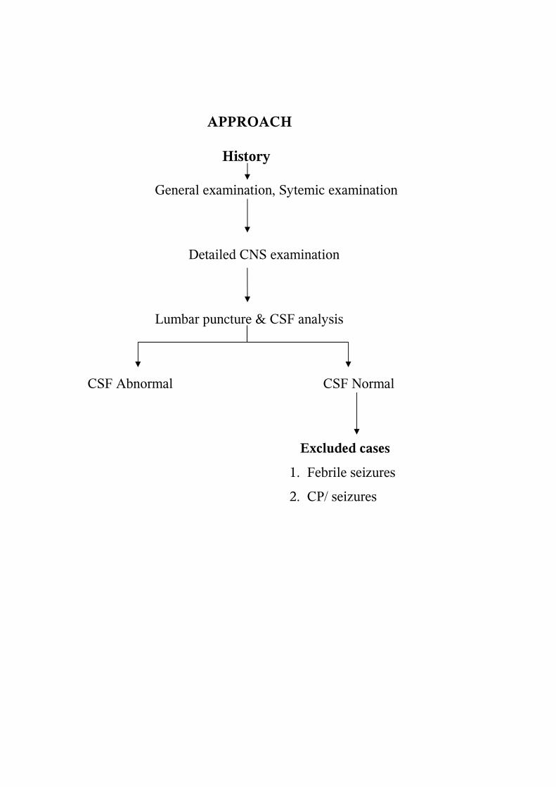
APPROACH
History
General examination, Sytemic examination
Detailed CNS examination
Lumbar puncture & CSF analysis
CSF Abnormal CSF Normal
Excluded cases
1. Febrile seizures
2. CP/ seizures
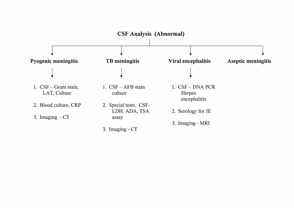
CSF Analysis (Abnormal)
Pyogenic meningitis TB meningitis Viral encephalitis Aseptic meningitis
1. CSF – Gram stain, LAT, Culture
2. Blood culture, CRP
3. Imaging - CT
1. CSF – AFB stain culture
2. Special tests. CSF-LDH, ADA, TSA assay
3. Imaging - CT
1. CSF – DNA PCR Herpes encephalitis
2. Serology for JE
3. Imaging - MRI
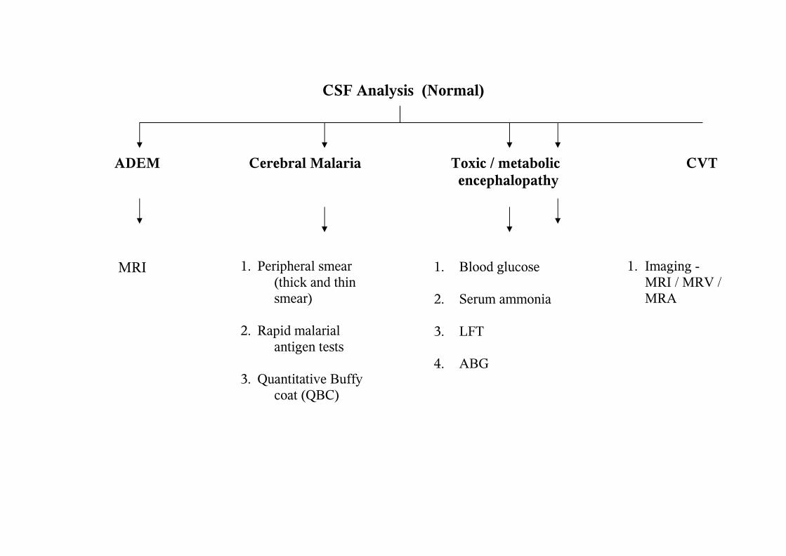
CSF Analysis (Normal)
ADEM Cerebral Malaria Toxic / metabolic CVT encephalopathy
MRI 1. Peripheral smear (thick and thin smear)
2. Rapid malarial antigen tests
3. Quantitative Buffy coat (QBC)
1. Blood glucose
2. Serum ammonia
3. LFT
4. ABG
1. Imaging - MRI / MRV / MRA
REVIEW OF LITERATURE
The importance of CSF analysis in particular the value of
CSF cytology has been stressed in various literatures, as a
preliminary step in the approval towards the diagnosis of acute
infections of CNS.
Berry Schumann et al1 stressed the importance of direct
active involvement of the clinician in the evaluation of bedside
investigation which had produced frequently more appropriate
diagnosis.
Dyken2 reports that counting chamber methods are useful
quantitative procedures which helps to distinguish various types of
cells in CSF.
In the words of Dougherty et al 4, CSF is obtained as an
indirect assessment of brain status.
Quincke first described the techniques in LP way back in
1821,the single most important investigation in the diagnosis of
CNS disease.
CSF cell count procedure is simple to perform with in a brief
period does not requires sophisticated equipments at all times.
Scarbrough M, Thwaites G E,5reports that analysis of CSF
is essential and helps in establishing the diagnosis of CNS disease
especially in resource poor settings where the yield of diagnostic
microbiology is low.
Neuman M,Tolford S,Harper M B,6 reported the value of
gram stain in the diagnosis of bacterial meningitis.
Oretega H W, Bonsu B K7, analysed the importance of CSF
pleocytosis along with biochemical tests in predicting bacterial
meningitis when gram stain is negative or unavailable.
Bonsu B K7, in their study on differentiating acute bacterial
meningitis from acute viral meningitis among the children with CSF
pleocytosis, observed that CSF neutrophils and CSF protein helps in
differentiating the two entities.
Deivanayagan N, Ashok TP,8 studied on the evaluation of CSF
variables as a diagnostic test for bacterial meningitis observed that
CSF polmorphonuclear pleocytosis along with low sugar and high
protein values can be used as a diagnostic test for bacterial
meningitis where positive culture rates are low .
Keith Morton 9in his review article considered the total and
differential cell count in CSF as standard tests.
CSF analysis being the most important in the diagnosis of CNS
disease, clinician must be aware of the contraindications for LP.
Contraindications:10
1.Elevated intracranial pressure.
2.Symptoms and signs of impending cerebral herniation.
3.Skin infection at the site of LP.
4.Thrombocytopenia- platelet count below 20,000 cells / cu.mm.
5.Critically ill child (rare occasions).
Cerebrospinal fluid analysis:10
Normal opening pressure ranges from 50-80 mm H20.In
the setting of elevated intracranial pressure, it’s usually above
100mmH20.
Normal CSF is crystal clear. Cloudy CSF results from an
increased cellularity. Normally CSF contains upto 5 WBC’S /mm3.
The presence of polymorphonuclear cells is always abnormal in a
child. There are no RBC’S in normal CSF. Presence of red blood
cells indicates either a traumatic tap or subarachnoid haemorrhage.
Gram stain is essential in the diagnosis of bacterial
meningitis.AFB stain should be done if TBM is suspected. Other
special tests like Indian ink preparation is done only in special
circumstances.
The CSF glucose concentration is about 60% of blood
glucose levels. Protein values normally range from 10 to 40 mgs% in
a child.
Latex agglutination test (LAT) helps in rapid detection of
bacterial antigens in CSF. Polymerase chain reaction (PCR) in CSF
has been a great advance in the diagnosis of Herpes encephalitis.
CSF may also be tested for metabolites such as lactate, aminoacids,
enolase levels in suspected metabolic diseases.
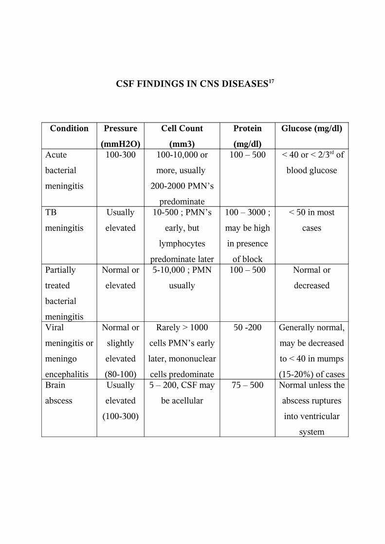
CSF FINDINGS IN CNS DISEASES17
Condition Pressure
(mmH2O)
Cell Count
(mm3)
Protein
(mg/dl)
Glucose (mg/dl)
Acute
bacterial
meningitis
100-300 100-10,000 or
more, usually
200-2000 PMN’s
predominate
100 – 500 < 40 or < 2/3rd of
blood glucose
TB
meningitis
Usually
elevated
10-500 ; PMN’s
early, but
lymphocytes
predominate later
100 – 3000 ;
may be high
in presence
of block
< 50 in most
cases
Partially
treated
bacterial
meningitis
Normal or
elevated
5-10,000 ; PMN
usually
100 – 500 Normal or
decreased
Viral
meningitis or
meningo
encephalitis
Normal or
slightly
elevated
(80-100)
Rarely > 1000
cells PMN’s early
later, mononuclear
cells predominate
50 -200 Generally normal,
may be decreased
to < 40 in mumps
(15-20%) of casesBrain
abscess
Usually
elevated
(100-300)
5 – 200, CSF may
be acellular
75 – 500 Normal unless the
abscess ruptures
into ventricular
system
TB MENINGITIS :
Tuberculosis of the central nervous system is the most serious
complication of TB infection in children. CNS involvement will be
present in 10% of children with TB infection .TB meningitis is the
most serious and commonest form of neuro tuberculosis in children
than tuberculoma . The highest incidence is between 9 months-
9years of age group. In a Phillipine based study 12,they revealed that
TBM in children usually arises as a complication of primary
infection,usually with in 6 months of onset. Similar concept was also
observed in Indian studies.13 The important determinants for the
development of TBM are age, poor nutritional status and associated
HIV infection. There are three stages of TBM.
I – Prodromal stage.
II – Stage of meningeal irritation.
III- Stage of diffuse cerebral involvement.
Diagnosis of TBM
Early diagnosis of TBM is difficult due to gradual onset and
vague symptomatology unless high index of suspicion is present.
The prodrome is often non specific with no one symptom
predominating.
Illingworth14 in his study reported 13% presented with
fever,25% with vomiting,28% with headache and only 2% with
meningitic symptoms.
In another study by Seth and Sharma, 15clinical presentation
were fever 100%, headache 80%,and neurological complications
50%.
Gold standard for the diagnosis of TBM is the isolation of the
bacteria in the CSF. But bacilli are seldom seen by AFB staining and
culture also takes several weeks. Children usually have
paucibacillary type of TB which accounts for the low
microbiological yield.
Poor response to treatment (or) morality in CNS TB is due to
failure to begin the appropriate therapy in early stages. Hence
various scoring systems are used to diagnose TBM. The most
commonly used for the diagnosis of TBM in children is AIIMS
criteria.

AIIMS criteria for diagnosis of TBM16
a) Essential criteria.
b) Supportive criteria.
Essential Criteria:
CSF showing
a) cells> 50cells /cu.mm predominant lymphocytes.
b)Proteins > 60 mg%.
c)Sugar <2/3rd of blood sugar.
Supportive Criteria:
a) H/o fever of 2 wks or more.
b) H/o contact with TB.
c) Mantoux positivity.
d) Radiological evidence of TB elsewhere in the body.
e) Generalised lymphadenopathy.
f) CT scan evidence – basal exudates, Hydrocephalus,
infarcts, gyral enhancement.
g) Isolation of AFB from gastric lavage.
h) Histologically proven TB adenitis.
Definitive diagnosis is by the isolation of AFB in CSF.
Probable TBM - Essential criteria + atleast 2 supportive criteria.
Neuroimaging studies have greatly enhanced the diagnostic
accuracy of neurotuberculosis, but still not pathognomonic for the
disease. Common CT findings includes basilar exudates, ventricular
dilatation, infarcts and intracranial granuloma.
CSF seldom reveals the acid fast bacilli (AFB), there is an need
for rapid diagnostic tests for the confirmation of TB. Many newer
methods have been developed to establish an early and definitive
diagnosis of TBM such as ADA assay, Bromide partition test, LDH
levels, TSA assay and immunological tests which detects the
mycobacterial antigen or antimycobacterial antibody in CSF, but no
single test with adequate sensitivity and specificity is available at
present.
Prognosis in CNS TB depends upon early diagnosis and
institution of appropriate therapy.
Bacterial meningitis:
Bacterial meningitis is an important potentially serious
infection in infants and in older children. Despite the advances in
vaccination and antibiotics , bacterial meningitis remains a major
cause of death and long term neurological disabilities such as mental
retardation, seizures etc. Hence early diagnosis is a must. Bacterial
meningitis should be considered in the differential diagnosis of
highly febrile children with altered mental status and other evidence
of neurological dysfunction.17
The etiological agents vary depending upon the age group.
< 2 months :
Group B streptococci, Listeria monocytogenes, E .Coli
2 months – 12 yrs:
Streptococcal pneumoniae, haemophilus influenza and
Neisseria meningitis.
Haemophilus influenza remains the most common etiological
agent in children between 2months – 2 yrs of age particularly in
developing countries due to lack of vaccination against H. influenza.
Most of the cases of pyogenic meningitis occurs in < 1 yr of
age group.Bandaru rao et al 18 in a study on acute bacterial
meningitis,reported that 65% of cases were <1 year age group.
Chandramuki et al 19 in the study on bacteriological profile on
community acquired acute bacterial meningitis, reported gram stain
remains a simple, rapid, inexpensive means of diagnosing the
organisms.
Bacterial meningitis most commonly results from
haematogeneous dissemination of micro organisms from a distant
site of infection. Bacteremia will be present and the blood cultures
detects the organisms in 50% of patients.17 The diagnosis of bacterial
meningitis is confirmed by CSF examination showing polymorpho
nuclear (pus) cells. Microbiological lab plays a critical role in early
identification of casuative organisms. Gram stain positivity varies
from 70-90%. Latex agglutination test (LAT) which is a rapid
diagnostic test which detects the antigens against H. influenza
S.pneumonia, N. Meningitidis, group B streptococci.
LAT is particularly useful in identifying the organisms when
antibiotics are used prior to CSF examination where Gram stain and
culture becomes negative.But latex test will not be of use in
detecting the gram negative organisms.
The role of neuroimaging is to evaluate the complications such
as abscess, ventriculitis and subdural effusion.
Kumar et el20 in the study on value of CT scan in the diagnosis
of meningitis, reported that subdural effusion was more common in
pyogenic meningitis.

VIRAL ENCEPHALITIS
Viral encephalitis is an acute inflammatory process involving
the meninges and the brain parenchyma. The most common
etiological agent for viral encephalitis are the enteroviruses. Herpes
simplex viruses (HSV-1) is an important cause of severe sporadic
encephalitis. Brain involvement is usually focal particularly the
temporal lobe which is most commonly affected. Arbovirus
encephalitis usually occurs in epidemics. Japanese Encephalitis (JE)
is the most common Arboviral encephalitis in south east Asia.
Neurological damage is caused by direct invasion of neural
tissue by virus (or) by host reaction to viral antigens. Some of the
viruses will have predilection towards certain areas of brain. Rabies
virus have predilection for basal structures and JE virus towards
basal ganglia and thalami.
Diagnosis of viral encephalitis is usually made on the basis of
clinical presentation along with CSF findings. Detection of viral
DNA by Polymerase chain Reaction (PCR) is particularly useful in
detecting HSV which is a potentially treatable condition among the
viral encephalitis. Serology for JE antibodies may be of use in
detecting JE viral encephalitis.
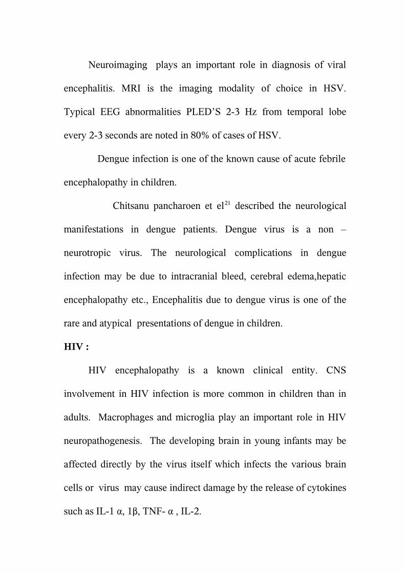
Neuroimaging plays an important role in diagnosis of viral
encephalitis. MRI is the imaging modality of choice in HSV.
Typical EEG abnormalities PLED’S 2-3 Hz from temporal lobe
every 2-3 seconds are noted in 80% of cases of HSV.
Dengue infection is one of the known cause of acute febrile
encephalopathy in children.
Chitsanu pancharoen et el21 described the neurological
manifestations in dengue patients. Dengue virus is a non –
neurotropic virus. The neurological complications in dengue
infection may be due to intracranial bleed, cerebral edema,hepatic
encephalopathy etc., Encephalitis due to dengue virus is one of the
rare and atypical presentations of dengue in children.
HIV :
HIV encephalopathy is a known clinical entity. CNS
involvement in HIV infection is more common in children than in
adults. Macrophages and microglia play an important role in HIV
neuropathogenesis. The developing brain in young infants may be
affected directly by the virus itself which infects the various brain
cells or virus may cause indirect damage by the release of cytokines
such as IL-1 α, 1β, TNF- α , IL-2.
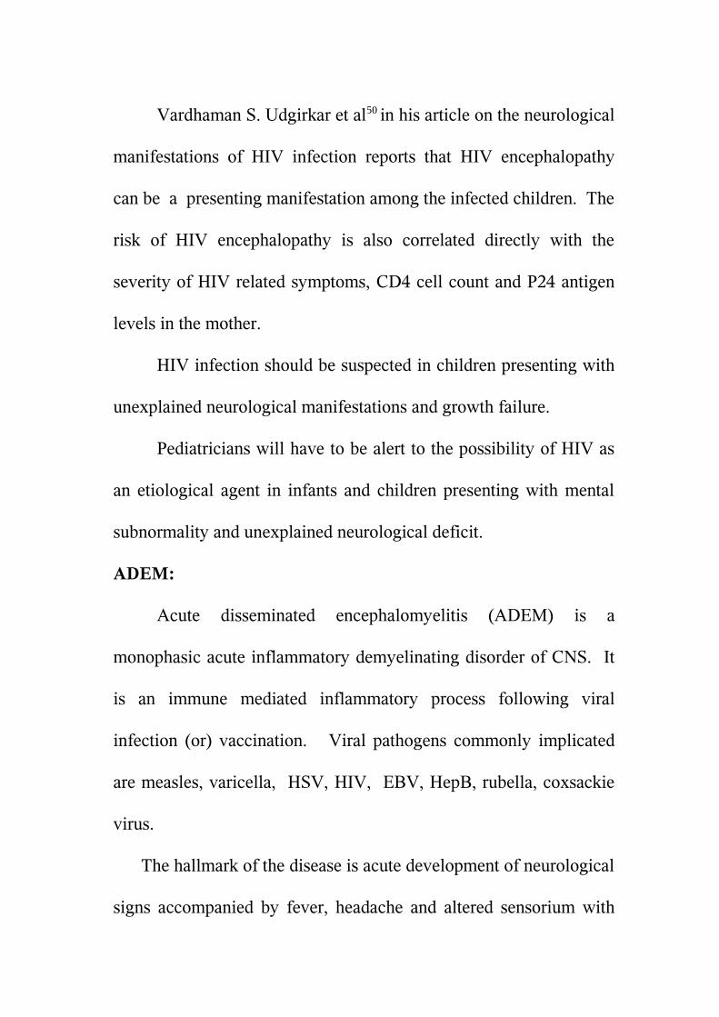
Vardhaman S. Udgirkar et al50 in his article on the neurological
manifestations of HIV infection reports that HIV encephalopathy
can be a presenting manifestation among the infected children. The
risk of HIV encephalopathy is also correlated directly with the
severity of HIV related symptoms, CD4 cell count and P24 antigen
levels in the mother.
HIV infection should be suspected in children presenting with
unexplained neurological manifestations and growth failure.
Pediatricians will have to be alert to the possibility of HIV as
an etiological agent in infants and children presenting with mental
subnormality and unexplained neurological deficit.
ADEM:
Acute disseminated encephalomyelitis (ADEM) is a
monophasic acute inflammatory demyelinating disorder of CNS. It
is an immune mediated inflammatory process following viral
infection (or) vaccination. Viral pathogens commonly implicated
are measles, varicella, HSV, HIV, EBV, HepB, rubella, coxsackie
virus.
The hallmark of the disease is acute development of neurological
signs accompanied by fever, headache and altered sensorium with
focal neurological signs. ADEM can mimic many other neurological
diseases especially meningo encephalitis.
There are two clinical forms of presentation in ADEM.
Encephalitic and myelitic form. In some cases, haemorrhagic forms
have also been reported.22
In a case series on ADEM, it was reported that ADEM presents
with symptoms and signs of meningoencephalitis.23
CSF analysis being normal (or) showing mild elevation of
protein and mild lymphocytic pleocytosis, the diagnosis of ADEM is
by neuroimaging especially MRI with gadolinium contrast.
Screening of the spinal cord is also necessary to detect the lesions in
the cord.
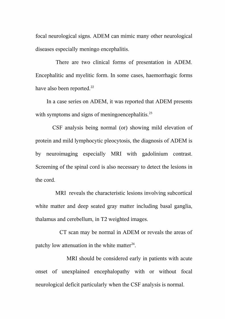
MRI reveals the characteristic lesions involving subcortical
white matter and deep seated gray matter including basal ganglia,
thalamus and cerebellum, in T2 weighted images.
CT scan may be normal in ADEM or reveals the areas of
patchy low attenuation in the white matter24.
MRI should be considered early in patients with acute
onset of unexplained encephalopathy with or without focal
neurological deficit particularly when the CSF analysis is normal.
Reye’s Syndrome :
Reye’s syndrome is characterized by acute non inflammatory
encephalopathy and hepatic failure. It is more common in children.
The exact etiology remains unknown but it is precipitated by a viral
infection especially influenza, varicella and the use of salicylates
during the illness.
Reye’s syndrome should be considered in the differential
diagnosis in any child with vomiting and altered mental status.25 It
results from mitochondrial dysfunction that inhibits oxidative
phosphorylation and fatty acid beta oxidation.
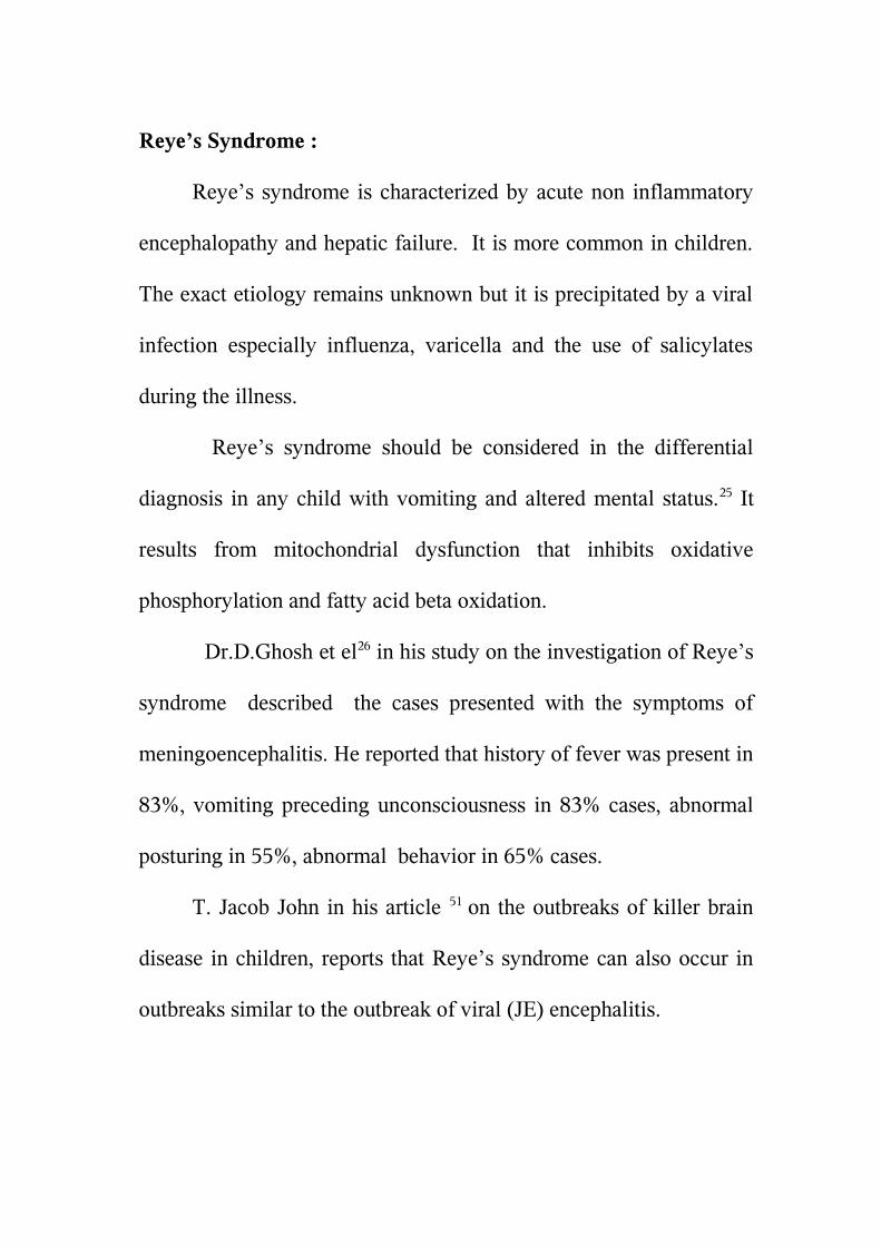
Dr.D.Ghosh et el26 in his study on the investigation of Reye’s
syndrome described the cases presented with the symptoms of
meningoencephalitis. He reported that history of fever was present in
83%, vomiting preceding unconsciousness in 83% cases, abnormal
posturing in 55%, abnormal behavior in 65% cases.
T. Jacob John in his article 51 on the outbreaks of killer brain
disease in children, reports that Reye’s syndrome can also occur in
outbreaks similar to the outbreak of viral (JE) encephalitis.
Unless a high index of suspicion is present most of the cases
will be erroneously misdiagnosed as encephalitis without performing
the necessary investigations for the diagnosis of Reye’s syndrome.
IEM that may mimic Reye’s syndrome include fatty acid
oxidation defects, amino and organic acidopathies and urea cycle
defects.
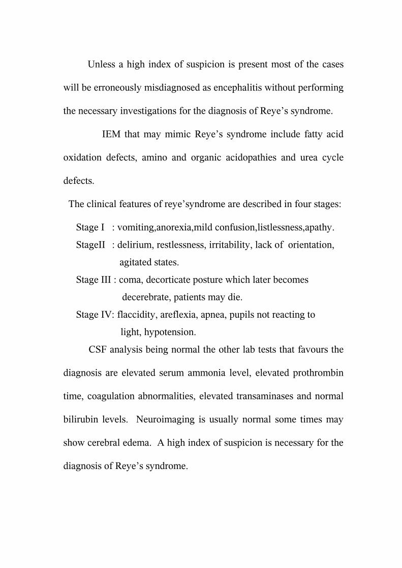
The clinical features of reye’syndrome are described in four stages:
Stage I : vomiting,anorexia,mild confusion,listlessness,apathy.
StageII : delirium, restlessness, irritability, lack of orientation,
agitated states.
Stage III : coma, decorticate posture which later becomes
decerebrate, patients may die.
Stage IV: flaccidity, areflexia, apnea, pupils not reacting to
light, hypotension.
CSF analysis being normal the other lab tests that favours the
diagnosis are elevated serum ammonia level, elevated prothrombin
time, coagulation abnormalities, elevated transaminases and normal
bilirubin levels. Neuroimaging is usually normal some times may
show cerebral edema. A high index of suspicion is necessary for the
diagnosis of Reye’s syndrome.
Cerebral Malaria :
Malaria is an important cause of morbidity and mortality in
South east Asia. 50% of these cases are due to infection with
plasmodium falciparum. Cerebral malaria is one of the dreadful
complication of P.falciparum infection.Clinical presentation includes
fever, seizures and altered mental status or unconsiousness which is
easily confused with diagnosis of AES .
There are case reports of cerebral malaria mimicking
meningoencephalitis with meningeal signs.27
Cerebral malaria is due to blockage and sequestration of
capillaries with parasitized RBC’s. CSF analysis is normal usually
and imaging studies is often normal or shows cerebral edema. The
gold standard test for the diagnosis of malaria is by identification of
malarial parasite by peripheral smear both thick and thin smear
examinations.
Quantitative Buffy Coat :
QBC test is a new method for the identification of the malarial
parasite in the peripheral blood.
In this test, blood sample is centrifuged and RBC’s are stained
with acridine orange and is examined under UV light source. QBC is
fast, easy and more sensitive than thick smear examination.
Rapid Diagnostic Tests :52
Rapid tests detect the malarial antigens such as pf HRP2 /
PMA / PLDH using the monoclonal antibodies. The presence of
parasite antigen in blood is detected by colour changes on the strip
test. Rapid tests are quick and easy to perform but they are only
qualitative tests not useful in prognostication of the disease.
Cerebral malaria unless diagnosed early and treated
appropriately will have a poor prognosis. It should be considered in
dd of AES particularly case from an endemic area, presence of
pallor, splenomegaly along with altered level of consciousness.
CVT :
Cerebral venous thrombosis (CVT) is an important cause of
stroke in children but is less common than other types of strokes.
But it is more challenging to diagnose. It results from thrombosis of
the dural venous sinuses which drains the blood from the brain.
Superior saggital sinus and lateral sinuses are the most
commonly involved sinuses. Signs and symptoms includes fever,
headache, seizures, altered mental status due to increased ICP, focal
neurological deficit. It can present as acute encephalopathy.
Neuroimaging studies forms the main stay of diagnosis of CVT.
IEM :
Inborn error of metabolism are conditions caused by genetic
defects related to synthesis, metabolism, transport or storage of
biochemical compounds. It usually result from deficiency of one or
more enzymes.
IEM presents as acute encephalopathy or chronic progressive
encephalopathy. IEM presenting as acute encephalopathy are
organic acidemias, urea cycle disorders, fatty acid oxidation defects
and mitochondrial disorders.28 Basic lab work up in these cases
includes blood glucose, serum ammonia, ABG and serum lactate
levels.
IEM mimics many of the common pediatric illness such as
sepsis.IEM should be considered in the differential diagnosis of any
sick neonate presenting with the clinical picture of sepsis,HIE,
congenital infections,duct dependant cardiac lesions.29
Prompt detection requires a high index of suspicion and the early
measurement of biochemical markers such as serum
ammonia.Diagnosis is important not only for treatment but also for
the genetic counselling.
Neuroimaging studies in IEM may provide helpful pointers
towards etiology in some cases where IEM may be associated with
structural malformations.29
In glutaric aciduria,frontotemporal atrophy may be
noted.Zellweger syndrome is associated with diffuse cortical
migration abnormalities.
Similarly EEG abnormalities may be suggestive of particular
IEM eg:burst suppression pattern in non –ketotic
hyperglycinemia,comb- like rhythm in MSUD.
AIM OF THE STUDY
1. To reemphasis the importance of CSF analysis in the
differential diagnosis of AES.
2. To correlate the CSF findings with neuroimaging
studies.
MATERIALS AND METHODS
Study Centre :
The study was conducted in the Institute of Child Health and
Research Centre, Government Rajaji Hospital, Madurai Medical
College, Madurai.
Study Period :
The study was carried out prospectively from December 2007
to May 2009.
Study Design :
Prospective Observational Study.
Study Population :
Children admitted in Govt. Rajaji Hospital, Madurai Medical
College, Madurai.
Sample size :
111 cases.
Inclusion Criteria :
Children between 2 months to 12 years with a clinical
diagnosis of AES.
Exclusion Criteria :
1. Age < 2 months and > 12 years.
2. Repeat lumbar puncture.
Conflict of interest : Nil
Financial Support : Nil
Ethical Committee clearance : Obtained.
Methodology :
For all children admitted with clinical picture of AES, a
detailed history, general examination, systemic examination and
detailed neurological examination were made. Informed written
consent was obtained from the parents for the procedure of lumbar
puncture. Lumbar puncture was done in all cases unless absolute
contraindications for LP such as skin infection at the site of the LP,
thrombocytopenia, increased ICP with signs of pending cerebral
herniation were present. CSF analysis was done.
For all the cases, CSF cytology was done by a single observer
who is the principal investigator in the study for better results.
Separate consent for doing cell count by the investigator was not
obtained as it is a part of service provided. CSF cell count was done
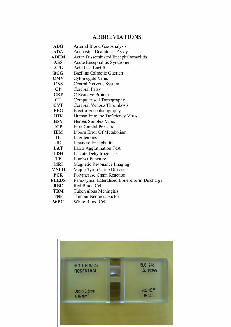
by using the Fuch’s Rosenthal chamber with in half an hour of
collecting the sample.
CSF cell count technique : 54
Uncentrifuged CSF is diluted 1 in 2 by mixing 1 drop of CSF
with 1 drop of diluting fluid using fine bore pipettes so that drops
will be of equal volume. Using the capillary tube, Fuch’s Rosenthal
chamber is filled with this diluted CSF carefully so that CSF does
not overflow into the channels on each side of the chamber.
Counting chamber and its rulings are focused and cells are counted
in 5 large squares and multiplied by 2 to give the total count per
cumm. Differential count done by centrifuging the CSF sample, and
by staining with methylene blue.
Biochemical analysis of CSF protein, glucose, chloride and
globulin were done. Simultaneous estimation of blood glucose
levels were done to avoid spuriously elevated blood / CSF glucose
ratio. Microbiological work up including CSF gram stain, Latex
agglutination test, CSF culture, blood culture and CRP was done in
cases where the CSF analysis favours pyogenic meningitis. AFB
stain in CSF, mantoux test, chest x ray, Gastric juice for AFB was
done in cases suggestive of TB meningitis. Serology for JE was done
in cases with viral encephalitis. All cases were subjected to
neuroimaging irrespective of CSF analysis.
All children were effectively managed with appropriate
measures along with nursing care. Children who developed
complication like hydrocephalus were referred to neurosurgery
department for surgical management.
Statistical Methods :
Data collected were recorded in a Master chart. Data analysis
was done with the help of computer using SSPS software. Data was
analysed using simple descriptive statistics.
RESULTS AND ANALYSIS
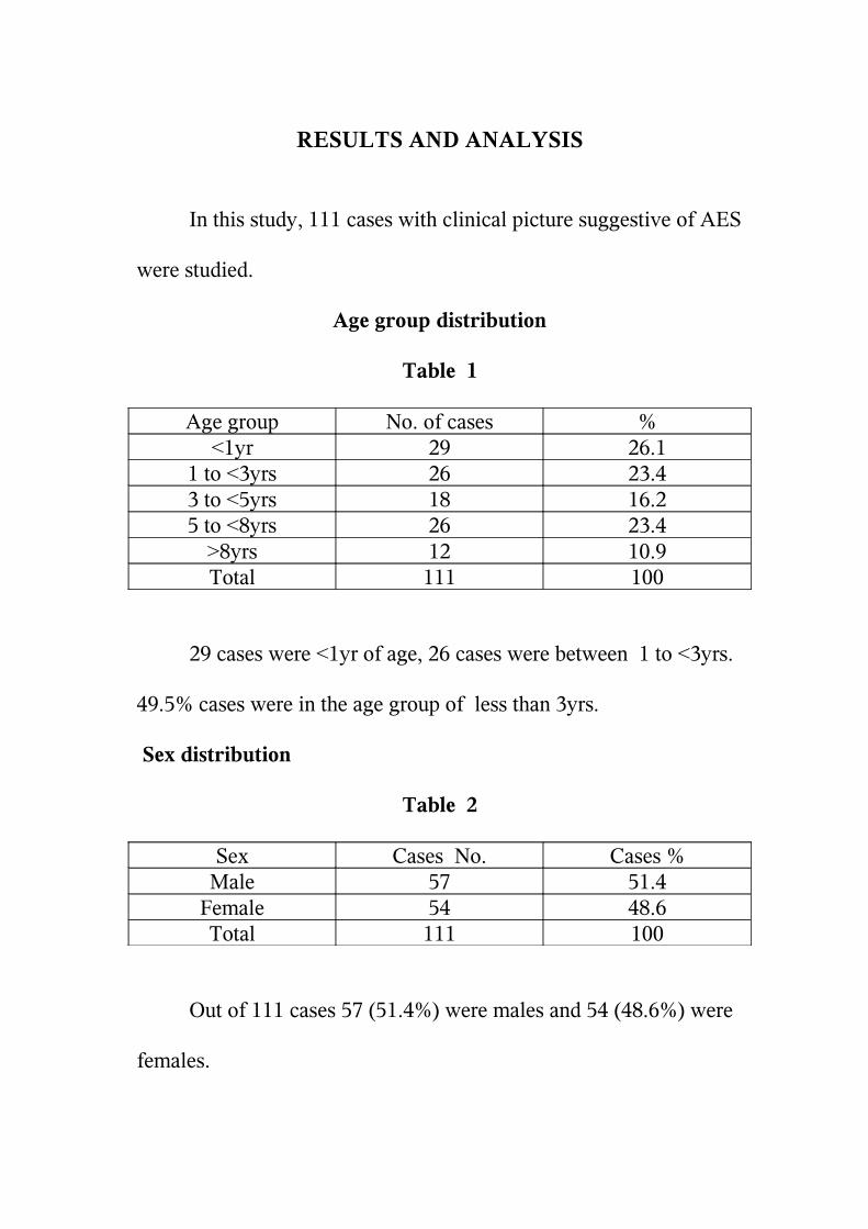
In this study, 111 cases with clinical picture suggestive of AES
were studied.
Age group distribution
Table 1
Age group No. of cases %<1yr 29 26.1
1 to <3yrs 26 23.43 to <5yrs 18 16.25 to <8yrs 26 23.4
>8yrs 12 10.9Total 111 100
29 cases were <1yr of age, 26 cases were between 1 to <3yrs.
49.5% cases were in the age group of less than 3yrs.
Sex distribution
Table 2
Sex Cases No. Cases %Male 57 51.4
Female 54 48.6Total 111 100
Out of 111 cases 57 (51.4%) were males and 54 (48.6%) were
females.
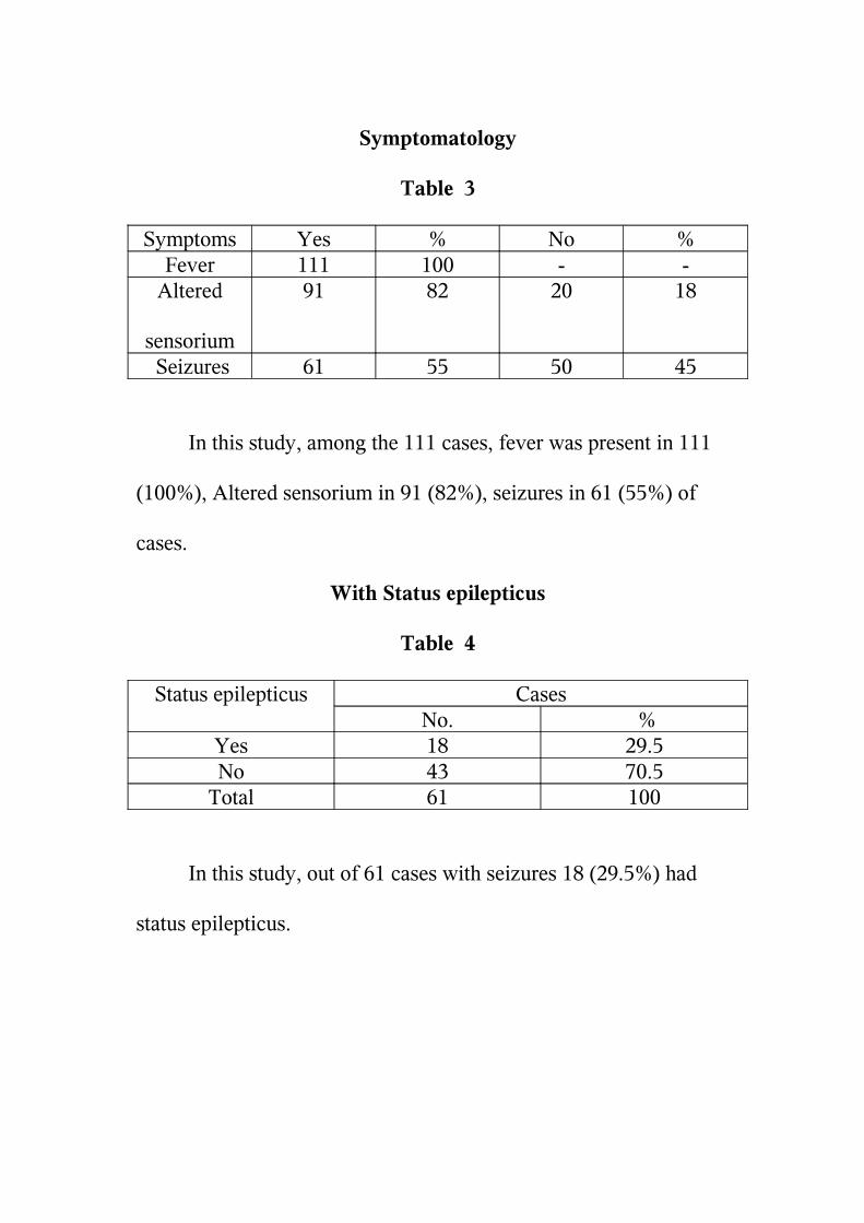
Symptomatology
Table 3
Symptoms Yes % No %Fever 111 100 - -
Altered
sensorium
91 82 20 18
Seizures 61 55 50 45
In this study, among the 111 cases, fever was present in 111
(100%), Altered sensorium in 91 (82%), seizures in 61 (55%) of
cases.
With Status epilepticus
Table 4
Status epilepticus CasesNo. %
Yes 18 29.5No 43 70.5
Total 61 100
In this study, out of 61 cases with seizures 18 (29.5%) had
status epilepticus.
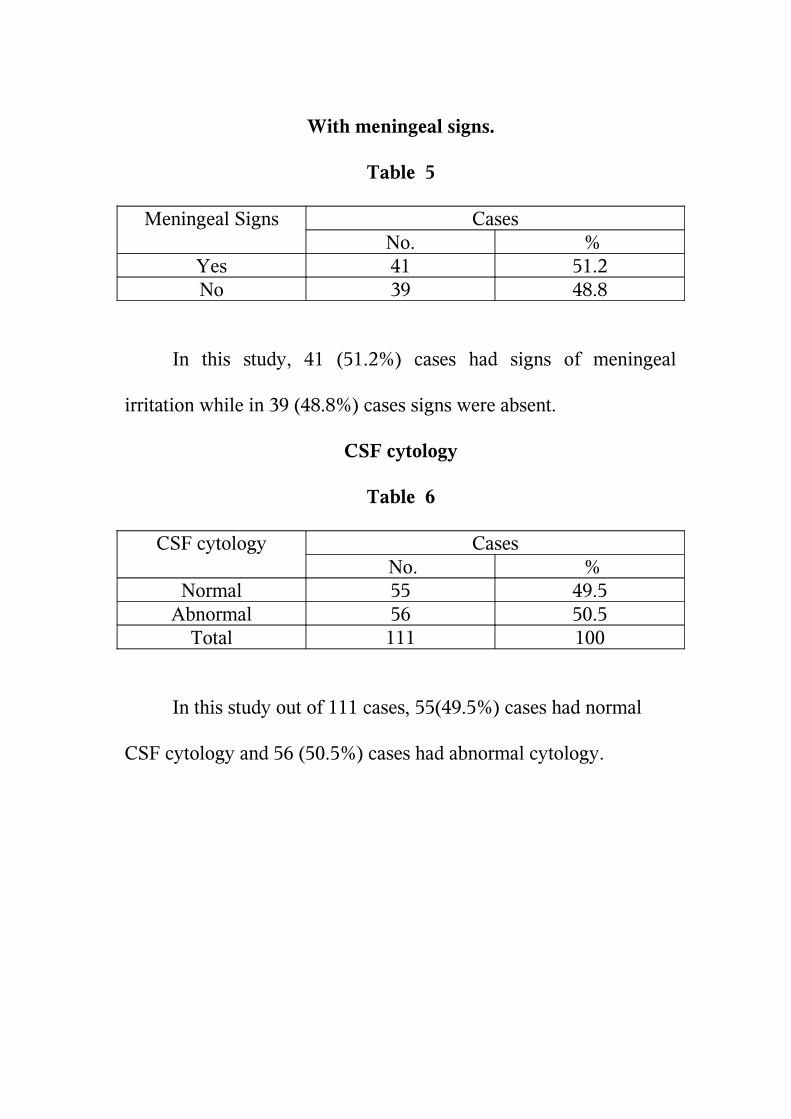
With meningeal signs.
Table 5
Meningeal Signs CasesNo. %
Yes 41 51.2No 39 48.8
In this study, 41 (51.2%) cases had signs of meningeal
irritation while in 39 (48.8%) cases signs were absent.
CSF cytology
Table 6
CSF cytology Cases No. %
Normal 55 49.5Abnormal 56 50.5
Total 111 100
In this study out of 111 cases, 55(49.5%) cases had normal
CSF cytology and 56 (50.5%) cases had abnormal cytology.
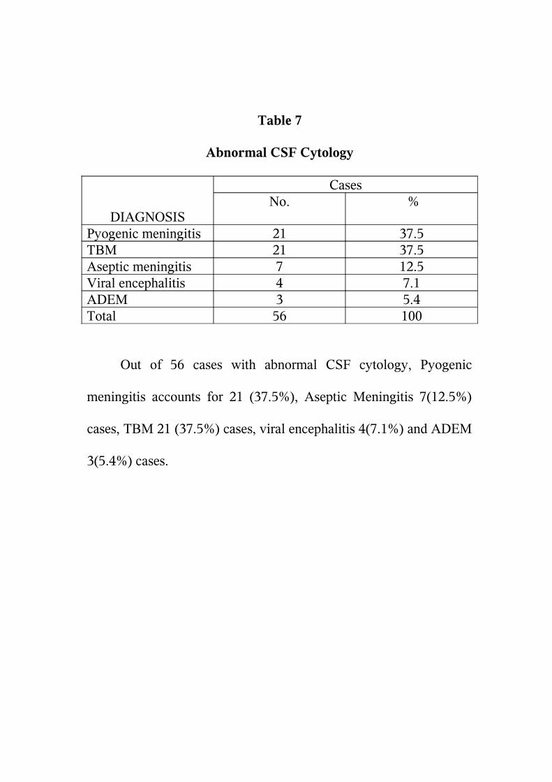
Table 7
Abnormal CSF Cytology
DIAGNOSIS
CasesNo. %
Pyogenic meningitis 21 37.5TBM 21 37.5Aseptic meningitis 7 12.5Viral encephalitis 4 7.1ADEM 3 5.4Total 56 100
Out of 56 cases with abnormal CSF cytology, Pyogenic
meningitis accounts for 21 (37.5%), Aseptic Meningitis 7(12.5%)
cases, TBM 21 (37.5%) cases, viral encephalitis 4(7.1%) and ADEM
3(5.4%) cases.
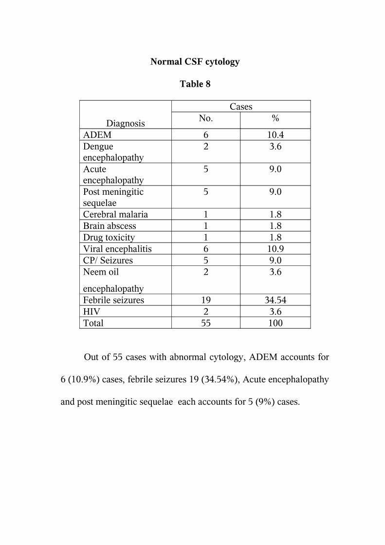
Normal CSF cytology
Table 8
Diagnosis
CasesNo. %
ADEM 6 10.4Dengue encephalopathy
2 3.6
Acute encephalopathy
5 9.0
Post meningitic sequelae
5 9.0
Cerebral malaria 1 1.8Brain abscess 1 1.8Drug toxicity 1 1.8Viral encephalitis 6 10.9CP/ Seizures 5 9.0Neem oil
encephalopathy
2 3.6
Febrile seizures 19 34.54HIV 2 3.6Total 55 100
Out of 55 cases with abnormal cytology, ADEM accounts for
6 (10.9%) cases, febrile seizures 19 (34.54%), Acute encephalopathy
and post meningitic sequelae each accounts for 5 (9%) cases.
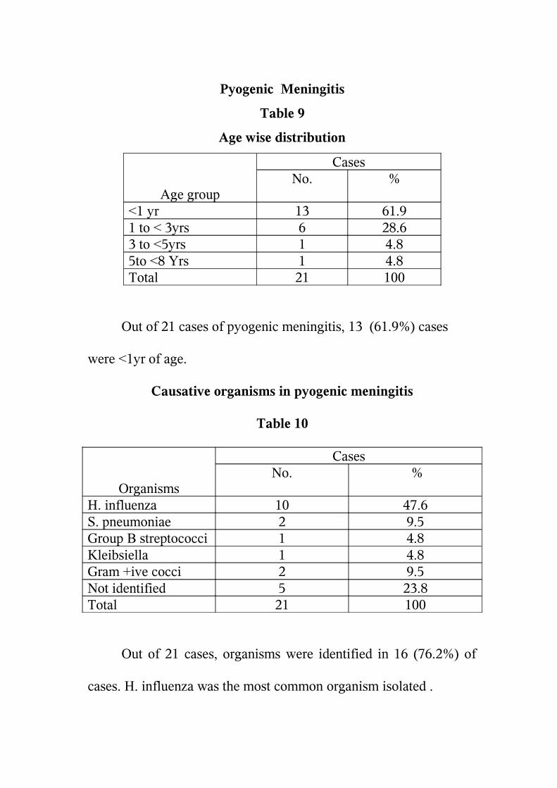
Pyogenic Meningitis
Table 9
Age wise distribution
Age group
CasesNo. %
<1 yr 13 61.91 to < 3yrs 6 28.63 to <5yrs 1 4.85to <8 Yrs 1 4.8Total 21 100
Out of 21 cases of pyogenic meningitis, 13 (61.9%) cases
were <1yr of age.
Causative organisms in pyogenic meningitis
Table 10
Organisms
CasesNo. %
H. influenza 10 47.6S. pneumoniae 2 9.5Group B streptococci 1 4.8Kleibsiella 1 4.8Gram +ive cocci 2 9.5Not identified 5 23.8Total 21 100
Out of 21 cases, organisms were identified in 16 (76.2%) of
cases. H. influenza was the most common organism isolated .
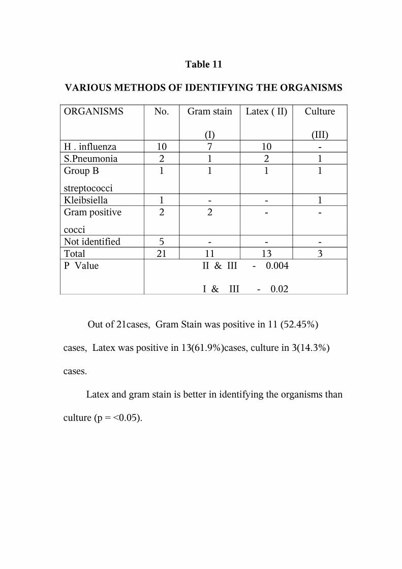
Table 11
VARIOUS METHODS OF IDENTIFYING THE ORGANISMS
ORGANISMS No. Gram stain
(I)
Latex ( II) Culture
(III)H . influenza 10 7 10 -S.Pneumonia 2 1 2 1Group B
streptococci
1 1 1 1
Kleibsiella 1 - - 1Gram positive
cocci
2 2 - -
Not identified 5 - - -Total 21 11 13 3P Value II & III - 0.004
I & III - 0.02
Out of 21cases, Gram Stain was positive in 11 (52.45%)
cases, Latex was positive in 13(61.9%)cases, culture in 3(14.3%)
cases.
Latex and gram stain is better in identifying the organisms than
culture (p = <0.05).
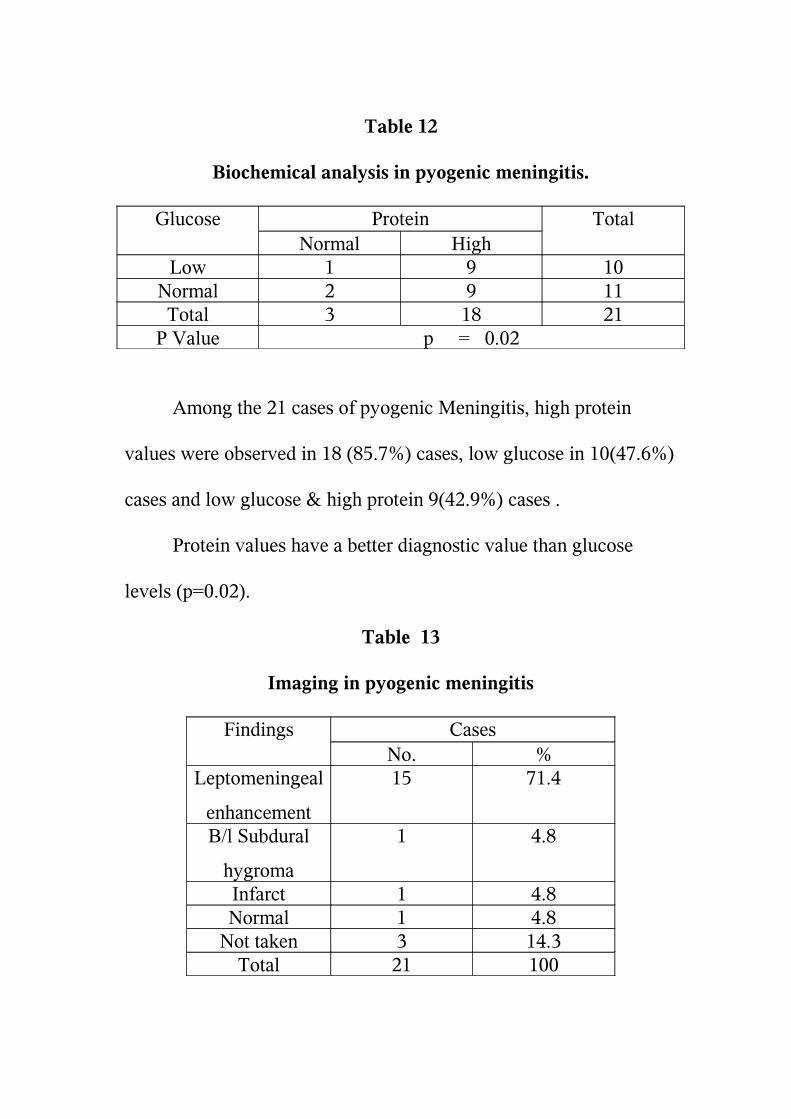
Table 12
Biochemical analysis in pyogenic meningitis.
Glucose Protein TotalNormal High
Low 1 9 10Normal 2 9 11Total 3 18 21
P Value p = 0.02
Among the 21 cases of pyogenic Meningitis, high protein
values were observed in 18 (85.7%) cases, low glucose in 10(47.6%)
cases and low glucose & high protein 9(42.9%) cases .
Protein values have a better diagnostic value than glucose
levels (p=0.02).
Table 13
Imaging in pyogenic meningitis
Findings CasesNo. %
Leptomeningeal
enhancement
15 71.4
B/l Subdural
hygroma
1 4.8
Infarct 1 4.8Normal 1 4.8
Not taken 3 14.3Total 21 100
In this study, out of 21 cases of pyogenic meningitis
Leptomeningeal enhancement was observed in 15(71.4%) cases in
the neuroimaging (CT).
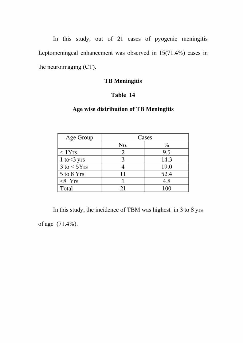
TB Meningitis
Table 14
Age wise distribution of TB Meningitis
Age Group CasesNo. %
< 1Yrs 2 9.51 to<3 yrs 3 14.33 to < 5Yrs 4 19.05 to 8 Yrs 11 52.4<8 Yrs 1 4.8Total 21 100
In this study, the incidence of TBM was highest in 3 to 8 yrs
of age (71.4%).
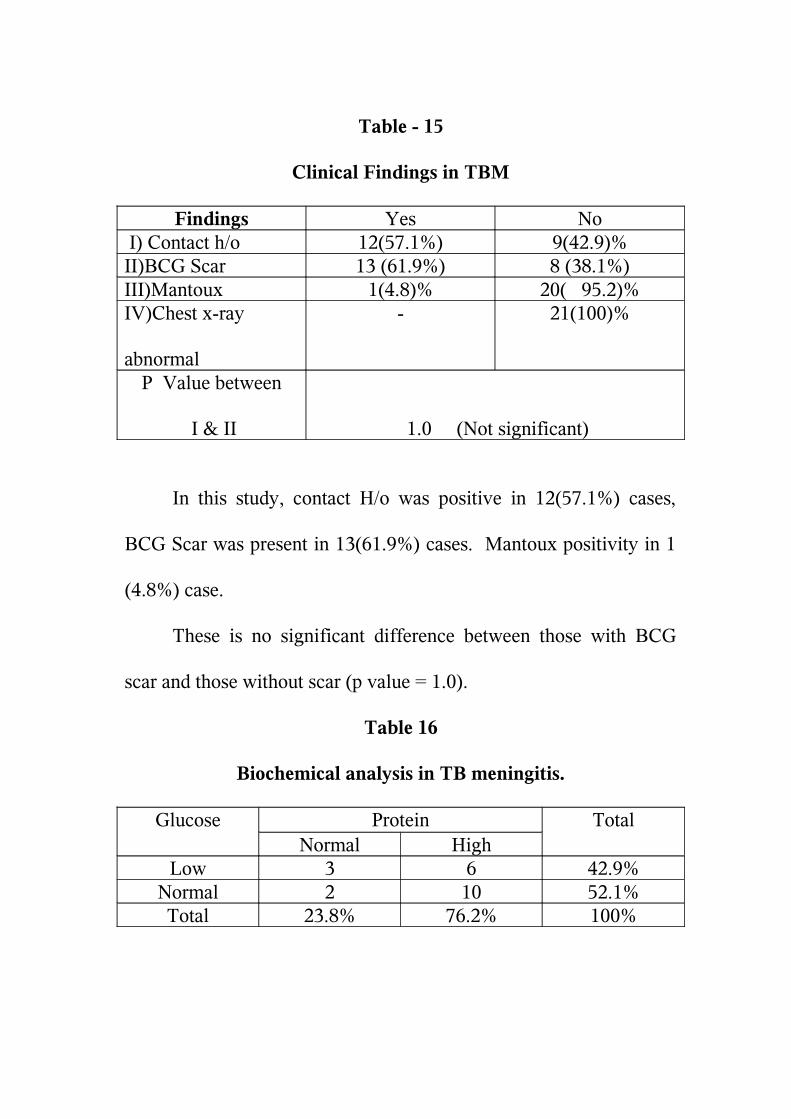
Table - 15
Clinical Findings in TBM
Findings Yes No I) Contact h/o 12(57.1%) 9(42.9)%II)BCG Scar 13 (61.9%) 8 (38.1%)III)Mantoux 1(4.8)% 20( 95.2)%IV)Chest x-ray
abnormal
- 21(100)%
P Value between
I & II 1.0 (Not significant)
In this study, contact H/o was positive in 12(57.1%) cases,
BCG Scar was present in 13(61.9%) cases. Mantoux positivity in 1
(4.8%) case.
These is no significant difference between those with BCG
scar and those without scar (p value = 1.0).
Table 16
Biochemical analysis in TB meningitis.
Glucose Protein TotalNormal High
Low 3 6 42.9%Normal 2 10 52.1%Total 23.8% 76.2% 100%
In this study, high protein values was observed in 16(76.2%)
cases. Protein values significantly correlate than glucose values
(P value = 0.03).
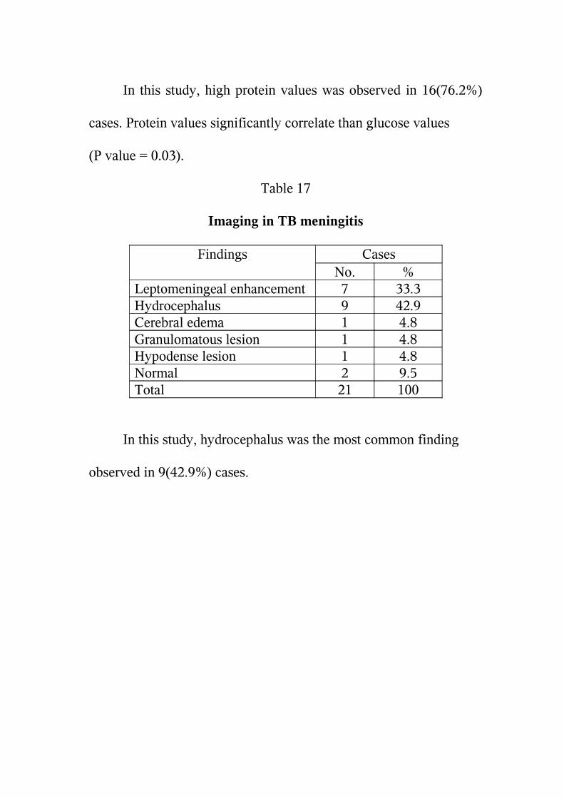
Table 17
Imaging in TB meningitis
Findings CasesNo. %
Leptomeningeal enhancement 7 33.3Hydrocephalus 9 42.9Cerebral edema 1 4.8Granulomatous lesion 1 4.8Hypodense lesion 1 4.8Normal 2 9.5Total 21 100
In this study, hydrocephalus was the most common finding
observed in 9(42.9%) cases.
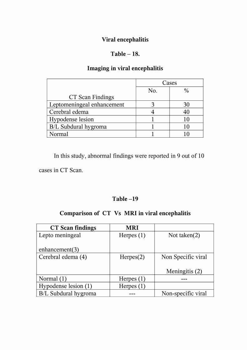
Viral encephalitis
Table – 18.
Imaging in viral encephalitis
CT Scan Findings
CasesNo. %
Leptomeningeal enhancement 3 30Cerebral edema 4 40Hypodense lesion 1 10B/L Subdural hygroma 1 10Normal 1 10
In this study, abnormal findings were reported in 9 out of 10
cases in CT Scan.
Table –19
Comparison of CT Vs MRI in viral encephalitis
CT Scan findings MRILepto meningeal
enhancement(3)
Herpes (1) Not taken(2)
Cerebral edema (4) Herpes(2) Non Specific viral
Meningitis (2)Normal (1) Herpes (1) ---Hypodense lesion (1) Herpes (1)B/L Subdural hygroma --- Non-specific viral
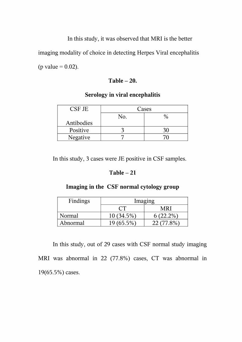
In this study, it was observed that MRI is the better
imaging modality of choice in detecting Herpes Viral encephalitis
(p value = 0.02).
Table – 20.
Serology in viral encephalitis
CSF JE
Antibodies
CasesNo. %
Positive 3 30Negative 7 70
In this study, 3 cases were JE positive in CSF samples.
Table – 21
Imaging in the CSF normal cytology group
Findings ImagingCT MRI
Normal 10 (34.5%) 6 (22.2%)Abnormal 19 (65.5%) 22 (77.8%)
In this study, out of 29 cases with CSF normal study imaging
MRI was abnormal in 22 (77.8%) cases, CT was abnormal in
19(65.5%) cases.
DISCUSSION
Acute encephalitis syndrome (AES) includes a list of
conditions with a similar clinical presentation posing a great
diagnostic challenge to every pediatrician. Correct diagnosis and
early institution of appropriate therapy is necessary to avoid
significant morbidity and mortality and also to avoid polypharmacy.
CSF analysis helps in the differential diagnosis of AES to a great
extent. The importance of CSF cell count analysis in the differential
diagnosis of AES was reemphasized in this study.
This study included 111 children in the age group of 2 months
– 12 years with a clinical picture suggestive of AES.
In this study, 57 (51.4%) were males and 54 (48.6%) were
females.
54 (49.5%) were less than 3 years of age, 18 (16.2%) were in
the age group between 3 to < 5 yrs, 26 (23.4%) were in the age
group between 5 to < 8 years and 12 (10.8%) were > 8 years.
In the clinical presentation, Fever was present in 111(100%)
cases, altered sensorium in 91 (82%) and seizures in 61 (55%).
Out of 61 cases with seizures, 18 (29.5%) cases had status
epilepticus. Febrile seizures was the most common cause of status
epilepticus in this study.
In this study,41(51.3%) cases had signs of meningeal
irritation where in 39(48.8%) cases the signs were absent.
Meningeal signs were applicable to only those children with
AF closed. Out of 111 cases , 80 cases falls into this category.
In this study, 55 (49.5%) cases had normal CSF cytology and
56 (50.5%) had abnormal CSF cytology.
PYOGENIC MENINGITIS :
In this study, among the 21 cases of pyogenic meningitis, 15
(61.9%) cases were in the age group of < 1 yr which is the most
vulnerable age group for bacterial meningitis.
Bandaru Rao et al 18, in their study on etiology and clinical
profile of acute bacterial meningitis reported that 65% of cases were
< 1 yr of age which correlates well with this study.
In a study by F. Clifford Rose30 on the diagnosis of pyogenic
meningitis, low glucose values in CSF was observed in 70% and
raised protein levels in 90% of cases .
In this study, raised protein levels in CSF was observed in
85.7% and low glucose values in 47.6% cases. Raised protein
values have a better diagnostic value than low glucose levels in this
study.
Causative organisms identified by combining Gramstain and
or latex agglutination and or culture of CSF were in 16 (76.2%)
cases.
Among the identified etiological agents, H.influenza accounts
for 10 (62.5%) cases, S.pneumonia 2 (12.5%) cases,group B
streptococci and kleibsiella each (6.5%) cases respectively.
In our study, Gram stain provided an evidence of the
etiological agents in 11 (52.45%) cases which correlated with
various other studies18,19 reporting the sensitivity of gram stain
between 60-90%.
Chandramukhi A et al 19 in their study on the bacteriological
profile of community acquired acute bacterial meningitis reported
the sensitivity of gram staining to be 65%.
In a similar study by Bandaru Rao et al18, Gram staining
revealed the probable etiological agent in 80% cases.
Yield of bacteria on a gram stain depends on several factors
such as number of organisms present, prior use of antibiotics,
techniques used for smear preparation like centrifuged
deposit,cytospin,direct smear etc, staining techniques and observer
skill and experience.
In this study, Latex test revealed the organisms in 13 (61.9%)
cases which correlates well with various other Indian studies19. H.
influenza is the most common organism isolated in this study.Latex
test can be useful to detect the organisms where the antibiotics
are used early before CSF analysis.
CSF culture was positive in 3 (14.3%) cases. Several Indian
studies report low CSF culture positivity ranging from 6 to 50%.
Various reasons for low yield of bacteria on culture are prior
antibiotic therapy, delay in transport of specimens to the lab, non
availability of special media for specific pathogens and lack of 24
hrs facility for processing CSF samples.
No case of meningococcal meningitis was reported during the
study period .This may be due to low prevalence of meningococcal
meningitis in children except during the epidemics.
Correlation of Gram stain & latex test with the CSF culture in
identifying the organisms shows that Gram stain & latex are equally
efficiacous in detecting the organisms and better than culture.
(p<0.05).
This study shows that simple Gram stain combined with the
latex test will give a rapid diagnosis on the etiology of bacterial
meningitis.
Storring and synder31 in their study of imaging in children with
bacterial meningitis reported ventricular dilatation in 75%, subdural
collection in 25%, cerebral edema in 9%, ischaemic infarcts in 19%.
In this study, most common finding observed was
leptomeningeal enhancement 15 (71.4%) cases. One case of
subdural hygroma32 was found which is positive for H.influenza.
Other findings were infarct in 1 case and normal study in 1 case.
We must anticipate complications while treating bacterial
meningitis if there is lack of satisfactory response to antibiotics in
the form of slow defervescence ,reappearance of fever while on
treatment,persistence of seizures, reappearance or persistence of
bulging fontanelle,deterioration of mental status or any increase in
head circumference.
Neuroimaging studies are typically used to monitor the
complications such as subdural effusion ,hydrocephalus ,infarction
etc.Imaging studies in acute meningitis may provide normal
findings. In this study , one case had a normal imaging.
Imaging studies does not prove or exclude the presence of
acute meningitis.It is the CSF analysis which is the single most
important diagnostic study.
TBM:
TBM occurs in 7-12% of tuberculous patients in developing
countries.33
Wilson et al34 in their series of cases of TBM, highest age
group incidence observed was between 3-9 yrs which correlates
with our study where the highest incidence of TBM were in the age
group 3-8 yrs. The youngest child reported in this study was 9
months old.
Of these 21 cases,10 (47.6%) were males and 11 (52.4%)
were females.
Gold standard for diagnosis of TBM is isolation of AFB bacilli
in CSF which is usually rare. In this study the diagnosis of TBM
was made by AIIMS criteria 16
Contact H/o was present in 12 (57.1%) cases which correlates
with Yaramis et al study35.
In a study on the clinical profile of TB meningitis by
Thilothammal et al36 contact H/o was present in 40% cases.
Thilothammal et al 36 in their study observed that Mantoux
positivity was present in 13% whereas in this study it was only 5%.
In a retrospective study of TBM in AIIMS37, BCG scar was
present in 50% which correlates with our study.
ICMR BCG trials38,42 in chingelput also report that BCG offers
no protection against primary TB infection or its progression to
severe forms which correlates with our study (pvalue < 0.05).
Mathur et al39 in a comparative study between BCG
vaccinated and non vaccinated groups of patients could not find any
significant difference in clinical pattern of TB which correlates
well with this study.
In the study by Thilothammal et al 36on TB meningitis, chest
x ray was abnormal in 30% cases whereas in this study x ray was
normal in all the cases.
Lymphadenopathy which is a common feature of primary TB
infection in children cannot be detected by routine chest x ray.
CT is superior in detecting the lymphadenopathy40 which may
explain the observation of normal x ray findings in this study.
Elevated protein levels in CSF was observed in 16 (76.2%)
cases which has a better diagnostic value in TBM than glucose
values.
CSF sugar content may be normal( or) low in TBM and this
alone should not be used as an evidence for (or) against TBM 34.
CSF protein values depends upon clinical stage of the disease. 36
Bacteriological confirmation of diagnosis of TBM varies from
10% to 70% in various Indian studies .
In this study, AFB bacilli was not identified in any case which
may be due to failure to examine a large volume of CSF and lack of
fluorescent microscopy as a routine examination.
ADA level in CSF was done only in 3 cases. Two cases shows
an increased value and one case shows normal value 41. Cut off
values of more than 8 IU/L favors the diagnosis.
In this study, hydrocephalus was the most common finding
observed 42.9% in the neuro imagingwhich correlates with a study
conducted at AIIMS (2000-2004).
In an recent analysis of TBM patients at AIIMS, CT
abnormalities detected were hydrocephalus 65.2%,basal exudates
47.8%,cerebral edema 21.7%,infarcts 34.8%,tuberculoma 30.4%.
Ravenscroft et al 43 in their study showed that 16% of patients
with TBM had associated intracranial granuloma. In our study one
case of TB meningitis showed associated intracranial granuloma in
MRI.
Imaging was normal in 9.5% of cases in this study which
correlates with Ozates et al study. 44
Imaging modalities have greatly enhanced the diagnostic
accuracy of neurotuberculosis but not pathognomonic for the
disease.
VIRAL ENCEPHALITIS:
In this study, 10 cases of viral encephalitis were
observed. Out of the 10 cases, 5 cases of herpes viral encephalitis,3
cases of JE were observed.The highest incidence of cases were in the
age group between 3 to 8 years.
Imaging studies remains the diagnostic modality of
choice.Out of the 5 cases of Herpes encephalitis,CT shows normal
study in 1, leptomeningeal enhancement in 1, cerebral edema in 2
and hypodense lesion in 1 case. None of the cases was picked up by
CT. MRI detects the diagnostic lesions in the temporal lobe in all
the cases.On comparison of CT and MRI ,herpes viral encephalitis
was better diagnosed by MRI (p <0.05).
In this study, 3 cases of Japanese encephalitis were observed.
Serology was positive for JE in the CSF sample and in the blood in
all the three cases. Imaging does not reveal the specific basal
ganglia, thalami involvement in any case. Imaging abnormalities
were cerebral edema (1) and leptomeningeal enhancement(2).
Seasonal outbreaks and epidemics of JE was not observed
during the study period probably due to JE vaccination compaign
undertaken in Madurai district 1 year back.
All the 3 cases of JE observed in this study had not undergone
the vaccination.
CSF cell count was normal in 6 out of 10 cases of viral
encephalitis in this study which could not be explained.
Out of these 6 cases with normal CSF cytology, diagnosis was
made by imaging (MRI) which detected the lesions in the temporal
lobe suggestive of Herpes Viral encephalitis in 4 cases.
The diagnosis of JE encephalitis was made in the remaining 2
cases based on CSF JE serology positivity.
In this study, 29 cases had normal CSF cytology with the
clinical picture of AES (after excluding febrile seizures first
episode,CP, neem oil encephalopathy).
ADEM an inflammatory demyelinating disease involving
CNS white matter can present with fever and altered sensorium
mimicking encephalitis. ADEM accounts for one third of all known
case of encephalitis. 23,45
In the case series on ADEM 23, 3 out of 7 cases presented with
altered sensorium, and CSF abnormality were noted in 2 cases
whereas in this study, out of 9 cases of ADEM, 5 cases presented
with altered sensorium and CSF abnormality in the form of mild
elevation of proteins and CSF lymphocytosis was observed in 3
cases .
In the case series on ADEM 23,youngest age group reported
was about 8 months old whereas in this study the youngest age
group in which ADEM was observed was 4 months old.
CT may be normal in ADEM where MRI is the diagnostic
modality of choice which reveals the characteristic lesions in T2
weighted images. 24
Haemorrhagic forms of ADEM have been reported 22. In this
study, among the 9 cases, one case had haemorrhagic type of ADEM
in which CSF cytology showed the presence of RBC’s and the
diagnosis was made by imaging.
On the comparison of CT vs MRI in the diagnosis of ADEM,
MRI remains the better imaging modality of choice.MRI should be
considered early in patients with acute onset unexplained
encephalopathy with or with out focal neurological deficit.
There are case reports of cerebral malaria presenting with
symptoms of diffuse meningo encephalitis and meningeal signs.27
In this study, we had one case of cerebral malaria. 7 yr old
female child from Paramakudi (malaria endemic area), was admitted
with the clinical picture of AES. In this case, CSF analysis and
imaging being normal simple peripheral smear examination revealed
the diagnosis. Smear was positive for mixed infection of Pl.Vivax
and Pl. falciparum.
In the clinical picture of AES, residing from an malaria
endemic area, presence of anaemia with splenomegaly are important
clues towards the diagnosis of cerebral malaria. This child was
treated with artemesinin combination therapy (ACT), other
supportive measures and a dramatic improvement was observed with
in 36 hours.
All efforts must be made to diagnose cerebral malaria by
examining peripheral smear repeatedly and also by using serological
tests in cases where index of suspicion is high.
Dengue infection is one of the known cause of acute febrile
encephalopathy in children.21,46
In this study, we report 2 cases of Dengue infection presenting
as AES. Causes of altered sensorium in Dengue infection may be
due to shock, hyponatremia, intracranial bleed, hepatic involvement ,
kidney failure and rarely encephalitis.
In this study, for both the cases CSF antibodies for dengue
virus was negative which possibly excludes the encephalitis caused
by dengue virus itself.
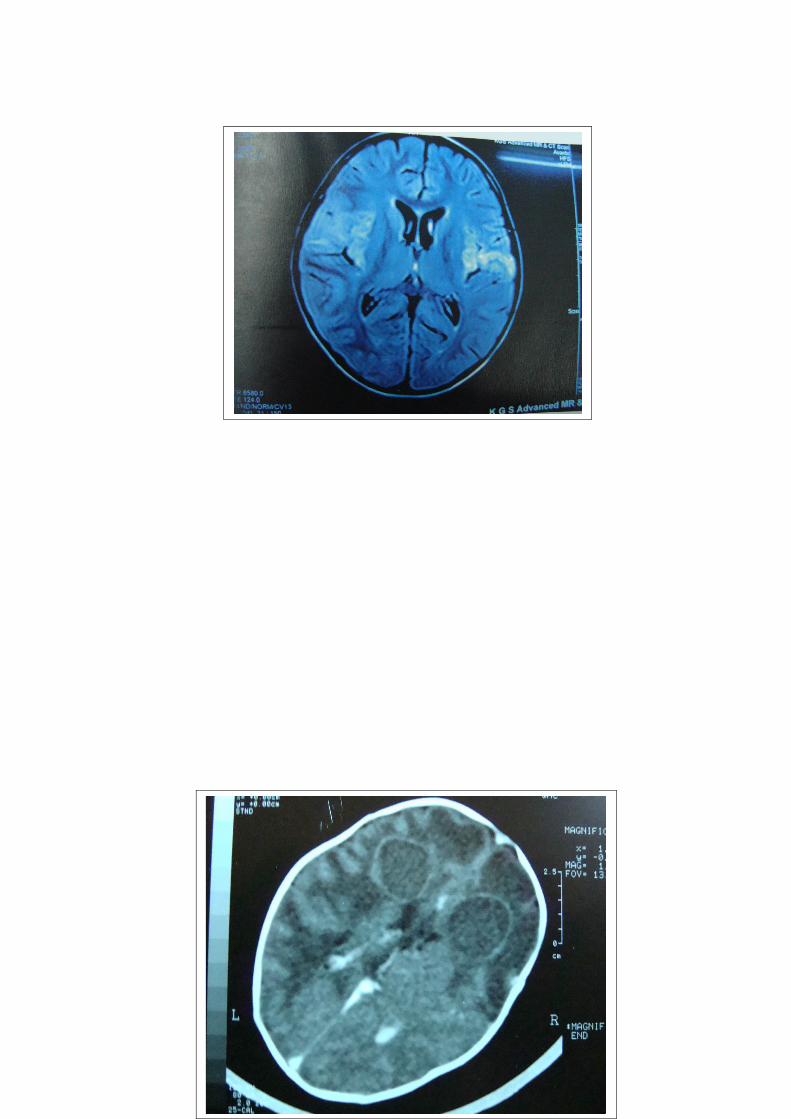
In this study, we had 2 cases of HIV presenting as AES. One
case a known case of HIV on ART presented with AES. Imaging
reveals HIV vasculitis49. Other case had progressive encephalopathy
imaging showing cerebral atrophy clinical suspicion clinches the
diagnosis and confirmed by ELISA.
Vardhaman S. Udgirkar et al50 in his article on the neurological
manifestations of HIV infection reports that HIV encephalopathy
can be a presenting manifestation among the infected children.
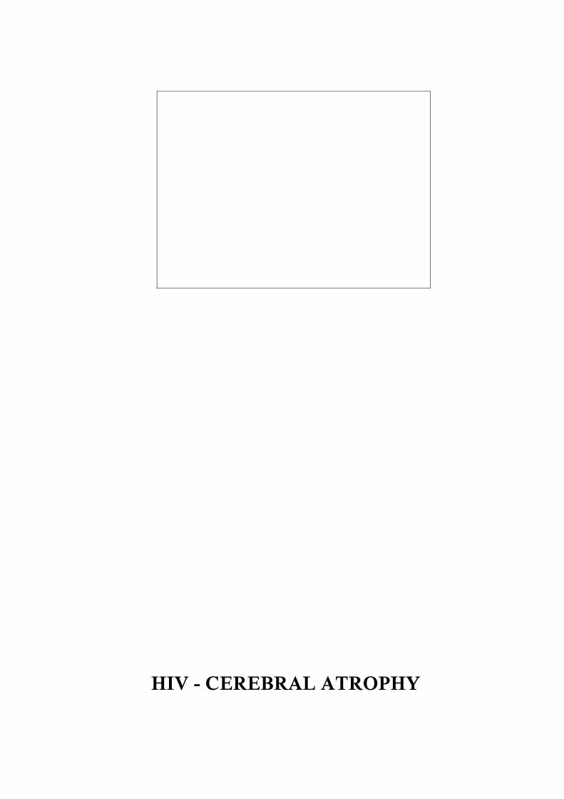
Brain abscess is an important differential diagnosis in children
with unexplained fever, altered sensorium ,increased ICP and focal
deficit.47
CSF examination may be normal or may show mild elevation
of proteins and WBC’S.Imaging remains the diagnostic modality of
choice48.
In this study, one case of brain abscess was observed. 1 ½
years old male child admitted with fever, altered sensorium and there
was no pupillary abnormalities, focal deficits or signs of increased
ICP such as papilledema and so CSF analysis was done. CSF being
normal, CT revealed the diagnosis and child was transferred out to
neuro surgery department for the drainage of abscess.
Toxic encephalopathy is one of the cause for non infectious
encephalopathy in children. Neem oil encephalopathy is one of the
important cause in the southern region of Tamilnadu .
In this study, 4 cases of neem oil encephalopathy were
observed.
Another case of toxic encephalopathy in this study includes a
case of carbamazepine toxicity presented with altered sensorium and
seizures and the child was treated symptomatically, regained
consciousness with in 24 hours.
In this study, 5 cases of Acute encephalopathy for which
etiological diagnosis could not be found due to non availability of
serum ammonia, serum lactate etc.
In this study, we had 5 cases with post meningitic sequalae in
the neuroimaging studies where the CSF analysis was normal.
In this study, we had 29 cases with the clinical picture of AES
and CSF analysis being normal, but showing abnormalities in the
neuroimaging studies 19 (65.5%) in CT and 22 (77.8%) in MRI.
This study shows clinical suspicion along with the appropriate
neuroimaging studies picks up the etiological diagnosis in the group
of patients presenting as AES with normal CSF analysis.
CONCLUSIONS
CSF analysis helps in the differential diagnosis of AES to a
great extent.
The incidence of pyogenic meningitis is highest in < 1 yr of
age group.
The most common organism isolated is H.influenza.
Gram stain & latex test are equally efficacious in identifying
the organism.
Protein levels in CSF has a significant diagnostic value than
glucose levels.
The role of neuroimaging in pyogenic meningitis is to detect
the complications like subdural effusion,hydrocephalus etc.
TBM is common in the age group between 3 to 8 yrs.
Similar to pyogenic meningitis, protein values has a significant
diagnostic value than glucose levels.
No significant difference was observed between those with
BCG scar & those without BCG scar.

Contact history was positive in only 57% of cases with TBM.
Mantoux positivity was observed in only 4%.
AFB bacilli was not identified in any case.
Hydrocephalus was the most common finding observed in
neuroimaging studies in TBM.
MRI is the better imaging modality of choice for the diagnosis
of herpes viral encephalitis.
JE positivity was observed in 30% of viral encephalitis.
When CSF analysis is normal, appropriate neuro imaging
helps in the differential diagnosis of AES.
LIMITATIONS
Microbiological confirmation of pyogenic meningitis was not
possible in all cases due to prior use of antibiotics, technical
difficulties in our set up.
Imaging could not be done in all cases due to poor general
condition, short duration of stay, patient on ventilator support.
Biochemical markers for TBM such as tuberculostearic acid
(TSA) assay, bromide partition tests, LDH levels were not
done.
CSF DNA PCR analysis for Herpes Viral encephalitis were
not done.
Etiology for acute encephalopathy could not be found in some
cases due to limited investigational facilities.
Certain metabolic conditions (IEM, Reye’s syndrome) could
not be ruled out due to non availability of serum ammonia,
lactate levels , ABG analysis.
EEG could not be done in all cases.
Follow up was not done to predict the long term outcome.
SUGGESTIONS /RECOMMENDATIONS
All the investigations for complete metabolic work up should
be made available in tertiary care hospitals.
Electrophysiological studies such as EEG should be made
available bed side for better diagnosis.
Technical problems in the transport & processing of CSF
samples needs further improvement for a better
microbiological results.
CSF cell count analysis should be done as a bed side
investigation.
Every clinician should be empowered with this skill as it’s an
extended part of clinical examination.
BIBLIOGRAPHY
1. Berry schumann et al, “Pyogenic Meningitis a study of 110
cases” pediatric clinics of India. Jan 1995, 31-38.
2. Dyken, CSF analysis in central nervous system infections,
Indian Journal of Pediatrics, Sep 1988, volume 35.
3. Case definition of AES – Guidelines for Surveillance of
Acute encephalitis syndrome (with special reference to JE)
by DGHS – 2006.
4. Dougherty et al, CNS infections in children. Pediatric
infect. Dis J. 2998
5. Scarborough M, ThWaites GE.The diagnosis and
management of acute bacterial meningitis in Resource –
poor settings. Lancet Neurology, 2008 Jul : 7(7) : 637-48.
6. Neuman M, Tolford S, Harper MB. Test characteristics
and interpretation of CSF gram stain in children. Paedr
Infectious Dis. J. 2008. Apr ; 27 (4) : 309 – 13.
7. Oretega HW, Bonsu BK, Marcon MJ, Harper MB –
Decision Rule for predicting bacterial meningitis in
children with CSF pleocytosis. Acad Emerg. Med. 2008,
May : 15 (5) : 437 – 44.
8. Deivanayagam N, Ashok TP, Neduncheliann. Mala N.
Ahamed SS. Evaluation of CSF variables as a diagnostic
test for bacterial meningitis. J. Trop. Pediatri 1993, Oct ;
39 (5) : 284 – 7.
9. Keith Morton in Hand book of Clinical Neurology.
10. Nelson Text book of Pediatrics, 18th edition, volume II
Neurological evaluation. P.no. 2440 – 2441.
11. Nelson Text book of Pediatrics 18th edition, volume II CSF
findings in central nervous system diseases. Table no. 602-
1. P.no. 2513.
12. Lee LV. Neurotuberculosis among Filipino children : an 11
years experience at the Philippine Children’s Medical
Centre. Brain Dev. 2000 ; 22 : 469-74..
13. Dastur DK, Lalitha VS, Udani PM et al. The brain and
meninges in TBM. Neurology of India, 1990 : 18 : 86-100.
14. Illingworth RS. Miliary and Meningeal tuberculosis.
Lancet 2 : 646-9.
15. Seth R, Sharma V. Diagnostic criteria for TB meningitis.
Indian J. Pediatr. 2002 ; 69 : 299-303.
16. Vimlesh Seth, S. Gulati, V. Kalva. Neurotuberculosis in:
Essentials of tuberculosis in children, 3rd edition. Jaypee
Brother’s Ltd, p.no. 170. Diagnosis of TBM Table (12-
1.4).
17. Nelson Text book of Pediatrics, 18th edition volume II,
Central nervous system infections. P.No. 2513 – 2516.
18. Bandaru Rao et al, On the etiology of acute bacterial
meningitis in children. Eastern Mediterranean health
Journal vol 4, Issue 1, 1998, page no. 50-57.
19. Chandramuki et al, Bacteriological profile of community
acquired acute bacterial meningitis. Journal of
Microbiology 2007, vol 25, p.no. 108-114.
20. Rashmi kumar. N. Value of CT scan in the diagnosis of
meningitis. Indian Pediatrics, vol 33, June 1996.
21. Chitsanu Pancharoen et al, Neurological manifestations in
dengue patients. South East Asian J. Trop. Med. Public
Health. Vol 32, No.2, June 2001.
22. Nelson Text book of pediatrics, Vol II, 18th edition.
Demyelinating disorder of the CNS. P.no. 2506 – 2507.
23. ADEM, A case series, Indian Pediatrics, vol 42 /17, April
2005.
24. Altavan a, Aroras, Azeer A, Altahan F, Malabarey T, Dait
A. ADEM. The importance of early MRI, Eur. J.
Neurology 1997 ; 4 : 52-58.
25. Nelson Text book of Pediatrics, volume II, Mitochondrial
hepatopathies, p.no. 1697.
26. D. Ghosh, D. Dhadwal, A. Aggarwal, Investigation of an
epidemic of Reye’s syndrome in Northern region of India.
Indian pediatrics 1999.
27. Indian Pediatrics vol. 44, June 17, 2007. Case Report on
cerebral Malaria, p.no. 433-444.
28. Ghai, Essential pediatrics, 7th edition, Inborn errors of
Metabolism, p.no. 624.
29. Suvasini sharma, pradeep kumar, Ramesh Agarwal, Ashok,
K. Deorani, Approach to IEM presenting in neonate.
Indian Journal of pediatrics, volume 75, March 2008.
30. F. Clifford Rose, The diagnosis of pyogenic meningitis
post graduate, Med. J. (May 2000) 43, 376-378.
31. Storring J. and Synder, R.D : CT in bacterial meningitis in
children , J. Pediatrics, 1990, 89 : 692.
32. Indian pediatrics Sep 17, 2004, Subdural empyema, p.no.
969-970.
33. Tandon PN. Tuberculosis Meningitis. In : Handbook of
clinical neurology, vol 33, Vinken PJ, Bruyn G.
Amsterdoam, North Holland Publishing 10, 1988, pp 195 –
262.
34. Vimlesh Seth, S. Gulati, V. Kalva in Essentials of TB in
children, 3rd edition, Jaypee Brothers Ltd, p.no. 162.
35. Yaramis A, Gurkan F, Elevlins, et al. CNS Tuberculosis in
children : A review of 214 cases. Pediatrics 1998 ; 102 :
120 – 130.
36. Thilothammal et al. Tuberculosis Meningitis in children.
Clinical profile of bacteriologically confirmed cases. Indian
pediatrics volume 32, June 1995.
37. Kalra V, Seth Vimlesh, Gulati S, et al. Neuro TB –
Experience at a tertiary health centre. The abstract book of
International child neurology Education conference, to held
from Nov 17-19, 2005 ; New Delhi 236 – 237.
38. Tuberculosis Research Centre ICMR, Chennai. 15 yr
followup of trial of BCG vaccine in South India for TB
patients. Indian J. Med. Research 1992 ; 110 : 56-59.
39. Mathur GP, Mathur S, Gupta V, Bhalva M, Bhalla JN,
Tripathi et al. TB in children with reference to
immunization status. A hospital based study. Indian
pediatrics, 1990 : 28 : 569 – 570.
40. Delacourt C, Mani TM, Bonnero N, et al, CT with a
normal chest radiograph in TB infection. Arch Dis Child
1993 ; 69 : 430-2.
41. Mittal M. Evaluation of ADA activity in CSF as a
diagnostic test for TB meningitis. Thesis submitted to
Department of Pediatrics, AIIMS, Delhi.
42. Sushma Bai et al. Clinical spectrum of TB in BCG
vaccinated children. Indian Pediatrics 2002 ; 39 : 458-462).
43. Raven Scroft A, Schoeman, J.F. Donald P.R, Tuberculous
granulomas in childhood to meningitis ; Radiological
features and course. J. Trop. Pediatrics 2001, Feb : 47 (1) :
5-12.
44. Ozates M, Kemaloglu S. Gurkan et al, CT Brain in TBM.
A review of 289 patients : Acta Radiology 2000 ; 41 : 13-7.
45. Indian Pediatrics Vol 44 : Feb 17 / 2009. Case Report :
Recurrent ADEM.
46. R. Kumar S. Tripath J. V. Arora, A Srivaster U. Nag.
Dengue encephalopathy in children in Northern India.
Journal of Neurological Sciences 2008, volume 269, Issue
-1, page 41-48.
47. Ghai Essential pediatrics, 7th edition, p.no. 547,
48. Foerster BR, Thurnher NM, Malani PN, Petrou M,
Sundgren DC, Intracranial infections : Clinical and imaging
characteristics. Acta Radiol 2007, Oct 48 (8) : 875-93.
49. Kaufman WM, Sivit CJ, Fitz et al : CT and MR evaluation
of intracranial involvement in paediatric HIV infection. A
clinical imaging correlation. AJNR, 1998, 13 : 949 – 957.
50. Vardhaman S. Udgirkar, Milind S. Tullu, Sandeep B.
Bavdekar, Vijayalaxmi B. Neurological manifestations of
HIV infection. Indian pediatric 2003 ; 40 : 230-234.
51. John TJ. Out breaks of Killer Brain Disease in children
mystery or missed diagnosis? Indian Pediatr 2003; 40 : 863
– 869.
52. Kundu R. Ganguly N. Ghosh TK, Shah RC. Diagnosis and
management of malaria in children : Recommendations and
IAP plan of action. Indian pediatr 2005 ; 42 : 1101 – 1114.
53. Management of Malaria in children : update 2008. Indian
Pediatrics vol. 45, 732 – 735.
54. District laboratory practice in Tropical countries by Monica
cheesbrough. CSF analysis, p.no. 119-120.
Proforma
Name : Age : Sex :
Address : Occupation: Income :
Presenting Complaints:H/O FeverH/O ConvulsionsH/O Altered SensoriumH/O Head AcheH/O VomitingH/O Cough/ColdH/O DiarrhoeaH/O Ear DischargeH/O RashesH/O Toxin IngestionH/O Drug IntakeH/O TraumaH/O Recent VaccinationH/O Abnormal Odour In Breath/ UrineH/O Contact With Tb
General Examination:Vitals AnthropometryHR Length/HeightRR WtBP HCCRT CCTemp MAC
O/E Child Febrile
Hydration Pallor Icterus Cyanosis Clubbing Generalised Lymphadenopathy Pedal Edema Anterior Fontanelle
Bcg ScarNeurocutaneous Markers
Examination of Ear : Discharge, Perforation
Foci of Sepsis
Examination of CNS :
Higher Functions
Examination of Cranial Nerves
Examination of Motor System
R LBulkTone UL LLPower UL LLDTR
Superficial Reflexes
Sensory System
Cerebellar Signs
Meningeal Signs
Involuntary Movements
Fundus
Cvs:
Rs:
Abdomen:
Investigations:
Hb% Urine AnalysisTc Blood Sugar Dc UreaESR CreatinineBT Serum ElectrolytesCT Liver Function TestPeripheral Smear Blood Culture Gastric Juice for AFBCRP Serology for JE
Mantoux Chest X Ray
CSF Analysis :
Cell Count Biohemical Analysis
Gram Stain
AFB Stain
Culture
ADA Assay
Cobweb Coagulum
Imaging Studies: CT/MRI
Special Tests: Serum Ammonia, Lactate, ABG
ABBREVIATIONS
ABG Arterial Blood Gas AnalysisADA Adenosine Deaminase Assay
ADEM Acute Disseminated EncephalomyelitisAES Acute Encephalitis SyndromeAFB Acid Fast BacilliBCG Bacillus Calmetie GuerienCMV Cytomegalo VirusCNS Central Nervous SystemCP Cerebral Palsy
CRP C Reactive ProteinCT Computerised Tomography
CVT Cerebral Venous ThrombosisEEG Electro EncephalographyHIV Human Immuno Deficiency VirusHSV Herpes Simplex VirusICP Intra Cranial PressureIEM Inborn Error Of MetabolismIL Inter leukinsJE Japanese Encephalitis
LAT Latex Agglutination TestLDH Lactate DehydrogenaseLP Lumbar Puncture
MRI Magnetic Resonance ImagingMSUD Maple Syrup Urine DiseasePCR Polymerase Chain Reaction
PLEDS Paroxsymal Lateralised Epileptiform DischargeRBC Red Blood CellTBM Tuberculous MeningitisTNF Tumour Necrosis FactorWBC White Blood Cell
FUCH’S ROSENTHAL CHAMBER
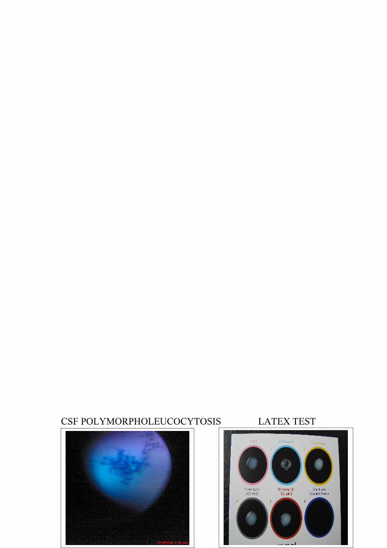
CSF POLYMORPHOLEUCOCYTOSIS LATEX TEST

GRAM STAIN H. INFLUENZA
SUBDURAL EFFUSION
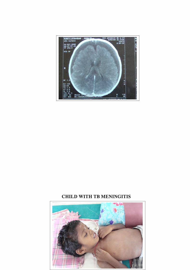
CHILD WITH TB MENINGITIS
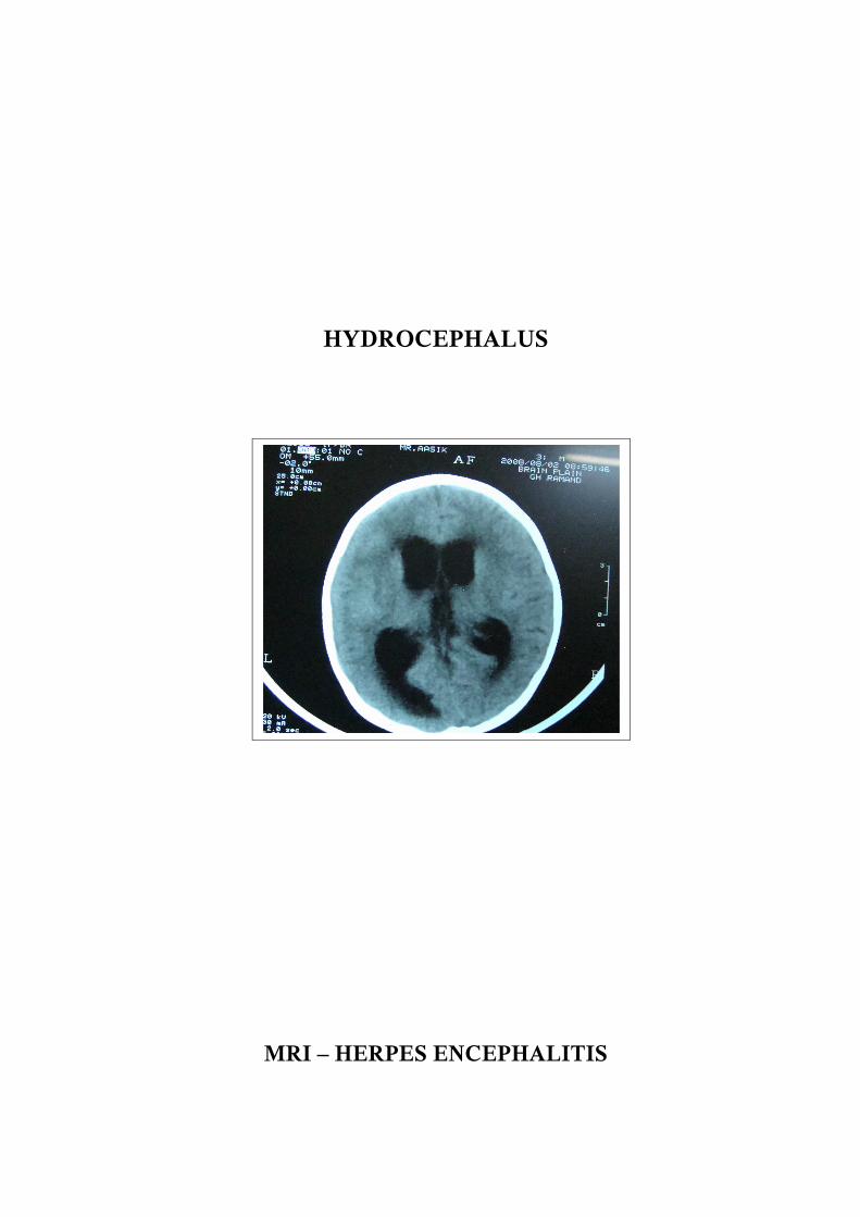
HYDROCEPHALUS
MRI – HERPES ENCEPHALITIS
BRAIN ABSCESS

MALARIAL PARASITE

ADEM
HIV - CEREBRAL ATROPHY

EEG IN HERPES ENCEPHALITIS

JE ENCEPHALITIS


Recommended

![Challenges in viral CNS infections [encephalitis] · Challenges in viral CNS infections [encephalitis] Definition Encephalitis is defined as a syndrome of neurological dysfunction](https://img.pdfslide.us/doc/110x75/5e220e3e60d1c1105809daf5/challenges-in-viral-cns-infections-encephalitis-challenges-in-viral-cns-infections.jpg)