1
ldquoThe Possible Protective Role of Green Tea
against Radiation induced Certain Biochemical
and Trace Element Changes in Ratsrdquo
Thesis
Submitted to Faculty of Pharmacy Cairo University
In Partial Fulfillment to the requirements for the
Master Degree in Pharmaceutical Sciences
(Pharmacology and Toxicology)
By
Maha Mourad Aziz Hanna (B Pharm Sciences ndash Cairo University)
Pharmacist in Drug Radiation Research Department
National Center for Radiation Research and Technology
Atomic Energy Authority
Under the Supervision of
Dr Afaf A Ain Shoka Dr Hekma Abd El Tawab
Professor of Professor of
Pharmacology amp Toxicology Pharmacology amp Toxicology
Faculty of Pharmacy Faculty of Pharmacy
Cairo University Cairo University
Dr Nour El-Din Amin Mohamed
Professor of biological chemistry
National Center for Radiation Research and Technology
Atomic Energy Authority
Department of Pharmacology and Toxicology
Faculty of Pharmacy
Cairo University
2012
2
Prerequisite postgraduate courses
Beside the work presented in this thesis the candidate Maha Mourad
Aziz had attended the prerequisite postgraduate courses for one year in the
following topics
General courses
Computer and its applications
Searching for literature and English language
Fundamentals of statistics
Special courses
Pharmacometrics
Toxicometrics
Immunopharmacology
Pathophysiology of disease
She had successfully passed the examinations in these courses with a
grade very good
Prof Dr Hanan Salah El-Din Hamdy El-Abhar
Head of pharmacology and toxicology
Faculty of pharmacy
Cairo university
3
Acknowledgment
I wish to express my grateful acknowledgement to Dr Afaf A Ain
Shoka professor of pharmacology and toxicology faculty of pharmacy
Cairo University for her keen supervision interest in the subject honesty
unlimited support and valuable time and effort she spread for me to revise
and accomplish this study
I wish to express my gratitude to Dr Hekma Abd El Tawab
professor of pharmacology and toxicology faculty of pharmacy Cairo
University for her valuable guidance and help which assisted me greatly in
completing this work
Deep thanks to Dr Nour El-Din Amin Mohamed professor of
biological chemistry national center for radiation research and technology
atomic energy authority for his continuous guidance and supervision
facilitating all necessities required for beginning and finishing this study
including chemicals and equipments and valuable advices
I am very appreciative to Dr Ahmed Shafik Nada assistant
professor of physiology national center for radiation research and
technology atomic energy authority for his great help encouragement
indispensable advice and constructive suggestions throughout this work
My thanks to all my colleagues at the department of drug radiation
research national center for radiation research and technology atomic
energy authority for their cooperation and support
Sincere thanks and graduate to my family and my friends for their
encouragement and help during this work
4
Contents Page
List of tables helliphelliphelliphelliphelliphelliphelliphelliphelliphelliphelliphelliphelliphelliphelliphelliphelliphelliphelliphellip I
List of figureshelliphelliphelliphelliphelliphelliphelliphelliphelliphelliphelliphelliphelliphelliphelliphelliphelliphelliphellip III
List of abbreviationshelliphelliphelliphelliphelliphelliphelliphelliphelliphelliphelliphelliphelliphelliphelliphelliphellip V
1 INTRODUCTIONhelliphelliphelliphelliphelliphelliphelliphelliphelliphelliphelliphelliphelliphelliphelliphelliphelliphellip 1
- Radiationhelliphelliphelliphelliphelliphelliphelliphelliphelliphelliphelliphelliphelliphelliphelliphelliphellip
1- Direct effects of ionizing radiationhelliphelliphelliphelliphelliphellip
2- Indirect effects of ionizing radiationhelliphelliphelliphelliphelliphelliphelliphellip
- Cell damage caused by ionizing radiationhelliphelliphelliphelliphelliphelliphelliphellip
- Oxidative stress induced by ionizing radiationhelliphelliphelliphelliphelliphellip
- Effect of whole body gamma radiationhelliphelliphelliphelliphelliphelliphelliphelliphellip
- Chemical consequences of ionizing radiationhelliphelliphelliphelliphelliphelliphellip
- Effects of ionizing radiation on liverhelliphelliphelliphelliphelliphelliphelliphelliphelliphellip
1- Effect of ionizing radiation on ALT and AST activities
2- Effect of ionizing radiation on ALP activityhelliphelliphelliphelliphellip
- Effects of ionizing radiation on renal functionshelliphelliphelliphelliphellip
1- Effect of ionizing radiation on creatinine levelhelliphelliphelliphelliphellip
2- Effect of ionizing radiation on urea levelhelliphelliphelliphelliphelliphellip
- Effect of ionizing radiation on lipid metabolismhelliphelliphelliphelliphellip
Effect of ionizing radiation on cholesterol and triglycerides levels
- Effect of ionizing radiation on the antioxidant defense status
1- Effect of ionizing radiation on lipid peroxidationhelliphellip
2- Effect of ionizing radiation on glutathione (GSH)helliphelliphellip
- Trace elementshelliphelliphelliphelliphelliphelliphelliphelliphelliphelliphelliphelliphelliphelliphelliphelliphelliphelliphellip
- Trace elements in radiation hazardshelliphelliphelliphelliphelliphelliphelliphelliphelliphellip
-Radiation protection and recovery with essential
metalloelementshelliphelliphelliphelliphelliphelliphelliphelliphelliphelliphelliphelliphelliphelliphelliphelliphelliphellip
- Role of iron in radiation protection and recoveryhelliphelliphelliphellip
- Effect of radiation on iron metabolismhelliphelliphelliphelliphelliphelliphelliphelliphellip
- Role of copper in radiation protection and recoveryhelliphelliphellip
- Effect of radiation on copper metabolismhelliphelliphelliphelliphelliphelliphelliphellip
- Role of zinc in radiation protection and recoveryhelliphelliphelliphellip
- Effect of radiation on zinc metabolismhelliphelliphelliphelliphelliphelliphelliphelliphellip
- Role of calcium in radiation protection and recoveryhelliphellip
1
1
1
2
3
3
3
4
5
5
6
7
7
8
8
10
10
11
12
14
14
15
16
16
17
17
19
19
5
- Effect of radiation on calcium metabolismhelliphelliphelliphelliphelliphelliphellip
- Role of magnesium in radiation protection and recovery
- Effect of radiation on magnesium metabolismhelliphelliphelliphelliphellip
- Role of selenium in radiation protection and recoveryhellip
- Effect of radiation on selenium metabolismhelliphelliphelliphelliphelliphellip
- Role of manganese in radiation protection and recoveryhellip
- Effect of radiation on manganese metabolismhelliphelliphelliphelliphellip
- Use of medicinal plants in radiation protection and recovery
- Green teahelliphelliphelliphelliphelliphelliphelliphelliphelliphelliphelliphelliphelliphelliphelliphelliphelliphelliphelliphelliphelliphellip
- Absorption metabolism and excretion of green teahelliphelliphelliphellip
- Mechanism of action of green teahelliphelliphelliphelliphelliphelliphelliphelliphelliphelliphelliphellip
- Biological efficiency of green teahelliphelliphelliphelliphelliphelliphelliphelliphelliphelliphelliphellip
- Radioprotective role of green teahelliphelliphelliphelliphelliphelliphelliphelliphelliphelliphelliphellip
- Green tea and trace elementshelliphelliphelliphelliphelliphelliphelliphelliphelliphelliphelliphelliphellip
- Vitamin Ehelliphelliphelliphelliphelliphelliphelliphelliphelliphelliphelliphelliphelliphelliphelliphelliphelliphelliphelliphelliphellip
20
20
21
21
22
23
23
24
25
27
28
29
31
32
33
2 AIM OF THE WORKhelliphelliphelliphelliphelliphelliphelliphelliphelliphelliphelliphellip 36
3 MATERIAL amp METHODShelliphelliphelliphelliphelliphelliphelliphelliphelliphellip 38
- Materialhelliphelliphelliphelliphelliphelliphelliphelliphelliphelliphelliphelliphelliphelliphelliphelliphelliphelliphelliphelliphellip
1- Experimental Animalshelliphelliphelliphelliphelliphelliphelliphelliphelliphelliphelliphelliphelliphellip
2- Therapeutic agentshelliphelliphelliphelliphelliphelliphelliphelliphelliphelliphelliphelliphelliphelliphellip
3- Chemicals and their sourceshelliphelliphelliphelliphelliphelliphelliphelliphelliphelliphellip
4- Instrumentshelliphelliphelliphelliphelliphelliphelliphelliphelliphelliphelliphelliphelliphelliphelliphelliphelliphellip
- Experimental designhelliphelliphelliphelliphelliphelliphelliphelliphelliphelliphelliphelliphelliphelliphelliphellip
- Methodshelliphelliphelliphelliphelliphelliphelliphelliphelliphelliphelliphelliphelliphelliphelliphelliphelliphelliphelliphelliphellip
- Irradiation of animalshelliphelliphelliphelliphelliphelliphelliphelliphelliphelliphelliphelliphelliphellip
- Samplinghelliphelliphelliphelliphelliphelliphelliphelliphelliphelliphelliphelliphelliphelliphelliphelliphelliphelliphelliphellip
- Measured parametershelliphelliphelliphelliphelliphelliphelliphelliphelliphelliphelliphelliphelliphellip
1- Parameters measured in serumhelliphelliphelliphelliphelliphelliphelliphelliphellip
A-Determination of serum alkaline phosphatase activityhelliphellip
B- Determination of alanine transaminase activity (ALT)helliphellip
C- Determination of aspartate transaminase activity (AST)hellip
D- Determination of serum urea levelhelliphelliphelliphelliphelliphelliphelliphelliphellip
E- Determination of serum creatinine levelhelliphelliphelliphelliphelliphelliphellip
F- Determination of serum cholesterol levelhelliphelliphelliphelliphelliphellip
G- Determination of serum triglycerides levelhelliphelliphellip helliphellip
2- Parameters measured in liver and kidney homogenate A- Determination of reduced glutathione (GSH) contenthelliphellip
38
38
38
38
39
40
40
40
40
41
41 41
42
44
45
46
47
48
49
49
6
B- Determination of lipid peroxidation helliphelliphelliphelliphelliphelliphelliphelliphellip
C- Determination of metallothioneins contenthelliphelliphelliphelliphelliphellip
3- Parameters measured in acid digest of some organshellip
- Microwave digestor technologyhelliphelliphelliphelliphelliphelliphelliphelliphelliphelliphellip
- Instrumentationhelliphelliphelliphelliphelliphelliphelliphelliphelliphelliphelliphelliphelliphelliphelliphelliphelliphellip
- Statistical analysishelliphelliphelliphelliphelliphelliphelliphelliphelliphelliphelliphelliphelliphelliphelliphelliphellip
51
52
54
54
54
55
4 RESULTS helliphelliphelliphelliphelliphelliphelliphelliphelliphelliphelliphelliphelliphelliphelliphelliphellip 56
5 DISCUSSION helliphelliphelliphelliphelliphelliphelliphelliphelliphelliphelliphelliphelliphelliphelliphelliphelliphellip 102
6 SUMMARY amp CONCLUSIONShelliphelliphelliphelliphelliphelliphelliphelliphellip 128
7 REFERENCES helliphelliphelliphelliphelliphelliphelliphelliphelliphelliphelliphelliphelliphelliphelliphelliphelliphellip 131
ARABIC SUMMARY helliphelliphelliphelliphelliphelliphelliphelliphelliphelliphelliphelliphelliphelliphelliphellip 1
7
Table Title Page
I Kits chemicals and their sources 38
1
Effect of green tea (300 mgkg) or vitamin E (40
mgkg) on liver function tests in normal and
irradiated rats
57
2
Effect of green tea (300 mgkg) or vitamin E (40
mgkg) on liver glutathione (GSH)
malondialdehyde (MDA) and metallothioneins
(MTs) contents in normal and irradiated rats
60
3
Effect of green tea (300 mgkg) or vitamin E (40
mgkg) on liver iron (Fe) copper (Cu) and zinc (Zn)
contents in normal and irradiated rats
63
4
Effect of green tea (300 mgkg) or vitamin E (40
mgkg) on liver calcium (Ca) and magnesium (Mg)
contents in normal and irradiated rats
66
5
Effect of green tea (300 mgkg) or vitamin E (40
mgkg) on liver manganese (Mn) and selenium (Se)
contents in normal and irradiated rats
69
6
Effect of green tea (300 mgkg) or vitamin E (40
mgkg) on serum cholesterol and triglycerides levels
in normal and irradiated rats
72
7
Effect of green tea (300 mgkg) or vitamin E (40
mgkg) on serum urea and creatinine levels in
normal and irradiated rats
75
8
Effect of green tea (300 mgkg) or vitamin E (40
mgkg) on kidney glutathione (GSH)
malondialdehyde (MDA) and metallothioneins
(MTs) contents in normal and irradiated rats
78
9
Effect of green tea (300 mgkg) or vitamin E (40
mgkg) on kidney iron (Fe) copper (Cu) and zinc
(Zn) contents in normal and irradiated rats
81
List of Tables 7
8
Table Title Page
10
Effect of green tea (300 mgkg) or vitamin E (40
mgkg) on kidney calcium (Ca) and magnesium
(Mg) contents in normal and irradiated rats
84
11
Effect of green tea (300 mgkg) or vitamin E (40
mgkg) on kidney manganese (Mn) and selenium
(Se) contents in normal and irradiated rats
87
12
Effect of green tea (300 mgkg) or vitamin E (40
mgkg) on spleen iron (Fe) copper (Cu) and zinc
(Zn) contents in normal and irradiated rats
90
13
Effect of green tea (300 mgkg) or vitamin E (40
mgkg) on spleen calcium (Ca) magnesium (Mg)
and selenium (Se) contents in normal and irradiated
rats
93
14
Effect of green tea (300 mgkg) or vitamin E (40
mgkg) on testis iron (Fe) copper (Cu) and zinc
(Zn) contents in normal and irradiated rats
96
15
Effect of green tea (300 mgkg) or vitamin E (40
mgkg) on testis calcium (Ca) magnesium (Mg) and
selenium (Se) contents in normal and irradiated rats
99
16
The concentrations of some metalloelements in
green tea plants and green tea extract represented as
(μgg) and (μgml) except for Se represented as
(ngg) and (ngml)
101
9
Figure Title Page
I Some mechanisms by which natural products render
radioprotection 24
II Chemical structures of EGCG EGC ECG and EC 26
III Summary of the formation of metabolites and
conjugates of flavonoids in humans 27
IV The chemical structure of alpha-tocopherol 33
V The standard curve of ALT 43
VI The standard curve of AST 45
VII The standard curve of MDA 52
1
Effect of green tea (300 mgkg) or vitamin E (40
mgkg) on liver function tests in normal and
irradiated rats
58
2
Effect of green tea (300 mgkg) or vitamin E (40
mgkg) on liver glutathione (GSH) malondialdehyde
(MDA) and metallothioneins (MTs) contents in
normal and irradiated rats
61
3
Effect of green tea (300 mgkg) or vitamin E (40
mgkg) on liver iron (Fe) copper (Cu) and zinc (Zn)
contents in normal and irradiated rats
64
4
Effect of green tea (300 mgkg) or vitamin E (40
mgkg) on liver calcium (Ca) and magnesium (Mg)
contents in normal and irradiated rats
67
5
Effect of green tea (300 mgkg) or vitamin E (40
mgkg) on liver manganese (Mn) and selenium (Se)
contents in normal and irradiated rats
70
6
Effect of green tea (300 mgkg) or vitamin E (40
mgkg) on serum cholesterol and triglycerides levels
in normal and irradiated rats
73
10
Figure Title Page
7
Effect of green tea (300 mgkg) or vitamin E (40
mgkg) on serum urea and creatinine levels in normal
and irradiated rats
76
8
Effect of green tea (300 mgkg) or vitamin E (40
mgkg) on kidney glutathione (GSH)
malondialdehyde (MDA) and metallothioneins (MTs)
contents in normal and irradiated rats
79
9
Effect of green tea (300 mgkg) or vitamin E (40
mgkg) on kidney iron (Fe) copper (Cu) and zinc
(Zn) contents in normal and irradiated rats
82
10
Effect of green tea (300 mgkg) or vitamin E (40
mgkg) on kidney calcium (Ca) and magnesium (Mg)
contents in normal and irradiated rats
85
11
Effect of green tea (300 mgkg) or vitamin E (40
mgkg) on kidney manganese (Mn) and selenium (Se)
contents in normal and irradiated rats
88
12
Effect of green tea (300 mgkg) or vitamin E (40
mgkg) on spleen iron (Fe) copper (Cu) and zinc (Zn)
contents in normal and irradiated rats
91
13
Effect of green tea (300 mgkg) or vitamin E (40
mgkg) on spleen calcium (Ca) magnesium (Mg) and
selenium (Se) contents in normal and irradiated rats
94
14
Effect of green tea (300 mgkg) or vitamin E (40
mgkg) on testis iron (Fe) copper (Cu) and zinc (Zn)
contents in normal and irradiated rats
97
15
Effect of green tea (300 mgkg) or vitamin E (40
mgkg) on testis calcium (Ca) magnesium (Mg) and
selenium (Se) contents in normal and irradiated rats
100
11
Adenosine diphosphate ADP
Alkaline phosphatase ALP
Alanine transaminase ALT
Analysis of variance ANOVA
Aspartate transaminase AST
Adenosine triphosphate ATP
Body weight bwt
Cyclic adenosine monophosphate cAMP
Catalase CAT
Cholecystokinin CCK
Cholesterol Ch
Central nervous system CNS
Catechol-O-methyl-transferase COMT
Dihydrofolate reductase DHFR
Diribonucleic acid DNA
55
dithiobis(2-nitrobenzoic acid) DTNB
Epicatechin EC
Epicatechin-3-gallate ECG
Ethylenediaminetetraacetic acid EDTA
Epigallocatechin EGC
Epigallocatechin-3-gallate EGCG
Epidermal growth factor receptor EGFR
Gallocatechin-gallate GCG
Glumerular filtration rate GFR
Reduced glutathione GSH
Glutathione peroxidase GSH-PX
Oxidized glutathione GSSG
Green tea GT
Green tea extract GTE
Green tea polyphenols GTP
Gray Gy
12
Hydrogen peroxide H2O2
High density lipoprotein HDL
Human immunodeficiency virus HIV
3- Hydroxyl - 3- methyl glutaryl coenzyme A HMG-COA
Interleukin-1 IL-1
Kilo base pair Kb
Kilo Dalton KDa
Lecithin cholesterol acyl transferase LCAT
Low density lipoprotein LDL
Malondialdehyde MDA
Messenger ribonucleic acid mRNA
Metallothioneins MTs
Nicotinamide adenine dinucleotide phosphate
hydrogen
NADPH
Norepinephrine NE
Nuclear magnetic resonance NMR
Nitric oxide NO
Superoxide radical O2-
Hydroxyl radical OH
Peroxynitrite ONOO-
Parts per million ppm
Red blood cells RBCs
Roentgen equivalent man Rem
Ribonucleic acid RNA
Reactive oxygen species ROS
Superoxide dismutase SOD
Triiodothyronine T3
Thyroxine T4
Thiobarbituric acid TBA
Thiobarbituric acid reactive substance TBARS
Trichloroacetic acid TCA
Triglyceride TG
Tumor necrosis factor TNF
Ultraviolet UV
Ultraviolet B UVB
Vascular endothelial growth factor receptor VEGFR
Very low density lipoprotein VLDL
13
14
Introduction
Radiation-
Radiation is defined as the emission and propagation of energy in the
form of waves or particles through space or matter (Zaider and Rossi
1986) Ionizing radiation is type of radiation having sufficient energy to
cause ion pairs to be formed in the medium through which it passes Ionizing
radiations consist of electromagnetic radiation (photons including X-rays
and gamma rays) and particulate radiation (such as electrons protons and
neutrons) (Cho and Glatstein 1998)
Radiation produces either direct or indirect chemical changes in
molecules Both the direct and indirect effects of ionizing radiation lead to
molecular damage which is translated to biochemical changes Exposure to
such radiation can induce alterations in the cellular macromolecules and
affect their functions (Roach et al 2009)
1-Direct effects of ionizing radiation Direct effects of radiation include
changes which appear as a result of the absorption of radiation energy by
biological materials (target molecules) which initiate a chain of reactions
leading to free radical formation (Michaels and Hunt 1978) Free radicals
are by definition species which contain a number of electrons they may be
positively charged negatively charged or neutral and all three types are
important A role for free radicals has been proposed in the toxicity diseases
(Kehrer and Lund 1994)
2-Indirect effects of ionizing radiation Indirect effects comprise the
changes occurring to the molecules in a solution induced by decomposition
products of water or other solutes and not by the radiant energy absorbed by
the molecule (Michaels and Hunt 1978)
The indirect effect of radiation in biological systems depends on the
effect of irradiation on water and the presence of oxygen in the tissue being
irradiated The end products of radiolysis of water without oxygen are γ-radiation
2H2O H + OH
+ H
+ + OH
-
H and OH
released by ionizing radiation are the most important free
radicals comprising 55 of the initial relative yield (Nair et al 2001)
15
In the presence of oxygen other radiolysis products also formed that
have oxidizing properties namely hydroperoxide radical (HOO) and
hydrogen peroxide (H2O2)
H + O2 rarr HOO
HOO
+ HOOrarr H2O2 + O2
Cell damage caused by ionizing radiation-
Ionizing radiation induces multiple biological effects through direct
interaction with DNA or production of activated free radical species from
water When tissues are exposed to ionizing radiation most of the energy
taken up is absorbed by the cell water largely because there is more water
than any other molecules thus creating two radicals a hydrogen radical (H)
and a hydroxyl radical (OH) The latter radical can attack and damage
almost every molecule found in living cells (Halliwell and Gutteridge
1999)
Ionizing radiation induces reactive oxygen species (ROS) in the form
of OH H
singlet oxygen and peroxyl radicals that follow a cascade of
events leading to DNA damage such as single or double strand breakages
base damage and DNA-protein cross-links These lesions cluster as complex
local multiply damage sites The DNA double strand breaks are considered
the most lethal events following ionizing radiation and have been found to
be the main target of cell killing by radiation (Jagetia 2007)
Mondelaers and Lahorte (2001) reported that the processes
leading to radiation damage are complex but can be considered to take place
in the following stages
The initial physical stage (Lasting for 10-13
second) in which
energy is deposited in the cell and caused ionization
The physicochemical stage (Lasting for 10-7
second) in which
the ions interact with other water molecules resulting in the
production of free radicals which are chemically highly reactive
due to the presence of an unpaired electron Another reaction
product is hydrogen peroxide which is a strong oxidizing agent
The chemical stage (Lasting for few minutes or hours) in which
the reaction products interact with the important organic
molecules of the cell
16
The biological stage In which the time scale varies from minutes
to tens of years and is depending on the type of the cell affected
Oxidative stress induced by ionizing radiation-
Oxidative stress is a state of imbalance between generation of (ROS)
and the levels of antioxidant defense system Antioxidant enzymes are part
of the endogenous system available for the removal or detoxification of free
radicals and their products formed by ionizing radiation (Bhatia and Jain
2004)
Oxidative stress has been linked to diseases including some allergic
and inflammatory skin diseases (Okayama 2005) neurodegeneration
(Moreira et al 2005) and atherosclerosis in diabetic patients (Lankin et
al 2005) As a defense mechanism the body produces a number of
endogenous antioxidants such as superoxide dismutase (SOD) catalase
(CAT) and glutathione peroxidase (GSH-PX) capable of scavenging harmful
ROS to maintain an optimal oxidantantioxidant balance thereby
maintaining normal cellular function and health (Droumlge 2002)
Effect of whole body gamma radiation
Factors that determine the biological effects of ionizing radiation
include the type of radiation the received dose the rate at which the
radiation dose is delivered nutritional factors the type of irradiated tissues
as well as the age and sex of the exposed person In addition whether the
dose was delivered in fractions or in a single exposure could determine the
biological effect (Beir 1990)
A single whole body exposure of mammals to ionizing radiation
results in a complex set of syndromes whose onset nature and severity are a
function of both total radiation dose and radiation quality At a cellular level
ionizing radiation can induce damage in biologically important
macromolecules such as DNA proteins lipids and carbohydrates in various
organs While some damage may be expressed early the other may be
expressed over a period of time depending upon cell kinetics and radiation
tolerance of the tissues (Baliga et al 2004)
Chemical consequences of ionizing radiation
17
The first consequence of ionizing radiation is ionization of water
Since water represents 70 of the chemical composition of the adult body
its chemical transformation by ionizing radiation merits serious
consideration Ionization of water is well understood and produces very
reactive aquated electrons monoatomic hydrogen atoms hydroxyl radicals
hydrogen peroxide and protonated water as well as superoxide and
hydroperoxyl radicals in the presence of oxygen Hydroperoxyl radical
hydroxyl radical monoatomic hydrogen and aquated electron have very
short half lives (10-1
to 10-3
sec) and consequently react rapidly with cellular
components in reduction oxidation initiation insertion propagation and
addition reactions causing loss of function and need for biochemical
replacement andor repair (Sorenson 2002) The second consequence of
ionizing radiation is its ability to impart sufficient energy to all biochemicals
to cause homolytic bond breaking and produce all conceivable organic
radicals in considering C-C C-N C-O C-H P-O S-O hellipetc bond
homolysis These radicals will undergo the reactions listed above causing
further destruction and requiring replacement andor repair (Droumlge 2002)
A third consequence of ionizing radiation is homolytic or heterolytic
bond breaking of coordinate-covalent bonded metalloelements These are the
weakest bonds in biochemical molecules and potential sites of the greatest
damage which may be most in need of replacement andor repair since
many repair enzymes are metalloelements-dependent as are the
metalloelement dependent protective SODs (Sorenson 2002)
Effects of ionizing radiation on liver
It was reported that ionizing radiation affects the liver function
(Feurgard et al 1998) Influence of stress on liver is of interest from the
clinical point of view because stress plays a potential role in aggravating
liver diseases in general and hepatic inflammation in particular probably
through generation of ROS (Zaidi et al 2005)
The serum transaminases activity is the most widely used parameter
as a measure of hepatic injury due to its ease of measurement and high
degree of sensitivity It is useful for the detection of early damage of hepatic
tissue and requires less effort than that for a histological analysis (Ray et al
2006) Serum elevation of alanine transaminase (ALT) activity is rarely
18
observed in condition other than parenchymal liver disease Moreover
elevation of ALT activity persists longer than does that of aspartate
transaminase (AST) activity (Tolman and Rej 1999) ALT is the enzyme
produced within the cells of the liver and its abnormality is increased in
conditions where cells of the liver have been inflamed or undergone cell
death Any form of hepatic cell damage can result in an elevation in ALT
activity which may or may not correlate with the degree of cell death or
inflammation ALT is the most sensitive marker for liver cell damage and
the most important test for recognition of acute and chronic hepatic failure
(Dufour et al 2000)
1-Effect of ionizing radiation on ALT and AST activities
AST and ALT are enzymes responsible for the catalization of the
transference of an amino group from α-amino acid to α-keto acid and they
are considered as indicators for liver injury caused by exposure to ionizing
radiation In view of the effect of radiation on transaminases many authors
reported that the activities of AST and ALT increased when mice or rats
exposed to gamma radiation at dose levels from 4 to 6 Gy (Bhatia et al
2007 Adaramoye 2010)
Roushdy et al (1984) showed that gamma irradiation at a dose level
of 6 Gy resulted in remarkable increases in the transaminases activities both
in serum and liver They indicated that the rise in the liver transaminases
activities may be due to the drastic physiological effects caused by
irradiation The increase in ALT activity may be related to extensive
breakdown of liver parenchyma with subsequent enzyme release or to
increase in permeability of the cell membrane that could enhance the
movement of enzymes from their sites of production (Manciluae et al
1978) Also Fahim et al (1991) suggested that the elevation in ALT and
AST activities in rats exposed to 75 Gy of gamma radiation may be due to
destruction of radio-sensitive cells of haematopoietic tissue and erythrocytes
haemolysis
2- Effect of ionizing radiation on ALP activity
Alkaline phosphatase (ALP) is a hydrolytic enzyme acting on
phosphoric esters with the liberation of inorganic phosphate from various
19
substrates In addition alkaline phosphatase is mainly involved in passive
transport mechanism (Verma and Nair 2001) It is well known that ALP
plays an important role in maintaining the cell membrane permeability
(Samarth and Kumar 2003) Magnesium and zinc ions are essential for
stability and maximum catalytic activity of ALP enzyme (Gowenlock et al
1988)
Exposure of rats or mice to radiation at dose levels range from 4 to
8Gy induced an increase in ALP activity that was recorded by many authors
(Sunila and Kuttan 2005 Adaramoye et al 2008 Pratheeshkumar and
kuttan 2011)
Abdel-Fattah et al (1999) stated that ALP activity in plasma of rats
increased significantly at 1 3 and 5 hours after exposure to single dose of 6
Gy gamma radiation They suggested that this increase could be considered
as a reflection of liver dysfunction in the acute radiation sickness Authors
also revealed that the increase in alkaline phosphatase activity may be due to
destruction of cell membrane or destruction of this enzyme inhibitor by
radiation
Furthermore Kafafy and Ashry (2001) found that whole body
gamma-irradiation affected liver structure and functions as indicated by
changes in the serum ALP activity which increased significantly along the
post-irradiation days where it reached its maximum at the tenth day
following exposure The authors deduced that this increase reflected
detectable changes in liver function due to the changes in tissue permeability
induced by irradiation which enhanced the movement of enzymes from their
subcellular sites of production to extracellular process and consequently into
the blood circulation
Effects of ionizing radiation on renal functions
It is well established that radiation exposure is known to impair the
biological integrity of living organisms It is also known that exposure to
acute radiation dose can cause substantial well detectable functional changes
in the organisms much earlier than morphological changes would develop
(Robbins and Bonsib 1995) Many authors reported that ionizing radiation
greatly affected renal function (Ramadan et al 1998 kafafy et al 2005)
Radiation-induced renal impairment occurs predictably after local kidney
20
irradiation or total body irradiation (Robbins and Bonsib 1995 Badr El-
Din 2004) Irradiation leads to progressive biochemical changes in the
irradiated animals The animals may suffer from continuous loss in body
weights which could be attributed to disturbance in nitrogen metabolism
usually recognized as negative nitrogen balance Accordingly it could be
expected that this may cause an increase in the urea ammonia and amino
acid levels in blood and urine due to great protein destruction induced by
irradiation that is an evidence of marked impairment of kidney function
(Robbins et al 1992)
1-Effect of ionizing radiation on creatinine level
It is well known that creatine is converted to creatine phosphate in the
muscle and that creatine phosphate is converted to creatinine before
excretion in the urine Ionizing radiation causes damage in muscle of
mammals which appears by increased excretion of nitrogenous metabolites
such as creatine (Gerber et al 1961)
Urinary output of creatinine may be taken as a sensitive parameter
indicating the degree of impaired tissue metabolism due to radiation effect
The kidney is relatively more resistant to ionizing radiation (Roushdy et al
1997 Cheng et al 2002)
Yildiz et al (1998) observed that serum creatinine level increased
when kidneys of male rats were irradiated with either 10 Gy single dose or
26Gy at a rate of 2 Gy per day and after 4 weeks of irradiation glomerular
and proximal tubular injury were observed Increased serum creatinine level
in the irradiated rats indicates development of nephritis and renal
dysfunction (Borg et al 2002) that may be attributed to impairment of
glomerular selective properties caused by irradiation (Berry et al 2001)
Studies of Hassan et al (1994) showed that serum creatinine level
was elevated when the rats were exposed to gamma-irradiation at
fractionated dose levels of 3 Gy to a cumulative dose of 9 Gy on the 2nd
hours 1st and 7
th days post-exposure They concluded that fractionated
exposure to gamma irradiation effectively altered the glomerular filtration
rate (GFR) in rats
21
Many authors observed significant increase in plasma level of
creatinine post whole body gamma irradiation with 65 Gy (Badr El-Din
2004) and 75 Gy (Omran et al 2009)
2-Effect of ionizing radiation on urea level
Most of ammonia formed by deamination of amino acids is converted
to urea The urea resulting from protein degradation is excreted by the
kidney so the level of urea in plasma of rats is an indicator for the effect of
radiation on kidney function (Kutchai 1993)
Studies of Geraci et al (1990) and Adaramoye (2010) showed that
an increase in serum urea level of animals is induced post-irradiation The
authors considered this increase as a reflection of deteriorating renal
performance
On the other hand Mahdy et al (1997) observed that whole body
gamma-irradiation of rats at 75 Gy (single dose) caused a significant
increase of urea level as recorded 7 10 and 14 days after irradiation The
authors suggested that elevation in serum urea level may be due to an
increased oxidative deamination of amino acids in the liver resulting in
excess urea formation
Badr El-Din (2004) declared that an increase in blood urea level has
been reported after exposure to radiation and secondary to renal damage
The elevation of urea may be attributed to an increase in nitrogen retention
or excessive protein breakdown Furthermore Omran et al (2009)
demonstrated that rats exposed to 75 Gy whole body gamma irradiation
showed significant increase in plasma urea level (50) at both time intervals
of 7 and 16 days
Effect of ionizing radiation on lipid metabolism
Lipid profile especially cholesterol has been representing a major
essential constituent for all animal cell membranes Plasma lipid levels are
affected by genetic and dietary factors medication and certain primary
disease states (Feldman and Kuske 1987) Hyperlipidemia occurring due
to exposure to ionizing radiation resulted in accumulation of cholesterol
22
triglycerides and phospholipids (Feurgard et al 1999) The accumulated
lipoproteins were susceptible to peroxidation process causing a shift and
imbalance in oxidative stress This imbalance manifested themselves
through exaggerated ROS production and cellular molecular damage
(Romero et al 1998)
Effect of ionizing radiation on cholesterol and triglycerides levels
Cholesterol is synthesized in the liver and its balance is maintained by
the livers ability to remove cholesterol from lipoproteins and use it to
produce bile acids and salts that excreted in the bile duct In obstructive
jaundice the bile can not be eliminated cholesterol and triacylglycerols may
accumulate in the blood In acute necrotic liver diseases triacylglycerols
may be elevated due to hepatic lipase deficiency In liver failure caused by
necrosis the livers ability to synthesize cholesterol is reduced and the blood
levels may be low (OacuteGrady et al 1993)
Free radical mediated oxidative damage induced by radiation is one of
the prime factors that increase the hepatic cholesterol and triglycerides levels
(Pote et al 2006) Radiation leads to hyperlipidemia through destruction of
cell membranes enhancement of lipid metabolism cholesterol release and
increased triglycerides synthesis (Bowden et al 1989)
Irradiation of rats induced increase in the total lipid synthesis in bone
marrow liver and blood that was attributed to the increase in stimulation of
the liver enzyme responsible for the biosynthesis of fatty acids and to the
mobilization of fat from adipose tissue to the blood stream leading to
hyperlipidemic state (Sedlakova et al 1988) Another explanation for this
hyperlipidemic state is the retention character caused by the diminished
utilization of circulating lipids by the damaged tissues (Abou Safi and
Ashry 2004 Kafafy 2004) Also some changes in the activities of hepatic
HMGCoA reductase (the rate-limiting enzyme for cholesterol synthesis) and
in hepatic cholesterol 7alpha-hydroxylase (the key enzyme involved in
degradation of cholesterol in the liver) were noted following radiation
exposure (Feurgard et al 1999)
Many authors concluded that whole body gamma-irradiation showed a
significant increase of serum cholesterol and triglycerides levels whether this
23
radiation is applied as a single dose (Feurgard et al 1998 Kafafy 2004
Baker et al 2009) or fractionated doses (Abou-Safi et al 2001)
Girgis et al (2000) showed that whole body gamma-irradiation of
rats at a dose level of 6 Gy significantly decreased the total cholesterol level
in plasma by 374 on the 1st day after irradiation as compared to the
control value However it increased by 4804 309 and 96 after 3 7
and 14 days from irradiation respectively as compared to the control value
The authors suggested that ionizing radiation by activating the cholesterol
esterase enzyme may play a role in the development of atherosclerosis in
experimental animals
The hypercholesterolemia induced by radiation was attributed to two
causes the first was the activation of cholesterologenesis in different cells of
tissue as an early reaction to harmful effect of the radiation for restoring the
cell membranes activity and the second was the decrease in the lecithin
cholesterol esterification where HDL cholesterol may be the vehicle for
reversed cholesterol transport and esterification (Abdel-Fattah et al 2003)
Effect of ionizing radiation on the antioxidant defense status
When cellular production of ROS overwhelms its antioxidant
capacity a state of oxidative stress is reached leading to serious cellular
injuries that contributes to the pathogenesis of several diseases (Gloire et
al 2006) The systemic damage observed following irradiation is
particularly due to the overproduction of ROS which disrupt the delicate
pro-oxidantanti-oxidant balance of tissues leading to proteins lipids and
DNA oxidation (Flora 2007) Free radicals are highly reactive and cause
tissue damage by reacting with poly unsaturated fatty acids found in cellular
membranes or by reacting with sulfhydryl bonds in proteins as reported by
Guney et al (2004)
The antioxidant defense system consists of numerous enzymes and
low molecular weight compounds that scavenge produced radicals and other
ROS and prevent production of more reactive radical species It also
removes lipid peroxides preventing further propagation (Sies 1993) This
antioxidant defense system is consisting of enzymes such as CAT SODs
GSH-PX and numerous non-enzymatic antioxidants including vitamins A E
and C glutathione (GSH) metallothioneins and flavonoids (Belviranli and
Goumlkbel 2006)
24
1-Effect of ionizing radiation on lipid peroxidation
ROS are relatively short lived molecules that exert local effects They
can attack poly unsaturated fatty acids and initiate lipid peroxidation within
the cell The process of lipid peroxidation is one of oxidative conversion of
poly unsaturated fatty acid to byproducts known as malondialdehyde (MDA)
or lipid peroxides which is the most studied biologically relevant free
radical reaction These byproducts can diffuse large distances from site of
their generation before mediating damage They are capable of inactivating
enzymes (Wilson et al 2003) Lipid peroxidation is a complex process
characterized by three distinct phases initiation propagation and
termination Radiation induced lipid peroxidation is initiated by direct or
indirect ionization or by free radical attack (Gupta et al 2000)
Lipid peroxidation is a chain reaction in which the interaction of the
lipid radical with another organic molecule results in conversion of that
molecule to the free radical state and propagation of damage Peroxidation
of membrane lipids can have numerous effects including increased
membrane rigidity decreased activity of membrane bound enzymes altered
activity of membrane receptors as well as altered permeability (Kamat et
al 2000) It was found that whole body gamma irradiation of male rats caused
changes in the antioxidant defense system of the organism which depend on
the intensity of lipid peroxidation level in the blood (Gatsko et al 1990)
Furthermore many authors deduced that irradiation of rats or mice at dose
range from 6-12 Gy either applied as single dose or fractionated doses
induced significant increase in liver and blood MDA levels (Baliga et al
2004 Samarth et al 2006 Kilciksiz et al 2008 Pratheeshkumar and
kuttan 2011)
Nunia et al (2007) noted a significant increase in blood level and
hepatic content of lipid peroxidation in mice after 75 Gy of gamma
irradiation They attributed this increase to the membrane damage caused by
ROS which may allow the entry of excess calcium into cells with sequential
biochemical and micro anatomical cellular degranulation and necrosis
2-Effect of ionizing radiation on glutathione (GSH)
25
GSH is a small molecule made up of three amino acids (tripeptide)
[glutamine ndash cysteine - glycine] whose antioxidant action is facilitated by the
sulfhydryl group of cysteine (Townsend et al 2003) GSH is the most
abundant non-protein thiol in mammalian cells It plays an important role in
regulation of cellular redox balance The most recognized function of GSH
is its role as a substrate for GSH-S-transferase and GSH-PX These enzymes
catalyze the antioxidation of ROS and free radicals (Weis et al 1993)
The presence of GSH is required to maintain the normal function of
the immune system It is essential for the activation of T-lymphocytes and
polymorphonuclear leukocytes as well as for cytokine production and
therefore for mounting successful immune responses (Townsend et al
2003)
GSH reacts directly with free radicals and can protect cells from
single oxygen radical (O) hydroxyl radical (OH
) and superoxide radical
(O2) (Cominacini et al 1996) GSH may stabilize membrane structure by
removing acyl peroxides formed by lipid peroxidation reactions (May et al
1998)
GSH with its sulfhydryl group functions in the maintenance of
sulfhydryl groups of other molecules (especially proteins) and as a catalyst
for disulfide exchange reactions It also functions in the detoxification of
foreign compounds hydrogen peroxide and free radicals When GSH acts as
reducing agent itrsquos SH becomes oxidized and forms a disulfide link with
other molecules of GSH (Manda et al 2007) The reduced GSH in
oxidationreduction cycling catalyzed by GSH-PX enzyme is critical in
reducing H2O2 thus breaks the chain reaction resulting from the superoxide
radical to the highly reactive hydroxyl radical (Hayes and Mclellan 1999)
GSH-PX
H2O2 + 2GSH GSSG + 2H2O
In addition to its action on H2O2 GSH-PX has the ability to use lipid
peroxides as substrate to convert them to inert compounds (Andersen et al
1997) GSH-PX
ROOH + 2GSH GSSG + ROH + H2O
26
Considerable evidence pointed to the fact that intracellular non-
protein sulfhydryl compounds play an important role in cellular response to
ionizing radiation (Bump and Brown 1990) In the same concern Jagetia
et al (2004) studied the effect of different doses of radiation in mice They
revealed that GSH content of mice livers decreased in a dose dependant
manner Also Inal et al (2002) observed that administration of GSH
appears to be useful approach to reduce radiation injury by reducing MDA
levels and increasing CAT activities
A lot of authors revealed that blood level and liver content of GSH
exhibited significant decrease after exposure of rats or mice to whole body
gamma radiation at dose levels of 6 Gy (Pratheeshkumar and kuttan
2011) 75 Gy (Nunia et al 2007) 8 and 10 Gy (Sharma and Kumar
2007)
Trace elements
Trace elements are elements that are present in the body at very low
amounts micro grams to milligrams but they are essential for certain
biochemical processes (Wada 2004) Trace elements act as essential
activators or cofactors for antioxidant enzymes to exert their action
(Ostrakhovitch and Cherian 2005)
An element is considered by Mertz (1970) to be essential if its
deficiency results in impairment of a function from optimal to suboptimal
Cotzais (1967) indicated that a trace element can be considered essential if it
meets the following criteria (1) it is present in all healthy tissues of all
living things (2) its concentration from one animal to the next is fairly
constant (3) its withdrawal from the body induces reproducibly the same
physiological and structural abnormalities regardless of the species studied
(4) its addition either reverses or prevents these abnormalities (5) the
abnormalities induced by deficiency are always accompanied by pertinent
and specific biochemical changes (6) these biochemical changes can be
prevented or cured when the deficiency is prevented or cured
Copper iron manganese and zinc are essential metalloelements
These essential metalloelements as well as essential amino acids essential
fatty acids and essential vitamins are required by all cells for normal
metabolic processes but can not be synthesized de novo and dietary intake
27
and absorption are required to obtain them Ionic forms of these
metalloelements have particularly high affinities for organic ligands found in
biological systems and rapidly undergo bonding interactions to form
complexes or chelates in biological systems Absorbed metalloelement
chelates undergo systemic circulation to all tissues and utilization by all cells
following ligand exchange with small molecular mass ligands apoproteins
and apoenzymes to form metalloproteins and metalloenzymes in de novo
synthesis The degree of radiation injury and nutritional state of health of an
individual may determine whether or not an individual will be able to
overcome metalloelement-dependent repairable radiation injury (Sorenson
2002)
The action of a very small amount of trace element is necessary for
optimal performance of a whole organism Lack of a small amount of a trace
element (eg iron) can result in disease (anemia) seemingly this
proportionate to the amount of element missing The bases for the
amplification of trace element action is that trace elements are constituents
ofor interact with enzymes or hormones that regulate the metabolism of
much larger amounts of biochemical substrates If the substrates are also
regulators the effect is even further amplified (Abdel-Mageed and Oehme
1990a)
Essential trace elements are specific for their in vivo functions They
cannot be effectively replaced by chemically similar elements Certain trace
elements are stable in more than one valence state (eg Fe Cu Mo)
allowing biochemical redox function while others are stable in only a single
state [eg Zn(II) Ni(II)] (Milne 2001) Specificity of trace element function
is also promoted by specific carrier and storage proteins such as transferrin
and ferritin for iron albumin and α-macroglobulin for zinc ceruplasmin for
copper transmanganin for manganese and nickeloplasmin for nickel These
carrier proteins recognize and bind specific metals and transport them toor
store them at specific site with the organism (Mensa et al 1995 Vivoli et
al 1995)
Interaction between metals may be important not only when one
metal is present in excess and the other is deficient but also when the lack of
one metal decreases the bioavailability of the other (Pallareacutes et al 1996)
Pallareacutes et al (1993) previously found that Fe deficiency affects Ca P and
Mg metabolism (at absorptive level) Also the addition of large amounts of
28
zinc to a diet interferes with the intestinal copper absorption system
resulting in copper deficiency (Mills 1981)
Changes in concentrations of essential trace elements in the body
associated with the progression of neoplastic diseases and have a profound
impact systemic metabolic activity (Siddiqui et al 2006) The deficiency of
trace elements may depress the antioxidant defense mechanisms (Kumar
and Shivakumar 1997) erythrocyte production (Morgan et al 1995)
enhance lipid abnormalities (Tajik and Nazifi 2010) While the toxicity of
trace elements may induce renal liver and erythropoietic abnormalities
(Chmielnicka et al 1993 Farinati et al 1995 Kadkhodaee and Gol
2004)
Trace elements in radiation hazards
Most of cellular alterations induced by ionizing radiation are indirect
and are mediated by the generation of free radicals and related reactive
species (Maurya et al 2007) Mammalian cells are equipped with both
enzymatic and non-enzymatic antioxidant mechanisms to minimize cellular
damage resulting from the interaction between cellular constituents and
ROS Ionizing radiation causes homolytic and heterolytic bond breaking of
covalent and coordinate covalent bonded metalloelements These are the
weakest bonds in biochemical molecules and potentially the sites of the
greatest damage so they are most in need of replacement andor repair
Many repair enzymes are metalloelements dependent as the metalloelement
dependent protective SODs (Sorenson 2002)
Radiation protection and recovery with essential metalloelements
Recognizing that loss of enzyme activity is dependent on essential
metalloelements may at least partially account for lethality of ionizing
radiation Cu Fe Mn and Zn dependent enzymes have roles in protecting
against accumulation of ROS as well as facilitating the repair (Sorenson
1978) which may explain the radiation protection and radiation recovery
activity of Cu Fe Mn and Zn compounds (Matsubara et al 1986) It is
suggested that the IL-1-mediated redistribution of essential metalloelements
may account for subsequent de novo synthesis of the metalloelement
dependent enzymes required for biochemical repair and replacement of
29
cellular and extracellular components needed for recovery from radiolytic
damage (Sorenson 1992)
De novo synthesis of metalloelements dependent enzymes is required
for utilization of oxygen and preventions of oxygen accumulation as well as
for tissue repair processes including metalloelement dependent DNA and
RNA repair This is the key to hypothesis that essential metalloelement
complexes prevent andor facilitate recovery from radiation-induced lesions
(Berg 1989)
Role of iron in radiation protection and recovery
Iron is the most important of the essential trace metals An appropriate
number of human diseases are related to iron deficiency or disorders of iron
metabolism (Kazi et al 2008) It is the oxygen carrier in hemoglobin and
myoglobin It also functions in the respiratory chain Iron in the body is
either functional or stored Functional iron is found in hemoglobin and
myoglobin whereas stored iron is found in association with transferrin
ferritin and hemosiderin The storage sites of ferritin and hemosiderin are the
liver spleen and bone marrow (McCarter and Holbrook 1992) Iron is
required in many biochemical processes ranging from oxidative metabolism
to DNA synthesis and cell division (Crowe and Morgan 1996) It has been
reported that iron and its complexes protect from ionizing radiation
(Sorenson et al 1990) play an important role in facilitation of iron
dependent enzymes required for tissue or cellular repair processes including
DNA repair (Ambroz et al 1998) and protect against radiation-induced
immunosupression (Tilbrook and Hider 1998)
The oxidative damage is thought to be a consequence of increased
free radical generation secondary to tissue iron accumulation The damage
may be also a consequence of the reduction in Zn or Cu dependent
antioxidizing processes as an increase in tissue iron was observed in Zn and
Cu deficiencies (Oteiza et al 1995)
ROS promote iron release from ferritin A free iron ion catalyzes
changes from relatively poor reactive O2 and H2O2 to highly reactive HO
(Fenton reaction) (Koike and Miyoshi 2006) In addition iron can catalyze
the decomposition of lipid hydroperoxides to form alkoxyl peroxyl and
other radicals (Halliwell and Gutteridge 1990)
30
Effect of radiation on iron metabolism
Exposure of rats to whole body gamma radiation with single dose of
6Gy and 4 Gy induced significant increase in liver content and serum level
of iron (Mansour et al 2006 Abdel-Gawad and Aiad 2008) In addition
an increase of iron content in liver and spleen of irradiated animals were
demonstrated by Nada et al (2008) The same increase in serum iron level
was demonstrated also in case of animalsrsquo exposure to fractionated 12 Gy
gamma rays (2 Gy weekly) (Ashry et al 2010)
Kotb et al (1990) reported that accumulation of iron in the spleen
after whole body gamma irradiation could be resulted from disturbances in
the biological function of RBCs including possible intravascular haemolysis
and subsequent storage of iron in the spleen Also Osman et al (2003) and
Harris (1995) attributed the increase of iron content in liver and spleen post
irradiation to the inhibition of ceruloplasmin which is essential for iron
metabolism and distribution
Role of copper in radiation protection and recovery
Cu is one of the essential trace elements in humans and disorders
associated with its deficiency and excess have been reported (Aoki 2004) It
is an integral component of many enzymes and proteins needed in a wide
range of metabolic processes (Ozcelik et al 2003) Copper in the divalent
state (Cu2+
) has the capacity to form complexes with many proteins These
metalloproteins form an important group of oxidase enzymes including
cytochrome C oxidase (in the mitochondrial electron transport chain) SOD
(part of the protection against ROS) and lysyl oxidase which is needed for
the cross-linking of collagen and elastin (Culotta and Gitlin 2000) Copper
also complexes with L-amino acids that facilitate its absorption from the
stomach and duodenum (Irato et al 1996) The importance of Cu in the
efficient use of iron makes it essential in hemoglobin synthesis (Han et al
2008)
It has been reported that Cu plays important role in the protection
from DNA damage induced by ionizing radiation (Cai et al 2001)
amelioration of oxidative stress induced by radiation (Abou Seif et al
31
2003) maintaining cellular homeostasis (Iakovleva et al 2002) and
enhancement of antioxidant defense mechanisms (Štarha et al 2009)
Chen et al (1995) studied the effect of severely depressed Cu
concentration on MTs induction in rats They found that Cu deficiency
induced MTs gene transcription selectively in the liver
Effect of radiation on copper metabolism
Kotb et al (1990) found that 24 hrs after irradiation disturbance in
Cu content was quite evident It was manifested as reduced content in
spleen heart and kidney Many authors found significant reduction in Cu
content of liver after whole body gamma irradiation at dose level of 4 Gy
and 65 Gy (Osman et al 2003 Nada et al 2008) In addition
Isoherranen et al (1997) stated that UVB irradiation reduced both the
enzymatic activity and the expression of the 07 and 09 Kb mRNA
transcripts of Cu Zn-SOD an antioxidant enzyme
Role of zinc in radiation protection and recovery
Zinc is known to have several biological actions Zn is known to serve
as the active center of many enzymes It protects various membranes system
from peroxidative damage induced by heavy metals and high oxygen tension
in addition to the stabilization of perturbation (Micheletti et al 2001) Zn is
an essential oligo element for cell growth and cell survival (Norii 2008)
The function of Zn can be categorized as catalytic (metalloenzymes)
structural (eg Zn finger domains of proteins) and regulatory (eg metal
response element of gene promoter) (Cousins 1996)
The protective effects of Zn against radiation hazards have been
reported in many investigations (Markant and Pallauf 1996 Morcillo et
al 2000) Zn ions can directly act as an antioxidant by stabilizing and
protecting sulfhydryl-containing proteins Zn can displace Fe and Cu from
cell membranes and proteins which can otherwise cause lipid peroxidation
and destruction of membrane protein lipid organization due to their ability to
promote the generation of hydroxyl ion from H2O2 and superoxide via the
Fenton reaction This is because Zn has only one oxidation state (II) and
therefore cannot undergo these redox reactions In addition Zn can accept a
32
spare pair of electrons from oxidants hence neutralizing their reactivity
(Truong-Tran et al 2001)
Floresheim and Floresheim (1986) concluded that Zn salts are class
of radioprotectors that might protect against radiation-induced tissue injury
The antioxidant role of Zn could be related to its ability to induce
metallothioneins (MTs) (Winum et al 2007) Metallothioneins are a family
of low molecular weight (about 67 KDa) Cystein rich (30) intracellular
proteins with high affinity for both essential (Zn and Cu) and non-essential
(Cd and Hg) metals (Krezel and Maret 2008) MTs are important
compounds on reducing the efficiency of zinc absorption at elevated zinc
intakes (Davis et al 1998) The major biological function of MTs is the
detoxification of potentially toxic heavy metals ions and regulation of the
homeostasis of essential trace elements
However there is increasing evidence that MTs can reduce toxic
effects of several types of free radicals including superoxide hydroxyl and
peroxyl radicals (Pierrel et al 2007) MTs play a protective role against the
toxic effects of free radicals and electerophiles produced by gamma
radiation (Liu et al 1999) The hepatic and renal MTs have been increased
after whole body X-irradiation (Shiraishi et al 1986) Furthermore the
whole body gamma-irradiation induced MTs-mRNA transcription protein
expression and accumulation in liver that implicates the organ specific
resistance to radiation-induced cellular damage (Koropatnick et al 1989)
MTs are involved in the protection of tissue against various forms of
oxidative injury including radiation lipid peroxidation and oxidative stress
(Kondoh and Sato 2002) Induction of MTs biosynthesis is involved in
protective mechanisms against radiation injuries (Azab et al 2004)
Nishiyma et al (1994) concluded that Zn may play a role in thyroid
hormone metabolism in low T3 patients and may in part contribute to
conversion of T4 to T3 in humans Sidhu et al (2005) studied the effects of
Zn treatment in conditions of protein deficiency on rat liver antioxidant
parameters which included CAT GSH-PX glutathione reductase SOD
GSH glutathione-S-transferase and the level of lipid peroxidation They
found significant elevation in the levels of GSH and SOD in protein
deficient animals treated with Zn Also it was reported that subcutaneous
injection of Zn pre-irradiation ameliorated and reduced the chromosomal
aberrations that occur by radiation hazards (El-Dawy and El-Sayed Aly
2004)
33
Effect of radiation on Zn metabolism
Kotb et al (1990) noticed that there was a significant reduction in
the content of Zn in kidney 24 hrs heart and spleen 3 days following
irradiation with doses between 10 and 25 rem This decrease was followed
up by a gradual increase of the element contents which exceeded the pre-
irradiation contents in most cases Also Ashry et al (2010) observed that
exposure of rats to fractionated 12 Gy γ-rays induced significant increase in
Zn serum level
A possible explanation for the increased MTs post-irradiation in liver
and kidney was suggested by Shiraishi et al (1986) where Zn accumulated
in these damaged tissues by irradiation thus stimulating the induction of
MTs synthesis Moreover Nada et al (2008) indicated that irradiation
andor 14 dioxane induced increases in Zn content of liver spleen lung
brain and intestine of irradiated rats
Role of calcium in radiation protection and recovery
Ca is the most common mineral in the human body About 99 of the
Ca in the body is found in bones and teeth while the other 1 is found in
the blood and soft tissue The physiological functions of Ca are so vital to
survival that the body will demineralize bone to maintain normal blood Ca
levels when Ca intake is inadequate (Weaver and Heaney 1999)
Ca is necessary to stabilize a number of proteins and enzymes
optimizing their activities The binding of Ca ion is required for the
activation of the seven vitamin K-dependent clotting factors in the
coagulation cascade (Olson 1999) Calcium also plays a role in mediating
the contraction and relaxation of blood vessels nerve impulse transmission
muscle contraction and the secretion of hormones like insulin (FNB 1997)
The binding of Ca to the protein calmodulin activates enzymes that break
down muscle glycogen to provide energy for muscle contraction A
chronically low Ca intake in growing individuals may prevent the attainment
of optimal peak bone mass Once peak bone mass is achieved inadequate Ca
intake may contribute to accelerated bone loss and ultimately to the
development of osteoporosis (Weaver and Heaney 1999)
34
Sorenson (2002) found that many calcium-channel blockers drugs act
as radioprotectors and radiorecovery prodrugs Also many investigators
found that nutrient extracts like propolis and rosemary which contain highly
contents of Ca Mg and Mn exert benefit protection against radiation injury
(Nada and Azab 2005 Nada 2008)
Effect of radiation on calcium metabolism
Cengiz et al (2003) exposed rats to 5 Gy of whole body γ-rays
Serum calcium level was studied 8 weeks after exposure and a significant
increase was recorded in its level While Ibrahim and Darwish (2009)
found that serum calcium level was decreased in pregnant rats subjected to a
dose level up to 15 Gy delivered as 3 fractionated doses of 05 Gy each
Kotb et al (1990) observed a reduction in calcium content of spleen
heart and kidney 24 hrs after irradiation In addition many authors noticed
that exposure of rats to whole body gamma radiation with single dose of 6 -
65 Gy induced significant increase in liver Ca content while a significant
decrease in kidney content was found (Mansour et al 2006 Nada et al
2008) Also a significant elevation in Ca content of spleen lung and brain
tissues post-irradiation was observed by Nada et al (2008)
Role of magnesium in radiation protection and recovery
Mg is the fourth most abundant mineral in the body and is essential to
good health Approximately 50 of total body Mg is found in bone The
other half is found predominantly inside cells of body tissues and organs
Only 1 of Mg is found in blood but the body works very hard to keep
blood levels of Mg constant (Rude 1998)
Mg is needed for more than 300 biochemical reactions in the body It
helps maintain normal muscle and nerve function keeps heart rhythm
steady supports a healthy immune system and keeps bones strong Mg also
helps regulate blood sugar level promotes normal blood pressure and is
known to be involved in energy metabolism and protein synthesis (Saris et
al 2000)
35
It is established that magnesium has two major priorities It can form
chelates with important intracellular anionic ligands notably adenosine
triphosphate (ATP) and it can compete with calcium for binding sites on
proteins and membranes (Jozanov-Stankov et al 2003) Severe Mg
deficiency can result in low levels of Ca in blood (hypocalcenomia) Mg
deficiency is also associated with low levels of K in the blood (hypokalemia)
(Rude 1998) Magnesium effects on the vasculature are opposite to Ca Mg
is found primarily intracellulary unlike Ca which is found extracellulary In
hypertention intracellular free Mg is deficient while Ca is elevated (Lim
and Herzog 1998)
Mg protects the cells against oxy-radical damage and assists
absorption and metabolism of B vitamins vitamin C and E which are
antioxidants important in cell protection Evidence suggests that vitamin E
enhances glutathione levels and may play a protective role in Mg deficiency-
induced cardiac lesions (Barbagallo et al 1999)
Effect of radiation on magnesium metabolism
Kotb et al (1990) found reduced magnesium content in heart kidney
and spleen 24 hours following irradiation doses between 10 and 25 rem
Meanwhile Cengiz et al (2003) stated that myocardium and lung contents
of magnesium did not show any significant change 8 weeks after whole
body irradiation of rats at dose level of 5 Gy in a single fraction
Salem (2007) revealed a significant elevation in plasma level and
liver content of Mg in groups of mice bearing tumor with or without
radiation exposure to fractionated dose (2times3 Gy) day after day In the same
concern Nada et al (2008) found that after whole body gamma irradiation
at 65 Gy the contents of Mg were insignificantly changed in liver brain
and intestine while significantly increased in spleen and lung and decreased
in kidney
Role of selenium in radiation protection and recovery
The role of Se as a biologic response modifier is thought to be
mediated by an antioxidative as well as immunomodulatory function (Ilbaumlck
et al 1998) The essential effects of Se in mammals are the result of several
36
biologically active Se compounds They include the family of GSH-PX (Sun
et al 1998)
It has been reported that Se plays important roles in the enhancement
of antioxidant defense system (Noaman et al 2002) increases the
resistance against ionizing radiation as well as fungal and viral infections
(Knizhnikov et al 1991) exerts marked amelioration in the biochemical
disorders (lipids cholesterol triglycerides GSH-PX SOD CAT T3 and
T4) induced by free radicals produced by ionizing radiation (El-Masry and
Saad 2005) protect mammalian cells against UV-induced DNA damage (Baliga et al 2007) protects kidney tissues from radiation damage
(Stevens et al 1989) and potentially affect cancer development through its
known effect on oxidative stress DNA methylation DNA repair
inflammation apoptosis cell proliferation carcinogen metabolism hormone
production and immune function (Taylor et al 2004) El-Nabarawy and
Abdel-Gawad (2001) reported that Se has protective effect against whole
body gamma irradiation induced-biochemical changes when given before
irradiation more than after
An important enzymatic function of Se was also identified when types
I II and III iodo thyronine deiodinases were identified as selenoenzymes
(Croteau et al 1995) The most recent selenoenzymes identified was
thioredoxin reductase
Se deficiency leads to variety of diseases in humans and experimental
animals such as coronary artery disease cardiomyopathy atherosclerosis
(Salonen et al 1988 Demirel-Yilmaz et al 1998) Se deficiency disturbs
the optimal functioning of several cellular mechanisms it generally impairs
immune function including the defense mechanisms that recognize and
eliminate infection agents and increase oxygen-induced tissue damage (Roy
et al 1993 Taylor et al 1994)
Effect of radiation on selenium metabolism
Studies of Borek et al (1986) and Stajn et al (1997) indicated that
Se and vitamin E act alone and in additive fashion as radioprotecting and
chemopreventing agents
37
Concerning the effect of gamma irradiation on Se metabolism Guumlney
et al (2006) reported that serum Se level of guinea pigs were not affected by
whole body gamma irradiation in doses of 8 Gy and 15 Gy 24 hours after
irradiation The authors explained that this period might not be enough to
influence serum selenium level Djujic et al (1992) found that radiation
induced a significant decrease in selenium content and distribution in liver
spleen heart and blood while an increase was observed in kidney testis and
brain at a single dose of 4 and 2 Gy Moreover Fahim (2008) demonstrated
that gamma irradiation of animals with fractionated dose of 6 Gy (6times1 Gy)
induced reduction in heart selenium content in 1st and 6
th days post-
irradiation
Role of manganese in radiation protection and recovery
Mn plays an important role in a number of physiologic processes as a
constituent of some enzymes and an activator of other enzymes (Nielsen
1999) Mn is a crucial component of the metalloenzyme manganese
superoxide dismutase (MnSOD) which is the principle antioxidant enzyme
of mitochondria because mitochondria consume over 90 of the oxygen
used by cells The superoxide radical is one of the (ROS) produced in
mitochondria during ATP synthesis MnSOD catalyzes the conversion of
superoxide radicals to hydrogen peroxide which can be reduced to water by
other antioxidant enzymes Arginase a manganese-containing enzyme is
required by liver for the urea cycle a process that detoxifies ammonia
generated during amino acid metabolism Pyruvate carboxylase and
phosphenol pyruvate carboxykinase another two manganese containing
enzymes play critical roles in gluconeogenesis ndash the production of glucose
from non-carbohydrate precursors (Leach and Harris 1997) Mn is a
cofactor for another number of enzymes including peptidase and glycosyl
transferases (Pierrel et al 2007)
Mn and its compounds were found to be effective in protecting from
CNS depression induced by ionizing radiation (Sorenson et al 1990)
protecting against riboflavin-mediated ultra violet phototoxicity (Ortel et
al 1990) radiorecovery agent from radiation-induced loss of body mass
(Irving et al 1996) radioprotective agent against increased lethality
(Sorenson et al 1990 Hosseinimehr et al 2007) and therapeutic agent in
treatment of neuropathies associated with oxidative stress and radiation
38
injury (Mackenzie et al 1999) Mn and its compounds were also reported
to inhibit radiation-induced apoptosis (Epperly et al 2002) enhance the
induction of MT synthesis (Shiraishi et al 1983) overcome inflammation
due to radiation injury (Booth et al 1999) and maintain cellular
homeostasis (Iakovleva et al 2002)
Effect of radiation on manganese metabolism
Studies of Nada and Azab (2005) indicated significant decrease in
brain and heart Mn content of irradiated rats after whole body gamma
irradiation (7 Gy) Meanwhile Cengiz et al (2003) found no change in
myocardium and lung Mn content after total body irradiation (5 Gy)
Use of medicinal plants in radiation protection and recovery
A large number of drugs have been screened for their radioprotective
efficacy however because of the inherent toxicity at useful concentrations
none of them could find clinical acceptance (Singh and Yadav 2005) No
ideal safe synthetic radioprotectors are available to date so the search for
alternative sources including plants has been on going for several decades
The use of plants is as old as the mankind Natural products are cheap and
claimed to be safe They are also suitable raw material for production of new
synthetic agents Medicinal plants play a key role in the human health care
About 80 of the world population relies on the use of traditional medicine
which is predominantly based on plant material A number of medicinal
plants have shown protective effects against ionizing radiation Plant
extracts eliciting radioprotective efficacy contain a variety of compounds
including antioxidants anti-inflammatory immunostimulants cell
proliferation stimulators and antimicrobial agents (Arora et al 2005)
Interest in polyphenols as antioxidants has been centered on a group
referred to as flavonoids which share a common molecular structure based
on diphenylpropane (Park et al 2002) Flavonoids are group of phenolic
compounds occurring abundantly in vegetables fruits and green plants that
had attracted special attention as they showed high antioxidant property The
major sources of flavonoids are apples onions mulberries and beverages
such as tea (Gupta et al 2008)
39
Figure (I) Some mechanisms by which natural products render
radioprotection (Arora et al 2005)
Green tea
Tea is a pleasant popular socially accepted economical and safe
drink that is enjoyed every day by hundreds of millions of people across all
continents All teas (green black and oolong) are derived from the same
plant Camellia sinensis Family Theaceae The difference is in how the
plucked leaves are prepared Green tea unlike black and oolong tea is not
fermented so the active constituents remain unaltered in the herb (Demeule
et al 2002)
The main green tea ingredients are polyphenols particularly catechins
It also contains proteins (15ndash20 dry weight) whose enzymes constitute an
important fraction aminoacids (1ndash4 dry weight) carbohydrates (5ndash7 dry
weight) lipids sterols vitamins (B C E) xanthic bases such as caffeine
and theophylline pigments as chlorophyll and carotenoids volatile
compounds as aldehydes alcohols esters lactones hydrocarbons etc
minerals and trace elements (5 dry weight) such as Ca Mg Cr Mn Fe
Cu Zn Mo Se Na P Co Sr Ni K F and Al Due to the great importance
of the mineral presence in tea many studies have been carried out to
determine their levels in green tea leaves and their infusions (Cabrera et al
2006)
40
Green tea is rich in flavonoids which are a large group of phenolic
products of plant metabolism with a variety of phenolic structures that have
unique biological properties and may be responsible for many of the health
benefits attributed to tea Depending on the structural features flavonoids
can be further subdivided into flavones flavonols isoflavones flavanone
and flavononols (Cook and Samman 1996) The flavanols particularly
catechin and catechin gallate ester family and the flavonols quercetin
kaempferal are the most abundant flavonoids in green and black tea
(Formica and Regelson 1995)
The major catehins found in GT are (-)-epicatechin (EC) 64 (-)-
epicatechin-3-gallate (ECG) 136 (-)- epigallocatechin (EGC) 19 and
(-)-epigallocatechin-3-gallate (EGCG) 59 of total catechins The later is
the most abundant component and has stronger physiological activities
compared to the other catechin compounds (Cabrera et al 2006)
Figure (II) Chemical structures of EGCG EGC ECG and EC
(Cabrera et al 2006)
41
Catechins represent up to one-third of green tea dry weight (Dufresne
and Farnworth 2001) Antioxidant activity of catechins is several folds
higher than that of vitamin C and E According to one study the total
equivalent antioxidant capacity of catechins increases from 099mmoll for
vitamin C and E to 240 250 301 382 475 and 493 mmoll for catechin
epicatechin gallic acid epigallocatechin epigallocatechin gallate and
epicatechin gallate respectively (Rice-Evans et al 1995)
Evidence suggests that catechins can prevent lipid hydroperoxide
formation and toxicity (Kaneko et al 1998) and scavenge superoxide and
other free radicals Intake of green tea extract also increases the activity of
superoxide dismutase (SOD) in serum and the expression of catalase in the
aorta which are enzymes implicated in cellular protection against reactive
oxygen species (Negishi et al 2004) Catechins were also shown to chelate
iron and copper thus preventing metal-catalyzed free radical formation
(Kashima 1999)
Absorption metabolism and excretion of green tea
Although flavanols such as catechin and epicatechin have long been
characterized as powerful antioxidants in vitro evidence suggests that these
compounds undergo significant metabolism and conjugation during
absorption in the small intestine and in the colon In the small intestine these
modifications lead primarily to the formation of glucuronide conjugates that
are more polar than the parent flavanol and are marked for renal excretion
Other phase II processes lead to the production of O-methylated forms that
have reduced antioxidant potential via the methylation of the
B-ring
catechol Significant modification of flavanols also occurs in the colon
where the resident microflora degrade them to smaller phenolic acids some
of which may be absorbed Remaining compounds derived from falvonoid
intake pass out in the feces Cell animal and human studies have confirmed
such metabolism by the detection of flavanol metabolites in the circulation
and tissues (Scalbert et al 2002 Spencer 2003)
42
Figure (III) Summary of the formation of metabolites and conjugates of
flavonoids in humans (Spencer 2003)
Mechanism of action of green tea
Recent human studies suggest that green tea may contribute to a
reduction in the risk of cardiovascular disease some forms of cancer oral
health and has other physiological functions that include anti-hypertensive
and anti-fibrotic properties body weight control antibacterial and antiviral
activity solar ultraviolet protection increases bone mineral density and
protects the nervous system (Hodgson et al 2000 Cabrera et al 2006)
Several studies have proved the effect of green tea as anticancer
EGCG plays the fundamental role as it inhibits many proteins and the
activity of many protein kinases involved in tumor cell proliferation and
survival These include the large multi-catalytic protease metalo-
proteionases involved in tumor survival and metastasis epidermal growth
factor receptor (EGFR) vascular endothelial growth factor receptor (VEGF)
platelete-derived growth factor receptor mitogen-activated protein kinase
and IĸB kinase (Kazi et al 2002) Furthermore it was found that EGCG
43
can inhibit dihydrofolate reductase (DHFR) activity which results in the
disruption of DNA biosynthesis This mechanism can explain why tea
extracts have been used as anticarcinogenicantibiotic agents or in the
treatment of conditions such as psoriasis (Navarro-Per n et al 2005)
Aqueous extracts of green tea posses marked antimutagenic potential
against a variety of important dietary and environmental mutagens Two
mechanisms appear to be responsible The first involves a direct interaction
between the reactive genotoxic species of the various promutagens and
nucleophilic tea components present in the aqueous extracts The second
mechanism involves inhibition of the cytochrome P450-dependant
bioactivation of the promutagens (Bu-Abbas et al 1994)
Also green tea can act as antimicrobial agent through direct binding
of tea catechins to peptide structure of bacterial components viruses and
enzymes (Shimamura et al 2007)
It was postulated that the action of catechins as hypocholesterolemic
is due to the formation of an insoluble complex between them and
cholesterol thereby decreasing intestinal absorption and increasing fecal
excretion (Elseweidy et al 2008)
The mechanism of action of green tea as a potent appetite suppressant
can be partly explained by the fact that it increases both norepinephrine (NE)
and dopamine (Dulloo et al 1999) but further mechanisms of action have
been hypothesized Specifically tea polyphenols have been known to elevate
levels of cholecystokinin (CCK) a hormone which depresses food intake
(Liao 2001)
Different explanations were suggested for the effect of green tea in
stimulating weight loss One of them is the inhibition of catechol-O-methyl-
transferase (COMT) by EGCG COMT is the enzyme that breaks down NE
one of the bodys most important lipolytic hormones Caffeine also plays a
synergistic role by inhibiting phosphdiesterases (enzymes that break down
cAMP which is further down the lipolytic pathway) Although EGCG is the
most responsible some flavonoids found in small amounts in green tea such
as quercetin and myricetin also inhibit COMT and may play a minor role in
the hypolipidemic effect (Dulloo et al 1999) Green tea also decreases the
digestibility of dietary fat The proposed mechanism of action is inhibition
of both gastric and pancreatic lipase the enzymes that play major role in the
44
digestion of fat so when they are inhibited a smaller proportion of fat is
absorbed and a greater proportion is excreted (Chantre and Lairon 2002)
Biological efficiency of green tea
Many of the ingredients in green tea are potent antioxidants In vitro
green tea andor EGCG prevent the development ofor directly quench a
variety of ROS including superoxide (Nakagawa and Yokozawa 2002)
peroxynitrite and hydroxyl radicals (Nagai et al 2002) Green tea was
found to be superior to both lipoic acid and melatonin in preventing lipid
peroxidation by hydrogen peroxide (Lee et al 2003) In humans acute
administration of 450ml of green tea (which would contain about 375mg
EGCG) significantly improves plasma antioxidant capacity causing an
increase of 127 after two hours (Sung et al 2000) In turn this enhanced
protection against oxidative stress offers a variety of health benefits
Green tea also exerts a protective effect in the liver acting in a
synergistic fashion with vitamin E (Cai et al 2002) as well as the digestive
organs It protects against or lessens liver damage caused by alcohol and
carbon tetrachloride in rats (Xiao et al 2002) or by chlorpyriphos pesticide
(Khan and Kour 2007) or by cyoproterone acetate which is a steroidal
antiandrogen (Barakat 2010)
Liu et al (2003) found that activities of large drug-metabolizing
enzymes of rats liver especially cytochrome P450 were significantly
improved after long-term consumption of tea polyphenols Also He et al
(2001) reported that green tea extract significantly suppressed
lipopolysaccharide ndash induced liver injury in D-galactoseamine-sensitized
rats They suggested that the protective effect of green tea was mainly
through the inhibition of TNF-α-induced apoptosis of hepatocytes rather
than through the suppression of TNF-α-production Skrzydlewska et al
(2002) studied the bioactive ingredients of green tea extract on rat liver they
found that an increase in the activity of GSH-PX glutathione reductase and
in the content of reduced glutathione as well as marked decrease in lipid
hydroperoxides and MDA
In a study about the activity and level of enzymatic and non-
enzymatic antioxidants and the level of markers of proteins and lipid
oxidation in the liver of aged rats intoxicated with ethanol Augustyniak et
45
al (2005) found that administration of green tea partly normalized the
activity of enzymes like SOD and CAT as well as the level of non-enzymatic
antioxidants like vitamins C E A and β-carotene It also decreased lipid and
protein oxidation The protective effect of green tea was confirmed by the
significantly lower activity of biomarkers of liver damage (AST and ALT)
Furthermore Ojo et al (2006) studied the inhibition of paracetamol-
induced oxidative stress in rats by green tea extract They revealed that the
extract produced significant antioxidant effect by inhibiting the elevation of
serum levels of MDA and CAT Moreover the extract was able to prevent
alteration to membrane lipids by preventing the increase in
cholesterolphospholipid ratio by paracetamol
In the gastrointestinal tract green tea reverses intestinal damage
induced by fasting in rats (Asfar et al 2003) and inhibits production of a
toxin (produced by helicopacter pyroli) associated with some gastric
diseases (Tombola et al 2003) Other preliminary studies indicate that
green tea may be useful in the treatment of arthritis (Meki et al 2009) and
cataracts (Gupta et al 2002)
Green tea also may aid in the prevention of insulin resistance in type
II diabetes which is often closely interrelated with other cardiovascular
conditions due to its ability to increase SOD and GSH levels In normal rats
green tea significantly increases glucose tolerance while in diabetic rats it
significantly reduces serum glucose (Sabu et al 2002) Also it was found
that green tea improved kidney function in diabetic rats (Rhee et al 2002)
and impedes dyslipidemia lipid peroxidation and protein glycation in the
heart of streptozotocin-diabetic rats (Babu et al 2006)
Another area in which the activity of green tea is particularly
important is in the brain Green tea protects against oxidative damage in the
brain (Nagai et al 2002) and improves brain recovery from ischemia-
reperfusion injury in rats (Hong et al 2000) It may also useful in
preventing Parkinsons disease through a fairly specific mechanism and this
has been an area of much study (Pan et al 2003)
Many in vivo and in vitro studies have been conducted on the effect of
green tea on cancer GTP especially EGCG may help to protect various
cells from chemical or physical damage that leads to carcinogenesis Tea
catechins could act as antitumorigenic agents and as immuno-modulators in
46
immuno-dysfunction caused by transplanted tumors or by carcinogen
treatment GTP has antiproliferative activity in hepatoma cells and
hypolipidemic activity in hepatoma-treated rats (Crespy and Williamson
2004)
Green tea polyphenols induce apoptosis of breast cancer cells
(Thangapazham et al 2007) Other in vitro data has found that green tea
inhibits the proliferation of cervical cancer (Ahn et al 2003) prostate
cancer (Adhami et al 2003) leukemia (Lung et al 2002) and pancreatic
carcinoma cells (Takada et al 2002)
Finally many cell culture studies have found that green tea may have
strong antiviral activity It has been tested successfully against influenza A
and B and has been found to inhibit their growth (Imanishi et al 2002) and
it may also decrease the chance of HIV infection (Weber et al 2003)
Radioprotective role of green tea
Concerning the radioprotective action of green tea Kafafy et al
(2005) studied the radioprotective antioxidative potential of two
concentrations of green tea extract (15 and 3) against fractionated 3 Gy
gamma radiation in pregnant rats They found that serum ALT (which was
significantly elevated by irradiation) was dropped approaching control level
with green tea extract 3 while AST (which was dropped by irradiation)
was normalized attaining control level Also Abu-Nour (2008) found that
daily oral administration of green tea for 30 days prior gamma radiation
exposure (6 Gy) to rats showed marked protection of the ultra-structure of
the liver and testis due to polyphenols that have the ability to inhibit lipid
peroxide formation which is the main toxic free radical that mediates liver
and testis damage
GTP protects normal salivary gland cells from the effect of gamma-
irradiation and the chemotherapy drug cis-platinum (II) diammine dichloride
(Yamamoto et al 2004) Also Green tea extract and EGCG protected
macrophages from ionizing radiation in patients with cancer of the head
neck and pelvic during radiotherapy (Pajonk et al 2006)
Green tea and trace elements
47
Metal complexes of all flavonoids were found to be considerably
more potent than parent flavonoids The advantage of their application could
be the strong increase in ROS scavenging ability and consequently a better
cell protection under the condition of cellular oxidative stress (Kostyuk et
al 2001)
Green tea shows pharmacological effects that include antioxidant and
iron chelating activities (Srichairatanakool et al 2006) The iron and
copper chelating ability of tea polyphenols may contribute to their
antioxidant activity by preventing redox ndash active transition metals from
catalyzing free radical formation (Record et al 1996 Rice-Evans et al
1997) These metal-chelating properties likely explain the ability of tea
polyphenols to inhibit copper-mediated LDL oxidation and other transition
metal-catalyzed oxidation in vitro (Brown et al 1998)
It has been suggested that tea consumption can disturb the
homeostasis of some trace elements particularly iron increasing the risk of
anemia in humans and animals Green tea consumption significantly reduced
the serum liver spleen kidney femur and heart iron stores (Greger and
Lyle 1988 Hamdaoui et al 1997 Hamdaoui et al 2005) As such
green tea could be relevant for the clinical management of iron overload and
oxidative stress (Srichairatanakool et al 2006)
Record et al (1996) examined the growth trace element status and
hematological parameters of weanling rats given either (1) water (2) 1
black tea (3) 1 green tea or (4) 02 crude green tea extract as their sole
drinking fluid while consuming diet containing either adequate or low
amounts of iron With the exception of manganese none of the trace
elements studied (iron copper zinc and manganese) or the hematological
indices measured were affected by the type of beverage supplied even
though the polyphenol extract was showed to chelate metals in vitro and all
the animals fed the low iron diet were showed to be anemic They also found
that lower level of brain manganese was associated with green tea
consumption and a higher level of this element in the kidney of animals fed
black tea
Hamadaoui et al (1997) reported that tea infusion significantly
increased copper levels in whole blood but not in liver Contrary to this
48
Greger and Lyle (1988) found that instant or black tea elevated liver copper
levels
Later Hamdaoui et al (2005) found that serum kidney heart and
femur levels of zinc in rats administrated with green tea increased in a dose-
dependant fashion In a recent study about the evaluation of trace metal
concentrations in some herbal teas Kara (2009) determined 16 trace
metallic analytes (Ba Ca Ce Co Cr Cu Fe K La Mg Mn Na Sr P and
Zn) in acid digest of 18 different herbal teas The results obtained showed
that black tea and green tea had got the highest concentration of Mn and also
higher concentration of Zn Cu Ni P and K comparatively
Vitamin E
Vitamin E is the major lipid soluble antioxidant It acts in adipose
tissue in plasma lipoproteins in membranes of mitochondria and cells
(Bjorneboe et al 1990)
Vitamin Es molecular formula is C29H50O2 its molecular weight is
4307 (Parfitt et al 1999) and its structural formula (Brigelius-Floheacute and
Traber 1999) is
Figure (IV) The chemical structure of alpha-tocopherol
Structural analysis of vitamin E have revealed that molecules having
vitamin E antioxidant activity include four tocopherols (α- β- γ- δ-)
(Brigelius-Floheacute and Traber 1999) One form α-tocopherol is the most
abundant form in nature (Sheppard et al 1993) and has the highest
biological activity (Weiser et al 1996)
Dietary vitamin E is absorbed in the intestine and carried by
lipoproteins to the liver In the liver the α-tocopherol fraction is
49
incorporated into very low density lipoprotein (VLDL) by α-tocopherol
transfer protein (Sato et al 1993) and then secreted into the blood stream
(Traber and Arai 1999) The control of the distribution and metabolism of
α-tocopherol throughout the body is closely linked to the complex
mechanisms that mediate and regulate cholesterol triglycerides and
lipoprotein metabolism (Mardones et al 2002)
α-tocopherol (vitamin E) has long been identified as constituting an
essential component of the cellular defense mechanisms against endogenous
and exogenous oxidants (Weiss and Landauer 2000 Kennedy et al
2001) In 2003 Mantovani et al reported that vitamin E with other
antioxidants was effective in reducing ROS levels Vitamin E
supplementation to diabetic rats augments the antioxidant defense
mechanism and provides evidence that vitamin E may have a therapeutic
role in free radical mediated diseases (Garg et al 2005)
Vitamin E plays an important protective role against radiation-induced
peroxidation of polyunsaturated fatty acids in vitro and erythrocyte damage
in vivo (Guumlney et al 2006) It is the primary chain breaking antioxidant in
membranes and reduces peroxyl hydroxyl supperoxide radical and singlet
oxygen (Mete et al 1999)
Vitamin E have been verified to be an effective modulator to GSH and
MDA disturbed levels in plasma and RBCs induced by fractionated and
acute single γ-irradiation at dose level of 9 Gy (Abou-Safi and Ashry
2004) Yet it had quenched the effect of γ-irradiation on plasma lipids
(Peker et al 2004) Its effect on lipid peroxidation is not only via direct
participation in free radical oxidation but also due to transformation of
biological membranes structure (Galkina 1984) Kagan and Packer (1993)
concluded that α-tocopherol is a potent inhibitor of lipid peroxidation in
microsomal and mitochondrial membranes and there is a strong correlation
between vitamin E content and the resistance to oxidative stress in
mitochondria and microsomes obtained from liver of rats fed diet enriched in
vitamin E
Abou-Safi et al (2005) evaluated the combined antioxidative
capacity of alpha tocopherol and N-acetyl-L-cystiene injected
intraperitoneally before gamma irradiation (2 Gy) to male rats They found
that triglycerides were decreased total cholesterol was dropped and liver
GSH was elevated while liver MDA was reduced
50
Prophylactic administration of α-tocopherol exerts an intense
antioxidant action by reducing lipid peroxidation and maintaining the
endogenous antioxidant defense against irradiation-induced injury
(Kotzampassi et al 2003) El-Nabarawy and Abdel-Gawad (2001) added
that vitamin E has protective effect against irradiation induced by chemical
changes when given before irradiation (55 Gy) more than after Also
Shaheen and Hassan (1991) recorded that administration of vitamin E
preceding gamma-irradiation (75 Gy) gave a significant radioprotection to
haematological levels Boerma et al (2008) reported that vitamin E
combined with pentoxifylline protected against radiation-induced heart
injury in rats when administered either before irradiation or after irradiation
during disease progression Vitamin E supplementation may play a role in
maintaining the integrity of cellular immunity which permits to continue
oxidative stress resistance to gamma radiation
Concerning the effect of vitamin E on disturbed kidney function
Haidara et al (2009) reported that vitamin E supplementation in addition to
insulin can have additive protective effects against deterioration of renal
function in streptozotocin-induced type 1 diabetes Also Moawad (2007)
investigated the prophylactic effect of vitamin E on renal toxicity induced by
CCl4 administration in albino rats Vitamin E was efficient to alleviate the
serum levels of urea and creatinine Also the attenuation in kidney content
of cholesterol triglycerides total lipids and MDA were obvious
Recently it was found that vitamin E supplementation modulates
endotoxin-induced liver damage by reducing the levels of MDA restoring
the levels of glutathione and decreasing the elevated activities of liver
function marker enzymes (ALP ALT and AST) (Bharrhan et al 2010)
51
52
Aim of the work
Ionizing radiation has been found to produce deleterious effects on the
biological system The cellular damage induced by ionizing radiation is
predominantly mediated through generation of ROS which when present in
excess can react with certain components of the cell and cause serious
systemic damage to various organs tissues cellular and sub-cellular
structures
Humans and animals are accomplished with antioxidant defense
system that scavenges and minimizes the formation of ROS Antioxidant
enzymes are part of this system available for removal and detoxification of
free radicals and their products formed by ionizing radiation Most of these
enzymes are affected by trace elements that act as essential activators or
cofactors for them to exert their action So any disturbance in trace element
level post-irradiation will in turn affect the activity of these enzymes
Ionizing radiation causes cell damage due to liberation of free
radicals This damage may be inhibited with exogenous antioxidant
supplementations Herbal drugs have been used by mankind to treat various
disorders and offer an alternative to the synthetic compounds as they have
been considered less toxic The radioprotective activity of plants and herbs
may be mediated through several mechanisms since they are complex
mixtures of many compounds Scavenging of radiation induced free radicals
and elevation of cellular antioxidant activity could be the leading
mechanisms of radioprotection by plants and herbs
Green tea (GT) is produced from freshly harvested leaves of the tea
plant Camellia sinensis The major polyphenols in GT are catechins which
constitute about one third of its total dry weight Recently green tea
catechins have received much attention as they can facilitate a number of
antioxidative mechanisms and improve health status
Evidence has been accumulating to show that animal consumption of
green tea and its polyphenols is associated with reduction of the incidence
and severity of many diseases The present study aimed to elucidate the
biochemical effects of whole body gamma irradiation (65 Gy) on male rats
and to investigate the possible protective role of Camellia sinensis against
the biochemical and trace element changes induced by irradiation In order
53
to achieve the goal of the present study transaminases alkaline phosphatase
cholesterol triglycerides urea and creatinine were measured in serum The
antioxidant status reduced glutathione and metallothioneins as well as the
content of thiobarbituric acid reactive substances were assayed in liver and
kidney tissues Also the present study was devoted to throw more light on
the essential trace elements (Fe Cu Zn Mg Ca Se and Mn) changes
induced by gamma radiation in different studied tissue organs (liver spleen
kidney and testis) and the possible ameliorating effect of green tea in the
modulation of these alterations induced by gamma irradiation Vitamin E
was selected and used as a reference standard
54
55
Material and methods
Material
1- Experimental animals
Adult male albino rats of Wistar strain weighing 120-150 g purchased
from the National Research Center (Giza Egypt) were used in this study
Animals were housed under appropriate conditions of controlled humidity
maintained at constant room temperature and were allowed free access to
water and standard chow diet ad-libitum The rats were left for an initial
adaptation period of at least one week before subjecting to the experimental
manipulations
2- Therapeutic agents
1- Green tea extract green tea (Isis company Egypt) was obtained
commercially from local market Green tea extract was prepared according
to Khan et al (2009) by adding 25g of green tea to 50ml boiling water and
steeped for 20 minutes The extract was cooled to room temperature then
filtered The tea leaves were extracted again with 50 ml of boiling water and
filtered then the two filtrates were combined to obtain 25 green tea
extract The extract was preserved in dark bottle and stored at 4 C Green
tea extract used in oral dose of 300mgkg (Arteel et al 2002)
2- Vitamin E (α-tocopherol) was obtained from Sigma-Aldrich
chemical Co St Louis MO USA Vitamin E was used in an oral dose of
40mgkg (Moawad 2007)
3- Chemicals and their sources
Table (I) kits chemicals and their sources
Item Source
Alanine aminotransferase kit
Alkaline phosphatase kit
Aspartate aminotransferase kit
Biodiagnostic Egypt
Biodiagnostic Egypt
Biodiagnostic Egypt
56
Cholesterol kit
Creatinine kit
Reduced glutathione kit
Triglycerides kit
Urea kit
Diethyl ether
Dipotassium hydrogen ortho-
phosphate (K2HPO4)
Disodium hydrogen phosphate
(Na2HPO4)
EDTA
Glycine
Hydrogen peroxide
N-butanol
Nitric acid
Potassium chloride (KCl)
Potassium dihydrogen phosphate
Silver nitrate (AgNO3)
Sodium chloride (NaCl)
Sodium hydroxide (NaOH)
Standard malondialdehyde
(1133tetrahydroxy propane)
Thiobarbituric acid
Trichloroacetic acid
Tris-hydrochloric acid
Biodiagnostic Egypt
Biodiagnostic Egypt
Biodiagnostic Egypt
Biodiagnostic Egypt
Biodiagnostic Egypt
ADWIC Egypt
ADWIC Egypt
ADWIC Egypt
Cambrian chemicals Beddington
England
ADWIC Egypt
Genlab Egypt
Merck Germany
Prolabo France
El-Nasr Egypt
El-Nasr Egypt
El-Nasr Egypt
ADWIC Egypt
ADWIC Egypt
Sigma-Aldrich St Louis MO USA
Sigma-Aldrich St Louis MO USA
Sigma-Aldrich St Louis MO USA
Sigma-Aldrich St Louis MO USA
4- Instruments
1- Animal balance Sartorius England
2- Analytical balance MITTLER-TOLEDO AB 104 Switzerland
3- Homogenizer Glas-Col USA
4- Ultra pure water station deionizer ELGA England
5- Atomic absorption spectrophotometer SOLAR System UNICAM
939 England
6- Spectrophotometer UNICAM 5625 UVVIS England
7- High performance microwave digestion unit Milestone MLS 1200
57
MEGA Italy
8- Water bath Green field Oldham England
9- Vortex VM-300 Taiwan
10- Centrifuge PLC-012 Taiwan
11- Cooling centrifuge Hettich MIKRO 22R Germany
12- Incubator Heraeus England
Experimental design
After adaptation period of one week animals were randomly located
in six groups each consisting of 8 rats and then categorized as follow
Effect of green tea or vitamin E on certain biochemical parameters in
normal rats
Group 1 normal rarr received saline once daily for 28 days
Group 2 green tea rarr received GTE once daily for 28 days
Group 3 vitamin E rarr received vitamin E once daily for 28 days
Effect of green tea or vitamin E on certain biochemical parameters in
irradiated rats
Group 4 irradiated control rarr received saline for 21 days then were
exposed to 65 Gy single dose whole body gamma radiation followed by
receiving saline for 7 days later
Group 5 irradiated + green tea rarr received GTE once daily for 21 days
and then were exposed to single dose whole body gamma radiation (65 Gy)
followed by treatment with GTE 7 days later to be 28 days as group 2
Group 6 Irradiated + vitamin E rarr received vitamin E once daily for 21
days and then were exposed to single dose whole body gamma radiation
(65Gy) followed by treatment with vitamin E 7 days later to be 28 days as
group 3
Rats were sacrificed after seven days of gamma irradiation
Methods
Irradiation of animals
58
Rats were exposed to whole body gamma radiation as acute single
dose of 65Gy delivered at a dose rate of 048Gymin The irradiation source
was cesium-137 irradiation unit (Gamma cell-40) produced by the Atomic
Energy of Canada Limited belonging to the National Center for Radiation
Research and Technology Cairo Egypt
Sampling
1- Blood samples at the end of the experiment animals were
subjected to diethyl ether light anesthesia and then were sacrificed by
decapitation Blood samples were collected left for 1 hr at 37C and
centrifuged at 3000 rpm for 15 minutes to separate serum for further
analysis
2- Tissue samples immediately after the animals were sacrificed
organs were removed as follow
i Parts of the liver and kidney of each animal were quickly excised
washed with normal saline and deionized water blotted with filter
paper weighed and were ready for homogenization for the
measurement of lipid peroxidation reduced glutathione and
metallothioneins
ii Another parts of the liver kidney spleen and testis of each animal
were quickly excised washed with deionized water weigthed and
digested in concentrated pure nitric acid (65) (specific gravity 142)
and hydrogen peroxide in 5 1 ratio (IAEA 1980) Sample digestion
was carried out at elevated temperature and pressure by using high
performance microwave digestion unit Milestone MLS 1200 MEGA
Italy Samples were converted to soluble matter in deionized water to
appropriate concentration level in which some elements (Fe Cu Zn
Ca Mg Se and Mn) were measured
Measured parameters
1- Parameters measured in serum
A Determination of serum alkaline phosphatase activity
59
Alkaline phasphatase activity was measured in serum according to the
method of Belfield and Goldberg (1971)
Principle
Alkaline phosphatase pH 10
Phenyl phosphate phenol + phosphate
The liberated phenol is measured colorimetrically in the presence of 4-
aminophenazone and potassium ferricyanide
Reagents
- R1 Standard phenol 159mmoll
- R2 Buffer-substrate buffer pH 10 50mmoll amp phenylphosphate
5mmoll
- R3 Enzyme inhibitor EDTA 100mmoll amp 4-aminophenazone
50mmoll
- R4 Color reagent potassium ferricyanide 200mmoll
Procedure
1 Test tubes were labeled (Tn) for test samples (S) for the reference
standard and (B) for the reagent blank
2 Standard (25μl) was pipetted in the tube (S)
3 25μl of each serum sample were pipetted in their tubes (Tn)
4 05ml of R2 was added to all tubes followed by incubation at 37C
exactly for 15 minutes
5 025ml of R3 was added to all tubes followed by well mixing
6 025 ml of R4 was added to all tubes followed by well mixing then
standing at room temperature in the dark for 10 minutes
7 The absorbance of samples (Asample) and the standard (Astandard) were
read against the blank at 510nm the color is stable for 1 hour
Calculation
Enzyme activity (IUl) = (Asample Astandard) x 100
B Determination of alanine transaminase activity (ALT)
60
Alanine amino transaminase activity was estimated according to the
method of Reitman and Frankel (1957) using a kit from Biodiagnostic
Company
Principle
ALT activity was determined colorimetrically according to the reaction
ALT
Alanine + α-ketoglutarate pyruvate + glutamate
The keto acid pyruvate formed was measured in its derivative form 24-
dinitrophenylhydrazone
Reagents
- R1 ALT buffer substrate formed of phosphate buffer (pH 75)
100mmoll + alanine 200mmoll + α-ketoglutarate 2mmoll
- R2 color reagent (24 dinitrophenylhydrazine 1mmoll)
- R3 standard pyruvate 2mmoll
- R4 sodium hydroxide 04 N
Procedure
1 Appropriate set of test tubes was labeled for each sample
2 05ml of R1 was added to each tube
3 All tubes were incubated at 37oC for exactly 5 minutes
4 01ml of serum was added to test tubes
5 All tubes were then incubated at 37oC for exactly 30 minutes
6 05ml of R2 was added to all tubes
7 Mix well and let tubes to stand for 20 minutes at 20 ndash 25oC
8 5ml of R4 was added to all tubes
9 All tubes were mixed by inversion were stood for 5 minutes then the
optical density was measured at 505 nm against distilled water
Calculation
The activity of ALT in the serum was obtained from the standard
curve
61
Figure (V) The standard curve of ALT
C Determination of aspartate transaminase activity (AST)
Aspartate aminotransferase activity was estimated according to the method
of Reitman and Frankel (1957) using a kit from Biodiagnostic Company
Principle
Colorimetric determination of AST according to the following reaction
AST
Aspartate + α-ketoglutarate oxaloacetate + glutamate
The keto acid oxaloacetate formed was measured in its derivative form 24
dinitrophenylhydrazone
Reagents
62
- R1 AST buffer substrate formed of phosphate buffer (pH 75)
100mmoll + aspartate 100mmoll + α-ketoglutarate 2mmoll
- R2 color reagent (24 dinitrophenylhydrazine 1mmoll)
- R3 standard pyruvate 2mmoll
- R4 sodium hydroxide 04 N
Procedure
1 Appropriate set of test tubes was labeled for each sample
2 05ml of R1 was added to each tube
3 All tubes were incubated at 37oC for exactly 5 minutes
4 01ml of serum was added to test tubes
5 All tubes were then incubated at 37oC for exactly 60 minutes
6 05ml of R2 was added to all tubes
7 Mix well and let tubes to stand for 20 minutes at 20 ndash 25oC
8 5ml of R4 was added to all tubes
9 All tubes were mixed by inversion were stood for 5 minutes then
the optical density was measured at 505 nm against distilled water
Calculation
The activity of AST in the serum was obtained from the standard
curve
63
Figure (VI) The standard curve of AST
D Determination of serum urea level
The urea level present in the samples were determined according to
the method of Fawcett and Scott (1960) using urea kit from Biodiagnostic
Company
Principle
The method is based on the following reaction
Urease
Urea + H2O 2NH3 + CO2
The ammonium ions formed were measured by the Berthelot reaction The
reaction product blue indophenol dye absorbs light between 530 and 560
nm proportional to initial urea concentration
Reagents
- R1 standard urea 50mgdl
- R2 buffer ndash enzyme contains 50mmoll phosphate buffer +
gt10000mmol urease
64
- R3 color reagent contains 100mmoll phenol + 02mmoll sodium
nitroprusside
- R4 alkaline reagent contains 150mmoll sodium hydroxide +
15mmoll sodium hypochlorite
Procedure
1 Appropriate set of test tubes was labeled for samples standard and
blank
2 Put 001 ml of R1 in standard tube
3 Put 001 ml of sample in sample tube
4 Add 02 ml of R2 in all tubes (standard sample and blank)
5 Mix and incubate for 10 minutes at 37oC
6 Add 1 ml of R3 in all tubes
7 Add 1 ml of R4 in all tubes
8 Mix and incubate for 15 minutes at 37oC
9 Measure the absorbance of the sample (Asample) and of standard
(Astandard) against the blank at 550nm
Calculation
Serum urea concentration (mgdl) = (A sample A standard) x standard
concentration
E Determination of serum creatinine level
The creatinine level presented in the sample was determined
according to the method of Schirmeister et al (1964) using a creatinine kit
from Biodiagnostic Company
Principle
Creatinine forms a colored complex with picrate in an alkaline
medium
Reagents
- R1 standard creatinine 2mgdl (177μmoll)
- R2 picric acid 20mmoll
65
- R3 sodium hydroxide 12mmoll
- R4 trichloroacetic acid 12moll
- R5 mix equal volumes of reagents R2 and R3 immediately before the
assay
Procedure
1- For deprotinization put 05 ml of R4 and 05 ml of serum in centrifuge
tube Mix well and wait 5 minutes then centrifuge for 10 minutes at
3000rpm and carefully pour the clear supernatant into dry test tube
2- Appropriate set of test tubes was labeled for samples standard and
blank
3- Put 025ml of distilled water in blank tube
4- Put 025ml of R1 in standard tube
5- Put 025ml of R4 in blank and standard tubes
6- Put 05ml of serum supernate in sample tube
7- Add 05ml of R5 in all tubes (blank standard and sample)
8- Mix and incubate for 5 minutes at 37oC
9- Measure the absorbance of the sample (Asample) and of standard
(Astandard) against the blank at 520nm
Calculation
Creatinine in serum (mgdl) = (A sample A standard) x standard concentration
F Determination of serum cholesterol level
Cholesterol was measured in serum according to the method of
Richmond (1973) and Allain et al (1974)
Principle
The cholesterol is determined after enzymatic hydrolysis and oxidation The
quinoneimine is formed from hydrogen peroxide and 4-aminoantipyrine in
the presence of phenol and peroxidase
Cholesterol
Cholestrol-ester + H2O cholesterol + fatty acid Esterase
66
Cholesterol
Cholesterol + O2 cholestene-3-one + H2O2
Oxidase
Peroxidase
2H2O2 + phenol + 4-aminoantipyrine quinoneimine + 4H2O
Reagents
- R1 Standard 200mgdl (517mmoll)
- R2 Buffer ndash chromogen (buffer 100mmoll phenol 20mmoll
surfactant)
- R3 Enzymes (cholesterol esterase gt170Ul cholesterol oxidase
gt270Ul peroxidase gt1000Ul 4-aminoantipyrine 06mmoll)
- R4 working reagents (mixture of equal volumes of R2 and R3
prepared immediately before assay)
Procedure
1 Test tubes were labeled (Tn) for test samples (S) for the reference
standard and (B) for the reagent blank
2 001ml of R1 was pipetted in its tube (S) and 001ml of each serum
sample was pipetted in its tube (Tn)
3 Add 1ml of R4 to all tubes
4 Mix well then incubate for 10 minutes at 37oC
5 Measure the absorbance of samples (A sample) and standard (A standard)
against the blank at 500nm The color intensity is stable for
30minutes
Calculation
Cholesterol in sample (mgdl) = (A sample A standard) x standard concentration
G Determination of serum triglycerides level
Triglycerides level was measured in serum according to the method of
Fossati and Prencipe (1982)
67
Principle
Lipase
Triglycerides glycerol + fatty acid
Glycerokinase
Glycerol + ATP glycerol-3-phosphate + ADP
Glycerol-3-phosphate
Glycerol-3-phosphate dihydroxyacetone
phosphate+H2O2 Oxidase
peroxidase 2H2O2 + 4-chlorophenol + 4-aminoantipyrine
Quinoneimine + 4H2O +
HCl
Reagents
- R1 Standard 200mgdl (229mmoll)
- R2 Buffer ndash chromogen (buffer pH 75 100mmoll 4-chlorophenol
3mmoll surfactant)
- R3 Enzymes (lipase gt1000Ul glycerokinase gt400Ul glycerol-3-
phosphate oxidase gt2000Ul peroxidase gt200Ul 4-aminoantipyrine
05mmoll ATP 05mmoll)
- R4 working reagents (mixture of equal volumes of R2 and R3
prepared immediately before assay)
Procedure
1 Test tubes were labeled (Tn) for test samples (S) for the reference
standard and (B) for the reagent blank
2 002ml of R1 was pipetted in its tube (S) and 002ml of each serum
sample was pipetted in its tube (Tn)
3 Add 1ml of R4 to all tubes
4 Mix well then incubate for 10 minutes at 37oC
68
5 Measure the absorbance of samples (Asample) and standard (Astandard)
against the blank at 500nm The color intensity is stable for
30minutes
Calculation
Triglycerides in sample (mgdl) = (Asample Astandard) x standard
concentration
2- Parameters measured in liver and kidney homogenate
A- Determination of reduced glutathione (GSH) content
Reduced glutathione content was measured in liver and kidney
according to the method of Beutler et al (1963)
Principle
The method is based on the reduction of 55 dithiobis(2-nitrobenzoic
acid) (DTNB) with glutathione (GSH) to produce a yellow compound The
reduced chromogen is directly proportional to GSH concentration and its
absorbance can be measured at 405nm
Reagents
- R1 Trichloroacetic acid (TCA) 500mmoll
- R2 Buffer 100mmoll
- R3 DTNB 1mmoll
Tissue homogenate preparation
1 Prior to dissection perfuse tissue with phosphate buffered saline
solution pH 74 containing 016mgml heparin to remove any blood
cells and clots
2 Homogenize the tissue in 5-10ml cold buffer (ie 50mM potassium
phosphate pH 75 1mM EDTA) per gram tissue
3 Centrifuge at 4000rpm for 15 minutes at 4oC
4 Remove the supernatant for assay and store in ice
69
Procedure
1 Test tubes were labeled (Tn) for test samples and (B) for blank
2 05ml of tissue homogenate was pipetted in Tn tubes And 05 ml of
distilled water was pipetted in B tube
3 Add 05ml of R1 to all tubes
4 Mix well and allow to stand for 5 minutes at room temperature then
centrifuge at 3000rpm for 15 minutes
5 Take 05ml of supernatant of all tubes (Tn and B) and add to each 1ml
of R2
6 To all tubes add 1ml of R3
7 Mix well and measure the absorbance of samples (Asample) after 5-10
minutes at 405nm against the blank
Calculation
GSH content in tissue (mgg tissue) = (Asample g tissue used) x 6666
B- Determination of lipid peroxidation
Lipid peroxidation in liver and kidney was determined by
malondialdehyde estimation using the method of Yoshioka et al (1979)
Principle
The coloremetric determination of thiobarbituric acid reactive
substance (TBARS) is based on the reaction of one molecule of
malondialdehyde (MDA) with two molecules of thiobarbituric acid (TBA) at
low pH (2-3) The resultant pink acid pigment product is extracted by n-
butanol and the absorbance is determined at 535nm
Reagents
- R1 025M sucrose
- R2 20 trichloroacetic acid (TCA)
- R3 067 thiobarbituric acid (TBA)
- R4 n-butyl alcohol
70
- R5 standard malonaldehyde (1133 tetraethoxy propane) Serial
dilutions of R5 (5-30 nmolml) were prepared to set up a standard
curve for lipid peroxidation
Procedure
Liver and kidney samples were weighed and perfused in saline then
rapidly removed and homogenized in four volumes of R1 The
homogenate was centrifuged at 3000rpm for 15 minutes at 4oC (Sarder
et al 1996)
1 05ml of supernatant was taken with 25 ml of R2 in 10 ml
centrifuge tube and the mixture was shaked
2 1 ml of R3 was added shaked and warmed for 30 minutes in a
boiling water bath followed by rapid cooling
3 4 ml of R4 was added and shaken The mixture was centrifuged at
3000 rpm for 10 minutes at 4oC
4 The resultant n-butyl alcohol was taken into separate tube and
TBARS content in samples was determined coloremetrically by
measuring the absorbance at 535nm against blank containing 05
ml distilled water instead of the sample
Calculation
TBARS in liver or kidney homogenate was estimated by first
calculation of TBARS as nmolml from the standard curve then converting it
to nmolg tissue according to the following equation
TBARS (nmolg tissue) = TBARS (nmolml) x dilution factor
71
Figure (VII) The standard curve of MDA
C- Determination of metallothioneins content
Metallothioneins content in liver and kidney was determined by Ag-
saturation hemolysate method according to Scheuhammer and Cherian
(1986) and Bienengraumlber et al (1995)
Principle
Ag demonstrates high affinity for the thiol groups of metallothioneins
When samples of perfused hepatic Zn-MTs or Cd-MTs were titrated with
Ag+ followed by hemolysate heat treatment it was found that saturation of
metallothioneins occurred at 17-18 g atom Ag+molecule protein which
indicated a probable metal to thiol ratio of 11 The rank order of potencies
of metals to displace Ag+ from
110Ag-labeled Ag-MTs was
Ag+gtCu
2+gtCd
2+gtHg
2+gtZn
2+ at pH 85 in 05 M glycine buffer The amount
of Ag+ was estimated by atomic absorption spectrometry
Reagents
y = 00893x - 04327 Rsup2 = 09037
0
05
1
15
2
25
3
0 5 10 15 20 25 30 35
Ab
so
rban
ce a
t 535 n
m
Concncentration of MDA n molml
72
- R1 025M sucrose
- R2 20 ppm Ag
- R3 05 M glycine-NaOH buffer pH 85 (freshly prepared)
Procedure
1 Liver and kidney samples were weighed and perfused in saline
rapidly removed and homogenized in four volumes of R1 The
homogenate was centrifuged at 3000rpm for 20 minutes at 4oC
2 After centrifugation 005ml of aliquot of the resulting supernatant
fraction was incubated with 05ml of R2 for 10 minutes at 20oC to
saturate the metal-binding sites of metallothioneins
3 The resulting Ag-MTs were incubated in 05ml of R3 for 5 minutes
4 Excess Ag will be removed by addition of 01ml rat RBCs
homogenate to the assay tube and shaked followed by heat treatment
in boiling water bath for 5 minutes The heat treatment caused
precipitation of Ag-band hemoglobin and other proteins except
metallothioneins which is heat stable and the denaturated proteins
were removed by centrifugation at 3000 rpm for 5 minutes at 4oC
5 The hemolysateheatcentrifugation steps (hem treatment) were
repeated 3 times to ensure the removal of unbound metal Ag
6 The amount of Ag+ in the final supernatant fraction was estimated by
atomic absorption spectrometry where it is proportional to the amount
of metallothioneins present (Irato et al 1996)
Rat RBCs hemolysate preparation
The method is according to Onosaka and Cherian (1982) and Irato
et al (1996)
Procedure
1 Control rat was anesthetized by diethyl ether then blood was collected
by heart puncture in heparinized tube
2 20ml of 115 KCL was added to 10 ml blood and mix well then
centrifuge at 3000 rpm for 5 minutes at 10oC
3 The pellete containing the RBCs was suspended in 30 ml of 115
KCL and centrifuged
4 The previous washing and centrifugation steps were repeated twice
73
5 The washed RBCs were resuspended in 20 ml of freshly prepared
30mM tris-HCl buffer at pH 8 and kept at room temperature for 10
minutes for hemolysis
6 The membrane fraction was removed by centrifugation at 3000 rpm
for 10 minutes at 20oC
7 The supernatant fraction was collected and used for hemolysate for
Ag-hem method
8 The hemolysate samples can be stored at 4oC for 2 to 3 weeks (until
they turned dark)
3- Parameters measured in acid digest of some organs
Some trace elements (Cu Mg Zn Ca Se Mn and Fe) were
determined in green tea plants green tea extract and some tissues (liver
kidney spleen and testis)
Microwave digestor technology
Microwave is electromagnetic energy Frequencies for microwave
heating are set by the Federal Communication Commission and International
Radio Regulations Microwave frequencies designed for industrial medical
and scientific uses The closed vessels technology included by microwave
heating gives rise to several advantages (1) Very fast heating rate (2)
temperature of an acid in a closed vessel is higher than the atmospheric
boiling point (3) reduction of digestion time (4) microwave heating raises
the temperature and vapor pressure of the solution (5) the reaction may also
generate gases further increasing the pressure inside the closed vessels This
approach significantly reduces overall sample preparation time and
eliminates the need for laboratories to invest in multiple conversional
apparatuses (vacuum drying oven digestion system and water or sanded
baths) (Kingston and Jassei 1988) (IAEA 1980)
Instrumentation
Some trace elements (Cu Mg Zn Ca Se and Fe) were determined in
plant extract and some tissue organs after digestion in concentrated pure
nitric acid and hydrogen peroxide in 51 ratio Sample digestion will be
carried out by using Microwave sample preparation Lab Station MLS-1200
MEGA Italy (IAEA 1980) The selected elements will be estimated by
using SOLAR system Unican 939 Atomic Absorption Spectrometer
74
England equipped with deuterium back ground corrections and supplied
with vapor system unit (hydride Kit) for the estimation of volatile
metals(Se) All solutions will be prepared with ultra pure water with specific
resistance of 18 Ω cm-1
obtained from ELGA Ultra pure water Station
Deionizer Feed water England The biochemical assay will be achieved by
using spectrometer Unican 5625 UV VIS England
The element concentration in the original sample could be determined from
the following equation
C1μg times D
C2μg g = ــــــــــــــــــــــــــــــــــــــــــــــــ (for solid sample)
Sample weight
Where
C1 = metal concentration in solution
C2 = metal concentration in sample
D = dilution factor
C1μg times D
C2μg g = ــــــــــــــــــــــــــــــــــــــــــــــــ (for liquid sample)
Sample volume
The samples were atomized under the instrumental condition shown
in the following list
Element Fe Cu Zn Mn Ca Mg Se
Wave length (nm)
Band pass (nm)
Lamb current (mA)
Integration period
Air flow rate (Lm)
Acetylene flow rate (Lm)
Sensitivity
Flame (mgL)
Furnace (pg)
2483
02
7-11
4 Sec
5
08-11
006
15
2139
05
2-4
4 Sec
5
08-11
0041
18
2139
05
4-7
4 Sec
5
09-12
0013
022
2795
02
6-9
4 Sec
5
09-12
0029
057
4227
05
5-7
4 Sec
5
4-44
0015
08
2855
05
2-3
4 Sec
5
09-12
0003
013
1960
05
15
4 Sec
5
384
029
74
Statistical analysis
Comparisons between different groups were carried out by one way
analysis variance (ANOVA) followed by Tukey-Kramer multiple
75
comparison test (Armitage and Berry 1987) The P value was set at P le
005 which mean significance (Dawson-Saunders and Trapp 1990)
Graph pad soft ware instant (version 2) was used to carry out these
statistical tests The figures were drawn using the excel program
76
77
1- Effect of green tea (300 mgkg) or vitamin E (40 mgkg) on liver
function tests in normal and irradiated rats
Results are shown in table (1) and illustrated in figure (1)
In normal rats serum aspartate transaminase (AST) alanine
transaminase (ALT) and Alkaline phosphatase (ALP) activities were 5313 plusmn
099 2963 plusmn 060 Uml and 8862 plusmn 142 IUI respectively The prolonged
administration of green tea extract (GTE) (300mgKg) or vitamin E (40
mgKg) for 28 consecutive days showed insignificant changes in serum
AST ALT and ALP activities in normal non-irradiated rats
Exposing rats to gamma-radiation (65 Gy) induced a significant
increase in serum AST ALT and ALP activities by about 37 32 and
35 respectively from normal value after 7 days of irradiation
Adminestration of GTE (300 mgkg) or vitamin E (40 mgkg) for 21
successive days before irradiation and 7 successive days after irradiation
induced significant decrease in serum AST activity by 17 and 26
respectively compared to irradiated control group Serum ALT was
ameliorated by green tea recording a percentage decrease of 14 while
vitamin E induced non significant change compared to irradiated control
group Concerning serum ALP activity significant decline was observed in
animals treated with GTE or vitamin E pre and post-irradiation recording
percentage change of 24 and 17 respectively compared to irradiated
control group
78
Table (1) Effect of green tea (300 mgkg) or vitamin E (40 mgkg) on
liver function tests in normal and irradiated rats
Parameter
Treatment
AST
(Uml)
of
normal
ALT
(Uml)
of
normal
ALP
(IUl)
of
normal
Normal 5313 plusmn 099 100 2963 plusmn 060 100 8862 plusmn 142 100
Green tea 5113 plusmn 081 96 2800 plusmn 065 94 8626 plusmn 176 97
Vitamin E 4925 plusmn 092 93 2725 plusmn 037 92 9452 plusmn 244 107
Irradiated
control
(a)
7300 plusmn 112 137
(a)
3913 plusmn 072 132
(a)
11990 plusmn 123 135
Irradiated
+
Green tea
(abc)
114
(abc)
114
(bc)
103 6075 plusmn 100 3375 plusmn 070 9167 plusmn 236
Irradiated
+
Vitamin E
(b)
102
(a)
124
(ab)
113 5425 plusmn 139 3663 plusmn 065 10010 plusmn 161
a significant difference from normal at P le 005
b significant difference from irradiated control at P le 005
c significant difference from irradiated + vitamin E at P le 005
n = 8 animals
All values are expressed as mean plusmn SE
Data were analyzed by one way ANOVA followed by Tukey-Kramer test
79
Fig (1) Effect of green tea (300 mgkg) or vitamin E (40 mgkg) on liver
function tests in normal and irradiated rats
a significant difference from normal at P le 005
b significant difference from irradiated control at P le 005
c significant difference from irradiated + vitamin E at P le 005
n = 8 animals
All values are expressed as mean plusmn SE
Data were analyzed by one way ANOVA followed by Tukey-Kramer test
0
20
40
60
80
100
120
140
160
AST ALT ALP
o
f n
orm
al v
alu
eNormal Green tea
Vitamin E Irradiated control
Irradiated + Green tea Irradiated + Vitamin E
a
abcb
a abc
aa
bcab
80
2- Effect of green tea (300 mgkg) or vitamin E (40 mgkg) on liver
glutathione (GSH) malondialdehyde (MDA) and metallothioneins
(MTs) contents in normal and irradiated rats
Results are shown in table (2) and illustrated in figure (2)
Obtained results demonstrated that normal control values of liver
glutathione (GSH) malondialdehyde (MDA) and metallothioneins (MTs)
contents were 3246plusmn108 mggtissue 19160plusmn208 nmolgtissue and
3018plusmn122 μggtissue respectively Compared to normal value
administration of GTE caused a significant decrease in liver MDA content
by 8 and a significant increase in liver MTs content by 15 Non
significant change was observed in liver GSH content On the other side
administration of vitamin E produced non-significant effect on liver content
of GSH MDA and MTs
Exposure of rats to whole body gamma radiation induced a significant
increase in liver MDA and MTs contents recording percent increase of 18
and 60 respectively while a significant decrease of GSH content (32)
was observed compared to normal value
GTE or vitamin E given to rats pre and post irradiation showed
marked modulation in liver MDA and MTs contents which were decreased
by 11 amp 10 respectively for GTE treated group and 6 amp 16
respectively for vitamin E treated group compared to irradiated control
group In addition treatment with GTE or vitamin E significantly increased
liver GSH content by 23 and 19 respectively compared to irradiated
control group
81
Table (2) Effect of green tea (300 mgkg) or vitamin E (40 mgkg) on
liver glutathione (GSH) malondialdehyde (MDA) and metallothioneins
(MTs) contents in normal and irradiated rats
Parameter
Treatment
Liver GSH
(mggtissue)
of
normal Liver MDA
(n molgtissue)
of
normal
liver MTs
(μggtissue)
of
normal
Normal 3246 plusmn 108 100 19160 plusmn 208 100 3018 plusmn 122 100
Green tea 3137 plusmn 071 97 (a)
17630 plusmn 147 92
(a)
3474 plusmn 102 115
Vitamin E 3185 plusmn 053 98 18970 plusmn 135 99 3148 plusmn 117 104
Irradiated
control
(a)
2213 plusmn 060 68
(a)
22640 plusmn 183 118
(a)
4840 plusmn 081 160
Irradiated
+
Green tea
(a b) 84
(a b c) 105
(a b) 144
2718 plusmn 063 20140 plusmn 207 4346 plusmn 103
Irradiated
+
Vitamin E
(a b) 81
(a b) 111
(a b) 134
2630 plusmn 057 21200 plusmn 160 4058 plusmn 071
a significant difference from normal at P le 005
b significant difference from irradiated control at P le 005
c significant difference from irradiated + vitamin E at P le 005
n = 8 animals
All values are expressed as mean plusmn SE
Data were analyzed by one way ANOVA followed by Tukey-Kramer test
82
Fig (2) Effect of green tea (300 mgkg) or vitamin E (40 mgkg) on liver
glutathione (GSH) malondialdehyde (MDA) and metallothioneins
(MTs) contents in normal and irradiated rats
a significant difference from normal at P le 005
b significant difference from irradiated control at P le 005
c significant difference from irradiated + vitamin E at P le 005
n = 8 animals
All values are expressed as mean plusmn SE
Data were analyzed by one way ANOVA followed by Tukey-Kramer test
0
20
40
60
80
100
120
140
160
180
Liver GSH Liver MDA liver MTs
o
f n
orm
al v
alu
eNormal Green tea
Vitamin E Irradiated control
Irradiated + Green tea Irradiated + Vitamin E
a
ab
ab
a
a
abc
ab
a
a
ab
ab
83
3- Effect of green tea (300 mgkg) or vitamin E (40 mgkg) on liver iron
(Fe) copper (Cu) and zinc (Zn) contents in normal and irradiated rats
Results are shown in table (3) and illustrated in figure (3)
In normal rats liver contents of Fe Cu and Zn were 11310plusmn260
361plusmn006 and 2664plusmn046 μgg respectively Rats received GTE recorded
remarkable percentage decrease of 18 in Fe liver content relative to normal
value On the other hand vitamin E did not produce any significant
alterations in liver Fe Cu and Zn contents
Regarding to results radiation induced significant increase of 64
and 36 in liver Fe and Zn contents respectively while Cu content was
decreased by 25 in comparison to normal content
Administration of GTE or vitamin E pre and post irradiation
significantly decreased hepatic content of Fe by 17 amp 13 respectively
They also decreased the elevation in hepatic Zn by 8 amp 19 respectively
while no change was observed on hepatic Cu content comparing with
irradiated control group
84
Table (3) Effect of green tea (300 mgkg) or vitamin E (40 mgkg) on
liver iron (Fe) copper (Cu) and zinc (Zn) contents in normal and
irradiated rats
Parameter
Treatment
Fe in liver
(μgg)
of
normal
Cu in liver
(μgg)
of
normal
Zn in liver
(μgg)
of
normal
Normal 11310 plusmn 260 100 361 plusmn 006 100 2664 plusmn 046 100
Green tea (a)
9296 plusmn 301 82 330 plusmn 010 91 2483 plusmn 056 93
Vitamin E 11630 plusmn 229 103 331 plusmn 011 92 2692 plusmn 055 101
Irradiated
control
(a)
18540 plusmn 458 164
(a)
269 plusmn 008 75
(a)
3611 plusmn 052 136
Irradiated
+
Green tea
(ab) 136
(a) 70
(abc) 124
15360 plusmn 377 254 plusmn 005 3309 plusmn 069
Irradiated
+
Vitamin E
(ab) 143
(a) 77
(ab) 109
16220 plusmn 331 277 plusmn 006 2914 plusmn 041
a significant difference from normal at P le 005
b significant difference from irradiated control at P le 005
c significant difference from irradiated + vitamin E at P le 005
n = 8 animals
All values are expressed as mean plusmn SE
Data were analyzed by one way ANOVA followed by Tukey-Kramer test
85
Fig (3) Effect of green tea (300 mgkg) or vitamin E (40 mgkg) on liver
iron (Fe) copper (Cu) and zinc (Zn) contents in normal and irradiated
rats
a significant difference from normal at P le 005
b significant difference from irradiated control at P le 005
c significant difference from irradiated + vitamin E at P le 005
n = 8 animals
All values are expressed as mean plusmn SE
Data were analyzed by one way ANOVA followed by Tukey-Kramer test
0
20
40
60
80
100
120
140
160
180
Fe in liver Cu in liver Zn in liver
o
f n
orm
al v
alu
eNormal Green tea
Vitamin E Irradiated control
Irradiated + Green tea Irradiated + Vitamin E
a
a
ab
ab
aa a
aabc
ab
86
4- Effect of green tea (300 mgkg) or vitamin E (40 mgkg) on liver
calcium (Ca) and magnesium (Mg) contents in normal and irradiated
rats
Results are shown in table (4) and illustrated in figure (4)
The normal values of liver Ca and Mg contents were 20910plusmn581 and
61420plusmn1033 μgg respectively Neither administration of GTE nor vitamin
E produced significant effect on liver Ca and Mg contents relative to normal
values
Irradiation of rats induced no significant effect on liver Mg content
while significant elevation in liver Ca content by 16 was observed in
comparison to normal content
Treatment with GTE or vitamin E pre and post irradiation
significantly decreased hepatic Ca content by 10 as compared to irradiated
control group
87
Table (4) Effect of green tea (300 mgkg) or vitamin E (40 mgkg) on
liver calcium (Ca) and magnesium (Mg) contents in normal and
irradiated rats
Parameter
Treatment
Ca in liver
(μgg)
of
normal
Mg in liver
(μgg)
of
normal
Normal 20910 plusmn 581 100 61420 plusmn 1033 100
Green tea 21130 plusmn 528 101 59940 plusmn 990 98
Vitamin E 21340 plusmn 317 102 57880 plusmn 1439 94
Irradiated
control
(a)
24340 plusmn 708 116 59780 plusmn 1603 97
Irradiated
+
Green tea
(b) 104
99
21830 plusmn 632 60760 plusmn 1007
Irradiated
+
Vitamin E
(b) 105
93
21980 plusmn 481 57290 plusmn 1408
a significant difference from normal at P le 005
b significant difference from irradiated control at P le 005
c significant difference from irradiated + vitamin E at P le 005
n = 8 animals
All values are expressed as mean plusmn SE
Data were analyzed by one way ANOVA followed by Tukey-Kramer test
88
Fig (4) Effect of green tea (300 mgkg) or vitamin E (40 mgkg) on liver
calcium (Ca) and magnesium (Mg) contents in normal and irradiated
rats
a significant difference from normal at P le 005
b significant difference from irradiated control at P le 005
c significant difference from irradiated + vitamin E at P le 005
n = 8 animals
All values are expressed as mean plusmn SE
Data were analyzed by one way ANOVA followed by Tukey-Kramer test
0
20
40
60
80
100
120
140
Ca in liver Mg in liver
o
f n
orm
al v
alu
eNormal Green tea
Vitamin E Irradiated control
Irradiated + Green tea Irradiated + Vitamin E
ab b
89
5- Effect of green tea (300 mgkg) or vitamin E (40 mgkg) on liver
manganese (Mn) and selenium (Se) contents in normal and irradiated
rats
Results are shown in table (5) and illustrated in figure (5)
The mean values of liver Mn and Se contents in normal rats were
249plusmn003 μgg and 19720plusmn723 ngg respectively It was found that rats
supplemented with GTE exhibited an increase of hepatic Se content by 20
while vitamin E induced significant decrease in hepatic Mn content by 6
as compared to normal group
Exposure of animals to whole body gamma-radiation (65 Gy)
significantly decreased liver Mn and Se contents by 25 and 24
respectively compared to normal rats
Administration of GTE pre and post irradiation significantly increased
liver Mn and Se contents by 11 amp 22 respectively as compared with
irradiated control group Treatment of rats with vitamin E pre and post
irradiation did not significantly change hepatic Mn and Se contents relative
to the corresponding irradiated control group content
90
Table (5) Effect of green tea (300 mgkg) or vitamin E (40 mgkg) on
liver manganese (Mn) and selenium (Se) contents in normal and
irradiated rats
Parameter
Treatment
Mn in liver
(μgg)
of
normal
Se in liver
(ngg)
of
normal
Normal 249 plusmn 003 100 19720 plusmn 723 100
Green tea 237 plusmn 003 95 (a)
23720 plusmn 858 120
Vitamin E (a)
233 plusmn 002 94 20150 plusmn 648 102
Irradiated
control
(a)
186 plusmn 004 75
(a)
14960 plusmn 467 76
Irradiated
+
Green tea
(abc) 83
(bc) 93
206 plusmn 005 18320 plusmn 530
Irradiated
+
Vitamin E
(a) 74
(a) 86
185 plusmn 002 16920 plusmn 423
a significant difference from normal at P le 005
b significant difference from irradiated control at P le 005
c significant difference from irradiated + vitamin E at P le 005
n = 8 animals
All values are expressed as mean plusmn SE
Data were analyzed by one way ANOVA followed by Tukey-Kramer test
91
Fig (5) Effect of green tea (300 mgkg) or vitamin E (40 mgkg) on liver
manganese (Mn) and selenium (Se) contents in normal and irradiated
rats
a significant difference from normal at P le 005
b significant difference from irradiated control at P le 005
c significant difference from irradiated + vitamin E at P le 005
n = 8 animals
All values are expressed as mean plusmn SE
Data were analyzed by one way ANOVA followed by Tukey-Kramer test
0
20
40
60
80
100
120
140
Mn in liver Se in liver
o
f n
orm
al v
alu
eNormal Green tea
Vitamin E Irradiated control
Irradiated + Green tea Irradiated + Vitamin E
a
a
abca
a
a
bc
a
92
6- Effect of green tea (300 mgkg) or vitamin E (40 mgkg) on serum
cholesterol and triglycerides levels in normal and irradiated rats
Results are shown in table (6) and illustrated in figure (6)
In normal rats serum cholesterol and triglycerides levels were
8761plusmn172 and 4278plusmn106 mgdl respectively The prolonged
administration of GTE for 28 consecutive days produced significant
decrease in serum cholesterol and triglycerides levels by 11 and 9
respectively from normal value However treatment with vitamin E did not
significantly change the cholesterol or triglycerides compared with normal
group
In irradiated rats there were observable elevations in serum
cholesterol and triglycerides levels by 34 and 62 respectively in
comparison with normal levels
Supplementation of rats with GTE produced decrease in cholesterol
and triglycerides levels by 13 amp 14 respectively Similarly vitamin E
significantly decreased cholesterol and triglycerides levels by 17 amp 20
respectively as compared to irradiated control rats
93
Table (6) Effect of green tea (300 mgkg) or vitamin E (40 mgkg) on
serum cholesterol and triglycerides levels in normal and irradiated rats
Parameter
Treatment
Cholesterol
(mgdl)
of
normal
Triglycerides
(mgdl)
of
normal
Normal 8761 plusmn 172 100 4278 plusmn 106 100
Green tea (a)
7794 plusmn 130 89 (a)
3875 plusmn 087 91
Vitamin E 8625 plusmn 168 98 4030 plusmn 066 94
Irradiated
control (a)
11710 plusmn 187 134
(a)
6948 plusmn 080 162
Irradiated
+
Green tea
(ab) 116
(abc) 140
10170 plusmn 135 5996 plusmn 088
Irradiated
+
Vitamin E
(ab) 111
(ab) 131
9705 plusmn 176 5592 plusmn 096
a significant difference from normal at P le 005
b significant difference from irradiated control at P le 005
c significant difference from irradiated + vitamin E at P le 005
n = 8 animals
All values are expressed as mean plusmn SE
Data were analyzed by one way ANOVA followed by Tukey-Kramer test
94
Fig (6) Effect of green tea (300 mgkg) or vitamin E (40 mgkg) on
serum cholesterol and triglycerides levels in normal and irradiated rats
a significant difference from normal at P le 005
b significant difference from irradiated control at P le 005
c significant difference from irradiated + vitamin E at P le 005
n = 8 animals
All values are expressed as mean plusmn SE
Data were analyzed by one way ANOVA followed by Tukey-Kramer test
0
20
40
60
80
100
120
140
160
180
Cholesterol Triglycerides
o
f n
orm
al v
alu
eNormal Green tea
Vitamin E Irradiated control
Irradiated + Green tea Irradiated + Vitamin E
a
aab
ab
a
a
abcab
95
7- Effect of green tea (300 mgkg) or vitamin E (40 mgkg) on serum
urea and creatinine levels in normal and irradiated rats
Results are shown in table (7) and illustrated in figure (7)
The mean values of serum urea and creatinine levels in normal rats
were 3910plusmn073 and 074plusmn001 mgdl respectively No changes were
recorded in serum urea and creatinine levels of groups supplemented with
GTE or vitamin E without irradiation in comparison with normal group
level
Exposure of the animals to whole body gamma-radiation (65 Gy)
significantly increased serum urea and creatinine levels by 59 and 50
respectively compared to normal level
Administration of GTE pre and post irradiation significantly
decreased serum urea and creatinine levels by 17 amp 16 respectively
Similarly vitamin E administration significantly decreased serum urea and
creatinine levels by 18 amp 15 respectively as compared with irradiated
control group level
96
Table (7) Effect of green tea (300 mgkg) or vitamin E (40 mgkg) on
serum urea and creatinine levels in normal and irradiated rats
Parameter
Treatment
Urea
(mgdl)
of
normal
Creatinine
(mgdl)
of
normal
Normal 3910 plusmn 073 100 074 plusmn 001 100
Green tea 3685 plusmn 080 94 072 plusmn 001 97
Vitamin E 3801 plusmn 103 97 074 plusmn 001 100
Irradiated
control
(a)
6209 plusmn 109 159
(a)
111 plusmn 002 150
Irradiated
+
Green tea
(ab) 132
(ab) 126
5154 plusmn 097 093 plusmn 001
Irradiated
+
Vitamin E
(ab) 130
(ab) 127
5099 plusmn 111 094 plusmn 002
a significant difference from normal at P le 005
b significant difference from irradiated control at P le 005
c significant difference from irradiated + vitamin E at P le 005
n = 8 animals
All values are expressed as mean plusmn SE
Data were analyzed by one way ANOVA followed by Tukey-Kramer test
97
Fig (7) Effect of green tea (300 mgkg) or vitamin E (40 mgkg) on
serum urea and creatinine levels in normal and irradiated rats
a significant difference from normal at P le 005
b significant difference from irradiated control at P le 005
c significant difference from irradiated + vitamin E at P le 005
n = 8 animals
All values are expressed as mean plusmn SE
Data were analyzed by one way ANOVA followed by Tukey-Kramer test
0
20
40
60
80
100
120
140
160
180
Urea Creatinine
o
f n
orm
al v
alu
eNormal Green tea
Vitamin E Irradiated control
Irradiated + Green tea Irradiated + Vitamin E
a
ab ab
a
ab ab
98
8- Effect of green tea (300 mgkg) or vitamin E (40 mgkg) on kidney
glutathione (GSH) malondialdehyde (MDA) and metallothioneins
(MTs) contents in normal and irradiated rats
Results are shown in table (8) and illustrated in figure (8)
Results demonstrated that normal values of kidney GSH MDA and
MTs contents were 2556plusmn067 mggtissue 5371plusmn099 nmolgtissue and
2365plusmn083 μggtissue and respectively As compared with normal group it
was found that administration of GTE induced a percentage decrease in
kidney MDA content by 7 and elevated MTs content by 35 Meanwhile
vitamin E administration induced no significant changes in kidney MDA and
MTs contents Kidney GSH content was not affected neither by GTE nor by
vitamin E administration
Exposing rats to whole body gamma-radiation induced marked
elevation in kidney MDA and MTs contents recording percentage of 20
and 64 respectively as compared to normal control group In contrast a
remarkable decrease in kidney GSH content was induced by radiation This
percent of decrease was 28 in comparison to normal control level
The supplementation of rats with GTE or vitamin E pre and post
exposure to whole body gamma radiation significantly decreased kidney
MDA content by 9 amp 8 respectively and also decreased kidney MTs
content by 13 for both of them as compared with irradiated control group
level On the other hand both GTE and vitamin E administration elevated
kidney GSH content by 29 and 27 respectively to reach its normal level
in normal rats
99
Table (8) Effect of green tea (300 mgkg) or vitamin E (40 mgkg) on
kidney glutathione (GSH) malondialdehyde (MDA) and
metallothioneins (MTs) contents in normal and irradiated rats
Parameter
Treatment
Kidney
GSH (mggtissue)
of
normal
Kidney
MDA (n molgtissue)
of
normal
Kidney
MTs
(μggtissue)
of
normal
Normal 2556 plusmn 067 100 5371 plusmn 099 100 2365 plusmn 083 100
Green tea 2622 plusmn 066 103 (a)
5006 plusmn 093 93
(a)
3183 plusmn 099 135
Vitamin E 2520 plusmn 048 99 5202 plusmn 086 97 2596 plusmn 076 110
Irradiated
control
(a)
1836 plusmn 069 72
(a)
6435 plusmn 099 120
(a)
3884 plusmn 060 164
Irradiated
+
Green tea
(b) 93
(ab) 109
(ab) 143
2374 plusmn 052 5852 plusmn 068 3387 plusmn 054
Irradiated
+
Vitamin E
(b) 91
(ab) 110
(ab) 143
2331 plusmn 045 5929 plusmn 064 3385 plusmn 071
a significant difference from normal at P le 005
b significant difference from irradiated control at P le 005
c significant difference from irradiated + vitamin E at P le 005
n = 8 animals
All values are expressed as mean plusmn SE
Data were analyzed by one way ANOVA followed by Tukey-Kramer test
100
Fig (8) Effect of green tea (300 mgkg) or vitamin E (40 mgkg) on
kidney glutathione (GSH) malondialdehyde (MDA) and
metallothioneins (MTs) contents in normal and irradiated rats
a significant difference from normal at P le 005
b significant difference from irradiated control at P le 005
c significant difference from irradiated + vitamin E at P le 005
n = 8 animals
All values are expressed as mean plusmn SE
Data were analyzed by one way ANOVA followed by Tukey-Kramer test
0
20
40
60
80
100
120
140
160
180
Kidney GSH Kidney MDA kidney MTs
o
f n
orm
al v
alu
eNormal Green tea
Vitamin E Irradiated control
Irradiated + Green tea Irradiated + Vitamin E
a
bb
a
a abab
a
a
ab
ab
101
9- Effect of green tea (300 mgkg) or vitamin E (40 mgkg) on kidney
iron (Fe) copper (Cu) and zinc (Zn) contents in normal and irradiated
rats
Results are shown in table (9) and illustrated in figure (9)
The normal values of kidney Fe Cu and Zn contents were
6492plusmn216 406plusmn009 and 2800plusmn065 μgg respectively No pronounced
effects were observed in kidney contents of Fe Cu and Zn due to
administration of GTE andor irradiation as compared with normal control
level Meanwhile treatment with vitamin E for normal as well as pre and
post irradiated animals significantly decreased kidney Cu content by 8
from normal level and 9 from irradiated control group level respectively
102
Table (9) Effect of green tea (300 mgkg) or vitamin E (40 mgkg) on
kidney iron (Fe) copper (Cu) and zinc (Zn) contents in normal and
irradiated rats
Parameter
Treatment
Fe in
kidney
(μgg)
of
normal
Cu in
kidney
(μgg)
of
normal
Zn in
kidney
(μgg)
of
normal
Normal 6492 plusmn 216 100 406 plusmn 009 100 2800 plusmn 065 100
Green tea 6026 plusmn299 93 394 plusmn 005 97 2760 plusmn 083 99
Vitamin E 6005 plusmn 083 92 (a)
375 plusmn 002 92 2701 plusmn 058 96
Irradiated
control 6843 plusmn 232 105 411 plusmn 009 101 2967 plusmn 035 106
Irradiated
+
Green tea
(b) 92
94
100
5982 plusmn 156 383 plusmn 007 2794 plusmn 084
Irradiated
+
Vitamin E
(b) 91
(ab) 93
99
5929 plusmn 101 376 plusmn 005 2768 plusmn 043
a significant difference from normal at P le 005
b significant difference from irradiated control at P le 005
c significant difference from irradiated + vitamin E at P le 005
n = 8 animals
All values are expressed as mean plusmn SE
Data were analyzed by one way ANOVA followed by Tukey-Kramer test
103
Fig (9) Effect of green tea (300 mgkg) or vitamin E (40 mgkg) on
kidney iron (Fe) copper (Cu) and zinc (Zn) contents in normal and
irradiated rats
a significant difference from normal at P le 005
b significant difference from irradiated control at P le 005
c significant difference from irradiated + vitamin E at P le 005
n = 8 animals
All values are expressed as mean plusmn SE
Data were analyzed by one way ANOVA followed by Tukey-Kramer test
80
85
90
95
100
105
110
Fe in kidney Cu in kidney Zn in kidney
o
f n
orm
al v
alu
eNormal Green tea
Vitamin E Irradiated control
Irradiated + Green tea Irradiated + Vitamin E
bb
a ab
104
10- Effect of green tea (300 mgkg) or vitamin E (40 mgkg) on kidney
calcium (Ca) and magnesium (Mg) contents in normal and irradiated
rats
Results are shown in table (10) and illustrated in figure (10)
It was found that normal control contents of Ca and Mg in kidney
were 35310plusmn588 and 69650plusmn1247 μgg respectively Treatment with GTE
or vitamin E did not significantly change kidney Ca content as compared to
normal animals Meanwhile administration of GTE or vitamin E for 28 days
significantly decreased Mg content by 12 and 26 respectively compared
with normal content
Rats submitted to sublethal dose (65 Gy) of gamma rays exhibited
significant decrease in kidney Ca and Mg contents by the same percent
change which was 20 for both when compared with normal value
The use of GTE or vitamin E as a protective therapy before and after
irradiation produced partial recovery to kidney content of Ca attending an
increase of 12 for the former and 14 for the later as compared with the
corresponding irradiated control group Concerning kidney Mg content both
of GTE and vitamin E produced further decrease of 5 and 11
respectively relative to the corresponding irradiated control group
105
Table (10) Effect of green tea (300 mgkg) or vitamin E (40 mgkg) on
kidney calcium (Ca) and magnesium (Mg) contents in normal and
irradiated rats
Parameter
Treatment
Ca in kidney
(μgg)
of
normal
Mg in kidney
(μgg)
of
normal
Normal 35310 plusmn 588 100 69650 plusmn 1247 100
Green tea 34010 plusmn 532 96 (a)
61270 plusmn 2415 88
Vitamin E 33910 plusmn 865 96 (a)
51560 plusmn 1243 74
Irradiated
control (a)
28150 plusmn 349 80 (a)
55580 plusmn 689 80
Irradiated
+
Green tea
(ab)
90
(a)
76 31610 plusmn 665 52800 plusmn 774
Irradiated
+
Vitamin E
(ab) 91
(ab) 71
32100 plusmn 1179 49490 plusmn 752
a significant difference from normal at P le 005
b significant difference from irradiated control at P le 005
c significant difference from irradiated + vitamin E at P le 005
n = 8 animals
All values are expressed as mean plusmn SE
Data were analyzed by one way ANOVA followed by Tukey-Kramer test
106
Fig (10) Effect of green tea (300 mgkg) or vitamin E (40 mgkg) on
kidney calcium (Ca) and magnesium (Mg) contents in normal and
irradiated rats
a significant difference from normal at P le 005
b significant difference from irradiated control at P le 005
c significant difference from irradiated + vitamin E at P le 005
n = 8 animals
All values are expressed as mean plusmn SE
Data were analyzed by one way ANOVA followed by Tukey-Kramer test
0
20
40
60
80
100
120
Ca in kidney Mg in kidney
o
f n
orm
al v
alu
eNormal Green tea
Vitamin E Irradiated control
Irradiated + Green tea Irradiated + Vitamin E
a
ab ab
a
a a a
ab
107
11- Effect of green tea (300 mgkg) or vitamin E (40 mgkg) on kidney
manganese (Mn) and selenium (Se) contents in normal and irradiated
rats
Results are shown in table (11) and illustrated in figure (11)
In normal rats the mean values of kidney Mn and Se contents were
146plusmn004 μgg and 52660plusmn716 ngg respectively Prolonged administration
of GTE or vitamin E showed insignificant changes in kidney Mn and Se
contents as compared with normal rats
In irradiated rats there was observable decline in kidney Mn and Se
contents by 22 and 17 respectively in comparison with normal group
Administration of GTE pre and post irradiation did not significantly
change kidney Mn content as compared with irradiated control group but it
normalized kidney Se content achieving a percent increase of 18 as
compared with irradiated control group level Treatment with vitamin E pre
and post irradiation of rats produced further decrease by 11 in kidney Mn
content and did not significantly change kidney Se content relative to the
corresponding irradiated control group content
108
Table (11) Effect of green tea (300 mgkg) or vitamin E (40 mgkg) on
kidney manganese (Mn) and selenium (Se) contents in normal and
irradiated rats
Parameter
Treatment
Mn in kidney
(μgg)
of
normal
Se in kidney
(ngg)
of
normal
Normal 146 plusmn 004 100 52660 plusmn 716 100
Green tea 144 plusmn 003 99 55850 plusmn 1089 106
Vitamin E 138 plusmn 002 95 52080 plusmn 671 99
Irradiated
control (a)
114 plusmn 002 78 (a)
43970 plusmn 667 83
Irradiated
+
Green tea
(ac) 77
(bc) 98
113 plusmn 003 51800 plusmn 981
Irradiated
+
Vitamin E
(ab)
69
(a)
87 101 plusmn 002 45860 plusmn 490
a significant difference from normal at P le 005
b significant difference from irradiated control at P le 005
c significant difference from irradiated + vitamin E at P le 005
n = 8 animals
All values are expressed as mean plusmn SE
Data were analyzed by one way ANOVA followed by Tukey-Kramer test
109
Fig (11) Effect of green tea (300 mgkg) or vitamin E (40 mgkg) on
kidney manganese (Mn) and selenium (Se) contents in normal and
irradiated rats
a significant difference from normal at P le 005
b significant difference from irradiated control at P le 005
c significant difference from irradiated + vitamin E at P le 005
n = 8 animals
All values are expressed as mean plusmn SE
Data were analyzed by one way ANOVA followed by Tukey-Kramer test
0
20
40
60
80
100
120
Mn in kidney Se in kidney
o
f n
orm
al v
alu
eNormal Green tea
Vitamin E Irradiated control
Irradiated + Green tea Irradiated + Vitamin E
a acab
a
bc
a
110
12- Effect of green tea (300 mgkg) or vitamin E (40 mgkg) on spleen
iron (Fe) copper (Cu) and zinc (Zn) contents in normal and irradiated
rats
Results are shown in table (12) and illustrated in figure (12)
The estimated normal values of spleen Fe Cu and Zn contents were
31700plusmn962 152plusmn008 and 2906plusmn061 μgg respectively It was found that
rats supplemented with GTE exhibited percent decrease of 23 amp 24 in
spleen contents of Fe and Zn respectively compared to normal group
Supplementation with vitamin E did not affect spleen Fe content but it
recorded a percent decrease of 33 from normal control in spleen Zn
content
Spleen content of Fe and Zn significantly increased after exposure to
radiation by 220 and 18 respectively as compared to normal control
group
Pronounced improvement were observed in spleen Fe content of rats
received GTE or vitamin E pre and post irradiation but GTE was superior
and recorded a decrease of 43 while vitamin E recorded a decrease of 10
as compared with corresponding irradiated control group Concerning spleen
Zn content both of GTE and vitamin E reduced significantly the elevation
induced by irradiation and recorded percent decrease of 33 amp 39
respectively from corresponding irradiated control group content
No significant changes were observed in Cu content of spleen due to
radiation exposure or due to administration of GTE or vitamin E with or
without irradiation as compared with normal group
111
Table (12) Effect of green tea (300 mgkg) or vitamin E (40 mgkg) on
spleen iron (Fe) copper (Cu) and zinc (Zn) contents in normal and
irradiated rats
Parameter
Treatment
Fe in spleen
(μgg)
of
normal
Cu in
spleen
(μgg)
of
normal
Zn in spleen
(μgg)
of
normal
Normal 31700 plusmn 962 100 152 plusmn 008 100 2906 plusmn 061 100
Green tea (a)
24560 plusmn 474 77 148 plusmn 003 97
(a)
2216 plusmn 044 76
Vitamin E 29530 plusmn 426 93 153 plusmn 004 101 (a)
1951 plusmn 032 67
Irradiated
control (a)
101500 plusmn 1900 320 141 plusmn 003 93 (a)
3415 plusmn 053 118
Irradiated
+
Green tea
(abc)
184
102
(ab)
79 58250 plusmn 1053 155 plusmn 004 2291 plusmn 059
Irradiated
+
Vitamin E
(ab) 287
105
(ab) 72
91050 plusmn 2086 159 plusmn 003 2079 plusmn 048
a significant difference from normal at P le 005
b significant difference from irradiated control at P le 005
c significant difference from irradiated + vitamin E at P le 005
n = 8 animals
All values are expressed as mean plusmn SE
Data were analyzed by one way ANOVA followed by Tukey-Kramer test
112
Fig (12) Effect of green tea (300 mgkg) or vitamin E (40 mgkg) on
spleen iron (Fe) copper (Cu) and zinc (Zn) contents in normal and
irradiated rats
a significant difference from normal at P le 005
b significant difference from irradiated control at P le 005
c significant difference from irradiated + vitamin E at P le 005
n = 8 animals
All values are expressed as mean plusmn SE
Data were analyzed by one way ANOVA followed by Tukey-Kramer test
0
50
100
150
200
250
300
350
Fe in spleen Cu in spleen Zn in spleen
o
f n
orm
al v
alu
eNormal Green tea
Vitamin E Irradiated control
Irradiated + Green tea Irradiated + Vitamin E
a
a
abc
ab
a
a
aab
ab
113
13- Effect of green tea (300 mgkg) or vitamin E (40 mgkg) on spleen
calcium (Ca) magnesium (Mg) and selenium (Se) contents in normal
and irradiated rats
Results are shown in table (13) and illustrated in figure (13)
It was found that normal contents of Ca Mg and Se in spleen were
32790plusmn688 63800plusmn1084 μgg and 15280plusmn282 ngg respectively
Administration of GTE did not significantly change spleen Ca content but it
caused significant decrease in spleen Mg content by 16 and significant
increase in spleen Se content by 35 as compared to normal animals
Administration of vitamin E did not significantly change Ca and Se contents
of spleen but it induced significant decrease in Mg content by 22 from
corresponding normal group
Rats submitted to sublethal dose (65 Gy) of gamma rays exhibited
significant increase in Ca Mg and Se contents of spleen by 50 56 and
100 respectively as compared to normal animals
The use of GTE or vitamin E as a protective therapy before and after
decreased spleen content of Ca by 9 for the former and 10 for the later as
compared with the corresponding irradiated control group level while both
of them normalized spleen Mg content achieving percent decrease of 38
for GTE and 41 for vitamin E when compared with irradiated control
group Concerning spleen Se content pre and post irradiation treatment with
vitamin E did not significantly decreased Se content while pre and post
irradiation treatment with GTE produced further increase in spleen Se
content by 54 relative to the corresponding irradiated control group
114
Table (13) Effect of green tea (300 mgkg) or vitamin E (40 mgkg) on
spleen calcium (Ca) magnesium (Mg) and selenium (Se) contents in
normal and irradiated rats
Parameter
Treatment
Ca in spleen
(μgg)
of
normal
Mg in spleen
(μgg)
of
normal
Se in spleen
(ngg)
of
normal
Normal 32790 plusmn 688 100 63800 plusmn 1084 100 15280 plusmn 282 100
Green tea 30550 plusmn 705 93 (a)
53870 plusmn 1280 84
(a)
20650 plusmn 533 135
Vitamin E 30340 plusmn 570 93 (a)
49660 plusmn 610 78 15660 plusmn 430 102
Irradiated
control
(a)
49200 plusmn 1154 150
(a)
99340 plusmn 3490 156
(a)
30550 plusmn 454 200
Irradiated
+
Green tea
(ab) 136
(b) 96
(abc) 307
44630 plusmn 980 61440 plusmn 1123 46970 plusmn 402
Irradiated
+
Vitamin E
(ab) 136
(b) 93
(a) 191
44480 plusmn 978 59050 plusmn 1479 29250 plusmn 535
a significant difference from normal at P le 005
b significant difference from irradiated control at P le 005
c significant difference from irradiated + vitamin E at P le 005
n = 8 animals
All values are expressed as mean plusmn SE
Data were analyzed by one way ANOVA followed by Tukey-Kramer test
115
Fig (13) Effect of green tea (300 mgkg) or vitamin E (40 mgkg) on
spleen calcium (Ca) magnesium (Mg) and selenium (Se) contents in
normal and irradiated rats
a significant difference from normal at P le 005
b significant difference from irradiated control at P le 005
c significant difference from irradiated + vitamin E at P le 005
n = 8 animals
All values are expressed as mean plusmn SE
Data were analyzed by one way ANOVA followed by Tukey-Kramer test
0
50
100
150
200
250
300
350
Ca in spleen Mg in spleen Se in spleen
o
f n
orm
al v
alu
eNormal Green tea
Vitamin E Irradiated control
Irradiated + Green tea Irradiated + Vitamin E
a abab
a a
a
bb
a
a
abc
a
116
14- Effect of green tea (300 mgkg) or vitamin E (40 mgkg) on testis
iron (Fe) copper (Cu) and zinc (Zn) contents in normal and irradiated
rats
Results are shown in table (14) and illustrated in figure (14)
Results indicated that normal contents of testis Fe Cu and Zn were
2641plusmn061 201plusmn003 and 3056plusmn066 μgg respectively Administration of
GTE or vitamin E for 28 consecutive days did not significantly change testis
Fe Cu and Zn contents as compared with normal values
Testis Fe and Zn contents exhibited significant increase after exposure
of rats to sublethal dose of 65 Gy by 68 amp 8 respectively relative to
normal group
Pre and post irradiation treatment with GTE or vitamin E normalized
both Fe and Zn contents of testis recording percentage decrease of 43 amp
12 respectively for GTE and 43 amp 11 for vitamin E as compared with
corresponding irradiated control group content
Concerning Cu content of testis No significant changes were
observed due to radiation exposure or due to administration of GTE or
vitamin E with or without irradiation
117
Table (14) Effect of green tea (300 mgkg) or vitamin E (40 mgkg) on
testis iron (Fe) copper (Cu) and zinc (Zn) contents in normal and
irradiated rats
Parameter
Treatment
Fe in testis
(μgg)
of
normal
Cu in testis
(μgg)
of
normal
Zn in testis
(μgg)
of
normal
Normal 2641 plusmn 061 100 201 plusmn 003 100 3056 plusmn 066 100
Green tea 2563 plusmn 069 97 203 plusmn 005 101 3121 plusmn 069 102
Vitamin E 2482 plusmn 053 94 211 plusmn 003 105 2862 plusmn 045 94
Irradiated
control (a)
4424 plusmn 122 168 201 plusmn 004 100 (a)
3302 plusmn 043 108
Irradiated
+
Green tea
(b) 95
(c) 93
(b) 95
2512 plusmn 038 186 plusmn 007 2891 plusmn 051
Irradiated
+
Vitamin E
(b)
95
105
(b)
96 2522 plusmn 035 212 plusmn 003 2928 plusmn 057
a significant difference from normal at P le 005
b significant difference from irradiated control at P le 005
c significant difference from irradiated + vitamin E at P le 005
n = 8 animals
All values are expressed as mean plusmn SE
Data were analyzed by one way ANOVA followed by Tukey-Kramer test
118
Fig (14) Effect of green tea (300 mgkg) or vitamin E (40 mgkg) on
testis iron (Fe) copper (Cu) and zinc (Zn) contents in normal and
irradiated rats
a significant difference from normal at P le 005
b significant difference from irradiated control at P le 005
c significant difference from irradiated + vitamin E at P le 005
n = 8 animals
All values are expressed as mean plusmn SE
Data were analyzed by one way ANOVA followed by Tukey-Kramer test
0
20
40
60
80
100
120
140
160
180
Fe in testis Cu in testis Zn in testis
o
f n
orm
al v
alu
eNormal Green tea
Vitamin E Irradiated control
Irradiated + Green tea Irradiated + Vitamin E
a
b b a
b bc
119
15- Effect of green tea (300 mgkg) or vitamin E (40 mgkg) on testis
calcium (Ca) magnesium (Mg) and selenium (Se) contents in normal
and irradiated rats
Results are shown in table (15) and illustrated in figure (15)
The estimated normal values of testis Ca Mg and Se contents were
19720plusmn450 44470plusmn341 μgg and 40640plusmn1144 ngg respectively No
pronounced effects were observed in testis content of Ca and Se due to
supplementation of rats with GTE or vitamin E while they induced percent
decrease of 12 and 6 respectively in testis Mg content relative to the
corresponding normal group content
Exposing rats to whole body gamma-radiation induced marked
elevation in Ca Mg and Se contents of testis by 59 48 and 18
respectively as compared with normal values
Partial improvement were observed in testis Ca content of rats
received GTE or vitamin E pre and post irradiation recording percentage
decrease of 7 and 15 respectively as compared with corresponding
irradiated control group level Also there was pronounced improvement in
testis Mg content due to pre and post irradiation treatment with GTE
recording a percentage decrease of 37 relative to the corresponding
irradiated control group content Meanwhile vitamin E could normalize Mg
content of testis achieving a percentage decrease of 31 relative to the
corresponding irradiated control group No changes were observed in testis
Se content due to supplementation with GTE or vitamin E pre and post
irradiation in comparison to irradiated control group
120
Table (15) Effect of green tea (300 mgkg) or vitamin E (40 mgkg) on
testis calcium (Ca) magnesium (Mg) and selenium (Se) contents in
normal and irradiated rats
Parameter
Treatment
Ca in testis
(μgg)
of
normal
Mg in testis
(μgg)
of
normal
Se in testis
(ngg)
of
normal
Normal 19720 plusmn 450 100 44470 plusmn 341 100 40640 plusmn 1144 100
Green tea 21350 plusmn 481 108 (a)
39000 plusmn 1202 88 40720 plusmn 1024 100
Vitamin E 19530 plusmn 313 99 (a)
41850 plusmn 359 94 40370 plusmn 731 99
Irradiated
control (a)
31260 plusmn 732 159 (a)
65980 plusmn 412 148 (a)
47980 plusmn 1228 118
Irradiated
+
Green tea
(abc) 148
(abc) 93
(a) 126
29110 plusmn 433 41330 plusmn 317 51280 plusmn 1534
Irradiated
+
Vitamin E
(ab) 134
(b) 102
(a) 124
26420 plusmn 407 45370 plusmn 264 50350 plusmn 1129
a significant difference from normal at P le 005
b significant difference from irradiated control at P le 005
c significant difference from irradiated + vitamin E at P le 005
n = 8 animals
All values are expressed as mean plusmn SE
Data were analyzed by one way ANOVA followed by Tukey-Kramer test
121
Fig (15) Effect of green tea (300 mgkg) or vitamin E (40 mgkg) on
testis calcium (Ca) magnesium (Mg) and selenium (Se) contents in
normal and irradiated rats
a significant difference from normal at P le 005
b significant difference from irradiated control at P le 005
c significant difference from irradiated + vitamin E at P le 005
n = 8 animals
All values are expressed as mean plusmn SE
Data were analyzed by one way ANOVA followed by Tukey-Kramer test
0
20
40
60
80
100
120
140
160
180
Ca in testis Mg in testis Se in testis
o
f n
orm
al v
alu
eNormal Green tea
Vitamin E Irradiated control
Irradiated + Green tea Irradiated + Vitamin E
aabc
ab
a a
abc
a
b
a a
a
122
Table (16) The concentrations of some metalloelements in green tea
plants and green tea extract represented as (μgg) and (μgml) except for
Se represented as (ngg) and (ngml)
Element Concentration in
green tea plants
Concentration in
green tea extract
Fe 2232 plusmn 1078 2195 plusmn 075
Cu 1594 plusmn 05467 468 plusmn 015
Zn 2728 plusmn 1067 688 plusmn 018
Ca 3679 plusmn 1886 4082 plusmn 786
Mg 99808 plusmn 7048 6128 plusmn 835
Mn 2931 plusmn 1044 2457 plusmn 865
Se 6583 plusmn 5455 1263 plusmn 060
All values are expressed as mean plusmn SE of 6 samples
123
124
Discussion
Exposure to ionizing radiation whether accidental or during
radiotherapy leads to serious systemic damage to various cellular and
subcellular structures and initiates a cascade of events that are based not
only on direct DNA damage (Moulder 2002) but also on other effects
including oxidative damage that leads to alteration of tissue physiological
functions (Ropenga et al 2004)
In the current study it was noticed that ionizing radiation at dose level
of 65 Gy produced a significant increase in serum ALT and AST activities
indicating liver damage The present results are in accordance with results of
Kafafy (2000) Pradeep et al (2008) and Adaramoye (2010) They
explained that changes in the enzymatic activities after irradiation is related
to either the release of enzymes from radio-sensitive tissues or to the
extensive breakdown of liver parenchyma Furthermore the change in
tissues permeability due to irradiation could enhance the release of
transaminase enzymes from their subcellular sites of production to
extracellular process and consequently to blood circulation (Saad and El
Masry 2005)
The results of the present study showed that whole body gamma
irradiation significantly increased ALP activity which is in agreement with
Sunila and Kuttan (2005) Adaramoye et al (2008) Pradeep et al (2008)
and Pratheeshkumar and kuttan (2011) It is well known that ALP plays
an important role in maintaining the cell membrane permeability (Samarth
and Kumar 2003) Radiation-exposure caused damage to the cell
membrane that increased the ALP activity This change in ALP activity also
might be due to the radiation-induced changes in the amino acid residues
and catalytic activity of ALP (Kumar et al 2003) or due to destruction of
the inhibitor of this enzyme by radiation (Abdel-Fattah et al 1999)
Ramadan et al (2001) attributed the higher activity of ALP to the
damage in the hematopiotic system and drastic dysfunction of liver cells by
irradiation Furthermore liver responds to hepatobiliary disease or injury by
synthesizing more enzymes which enter the circulation rising the enzyme
activity in serum (Moss et al 1987)
125
In addition the increase in ALP activity may be due to the increase in
Zn contents of liver spleen amp testis and Mg contents of spleen amp testis post-
irradiation as indicated in this study The enzyme requires these metal ions
both for preservation of its structure and for its enzymatic activity The
enzyme molecule contains one to two atoms of Mg beside two to four atoms
of Zn (Ahlers 1974) Since the activity of ALP in erythrocytes decreases as
a result of low Zn diet (Samman et al 1996) and since heat inactivation of
ALP decreases when Mg ions are in the assay it was suggested that Mg and
Zn ions are essential for stability and maximum catalytic activity of ALP
enzyme (Gowenlock et al 1988) So the increase in levels of these metals
will lead to an increase in ALP activity
Current study revealed elevation of serum creatinine level in response
to whole body gamma irradiation These results are in accordance with
results of Yildiz et al (1998) and Abou-Safi and Ashry (2004) They
reported that this elevation denoted renal damage or impairment In the
same sense Hassan et al (1994) concluded that elevation in serum
creatinine level post-irradiation may be due to the back-leakage of the
filtered creatinine which may occur through the damaged tubular epithelium
along the concentration gradient established by salt and water reabsorption
Moreover the present study showed that gamma irradiation induced a
significant increase in serum urea level Uremia has similarly been reported
in whole body gamma irradiated rats at dose level of 85 Gy (Konnova et
al 1991) 65 Gy (Mohamed 1997) 6 Gy (El-Gabry et al 2003) 5 Gy
(Adaramoye 2010) and fractionated doses of 9 Gy (6times15) (Gharib 2007)
Ammonia is either formed from the deamination of amino acids or in
the gastrointestinal tract by the action of intestinal bacteria on nitrogenous
substrate (Olde Damink et al 2002) Being toxic compound it is
transformed in the liver to urea The liver is probably the only site for urea
formation which excreted by the kidney So the elevation in urea level in the
serum may be due to an increased oxidative deamination of amino acids in
liver resulting in excess urea formation (Mahdy et al 1997) or due to the
disturbance in renal function after irradiation The impaired detoxification
function of the liver caused by irradiation could also contribute in the
increase of urea in the blood (Robbins et al 2001) Moreover
accumulating evidence suggested that carbamoyl phosphate synthetase
which initiate the controlling step in urea biosynthesis act in conjunction
with mitochondrial glutamate dehydrogenase to channel nitrogen from
126
glutamate into carbamoyl phosphate and thus into urea The activity of
glutamate dehydrogenase was shown to increase after radiation exposure
and this may increase carbamoyl phosphate synthetase activity leading to
increased urea level in blood (Ramadan et al 2001 Barakat et al 2011)
In the present study marked significant elevation was observed in
serum cholesterol and triglycerides of irradiated rats These results are in
agreement with results of Markevich and Kolomiĭtseva (1994) and Baker
et al (2009) They reported an increase in plasma lipids level of rats post-
irradiation They attributed the hypercholesterolemia conditions to the
stimulation of cholesterol synthesis in the liver after gamma irradiation
Also radiation-induced hypercholesterolemia could be attributed to
the decrease in lecithin cholesterol acyl transferase (LCAT) leading to
decrease in cholesterol esterification of rat plasma (Kafafy and Ashry
2001) or due to the increase in activation of β-hydroxy-3-methyl-gluyaryl
CoA (HMG-COA) reductase which is the key regulatory enzyme of reaction
process of cholesterol biosynthesis resulting in reduction of lipoprotein
catabolism (Abd El-Gawad and Aiad 2008)
Chaialo et al (1992) and Feurgard et al (1999) attributed the
increase of cholesterol and triglycerides levels after radiation exposure to the
degeneration effect on hepatic cell and biomembranes resulting in releasing
of structural phosphorlipids
The increase in serum triglycerides level after irradiation might result
from inhibition of lipoprotein lipase activity leading to reduction in uptake
of triacylglycerols (Sedlakova et al 1986) in addition to decreased fatty
acid oxidation (Clarke 2001) Also the stimulation of liver enzymes
responsible for the biosynthesis of fatty acids by gamma irradiation may be a
possible explanation for the hyperlipidemic state under the effect of gamma
irradiation (Kafafy 2004)
The deleterious effects of ionizing radiation could be related to free
radicals propagation as observed in the present study This was evidenced by
significant elevation in liver and kidney TBARS contents Similar increase
in lipid peroxidation was obtained previously after exposure to gamma
radiation at dose level of 5 Gy (Karslioglu et al 2004) 65 Gy (Abdel-
Fattah et al 2005) 8 Gy (Samarth et al 2006) and 6 Gy (Kilciksiz et al
2008 Pratheeshkumar and kuttan 2011)
127
Free radicals generated by irradiation react with unsaturated lipids
generating hydroperoxides which in turn can induce changes in lipid bilayer
thereby altering the membrane permeability and inducing lipid peroxidation
Lipid hydroperoxides or related peroxidative intermediates by-products may
trigger signal transduction pathways calling for either greater cytoprotection
through up-regulation of detoxifing and antioxidant enzymes or deliberate
termination to apoptotic or necrotic death (Suzuki et al 1997) Moreover
the increase of liver iron content in the present study post-irradiation can
further explain the increase in liver TBARS content as free iron facilitates
the decomposition of lipid hydroperoxides resulting in lipid peroxidation and
induces the generation of OH
radicals and also accelerates the non-
enzymatic oxidation of glutathione to form O2
radicals (Gavin et al 2004)
Excessive lipid peroxidation can cause increased glutathione
consumption (Manda and Bhatia 2003) GSH is the most abundant non
protein sulfhydryl-containing compound and constitutes the largest
component of the endogenous thiol buffer (Holmgren et al 2005)
Assessment of GSH in biological samples is essential for evaluation of the
redox homeostasis and detoxification status of cells in relation to its
protective role against oxidative and free radical-mediated cell injury (Rossi
et al 2006)
Significant depletion of liver and kidney GSH content was observed
in irradiated rats Likewise the decrease in GSH level post-gamma
irradiation was reported at dose level of 65 Gy (Abdel-Fattah et al 2005)
75 Gy (Nunia et al 2007) 8 and 10Gy (Sharma and Kumar 2007) and
6Gy (Kilciksiz et al 2008 Pratheeshkumar and kuttan 2011) This
decrease may be due to the inactivation of glutathione reductase and
peroxidase activities with subsequent production of GSSG (Savoureacute et al
1996) the deficiency of NADPH which is necessary to change oxidized
glutathione to its reduced form (Pulpanova et al 1982) or due to its
utilization by the enhanced production of reactive oxygen species
(Srinivasan et al 2007)
The inability of the cells to generate enough GSH due to severe
cellular damage and the greater utility in combating the oxidative stress is
another possible explanation for the decreased liver GSH content post-
irradiation (Bhartiya et al 2008) Reduced glutathione has been reported to
form either nucleophil-forming conjugates with the active metabolites or act
as a reductant for peroxides and free radicals (Moldeus and Quanguan
128
1987) which might explain its depletion The resultant reduction in GSH
level may thus increase susceptibility of the tissue to oxidative damage
including lipid peroxidation
The obtained data revealed significant increase of liver and kidney
metallothioneins (MTs) contents post-irradiation The mechanisms of MTs
induction by irradiation are unknown However MTs synthesis can be
induced by physical and chemical oxidative stress including free radicals
generators so it may be induced directly or indirectly by free radicals
induced from irradiation (Sato and Bremner 1993) especially in liver and
kidney which will bond Zn MTs synthesis can be induced by a wide variety
of metal ion including zinc cadmium copper mercury and cobalt (Sanders
1990) In accordance with previous studies (Shiraishi et al 1986 and Nada
et al 2008) and the present study gamma-irradiation led to marked
elevation of Zn content in liver tissues Alternatively the increased Zn
content in this tissue might be caused by an increased liberation of
interleukin (Weglicki et al 1992) which will lead to induction of MTs
(Davis and Cousins 2000) Additionally the increased Fe content in liver -
as present in the current study- may have induced the synthesis of MTs
which in turn bind Zn (Fleet et al 1990)
Also it was assumed by Matsubara et al (1987) that MTs can work
as the alternative of glutathione when cells are in need of glutathione They
speculated that zinc-copper-thionein has a function almost equivalent to that
of glutathione and seems to be a sort of energy protein which has a
protective role against radiation stress Since radiation induced depression in
glutathione (Nada and Azab 2005 and Noaman and Gharib 2005)
therefore elevation of MTs as a stimulated defense mechanism against
radiation damage could occur
Iron homeostasis has to be tightly controlled Free iron has the ability
to catalyze the generation of radicals which attack and damage cellular
macromolecules and promote cell death and tissue injury (Papanikolaou
and Pantopoulos 2005) Concerning the current study one can observe that
after gamma irradiation at dose level of 65 Gy iron content was
significantly increased in liver spleen and testis while almost no detectable
changes in its kidney content These results are in agreement with those of
Beregovskaia et al (1988) and Nada et al (2008) who reported an increase
of iron content in liver and spleen after whole body irradiation while in the
kidney the changes of iron were comparatively small According to
129
Hampton and Mayerson (1950) the kidney is capable of forming ferritin
from iron released from haemoglobin while in liver the oxidative stress
induced by radiation causes damage resulting in ferritin degeneration and
increases in the intracellular free iron content (Atkinson et al 2005) Iron
overload is associated with liver damage characterized by massive iron
deposition in hepatic parenchymal cells leading to fibrosis and eventually to
hepatic cirrhosis (Ashry et al 2010) Meanwhile the accumulation of iron
in the spleen may result from disturbance in the biological functions of red
blood cells including possible intravascular haemolysis and subsequent
storage of iron in the spleen (Kotb et al 1990) However Ludewing and
Chanutin (1951) attributed the increase in value of iron post-irradiation to
the inability of bone marrow to utilize the iron available in the diet and
released from destroyed red blood cells
Moreover the high accumulation of iron in liver and spleen due to
radiation is closely correlated with the inhibition of ceruloplasmin which is
essential for iron metabolism and distribution (Osman et al 2003 and
Harris 1995) The loss of feroxidase activity of ceruloplasmin resulted in
systemic iron deposition and tissue damage (Okamoto et al 1996)
In the course of the present work irradiation dose of 65 Gy induced
depression in liver copper content while non-significant changes in kidney
spleen and testis contents Similar observations were obtained by many
investigators (Kotb et al 1990 and Osman et al 2003) who recorded that
radiation induced a decrease in liver Cu content Cuproenzymes posses high
affinity for oxygen depending on the number of incorporated copper atoms
and are able to reduce oxygen to water or to hydrogen peroxide (Abdel
Mageed and Oehme 1990b) these may explain the decrease of copper due
to excess utilization of cuproenzymes after irradiation or may be due to de
novo synthesis of Cu-SODs and CAT which prevent the formation of O2 and
hydroxyl radical associated with irradiation (Sorenson 2002) Also it has
been reported that as a result of accumulation of lipid peroxidation hepatic
synthesis of ceruloplasmin (the major copper carrying protein in blood) is
decreased which resulted in a decreased content of copper in liver (Noaman
and El-Kabany 2002)
A significant inverse correlation between hepatic iron and copper
contents has been demonstrated in rats (Thomas and Oates 2003) In the
present study the copper depression may enhance the retention of iron in
130
many organs Both absence and excess of essential trace elements may
produce undesirable effects (Takacs and Tatar 1987)
Zinc is an essential component of many metalloenzymes In the
current study irradiation induced increases in zinc content of liver spleen
and testis Similar observations were obtained by many investigators (Nada
et al 2008 Ashry et al 2010) they found that whole body gamma-
irradiation induced an elevation of zinc in different organs During the cell
damage and inflammation liver cells take up more Zn to synthesize nucleic
acids proteins and enzymes related with zinc Aslo radiation exposure
produces alteration in the plasma protein and protein fractions which could
affect the transport of Zn (Noaman and El-Kabany 2002) However
Heggen et al (1958) reported that the most striking changes in irradiated
rats were found in spleen where iron and zinc contents were increased
shortly after irradiation Lymphoid organs as spleen lymph nodes and bone
marrow are extremely radiosensitive Zinc derived from these tissues that
were damaged by irradiation could be accumulated in liver thus stimulating
the induction of MTs (Okada 1970)
MTs are involved in the regulation of zinc metabolism Since
radiation exposure produces lipid peroxidation and increases in MTs
synthesis it was suggested that the redistribution of zinc after irradiation
may be a biological protection behavior against irradiation these may
include DNA repair protein synthesis and scavenging the toxic free radicals
Accordingly it was assumed that an increase in zinciron ratio in some
organs may confer protection from iron catalyzed free radicals-induced
damage as explained by Sorenson (2002) As essential metal zinc is
required for many cellular functions It has a major role in some
metalloenzymes like thymidine kinase and DNA amp RNA polymerase
(Powell 2000) It protects various membrane systems from peroxidation
damages induced by irradiation (Shiraishi et al 1983 Matsubara et al
1987) and stabilizes the membrane perturbation (Markant and Pallauf
1996 Morcillo et al 2000 Micheletti et al 2001)
Magnesium is clearly associated with calcium both in its functional
role and the homeostatic mechanisms Chemical and physiological
properties of calcium and magnesium show similarities which have led to
the correlations between the two divalent cations in human and other
animals (Brown 1986) The results of the present study for both elements
(Ca and Mg) showed significant increase of their contents in spleen and
131
testis while significant decrease in kidney The difference was in liver where
Ca content showed significant increase while Mg content displayed
insignificant change The increase of calcium content in liver spleen and
testis may be attributed to hypoxia induced by oxidative stress (Berna et al
2001) In addition during oxidative stress the inadequate generation of ATP
can cause malfunctioning of calcium ATPase pumps and an increase in
intracellular calcium (Heunks et al 1999) Irradiation causes ischemic cell
injury associated with rushed influx of calcium from extracellular into
intracellular compartment and such ischemia results from the damaged small
blood vessels (Alden and Frith 1991)
The current results are in accordance with the findings of Nada et al
(2008) who found that radiation induced significant increase of Ca and Mg
spleen contents while it induced significant decrease of their contents in
kidney Sarker et al (1982) recorded that lethal radiation dose increased
plasma calcium while Kotb et al (1990) observed reduction of Ca amp Mg
contents in kidney Also Jozanov-Stankov et al (2003) found that testis
from irradiated rats had a significantly higher content of Mg They explained
that Mg is concentrated with the purpose of protecting the homeostasis of
this reproductive organ
The disturbances of calcium and magnesium metabolism after
irradiation may be attributed to the insufficient renal function (Kotb et al
1990) It is interesting to note that various radioprotective agents are known
to influence calcium metabolism The redistribution of calcium and
magnesium in tissue organs may be responsible for the recovery from
radiation-induced pathology and for repairing the damage in biomembrane
to prevent irreversible cell damage (Nada et al 2008)
Selenium is a micronutrient essential for the immune system and can
also modulate radiation-induced reaction (Mckenzie 2000 Rafferty et al
2002) The results of the present study showed significant decrease of
selenium content in liver and kidney of irradiated group These results are in
agreement with the results that previously obtained by Djujic et al (1992)
and Fahim (2008) They recorded a decrease of Se concentration in many
organs after irradiation at doses of 42 Gy (one shot) and 6 Gy (fractionated)
respectively Previous results on animal experiments suggested that low
selenium concentration is a biological result of the acute-phase response of
pathological conditions (Maehira et al 2002) The decrease of selenium
might indirectly be contributed to the decrease of GSH content and its
132
related antioxidant enzymes namely glutathione peroxidase (Pigeolet et al
1990) This idea might be supported by the well known fact that Se is
present in the active site of the antioxidant enzyme GSH-PX (Rotruck et al
1973) and that Se deficiency decreased GSH-PX in response to radiation
(Savoureacute et al 1996)
Meanwhile results indicated that there was an increase in spleen and
testis Se contents of irradiated rats There are regulation mechanisms exist
for selenium distribution which in nutritional selenium deficiency cause
reduced excretion of the element and priority of supply to certain tissues
This in turn leads to a redistribution of selenium in the organism (Behne
and Houmlfer-Bosse 1984) These results are in agreement with results of
Djujic et al (1992) who found that ionizing radiation at dose level of 42 Gy
induced significant changes in Se content and distribution as it induced
significant decrease in some tissues like liver and blood while it induced
significant increase in other tissues like testis and adrenal glands of
irradiated rats Behne and Houmlfer-Bosse (1984) investigated the effect of the
low selenium status on the retention of 75
Se in different tissues The highest
retention factors were obtained for the testis and the adrenal then for thymus
and spleen ie the animals fed the selenium-deficient diet retained more
selenium in these tissues than the controls while the lowest retention factor
was for liver and erythrocytes In the testis and in the adrenal the two tissues
with the highest retention factors the decreases in the selenium content were
the lowest The priority supply of the element to the testis of rats with a low
selenium status was explained by hormone-controlled regulation
mechanisms with the help of which the organism strives to maintain the
selenium content in the male gonads at a certain level
Concerning Mn content current results showed a significant decrease
of Mn content in liver and kidney after irradiation These results are in
accordance with those of Nada and Azab (2005) who reported a significant
decrease in Mn content of liver and other organs post-irradiation This
decrease may be due to excess utilization in de novo synthesis of Mn
dependent enzymes required for utilization of oxygen and prevention of O
accumulation as well as tissue repair processes including metalloelement-
dependent DNA and RNA repair which are key to the hypothesis that
essential metalloelement chelates facilitate recovery from radiation-induced
pathology (Sorenson 2002) The decrease of Mn might indirectly contribute
to the decrease of many enzymes especially the antioxidant enzyme SOD
(Pigeolet et al 1990) This idea might be supported by the well Known fact
133
that Mn is present in the active site of the enzyme Mn-SOD It has been
reported that Mn and its compounds protect from CNS depression induced
by ionizing radiation (Sorenson et al 1990) increase metallothioneins
level as a protective mechanism against radiation (Matsubara et al 1987)
and inhibit radiation-induced apoptosis (Epperly et al 2002)
The present study revealed that long term pretreatment with green tea
extract for 21 days prior to irradiation then treatment with green tea extract
for 7 days post-irradiation attenuated the increase in transaminases (ALT amp
AST) and ALP activities induced by gamma radiation These results are in
accordance with those of Barakat (2010) who reported that GTE either
before or along with cyproterone acetate administration gave a high
hepatoprotective effect by suppressing the increment of serum ALT AST
ALP activities The observed decrease in these parameters showed that GTE
had liver injury preventative effect and preserved the structural integrity of
the liver from the toxic effects The hepatoprotective effect of green tea
polyphenols was confirmed also against ethanol (Augustyniak et al 2005
Balouchzadeh et al 2011) and chlorpyriphos in rats (Khan and Kour
2007) The protective effect of green tea polyphenols against radiation
induced AST ALT and ALP enzyme changes may be due to green tea
polyphenols antioxidant capacity to scavenge free radicals and their
intermediates that can inhibit biomembrane damage of subcellular structures
and reversed release of the enzymes (Kafafy et al 2005) In addition
Oyejide and Olushola (2005) suggested that tea may have a chemical
component that stabilizes the integrity of the cell membrane keeping the
membrane intact and the enzymes enclosed
GTE administration resulted in remarkable reduction in the radiation-
induced increases of serum urea and creatinine levels This ameliorative
effect may be due to the decrease in uremic toxin nitric oxide (NO)
production and increasing radical-scavenging enzyme activity thus
eliminating reactive oxygen and nitrogen species and chelating redox active
transition metal ions It was found that green tea could reduce the increases
of blood urea nitrogen and creatinine in rats with arginine-induced renal
failure (Yokozawa et al 2003) and gentamicin-induced nephrotoxicity
(Abdel-Raheem et al 2010) reflecting less damage to the kidney Also
EGCG was shown to have antioxidant effect on creatinine oxidation in rats
with chronic renal failure and thus inhibited methylguanidine production in
an adenine-induced renal failure model (Nakagawa et al 2004) Likewise
134
it is expected to decrease serum urea and creatinine increases induced by
gamma radiation
The obtained results indicated that GTE caused significant reduction
in serum cholesterol and triglycerides levels of normal rats and have
ameliorative effect against radiation-induced increase of their levels in
irradiated rats The results are in accordance with those of Lee et al (2008)
who found that GCG-rich tea catechins were effective in lowering
cholesterol and triglycerides levels in hyperlipidemic rats Similar
observations were obtained by Sayama et al (2000) who reported that
concentrations of total cholesterol in the liver triglycerides in serum amp liver
and serum non-esterified fatty acids from mice which were administered
green tea diet (1 2 and 4 green tea diets) were lower than those in the
control Also Hasegawa et al (2003) studied the effect of powdered green
tea on lipid metabolism in male Zucker rats fed high fat diet and found
lowered plasma total cholesterol and total lipid as well as triglycerides
levels They indicated that the hypocholesterolemic activity of powdered
green tea might be due to the inhibition of the synthesis of cholesterol in
liver
The decrease in cholesterol and triglycerides levels may be attributed
to the effect of tea polyphenols via their scavenging potency towards free
radicals leading to reduced oxidation of lipid molecules thus rendering
them easily catabolized via their receptors and in turn reduce their levels It
has been also suggested that green tea catechins may have a hypolipidemic
effect and their ingestion has been associated with decreased serum
triacylglycerols and cholesterol Possible mechanism of action include
downregulation of liver fatty acid synthase HMG-CoA-reductase ndash a key
enzyme in cholesterol synthesis ndash and cholesterol acyltransferase which is
believed to play an important role in intestinal cholesteryl esterification
before cholesterol is absorbed in the chylomicrons (Kono et al 1992
Chan et al 1999 Van Het Hof et al 1999)
In addition it was reported by Hasegawa and Mori (2000) that when
mature adipocytes were exposed to 01mgml of powdered green tea smaller
intracytoplasmic lipid droplets selectively disappeared There is some
evidence that catechins can influence the adipocyte triglycerides level
Catechins was found to inhibit triglycerides accumulation in 3T3-L1 cells by
inhibiting acetyl-CoA carboxylase activity (Watanabe et al 1998)
135
In vitro studies with green tea extracts containing 25 of catechins
have shown its capacity (in conditions similar to physiological ones) to
significantly inhibit the gastric lipase and to a
lower extent also the
pancreatic lipase (Juhel et al 2000) In vitro studies have also shown that
green tea extracts interfere in the fat emulsification process which occurs
before enzymes act and is indispensable for lipid intestinal absorption
(Juhel et al 2000 Chantre and Lairon 2002) Moreover Raederstorff
et al (2003) investigated the dose-response and the mechanism of action of
EGCG in rats which were fed a diet high in cholesterol and fat after 4 weeks
of treatment total cholesterol and LDL-cholesterol plasma levels were
significantly reduced in the group fed 1 EGCG when compared to the non-
treated group These authors suggested that one
of the underlying
mechanisms by which EGCG affects lipid metabolism is by interfering with
the micellar solubilization of cholesterol in the digestive tract which then in
turn decreases cholesterol absorption
In addition it was found that EGCG has the ability to inhibit COMT
(the enzyme that breaks down the lipolytic hormone NE) Caffeine also
plays a synergistic role by inhibiting phosphdiesterases (enzymes that break
down cAMP which is further down the lipolytic pathway) Although EGCG
is the most responsible some flavonoids found in small amounts in green tea
such as quercetin and myricetin also inhibit COMT and may play a minor
role in the hypolipidemic effect (Dulloo et al 1999) All the previous
mechanisms may explain the hypolipidemic effect of green tea extract
supplemented to irradiated rats
Results of current study indicated that administration of GTE to
normal control rats caused a marked decrease in liver and kidney MDA
contents These results are in accordance with the results that previously
obtained by Skrzydlewska et al (2002) who demonstrated that giving green
tea extract in drinking water to healthy young rats for five weeks lowered the
concentration of the lipid peroxidation products and increases the total
antioxidant potential of the liver serum and central nervous tissue The
present results also indicated that GTE administration reduced the increase
induced by irradiation of liver and kidney MDA contents that are in
agreement with the results of Wang et al (2003) who evaluated the
protective effects of green tea at concentrations of 12 25 and 5 on
mice with the irradiation damage They reported that compared with
irradiated control group the serum level of MDA decreased significantly in
all experimental groups Also it was found that liver MDA content
136
decreased significantly after treatment with GTE in cadmium chloride-
intoxicated rats (kumar et al 2010) or ethanol-intoxicated rats
(Balouchzadeh et al 2011)
The antioxidant activity of flavonoids may be attributed to the
scavenging of free radicals and other oxidizing intermediates as well as
chelating of iron or copper ions which are capable of catalyzing lipid
peroxidation Most antioxidant polyphenols interfere with the oxidation of
lipids and other molecules by rapid donation of a hydrogen atom to radicals
The phenoxy radical intermediates are relatively stable and also act as
terminators of the propagation route by reacting with other free radicals
(Ferguson 2001)
Furthermore Ahlenstiel et al (2003) reported that quercetin and
catechins attenuated the substantial loss of cell integrity significantly
enhanced survival and reduced lipid peroxidation The effects of
bioflavonoids were governed by the number and arrangement of hydroxyl
substitutes electron delocalization and lipophilicity of the basic skeleton
They further suggested that flavonoids were incorporated into membrane
lipid bilayers and interfere with membrane lipid peroxidation Tea
flavonoids serve as derivatives of conjugated ring structures and hydroxyl
groups that have the potential to function as in vitro antioxidants by
scavenging superoxide anion (Razali et al 2008) singlet oxygen (Almeida
et al 2008) lipid peroxy-radicals (Alejandro et al 2000) andor
stabilizing free radicals involved in oxidative processes through
hydrogenation or complexing with oxidizing species (Shahidi et al 1992)
Structure of polyphenols occurring in the green tea suggests that o-
dihydroxy or o-trihydroxyphenyl B-ring (catechol structure) is responsible
for the most effective property in inhibition of lipid peroxidation (Bors et
al 1990 Jovanovic et al 1996) Catechins react with peroxyl radicals in
phospholipid bilayers via a single electron transfer followed by
deprotonation (Jovanovic et al 1996) Previous studies proposed that the
B-ring in green tea catechins is finally the principal site of antioxidant
reactivity (Valcic et al 1999) Apart from scavenging of radicals green tea
polyphenols may also repair α-tocopherol radicals (Jovanovic et al 1996)
Green tea catechins (EGCG in particular) regenerate tocopherol radical to
tocopherol through the ability to release hydrogen atom Moreover
catechins having lower reducing potentials than oxygen free radicals may
prevent reduction of vitamin E concentration through scavenging oxygen
137
radicals such as hydroxyl radical superoxide anion peroxide and lipid
radicals which occurred in the presence of Cu2+
ions (Cherubini et al
1999) Catechins ability to scavenge radicals is also connected with its di- or
trihydroxyl structure of the phenyl ring which secures stability for radical
forms (Ostrowska and Skrzydlewska 2006) All previous data explain
why green tea was effective in minimization of liver and kidney MDA
contents post-irradiation
The present study demonstrated that administration of GTE before
and after irradiation caused a significant increase in liver and kidney GSH
contents Results are in accordance with earlier observations of Babu et al
(2006) who have reported that green tea by scavenging the free radicals
directly in rats may reduce the utilization of GSH and thereby exhibiting an
increase in heart GSH content of diabetic rats treated with green tea extract
Consumption of GTE prevented liver depletion of GSH in male rats induced
by cadmium chloride (kumar et al 2010) or induced by ethanol
administration (Skrzydlewska et al 2002)
Indeed polyphenols can inhibit the expression
of inducible NO
synthase and NO production (Wu and Meininger 2002) and hence prevent
or attenuate GSH depletion in cells because increase in NO production
causes γ-glutamylcysteine synthetase inhibition (the enzyme responsible for
GSH synthesis) and consequently GSH depletion (Canals et al 2003) By
this way it is confirmed that green tea was effective in the maintenance of
liver and kidney GSH contents which were depleted by gamma irradiation
In this study supplementation of GTE to non-irradiated rats induced
significant increase in liver and kidney MTs contents Green tea contains
about seventeen amino acids (Liang et al 1990) It was suggested by
Hamdaoui et al (2005) that green tea by providing important amounts of
amino acids such as cysteine can increase Se level Consequently the same
mechanism might be suggested as an explanation for the increase in liver
and kidney MTs which is dependent in its production on cysteine
Results also demonstrated that supplementation of GTE to irradiated
rats attenuated the increase in liver and kidney MTs contents induced by
irradiation It was found by Quesada et al (2011) that green tea flavonoid
EGCG can bind zinc cations in solution with higher affinity than the zinc-
specific chelator zinquin and dose-dependently prevent zinc-induced
toxicity in the human hepatocarcinoma cell line HepG2 Since radiation
138
increased Zn content in liver spleen and testis and consequently induced
MTs synthesis as obtained from the results of the current study also green
tea flavonoids can bind Zn and prevent Zn toxicity induced by irradiation
which in turn prevent Zn-induced MTs synthesis and consequently
decreased liver and kidney MTs contents
Regarding the main principal constituents of Camellia sinensis plants
considerable concentrations of essential trace elements were identified (Fe
Cu Zn Mg Ca Mn and Se) These essential trace elements are involved in
multiple biological processes as constituents of enzymes system Sorenson
(1992) has found that iron selenium manganese copper calcium
magnesium and Zn-complexes prevent death in lethally irradiated mice due
to facilitation of de novo synthesis of essentially metalloelements-dependent
enzymes especially metallothioneins
The results obtained in this work showed that green tea administration
to normal control rats significantly decreased Fe content in liver and spleen
This finding is supported by previous finding reported by Hamdaoui et al
(2005) who found that green tea decoction induced significant decrease in
serum liver spleen and femur Fe content Also Samman et al (2001)
reported in young women that the addition of green tea extracts to a meal
significantly decreased nonheme iron absorption by 265 These authors
concluded that phenolic-rich extracts used as antioxidants in foods reduced
the utilization of dietery Fe When Fe is absorbed it is transported by serum
transferring to the cells or to the bone marrow for erythropoiesis (Hamdaoui
et al 2005) An excess of absorbed iron is stored as ferritin or hemosiderin
particularly in liver intestine spleen and bone marrow Fe content in the
spleen is a good indicator for Fe metabolism because it indicates the level of
erythrocytes degradation which gives a rapid Fe release in the spleen
(Hurrell 1997) The principal tea compounds responsible for the inhibition
of Fe absorption are the polyphenols including catechins These compounds
are known to interfere with Fe by forming insoluble phenol iron complex in
the gastrointestinal lumen making the iron less available for absorption
(Disler et al 1975)
It is obvious from results that administration of GTE pre and post-
irradiation significantly decreased Fe content in all estimated tissues as
compared with irradiated control animals (which exhibited significant
increase in liver spleen and testis Fe contents) It normalized Fe content in
testis as compared with normal control animals Although green tea
139
polyphenols have negative effect on iron status evidence suggests that the
reduction of Fe absorption especially in patients with low Fe requirements
may protect tissues against damage caused by oxygen free radicals and ion-
dependent metal lipid peroxidation (Samman et al 2001) Indeed it has
been demonstrated that most of lipid peroxidation observed in vivo is
involved with Fe and sometimes Cu (Halliwell 1995) Iron ndash which already
increased post irradiation ndash participates in Fenton chemistry generating
hydroxyl radicals that are particularly reactive with lipids (Halliwell 1995
Grinberg et al 1997) So the cytoprotective effect of tea polyphenols
against lipid peroxidation arises not only from their antioxidant properties
including the scavenging of oxygen radicals and lipid radicals but also from
their iron-chelating activity that attenuate the accumulation of Fe after
irradiation Guo et al (1996) demonstrated that the ability of green tea
polyphenols EGCG ECG EGC and EC to protect synaptosomes against
damage from lipid peroxidation initiated by Fe2+
Fe3+
depends on the ratio of
these compounds to iron They showed that the inhibitory effects of those
compounds on TBAR materials from lipid peroxidation decreased in the
order of EGCG gt ECG gt EGC gt EC Furthermore Erba et al (1999)
showed that supplementation of the Jurkat T-cell line with green tea extract
significantly decreased malondialdehyde production and DNA damage after
Fe2+
oxidative treatment
Although supplementation of rats with GTE pre and post-irradiation
did not attenuate the decrease in liver Cu content induced by irradiation but
it is expected that the presence of considerable amount of Cu in it affected
its radioprotective role Cu is one of the essential trace elements in humans
and disorders associated with its deficiency and excess have been reported
(Aoki 2004) In a large number of cuproproteins in mammals Cu is part of
the molecule and hence is present as a fixed proportion of the molecular
structure These metalloproteins form an important group of oxidase
enzymes and include ceruloplasmin (ferroxidase) superoxide dismutase
cytochrome-C-oxidase lysyl oxidase dopamine beta-hydroxylase
tyrosinase uricase spermine oxidase benzylamine oxidase diamine oxidase
and tryptophan 2 3 dioxygenase (tryptophan pyrrolase) (Culotta and
Gitlin 2000) The importance of Cu in the efficient use of iron makes it
essential in hemoglobin synthesis (Han et al 2008) It has been reported
that Cu can protect from DNA damage induced by ionizing radiation (Cai et
al 2001) plays important role in the amelioration of oxidative stress
induced by radiation (Abou Seif et al 2003) maintaining cellular
140
homeostasis ((Iakovleva et al 2002) and enhancement of antioxidant
defense mechanisms (Štarha et al 2009)
The present results revealed that gamma irradiation induced
depression in copper content in liver Pan and Loo (2000) observed the
effect of Cu deficiency induced by high affinity Cu chelator on JurKat
lymphocytes They found that Cu deficient cells were significantly more
susceptible to hydrogen peroxide and this susceptibility could be prevented
by Cu supplementation The highly copper content in green tea (table 16)
may attenuate the depletion in cuproenzymes induced by irradiation It may
induce the proper function of copper dependant enzymes including
cytochrome-C-oxidase (energy production) tyrosinase (pigmentation)
dopamine hydroxylase (catecholamine production) lysyl oxidase (collagen
and elastin formation) and clotting factor V (blood clotting) (Solomons
1985) It may also induce the de novo synthesis of Cu-ZnSOD and catalase
which prevent the formation of free radicals associated with irradiation (Wei
et al 2001) and prevention of lipid peroxidation (Pan and Loo 2000) so
that it causes an enhancement of antioxidant defense mechanisms
Regarding to results consumption of GTE before and after irradiation
of rats minimized the increase in liver Zn content induced by irradiation and
normalized its content in testis while in spleen administration of GTE to
both normal and irradiated rats caused a significant decrease in Zn content
This decrease is consistent with other studies that found a decrease in Zn
content of male but not female guinea pig liver after receiving GTE
(Kilicalp et al 2009) Indeed it was found that green tea leaves and green
tea water extract decreased the apparent absorption of Zn in tibia and
cerebrum of old rats (Zeyuan et al 1998) It was demonstrated by Quesada
et al (2011) that green tea flavonoid EGCG can bind zinc cations in solution
with higher affinity than the zinc-specific chelator zinquin and dose-
dependently prevent zinc-induced toxicity in the human hepatocarcinoma
cell line HepG2 So it is expected that green tea flavonoids by binding Zn
could relieve Zn overload in some organs induced by gamma irradiation
Results demonstrated that normal rats supplemented with GTE had a
decreased Mg content in kidney spleen and testis This may be due to the
fact that tea hinders the absorption of Mg (Phyllis and Balch 2006) So
administration of green tea pre and post-irradiation brought Mg content
(which increased dramatically in spleen and testis due to radiation) to the
normal range in spleen and attenuated the increase in its content in testis
141
Also the results obtained indicated that GTE administration to irradiated rats
normalized liver Ca content In spleen and testis GTE reduced the increase
in Ca content post-irradiation while in kidney it could attenuate the decrease
in Ca content induced by radiation It is known that lipoperoxides lower the
membrane fluidity and disrupt the integrity of cell membrane thus
increasing the transmembrane inflow of Ca2+
(Rolo et al 2002) So
consumption of green tea ndashwhich is a potent antioxidant that interferes with
the oxidation of lipids and other molecules by rapid donation of a hydrogen
atom to radicalsndash decreased lipid peroxidation and returned Ca content to
normal range in liver while improving its concentration in kidney spleen
and testis
In the current study consumption of green tea in normal animals
increased Se content significantly in liver and spleen Meanwhile its
consumption to irradiated animals normalized Se content in liver amp kidney
(which was decreased due to irradiation) and induced further increase in
spleen Se content No effect was observed in testis Se content due to green
tea consumption pre and post-irradiation as compared with irradiated control
animals Green tea represents a source of selenium needed for the body The
present data are consistent with those of Borawska et al (2004) who
showed that regular tea consumption increased serum Se in subjects It was
found that green tea contains about seventeen amino acids (Liang et al
1990) Hamdaoui et al (2005) suggested that green tea can increase Se by
providing important amounts of amino acids such as cysteine serine and
methionine which have the potential to increase the solubility of ingested
sodium selenite and facilitate its absorption (Schrauzer 2000)
The heart kidney lung liver pancreas and muscle had very high
contents of selenium as a component of glutathione (Groff et al 1995
Burk and Levander 1999) Reduced glutathione is the first line of defense
against free radicals The glutathione system is the key in the coordination of
the water and lipid soluble antioxidant defense systems (Balakrishnan and
Anuradha 1998) The peroxidases use reduced glutathione to stop
peroxidation of cells by breaking down hydrogen peroxide (H2O2) and lipid
peroxides Adequate levels of the intracellular substrate reduced
glutathione are required in order for GSH-PX to exhibit antioxidant
properties (Ji 1995) The enzyme glutathione peroxidase (GSH-PX) is
dependent upon selenium Without selenium GSH-PX relinquishes the
ability to degrade H2O2 (Powers and Ji 1999) It has been reported that Se
plays important roles in the enhancement of antioxidant defense system
142
(Noaman et al 2002) exerts marked amelioration in the biochemical
disorders (lipids cholesterol triglyceroides GSH-PX SOD CAT T3 and
T4) induced by free radicals produced by ionizing radiation (El-Masry and
Saad 2005) and also protects kidney tissues from radiation damage
(Stevens et al 1989) Selenium involved in the deactivation of singlet
molecular oxygen and lipid peroxidation induced by oxidative stress
(Scurlock et al 1991 Pietschmann et al 1992) These may explain the
marked amelioration in the lipid metabolism noticeable enhancement in the
antioxidant GSH status in liver and kidney as well as the induction of MTs
in addition to the minimization of lipid peroxidation in some organs of
irradiated animals after supplementation with green tea before and after
whole body gamma irradiation
Results demonstrated that administration of GTE pre and post-
irradiation attenuated the decrease in liver Mn content due to exposure of
animals to gamma radiation Kara (2009) found that among 18 different
herbal teas black tea and green tea had got the highest concentration of Mn
Manganese is a constituent of three metalloenzymes (arginase pyruvate
carboxylase and Mn-superoxide dismutase) and it activates a large number
of enzymes such as glycosyl transferases involved in mucopolysaccharide
synthesis (Leach and Harris 1997) Manganese deficiency can cause
abnormalities in the metabolism of carbohydrates glycosaminoglycans and
cholesterol (Rude 2000) Also Mn has a role in enhancement the induction
of MTs synthesis (Shiraishi et al 1983)
Essential trace elements are involved in multiple biological processes
as constituents of enzyme system These metals increased the antioxidant
capacities and the induction of metalloelements dependent enzymes which
play an important role in preventing the accumulation of pathological
concentration of oxygen radicals or in repairing damage caused by
irradiation injury (Sorenson 1992) The highly content of essential trace
elements in Camellia sinensis plants may offer a medicinal chemistry
approach to overcoming radiation injury
Vitamin E is a well-known antioxidant and an effective primary
defense against lipid peroxidation of cell membrane (Niki et al 1989)
Vitamin E comprises 8 natural fat-soluble compounds including 4
tocopherols and 4 tocotrienols Among them α-tocopherol is the most
prevalent and the most active Due to its effective antioxidant property and
143
free radical scavenging capability administration of α-tocopherol has been
proposed as a potential radio-protectant
The present data demonstrated that pre and post irradiation treatment
with vitamin E normalized serum AST and afforded protection against
elevation in ALP activities These results are consistent with the study of
Zaidi et al (2005) who revealed that vitamin E can be given as a
prophylactic therapeutic supplement for combating free radicals generated in
liver tissue So it may reduce oxidative stress caused by diseases such as
cirrhosis Also Lavine (2000) have demonstrated that vitamin E could
reduce aminotransferases activities of obese children with nonalcoholic
steatohepatitis
It has been well established that pre-treatment with vitamin E has
been reported to confer protection against such changes of liver marker
enzymes in formaldehyde (Gulec et al 2006) monosodium glutamate
(Onyema et al 2006) and endotoxin (Bharrhan et al 2010) induced-
hepatotoxicity and oxidative stress in rats Also the esters of vitamin E and
synthetic vitamin E-like antioxidant have been found to reduce carbon
tetrachloride-induced liver injury (Campo et al 2001) Furthermore
vitamin E was found to be more effective in restoring the endogenous
antioxidant system than vitamin A The beneficial effects of vitamin E
treatment were reflected in reversion of altered aminotransferases activities
towards their control values (Zaidi et al 2005)
ALP is considered as an enzyme of the hepatocytes plasma
membrane thus an increase in its serum activity has been related to damage
of the liver cell membranes (Kaplan 1986) α-tocopherol form complexes
with membrane lipid components that have tendency to destabilize the
bilayer structure thereby countering their effects and rendering the
membrane more stable It also can maintain the balance between the
hydrophilic and hydrophobic clusters inside the cell membrane and suppress
the effect of hydrolyzed products that affect membrane stability (Wang and
Quinn 1999) and by this way it can keep the membrane intact and reduce
the release of ALP into blood circulation post-irradiation
In rat kidney the current results revealed that pre and post-irradiation
treatment with vitamin E afforded protection against elevation in serum urea
and creatinine levels Results are in accordance with previous studies which
revealed that vitamin E either alone or in combination with other
144
antioxidants was effective in reducing elevated urea and creatinine levels in
carbon tetra chloride-intoxicated rats (Moawad 2007) and diabetic aged
male rats (Oumlzkaya et al 2011) In addition vitamin E supplementation
normalized renal dysfunction regulated blood pressure and improved
glomerular filtration rate (GFR) in chronic cadmium-poisoned rats (Choi
and Rhee 2003) and in streptozotocin-induced type 1 diabetes rats
(Haidara et al 2009) Also it was found that excess vitamin E completely
prevented calcium oxalate deposition by preventing peroxidative injury and
restoring renal tissue antioxidants and glutathione redox balance
(Thamilselvan and Menon 2005) Vitamin E attenuates the chronic renal
injury scavenges free radicals (Diplock 1994) and attenuates redox-
sensitive mechanisms (Pryor 2000)
The present data indicated that pre and post-irradiation treatment with
vitamin E ameliorated the increase in serum levels of cholesterol and
triglycerides induced by irradiation In animal models of diet-induced
hypercholesterolemia α-tocopherol supplementation often decreases plasma
cholesterol (Ozer et al 1998 kurtoglu et al 2008) because
supplementation with antioxidant vitamin E lead to a significant rise in
plasma vitamin E level thus preventing or minimizing cholesterol oxidation
Changes in the plasma cholesterol level result from the effect of vitamin E
on liver cholesterol metabolism Hepatic cholesterol synthesis has been
found to be increased in vitamin E-deficient rabbits and the conversion of
cholesterol into bile acids was observed to be decreased (Eskelson et al
1973) Such increase in cholesterogenesis and decrease in cholesterol
catabolism are consistent with the increase in liver cholesterol content found
in the vitamin E-deficient rat (Kaseki et al 1986)
Pritchard et al (1986) found that high vitamin E supplementation in
the diets of streptozocin-induced diabetic rats returned the plasma
triglycerides towards normal level and increased the activity of lipoprotein
lipase They suggested that vitamin E increases the total hepatic triglyceride
lipase activity by increasing the lipoprotein lipase activity possibly by
protecting the membrane-bound lipase against peroxidative damage
The potential role of vitamin E to prevent radiation-induced lipid
peroxidation has been investigated in the present study Results
demonstrated that administration of α-tocopherol before and after irradiation
led to a significant diminution of liver and kidney MDA contents This effect
of vitamin E has been reported by several studies (Schmitt et al 1995
145
Kotzampassi et al 2003 Bharrhan et al 2010) It has been observed by
Ramadan and El-Ghazaly (1997) that administration of vitamin E before
exposure to radiation caused a reduction of MDA content in liver and spleen
homogenates as well as in plasma of irradiated rats at 1st 2
nd 7
th and 14
th day
post-irradiation
Also Schmitt et al (1995) showed that effective concentration of α-
tocopherol inhibited cellular lipid peroxidation induced by oxidized LDL in
cultured endothelial cells The principal role of vitamin E as an antioxidant
is to scavenge the lipid peroxyl radical before it is able to attack the target
lipid substrate (Wang and Quinn 1999) Scavenging of lipid peroxyl
radicals (LOO) by vitamin E through hydrogen atom transfer could be
represented by the following equation (Burton and Ingold 1986)
α-TOH + LOO
rarr α-TO
+ LOOH
The current concept is that the tocopheroxyl radical (α-TO) is
reduced back to α-tocopherol by ascorbate or other reducing systems As a
reducing agent vitamin C reacts with vitamin E radical to yield vitamin C
radical while regenerating vitamin E Vitamin C radical is not a reactive
species because its unpaired electron is energetically stable
Data obtained from the results indicated that vitamin E was effective
in reducing the increase in liver and kidney MDA contents induced by
radiation This effect may be explained by capability of vitamin E to make
strong physical interaction with polyunsaturated fatty acids in the cell
membrane (Lucy 1972) It can effectively protect the cell membranes
through its protection of polyunsaturated fatty acids against radiation-
induced peroxidation (Konings and Drijver 1979) The mode of interaction
of unsaturated fatty acids with α-tocopherol has been investigated by Urano
et al (1993) using fluorescence and NMR methods They showed that the
three methyl groups attached to the aromatic ring rather than the isoprenoid
side chain have the strongest affinity for unsaturated lipids Lipid radicals
react with vitamin E 1000 times more rapidly than they do with
polyunsaturated fatty acids (Packer 1984) Vitamin E provides easily
donated hydrogen to the lipid reaction and an antioxidant radical is created
(Halliwell and Chirico 1993) Then the new antioxidant radical combines
with other antioxidant radicals and becomes harmless or combines with
ascorbic acid and is converted back to α-tocopherol
146
Supplementation of rats with α-tocopherol before and after exposure
to gamma radiation in the current study attenuated GSH depletion induced
by radiation in liver and inhibited its depletion in kidney Results are in
accordance with the finding of previous studies (Schmitt et al 1995
Kotzampassi et al 2003 Bharrhan et al 2010) The compensation of
GSH depletion by α-tocopherol is consistent with the block of the cellular
oxidative process triggered by oxidized LDL (Schmitt et al 1995) The
cytoprotective mechanisms of vitamin E include quenching ROS and
maintaining normal levels of thiols (Pascoe et al 1987)
The current results also indicated that vitamin E administration before
and after exposure to radiation decreased the content of liver and kidney
MTs Induction of MTs biosynthesis is involved as a protective mechanism
against radiation injuries (Azab et al 2004) MTs are induced in response
to free radicals formed in tissues and lipid peroxidation So vitamin E by its
antioxidant action that decreases lipid peroxidation and scavenges free
radicals decreased liver and kidney MTs content
Administration of vitamin E before and after exposure to radiation
resulted in ameliorative effects in contents of most trace elements that
disturbed due to irradiation The recovery of these metals is attributed to the
enhancement of immune response and the powerful antioxidant action of
vitamin E Galan et al (1997) indicated that vitamin supplementation in
elderly subjects treated with relatively low amounts of antioxidant nutrients
lead to improvement in vitamin and mineral status Also Shahin et al
(2001) investigated the protective role of vitamin E supplementation on
some mineral levels (Fe Zn and Cu) in broilers under heat stress They
found that vitamin E reduced the abnormal effects induced by heat stress on
the levels of these minerals
Results indicated that vitamin E administration before and after
irradiation caused a significant decrease in liver and spleen Fe contents
(which were increased by irradiation) as well as normalization of its content
in testis These results are in accordance with those of Ibrahim and Chow
(2005) who found that dietary vitamin E dose dependently reduced the
contents of iron and lipid peroxidation products in liver kidney spleen and
skeletal muscle of male and female rats They suggested that dietary vitamin
E may protect against oxidative tissue damage by reducing the generation
andor level of superoxide which in turn attenuates the release of iron from
its protein complexes Vitamin E is the most potent liposoluble antioxidant
147
and has the potential to improve tolerance of iron supplementation and
prevent further tissue damage It was suggested by Omara and Blakley
(1993) that vitamin E is a useful antidote for iron toxicity and that iron-
induced depletion of vitamin E may play a role in the pathogenesis of iron
toxicity Also Carrier et al (2002) indicated that vitamin E significantly
reduced intestinal inflammation and disease activity produced by concurrent
iron supplementation This suggests that adding of vitamin E to oral iron
therapy may improve gastrointestinal tolerance in patients with
inflammatory bowel disease
It was observed that rats supplemented with vitamin E either non-
irradiated or irradiated suffered from significant decrease in kidney Cu
content Also vitamin E failed to improve liver Cu content that was
decreased due to irradiation These results are in agreement with Ewan
(1971) who found significant decrease in kidney Cu content of rats fed
vitamin E An opposite relation has been reported by many authors between
dietary Cu and serum or liver concentrations of vitamin E (Kutsky 1981
and Shahin et al 2001) In addition it was found that incubation of
hepatocytes from copper over-loaded rats with D-α-tocopheryl succinate
completely ameliorated the copper-induced changes in viability and lipid
peroxidation that was better than the specific Cu chelator 232 tetramine
(Sokol et al 1996)
Results demonstrated that administration of vitamin E to irradiated
rats could minimize the increase of Zn content induced by irradiation in liver
and normalized it in testis while administration of vitamin E to normal and
irradiated rats induced significant decrease in spleen Zn content Vitamin E
can increase the activity and synthesis of antioxidant enzymes such as SOD
(Bharrhan et al 2010) This may explain the decrease in Zn content upon
vitamin E supplementation due to excess utilization by SOD which is Zn-
dependent enzyme
Radiation induced disturbances in Ca concentration Results obtained
indicated that vitamin E administration to irradiated rats normalized liver Ca
content In spleen and testis vitamin E reduced the increase in Ca content
post-irradiation while in kidney it could attenuate the decrease in Ca content
induced by radiation These results are in accordance with the study of
Moawad (2007) who found that treatment of carbon tetra chloride-
intoxicated rats with vitamin E caused improvement in serum Ca level The
biological activity of the isoprene side chain of vitamin E in restoration of
148
normal mitochondrial enzymes activities has been reported by Weber et al
(1958) and the restoration of their activities could in turn improve the
intracellular Ca homeostatic mechanisms In the same concern Seyama et
al (1999) observed that vitamin E in a dose of (40 mgkg) inhibited the
accumulation of Ca in the aorta and the elastin fraction from the
arteriosclerotic rats It may be stated that this result can be applied on the
other organs where vitamin E administration minimized the increase in Ca
content induced by irradiation
Concerning Mg level current study indicated that vitamin E
supplementation to normal rats induced a significant decrease in kidney
spleen and testis Mg content Meanwhile vitamin E supplementation to
irradiated rats was effective in some organs like spleen and testis as it caused
a significant decline in Mg content which increased by irradiation while it
worsen the case in kidney where radiation caused a significant decrease in
Mg content and vitamin E induced further decrease The decrease in Mg
content was expected to be due to the fact that vitamin E hinders the
absorption of Mg (Phyllis and Balch 2006)
This study also demonstrated that vitamin E supplementation to
normal and irradiated rats did not significantly change Se content in any of
the tissues used as compared with normal and irradiated control rats
respectively These results are in agreement with those of Ewan (1971) who
found that supplementation with vitamin E had no significant effect on the
content of Se in muscle liver and kidney of young pigs
Present study indicated that vitamin E supplementation induced
significant decrease in liver Mn content of normal rats However in
irradiated rats it could not attenuate the decrease in liver Mn content and
induced further decrease in kidney Mn content In the same concern Koch
et al (2000) found significant increase in liver Mn content of vitamin E-
deficient and not vitamin E-supplemented group in response to chronic
alcoholism So it is expected the presence of inverse relation between
vitamin E supplementation and Mn content in these organs which may be
due to excess utilization by SOD enzyme
In conclusion it was found in this study that the effect of 300mgkg
green tea was nearly equivalent to that of 40mgkg vitamin E in reducing
irradiation damage The antioxidant mechanism of green tea may include
one or more of the following interactions scavenging or neutralizing of free
149
radicals (Shahidi et al 1992) interacting with oxidative cascade and
preventing its outcome by acting as terminators of the propagation route
(Ferguson 2001) oxygen quenching and making it less available for
oxidative reaction (Almeida et al 2008) alteration of the catalytic activity
of oxidative enzymes (De Groot and Rauen 1998) enhancement of
antioxidant status (Sung et al 2000) increasing the levels of enzymatic and
non-enzymatic antioxidants (Augustyniak et al 2005) as well as chelating
and disarming the oxidative properties of some metal ions (Kashima 1999)
Thus in this work green tea effectively modulated radiation-induced some
biochemical disorders by decreasing the oxidative stress enhancing the
antioxidant status and restoring some of the metalloelement contents in some
organs Such results lend further support to the reported antioxidant
properties of green tea
150
151
Summary and conclusions
The process of ionization occurring after radiation energy absorption
in atoms and molecules of biological matter results in biochemical
alterations which cause damage to cellular elements This damage is
mediated through generation of reactive oxygen species (ROS) that in turn
damage proteins lipids nucleic-acids and trace elements They also can
attack poly unsaturated fatty acids and initiate lipid peroxidation within the
cell
So the present study was constructed in order to assess the role of
green tea extract (GTE) (300 mgkg) to overcome the hazards of ionizing
radiation The parameters studied in the current work were serum AST ALT
and ALP activities as well as serum levels of cholesterol triglyceride urea
and creatinine Liver and kidney glutathione (GSH) lipid peroxidation
(TBARS) and metallothioneins (MTs) contents were also investigated In
addition contents of some trace elements (Fe Cu Zn Ca Mg Se and Mn)
in liver kidney spleen and testis tissues as well as the content of these trace
elements in green tea plant and green tea extract were also estimated
Vitamin E was selected and used at dose of 40 mgkg as reference standard
Male Wistar albino rats (48) were used weighing 120-150 g divided
into 6 groups each consists of 8 rats
Group (1) rarr received saline for 28 days and served as normal group
Group (2) rarr received GTE once daily for 28 days Group (3) rarr received
vitamin E once daily for 28 days Group 4 rarr received saline for 21 days
then were exposed to 65 Gy single dose whole body gamma irradiation
followed by receiving saline for 7 days later and served as irradiated
control Group (5) rarr received GTE once daily for 21 days and then were
exposed to single dose whole body gamma irradiation (65 Gy) followed by
treatment with GTE 7 days later to be 28 days as group 2 and Group (6) rarr
received vitamin E once daily for 21 days and then were exposed to single
dose whole body gamma irradiation (65 Gy) followed by treatment with
vitamin E 7 days later to be 28 days as group 3 Sacrifice of all animals was
performed at the end of the experiment and blood liver kidney spleen and
testis were obtained for determination of different biochemical parameters
152
The results of the present study can be summarized as follows
1- Rats exposed to gamma radiation exhibited a profound elevation of
serum aspartate transaminase (AST) alanine transaminase (ALT)
alkaline phosphatase activities urea creatinine and lipids levels
(cholesterol triglyceride) as well as an increase in lipid peroxidation
and metallothioneins contents of liver and kidney Noticeable drop in
liver and kidney glutathione content was found Moreover tissues
displayed some changes in trace element contents that are
summarized as increase in Fe Zn and Ca contents of liver spleen and
testis as well as Mg and Se contents of spleen and testis while there
was a decrease in Cu Se and Mn contents of liver and Ca Mg Se and
Mn contents of kidney comparing with normal rats
2- Normal rats that administered green tea extract exhibited little
decrease in serum cholesterol and triglycerides levels as well as liver
and kidney lipid peroxidation Some increase in liver and kidney
metallothioneins contents also was achieved Concerning tissues trace
element contents there was an increase in Se content of liver and
spleen On the other hand there was a decrease in Fe content of liver
and spleen Zn content of spleen as well as Mg content of kidney
spleen and testis comparing with normal rats
3- Normal rats that administered vitamin E also exhibited some changes
in tissues trace element contents which manifested as a decrease in
Cu content of kidney Zn content of spleen Mg content of kidney
spleen and testis as well as Mn content of liver comparing with
normal rats
4- Rats treated with green tea extract before and after whole body
gamma irradiation showed significant decrease in transaminases
alkaline phosphatase activities urea creatinine cholesterol and
triglycerides levels Concerning the level of antioxidants green tea
extract was effective in minimizing the radiation-induced increase in
lipid peroxidation and metallothioneins while increasing the depleted
glutathione contents of liver and kidney In addition green tea extract
decreased Fe contents of all estimated tissues Zn and Ca contents of
liver spleen and testis as well as Mg content of spleen and testis
Meanwhile it increased Mn content of liver Se contents of liver
153
kidney and spleen as well as Ca content of kidney comparing with
irradiated control rats
5- Treatment with vitamin E before and after whole body gamma
irradiation attenuated the increase in AST ALP activities urea
creatinine cholesterol triglycerides levels Vitamin E reduced liver
and kidney lipid peroxidation as well as metallothioneins contents and
increased the contents of liver and kidney glutathione In addition it
decreased Fe content of all estimated tissues Zn and Ca contents of
liver spleen and testis Mg content of kidney spleen and testis as
well as Cu and Mn contents of kidney while it achieved significant
increase in Ca content of kidney comparing with irradiated control
rats
It was found in this study that the effect of green tea was nearly
equivalent to that of vitamin E in reducing irradiation-induced damage It
could be concluded that green tea extract by its content of bioactive
compounds and trace elements might scavenge or neutralize free radicals
increase the levels of enzymatic and non-enzymatic antioxidants chelate and
disarm the oxidative properties of some metal ions Green tea can exert
beneficial protective potentials against many radiation-induced biochemical
perturbations and disturbed oxidative stress biomarkers Then green tea is
recommended as a promising approach for radioprotection
154
155
References
Abd El-Gawad EI and Aiad SK (2008) Antioxidant and hypolipidemic
effect of raw green snap (Phaseolus vulgaris) on aged male rats exposed to
γ-irradiation Egypt J Rad Sci Applic 21(1) 131-144
Abdel-Fattah KI El-Sayed NM Abou-Safi HM and Hussain AH (1999)
The early alterations in some enzymatic activity blood glucose and liver
glycogen levels induced by atropine injection and whole body irradiation of
rats Egypt J Rad Sci Applic 12(2) 37-50
Abdel-Fattah KI Abou-Safi HM and Haggag AM (2003) Radiation and
heat stress impact on plasma levels of thyroid hormones lipid fractions
glucose and liver glycogen in rats Egypt J Rad Sci Applic 16(1) 113-
126
Abdel-Fattah KI El-Gawish MA and Abou-Safi HM (2005) Protective
effect of exogenous dehydroepiandrosterone sulfate (DHEAS) on liver cells
organelles of whole body gamma irradiation rats Egypt J Rad Sci Applic
18(2) 277-293
Abdel-Mageed AB and Oehme FW (1990a) A review of the biochemical
roles toxicity and interactions of zinc copper and iron I zinc Vet Hum
Toxicol 32(1) 34-43
Abdel-Mageed AB and Oehme FW (1990b) A review of the biochemical
roles toxicity and interactions of zinc copper and iron II copper Vet Hum Toxicol 32 (3) 230-234
Abdel-Raheem IT El-Sherbiny GA and Taye A (2010) Green tea
ameliorates renal oxidative damage induced by gentamicin in rats Pak J Pharm Sci 23(1) 21-28
Abou-Safi HM and Ashry OM (2004) Modulation of radiation-induced
disorders in plasma and red blood cells of female rats by combined treatment
with vitamin E C and β -carotene Egypt J Rad Sci Applic 17(2) 251-
266
156
Abou-Safi HM Ashry OM and Kafafy YA (2005) Effect of N-acetyl-L-
cysteine and α-tocopherol administration on endogenous antioxidant
protection of liver DNA and RNA and plasma lipid profile in γ-irradiated
rats Egypt J Rad Sci Applic 18(1) 81-96
Abou-Safi HM Waer HF Abdel-Khalek LG and Bahgat MM (2001) Biochemical and histological studies on the biological role of selenium and
vitamin E as antioxidants on irradiated rats Egypt J Med Sci 22(1-2) 51-
63
Abou-Seif MA El-Naggar MM El-Far M Ramadan M and Salah N
(2003) Prevention of biochemical changes in gamma-irradiated rats by
some metal complexes Clin Chem Lab Med 41(7) 926-933
Abu Nour S (2008) Ultrastructural study on the possible protective role of
green tea against gamma radiation on liver and testis of male adult rats J Rad Res Appl Sci 1(1) 1-13
Adaramoye O (2010) Protective effect of kolaviron a biflavonoid from
Garcinia kola seeds in brain of wistar albino rats exposed to gamma-
radiation Biol Pharm Bull 33(2) 260-266
Adaramoye O Ogungbenro B Anyaegbu O and Fafunso M (2008)
Protective effects of extracts of Vernonia amygdalina Hibiscus sabdariffa
and vitamin C against radiation-induced liver damage in rats J Radiat Res 49 123-131
Adhami VM Ahmad N and Mukhtar H (2003) Molecular targets for
green tea in prostate cancer prevention J Nutr 133(7) 2417S-2424S
Ahlenstiel T Burkhardt G Kohler H and Kuhlmann MK (2003) Bioflavonoids attenuate renal proximal tubular cell injury during cold
preservation in Euro-Collins and University of Wisconsin solutions Kidney
Int 63(2) 554-563
Ahlers J (1974) Kinetics of alkaline phosphatase from pig kidney
mechanism of activation by magnesium ions Biochem J 141 257-263
Ahn WS Huh SW Bae SM Lee IP Lee JM Namkoong SE Kim CK
and Sin JI (2003) A major constituent of green tea EGCG inhibits the
growth of a human cervical cancer cell line CaSki cells through apoptosis
157
G(1) arrest and regulation of gene expression DNA Cell Biol 22(3) 217-
224
Alden CL and Frith CH (1991) Urinary system In Hand book of
toxicologic pathology Haschek WM Rousseaux CG (Eds) Academic Press
San Diego pp 315-387
Alejandro DB Andreo G and Susana P (2000) Effect of nitric oxide and
plant antioxidants on microsomal content of lipid radicaacuteis Biol Res 33 159-
165
Allain CC Poon LS Chan CSG Richmond W and Fu PC (1974)
Enzymatic determination of total serum cholesterol Clin Chem 20(4) 470-
475
Almeida IF Fernandes E Lima JL Costa PC and Bahia MF (2008)
Protective effect of Castanea sativa and Quercus robur leaf extracts against
oxygen and nitrogen reactive species J Photochem Photobiol B 91 87-
95
Ambroz HB Kemp TJ Kornacka EM and Przybytniak GK (1998) The
role of copper and iron ions in the gamma radiolysis of DNA Rad Phys
Chem 53 491-499
Andersen HR Nielsen JB Nielsen F and Grandjean P (1997) Antioxidative enzyme activities in human erythrocytes Clin Chem 43 562-
568
Aoki T (2004) Copper deficiency and the clinical practice JMAJ 47(8)
365-370
Armitage P and Berry G (1987) Comparison of several groups In
statistical methods in medical research (2nd edn) Armitage P Berry G
(Eds) Blackwel Science Oxford England pp 186-213
Arora R Gupta D Chawla R Sagar R Sharma A Kumar R Prasad J
Singh S Samanta N and Sharma RK (2005) Radioprotection by plant
products present status and future prospects J Phytother Res 19(1) 1-22
158
Arteel GE Uesugi T Bevan LN Gaumlbele E Wheeler MD McKim SE
and Thurman RG (2002) Green tea extract protects against early alcohol-
induced liver injury in rats Biol Chem 383 (3-4) 663ndash670
Asfar S Abdeen S Dashti H Khoursheed M Al-Sayer H Mathew T
and Al-Bader A (2003) Effect of green tea in the prevention and reversal of
fasting-induced intestinal mucosal damage Nutrition 19(6) 536-540
Ashry OM Kafafy YA and El-Tawil GA (2010) Influence of hesperidin
on lipid metabolism oxidative stress and related trace elements in γ-
irradiated rats Egypt J Rad Sci Applic 23(1) 15-28
Atkinson MJ Spanner MT Rosemann M Linzner V Muller WA and
Gossner W (2005) Intracellular sequestration of 223Ra by the iron-storage
protein ferritin Rad Res 164(2) 230-233
Augustyniak A Waszkiewicz E and Skrzydlewska E (2005) Preventive
action of green tea from changes in the liver antioxidant abilities of different
aged rats intoxicated with ethanol Nutrition 21(9) 925-932
Azab Kh Sh Zahran AM and Noaman E (2004) Role of zinc cysteine in
the regulation of metallothionein induction in whole body γ-irradiated rats
Egypt J Rad Sci Applic 17(2) 211-223
Babu PV Sabitha KE and Shyamaladevi CS (2006) Green tea impedes
dyslipidemia lipid peroxidation protein glycation and ameliorates Ca2+
-
ATPase and Na+K
+ -ATPase activity in the heart of streptozotocin diabetic
rats Chem Biol Interact 162(2) 157-164
Badr El-Din NK (2004) Influence of pretreatment with sanumgerman on
total body γndashirradiation induced kidney impairment in mice Egypt J Rad
Sci Applic 17(1) 61-74
Baker JE Fish BL Su J Haworth ST Strande JL Komorowski RA
Migrino RQ Doppalapudi A Harmann L Allen Li X Hopewell JW
and Moulder JE (2009) 10 Gy total body irradiation increases risk of
coronary sclerosis degeneration of heart structure and function in a rat
model Int J Radiat Biol 85(12)1089-1100
159
Balakrishnan SD and Anuradha CV (1998) Exercise depletion of
antioxidants and antioxidant manipulation Cell Biochem Funct 16(4) 269-
275
Baliga MS Jagetia GC Venkatesh P Reddy R and Ulloor JN (2004)
Radioprotective effect of abana a polyherbal drug following total body
irradiation Br J Radiol 77 1027-1035
Baliga MS Wang H Zhuo P Schwartz JL and Diamond AM (2007) Selenium and GPx-1 overexpression protect mammalian cells against UV-
induced DNA damage Biol Trace Elem Res 115(3) 227-241
Balouchzadeh A Rahimi HR Ebadollahi-Natanzi AR Minaei-Zangi B
and Sabzevari O (2011) Aqueous extract of Iranian green tea prevents lipid
peroxidation and chronic ethanol liver toxicity in rat J Pharmacol Toxicol
6 691-700
Barakat H (2010) Green tea extract ameliorate liver toxicity and immune
system dysfunction induced by cyproterone acetate in female rats J Amer
Sci 6(5) 179-185
Barakat IAH Abbas OA Ayad S and Hassan A (2011) Evaluation of
radio protective effects of wheat germ oil in male rats J Amer Sci 7(2)
664-673
Barbagallo M Dominguez LJ Tagliamonte MR Resnick LM and
Paolisso G (1999) Effects of vitamin E and glutathione on glucose
metabolism role of magnesium Hypertension 34 1002-1006
Behne D and Houmlfer-Bosse A (1984) Effects of a low selenium status on the
distribution and retention of selenium in the rat J Nutr 114 1289-1296
Beir V (1990) Health effects of exposure to low levels of ionizing radiation
committee on the biological effects of ionizing radiations National Research
Council National Academy Press Washington DC pp 161-241
Belfield A and Goldberg DM (1971) Revised assay for serum phenyl
phosphatase activity using 4-amino-antipyrine Enzyme 12(5) 561-586
160
Belviranli M and Goumlkbel H (2006) Acute exercise induced oxidative stress
and antioxidant changes Eur J Gen Med 3(3) 126-131
Beregovskaia NN Sidorik EP Udovichenko TV and Chebotarev EE
(1988) The state of the molecular electron carriers of the cell energy system
at early periods following irradiation Radiobiol 28(5) 588-590
Berg JM (1989) Zinc fingers The role of zinc (II) in transcription factor
IIIA and related proteins Met Ions in Biol Sys 25 235-254
Berna N Arnould T Remacle J and Michiels C (2001) Hypoxia-induced
increase in intracellular calcium concentration in endothelial cells role of
the Na+-glucose cotransporter J Cell Biochem 84(1) 115-131
Berry C Brosman MJ Fennell J Hamilton CA and Dominic AF (2001) Oxidative stress and vascular damage in hypertension Curr Opin Nephrol
Hypertens 10 247-255
Beutler E Duron O and Kelly MB (1963) Improved method for
determination of blood glutathione reduced J Lab Clin Med 61 882-888
Bharrhan S Chopra K and Rishi P (2010) Vitamin E supplementation
modulates endotoxin-induced liver damage in a rat model Am J Biomed
Sci 2(1) 51-62
Bhartiya US Raut YS Joseph LJ Hawaldar RW and Rao BS (2008) Evaluation of the radioprotective effect of turmeric extract and vitamin E in
mice exposed to therapeutic dose of radioiodine Ind J Clin Biochem
23(4) 382-386
Bhatia AL and Jain M (2004) Spinacia oleracea L protects against gamma
radiation a study on glutathione and lipid peroxidation in mouse liver
Phytomed 11(7-8) 607-615
Bhatia AL Sharma A Patni S and Sharma AL (2007) Prophylactic
effect of flaxseed oil against radiation-induced hepatotoxicity in mice
Phytother Res 21(9) 852-859
Bienengraumlber M Forderkunz S Klein D and Summer KH (1995)
Determination of Cu-containing metallothionein comparison of Ag
161
saturation assay thiomolybdate assay and enzyme-linked immunosorbent
assay Anal Biochem 228 69-73
Bjorneboe A Bjorneboe GE and Drevon CA (1990) Absorption
transport and distribution of vitamin E J Nutr 120(3) 233-242
Boerma M Roberto KA and Hauer-Jensen M (2008) Prevention and
treatment of functional and structural radiation injury in the rat heart by
pentoxifylline and alpha-tocopherol Int J Radiat Oncol Biol Phys 72
170ndash177
Booth BL Pitters E Mayer B and Sorenson JRJ (1999) Down-
regulation of porcine heart diaphorease reactivity by trimanganese hexakis
(35- Diisopropyl salisylate) Mn(3)(35- DIPS)(6) and down regulation of
nitric oxide synthesis reactivity by Mn(3)(35DIPS)(6) and Cu(II)(2)(35-
DIPS)(4) Met Based Drugs 6(2) 111-120
Borawska MH Witkowska AM Hukalowicz K and Markiewicz R
(2004) Influence of dietary habits on serum selenium concentration Ann
Nutr Metab 48 134-140
Borek C Ong A Mason H Donanue L and Biaglow JE (1986) Selenium
and vitamin E inhibit radiogenic and chemically induced transformation in
vitro via different mechanisms Proc Nalt Acad Sci USA 83(5) 1490-
1494
Borg M Hughes T Horrath N Rice M and Thomas A (2002) Renal
toxicity after total body irradiation Int J Radiat Oncol Biol Phys 54(4)
1165-1173
Bors W Heller W and Michel C (1990) Flavonoids as antioxidants
determination of radical-scavenging efficiencies Methods Enzymol 186
343-355
Bowden DA Nclean P and Steinmetz A (1989) Lipoprotein
apolipoprotein and lipolytic enzyme changes following estrogen
administration in postmenopausal women Lipid Res 30 1895-1906
Brigelius-Floh R and Traber MG (1999) Vitamin E function and
metabolism FASEB J 13 1145-1155
162
Brown IR (1986) More on the relation between magnesium and calcium in
serum and plasma Clin Chem 32(1Pt 1) 221-222
Brown JE Khodr H Hider RC and Rice-Evans CA (1998) Structural
dependence of flavonoid interactions with Cu2+
ions implications for their
antioxidant properties Biochem J 330 1173-1178
Bu-Abbas A Clifford MN Walker R and Ioannides C (1994) Marked
antimutagenic potential of aqueous green tea extracts mechanism of action
Mutagenesis 9(4) 325-331
Bump EA and Brown JM (1990) Role of glutathione in the radiation
response of mammalian cells in vitro and in vivo Pharmac Ther 47 117-
136
Burk RF and Levander OA (1999) Selenium In Modern nutrition in
health and disease (9th
edn) Shils M Olson J Shike M Ross AC (Eds)
Baltimore Williams amp Wilkins pp 265-276
Burton GW and Ingold KU (1986) Vitamin E application of principles of
physical organic chemistry to the exploration of structure and function Acc
Chem Res 19 194-201
Cabrera C Artacho R and Gimenez R (2006) Beneficial effects of green
tea A review J Am Coll Nutr 25 79-99
Cai L Koropatnick J and Cherian MGC (2001) Roles of vitamin C in
radiation induced DNA damage in presence and absence of copper Chem
Biol Interact 137(1) 75-88
Cai YJ Ma LP Hou LF Zhou B Yang L and Liu ZL (2002) Antioxidant effects of green tea polyphenols on free radical initiated
peroxidation of rat liver microsomes Chem Phys Lipids 120(1-2) 109-117
Campo GM Squadrito F Ceccarelli S Calo M Avenoso A Campo S
Squadrito G and Altavilla D (2001) Reduction of carbon tetrachloride-
induced rat liver injury by IRFI 042 a novel dual vitamin E-like antioxidant
Free Radical Res 34 379-393
163
Canals S Casarejos MJ De Bernardo S Rodriguez-Martin E and Mena
MA (2003) Nitric oxide triggers the toxicity due to glutathione depletion in
midbrain cultures through 12-lipoxygenase J Biol Chem 278 21542-
21549
Carrier J Aghdassi E Cullen J and Allard JP (2002) Iron
supplementation increases disease activity and vitamin E ameliorates the
effect in rats with dextran sulfate sodium-induced colitis J Nutr 132 3146-
3150
Cengiz M Gurkaynak M Vural H Aksoy N Cengiz B Yildiz F and
Atahan IL (2003) Tissue trace element change after total body irradiation
Nephron Exp Nephrol 94(1)e12-e16
Chaialo PP Liakhovchnk NN and Chovotprimeko GM (1992) Effect of a
sigle and fractionated gamma-irradiation on lipid content in rat blood and
tissues Ukr BioKhim Zh 64(5) 60-66
Chan PT Fong WP Cheung YL Huang Y Ho WK and Chen ZY
(1999) Jasimine green tea epicatechins are hypolipidemic in hamsters
(Mesocricetus auratus) fed a high fat diet J Nutr 129 1094-1101
Chantre P and Lairon D (2002) Recent findings of green tea extract AR25
(Exolise) and its activity for the treatment of obesity Phytomedicine 9 3-8
Chen Q Conner JR and Beard JL (1995) Brain iron transferrin and
ferritin concentrations are altered in developing iron deficient rats J Nutr 125(6) 1529-1535
Cheng JC Wu JK Huang CM Liu HS Huang DY Cheng SH Tsai SY
Jian JJ Lin YM Cheng TI Horng CF and Huang AT (2002) Radiation-
induced liver disease after three-dimensional conformal radiotherapy for
patients with hepatocellular carcinoma dosimetric analysis and implication
Int JRadiat Oncol Biol Phys 54(1) 156-162
Cherubini A Beal MF and Frei B (1999) Black tea increases the
resistance of human plasma to lipid peroxidation in vitro but not ex vivo
Free Radic Biol Med 27 381-387
164
Chmielnicka J Zareba G Polkowska-Kulesza E Najder M and
Korycka A (1993) Comparison of tin and lead toxic action on
erythropioetic system in blood and bone marrow of rabbits Biol Trace
Elem Res 36(1) 73-87
Cho LC and Glatstein E (1998) Radiation injury In Harrisons Principles
of Internal Medicine (14th edn) Fauci AS Braunwald E Isselbacher KJ
Hauser SL Wilson JD Martin J Kasper DL and Longo DL (Eds) New
York McGraw Hill pp 2259-2504
Choi JH and Rhee SJ (2003) Effects of vitamin E on renal dysfunction in
chronic cadmium-poisoned rats J Med Food 6(3) 209-215
Clarke SD (2001) Poly unsaturated fatty acid regulation of gene
transcription A molecular mechanism to improve the metabolic syndrome
J Nutr 131(4) 1129-1132
Cominacini L Garbin U and Cascio V (1996) The need for a Primefree radical
initiativePrime Diabetologia 39 364-366
Cook NC and Samman S (1996) Flavonoids-chemistry metabolism
cardioprotective effects and dietary sources Nutr Biochem 7 66-76
Cotzais GC (1967) Importance of trace substances in environmental health
as exemplified by manganese In Proc 1st Ann Conf Trace Substances in
Environmental Health Hemphill DD (Eds) University of Missouri
Columbia Mo pp 5-19
Cousins RJ (1996) Zinc In Present knowledge in nutrition (7th
edn) Filer
LJ Ziegler EE (Eds) International Life Sciences Institute Nutrition
Foundation Washington DC pp 293-306
Crespy V and Williamson G (2004) A review of the health effects of
green tea catechins in vivo animal models J Nutr 134(12 suppl) 3431S-
3440S
Croteau W Whittemore SL Schneider MJ and St-Germain DL (1995) Cloning and expression of a cDNA for a mammalian type III iodothyronine
deiodinase J Biol Chem 270(28) 16569-16575
165
Crowe A and Morgan EH (1996) Interactions between tissue uptake of
lead and iron in normal and iron-deficient rats during development Biol
Trace Elem Res 52(3) 249 -261
Culotta VC and Gitlin JD (2000) Disorders of copper transport In The
metabolic and molecular bases of inherited disease (8th
edn) Scriver CR Sly
WS (Eds) vol II Mc Graw Hill New York pp 3105-3126
Davis SR and Cousins RJ (2000) Metallothionein expression in animals a
physiological perspective on function J Nutr 130(5)1085-1088
Davis SR McMahon RJ and Cousins RJ (1998) Metallothionein
knochout and transgenic mice exhibit altered intestinal processing of zinc
with uniform zinc-dependent zinc trasporter-1 expression J Nutr
128(5)825-831
Dawson-Saunders B and Trapp RG (1990) Methods for analysis In
Basic and Clinical Biostatistics Dawson-Saunders B Trapp RG (Eds) East
Norwalk AppletonampLange pp 99-123 and 161-182
De Groot H and Rauen U (1998) Tissue injury by reactive oxygen species
and the protective effects of flavonoids Fundam Clin Pharmacol 12 249
ndash255
Demeule M Michaud-Levesque J Annabi B Gingras D Boivin D
Jodoin J Lamy S Bertrand Y and Beliveau R (2002) Green tea
catechins as novel antitumor and antiangiogenic compounds Curr Med
Chem Anticanc Agents 2(4) 441-463
Demirel-Yilmaz E Dincer D Yilmaz G and Turan B (1998) The effect
of selenium and vitamin E on microvascular permeability of rat organs Biol
Trace Elem Res 64 161-168
Diplock AT (1994) Antioxidants and disease prevention Mole Aspects
Med 15 293-376
Disler PB Lynch SR Torrance JD Sayers MH Bothwell TH and
Charlton RW (1975) The mechanism of the inhibition of iron absorption
by tea S Afr J Med Sci 40 109-116
166
Djujic IS Jozanov-Stankov O Mandic M Demajo M and Vrvic MM
(1992) Selenium content and distribution in rat tissues irradiated with
gamma rays Biol Trace Elem Res 33(1-3) 197-204
Droumlge W (2002) Free radicals in the physiological control of cell function
Physiol Rev 82(1) 47- 95
Dufour D Lott J Notle F Gretch D Koff R and Seeff L (2000) Diagnosis and monitoring of hepatic injury I performance characteristics of
laboratory tests Clin Chem 46(12) 2027-2049
Dufresne CJ and Farnworth ER (2001) A review of latest research
findings on the health promotion properties of tea J Nutr Biochem 12 404
ndash421
Dulloo AG Duret C Rohrer D Girardier L Mensi N Fathi M Chantre
P and Vandermander J (1999) Efficacy of a green tea extract rich in
catechin polyphenols and caffeine in increasing 24-h energy expenditure and
fat oxidation in humans Am J Clin Nutr 70(6) 1040-1045
El-Dawy HA and El-Sayed Aly SM (2004) Radioprotective role of zinc
administration pre-exposure to gamma irradiation in male albino mice
Egypt J Rad Sci Applic 17(1) 13-24
El-Gabry MS Abou-Safi HM El-Yamany NA and Abdel Hamid GR
(2003) Physiological studies on the efficacy of silymarin as antioxidant
against the disorders in some blood constituents induced by irradiation in
female rats Egypt J Hospital Med 11 1-14
El-Masry FS and Saad TM (2005) Roles of selenium and vitamin E in
modification of radiation disorders in male albino rats Isotope Rad Res
37(5) 1261-1273
El-Nabarawy SK and Abdel-Gawad II (2001) Electrophoretic protein
analysis and some biochemical studies on the effect of selenium and vitamin
E in rats subjected to whole body gamma irradiation Isotope Rad Res
33(3) 295-317
167
Elseweidy MM Abd El-Baky A and Mahmoud M (2008) Green tea
catechins as hypolipemic agents - probable mode of action JASMR 3 95-
101
Epperly MW Sikora CA Defilippi SJ Gretton JA Zhan Q Kufe DW
and Greenberger JS (2002) Manganese superoxide dismutase (SOD2)
inhibits radiation-induced apoptosis by stabilization of the mitochondrial
membrane Radiat Res 157(5) 568-577
Erba D Riso P Colombo A and Testolin G (1999) Supplementation of
Jurkat T cells with green tea extract decreases oxidative damage due to iron
treatment J Nutr 129 2130-2134
Eskelson CD Jacobi HP and Fitch DM (1973) Some effects of vitamin E
deficiency on in vitro cholesterol metabolism Physiol Chem Phys 5 319-
329
Ewan RC (1971) Effect of vitamin E and selenium on tissue composition of
young pigs J Anim Sci 32 883-887
Fahim FA Yousri RM Roushdy HM and Abady MI (1991) Some
biochemical aspects of the protective effect of Strontium chloride on gamma
irradiated rats Isotopenpraxis 27(7) 318-324
Fahim ThM (2008) Possible radio-protective efficiency of bee-pollen
against radiation induced cardiotoxicity in male rats Egypt J Rad Sci
Applic 21(2) 547-563
Farinati F Cardin R De Maria N Lecis PE Dilla Libera G Burra P
Marafin C Sturniolo GC and Naccarato R (1995) Zinc iron and
peroxidation in liver tissue Cumulative effects of alcohol consumption and
virus-mediated damage-a preliminary report Biol Trace Elem Res 47(1-3)
193- 199
Fawcett JK and Scott JE (1960) A rapid and precise method for
determination of urea J Clin Pathol 13 156-159
Feldman EB and Kuske TT (1987) Why what and how to implement
reduction of cardiovascular risk factors by diet J Am Coll Nutr 6 475-
484
168
Ferguson LR (2001) Role of plant polyphenols in genomic stability Mutat
Res 475(1-2) 89-111
Feurgard C Bayle D Gueacutezingar F Seacuterougne C Mazur A Lutton C
Aigueperse J Gourmelon P and Matheacute D (1998) Effects of ionizing
radiation (neutrongamma rays) on plasma lipids and lipoproteins in rats
Rad Res 150(1) 43- 51
Feurgard C Boehler N Ferezon J Serougne C Aigueperse J
Gourmelon P Lutton C and Math D (1999) Ionizing radiation alters
hepatic cholesterol metabolism and plasma lipoproteins in Syrian hamster
Int J Radiat Biol 75(6) 757-766
Fleet JC Andrews GK and McCormick CC (1990) Iron induced
metallothionein in chick liver A rapid route-dependent effect independent of
Zn status J Nutr 120 1214-1222
Floersheim GL and Floersheim P (1986) Protection against ionizing -
radiation and synergism with thiols by zinc aspartate Br J Radiol 59 597-
602
Flora SJ (2007) Role of free radicals and antioxidants in health and disease
Cell Mol Biol (Noisy-le-grand) 53(1) 1-2
FNB (1997) Food and Nutrition Board (FNB) Institute of Medicine
Dietary reference intakes for calcium phosphorus magnesium vitamin D
and fluoride Washington DC National Academy Press pp71-145
Formica JV and Regelson W (1995) Review of the biology of quercetin
and related bioflavonoids Food Chem Toxicol 33 1061-1080
Fossati P and Prencipe L (1982) Serum triglycerides determined
colorimetrically with an enzyme that produces hydrogen peroxide Clin Chem 28 2077-2080
Galan P Preziosi P Monget AL Richard MJ Arnaud J Lesourd B
Girodon F Munoz Alferez MJ Bourgeois C Keller H Favier A and
Hercberg S (1997) Effects of trace element andor vitamin supplementation
on vitamin and mineral status free radicals metabolism and immunological
169
markers in eldery long term hospitalized subjects Geriatric Network MIN
VIT AOX Int J Vitam Nutr Res 67 450-460
Galkina SI (1984) Effect of different forms of vitamin A and its
combination with vitamin E on the peroxidation of lipids Vopr Med Khim
30(4) 91-94
Garg MC Chaudhary DP and Bansal DD (2005) Effect of vitamin E
supplementation on diabetes induced oxidative stress in experimental
diabetes in rats Ind J Exp Biol 43(2) 177- 180
Gatsko GG Mazhul LM Shablinskaya OV and Volykhina VE (1990)
The effect of ionizing radiation on lipid peroxidation in rat blood Radiobiol
30(3) 413-415
Gavin Y Maria G Neelam K Taneya H Gres J Peter L Michael J
Peter H and Backx D (2004) Taurine supplementation reduces oxidative
stress and improves cardiovascular function in an iron-overloaded murine
model Circulation 109 1877- 1885
Geraci JP Jackson KL Mariano MS and Michieli BM (1990) Kidney
and lung injury in irradiated rats protected from acute death by partial-body
Shielding Rad Res 122(1) 95-100
Gerber GB Gentler P Altman KI and Hempelmann LH (1961) Dose
dependency of radiation induced creatine excretion in rat urine Rad Res
15 307-313
Gharib OA (2007) Does kombucha tea reduce the damage-induced by
radiation exposure Egypt J Rad Sci Applic 20(1) 141-157
Girgis RB Abdel-Fattah KI Khamis FI and Abu-Zeid NM (2000)
Changes in hormonal levels relationships of hypophyseal adrenal axis
induced by food deprivation and gamma irradiation and the protective role
of serotonin in rats J Egypt Ger Soc Zool pp 211-227
Gloire G Legrand-Poels S and Piette J (2006) NF-κB activation by
reactive oxygen species Fifteen years later Biochem Pharmacol 72(11)
1493-1505
170
Gowenlock AH McMurray JR and McLauchlan DM (1988) Varleys
practical clinical biochemistery (6th
edn) Heinemann Medical Books
London p 546
Greger JL and Lyle BJ (1988) Iron copper and zinc metabolism of rats
fed various levels and types of tea J Nutr 118 52-60
Grinberg LN Newmark H Kitrossky N Rahamim E Chevion M and
Rachmilewitz EA (1997) Protective effects of tea polyphenols against
oxidative damage to red blood cells Biochem Pharmacol 54 973-978
Groff JL Gropper SS and Hunt SM (1995) Microminerals In Advanced
nutrition and human metabolism West Publishing Company Minneapolis
pp 381-384
Gulec M Gurel A and Armutcu F (2006) Vitamin E protects against
oxidative damage caused by formaldehyde in the liver and plasma of rats
Mol Cell Biochem 290 61ndash67
Guney Y Bukan N Dizman A Hicsonmez A and Bilglhan A (2004) Effects of two different high doses of irradiation on antioxidant system in
the liver of ginea pigs Eksp Onkol 26(1) 71-74
Guumlney Y Tuumlrkcuuml UumlOuml Mertoğlu Ouml Bilgihan A Hicsoumlnmez A Andrieu
MN and Kurtman C (2006) Serum AOPP selenium and vitamin E levels
after irradiation Turk J Cancer 36(1) 19-22
Guo Q Zhao B Li M Shen S and Xin W (1996) Studies on protective
mechanisms of four components of green tea polyphenols against lipid
peroxidation in synaptosomes Biochem Biophys Acta 1304 210-222
Gupta SK Halder N Srivastava S Trivedi D Joshi S and Varma SD
(2002) Green tea (Camellia sinensis) protects against selenite-induced
oxidative stress in experimental cataractogenesis Ophthalmic Res 34(4)
258-263
Gupta S Reddy M and Harinath B (2000) Assessment of oxidative stress
and effect of antioxidant supplementation during radiotherapy in carcinoma
of upper digestive tract Ind J Clin Bioch 15(1) 52-55
171
Gupta J Siddique YH Ara G Beg T and Afzal M (2008) A review on
the beneficial effects of tea polyphenols on human health Int J Pharmaco 4(5) 314-338
Haidara MA Mikhailidis DP Rateb MA Ahmed ZA Yassin HZ
Ibrahim IM and Rashed LA (2009) Evaluation of the effect of oxidative
stress and vitamin E supplementation on renal function in rats with
streptozotocin-induced Type 1 diabetes J Diabetes Complications 23(2)
130-136
Halliwell B (1995) How to characterize an antioxidant An update
Biochem Soc Symp 61 73-101
Halliwell B and Chirico S (1993) Lipid peroxidation its mechanism
measurement and significance Am J Clin Nutr 57(5 suppl) 715S-725S
Halliwell B and Gutteridge JMC (1990) Role of free radicals and catalytic
metal ions in human disease an overview Methods Enzymol 186 1-85
Halliwell B and Gutteridge JMC (1999) Oxidative stress adaptation
damage repair and death In Free radicals in biology and medicine (3rd
edn) Oxford University Press Inc USA pp 246-350
Hamdaoui MH Chahed A Ellouze-Chabchoub S Marouani N Ben-
Abida Z and H dhili A (2005) Effect of green tea decoction on long-term
iron zinc and selenium status of rats Ann Nutr Metab 49 118-124
Hamdaoui M H dhili A Doghri T and Tritar B (1997) Effect of tea
decoction given to rats ad libitum for a relatively long time on body weight
gains and iron copper zinc magnesium concentrations in blood liver
duodenum and spleen Ann Nutr Metab 41 196-202
Hampton JKJr and Mayerson HS (1950) Hemoglobin iron as a stimulus
for the production of ferritin by the kidney Am J Physiol 160(1) 1-8
Han C Li J and Hui Q (2008) Determination of trace elements in jinqi a
traditional chenese medicine Biol Trace Elem Res 122(2) 122-126
Harris ED (1995) The iron-copper connection the link to ceruloplasmin
grows stronger Nutr Rev 53(6) 170-173
172
Hasegawa N and Mori M (2000) Effect of powdered green tea and the
caffeine content on lipogensis and lipolysis in 3T3-L1 cell J Health Sci 46
153-155
Hasegawa N Yamda N and Mori M (2003) Powdered green tea has
antilipogenic effect on Zuker rats fed a high-fat diet Phytother Res 17(5)
477-480
Hassan SHM Abu-Ghadeer ARM Osman SAA and Roushdy HM
(1994) Possible curative role of the antipsychotic drug Fluphenazine
against post-irradiation injury in rats Egypt J Rad Sci Applic 7(2) 181-
200
Hayes JD and McLellan LI (1999) Glutathione and glutathione-dependent
enzymes represent a co-ordinately regulated defense against oxidative stress
Free Radic Res 31 273-300
He P Noda Y and Sugiyama K (2001) Green tea suppresses
lipopolysaccharide-induced liver injury in D-galactosaminendashsensitized rats
J Nutr 131 1560-1567
Heggen GE Olson KB Edwards CF Clark LB and Maisal M (1958)
Effect of X-irradiation on trace element levels in rat tissues Rad Res 9(2)
285-290
Heunks LM Vina J Van Herwaarden CL Flogering HT Gimeno A
and Dekhuijzen PN (1999) Xanthine oxidase is involved in exercise-
induced oxidative stress in chronic obstructive pulmonary disease Am J Physiol 277 R1697-R1704
Hodgson JM Puddey IB Croft KD Burke V Mori TA Caccetta RA
and Beilin LJ (2000) Acute effects of ingestion of black and green tea on
lipoprotein oxidation Am J Clin Nutr 71 1103-1107
Holmgren A Johansson C Berndt C Lonn ME Hudemann C and
Lillig CH (2005) Thiol redox control via thioredoxin and glutaredoxin
systems Biochem Soc Trans 33 1375-1377
Hong JT Ryu SR Kim HJ Lee JK Lee SH Kim DB Yun YP Ryu JH
Lee BM and Kim PY (2000) Neuroprotective effect of green tea extract in
experimental ischemia-reperfusion brain injury Brain Res Bull 53 743-
173
749
Hosseinimehr SJ Emami S Taghdisi SM and Akhalaghpoor S (2007) 5
7-Dihydroxychromone-2-carboxylic acid and its transition-metal (Mn and Zn)
chelates as non-thiol radioprotective agents Eur J Med Chem 43(3) 557-
561
Hurrell RF (1997) Bioavailability of iron Eur J Clin Nutr 51 S4-S8
IAEA (1980) Elemental analysis of biological materials International
Atomic Energy Agency IAEA Veinna Technical Reports Series No 197
379
Iakovleva MN Sinelshchikova TA Perminova IN and Zasukhina GD
(2002) Role of superoxide dismutase in maintaining cellular homeostasis
upon exposure to gamma-rays and nickle sulfate Radiats Biol Radioecol
42(3) 299-301
Ibrahim MF and Darwish MM (2009) Impact of mono sodium glutamate
andor gamma irradiation on pregnant rats and their embryos Egypt J Rad
Sci Applic 22(1) 139-165
Ibrahim W and Chow CK (2005) Dietary vitamin E reduces labile iron in
rat tissues J Biochem Mol Toxicol 19 298-303
Ilbaumlck NG Fohlman J and Friman G (1998) Effects of selenium
supplementation on virus-induced inflammatory heart disease Biol Trace
Elem Res 63 51-62
Imanishi N Tuji Y Katada Y Maruhashi M Konosu S Mantani N
Terasawa K and Ochiai H (2002) Additional inhibitory effect of tea
extract on the growth of influenza A and B viruses in MDCK cells
Microbiol Immunol 46(7) 491-494
Inal ME Akgun A and Kahraman A (2002) Radioprotective effects of
exogenous glutathione against whole-body gamma-ray irradiation age-and
gender-related changes in malondialdehyde levels superoxide dismutase and
catalase activities in rat liver Methods Find Exp Clin Pharmacol 24(4)
209-212
174
Irato p Sturniolo G Giacon G Margo A DInca R Mestriner C and
Albergoni V (1996) Effect of zinc supplementation on metallothionein
copper and zinc concentration in various tissues of copper-loaded rats Biol Trace Elem Res 51(1) 87-96
Irving HJ Wear MA Simmons H Tipton LG Tipton JB Maddox KM
willingam WM and Sorenson JRJ (1996) An examination of the
radioprotective and radiorecovery activities of Fe(III)(35-
diisopropylsalicylate)3 and Mn(III)2(II)(μ3-O) (3 5 diisopropylsalicylate)6
Inflammopharmacol 4 309-321
Isoherranen K Peltola V Lauri-Kainen L Punnonen J Laihia J
Ahotupa M and Punnonen K (1997) Regulation of copperzinc and
manganese superoxide dismutase by UVB-irradiation oxidative stress and
cytokines J Photohem Photobiol B 40(3) 288-293
Jagetia GC (2007) Radioprotective potential of plants and herbs against the
effects of ionizing radiation J Clin Biochem Nutr 40(2) 74ndash81
Jagetia GC Venkatesh P and Baliga MS (2004) Fruit extract of Aegle
marmelos protects mice against radiation induced lethality Integr Cancer
Ther 3(4) 323-332
Ji LL (1995) Exercise and oxidative stress role of the cellular antioxidant
systems In Exercise and sport science reviews Holloszy J (Ed) Baltimore
Williams and Wilkins pp 135-166
Jovanovic SV Steenken S Hara Y and Simic MG (1996) Reduction
potential of flavonoid and model phenoxyl radicals Which ring in
flavonoids is responsible for antioxidant activity J Chem Soc Perkin
Trans 2 2497-2504
Jozanov-Stankov O Demajo M Djujic I and Mandic M (2003) Effect of
gamma irradiation on magnesium content in rat tissue Magnes Res 16(3)
176-182
Juhel C Armand M Pafumi Y Rosier C Vandermander J and Larson
D (2000) Green tea extract (AR25) inhibits lipolysis of triglycerides in
gastric and duodenal medium in vitro J Nutr Biochem 11 45 ndash51
175
Kadkhodaee M and Gol A (2004) The role of nitric oxide in iron-induced
rat renal injury Hum Exp Toxicol 23 (11) 533-536
Kafafy YA (2000) Protective effect of cysteine and vitamin E on gamma
irradiation injury in rats Egypt J Rad Sci Applic 13(2) 17-27
Kafafy YA (2004) Concomitant effects of caffeine and gamma irradiation
in female rats Egypt J Rad Sci Applic 17(2) 289-304
Kafafy YA and Ashry O (2001) Antioxidative potential of parsley on
gamma irradiated rats Egypt J Rad Sci Applic 14(1) 25-42
Kafafy YA Roushdy H Abdel-Haliem M Mossad M Ashry O and
Salam S (2005) Protective role of green tea administration against
radiation-induced changes in pregnant rats Egypt J Rad Sci Applic 18(2)
367-384
Kagan VE and Packer L (1993) Electron transport regenerates vitamin E
in mitochondria and microsomes via ubiquinone an antioxidant duet In
free radicals and antioxidants in nutrition Corongiu F Banni S Dessi MA
Rice-Evans C (Eds) Richelieu press London pp 27-36
Kamat JP Boloor KK Devasagyam TPP and Venkatachalm SR (2000)
Antioxidant properties of Asparaglus racemosus against damage induced by
gamma-radiation in rat liver mitochondria J Ethnopharmacol 71 425-435
Kaneko T Matsuo M and Baba N (1998) Inhibition of linoleic acid
hydroperoxide-induced toxicity in cultured human umbilical vein endothelial
cells by catechins Chem Biol Interact 114 109 ndash119
Kaplan MM (1986) Serum alkaline phosphatase-another piece is added to
the puzzle Hepatology 6(3) 526-528
Kara D (2009) Evaluation of trace metal concentrations in some herbs and
herbal teas by principal component analysis Food Chem 114(1) 347-354
Karslioglu I Ertekin MV Kocer I Taysi S Sezen O Gepdiremen A
and Balci E (2004) Protective role of intramuscularly administered vitamin
E on the levels of lipid peroxidation and the activities of antioxidant
enzymes in the lens of rats made cataractous with gamma-irradiation Eur J
Ophthalmol 14(6) 478-485
176
Kaseki H Kim EY Whisler RL and Cornwell DG (1986) Effect of an
oral dose of vitamin E on the vitamin E and cholesterol content of tissues of
the vitamin E-deficient rat J Nutr 116 1631-1639
Kashima M (1999) Effects of catechins on superoxide and hydroxyl
radical Chem Pharm Bull (Tokyo) 47 279 ndash283
Kazi A Smith DM Daniel K Zhong S Gupta P Bosley ME and Dou
QP (2002) Potential molecular targets of tea polyphenols in human tumor
cells significance in cancer prevention In Vivo 16(6) 397-403
Kazi TG Afridi HI Kazi N Jamali MK Arain MB Jalbani N and
Kandhro GA (2008) Copper chromium manganese iron nickel and zinc
levels in biological samples of diabetes mellitus patients Biol Trace Elem
Res 122(1) 1-8
Kehrer JP and Lund LG (1994) Cellular reducing equivalents and
oxidative stress Free Radic Biol Med 17 65-75
Kennedy M Bruninga K Mutlu EA Losurdo J Choudhary S and
Keshavarzian A (2001) Successful and sustained treatment of chronic
radiation proctitis with antioxidant vitamins E and C Am J Gastroenterol
96 1080-1084
Khan SM and Kour G (2007) Subacute oral toxicity of chlorpyriphos and
protective effect of green tea extract Pesticide Biochem Physiol 89 118-
123
Khan SA Priyamvada S Farooq N Khan S Khan M and Yusufi A
(2009) Protective effect of green tea extract on gentamicin-induced
nephrotoxicity and oxidative damage in rat kidney Pharmacol Res 59 254
-262
Kilciksiz S Demirel C Erdal N Guumlrguumll S Tamer L Ayaz L and Oumlrs Y
(2008) The effect of N-acetylcysteine on biomarkers for radiation-induced
oxidative damage in a rat model Acta Med Okayama 62(6) 403-409
Kilicalp D Dede S Deger Y and Aslan L (2009) Effects of green tea on
mineral levels of liver and testis of guinea pigs electromagnetic field emitted
by mobil phones Asian J Anim Vet Adv 4(2) 86-92
177
Kingston HM and Jassei LB (1988) Introduction to microwave sample
preparation ACS Washington DC pp 126-130
Knizhnikov VA Komleva VA Tutelian VA Novoselova GP Golubkina
NA Trushina EN Kumpulaĭnen I and Edelman K (1991) The effect of
an increased dietary intake of organic selenium on the resistance of rats to
ionizing radiation aflatoxin B1 and infection Vopr Pitan 4 52-55
Koch O Farreacute S De Leo ME Palozza P Palazzotti B Borrelo S
Palombini G Cravero A and Galeotti T (2000) Regulation of manganese
superoxide dismutase (MnSOD) in chronic experimental alcoholism effects
of vitamin E-supplemented and deficient diets Alcohol Alcohol 35 159-
163
Koike K and Miyoshi H (2006) Oxidative stress and hepatitis C viral
infection J Hepatol Res 34(2) 65-73
Kondoh M and Sato M (2002) Protective stress responsive protein
metallothionein Nippon Eiseigaku Zasshi 56(4) 634-640
Konings AW and Drijver EB (1979) Radiation effects on membranes
IVitamin E deficiency and lipid peroxidation Rad Res 80 494-501
Konnova L Konnov B and Komar V (1991) Indices of protein and amino
acid metabolism at early periods following whole body uniform gamma and
proton irradiation Radiobiol 31(3) 410-413
Kono S Shinchi K Ikeda N Yanai F and Imanishi K (1992) Green tea
consumption and serum lipid profiles a cross-sectional study in Northern
Kyushu Japan Prev Med 21 526-531
Koropatnick J Leibbrand M and cherian MG (1989) Organ-specific
metallothionein induction in mice by X-irradiation Rad Res 119 356-365
Kostyuk VA Potapovich AI Vladykovskaya EN Korkina LG and
Afanasprimeev IB (2001) Influence of metal ions on flavonoid protection
against asbestos-induced cell injury Arch Biochem Biophys 385 129-137
Kotb MA El-khatib AM Morsey AA Ramadan MIA and El-Bassiouni
EA (1990) Changes in mineral elements in some tissues of mice following
neutron irradiation Isotop-enpaxis 26(7) 297-300
178
Kotzampassi K Paramythiotis D Tsiakitzis K Kourounakis AP
Tzitzikas J Metaxas G Kourounakis PN Harlaftis N and Eleftheriadis
E (2003) The impact of α-tocopherol on radiation-induced liver injury
Nutr Res 23(1) 103-109
Krezel A and Maret W (2008) Thioneinmetallothionein control Zn (II)
availability and the activity of enzymes J Biol Inorg Chem 13(3) 401-
409
Kumar BP and Shivakumar K (1997) Depressed antioxidant defense in
rat heart in experimental magnesium deficiency Implications for the
pathogensis of myocardial lesions Biol Trace Elem Res 60(1-2)139-144
Kumar M Sharma M Saxena P and Kumar A (2003) Radioprotective
effect of panax gensing on the phosphatases and lipid peroxidation level in
testis of Swiss albino mice Biol Pharm Bull 26(3) 308-312
Kumar PV Bricey AA Selvi VVT Kumar CS and Ramesh N (2010)
Antioxidant effect of green tea extract in cadmium chloride intoxicated rats
Adv Appl Sci Res 1(2) 9-13
Kurtoglu F Kurtoglu V and Sivrikaya A (2008) Effects of vitamin E
supplementation on antioxidation and lipid profiles of rats on diets
supplemented with cholesterol and olive oil Asian-Australasian J Anim
Sci 21(6) 883-889
Kutchai HC (1993) The gastrointestinal system motility digestion and
absorption In Physiology (3rd
edn) Berne N Levy MD (Eds) Mosby Year
Book pp 615ndash685
Kutsky RJ (1981) Handbook of vitamins minerals and hormones (2nd
ed)
Van Nostrand Reinhold Co New York pp 157-207
Lankin VZ Lisina MO Arzamastseva NE Konovalova GG
Nedosugova LV Kaminnyi AI Tikhaze Ak Ageev FT Kukharchuk VV
and Belenkov YN (2005) Oxidative stress in atherosclerosis and diabetes
Bull Exp Biol Med 140 41-43
Lavine JE (2000) Vitamin E treatment of nonalcoholic steatohepatitis in
children a pilot study J Pediatr 136(6) 734-738
179
Leach RM and Harris ED (1997) Manganese In Handbook of
nutritionally essential minerals OacuteDell BL Sunde RA (eds) Marcel Dekker
Inc New York pp 335-355
Lee SM Kim CW Kim JK Shin HJ and Baik JH (2008) GCG-rich tea
catechins are effective in lowering cholesterol and triglyceride
concentrations in hyperlipidemic rats Lipids 43 419-429
Lee SR Im KJ Suh SI and Jung JG (2003) Protective effect of green tea
polyphenol (-)-epigallocatechin gallate and other antioxidants on lipid
peroxidation in gerbil brain homogenates Phytother Res 17(3) 206-209
Liang YR Liu ZS Xu YR and Hu YL (1990) A study on chemical
composition of two special green teas (Camellia sinensis) J Sci Food
Agric 53(4) 541ndash548
Liao S (2001) The medicinal action of androgens and green tea
epigallocatechin gallate Hong Kong Med J 7(4) 369-374
Lim R and Herzog W (1998) Magnesium for cardiac patients is it a
valuable treatment supplement Contemp Intern Med 10 6-9
Liu J Kimler BF Liu Y and Klaassen CD (1999) Metallothionein-I
transgenic mice are not protected from gamma-radiation Toxicol Lett
104(3) 183-187
Liu T Liang N Li Y Yang F Lu Y Meng Z and Zhang L (2003) Effects of long-term tea polyphenols consumption on hepatic microsomal
drug-metabolising enzymes and liver function in wistar rats World J
Gastroenterol 9(12) 2742-2744
Lucy JA (1972) Functional and structural aspects of biological membranes
a suggested structural role for vitamin E in the control of membrane
permeability and stability Ann N Y Acad Sci 203 4-11
Ludewing S and Chanutin A (1951) Effect of whole body X-irradiation on
the storage of iron in liver spleen and kidney of rats Am J Physiol 166(2)
384-386
180
Lung HL Ip WK Wong CK Mak NK Chen ZY and Leung KN (2002) Anti-proliferative and differentiation-inducing activities of the green tea
catechin epigallocatechin-3-gallate (EGCG) on the human eosinophilic
leukemia EoL-1 cell line Life sci 72(3) 257-268
Mackenzie A Filippini S and Martin W (1999) Effect of superoxide
dismutase mimetics on the activity of nitric oxide in rat aorta Br J
Pharmacol 127(5) 1159-1164
Maehira F Luyo GA Miyagi I Oshiro M Yamane N Kuba M and
Nakazato Y (2002) Alterations of serum selenium concentrations in the
acute phase of pathological conditions Clin Chem Acta 316 137-146
Mahdy AM Saada HN El-Naggar AM Abdel-Salam AM and Osama
ZS (1997) The combined effect of vitamin C and single or fractionated
gamma irradiation on serum contents of proteins and urea in albino rats
Isotope Rad Res 29(1 2) 31-37
Manciluae S Giurgea R and Ilyes I (1978) Biochemical changes in white
rats subjected to sublethal irradiation Radiobiol Radiother 19 331-336
Manda K Anzai K Kumari S and Bhatia AL (2007) Melatonin
attenuates radiation-induced learning deficit and brain oxidative stress in
mice Acta Neurobiol Exp 67 63-70
Manda K and Bhatia AL (2003) Role of β-carotene against
acetaminophen-induced hepatotoxicity in mice Nutr Res 23 1097-1103
Mansour SZ Aly SME and Ibrahim NK (2006) Antioxidant role of
inositol hexaphosphate on male rats after short term exposure to ionizing
radiation Egypt J Rad Sci Applic 19(1) 317-329
Mantovani G Maccio A Madeddu C Mura L Gramignano G Lusso
MR Murgia V Camboni P Ferreli L Mocci M and Massa E (2003)
The impact of different antioxidant agents alone or in combination on
reactive oxygen species antioxidant enzymes and cytokines in a series of
advanced cancer patients at different sites correlation with disease
progression Free Radic Res 37(2) 213-223
181
Mardones P Strobel P Miranda S Leighton F Quintildeones V Amigo L
Rosowski J Krieger M and Rigotti A (2002) Alpha-tocopherol
metabolism is abnormal in scavenger receptor class B type 1 (SR-BI)-
deficient mice J Nutr 132 443-449
Markant A and Pallauf J (1996) Metallothionein and zinc as potential
antioxidants in radical-induced lipid peroxidation in cultured hepatocytes J
Trace Elem Med Biol 10(2) 88-95
Markevich LN and Kolomiĭtseva IK (1994) Lipids from liver microsomes
and mitochonderia upon acute and chronic gamma irradiation of rats
Biokhimiia 59(7) 1027-1033
Matsubara J Ishioka K Shibata Y and Katoh K (1986) Risk analysis of
multiple environmental factors radiation zinc cadmium and calcium
Environ Res 40(2) 525-530
Matsubara J Tajima Y and Karasawa M (1987) Promotion of
radioresistance by metallothionein induction prior to irradiation Environ
Res 43(1) 66-74
Maurya DK Adhikari S Nair CK and Devasagayam PA (2007) DNA
protective properties of vanillin against γ-radiation under different
conditions Possible mechanisms Mutat Res 634 69-80
May MJ Vernoux T Leaver CJ Van Montagu M and Inzeacute D (1998)
Glutathione homeostasis in plants Implications for environmental sensing
and plant development J Exp Bot 49(321) 649-667
McCarter DN and Holbrook J (1992) Vitamins and minerals In Clinical
pharmacy and therpeutics (5th edn) Herfindal ET Gourley DR Hart LL
(Eds) Baltimore Williams and Wilkins pp 133-149
Mckenzie CK (2000) Selenium ultraviolet radiation and the skin Clin
Exp Dermatol 25 631-636
Meki AMA Hamed EA and Ezam KA (2009) Effect of green tea extract
and vitamin C on oxidant or antioxidant status of rheumatoid arthritis rat
model Ind J Clin Biochem 24 (3) 280-287
182
Mensa OJ Steffensen IL Hjertholm H and Andersen RA (1995) Accumulation of metallothionein and its multiple forms by zinc cadmium
and dexamethasone in human peripheral T and B lymphocytes and
monocytes Chemico-Biol Interact 94 225-242
Mertz W (1970) Some aspects of nutritional trace elements research Fed
Proc 29(4) 1482-1488
Mete N Isik B Erdinc L and Gurkan F (1999) The effect of fish oil on
liver and plasma MDA and antioxidant status of rats Turk J Med Sci 29
1-6
Michaels HB and Hunt JW (1978) A model for radiation damage in cells
by direct effect and indirect effect a radiation chemistry approach Rad Res
74 23-34
Micheletti A Rossi R and Rufini S (2001) Zinc status in athletes relation
to diet and exercise Sports Med 31(8) 577-582
Mills CF (1981) Interactions between elements in tissues studies in animal
models Fed Proc 40 2138-2143
Milne DB (2001) Trace elements In Tietz fundamentals of clinical
chemistery (5th edn) Burtis CA Ashwood ER (Eds) WB Saunders
Company Philadelphia New York pp 568-583
Moawad KM (2007) Possible prophylactic effects of vitamin E or lycopene
treatment on renal toxicity induced by CCl4 administration in albino rats
World J Zool 2(2) 19-28
Mohamed ET (1997) A study of certain physiological changes in male
albino rats under condition of exposure to gamma irradiation and
radioprotection M Sc Thesis Faculty of Science Cairo Univ
Moldeus P and Quanguan J (1987) Importance of GSH cycle in drug
metabolism Pharmacol Ther 33(3) 37-40
Mondelaers W and Lahorte P (2001) Radiation-induced bioradicals
physical chemical and biological aspects Pys Chem Bas Biotech 7 249-
276
183
Morcillo MA Rucandio MI and Snatamaria J (2000) Effect of gamma
radiation on liver metallothionein synthesis and lipid peroxidation in rats
Cell Mol Biol (Noisy- Le- Grand) 46(2) 435-444
Moreira PI Smith MA Zhu X Nunomura A Castellani RJ and Perry
G (2005) Oxidative stress and neurodegeneration Ann N Y Acad Sci
1043 545-552
Morgan PN Wehr CM Mac-Gregor JT Woodhouse LR and King JC
(1995) Zinc deficiency erythrocyte production and chromosomal damage in
pregnant rats and their fetuses Nutr Biochem 6 263-268
Moss DW Henderson AR and Kachmar JF (1987) Enzymes In
Fundamentals of clinical chemistry (3rd
edn) Tietz NW (Ed) Philadelphia
WB Saunders company pp 346-421
Moulder JE (2002) Report on the interagency workshop on the
radiobiology of nuclear terrorism Molecular and cellular biology of
moderate dose (1-10Sv) radiation and potential mechanisms of radiation
protection (Bethesda Maryland December17-18 2001) Rad Res 158
118-124
Nada AS (2008) Modulating efficacy of rosemary extracts in rats exposed
to oxidative stress Egypt J Rad Sci Applic 21(2) 499- 514
Nada AS and Azab KhSh (2005) Induction of cellular metallothionein in
irradiated rats supplemented with Egyptian proplis extract Egypt J Rad
Sci Applic 18(2) 351-365
Nada AS Gharib OA Noaman E and Amin NE (2008) Early sings of
trace element alterations induced by environmental pollutants and radiation
exposure in rats Egypt J Rad Sci Applic 21(2) 515-530
Nagai K Jiang MH Hada J Nagata T Yajima Y Yamamoto S and
Nishizaki T (2002) (-)-Epigallocatechin gallate protects against NO stress-
induced neuronal damage after ischemia by acting as an anti-oxidant Brain
Res 956(2) 319-322
Nair CK Parida DK and Nomura T (2001) Radioprotectors in
radiotherapy J Radiat Res 42 21-37
184
Nakagawa T and Yokozawa T (2002) Direct scavenging of nitric oxide
and superoxide by green tea Food Chem Toxicol 40(12) 1745-1750
Nakagawa T Yokozawa T Sano M Takeuchi S Kim M and Minamoto
S (2004) Activity of (-)-epigallocatechin 3-O-gallate against oxidative stress
in rats with adenine-induced renal failure J Agric Food Chem 52 2103-
2107
Navarro-Per n E Cabezas-Herrera J Garc a-C novas F Durrant MC
Thorneley RNF and Rodr guez-L pez JN (2005) The antifolate activity
of tea catechins Cancer Res 65 2059-2064
Negishi H Xu JW Ikeda K Njelekela M Nara Y and Yamori Y (2004) Black and green tea polyphenols attenuate blood pressure increases in
stroke-prone spontaneously hypertensive rats J Nutr 134 38-42
Nielsen FH (1999) Ultra trace minerals In Modern nutrition in health and
disease (9th edn) Shils ME Olson JA Shike M Ross AC (Eds) Baltimore
Williams and Wilkins pp 283-303
Niki E Yamamoto Y Takahashi M Komuro E and Miyama Y (1989) Inhibition of oxidation of biomembranes by tocopherol Ann N Y Acad
Sci 570 23-31
Nishiyama S Futagoishi-Suginohara Y Matsukura M Nakamura T
Higashi A Shinohara M and Matsuda I (1994) Zinc supplementation
alters thyroid hormone metabolism in disabled patients with zinc deficiency
J Am Coll Nutr 13(1) 62-67
Noaman E and El-Kabany H (2002) Effect of novel copper compound
(Cu-BISDO) and γ-irradiation on copper and zinc concentration
ceruloplasmin and superoxide dismutase activity in mice loaded with
Ehlirich tumor Egypt J Biomed Sci 9 209-223
Noaman E and Gharib OA (2005) Early signs of oxidative stress induced
by environmental pollutants and radiation Egypt J Rad Sci Applic 18
335-349
185
Noaman E Zahran AM Kamel AM and Omran MF (2002) Vitamin E
and selenium administration as a modulator of antioxidant defense system
Biol Trace Elem Res 86(1) 55-64
Norii T (2008) Growth of zinc-dificient rats during intra-gastric tube
feeding Biol Trace Elem Res 122(3) 266-275
Nunia V Sancheti G and Goyal PK (2007) Protection of Swiss albino
mice against whole-body gamma irradiation by diltiazem Brit J Radiol 80
77-84
OGrady JG Schalm SW and Williams R (1993) Acute liver failure
redefining the syndromes Lancet 342 (8866) 273ndash275
Ojo OO Kabutu FR Bello M and Babayo U (2006) Inhibition of
paracetamol-induced oxidative stress in rats by extracts of lemongrass
(Cymbropogon citratus) and green tea (Camellia sinensis) in rats Afr J
Biotechnol 5(12) 1227-1232
Okada S (1970) Radiation induced death In Radiation biochemistery
Altman KI Gerber GB Okada S (Eds) Academic Press New York pp
247-285
Okamoto N Wada S Oga T Kawabata Y Habu D Takada Z and
Wada Y (1996) Hereditary ceruloplasmin deficiency with hemosiderosis
Hum Genet 97(6) 755-758
Okayama Y (2005) Oxidative stress in allergic and inflammatory skin
diseases Curr Drug Targets Inflamm Allergy 4 517-519
Olde Damink S Deutz N Dejong C Soeters PB and Jalan R (2002) Interorgan ammonia metabolism in liver failure Neurochem Int 41 177-
188
Olson RE (1999) Vitamin K In Modern nutrition in health and disease (9th
edn) Shils M Olson JA Shike M Ross AC (Eds) Baltimore Lippincott
Williams amp Wilkins pp 363-380
Omara FO and Blakley BR (1993) Vitamin E is protective against iron
toxicity and iron-induced hepatic vitamin E depletion in mice J Nutr 123
1649-1655
186
Omran MF Abouelella AMK and Shahein YE (2009) Amifostine
(WR2721) drug controls radiation induced damage in rats Egypt J Rad
Sci Applic 22(2) 397-411
Onosaka S and Cherian MG (1982) The induced synthesis of
metallothionein in various tissues of rats in response to metals II Influence
of Zinc status and specific effect on pancreatic metallothionein Toxicol 23(1) 11-20
Onyema OO Farombi EO Emerole GO Ukoha AI and Onyeze GO
(2006) Effect of vitamin E on monosodium glutamate induced
hepatotoxicity and oxidative stress in rats Ind J Biochem Biophys 43 20-
24
Ortel B Gange RW and Hasan TJ (1990) Investigations of manganese-
containing mimic of superoxide dismutase in riboflavin phototoxicity in
human cells in vitro J Photochem Photobiol B 7(2-4) 261-276
Osman SA Abu Ghadeer AR Yousri RM Abdallah NM and El-Fatih
NM (2003) Protective role of 6 8 thioctic acid against metalloproteins
impairment induced by gamma irradiation in rats Egypt J Rad Sci Applic 16(1) 27-39
Ostrakhovitch EA and Cherian MG (2005) Role of p53 and reactive
oxygen species in apoptotic resonse to copper and zinc in epithelial breast
cancer cells Apoptosis 10(1) 111-121
Ostrowska J and Skrzydlewska E (2006) The comparison of effect of
catechins and green tea extract on oxidative modification of LDL in vitro
Adv Med Sci 51 298-303
Oteiza PI Olin KL Fraga CG and Keen CL (1995) Zinc deficiency
causes oxidative damage to proteins lipids and DNA in rat testes J Nutr
125 823-829
Oyejide OO and Olushola L (2005) Hepatoprotective and antioxidant
properties of extract of Carmellia sinensis (black tea) in rats Afr J
Biotechnol 4(11) 1432-1438
187
Ozcelik D Ozaras R Gurel Z Uzun H and Aydin S (2003) Copper-
mediated oxidative stress in rat liver Biol Trace Elem Res 96 209-215
Ozer NK Sirikci O Taha S San T Moser U and Azzi A (1998) Effect
of vitamin E and probucol on dietary cholesterol-induced atherosclerosis in
rabbits Free Rad Biol Med 24 226-233
Oumlzkaya D Nazıroğlu M Armağan A Demirel A Koumlroglu BK
Ccedilolakoğlu N Kuumlkner A and Soumlnmez TT (2011) Dietary vitamin C and E
modulates oxidative stress induced-kidney and lens injury in diabetic aged
male rats through modulating glucose homeostasis and antioxidant systems
Cell Biochem Funct 29(4) 287-293
Packer L (1984) Vitamin E physical exercise and tissue damage in
animals Med Biol 62 105-109
Pajonk F Riedisser A Henke M McBride WH and Fiebich B (2006)
The effects of tea extracts on proinflammatory signaling BMC Med 4 28
Pallar s I Lisbona F L pez-Aliaga I Barrionuevo M Alferez MJM
and Campos MS (1993) Effect of iron deficiency on the digestive
utilization of iron phosphorus calcium and magnesium in rats Br J Nutr
70 609-620
Pallar s I L pez-Aliaga I Lisbona F Moratalla A G mez-Ayala AE
Barrionuevo M Hartiti S Alferez MJM and Campos MS (1996) Effects
of iron replenishment on iron calcium phosphorus and magnesium
metabolism in iron-deficient rats Int J Vit Nutr Res 66 158-165
Pan T Fei J Zhou X Jankovic J and Le W (2003) Effects of green tea
polyphenols on dopamine uptake and on MPP+ -induced dopamine neuron
injury Life Sci 72(9) 1073-1083
Pan Y and Loo G (2000) Effect of copper deficiency on oxidative DNA
damage in Jurkat T-lymphocytes Free Radic Biol Med 28 (5) 824-830
Papanikolaou G and Pantopoulos K (2005) Iron metabolism and toxicity
Toxicol Appl Pharmacol 202 199-211
188
Pascoe G Olafsdottier F and Reed D (1987) Vitamin E protection against
chemical induced cell injury 1 Maintenance of cellular protein thiols as a
cytoprotective mechanism Arch Biochem Biophys 256 150-158
Parfitt K Sweetman SC Blake PS and Parsons AV (1999) Nutritional
Agents and Vitamins In Martindale The complete drug reference (32nd
edn) Pharmaceutical Press London pp 1338-1369
Park S Bok S Jeon S Park Y Lee S Jeong T and Choi M (2002) Effect
of rutin and tannic acid supplements on cholesterol metabolism in rats Nutr
Res 22(3) 283-295
Peker S Abacioglu U Sun I Konya D Yuumlksel M and Pamir NM (2004) Prophylactic effects of magnesium and vitamin E in rat spinal cord radiation
damage evaluation based on lipid peroxidation levels Life Sci 75(12)
1523-1530
Phyllis A and Balch CNC (2006) Minerals In Prescription for nutritional
healing (4th edn) Avery Trade p 67
Pierrel F Cobine PA and Winge DR (2007) Metal ion availability in
mitochondria Biometals 20(3-4) 675-682
Pietschmann A Kukliniski B and Otterstein A (1992) Protection from
UV-light induced oxidative stress by nutritional radical scavengers Z
Gesamate Inn Med 47(11) 518-522
Pigeolet E Corbisier P Houbion A Lambert D Michiels C Raes M
Zachary MD and Remacle J (1990) Glutathione peroxidase superoxide
dismutase and catalase inactivation by peroxides and oxygen derived free
radicals Mech Ageing Dev 51(3) 283-297
Pote MS Gandhi NM and Mishra KP (2006) Antiatherogenic and
radioprotective role of folic acid in whole body gamma-irradiated mice Mol
Cell Biochem 292(1-2) 19-25
Powell SR (2000) The antioxidant properties of zinc J Nutr 130 1447S-
1454S
189
Powers SK and Ji LL (1999) Exercise training-induced alterations in
skeletal muscle antioxidant capacity a brief review Med Sci Sports Exerc
31(7) 987-997
Pradeep K Park SH and Ko KC (2008) Hesperidin a flavanoglycone
protects against gamma-irradiation induced hepatocellular damage and
oxidative stress in SpraguendashDawley rats Eur J pharmacol 587(1-3) 273-
280
Pratheeshkumar P and Kuttan G (2011) Protective role of Vernonia
cinerea L against gamma radiation-induced immunosupression and
oxidative stress in mice Hum Exp Toxicol 30(8) 1022-1038
Pritchard KA Jr Patel ST Karpen CW Newman HA and
Panganamala RV (1986) Triglyceride-lowering effect of dietary vitamin E
in streptozocin-induced diabetic rats Increased lipoprotein lipase activity in
livers of diabetic rats fed high dietary vitamin E Diabetes 35(3) 278-281
Pryor WA (2000) Vitamin E and heart disease Basic science to clinical
intervention trials Free Radic Biol Med 28 141-164
Pulpanova J Kovarova H and Ledvina M (1982) Changes of the reduced
glutathione concentration and the activity of glutathione reductase in some
tissues of ratrsquos gamma irradiated after the administration of cystamine
Radiobiol Radiother 23(1) 50-56
Quesada IM Bustos M Blay M Pujadas G Ardegravevol A Salvadoacute
MJ Bladeacute C Arola L and Fernaacutendez-Larrea J (2011) Dietary catechins
and procyanidins modulate zinc homeostasis in human HepG2 cells J Nutr
Biochem 22(2) 153-163
Raederstorff DG Schlachter MF Elste V and Weber P (2003) Effect of
EGCG on lipid absorption and plasma lipid levels in rats J Nutr Biochem
14(6) 326 ndash332
Rafferty TS Walker C Hunter JA Beckett GJ and Mckenzie RC
(2002) Inhibition of ultraviolet B radiation-induced interleukin 10
expression in murine keratinocytes by selenium compounds Br J Dermatol 146 485-489
190
Ramadan LA and El-Ghazaly MA (1997) Effect of vitamin E on lipid
peroxidation in spleen liver and plasma of irradiated rats J Egypt Soc
Pharmacol Exp Ther 16(2) 711-728
Ramadan L El-Ghazaly MA Moussa L Saleh S El Denshary E and
Roushdy HM (1998) Radioprotective effect of thiola on radiation induced
functional and structural changes in rat intestine Egypt J Rad Sci Applic
10-11 39-58
Ramadan LA Shoman SA Sayed-Ahmed M and El-Habit OH (2001)
Modulation of radiation induced organ toxicity by Cremopher-el in
experimental animals Pharmacol Res 43(2) 185-191
Ray D Sharatchandra K and Thokchom I (2006) Antipyretic
antidiarrhoeal hypoglycemic and hepatoprotective activities of ethyl acetate
extract of Acacia catechu Willd in albino rats Ind J Pharmacol 38(6)
408-413
Razali N Razab R Junit SN and Aziz AA (2008) Radical scavenging and
reducing properties of extracts of cashew shoots (Anacardium occidentale)
Food Chem 111 38-44
Record IR McInerney JK and Dreosti IE (1996) Black tea green tea and
tea polyphenols Effects on trace element status in weanling rats Biol Trace Elem Res 53 27-43
Reitman S and Frankel S (1957) A colorimetric method for the
determination of serum glutamic oxalacetic and glutamic pyruvic
transaminases Am J Clin Path 28(1) 56-63
Rhee SJ Kim MJ and Kwag OG (2002) Effects of green tea catechin on
prostaglandin synthesis of renal glomerular and renal dysfunction in
streptozotocin-induced diabetic rats Asia Pac J Clin Nutr 11(3) 232-236
Rice-Evans CA Miller NJ Bolwell PG Bramley PM and Pridham JB
(1995) The relative antioxidant activities of plant-derived polyphenolic
flavonoids Free Radic Res 22 375 ndash383
Rice-Evans CA Miller NJ and Paganga G (1997) Antioxidant properties
of phenolic compounds Trends Plant Sci 2 152-159
191
Richmond W (1973) Preparation and properties of a cholesterol oxidase
from Nocardia sp and its application to the enzymatic assay of total
cholesterol in serum Clin Chem 19(12) 1350-1356
Roach M Waldman F and Pollack A (2009) Predictive models in
external beam radiotherapy for clinically localized prostate cancer Cancer 115(13 Suppl) 3112-3120
Robbins ME Barnes DW Hopewell JW Knowles JF Rezvani M and
Sansom JM (1992) Functional changes in the pig kidney following
irradiation with fractionated doses of fast neutrons (42 MeVd--gtBe) Br J Radiol 65(778) 910-917
Robbins ME and Bonsib SM (1995) Radiation nephropathy A review
Scanning Microsc 9 535-560
Robbins M OrsquoMalley Y Davis C and Bonsib S (2001) The role of
tubulointerstitium in radiation-induced renal fibrosis Rad Res 155 481-
489
Rolo AP Oliveira PJ Seica R Santos MS Moreno AJ and Palmeira
CM (2002) Disruption of mitochondrial calcium homeostasis after chronic
alpha-naphthylisothiocyanate administration relevance for cholestasis J
Investig Med 50 193-200
Romero FJ Morell FB Romero MJ Jareno EJ Romero B Marin N
and Roma J (1998) Lipid peroxidation products and antioxidants in human
diseases Environ Heal Persp 106(S5) 1229-1234
Ropenga A Chaple A Vandamme M and Griffiths N (2004) Use of
reference gene expression in rat distal colon after radiation exposure a
caveat Technical advance Rad Res 161 597-602
Rossi R Dalle-Donne I Milzani A and Giustarini D (2006) Oxidized
forms of glutathione in peripheral blood as biomarkers of oxidative stress
Clin Chem 52(7) 1406-1414
Rotruck JT Pope AL Ganther HE Swanson AB Hafeman DG and
Hoekstra WG (1973) Selenium biochemical role as a component of
glutathione peroxidase Science 179(73) 588-590
192
Roushdy HM Abdel-Hamid FM and Abu-Ghadeer AR (1997) Impairment of liver and kidney functions in gamma irradiated rats suffering
from pesticide toxicity Isotope Rad Res 29 11-21
Roushdy HM Ibrahim HA and Edrees GM (1984) Spleen homogenate
for biological treatment of radiation induced disorder in the activity of
certain serum and liver enzymes Egypt J Rad Sci Applic 1(2) 107-124
Roy M Kiremidjian-Schumacher L and Wishe HI (1993) Selenium
supplementation enhances the expression of interleukin 2 receptor subunits
and internalization of interleukin 2 Soc Exp Biol Med 202(3) 295-301
Rude RK (1998) Magnesium deficiency A cause of heterogeneous disease
in humans J Bone Miner Res 13 749-758
Rude R (2000) Minerals Magnesium In Biochemical and physiological
basis of human nutrition Stipanuk M (Ed) Orlando Saunders pp 671-685
Saad TMM and El Masry FS (2005) Antioxidant activity of lycopene
extracted from tomato pomace toward gamma irradiation hazards in male
albino rats Isotope Rad Res 37(4) 937-946
Sabu MC Smitha K and Kuttan R (2002) Anti-diabetic activity of green
tea polyphenols and their role in reducing oxidative stress in experimental
diabetes J Ethnopharmacol 83(1-2) 109-116
Salem S (2007) Possible protective role of certain flavonoids against ehrlich
carcinoma and γ-irradiation in tumor bearing animals M Sc Faculty of
scince Monoufia University
Salonen JT Salonen R Steppamen K Kantola M Parviainen M
Alfthan G Maenpaa P Taskinen E and Rauramaa R (1988) Relation of
serum selenium and antioxidants to plasma lipoproteins platelet
aggregabililty and prevalent ischaemic heart disease in Estern Finnish men
Atherosclerosis 70 155-160
Samarth RM and Kumar A (2003) Radioprotection of Swiss albino mice
by plant extract Meentha piperita (Linn) J Radiat Res 44(2) 101-109
Samarth RM Panwar M Kumar M and Kumar A (2006) Radioprotective influence of Mentha piperita (Linn) against gamma
193
irradiation in mice Antioxidant and radical scavenging activity Int J Radiat Biol 82 331-337
Samman S Sandstorm B Toft MB Bukhave K Jensen M Sorensen SS
and Hansen M (2001) Green tea or rosemary extract added to food reduces
nonheme-iron absorption Am J Clin Nutr 73 607-612
Samman S Soto C Cooke L Ahmad Z and Farmakalidis E (1996) Is
erythrocyte alkaline phosphatase activity a marker of zinc status in humans
Biol Trace Elem Res 51(3) 285-291
Sanders B (1990) Stress proteins potential as multitiered biomarkers In
Biomarkers of environmental contamination McCarthy JF Shugart LR
(Eds) Lewis publisher Chelsea pp 165-191
Sarder S Chakraborty A and Chatterjee M (1996) Comparative
effectiveness of vitamin D3 and dietary vitamin E on peroxidation of lipids
and enzymes of the hepatic antioxidant system in Sprague-Dawley rats Int J Vitam Nutr Res 66 39-45
Saris NE Mervaala E Karppanen H Khawaja JA and Lewenstam A
(2000) Magnesium an update on physiological clinical and analytical
aspects Clin Chim Acta 294 1-26
Sarker SR Singh R Uniyal BP and Chandhuri BN (1982) Effect of
whole-body gamma irradiation on some metal of rat plasma
Strahlentherapie 158(2) 125-128
Sato Y Arai H Miyata A Tokita S Yamamoto K Tanabe T and Inoue
K (1993) Primary structure of alpha-tocopherol transfer protein from rat
liver homology with cellular retinaldehyde-binding protein J Biol Chem
268 17705-17710
Sato M and Bremner I (1993) Oxygen free radicals and metallothionein
Free Radic Biol Med 14(3) 325-337
Savoureacute N Maudet M Nicol M Pelissier MA Albrecht R Briand G
and Combre A (1996) Modulation of ultraviolet light induced oxidative
stress in mice skin related to dietary vitamin A and selenium intake Int J
Vit Nutr Res 66(4) 306-315
194
Sayama K Lin S Zheng G and Oguni L (2000) Effects of green tea on
growth food utilization and lipid metabolism in mice In vivo 14 481-484
Scalbert A Morand C Manach C and Reacutemeacutesy C (2002) Absorption and
metabolism of polyphenols in the gut and impact on health Biomed
Pharmacother 56(6) 276-282
Scheuhammer AM and Cherian MG (1986) Quantification of
metallothioneins by a silver-saturation method Toxicol Appl Pharmacol 82 417-425
Schirmeister J Willmann H Kiefer H and Hallauer W (1964) For and
against the usefulness of endogenous creatinine clearance in functional
kidney diagnosis Dtsch Med Wochenschr 89 1640-1647
Schmitt A Salvayre R Delchambre J and Negre-Salvayre A (1995) Prevention by alpha-tocopherol and rutin of glutathione and ATP depletion
induced by oxidized LDL in cultured endothelial cells Br J Pharmacol
116(3) 1985-1990
Schrauzer GN (2000) Selenomethionine A review of its nutritional
significance metabolism and toxicity J Nutr 130 1653-1656
Scurlock R Rougeacutee M Bensasson RV Evers M and Dereu N (1991) Deactivation of singlet molecular oxygen by orango-selenium copmpounds
exhibiting glautathione peroxidase activity and by sulfur-containing
homologos Photochem Phostobiol 54(5) 733-736
Sedlakova A Borikova K and Ahler L (1986) Changes in lipoprotein
lipase activity in the adipose tissue and heart of non-lethally X-irradiated
rats Physiol Bohemoslov 35 400-405
Sedlakova A Paulikova E and Timko J (1988) Lipids in bone marrow
and thymus of continuously irradiated rats Radiobiol Radiother 29(2)
171-178
Seyama Y Hayashi M Takegami H and Usami E (1999) Comparative
effects of vitamin K2 and vitamin E on experimental arteriosclerosis Int J
Vitam Nutr Res 69(1) 23-26
195
Shaheen AA and Hassan SM (1991) Radioprotection of whole body
gamma-irradiation-induced alteration in some haematological parameters by
cysteine vitamin E and their combination in rats Strahlenther Onkolo
167(8) 498-501
Shahidi F Janitha PK and Wanasundara PD (1992) Phenolic
antioxidants Crit Rev Food Sci Nutr 32 67-103
Shahin K Shahin N Onderci M Yaralioglu S and Kucuk O (2001)
Protective role of supplemental vitamin E on lipid peroxidation vitamins E
A and some mineral concentrations of broilers reared under heat stress Vet Med 46(5) 140-144
Sharma M and Kumar M (2007) Radioprotection of Swiss albino mice by
Myristica fragrans houtt J Radiat Res 48(2) 135-141
Sheppard AJ Pennington JAT and Weihrauch JL (1993) Analysis and
distribution of vitamin E in vegetable oils and foods In Vitamin E in health
and disease Packer L Fuchs J (Eds) Marcel Dekker New York pp 9-31
Shimamura T Zhao W and Hu Z (2007) Mechanism of action and
potential for use of tea catechin as an antiinfective agent Anti-Infect Agents
Med Chem 6 57-62
Shiraishi N Aono K and Utsumi K (1983) Increased metallothionein
content in rat liver induced by x-irradiation and exposure to high oxygen
tension Rad Res 95(2) 298-302
Shiraishi N Yamamato H Takeda Y Kondoh S Hayashi H Hashimoto
K and Aono K (1986) Increased metallothionein content in rat liver and
kidney following X-irradiation Toxicol Appl Pharmacol 85(2) 128-134
Siddiqui MK Jyoti Singh S Mehrotra PK Singh K and Sarangi R
(2006) Comparison of some trace elements concentration in blood tumor
free breast and tumor tissue of women with benign and malignant breast
lesions an Indian study Environ Int 32(5) 630-637
Sidhu P Garg ML and Dhawan DK (2005) Protective effects of zinc on
oxidative stress enzymes in liver of protein-deficient rats J Drug Chem
Toxicol 28(2) 211-230
196
Sies H (1993) Strategies of antioxidant defense Eur J Biochem 215 213-
219
Singh VK and Yadav VS (2005) Role of cytokines and growth factors in
radioprotection Exp Mol Pathol 78 156-169
Skrzydlewska E Ostrowska J Farbiszewski R and Michalak K (2002) Protective effect of green tea against lipid peroxidation in the rat liver blood
serum and the brain Phytomedicine 9(3) 232-238
Sokol RJ McKim JM Jr and Devereaux MW (1996) [alpha]-Tocopherol ameliorates oxidant injury in isolated copper-
overloaded rat hepatocytes Pediatr Res 39(2) 259-263
Solomons NW (1985) Biochemical metabolic and clinical role of copper in
human nutrition J Am Coll Nutr 4 83-105
Sorenson JRJ (1978) An evaluation of altered copper iron magnesium
manganese and zinc concentrations in rheumatoid arthritis Inorg Perspect
Biol Med 2 1-26
Sorenson JRJ (1992) Essential metalloelement metabolism and radiation
protection and recovery Rad Res 132(1) 19-29
Sorenson JRJ (2002) Cu Fe Mn and Zn Chelates offer a medicinal
chemistry approach to overcoming radiation injury Curr Med Chem 9(6)
639-662
Sorenson JRJ Soderberg LSF Baker ML Barnett JB Chang LW
Salari H and Willingham WM (1990) Radiation recovery agents Cu (II)
Mn (II) Zn (II) or Fe (III) 3 5 diisopropylsalicylate complexes facilitates
recovery from ionizing radiation induced radical mediated tissue damage
Adv Exp Med Biol 264 69-77
Spencer JPE (2003) Metabolism of tea flavonoids in the gastrointestinal
tract J Nutr 133 3255S-3261S
Srichairatanakool S Ounjaijean S Thephinlap C Khansuwan U
Phisalpong C and Fucharoen S (2006) Iron-chelating and free-radical
scavenging activities of microwave-processed green tea in iron overload
Hemoglobin 30(2) 311-327
197
Srinivasan M Sudheer AR Pillai KR Kumar PR Sudhakaran PR and
Menon VP (2007) Modulatory effects of curcumin on gamma-radiation
induced cellular damage in primary culture of isolated rat hepatocytes
Envir Toxic Pharma 24 98-105
Stajn A Zikic RV Ognjanovic B Saicic ZS Pauloric SZ Kosti MM
and Petrovic VM (1997) Effect of cadmium and selenium on the
antioxidant defense system in rat kidneys Comp Biochem Physiol
117C(2) 167-172
Štarha P Tr vn ček Z Herchel R Popa I Suchyacute P and Vančo J (2009) Dinuclear copper(II) complexes containing 6-(benzylamino)purines as
bridging ligands synthesis characterization and in vitro and in vivo
antioxidant activities J Inorg Biochem 103(3) 432-440
Stevens GN Joiner MC Joiner B John H and Stratford MR (1989) Role of glutathione peroxidase in the radiation response of mouse kidney
Int J Radiat Oncol Biol Phys 16(5) 1213-1217
Sun Y Ha PC Butler JA Ou BR Yeh Jy and Whanger P (1998) Effect
of dietary selenium on selenoprotein W and glutathione peroxidase in 28
tissues of the rat J Nutr Biochem 9(1) 23-27
Sung H Nah J Chun S Park H Yang SE and Min WK (2000) In vivo
antioxidant effect of green tea Eur J Clin Nutr 54(7) 527-529
Sunila ES and Kuttan G (2005) Protective effect of Thuja occidentalis
against radiation induced toxicity in mice Integr Cancer Ther 4(4) 322-
328
Suzuki YJ Forman HF and Sevanian A (1997) Oxidants as stimulators
of signal transduction J Free Radic Biol Med 22(1-2) 269-285
Tajik J and Nazifi S (2010) Correlation of serum lipids and lipoproteins
with trace elements in water buffalo (Bubalus bubalis) Vet Res forum 1(2)
91-97
Takacs S and Tatar A (1987) Trace element in the environment and in
human organsI Methods and results Environ Res 42(2) 312-320
Takada M Nakamura Y Koizumi T Toyama H Kamigaki T Suzuki
Y Takeyama Y and Kuroda Y (2002) Suppression of human pancreatic
198
carcinoma cell growth and invasion by epigallocatechin-3-gallate Pancreas
25(1) 45-48
Taylor EW Rammathan CS Jalluri RK and Nadimpalli RG (1994) A
basis for new approaches to the chemotherapy of AIDS novel genes in HIV-
1 potentially encode selenoproteins exposed by ribosomal frameshifting and
termination suppression J Med Chem 37(17) 2637-2654
Taylor PR Parnes HL and Lippman SM (2004) Science peels the onion
of selenium effects on prostate carcinogenesis J Natl Cancer Inst 96 645-
647
Thamilselvan S and Menon M (2005) Vitamin E therapy prevents
hyperoxaluria-induced calcium oxalate crystal deposition in the kidney by
improving renal tissue antioxidant status BJU Int 96 117-126
Thangapazham RL Singh AK Sharma A Warren J Gaddipati JP and
Maheshwari RK (2007) Green tea polyphenols and its constituent
epigallocatechin gallate inhibits proliferation of human breast cancer cells in
vitro and in vivo Cancer Lett 245(1-2) 232-241
Thomas C and Oates PS (2003) Copper deficiency increases iron
absorption in the rat Am J Physiol Gastrointest Liver Physiol 285(5)
G789-G795
Tilbrook H and Hider L (1998) Iron chelators for clinical use In Metal
ions in biological systems Iron transport and storage in microorganisms
plants and animals Sigal A Sigal H (Eds) Marcel Dekker New York Vol
35 pp 691-730
Tolman K and Rej R (1999) Liver function In Tietz text book of clinical
chemistery (3rd
edn) Burtis CA Ashwood ER (Eds) WB Saunders Co
Philadelphia pp1125-1177
Tombola F Campello S De Luca L Ruggiero P Del Giudice G Papini
E and Zoratti M (2003) Plant polyphenols inhibit VacA a toxin secreted
by the gastric pathogen Helicobacter pylori FEBS Lett 543(1-3) 184-189
Townsend DM Tew KD and Tapiero H (2003) The importance of
glutathione in human disease Biomed Pharmacother 57 145-155
199
Traber MG and Arai H (1999) Molecular mechanisms of vitamin E
transport Annu Rev Nutr 19 343-355
Truong-Tran AQ Carter J Ruffin R and Zalewski PD (2001) New
insights into the role of zinc in the respiratory epithelium Immunol Cell
Biol 79 170ndash177
Urano S Matsuo M Sakanaka T Uemura I Koyama M Kumadaki I
and Fukuzawa K (1993) Mobility and molecular orientation of vitamin E
in liposomal membranes as determined by 19F NMR and fluorescence
polarization techniques Arch Biochem Biophys 303 10-14
Valcic S Muders A Jacobsen NE Liebler DC and Timmermann BN
(1999) Antioxidant chemistry of green tea catechins Identification of
products of the reaction of (-) epigallocatechin gallate with peroxyl radicals
Chem Res Toxicol 12 382-386
Van Het Hof KH Wiseman SA Yang CS and Tijburg LB (1999) Plasma and lipoprotein levels of tea catechins following repeated tea
consumption Proc Soc Exp Biol Med 220 203-209
Verma RJ and Nair A (2001) Vitamin E ameliorates aflatoxin-induced
biochemical changes in the testis of mice Asian J Androl 3 305-309
Vivoli G Bergomi M Rovesti S Pinotti M and Caselgrandi E (1995) Zinc copper and zinc- or copper-dependent enzymes in human
hypertension Biol Trace Elem Res 49(2-3) 97-106
Wada O (2004) What are trace elements Their deficiency and excess
states JMAJ 47(8) 351-358
Wang X and Quinn PJ (1999) Vitamin E and its function in membranes
Prog Lipid Res 38(4) 309-336
Wang Z Zeng L Xiao Y Lu S and Gao X (2003) Protective effects of
green tea on mice with the irradiating damage induced by gamma rays
Sichuan Da Xue Xue Bao Yi Xue Ban 34(2) 303-305
200
Watanabe J Kawabata J and Niki R (1998) Isolation and identification
of acetyl-CoA carboxylase inhibitors from green tea (Camellia sinesis)
Biosci Biotechnol Biochem 62 532ndash534
Weaver CM and Heaney RP (1999) Calcium In Modern nutrition in
health and disease (9th
edn) Shils M Olson JA Shike M Ross AC (Eds)
Baltimore Lippincott Williams amp Wilkins pp 141-155
Weber F Gloor U and Wiss O (1958) Mechanism of reactivation of
succinic cytochrome -C- reductase by vitamins E and K Helv Chim Acta
41 1038-1046
Weber JM Ruzindana-Umunyana A Imbeault L and Sircar S (2003) Inhibition of adenovirus infection and adenain by green tea catechins
Antiviral Res 58(2) 167-173
Weglicki WB Phillips TM Freedman AM Cassidy MM and Dickens
BF (1992) Magnesium-deficiency elevates circulating levels of
inflammatory cytokines and endothelin Mol Cell Biochem 110(2) 169-
173
Wei JP Srinivasan C Han H Valentine JS and Gralla EB (2001)
Evidence for a novel role of copper-zinc superoxide dismutase in zinc
metabolism J Biol Chem 276 44798-44803
Weis M Cotgreave IC Moore GA Norbeck K and Moldeus P (1993)
Accessibility of hepatocyte protein thiols to monobromobimane Biochim
Biophys Acta 1176 13-19
Weiser H Riss G and Kormann AW (1996) Bio-discrimination of the
eigth alpha-tocopherol stereoisomers results in preferential accumulation of
the four 2R forms in tissues and plasma of rats J Nutr 126 2539-2549
Weiss JF and Landauer MR (2000) Radioprotection by antioxidants Ann
NY Acad Sci 899 44-60
Wilson DM 3rd Sofinowski TM and McNeill DR (2003) Repair
mechanisms for oxidative DNA damage Front BioSci 8 d963-d981
201
Winum JY Scozzafava A Montero JL and Supuran CT (2007) Metal
binding functions in design of carbonic anhydrase inhibitors Curr Top Med Chem 7(9) 835-848
Wu G and Meininger CJ (2002) Regulation of nitric oxide synthesis by
dietary factors Annu Rev Nutr 22 61-86
Xiao J Lu R Shen X and Wu M (2002) Green tea extracts protected
against carbon tetrachloride-induced chronic liver damage and cirrhosis
Zhonghua Yu Fang Yi Xue Za Zhi 36(4) 243-246
Yamamoto T Stapies J Wataha J Lewis J Lockwood P Schoenlein P
Rao S Osaki T Dickinson D Kamatani T Schuster G and Hsu S
(2004) Protective effects of EGCG on salivary gland cells treated with
gamma-radiation or cis-platinum (II) diammine dichloride Anticancer Res
24(5A) 3065-3073
Yildiz F Atahan IL Tuncel M and Konan A (1998) The influence of
dose per fraction on the pathogenesis of radiation nephropathy Australas Radiol 42(4) 347-353
Yokozawa T Cho J and Nakagawa T (2003) Influence of green tea
polyphenol in rats with arginine induced renal failure J Agric Food Chem
51(8) 2421-2425
Yoshioka T Kawada K Shimada T and Mori M (1979) Lipid
peroxidation in maternal and cord blood protective mechanism against
activated-oxygen toxicity in the blood Am J Obstet Gynecol 135(3) 372-
376
Zaider M and Rossi HH (1986) Microdosimetry and its application to
biological processes In Radiation dosimetry Physical and biological
aspects Orton CG (Ed) Plenum Publishing New York pp 171-242
Zaidi SM Al-Qirim TM and Banu N (2005) Effects of antioxidant
vitamins on glutathione depletion and lipid peroxidation induced by restraint
stress in the rat liver Drugs RD 6(3) 157-165
Zeyuan D Bingying T Xiaolin L Jinming H and Yifeng C (1998) Effect
of green tea and black tea on the metabolisms of mineral elements in old
rats Biol Trace Elem Res 65(1) 75-86
202
203
ػ١ت اخأ٠ اخ ححذد بؼذ إخظبص اطبلت الإشؼبػ١ ف رساث حؤد
جض٠ئبث ابد اح١ إ حغ١١شاث ح٠١ ب ٠سبب حف ف اؼبطش اخ٠ زا
خ١ك شاسد حش )رساث أوسح١ خفبػ( حخف ابشح١بث حاخف ٠بذأ خلاي
ػبف إ بجت الأحبع ا١ب١ذاث الأحبع ا٠ اؼبطش اشح١ح ببلإ
اذ١ اغ١ش شبؼ حى اذ افق ؤوسذة داخ اخ١
زه فئ اذساس احب١ حذف إ حم١١ دس سخخض اشب الأخؼش بجشػ
إض٠بث ابل شبؽجوج ف اخغب ػ ػشس الأشؼ اؤ٠ لذ ح ل١بط 033
وزه سخ (ALP)٠ افسفبح١ض ام إضALT AST)) الأ١
ف إ ل١بط ػباىش٠بح١ ف ظ اذ ببلإ اب١ب اى١سخ١شي اذ ازلار١
)حخ اجحبر١ اخخضي ا١خبر١١( وزه بؼغ اذلالاث اؼبد لأوسذ
غ )ااد اخفبػ دساست اخغ١شاث اخ ححذد ف سخ اذ افق ؤوسذ
حمذ٠ش حخ بؼغ اؼبطش اشح١ححغ از١بسب١خ١سن( ف اىبذ اى غ
ف اىبذ اى )احذ٠ذ احبط اضه اىبس١ ابغس١ اج١ض اس١١١(
جوج 03لذ ح إخخ١بس ف١خب١ ـ وشجغ مبس بجشػت اطحبي اخظ١
( روس اجشرا اب١ؼبء اخ 04سخخذا ػذد )لذ حؼج ز اذساس إ
جػبث ححخ و جػ سختجشا لسج إ 150-120 ٠خشاح صب
(جشرا 4ػ )
اطب١ؼ١ اخ ح ؼبجخب بحي ح اجػ الأ جشرا اجػ
جشرا حج ؼبجخب ب١از اجػ ٠ ػ اخا ػ طش٠ك اف 84ذة
ػ ػ اخا٠ب 84ذة ش ١٠ب وج(ج 033) بسخخض اشب الأخؼش
وج( ش ج 03اجػت ازبز جشرا حج ؼبجخب بف١خب١ ـ )طش٠ك اف
ؼ اجػ اشاب اجػ ػ طش٠ك اف ػ اخا٠ب 84ذة ١٠ب
حؼشػب ٠ ر ح 82ؼبجخب بحي ح ذة جشرا حاشؼؼ اؼببط
ببحي اح جشا( ر ػجج شة أخش 56 أشؼت جبب ) فشد إ جشػ
بسخخض اشب الأخؼشجشرا حج ؼبجخب اخبس اجػ ا٠ب 7ذة
جشا( ر ٠56ب ر ح حؼشػب لأشؼت جبب ) 82ذة ش ١٠ب وج(ج 033)
٠ب )وب ف 84أ٠ب خى 7ذة بسخخض اشب الأخؼشػجج شة أخش
03) خب١ ـبف١جشرا حج ؼبجخب اجػ اسبدس (١ازب اجػ
جشا( ر ػجج ٠56ب ر ح حؼشػب لأشؼت جبب ) 82ذة ش ١٠ب وج(ج
204
ف (زازب ٠ب )وب ف اجػ 84 خى أ٠ب 7ذة بف١خب١ ـأخش ش
خؼ١١ ي اخظ١اذ اىبذ اى اطحب ظ أخزث ػ١بث ب٠ت اخجشب
اسبف روشب سببمب اخخف اخغ١شاث اب١و١١بئ١
٠ى حخ١ض خبئج ابحذ وبلاح
شبؽ سحفبػب ف إ جشا( 56 اخ حؼشػج لإشؼبع )اجشراأظشث 2
( (ALP إض٠ افسفبح١ض ام AST ALT)ابل الأ١ ) بثض٠إ
ف ظ اىش٠بح١ازلار١ اب١ب وزه سخ اى١سخ١شي اذ
اذ أ٠ؼب جذ اسحفبع ف حخ اىبذ اى اذ افق ؤوسذ
إخفغ غ حغ از١بسب١خ١سن( ا١خبر١١ ب١ب )ااد اخفبػ
بؼغ اخغ١١شاث ببلإػبف إخفبػب حظبإاجحبر١ حخاب
احذ٠ذ اضه حخسحفغ إح١ذ سج اؼبطش اشح١حف حخ الأ
٠ؼب ابغ١س١ أ و اىبذ اطحبي اخظ١اىبس١ ف
احبط إخفغ حخب١ب اس١١١ ف و اطحبي اخظ١
١ ٠ؼب اىبس١ ابغ١س١ اس١١أاس١١١ اج١ض ف اىبذ
اجػ اطب١ؼ١ اؼببط غ جشرا ض ف اى ببمبساج١
إخفغ سخ اى١سخ١شي اذ ازلار١ ف ظ اذ أ٠ؼب 8
حخ اىبذ اى اذ افق ؤوسذ ب١ب إسحفغ حخاب خفغإ
ا١خبر١١ ف اجشرا اخ حبج سخخض اشب الأخؼش د
اخؼشع لإشؼبع أب ببسب خغ١١شاث ف اؼبطش اشح١ح فمذ جذ
بع ف إسحفبع ف حخ اس١١١ بى اىبذ اطحبي ب١ب جذ إخف
خفبع حخ اضه ببطحبي إحخ احذ٠ذ بىلاب ببلاػبف ا
ببمبس غ جشرا حخ ابغ١س١ بى اى اطحبي اخظ١
اجػ اطب١ؼ١ اؼببط
أظشث اجشرا اخ حبج ف١خب١ ـ د اخؼشع لإشؼبع بؼغ 0
بطش اشح١ح إخفبع ف و اخغ١١شاث ف حخ الأسج اؼ
حخ احبط ف اى حخ اضه ف اطحبي حخ
ابغ١س١ ف اى اطحبي اخظ١ حخ اج١ض ف اىبذ ره
ببمبس غ اجػ اطب١ؼ١ اؼببط
أظشث اجشرا اخ ػجج بسخخض اشب الأخؼش لب بؼذ اخؼشع 0
(AST ALTشؼت جبب إخفبػب ف شبؽ إض٠بث ابل الأ١ )لأ
205
اى١سخ١شي اذ ازلار١ وزه سخإض٠ افسفبح١ض ام
ف ظ اذ أ٠ؼب أظش سخخض اشب الأخؼش اىش٠بح١اب١ب
ابحج فبػ١ ف حم١ إسحفبع حخ اذ افق ؤوسذ ا١خبر١١
ػ اخؼشع لأشؼ ب١ب إسحفغ حخ اجحبر١ اخخضي از حسببج
أشؼت جبب ف اخفبػ ره ف و اىبذ اى ببلإػبفت إ ره فمذ
جذ أ سخخض اشب الأخؼش امذس ػ حم١ و حخ احذ٠ذ
اىبس١ ف اىبذ ف و الأسج اخ ح ل١بس بب حخ اضه
اطحبي اخظ١ حخ ابغ١س١ ف اطحبي اخظ١ ب١ب
امذس ػ سفغ حخ اج١ض ف اىبذ حخ اس١١١ ف اىبذ
اى اطحبي حخ اىبس١ ف اى ب ٠حس ػغ ؼظ
ببمبس غ اجػ اؼبطش اخ حسببج أشؼت جبب ف اخ بب ره
اشؼؼ اؼببط
أظشث اجشرا اخ ػجج بف١خب١ ـ لب بؼذ اخؼشع لأشؼت جبب 6
ض٠ افسفبح١ض ام ( إASTإخفبػب ف شبؽ إض٠ ابل الأ١ )
ف ظ اىش٠بح١ اب١ب اى١سخ١شي اذ ازلار١ وزه سخ
أ٠ؼب ف١خب١ ـ احذ إسحفبع حخ اذ افق ؤوسذ اذ إسخطبع
ا١خبر١١ ف اىبذ اى أ٠ؼب احذ إخفبع حخ اجحبر١
اخخضي ف اىبذ اى ابحج ػ اخؼشع لأشؼ ػلا ػ ره فمذ
الأسج جذ أ ف١خب١ ـ امذس ػ حم١ و حخ احذ٠ذ ف و
اخ ح ل١بس بب حخ اضه اىبس١ ف اىبذ اطحبي اخظ١
حخ ابغ١س١ ف اى اطحبي اخظ١ حخ احبط
اح١ض ف اى ب١ب امذس ػ سفغ حخ اىبس١ ف اى ب
خ بب ره ٠حس ػغ بؼغ اؼبطش اخ حسببج أشؼت جبب ف ا
ببمبس غ اجػ اشؼؼ اؼببط
لذ جذ ف ز اذساس أ حأر١ش اشب الأخؼش ٠ؼبدي حمش٠بب حأر١ش ف١خب١ ـ
بك ٠ى إسخخلاص أ اشب ب سف حم١ اؼشس ابج ػ اخؼشع لإشؼبع
الأخؼش بب ٠حخ٠ ىبث فؼب ػبطش شح١ح ٠سخط١غ أ ٠ج أ ٠ؼبدي
اشاسد احش ٠شفغ سخ ؼبداث الأوسذ الإض١٠ اغ١ش إض١٠ ببخب
فئ اشب الأخؼش خظبئض فؼب ف احب٠ ػذ الأػشاس ابحج ػ اخؼشع
اؤ٠ لأشؼت جبب
206
اذس البئ احخ شب الأخؼش ػذ الإشؼبع احذد
رابؼغ اخغ١١شاث اب١و١١بئ١ت اؼبطش اشح١حت ف اجش
إ و١ت اظ١ذت سسبت مذت ndash جبؼت امبشة
)أد٠ت س( حظي ػ دسجت ابجسخ١ش ف اؼ اظ١ذ١ت
مراد عزيز حنامها الصيدلانيه
جبمعة القبهسه ndashالحبصله على بكبلوزيوس العلوم الصيدليه
ط١ذلا١ بمس ابحد اذائ١ الاشؼبػ١
اشوض ام بحد حىج١ب الإشؼبع
١ئت اطبلت ازس٠ت
تحت إشراف
عبد التواب عبد اللطيف هحكمأد أد عفاف عبد المنعم عين شوكة
الأدوية والسموم أستبذ الأدوية والسموم أستبذ
ndashو١ت اظ١ذت جبؼت امبشة و١ت اظ١ذت ndash جبؼت امبشة
محمدأمين أد نور الدين
الكيميبء البيولوجية أستبذ
اشوض ام بحد حىج١ب الإشؼبع
هيئة الطبقة الرزية
8328
2
Prerequisite postgraduate courses
Beside the work presented in this thesis the candidate Maha Mourad
Aziz had attended the prerequisite postgraduate courses for one year in the
following topics
General courses
Computer and its applications
Searching for literature and English language
Fundamentals of statistics
Special courses
Pharmacometrics
Toxicometrics
Immunopharmacology
Pathophysiology of disease
She had successfully passed the examinations in these courses with a
grade very good
Prof Dr Hanan Salah El-Din Hamdy El-Abhar
Head of pharmacology and toxicology
Faculty of pharmacy
Cairo university
3
Acknowledgment
I wish to express my grateful acknowledgement to Dr Afaf A Ain
Shoka professor of pharmacology and toxicology faculty of pharmacy
Cairo University for her keen supervision interest in the subject honesty
unlimited support and valuable time and effort she spread for me to revise
and accomplish this study
I wish to express my gratitude to Dr Hekma Abd El Tawab
professor of pharmacology and toxicology faculty of pharmacy Cairo
University for her valuable guidance and help which assisted me greatly in
completing this work
Deep thanks to Dr Nour El-Din Amin Mohamed professor of
biological chemistry national center for radiation research and technology
atomic energy authority for his continuous guidance and supervision
facilitating all necessities required for beginning and finishing this study
including chemicals and equipments and valuable advices
I am very appreciative to Dr Ahmed Shafik Nada assistant
professor of physiology national center for radiation research and
technology atomic energy authority for his great help encouragement
indispensable advice and constructive suggestions throughout this work
My thanks to all my colleagues at the department of drug radiation
research national center for radiation research and technology atomic
energy authority for their cooperation and support
Sincere thanks and graduate to my family and my friends for their
encouragement and help during this work
4
Contents Page
List of tables helliphelliphelliphelliphelliphelliphelliphelliphelliphelliphelliphelliphelliphelliphelliphelliphelliphelliphelliphellip I
List of figureshelliphelliphelliphelliphelliphelliphelliphelliphelliphelliphelliphelliphelliphelliphelliphelliphelliphelliphellip III
List of abbreviationshelliphelliphelliphelliphelliphelliphelliphelliphelliphelliphelliphelliphelliphelliphelliphelliphellip V
1 INTRODUCTIONhelliphelliphelliphelliphelliphelliphelliphelliphelliphelliphelliphelliphelliphelliphelliphelliphelliphellip 1
- Radiationhelliphelliphelliphelliphelliphelliphelliphelliphelliphelliphelliphelliphelliphelliphelliphelliphellip
1- Direct effects of ionizing radiationhelliphelliphelliphelliphelliphellip
2- Indirect effects of ionizing radiationhelliphelliphelliphelliphelliphelliphelliphellip
- Cell damage caused by ionizing radiationhelliphelliphelliphelliphelliphelliphelliphellip
- Oxidative stress induced by ionizing radiationhelliphelliphelliphelliphelliphellip
- Effect of whole body gamma radiationhelliphelliphelliphelliphelliphelliphelliphelliphellip
- Chemical consequences of ionizing radiationhelliphelliphelliphelliphelliphelliphellip
- Effects of ionizing radiation on liverhelliphelliphelliphelliphelliphelliphelliphelliphelliphellip
1- Effect of ionizing radiation on ALT and AST activities
2- Effect of ionizing radiation on ALP activityhelliphelliphelliphelliphellip
- Effects of ionizing radiation on renal functionshelliphelliphelliphelliphellip
1- Effect of ionizing radiation on creatinine levelhelliphelliphelliphelliphellip
2- Effect of ionizing radiation on urea levelhelliphelliphelliphelliphelliphellip
- Effect of ionizing radiation on lipid metabolismhelliphelliphelliphelliphellip
Effect of ionizing radiation on cholesterol and triglycerides levels
- Effect of ionizing radiation on the antioxidant defense status
1- Effect of ionizing radiation on lipid peroxidationhelliphellip
2- Effect of ionizing radiation on glutathione (GSH)helliphelliphellip
- Trace elementshelliphelliphelliphelliphelliphelliphelliphelliphelliphelliphelliphelliphelliphelliphelliphelliphelliphelliphellip
- Trace elements in radiation hazardshelliphelliphelliphelliphelliphelliphelliphelliphelliphellip
-Radiation protection and recovery with essential
metalloelementshelliphelliphelliphelliphelliphelliphelliphelliphelliphelliphelliphelliphelliphelliphelliphelliphelliphellip
- Role of iron in radiation protection and recoveryhelliphelliphelliphellip
- Effect of radiation on iron metabolismhelliphelliphelliphelliphelliphelliphelliphelliphellip
- Role of copper in radiation protection and recoveryhelliphelliphellip
- Effect of radiation on copper metabolismhelliphelliphelliphelliphelliphelliphelliphellip
- Role of zinc in radiation protection and recoveryhelliphelliphelliphellip
- Effect of radiation on zinc metabolismhelliphelliphelliphelliphelliphelliphelliphelliphellip
- Role of calcium in radiation protection and recoveryhelliphellip
1
1
1
2
3
3
3
4
5
5
6
7
7
8
8
10
10
11
12
14
14
15
16
16
17
17
19
19
5
- Effect of radiation on calcium metabolismhelliphelliphelliphelliphelliphelliphellip
- Role of magnesium in radiation protection and recovery
- Effect of radiation on magnesium metabolismhelliphelliphelliphelliphellip
- Role of selenium in radiation protection and recoveryhellip
- Effect of radiation on selenium metabolismhelliphelliphelliphelliphelliphellip
- Role of manganese in radiation protection and recoveryhellip
- Effect of radiation on manganese metabolismhelliphelliphelliphelliphellip
- Use of medicinal plants in radiation protection and recovery
- Green teahelliphelliphelliphelliphelliphelliphelliphelliphelliphelliphelliphelliphelliphelliphelliphelliphelliphelliphelliphelliphelliphellip
- Absorption metabolism and excretion of green teahelliphelliphelliphellip
- Mechanism of action of green teahelliphelliphelliphelliphelliphelliphelliphelliphelliphelliphelliphellip
- Biological efficiency of green teahelliphelliphelliphelliphelliphelliphelliphelliphelliphelliphelliphellip
- Radioprotective role of green teahelliphelliphelliphelliphelliphelliphelliphelliphelliphelliphelliphellip
- Green tea and trace elementshelliphelliphelliphelliphelliphelliphelliphelliphelliphelliphelliphelliphellip
- Vitamin Ehelliphelliphelliphelliphelliphelliphelliphelliphelliphelliphelliphelliphelliphelliphelliphelliphelliphelliphelliphelliphellip
20
20
21
21
22
23
23
24
25
27
28
29
31
32
33
2 AIM OF THE WORKhelliphelliphelliphelliphelliphelliphelliphelliphelliphelliphelliphellip 36
3 MATERIAL amp METHODShelliphelliphelliphelliphelliphelliphelliphelliphelliphellip 38
- Materialhelliphelliphelliphelliphelliphelliphelliphelliphelliphelliphelliphelliphelliphelliphelliphelliphelliphelliphelliphelliphellip
1- Experimental Animalshelliphelliphelliphelliphelliphelliphelliphelliphelliphelliphelliphelliphelliphellip
2- Therapeutic agentshelliphelliphelliphelliphelliphelliphelliphelliphelliphelliphelliphelliphelliphelliphellip
3- Chemicals and their sourceshelliphelliphelliphelliphelliphelliphelliphelliphelliphelliphellip
4- Instrumentshelliphelliphelliphelliphelliphelliphelliphelliphelliphelliphelliphelliphelliphelliphelliphelliphelliphellip
- Experimental designhelliphelliphelliphelliphelliphelliphelliphelliphelliphelliphelliphelliphelliphelliphelliphellip
- Methodshelliphelliphelliphelliphelliphelliphelliphelliphelliphelliphelliphelliphelliphelliphelliphelliphelliphelliphelliphelliphellip
- Irradiation of animalshelliphelliphelliphelliphelliphelliphelliphelliphelliphelliphelliphelliphelliphellip
- Samplinghelliphelliphelliphelliphelliphelliphelliphelliphelliphelliphelliphelliphelliphelliphelliphelliphelliphelliphelliphellip
- Measured parametershelliphelliphelliphelliphelliphelliphelliphelliphelliphelliphelliphelliphelliphellip
1- Parameters measured in serumhelliphelliphelliphelliphelliphelliphelliphelliphellip
A-Determination of serum alkaline phosphatase activityhelliphellip
B- Determination of alanine transaminase activity (ALT)helliphellip
C- Determination of aspartate transaminase activity (AST)hellip
D- Determination of serum urea levelhelliphelliphelliphelliphelliphelliphelliphelliphellip
E- Determination of serum creatinine levelhelliphelliphelliphelliphelliphelliphellip
F- Determination of serum cholesterol levelhelliphelliphelliphelliphelliphellip
G- Determination of serum triglycerides levelhelliphelliphellip helliphellip
2- Parameters measured in liver and kidney homogenate A- Determination of reduced glutathione (GSH) contenthelliphellip
38
38
38
38
39
40
40
40
40
41
41 41
42
44
45
46
47
48
49
49
6
B- Determination of lipid peroxidation helliphelliphelliphelliphelliphelliphelliphelliphellip
C- Determination of metallothioneins contenthelliphelliphelliphelliphelliphellip
3- Parameters measured in acid digest of some organshellip
- Microwave digestor technologyhelliphelliphelliphelliphelliphelliphelliphelliphelliphelliphellip
- Instrumentationhelliphelliphelliphelliphelliphelliphelliphelliphelliphelliphelliphelliphelliphelliphelliphelliphelliphellip
- Statistical analysishelliphelliphelliphelliphelliphelliphelliphelliphelliphelliphelliphelliphelliphelliphelliphelliphellip
51
52
54
54
54
55
4 RESULTS helliphelliphelliphelliphelliphelliphelliphelliphelliphelliphelliphelliphelliphelliphelliphelliphellip 56
5 DISCUSSION helliphelliphelliphelliphelliphelliphelliphelliphelliphelliphelliphelliphelliphelliphelliphelliphelliphellip 102
6 SUMMARY amp CONCLUSIONShelliphelliphelliphelliphelliphelliphelliphelliphellip 128
7 REFERENCES helliphelliphelliphelliphelliphelliphelliphelliphelliphelliphelliphelliphelliphelliphelliphelliphelliphellip 131
ARABIC SUMMARY helliphelliphelliphelliphelliphelliphelliphelliphelliphelliphelliphelliphelliphelliphelliphellip 1
7
Table Title Page
I Kits chemicals and their sources 38
1
Effect of green tea (300 mgkg) or vitamin E (40
mgkg) on liver function tests in normal and
irradiated rats
57
2
Effect of green tea (300 mgkg) or vitamin E (40
mgkg) on liver glutathione (GSH)
malondialdehyde (MDA) and metallothioneins
(MTs) contents in normal and irradiated rats
60
3
Effect of green tea (300 mgkg) or vitamin E (40
mgkg) on liver iron (Fe) copper (Cu) and zinc (Zn)
contents in normal and irradiated rats
63
4
Effect of green tea (300 mgkg) or vitamin E (40
mgkg) on liver calcium (Ca) and magnesium (Mg)
contents in normal and irradiated rats
66
5
Effect of green tea (300 mgkg) or vitamin E (40
mgkg) on liver manganese (Mn) and selenium (Se)
contents in normal and irradiated rats
69
6
Effect of green tea (300 mgkg) or vitamin E (40
mgkg) on serum cholesterol and triglycerides levels
in normal and irradiated rats
72
7
Effect of green tea (300 mgkg) or vitamin E (40
mgkg) on serum urea and creatinine levels in
normal and irradiated rats
75
8
Effect of green tea (300 mgkg) or vitamin E (40
mgkg) on kidney glutathione (GSH)
malondialdehyde (MDA) and metallothioneins
(MTs) contents in normal and irradiated rats
78
9
Effect of green tea (300 mgkg) or vitamin E (40
mgkg) on kidney iron (Fe) copper (Cu) and zinc
(Zn) contents in normal and irradiated rats
81
List of Tables 7
8
Table Title Page
10
Effect of green tea (300 mgkg) or vitamin E (40
mgkg) on kidney calcium (Ca) and magnesium
(Mg) contents in normal and irradiated rats
84
11
Effect of green tea (300 mgkg) or vitamin E (40
mgkg) on kidney manganese (Mn) and selenium
(Se) contents in normal and irradiated rats
87
12
Effect of green tea (300 mgkg) or vitamin E (40
mgkg) on spleen iron (Fe) copper (Cu) and zinc
(Zn) contents in normal and irradiated rats
90
13
Effect of green tea (300 mgkg) or vitamin E (40
mgkg) on spleen calcium (Ca) magnesium (Mg)
and selenium (Se) contents in normal and irradiated
rats
93
14
Effect of green tea (300 mgkg) or vitamin E (40
mgkg) on testis iron (Fe) copper (Cu) and zinc
(Zn) contents in normal and irradiated rats
96
15
Effect of green tea (300 mgkg) or vitamin E (40
mgkg) on testis calcium (Ca) magnesium (Mg) and
selenium (Se) contents in normal and irradiated rats
99
16
The concentrations of some metalloelements in
green tea plants and green tea extract represented as
(μgg) and (μgml) except for Se represented as
(ngg) and (ngml)
101
9
Figure Title Page
I Some mechanisms by which natural products render
radioprotection 24
II Chemical structures of EGCG EGC ECG and EC 26
III Summary of the formation of metabolites and
conjugates of flavonoids in humans 27
IV The chemical structure of alpha-tocopherol 33
V The standard curve of ALT 43
VI The standard curve of AST 45
VII The standard curve of MDA 52
1
Effect of green tea (300 mgkg) or vitamin E (40
mgkg) on liver function tests in normal and
irradiated rats
58
2
Effect of green tea (300 mgkg) or vitamin E (40
mgkg) on liver glutathione (GSH) malondialdehyde
(MDA) and metallothioneins (MTs) contents in
normal and irradiated rats
61
3
Effect of green tea (300 mgkg) or vitamin E (40
mgkg) on liver iron (Fe) copper (Cu) and zinc (Zn)
contents in normal and irradiated rats
64
4
Effect of green tea (300 mgkg) or vitamin E (40
mgkg) on liver calcium (Ca) and magnesium (Mg)
contents in normal and irradiated rats
67
5
Effect of green tea (300 mgkg) or vitamin E (40
mgkg) on liver manganese (Mn) and selenium (Se)
contents in normal and irradiated rats
70
6
Effect of green tea (300 mgkg) or vitamin E (40
mgkg) on serum cholesterol and triglycerides levels
in normal and irradiated rats
73
10
Figure Title Page
7
Effect of green tea (300 mgkg) or vitamin E (40
mgkg) on serum urea and creatinine levels in normal
and irradiated rats
76
8
Effect of green tea (300 mgkg) or vitamin E (40
mgkg) on kidney glutathione (GSH)
malondialdehyde (MDA) and metallothioneins (MTs)
contents in normal and irradiated rats
79
9
Effect of green tea (300 mgkg) or vitamin E (40
mgkg) on kidney iron (Fe) copper (Cu) and zinc
(Zn) contents in normal and irradiated rats
82
10
Effect of green tea (300 mgkg) or vitamin E (40
mgkg) on kidney calcium (Ca) and magnesium (Mg)
contents in normal and irradiated rats
85
11
Effect of green tea (300 mgkg) or vitamin E (40
mgkg) on kidney manganese (Mn) and selenium (Se)
contents in normal and irradiated rats
88
12
Effect of green tea (300 mgkg) or vitamin E (40
mgkg) on spleen iron (Fe) copper (Cu) and zinc (Zn)
contents in normal and irradiated rats
91
13
Effect of green tea (300 mgkg) or vitamin E (40
mgkg) on spleen calcium (Ca) magnesium (Mg) and
selenium (Se) contents in normal and irradiated rats
94
14
Effect of green tea (300 mgkg) or vitamin E (40
mgkg) on testis iron (Fe) copper (Cu) and zinc (Zn)
contents in normal and irradiated rats
97
15
Effect of green tea (300 mgkg) or vitamin E (40
mgkg) on testis calcium (Ca) magnesium (Mg) and
selenium (Se) contents in normal and irradiated rats
100
11
Adenosine diphosphate ADP
Alkaline phosphatase ALP
Alanine transaminase ALT
Analysis of variance ANOVA
Aspartate transaminase AST
Adenosine triphosphate ATP
Body weight bwt
Cyclic adenosine monophosphate cAMP
Catalase CAT
Cholecystokinin CCK
Cholesterol Ch
Central nervous system CNS
Catechol-O-methyl-transferase COMT
Dihydrofolate reductase DHFR
Diribonucleic acid DNA
55
dithiobis(2-nitrobenzoic acid) DTNB
Epicatechin EC
Epicatechin-3-gallate ECG
Ethylenediaminetetraacetic acid EDTA
Epigallocatechin EGC
Epigallocatechin-3-gallate EGCG
Epidermal growth factor receptor EGFR
Gallocatechin-gallate GCG
Glumerular filtration rate GFR
Reduced glutathione GSH
Glutathione peroxidase GSH-PX
Oxidized glutathione GSSG
Green tea GT
Green tea extract GTE
Green tea polyphenols GTP
Gray Gy
12
Hydrogen peroxide H2O2
High density lipoprotein HDL
Human immunodeficiency virus HIV
3- Hydroxyl - 3- methyl glutaryl coenzyme A HMG-COA
Interleukin-1 IL-1
Kilo base pair Kb
Kilo Dalton KDa
Lecithin cholesterol acyl transferase LCAT
Low density lipoprotein LDL
Malondialdehyde MDA
Messenger ribonucleic acid mRNA
Metallothioneins MTs
Nicotinamide adenine dinucleotide phosphate
hydrogen
NADPH
Norepinephrine NE
Nuclear magnetic resonance NMR
Nitric oxide NO
Superoxide radical O2-
Hydroxyl radical OH
Peroxynitrite ONOO-
Parts per million ppm
Red blood cells RBCs
Roentgen equivalent man Rem
Ribonucleic acid RNA
Reactive oxygen species ROS
Superoxide dismutase SOD
Triiodothyronine T3
Thyroxine T4
Thiobarbituric acid TBA
Thiobarbituric acid reactive substance TBARS
Trichloroacetic acid TCA
Triglyceride TG
Tumor necrosis factor TNF
Ultraviolet UV
Ultraviolet B UVB
Vascular endothelial growth factor receptor VEGFR
Very low density lipoprotein VLDL
13
14
Introduction
Radiation-
Radiation is defined as the emission and propagation of energy in the
form of waves or particles through space or matter (Zaider and Rossi
1986) Ionizing radiation is type of radiation having sufficient energy to
cause ion pairs to be formed in the medium through which it passes Ionizing
radiations consist of electromagnetic radiation (photons including X-rays
and gamma rays) and particulate radiation (such as electrons protons and
neutrons) (Cho and Glatstein 1998)
Radiation produces either direct or indirect chemical changes in
molecules Both the direct and indirect effects of ionizing radiation lead to
molecular damage which is translated to biochemical changes Exposure to
such radiation can induce alterations in the cellular macromolecules and
affect their functions (Roach et al 2009)
1-Direct effects of ionizing radiation Direct effects of radiation include
changes which appear as a result of the absorption of radiation energy by
biological materials (target molecules) which initiate a chain of reactions
leading to free radical formation (Michaels and Hunt 1978) Free radicals
are by definition species which contain a number of electrons they may be
positively charged negatively charged or neutral and all three types are
important A role for free radicals has been proposed in the toxicity diseases
(Kehrer and Lund 1994)
2-Indirect effects of ionizing radiation Indirect effects comprise the
changes occurring to the molecules in a solution induced by decomposition
products of water or other solutes and not by the radiant energy absorbed by
the molecule (Michaels and Hunt 1978)
The indirect effect of radiation in biological systems depends on the
effect of irradiation on water and the presence of oxygen in the tissue being
irradiated The end products of radiolysis of water without oxygen are γ-radiation
2H2O H + OH
+ H
+ + OH
-
H and OH
released by ionizing radiation are the most important free
radicals comprising 55 of the initial relative yield (Nair et al 2001)
15
In the presence of oxygen other radiolysis products also formed that
have oxidizing properties namely hydroperoxide radical (HOO) and
hydrogen peroxide (H2O2)
H + O2 rarr HOO
HOO
+ HOOrarr H2O2 + O2
Cell damage caused by ionizing radiation-
Ionizing radiation induces multiple biological effects through direct
interaction with DNA or production of activated free radical species from
water When tissues are exposed to ionizing radiation most of the energy
taken up is absorbed by the cell water largely because there is more water
than any other molecules thus creating two radicals a hydrogen radical (H)
and a hydroxyl radical (OH) The latter radical can attack and damage
almost every molecule found in living cells (Halliwell and Gutteridge
1999)
Ionizing radiation induces reactive oxygen species (ROS) in the form
of OH H
singlet oxygen and peroxyl radicals that follow a cascade of
events leading to DNA damage such as single or double strand breakages
base damage and DNA-protein cross-links These lesions cluster as complex
local multiply damage sites The DNA double strand breaks are considered
the most lethal events following ionizing radiation and have been found to
be the main target of cell killing by radiation (Jagetia 2007)
Mondelaers and Lahorte (2001) reported that the processes
leading to radiation damage are complex but can be considered to take place
in the following stages
The initial physical stage (Lasting for 10-13
second) in which
energy is deposited in the cell and caused ionization
The physicochemical stage (Lasting for 10-7
second) in which
the ions interact with other water molecules resulting in the
production of free radicals which are chemically highly reactive
due to the presence of an unpaired electron Another reaction
product is hydrogen peroxide which is a strong oxidizing agent
The chemical stage (Lasting for few minutes or hours) in which
the reaction products interact with the important organic
molecules of the cell
16
The biological stage In which the time scale varies from minutes
to tens of years and is depending on the type of the cell affected
Oxidative stress induced by ionizing radiation-
Oxidative stress is a state of imbalance between generation of (ROS)
and the levels of antioxidant defense system Antioxidant enzymes are part
of the endogenous system available for the removal or detoxification of free
radicals and their products formed by ionizing radiation (Bhatia and Jain
2004)
Oxidative stress has been linked to diseases including some allergic
and inflammatory skin diseases (Okayama 2005) neurodegeneration
(Moreira et al 2005) and atherosclerosis in diabetic patients (Lankin et
al 2005) As a defense mechanism the body produces a number of
endogenous antioxidants such as superoxide dismutase (SOD) catalase
(CAT) and glutathione peroxidase (GSH-PX) capable of scavenging harmful
ROS to maintain an optimal oxidantantioxidant balance thereby
maintaining normal cellular function and health (Droumlge 2002)
Effect of whole body gamma radiation
Factors that determine the biological effects of ionizing radiation
include the type of radiation the received dose the rate at which the
radiation dose is delivered nutritional factors the type of irradiated tissues
as well as the age and sex of the exposed person In addition whether the
dose was delivered in fractions or in a single exposure could determine the
biological effect (Beir 1990)
A single whole body exposure of mammals to ionizing radiation
results in a complex set of syndromes whose onset nature and severity are a
function of both total radiation dose and radiation quality At a cellular level
ionizing radiation can induce damage in biologically important
macromolecules such as DNA proteins lipids and carbohydrates in various
organs While some damage may be expressed early the other may be
expressed over a period of time depending upon cell kinetics and radiation
tolerance of the tissues (Baliga et al 2004)
Chemical consequences of ionizing radiation
17
The first consequence of ionizing radiation is ionization of water
Since water represents 70 of the chemical composition of the adult body
its chemical transformation by ionizing radiation merits serious
consideration Ionization of water is well understood and produces very
reactive aquated electrons monoatomic hydrogen atoms hydroxyl radicals
hydrogen peroxide and protonated water as well as superoxide and
hydroperoxyl radicals in the presence of oxygen Hydroperoxyl radical
hydroxyl radical monoatomic hydrogen and aquated electron have very
short half lives (10-1
to 10-3
sec) and consequently react rapidly with cellular
components in reduction oxidation initiation insertion propagation and
addition reactions causing loss of function and need for biochemical
replacement andor repair (Sorenson 2002) The second consequence of
ionizing radiation is its ability to impart sufficient energy to all biochemicals
to cause homolytic bond breaking and produce all conceivable organic
radicals in considering C-C C-N C-O C-H P-O S-O hellipetc bond
homolysis These radicals will undergo the reactions listed above causing
further destruction and requiring replacement andor repair (Droumlge 2002)
A third consequence of ionizing radiation is homolytic or heterolytic
bond breaking of coordinate-covalent bonded metalloelements These are the
weakest bonds in biochemical molecules and potential sites of the greatest
damage which may be most in need of replacement andor repair since
many repair enzymes are metalloelements-dependent as are the
metalloelement dependent protective SODs (Sorenson 2002)
Effects of ionizing radiation on liver
It was reported that ionizing radiation affects the liver function
(Feurgard et al 1998) Influence of stress on liver is of interest from the
clinical point of view because stress plays a potential role in aggravating
liver diseases in general and hepatic inflammation in particular probably
through generation of ROS (Zaidi et al 2005)
The serum transaminases activity is the most widely used parameter
as a measure of hepatic injury due to its ease of measurement and high
degree of sensitivity It is useful for the detection of early damage of hepatic
tissue and requires less effort than that for a histological analysis (Ray et al
2006) Serum elevation of alanine transaminase (ALT) activity is rarely
18
observed in condition other than parenchymal liver disease Moreover
elevation of ALT activity persists longer than does that of aspartate
transaminase (AST) activity (Tolman and Rej 1999) ALT is the enzyme
produced within the cells of the liver and its abnormality is increased in
conditions where cells of the liver have been inflamed or undergone cell
death Any form of hepatic cell damage can result in an elevation in ALT
activity which may or may not correlate with the degree of cell death or
inflammation ALT is the most sensitive marker for liver cell damage and
the most important test for recognition of acute and chronic hepatic failure
(Dufour et al 2000)
1-Effect of ionizing radiation on ALT and AST activities
AST and ALT are enzymes responsible for the catalization of the
transference of an amino group from α-amino acid to α-keto acid and they
are considered as indicators for liver injury caused by exposure to ionizing
radiation In view of the effect of radiation on transaminases many authors
reported that the activities of AST and ALT increased when mice or rats
exposed to gamma radiation at dose levels from 4 to 6 Gy (Bhatia et al
2007 Adaramoye 2010)
Roushdy et al (1984) showed that gamma irradiation at a dose level
of 6 Gy resulted in remarkable increases in the transaminases activities both
in serum and liver They indicated that the rise in the liver transaminases
activities may be due to the drastic physiological effects caused by
irradiation The increase in ALT activity may be related to extensive
breakdown of liver parenchyma with subsequent enzyme release or to
increase in permeability of the cell membrane that could enhance the
movement of enzymes from their sites of production (Manciluae et al
1978) Also Fahim et al (1991) suggested that the elevation in ALT and
AST activities in rats exposed to 75 Gy of gamma radiation may be due to
destruction of radio-sensitive cells of haematopoietic tissue and erythrocytes
haemolysis
2- Effect of ionizing radiation on ALP activity
Alkaline phosphatase (ALP) is a hydrolytic enzyme acting on
phosphoric esters with the liberation of inorganic phosphate from various
19
substrates In addition alkaline phosphatase is mainly involved in passive
transport mechanism (Verma and Nair 2001) It is well known that ALP
plays an important role in maintaining the cell membrane permeability
(Samarth and Kumar 2003) Magnesium and zinc ions are essential for
stability and maximum catalytic activity of ALP enzyme (Gowenlock et al
1988)
Exposure of rats or mice to radiation at dose levels range from 4 to
8Gy induced an increase in ALP activity that was recorded by many authors
(Sunila and Kuttan 2005 Adaramoye et al 2008 Pratheeshkumar and
kuttan 2011)
Abdel-Fattah et al (1999) stated that ALP activity in plasma of rats
increased significantly at 1 3 and 5 hours after exposure to single dose of 6
Gy gamma radiation They suggested that this increase could be considered
as a reflection of liver dysfunction in the acute radiation sickness Authors
also revealed that the increase in alkaline phosphatase activity may be due to
destruction of cell membrane or destruction of this enzyme inhibitor by
radiation
Furthermore Kafafy and Ashry (2001) found that whole body
gamma-irradiation affected liver structure and functions as indicated by
changes in the serum ALP activity which increased significantly along the
post-irradiation days where it reached its maximum at the tenth day
following exposure The authors deduced that this increase reflected
detectable changes in liver function due to the changes in tissue permeability
induced by irradiation which enhanced the movement of enzymes from their
subcellular sites of production to extracellular process and consequently into
the blood circulation
Effects of ionizing radiation on renal functions
It is well established that radiation exposure is known to impair the
biological integrity of living organisms It is also known that exposure to
acute radiation dose can cause substantial well detectable functional changes
in the organisms much earlier than morphological changes would develop
(Robbins and Bonsib 1995) Many authors reported that ionizing radiation
greatly affected renal function (Ramadan et al 1998 kafafy et al 2005)
Radiation-induced renal impairment occurs predictably after local kidney
20
irradiation or total body irradiation (Robbins and Bonsib 1995 Badr El-
Din 2004) Irradiation leads to progressive biochemical changes in the
irradiated animals The animals may suffer from continuous loss in body
weights which could be attributed to disturbance in nitrogen metabolism
usually recognized as negative nitrogen balance Accordingly it could be
expected that this may cause an increase in the urea ammonia and amino
acid levels in blood and urine due to great protein destruction induced by
irradiation that is an evidence of marked impairment of kidney function
(Robbins et al 1992)
1-Effect of ionizing radiation on creatinine level
It is well known that creatine is converted to creatine phosphate in the
muscle and that creatine phosphate is converted to creatinine before
excretion in the urine Ionizing radiation causes damage in muscle of
mammals which appears by increased excretion of nitrogenous metabolites
such as creatine (Gerber et al 1961)
Urinary output of creatinine may be taken as a sensitive parameter
indicating the degree of impaired tissue metabolism due to radiation effect
The kidney is relatively more resistant to ionizing radiation (Roushdy et al
1997 Cheng et al 2002)
Yildiz et al (1998) observed that serum creatinine level increased
when kidneys of male rats were irradiated with either 10 Gy single dose or
26Gy at a rate of 2 Gy per day and after 4 weeks of irradiation glomerular
and proximal tubular injury were observed Increased serum creatinine level
in the irradiated rats indicates development of nephritis and renal
dysfunction (Borg et al 2002) that may be attributed to impairment of
glomerular selective properties caused by irradiation (Berry et al 2001)
Studies of Hassan et al (1994) showed that serum creatinine level
was elevated when the rats were exposed to gamma-irradiation at
fractionated dose levels of 3 Gy to a cumulative dose of 9 Gy on the 2nd
hours 1st and 7
th days post-exposure They concluded that fractionated
exposure to gamma irradiation effectively altered the glomerular filtration
rate (GFR) in rats
21
Many authors observed significant increase in plasma level of
creatinine post whole body gamma irradiation with 65 Gy (Badr El-Din
2004) and 75 Gy (Omran et al 2009)
2-Effect of ionizing radiation on urea level
Most of ammonia formed by deamination of amino acids is converted
to urea The urea resulting from protein degradation is excreted by the
kidney so the level of urea in plasma of rats is an indicator for the effect of
radiation on kidney function (Kutchai 1993)
Studies of Geraci et al (1990) and Adaramoye (2010) showed that
an increase in serum urea level of animals is induced post-irradiation The
authors considered this increase as a reflection of deteriorating renal
performance
On the other hand Mahdy et al (1997) observed that whole body
gamma-irradiation of rats at 75 Gy (single dose) caused a significant
increase of urea level as recorded 7 10 and 14 days after irradiation The
authors suggested that elevation in serum urea level may be due to an
increased oxidative deamination of amino acids in the liver resulting in
excess urea formation
Badr El-Din (2004) declared that an increase in blood urea level has
been reported after exposure to radiation and secondary to renal damage
The elevation of urea may be attributed to an increase in nitrogen retention
or excessive protein breakdown Furthermore Omran et al (2009)
demonstrated that rats exposed to 75 Gy whole body gamma irradiation
showed significant increase in plasma urea level (50) at both time intervals
of 7 and 16 days
Effect of ionizing radiation on lipid metabolism
Lipid profile especially cholesterol has been representing a major
essential constituent for all animal cell membranes Plasma lipid levels are
affected by genetic and dietary factors medication and certain primary
disease states (Feldman and Kuske 1987) Hyperlipidemia occurring due
to exposure to ionizing radiation resulted in accumulation of cholesterol
22
triglycerides and phospholipids (Feurgard et al 1999) The accumulated
lipoproteins were susceptible to peroxidation process causing a shift and
imbalance in oxidative stress This imbalance manifested themselves
through exaggerated ROS production and cellular molecular damage
(Romero et al 1998)
Effect of ionizing radiation on cholesterol and triglycerides levels
Cholesterol is synthesized in the liver and its balance is maintained by
the livers ability to remove cholesterol from lipoproteins and use it to
produce bile acids and salts that excreted in the bile duct In obstructive
jaundice the bile can not be eliminated cholesterol and triacylglycerols may
accumulate in the blood In acute necrotic liver diseases triacylglycerols
may be elevated due to hepatic lipase deficiency In liver failure caused by
necrosis the livers ability to synthesize cholesterol is reduced and the blood
levels may be low (OacuteGrady et al 1993)
Free radical mediated oxidative damage induced by radiation is one of
the prime factors that increase the hepatic cholesterol and triglycerides levels
(Pote et al 2006) Radiation leads to hyperlipidemia through destruction of
cell membranes enhancement of lipid metabolism cholesterol release and
increased triglycerides synthesis (Bowden et al 1989)
Irradiation of rats induced increase in the total lipid synthesis in bone
marrow liver and blood that was attributed to the increase in stimulation of
the liver enzyme responsible for the biosynthesis of fatty acids and to the
mobilization of fat from adipose tissue to the blood stream leading to
hyperlipidemic state (Sedlakova et al 1988) Another explanation for this
hyperlipidemic state is the retention character caused by the diminished
utilization of circulating lipids by the damaged tissues (Abou Safi and
Ashry 2004 Kafafy 2004) Also some changes in the activities of hepatic
HMGCoA reductase (the rate-limiting enzyme for cholesterol synthesis) and
in hepatic cholesterol 7alpha-hydroxylase (the key enzyme involved in
degradation of cholesterol in the liver) were noted following radiation
exposure (Feurgard et al 1999)
Many authors concluded that whole body gamma-irradiation showed a
significant increase of serum cholesterol and triglycerides levels whether this
23
radiation is applied as a single dose (Feurgard et al 1998 Kafafy 2004
Baker et al 2009) or fractionated doses (Abou-Safi et al 2001)
Girgis et al (2000) showed that whole body gamma-irradiation of
rats at a dose level of 6 Gy significantly decreased the total cholesterol level
in plasma by 374 on the 1st day after irradiation as compared to the
control value However it increased by 4804 309 and 96 after 3 7
and 14 days from irradiation respectively as compared to the control value
The authors suggested that ionizing radiation by activating the cholesterol
esterase enzyme may play a role in the development of atherosclerosis in
experimental animals
The hypercholesterolemia induced by radiation was attributed to two
causes the first was the activation of cholesterologenesis in different cells of
tissue as an early reaction to harmful effect of the radiation for restoring the
cell membranes activity and the second was the decrease in the lecithin
cholesterol esterification where HDL cholesterol may be the vehicle for
reversed cholesterol transport and esterification (Abdel-Fattah et al 2003)
Effect of ionizing radiation on the antioxidant defense status
When cellular production of ROS overwhelms its antioxidant
capacity a state of oxidative stress is reached leading to serious cellular
injuries that contributes to the pathogenesis of several diseases (Gloire et
al 2006) The systemic damage observed following irradiation is
particularly due to the overproduction of ROS which disrupt the delicate
pro-oxidantanti-oxidant balance of tissues leading to proteins lipids and
DNA oxidation (Flora 2007) Free radicals are highly reactive and cause
tissue damage by reacting with poly unsaturated fatty acids found in cellular
membranes or by reacting with sulfhydryl bonds in proteins as reported by
Guney et al (2004)
The antioxidant defense system consists of numerous enzymes and
low molecular weight compounds that scavenge produced radicals and other
ROS and prevent production of more reactive radical species It also
removes lipid peroxides preventing further propagation (Sies 1993) This
antioxidant defense system is consisting of enzymes such as CAT SODs
GSH-PX and numerous non-enzymatic antioxidants including vitamins A E
and C glutathione (GSH) metallothioneins and flavonoids (Belviranli and
Goumlkbel 2006)
24
1-Effect of ionizing radiation on lipid peroxidation
ROS are relatively short lived molecules that exert local effects They
can attack poly unsaturated fatty acids and initiate lipid peroxidation within
the cell The process of lipid peroxidation is one of oxidative conversion of
poly unsaturated fatty acid to byproducts known as malondialdehyde (MDA)
or lipid peroxides which is the most studied biologically relevant free
radical reaction These byproducts can diffuse large distances from site of
their generation before mediating damage They are capable of inactivating
enzymes (Wilson et al 2003) Lipid peroxidation is a complex process
characterized by three distinct phases initiation propagation and
termination Radiation induced lipid peroxidation is initiated by direct or
indirect ionization or by free radical attack (Gupta et al 2000)
Lipid peroxidation is a chain reaction in which the interaction of the
lipid radical with another organic molecule results in conversion of that
molecule to the free radical state and propagation of damage Peroxidation
of membrane lipids can have numerous effects including increased
membrane rigidity decreased activity of membrane bound enzymes altered
activity of membrane receptors as well as altered permeability (Kamat et
al 2000) It was found that whole body gamma irradiation of male rats caused
changes in the antioxidant defense system of the organism which depend on
the intensity of lipid peroxidation level in the blood (Gatsko et al 1990)
Furthermore many authors deduced that irradiation of rats or mice at dose
range from 6-12 Gy either applied as single dose or fractionated doses
induced significant increase in liver and blood MDA levels (Baliga et al
2004 Samarth et al 2006 Kilciksiz et al 2008 Pratheeshkumar and
kuttan 2011)
Nunia et al (2007) noted a significant increase in blood level and
hepatic content of lipid peroxidation in mice after 75 Gy of gamma
irradiation They attributed this increase to the membrane damage caused by
ROS which may allow the entry of excess calcium into cells with sequential
biochemical and micro anatomical cellular degranulation and necrosis
2-Effect of ionizing radiation on glutathione (GSH)
25
GSH is a small molecule made up of three amino acids (tripeptide)
[glutamine ndash cysteine - glycine] whose antioxidant action is facilitated by the
sulfhydryl group of cysteine (Townsend et al 2003) GSH is the most
abundant non-protein thiol in mammalian cells It plays an important role in
regulation of cellular redox balance The most recognized function of GSH
is its role as a substrate for GSH-S-transferase and GSH-PX These enzymes
catalyze the antioxidation of ROS and free radicals (Weis et al 1993)
The presence of GSH is required to maintain the normal function of
the immune system It is essential for the activation of T-lymphocytes and
polymorphonuclear leukocytes as well as for cytokine production and
therefore for mounting successful immune responses (Townsend et al
2003)
GSH reacts directly with free radicals and can protect cells from
single oxygen radical (O) hydroxyl radical (OH
) and superoxide radical
(O2) (Cominacini et al 1996) GSH may stabilize membrane structure by
removing acyl peroxides formed by lipid peroxidation reactions (May et al
1998)
GSH with its sulfhydryl group functions in the maintenance of
sulfhydryl groups of other molecules (especially proteins) and as a catalyst
for disulfide exchange reactions It also functions in the detoxification of
foreign compounds hydrogen peroxide and free radicals When GSH acts as
reducing agent itrsquos SH becomes oxidized and forms a disulfide link with
other molecules of GSH (Manda et al 2007) The reduced GSH in
oxidationreduction cycling catalyzed by GSH-PX enzyme is critical in
reducing H2O2 thus breaks the chain reaction resulting from the superoxide
radical to the highly reactive hydroxyl radical (Hayes and Mclellan 1999)
GSH-PX
H2O2 + 2GSH GSSG + 2H2O
In addition to its action on H2O2 GSH-PX has the ability to use lipid
peroxides as substrate to convert them to inert compounds (Andersen et al
1997) GSH-PX
ROOH + 2GSH GSSG + ROH + H2O
26
Considerable evidence pointed to the fact that intracellular non-
protein sulfhydryl compounds play an important role in cellular response to
ionizing radiation (Bump and Brown 1990) In the same concern Jagetia
et al (2004) studied the effect of different doses of radiation in mice They
revealed that GSH content of mice livers decreased in a dose dependant
manner Also Inal et al (2002) observed that administration of GSH
appears to be useful approach to reduce radiation injury by reducing MDA
levels and increasing CAT activities
A lot of authors revealed that blood level and liver content of GSH
exhibited significant decrease after exposure of rats or mice to whole body
gamma radiation at dose levels of 6 Gy (Pratheeshkumar and kuttan
2011) 75 Gy (Nunia et al 2007) 8 and 10 Gy (Sharma and Kumar
2007)
Trace elements
Trace elements are elements that are present in the body at very low
amounts micro grams to milligrams but they are essential for certain
biochemical processes (Wada 2004) Trace elements act as essential
activators or cofactors for antioxidant enzymes to exert their action
(Ostrakhovitch and Cherian 2005)
An element is considered by Mertz (1970) to be essential if its
deficiency results in impairment of a function from optimal to suboptimal
Cotzais (1967) indicated that a trace element can be considered essential if it
meets the following criteria (1) it is present in all healthy tissues of all
living things (2) its concentration from one animal to the next is fairly
constant (3) its withdrawal from the body induces reproducibly the same
physiological and structural abnormalities regardless of the species studied
(4) its addition either reverses or prevents these abnormalities (5) the
abnormalities induced by deficiency are always accompanied by pertinent
and specific biochemical changes (6) these biochemical changes can be
prevented or cured when the deficiency is prevented or cured
Copper iron manganese and zinc are essential metalloelements
These essential metalloelements as well as essential amino acids essential
fatty acids and essential vitamins are required by all cells for normal
metabolic processes but can not be synthesized de novo and dietary intake
27
and absorption are required to obtain them Ionic forms of these
metalloelements have particularly high affinities for organic ligands found in
biological systems and rapidly undergo bonding interactions to form
complexes or chelates in biological systems Absorbed metalloelement
chelates undergo systemic circulation to all tissues and utilization by all cells
following ligand exchange with small molecular mass ligands apoproteins
and apoenzymes to form metalloproteins and metalloenzymes in de novo
synthesis The degree of radiation injury and nutritional state of health of an
individual may determine whether or not an individual will be able to
overcome metalloelement-dependent repairable radiation injury (Sorenson
2002)
The action of a very small amount of trace element is necessary for
optimal performance of a whole organism Lack of a small amount of a trace
element (eg iron) can result in disease (anemia) seemingly this
proportionate to the amount of element missing The bases for the
amplification of trace element action is that trace elements are constituents
ofor interact with enzymes or hormones that regulate the metabolism of
much larger amounts of biochemical substrates If the substrates are also
regulators the effect is even further amplified (Abdel-Mageed and Oehme
1990a)
Essential trace elements are specific for their in vivo functions They
cannot be effectively replaced by chemically similar elements Certain trace
elements are stable in more than one valence state (eg Fe Cu Mo)
allowing biochemical redox function while others are stable in only a single
state [eg Zn(II) Ni(II)] (Milne 2001) Specificity of trace element function
is also promoted by specific carrier and storage proteins such as transferrin
and ferritin for iron albumin and α-macroglobulin for zinc ceruplasmin for
copper transmanganin for manganese and nickeloplasmin for nickel These
carrier proteins recognize and bind specific metals and transport them toor
store them at specific site with the organism (Mensa et al 1995 Vivoli et
al 1995)
Interaction between metals may be important not only when one
metal is present in excess and the other is deficient but also when the lack of
one metal decreases the bioavailability of the other (Pallareacutes et al 1996)
Pallareacutes et al (1993) previously found that Fe deficiency affects Ca P and
Mg metabolism (at absorptive level) Also the addition of large amounts of
28
zinc to a diet interferes with the intestinal copper absorption system
resulting in copper deficiency (Mills 1981)
Changes in concentrations of essential trace elements in the body
associated with the progression of neoplastic diseases and have a profound
impact systemic metabolic activity (Siddiqui et al 2006) The deficiency of
trace elements may depress the antioxidant defense mechanisms (Kumar
and Shivakumar 1997) erythrocyte production (Morgan et al 1995)
enhance lipid abnormalities (Tajik and Nazifi 2010) While the toxicity of
trace elements may induce renal liver and erythropoietic abnormalities
(Chmielnicka et al 1993 Farinati et al 1995 Kadkhodaee and Gol
2004)
Trace elements in radiation hazards
Most of cellular alterations induced by ionizing radiation are indirect
and are mediated by the generation of free radicals and related reactive
species (Maurya et al 2007) Mammalian cells are equipped with both
enzymatic and non-enzymatic antioxidant mechanisms to minimize cellular
damage resulting from the interaction between cellular constituents and
ROS Ionizing radiation causes homolytic and heterolytic bond breaking of
covalent and coordinate covalent bonded metalloelements These are the
weakest bonds in biochemical molecules and potentially the sites of the
greatest damage so they are most in need of replacement andor repair
Many repair enzymes are metalloelements dependent as the metalloelement
dependent protective SODs (Sorenson 2002)
Radiation protection and recovery with essential metalloelements
Recognizing that loss of enzyme activity is dependent on essential
metalloelements may at least partially account for lethality of ionizing
radiation Cu Fe Mn and Zn dependent enzymes have roles in protecting
against accumulation of ROS as well as facilitating the repair (Sorenson
1978) which may explain the radiation protection and radiation recovery
activity of Cu Fe Mn and Zn compounds (Matsubara et al 1986) It is
suggested that the IL-1-mediated redistribution of essential metalloelements
may account for subsequent de novo synthesis of the metalloelement
dependent enzymes required for biochemical repair and replacement of
29
cellular and extracellular components needed for recovery from radiolytic
damage (Sorenson 1992)
De novo synthesis of metalloelements dependent enzymes is required
for utilization of oxygen and preventions of oxygen accumulation as well as
for tissue repair processes including metalloelement dependent DNA and
RNA repair This is the key to hypothesis that essential metalloelement
complexes prevent andor facilitate recovery from radiation-induced lesions
(Berg 1989)
Role of iron in radiation protection and recovery
Iron is the most important of the essential trace metals An appropriate
number of human diseases are related to iron deficiency or disorders of iron
metabolism (Kazi et al 2008) It is the oxygen carrier in hemoglobin and
myoglobin It also functions in the respiratory chain Iron in the body is
either functional or stored Functional iron is found in hemoglobin and
myoglobin whereas stored iron is found in association with transferrin
ferritin and hemosiderin The storage sites of ferritin and hemosiderin are the
liver spleen and bone marrow (McCarter and Holbrook 1992) Iron is
required in many biochemical processes ranging from oxidative metabolism
to DNA synthesis and cell division (Crowe and Morgan 1996) It has been
reported that iron and its complexes protect from ionizing radiation
(Sorenson et al 1990) play an important role in facilitation of iron
dependent enzymes required for tissue or cellular repair processes including
DNA repair (Ambroz et al 1998) and protect against radiation-induced
immunosupression (Tilbrook and Hider 1998)
The oxidative damage is thought to be a consequence of increased
free radical generation secondary to tissue iron accumulation The damage
may be also a consequence of the reduction in Zn or Cu dependent
antioxidizing processes as an increase in tissue iron was observed in Zn and
Cu deficiencies (Oteiza et al 1995)
ROS promote iron release from ferritin A free iron ion catalyzes
changes from relatively poor reactive O2 and H2O2 to highly reactive HO
(Fenton reaction) (Koike and Miyoshi 2006) In addition iron can catalyze
the decomposition of lipid hydroperoxides to form alkoxyl peroxyl and
other radicals (Halliwell and Gutteridge 1990)
30
Effect of radiation on iron metabolism
Exposure of rats to whole body gamma radiation with single dose of
6Gy and 4 Gy induced significant increase in liver content and serum level
of iron (Mansour et al 2006 Abdel-Gawad and Aiad 2008) In addition
an increase of iron content in liver and spleen of irradiated animals were
demonstrated by Nada et al (2008) The same increase in serum iron level
was demonstrated also in case of animalsrsquo exposure to fractionated 12 Gy
gamma rays (2 Gy weekly) (Ashry et al 2010)
Kotb et al (1990) reported that accumulation of iron in the spleen
after whole body gamma irradiation could be resulted from disturbances in
the biological function of RBCs including possible intravascular haemolysis
and subsequent storage of iron in the spleen Also Osman et al (2003) and
Harris (1995) attributed the increase of iron content in liver and spleen post
irradiation to the inhibition of ceruloplasmin which is essential for iron
metabolism and distribution
Role of copper in radiation protection and recovery
Cu is one of the essential trace elements in humans and disorders
associated with its deficiency and excess have been reported (Aoki 2004) It
is an integral component of many enzymes and proteins needed in a wide
range of metabolic processes (Ozcelik et al 2003) Copper in the divalent
state (Cu2+
) has the capacity to form complexes with many proteins These
metalloproteins form an important group of oxidase enzymes including
cytochrome C oxidase (in the mitochondrial electron transport chain) SOD
(part of the protection against ROS) and lysyl oxidase which is needed for
the cross-linking of collagen and elastin (Culotta and Gitlin 2000) Copper
also complexes with L-amino acids that facilitate its absorption from the
stomach and duodenum (Irato et al 1996) The importance of Cu in the
efficient use of iron makes it essential in hemoglobin synthesis (Han et al
2008)
It has been reported that Cu plays important role in the protection
from DNA damage induced by ionizing radiation (Cai et al 2001)
amelioration of oxidative stress induced by radiation (Abou Seif et al
31
2003) maintaining cellular homeostasis (Iakovleva et al 2002) and
enhancement of antioxidant defense mechanisms (Štarha et al 2009)
Chen et al (1995) studied the effect of severely depressed Cu
concentration on MTs induction in rats They found that Cu deficiency
induced MTs gene transcription selectively in the liver
Effect of radiation on copper metabolism
Kotb et al (1990) found that 24 hrs after irradiation disturbance in
Cu content was quite evident It was manifested as reduced content in
spleen heart and kidney Many authors found significant reduction in Cu
content of liver after whole body gamma irradiation at dose level of 4 Gy
and 65 Gy (Osman et al 2003 Nada et al 2008) In addition
Isoherranen et al (1997) stated that UVB irradiation reduced both the
enzymatic activity and the expression of the 07 and 09 Kb mRNA
transcripts of Cu Zn-SOD an antioxidant enzyme
Role of zinc in radiation protection and recovery
Zinc is known to have several biological actions Zn is known to serve
as the active center of many enzymes It protects various membranes system
from peroxidative damage induced by heavy metals and high oxygen tension
in addition to the stabilization of perturbation (Micheletti et al 2001) Zn is
an essential oligo element for cell growth and cell survival (Norii 2008)
The function of Zn can be categorized as catalytic (metalloenzymes)
structural (eg Zn finger domains of proteins) and regulatory (eg metal
response element of gene promoter) (Cousins 1996)
The protective effects of Zn against radiation hazards have been
reported in many investigations (Markant and Pallauf 1996 Morcillo et
al 2000) Zn ions can directly act as an antioxidant by stabilizing and
protecting sulfhydryl-containing proteins Zn can displace Fe and Cu from
cell membranes and proteins which can otherwise cause lipid peroxidation
and destruction of membrane protein lipid organization due to their ability to
promote the generation of hydroxyl ion from H2O2 and superoxide via the
Fenton reaction This is because Zn has only one oxidation state (II) and
therefore cannot undergo these redox reactions In addition Zn can accept a
32
spare pair of electrons from oxidants hence neutralizing their reactivity
(Truong-Tran et al 2001)
Floresheim and Floresheim (1986) concluded that Zn salts are class
of radioprotectors that might protect against radiation-induced tissue injury
The antioxidant role of Zn could be related to its ability to induce
metallothioneins (MTs) (Winum et al 2007) Metallothioneins are a family
of low molecular weight (about 67 KDa) Cystein rich (30) intracellular
proteins with high affinity for both essential (Zn and Cu) and non-essential
(Cd and Hg) metals (Krezel and Maret 2008) MTs are important
compounds on reducing the efficiency of zinc absorption at elevated zinc
intakes (Davis et al 1998) The major biological function of MTs is the
detoxification of potentially toxic heavy metals ions and regulation of the
homeostasis of essential trace elements
However there is increasing evidence that MTs can reduce toxic
effects of several types of free radicals including superoxide hydroxyl and
peroxyl radicals (Pierrel et al 2007) MTs play a protective role against the
toxic effects of free radicals and electerophiles produced by gamma
radiation (Liu et al 1999) The hepatic and renal MTs have been increased
after whole body X-irradiation (Shiraishi et al 1986) Furthermore the
whole body gamma-irradiation induced MTs-mRNA transcription protein
expression and accumulation in liver that implicates the organ specific
resistance to radiation-induced cellular damage (Koropatnick et al 1989)
MTs are involved in the protection of tissue against various forms of
oxidative injury including radiation lipid peroxidation and oxidative stress
(Kondoh and Sato 2002) Induction of MTs biosynthesis is involved in
protective mechanisms against radiation injuries (Azab et al 2004)
Nishiyma et al (1994) concluded that Zn may play a role in thyroid
hormone metabolism in low T3 patients and may in part contribute to
conversion of T4 to T3 in humans Sidhu et al (2005) studied the effects of
Zn treatment in conditions of protein deficiency on rat liver antioxidant
parameters which included CAT GSH-PX glutathione reductase SOD
GSH glutathione-S-transferase and the level of lipid peroxidation They
found significant elevation in the levels of GSH and SOD in protein
deficient animals treated with Zn Also it was reported that subcutaneous
injection of Zn pre-irradiation ameliorated and reduced the chromosomal
aberrations that occur by radiation hazards (El-Dawy and El-Sayed Aly
2004)
33
Effect of radiation on Zn metabolism
Kotb et al (1990) noticed that there was a significant reduction in
the content of Zn in kidney 24 hrs heart and spleen 3 days following
irradiation with doses between 10 and 25 rem This decrease was followed
up by a gradual increase of the element contents which exceeded the pre-
irradiation contents in most cases Also Ashry et al (2010) observed that
exposure of rats to fractionated 12 Gy γ-rays induced significant increase in
Zn serum level
A possible explanation for the increased MTs post-irradiation in liver
and kidney was suggested by Shiraishi et al (1986) where Zn accumulated
in these damaged tissues by irradiation thus stimulating the induction of
MTs synthesis Moreover Nada et al (2008) indicated that irradiation
andor 14 dioxane induced increases in Zn content of liver spleen lung
brain and intestine of irradiated rats
Role of calcium in radiation protection and recovery
Ca is the most common mineral in the human body About 99 of the
Ca in the body is found in bones and teeth while the other 1 is found in
the blood and soft tissue The physiological functions of Ca are so vital to
survival that the body will demineralize bone to maintain normal blood Ca
levels when Ca intake is inadequate (Weaver and Heaney 1999)
Ca is necessary to stabilize a number of proteins and enzymes
optimizing their activities The binding of Ca ion is required for the
activation of the seven vitamin K-dependent clotting factors in the
coagulation cascade (Olson 1999) Calcium also plays a role in mediating
the contraction and relaxation of blood vessels nerve impulse transmission
muscle contraction and the secretion of hormones like insulin (FNB 1997)
The binding of Ca to the protein calmodulin activates enzymes that break
down muscle glycogen to provide energy for muscle contraction A
chronically low Ca intake in growing individuals may prevent the attainment
of optimal peak bone mass Once peak bone mass is achieved inadequate Ca
intake may contribute to accelerated bone loss and ultimately to the
development of osteoporosis (Weaver and Heaney 1999)
34
Sorenson (2002) found that many calcium-channel blockers drugs act
as radioprotectors and radiorecovery prodrugs Also many investigators
found that nutrient extracts like propolis and rosemary which contain highly
contents of Ca Mg and Mn exert benefit protection against radiation injury
(Nada and Azab 2005 Nada 2008)
Effect of radiation on calcium metabolism
Cengiz et al (2003) exposed rats to 5 Gy of whole body γ-rays
Serum calcium level was studied 8 weeks after exposure and a significant
increase was recorded in its level While Ibrahim and Darwish (2009)
found that serum calcium level was decreased in pregnant rats subjected to a
dose level up to 15 Gy delivered as 3 fractionated doses of 05 Gy each
Kotb et al (1990) observed a reduction in calcium content of spleen
heart and kidney 24 hrs after irradiation In addition many authors noticed
that exposure of rats to whole body gamma radiation with single dose of 6 -
65 Gy induced significant increase in liver Ca content while a significant
decrease in kidney content was found (Mansour et al 2006 Nada et al
2008) Also a significant elevation in Ca content of spleen lung and brain
tissues post-irradiation was observed by Nada et al (2008)
Role of magnesium in radiation protection and recovery
Mg is the fourth most abundant mineral in the body and is essential to
good health Approximately 50 of total body Mg is found in bone The
other half is found predominantly inside cells of body tissues and organs
Only 1 of Mg is found in blood but the body works very hard to keep
blood levels of Mg constant (Rude 1998)
Mg is needed for more than 300 biochemical reactions in the body It
helps maintain normal muscle and nerve function keeps heart rhythm
steady supports a healthy immune system and keeps bones strong Mg also
helps regulate blood sugar level promotes normal blood pressure and is
known to be involved in energy metabolism and protein synthesis (Saris et
al 2000)
35
It is established that magnesium has two major priorities It can form
chelates with important intracellular anionic ligands notably adenosine
triphosphate (ATP) and it can compete with calcium for binding sites on
proteins and membranes (Jozanov-Stankov et al 2003) Severe Mg
deficiency can result in low levels of Ca in blood (hypocalcenomia) Mg
deficiency is also associated with low levels of K in the blood (hypokalemia)
(Rude 1998) Magnesium effects on the vasculature are opposite to Ca Mg
is found primarily intracellulary unlike Ca which is found extracellulary In
hypertention intracellular free Mg is deficient while Ca is elevated (Lim
and Herzog 1998)
Mg protects the cells against oxy-radical damage and assists
absorption and metabolism of B vitamins vitamin C and E which are
antioxidants important in cell protection Evidence suggests that vitamin E
enhances glutathione levels and may play a protective role in Mg deficiency-
induced cardiac lesions (Barbagallo et al 1999)
Effect of radiation on magnesium metabolism
Kotb et al (1990) found reduced magnesium content in heart kidney
and spleen 24 hours following irradiation doses between 10 and 25 rem
Meanwhile Cengiz et al (2003) stated that myocardium and lung contents
of magnesium did not show any significant change 8 weeks after whole
body irradiation of rats at dose level of 5 Gy in a single fraction
Salem (2007) revealed a significant elevation in plasma level and
liver content of Mg in groups of mice bearing tumor with or without
radiation exposure to fractionated dose (2times3 Gy) day after day In the same
concern Nada et al (2008) found that after whole body gamma irradiation
at 65 Gy the contents of Mg were insignificantly changed in liver brain
and intestine while significantly increased in spleen and lung and decreased
in kidney
Role of selenium in radiation protection and recovery
The role of Se as a biologic response modifier is thought to be
mediated by an antioxidative as well as immunomodulatory function (Ilbaumlck
et al 1998) The essential effects of Se in mammals are the result of several
36
biologically active Se compounds They include the family of GSH-PX (Sun
et al 1998)
It has been reported that Se plays important roles in the enhancement
of antioxidant defense system (Noaman et al 2002) increases the
resistance against ionizing radiation as well as fungal and viral infections
(Knizhnikov et al 1991) exerts marked amelioration in the biochemical
disorders (lipids cholesterol triglycerides GSH-PX SOD CAT T3 and
T4) induced by free radicals produced by ionizing radiation (El-Masry and
Saad 2005) protect mammalian cells against UV-induced DNA damage (Baliga et al 2007) protects kidney tissues from radiation damage
(Stevens et al 1989) and potentially affect cancer development through its
known effect on oxidative stress DNA methylation DNA repair
inflammation apoptosis cell proliferation carcinogen metabolism hormone
production and immune function (Taylor et al 2004) El-Nabarawy and
Abdel-Gawad (2001) reported that Se has protective effect against whole
body gamma irradiation induced-biochemical changes when given before
irradiation more than after
An important enzymatic function of Se was also identified when types
I II and III iodo thyronine deiodinases were identified as selenoenzymes
(Croteau et al 1995) The most recent selenoenzymes identified was
thioredoxin reductase
Se deficiency leads to variety of diseases in humans and experimental
animals such as coronary artery disease cardiomyopathy atherosclerosis
(Salonen et al 1988 Demirel-Yilmaz et al 1998) Se deficiency disturbs
the optimal functioning of several cellular mechanisms it generally impairs
immune function including the defense mechanisms that recognize and
eliminate infection agents and increase oxygen-induced tissue damage (Roy
et al 1993 Taylor et al 1994)
Effect of radiation on selenium metabolism
Studies of Borek et al (1986) and Stajn et al (1997) indicated that
Se and vitamin E act alone and in additive fashion as radioprotecting and
chemopreventing agents
37
Concerning the effect of gamma irradiation on Se metabolism Guumlney
et al (2006) reported that serum Se level of guinea pigs were not affected by
whole body gamma irradiation in doses of 8 Gy and 15 Gy 24 hours after
irradiation The authors explained that this period might not be enough to
influence serum selenium level Djujic et al (1992) found that radiation
induced a significant decrease in selenium content and distribution in liver
spleen heart and blood while an increase was observed in kidney testis and
brain at a single dose of 4 and 2 Gy Moreover Fahim (2008) demonstrated
that gamma irradiation of animals with fractionated dose of 6 Gy (6times1 Gy)
induced reduction in heart selenium content in 1st and 6
th days post-
irradiation
Role of manganese in radiation protection and recovery
Mn plays an important role in a number of physiologic processes as a
constituent of some enzymes and an activator of other enzymes (Nielsen
1999) Mn is a crucial component of the metalloenzyme manganese
superoxide dismutase (MnSOD) which is the principle antioxidant enzyme
of mitochondria because mitochondria consume over 90 of the oxygen
used by cells The superoxide radical is one of the (ROS) produced in
mitochondria during ATP synthesis MnSOD catalyzes the conversion of
superoxide radicals to hydrogen peroxide which can be reduced to water by
other antioxidant enzymes Arginase a manganese-containing enzyme is
required by liver for the urea cycle a process that detoxifies ammonia
generated during amino acid metabolism Pyruvate carboxylase and
phosphenol pyruvate carboxykinase another two manganese containing
enzymes play critical roles in gluconeogenesis ndash the production of glucose
from non-carbohydrate precursors (Leach and Harris 1997) Mn is a
cofactor for another number of enzymes including peptidase and glycosyl
transferases (Pierrel et al 2007)
Mn and its compounds were found to be effective in protecting from
CNS depression induced by ionizing radiation (Sorenson et al 1990)
protecting against riboflavin-mediated ultra violet phototoxicity (Ortel et
al 1990) radiorecovery agent from radiation-induced loss of body mass
(Irving et al 1996) radioprotective agent against increased lethality
(Sorenson et al 1990 Hosseinimehr et al 2007) and therapeutic agent in
treatment of neuropathies associated with oxidative stress and radiation
38
injury (Mackenzie et al 1999) Mn and its compounds were also reported
to inhibit radiation-induced apoptosis (Epperly et al 2002) enhance the
induction of MT synthesis (Shiraishi et al 1983) overcome inflammation
due to radiation injury (Booth et al 1999) and maintain cellular
homeostasis (Iakovleva et al 2002)
Effect of radiation on manganese metabolism
Studies of Nada and Azab (2005) indicated significant decrease in
brain and heart Mn content of irradiated rats after whole body gamma
irradiation (7 Gy) Meanwhile Cengiz et al (2003) found no change in
myocardium and lung Mn content after total body irradiation (5 Gy)
Use of medicinal plants in radiation protection and recovery
A large number of drugs have been screened for their radioprotective
efficacy however because of the inherent toxicity at useful concentrations
none of them could find clinical acceptance (Singh and Yadav 2005) No
ideal safe synthetic radioprotectors are available to date so the search for
alternative sources including plants has been on going for several decades
The use of plants is as old as the mankind Natural products are cheap and
claimed to be safe They are also suitable raw material for production of new
synthetic agents Medicinal plants play a key role in the human health care
About 80 of the world population relies on the use of traditional medicine
which is predominantly based on plant material A number of medicinal
plants have shown protective effects against ionizing radiation Plant
extracts eliciting radioprotective efficacy contain a variety of compounds
including antioxidants anti-inflammatory immunostimulants cell
proliferation stimulators and antimicrobial agents (Arora et al 2005)
Interest in polyphenols as antioxidants has been centered on a group
referred to as flavonoids which share a common molecular structure based
on diphenylpropane (Park et al 2002) Flavonoids are group of phenolic
compounds occurring abundantly in vegetables fruits and green plants that
had attracted special attention as they showed high antioxidant property The
major sources of flavonoids are apples onions mulberries and beverages
such as tea (Gupta et al 2008)
39
Figure (I) Some mechanisms by which natural products render
radioprotection (Arora et al 2005)
Green tea
Tea is a pleasant popular socially accepted economical and safe
drink that is enjoyed every day by hundreds of millions of people across all
continents All teas (green black and oolong) are derived from the same
plant Camellia sinensis Family Theaceae The difference is in how the
plucked leaves are prepared Green tea unlike black and oolong tea is not
fermented so the active constituents remain unaltered in the herb (Demeule
et al 2002)
The main green tea ingredients are polyphenols particularly catechins
It also contains proteins (15ndash20 dry weight) whose enzymes constitute an
important fraction aminoacids (1ndash4 dry weight) carbohydrates (5ndash7 dry
weight) lipids sterols vitamins (B C E) xanthic bases such as caffeine
and theophylline pigments as chlorophyll and carotenoids volatile
compounds as aldehydes alcohols esters lactones hydrocarbons etc
minerals and trace elements (5 dry weight) such as Ca Mg Cr Mn Fe
Cu Zn Mo Se Na P Co Sr Ni K F and Al Due to the great importance
of the mineral presence in tea many studies have been carried out to
determine their levels in green tea leaves and their infusions (Cabrera et al
2006)
40
Green tea is rich in flavonoids which are a large group of phenolic
products of plant metabolism with a variety of phenolic structures that have
unique biological properties and may be responsible for many of the health
benefits attributed to tea Depending on the structural features flavonoids
can be further subdivided into flavones flavonols isoflavones flavanone
and flavononols (Cook and Samman 1996) The flavanols particularly
catechin and catechin gallate ester family and the flavonols quercetin
kaempferal are the most abundant flavonoids in green and black tea
(Formica and Regelson 1995)
The major catehins found in GT are (-)-epicatechin (EC) 64 (-)-
epicatechin-3-gallate (ECG) 136 (-)- epigallocatechin (EGC) 19 and
(-)-epigallocatechin-3-gallate (EGCG) 59 of total catechins The later is
the most abundant component and has stronger physiological activities
compared to the other catechin compounds (Cabrera et al 2006)
Figure (II) Chemical structures of EGCG EGC ECG and EC
(Cabrera et al 2006)
41
Catechins represent up to one-third of green tea dry weight (Dufresne
and Farnworth 2001) Antioxidant activity of catechins is several folds
higher than that of vitamin C and E According to one study the total
equivalent antioxidant capacity of catechins increases from 099mmoll for
vitamin C and E to 240 250 301 382 475 and 493 mmoll for catechin
epicatechin gallic acid epigallocatechin epigallocatechin gallate and
epicatechin gallate respectively (Rice-Evans et al 1995)
Evidence suggests that catechins can prevent lipid hydroperoxide
formation and toxicity (Kaneko et al 1998) and scavenge superoxide and
other free radicals Intake of green tea extract also increases the activity of
superoxide dismutase (SOD) in serum and the expression of catalase in the
aorta which are enzymes implicated in cellular protection against reactive
oxygen species (Negishi et al 2004) Catechins were also shown to chelate
iron and copper thus preventing metal-catalyzed free radical formation
(Kashima 1999)
Absorption metabolism and excretion of green tea
Although flavanols such as catechin and epicatechin have long been
characterized as powerful antioxidants in vitro evidence suggests that these
compounds undergo significant metabolism and conjugation during
absorption in the small intestine and in the colon In the small intestine these
modifications lead primarily to the formation of glucuronide conjugates that
are more polar than the parent flavanol and are marked for renal excretion
Other phase II processes lead to the production of O-methylated forms that
have reduced antioxidant potential via the methylation of the
B-ring
catechol Significant modification of flavanols also occurs in the colon
where the resident microflora degrade them to smaller phenolic acids some
of which may be absorbed Remaining compounds derived from falvonoid
intake pass out in the feces Cell animal and human studies have confirmed
such metabolism by the detection of flavanol metabolites in the circulation
and tissues (Scalbert et al 2002 Spencer 2003)
42
Figure (III) Summary of the formation of metabolites and conjugates of
flavonoids in humans (Spencer 2003)
Mechanism of action of green tea
Recent human studies suggest that green tea may contribute to a
reduction in the risk of cardiovascular disease some forms of cancer oral
health and has other physiological functions that include anti-hypertensive
and anti-fibrotic properties body weight control antibacterial and antiviral
activity solar ultraviolet protection increases bone mineral density and
protects the nervous system (Hodgson et al 2000 Cabrera et al 2006)
Several studies have proved the effect of green tea as anticancer
EGCG plays the fundamental role as it inhibits many proteins and the
activity of many protein kinases involved in tumor cell proliferation and
survival These include the large multi-catalytic protease metalo-
proteionases involved in tumor survival and metastasis epidermal growth
factor receptor (EGFR) vascular endothelial growth factor receptor (VEGF)
platelete-derived growth factor receptor mitogen-activated protein kinase
and IĸB kinase (Kazi et al 2002) Furthermore it was found that EGCG
43
can inhibit dihydrofolate reductase (DHFR) activity which results in the
disruption of DNA biosynthesis This mechanism can explain why tea
extracts have been used as anticarcinogenicantibiotic agents or in the
treatment of conditions such as psoriasis (Navarro-Per n et al 2005)
Aqueous extracts of green tea posses marked antimutagenic potential
against a variety of important dietary and environmental mutagens Two
mechanisms appear to be responsible The first involves a direct interaction
between the reactive genotoxic species of the various promutagens and
nucleophilic tea components present in the aqueous extracts The second
mechanism involves inhibition of the cytochrome P450-dependant
bioactivation of the promutagens (Bu-Abbas et al 1994)
Also green tea can act as antimicrobial agent through direct binding
of tea catechins to peptide structure of bacterial components viruses and
enzymes (Shimamura et al 2007)
It was postulated that the action of catechins as hypocholesterolemic
is due to the formation of an insoluble complex between them and
cholesterol thereby decreasing intestinal absorption and increasing fecal
excretion (Elseweidy et al 2008)
The mechanism of action of green tea as a potent appetite suppressant
can be partly explained by the fact that it increases both norepinephrine (NE)
and dopamine (Dulloo et al 1999) but further mechanisms of action have
been hypothesized Specifically tea polyphenols have been known to elevate
levels of cholecystokinin (CCK) a hormone which depresses food intake
(Liao 2001)
Different explanations were suggested for the effect of green tea in
stimulating weight loss One of them is the inhibition of catechol-O-methyl-
transferase (COMT) by EGCG COMT is the enzyme that breaks down NE
one of the bodys most important lipolytic hormones Caffeine also plays a
synergistic role by inhibiting phosphdiesterases (enzymes that break down
cAMP which is further down the lipolytic pathway) Although EGCG is the
most responsible some flavonoids found in small amounts in green tea such
as quercetin and myricetin also inhibit COMT and may play a minor role in
the hypolipidemic effect (Dulloo et al 1999) Green tea also decreases the
digestibility of dietary fat The proposed mechanism of action is inhibition
of both gastric and pancreatic lipase the enzymes that play major role in the
44
digestion of fat so when they are inhibited a smaller proportion of fat is
absorbed and a greater proportion is excreted (Chantre and Lairon 2002)
Biological efficiency of green tea
Many of the ingredients in green tea are potent antioxidants In vitro
green tea andor EGCG prevent the development ofor directly quench a
variety of ROS including superoxide (Nakagawa and Yokozawa 2002)
peroxynitrite and hydroxyl radicals (Nagai et al 2002) Green tea was
found to be superior to both lipoic acid and melatonin in preventing lipid
peroxidation by hydrogen peroxide (Lee et al 2003) In humans acute
administration of 450ml of green tea (which would contain about 375mg
EGCG) significantly improves plasma antioxidant capacity causing an
increase of 127 after two hours (Sung et al 2000) In turn this enhanced
protection against oxidative stress offers a variety of health benefits
Green tea also exerts a protective effect in the liver acting in a
synergistic fashion with vitamin E (Cai et al 2002) as well as the digestive
organs It protects against or lessens liver damage caused by alcohol and
carbon tetrachloride in rats (Xiao et al 2002) or by chlorpyriphos pesticide
(Khan and Kour 2007) or by cyoproterone acetate which is a steroidal
antiandrogen (Barakat 2010)
Liu et al (2003) found that activities of large drug-metabolizing
enzymes of rats liver especially cytochrome P450 were significantly
improved after long-term consumption of tea polyphenols Also He et al
(2001) reported that green tea extract significantly suppressed
lipopolysaccharide ndash induced liver injury in D-galactoseamine-sensitized
rats They suggested that the protective effect of green tea was mainly
through the inhibition of TNF-α-induced apoptosis of hepatocytes rather
than through the suppression of TNF-α-production Skrzydlewska et al
(2002) studied the bioactive ingredients of green tea extract on rat liver they
found that an increase in the activity of GSH-PX glutathione reductase and
in the content of reduced glutathione as well as marked decrease in lipid
hydroperoxides and MDA
In a study about the activity and level of enzymatic and non-
enzymatic antioxidants and the level of markers of proteins and lipid
oxidation in the liver of aged rats intoxicated with ethanol Augustyniak et
45
al (2005) found that administration of green tea partly normalized the
activity of enzymes like SOD and CAT as well as the level of non-enzymatic
antioxidants like vitamins C E A and β-carotene It also decreased lipid and
protein oxidation The protective effect of green tea was confirmed by the
significantly lower activity of biomarkers of liver damage (AST and ALT)
Furthermore Ojo et al (2006) studied the inhibition of paracetamol-
induced oxidative stress in rats by green tea extract They revealed that the
extract produced significant antioxidant effect by inhibiting the elevation of
serum levels of MDA and CAT Moreover the extract was able to prevent
alteration to membrane lipids by preventing the increase in
cholesterolphospholipid ratio by paracetamol
In the gastrointestinal tract green tea reverses intestinal damage
induced by fasting in rats (Asfar et al 2003) and inhibits production of a
toxin (produced by helicopacter pyroli) associated with some gastric
diseases (Tombola et al 2003) Other preliminary studies indicate that
green tea may be useful in the treatment of arthritis (Meki et al 2009) and
cataracts (Gupta et al 2002)
Green tea also may aid in the prevention of insulin resistance in type
II diabetes which is often closely interrelated with other cardiovascular
conditions due to its ability to increase SOD and GSH levels In normal rats
green tea significantly increases glucose tolerance while in diabetic rats it
significantly reduces serum glucose (Sabu et al 2002) Also it was found
that green tea improved kidney function in diabetic rats (Rhee et al 2002)
and impedes dyslipidemia lipid peroxidation and protein glycation in the
heart of streptozotocin-diabetic rats (Babu et al 2006)
Another area in which the activity of green tea is particularly
important is in the brain Green tea protects against oxidative damage in the
brain (Nagai et al 2002) and improves brain recovery from ischemia-
reperfusion injury in rats (Hong et al 2000) It may also useful in
preventing Parkinsons disease through a fairly specific mechanism and this
has been an area of much study (Pan et al 2003)
Many in vivo and in vitro studies have been conducted on the effect of
green tea on cancer GTP especially EGCG may help to protect various
cells from chemical or physical damage that leads to carcinogenesis Tea
catechins could act as antitumorigenic agents and as immuno-modulators in
46
immuno-dysfunction caused by transplanted tumors or by carcinogen
treatment GTP has antiproliferative activity in hepatoma cells and
hypolipidemic activity in hepatoma-treated rats (Crespy and Williamson
2004)
Green tea polyphenols induce apoptosis of breast cancer cells
(Thangapazham et al 2007) Other in vitro data has found that green tea
inhibits the proliferation of cervical cancer (Ahn et al 2003) prostate
cancer (Adhami et al 2003) leukemia (Lung et al 2002) and pancreatic
carcinoma cells (Takada et al 2002)
Finally many cell culture studies have found that green tea may have
strong antiviral activity It has been tested successfully against influenza A
and B and has been found to inhibit their growth (Imanishi et al 2002) and
it may also decrease the chance of HIV infection (Weber et al 2003)
Radioprotective role of green tea
Concerning the radioprotective action of green tea Kafafy et al
(2005) studied the radioprotective antioxidative potential of two
concentrations of green tea extract (15 and 3) against fractionated 3 Gy
gamma radiation in pregnant rats They found that serum ALT (which was
significantly elevated by irradiation) was dropped approaching control level
with green tea extract 3 while AST (which was dropped by irradiation)
was normalized attaining control level Also Abu-Nour (2008) found that
daily oral administration of green tea for 30 days prior gamma radiation
exposure (6 Gy) to rats showed marked protection of the ultra-structure of
the liver and testis due to polyphenols that have the ability to inhibit lipid
peroxide formation which is the main toxic free radical that mediates liver
and testis damage
GTP protects normal salivary gland cells from the effect of gamma-
irradiation and the chemotherapy drug cis-platinum (II) diammine dichloride
(Yamamoto et al 2004) Also Green tea extract and EGCG protected
macrophages from ionizing radiation in patients with cancer of the head
neck and pelvic during radiotherapy (Pajonk et al 2006)
Green tea and trace elements
47
Metal complexes of all flavonoids were found to be considerably
more potent than parent flavonoids The advantage of their application could
be the strong increase in ROS scavenging ability and consequently a better
cell protection under the condition of cellular oxidative stress (Kostyuk et
al 2001)
Green tea shows pharmacological effects that include antioxidant and
iron chelating activities (Srichairatanakool et al 2006) The iron and
copper chelating ability of tea polyphenols may contribute to their
antioxidant activity by preventing redox ndash active transition metals from
catalyzing free radical formation (Record et al 1996 Rice-Evans et al
1997) These metal-chelating properties likely explain the ability of tea
polyphenols to inhibit copper-mediated LDL oxidation and other transition
metal-catalyzed oxidation in vitro (Brown et al 1998)
It has been suggested that tea consumption can disturb the
homeostasis of some trace elements particularly iron increasing the risk of
anemia in humans and animals Green tea consumption significantly reduced
the serum liver spleen kidney femur and heart iron stores (Greger and
Lyle 1988 Hamdaoui et al 1997 Hamdaoui et al 2005) As such
green tea could be relevant for the clinical management of iron overload and
oxidative stress (Srichairatanakool et al 2006)
Record et al (1996) examined the growth trace element status and
hematological parameters of weanling rats given either (1) water (2) 1
black tea (3) 1 green tea or (4) 02 crude green tea extract as their sole
drinking fluid while consuming diet containing either adequate or low
amounts of iron With the exception of manganese none of the trace
elements studied (iron copper zinc and manganese) or the hematological
indices measured were affected by the type of beverage supplied even
though the polyphenol extract was showed to chelate metals in vitro and all
the animals fed the low iron diet were showed to be anemic They also found
that lower level of brain manganese was associated with green tea
consumption and a higher level of this element in the kidney of animals fed
black tea
Hamadaoui et al (1997) reported that tea infusion significantly
increased copper levels in whole blood but not in liver Contrary to this
48
Greger and Lyle (1988) found that instant or black tea elevated liver copper
levels
Later Hamdaoui et al (2005) found that serum kidney heart and
femur levels of zinc in rats administrated with green tea increased in a dose-
dependant fashion In a recent study about the evaluation of trace metal
concentrations in some herbal teas Kara (2009) determined 16 trace
metallic analytes (Ba Ca Ce Co Cr Cu Fe K La Mg Mn Na Sr P and
Zn) in acid digest of 18 different herbal teas The results obtained showed
that black tea and green tea had got the highest concentration of Mn and also
higher concentration of Zn Cu Ni P and K comparatively
Vitamin E
Vitamin E is the major lipid soluble antioxidant It acts in adipose
tissue in plasma lipoproteins in membranes of mitochondria and cells
(Bjorneboe et al 1990)
Vitamin Es molecular formula is C29H50O2 its molecular weight is
4307 (Parfitt et al 1999) and its structural formula (Brigelius-Floheacute and
Traber 1999) is
Figure (IV) The chemical structure of alpha-tocopherol
Structural analysis of vitamin E have revealed that molecules having
vitamin E antioxidant activity include four tocopherols (α- β- γ- δ-)
(Brigelius-Floheacute and Traber 1999) One form α-tocopherol is the most
abundant form in nature (Sheppard et al 1993) and has the highest
biological activity (Weiser et al 1996)
Dietary vitamin E is absorbed in the intestine and carried by
lipoproteins to the liver In the liver the α-tocopherol fraction is
49
incorporated into very low density lipoprotein (VLDL) by α-tocopherol
transfer protein (Sato et al 1993) and then secreted into the blood stream
(Traber and Arai 1999) The control of the distribution and metabolism of
α-tocopherol throughout the body is closely linked to the complex
mechanisms that mediate and regulate cholesterol triglycerides and
lipoprotein metabolism (Mardones et al 2002)
α-tocopherol (vitamin E) has long been identified as constituting an
essential component of the cellular defense mechanisms against endogenous
and exogenous oxidants (Weiss and Landauer 2000 Kennedy et al
2001) In 2003 Mantovani et al reported that vitamin E with other
antioxidants was effective in reducing ROS levels Vitamin E
supplementation to diabetic rats augments the antioxidant defense
mechanism and provides evidence that vitamin E may have a therapeutic
role in free radical mediated diseases (Garg et al 2005)
Vitamin E plays an important protective role against radiation-induced
peroxidation of polyunsaturated fatty acids in vitro and erythrocyte damage
in vivo (Guumlney et al 2006) It is the primary chain breaking antioxidant in
membranes and reduces peroxyl hydroxyl supperoxide radical and singlet
oxygen (Mete et al 1999)
Vitamin E have been verified to be an effective modulator to GSH and
MDA disturbed levels in plasma and RBCs induced by fractionated and
acute single γ-irradiation at dose level of 9 Gy (Abou-Safi and Ashry
2004) Yet it had quenched the effect of γ-irradiation on plasma lipids
(Peker et al 2004) Its effect on lipid peroxidation is not only via direct
participation in free radical oxidation but also due to transformation of
biological membranes structure (Galkina 1984) Kagan and Packer (1993)
concluded that α-tocopherol is a potent inhibitor of lipid peroxidation in
microsomal and mitochondrial membranes and there is a strong correlation
between vitamin E content and the resistance to oxidative stress in
mitochondria and microsomes obtained from liver of rats fed diet enriched in
vitamin E
Abou-Safi et al (2005) evaluated the combined antioxidative
capacity of alpha tocopherol and N-acetyl-L-cystiene injected
intraperitoneally before gamma irradiation (2 Gy) to male rats They found
that triglycerides were decreased total cholesterol was dropped and liver
GSH was elevated while liver MDA was reduced
50
Prophylactic administration of α-tocopherol exerts an intense
antioxidant action by reducing lipid peroxidation and maintaining the
endogenous antioxidant defense against irradiation-induced injury
(Kotzampassi et al 2003) El-Nabarawy and Abdel-Gawad (2001) added
that vitamin E has protective effect against irradiation induced by chemical
changes when given before irradiation (55 Gy) more than after Also
Shaheen and Hassan (1991) recorded that administration of vitamin E
preceding gamma-irradiation (75 Gy) gave a significant radioprotection to
haematological levels Boerma et al (2008) reported that vitamin E
combined with pentoxifylline protected against radiation-induced heart
injury in rats when administered either before irradiation or after irradiation
during disease progression Vitamin E supplementation may play a role in
maintaining the integrity of cellular immunity which permits to continue
oxidative stress resistance to gamma radiation
Concerning the effect of vitamin E on disturbed kidney function
Haidara et al (2009) reported that vitamin E supplementation in addition to
insulin can have additive protective effects against deterioration of renal
function in streptozotocin-induced type 1 diabetes Also Moawad (2007)
investigated the prophylactic effect of vitamin E on renal toxicity induced by
CCl4 administration in albino rats Vitamin E was efficient to alleviate the
serum levels of urea and creatinine Also the attenuation in kidney content
of cholesterol triglycerides total lipids and MDA were obvious
Recently it was found that vitamin E supplementation modulates
endotoxin-induced liver damage by reducing the levels of MDA restoring
the levels of glutathione and decreasing the elevated activities of liver
function marker enzymes (ALP ALT and AST) (Bharrhan et al 2010)
51
52
Aim of the work
Ionizing radiation has been found to produce deleterious effects on the
biological system The cellular damage induced by ionizing radiation is
predominantly mediated through generation of ROS which when present in
excess can react with certain components of the cell and cause serious
systemic damage to various organs tissues cellular and sub-cellular
structures
Humans and animals are accomplished with antioxidant defense
system that scavenges and minimizes the formation of ROS Antioxidant
enzymes are part of this system available for removal and detoxification of
free radicals and their products formed by ionizing radiation Most of these
enzymes are affected by trace elements that act as essential activators or
cofactors for them to exert their action So any disturbance in trace element
level post-irradiation will in turn affect the activity of these enzymes
Ionizing radiation causes cell damage due to liberation of free
radicals This damage may be inhibited with exogenous antioxidant
supplementations Herbal drugs have been used by mankind to treat various
disorders and offer an alternative to the synthetic compounds as they have
been considered less toxic The radioprotective activity of plants and herbs
may be mediated through several mechanisms since they are complex
mixtures of many compounds Scavenging of radiation induced free radicals
and elevation of cellular antioxidant activity could be the leading
mechanisms of radioprotection by plants and herbs
Green tea (GT) is produced from freshly harvested leaves of the tea
plant Camellia sinensis The major polyphenols in GT are catechins which
constitute about one third of its total dry weight Recently green tea
catechins have received much attention as they can facilitate a number of
antioxidative mechanisms and improve health status
Evidence has been accumulating to show that animal consumption of
green tea and its polyphenols is associated with reduction of the incidence
and severity of many diseases The present study aimed to elucidate the
biochemical effects of whole body gamma irradiation (65 Gy) on male rats
and to investigate the possible protective role of Camellia sinensis against
the biochemical and trace element changes induced by irradiation In order
53
to achieve the goal of the present study transaminases alkaline phosphatase
cholesterol triglycerides urea and creatinine were measured in serum The
antioxidant status reduced glutathione and metallothioneins as well as the
content of thiobarbituric acid reactive substances were assayed in liver and
kidney tissues Also the present study was devoted to throw more light on
the essential trace elements (Fe Cu Zn Mg Ca Se and Mn) changes
induced by gamma radiation in different studied tissue organs (liver spleen
kidney and testis) and the possible ameliorating effect of green tea in the
modulation of these alterations induced by gamma irradiation Vitamin E
was selected and used as a reference standard
54
55
Material and methods
Material
1- Experimental animals
Adult male albino rats of Wistar strain weighing 120-150 g purchased
from the National Research Center (Giza Egypt) were used in this study
Animals were housed under appropriate conditions of controlled humidity
maintained at constant room temperature and were allowed free access to
water and standard chow diet ad-libitum The rats were left for an initial
adaptation period of at least one week before subjecting to the experimental
manipulations
2- Therapeutic agents
1- Green tea extract green tea (Isis company Egypt) was obtained
commercially from local market Green tea extract was prepared according
to Khan et al (2009) by adding 25g of green tea to 50ml boiling water and
steeped for 20 minutes The extract was cooled to room temperature then
filtered The tea leaves were extracted again with 50 ml of boiling water and
filtered then the two filtrates were combined to obtain 25 green tea
extract The extract was preserved in dark bottle and stored at 4 C Green
tea extract used in oral dose of 300mgkg (Arteel et al 2002)
2- Vitamin E (α-tocopherol) was obtained from Sigma-Aldrich
chemical Co St Louis MO USA Vitamin E was used in an oral dose of
40mgkg (Moawad 2007)
3- Chemicals and their sources
Table (I) kits chemicals and their sources
Item Source
Alanine aminotransferase kit
Alkaline phosphatase kit
Aspartate aminotransferase kit
Biodiagnostic Egypt
Biodiagnostic Egypt
Biodiagnostic Egypt
56
Cholesterol kit
Creatinine kit
Reduced glutathione kit
Triglycerides kit
Urea kit
Diethyl ether
Dipotassium hydrogen ortho-
phosphate (K2HPO4)
Disodium hydrogen phosphate
(Na2HPO4)
EDTA
Glycine
Hydrogen peroxide
N-butanol
Nitric acid
Potassium chloride (KCl)
Potassium dihydrogen phosphate
Silver nitrate (AgNO3)
Sodium chloride (NaCl)
Sodium hydroxide (NaOH)
Standard malondialdehyde
(1133tetrahydroxy propane)
Thiobarbituric acid
Trichloroacetic acid
Tris-hydrochloric acid
Biodiagnostic Egypt
Biodiagnostic Egypt
Biodiagnostic Egypt
Biodiagnostic Egypt
Biodiagnostic Egypt
ADWIC Egypt
ADWIC Egypt
ADWIC Egypt
Cambrian chemicals Beddington
England
ADWIC Egypt
Genlab Egypt
Merck Germany
Prolabo France
El-Nasr Egypt
El-Nasr Egypt
El-Nasr Egypt
ADWIC Egypt
ADWIC Egypt
Sigma-Aldrich St Louis MO USA
Sigma-Aldrich St Louis MO USA
Sigma-Aldrich St Louis MO USA
Sigma-Aldrich St Louis MO USA
4- Instruments
1- Animal balance Sartorius England
2- Analytical balance MITTLER-TOLEDO AB 104 Switzerland
3- Homogenizer Glas-Col USA
4- Ultra pure water station deionizer ELGA England
5- Atomic absorption spectrophotometer SOLAR System UNICAM
939 England
6- Spectrophotometer UNICAM 5625 UVVIS England
7- High performance microwave digestion unit Milestone MLS 1200
57
MEGA Italy
8- Water bath Green field Oldham England
9- Vortex VM-300 Taiwan
10- Centrifuge PLC-012 Taiwan
11- Cooling centrifuge Hettich MIKRO 22R Germany
12- Incubator Heraeus England
Experimental design
After adaptation period of one week animals were randomly located
in six groups each consisting of 8 rats and then categorized as follow
Effect of green tea or vitamin E on certain biochemical parameters in
normal rats
Group 1 normal rarr received saline once daily for 28 days
Group 2 green tea rarr received GTE once daily for 28 days
Group 3 vitamin E rarr received vitamin E once daily for 28 days
Effect of green tea or vitamin E on certain biochemical parameters in
irradiated rats
Group 4 irradiated control rarr received saline for 21 days then were
exposed to 65 Gy single dose whole body gamma radiation followed by
receiving saline for 7 days later
Group 5 irradiated + green tea rarr received GTE once daily for 21 days
and then were exposed to single dose whole body gamma radiation (65 Gy)
followed by treatment with GTE 7 days later to be 28 days as group 2
Group 6 Irradiated + vitamin E rarr received vitamin E once daily for 21
days and then were exposed to single dose whole body gamma radiation
(65Gy) followed by treatment with vitamin E 7 days later to be 28 days as
group 3
Rats were sacrificed after seven days of gamma irradiation
Methods
Irradiation of animals
58
Rats were exposed to whole body gamma radiation as acute single
dose of 65Gy delivered at a dose rate of 048Gymin The irradiation source
was cesium-137 irradiation unit (Gamma cell-40) produced by the Atomic
Energy of Canada Limited belonging to the National Center for Radiation
Research and Technology Cairo Egypt
Sampling
1- Blood samples at the end of the experiment animals were
subjected to diethyl ether light anesthesia and then were sacrificed by
decapitation Blood samples were collected left for 1 hr at 37C and
centrifuged at 3000 rpm for 15 minutes to separate serum for further
analysis
2- Tissue samples immediately after the animals were sacrificed
organs were removed as follow
i Parts of the liver and kidney of each animal were quickly excised
washed with normal saline and deionized water blotted with filter
paper weighed and were ready for homogenization for the
measurement of lipid peroxidation reduced glutathione and
metallothioneins
ii Another parts of the liver kidney spleen and testis of each animal
were quickly excised washed with deionized water weigthed and
digested in concentrated pure nitric acid (65) (specific gravity 142)
and hydrogen peroxide in 5 1 ratio (IAEA 1980) Sample digestion
was carried out at elevated temperature and pressure by using high
performance microwave digestion unit Milestone MLS 1200 MEGA
Italy Samples were converted to soluble matter in deionized water to
appropriate concentration level in which some elements (Fe Cu Zn
Ca Mg Se and Mn) were measured
Measured parameters
1- Parameters measured in serum
A Determination of serum alkaline phosphatase activity
59
Alkaline phasphatase activity was measured in serum according to the
method of Belfield and Goldberg (1971)
Principle
Alkaline phosphatase pH 10
Phenyl phosphate phenol + phosphate
The liberated phenol is measured colorimetrically in the presence of 4-
aminophenazone and potassium ferricyanide
Reagents
- R1 Standard phenol 159mmoll
- R2 Buffer-substrate buffer pH 10 50mmoll amp phenylphosphate
5mmoll
- R3 Enzyme inhibitor EDTA 100mmoll amp 4-aminophenazone
50mmoll
- R4 Color reagent potassium ferricyanide 200mmoll
Procedure
1 Test tubes were labeled (Tn) for test samples (S) for the reference
standard and (B) for the reagent blank
2 Standard (25μl) was pipetted in the tube (S)
3 25μl of each serum sample were pipetted in their tubes (Tn)
4 05ml of R2 was added to all tubes followed by incubation at 37C
exactly for 15 minutes
5 025ml of R3 was added to all tubes followed by well mixing
6 025 ml of R4 was added to all tubes followed by well mixing then
standing at room temperature in the dark for 10 minutes
7 The absorbance of samples (Asample) and the standard (Astandard) were
read against the blank at 510nm the color is stable for 1 hour
Calculation
Enzyme activity (IUl) = (Asample Astandard) x 100
B Determination of alanine transaminase activity (ALT)
60
Alanine amino transaminase activity was estimated according to the
method of Reitman and Frankel (1957) using a kit from Biodiagnostic
Company
Principle
ALT activity was determined colorimetrically according to the reaction
ALT
Alanine + α-ketoglutarate pyruvate + glutamate
The keto acid pyruvate formed was measured in its derivative form 24-
dinitrophenylhydrazone
Reagents
- R1 ALT buffer substrate formed of phosphate buffer (pH 75)
100mmoll + alanine 200mmoll + α-ketoglutarate 2mmoll
- R2 color reagent (24 dinitrophenylhydrazine 1mmoll)
- R3 standard pyruvate 2mmoll
- R4 sodium hydroxide 04 N
Procedure
1 Appropriate set of test tubes was labeled for each sample
2 05ml of R1 was added to each tube
3 All tubes were incubated at 37oC for exactly 5 minutes
4 01ml of serum was added to test tubes
5 All tubes were then incubated at 37oC for exactly 30 minutes
6 05ml of R2 was added to all tubes
7 Mix well and let tubes to stand for 20 minutes at 20 ndash 25oC
8 5ml of R4 was added to all tubes
9 All tubes were mixed by inversion were stood for 5 minutes then the
optical density was measured at 505 nm against distilled water
Calculation
The activity of ALT in the serum was obtained from the standard
curve
61
Figure (V) The standard curve of ALT
C Determination of aspartate transaminase activity (AST)
Aspartate aminotransferase activity was estimated according to the method
of Reitman and Frankel (1957) using a kit from Biodiagnostic Company
Principle
Colorimetric determination of AST according to the following reaction
AST
Aspartate + α-ketoglutarate oxaloacetate + glutamate
The keto acid oxaloacetate formed was measured in its derivative form 24
dinitrophenylhydrazone
Reagents
62
- R1 AST buffer substrate formed of phosphate buffer (pH 75)
100mmoll + aspartate 100mmoll + α-ketoglutarate 2mmoll
- R2 color reagent (24 dinitrophenylhydrazine 1mmoll)
- R3 standard pyruvate 2mmoll
- R4 sodium hydroxide 04 N
Procedure
1 Appropriate set of test tubes was labeled for each sample
2 05ml of R1 was added to each tube
3 All tubes were incubated at 37oC for exactly 5 minutes
4 01ml of serum was added to test tubes
5 All tubes were then incubated at 37oC for exactly 60 minutes
6 05ml of R2 was added to all tubes
7 Mix well and let tubes to stand for 20 minutes at 20 ndash 25oC
8 5ml of R4 was added to all tubes
9 All tubes were mixed by inversion were stood for 5 minutes then
the optical density was measured at 505 nm against distilled water
Calculation
The activity of AST in the serum was obtained from the standard
curve
63
Figure (VI) The standard curve of AST
D Determination of serum urea level
The urea level present in the samples were determined according to
the method of Fawcett and Scott (1960) using urea kit from Biodiagnostic
Company
Principle
The method is based on the following reaction
Urease
Urea + H2O 2NH3 + CO2
The ammonium ions formed were measured by the Berthelot reaction The
reaction product blue indophenol dye absorbs light between 530 and 560
nm proportional to initial urea concentration
Reagents
- R1 standard urea 50mgdl
- R2 buffer ndash enzyme contains 50mmoll phosphate buffer +
gt10000mmol urease
64
- R3 color reagent contains 100mmoll phenol + 02mmoll sodium
nitroprusside
- R4 alkaline reagent contains 150mmoll sodium hydroxide +
15mmoll sodium hypochlorite
Procedure
1 Appropriate set of test tubes was labeled for samples standard and
blank
2 Put 001 ml of R1 in standard tube
3 Put 001 ml of sample in sample tube
4 Add 02 ml of R2 in all tubes (standard sample and blank)
5 Mix and incubate for 10 minutes at 37oC
6 Add 1 ml of R3 in all tubes
7 Add 1 ml of R4 in all tubes
8 Mix and incubate for 15 minutes at 37oC
9 Measure the absorbance of the sample (Asample) and of standard
(Astandard) against the blank at 550nm
Calculation
Serum urea concentration (mgdl) = (A sample A standard) x standard
concentration
E Determination of serum creatinine level
The creatinine level presented in the sample was determined
according to the method of Schirmeister et al (1964) using a creatinine kit
from Biodiagnostic Company
Principle
Creatinine forms a colored complex with picrate in an alkaline
medium
Reagents
- R1 standard creatinine 2mgdl (177μmoll)
- R2 picric acid 20mmoll
65
- R3 sodium hydroxide 12mmoll
- R4 trichloroacetic acid 12moll
- R5 mix equal volumes of reagents R2 and R3 immediately before the
assay
Procedure
1- For deprotinization put 05 ml of R4 and 05 ml of serum in centrifuge
tube Mix well and wait 5 minutes then centrifuge for 10 minutes at
3000rpm and carefully pour the clear supernatant into dry test tube
2- Appropriate set of test tubes was labeled for samples standard and
blank
3- Put 025ml of distilled water in blank tube
4- Put 025ml of R1 in standard tube
5- Put 025ml of R4 in blank and standard tubes
6- Put 05ml of serum supernate in sample tube
7- Add 05ml of R5 in all tubes (blank standard and sample)
8- Mix and incubate for 5 minutes at 37oC
9- Measure the absorbance of the sample (Asample) and of standard
(Astandard) against the blank at 520nm
Calculation
Creatinine in serum (mgdl) = (A sample A standard) x standard concentration
F Determination of serum cholesterol level
Cholesterol was measured in serum according to the method of
Richmond (1973) and Allain et al (1974)
Principle
The cholesterol is determined after enzymatic hydrolysis and oxidation The
quinoneimine is formed from hydrogen peroxide and 4-aminoantipyrine in
the presence of phenol and peroxidase
Cholesterol
Cholestrol-ester + H2O cholesterol + fatty acid Esterase
66
Cholesterol
Cholesterol + O2 cholestene-3-one + H2O2
Oxidase
Peroxidase
2H2O2 + phenol + 4-aminoantipyrine quinoneimine + 4H2O
Reagents
- R1 Standard 200mgdl (517mmoll)
- R2 Buffer ndash chromogen (buffer 100mmoll phenol 20mmoll
surfactant)
- R3 Enzymes (cholesterol esterase gt170Ul cholesterol oxidase
gt270Ul peroxidase gt1000Ul 4-aminoantipyrine 06mmoll)
- R4 working reagents (mixture of equal volumes of R2 and R3
prepared immediately before assay)
Procedure
1 Test tubes were labeled (Tn) for test samples (S) for the reference
standard and (B) for the reagent blank
2 001ml of R1 was pipetted in its tube (S) and 001ml of each serum
sample was pipetted in its tube (Tn)
3 Add 1ml of R4 to all tubes
4 Mix well then incubate for 10 minutes at 37oC
5 Measure the absorbance of samples (A sample) and standard (A standard)
against the blank at 500nm The color intensity is stable for
30minutes
Calculation
Cholesterol in sample (mgdl) = (A sample A standard) x standard concentration
G Determination of serum triglycerides level
Triglycerides level was measured in serum according to the method of
Fossati and Prencipe (1982)
67
Principle
Lipase
Triglycerides glycerol + fatty acid
Glycerokinase
Glycerol + ATP glycerol-3-phosphate + ADP
Glycerol-3-phosphate
Glycerol-3-phosphate dihydroxyacetone
phosphate+H2O2 Oxidase
peroxidase 2H2O2 + 4-chlorophenol + 4-aminoantipyrine
Quinoneimine + 4H2O +
HCl
Reagents
- R1 Standard 200mgdl (229mmoll)
- R2 Buffer ndash chromogen (buffer pH 75 100mmoll 4-chlorophenol
3mmoll surfactant)
- R3 Enzymes (lipase gt1000Ul glycerokinase gt400Ul glycerol-3-
phosphate oxidase gt2000Ul peroxidase gt200Ul 4-aminoantipyrine
05mmoll ATP 05mmoll)
- R4 working reagents (mixture of equal volumes of R2 and R3
prepared immediately before assay)
Procedure
1 Test tubes were labeled (Tn) for test samples (S) for the reference
standard and (B) for the reagent blank
2 002ml of R1 was pipetted in its tube (S) and 002ml of each serum
sample was pipetted in its tube (Tn)
3 Add 1ml of R4 to all tubes
4 Mix well then incubate for 10 minutes at 37oC
68
5 Measure the absorbance of samples (Asample) and standard (Astandard)
against the blank at 500nm The color intensity is stable for
30minutes
Calculation
Triglycerides in sample (mgdl) = (Asample Astandard) x standard
concentration
2- Parameters measured in liver and kidney homogenate
A- Determination of reduced glutathione (GSH) content
Reduced glutathione content was measured in liver and kidney
according to the method of Beutler et al (1963)
Principle
The method is based on the reduction of 55 dithiobis(2-nitrobenzoic
acid) (DTNB) with glutathione (GSH) to produce a yellow compound The
reduced chromogen is directly proportional to GSH concentration and its
absorbance can be measured at 405nm
Reagents
- R1 Trichloroacetic acid (TCA) 500mmoll
- R2 Buffer 100mmoll
- R3 DTNB 1mmoll
Tissue homogenate preparation
1 Prior to dissection perfuse tissue with phosphate buffered saline
solution pH 74 containing 016mgml heparin to remove any blood
cells and clots
2 Homogenize the tissue in 5-10ml cold buffer (ie 50mM potassium
phosphate pH 75 1mM EDTA) per gram tissue
3 Centrifuge at 4000rpm for 15 minutes at 4oC
4 Remove the supernatant for assay and store in ice
69
Procedure
1 Test tubes were labeled (Tn) for test samples and (B) for blank
2 05ml of tissue homogenate was pipetted in Tn tubes And 05 ml of
distilled water was pipetted in B tube
3 Add 05ml of R1 to all tubes
4 Mix well and allow to stand for 5 minutes at room temperature then
centrifuge at 3000rpm for 15 minutes
5 Take 05ml of supernatant of all tubes (Tn and B) and add to each 1ml
of R2
6 To all tubes add 1ml of R3
7 Mix well and measure the absorbance of samples (Asample) after 5-10
minutes at 405nm against the blank
Calculation
GSH content in tissue (mgg tissue) = (Asample g tissue used) x 6666
B- Determination of lipid peroxidation
Lipid peroxidation in liver and kidney was determined by
malondialdehyde estimation using the method of Yoshioka et al (1979)
Principle
The coloremetric determination of thiobarbituric acid reactive
substance (TBARS) is based on the reaction of one molecule of
malondialdehyde (MDA) with two molecules of thiobarbituric acid (TBA) at
low pH (2-3) The resultant pink acid pigment product is extracted by n-
butanol and the absorbance is determined at 535nm
Reagents
- R1 025M sucrose
- R2 20 trichloroacetic acid (TCA)
- R3 067 thiobarbituric acid (TBA)
- R4 n-butyl alcohol
70
- R5 standard malonaldehyde (1133 tetraethoxy propane) Serial
dilutions of R5 (5-30 nmolml) were prepared to set up a standard
curve for lipid peroxidation
Procedure
Liver and kidney samples were weighed and perfused in saline then
rapidly removed and homogenized in four volumes of R1 The
homogenate was centrifuged at 3000rpm for 15 minutes at 4oC (Sarder
et al 1996)
1 05ml of supernatant was taken with 25 ml of R2 in 10 ml
centrifuge tube and the mixture was shaked
2 1 ml of R3 was added shaked and warmed for 30 minutes in a
boiling water bath followed by rapid cooling
3 4 ml of R4 was added and shaken The mixture was centrifuged at
3000 rpm for 10 minutes at 4oC
4 The resultant n-butyl alcohol was taken into separate tube and
TBARS content in samples was determined coloremetrically by
measuring the absorbance at 535nm against blank containing 05
ml distilled water instead of the sample
Calculation
TBARS in liver or kidney homogenate was estimated by first
calculation of TBARS as nmolml from the standard curve then converting it
to nmolg tissue according to the following equation
TBARS (nmolg tissue) = TBARS (nmolml) x dilution factor
71
Figure (VII) The standard curve of MDA
C- Determination of metallothioneins content
Metallothioneins content in liver and kidney was determined by Ag-
saturation hemolysate method according to Scheuhammer and Cherian
(1986) and Bienengraumlber et al (1995)
Principle
Ag demonstrates high affinity for the thiol groups of metallothioneins
When samples of perfused hepatic Zn-MTs or Cd-MTs were titrated with
Ag+ followed by hemolysate heat treatment it was found that saturation of
metallothioneins occurred at 17-18 g atom Ag+molecule protein which
indicated a probable metal to thiol ratio of 11 The rank order of potencies
of metals to displace Ag+ from
110Ag-labeled Ag-MTs was
Ag+gtCu
2+gtCd
2+gtHg
2+gtZn
2+ at pH 85 in 05 M glycine buffer The amount
of Ag+ was estimated by atomic absorption spectrometry
Reagents
y = 00893x - 04327 Rsup2 = 09037
0
05
1
15
2
25
3
0 5 10 15 20 25 30 35
Ab
so
rban
ce a
t 535 n
m
Concncentration of MDA n molml
72
- R1 025M sucrose
- R2 20 ppm Ag
- R3 05 M glycine-NaOH buffer pH 85 (freshly prepared)
Procedure
1 Liver and kidney samples were weighed and perfused in saline
rapidly removed and homogenized in four volumes of R1 The
homogenate was centrifuged at 3000rpm for 20 minutes at 4oC
2 After centrifugation 005ml of aliquot of the resulting supernatant
fraction was incubated with 05ml of R2 for 10 minutes at 20oC to
saturate the metal-binding sites of metallothioneins
3 The resulting Ag-MTs were incubated in 05ml of R3 for 5 minutes
4 Excess Ag will be removed by addition of 01ml rat RBCs
homogenate to the assay tube and shaked followed by heat treatment
in boiling water bath for 5 minutes The heat treatment caused
precipitation of Ag-band hemoglobin and other proteins except
metallothioneins which is heat stable and the denaturated proteins
were removed by centrifugation at 3000 rpm for 5 minutes at 4oC
5 The hemolysateheatcentrifugation steps (hem treatment) were
repeated 3 times to ensure the removal of unbound metal Ag
6 The amount of Ag+ in the final supernatant fraction was estimated by
atomic absorption spectrometry where it is proportional to the amount
of metallothioneins present (Irato et al 1996)
Rat RBCs hemolysate preparation
The method is according to Onosaka and Cherian (1982) and Irato
et al (1996)
Procedure
1 Control rat was anesthetized by diethyl ether then blood was collected
by heart puncture in heparinized tube
2 20ml of 115 KCL was added to 10 ml blood and mix well then
centrifuge at 3000 rpm for 5 minutes at 10oC
3 The pellete containing the RBCs was suspended in 30 ml of 115
KCL and centrifuged
4 The previous washing and centrifugation steps were repeated twice
73
5 The washed RBCs were resuspended in 20 ml of freshly prepared
30mM tris-HCl buffer at pH 8 and kept at room temperature for 10
minutes for hemolysis
6 The membrane fraction was removed by centrifugation at 3000 rpm
for 10 minutes at 20oC
7 The supernatant fraction was collected and used for hemolysate for
Ag-hem method
8 The hemolysate samples can be stored at 4oC for 2 to 3 weeks (until
they turned dark)
3- Parameters measured in acid digest of some organs
Some trace elements (Cu Mg Zn Ca Se Mn and Fe) were
determined in green tea plants green tea extract and some tissues (liver
kidney spleen and testis)
Microwave digestor technology
Microwave is electromagnetic energy Frequencies for microwave
heating are set by the Federal Communication Commission and International
Radio Regulations Microwave frequencies designed for industrial medical
and scientific uses The closed vessels technology included by microwave
heating gives rise to several advantages (1) Very fast heating rate (2)
temperature of an acid in a closed vessel is higher than the atmospheric
boiling point (3) reduction of digestion time (4) microwave heating raises
the temperature and vapor pressure of the solution (5) the reaction may also
generate gases further increasing the pressure inside the closed vessels This
approach significantly reduces overall sample preparation time and
eliminates the need for laboratories to invest in multiple conversional
apparatuses (vacuum drying oven digestion system and water or sanded
baths) (Kingston and Jassei 1988) (IAEA 1980)
Instrumentation
Some trace elements (Cu Mg Zn Ca Se and Fe) were determined in
plant extract and some tissue organs after digestion in concentrated pure
nitric acid and hydrogen peroxide in 51 ratio Sample digestion will be
carried out by using Microwave sample preparation Lab Station MLS-1200
MEGA Italy (IAEA 1980) The selected elements will be estimated by
using SOLAR system Unican 939 Atomic Absorption Spectrometer
74
England equipped with deuterium back ground corrections and supplied
with vapor system unit (hydride Kit) for the estimation of volatile
metals(Se) All solutions will be prepared with ultra pure water with specific
resistance of 18 Ω cm-1
obtained from ELGA Ultra pure water Station
Deionizer Feed water England The biochemical assay will be achieved by
using spectrometer Unican 5625 UV VIS England
The element concentration in the original sample could be determined from
the following equation
C1μg times D
C2μg g = ــــــــــــــــــــــــــــــــــــــــــــــــ (for solid sample)
Sample weight
Where
C1 = metal concentration in solution
C2 = metal concentration in sample
D = dilution factor
C1μg times D
C2μg g = ــــــــــــــــــــــــــــــــــــــــــــــــ (for liquid sample)
Sample volume
The samples were atomized under the instrumental condition shown
in the following list
Element Fe Cu Zn Mn Ca Mg Se
Wave length (nm)
Band pass (nm)
Lamb current (mA)
Integration period
Air flow rate (Lm)
Acetylene flow rate (Lm)
Sensitivity
Flame (mgL)
Furnace (pg)
2483
02
7-11
4 Sec
5
08-11
006
15
2139
05
2-4
4 Sec
5
08-11
0041
18
2139
05
4-7
4 Sec
5
09-12
0013
022
2795
02
6-9
4 Sec
5
09-12
0029
057
4227
05
5-7
4 Sec
5
4-44
0015
08
2855
05
2-3
4 Sec
5
09-12
0003
013
1960
05
15
4 Sec
5
384
029
74
Statistical analysis
Comparisons between different groups were carried out by one way
analysis variance (ANOVA) followed by Tukey-Kramer multiple
75
comparison test (Armitage and Berry 1987) The P value was set at P le
005 which mean significance (Dawson-Saunders and Trapp 1990)
Graph pad soft ware instant (version 2) was used to carry out these
statistical tests The figures were drawn using the excel program
76
77
1- Effect of green tea (300 mgkg) or vitamin E (40 mgkg) on liver
function tests in normal and irradiated rats
Results are shown in table (1) and illustrated in figure (1)
In normal rats serum aspartate transaminase (AST) alanine
transaminase (ALT) and Alkaline phosphatase (ALP) activities were 5313 plusmn
099 2963 plusmn 060 Uml and 8862 plusmn 142 IUI respectively The prolonged
administration of green tea extract (GTE) (300mgKg) or vitamin E (40
mgKg) for 28 consecutive days showed insignificant changes in serum
AST ALT and ALP activities in normal non-irradiated rats
Exposing rats to gamma-radiation (65 Gy) induced a significant
increase in serum AST ALT and ALP activities by about 37 32 and
35 respectively from normal value after 7 days of irradiation
Adminestration of GTE (300 mgkg) or vitamin E (40 mgkg) for 21
successive days before irradiation and 7 successive days after irradiation
induced significant decrease in serum AST activity by 17 and 26
respectively compared to irradiated control group Serum ALT was
ameliorated by green tea recording a percentage decrease of 14 while
vitamin E induced non significant change compared to irradiated control
group Concerning serum ALP activity significant decline was observed in
animals treated with GTE or vitamin E pre and post-irradiation recording
percentage change of 24 and 17 respectively compared to irradiated
control group
78
Table (1) Effect of green tea (300 mgkg) or vitamin E (40 mgkg) on
liver function tests in normal and irradiated rats
Parameter
Treatment
AST
(Uml)
of
normal
ALT
(Uml)
of
normal
ALP
(IUl)
of
normal
Normal 5313 plusmn 099 100 2963 plusmn 060 100 8862 plusmn 142 100
Green tea 5113 plusmn 081 96 2800 plusmn 065 94 8626 plusmn 176 97
Vitamin E 4925 plusmn 092 93 2725 plusmn 037 92 9452 plusmn 244 107
Irradiated
control
(a)
7300 plusmn 112 137
(a)
3913 plusmn 072 132
(a)
11990 plusmn 123 135
Irradiated
+
Green tea
(abc)
114
(abc)
114
(bc)
103 6075 plusmn 100 3375 plusmn 070 9167 plusmn 236
Irradiated
+
Vitamin E
(b)
102
(a)
124
(ab)
113 5425 plusmn 139 3663 plusmn 065 10010 plusmn 161
a significant difference from normal at P le 005
b significant difference from irradiated control at P le 005
c significant difference from irradiated + vitamin E at P le 005
n = 8 animals
All values are expressed as mean plusmn SE
Data were analyzed by one way ANOVA followed by Tukey-Kramer test
79
Fig (1) Effect of green tea (300 mgkg) or vitamin E (40 mgkg) on liver
function tests in normal and irradiated rats
a significant difference from normal at P le 005
b significant difference from irradiated control at P le 005
c significant difference from irradiated + vitamin E at P le 005
n = 8 animals
All values are expressed as mean plusmn SE
Data were analyzed by one way ANOVA followed by Tukey-Kramer test
0
20
40
60
80
100
120
140
160
AST ALT ALP
o
f n
orm
al v
alu
eNormal Green tea
Vitamin E Irradiated control
Irradiated + Green tea Irradiated + Vitamin E
a
abcb
a abc
aa
bcab
80
2- Effect of green tea (300 mgkg) or vitamin E (40 mgkg) on liver
glutathione (GSH) malondialdehyde (MDA) and metallothioneins
(MTs) contents in normal and irradiated rats
Results are shown in table (2) and illustrated in figure (2)
Obtained results demonstrated that normal control values of liver
glutathione (GSH) malondialdehyde (MDA) and metallothioneins (MTs)
contents were 3246plusmn108 mggtissue 19160plusmn208 nmolgtissue and
3018plusmn122 μggtissue respectively Compared to normal value
administration of GTE caused a significant decrease in liver MDA content
by 8 and a significant increase in liver MTs content by 15 Non
significant change was observed in liver GSH content On the other side
administration of vitamin E produced non-significant effect on liver content
of GSH MDA and MTs
Exposure of rats to whole body gamma radiation induced a significant
increase in liver MDA and MTs contents recording percent increase of 18
and 60 respectively while a significant decrease of GSH content (32)
was observed compared to normal value
GTE or vitamin E given to rats pre and post irradiation showed
marked modulation in liver MDA and MTs contents which were decreased
by 11 amp 10 respectively for GTE treated group and 6 amp 16
respectively for vitamin E treated group compared to irradiated control
group In addition treatment with GTE or vitamin E significantly increased
liver GSH content by 23 and 19 respectively compared to irradiated
control group
81
Table (2) Effect of green tea (300 mgkg) or vitamin E (40 mgkg) on
liver glutathione (GSH) malondialdehyde (MDA) and metallothioneins
(MTs) contents in normal and irradiated rats
Parameter
Treatment
Liver GSH
(mggtissue)
of
normal Liver MDA
(n molgtissue)
of
normal
liver MTs
(μggtissue)
of
normal
Normal 3246 plusmn 108 100 19160 plusmn 208 100 3018 plusmn 122 100
Green tea 3137 plusmn 071 97 (a)
17630 plusmn 147 92
(a)
3474 plusmn 102 115
Vitamin E 3185 plusmn 053 98 18970 plusmn 135 99 3148 plusmn 117 104
Irradiated
control
(a)
2213 plusmn 060 68
(a)
22640 plusmn 183 118
(a)
4840 plusmn 081 160
Irradiated
+
Green tea
(a b) 84
(a b c) 105
(a b) 144
2718 plusmn 063 20140 plusmn 207 4346 plusmn 103
Irradiated
+
Vitamin E
(a b) 81
(a b) 111
(a b) 134
2630 plusmn 057 21200 plusmn 160 4058 plusmn 071
a significant difference from normal at P le 005
b significant difference from irradiated control at P le 005
c significant difference from irradiated + vitamin E at P le 005
n = 8 animals
All values are expressed as mean plusmn SE
Data were analyzed by one way ANOVA followed by Tukey-Kramer test
82
Fig (2) Effect of green tea (300 mgkg) or vitamin E (40 mgkg) on liver
glutathione (GSH) malondialdehyde (MDA) and metallothioneins
(MTs) contents in normal and irradiated rats
a significant difference from normal at P le 005
b significant difference from irradiated control at P le 005
c significant difference from irradiated + vitamin E at P le 005
n = 8 animals
All values are expressed as mean plusmn SE
Data were analyzed by one way ANOVA followed by Tukey-Kramer test
0
20
40
60
80
100
120
140
160
180
Liver GSH Liver MDA liver MTs
o
f n
orm
al v
alu
eNormal Green tea
Vitamin E Irradiated control
Irradiated + Green tea Irradiated + Vitamin E
a
ab
ab
a
a
abc
ab
a
a
ab
ab
83
3- Effect of green tea (300 mgkg) or vitamin E (40 mgkg) on liver iron
(Fe) copper (Cu) and zinc (Zn) contents in normal and irradiated rats
Results are shown in table (3) and illustrated in figure (3)
In normal rats liver contents of Fe Cu and Zn were 11310plusmn260
361plusmn006 and 2664plusmn046 μgg respectively Rats received GTE recorded
remarkable percentage decrease of 18 in Fe liver content relative to normal
value On the other hand vitamin E did not produce any significant
alterations in liver Fe Cu and Zn contents
Regarding to results radiation induced significant increase of 64
and 36 in liver Fe and Zn contents respectively while Cu content was
decreased by 25 in comparison to normal content
Administration of GTE or vitamin E pre and post irradiation
significantly decreased hepatic content of Fe by 17 amp 13 respectively
They also decreased the elevation in hepatic Zn by 8 amp 19 respectively
while no change was observed on hepatic Cu content comparing with
irradiated control group
84
Table (3) Effect of green tea (300 mgkg) or vitamin E (40 mgkg) on
liver iron (Fe) copper (Cu) and zinc (Zn) contents in normal and
irradiated rats
Parameter
Treatment
Fe in liver
(μgg)
of
normal
Cu in liver
(μgg)
of
normal
Zn in liver
(μgg)
of
normal
Normal 11310 plusmn 260 100 361 plusmn 006 100 2664 plusmn 046 100
Green tea (a)
9296 plusmn 301 82 330 plusmn 010 91 2483 plusmn 056 93
Vitamin E 11630 plusmn 229 103 331 plusmn 011 92 2692 plusmn 055 101
Irradiated
control
(a)
18540 plusmn 458 164
(a)
269 plusmn 008 75
(a)
3611 plusmn 052 136
Irradiated
+
Green tea
(ab) 136
(a) 70
(abc) 124
15360 plusmn 377 254 plusmn 005 3309 plusmn 069
Irradiated
+
Vitamin E
(ab) 143
(a) 77
(ab) 109
16220 plusmn 331 277 plusmn 006 2914 plusmn 041
a significant difference from normal at P le 005
b significant difference from irradiated control at P le 005
c significant difference from irradiated + vitamin E at P le 005
n = 8 animals
All values are expressed as mean plusmn SE
Data were analyzed by one way ANOVA followed by Tukey-Kramer test
85
Fig (3) Effect of green tea (300 mgkg) or vitamin E (40 mgkg) on liver
iron (Fe) copper (Cu) and zinc (Zn) contents in normal and irradiated
rats
a significant difference from normal at P le 005
b significant difference from irradiated control at P le 005
c significant difference from irradiated + vitamin E at P le 005
n = 8 animals
All values are expressed as mean plusmn SE
Data were analyzed by one way ANOVA followed by Tukey-Kramer test
0
20
40
60
80
100
120
140
160
180
Fe in liver Cu in liver Zn in liver
o
f n
orm
al v
alu
eNormal Green tea
Vitamin E Irradiated control
Irradiated + Green tea Irradiated + Vitamin E
a
a
ab
ab
aa a
aabc
ab
86
4- Effect of green tea (300 mgkg) or vitamin E (40 mgkg) on liver
calcium (Ca) and magnesium (Mg) contents in normal and irradiated
rats
Results are shown in table (4) and illustrated in figure (4)
The normal values of liver Ca and Mg contents were 20910plusmn581 and
61420plusmn1033 μgg respectively Neither administration of GTE nor vitamin
E produced significant effect on liver Ca and Mg contents relative to normal
values
Irradiation of rats induced no significant effect on liver Mg content
while significant elevation in liver Ca content by 16 was observed in
comparison to normal content
Treatment with GTE or vitamin E pre and post irradiation
significantly decreased hepatic Ca content by 10 as compared to irradiated
control group
87
Table (4) Effect of green tea (300 mgkg) or vitamin E (40 mgkg) on
liver calcium (Ca) and magnesium (Mg) contents in normal and
irradiated rats
Parameter
Treatment
Ca in liver
(μgg)
of
normal
Mg in liver
(μgg)
of
normal
Normal 20910 plusmn 581 100 61420 plusmn 1033 100
Green tea 21130 plusmn 528 101 59940 plusmn 990 98
Vitamin E 21340 plusmn 317 102 57880 plusmn 1439 94
Irradiated
control
(a)
24340 plusmn 708 116 59780 plusmn 1603 97
Irradiated
+
Green tea
(b) 104
99
21830 plusmn 632 60760 plusmn 1007
Irradiated
+
Vitamin E
(b) 105
93
21980 plusmn 481 57290 plusmn 1408
a significant difference from normal at P le 005
b significant difference from irradiated control at P le 005
c significant difference from irradiated + vitamin E at P le 005
n = 8 animals
All values are expressed as mean plusmn SE
Data were analyzed by one way ANOVA followed by Tukey-Kramer test
88
Fig (4) Effect of green tea (300 mgkg) or vitamin E (40 mgkg) on liver
calcium (Ca) and magnesium (Mg) contents in normal and irradiated
rats
a significant difference from normal at P le 005
b significant difference from irradiated control at P le 005
c significant difference from irradiated + vitamin E at P le 005
n = 8 animals
All values are expressed as mean plusmn SE
Data were analyzed by one way ANOVA followed by Tukey-Kramer test
0
20
40
60
80
100
120
140
Ca in liver Mg in liver
o
f n
orm
al v
alu
eNormal Green tea
Vitamin E Irradiated control
Irradiated + Green tea Irradiated + Vitamin E
ab b
89
5- Effect of green tea (300 mgkg) or vitamin E (40 mgkg) on liver
manganese (Mn) and selenium (Se) contents in normal and irradiated
rats
Results are shown in table (5) and illustrated in figure (5)
The mean values of liver Mn and Se contents in normal rats were
249plusmn003 μgg and 19720plusmn723 ngg respectively It was found that rats
supplemented with GTE exhibited an increase of hepatic Se content by 20
while vitamin E induced significant decrease in hepatic Mn content by 6
as compared to normal group
Exposure of animals to whole body gamma-radiation (65 Gy)
significantly decreased liver Mn and Se contents by 25 and 24
respectively compared to normal rats
Administration of GTE pre and post irradiation significantly increased
liver Mn and Se contents by 11 amp 22 respectively as compared with
irradiated control group Treatment of rats with vitamin E pre and post
irradiation did not significantly change hepatic Mn and Se contents relative
to the corresponding irradiated control group content
90
Table (5) Effect of green tea (300 mgkg) or vitamin E (40 mgkg) on
liver manganese (Mn) and selenium (Se) contents in normal and
irradiated rats
Parameter
Treatment
Mn in liver
(μgg)
of
normal
Se in liver
(ngg)
of
normal
Normal 249 plusmn 003 100 19720 plusmn 723 100
Green tea 237 plusmn 003 95 (a)
23720 plusmn 858 120
Vitamin E (a)
233 plusmn 002 94 20150 plusmn 648 102
Irradiated
control
(a)
186 plusmn 004 75
(a)
14960 plusmn 467 76
Irradiated
+
Green tea
(abc) 83
(bc) 93
206 plusmn 005 18320 plusmn 530
Irradiated
+
Vitamin E
(a) 74
(a) 86
185 plusmn 002 16920 plusmn 423
a significant difference from normal at P le 005
b significant difference from irradiated control at P le 005
c significant difference from irradiated + vitamin E at P le 005
n = 8 animals
All values are expressed as mean plusmn SE
Data were analyzed by one way ANOVA followed by Tukey-Kramer test
91
Fig (5) Effect of green tea (300 mgkg) or vitamin E (40 mgkg) on liver
manganese (Mn) and selenium (Se) contents in normal and irradiated
rats
a significant difference from normal at P le 005
b significant difference from irradiated control at P le 005
c significant difference from irradiated + vitamin E at P le 005
n = 8 animals
All values are expressed as mean plusmn SE
Data were analyzed by one way ANOVA followed by Tukey-Kramer test
0
20
40
60
80
100
120
140
Mn in liver Se in liver
o
f n
orm
al v
alu
eNormal Green tea
Vitamin E Irradiated control
Irradiated + Green tea Irradiated + Vitamin E
a
a
abca
a
a
bc
a
92
6- Effect of green tea (300 mgkg) or vitamin E (40 mgkg) on serum
cholesterol and triglycerides levels in normal and irradiated rats
Results are shown in table (6) and illustrated in figure (6)
In normal rats serum cholesterol and triglycerides levels were
8761plusmn172 and 4278plusmn106 mgdl respectively The prolonged
administration of GTE for 28 consecutive days produced significant
decrease in serum cholesterol and triglycerides levels by 11 and 9
respectively from normal value However treatment with vitamin E did not
significantly change the cholesterol or triglycerides compared with normal
group
In irradiated rats there were observable elevations in serum
cholesterol and triglycerides levels by 34 and 62 respectively in
comparison with normal levels
Supplementation of rats with GTE produced decrease in cholesterol
and triglycerides levels by 13 amp 14 respectively Similarly vitamin E
significantly decreased cholesterol and triglycerides levels by 17 amp 20
respectively as compared to irradiated control rats
93
Table (6) Effect of green tea (300 mgkg) or vitamin E (40 mgkg) on
serum cholesterol and triglycerides levels in normal and irradiated rats
Parameter
Treatment
Cholesterol
(mgdl)
of
normal
Triglycerides
(mgdl)
of
normal
Normal 8761 plusmn 172 100 4278 plusmn 106 100
Green tea (a)
7794 plusmn 130 89 (a)
3875 plusmn 087 91
Vitamin E 8625 plusmn 168 98 4030 plusmn 066 94
Irradiated
control (a)
11710 plusmn 187 134
(a)
6948 plusmn 080 162
Irradiated
+
Green tea
(ab) 116
(abc) 140
10170 plusmn 135 5996 plusmn 088
Irradiated
+
Vitamin E
(ab) 111
(ab) 131
9705 plusmn 176 5592 plusmn 096
a significant difference from normal at P le 005
b significant difference from irradiated control at P le 005
c significant difference from irradiated + vitamin E at P le 005
n = 8 animals
All values are expressed as mean plusmn SE
Data were analyzed by one way ANOVA followed by Tukey-Kramer test
94
Fig (6) Effect of green tea (300 mgkg) or vitamin E (40 mgkg) on
serum cholesterol and triglycerides levels in normal and irradiated rats
a significant difference from normal at P le 005
b significant difference from irradiated control at P le 005
c significant difference from irradiated + vitamin E at P le 005
n = 8 animals
All values are expressed as mean plusmn SE
Data were analyzed by one way ANOVA followed by Tukey-Kramer test
0
20
40
60
80
100
120
140
160
180
Cholesterol Triglycerides
o
f n
orm
al v
alu
eNormal Green tea
Vitamin E Irradiated control
Irradiated + Green tea Irradiated + Vitamin E
a
aab
ab
a
a
abcab
95
7- Effect of green tea (300 mgkg) or vitamin E (40 mgkg) on serum
urea and creatinine levels in normal and irradiated rats
Results are shown in table (7) and illustrated in figure (7)
The mean values of serum urea and creatinine levels in normal rats
were 3910plusmn073 and 074plusmn001 mgdl respectively No changes were
recorded in serum urea and creatinine levels of groups supplemented with
GTE or vitamin E without irradiation in comparison with normal group
level
Exposure of the animals to whole body gamma-radiation (65 Gy)
significantly increased serum urea and creatinine levels by 59 and 50
respectively compared to normal level
Administration of GTE pre and post irradiation significantly
decreased serum urea and creatinine levels by 17 amp 16 respectively
Similarly vitamin E administration significantly decreased serum urea and
creatinine levels by 18 amp 15 respectively as compared with irradiated
control group level
96
Table (7) Effect of green tea (300 mgkg) or vitamin E (40 mgkg) on
serum urea and creatinine levels in normal and irradiated rats
Parameter
Treatment
Urea
(mgdl)
of
normal
Creatinine
(mgdl)
of
normal
Normal 3910 plusmn 073 100 074 plusmn 001 100
Green tea 3685 plusmn 080 94 072 plusmn 001 97
Vitamin E 3801 plusmn 103 97 074 plusmn 001 100
Irradiated
control
(a)
6209 plusmn 109 159
(a)
111 plusmn 002 150
Irradiated
+
Green tea
(ab) 132
(ab) 126
5154 plusmn 097 093 plusmn 001
Irradiated
+
Vitamin E
(ab) 130
(ab) 127
5099 plusmn 111 094 plusmn 002
a significant difference from normal at P le 005
b significant difference from irradiated control at P le 005
c significant difference from irradiated + vitamin E at P le 005
n = 8 animals
All values are expressed as mean plusmn SE
Data were analyzed by one way ANOVA followed by Tukey-Kramer test
97
Fig (7) Effect of green tea (300 mgkg) or vitamin E (40 mgkg) on
serum urea and creatinine levels in normal and irradiated rats
a significant difference from normal at P le 005
b significant difference from irradiated control at P le 005
c significant difference from irradiated + vitamin E at P le 005
n = 8 animals
All values are expressed as mean plusmn SE
Data were analyzed by one way ANOVA followed by Tukey-Kramer test
0
20
40
60
80
100
120
140
160
180
Urea Creatinine
o
f n
orm
al v
alu
eNormal Green tea
Vitamin E Irradiated control
Irradiated + Green tea Irradiated + Vitamin E
a
ab ab
a
ab ab
98
8- Effect of green tea (300 mgkg) or vitamin E (40 mgkg) on kidney
glutathione (GSH) malondialdehyde (MDA) and metallothioneins
(MTs) contents in normal and irradiated rats
Results are shown in table (8) and illustrated in figure (8)
Results demonstrated that normal values of kidney GSH MDA and
MTs contents were 2556plusmn067 mggtissue 5371plusmn099 nmolgtissue and
2365plusmn083 μggtissue and respectively As compared with normal group it
was found that administration of GTE induced a percentage decrease in
kidney MDA content by 7 and elevated MTs content by 35 Meanwhile
vitamin E administration induced no significant changes in kidney MDA and
MTs contents Kidney GSH content was not affected neither by GTE nor by
vitamin E administration
Exposing rats to whole body gamma-radiation induced marked
elevation in kidney MDA and MTs contents recording percentage of 20
and 64 respectively as compared to normal control group In contrast a
remarkable decrease in kidney GSH content was induced by radiation This
percent of decrease was 28 in comparison to normal control level
The supplementation of rats with GTE or vitamin E pre and post
exposure to whole body gamma radiation significantly decreased kidney
MDA content by 9 amp 8 respectively and also decreased kidney MTs
content by 13 for both of them as compared with irradiated control group
level On the other hand both GTE and vitamin E administration elevated
kidney GSH content by 29 and 27 respectively to reach its normal level
in normal rats
99
Table (8) Effect of green tea (300 mgkg) or vitamin E (40 mgkg) on
kidney glutathione (GSH) malondialdehyde (MDA) and
metallothioneins (MTs) contents in normal and irradiated rats
Parameter
Treatment
Kidney
GSH (mggtissue)
of
normal
Kidney
MDA (n molgtissue)
of
normal
Kidney
MTs
(μggtissue)
of
normal
Normal 2556 plusmn 067 100 5371 plusmn 099 100 2365 plusmn 083 100
Green tea 2622 plusmn 066 103 (a)
5006 plusmn 093 93
(a)
3183 plusmn 099 135
Vitamin E 2520 plusmn 048 99 5202 plusmn 086 97 2596 plusmn 076 110
Irradiated
control
(a)
1836 plusmn 069 72
(a)
6435 plusmn 099 120
(a)
3884 plusmn 060 164
Irradiated
+
Green tea
(b) 93
(ab) 109
(ab) 143
2374 plusmn 052 5852 plusmn 068 3387 plusmn 054
Irradiated
+
Vitamin E
(b) 91
(ab) 110
(ab) 143
2331 plusmn 045 5929 plusmn 064 3385 plusmn 071
a significant difference from normal at P le 005
b significant difference from irradiated control at P le 005
c significant difference from irradiated + vitamin E at P le 005
n = 8 animals
All values are expressed as mean plusmn SE
Data were analyzed by one way ANOVA followed by Tukey-Kramer test
100
Fig (8) Effect of green tea (300 mgkg) or vitamin E (40 mgkg) on
kidney glutathione (GSH) malondialdehyde (MDA) and
metallothioneins (MTs) contents in normal and irradiated rats
a significant difference from normal at P le 005
b significant difference from irradiated control at P le 005
c significant difference from irradiated + vitamin E at P le 005
n = 8 animals
All values are expressed as mean plusmn SE
Data were analyzed by one way ANOVA followed by Tukey-Kramer test
0
20
40
60
80
100
120
140
160
180
Kidney GSH Kidney MDA kidney MTs
o
f n
orm
al v
alu
eNormal Green tea
Vitamin E Irradiated control
Irradiated + Green tea Irradiated + Vitamin E
a
bb
a
a abab
a
a
ab
ab
101
9- Effect of green tea (300 mgkg) or vitamin E (40 mgkg) on kidney
iron (Fe) copper (Cu) and zinc (Zn) contents in normal and irradiated
rats
Results are shown in table (9) and illustrated in figure (9)
The normal values of kidney Fe Cu and Zn contents were
6492plusmn216 406plusmn009 and 2800plusmn065 μgg respectively No pronounced
effects were observed in kidney contents of Fe Cu and Zn due to
administration of GTE andor irradiation as compared with normal control
level Meanwhile treatment with vitamin E for normal as well as pre and
post irradiated animals significantly decreased kidney Cu content by 8
from normal level and 9 from irradiated control group level respectively
102
Table (9) Effect of green tea (300 mgkg) or vitamin E (40 mgkg) on
kidney iron (Fe) copper (Cu) and zinc (Zn) contents in normal and
irradiated rats
Parameter
Treatment
Fe in
kidney
(μgg)
of
normal
Cu in
kidney
(μgg)
of
normal
Zn in
kidney
(μgg)
of
normal
Normal 6492 plusmn 216 100 406 plusmn 009 100 2800 plusmn 065 100
Green tea 6026 plusmn299 93 394 plusmn 005 97 2760 plusmn 083 99
Vitamin E 6005 plusmn 083 92 (a)
375 plusmn 002 92 2701 plusmn 058 96
Irradiated
control 6843 plusmn 232 105 411 plusmn 009 101 2967 plusmn 035 106
Irradiated
+
Green tea
(b) 92
94
100
5982 plusmn 156 383 plusmn 007 2794 plusmn 084
Irradiated
+
Vitamin E
(b) 91
(ab) 93
99
5929 plusmn 101 376 plusmn 005 2768 plusmn 043
a significant difference from normal at P le 005
b significant difference from irradiated control at P le 005
c significant difference from irradiated + vitamin E at P le 005
n = 8 animals
All values are expressed as mean plusmn SE
Data were analyzed by one way ANOVA followed by Tukey-Kramer test
103
Fig (9) Effect of green tea (300 mgkg) or vitamin E (40 mgkg) on
kidney iron (Fe) copper (Cu) and zinc (Zn) contents in normal and
irradiated rats
a significant difference from normal at P le 005
b significant difference from irradiated control at P le 005
c significant difference from irradiated + vitamin E at P le 005
n = 8 animals
All values are expressed as mean plusmn SE
Data were analyzed by one way ANOVA followed by Tukey-Kramer test
80
85
90
95
100
105
110
Fe in kidney Cu in kidney Zn in kidney
o
f n
orm
al v
alu
eNormal Green tea
Vitamin E Irradiated control
Irradiated + Green tea Irradiated + Vitamin E
bb
a ab
104
10- Effect of green tea (300 mgkg) or vitamin E (40 mgkg) on kidney
calcium (Ca) and magnesium (Mg) contents in normal and irradiated
rats
Results are shown in table (10) and illustrated in figure (10)
It was found that normal control contents of Ca and Mg in kidney
were 35310plusmn588 and 69650plusmn1247 μgg respectively Treatment with GTE
or vitamin E did not significantly change kidney Ca content as compared to
normal animals Meanwhile administration of GTE or vitamin E for 28 days
significantly decreased Mg content by 12 and 26 respectively compared
with normal content
Rats submitted to sublethal dose (65 Gy) of gamma rays exhibited
significant decrease in kidney Ca and Mg contents by the same percent
change which was 20 for both when compared with normal value
The use of GTE or vitamin E as a protective therapy before and after
irradiation produced partial recovery to kidney content of Ca attending an
increase of 12 for the former and 14 for the later as compared with the
corresponding irradiated control group Concerning kidney Mg content both
of GTE and vitamin E produced further decrease of 5 and 11
respectively relative to the corresponding irradiated control group
105
Table (10) Effect of green tea (300 mgkg) or vitamin E (40 mgkg) on
kidney calcium (Ca) and magnesium (Mg) contents in normal and
irradiated rats
Parameter
Treatment
Ca in kidney
(μgg)
of
normal
Mg in kidney
(μgg)
of
normal
Normal 35310 plusmn 588 100 69650 plusmn 1247 100
Green tea 34010 plusmn 532 96 (a)
61270 plusmn 2415 88
Vitamin E 33910 plusmn 865 96 (a)
51560 plusmn 1243 74
Irradiated
control (a)
28150 plusmn 349 80 (a)
55580 plusmn 689 80
Irradiated
+
Green tea
(ab)
90
(a)
76 31610 plusmn 665 52800 plusmn 774
Irradiated
+
Vitamin E
(ab) 91
(ab) 71
32100 plusmn 1179 49490 plusmn 752
a significant difference from normal at P le 005
b significant difference from irradiated control at P le 005
c significant difference from irradiated + vitamin E at P le 005
n = 8 animals
All values are expressed as mean plusmn SE
Data were analyzed by one way ANOVA followed by Tukey-Kramer test
106
Fig (10) Effect of green tea (300 mgkg) or vitamin E (40 mgkg) on
kidney calcium (Ca) and magnesium (Mg) contents in normal and
irradiated rats
a significant difference from normal at P le 005
b significant difference from irradiated control at P le 005
c significant difference from irradiated + vitamin E at P le 005
n = 8 animals
All values are expressed as mean plusmn SE
Data were analyzed by one way ANOVA followed by Tukey-Kramer test
0
20
40
60
80
100
120
Ca in kidney Mg in kidney
o
f n
orm
al v
alu
eNormal Green tea
Vitamin E Irradiated control
Irradiated + Green tea Irradiated + Vitamin E
a
ab ab
a
a a a
ab
107
11- Effect of green tea (300 mgkg) or vitamin E (40 mgkg) on kidney
manganese (Mn) and selenium (Se) contents in normal and irradiated
rats
Results are shown in table (11) and illustrated in figure (11)
In normal rats the mean values of kidney Mn and Se contents were
146plusmn004 μgg and 52660plusmn716 ngg respectively Prolonged administration
of GTE or vitamin E showed insignificant changes in kidney Mn and Se
contents as compared with normal rats
In irradiated rats there was observable decline in kidney Mn and Se
contents by 22 and 17 respectively in comparison with normal group
Administration of GTE pre and post irradiation did not significantly
change kidney Mn content as compared with irradiated control group but it
normalized kidney Se content achieving a percent increase of 18 as
compared with irradiated control group level Treatment with vitamin E pre
and post irradiation of rats produced further decrease by 11 in kidney Mn
content and did not significantly change kidney Se content relative to the
corresponding irradiated control group content
108
Table (11) Effect of green tea (300 mgkg) or vitamin E (40 mgkg) on
kidney manganese (Mn) and selenium (Se) contents in normal and
irradiated rats
Parameter
Treatment
Mn in kidney
(μgg)
of
normal
Se in kidney
(ngg)
of
normal
Normal 146 plusmn 004 100 52660 plusmn 716 100
Green tea 144 plusmn 003 99 55850 plusmn 1089 106
Vitamin E 138 plusmn 002 95 52080 plusmn 671 99
Irradiated
control (a)
114 plusmn 002 78 (a)
43970 plusmn 667 83
Irradiated
+
Green tea
(ac) 77
(bc) 98
113 plusmn 003 51800 plusmn 981
Irradiated
+
Vitamin E
(ab)
69
(a)
87 101 plusmn 002 45860 plusmn 490
a significant difference from normal at P le 005
b significant difference from irradiated control at P le 005
c significant difference from irradiated + vitamin E at P le 005
n = 8 animals
All values are expressed as mean plusmn SE
Data were analyzed by one way ANOVA followed by Tukey-Kramer test
109
Fig (11) Effect of green tea (300 mgkg) or vitamin E (40 mgkg) on
kidney manganese (Mn) and selenium (Se) contents in normal and
irradiated rats
a significant difference from normal at P le 005
b significant difference from irradiated control at P le 005
c significant difference from irradiated + vitamin E at P le 005
n = 8 animals
All values are expressed as mean plusmn SE
Data were analyzed by one way ANOVA followed by Tukey-Kramer test
0
20
40
60
80
100
120
Mn in kidney Se in kidney
o
f n
orm
al v
alu
eNormal Green tea
Vitamin E Irradiated control
Irradiated + Green tea Irradiated + Vitamin E
a acab
a
bc
a
110
12- Effect of green tea (300 mgkg) or vitamin E (40 mgkg) on spleen
iron (Fe) copper (Cu) and zinc (Zn) contents in normal and irradiated
rats
Results are shown in table (12) and illustrated in figure (12)
The estimated normal values of spleen Fe Cu and Zn contents were
31700plusmn962 152plusmn008 and 2906plusmn061 μgg respectively It was found that
rats supplemented with GTE exhibited percent decrease of 23 amp 24 in
spleen contents of Fe and Zn respectively compared to normal group
Supplementation with vitamin E did not affect spleen Fe content but it
recorded a percent decrease of 33 from normal control in spleen Zn
content
Spleen content of Fe and Zn significantly increased after exposure to
radiation by 220 and 18 respectively as compared to normal control
group
Pronounced improvement were observed in spleen Fe content of rats
received GTE or vitamin E pre and post irradiation but GTE was superior
and recorded a decrease of 43 while vitamin E recorded a decrease of 10
as compared with corresponding irradiated control group Concerning spleen
Zn content both of GTE and vitamin E reduced significantly the elevation
induced by irradiation and recorded percent decrease of 33 amp 39
respectively from corresponding irradiated control group content
No significant changes were observed in Cu content of spleen due to
radiation exposure or due to administration of GTE or vitamin E with or
without irradiation as compared with normal group
111
Table (12) Effect of green tea (300 mgkg) or vitamin E (40 mgkg) on
spleen iron (Fe) copper (Cu) and zinc (Zn) contents in normal and
irradiated rats
Parameter
Treatment
Fe in spleen
(μgg)
of
normal
Cu in
spleen
(μgg)
of
normal
Zn in spleen
(μgg)
of
normal
Normal 31700 plusmn 962 100 152 plusmn 008 100 2906 plusmn 061 100
Green tea (a)
24560 plusmn 474 77 148 plusmn 003 97
(a)
2216 plusmn 044 76
Vitamin E 29530 plusmn 426 93 153 plusmn 004 101 (a)
1951 plusmn 032 67
Irradiated
control (a)
101500 plusmn 1900 320 141 plusmn 003 93 (a)
3415 plusmn 053 118
Irradiated
+
Green tea
(abc)
184
102
(ab)
79 58250 plusmn 1053 155 plusmn 004 2291 plusmn 059
Irradiated
+
Vitamin E
(ab) 287
105
(ab) 72
91050 plusmn 2086 159 plusmn 003 2079 plusmn 048
a significant difference from normal at P le 005
b significant difference from irradiated control at P le 005
c significant difference from irradiated + vitamin E at P le 005
n = 8 animals
All values are expressed as mean plusmn SE
Data were analyzed by one way ANOVA followed by Tukey-Kramer test
112
Fig (12) Effect of green tea (300 mgkg) or vitamin E (40 mgkg) on
spleen iron (Fe) copper (Cu) and zinc (Zn) contents in normal and
irradiated rats
a significant difference from normal at P le 005
b significant difference from irradiated control at P le 005
c significant difference from irradiated + vitamin E at P le 005
n = 8 animals
All values are expressed as mean plusmn SE
Data were analyzed by one way ANOVA followed by Tukey-Kramer test
0
50
100
150
200
250
300
350
Fe in spleen Cu in spleen Zn in spleen
o
f n
orm
al v
alu
eNormal Green tea
Vitamin E Irradiated control
Irradiated + Green tea Irradiated + Vitamin E
a
a
abc
ab
a
a
aab
ab
113
13- Effect of green tea (300 mgkg) or vitamin E (40 mgkg) on spleen
calcium (Ca) magnesium (Mg) and selenium (Se) contents in normal
and irradiated rats
Results are shown in table (13) and illustrated in figure (13)
It was found that normal contents of Ca Mg and Se in spleen were
32790plusmn688 63800plusmn1084 μgg and 15280plusmn282 ngg respectively
Administration of GTE did not significantly change spleen Ca content but it
caused significant decrease in spleen Mg content by 16 and significant
increase in spleen Se content by 35 as compared to normal animals
Administration of vitamin E did not significantly change Ca and Se contents
of spleen but it induced significant decrease in Mg content by 22 from
corresponding normal group
Rats submitted to sublethal dose (65 Gy) of gamma rays exhibited
significant increase in Ca Mg and Se contents of spleen by 50 56 and
100 respectively as compared to normal animals
The use of GTE or vitamin E as a protective therapy before and after
decreased spleen content of Ca by 9 for the former and 10 for the later as
compared with the corresponding irradiated control group level while both
of them normalized spleen Mg content achieving percent decrease of 38
for GTE and 41 for vitamin E when compared with irradiated control
group Concerning spleen Se content pre and post irradiation treatment with
vitamin E did not significantly decreased Se content while pre and post
irradiation treatment with GTE produced further increase in spleen Se
content by 54 relative to the corresponding irradiated control group
114
Table (13) Effect of green tea (300 mgkg) or vitamin E (40 mgkg) on
spleen calcium (Ca) magnesium (Mg) and selenium (Se) contents in
normal and irradiated rats
Parameter
Treatment
Ca in spleen
(μgg)
of
normal
Mg in spleen
(μgg)
of
normal
Se in spleen
(ngg)
of
normal
Normal 32790 plusmn 688 100 63800 plusmn 1084 100 15280 plusmn 282 100
Green tea 30550 plusmn 705 93 (a)
53870 plusmn 1280 84
(a)
20650 plusmn 533 135
Vitamin E 30340 plusmn 570 93 (a)
49660 plusmn 610 78 15660 plusmn 430 102
Irradiated
control
(a)
49200 plusmn 1154 150
(a)
99340 plusmn 3490 156
(a)
30550 plusmn 454 200
Irradiated
+
Green tea
(ab) 136
(b) 96
(abc) 307
44630 plusmn 980 61440 plusmn 1123 46970 plusmn 402
Irradiated
+
Vitamin E
(ab) 136
(b) 93
(a) 191
44480 plusmn 978 59050 plusmn 1479 29250 plusmn 535
a significant difference from normal at P le 005
b significant difference from irradiated control at P le 005
c significant difference from irradiated + vitamin E at P le 005
n = 8 animals
All values are expressed as mean plusmn SE
Data were analyzed by one way ANOVA followed by Tukey-Kramer test
115
Fig (13) Effect of green tea (300 mgkg) or vitamin E (40 mgkg) on
spleen calcium (Ca) magnesium (Mg) and selenium (Se) contents in
normal and irradiated rats
a significant difference from normal at P le 005
b significant difference from irradiated control at P le 005
c significant difference from irradiated + vitamin E at P le 005
n = 8 animals
All values are expressed as mean plusmn SE
Data were analyzed by one way ANOVA followed by Tukey-Kramer test
0
50
100
150
200
250
300
350
Ca in spleen Mg in spleen Se in spleen
o
f n
orm
al v
alu
eNormal Green tea
Vitamin E Irradiated control
Irradiated + Green tea Irradiated + Vitamin E
a abab
a a
a
bb
a
a
abc
a
116
14- Effect of green tea (300 mgkg) or vitamin E (40 mgkg) on testis
iron (Fe) copper (Cu) and zinc (Zn) contents in normal and irradiated
rats
Results are shown in table (14) and illustrated in figure (14)
Results indicated that normal contents of testis Fe Cu and Zn were
2641plusmn061 201plusmn003 and 3056plusmn066 μgg respectively Administration of
GTE or vitamin E for 28 consecutive days did not significantly change testis
Fe Cu and Zn contents as compared with normal values
Testis Fe and Zn contents exhibited significant increase after exposure
of rats to sublethal dose of 65 Gy by 68 amp 8 respectively relative to
normal group
Pre and post irradiation treatment with GTE or vitamin E normalized
both Fe and Zn contents of testis recording percentage decrease of 43 amp
12 respectively for GTE and 43 amp 11 for vitamin E as compared with
corresponding irradiated control group content
Concerning Cu content of testis No significant changes were
observed due to radiation exposure or due to administration of GTE or
vitamin E with or without irradiation
117
Table (14) Effect of green tea (300 mgkg) or vitamin E (40 mgkg) on
testis iron (Fe) copper (Cu) and zinc (Zn) contents in normal and
irradiated rats
Parameter
Treatment
Fe in testis
(μgg)
of
normal
Cu in testis
(μgg)
of
normal
Zn in testis
(μgg)
of
normal
Normal 2641 plusmn 061 100 201 plusmn 003 100 3056 plusmn 066 100
Green tea 2563 plusmn 069 97 203 plusmn 005 101 3121 plusmn 069 102
Vitamin E 2482 plusmn 053 94 211 plusmn 003 105 2862 plusmn 045 94
Irradiated
control (a)
4424 plusmn 122 168 201 plusmn 004 100 (a)
3302 plusmn 043 108
Irradiated
+
Green tea
(b) 95
(c) 93
(b) 95
2512 plusmn 038 186 plusmn 007 2891 plusmn 051
Irradiated
+
Vitamin E
(b)
95
105
(b)
96 2522 plusmn 035 212 plusmn 003 2928 plusmn 057
a significant difference from normal at P le 005
b significant difference from irradiated control at P le 005
c significant difference from irradiated + vitamin E at P le 005
n = 8 animals
All values are expressed as mean plusmn SE
Data were analyzed by one way ANOVA followed by Tukey-Kramer test
118
Fig (14) Effect of green tea (300 mgkg) or vitamin E (40 mgkg) on
testis iron (Fe) copper (Cu) and zinc (Zn) contents in normal and
irradiated rats
a significant difference from normal at P le 005
b significant difference from irradiated control at P le 005
c significant difference from irradiated + vitamin E at P le 005
n = 8 animals
All values are expressed as mean plusmn SE
Data were analyzed by one way ANOVA followed by Tukey-Kramer test
0
20
40
60
80
100
120
140
160
180
Fe in testis Cu in testis Zn in testis
o
f n
orm
al v
alu
eNormal Green tea
Vitamin E Irradiated control
Irradiated + Green tea Irradiated + Vitamin E
a
b b a
b bc
119
15- Effect of green tea (300 mgkg) or vitamin E (40 mgkg) on testis
calcium (Ca) magnesium (Mg) and selenium (Se) contents in normal
and irradiated rats
Results are shown in table (15) and illustrated in figure (15)
The estimated normal values of testis Ca Mg and Se contents were
19720plusmn450 44470plusmn341 μgg and 40640plusmn1144 ngg respectively No
pronounced effects were observed in testis content of Ca and Se due to
supplementation of rats with GTE or vitamin E while they induced percent
decrease of 12 and 6 respectively in testis Mg content relative to the
corresponding normal group content
Exposing rats to whole body gamma-radiation induced marked
elevation in Ca Mg and Se contents of testis by 59 48 and 18
respectively as compared with normal values
Partial improvement were observed in testis Ca content of rats
received GTE or vitamin E pre and post irradiation recording percentage
decrease of 7 and 15 respectively as compared with corresponding
irradiated control group level Also there was pronounced improvement in
testis Mg content due to pre and post irradiation treatment with GTE
recording a percentage decrease of 37 relative to the corresponding
irradiated control group content Meanwhile vitamin E could normalize Mg
content of testis achieving a percentage decrease of 31 relative to the
corresponding irradiated control group No changes were observed in testis
Se content due to supplementation with GTE or vitamin E pre and post
irradiation in comparison to irradiated control group
120
Table (15) Effect of green tea (300 mgkg) or vitamin E (40 mgkg) on
testis calcium (Ca) magnesium (Mg) and selenium (Se) contents in
normal and irradiated rats
Parameter
Treatment
Ca in testis
(μgg)
of
normal
Mg in testis
(μgg)
of
normal
Se in testis
(ngg)
of
normal
Normal 19720 plusmn 450 100 44470 plusmn 341 100 40640 plusmn 1144 100
Green tea 21350 plusmn 481 108 (a)
39000 plusmn 1202 88 40720 plusmn 1024 100
Vitamin E 19530 plusmn 313 99 (a)
41850 plusmn 359 94 40370 plusmn 731 99
Irradiated
control (a)
31260 plusmn 732 159 (a)
65980 plusmn 412 148 (a)
47980 plusmn 1228 118
Irradiated
+
Green tea
(abc) 148
(abc) 93
(a) 126
29110 plusmn 433 41330 plusmn 317 51280 plusmn 1534
Irradiated
+
Vitamin E
(ab) 134
(b) 102
(a) 124
26420 plusmn 407 45370 plusmn 264 50350 plusmn 1129
a significant difference from normal at P le 005
b significant difference from irradiated control at P le 005
c significant difference from irradiated + vitamin E at P le 005
n = 8 animals
All values are expressed as mean plusmn SE
Data were analyzed by one way ANOVA followed by Tukey-Kramer test
121
Fig (15) Effect of green tea (300 mgkg) or vitamin E (40 mgkg) on
testis calcium (Ca) magnesium (Mg) and selenium (Se) contents in
normal and irradiated rats
a significant difference from normal at P le 005
b significant difference from irradiated control at P le 005
c significant difference from irradiated + vitamin E at P le 005
n = 8 animals
All values are expressed as mean plusmn SE
Data were analyzed by one way ANOVA followed by Tukey-Kramer test
0
20
40
60
80
100
120
140
160
180
Ca in testis Mg in testis Se in testis
o
f n
orm
al v
alu
eNormal Green tea
Vitamin E Irradiated control
Irradiated + Green tea Irradiated + Vitamin E
aabc
ab
a a
abc
a
b
a a
a
122
Table (16) The concentrations of some metalloelements in green tea
plants and green tea extract represented as (μgg) and (μgml) except for
Se represented as (ngg) and (ngml)
Element Concentration in
green tea plants
Concentration in
green tea extract
Fe 2232 plusmn 1078 2195 plusmn 075
Cu 1594 plusmn 05467 468 plusmn 015
Zn 2728 plusmn 1067 688 plusmn 018
Ca 3679 plusmn 1886 4082 plusmn 786
Mg 99808 plusmn 7048 6128 plusmn 835
Mn 2931 plusmn 1044 2457 plusmn 865
Se 6583 plusmn 5455 1263 plusmn 060
All values are expressed as mean plusmn SE of 6 samples
123
124
Discussion
Exposure to ionizing radiation whether accidental or during
radiotherapy leads to serious systemic damage to various cellular and
subcellular structures and initiates a cascade of events that are based not
only on direct DNA damage (Moulder 2002) but also on other effects
including oxidative damage that leads to alteration of tissue physiological
functions (Ropenga et al 2004)
In the current study it was noticed that ionizing radiation at dose level
of 65 Gy produced a significant increase in serum ALT and AST activities
indicating liver damage The present results are in accordance with results of
Kafafy (2000) Pradeep et al (2008) and Adaramoye (2010) They
explained that changes in the enzymatic activities after irradiation is related
to either the release of enzymes from radio-sensitive tissues or to the
extensive breakdown of liver parenchyma Furthermore the change in
tissues permeability due to irradiation could enhance the release of
transaminase enzymes from their subcellular sites of production to
extracellular process and consequently to blood circulation (Saad and El
Masry 2005)
The results of the present study showed that whole body gamma
irradiation significantly increased ALP activity which is in agreement with
Sunila and Kuttan (2005) Adaramoye et al (2008) Pradeep et al (2008)
and Pratheeshkumar and kuttan (2011) It is well known that ALP plays
an important role in maintaining the cell membrane permeability (Samarth
and Kumar 2003) Radiation-exposure caused damage to the cell
membrane that increased the ALP activity This change in ALP activity also
might be due to the radiation-induced changes in the amino acid residues
and catalytic activity of ALP (Kumar et al 2003) or due to destruction of
the inhibitor of this enzyme by radiation (Abdel-Fattah et al 1999)
Ramadan et al (2001) attributed the higher activity of ALP to the
damage in the hematopiotic system and drastic dysfunction of liver cells by
irradiation Furthermore liver responds to hepatobiliary disease or injury by
synthesizing more enzymes which enter the circulation rising the enzyme
activity in serum (Moss et al 1987)
125
In addition the increase in ALP activity may be due to the increase in
Zn contents of liver spleen amp testis and Mg contents of spleen amp testis post-
irradiation as indicated in this study The enzyme requires these metal ions
both for preservation of its structure and for its enzymatic activity The
enzyme molecule contains one to two atoms of Mg beside two to four atoms
of Zn (Ahlers 1974) Since the activity of ALP in erythrocytes decreases as
a result of low Zn diet (Samman et al 1996) and since heat inactivation of
ALP decreases when Mg ions are in the assay it was suggested that Mg and
Zn ions are essential for stability and maximum catalytic activity of ALP
enzyme (Gowenlock et al 1988) So the increase in levels of these metals
will lead to an increase in ALP activity
Current study revealed elevation of serum creatinine level in response
to whole body gamma irradiation These results are in accordance with
results of Yildiz et al (1998) and Abou-Safi and Ashry (2004) They
reported that this elevation denoted renal damage or impairment In the
same sense Hassan et al (1994) concluded that elevation in serum
creatinine level post-irradiation may be due to the back-leakage of the
filtered creatinine which may occur through the damaged tubular epithelium
along the concentration gradient established by salt and water reabsorption
Moreover the present study showed that gamma irradiation induced a
significant increase in serum urea level Uremia has similarly been reported
in whole body gamma irradiated rats at dose level of 85 Gy (Konnova et
al 1991) 65 Gy (Mohamed 1997) 6 Gy (El-Gabry et al 2003) 5 Gy
(Adaramoye 2010) and fractionated doses of 9 Gy (6times15) (Gharib 2007)
Ammonia is either formed from the deamination of amino acids or in
the gastrointestinal tract by the action of intestinal bacteria on nitrogenous
substrate (Olde Damink et al 2002) Being toxic compound it is
transformed in the liver to urea The liver is probably the only site for urea
formation which excreted by the kidney So the elevation in urea level in the
serum may be due to an increased oxidative deamination of amino acids in
liver resulting in excess urea formation (Mahdy et al 1997) or due to the
disturbance in renal function after irradiation The impaired detoxification
function of the liver caused by irradiation could also contribute in the
increase of urea in the blood (Robbins et al 2001) Moreover
accumulating evidence suggested that carbamoyl phosphate synthetase
which initiate the controlling step in urea biosynthesis act in conjunction
with mitochondrial glutamate dehydrogenase to channel nitrogen from
126
glutamate into carbamoyl phosphate and thus into urea The activity of
glutamate dehydrogenase was shown to increase after radiation exposure
and this may increase carbamoyl phosphate synthetase activity leading to
increased urea level in blood (Ramadan et al 2001 Barakat et al 2011)
In the present study marked significant elevation was observed in
serum cholesterol and triglycerides of irradiated rats These results are in
agreement with results of Markevich and Kolomiĭtseva (1994) and Baker
et al (2009) They reported an increase in plasma lipids level of rats post-
irradiation They attributed the hypercholesterolemia conditions to the
stimulation of cholesterol synthesis in the liver after gamma irradiation
Also radiation-induced hypercholesterolemia could be attributed to
the decrease in lecithin cholesterol acyl transferase (LCAT) leading to
decrease in cholesterol esterification of rat plasma (Kafafy and Ashry
2001) or due to the increase in activation of β-hydroxy-3-methyl-gluyaryl
CoA (HMG-COA) reductase which is the key regulatory enzyme of reaction
process of cholesterol biosynthesis resulting in reduction of lipoprotein
catabolism (Abd El-Gawad and Aiad 2008)
Chaialo et al (1992) and Feurgard et al (1999) attributed the
increase of cholesterol and triglycerides levels after radiation exposure to the
degeneration effect on hepatic cell and biomembranes resulting in releasing
of structural phosphorlipids
The increase in serum triglycerides level after irradiation might result
from inhibition of lipoprotein lipase activity leading to reduction in uptake
of triacylglycerols (Sedlakova et al 1986) in addition to decreased fatty
acid oxidation (Clarke 2001) Also the stimulation of liver enzymes
responsible for the biosynthesis of fatty acids by gamma irradiation may be a
possible explanation for the hyperlipidemic state under the effect of gamma
irradiation (Kafafy 2004)
The deleterious effects of ionizing radiation could be related to free
radicals propagation as observed in the present study This was evidenced by
significant elevation in liver and kidney TBARS contents Similar increase
in lipid peroxidation was obtained previously after exposure to gamma
radiation at dose level of 5 Gy (Karslioglu et al 2004) 65 Gy (Abdel-
Fattah et al 2005) 8 Gy (Samarth et al 2006) and 6 Gy (Kilciksiz et al
2008 Pratheeshkumar and kuttan 2011)
127
Free radicals generated by irradiation react with unsaturated lipids
generating hydroperoxides which in turn can induce changes in lipid bilayer
thereby altering the membrane permeability and inducing lipid peroxidation
Lipid hydroperoxides or related peroxidative intermediates by-products may
trigger signal transduction pathways calling for either greater cytoprotection
through up-regulation of detoxifing and antioxidant enzymes or deliberate
termination to apoptotic or necrotic death (Suzuki et al 1997) Moreover
the increase of liver iron content in the present study post-irradiation can
further explain the increase in liver TBARS content as free iron facilitates
the decomposition of lipid hydroperoxides resulting in lipid peroxidation and
induces the generation of OH
radicals and also accelerates the non-
enzymatic oxidation of glutathione to form O2
radicals (Gavin et al 2004)
Excessive lipid peroxidation can cause increased glutathione
consumption (Manda and Bhatia 2003) GSH is the most abundant non
protein sulfhydryl-containing compound and constitutes the largest
component of the endogenous thiol buffer (Holmgren et al 2005)
Assessment of GSH in biological samples is essential for evaluation of the
redox homeostasis and detoxification status of cells in relation to its
protective role against oxidative and free radical-mediated cell injury (Rossi
et al 2006)
Significant depletion of liver and kidney GSH content was observed
in irradiated rats Likewise the decrease in GSH level post-gamma
irradiation was reported at dose level of 65 Gy (Abdel-Fattah et al 2005)
75 Gy (Nunia et al 2007) 8 and 10Gy (Sharma and Kumar 2007) and
6Gy (Kilciksiz et al 2008 Pratheeshkumar and kuttan 2011) This
decrease may be due to the inactivation of glutathione reductase and
peroxidase activities with subsequent production of GSSG (Savoureacute et al
1996) the deficiency of NADPH which is necessary to change oxidized
glutathione to its reduced form (Pulpanova et al 1982) or due to its
utilization by the enhanced production of reactive oxygen species
(Srinivasan et al 2007)
The inability of the cells to generate enough GSH due to severe
cellular damage and the greater utility in combating the oxidative stress is
another possible explanation for the decreased liver GSH content post-
irradiation (Bhartiya et al 2008) Reduced glutathione has been reported to
form either nucleophil-forming conjugates with the active metabolites or act
as a reductant for peroxides and free radicals (Moldeus and Quanguan
128
1987) which might explain its depletion The resultant reduction in GSH
level may thus increase susceptibility of the tissue to oxidative damage
including lipid peroxidation
The obtained data revealed significant increase of liver and kidney
metallothioneins (MTs) contents post-irradiation The mechanisms of MTs
induction by irradiation are unknown However MTs synthesis can be
induced by physical and chemical oxidative stress including free radicals
generators so it may be induced directly or indirectly by free radicals
induced from irradiation (Sato and Bremner 1993) especially in liver and
kidney which will bond Zn MTs synthesis can be induced by a wide variety
of metal ion including zinc cadmium copper mercury and cobalt (Sanders
1990) In accordance with previous studies (Shiraishi et al 1986 and Nada
et al 2008) and the present study gamma-irradiation led to marked
elevation of Zn content in liver tissues Alternatively the increased Zn
content in this tissue might be caused by an increased liberation of
interleukin (Weglicki et al 1992) which will lead to induction of MTs
(Davis and Cousins 2000) Additionally the increased Fe content in liver -
as present in the current study- may have induced the synthesis of MTs
which in turn bind Zn (Fleet et al 1990)
Also it was assumed by Matsubara et al (1987) that MTs can work
as the alternative of glutathione when cells are in need of glutathione They
speculated that zinc-copper-thionein has a function almost equivalent to that
of glutathione and seems to be a sort of energy protein which has a
protective role against radiation stress Since radiation induced depression in
glutathione (Nada and Azab 2005 and Noaman and Gharib 2005)
therefore elevation of MTs as a stimulated defense mechanism against
radiation damage could occur
Iron homeostasis has to be tightly controlled Free iron has the ability
to catalyze the generation of radicals which attack and damage cellular
macromolecules and promote cell death and tissue injury (Papanikolaou
and Pantopoulos 2005) Concerning the current study one can observe that
after gamma irradiation at dose level of 65 Gy iron content was
significantly increased in liver spleen and testis while almost no detectable
changes in its kidney content These results are in agreement with those of
Beregovskaia et al (1988) and Nada et al (2008) who reported an increase
of iron content in liver and spleen after whole body irradiation while in the
kidney the changes of iron were comparatively small According to
129
Hampton and Mayerson (1950) the kidney is capable of forming ferritin
from iron released from haemoglobin while in liver the oxidative stress
induced by radiation causes damage resulting in ferritin degeneration and
increases in the intracellular free iron content (Atkinson et al 2005) Iron
overload is associated with liver damage characterized by massive iron
deposition in hepatic parenchymal cells leading to fibrosis and eventually to
hepatic cirrhosis (Ashry et al 2010) Meanwhile the accumulation of iron
in the spleen may result from disturbance in the biological functions of red
blood cells including possible intravascular haemolysis and subsequent
storage of iron in the spleen (Kotb et al 1990) However Ludewing and
Chanutin (1951) attributed the increase in value of iron post-irradiation to
the inability of bone marrow to utilize the iron available in the diet and
released from destroyed red blood cells
Moreover the high accumulation of iron in liver and spleen due to
radiation is closely correlated with the inhibition of ceruloplasmin which is
essential for iron metabolism and distribution (Osman et al 2003 and
Harris 1995) The loss of feroxidase activity of ceruloplasmin resulted in
systemic iron deposition and tissue damage (Okamoto et al 1996)
In the course of the present work irradiation dose of 65 Gy induced
depression in liver copper content while non-significant changes in kidney
spleen and testis contents Similar observations were obtained by many
investigators (Kotb et al 1990 and Osman et al 2003) who recorded that
radiation induced a decrease in liver Cu content Cuproenzymes posses high
affinity for oxygen depending on the number of incorporated copper atoms
and are able to reduce oxygen to water or to hydrogen peroxide (Abdel
Mageed and Oehme 1990b) these may explain the decrease of copper due
to excess utilization of cuproenzymes after irradiation or may be due to de
novo synthesis of Cu-SODs and CAT which prevent the formation of O2 and
hydroxyl radical associated with irradiation (Sorenson 2002) Also it has
been reported that as a result of accumulation of lipid peroxidation hepatic
synthesis of ceruloplasmin (the major copper carrying protein in blood) is
decreased which resulted in a decreased content of copper in liver (Noaman
and El-Kabany 2002)
A significant inverse correlation between hepatic iron and copper
contents has been demonstrated in rats (Thomas and Oates 2003) In the
present study the copper depression may enhance the retention of iron in
130
many organs Both absence and excess of essential trace elements may
produce undesirable effects (Takacs and Tatar 1987)
Zinc is an essential component of many metalloenzymes In the
current study irradiation induced increases in zinc content of liver spleen
and testis Similar observations were obtained by many investigators (Nada
et al 2008 Ashry et al 2010) they found that whole body gamma-
irradiation induced an elevation of zinc in different organs During the cell
damage and inflammation liver cells take up more Zn to synthesize nucleic
acids proteins and enzymes related with zinc Aslo radiation exposure
produces alteration in the plasma protein and protein fractions which could
affect the transport of Zn (Noaman and El-Kabany 2002) However
Heggen et al (1958) reported that the most striking changes in irradiated
rats were found in spleen where iron and zinc contents were increased
shortly after irradiation Lymphoid organs as spleen lymph nodes and bone
marrow are extremely radiosensitive Zinc derived from these tissues that
were damaged by irradiation could be accumulated in liver thus stimulating
the induction of MTs (Okada 1970)
MTs are involved in the regulation of zinc metabolism Since
radiation exposure produces lipid peroxidation and increases in MTs
synthesis it was suggested that the redistribution of zinc after irradiation
may be a biological protection behavior against irradiation these may
include DNA repair protein synthesis and scavenging the toxic free radicals
Accordingly it was assumed that an increase in zinciron ratio in some
organs may confer protection from iron catalyzed free radicals-induced
damage as explained by Sorenson (2002) As essential metal zinc is
required for many cellular functions It has a major role in some
metalloenzymes like thymidine kinase and DNA amp RNA polymerase
(Powell 2000) It protects various membrane systems from peroxidation
damages induced by irradiation (Shiraishi et al 1983 Matsubara et al
1987) and stabilizes the membrane perturbation (Markant and Pallauf
1996 Morcillo et al 2000 Micheletti et al 2001)
Magnesium is clearly associated with calcium both in its functional
role and the homeostatic mechanisms Chemical and physiological
properties of calcium and magnesium show similarities which have led to
the correlations between the two divalent cations in human and other
animals (Brown 1986) The results of the present study for both elements
(Ca and Mg) showed significant increase of their contents in spleen and
131
testis while significant decrease in kidney The difference was in liver where
Ca content showed significant increase while Mg content displayed
insignificant change The increase of calcium content in liver spleen and
testis may be attributed to hypoxia induced by oxidative stress (Berna et al
2001) In addition during oxidative stress the inadequate generation of ATP
can cause malfunctioning of calcium ATPase pumps and an increase in
intracellular calcium (Heunks et al 1999) Irradiation causes ischemic cell
injury associated with rushed influx of calcium from extracellular into
intracellular compartment and such ischemia results from the damaged small
blood vessels (Alden and Frith 1991)
The current results are in accordance with the findings of Nada et al
(2008) who found that radiation induced significant increase of Ca and Mg
spleen contents while it induced significant decrease of their contents in
kidney Sarker et al (1982) recorded that lethal radiation dose increased
plasma calcium while Kotb et al (1990) observed reduction of Ca amp Mg
contents in kidney Also Jozanov-Stankov et al (2003) found that testis
from irradiated rats had a significantly higher content of Mg They explained
that Mg is concentrated with the purpose of protecting the homeostasis of
this reproductive organ
The disturbances of calcium and magnesium metabolism after
irradiation may be attributed to the insufficient renal function (Kotb et al
1990) It is interesting to note that various radioprotective agents are known
to influence calcium metabolism The redistribution of calcium and
magnesium in tissue organs may be responsible for the recovery from
radiation-induced pathology and for repairing the damage in biomembrane
to prevent irreversible cell damage (Nada et al 2008)
Selenium is a micronutrient essential for the immune system and can
also modulate radiation-induced reaction (Mckenzie 2000 Rafferty et al
2002) The results of the present study showed significant decrease of
selenium content in liver and kidney of irradiated group These results are in
agreement with the results that previously obtained by Djujic et al (1992)
and Fahim (2008) They recorded a decrease of Se concentration in many
organs after irradiation at doses of 42 Gy (one shot) and 6 Gy (fractionated)
respectively Previous results on animal experiments suggested that low
selenium concentration is a biological result of the acute-phase response of
pathological conditions (Maehira et al 2002) The decrease of selenium
might indirectly be contributed to the decrease of GSH content and its
132
related antioxidant enzymes namely glutathione peroxidase (Pigeolet et al
1990) This idea might be supported by the well known fact that Se is
present in the active site of the antioxidant enzyme GSH-PX (Rotruck et al
1973) and that Se deficiency decreased GSH-PX in response to radiation
(Savoureacute et al 1996)
Meanwhile results indicated that there was an increase in spleen and
testis Se contents of irradiated rats There are regulation mechanisms exist
for selenium distribution which in nutritional selenium deficiency cause
reduced excretion of the element and priority of supply to certain tissues
This in turn leads to a redistribution of selenium in the organism (Behne
and Houmlfer-Bosse 1984) These results are in agreement with results of
Djujic et al (1992) who found that ionizing radiation at dose level of 42 Gy
induced significant changes in Se content and distribution as it induced
significant decrease in some tissues like liver and blood while it induced
significant increase in other tissues like testis and adrenal glands of
irradiated rats Behne and Houmlfer-Bosse (1984) investigated the effect of the
low selenium status on the retention of 75
Se in different tissues The highest
retention factors were obtained for the testis and the adrenal then for thymus
and spleen ie the animals fed the selenium-deficient diet retained more
selenium in these tissues than the controls while the lowest retention factor
was for liver and erythrocytes In the testis and in the adrenal the two tissues
with the highest retention factors the decreases in the selenium content were
the lowest The priority supply of the element to the testis of rats with a low
selenium status was explained by hormone-controlled regulation
mechanisms with the help of which the organism strives to maintain the
selenium content in the male gonads at a certain level
Concerning Mn content current results showed a significant decrease
of Mn content in liver and kidney after irradiation These results are in
accordance with those of Nada and Azab (2005) who reported a significant
decrease in Mn content of liver and other organs post-irradiation This
decrease may be due to excess utilization in de novo synthesis of Mn
dependent enzymes required for utilization of oxygen and prevention of O
accumulation as well as tissue repair processes including metalloelement-
dependent DNA and RNA repair which are key to the hypothesis that
essential metalloelement chelates facilitate recovery from radiation-induced
pathology (Sorenson 2002) The decrease of Mn might indirectly contribute
to the decrease of many enzymes especially the antioxidant enzyme SOD
(Pigeolet et al 1990) This idea might be supported by the well Known fact
133
that Mn is present in the active site of the enzyme Mn-SOD It has been
reported that Mn and its compounds protect from CNS depression induced
by ionizing radiation (Sorenson et al 1990) increase metallothioneins
level as a protective mechanism against radiation (Matsubara et al 1987)
and inhibit radiation-induced apoptosis (Epperly et al 2002)
The present study revealed that long term pretreatment with green tea
extract for 21 days prior to irradiation then treatment with green tea extract
for 7 days post-irradiation attenuated the increase in transaminases (ALT amp
AST) and ALP activities induced by gamma radiation These results are in
accordance with those of Barakat (2010) who reported that GTE either
before or along with cyproterone acetate administration gave a high
hepatoprotective effect by suppressing the increment of serum ALT AST
ALP activities The observed decrease in these parameters showed that GTE
had liver injury preventative effect and preserved the structural integrity of
the liver from the toxic effects The hepatoprotective effect of green tea
polyphenols was confirmed also against ethanol (Augustyniak et al 2005
Balouchzadeh et al 2011) and chlorpyriphos in rats (Khan and Kour
2007) The protective effect of green tea polyphenols against radiation
induced AST ALT and ALP enzyme changes may be due to green tea
polyphenols antioxidant capacity to scavenge free radicals and their
intermediates that can inhibit biomembrane damage of subcellular structures
and reversed release of the enzymes (Kafafy et al 2005) In addition
Oyejide and Olushola (2005) suggested that tea may have a chemical
component that stabilizes the integrity of the cell membrane keeping the
membrane intact and the enzymes enclosed
GTE administration resulted in remarkable reduction in the radiation-
induced increases of serum urea and creatinine levels This ameliorative
effect may be due to the decrease in uremic toxin nitric oxide (NO)
production and increasing radical-scavenging enzyme activity thus
eliminating reactive oxygen and nitrogen species and chelating redox active
transition metal ions It was found that green tea could reduce the increases
of blood urea nitrogen and creatinine in rats with arginine-induced renal
failure (Yokozawa et al 2003) and gentamicin-induced nephrotoxicity
(Abdel-Raheem et al 2010) reflecting less damage to the kidney Also
EGCG was shown to have antioxidant effect on creatinine oxidation in rats
with chronic renal failure and thus inhibited methylguanidine production in
an adenine-induced renal failure model (Nakagawa et al 2004) Likewise
134
it is expected to decrease serum urea and creatinine increases induced by
gamma radiation
The obtained results indicated that GTE caused significant reduction
in serum cholesterol and triglycerides levels of normal rats and have
ameliorative effect against radiation-induced increase of their levels in
irradiated rats The results are in accordance with those of Lee et al (2008)
who found that GCG-rich tea catechins were effective in lowering
cholesterol and triglycerides levels in hyperlipidemic rats Similar
observations were obtained by Sayama et al (2000) who reported that
concentrations of total cholesterol in the liver triglycerides in serum amp liver
and serum non-esterified fatty acids from mice which were administered
green tea diet (1 2 and 4 green tea diets) were lower than those in the
control Also Hasegawa et al (2003) studied the effect of powdered green
tea on lipid metabolism in male Zucker rats fed high fat diet and found
lowered plasma total cholesterol and total lipid as well as triglycerides
levels They indicated that the hypocholesterolemic activity of powdered
green tea might be due to the inhibition of the synthesis of cholesterol in
liver
The decrease in cholesterol and triglycerides levels may be attributed
to the effect of tea polyphenols via their scavenging potency towards free
radicals leading to reduced oxidation of lipid molecules thus rendering
them easily catabolized via their receptors and in turn reduce their levels It
has been also suggested that green tea catechins may have a hypolipidemic
effect and their ingestion has been associated with decreased serum
triacylglycerols and cholesterol Possible mechanism of action include
downregulation of liver fatty acid synthase HMG-CoA-reductase ndash a key
enzyme in cholesterol synthesis ndash and cholesterol acyltransferase which is
believed to play an important role in intestinal cholesteryl esterification
before cholesterol is absorbed in the chylomicrons (Kono et al 1992
Chan et al 1999 Van Het Hof et al 1999)
In addition it was reported by Hasegawa and Mori (2000) that when
mature adipocytes were exposed to 01mgml of powdered green tea smaller
intracytoplasmic lipid droplets selectively disappeared There is some
evidence that catechins can influence the adipocyte triglycerides level
Catechins was found to inhibit triglycerides accumulation in 3T3-L1 cells by
inhibiting acetyl-CoA carboxylase activity (Watanabe et al 1998)
135
In vitro studies with green tea extracts containing 25 of catechins
have shown its capacity (in conditions similar to physiological ones) to
significantly inhibit the gastric lipase and to a
lower extent also the
pancreatic lipase (Juhel et al 2000) In vitro studies have also shown that
green tea extracts interfere in the fat emulsification process which occurs
before enzymes act and is indispensable for lipid intestinal absorption
(Juhel et al 2000 Chantre and Lairon 2002) Moreover Raederstorff
et al (2003) investigated the dose-response and the mechanism of action of
EGCG in rats which were fed a diet high in cholesterol and fat after 4 weeks
of treatment total cholesterol and LDL-cholesterol plasma levels were
significantly reduced in the group fed 1 EGCG when compared to the non-
treated group These authors suggested that one
of the underlying
mechanisms by which EGCG affects lipid metabolism is by interfering with
the micellar solubilization of cholesterol in the digestive tract which then in
turn decreases cholesterol absorption
In addition it was found that EGCG has the ability to inhibit COMT
(the enzyme that breaks down the lipolytic hormone NE) Caffeine also
plays a synergistic role by inhibiting phosphdiesterases (enzymes that break
down cAMP which is further down the lipolytic pathway) Although EGCG
is the most responsible some flavonoids found in small amounts in green tea
such as quercetin and myricetin also inhibit COMT and may play a minor
role in the hypolipidemic effect (Dulloo et al 1999) All the previous
mechanisms may explain the hypolipidemic effect of green tea extract
supplemented to irradiated rats
Results of current study indicated that administration of GTE to
normal control rats caused a marked decrease in liver and kidney MDA
contents These results are in accordance with the results that previously
obtained by Skrzydlewska et al (2002) who demonstrated that giving green
tea extract in drinking water to healthy young rats for five weeks lowered the
concentration of the lipid peroxidation products and increases the total
antioxidant potential of the liver serum and central nervous tissue The
present results also indicated that GTE administration reduced the increase
induced by irradiation of liver and kidney MDA contents that are in
agreement with the results of Wang et al (2003) who evaluated the
protective effects of green tea at concentrations of 12 25 and 5 on
mice with the irradiation damage They reported that compared with
irradiated control group the serum level of MDA decreased significantly in
all experimental groups Also it was found that liver MDA content
136
decreased significantly after treatment with GTE in cadmium chloride-
intoxicated rats (kumar et al 2010) or ethanol-intoxicated rats
(Balouchzadeh et al 2011)
The antioxidant activity of flavonoids may be attributed to the
scavenging of free radicals and other oxidizing intermediates as well as
chelating of iron or copper ions which are capable of catalyzing lipid
peroxidation Most antioxidant polyphenols interfere with the oxidation of
lipids and other molecules by rapid donation of a hydrogen atom to radicals
The phenoxy radical intermediates are relatively stable and also act as
terminators of the propagation route by reacting with other free radicals
(Ferguson 2001)
Furthermore Ahlenstiel et al (2003) reported that quercetin and
catechins attenuated the substantial loss of cell integrity significantly
enhanced survival and reduced lipid peroxidation The effects of
bioflavonoids were governed by the number and arrangement of hydroxyl
substitutes electron delocalization and lipophilicity of the basic skeleton
They further suggested that flavonoids were incorporated into membrane
lipid bilayers and interfere with membrane lipid peroxidation Tea
flavonoids serve as derivatives of conjugated ring structures and hydroxyl
groups that have the potential to function as in vitro antioxidants by
scavenging superoxide anion (Razali et al 2008) singlet oxygen (Almeida
et al 2008) lipid peroxy-radicals (Alejandro et al 2000) andor
stabilizing free radicals involved in oxidative processes through
hydrogenation or complexing with oxidizing species (Shahidi et al 1992)
Structure of polyphenols occurring in the green tea suggests that o-
dihydroxy or o-trihydroxyphenyl B-ring (catechol structure) is responsible
for the most effective property in inhibition of lipid peroxidation (Bors et
al 1990 Jovanovic et al 1996) Catechins react with peroxyl radicals in
phospholipid bilayers via a single electron transfer followed by
deprotonation (Jovanovic et al 1996) Previous studies proposed that the
B-ring in green tea catechins is finally the principal site of antioxidant
reactivity (Valcic et al 1999) Apart from scavenging of radicals green tea
polyphenols may also repair α-tocopherol radicals (Jovanovic et al 1996)
Green tea catechins (EGCG in particular) regenerate tocopherol radical to
tocopherol through the ability to release hydrogen atom Moreover
catechins having lower reducing potentials than oxygen free radicals may
prevent reduction of vitamin E concentration through scavenging oxygen
137
radicals such as hydroxyl radical superoxide anion peroxide and lipid
radicals which occurred in the presence of Cu2+
ions (Cherubini et al
1999) Catechins ability to scavenge radicals is also connected with its di- or
trihydroxyl structure of the phenyl ring which secures stability for radical
forms (Ostrowska and Skrzydlewska 2006) All previous data explain
why green tea was effective in minimization of liver and kidney MDA
contents post-irradiation
The present study demonstrated that administration of GTE before
and after irradiation caused a significant increase in liver and kidney GSH
contents Results are in accordance with earlier observations of Babu et al
(2006) who have reported that green tea by scavenging the free radicals
directly in rats may reduce the utilization of GSH and thereby exhibiting an
increase in heart GSH content of diabetic rats treated with green tea extract
Consumption of GTE prevented liver depletion of GSH in male rats induced
by cadmium chloride (kumar et al 2010) or induced by ethanol
administration (Skrzydlewska et al 2002)
Indeed polyphenols can inhibit the expression
of inducible NO
synthase and NO production (Wu and Meininger 2002) and hence prevent
or attenuate GSH depletion in cells because increase in NO production
causes γ-glutamylcysteine synthetase inhibition (the enzyme responsible for
GSH synthesis) and consequently GSH depletion (Canals et al 2003) By
this way it is confirmed that green tea was effective in the maintenance of
liver and kidney GSH contents which were depleted by gamma irradiation
In this study supplementation of GTE to non-irradiated rats induced
significant increase in liver and kidney MTs contents Green tea contains
about seventeen amino acids (Liang et al 1990) It was suggested by
Hamdaoui et al (2005) that green tea by providing important amounts of
amino acids such as cysteine can increase Se level Consequently the same
mechanism might be suggested as an explanation for the increase in liver
and kidney MTs which is dependent in its production on cysteine
Results also demonstrated that supplementation of GTE to irradiated
rats attenuated the increase in liver and kidney MTs contents induced by
irradiation It was found by Quesada et al (2011) that green tea flavonoid
EGCG can bind zinc cations in solution with higher affinity than the zinc-
specific chelator zinquin and dose-dependently prevent zinc-induced
toxicity in the human hepatocarcinoma cell line HepG2 Since radiation
138
increased Zn content in liver spleen and testis and consequently induced
MTs synthesis as obtained from the results of the current study also green
tea flavonoids can bind Zn and prevent Zn toxicity induced by irradiation
which in turn prevent Zn-induced MTs synthesis and consequently
decreased liver and kidney MTs contents
Regarding the main principal constituents of Camellia sinensis plants
considerable concentrations of essential trace elements were identified (Fe
Cu Zn Mg Ca Mn and Se) These essential trace elements are involved in
multiple biological processes as constituents of enzymes system Sorenson
(1992) has found that iron selenium manganese copper calcium
magnesium and Zn-complexes prevent death in lethally irradiated mice due
to facilitation of de novo synthesis of essentially metalloelements-dependent
enzymes especially metallothioneins
The results obtained in this work showed that green tea administration
to normal control rats significantly decreased Fe content in liver and spleen
This finding is supported by previous finding reported by Hamdaoui et al
(2005) who found that green tea decoction induced significant decrease in
serum liver spleen and femur Fe content Also Samman et al (2001)
reported in young women that the addition of green tea extracts to a meal
significantly decreased nonheme iron absorption by 265 These authors
concluded that phenolic-rich extracts used as antioxidants in foods reduced
the utilization of dietery Fe When Fe is absorbed it is transported by serum
transferring to the cells or to the bone marrow for erythropoiesis (Hamdaoui
et al 2005) An excess of absorbed iron is stored as ferritin or hemosiderin
particularly in liver intestine spleen and bone marrow Fe content in the
spleen is a good indicator for Fe metabolism because it indicates the level of
erythrocytes degradation which gives a rapid Fe release in the spleen
(Hurrell 1997) The principal tea compounds responsible for the inhibition
of Fe absorption are the polyphenols including catechins These compounds
are known to interfere with Fe by forming insoluble phenol iron complex in
the gastrointestinal lumen making the iron less available for absorption
(Disler et al 1975)
It is obvious from results that administration of GTE pre and post-
irradiation significantly decreased Fe content in all estimated tissues as
compared with irradiated control animals (which exhibited significant
increase in liver spleen and testis Fe contents) It normalized Fe content in
testis as compared with normal control animals Although green tea
139
polyphenols have negative effect on iron status evidence suggests that the
reduction of Fe absorption especially in patients with low Fe requirements
may protect tissues against damage caused by oxygen free radicals and ion-
dependent metal lipid peroxidation (Samman et al 2001) Indeed it has
been demonstrated that most of lipid peroxidation observed in vivo is
involved with Fe and sometimes Cu (Halliwell 1995) Iron ndash which already
increased post irradiation ndash participates in Fenton chemistry generating
hydroxyl radicals that are particularly reactive with lipids (Halliwell 1995
Grinberg et al 1997) So the cytoprotective effect of tea polyphenols
against lipid peroxidation arises not only from their antioxidant properties
including the scavenging of oxygen radicals and lipid radicals but also from
their iron-chelating activity that attenuate the accumulation of Fe after
irradiation Guo et al (1996) demonstrated that the ability of green tea
polyphenols EGCG ECG EGC and EC to protect synaptosomes against
damage from lipid peroxidation initiated by Fe2+
Fe3+
depends on the ratio of
these compounds to iron They showed that the inhibitory effects of those
compounds on TBAR materials from lipid peroxidation decreased in the
order of EGCG gt ECG gt EGC gt EC Furthermore Erba et al (1999)
showed that supplementation of the Jurkat T-cell line with green tea extract
significantly decreased malondialdehyde production and DNA damage after
Fe2+
oxidative treatment
Although supplementation of rats with GTE pre and post-irradiation
did not attenuate the decrease in liver Cu content induced by irradiation but
it is expected that the presence of considerable amount of Cu in it affected
its radioprotective role Cu is one of the essential trace elements in humans
and disorders associated with its deficiency and excess have been reported
(Aoki 2004) In a large number of cuproproteins in mammals Cu is part of
the molecule and hence is present as a fixed proportion of the molecular
structure These metalloproteins form an important group of oxidase
enzymes and include ceruloplasmin (ferroxidase) superoxide dismutase
cytochrome-C-oxidase lysyl oxidase dopamine beta-hydroxylase
tyrosinase uricase spermine oxidase benzylamine oxidase diamine oxidase
and tryptophan 2 3 dioxygenase (tryptophan pyrrolase) (Culotta and
Gitlin 2000) The importance of Cu in the efficient use of iron makes it
essential in hemoglobin synthesis (Han et al 2008) It has been reported
that Cu can protect from DNA damage induced by ionizing radiation (Cai et
al 2001) plays important role in the amelioration of oxidative stress
induced by radiation (Abou Seif et al 2003) maintaining cellular
140
homeostasis ((Iakovleva et al 2002) and enhancement of antioxidant
defense mechanisms (Štarha et al 2009)
The present results revealed that gamma irradiation induced
depression in copper content in liver Pan and Loo (2000) observed the
effect of Cu deficiency induced by high affinity Cu chelator on JurKat
lymphocytes They found that Cu deficient cells were significantly more
susceptible to hydrogen peroxide and this susceptibility could be prevented
by Cu supplementation The highly copper content in green tea (table 16)
may attenuate the depletion in cuproenzymes induced by irradiation It may
induce the proper function of copper dependant enzymes including
cytochrome-C-oxidase (energy production) tyrosinase (pigmentation)
dopamine hydroxylase (catecholamine production) lysyl oxidase (collagen
and elastin formation) and clotting factor V (blood clotting) (Solomons
1985) It may also induce the de novo synthesis of Cu-ZnSOD and catalase
which prevent the formation of free radicals associated with irradiation (Wei
et al 2001) and prevention of lipid peroxidation (Pan and Loo 2000) so
that it causes an enhancement of antioxidant defense mechanisms
Regarding to results consumption of GTE before and after irradiation
of rats minimized the increase in liver Zn content induced by irradiation and
normalized its content in testis while in spleen administration of GTE to
both normal and irradiated rats caused a significant decrease in Zn content
This decrease is consistent with other studies that found a decrease in Zn
content of male but not female guinea pig liver after receiving GTE
(Kilicalp et al 2009) Indeed it was found that green tea leaves and green
tea water extract decreased the apparent absorption of Zn in tibia and
cerebrum of old rats (Zeyuan et al 1998) It was demonstrated by Quesada
et al (2011) that green tea flavonoid EGCG can bind zinc cations in solution
with higher affinity than the zinc-specific chelator zinquin and dose-
dependently prevent zinc-induced toxicity in the human hepatocarcinoma
cell line HepG2 So it is expected that green tea flavonoids by binding Zn
could relieve Zn overload in some organs induced by gamma irradiation
Results demonstrated that normal rats supplemented with GTE had a
decreased Mg content in kidney spleen and testis This may be due to the
fact that tea hinders the absorption of Mg (Phyllis and Balch 2006) So
administration of green tea pre and post-irradiation brought Mg content
(which increased dramatically in spleen and testis due to radiation) to the
normal range in spleen and attenuated the increase in its content in testis
141
Also the results obtained indicated that GTE administration to irradiated rats
normalized liver Ca content In spleen and testis GTE reduced the increase
in Ca content post-irradiation while in kidney it could attenuate the decrease
in Ca content induced by radiation It is known that lipoperoxides lower the
membrane fluidity and disrupt the integrity of cell membrane thus
increasing the transmembrane inflow of Ca2+
(Rolo et al 2002) So
consumption of green tea ndashwhich is a potent antioxidant that interferes with
the oxidation of lipids and other molecules by rapid donation of a hydrogen
atom to radicalsndash decreased lipid peroxidation and returned Ca content to
normal range in liver while improving its concentration in kidney spleen
and testis
In the current study consumption of green tea in normal animals
increased Se content significantly in liver and spleen Meanwhile its
consumption to irradiated animals normalized Se content in liver amp kidney
(which was decreased due to irradiation) and induced further increase in
spleen Se content No effect was observed in testis Se content due to green
tea consumption pre and post-irradiation as compared with irradiated control
animals Green tea represents a source of selenium needed for the body The
present data are consistent with those of Borawska et al (2004) who
showed that regular tea consumption increased serum Se in subjects It was
found that green tea contains about seventeen amino acids (Liang et al
1990) Hamdaoui et al (2005) suggested that green tea can increase Se by
providing important amounts of amino acids such as cysteine serine and
methionine which have the potential to increase the solubility of ingested
sodium selenite and facilitate its absorption (Schrauzer 2000)
The heart kidney lung liver pancreas and muscle had very high
contents of selenium as a component of glutathione (Groff et al 1995
Burk and Levander 1999) Reduced glutathione is the first line of defense
against free radicals The glutathione system is the key in the coordination of
the water and lipid soluble antioxidant defense systems (Balakrishnan and
Anuradha 1998) The peroxidases use reduced glutathione to stop
peroxidation of cells by breaking down hydrogen peroxide (H2O2) and lipid
peroxides Adequate levels of the intracellular substrate reduced
glutathione are required in order for GSH-PX to exhibit antioxidant
properties (Ji 1995) The enzyme glutathione peroxidase (GSH-PX) is
dependent upon selenium Without selenium GSH-PX relinquishes the
ability to degrade H2O2 (Powers and Ji 1999) It has been reported that Se
plays important roles in the enhancement of antioxidant defense system
142
(Noaman et al 2002) exerts marked amelioration in the biochemical
disorders (lipids cholesterol triglyceroides GSH-PX SOD CAT T3 and
T4) induced by free radicals produced by ionizing radiation (El-Masry and
Saad 2005) and also protects kidney tissues from radiation damage
(Stevens et al 1989) Selenium involved in the deactivation of singlet
molecular oxygen and lipid peroxidation induced by oxidative stress
(Scurlock et al 1991 Pietschmann et al 1992) These may explain the
marked amelioration in the lipid metabolism noticeable enhancement in the
antioxidant GSH status in liver and kidney as well as the induction of MTs
in addition to the minimization of lipid peroxidation in some organs of
irradiated animals after supplementation with green tea before and after
whole body gamma irradiation
Results demonstrated that administration of GTE pre and post-
irradiation attenuated the decrease in liver Mn content due to exposure of
animals to gamma radiation Kara (2009) found that among 18 different
herbal teas black tea and green tea had got the highest concentration of Mn
Manganese is a constituent of three metalloenzymes (arginase pyruvate
carboxylase and Mn-superoxide dismutase) and it activates a large number
of enzymes such as glycosyl transferases involved in mucopolysaccharide
synthesis (Leach and Harris 1997) Manganese deficiency can cause
abnormalities in the metabolism of carbohydrates glycosaminoglycans and
cholesterol (Rude 2000) Also Mn has a role in enhancement the induction
of MTs synthesis (Shiraishi et al 1983)
Essential trace elements are involved in multiple biological processes
as constituents of enzyme system These metals increased the antioxidant
capacities and the induction of metalloelements dependent enzymes which
play an important role in preventing the accumulation of pathological
concentration of oxygen radicals or in repairing damage caused by
irradiation injury (Sorenson 1992) The highly content of essential trace
elements in Camellia sinensis plants may offer a medicinal chemistry
approach to overcoming radiation injury
Vitamin E is a well-known antioxidant and an effective primary
defense against lipid peroxidation of cell membrane (Niki et al 1989)
Vitamin E comprises 8 natural fat-soluble compounds including 4
tocopherols and 4 tocotrienols Among them α-tocopherol is the most
prevalent and the most active Due to its effective antioxidant property and
143
free radical scavenging capability administration of α-tocopherol has been
proposed as a potential radio-protectant
The present data demonstrated that pre and post irradiation treatment
with vitamin E normalized serum AST and afforded protection against
elevation in ALP activities These results are consistent with the study of
Zaidi et al (2005) who revealed that vitamin E can be given as a
prophylactic therapeutic supplement for combating free radicals generated in
liver tissue So it may reduce oxidative stress caused by diseases such as
cirrhosis Also Lavine (2000) have demonstrated that vitamin E could
reduce aminotransferases activities of obese children with nonalcoholic
steatohepatitis
It has been well established that pre-treatment with vitamin E has
been reported to confer protection against such changes of liver marker
enzymes in formaldehyde (Gulec et al 2006) monosodium glutamate
(Onyema et al 2006) and endotoxin (Bharrhan et al 2010) induced-
hepatotoxicity and oxidative stress in rats Also the esters of vitamin E and
synthetic vitamin E-like antioxidant have been found to reduce carbon
tetrachloride-induced liver injury (Campo et al 2001) Furthermore
vitamin E was found to be more effective in restoring the endogenous
antioxidant system than vitamin A The beneficial effects of vitamin E
treatment were reflected in reversion of altered aminotransferases activities
towards their control values (Zaidi et al 2005)
ALP is considered as an enzyme of the hepatocytes plasma
membrane thus an increase in its serum activity has been related to damage
of the liver cell membranes (Kaplan 1986) α-tocopherol form complexes
with membrane lipid components that have tendency to destabilize the
bilayer structure thereby countering their effects and rendering the
membrane more stable It also can maintain the balance between the
hydrophilic and hydrophobic clusters inside the cell membrane and suppress
the effect of hydrolyzed products that affect membrane stability (Wang and
Quinn 1999) and by this way it can keep the membrane intact and reduce
the release of ALP into blood circulation post-irradiation
In rat kidney the current results revealed that pre and post-irradiation
treatment with vitamin E afforded protection against elevation in serum urea
and creatinine levels Results are in accordance with previous studies which
revealed that vitamin E either alone or in combination with other
144
antioxidants was effective in reducing elevated urea and creatinine levels in
carbon tetra chloride-intoxicated rats (Moawad 2007) and diabetic aged
male rats (Oumlzkaya et al 2011) In addition vitamin E supplementation
normalized renal dysfunction regulated blood pressure and improved
glomerular filtration rate (GFR) in chronic cadmium-poisoned rats (Choi
and Rhee 2003) and in streptozotocin-induced type 1 diabetes rats
(Haidara et al 2009) Also it was found that excess vitamin E completely
prevented calcium oxalate deposition by preventing peroxidative injury and
restoring renal tissue antioxidants and glutathione redox balance
(Thamilselvan and Menon 2005) Vitamin E attenuates the chronic renal
injury scavenges free radicals (Diplock 1994) and attenuates redox-
sensitive mechanisms (Pryor 2000)
The present data indicated that pre and post-irradiation treatment with
vitamin E ameliorated the increase in serum levels of cholesterol and
triglycerides induced by irradiation In animal models of diet-induced
hypercholesterolemia α-tocopherol supplementation often decreases plasma
cholesterol (Ozer et al 1998 kurtoglu et al 2008) because
supplementation with antioxidant vitamin E lead to a significant rise in
plasma vitamin E level thus preventing or minimizing cholesterol oxidation
Changes in the plasma cholesterol level result from the effect of vitamin E
on liver cholesterol metabolism Hepatic cholesterol synthesis has been
found to be increased in vitamin E-deficient rabbits and the conversion of
cholesterol into bile acids was observed to be decreased (Eskelson et al
1973) Such increase in cholesterogenesis and decrease in cholesterol
catabolism are consistent with the increase in liver cholesterol content found
in the vitamin E-deficient rat (Kaseki et al 1986)
Pritchard et al (1986) found that high vitamin E supplementation in
the diets of streptozocin-induced diabetic rats returned the plasma
triglycerides towards normal level and increased the activity of lipoprotein
lipase They suggested that vitamin E increases the total hepatic triglyceride
lipase activity by increasing the lipoprotein lipase activity possibly by
protecting the membrane-bound lipase against peroxidative damage
The potential role of vitamin E to prevent radiation-induced lipid
peroxidation has been investigated in the present study Results
demonstrated that administration of α-tocopherol before and after irradiation
led to a significant diminution of liver and kidney MDA contents This effect
of vitamin E has been reported by several studies (Schmitt et al 1995
145
Kotzampassi et al 2003 Bharrhan et al 2010) It has been observed by
Ramadan and El-Ghazaly (1997) that administration of vitamin E before
exposure to radiation caused a reduction of MDA content in liver and spleen
homogenates as well as in plasma of irradiated rats at 1st 2
nd 7
th and 14
th day
post-irradiation
Also Schmitt et al (1995) showed that effective concentration of α-
tocopherol inhibited cellular lipid peroxidation induced by oxidized LDL in
cultured endothelial cells The principal role of vitamin E as an antioxidant
is to scavenge the lipid peroxyl radical before it is able to attack the target
lipid substrate (Wang and Quinn 1999) Scavenging of lipid peroxyl
radicals (LOO) by vitamin E through hydrogen atom transfer could be
represented by the following equation (Burton and Ingold 1986)
α-TOH + LOO
rarr α-TO
+ LOOH
The current concept is that the tocopheroxyl radical (α-TO) is
reduced back to α-tocopherol by ascorbate or other reducing systems As a
reducing agent vitamin C reacts with vitamin E radical to yield vitamin C
radical while regenerating vitamin E Vitamin C radical is not a reactive
species because its unpaired electron is energetically stable
Data obtained from the results indicated that vitamin E was effective
in reducing the increase in liver and kidney MDA contents induced by
radiation This effect may be explained by capability of vitamin E to make
strong physical interaction with polyunsaturated fatty acids in the cell
membrane (Lucy 1972) It can effectively protect the cell membranes
through its protection of polyunsaturated fatty acids against radiation-
induced peroxidation (Konings and Drijver 1979) The mode of interaction
of unsaturated fatty acids with α-tocopherol has been investigated by Urano
et al (1993) using fluorescence and NMR methods They showed that the
three methyl groups attached to the aromatic ring rather than the isoprenoid
side chain have the strongest affinity for unsaturated lipids Lipid radicals
react with vitamin E 1000 times more rapidly than they do with
polyunsaturated fatty acids (Packer 1984) Vitamin E provides easily
donated hydrogen to the lipid reaction and an antioxidant radical is created
(Halliwell and Chirico 1993) Then the new antioxidant radical combines
with other antioxidant radicals and becomes harmless or combines with
ascorbic acid and is converted back to α-tocopherol
146
Supplementation of rats with α-tocopherol before and after exposure
to gamma radiation in the current study attenuated GSH depletion induced
by radiation in liver and inhibited its depletion in kidney Results are in
accordance with the finding of previous studies (Schmitt et al 1995
Kotzampassi et al 2003 Bharrhan et al 2010) The compensation of
GSH depletion by α-tocopherol is consistent with the block of the cellular
oxidative process triggered by oxidized LDL (Schmitt et al 1995) The
cytoprotective mechanisms of vitamin E include quenching ROS and
maintaining normal levels of thiols (Pascoe et al 1987)
The current results also indicated that vitamin E administration before
and after exposure to radiation decreased the content of liver and kidney
MTs Induction of MTs biosynthesis is involved as a protective mechanism
against radiation injuries (Azab et al 2004) MTs are induced in response
to free radicals formed in tissues and lipid peroxidation So vitamin E by its
antioxidant action that decreases lipid peroxidation and scavenges free
radicals decreased liver and kidney MTs content
Administration of vitamin E before and after exposure to radiation
resulted in ameliorative effects in contents of most trace elements that
disturbed due to irradiation The recovery of these metals is attributed to the
enhancement of immune response and the powerful antioxidant action of
vitamin E Galan et al (1997) indicated that vitamin supplementation in
elderly subjects treated with relatively low amounts of antioxidant nutrients
lead to improvement in vitamin and mineral status Also Shahin et al
(2001) investigated the protective role of vitamin E supplementation on
some mineral levels (Fe Zn and Cu) in broilers under heat stress They
found that vitamin E reduced the abnormal effects induced by heat stress on
the levels of these minerals
Results indicated that vitamin E administration before and after
irradiation caused a significant decrease in liver and spleen Fe contents
(which were increased by irradiation) as well as normalization of its content
in testis These results are in accordance with those of Ibrahim and Chow
(2005) who found that dietary vitamin E dose dependently reduced the
contents of iron and lipid peroxidation products in liver kidney spleen and
skeletal muscle of male and female rats They suggested that dietary vitamin
E may protect against oxidative tissue damage by reducing the generation
andor level of superoxide which in turn attenuates the release of iron from
its protein complexes Vitamin E is the most potent liposoluble antioxidant
147
and has the potential to improve tolerance of iron supplementation and
prevent further tissue damage It was suggested by Omara and Blakley
(1993) that vitamin E is a useful antidote for iron toxicity and that iron-
induced depletion of vitamin E may play a role in the pathogenesis of iron
toxicity Also Carrier et al (2002) indicated that vitamin E significantly
reduced intestinal inflammation and disease activity produced by concurrent
iron supplementation This suggests that adding of vitamin E to oral iron
therapy may improve gastrointestinal tolerance in patients with
inflammatory bowel disease
It was observed that rats supplemented with vitamin E either non-
irradiated or irradiated suffered from significant decrease in kidney Cu
content Also vitamin E failed to improve liver Cu content that was
decreased due to irradiation These results are in agreement with Ewan
(1971) who found significant decrease in kidney Cu content of rats fed
vitamin E An opposite relation has been reported by many authors between
dietary Cu and serum or liver concentrations of vitamin E (Kutsky 1981
and Shahin et al 2001) In addition it was found that incubation of
hepatocytes from copper over-loaded rats with D-α-tocopheryl succinate
completely ameliorated the copper-induced changes in viability and lipid
peroxidation that was better than the specific Cu chelator 232 tetramine
(Sokol et al 1996)
Results demonstrated that administration of vitamin E to irradiated
rats could minimize the increase of Zn content induced by irradiation in liver
and normalized it in testis while administration of vitamin E to normal and
irradiated rats induced significant decrease in spleen Zn content Vitamin E
can increase the activity and synthesis of antioxidant enzymes such as SOD
(Bharrhan et al 2010) This may explain the decrease in Zn content upon
vitamin E supplementation due to excess utilization by SOD which is Zn-
dependent enzyme
Radiation induced disturbances in Ca concentration Results obtained
indicated that vitamin E administration to irradiated rats normalized liver Ca
content In spleen and testis vitamin E reduced the increase in Ca content
post-irradiation while in kidney it could attenuate the decrease in Ca content
induced by radiation These results are in accordance with the study of
Moawad (2007) who found that treatment of carbon tetra chloride-
intoxicated rats with vitamin E caused improvement in serum Ca level The
biological activity of the isoprene side chain of vitamin E in restoration of
148
normal mitochondrial enzymes activities has been reported by Weber et al
(1958) and the restoration of their activities could in turn improve the
intracellular Ca homeostatic mechanisms In the same concern Seyama et
al (1999) observed that vitamin E in a dose of (40 mgkg) inhibited the
accumulation of Ca in the aorta and the elastin fraction from the
arteriosclerotic rats It may be stated that this result can be applied on the
other organs where vitamin E administration minimized the increase in Ca
content induced by irradiation
Concerning Mg level current study indicated that vitamin E
supplementation to normal rats induced a significant decrease in kidney
spleen and testis Mg content Meanwhile vitamin E supplementation to
irradiated rats was effective in some organs like spleen and testis as it caused
a significant decline in Mg content which increased by irradiation while it
worsen the case in kidney where radiation caused a significant decrease in
Mg content and vitamin E induced further decrease The decrease in Mg
content was expected to be due to the fact that vitamin E hinders the
absorption of Mg (Phyllis and Balch 2006)
This study also demonstrated that vitamin E supplementation to
normal and irradiated rats did not significantly change Se content in any of
the tissues used as compared with normal and irradiated control rats
respectively These results are in agreement with those of Ewan (1971) who
found that supplementation with vitamin E had no significant effect on the
content of Se in muscle liver and kidney of young pigs
Present study indicated that vitamin E supplementation induced
significant decrease in liver Mn content of normal rats However in
irradiated rats it could not attenuate the decrease in liver Mn content and
induced further decrease in kidney Mn content In the same concern Koch
et al (2000) found significant increase in liver Mn content of vitamin E-
deficient and not vitamin E-supplemented group in response to chronic
alcoholism So it is expected the presence of inverse relation between
vitamin E supplementation and Mn content in these organs which may be
due to excess utilization by SOD enzyme
In conclusion it was found in this study that the effect of 300mgkg
green tea was nearly equivalent to that of 40mgkg vitamin E in reducing
irradiation damage The antioxidant mechanism of green tea may include
one or more of the following interactions scavenging or neutralizing of free
149
radicals (Shahidi et al 1992) interacting with oxidative cascade and
preventing its outcome by acting as terminators of the propagation route
(Ferguson 2001) oxygen quenching and making it less available for
oxidative reaction (Almeida et al 2008) alteration of the catalytic activity
of oxidative enzymes (De Groot and Rauen 1998) enhancement of
antioxidant status (Sung et al 2000) increasing the levels of enzymatic and
non-enzymatic antioxidants (Augustyniak et al 2005) as well as chelating
and disarming the oxidative properties of some metal ions (Kashima 1999)
Thus in this work green tea effectively modulated radiation-induced some
biochemical disorders by decreasing the oxidative stress enhancing the
antioxidant status and restoring some of the metalloelement contents in some
organs Such results lend further support to the reported antioxidant
properties of green tea
150
151
Summary and conclusions
The process of ionization occurring after radiation energy absorption
in atoms and molecules of biological matter results in biochemical
alterations which cause damage to cellular elements This damage is
mediated through generation of reactive oxygen species (ROS) that in turn
damage proteins lipids nucleic-acids and trace elements They also can
attack poly unsaturated fatty acids and initiate lipid peroxidation within the
cell
So the present study was constructed in order to assess the role of
green tea extract (GTE) (300 mgkg) to overcome the hazards of ionizing
radiation The parameters studied in the current work were serum AST ALT
and ALP activities as well as serum levels of cholesterol triglyceride urea
and creatinine Liver and kidney glutathione (GSH) lipid peroxidation
(TBARS) and metallothioneins (MTs) contents were also investigated In
addition contents of some trace elements (Fe Cu Zn Ca Mg Se and Mn)
in liver kidney spleen and testis tissues as well as the content of these trace
elements in green tea plant and green tea extract were also estimated
Vitamin E was selected and used at dose of 40 mgkg as reference standard
Male Wistar albino rats (48) were used weighing 120-150 g divided
into 6 groups each consists of 8 rats
Group (1) rarr received saline for 28 days and served as normal group
Group (2) rarr received GTE once daily for 28 days Group (3) rarr received
vitamin E once daily for 28 days Group 4 rarr received saline for 21 days
then were exposed to 65 Gy single dose whole body gamma irradiation
followed by receiving saline for 7 days later and served as irradiated
control Group (5) rarr received GTE once daily for 21 days and then were
exposed to single dose whole body gamma irradiation (65 Gy) followed by
treatment with GTE 7 days later to be 28 days as group 2 and Group (6) rarr
received vitamin E once daily for 21 days and then were exposed to single
dose whole body gamma irradiation (65 Gy) followed by treatment with
vitamin E 7 days later to be 28 days as group 3 Sacrifice of all animals was
performed at the end of the experiment and blood liver kidney spleen and
testis were obtained for determination of different biochemical parameters
152
The results of the present study can be summarized as follows
1- Rats exposed to gamma radiation exhibited a profound elevation of
serum aspartate transaminase (AST) alanine transaminase (ALT)
alkaline phosphatase activities urea creatinine and lipids levels
(cholesterol triglyceride) as well as an increase in lipid peroxidation
and metallothioneins contents of liver and kidney Noticeable drop in
liver and kidney glutathione content was found Moreover tissues
displayed some changes in trace element contents that are
summarized as increase in Fe Zn and Ca contents of liver spleen and
testis as well as Mg and Se contents of spleen and testis while there
was a decrease in Cu Se and Mn contents of liver and Ca Mg Se and
Mn contents of kidney comparing with normal rats
2- Normal rats that administered green tea extract exhibited little
decrease in serum cholesterol and triglycerides levels as well as liver
and kidney lipid peroxidation Some increase in liver and kidney
metallothioneins contents also was achieved Concerning tissues trace
element contents there was an increase in Se content of liver and
spleen On the other hand there was a decrease in Fe content of liver
and spleen Zn content of spleen as well as Mg content of kidney
spleen and testis comparing with normal rats
3- Normal rats that administered vitamin E also exhibited some changes
in tissues trace element contents which manifested as a decrease in
Cu content of kidney Zn content of spleen Mg content of kidney
spleen and testis as well as Mn content of liver comparing with
normal rats
4- Rats treated with green tea extract before and after whole body
gamma irradiation showed significant decrease in transaminases
alkaline phosphatase activities urea creatinine cholesterol and
triglycerides levels Concerning the level of antioxidants green tea
extract was effective in minimizing the radiation-induced increase in
lipid peroxidation and metallothioneins while increasing the depleted
glutathione contents of liver and kidney In addition green tea extract
decreased Fe contents of all estimated tissues Zn and Ca contents of
liver spleen and testis as well as Mg content of spleen and testis
Meanwhile it increased Mn content of liver Se contents of liver
153
kidney and spleen as well as Ca content of kidney comparing with
irradiated control rats
5- Treatment with vitamin E before and after whole body gamma
irradiation attenuated the increase in AST ALP activities urea
creatinine cholesterol triglycerides levels Vitamin E reduced liver
and kidney lipid peroxidation as well as metallothioneins contents and
increased the contents of liver and kidney glutathione In addition it
decreased Fe content of all estimated tissues Zn and Ca contents of
liver spleen and testis Mg content of kidney spleen and testis as
well as Cu and Mn contents of kidney while it achieved significant
increase in Ca content of kidney comparing with irradiated control
rats
It was found in this study that the effect of green tea was nearly
equivalent to that of vitamin E in reducing irradiation-induced damage It
could be concluded that green tea extract by its content of bioactive
compounds and trace elements might scavenge or neutralize free radicals
increase the levels of enzymatic and non-enzymatic antioxidants chelate and
disarm the oxidative properties of some metal ions Green tea can exert
beneficial protective potentials against many radiation-induced biochemical
perturbations and disturbed oxidative stress biomarkers Then green tea is
recommended as a promising approach for radioprotection
154
155
References
Abd El-Gawad EI and Aiad SK (2008) Antioxidant and hypolipidemic
effect of raw green snap (Phaseolus vulgaris) on aged male rats exposed to
γ-irradiation Egypt J Rad Sci Applic 21(1) 131-144
Abdel-Fattah KI El-Sayed NM Abou-Safi HM and Hussain AH (1999)
The early alterations in some enzymatic activity blood glucose and liver
glycogen levels induced by atropine injection and whole body irradiation of
rats Egypt J Rad Sci Applic 12(2) 37-50
Abdel-Fattah KI Abou-Safi HM and Haggag AM (2003) Radiation and
heat stress impact on plasma levels of thyroid hormones lipid fractions
glucose and liver glycogen in rats Egypt J Rad Sci Applic 16(1) 113-
126
Abdel-Fattah KI El-Gawish MA and Abou-Safi HM (2005) Protective
effect of exogenous dehydroepiandrosterone sulfate (DHEAS) on liver cells
organelles of whole body gamma irradiation rats Egypt J Rad Sci Applic
18(2) 277-293
Abdel-Mageed AB and Oehme FW (1990a) A review of the biochemical
roles toxicity and interactions of zinc copper and iron I zinc Vet Hum
Toxicol 32(1) 34-43
Abdel-Mageed AB and Oehme FW (1990b) A review of the biochemical
roles toxicity and interactions of zinc copper and iron II copper Vet Hum Toxicol 32 (3) 230-234
Abdel-Raheem IT El-Sherbiny GA and Taye A (2010) Green tea
ameliorates renal oxidative damage induced by gentamicin in rats Pak J Pharm Sci 23(1) 21-28
Abou-Safi HM and Ashry OM (2004) Modulation of radiation-induced
disorders in plasma and red blood cells of female rats by combined treatment
with vitamin E C and β -carotene Egypt J Rad Sci Applic 17(2) 251-
266
156
Abou-Safi HM Ashry OM and Kafafy YA (2005) Effect of N-acetyl-L-
cysteine and α-tocopherol administration on endogenous antioxidant
protection of liver DNA and RNA and plasma lipid profile in γ-irradiated
rats Egypt J Rad Sci Applic 18(1) 81-96
Abou-Safi HM Waer HF Abdel-Khalek LG and Bahgat MM (2001) Biochemical and histological studies on the biological role of selenium and
vitamin E as antioxidants on irradiated rats Egypt J Med Sci 22(1-2) 51-
63
Abou-Seif MA El-Naggar MM El-Far M Ramadan M and Salah N
(2003) Prevention of biochemical changes in gamma-irradiated rats by
some metal complexes Clin Chem Lab Med 41(7) 926-933
Abu Nour S (2008) Ultrastructural study on the possible protective role of
green tea against gamma radiation on liver and testis of male adult rats J Rad Res Appl Sci 1(1) 1-13
Adaramoye O (2010) Protective effect of kolaviron a biflavonoid from
Garcinia kola seeds in brain of wistar albino rats exposed to gamma-
radiation Biol Pharm Bull 33(2) 260-266
Adaramoye O Ogungbenro B Anyaegbu O and Fafunso M (2008)
Protective effects of extracts of Vernonia amygdalina Hibiscus sabdariffa
and vitamin C against radiation-induced liver damage in rats J Radiat Res 49 123-131
Adhami VM Ahmad N and Mukhtar H (2003) Molecular targets for
green tea in prostate cancer prevention J Nutr 133(7) 2417S-2424S
Ahlenstiel T Burkhardt G Kohler H and Kuhlmann MK (2003) Bioflavonoids attenuate renal proximal tubular cell injury during cold
preservation in Euro-Collins and University of Wisconsin solutions Kidney
Int 63(2) 554-563
Ahlers J (1974) Kinetics of alkaline phosphatase from pig kidney
mechanism of activation by magnesium ions Biochem J 141 257-263
Ahn WS Huh SW Bae SM Lee IP Lee JM Namkoong SE Kim CK
and Sin JI (2003) A major constituent of green tea EGCG inhibits the
growth of a human cervical cancer cell line CaSki cells through apoptosis
157
G(1) arrest and regulation of gene expression DNA Cell Biol 22(3) 217-
224
Alden CL and Frith CH (1991) Urinary system In Hand book of
toxicologic pathology Haschek WM Rousseaux CG (Eds) Academic Press
San Diego pp 315-387
Alejandro DB Andreo G and Susana P (2000) Effect of nitric oxide and
plant antioxidants on microsomal content of lipid radicaacuteis Biol Res 33 159-
165
Allain CC Poon LS Chan CSG Richmond W and Fu PC (1974)
Enzymatic determination of total serum cholesterol Clin Chem 20(4) 470-
475
Almeida IF Fernandes E Lima JL Costa PC and Bahia MF (2008)
Protective effect of Castanea sativa and Quercus robur leaf extracts against
oxygen and nitrogen reactive species J Photochem Photobiol B 91 87-
95
Ambroz HB Kemp TJ Kornacka EM and Przybytniak GK (1998) The
role of copper and iron ions in the gamma radiolysis of DNA Rad Phys
Chem 53 491-499
Andersen HR Nielsen JB Nielsen F and Grandjean P (1997) Antioxidative enzyme activities in human erythrocytes Clin Chem 43 562-
568
Aoki T (2004) Copper deficiency and the clinical practice JMAJ 47(8)
365-370
Armitage P and Berry G (1987) Comparison of several groups In
statistical methods in medical research (2nd edn) Armitage P Berry G
(Eds) Blackwel Science Oxford England pp 186-213
Arora R Gupta D Chawla R Sagar R Sharma A Kumar R Prasad J
Singh S Samanta N and Sharma RK (2005) Radioprotection by plant
products present status and future prospects J Phytother Res 19(1) 1-22
158
Arteel GE Uesugi T Bevan LN Gaumlbele E Wheeler MD McKim SE
and Thurman RG (2002) Green tea extract protects against early alcohol-
induced liver injury in rats Biol Chem 383 (3-4) 663ndash670
Asfar S Abdeen S Dashti H Khoursheed M Al-Sayer H Mathew T
and Al-Bader A (2003) Effect of green tea in the prevention and reversal of
fasting-induced intestinal mucosal damage Nutrition 19(6) 536-540
Ashry OM Kafafy YA and El-Tawil GA (2010) Influence of hesperidin
on lipid metabolism oxidative stress and related trace elements in γ-
irradiated rats Egypt J Rad Sci Applic 23(1) 15-28
Atkinson MJ Spanner MT Rosemann M Linzner V Muller WA and
Gossner W (2005) Intracellular sequestration of 223Ra by the iron-storage
protein ferritin Rad Res 164(2) 230-233
Augustyniak A Waszkiewicz E and Skrzydlewska E (2005) Preventive
action of green tea from changes in the liver antioxidant abilities of different
aged rats intoxicated with ethanol Nutrition 21(9) 925-932
Azab Kh Sh Zahran AM and Noaman E (2004) Role of zinc cysteine in
the regulation of metallothionein induction in whole body γ-irradiated rats
Egypt J Rad Sci Applic 17(2) 211-223
Babu PV Sabitha KE and Shyamaladevi CS (2006) Green tea impedes
dyslipidemia lipid peroxidation protein glycation and ameliorates Ca2+
-
ATPase and Na+K
+ -ATPase activity in the heart of streptozotocin diabetic
rats Chem Biol Interact 162(2) 157-164
Badr El-Din NK (2004) Influence of pretreatment with sanumgerman on
total body γndashirradiation induced kidney impairment in mice Egypt J Rad
Sci Applic 17(1) 61-74
Baker JE Fish BL Su J Haworth ST Strande JL Komorowski RA
Migrino RQ Doppalapudi A Harmann L Allen Li X Hopewell JW
and Moulder JE (2009) 10 Gy total body irradiation increases risk of
coronary sclerosis degeneration of heart structure and function in a rat
model Int J Radiat Biol 85(12)1089-1100
159
Balakrishnan SD and Anuradha CV (1998) Exercise depletion of
antioxidants and antioxidant manipulation Cell Biochem Funct 16(4) 269-
275
Baliga MS Jagetia GC Venkatesh P Reddy R and Ulloor JN (2004)
Radioprotective effect of abana a polyherbal drug following total body
irradiation Br J Radiol 77 1027-1035
Baliga MS Wang H Zhuo P Schwartz JL and Diamond AM (2007) Selenium and GPx-1 overexpression protect mammalian cells against UV-
induced DNA damage Biol Trace Elem Res 115(3) 227-241
Balouchzadeh A Rahimi HR Ebadollahi-Natanzi AR Minaei-Zangi B
and Sabzevari O (2011) Aqueous extract of Iranian green tea prevents lipid
peroxidation and chronic ethanol liver toxicity in rat J Pharmacol Toxicol
6 691-700
Barakat H (2010) Green tea extract ameliorate liver toxicity and immune
system dysfunction induced by cyproterone acetate in female rats J Amer
Sci 6(5) 179-185
Barakat IAH Abbas OA Ayad S and Hassan A (2011) Evaluation of
radio protective effects of wheat germ oil in male rats J Amer Sci 7(2)
664-673
Barbagallo M Dominguez LJ Tagliamonte MR Resnick LM and
Paolisso G (1999) Effects of vitamin E and glutathione on glucose
metabolism role of magnesium Hypertension 34 1002-1006
Behne D and Houmlfer-Bosse A (1984) Effects of a low selenium status on the
distribution and retention of selenium in the rat J Nutr 114 1289-1296
Beir V (1990) Health effects of exposure to low levels of ionizing radiation
committee on the biological effects of ionizing radiations National Research
Council National Academy Press Washington DC pp 161-241
Belfield A and Goldberg DM (1971) Revised assay for serum phenyl
phosphatase activity using 4-amino-antipyrine Enzyme 12(5) 561-586
160
Belviranli M and Goumlkbel H (2006) Acute exercise induced oxidative stress
and antioxidant changes Eur J Gen Med 3(3) 126-131
Beregovskaia NN Sidorik EP Udovichenko TV and Chebotarev EE
(1988) The state of the molecular electron carriers of the cell energy system
at early periods following irradiation Radiobiol 28(5) 588-590
Berg JM (1989) Zinc fingers The role of zinc (II) in transcription factor
IIIA and related proteins Met Ions in Biol Sys 25 235-254
Berna N Arnould T Remacle J and Michiels C (2001) Hypoxia-induced
increase in intracellular calcium concentration in endothelial cells role of
the Na+-glucose cotransporter J Cell Biochem 84(1) 115-131
Berry C Brosman MJ Fennell J Hamilton CA and Dominic AF (2001) Oxidative stress and vascular damage in hypertension Curr Opin Nephrol
Hypertens 10 247-255
Beutler E Duron O and Kelly MB (1963) Improved method for
determination of blood glutathione reduced J Lab Clin Med 61 882-888
Bharrhan S Chopra K and Rishi P (2010) Vitamin E supplementation
modulates endotoxin-induced liver damage in a rat model Am J Biomed
Sci 2(1) 51-62
Bhartiya US Raut YS Joseph LJ Hawaldar RW and Rao BS (2008) Evaluation of the radioprotective effect of turmeric extract and vitamin E in
mice exposed to therapeutic dose of radioiodine Ind J Clin Biochem
23(4) 382-386
Bhatia AL and Jain M (2004) Spinacia oleracea L protects against gamma
radiation a study on glutathione and lipid peroxidation in mouse liver
Phytomed 11(7-8) 607-615
Bhatia AL Sharma A Patni S and Sharma AL (2007) Prophylactic
effect of flaxseed oil against radiation-induced hepatotoxicity in mice
Phytother Res 21(9) 852-859
Bienengraumlber M Forderkunz S Klein D and Summer KH (1995)
Determination of Cu-containing metallothionein comparison of Ag
161
saturation assay thiomolybdate assay and enzyme-linked immunosorbent
assay Anal Biochem 228 69-73
Bjorneboe A Bjorneboe GE and Drevon CA (1990) Absorption
transport and distribution of vitamin E J Nutr 120(3) 233-242
Boerma M Roberto KA and Hauer-Jensen M (2008) Prevention and
treatment of functional and structural radiation injury in the rat heart by
pentoxifylline and alpha-tocopherol Int J Radiat Oncol Biol Phys 72
170ndash177
Booth BL Pitters E Mayer B and Sorenson JRJ (1999) Down-
regulation of porcine heart diaphorease reactivity by trimanganese hexakis
(35- Diisopropyl salisylate) Mn(3)(35- DIPS)(6) and down regulation of
nitric oxide synthesis reactivity by Mn(3)(35DIPS)(6) and Cu(II)(2)(35-
DIPS)(4) Met Based Drugs 6(2) 111-120
Borawska MH Witkowska AM Hukalowicz K and Markiewicz R
(2004) Influence of dietary habits on serum selenium concentration Ann
Nutr Metab 48 134-140
Borek C Ong A Mason H Donanue L and Biaglow JE (1986) Selenium
and vitamin E inhibit radiogenic and chemically induced transformation in
vitro via different mechanisms Proc Nalt Acad Sci USA 83(5) 1490-
1494
Borg M Hughes T Horrath N Rice M and Thomas A (2002) Renal
toxicity after total body irradiation Int J Radiat Oncol Biol Phys 54(4)
1165-1173
Bors W Heller W and Michel C (1990) Flavonoids as antioxidants
determination of radical-scavenging efficiencies Methods Enzymol 186
343-355
Bowden DA Nclean P and Steinmetz A (1989) Lipoprotein
apolipoprotein and lipolytic enzyme changes following estrogen
administration in postmenopausal women Lipid Res 30 1895-1906
Brigelius-Floh R and Traber MG (1999) Vitamin E function and
metabolism FASEB J 13 1145-1155
162
Brown IR (1986) More on the relation between magnesium and calcium in
serum and plasma Clin Chem 32(1Pt 1) 221-222
Brown JE Khodr H Hider RC and Rice-Evans CA (1998) Structural
dependence of flavonoid interactions with Cu2+
ions implications for their
antioxidant properties Biochem J 330 1173-1178
Bu-Abbas A Clifford MN Walker R and Ioannides C (1994) Marked
antimutagenic potential of aqueous green tea extracts mechanism of action
Mutagenesis 9(4) 325-331
Bump EA and Brown JM (1990) Role of glutathione in the radiation
response of mammalian cells in vitro and in vivo Pharmac Ther 47 117-
136
Burk RF and Levander OA (1999) Selenium In Modern nutrition in
health and disease (9th
edn) Shils M Olson J Shike M Ross AC (Eds)
Baltimore Williams amp Wilkins pp 265-276
Burton GW and Ingold KU (1986) Vitamin E application of principles of
physical organic chemistry to the exploration of structure and function Acc
Chem Res 19 194-201
Cabrera C Artacho R and Gimenez R (2006) Beneficial effects of green
tea A review J Am Coll Nutr 25 79-99
Cai L Koropatnick J and Cherian MGC (2001) Roles of vitamin C in
radiation induced DNA damage in presence and absence of copper Chem
Biol Interact 137(1) 75-88
Cai YJ Ma LP Hou LF Zhou B Yang L and Liu ZL (2002) Antioxidant effects of green tea polyphenols on free radical initiated
peroxidation of rat liver microsomes Chem Phys Lipids 120(1-2) 109-117
Campo GM Squadrito F Ceccarelli S Calo M Avenoso A Campo S
Squadrito G and Altavilla D (2001) Reduction of carbon tetrachloride-
induced rat liver injury by IRFI 042 a novel dual vitamin E-like antioxidant
Free Radical Res 34 379-393
163
Canals S Casarejos MJ De Bernardo S Rodriguez-Martin E and Mena
MA (2003) Nitric oxide triggers the toxicity due to glutathione depletion in
midbrain cultures through 12-lipoxygenase J Biol Chem 278 21542-
21549
Carrier J Aghdassi E Cullen J and Allard JP (2002) Iron
supplementation increases disease activity and vitamin E ameliorates the
effect in rats with dextran sulfate sodium-induced colitis J Nutr 132 3146-
3150
Cengiz M Gurkaynak M Vural H Aksoy N Cengiz B Yildiz F and
Atahan IL (2003) Tissue trace element change after total body irradiation
Nephron Exp Nephrol 94(1)e12-e16
Chaialo PP Liakhovchnk NN and Chovotprimeko GM (1992) Effect of a
sigle and fractionated gamma-irradiation on lipid content in rat blood and
tissues Ukr BioKhim Zh 64(5) 60-66
Chan PT Fong WP Cheung YL Huang Y Ho WK and Chen ZY
(1999) Jasimine green tea epicatechins are hypolipidemic in hamsters
(Mesocricetus auratus) fed a high fat diet J Nutr 129 1094-1101
Chantre P and Lairon D (2002) Recent findings of green tea extract AR25
(Exolise) and its activity for the treatment of obesity Phytomedicine 9 3-8
Chen Q Conner JR and Beard JL (1995) Brain iron transferrin and
ferritin concentrations are altered in developing iron deficient rats J Nutr 125(6) 1529-1535
Cheng JC Wu JK Huang CM Liu HS Huang DY Cheng SH Tsai SY
Jian JJ Lin YM Cheng TI Horng CF and Huang AT (2002) Radiation-
induced liver disease after three-dimensional conformal radiotherapy for
patients with hepatocellular carcinoma dosimetric analysis and implication
Int JRadiat Oncol Biol Phys 54(1) 156-162
Cherubini A Beal MF and Frei B (1999) Black tea increases the
resistance of human plasma to lipid peroxidation in vitro but not ex vivo
Free Radic Biol Med 27 381-387
164
Chmielnicka J Zareba G Polkowska-Kulesza E Najder M and
Korycka A (1993) Comparison of tin and lead toxic action on
erythropioetic system in blood and bone marrow of rabbits Biol Trace
Elem Res 36(1) 73-87
Cho LC and Glatstein E (1998) Radiation injury In Harrisons Principles
of Internal Medicine (14th edn) Fauci AS Braunwald E Isselbacher KJ
Hauser SL Wilson JD Martin J Kasper DL and Longo DL (Eds) New
York McGraw Hill pp 2259-2504
Choi JH and Rhee SJ (2003) Effects of vitamin E on renal dysfunction in
chronic cadmium-poisoned rats J Med Food 6(3) 209-215
Clarke SD (2001) Poly unsaturated fatty acid regulation of gene
transcription A molecular mechanism to improve the metabolic syndrome
J Nutr 131(4) 1129-1132
Cominacini L Garbin U and Cascio V (1996) The need for a Primefree radical
initiativePrime Diabetologia 39 364-366
Cook NC and Samman S (1996) Flavonoids-chemistry metabolism
cardioprotective effects and dietary sources Nutr Biochem 7 66-76
Cotzais GC (1967) Importance of trace substances in environmental health
as exemplified by manganese In Proc 1st Ann Conf Trace Substances in
Environmental Health Hemphill DD (Eds) University of Missouri
Columbia Mo pp 5-19
Cousins RJ (1996) Zinc In Present knowledge in nutrition (7th
edn) Filer
LJ Ziegler EE (Eds) International Life Sciences Institute Nutrition
Foundation Washington DC pp 293-306
Crespy V and Williamson G (2004) A review of the health effects of
green tea catechins in vivo animal models J Nutr 134(12 suppl) 3431S-
3440S
Croteau W Whittemore SL Schneider MJ and St-Germain DL (1995) Cloning and expression of a cDNA for a mammalian type III iodothyronine
deiodinase J Biol Chem 270(28) 16569-16575
165
Crowe A and Morgan EH (1996) Interactions between tissue uptake of
lead and iron in normal and iron-deficient rats during development Biol
Trace Elem Res 52(3) 249 -261
Culotta VC and Gitlin JD (2000) Disorders of copper transport In The
metabolic and molecular bases of inherited disease (8th
edn) Scriver CR Sly
WS (Eds) vol II Mc Graw Hill New York pp 3105-3126
Davis SR and Cousins RJ (2000) Metallothionein expression in animals a
physiological perspective on function J Nutr 130(5)1085-1088
Davis SR McMahon RJ and Cousins RJ (1998) Metallothionein
knochout and transgenic mice exhibit altered intestinal processing of zinc
with uniform zinc-dependent zinc trasporter-1 expression J Nutr
128(5)825-831
Dawson-Saunders B and Trapp RG (1990) Methods for analysis In
Basic and Clinical Biostatistics Dawson-Saunders B Trapp RG (Eds) East
Norwalk AppletonampLange pp 99-123 and 161-182
De Groot H and Rauen U (1998) Tissue injury by reactive oxygen species
and the protective effects of flavonoids Fundam Clin Pharmacol 12 249
ndash255
Demeule M Michaud-Levesque J Annabi B Gingras D Boivin D
Jodoin J Lamy S Bertrand Y and Beliveau R (2002) Green tea
catechins as novel antitumor and antiangiogenic compounds Curr Med
Chem Anticanc Agents 2(4) 441-463
Demirel-Yilmaz E Dincer D Yilmaz G and Turan B (1998) The effect
of selenium and vitamin E on microvascular permeability of rat organs Biol
Trace Elem Res 64 161-168
Diplock AT (1994) Antioxidants and disease prevention Mole Aspects
Med 15 293-376
Disler PB Lynch SR Torrance JD Sayers MH Bothwell TH and
Charlton RW (1975) The mechanism of the inhibition of iron absorption
by tea S Afr J Med Sci 40 109-116
166
Djujic IS Jozanov-Stankov O Mandic M Demajo M and Vrvic MM
(1992) Selenium content and distribution in rat tissues irradiated with
gamma rays Biol Trace Elem Res 33(1-3) 197-204
Droumlge W (2002) Free radicals in the physiological control of cell function
Physiol Rev 82(1) 47- 95
Dufour D Lott J Notle F Gretch D Koff R and Seeff L (2000) Diagnosis and monitoring of hepatic injury I performance characteristics of
laboratory tests Clin Chem 46(12) 2027-2049
Dufresne CJ and Farnworth ER (2001) A review of latest research
findings on the health promotion properties of tea J Nutr Biochem 12 404
ndash421
Dulloo AG Duret C Rohrer D Girardier L Mensi N Fathi M Chantre
P and Vandermander J (1999) Efficacy of a green tea extract rich in
catechin polyphenols and caffeine in increasing 24-h energy expenditure and
fat oxidation in humans Am J Clin Nutr 70(6) 1040-1045
El-Dawy HA and El-Sayed Aly SM (2004) Radioprotective role of zinc
administration pre-exposure to gamma irradiation in male albino mice
Egypt J Rad Sci Applic 17(1) 13-24
El-Gabry MS Abou-Safi HM El-Yamany NA and Abdel Hamid GR
(2003) Physiological studies on the efficacy of silymarin as antioxidant
against the disorders in some blood constituents induced by irradiation in
female rats Egypt J Hospital Med 11 1-14
El-Masry FS and Saad TM (2005) Roles of selenium and vitamin E in
modification of radiation disorders in male albino rats Isotope Rad Res
37(5) 1261-1273
El-Nabarawy SK and Abdel-Gawad II (2001) Electrophoretic protein
analysis and some biochemical studies on the effect of selenium and vitamin
E in rats subjected to whole body gamma irradiation Isotope Rad Res
33(3) 295-317
167
Elseweidy MM Abd El-Baky A and Mahmoud M (2008) Green tea
catechins as hypolipemic agents - probable mode of action JASMR 3 95-
101
Epperly MW Sikora CA Defilippi SJ Gretton JA Zhan Q Kufe DW
and Greenberger JS (2002) Manganese superoxide dismutase (SOD2)
inhibits radiation-induced apoptosis by stabilization of the mitochondrial
membrane Radiat Res 157(5) 568-577
Erba D Riso P Colombo A and Testolin G (1999) Supplementation of
Jurkat T cells with green tea extract decreases oxidative damage due to iron
treatment J Nutr 129 2130-2134
Eskelson CD Jacobi HP and Fitch DM (1973) Some effects of vitamin E
deficiency on in vitro cholesterol metabolism Physiol Chem Phys 5 319-
329
Ewan RC (1971) Effect of vitamin E and selenium on tissue composition of
young pigs J Anim Sci 32 883-887
Fahim FA Yousri RM Roushdy HM and Abady MI (1991) Some
biochemical aspects of the protective effect of Strontium chloride on gamma
irradiated rats Isotopenpraxis 27(7) 318-324
Fahim ThM (2008) Possible radio-protective efficiency of bee-pollen
against radiation induced cardiotoxicity in male rats Egypt J Rad Sci
Applic 21(2) 547-563
Farinati F Cardin R De Maria N Lecis PE Dilla Libera G Burra P
Marafin C Sturniolo GC and Naccarato R (1995) Zinc iron and
peroxidation in liver tissue Cumulative effects of alcohol consumption and
virus-mediated damage-a preliminary report Biol Trace Elem Res 47(1-3)
193- 199
Fawcett JK and Scott JE (1960) A rapid and precise method for
determination of urea J Clin Pathol 13 156-159
Feldman EB and Kuske TT (1987) Why what and how to implement
reduction of cardiovascular risk factors by diet J Am Coll Nutr 6 475-
484
168
Ferguson LR (2001) Role of plant polyphenols in genomic stability Mutat
Res 475(1-2) 89-111
Feurgard C Bayle D Gueacutezingar F Seacuterougne C Mazur A Lutton C
Aigueperse J Gourmelon P and Matheacute D (1998) Effects of ionizing
radiation (neutrongamma rays) on plasma lipids and lipoproteins in rats
Rad Res 150(1) 43- 51
Feurgard C Boehler N Ferezon J Serougne C Aigueperse J
Gourmelon P Lutton C and Math D (1999) Ionizing radiation alters
hepatic cholesterol metabolism and plasma lipoproteins in Syrian hamster
Int J Radiat Biol 75(6) 757-766
Fleet JC Andrews GK and McCormick CC (1990) Iron induced
metallothionein in chick liver A rapid route-dependent effect independent of
Zn status J Nutr 120 1214-1222
Floersheim GL and Floersheim P (1986) Protection against ionizing -
radiation and synergism with thiols by zinc aspartate Br J Radiol 59 597-
602
Flora SJ (2007) Role of free radicals and antioxidants in health and disease
Cell Mol Biol (Noisy-le-grand) 53(1) 1-2
FNB (1997) Food and Nutrition Board (FNB) Institute of Medicine
Dietary reference intakes for calcium phosphorus magnesium vitamin D
and fluoride Washington DC National Academy Press pp71-145
Formica JV and Regelson W (1995) Review of the biology of quercetin
and related bioflavonoids Food Chem Toxicol 33 1061-1080
Fossati P and Prencipe L (1982) Serum triglycerides determined
colorimetrically with an enzyme that produces hydrogen peroxide Clin Chem 28 2077-2080
Galan P Preziosi P Monget AL Richard MJ Arnaud J Lesourd B
Girodon F Munoz Alferez MJ Bourgeois C Keller H Favier A and
Hercberg S (1997) Effects of trace element andor vitamin supplementation
on vitamin and mineral status free radicals metabolism and immunological
169
markers in eldery long term hospitalized subjects Geriatric Network MIN
VIT AOX Int J Vitam Nutr Res 67 450-460
Galkina SI (1984) Effect of different forms of vitamin A and its
combination with vitamin E on the peroxidation of lipids Vopr Med Khim
30(4) 91-94
Garg MC Chaudhary DP and Bansal DD (2005) Effect of vitamin E
supplementation on diabetes induced oxidative stress in experimental
diabetes in rats Ind J Exp Biol 43(2) 177- 180
Gatsko GG Mazhul LM Shablinskaya OV and Volykhina VE (1990)
The effect of ionizing radiation on lipid peroxidation in rat blood Radiobiol
30(3) 413-415
Gavin Y Maria G Neelam K Taneya H Gres J Peter L Michael J
Peter H and Backx D (2004) Taurine supplementation reduces oxidative
stress and improves cardiovascular function in an iron-overloaded murine
model Circulation 109 1877- 1885
Geraci JP Jackson KL Mariano MS and Michieli BM (1990) Kidney
and lung injury in irradiated rats protected from acute death by partial-body
Shielding Rad Res 122(1) 95-100
Gerber GB Gentler P Altman KI and Hempelmann LH (1961) Dose
dependency of radiation induced creatine excretion in rat urine Rad Res
15 307-313
Gharib OA (2007) Does kombucha tea reduce the damage-induced by
radiation exposure Egypt J Rad Sci Applic 20(1) 141-157
Girgis RB Abdel-Fattah KI Khamis FI and Abu-Zeid NM (2000)
Changes in hormonal levels relationships of hypophyseal adrenal axis
induced by food deprivation and gamma irradiation and the protective role
of serotonin in rats J Egypt Ger Soc Zool pp 211-227
Gloire G Legrand-Poels S and Piette J (2006) NF-κB activation by
reactive oxygen species Fifteen years later Biochem Pharmacol 72(11)
1493-1505
170
Gowenlock AH McMurray JR and McLauchlan DM (1988) Varleys
practical clinical biochemistery (6th
edn) Heinemann Medical Books
London p 546
Greger JL and Lyle BJ (1988) Iron copper and zinc metabolism of rats
fed various levels and types of tea J Nutr 118 52-60
Grinberg LN Newmark H Kitrossky N Rahamim E Chevion M and
Rachmilewitz EA (1997) Protective effects of tea polyphenols against
oxidative damage to red blood cells Biochem Pharmacol 54 973-978
Groff JL Gropper SS and Hunt SM (1995) Microminerals In Advanced
nutrition and human metabolism West Publishing Company Minneapolis
pp 381-384
Gulec M Gurel A and Armutcu F (2006) Vitamin E protects against
oxidative damage caused by formaldehyde in the liver and plasma of rats
Mol Cell Biochem 290 61ndash67
Guney Y Bukan N Dizman A Hicsonmez A and Bilglhan A (2004) Effects of two different high doses of irradiation on antioxidant system in
the liver of ginea pigs Eksp Onkol 26(1) 71-74
Guumlney Y Tuumlrkcuuml UumlOuml Mertoğlu Ouml Bilgihan A Hicsoumlnmez A Andrieu
MN and Kurtman C (2006) Serum AOPP selenium and vitamin E levels
after irradiation Turk J Cancer 36(1) 19-22
Guo Q Zhao B Li M Shen S and Xin W (1996) Studies on protective
mechanisms of four components of green tea polyphenols against lipid
peroxidation in synaptosomes Biochem Biophys Acta 1304 210-222
Gupta SK Halder N Srivastava S Trivedi D Joshi S and Varma SD
(2002) Green tea (Camellia sinensis) protects against selenite-induced
oxidative stress in experimental cataractogenesis Ophthalmic Res 34(4)
258-263
Gupta S Reddy M and Harinath B (2000) Assessment of oxidative stress
and effect of antioxidant supplementation during radiotherapy in carcinoma
of upper digestive tract Ind J Clin Bioch 15(1) 52-55
171
Gupta J Siddique YH Ara G Beg T and Afzal M (2008) A review on
the beneficial effects of tea polyphenols on human health Int J Pharmaco 4(5) 314-338
Haidara MA Mikhailidis DP Rateb MA Ahmed ZA Yassin HZ
Ibrahim IM and Rashed LA (2009) Evaluation of the effect of oxidative
stress and vitamin E supplementation on renal function in rats with
streptozotocin-induced Type 1 diabetes J Diabetes Complications 23(2)
130-136
Halliwell B (1995) How to characterize an antioxidant An update
Biochem Soc Symp 61 73-101
Halliwell B and Chirico S (1993) Lipid peroxidation its mechanism
measurement and significance Am J Clin Nutr 57(5 suppl) 715S-725S
Halliwell B and Gutteridge JMC (1990) Role of free radicals and catalytic
metal ions in human disease an overview Methods Enzymol 186 1-85
Halliwell B and Gutteridge JMC (1999) Oxidative stress adaptation
damage repair and death In Free radicals in biology and medicine (3rd
edn) Oxford University Press Inc USA pp 246-350
Hamdaoui MH Chahed A Ellouze-Chabchoub S Marouani N Ben-
Abida Z and H dhili A (2005) Effect of green tea decoction on long-term
iron zinc and selenium status of rats Ann Nutr Metab 49 118-124
Hamdaoui M H dhili A Doghri T and Tritar B (1997) Effect of tea
decoction given to rats ad libitum for a relatively long time on body weight
gains and iron copper zinc magnesium concentrations in blood liver
duodenum and spleen Ann Nutr Metab 41 196-202
Hampton JKJr and Mayerson HS (1950) Hemoglobin iron as a stimulus
for the production of ferritin by the kidney Am J Physiol 160(1) 1-8
Han C Li J and Hui Q (2008) Determination of trace elements in jinqi a
traditional chenese medicine Biol Trace Elem Res 122(2) 122-126
Harris ED (1995) The iron-copper connection the link to ceruloplasmin
grows stronger Nutr Rev 53(6) 170-173
172
Hasegawa N and Mori M (2000) Effect of powdered green tea and the
caffeine content on lipogensis and lipolysis in 3T3-L1 cell J Health Sci 46
153-155
Hasegawa N Yamda N and Mori M (2003) Powdered green tea has
antilipogenic effect on Zuker rats fed a high-fat diet Phytother Res 17(5)
477-480
Hassan SHM Abu-Ghadeer ARM Osman SAA and Roushdy HM
(1994) Possible curative role of the antipsychotic drug Fluphenazine
against post-irradiation injury in rats Egypt J Rad Sci Applic 7(2) 181-
200
Hayes JD and McLellan LI (1999) Glutathione and glutathione-dependent
enzymes represent a co-ordinately regulated defense against oxidative stress
Free Radic Res 31 273-300
He P Noda Y and Sugiyama K (2001) Green tea suppresses
lipopolysaccharide-induced liver injury in D-galactosaminendashsensitized rats
J Nutr 131 1560-1567
Heggen GE Olson KB Edwards CF Clark LB and Maisal M (1958)
Effect of X-irradiation on trace element levels in rat tissues Rad Res 9(2)
285-290
Heunks LM Vina J Van Herwaarden CL Flogering HT Gimeno A
and Dekhuijzen PN (1999) Xanthine oxidase is involved in exercise-
induced oxidative stress in chronic obstructive pulmonary disease Am J Physiol 277 R1697-R1704
Hodgson JM Puddey IB Croft KD Burke V Mori TA Caccetta RA
and Beilin LJ (2000) Acute effects of ingestion of black and green tea on
lipoprotein oxidation Am J Clin Nutr 71 1103-1107
Holmgren A Johansson C Berndt C Lonn ME Hudemann C and
Lillig CH (2005) Thiol redox control via thioredoxin and glutaredoxin
systems Biochem Soc Trans 33 1375-1377
Hong JT Ryu SR Kim HJ Lee JK Lee SH Kim DB Yun YP Ryu JH
Lee BM and Kim PY (2000) Neuroprotective effect of green tea extract in
experimental ischemia-reperfusion brain injury Brain Res Bull 53 743-
173
749
Hosseinimehr SJ Emami S Taghdisi SM and Akhalaghpoor S (2007) 5
7-Dihydroxychromone-2-carboxylic acid and its transition-metal (Mn and Zn)
chelates as non-thiol radioprotective agents Eur J Med Chem 43(3) 557-
561
Hurrell RF (1997) Bioavailability of iron Eur J Clin Nutr 51 S4-S8
IAEA (1980) Elemental analysis of biological materials International
Atomic Energy Agency IAEA Veinna Technical Reports Series No 197
379
Iakovleva MN Sinelshchikova TA Perminova IN and Zasukhina GD
(2002) Role of superoxide dismutase in maintaining cellular homeostasis
upon exposure to gamma-rays and nickle sulfate Radiats Biol Radioecol
42(3) 299-301
Ibrahim MF and Darwish MM (2009) Impact of mono sodium glutamate
andor gamma irradiation on pregnant rats and their embryos Egypt J Rad
Sci Applic 22(1) 139-165
Ibrahim W and Chow CK (2005) Dietary vitamin E reduces labile iron in
rat tissues J Biochem Mol Toxicol 19 298-303
Ilbaumlck NG Fohlman J and Friman G (1998) Effects of selenium
supplementation on virus-induced inflammatory heart disease Biol Trace
Elem Res 63 51-62
Imanishi N Tuji Y Katada Y Maruhashi M Konosu S Mantani N
Terasawa K and Ochiai H (2002) Additional inhibitory effect of tea
extract on the growth of influenza A and B viruses in MDCK cells
Microbiol Immunol 46(7) 491-494
Inal ME Akgun A and Kahraman A (2002) Radioprotective effects of
exogenous glutathione against whole-body gamma-ray irradiation age-and
gender-related changes in malondialdehyde levels superoxide dismutase and
catalase activities in rat liver Methods Find Exp Clin Pharmacol 24(4)
209-212
174
Irato p Sturniolo G Giacon G Margo A DInca R Mestriner C and
Albergoni V (1996) Effect of zinc supplementation on metallothionein
copper and zinc concentration in various tissues of copper-loaded rats Biol Trace Elem Res 51(1) 87-96
Irving HJ Wear MA Simmons H Tipton LG Tipton JB Maddox KM
willingam WM and Sorenson JRJ (1996) An examination of the
radioprotective and radiorecovery activities of Fe(III)(35-
diisopropylsalicylate)3 and Mn(III)2(II)(μ3-O) (3 5 diisopropylsalicylate)6
Inflammopharmacol 4 309-321
Isoherranen K Peltola V Lauri-Kainen L Punnonen J Laihia J
Ahotupa M and Punnonen K (1997) Regulation of copperzinc and
manganese superoxide dismutase by UVB-irradiation oxidative stress and
cytokines J Photohem Photobiol B 40(3) 288-293
Jagetia GC (2007) Radioprotective potential of plants and herbs against the
effects of ionizing radiation J Clin Biochem Nutr 40(2) 74ndash81
Jagetia GC Venkatesh P and Baliga MS (2004) Fruit extract of Aegle
marmelos protects mice against radiation induced lethality Integr Cancer
Ther 3(4) 323-332
Ji LL (1995) Exercise and oxidative stress role of the cellular antioxidant
systems In Exercise and sport science reviews Holloszy J (Ed) Baltimore
Williams and Wilkins pp 135-166
Jovanovic SV Steenken S Hara Y and Simic MG (1996) Reduction
potential of flavonoid and model phenoxyl radicals Which ring in
flavonoids is responsible for antioxidant activity J Chem Soc Perkin
Trans 2 2497-2504
Jozanov-Stankov O Demajo M Djujic I and Mandic M (2003) Effect of
gamma irradiation on magnesium content in rat tissue Magnes Res 16(3)
176-182
Juhel C Armand M Pafumi Y Rosier C Vandermander J and Larson
D (2000) Green tea extract (AR25) inhibits lipolysis of triglycerides in
gastric and duodenal medium in vitro J Nutr Biochem 11 45 ndash51
175
Kadkhodaee M and Gol A (2004) The role of nitric oxide in iron-induced
rat renal injury Hum Exp Toxicol 23 (11) 533-536
Kafafy YA (2000) Protective effect of cysteine and vitamin E on gamma
irradiation injury in rats Egypt J Rad Sci Applic 13(2) 17-27
Kafafy YA (2004) Concomitant effects of caffeine and gamma irradiation
in female rats Egypt J Rad Sci Applic 17(2) 289-304
Kafafy YA and Ashry O (2001) Antioxidative potential of parsley on
gamma irradiated rats Egypt J Rad Sci Applic 14(1) 25-42
Kafafy YA Roushdy H Abdel-Haliem M Mossad M Ashry O and
Salam S (2005) Protective role of green tea administration against
radiation-induced changes in pregnant rats Egypt J Rad Sci Applic 18(2)
367-384
Kagan VE and Packer L (1993) Electron transport regenerates vitamin E
in mitochondria and microsomes via ubiquinone an antioxidant duet In
free radicals and antioxidants in nutrition Corongiu F Banni S Dessi MA
Rice-Evans C (Eds) Richelieu press London pp 27-36
Kamat JP Boloor KK Devasagyam TPP and Venkatachalm SR (2000)
Antioxidant properties of Asparaglus racemosus against damage induced by
gamma-radiation in rat liver mitochondria J Ethnopharmacol 71 425-435
Kaneko T Matsuo M and Baba N (1998) Inhibition of linoleic acid
hydroperoxide-induced toxicity in cultured human umbilical vein endothelial
cells by catechins Chem Biol Interact 114 109 ndash119
Kaplan MM (1986) Serum alkaline phosphatase-another piece is added to
the puzzle Hepatology 6(3) 526-528
Kara D (2009) Evaluation of trace metal concentrations in some herbs and
herbal teas by principal component analysis Food Chem 114(1) 347-354
Karslioglu I Ertekin MV Kocer I Taysi S Sezen O Gepdiremen A
and Balci E (2004) Protective role of intramuscularly administered vitamin
E on the levels of lipid peroxidation and the activities of antioxidant
enzymes in the lens of rats made cataractous with gamma-irradiation Eur J
Ophthalmol 14(6) 478-485
176
Kaseki H Kim EY Whisler RL and Cornwell DG (1986) Effect of an
oral dose of vitamin E on the vitamin E and cholesterol content of tissues of
the vitamin E-deficient rat J Nutr 116 1631-1639
Kashima M (1999) Effects of catechins on superoxide and hydroxyl
radical Chem Pharm Bull (Tokyo) 47 279 ndash283
Kazi A Smith DM Daniel K Zhong S Gupta P Bosley ME and Dou
QP (2002) Potential molecular targets of tea polyphenols in human tumor
cells significance in cancer prevention In Vivo 16(6) 397-403
Kazi TG Afridi HI Kazi N Jamali MK Arain MB Jalbani N and
Kandhro GA (2008) Copper chromium manganese iron nickel and zinc
levels in biological samples of diabetes mellitus patients Biol Trace Elem
Res 122(1) 1-8
Kehrer JP and Lund LG (1994) Cellular reducing equivalents and
oxidative stress Free Radic Biol Med 17 65-75
Kennedy M Bruninga K Mutlu EA Losurdo J Choudhary S and
Keshavarzian A (2001) Successful and sustained treatment of chronic
radiation proctitis with antioxidant vitamins E and C Am J Gastroenterol
96 1080-1084
Khan SM and Kour G (2007) Subacute oral toxicity of chlorpyriphos and
protective effect of green tea extract Pesticide Biochem Physiol 89 118-
123
Khan SA Priyamvada S Farooq N Khan S Khan M and Yusufi A
(2009) Protective effect of green tea extract on gentamicin-induced
nephrotoxicity and oxidative damage in rat kidney Pharmacol Res 59 254
-262
Kilciksiz S Demirel C Erdal N Guumlrguumll S Tamer L Ayaz L and Oumlrs Y
(2008) The effect of N-acetylcysteine on biomarkers for radiation-induced
oxidative damage in a rat model Acta Med Okayama 62(6) 403-409
Kilicalp D Dede S Deger Y and Aslan L (2009) Effects of green tea on
mineral levels of liver and testis of guinea pigs electromagnetic field emitted
by mobil phones Asian J Anim Vet Adv 4(2) 86-92
177
Kingston HM and Jassei LB (1988) Introduction to microwave sample
preparation ACS Washington DC pp 126-130
Knizhnikov VA Komleva VA Tutelian VA Novoselova GP Golubkina
NA Trushina EN Kumpulaĭnen I and Edelman K (1991) The effect of
an increased dietary intake of organic selenium on the resistance of rats to
ionizing radiation aflatoxin B1 and infection Vopr Pitan 4 52-55
Koch O Farreacute S De Leo ME Palozza P Palazzotti B Borrelo S
Palombini G Cravero A and Galeotti T (2000) Regulation of manganese
superoxide dismutase (MnSOD) in chronic experimental alcoholism effects
of vitamin E-supplemented and deficient diets Alcohol Alcohol 35 159-
163
Koike K and Miyoshi H (2006) Oxidative stress and hepatitis C viral
infection J Hepatol Res 34(2) 65-73
Kondoh M and Sato M (2002) Protective stress responsive protein
metallothionein Nippon Eiseigaku Zasshi 56(4) 634-640
Konings AW and Drijver EB (1979) Radiation effects on membranes
IVitamin E deficiency and lipid peroxidation Rad Res 80 494-501
Konnova L Konnov B and Komar V (1991) Indices of protein and amino
acid metabolism at early periods following whole body uniform gamma and
proton irradiation Radiobiol 31(3) 410-413
Kono S Shinchi K Ikeda N Yanai F and Imanishi K (1992) Green tea
consumption and serum lipid profiles a cross-sectional study in Northern
Kyushu Japan Prev Med 21 526-531
Koropatnick J Leibbrand M and cherian MG (1989) Organ-specific
metallothionein induction in mice by X-irradiation Rad Res 119 356-365
Kostyuk VA Potapovich AI Vladykovskaya EN Korkina LG and
Afanasprimeev IB (2001) Influence of metal ions on flavonoid protection
against asbestos-induced cell injury Arch Biochem Biophys 385 129-137
Kotb MA El-khatib AM Morsey AA Ramadan MIA and El-Bassiouni
EA (1990) Changes in mineral elements in some tissues of mice following
neutron irradiation Isotop-enpaxis 26(7) 297-300
178
Kotzampassi K Paramythiotis D Tsiakitzis K Kourounakis AP
Tzitzikas J Metaxas G Kourounakis PN Harlaftis N and Eleftheriadis
E (2003) The impact of α-tocopherol on radiation-induced liver injury
Nutr Res 23(1) 103-109
Krezel A and Maret W (2008) Thioneinmetallothionein control Zn (II)
availability and the activity of enzymes J Biol Inorg Chem 13(3) 401-
409
Kumar BP and Shivakumar K (1997) Depressed antioxidant defense in
rat heart in experimental magnesium deficiency Implications for the
pathogensis of myocardial lesions Biol Trace Elem Res 60(1-2)139-144
Kumar M Sharma M Saxena P and Kumar A (2003) Radioprotective
effect of panax gensing on the phosphatases and lipid peroxidation level in
testis of Swiss albino mice Biol Pharm Bull 26(3) 308-312
Kumar PV Bricey AA Selvi VVT Kumar CS and Ramesh N (2010)
Antioxidant effect of green tea extract in cadmium chloride intoxicated rats
Adv Appl Sci Res 1(2) 9-13
Kurtoglu F Kurtoglu V and Sivrikaya A (2008) Effects of vitamin E
supplementation on antioxidation and lipid profiles of rats on diets
supplemented with cholesterol and olive oil Asian-Australasian J Anim
Sci 21(6) 883-889
Kutchai HC (1993) The gastrointestinal system motility digestion and
absorption In Physiology (3rd
edn) Berne N Levy MD (Eds) Mosby Year
Book pp 615ndash685
Kutsky RJ (1981) Handbook of vitamins minerals and hormones (2nd
ed)
Van Nostrand Reinhold Co New York pp 157-207
Lankin VZ Lisina MO Arzamastseva NE Konovalova GG
Nedosugova LV Kaminnyi AI Tikhaze Ak Ageev FT Kukharchuk VV
and Belenkov YN (2005) Oxidative stress in atherosclerosis and diabetes
Bull Exp Biol Med 140 41-43
Lavine JE (2000) Vitamin E treatment of nonalcoholic steatohepatitis in
children a pilot study J Pediatr 136(6) 734-738
179
Leach RM and Harris ED (1997) Manganese In Handbook of
nutritionally essential minerals OacuteDell BL Sunde RA (eds) Marcel Dekker
Inc New York pp 335-355
Lee SM Kim CW Kim JK Shin HJ and Baik JH (2008) GCG-rich tea
catechins are effective in lowering cholesterol and triglyceride
concentrations in hyperlipidemic rats Lipids 43 419-429
Lee SR Im KJ Suh SI and Jung JG (2003) Protective effect of green tea
polyphenol (-)-epigallocatechin gallate and other antioxidants on lipid
peroxidation in gerbil brain homogenates Phytother Res 17(3) 206-209
Liang YR Liu ZS Xu YR and Hu YL (1990) A study on chemical
composition of two special green teas (Camellia sinensis) J Sci Food
Agric 53(4) 541ndash548
Liao S (2001) The medicinal action of androgens and green tea
epigallocatechin gallate Hong Kong Med J 7(4) 369-374
Lim R and Herzog W (1998) Magnesium for cardiac patients is it a
valuable treatment supplement Contemp Intern Med 10 6-9
Liu J Kimler BF Liu Y and Klaassen CD (1999) Metallothionein-I
transgenic mice are not protected from gamma-radiation Toxicol Lett
104(3) 183-187
Liu T Liang N Li Y Yang F Lu Y Meng Z and Zhang L (2003) Effects of long-term tea polyphenols consumption on hepatic microsomal
drug-metabolising enzymes and liver function in wistar rats World J
Gastroenterol 9(12) 2742-2744
Lucy JA (1972) Functional and structural aspects of biological membranes
a suggested structural role for vitamin E in the control of membrane
permeability and stability Ann N Y Acad Sci 203 4-11
Ludewing S and Chanutin A (1951) Effect of whole body X-irradiation on
the storage of iron in liver spleen and kidney of rats Am J Physiol 166(2)
384-386
180
Lung HL Ip WK Wong CK Mak NK Chen ZY and Leung KN (2002) Anti-proliferative and differentiation-inducing activities of the green tea
catechin epigallocatechin-3-gallate (EGCG) on the human eosinophilic
leukemia EoL-1 cell line Life sci 72(3) 257-268
Mackenzie A Filippini S and Martin W (1999) Effect of superoxide
dismutase mimetics on the activity of nitric oxide in rat aorta Br J
Pharmacol 127(5) 1159-1164
Maehira F Luyo GA Miyagi I Oshiro M Yamane N Kuba M and
Nakazato Y (2002) Alterations of serum selenium concentrations in the
acute phase of pathological conditions Clin Chem Acta 316 137-146
Mahdy AM Saada HN El-Naggar AM Abdel-Salam AM and Osama
ZS (1997) The combined effect of vitamin C and single or fractionated
gamma irradiation on serum contents of proteins and urea in albino rats
Isotope Rad Res 29(1 2) 31-37
Manciluae S Giurgea R and Ilyes I (1978) Biochemical changes in white
rats subjected to sublethal irradiation Radiobiol Radiother 19 331-336
Manda K Anzai K Kumari S and Bhatia AL (2007) Melatonin
attenuates radiation-induced learning deficit and brain oxidative stress in
mice Acta Neurobiol Exp 67 63-70
Manda K and Bhatia AL (2003) Role of β-carotene against
acetaminophen-induced hepatotoxicity in mice Nutr Res 23 1097-1103
Mansour SZ Aly SME and Ibrahim NK (2006) Antioxidant role of
inositol hexaphosphate on male rats after short term exposure to ionizing
radiation Egypt J Rad Sci Applic 19(1) 317-329
Mantovani G Maccio A Madeddu C Mura L Gramignano G Lusso
MR Murgia V Camboni P Ferreli L Mocci M and Massa E (2003)
The impact of different antioxidant agents alone or in combination on
reactive oxygen species antioxidant enzymes and cytokines in a series of
advanced cancer patients at different sites correlation with disease
progression Free Radic Res 37(2) 213-223
181
Mardones P Strobel P Miranda S Leighton F Quintildeones V Amigo L
Rosowski J Krieger M and Rigotti A (2002) Alpha-tocopherol
metabolism is abnormal in scavenger receptor class B type 1 (SR-BI)-
deficient mice J Nutr 132 443-449
Markant A and Pallauf J (1996) Metallothionein and zinc as potential
antioxidants in radical-induced lipid peroxidation in cultured hepatocytes J
Trace Elem Med Biol 10(2) 88-95
Markevich LN and Kolomiĭtseva IK (1994) Lipids from liver microsomes
and mitochonderia upon acute and chronic gamma irradiation of rats
Biokhimiia 59(7) 1027-1033
Matsubara J Ishioka K Shibata Y and Katoh K (1986) Risk analysis of
multiple environmental factors radiation zinc cadmium and calcium
Environ Res 40(2) 525-530
Matsubara J Tajima Y and Karasawa M (1987) Promotion of
radioresistance by metallothionein induction prior to irradiation Environ
Res 43(1) 66-74
Maurya DK Adhikari S Nair CK and Devasagayam PA (2007) DNA
protective properties of vanillin against γ-radiation under different
conditions Possible mechanisms Mutat Res 634 69-80
May MJ Vernoux T Leaver CJ Van Montagu M and Inzeacute D (1998)
Glutathione homeostasis in plants Implications for environmental sensing
and plant development J Exp Bot 49(321) 649-667
McCarter DN and Holbrook J (1992) Vitamins and minerals In Clinical
pharmacy and therpeutics (5th edn) Herfindal ET Gourley DR Hart LL
(Eds) Baltimore Williams and Wilkins pp 133-149
Mckenzie CK (2000) Selenium ultraviolet radiation and the skin Clin
Exp Dermatol 25 631-636
Meki AMA Hamed EA and Ezam KA (2009) Effect of green tea extract
and vitamin C on oxidant or antioxidant status of rheumatoid arthritis rat
model Ind J Clin Biochem 24 (3) 280-287
182
Mensa OJ Steffensen IL Hjertholm H and Andersen RA (1995) Accumulation of metallothionein and its multiple forms by zinc cadmium
and dexamethasone in human peripheral T and B lymphocytes and
monocytes Chemico-Biol Interact 94 225-242
Mertz W (1970) Some aspects of nutritional trace elements research Fed
Proc 29(4) 1482-1488
Mete N Isik B Erdinc L and Gurkan F (1999) The effect of fish oil on
liver and plasma MDA and antioxidant status of rats Turk J Med Sci 29
1-6
Michaels HB and Hunt JW (1978) A model for radiation damage in cells
by direct effect and indirect effect a radiation chemistry approach Rad Res
74 23-34
Micheletti A Rossi R and Rufini S (2001) Zinc status in athletes relation
to diet and exercise Sports Med 31(8) 577-582
Mills CF (1981) Interactions between elements in tissues studies in animal
models Fed Proc 40 2138-2143
Milne DB (2001) Trace elements In Tietz fundamentals of clinical
chemistery (5th edn) Burtis CA Ashwood ER (Eds) WB Saunders
Company Philadelphia New York pp 568-583
Moawad KM (2007) Possible prophylactic effects of vitamin E or lycopene
treatment on renal toxicity induced by CCl4 administration in albino rats
World J Zool 2(2) 19-28
Mohamed ET (1997) A study of certain physiological changes in male
albino rats under condition of exposure to gamma irradiation and
radioprotection M Sc Thesis Faculty of Science Cairo Univ
Moldeus P and Quanguan J (1987) Importance of GSH cycle in drug
metabolism Pharmacol Ther 33(3) 37-40
Mondelaers W and Lahorte P (2001) Radiation-induced bioradicals
physical chemical and biological aspects Pys Chem Bas Biotech 7 249-
276
183
Morcillo MA Rucandio MI and Snatamaria J (2000) Effect of gamma
radiation on liver metallothionein synthesis and lipid peroxidation in rats
Cell Mol Biol (Noisy- Le- Grand) 46(2) 435-444
Moreira PI Smith MA Zhu X Nunomura A Castellani RJ and Perry
G (2005) Oxidative stress and neurodegeneration Ann N Y Acad Sci
1043 545-552
Morgan PN Wehr CM Mac-Gregor JT Woodhouse LR and King JC
(1995) Zinc deficiency erythrocyte production and chromosomal damage in
pregnant rats and their fetuses Nutr Biochem 6 263-268
Moss DW Henderson AR and Kachmar JF (1987) Enzymes In
Fundamentals of clinical chemistry (3rd
edn) Tietz NW (Ed) Philadelphia
WB Saunders company pp 346-421
Moulder JE (2002) Report on the interagency workshop on the
radiobiology of nuclear terrorism Molecular and cellular biology of
moderate dose (1-10Sv) radiation and potential mechanisms of radiation
protection (Bethesda Maryland December17-18 2001) Rad Res 158
118-124
Nada AS (2008) Modulating efficacy of rosemary extracts in rats exposed
to oxidative stress Egypt J Rad Sci Applic 21(2) 499- 514
Nada AS and Azab KhSh (2005) Induction of cellular metallothionein in
irradiated rats supplemented with Egyptian proplis extract Egypt J Rad
Sci Applic 18(2) 351-365
Nada AS Gharib OA Noaman E and Amin NE (2008) Early sings of
trace element alterations induced by environmental pollutants and radiation
exposure in rats Egypt J Rad Sci Applic 21(2) 515-530
Nagai K Jiang MH Hada J Nagata T Yajima Y Yamamoto S and
Nishizaki T (2002) (-)-Epigallocatechin gallate protects against NO stress-
induced neuronal damage after ischemia by acting as an anti-oxidant Brain
Res 956(2) 319-322
Nair CK Parida DK and Nomura T (2001) Radioprotectors in
radiotherapy J Radiat Res 42 21-37
184
Nakagawa T and Yokozawa T (2002) Direct scavenging of nitric oxide
and superoxide by green tea Food Chem Toxicol 40(12) 1745-1750
Nakagawa T Yokozawa T Sano M Takeuchi S Kim M and Minamoto
S (2004) Activity of (-)-epigallocatechin 3-O-gallate against oxidative stress
in rats with adenine-induced renal failure J Agric Food Chem 52 2103-
2107
Navarro-Per n E Cabezas-Herrera J Garc a-C novas F Durrant MC
Thorneley RNF and Rodr guez-L pez JN (2005) The antifolate activity
of tea catechins Cancer Res 65 2059-2064
Negishi H Xu JW Ikeda K Njelekela M Nara Y and Yamori Y (2004) Black and green tea polyphenols attenuate blood pressure increases in
stroke-prone spontaneously hypertensive rats J Nutr 134 38-42
Nielsen FH (1999) Ultra trace minerals In Modern nutrition in health and
disease (9th edn) Shils ME Olson JA Shike M Ross AC (Eds) Baltimore
Williams and Wilkins pp 283-303
Niki E Yamamoto Y Takahashi M Komuro E and Miyama Y (1989) Inhibition of oxidation of biomembranes by tocopherol Ann N Y Acad
Sci 570 23-31
Nishiyama S Futagoishi-Suginohara Y Matsukura M Nakamura T
Higashi A Shinohara M and Matsuda I (1994) Zinc supplementation
alters thyroid hormone metabolism in disabled patients with zinc deficiency
J Am Coll Nutr 13(1) 62-67
Noaman E and El-Kabany H (2002) Effect of novel copper compound
(Cu-BISDO) and γ-irradiation on copper and zinc concentration
ceruloplasmin and superoxide dismutase activity in mice loaded with
Ehlirich tumor Egypt J Biomed Sci 9 209-223
Noaman E and Gharib OA (2005) Early signs of oxidative stress induced
by environmental pollutants and radiation Egypt J Rad Sci Applic 18
335-349
185
Noaman E Zahran AM Kamel AM and Omran MF (2002) Vitamin E
and selenium administration as a modulator of antioxidant defense system
Biol Trace Elem Res 86(1) 55-64
Norii T (2008) Growth of zinc-dificient rats during intra-gastric tube
feeding Biol Trace Elem Res 122(3) 266-275
Nunia V Sancheti G and Goyal PK (2007) Protection of Swiss albino
mice against whole-body gamma irradiation by diltiazem Brit J Radiol 80
77-84
OGrady JG Schalm SW and Williams R (1993) Acute liver failure
redefining the syndromes Lancet 342 (8866) 273ndash275
Ojo OO Kabutu FR Bello M and Babayo U (2006) Inhibition of
paracetamol-induced oxidative stress in rats by extracts of lemongrass
(Cymbropogon citratus) and green tea (Camellia sinensis) in rats Afr J
Biotechnol 5(12) 1227-1232
Okada S (1970) Radiation induced death In Radiation biochemistery
Altman KI Gerber GB Okada S (Eds) Academic Press New York pp
247-285
Okamoto N Wada S Oga T Kawabata Y Habu D Takada Z and
Wada Y (1996) Hereditary ceruloplasmin deficiency with hemosiderosis
Hum Genet 97(6) 755-758
Okayama Y (2005) Oxidative stress in allergic and inflammatory skin
diseases Curr Drug Targets Inflamm Allergy 4 517-519
Olde Damink S Deutz N Dejong C Soeters PB and Jalan R (2002) Interorgan ammonia metabolism in liver failure Neurochem Int 41 177-
188
Olson RE (1999) Vitamin K In Modern nutrition in health and disease (9th
edn) Shils M Olson JA Shike M Ross AC (Eds) Baltimore Lippincott
Williams amp Wilkins pp 363-380
Omara FO and Blakley BR (1993) Vitamin E is protective against iron
toxicity and iron-induced hepatic vitamin E depletion in mice J Nutr 123
1649-1655
186
Omran MF Abouelella AMK and Shahein YE (2009) Amifostine
(WR2721) drug controls radiation induced damage in rats Egypt J Rad
Sci Applic 22(2) 397-411
Onosaka S and Cherian MG (1982) The induced synthesis of
metallothionein in various tissues of rats in response to metals II Influence
of Zinc status and specific effect on pancreatic metallothionein Toxicol 23(1) 11-20
Onyema OO Farombi EO Emerole GO Ukoha AI and Onyeze GO
(2006) Effect of vitamin E on monosodium glutamate induced
hepatotoxicity and oxidative stress in rats Ind J Biochem Biophys 43 20-
24
Ortel B Gange RW and Hasan TJ (1990) Investigations of manganese-
containing mimic of superoxide dismutase in riboflavin phototoxicity in
human cells in vitro J Photochem Photobiol B 7(2-4) 261-276
Osman SA Abu Ghadeer AR Yousri RM Abdallah NM and El-Fatih
NM (2003) Protective role of 6 8 thioctic acid against metalloproteins
impairment induced by gamma irradiation in rats Egypt J Rad Sci Applic 16(1) 27-39
Ostrakhovitch EA and Cherian MG (2005) Role of p53 and reactive
oxygen species in apoptotic resonse to copper and zinc in epithelial breast
cancer cells Apoptosis 10(1) 111-121
Ostrowska J and Skrzydlewska E (2006) The comparison of effect of
catechins and green tea extract on oxidative modification of LDL in vitro
Adv Med Sci 51 298-303
Oteiza PI Olin KL Fraga CG and Keen CL (1995) Zinc deficiency
causes oxidative damage to proteins lipids and DNA in rat testes J Nutr
125 823-829
Oyejide OO and Olushola L (2005) Hepatoprotective and antioxidant
properties of extract of Carmellia sinensis (black tea) in rats Afr J
Biotechnol 4(11) 1432-1438
187
Ozcelik D Ozaras R Gurel Z Uzun H and Aydin S (2003) Copper-
mediated oxidative stress in rat liver Biol Trace Elem Res 96 209-215
Ozer NK Sirikci O Taha S San T Moser U and Azzi A (1998) Effect
of vitamin E and probucol on dietary cholesterol-induced atherosclerosis in
rabbits Free Rad Biol Med 24 226-233
Oumlzkaya D Nazıroğlu M Armağan A Demirel A Koumlroglu BK
Ccedilolakoğlu N Kuumlkner A and Soumlnmez TT (2011) Dietary vitamin C and E
modulates oxidative stress induced-kidney and lens injury in diabetic aged
male rats through modulating glucose homeostasis and antioxidant systems
Cell Biochem Funct 29(4) 287-293
Packer L (1984) Vitamin E physical exercise and tissue damage in
animals Med Biol 62 105-109
Pajonk F Riedisser A Henke M McBride WH and Fiebich B (2006)
The effects of tea extracts on proinflammatory signaling BMC Med 4 28
Pallar s I Lisbona F L pez-Aliaga I Barrionuevo M Alferez MJM
and Campos MS (1993) Effect of iron deficiency on the digestive
utilization of iron phosphorus calcium and magnesium in rats Br J Nutr
70 609-620
Pallar s I L pez-Aliaga I Lisbona F Moratalla A G mez-Ayala AE
Barrionuevo M Hartiti S Alferez MJM and Campos MS (1996) Effects
of iron replenishment on iron calcium phosphorus and magnesium
metabolism in iron-deficient rats Int J Vit Nutr Res 66 158-165
Pan T Fei J Zhou X Jankovic J and Le W (2003) Effects of green tea
polyphenols on dopamine uptake and on MPP+ -induced dopamine neuron
injury Life Sci 72(9) 1073-1083
Pan Y and Loo G (2000) Effect of copper deficiency on oxidative DNA
damage in Jurkat T-lymphocytes Free Radic Biol Med 28 (5) 824-830
Papanikolaou G and Pantopoulos K (2005) Iron metabolism and toxicity
Toxicol Appl Pharmacol 202 199-211
188
Pascoe G Olafsdottier F and Reed D (1987) Vitamin E protection against
chemical induced cell injury 1 Maintenance of cellular protein thiols as a
cytoprotective mechanism Arch Biochem Biophys 256 150-158
Parfitt K Sweetman SC Blake PS and Parsons AV (1999) Nutritional
Agents and Vitamins In Martindale The complete drug reference (32nd
edn) Pharmaceutical Press London pp 1338-1369
Park S Bok S Jeon S Park Y Lee S Jeong T and Choi M (2002) Effect
of rutin and tannic acid supplements on cholesterol metabolism in rats Nutr
Res 22(3) 283-295
Peker S Abacioglu U Sun I Konya D Yuumlksel M and Pamir NM (2004) Prophylactic effects of magnesium and vitamin E in rat spinal cord radiation
damage evaluation based on lipid peroxidation levels Life Sci 75(12)
1523-1530
Phyllis A and Balch CNC (2006) Minerals In Prescription for nutritional
healing (4th edn) Avery Trade p 67
Pierrel F Cobine PA and Winge DR (2007) Metal ion availability in
mitochondria Biometals 20(3-4) 675-682
Pietschmann A Kukliniski B and Otterstein A (1992) Protection from
UV-light induced oxidative stress by nutritional radical scavengers Z
Gesamate Inn Med 47(11) 518-522
Pigeolet E Corbisier P Houbion A Lambert D Michiels C Raes M
Zachary MD and Remacle J (1990) Glutathione peroxidase superoxide
dismutase and catalase inactivation by peroxides and oxygen derived free
radicals Mech Ageing Dev 51(3) 283-297
Pote MS Gandhi NM and Mishra KP (2006) Antiatherogenic and
radioprotective role of folic acid in whole body gamma-irradiated mice Mol
Cell Biochem 292(1-2) 19-25
Powell SR (2000) The antioxidant properties of zinc J Nutr 130 1447S-
1454S
189
Powers SK and Ji LL (1999) Exercise training-induced alterations in
skeletal muscle antioxidant capacity a brief review Med Sci Sports Exerc
31(7) 987-997
Pradeep K Park SH and Ko KC (2008) Hesperidin a flavanoglycone
protects against gamma-irradiation induced hepatocellular damage and
oxidative stress in SpraguendashDawley rats Eur J pharmacol 587(1-3) 273-
280
Pratheeshkumar P and Kuttan G (2011) Protective role of Vernonia
cinerea L against gamma radiation-induced immunosupression and
oxidative stress in mice Hum Exp Toxicol 30(8) 1022-1038
Pritchard KA Jr Patel ST Karpen CW Newman HA and
Panganamala RV (1986) Triglyceride-lowering effect of dietary vitamin E
in streptozocin-induced diabetic rats Increased lipoprotein lipase activity in
livers of diabetic rats fed high dietary vitamin E Diabetes 35(3) 278-281
Pryor WA (2000) Vitamin E and heart disease Basic science to clinical
intervention trials Free Radic Biol Med 28 141-164
Pulpanova J Kovarova H and Ledvina M (1982) Changes of the reduced
glutathione concentration and the activity of glutathione reductase in some
tissues of ratrsquos gamma irradiated after the administration of cystamine
Radiobiol Radiother 23(1) 50-56
Quesada IM Bustos M Blay M Pujadas G Ardegravevol A Salvadoacute
MJ Bladeacute C Arola L and Fernaacutendez-Larrea J (2011) Dietary catechins
and procyanidins modulate zinc homeostasis in human HepG2 cells J Nutr
Biochem 22(2) 153-163
Raederstorff DG Schlachter MF Elste V and Weber P (2003) Effect of
EGCG on lipid absorption and plasma lipid levels in rats J Nutr Biochem
14(6) 326 ndash332
Rafferty TS Walker C Hunter JA Beckett GJ and Mckenzie RC
(2002) Inhibition of ultraviolet B radiation-induced interleukin 10
expression in murine keratinocytes by selenium compounds Br J Dermatol 146 485-489
190
Ramadan LA and El-Ghazaly MA (1997) Effect of vitamin E on lipid
peroxidation in spleen liver and plasma of irradiated rats J Egypt Soc
Pharmacol Exp Ther 16(2) 711-728
Ramadan L El-Ghazaly MA Moussa L Saleh S El Denshary E and
Roushdy HM (1998) Radioprotective effect of thiola on radiation induced
functional and structural changes in rat intestine Egypt J Rad Sci Applic
10-11 39-58
Ramadan LA Shoman SA Sayed-Ahmed M and El-Habit OH (2001)
Modulation of radiation induced organ toxicity by Cremopher-el in
experimental animals Pharmacol Res 43(2) 185-191
Ray D Sharatchandra K and Thokchom I (2006) Antipyretic
antidiarrhoeal hypoglycemic and hepatoprotective activities of ethyl acetate
extract of Acacia catechu Willd in albino rats Ind J Pharmacol 38(6)
408-413
Razali N Razab R Junit SN and Aziz AA (2008) Radical scavenging and
reducing properties of extracts of cashew shoots (Anacardium occidentale)
Food Chem 111 38-44
Record IR McInerney JK and Dreosti IE (1996) Black tea green tea and
tea polyphenols Effects on trace element status in weanling rats Biol Trace Elem Res 53 27-43
Reitman S and Frankel S (1957) A colorimetric method for the
determination of serum glutamic oxalacetic and glutamic pyruvic
transaminases Am J Clin Path 28(1) 56-63
Rhee SJ Kim MJ and Kwag OG (2002) Effects of green tea catechin on
prostaglandin synthesis of renal glomerular and renal dysfunction in
streptozotocin-induced diabetic rats Asia Pac J Clin Nutr 11(3) 232-236
Rice-Evans CA Miller NJ Bolwell PG Bramley PM and Pridham JB
(1995) The relative antioxidant activities of plant-derived polyphenolic
flavonoids Free Radic Res 22 375 ndash383
Rice-Evans CA Miller NJ and Paganga G (1997) Antioxidant properties
of phenolic compounds Trends Plant Sci 2 152-159
191
Richmond W (1973) Preparation and properties of a cholesterol oxidase
from Nocardia sp and its application to the enzymatic assay of total
cholesterol in serum Clin Chem 19(12) 1350-1356
Roach M Waldman F and Pollack A (2009) Predictive models in
external beam radiotherapy for clinically localized prostate cancer Cancer 115(13 Suppl) 3112-3120
Robbins ME Barnes DW Hopewell JW Knowles JF Rezvani M and
Sansom JM (1992) Functional changes in the pig kidney following
irradiation with fractionated doses of fast neutrons (42 MeVd--gtBe) Br J Radiol 65(778) 910-917
Robbins ME and Bonsib SM (1995) Radiation nephropathy A review
Scanning Microsc 9 535-560
Robbins M OrsquoMalley Y Davis C and Bonsib S (2001) The role of
tubulointerstitium in radiation-induced renal fibrosis Rad Res 155 481-
489
Rolo AP Oliveira PJ Seica R Santos MS Moreno AJ and Palmeira
CM (2002) Disruption of mitochondrial calcium homeostasis after chronic
alpha-naphthylisothiocyanate administration relevance for cholestasis J
Investig Med 50 193-200
Romero FJ Morell FB Romero MJ Jareno EJ Romero B Marin N
and Roma J (1998) Lipid peroxidation products and antioxidants in human
diseases Environ Heal Persp 106(S5) 1229-1234
Ropenga A Chaple A Vandamme M and Griffiths N (2004) Use of
reference gene expression in rat distal colon after radiation exposure a
caveat Technical advance Rad Res 161 597-602
Rossi R Dalle-Donne I Milzani A and Giustarini D (2006) Oxidized
forms of glutathione in peripheral blood as biomarkers of oxidative stress
Clin Chem 52(7) 1406-1414
Rotruck JT Pope AL Ganther HE Swanson AB Hafeman DG and
Hoekstra WG (1973) Selenium biochemical role as a component of
glutathione peroxidase Science 179(73) 588-590
192
Roushdy HM Abdel-Hamid FM and Abu-Ghadeer AR (1997) Impairment of liver and kidney functions in gamma irradiated rats suffering
from pesticide toxicity Isotope Rad Res 29 11-21
Roushdy HM Ibrahim HA and Edrees GM (1984) Spleen homogenate
for biological treatment of radiation induced disorder in the activity of
certain serum and liver enzymes Egypt J Rad Sci Applic 1(2) 107-124
Roy M Kiremidjian-Schumacher L and Wishe HI (1993) Selenium
supplementation enhances the expression of interleukin 2 receptor subunits
and internalization of interleukin 2 Soc Exp Biol Med 202(3) 295-301
Rude RK (1998) Magnesium deficiency A cause of heterogeneous disease
in humans J Bone Miner Res 13 749-758
Rude R (2000) Minerals Magnesium In Biochemical and physiological
basis of human nutrition Stipanuk M (Ed) Orlando Saunders pp 671-685
Saad TMM and El Masry FS (2005) Antioxidant activity of lycopene
extracted from tomato pomace toward gamma irradiation hazards in male
albino rats Isotope Rad Res 37(4) 937-946
Sabu MC Smitha K and Kuttan R (2002) Anti-diabetic activity of green
tea polyphenols and their role in reducing oxidative stress in experimental
diabetes J Ethnopharmacol 83(1-2) 109-116
Salem S (2007) Possible protective role of certain flavonoids against ehrlich
carcinoma and γ-irradiation in tumor bearing animals M Sc Faculty of
scince Monoufia University
Salonen JT Salonen R Steppamen K Kantola M Parviainen M
Alfthan G Maenpaa P Taskinen E and Rauramaa R (1988) Relation of
serum selenium and antioxidants to plasma lipoproteins platelet
aggregabililty and prevalent ischaemic heart disease in Estern Finnish men
Atherosclerosis 70 155-160
Samarth RM and Kumar A (2003) Radioprotection of Swiss albino mice
by plant extract Meentha piperita (Linn) J Radiat Res 44(2) 101-109
Samarth RM Panwar M Kumar M and Kumar A (2006) Radioprotective influence of Mentha piperita (Linn) against gamma
193
irradiation in mice Antioxidant and radical scavenging activity Int J Radiat Biol 82 331-337
Samman S Sandstorm B Toft MB Bukhave K Jensen M Sorensen SS
and Hansen M (2001) Green tea or rosemary extract added to food reduces
nonheme-iron absorption Am J Clin Nutr 73 607-612
Samman S Soto C Cooke L Ahmad Z and Farmakalidis E (1996) Is
erythrocyte alkaline phosphatase activity a marker of zinc status in humans
Biol Trace Elem Res 51(3) 285-291
Sanders B (1990) Stress proteins potential as multitiered biomarkers In
Biomarkers of environmental contamination McCarthy JF Shugart LR
(Eds) Lewis publisher Chelsea pp 165-191
Sarder S Chakraborty A and Chatterjee M (1996) Comparative
effectiveness of vitamin D3 and dietary vitamin E on peroxidation of lipids
and enzymes of the hepatic antioxidant system in Sprague-Dawley rats Int J Vitam Nutr Res 66 39-45
Saris NE Mervaala E Karppanen H Khawaja JA and Lewenstam A
(2000) Magnesium an update on physiological clinical and analytical
aspects Clin Chim Acta 294 1-26
Sarker SR Singh R Uniyal BP and Chandhuri BN (1982) Effect of
whole-body gamma irradiation on some metal of rat plasma
Strahlentherapie 158(2) 125-128
Sato Y Arai H Miyata A Tokita S Yamamoto K Tanabe T and Inoue
K (1993) Primary structure of alpha-tocopherol transfer protein from rat
liver homology with cellular retinaldehyde-binding protein J Biol Chem
268 17705-17710
Sato M and Bremner I (1993) Oxygen free radicals and metallothionein
Free Radic Biol Med 14(3) 325-337
Savoureacute N Maudet M Nicol M Pelissier MA Albrecht R Briand G
and Combre A (1996) Modulation of ultraviolet light induced oxidative
stress in mice skin related to dietary vitamin A and selenium intake Int J
Vit Nutr Res 66(4) 306-315
194
Sayama K Lin S Zheng G and Oguni L (2000) Effects of green tea on
growth food utilization and lipid metabolism in mice In vivo 14 481-484
Scalbert A Morand C Manach C and Reacutemeacutesy C (2002) Absorption and
metabolism of polyphenols in the gut and impact on health Biomed
Pharmacother 56(6) 276-282
Scheuhammer AM and Cherian MG (1986) Quantification of
metallothioneins by a silver-saturation method Toxicol Appl Pharmacol 82 417-425
Schirmeister J Willmann H Kiefer H and Hallauer W (1964) For and
against the usefulness of endogenous creatinine clearance in functional
kidney diagnosis Dtsch Med Wochenschr 89 1640-1647
Schmitt A Salvayre R Delchambre J and Negre-Salvayre A (1995) Prevention by alpha-tocopherol and rutin of glutathione and ATP depletion
induced by oxidized LDL in cultured endothelial cells Br J Pharmacol
116(3) 1985-1990
Schrauzer GN (2000) Selenomethionine A review of its nutritional
significance metabolism and toxicity J Nutr 130 1653-1656
Scurlock R Rougeacutee M Bensasson RV Evers M and Dereu N (1991) Deactivation of singlet molecular oxygen by orango-selenium copmpounds
exhibiting glautathione peroxidase activity and by sulfur-containing
homologos Photochem Phostobiol 54(5) 733-736
Sedlakova A Borikova K and Ahler L (1986) Changes in lipoprotein
lipase activity in the adipose tissue and heart of non-lethally X-irradiated
rats Physiol Bohemoslov 35 400-405
Sedlakova A Paulikova E and Timko J (1988) Lipids in bone marrow
and thymus of continuously irradiated rats Radiobiol Radiother 29(2)
171-178
Seyama Y Hayashi M Takegami H and Usami E (1999) Comparative
effects of vitamin K2 and vitamin E on experimental arteriosclerosis Int J
Vitam Nutr Res 69(1) 23-26
195
Shaheen AA and Hassan SM (1991) Radioprotection of whole body
gamma-irradiation-induced alteration in some haematological parameters by
cysteine vitamin E and their combination in rats Strahlenther Onkolo
167(8) 498-501
Shahidi F Janitha PK and Wanasundara PD (1992) Phenolic
antioxidants Crit Rev Food Sci Nutr 32 67-103
Shahin K Shahin N Onderci M Yaralioglu S and Kucuk O (2001)
Protective role of supplemental vitamin E on lipid peroxidation vitamins E
A and some mineral concentrations of broilers reared under heat stress Vet Med 46(5) 140-144
Sharma M and Kumar M (2007) Radioprotection of Swiss albino mice by
Myristica fragrans houtt J Radiat Res 48(2) 135-141
Sheppard AJ Pennington JAT and Weihrauch JL (1993) Analysis and
distribution of vitamin E in vegetable oils and foods In Vitamin E in health
and disease Packer L Fuchs J (Eds) Marcel Dekker New York pp 9-31
Shimamura T Zhao W and Hu Z (2007) Mechanism of action and
potential for use of tea catechin as an antiinfective agent Anti-Infect Agents
Med Chem 6 57-62
Shiraishi N Aono K and Utsumi K (1983) Increased metallothionein
content in rat liver induced by x-irradiation and exposure to high oxygen
tension Rad Res 95(2) 298-302
Shiraishi N Yamamato H Takeda Y Kondoh S Hayashi H Hashimoto
K and Aono K (1986) Increased metallothionein content in rat liver and
kidney following X-irradiation Toxicol Appl Pharmacol 85(2) 128-134
Siddiqui MK Jyoti Singh S Mehrotra PK Singh K and Sarangi R
(2006) Comparison of some trace elements concentration in blood tumor
free breast and tumor tissue of women with benign and malignant breast
lesions an Indian study Environ Int 32(5) 630-637
Sidhu P Garg ML and Dhawan DK (2005) Protective effects of zinc on
oxidative stress enzymes in liver of protein-deficient rats J Drug Chem
Toxicol 28(2) 211-230
196
Sies H (1993) Strategies of antioxidant defense Eur J Biochem 215 213-
219
Singh VK and Yadav VS (2005) Role of cytokines and growth factors in
radioprotection Exp Mol Pathol 78 156-169
Skrzydlewska E Ostrowska J Farbiszewski R and Michalak K (2002) Protective effect of green tea against lipid peroxidation in the rat liver blood
serum and the brain Phytomedicine 9(3) 232-238
Sokol RJ McKim JM Jr and Devereaux MW (1996) [alpha]-Tocopherol ameliorates oxidant injury in isolated copper-
overloaded rat hepatocytes Pediatr Res 39(2) 259-263
Solomons NW (1985) Biochemical metabolic and clinical role of copper in
human nutrition J Am Coll Nutr 4 83-105
Sorenson JRJ (1978) An evaluation of altered copper iron magnesium
manganese and zinc concentrations in rheumatoid arthritis Inorg Perspect
Biol Med 2 1-26
Sorenson JRJ (1992) Essential metalloelement metabolism and radiation
protection and recovery Rad Res 132(1) 19-29
Sorenson JRJ (2002) Cu Fe Mn and Zn Chelates offer a medicinal
chemistry approach to overcoming radiation injury Curr Med Chem 9(6)
639-662
Sorenson JRJ Soderberg LSF Baker ML Barnett JB Chang LW
Salari H and Willingham WM (1990) Radiation recovery agents Cu (II)
Mn (II) Zn (II) or Fe (III) 3 5 diisopropylsalicylate complexes facilitates
recovery from ionizing radiation induced radical mediated tissue damage
Adv Exp Med Biol 264 69-77
Spencer JPE (2003) Metabolism of tea flavonoids in the gastrointestinal
tract J Nutr 133 3255S-3261S
Srichairatanakool S Ounjaijean S Thephinlap C Khansuwan U
Phisalpong C and Fucharoen S (2006) Iron-chelating and free-radical
scavenging activities of microwave-processed green tea in iron overload
Hemoglobin 30(2) 311-327
197
Srinivasan M Sudheer AR Pillai KR Kumar PR Sudhakaran PR and
Menon VP (2007) Modulatory effects of curcumin on gamma-radiation
induced cellular damage in primary culture of isolated rat hepatocytes
Envir Toxic Pharma 24 98-105
Stajn A Zikic RV Ognjanovic B Saicic ZS Pauloric SZ Kosti MM
and Petrovic VM (1997) Effect of cadmium and selenium on the
antioxidant defense system in rat kidneys Comp Biochem Physiol
117C(2) 167-172
Štarha P Tr vn ček Z Herchel R Popa I Suchyacute P and Vančo J (2009) Dinuclear copper(II) complexes containing 6-(benzylamino)purines as
bridging ligands synthesis characterization and in vitro and in vivo
antioxidant activities J Inorg Biochem 103(3) 432-440
Stevens GN Joiner MC Joiner B John H and Stratford MR (1989) Role of glutathione peroxidase in the radiation response of mouse kidney
Int J Radiat Oncol Biol Phys 16(5) 1213-1217
Sun Y Ha PC Butler JA Ou BR Yeh Jy and Whanger P (1998) Effect
of dietary selenium on selenoprotein W and glutathione peroxidase in 28
tissues of the rat J Nutr Biochem 9(1) 23-27
Sung H Nah J Chun S Park H Yang SE and Min WK (2000) In vivo
antioxidant effect of green tea Eur J Clin Nutr 54(7) 527-529
Sunila ES and Kuttan G (2005) Protective effect of Thuja occidentalis
against radiation induced toxicity in mice Integr Cancer Ther 4(4) 322-
328
Suzuki YJ Forman HF and Sevanian A (1997) Oxidants as stimulators
of signal transduction J Free Radic Biol Med 22(1-2) 269-285
Tajik J and Nazifi S (2010) Correlation of serum lipids and lipoproteins
with trace elements in water buffalo (Bubalus bubalis) Vet Res forum 1(2)
91-97
Takacs S and Tatar A (1987) Trace element in the environment and in
human organsI Methods and results Environ Res 42(2) 312-320
Takada M Nakamura Y Koizumi T Toyama H Kamigaki T Suzuki
Y Takeyama Y and Kuroda Y (2002) Suppression of human pancreatic
198
carcinoma cell growth and invasion by epigallocatechin-3-gallate Pancreas
25(1) 45-48
Taylor EW Rammathan CS Jalluri RK and Nadimpalli RG (1994) A
basis for new approaches to the chemotherapy of AIDS novel genes in HIV-
1 potentially encode selenoproteins exposed by ribosomal frameshifting and
termination suppression J Med Chem 37(17) 2637-2654
Taylor PR Parnes HL and Lippman SM (2004) Science peels the onion
of selenium effects on prostate carcinogenesis J Natl Cancer Inst 96 645-
647
Thamilselvan S and Menon M (2005) Vitamin E therapy prevents
hyperoxaluria-induced calcium oxalate crystal deposition in the kidney by
improving renal tissue antioxidant status BJU Int 96 117-126
Thangapazham RL Singh AK Sharma A Warren J Gaddipati JP and
Maheshwari RK (2007) Green tea polyphenols and its constituent
epigallocatechin gallate inhibits proliferation of human breast cancer cells in
vitro and in vivo Cancer Lett 245(1-2) 232-241
Thomas C and Oates PS (2003) Copper deficiency increases iron
absorption in the rat Am J Physiol Gastrointest Liver Physiol 285(5)
G789-G795
Tilbrook H and Hider L (1998) Iron chelators for clinical use In Metal
ions in biological systems Iron transport and storage in microorganisms
plants and animals Sigal A Sigal H (Eds) Marcel Dekker New York Vol
35 pp 691-730
Tolman K and Rej R (1999) Liver function In Tietz text book of clinical
chemistery (3rd
edn) Burtis CA Ashwood ER (Eds) WB Saunders Co
Philadelphia pp1125-1177
Tombola F Campello S De Luca L Ruggiero P Del Giudice G Papini
E and Zoratti M (2003) Plant polyphenols inhibit VacA a toxin secreted
by the gastric pathogen Helicobacter pylori FEBS Lett 543(1-3) 184-189
Townsend DM Tew KD and Tapiero H (2003) The importance of
glutathione in human disease Biomed Pharmacother 57 145-155
199
Traber MG and Arai H (1999) Molecular mechanisms of vitamin E
transport Annu Rev Nutr 19 343-355
Truong-Tran AQ Carter J Ruffin R and Zalewski PD (2001) New
insights into the role of zinc in the respiratory epithelium Immunol Cell
Biol 79 170ndash177
Urano S Matsuo M Sakanaka T Uemura I Koyama M Kumadaki I
and Fukuzawa K (1993) Mobility and molecular orientation of vitamin E
in liposomal membranes as determined by 19F NMR and fluorescence
polarization techniques Arch Biochem Biophys 303 10-14
Valcic S Muders A Jacobsen NE Liebler DC and Timmermann BN
(1999) Antioxidant chemistry of green tea catechins Identification of
products of the reaction of (-) epigallocatechin gallate with peroxyl radicals
Chem Res Toxicol 12 382-386
Van Het Hof KH Wiseman SA Yang CS and Tijburg LB (1999) Plasma and lipoprotein levels of tea catechins following repeated tea
consumption Proc Soc Exp Biol Med 220 203-209
Verma RJ and Nair A (2001) Vitamin E ameliorates aflatoxin-induced
biochemical changes in the testis of mice Asian J Androl 3 305-309
Vivoli G Bergomi M Rovesti S Pinotti M and Caselgrandi E (1995) Zinc copper and zinc- or copper-dependent enzymes in human
hypertension Biol Trace Elem Res 49(2-3) 97-106
Wada O (2004) What are trace elements Their deficiency and excess
states JMAJ 47(8) 351-358
Wang X and Quinn PJ (1999) Vitamin E and its function in membranes
Prog Lipid Res 38(4) 309-336
Wang Z Zeng L Xiao Y Lu S and Gao X (2003) Protective effects of
green tea on mice with the irradiating damage induced by gamma rays
Sichuan Da Xue Xue Bao Yi Xue Ban 34(2) 303-305
200
Watanabe J Kawabata J and Niki R (1998) Isolation and identification
of acetyl-CoA carboxylase inhibitors from green tea (Camellia sinesis)
Biosci Biotechnol Biochem 62 532ndash534
Weaver CM and Heaney RP (1999) Calcium In Modern nutrition in
health and disease (9th
edn) Shils M Olson JA Shike M Ross AC (Eds)
Baltimore Lippincott Williams amp Wilkins pp 141-155
Weber F Gloor U and Wiss O (1958) Mechanism of reactivation of
succinic cytochrome -C- reductase by vitamins E and K Helv Chim Acta
41 1038-1046
Weber JM Ruzindana-Umunyana A Imbeault L and Sircar S (2003) Inhibition of adenovirus infection and adenain by green tea catechins
Antiviral Res 58(2) 167-173
Weglicki WB Phillips TM Freedman AM Cassidy MM and Dickens
BF (1992) Magnesium-deficiency elevates circulating levels of
inflammatory cytokines and endothelin Mol Cell Biochem 110(2) 169-
173
Wei JP Srinivasan C Han H Valentine JS and Gralla EB (2001)
Evidence for a novel role of copper-zinc superoxide dismutase in zinc
metabolism J Biol Chem 276 44798-44803
Weis M Cotgreave IC Moore GA Norbeck K and Moldeus P (1993)
Accessibility of hepatocyte protein thiols to monobromobimane Biochim
Biophys Acta 1176 13-19
Weiser H Riss G and Kormann AW (1996) Bio-discrimination of the
eigth alpha-tocopherol stereoisomers results in preferential accumulation of
the four 2R forms in tissues and plasma of rats J Nutr 126 2539-2549
Weiss JF and Landauer MR (2000) Radioprotection by antioxidants Ann
NY Acad Sci 899 44-60
Wilson DM 3rd Sofinowski TM and McNeill DR (2003) Repair
mechanisms for oxidative DNA damage Front BioSci 8 d963-d981
201
Winum JY Scozzafava A Montero JL and Supuran CT (2007) Metal
binding functions in design of carbonic anhydrase inhibitors Curr Top Med Chem 7(9) 835-848
Wu G and Meininger CJ (2002) Regulation of nitric oxide synthesis by
dietary factors Annu Rev Nutr 22 61-86
Xiao J Lu R Shen X and Wu M (2002) Green tea extracts protected
against carbon tetrachloride-induced chronic liver damage and cirrhosis
Zhonghua Yu Fang Yi Xue Za Zhi 36(4) 243-246
Yamamoto T Stapies J Wataha J Lewis J Lockwood P Schoenlein P
Rao S Osaki T Dickinson D Kamatani T Schuster G and Hsu S
(2004) Protective effects of EGCG on salivary gland cells treated with
gamma-radiation or cis-platinum (II) diammine dichloride Anticancer Res
24(5A) 3065-3073
Yildiz F Atahan IL Tuncel M and Konan A (1998) The influence of
dose per fraction on the pathogenesis of radiation nephropathy Australas Radiol 42(4) 347-353
Yokozawa T Cho J and Nakagawa T (2003) Influence of green tea
polyphenol in rats with arginine induced renal failure J Agric Food Chem
51(8) 2421-2425
Yoshioka T Kawada K Shimada T and Mori M (1979) Lipid
peroxidation in maternal and cord blood protective mechanism against
activated-oxygen toxicity in the blood Am J Obstet Gynecol 135(3) 372-
376
Zaider M and Rossi HH (1986) Microdosimetry and its application to
biological processes In Radiation dosimetry Physical and biological
aspects Orton CG (Ed) Plenum Publishing New York pp 171-242
Zaidi SM Al-Qirim TM and Banu N (2005) Effects of antioxidant
vitamins on glutathione depletion and lipid peroxidation induced by restraint
stress in the rat liver Drugs RD 6(3) 157-165
Zeyuan D Bingying T Xiaolin L Jinming H and Yifeng C (1998) Effect
of green tea and black tea on the metabolisms of mineral elements in old
rats Biol Trace Elem Res 65(1) 75-86
202
203
ػ١ت اخأ٠ اخ ححذد بؼذ إخظبص اطبلت الإشؼبػ١ ف رساث حؤد
جض٠ئبث ابد اح١ إ حغ١١شاث ح٠١ ب ٠سبب حف ف اؼبطش اخ٠ زا
خ١ك شاسد حش )رساث أوسح١ خفبػ( حخف ابشح١بث حاخف ٠بذأ خلاي
ػبف إ بجت الأحبع ا١ب١ذاث الأحبع ا٠ اؼبطش اشح١ح ببلإ
اذ١ اغ١ش شبؼ حى اذ افق ؤوسذة داخ اخ١
زه فئ اذساس احب١ حذف إ حم١١ دس سخخض اشب الأخؼش بجشػ
إض٠بث ابل شبؽجوج ف اخغب ػ ػشس الأشؼ اؤ٠ لذ ح ل١بط 033
وزه سخ (ALP)٠ افسفبح١ض ام إضALT AST)) الأ١
ف إ ل١بط ػباىش٠بح١ ف ظ اذ ببلإ اب١ب اى١سخ١شي اذ ازلار١
)حخ اجحبر١ اخخضي ا١خبر١١( وزه بؼغ اذلالاث اؼبد لأوسذ
غ )ااد اخفبػ دساست اخغ١شاث اخ ححذد ف سخ اذ افق ؤوسذ
حمذ٠ش حخ بؼغ اؼبطش اشح١ححغ از١بسب١خ١سن( ف اىبذ اى غ
ف اىبذ اى )احذ٠ذ احبط اضه اىبس١ ابغس١ اج١ض اس١١١(
جوج 03لذ ح إخخ١بس ف١خب١ ـ وشجغ مبس بجشػت اطحبي اخظ١
( روس اجشرا اب١ؼبء اخ 04سخخذا ػذد )لذ حؼج ز اذساس إ
جػبث ححخ و جػ سختجشا لسج إ 150-120 ٠خشاح صب
(جشرا 4ػ )
اطب١ؼ١ اخ ح ؼبجخب بحي ح اجػ الأ جشرا اجػ
جشرا حج ؼبجخب ب١از اجػ ٠ ػ اخا ػ طش٠ك اف 84ذة
ػ ػ اخا٠ب 84ذة ش ١٠ب وج(ج 033) بسخخض اشب الأخؼش
وج( ش ج 03اجػت ازبز جشرا حج ؼبجخب بف١خب١ ـ )طش٠ك اف
ؼ اجػ اشاب اجػ ػ طش٠ك اف ػ اخا٠ب 84ذة ١٠ب
حؼشػب ٠ ر ح 82ؼبجخب بحي ح ذة جشرا حاشؼؼ اؼببط
ببحي اح جشا( ر ػجج شة أخش 56 أشؼت جبب ) فشد إ جشػ
بسخخض اشب الأخؼشجشرا حج ؼبجخب اخبس اجػ ا٠ب 7ذة
جشا( ر ٠56ب ر ح حؼشػب لأشؼت جبب ) 82ذة ش ١٠ب وج(ج 033)
٠ب )وب ف 84أ٠ب خى 7ذة بسخخض اشب الأخؼشػجج شة أخش
03) خب١ ـبف١جشرا حج ؼبجخب اجػ اسبدس (١ازب اجػ
جشا( ر ػجج ٠56ب ر ح حؼشػب لأشؼت جبب ) 82ذة ش ١٠ب وج(ج
204
ف (زازب ٠ب )وب ف اجػ 84 خى أ٠ب 7ذة بف١خب١ ـأخش ش
خؼ١١ ي اخظ١اذ اىبذ اى اطحب ظ أخزث ػ١بث ب٠ت اخجشب
اسبف روشب سببمب اخخف اخغ١شاث اب١و١١بئ١
٠ى حخ١ض خبئج ابحذ وبلاح
شبؽ سحفبػب ف إ جشا( 56 اخ حؼشػج لإشؼبع )اجشراأظشث 2
( (ALP إض٠ افسفبح١ض ام AST ALT)ابل الأ١ ) بثض٠إ
ف ظ اىش٠بح١ازلار١ اب١ب وزه سخ اى١سخ١شي اذ
اذ أ٠ؼب جذ اسحفبع ف حخ اىبذ اى اذ افق ؤوسذ
إخفغ غ حغ از١بسب١خ١سن( ا١خبر١١ ب١ب )ااد اخفبػ
بؼغ اخغ١١شاث ببلإػبف إخفبػب حظبإاجحبر١ حخاب
احذ٠ذ اضه حخسحفغ إح١ذ سج اؼبطش اشح١حف حخ الأ
٠ؼب ابغ١س١ أ و اىبذ اطحبي اخظ١اىبس١ ف
احبط إخفغ حخب١ب اس١١١ ف و اطحبي اخظ١
١ ٠ؼب اىبس١ ابغ١س١ اس١١أاس١١١ اج١ض ف اىبذ
اجػ اطب١ؼ١ اؼببط غ جشرا ض ف اى ببمبساج١
إخفغ سخ اى١سخ١شي اذ ازلار١ ف ظ اذ أ٠ؼب 8
حخ اىبذ اى اذ افق ؤوسذ ب١ب إسحفغ حخاب خفغإ
ا١خبر١١ ف اجشرا اخ حبج سخخض اشب الأخؼش د
اخؼشع لإشؼبع أب ببسب خغ١١شاث ف اؼبطش اشح١ح فمذ جذ
بع ف إسحفبع ف حخ اس١١١ بى اىبذ اطحبي ب١ب جذ إخف
خفبع حخ اضه ببطحبي إحخ احذ٠ذ بىلاب ببلاػبف ا
ببمبس غ جشرا حخ ابغ١س١ بى اى اطحبي اخظ١
اجػ اطب١ؼ١ اؼببط
أظشث اجشرا اخ حبج ف١خب١ ـ د اخؼشع لإشؼبع بؼغ 0
بطش اشح١ح إخفبع ف و اخغ١١شاث ف حخ الأسج اؼ
حخ احبط ف اى حخ اضه ف اطحبي حخ
ابغ١س١ ف اى اطحبي اخظ١ حخ اج١ض ف اىبذ ره
ببمبس غ اجػ اطب١ؼ١ اؼببط
أظشث اجشرا اخ ػجج بسخخض اشب الأخؼش لب بؼذ اخؼشع 0
(AST ALTشؼت جبب إخفبػب ف شبؽ إض٠بث ابل الأ١ )لأ
205
اى١سخ١شي اذ ازلار١ وزه سخإض٠ افسفبح١ض ام
ف ظ اذ أ٠ؼب أظش سخخض اشب الأخؼش اىش٠بح١اب١ب
ابحج فبػ١ ف حم١ إسحفبع حخ اذ افق ؤوسذ ا١خبر١١
ػ اخؼشع لأشؼ ب١ب إسحفغ حخ اجحبر١ اخخضي از حسببج
أشؼت جبب ف اخفبػ ره ف و اىبذ اى ببلإػبفت إ ره فمذ
جذ أ سخخض اشب الأخؼش امذس ػ حم١ و حخ احذ٠ذ
اىبس١ ف اىبذ ف و الأسج اخ ح ل١بس بب حخ اضه
اطحبي اخظ١ حخ ابغ١س١ ف اطحبي اخظ١ ب١ب
امذس ػ سفغ حخ اج١ض ف اىبذ حخ اس١١١ ف اىبذ
اى اطحبي حخ اىبس١ ف اى ب ٠حس ػغ ؼظ
ببمبس غ اجػ اؼبطش اخ حسببج أشؼت جبب ف اخ بب ره
اشؼؼ اؼببط
أظشث اجشرا اخ ػجج بف١خب١ ـ لب بؼذ اخؼشع لأشؼت جبب 6
ض٠ افسفبح١ض ام ( إASTإخفبػب ف شبؽ إض٠ ابل الأ١ )
ف ظ اىش٠بح١ اب١ب اى١سخ١شي اذ ازلار١ وزه سخ
أ٠ؼب ف١خب١ ـ احذ إسحفبع حخ اذ افق ؤوسذ اذ إسخطبع
ا١خبر١١ ف اىبذ اى أ٠ؼب احذ إخفبع حخ اجحبر١
اخخضي ف اىبذ اى ابحج ػ اخؼشع لأشؼ ػلا ػ ره فمذ
الأسج جذ أ ف١خب١ ـ امذس ػ حم١ و حخ احذ٠ذ ف و
اخ ح ل١بس بب حخ اضه اىبس١ ف اىبذ اطحبي اخظ١
حخ ابغ١س١ ف اى اطحبي اخظ١ حخ احبط
اح١ض ف اى ب١ب امذس ػ سفغ حخ اىبس١ ف اى ب
خ بب ره ٠حس ػغ بؼغ اؼبطش اخ حسببج أشؼت جبب ف ا
ببمبس غ اجػ اشؼؼ اؼببط
لذ جذ ف ز اذساس أ حأر١ش اشب الأخؼش ٠ؼبدي حمش٠بب حأر١ش ف١خب١ ـ
بك ٠ى إسخخلاص أ اشب ب سف حم١ اؼشس ابج ػ اخؼشع لإشؼبع
الأخؼش بب ٠حخ٠ ىبث فؼب ػبطش شح١ح ٠سخط١غ أ ٠ج أ ٠ؼبدي
اشاسد احش ٠شفغ سخ ؼبداث الأوسذ الإض١٠ اغ١ش إض١٠ ببخب
فئ اشب الأخؼش خظبئض فؼب ف احب٠ ػذ الأػشاس ابحج ػ اخؼشع
اؤ٠ لأشؼت جبب
206
اذس البئ احخ شب الأخؼش ػذ الإشؼبع احذد
رابؼغ اخغ١١شاث اب١و١١بئ١ت اؼبطش اشح١حت ف اجش
إ و١ت اظ١ذت سسبت مذت ndash جبؼت امبشة
)أد٠ت س( حظي ػ دسجت ابجسخ١ش ف اؼ اظ١ذ١ت
مراد عزيز حنامها الصيدلانيه
جبمعة القبهسه ndashالحبصله على بكبلوزيوس العلوم الصيدليه
ط١ذلا١ بمس ابحد اذائ١ الاشؼبػ١
اشوض ام بحد حىج١ب الإشؼبع
١ئت اطبلت ازس٠ت
تحت إشراف
عبد التواب عبد اللطيف هحكمأد أد عفاف عبد المنعم عين شوكة
الأدوية والسموم أستبذ الأدوية والسموم أستبذ
ndashو١ت اظ١ذت جبؼت امبشة و١ت اظ١ذت ndash جبؼت امبشة
محمدأمين أد نور الدين
الكيميبء البيولوجية أستبذ
اشوض ام بحد حىج١ب الإشؼبع
هيئة الطبقة الرزية
8328
3
Acknowledgment
I wish to express my grateful acknowledgement to Dr Afaf A Ain
Shoka professor of pharmacology and toxicology faculty of pharmacy
Cairo University for her keen supervision interest in the subject honesty
unlimited support and valuable time and effort she spread for me to revise
and accomplish this study
I wish to express my gratitude to Dr Hekma Abd El Tawab
professor of pharmacology and toxicology faculty of pharmacy Cairo
University for her valuable guidance and help which assisted me greatly in
completing this work
Deep thanks to Dr Nour El-Din Amin Mohamed professor of
biological chemistry national center for radiation research and technology
atomic energy authority for his continuous guidance and supervision
facilitating all necessities required for beginning and finishing this study
including chemicals and equipments and valuable advices
I am very appreciative to Dr Ahmed Shafik Nada assistant
professor of physiology national center for radiation research and
technology atomic energy authority for his great help encouragement
indispensable advice and constructive suggestions throughout this work
My thanks to all my colleagues at the department of drug radiation
research national center for radiation research and technology atomic
energy authority for their cooperation and support
Sincere thanks and graduate to my family and my friends for their
encouragement and help during this work
4
Contents Page
List of tables helliphelliphelliphelliphelliphelliphelliphelliphelliphelliphelliphelliphelliphelliphelliphelliphelliphelliphelliphellip I
List of figureshelliphelliphelliphelliphelliphelliphelliphelliphelliphelliphelliphelliphelliphelliphelliphelliphelliphelliphellip III
List of abbreviationshelliphelliphelliphelliphelliphelliphelliphelliphelliphelliphelliphelliphelliphelliphelliphelliphellip V
1 INTRODUCTIONhelliphelliphelliphelliphelliphelliphelliphelliphelliphelliphelliphelliphelliphelliphelliphelliphelliphellip 1
- Radiationhelliphelliphelliphelliphelliphelliphelliphelliphelliphelliphelliphelliphelliphelliphelliphelliphellip
1- Direct effects of ionizing radiationhelliphelliphelliphelliphelliphellip
2- Indirect effects of ionizing radiationhelliphelliphelliphelliphelliphelliphelliphellip
- Cell damage caused by ionizing radiationhelliphelliphelliphelliphelliphelliphelliphellip
- Oxidative stress induced by ionizing radiationhelliphelliphelliphelliphelliphellip
- Effect of whole body gamma radiationhelliphelliphelliphelliphelliphelliphelliphelliphellip
- Chemical consequences of ionizing radiationhelliphelliphelliphelliphelliphelliphellip
- Effects of ionizing radiation on liverhelliphelliphelliphelliphelliphelliphelliphelliphelliphellip
1- Effect of ionizing radiation on ALT and AST activities
2- Effect of ionizing radiation on ALP activityhelliphelliphelliphelliphellip
- Effects of ionizing radiation on renal functionshelliphelliphelliphelliphellip
1- Effect of ionizing radiation on creatinine levelhelliphelliphelliphelliphellip
2- Effect of ionizing radiation on urea levelhelliphelliphelliphelliphelliphellip
- Effect of ionizing radiation on lipid metabolismhelliphelliphelliphelliphellip
Effect of ionizing radiation on cholesterol and triglycerides levels
- Effect of ionizing radiation on the antioxidant defense status
1- Effect of ionizing radiation on lipid peroxidationhelliphellip
2- Effect of ionizing radiation on glutathione (GSH)helliphelliphellip
- Trace elementshelliphelliphelliphelliphelliphelliphelliphelliphelliphelliphelliphelliphelliphelliphelliphelliphelliphelliphellip
- Trace elements in radiation hazardshelliphelliphelliphelliphelliphelliphelliphelliphelliphellip
-Radiation protection and recovery with essential
metalloelementshelliphelliphelliphelliphelliphelliphelliphelliphelliphelliphelliphelliphelliphelliphelliphelliphelliphellip
- Role of iron in radiation protection and recoveryhelliphelliphelliphellip
- Effect of radiation on iron metabolismhelliphelliphelliphelliphelliphelliphelliphelliphellip
- Role of copper in radiation protection and recoveryhelliphelliphellip
- Effect of radiation on copper metabolismhelliphelliphelliphelliphelliphelliphelliphellip
- Role of zinc in radiation protection and recoveryhelliphelliphelliphellip
- Effect of radiation on zinc metabolismhelliphelliphelliphelliphelliphelliphelliphelliphellip
- Role of calcium in radiation protection and recoveryhelliphellip
1
1
1
2
3
3
3
4
5
5
6
7
7
8
8
10
10
11
12
14
14
15
16
16
17
17
19
19
5
- Effect of radiation on calcium metabolismhelliphelliphelliphelliphelliphelliphellip
- Role of magnesium in radiation protection and recovery
- Effect of radiation on magnesium metabolismhelliphelliphelliphelliphellip
- Role of selenium in radiation protection and recoveryhellip
- Effect of radiation on selenium metabolismhelliphelliphelliphelliphelliphellip
- Role of manganese in radiation protection and recoveryhellip
- Effect of radiation on manganese metabolismhelliphelliphelliphelliphellip
- Use of medicinal plants in radiation protection and recovery
- Green teahelliphelliphelliphelliphelliphelliphelliphelliphelliphelliphelliphelliphelliphelliphelliphelliphelliphelliphelliphelliphelliphellip
- Absorption metabolism and excretion of green teahelliphelliphelliphellip
- Mechanism of action of green teahelliphelliphelliphelliphelliphelliphelliphelliphelliphelliphelliphellip
- Biological efficiency of green teahelliphelliphelliphelliphelliphelliphelliphelliphelliphelliphelliphellip
- Radioprotective role of green teahelliphelliphelliphelliphelliphelliphelliphelliphelliphelliphelliphellip
- Green tea and trace elementshelliphelliphelliphelliphelliphelliphelliphelliphelliphelliphelliphelliphellip
- Vitamin Ehelliphelliphelliphelliphelliphelliphelliphelliphelliphelliphelliphelliphelliphelliphelliphelliphelliphelliphelliphelliphellip
20
20
21
21
22
23
23
24
25
27
28
29
31
32
33
2 AIM OF THE WORKhelliphelliphelliphelliphelliphelliphelliphelliphelliphelliphelliphellip 36
3 MATERIAL amp METHODShelliphelliphelliphelliphelliphelliphelliphelliphelliphellip 38
- Materialhelliphelliphelliphelliphelliphelliphelliphelliphelliphelliphelliphelliphelliphelliphelliphelliphelliphelliphelliphelliphellip
1- Experimental Animalshelliphelliphelliphelliphelliphelliphelliphelliphelliphelliphelliphelliphelliphellip
2- Therapeutic agentshelliphelliphelliphelliphelliphelliphelliphelliphelliphelliphelliphelliphelliphelliphellip
3- Chemicals and their sourceshelliphelliphelliphelliphelliphelliphelliphelliphelliphelliphellip
4- Instrumentshelliphelliphelliphelliphelliphelliphelliphelliphelliphelliphelliphelliphelliphelliphelliphelliphelliphellip
- Experimental designhelliphelliphelliphelliphelliphelliphelliphelliphelliphelliphelliphelliphelliphelliphelliphellip
- Methodshelliphelliphelliphelliphelliphelliphelliphelliphelliphelliphelliphelliphelliphelliphelliphelliphelliphelliphelliphelliphellip
- Irradiation of animalshelliphelliphelliphelliphelliphelliphelliphelliphelliphelliphelliphelliphelliphellip
- Samplinghelliphelliphelliphelliphelliphelliphelliphelliphelliphelliphelliphelliphelliphelliphelliphelliphelliphelliphelliphellip
- Measured parametershelliphelliphelliphelliphelliphelliphelliphelliphelliphelliphelliphelliphelliphellip
1- Parameters measured in serumhelliphelliphelliphelliphelliphelliphelliphelliphellip
A-Determination of serum alkaline phosphatase activityhelliphellip
B- Determination of alanine transaminase activity (ALT)helliphellip
C- Determination of aspartate transaminase activity (AST)hellip
D- Determination of serum urea levelhelliphelliphelliphelliphelliphelliphelliphelliphellip
E- Determination of serum creatinine levelhelliphelliphelliphelliphelliphelliphellip
F- Determination of serum cholesterol levelhelliphelliphelliphelliphelliphellip
G- Determination of serum triglycerides levelhelliphelliphellip helliphellip
2- Parameters measured in liver and kidney homogenate A- Determination of reduced glutathione (GSH) contenthelliphellip
38
38
38
38
39
40
40
40
40
41
41 41
42
44
45
46
47
48
49
49
6
B- Determination of lipid peroxidation helliphelliphelliphelliphelliphelliphelliphelliphellip
C- Determination of metallothioneins contenthelliphelliphelliphelliphelliphellip
3- Parameters measured in acid digest of some organshellip
- Microwave digestor technologyhelliphelliphelliphelliphelliphelliphelliphelliphelliphelliphellip
- Instrumentationhelliphelliphelliphelliphelliphelliphelliphelliphelliphelliphelliphelliphelliphelliphelliphelliphelliphellip
- Statistical analysishelliphelliphelliphelliphelliphelliphelliphelliphelliphelliphelliphelliphelliphelliphelliphelliphellip
51
52
54
54
54
55
4 RESULTS helliphelliphelliphelliphelliphelliphelliphelliphelliphelliphelliphelliphelliphelliphelliphelliphellip 56
5 DISCUSSION helliphelliphelliphelliphelliphelliphelliphelliphelliphelliphelliphelliphelliphelliphelliphelliphelliphellip 102
6 SUMMARY amp CONCLUSIONShelliphelliphelliphelliphelliphelliphelliphelliphellip 128
7 REFERENCES helliphelliphelliphelliphelliphelliphelliphelliphelliphelliphelliphelliphelliphelliphelliphelliphelliphellip 131
ARABIC SUMMARY helliphelliphelliphelliphelliphelliphelliphelliphelliphelliphelliphelliphelliphelliphelliphellip 1
7
Table Title Page
I Kits chemicals and their sources 38
1
Effect of green tea (300 mgkg) or vitamin E (40
mgkg) on liver function tests in normal and
irradiated rats
57
2
Effect of green tea (300 mgkg) or vitamin E (40
mgkg) on liver glutathione (GSH)
malondialdehyde (MDA) and metallothioneins
(MTs) contents in normal and irradiated rats
60
3
Effect of green tea (300 mgkg) or vitamin E (40
mgkg) on liver iron (Fe) copper (Cu) and zinc (Zn)
contents in normal and irradiated rats
63
4
Effect of green tea (300 mgkg) or vitamin E (40
mgkg) on liver calcium (Ca) and magnesium (Mg)
contents in normal and irradiated rats
66
5
Effect of green tea (300 mgkg) or vitamin E (40
mgkg) on liver manganese (Mn) and selenium (Se)
contents in normal and irradiated rats
69
6
Effect of green tea (300 mgkg) or vitamin E (40
mgkg) on serum cholesterol and triglycerides levels
in normal and irradiated rats
72
7
Effect of green tea (300 mgkg) or vitamin E (40
mgkg) on serum urea and creatinine levels in
normal and irradiated rats
75
8
Effect of green tea (300 mgkg) or vitamin E (40
mgkg) on kidney glutathione (GSH)
malondialdehyde (MDA) and metallothioneins
(MTs) contents in normal and irradiated rats
78
9
Effect of green tea (300 mgkg) or vitamin E (40
mgkg) on kidney iron (Fe) copper (Cu) and zinc
(Zn) contents in normal and irradiated rats
81
List of Tables 7
8
Table Title Page
10
Effect of green tea (300 mgkg) or vitamin E (40
mgkg) on kidney calcium (Ca) and magnesium
(Mg) contents in normal and irradiated rats
84
11
Effect of green tea (300 mgkg) or vitamin E (40
mgkg) on kidney manganese (Mn) and selenium
(Se) contents in normal and irradiated rats
87
12
Effect of green tea (300 mgkg) or vitamin E (40
mgkg) on spleen iron (Fe) copper (Cu) and zinc
(Zn) contents in normal and irradiated rats
90
13
Effect of green tea (300 mgkg) or vitamin E (40
mgkg) on spleen calcium (Ca) magnesium (Mg)
and selenium (Se) contents in normal and irradiated
rats
93
14
Effect of green tea (300 mgkg) or vitamin E (40
mgkg) on testis iron (Fe) copper (Cu) and zinc
(Zn) contents in normal and irradiated rats
96
15
Effect of green tea (300 mgkg) or vitamin E (40
mgkg) on testis calcium (Ca) magnesium (Mg) and
selenium (Se) contents in normal and irradiated rats
99
16
The concentrations of some metalloelements in
green tea plants and green tea extract represented as
(μgg) and (μgml) except for Se represented as
(ngg) and (ngml)
101
9
Figure Title Page
I Some mechanisms by which natural products render
radioprotection 24
II Chemical structures of EGCG EGC ECG and EC 26
III Summary of the formation of metabolites and
conjugates of flavonoids in humans 27
IV The chemical structure of alpha-tocopherol 33
V The standard curve of ALT 43
VI The standard curve of AST 45
VII The standard curve of MDA 52
1
Effect of green tea (300 mgkg) or vitamin E (40
mgkg) on liver function tests in normal and
irradiated rats
58
2
Effect of green tea (300 mgkg) or vitamin E (40
mgkg) on liver glutathione (GSH) malondialdehyde
(MDA) and metallothioneins (MTs) contents in
normal and irradiated rats
61
3
Effect of green tea (300 mgkg) or vitamin E (40
mgkg) on liver iron (Fe) copper (Cu) and zinc (Zn)
contents in normal and irradiated rats
64
4
Effect of green tea (300 mgkg) or vitamin E (40
mgkg) on liver calcium (Ca) and magnesium (Mg)
contents in normal and irradiated rats
67
5
Effect of green tea (300 mgkg) or vitamin E (40
mgkg) on liver manganese (Mn) and selenium (Se)
contents in normal and irradiated rats
70
6
Effect of green tea (300 mgkg) or vitamin E (40
mgkg) on serum cholesterol and triglycerides levels
in normal and irradiated rats
73
10
Figure Title Page
7
Effect of green tea (300 mgkg) or vitamin E (40
mgkg) on serum urea and creatinine levels in normal
and irradiated rats
76
8
Effect of green tea (300 mgkg) or vitamin E (40
mgkg) on kidney glutathione (GSH)
malondialdehyde (MDA) and metallothioneins (MTs)
contents in normal and irradiated rats
79
9
Effect of green tea (300 mgkg) or vitamin E (40
mgkg) on kidney iron (Fe) copper (Cu) and zinc
(Zn) contents in normal and irradiated rats
82
10
Effect of green tea (300 mgkg) or vitamin E (40
mgkg) on kidney calcium (Ca) and magnesium (Mg)
contents in normal and irradiated rats
85
11
Effect of green tea (300 mgkg) or vitamin E (40
mgkg) on kidney manganese (Mn) and selenium (Se)
contents in normal and irradiated rats
88
12
Effect of green tea (300 mgkg) or vitamin E (40
mgkg) on spleen iron (Fe) copper (Cu) and zinc (Zn)
contents in normal and irradiated rats
91
13
Effect of green tea (300 mgkg) or vitamin E (40
mgkg) on spleen calcium (Ca) magnesium (Mg) and
selenium (Se) contents in normal and irradiated rats
94
14
Effect of green tea (300 mgkg) or vitamin E (40
mgkg) on testis iron (Fe) copper (Cu) and zinc (Zn)
contents in normal and irradiated rats
97
15
Effect of green tea (300 mgkg) or vitamin E (40
mgkg) on testis calcium (Ca) magnesium (Mg) and
selenium (Se) contents in normal and irradiated rats
100
11
Adenosine diphosphate ADP
Alkaline phosphatase ALP
Alanine transaminase ALT
Analysis of variance ANOVA
Aspartate transaminase AST
Adenosine triphosphate ATP
Body weight bwt
Cyclic adenosine monophosphate cAMP
Catalase CAT
Cholecystokinin CCK
Cholesterol Ch
Central nervous system CNS
Catechol-O-methyl-transferase COMT
Dihydrofolate reductase DHFR
Diribonucleic acid DNA
55
dithiobis(2-nitrobenzoic acid) DTNB
Epicatechin EC
Epicatechin-3-gallate ECG
Ethylenediaminetetraacetic acid EDTA
Epigallocatechin EGC
Epigallocatechin-3-gallate EGCG
Epidermal growth factor receptor EGFR
Gallocatechin-gallate GCG
Glumerular filtration rate GFR
Reduced glutathione GSH
Glutathione peroxidase GSH-PX
Oxidized glutathione GSSG
Green tea GT
Green tea extract GTE
Green tea polyphenols GTP
Gray Gy
12
Hydrogen peroxide H2O2
High density lipoprotein HDL
Human immunodeficiency virus HIV
3- Hydroxyl - 3- methyl glutaryl coenzyme A HMG-COA
Interleukin-1 IL-1
Kilo base pair Kb
Kilo Dalton KDa
Lecithin cholesterol acyl transferase LCAT
Low density lipoprotein LDL
Malondialdehyde MDA
Messenger ribonucleic acid mRNA
Metallothioneins MTs
Nicotinamide adenine dinucleotide phosphate
hydrogen
NADPH
Norepinephrine NE
Nuclear magnetic resonance NMR
Nitric oxide NO
Superoxide radical O2-
Hydroxyl radical OH
Peroxynitrite ONOO-
Parts per million ppm
Red blood cells RBCs
Roentgen equivalent man Rem
Ribonucleic acid RNA
Reactive oxygen species ROS
Superoxide dismutase SOD
Triiodothyronine T3
Thyroxine T4
Thiobarbituric acid TBA
Thiobarbituric acid reactive substance TBARS
Trichloroacetic acid TCA
Triglyceride TG
Tumor necrosis factor TNF
Ultraviolet UV
Ultraviolet B UVB
Vascular endothelial growth factor receptor VEGFR
Very low density lipoprotein VLDL
13
14
Introduction
Radiation-
Radiation is defined as the emission and propagation of energy in the
form of waves or particles through space or matter (Zaider and Rossi
1986) Ionizing radiation is type of radiation having sufficient energy to
cause ion pairs to be formed in the medium through which it passes Ionizing
radiations consist of electromagnetic radiation (photons including X-rays
and gamma rays) and particulate radiation (such as electrons protons and
neutrons) (Cho and Glatstein 1998)
Radiation produces either direct or indirect chemical changes in
molecules Both the direct and indirect effects of ionizing radiation lead to
molecular damage which is translated to biochemical changes Exposure to
such radiation can induce alterations in the cellular macromolecules and
affect their functions (Roach et al 2009)
1-Direct effects of ionizing radiation Direct effects of radiation include
changes which appear as a result of the absorption of radiation energy by
biological materials (target molecules) which initiate a chain of reactions
leading to free radical formation (Michaels and Hunt 1978) Free radicals
are by definition species which contain a number of electrons they may be
positively charged negatively charged or neutral and all three types are
important A role for free radicals has been proposed in the toxicity diseases
(Kehrer and Lund 1994)
2-Indirect effects of ionizing radiation Indirect effects comprise the
changes occurring to the molecules in a solution induced by decomposition
products of water or other solutes and not by the radiant energy absorbed by
the molecule (Michaels and Hunt 1978)
The indirect effect of radiation in biological systems depends on the
effect of irradiation on water and the presence of oxygen in the tissue being
irradiated The end products of radiolysis of water without oxygen are γ-radiation
2H2O H + OH
+ H
+ + OH
-
H and OH
released by ionizing radiation are the most important free
radicals comprising 55 of the initial relative yield (Nair et al 2001)
15
In the presence of oxygen other radiolysis products also formed that
have oxidizing properties namely hydroperoxide radical (HOO) and
hydrogen peroxide (H2O2)
H + O2 rarr HOO
HOO
+ HOOrarr H2O2 + O2
Cell damage caused by ionizing radiation-
Ionizing radiation induces multiple biological effects through direct
interaction with DNA or production of activated free radical species from
water When tissues are exposed to ionizing radiation most of the energy
taken up is absorbed by the cell water largely because there is more water
than any other molecules thus creating two radicals a hydrogen radical (H)
and a hydroxyl radical (OH) The latter radical can attack and damage
almost every molecule found in living cells (Halliwell and Gutteridge
1999)
Ionizing radiation induces reactive oxygen species (ROS) in the form
of OH H
singlet oxygen and peroxyl radicals that follow a cascade of
events leading to DNA damage such as single or double strand breakages
base damage and DNA-protein cross-links These lesions cluster as complex
local multiply damage sites The DNA double strand breaks are considered
the most lethal events following ionizing radiation and have been found to
be the main target of cell killing by radiation (Jagetia 2007)
Mondelaers and Lahorte (2001) reported that the processes
leading to radiation damage are complex but can be considered to take place
in the following stages
The initial physical stage (Lasting for 10-13
second) in which
energy is deposited in the cell and caused ionization
The physicochemical stage (Lasting for 10-7
second) in which
the ions interact with other water molecules resulting in the
production of free radicals which are chemically highly reactive
due to the presence of an unpaired electron Another reaction
product is hydrogen peroxide which is a strong oxidizing agent
The chemical stage (Lasting for few minutes or hours) in which
the reaction products interact with the important organic
molecules of the cell
16
The biological stage In which the time scale varies from minutes
to tens of years and is depending on the type of the cell affected
Oxidative stress induced by ionizing radiation-
Oxidative stress is a state of imbalance between generation of (ROS)
and the levels of antioxidant defense system Antioxidant enzymes are part
of the endogenous system available for the removal or detoxification of free
radicals and their products formed by ionizing radiation (Bhatia and Jain
2004)
Oxidative stress has been linked to diseases including some allergic
and inflammatory skin diseases (Okayama 2005) neurodegeneration
(Moreira et al 2005) and atherosclerosis in diabetic patients (Lankin et
al 2005) As a defense mechanism the body produces a number of
endogenous antioxidants such as superoxide dismutase (SOD) catalase
(CAT) and glutathione peroxidase (GSH-PX) capable of scavenging harmful
ROS to maintain an optimal oxidantantioxidant balance thereby
maintaining normal cellular function and health (Droumlge 2002)
Effect of whole body gamma radiation
Factors that determine the biological effects of ionizing radiation
include the type of radiation the received dose the rate at which the
radiation dose is delivered nutritional factors the type of irradiated tissues
as well as the age and sex of the exposed person In addition whether the
dose was delivered in fractions or in a single exposure could determine the
biological effect (Beir 1990)
A single whole body exposure of mammals to ionizing radiation
results in a complex set of syndromes whose onset nature and severity are a
function of both total radiation dose and radiation quality At a cellular level
ionizing radiation can induce damage in biologically important
macromolecules such as DNA proteins lipids and carbohydrates in various
organs While some damage may be expressed early the other may be
expressed over a period of time depending upon cell kinetics and radiation
tolerance of the tissues (Baliga et al 2004)
Chemical consequences of ionizing radiation
17
The first consequence of ionizing radiation is ionization of water
Since water represents 70 of the chemical composition of the adult body
its chemical transformation by ionizing radiation merits serious
consideration Ionization of water is well understood and produces very
reactive aquated electrons monoatomic hydrogen atoms hydroxyl radicals
hydrogen peroxide and protonated water as well as superoxide and
hydroperoxyl radicals in the presence of oxygen Hydroperoxyl radical
hydroxyl radical monoatomic hydrogen and aquated electron have very
short half lives (10-1
to 10-3
sec) and consequently react rapidly with cellular
components in reduction oxidation initiation insertion propagation and
addition reactions causing loss of function and need for biochemical
replacement andor repair (Sorenson 2002) The second consequence of
ionizing radiation is its ability to impart sufficient energy to all biochemicals
to cause homolytic bond breaking and produce all conceivable organic
radicals in considering C-C C-N C-O C-H P-O S-O hellipetc bond
homolysis These radicals will undergo the reactions listed above causing
further destruction and requiring replacement andor repair (Droumlge 2002)
A third consequence of ionizing radiation is homolytic or heterolytic
bond breaking of coordinate-covalent bonded metalloelements These are the
weakest bonds in biochemical molecules and potential sites of the greatest
damage which may be most in need of replacement andor repair since
many repair enzymes are metalloelements-dependent as are the
metalloelement dependent protective SODs (Sorenson 2002)
Effects of ionizing radiation on liver
It was reported that ionizing radiation affects the liver function
(Feurgard et al 1998) Influence of stress on liver is of interest from the
clinical point of view because stress plays a potential role in aggravating
liver diseases in general and hepatic inflammation in particular probably
through generation of ROS (Zaidi et al 2005)
The serum transaminases activity is the most widely used parameter
as a measure of hepatic injury due to its ease of measurement and high
degree of sensitivity It is useful for the detection of early damage of hepatic
tissue and requires less effort than that for a histological analysis (Ray et al
2006) Serum elevation of alanine transaminase (ALT) activity is rarely
18
observed in condition other than parenchymal liver disease Moreover
elevation of ALT activity persists longer than does that of aspartate
transaminase (AST) activity (Tolman and Rej 1999) ALT is the enzyme
produced within the cells of the liver and its abnormality is increased in
conditions where cells of the liver have been inflamed or undergone cell
death Any form of hepatic cell damage can result in an elevation in ALT
activity which may or may not correlate with the degree of cell death or
inflammation ALT is the most sensitive marker for liver cell damage and
the most important test for recognition of acute and chronic hepatic failure
(Dufour et al 2000)
1-Effect of ionizing radiation on ALT and AST activities
AST and ALT are enzymes responsible for the catalization of the
transference of an amino group from α-amino acid to α-keto acid and they
are considered as indicators for liver injury caused by exposure to ionizing
radiation In view of the effect of radiation on transaminases many authors
reported that the activities of AST and ALT increased when mice or rats
exposed to gamma radiation at dose levels from 4 to 6 Gy (Bhatia et al
2007 Adaramoye 2010)
Roushdy et al (1984) showed that gamma irradiation at a dose level
of 6 Gy resulted in remarkable increases in the transaminases activities both
in serum and liver They indicated that the rise in the liver transaminases
activities may be due to the drastic physiological effects caused by
irradiation The increase in ALT activity may be related to extensive
breakdown of liver parenchyma with subsequent enzyme release or to
increase in permeability of the cell membrane that could enhance the
movement of enzymes from their sites of production (Manciluae et al
1978) Also Fahim et al (1991) suggested that the elevation in ALT and
AST activities in rats exposed to 75 Gy of gamma radiation may be due to
destruction of radio-sensitive cells of haematopoietic tissue and erythrocytes
haemolysis
2- Effect of ionizing radiation on ALP activity
Alkaline phosphatase (ALP) is a hydrolytic enzyme acting on
phosphoric esters with the liberation of inorganic phosphate from various
19
substrates In addition alkaline phosphatase is mainly involved in passive
transport mechanism (Verma and Nair 2001) It is well known that ALP
plays an important role in maintaining the cell membrane permeability
(Samarth and Kumar 2003) Magnesium and zinc ions are essential for
stability and maximum catalytic activity of ALP enzyme (Gowenlock et al
1988)
Exposure of rats or mice to radiation at dose levels range from 4 to
8Gy induced an increase in ALP activity that was recorded by many authors
(Sunila and Kuttan 2005 Adaramoye et al 2008 Pratheeshkumar and
kuttan 2011)
Abdel-Fattah et al (1999) stated that ALP activity in plasma of rats
increased significantly at 1 3 and 5 hours after exposure to single dose of 6
Gy gamma radiation They suggested that this increase could be considered
as a reflection of liver dysfunction in the acute radiation sickness Authors
also revealed that the increase in alkaline phosphatase activity may be due to
destruction of cell membrane or destruction of this enzyme inhibitor by
radiation
Furthermore Kafafy and Ashry (2001) found that whole body
gamma-irradiation affected liver structure and functions as indicated by
changes in the serum ALP activity which increased significantly along the
post-irradiation days where it reached its maximum at the tenth day
following exposure The authors deduced that this increase reflected
detectable changes in liver function due to the changes in tissue permeability
induced by irradiation which enhanced the movement of enzymes from their
subcellular sites of production to extracellular process and consequently into
the blood circulation
Effects of ionizing radiation on renal functions
It is well established that radiation exposure is known to impair the
biological integrity of living organisms It is also known that exposure to
acute radiation dose can cause substantial well detectable functional changes
in the organisms much earlier than morphological changes would develop
(Robbins and Bonsib 1995) Many authors reported that ionizing radiation
greatly affected renal function (Ramadan et al 1998 kafafy et al 2005)
Radiation-induced renal impairment occurs predictably after local kidney
20
irradiation or total body irradiation (Robbins and Bonsib 1995 Badr El-
Din 2004) Irradiation leads to progressive biochemical changes in the
irradiated animals The animals may suffer from continuous loss in body
weights which could be attributed to disturbance in nitrogen metabolism
usually recognized as negative nitrogen balance Accordingly it could be
expected that this may cause an increase in the urea ammonia and amino
acid levels in blood and urine due to great protein destruction induced by
irradiation that is an evidence of marked impairment of kidney function
(Robbins et al 1992)
1-Effect of ionizing radiation on creatinine level
It is well known that creatine is converted to creatine phosphate in the
muscle and that creatine phosphate is converted to creatinine before
excretion in the urine Ionizing radiation causes damage in muscle of
mammals which appears by increased excretion of nitrogenous metabolites
such as creatine (Gerber et al 1961)
Urinary output of creatinine may be taken as a sensitive parameter
indicating the degree of impaired tissue metabolism due to radiation effect
The kidney is relatively more resistant to ionizing radiation (Roushdy et al
1997 Cheng et al 2002)
Yildiz et al (1998) observed that serum creatinine level increased
when kidneys of male rats were irradiated with either 10 Gy single dose or
26Gy at a rate of 2 Gy per day and after 4 weeks of irradiation glomerular
and proximal tubular injury were observed Increased serum creatinine level
in the irradiated rats indicates development of nephritis and renal
dysfunction (Borg et al 2002) that may be attributed to impairment of
glomerular selective properties caused by irradiation (Berry et al 2001)
Studies of Hassan et al (1994) showed that serum creatinine level
was elevated when the rats were exposed to gamma-irradiation at
fractionated dose levels of 3 Gy to a cumulative dose of 9 Gy on the 2nd
hours 1st and 7
th days post-exposure They concluded that fractionated
exposure to gamma irradiation effectively altered the glomerular filtration
rate (GFR) in rats
21
Many authors observed significant increase in plasma level of
creatinine post whole body gamma irradiation with 65 Gy (Badr El-Din
2004) and 75 Gy (Omran et al 2009)
2-Effect of ionizing radiation on urea level
Most of ammonia formed by deamination of amino acids is converted
to urea The urea resulting from protein degradation is excreted by the
kidney so the level of urea in plasma of rats is an indicator for the effect of
radiation on kidney function (Kutchai 1993)
Studies of Geraci et al (1990) and Adaramoye (2010) showed that
an increase in serum urea level of animals is induced post-irradiation The
authors considered this increase as a reflection of deteriorating renal
performance
On the other hand Mahdy et al (1997) observed that whole body
gamma-irradiation of rats at 75 Gy (single dose) caused a significant
increase of urea level as recorded 7 10 and 14 days after irradiation The
authors suggested that elevation in serum urea level may be due to an
increased oxidative deamination of amino acids in the liver resulting in
excess urea formation
Badr El-Din (2004) declared that an increase in blood urea level has
been reported after exposure to radiation and secondary to renal damage
The elevation of urea may be attributed to an increase in nitrogen retention
or excessive protein breakdown Furthermore Omran et al (2009)
demonstrated that rats exposed to 75 Gy whole body gamma irradiation
showed significant increase in plasma urea level (50) at both time intervals
of 7 and 16 days
Effect of ionizing radiation on lipid metabolism
Lipid profile especially cholesterol has been representing a major
essential constituent for all animal cell membranes Plasma lipid levels are
affected by genetic and dietary factors medication and certain primary
disease states (Feldman and Kuske 1987) Hyperlipidemia occurring due
to exposure to ionizing radiation resulted in accumulation of cholesterol
22
triglycerides and phospholipids (Feurgard et al 1999) The accumulated
lipoproteins were susceptible to peroxidation process causing a shift and
imbalance in oxidative stress This imbalance manifested themselves
through exaggerated ROS production and cellular molecular damage
(Romero et al 1998)
Effect of ionizing radiation on cholesterol and triglycerides levels
Cholesterol is synthesized in the liver and its balance is maintained by
the livers ability to remove cholesterol from lipoproteins and use it to
produce bile acids and salts that excreted in the bile duct In obstructive
jaundice the bile can not be eliminated cholesterol and triacylglycerols may
accumulate in the blood In acute necrotic liver diseases triacylglycerols
may be elevated due to hepatic lipase deficiency In liver failure caused by
necrosis the livers ability to synthesize cholesterol is reduced and the blood
levels may be low (OacuteGrady et al 1993)
Free radical mediated oxidative damage induced by radiation is one of
the prime factors that increase the hepatic cholesterol and triglycerides levels
(Pote et al 2006) Radiation leads to hyperlipidemia through destruction of
cell membranes enhancement of lipid metabolism cholesterol release and
increased triglycerides synthesis (Bowden et al 1989)
Irradiation of rats induced increase in the total lipid synthesis in bone
marrow liver and blood that was attributed to the increase in stimulation of
the liver enzyme responsible for the biosynthesis of fatty acids and to the
mobilization of fat from adipose tissue to the blood stream leading to
hyperlipidemic state (Sedlakova et al 1988) Another explanation for this
hyperlipidemic state is the retention character caused by the diminished
utilization of circulating lipids by the damaged tissues (Abou Safi and
Ashry 2004 Kafafy 2004) Also some changes in the activities of hepatic
HMGCoA reductase (the rate-limiting enzyme for cholesterol synthesis) and
in hepatic cholesterol 7alpha-hydroxylase (the key enzyme involved in
degradation of cholesterol in the liver) were noted following radiation
exposure (Feurgard et al 1999)
Many authors concluded that whole body gamma-irradiation showed a
significant increase of serum cholesterol and triglycerides levels whether this
23
radiation is applied as a single dose (Feurgard et al 1998 Kafafy 2004
Baker et al 2009) or fractionated doses (Abou-Safi et al 2001)
Girgis et al (2000) showed that whole body gamma-irradiation of
rats at a dose level of 6 Gy significantly decreased the total cholesterol level
in plasma by 374 on the 1st day after irradiation as compared to the
control value However it increased by 4804 309 and 96 after 3 7
and 14 days from irradiation respectively as compared to the control value
The authors suggested that ionizing radiation by activating the cholesterol
esterase enzyme may play a role in the development of atherosclerosis in
experimental animals
The hypercholesterolemia induced by radiation was attributed to two
causes the first was the activation of cholesterologenesis in different cells of
tissue as an early reaction to harmful effect of the radiation for restoring the
cell membranes activity and the second was the decrease in the lecithin
cholesterol esterification where HDL cholesterol may be the vehicle for
reversed cholesterol transport and esterification (Abdel-Fattah et al 2003)
Effect of ionizing radiation on the antioxidant defense status
When cellular production of ROS overwhelms its antioxidant
capacity a state of oxidative stress is reached leading to serious cellular
injuries that contributes to the pathogenesis of several diseases (Gloire et
al 2006) The systemic damage observed following irradiation is
particularly due to the overproduction of ROS which disrupt the delicate
pro-oxidantanti-oxidant balance of tissues leading to proteins lipids and
DNA oxidation (Flora 2007) Free radicals are highly reactive and cause
tissue damage by reacting with poly unsaturated fatty acids found in cellular
membranes or by reacting with sulfhydryl bonds in proteins as reported by
Guney et al (2004)
The antioxidant defense system consists of numerous enzymes and
low molecular weight compounds that scavenge produced radicals and other
ROS and prevent production of more reactive radical species It also
removes lipid peroxides preventing further propagation (Sies 1993) This
antioxidant defense system is consisting of enzymes such as CAT SODs
GSH-PX and numerous non-enzymatic antioxidants including vitamins A E
and C glutathione (GSH) metallothioneins and flavonoids (Belviranli and
Goumlkbel 2006)
24
1-Effect of ionizing radiation on lipid peroxidation
ROS are relatively short lived molecules that exert local effects They
can attack poly unsaturated fatty acids and initiate lipid peroxidation within
the cell The process of lipid peroxidation is one of oxidative conversion of
poly unsaturated fatty acid to byproducts known as malondialdehyde (MDA)
or lipid peroxides which is the most studied biologically relevant free
radical reaction These byproducts can diffuse large distances from site of
their generation before mediating damage They are capable of inactivating
enzymes (Wilson et al 2003) Lipid peroxidation is a complex process
characterized by three distinct phases initiation propagation and
termination Radiation induced lipid peroxidation is initiated by direct or
indirect ionization or by free radical attack (Gupta et al 2000)
Lipid peroxidation is a chain reaction in which the interaction of the
lipid radical with another organic molecule results in conversion of that
molecule to the free radical state and propagation of damage Peroxidation
of membrane lipids can have numerous effects including increased
membrane rigidity decreased activity of membrane bound enzymes altered
activity of membrane receptors as well as altered permeability (Kamat et
al 2000) It was found that whole body gamma irradiation of male rats caused
changes in the antioxidant defense system of the organism which depend on
the intensity of lipid peroxidation level in the blood (Gatsko et al 1990)
Furthermore many authors deduced that irradiation of rats or mice at dose
range from 6-12 Gy either applied as single dose or fractionated doses
induced significant increase in liver and blood MDA levels (Baliga et al
2004 Samarth et al 2006 Kilciksiz et al 2008 Pratheeshkumar and
kuttan 2011)
Nunia et al (2007) noted a significant increase in blood level and
hepatic content of lipid peroxidation in mice after 75 Gy of gamma
irradiation They attributed this increase to the membrane damage caused by
ROS which may allow the entry of excess calcium into cells with sequential
biochemical and micro anatomical cellular degranulation and necrosis
2-Effect of ionizing radiation on glutathione (GSH)
25
GSH is a small molecule made up of three amino acids (tripeptide)
[glutamine ndash cysteine - glycine] whose antioxidant action is facilitated by the
sulfhydryl group of cysteine (Townsend et al 2003) GSH is the most
abundant non-protein thiol in mammalian cells It plays an important role in
regulation of cellular redox balance The most recognized function of GSH
is its role as a substrate for GSH-S-transferase and GSH-PX These enzymes
catalyze the antioxidation of ROS and free radicals (Weis et al 1993)
The presence of GSH is required to maintain the normal function of
the immune system It is essential for the activation of T-lymphocytes and
polymorphonuclear leukocytes as well as for cytokine production and
therefore for mounting successful immune responses (Townsend et al
2003)
GSH reacts directly with free radicals and can protect cells from
single oxygen radical (O) hydroxyl radical (OH
) and superoxide radical
(O2) (Cominacini et al 1996) GSH may stabilize membrane structure by
removing acyl peroxides formed by lipid peroxidation reactions (May et al
1998)
GSH with its sulfhydryl group functions in the maintenance of
sulfhydryl groups of other molecules (especially proteins) and as a catalyst
for disulfide exchange reactions It also functions in the detoxification of
foreign compounds hydrogen peroxide and free radicals When GSH acts as
reducing agent itrsquos SH becomes oxidized and forms a disulfide link with
other molecules of GSH (Manda et al 2007) The reduced GSH in
oxidationreduction cycling catalyzed by GSH-PX enzyme is critical in
reducing H2O2 thus breaks the chain reaction resulting from the superoxide
radical to the highly reactive hydroxyl radical (Hayes and Mclellan 1999)
GSH-PX
H2O2 + 2GSH GSSG + 2H2O
In addition to its action on H2O2 GSH-PX has the ability to use lipid
peroxides as substrate to convert them to inert compounds (Andersen et al
1997) GSH-PX
ROOH + 2GSH GSSG + ROH + H2O
26
Considerable evidence pointed to the fact that intracellular non-
protein sulfhydryl compounds play an important role in cellular response to
ionizing radiation (Bump and Brown 1990) In the same concern Jagetia
et al (2004) studied the effect of different doses of radiation in mice They
revealed that GSH content of mice livers decreased in a dose dependant
manner Also Inal et al (2002) observed that administration of GSH
appears to be useful approach to reduce radiation injury by reducing MDA
levels and increasing CAT activities
A lot of authors revealed that blood level and liver content of GSH
exhibited significant decrease after exposure of rats or mice to whole body
gamma radiation at dose levels of 6 Gy (Pratheeshkumar and kuttan
2011) 75 Gy (Nunia et al 2007) 8 and 10 Gy (Sharma and Kumar
2007)
Trace elements
Trace elements are elements that are present in the body at very low
amounts micro grams to milligrams but they are essential for certain
biochemical processes (Wada 2004) Trace elements act as essential
activators or cofactors for antioxidant enzymes to exert their action
(Ostrakhovitch and Cherian 2005)
An element is considered by Mertz (1970) to be essential if its
deficiency results in impairment of a function from optimal to suboptimal
Cotzais (1967) indicated that a trace element can be considered essential if it
meets the following criteria (1) it is present in all healthy tissues of all
living things (2) its concentration from one animal to the next is fairly
constant (3) its withdrawal from the body induces reproducibly the same
physiological and structural abnormalities regardless of the species studied
(4) its addition either reverses or prevents these abnormalities (5) the
abnormalities induced by deficiency are always accompanied by pertinent
and specific biochemical changes (6) these biochemical changes can be
prevented or cured when the deficiency is prevented or cured
Copper iron manganese and zinc are essential metalloelements
These essential metalloelements as well as essential amino acids essential
fatty acids and essential vitamins are required by all cells for normal
metabolic processes but can not be synthesized de novo and dietary intake
27
and absorption are required to obtain them Ionic forms of these
metalloelements have particularly high affinities for organic ligands found in
biological systems and rapidly undergo bonding interactions to form
complexes or chelates in biological systems Absorbed metalloelement
chelates undergo systemic circulation to all tissues and utilization by all cells
following ligand exchange with small molecular mass ligands apoproteins
and apoenzymes to form metalloproteins and metalloenzymes in de novo
synthesis The degree of radiation injury and nutritional state of health of an
individual may determine whether or not an individual will be able to
overcome metalloelement-dependent repairable radiation injury (Sorenson
2002)
The action of a very small amount of trace element is necessary for
optimal performance of a whole organism Lack of a small amount of a trace
element (eg iron) can result in disease (anemia) seemingly this
proportionate to the amount of element missing The bases for the
amplification of trace element action is that trace elements are constituents
ofor interact with enzymes or hormones that regulate the metabolism of
much larger amounts of biochemical substrates If the substrates are also
regulators the effect is even further amplified (Abdel-Mageed and Oehme
1990a)
Essential trace elements are specific for their in vivo functions They
cannot be effectively replaced by chemically similar elements Certain trace
elements are stable in more than one valence state (eg Fe Cu Mo)
allowing biochemical redox function while others are stable in only a single
state [eg Zn(II) Ni(II)] (Milne 2001) Specificity of trace element function
is also promoted by specific carrier and storage proteins such as transferrin
and ferritin for iron albumin and α-macroglobulin for zinc ceruplasmin for
copper transmanganin for manganese and nickeloplasmin for nickel These
carrier proteins recognize and bind specific metals and transport them toor
store them at specific site with the organism (Mensa et al 1995 Vivoli et
al 1995)
Interaction between metals may be important not only when one
metal is present in excess and the other is deficient but also when the lack of
one metal decreases the bioavailability of the other (Pallareacutes et al 1996)
Pallareacutes et al (1993) previously found that Fe deficiency affects Ca P and
Mg metabolism (at absorptive level) Also the addition of large amounts of
28
zinc to a diet interferes with the intestinal copper absorption system
resulting in copper deficiency (Mills 1981)
Changes in concentrations of essential trace elements in the body
associated with the progression of neoplastic diseases and have a profound
impact systemic metabolic activity (Siddiqui et al 2006) The deficiency of
trace elements may depress the antioxidant defense mechanisms (Kumar
and Shivakumar 1997) erythrocyte production (Morgan et al 1995)
enhance lipid abnormalities (Tajik and Nazifi 2010) While the toxicity of
trace elements may induce renal liver and erythropoietic abnormalities
(Chmielnicka et al 1993 Farinati et al 1995 Kadkhodaee and Gol
2004)
Trace elements in radiation hazards
Most of cellular alterations induced by ionizing radiation are indirect
and are mediated by the generation of free radicals and related reactive
species (Maurya et al 2007) Mammalian cells are equipped with both
enzymatic and non-enzymatic antioxidant mechanisms to minimize cellular
damage resulting from the interaction between cellular constituents and
ROS Ionizing radiation causes homolytic and heterolytic bond breaking of
covalent and coordinate covalent bonded metalloelements These are the
weakest bonds in biochemical molecules and potentially the sites of the
greatest damage so they are most in need of replacement andor repair
Many repair enzymes are metalloelements dependent as the metalloelement
dependent protective SODs (Sorenson 2002)
Radiation protection and recovery with essential metalloelements
Recognizing that loss of enzyme activity is dependent on essential
metalloelements may at least partially account for lethality of ionizing
radiation Cu Fe Mn and Zn dependent enzymes have roles in protecting
against accumulation of ROS as well as facilitating the repair (Sorenson
1978) which may explain the radiation protection and radiation recovery
activity of Cu Fe Mn and Zn compounds (Matsubara et al 1986) It is
suggested that the IL-1-mediated redistribution of essential metalloelements
may account for subsequent de novo synthesis of the metalloelement
dependent enzymes required for biochemical repair and replacement of
29
cellular and extracellular components needed for recovery from radiolytic
damage (Sorenson 1992)
De novo synthesis of metalloelements dependent enzymes is required
for utilization of oxygen and preventions of oxygen accumulation as well as
for tissue repair processes including metalloelement dependent DNA and
RNA repair This is the key to hypothesis that essential metalloelement
complexes prevent andor facilitate recovery from radiation-induced lesions
(Berg 1989)
Role of iron in radiation protection and recovery
Iron is the most important of the essential trace metals An appropriate
number of human diseases are related to iron deficiency or disorders of iron
metabolism (Kazi et al 2008) It is the oxygen carrier in hemoglobin and
myoglobin It also functions in the respiratory chain Iron in the body is
either functional or stored Functional iron is found in hemoglobin and
myoglobin whereas stored iron is found in association with transferrin
ferritin and hemosiderin The storage sites of ferritin and hemosiderin are the
liver spleen and bone marrow (McCarter and Holbrook 1992) Iron is
required in many biochemical processes ranging from oxidative metabolism
to DNA synthesis and cell division (Crowe and Morgan 1996) It has been
reported that iron and its complexes protect from ionizing radiation
(Sorenson et al 1990) play an important role in facilitation of iron
dependent enzymes required for tissue or cellular repair processes including
DNA repair (Ambroz et al 1998) and protect against radiation-induced
immunosupression (Tilbrook and Hider 1998)
The oxidative damage is thought to be a consequence of increased
free radical generation secondary to tissue iron accumulation The damage
may be also a consequence of the reduction in Zn or Cu dependent
antioxidizing processes as an increase in tissue iron was observed in Zn and
Cu deficiencies (Oteiza et al 1995)
ROS promote iron release from ferritin A free iron ion catalyzes
changes from relatively poor reactive O2 and H2O2 to highly reactive HO
(Fenton reaction) (Koike and Miyoshi 2006) In addition iron can catalyze
the decomposition of lipid hydroperoxides to form alkoxyl peroxyl and
other radicals (Halliwell and Gutteridge 1990)
30
Effect of radiation on iron metabolism
Exposure of rats to whole body gamma radiation with single dose of
6Gy and 4 Gy induced significant increase in liver content and serum level
of iron (Mansour et al 2006 Abdel-Gawad and Aiad 2008) In addition
an increase of iron content in liver and spleen of irradiated animals were
demonstrated by Nada et al (2008) The same increase in serum iron level
was demonstrated also in case of animalsrsquo exposure to fractionated 12 Gy
gamma rays (2 Gy weekly) (Ashry et al 2010)
Kotb et al (1990) reported that accumulation of iron in the spleen
after whole body gamma irradiation could be resulted from disturbances in
the biological function of RBCs including possible intravascular haemolysis
and subsequent storage of iron in the spleen Also Osman et al (2003) and
Harris (1995) attributed the increase of iron content in liver and spleen post
irradiation to the inhibition of ceruloplasmin which is essential for iron
metabolism and distribution
Role of copper in radiation protection and recovery
Cu is one of the essential trace elements in humans and disorders
associated with its deficiency and excess have been reported (Aoki 2004) It
is an integral component of many enzymes and proteins needed in a wide
range of metabolic processes (Ozcelik et al 2003) Copper in the divalent
state (Cu2+
) has the capacity to form complexes with many proteins These
metalloproteins form an important group of oxidase enzymes including
cytochrome C oxidase (in the mitochondrial electron transport chain) SOD
(part of the protection against ROS) and lysyl oxidase which is needed for
the cross-linking of collagen and elastin (Culotta and Gitlin 2000) Copper
also complexes with L-amino acids that facilitate its absorption from the
stomach and duodenum (Irato et al 1996) The importance of Cu in the
efficient use of iron makes it essential in hemoglobin synthesis (Han et al
2008)
It has been reported that Cu plays important role in the protection
from DNA damage induced by ionizing radiation (Cai et al 2001)
amelioration of oxidative stress induced by radiation (Abou Seif et al
31
2003) maintaining cellular homeostasis (Iakovleva et al 2002) and
enhancement of antioxidant defense mechanisms (Štarha et al 2009)
Chen et al (1995) studied the effect of severely depressed Cu
concentration on MTs induction in rats They found that Cu deficiency
induced MTs gene transcription selectively in the liver
Effect of radiation on copper metabolism
Kotb et al (1990) found that 24 hrs after irradiation disturbance in
Cu content was quite evident It was manifested as reduced content in
spleen heart and kidney Many authors found significant reduction in Cu
content of liver after whole body gamma irradiation at dose level of 4 Gy
and 65 Gy (Osman et al 2003 Nada et al 2008) In addition
Isoherranen et al (1997) stated that UVB irradiation reduced both the
enzymatic activity and the expression of the 07 and 09 Kb mRNA
transcripts of Cu Zn-SOD an antioxidant enzyme
Role of zinc in radiation protection and recovery
Zinc is known to have several biological actions Zn is known to serve
as the active center of many enzymes It protects various membranes system
from peroxidative damage induced by heavy metals and high oxygen tension
in addition to the stabilization of perturbation (Micheletti et al 2001) Zn is
an essential oligo element for cell growth and cell survival (Norii 2008)
The function of Zn can be categorized as catalytic (metalloenzymes)
structural (eg Zn finger domains of proteins) and regulatory (eg metal
response element of gene promoter) (Cousins 1996)
The protective effects of Zn against radiation hazards have been
reported in many investigations (Markant and Pallauf 1996 Morcillo et
al 2000) Zn ions can directly act as an antioxidant by stabilizing and
protecting sulfhydryl-containing proteins Zn can displace Fe and Cu from
cell membranes and proteins which can otherwise cause lipid peroxidation
and destruction of membrane protein lipid organization due to their ability to
promote the generation of hydroxyl ion from H2O2 and superoxide via the
Fenton reaction This is because Zn has only one oxidation state (II) and
therefore cannot undergo these redox reactions In addition Zn can accept a
32
spare pair of electrons from oxidants hence neutralizing their reactivity
(Truong-Tran et al 2001)
Floresheim and Floresheim (1986) concluded that Zn salts are class
of radioprotectors that might protect against radiation-induced tissue injury
The antioxidant role of Zn could be related to its ability to induce
metallothioneins (MTs) (Winum et al 2007) Metallothioneins are a family
of low molecular weight (about 67 KDa) Cystein rich (30) intracellular
proteins with high affinity for both essential (Zn and Cu) and non-essential
(Cd and Hg) metals (Krezel and Maret 2008) MTs are important
compounds on reducing the efficiency of zinc absorption at elevated zinc
intakes (Davis et al 1998) The major biological function of MTs is the
detoxification of potentially toxic heavy metals ions and regulation of the
homeostasis of essential trace elements
However there is increasing evidence that MTs can reduce toxic
effects of several types of free radicals including superoxide hydroxyl and
peroxyl radicals (Pierrel et al 2007) MTs play a protective role against the
toxic effects of free radicals and electerophiles produced by gamma
radiation (Liu et al 1999) The hepatic and renal MTs have been increased
after whole body X-irradiation (Shiraishi et al 1986) Furthermore the
whole body gamma-irradiation induced MTs-mRNA transcription protein
expression and accumulation in liver that implicates the organ specific
resistance to radiation-induced cellular damage (Koropatnick et al 1989)
MTs are involved in the protection of tissue against various forms of
oxidative injury including radiation lipid peroxidation and oxidative stress
(Kondoh and Sato 2002) Induction of MTs biosynthesis is involved in
protective mechanisms against radiation injuries (Azab et al 2004)
Nishiyma et al (1994) concluded that Zn may play a role in thyroid
hormone metabolism in low T3 patients and may in part contribute to
conversion of T4 to T3 in humans Sidhu et al (2005) studied the effects of
Zn treatment in conditions of protein deficiency on rat liver antioxidant
parameters which included CAT GSH-PX glutathione reductase SOD
GSH glutathione-S-transferase and the level of lipid peroxidation They
found significant elevation in the levels of GSH and SOD in protein
deficient animals treated with Zn Also it was reported that subcutaneous
injection of Zn pre-irradiation ameliorated and reduced the chromosomal
aberrations that occur by radiation hazards (El-Dawy and El-Sayed Aly
2004)
33
Effect of radiation on Zn metabolism
Kotb et al (1990) noticed that there was a significant reduction in
the content of Zn in kidney 24 hrs heart and spleen 3 days following
irradiation with doses between 10 and 25 rem This decrease was followed
up by a gradual increase of the element contents which exceeded the pre-
irradiation contents in most cases Also Ashry et al (2010) observed that
exposure of rats to fractionated 12 Gy γ-rays induced significant increase in
Zn serum level
A possible explanation for the increased MTs post-irradiation in liver
and kidney was suggested by Shiraishi et al (1986) where Zn accumulated
in these damaged tissues by irradiation thus stimulating the induction of
MTs synthesis Moreover Nada et al (2008) indicated that irradiation
andor 14 dioxane induced increases in Zn content of liver spleen lung
brain and intestine of irradiated rats
Role of calcium in radiation protection and recovery
Ca is the most common mineral in the human body About 99 of the
Ca in the body is found in bones and teeth while the other 1 is found in
the blood and soft tissue The physiological functions of Ca are so vital to
survival that the body will demineralize bone to maintain normal blood Ca
levels when Ca intake is inadequate (Weaver and Heaney 1999)
Ca is necessary to stabilize a number of proteins and enzymes
optimizing their activities The binding of Ca ion is required for the
activation of the seven vitamin K-dependent clotting factors in the
coagulation cascade (Olson 1999) Calcium also plays a role in mediating
the contraction and relaxation of blood vessels nerve impulse transmission
muscle contraction and the secretion of hormones like insulin (FNB 1997)
The binding of Ca to the protein calmodulin activates enzymes that break
down muscle glycogen to provide energy for muscle contraction A
chronically low Ca intake in growing individuals may prevent the attainment
of optimal peak bone mass Once peak bone mass is achieved inadequate Ca
intake may contribute to accelerated bone loss and ultimately to the
development of osteoporosis (Weaver and Heaney 1999)
34
Sorenson (2002) found that many calcium-channel blockers drugs act
as radioprotectors and radiorecovery prodrugs Also many investigators
found that nutrient extracts like propolis and rosemary which contain highly
contents of Ca Mg and Mn exert benefit protection against radiation injury
(Nada and Azab 2005 Nada 2008)
Effect of radiation on calcium metabolism
Cengiz et al (2003) exposed rats to 5 Gy of whole body γ-rays
Serum calcium level was studied 8 weeks after exposure and a significant
increase was recorded in its level While Ibrahim and Darwish (2009)
found that serum calcium level was decreased in pregnant rats subjected to a
dose level up to 15 Gy delivered as 3 fractionated doses of 05 Gy each
Kotb et al (1990) observed a reduction in calcium content of spleen
heart and kidney 24 hrs after irradiation In addition many authors noticed
that exposure of rats to whole body gamma radiation with single dose of 6 -
65 Gy induced significant increase in liver Ca content while a significant
decrease in kidney content was found (Mansour et al 2006 Nada et al
2008) Also a significant elevation in Ca content of spleen lung and brain
tissues post-irradiation was observed by Nada et al (2008)
Role of magnesium in radiation protection and recovery
Mg is the fourth most abundant mineral in the body and is essential to
good health Approximately 50 of total body Mg is found in bone The
other half is found predominantly inside cells of body tissues and organs
Only 1 of Mg is found in blood but the body works very hard to keep
blood levels of Mg constant (Rude 1998)
Mg is needed for more than 300 biochemical reactions in the body It
helps maintain normal muscle and nerve function keeps heart rhythm
steady supports a healthy immune system and keeps bones strong Mg also
helps regulate blood sugar level promotes normal blood pressure and is
known to be involved in energy metabolism and protein synthesis (Saris et
al 2000)
35
It is established that magnesium has two major priorities It can form
chelates with important intracellular anionic ligands notably adenosine
triphosphate (ATP) and it can compete with calcium for binding sites on
proteins and membranes (Jozanov-Stankov et al 2003) Severe Mg
deficiency can result in low levels of Ca in blood (hypocalcenomia) Mg
deficiency is also associated with low levels of K in the blood (hypokalemia)
(Rude 1998) Magnesium effects on the vasculature are opposite to Ca Mg
is found primarily intracellulary unlike Ca which is found extracellulary In
hypertention intracellular free Mg is deficient while Ca is elevated (Lim
and Herzog 1998)
Mg protects the cells against oxy-radical damage and assists
absorption and metabolism of B vitamins vitamin C and E which are
antioxidants important in cell protection Evidence suggests that vitamin E
enhances glutathione levels and may play a protective role in Mg deficiency-
induced cardiac lesions (Barbagallo et al 1999)
Effect of radiation on magnesium metabolism
Kotb et al (1990) found reduced magnesium content in heart kidney
and spleen 24 hours following irradiation doses between 10 and 25 rem
Meanwhile Cengiz et al (2003) stated that myocardium and lung contents
of magnesium did not show any significant change 8 weeks after whole
body irradiation of rats at dose level of 5 Gy in a single fraction
Salem (2007) revealed a significant elevation in plasma level and
liver content of Mg in groups of mice bearing tumor with or without
radiation exposure to fractionated dose (2times3 Gy) day after day In the same
concern Nada et al (2008) found that after whole body gamma irradiation
at 65 Gy the contents of Mg were insignificantly changed in liver brain
and intestine while significantly increased in spleen and lung and decreased
in kidney
Role of selenium in radiation protection and recovery
The role of Se as a biologic response modifier is thought to be
mediated by an antioxidative as well as immunomodulatory function (Ilbaumlck
et al 1998) The essential effects of Se in mammals are the result of several
36
biologically active Se compounds They include the family of GSH-PX (Sun
et al 1998)
It has been reported that Se plays important roles in the enhancement
of antioxidant defense system (Noaman et al 2002) increases the
resistance against ionizing radiation as well as fungal and viral infections
(Knizhnikov et al 1991) exerts marked amelioration in the biochemical
disorders (lipids cholesterol triglycerides GSH-PX SOD CAT T3 and
T4) induced by free radicals produced by ionizing radiation (El-Masry and
Saad 2005) protect mammalian cells against UV-induced DNA damage (Baliga et al 2007) protects kidney tissues from radiation damage
(Stevens et al 1989) and potentially affect cancer development through its
known effect on oxidative stress DNA methylation DNA repair
inflammation apoptosis cell proliferation carcinogen metabolism hormone
production and immune function (Taylor et al 2004) El-Nabarawy and
Abdel-Gawad (2001) reported that Se has protective effect against whole
body gamma irradiation induced-biochemical changes when given before
irradiation more than after
An important enzymatic function of Se was also identified when types
I II and III iodo thyronine deiodinases were identified as selenoenzymes
(Croteau et al 1995) The most recent selenoenzymes identified was
thioredoxin reductase
Se deficiency leads to variety of diseases in humans and experimental
animals such as coronary artery disease cardiomyopathy atherosclerosis
(Salonen et al 1988 Demirel-Yilmaz et al 1998) Se deficiency disturbs
the optimal functioning of several cellular mechanisms it generally impairs
immune function including the defense mechanisms that recognize and
eliminate infection agents and increase oxygen-induced tissue damage (Roy
et al 1993 Taylor et al 1994)
Effect of radiation on selenium metabolism
Studies of Borek et al (1986) and Stajn et al (1997) indicated that
Se and vitamin E act alone and in additive fashion as radioprotecting and
chemopreventing agents
37
Concerning the effect of gamma irradiation on Se metabolism Guumlney
et al (2006) reported that serum Se level of guinea pigs were not affected by
whole body gamma irradiation in doses of 8 Gy and 15 Gy 24 hours after
irradiation The authors explained that this period might not be enough to
influence serum selenium level Djujic et al (1992) found that radiation
induced a significant decrease in selenium content and distribution in liver
spleen heart and blood while an increase was observed in kidney testis and
brain at a single dose of 4 and 2 Gy Moreover Fahim (2008) demonstrated
that gamma irradiation of animals with fractionated dose of 6 Gy (6times1 Gy)
induced reduction in heart selenium content in 1st and 6
th days post-
irradiation
Role of manganese in radiation protection and recovery
Mn plays an important role in a number of physiologic processes as a
constituent of some enzymes and an activator of other enzymes (Nielsen
1999) Mn is a crucial component of the metalloenzyme manganese
superoxide dismutase (MnSOD) which is the principle antioxidant enzyme
of mitochondria because mitochondria consume over 90 of the oxygen
used by cells The superoxide radical is one of the (ROS) produced in
mitochondria during ATP synthesis MnSOD catalyzes the conversion of
superoxide radicals to hydrogen peroxide which can be reduced to water by
other antioxidant enzymes Arginase a manganese-containing enzyme is
required by liver for the urea cycle a process that detoxifies ammonia
generated during amino acid metabolism Pyruvate carboxylase and
phosphenol pyruvate carboxykinase another two manganese containing
enzymes play critical roles in gluconeogenesis ndash the production of glucose
from non-carbohydrate precursors (Leach and Harris 1997) Mn is a
cofactor for another number of enzymes including peptidase and glycosyl
transferases (Pierrel et al 2007)
Mn and its compounds were found to be effective in protecting from
CNS depression induced by ionizing radiation (Sorenson et al 1990)
protecting against riboflavin-mediated ultra violet phototoxicity (Ortel et
al 1990) radiorecovery agent from radiation-induced loss of body mass
(Irving et al 1996) radioprotective agent against increased lethality
(Sorenson et al 1990 Hosseinimehr et al 2007) and therapeutic agent in
treatment of neuropathies associated with oxidative stress and radiation
38
injury (Mackenzie et al 1999) Mn and its compounds were also reported
to inhibit radiation-induced apoptosis (Epperly et al 2002) enhance the
induction of MT synthesis (Shiraishi et al 1983) overcome inflammation
due to radiation injury (Booth et al 1999) and maintain cellular
homeostasis (Iakovleva et al 2002)
Effect of radiation on manganese metabolism
Studies of Nada and Azab (2005) indicated significant decrease in
brain and heart Mn content of irradiated rats after whole body gamma
irradiation (7 Gy) Meanwhile Cengiz et al (2003) found no change in
myocardium and lung Mn content after total body irradiation (5 Gy)
Use of medicinal plants in radiation protection and recovery
A large number of drugs have been screened for their radioprotective
efficacy however because of the inherent toxicity at useful concentrations
none of them could find clinical acceptance (Singh and Yadav 2005) No
ideal safe synthetic radioprotectors are available to date so the search for
alternative sources including plants has been on going for several decades
The use of plants is as old as the mankind Natural products are cheap and
claimed to be safe They are also suitable raw material for production of new
synthetic agents Medicinal plants play a key role in the human health care
About 80 of the world population relies on the use of traditional medicine
which is predominantly based on plant material A number of medicinal
plants have shown protective effects against ionizing radiation Plant
extracts eliciting radioprotective efficacy contain a variety of compounds
including antioxidants anti-inflammatory immunostimulants cell
proliferation stimulators and antimicrobial agents (Arora et al 2005)
Interest in polyphenols as antioxidants has been centered on a group
referred to as flavonoids which share a common molecular structure based
on diphenylpropane (Park et al 2002) Flavonoids are group of phenolic
compounds occurring abundantly in vegetables fruits and green plants that
had attracted special attention as they showed high antioxidant property The
major sources of flavonoids are apples onions mulberries and beverages
such as tea (Gupta et al 2008)
39
Figure (I) Some mechanisms by which natural products render
radioprotection (Arora et al 2005)
Green tea
Tea is a pleasant popular socially accepted economical and safe
drink that is enjoyed every day by hundreds of millions of people across all
continents All teas (green black and oolong) are derived from the same
plant Camellia sinensis Family Theaceae The difference is in how the
plucked leaves are prepared Green tea unlike black and oolong tea is not
fermented so the active constituents remain unaltered in the herb (Demeule
et al 2002)
The main green tea ingredients are polyphenols particularly catechins
It also contains proteins (15ndash20 dry weight) whose enzymes constitute an
important fraction aminoacids (1ndash4 dry weight) carbohydrates (5ndash7 dry
weight) lipids sterols vitamins (B C E) xanthic bases such as caffeine
and theophylline pigments as chlorophyll and carotenoids volatile
compounds as aldehydes alcohols esters lactones hydrocarbons etc
minerals and trace elements (5 dry weight) such as Ca Mg Cr Mn Fe
Cu Zn Mo Se Na P Co Sr Ni K F and Al Due to the great importance
of the mineral presence in tea many studies have been carried out to
determine their levels in green tea leaves and their infusions (Cabrera et al
2006)
40
Green tea is rich in flavonoids which are a large group of phenolic
products of plant metabolism with a variety of phenolic structures that have
unique biological properties and may be responsible for many of the health
benefits attributed to tea Depending on the structural features flavonoids
can be further subdivided into flavones flavonols isoflavones flavanone
and flavononols (Cook and Samman 1996) The flavanols particularly
catechin and catechin gallate ester family and the flavonols quercetin
kaempferal are the most abundant flavonoids in green and black tea
(Formica and Regelson 1995)
The major catehins found in GT are (-)-epicatechin (EC) 64 (-)-
epicatechin-3-gallate (ECG) 136 (-)- epigallocatechin (EGC) 19 and
(-)-epigallocatechin-3-gallate (EGCG) 59 of total catechins The later is
the most abundant component and has stronger physiological activities
compared to the other catechin compounds (Cabrera et al 2006)
Figure (II) Chemical structures of EGCG EGC ECG and EC
(Cabrera et al 2006)
41
Catechins represent up to one-third of green tea dry weight (Dufresne
and Farnworth 2001) Antioxidant activity of catechins is several folds
higher than that of vitamin C and E According to one study the total
equivalent antioxidant capacity of catechins increases from 099mmoll for
vitamin C and E to 240 250 301 382 475 and 493 mmoll for catechin
epicatechin gallic acid epigallocatechin epigallocatechin gallate and
epicatechin gallate respectively (Rice-Evans et al 1995)
Evidence suggests that catechins can prevent lipid hydroperoxide
formation and toxicity (Kaneko et al 1998) and scavenge superoxide and
other free radicals Intake of green tea extract also increases the activity of
superoxide dismutase (SOD) in serum and the expression of catalase in the
aorta which are enzymes implicated in cellular protection against reactive
oxygen species (Negishi et al 2004) Catechins were also shown to chelate
iron and copper thus preventing metal-catalyzed free radical formation
(Kashima 1999)
Absorption metabolism and excretion of green tea
Although flavanols such as catechin and epicatechin have long been
characterized as powerful antioxidants in vitro evidence suggests that these
compounds undergo significant metabolism and conjugation during
absorption in the small intestine and in the colon In the small intestine these
modifications lead primarily to the formation of glucuronide conjugates that
are more polar than the parent flavanol and are marked for renal excretion
Other phase II processes lead to the production of O-methylated forms that
have reduced antioxidant potential via the methylation of the
B-ring
catechol Significant modification of flavanols also occurs in the colon
where the resident microflora degrade them to smaller phenolic acids some
of which may be absorbed Remaining compounds derived from falvonoid
intake pass out in the feces Cell animal and human studies have confirmed
such metabolism by the detection of flavanol metabolites in the circulation
and tissues (Scalbert et al 2002 Spencer 2003)
42
Figure (III) Summary of the formation of metabolites and conjugates of
flavonoids in humans (Spencer 2003)
Mechanism of action of green tea
Recent human studies suggest that green tea may contribute to a
reduction in the risk of cardiovascular disease some forms of cancer oral
health and has other physiological functions that include anti-hypertensive
and anti-fibrotic properties body weight control antibacterial and antiviral
activity solar ultraviolet protection increases bone mineral density and
protects the nervous system (Hodgson et al 2000 Cabrera et al 2006)
Several studies have proved the effect of green tea as anticancer
EGCG plays the fundamental role as it inhibits many proteins and the
activity of many protein kinases involved in tumor cell proliferation and
survival These include the large multi-catalytic protease metalo-
proteionases involved in tumor survival and metastasis epidermal growth
factor receptor (EGFR) vascular endothelial growth factor receptor (VEGF)
platelete-derived growth factor receptor mitogen-activated protein kinase
and IĸB kinase (Kazi et al 2002) Furthermore it was found that EGCG
43
can inhibit dihydrofolate reductase (DHFR) activity which results in the
disruption of DNA biosynthesis This mechanism can explain why tea
extracts have been used as anticarcinogenicantibiotic agents or in the
treatment of conditions such as psoriasis (Navarro-Per n et al 2005)
Aqueous extracts of green tea posses marked antimutagenic potential
against a variety of important dietary and environmental mutagens Two
mechanisms appear to be responsible The first involves a direct interaction
between the reactive genotoxic species of the various promutagens and
nucleophilic tea components present in the aqueous extracts The second
mechanism involves inhibition of the cytochrome P450-dependant
bioactivation of the promutagens (Bu-Abbas et al 1994)
Also green tea can act as antimicrobial agent through direct binding
of tea catechins to peptide structure of bacterial components viruses and
enzymes (Shimamura et al 2007)
It was postulated that the action of catechins as hypocholesterolemic
is due to the formation of an insoluble complex between them and
cholesterol thereby decreasing intestinal absorption and increasing fecal
excretion (Elseweidy et al 2008)
The mechanism of action of green tea as a potent appetite suppressant
can be partly explained by the fact that it increases both norepinephrine (NE)
and dopamine (Dulloo et al 1999) but further mechanisms of action have
been hypothesized Specifically tea polyphenols have been known to elevate
levels of cholecystokinin (CCK) a hormone which depresses food intake
(Liao 2001)
Different explanations were suggested for the effect of green tea in
stimulating weight loss One of them is the inhibition of catechol-O-methyl-
transferase (COMT) by EGCG COMT is the enzyme that breaks down NE
one of the bodys most important lipolytic hormones Caffeine also plays a
synergistic role by inhibiting phosphdiesterases (enzymes that break down
cAMP which is further down the lipolytic pathway) Although EGCG is the
most responsible some flavonoids found in small amounts in green tea such
as quercetin and myricetin also inhibit COMT and may play a minor role in
the hypolipidemic effect (Dulloo et al 1999) Green tea also decreases the
digestibility of dietary fat The proposed mechanism of action is inhibition
of both gastric and pancreatic lipase the enzymes that play major role in the
44
digestion of fat so when they are inhibited a smaller proportion of fat is
absorbed and a greater proportion is excreted (Chantre and Lairon 2002)
Biological efficiency of green tea
Many of the ingredients in green tea are potent antioxidants In vitro
green tea andor EGCG prevent the development ofor directly quench a
variety of ROS including superoxide (Nakagawa and Yokozawa 2002)
peroxynitrite and hydroxyl radicals (Nagai et al 2002) Green tea was
found to be superior to both lipoic acid and melatonin in preventing lipid
peroxidation by hydrogen peroxide (Lee et al 2003) In humans acute
administration of 450ml of green tea (which would contain about 375mg
EGCG) significantly improves plasma antioxidant capacity causing an
increase of 127 after two hours (Sung et al 2000) In turn this enhanced
protection against oxidative stress offers a variety of health benefits
Green tea also exerts a protective effect in the liver acting in a
synergistic fashion with vitamin E (Cai et al 2002) as well as the digestive
organs It protects against or lessens liver damage caused by alcohol and
carbon tetrachloride in rats (Xiao et al 2002) or by chlorpyriphos pesticide
(Khan and Kour 2007) or by cyoproterone acetate which is a steroidal
antiandrogen (Barakat 2010)
Liu et al (2003) found that activities of large drug-metabolizing
enzymes of rats liver especially cytochrome P450 were significantly
improved after long-term consumption of tea polyphenols Also He et al
(2001) reported that green tea extract significantly suppressed
lipopolysaccharide ndash induced liver injury in D-galactoseamine-sensitized
rats They suggested that the protective effect of green tea was mainly
through the inhibition of TNF-α-induced apoptosis of hepatocytes rather
than through the suppression of TNF-α-production Skrzydlewska et al
(2002) studied the bioactive ingredients of green tea extract on rat liver they
found that an increase in the activity of GSH-PX glutathione reductase and
in the content of reduced glutathione as well as marked decrease in lipid
hydroperoxides and MDA
In a study about the activity and level of enzymatic and non-
enzymatic antioxidants and the level of markers of proteins and lipid
oxidation in the liver of aged rats intoxicated with ethanol Augustyniak et
45
al (2005) found that administration of green tea partly normalized the
activity of enzymes like SOD and CAT as well as the level of non-enzymatic
antioxidants like vitamins C E A and β-carotene It also decreased lipid and
protein oxidation The protective effect of green tea was confirmed by the
significantly lower activity of biomarkers of liver damage (AST and ALT)
Furthermore Ojo et al (2006) studied the inhibition of paracetamol-
induced oxidative stress in rats by green tea extract They revealed that the
extract produced significant antioxidant effect by inhibiting the elevation of
serum levels of MDA and CAT Moreover the extract was able to prevent
alteration to membrane lipids by preventing the increase in
cholesterolphospholipid ratio by paracetamol
In the gastrointestinal tract green tea reverses intestinal damage
induced by fasting in rats (Asfar et al 2003) and inhibits production of a
toxin (produced by helicopacter pyroli) associated with some gastric
diseases (Tombola et al 2003) Other preliminary studies indicate that
green tea may be useful in the treatment of arthritis (Meki et al 2009) and
cataracts (Gupta et al 2002)
Green tea also may aid in the prevention of insulin resistance in type
II diabetes which is often closely interrelated with other cardiovascular
conditions due to its ability to increase SOD and GSH levels In normal rats
green tea significantly increases glucose tolerance while in diabetic rats it
significantly reduces serum glucose (Sabu et al 2002) Also it was found
that green tea improved kidney function in diabetic rats (Rhee et al 2002)
and impedes dyslipidemia lipid peroxidation and protein glycation in the
heart of streptozotocin-diabetic rats (Babu et al 2006)
Another area in which the activity of green tea is particularly
important is in the brain Green tea protects against oxidative damage in the
brain (Nagai et al 2002) and improves brain recovery from ischemia-
reperfusion injury in rats (Hong et al 2000) It may also useful in
preventing Parkinsons disease through a fairly specific mechanism and this
has been an area of much study (Pan et al 2003)
Many in vivo and in vitro studies have been conducted on the effect of
green tea on cancer GTP especially EGCG may help to protect various
cells from chemical or physical damage that leads to carcinogenesis Tea
catechins could act as antitumorigenic agents and as immuno-modulators in
46
immuno-dysfunction caused by transplanted tumors or by carcinogen
treatment GTP has antiproliferative activity in hepatoma cells and
hypolipidemic activity in hepatoma-treated rats (Crespy and Williamson
2004)
Green tea polyphenols induce apoptosis of breast cancer cells
(Thangapazham et al 2007) Other in vitro data has found that green tea
inhibits the proliferation of cervical cancer (Ahn et al 2003) prostate
cancer (Adhami et al 2003) leukemia (Lung et al 2002) and pancreatic
carcinoma cells (Takada et al 2002)
Finally many cell culture studies have found that green tea may have
strong antiviral activity It has been tested successfully against influenza A
and B and has been found to inhibit their growth (Imanishi et al 2002) and
it may also decrease the chance of HIV infection (Weber et al 2003)
Radioprotective role of green tea
Concerning the radioprotective action of green tea Kafafy et al
(2005) studied the radioprotective antioxidative potential of two
concentrations of green tea extract (15 and 3) against fractionated 3 Gy
gamma radiation in pregnant rats They found that serum ALT (which was
significantly elevated by irradiation) was dropped approaching control level
with green tea extract 3 while AST (which was dropped by irradiation)
was normalized attaining control level Also Abu-Nour (2008) found that
daily oral administration of green tea for 30 days prior gamma radiation
exposure (6 Gy) to rats showed marked protection of the ultra-structure of
the liver and testis due to polyphenols that have the ability to inhibit lipid
peroxide formation which is the main toxic free radical that mediates liver
and testis damage
GTP protects normal salivary gland cells from the effect of gamma-
irradiation and the chemotherapy drug cis-platinum (II) diammine dichloride
(Yamamoto et al 2004) Also Green tea extract and EGCG protected
macrophages from ionizing radiation in patients with cancer of the head
neck and pelvic during radiotherapy (Pajonk et al 2006)
Green tea and trace elements
47
Metal complexes of all flavonoids were found to be considerably
more potent than parent flavonoids The advantage of their application could
be the strong increase in ROS scavenging ability and consequently a better
cell protection under the condition of cellular oxidative stress (Kostyuk et
al 2001)
Green tea shows pharmacological effects that include antioxidant and
iron chelating activities (Srichairatanakool et al 2006) The iron and
copper chelating ability of tea polyphenols may contribute to their
antioxidant activity by preventing redox ndash active transition metals from
catalyzing free radical formation (Record et al 1996 Rice-Evans et al
1997) These metal-chelating properties likely explain the ability of tea
polyphenols to inhibit copper-mediated LDL oxidation and other transition
metal-catalyzed oxidation in vitro (Brown et al 1998)
It has been suggested that tea consumption can disturb the
homeostasis of some trace elements particularly iron increasing the risk of
anemia in humans and animals Green tea consumption significantly reduced
the serum liver spleen kidney femur and heart iron stores (Greger and
Lyle 1988 Hamdaoui et al 1997 Hamdaoui et al 2005) As such
green tea could be relevant for the clinical management of iron overload and
oxidative stress (Srichairatanakool et al 2006)
Record et al (1996) examined the growth trace element status and
hematological parameters of weanling rats given either (1) water (2) 1
black tea (3) 1 green tea or (4) 02 crude green tea extract as their sole
drinking fluid while consuming diet containing either adequate or low
amounts of iron With the exception of manganese none of the trace
elements studied (iron copper zinc and manganese) or the hematological
indices measured were affected by the type of beverage supplied even
though the polyphenol extract was showed to chelate metals in vitro and all
the animals fed the low iron diet were showed to be anemic They also found
that lower level of brain manganese was associated with green tea
consumption and a higher level of this element in the kidney of animals fed
black tea
Hamadaoui et al (1997) reported that tea infusion significantly
increased copper levels in whole blood but not in liver Contrary to this
48
Greger and Lyle (1988) found that instant or black tea elevated liver copper
levels
Later Hamdaoui et al (2005) found that serum kidney heart and
femur levels of zinc in rats administrated with green tea increased in a dose-
dependant fashion In a recent study about the evaluation of trace metal
concentrations in some herbal teas Kara (2009) determined 16 trace
metallic analytes (Ba Ca Ce Co Cr Cu Fe K La Mg Mn Na Sr P and
Zn) in acid digest of 18 different herbal teas The results obtained showed
that black tea and green tea had got the highest concentration of Mn and also
higher concentration of Zn Cu Ni P and K comparatively
Vitamin E
Vitamin E is the major lipid soluble antioxidant It acts in adipose
tissue in plasma lipoproteins in membranes of mitochondria and cells
(Bjorneboe et al 1990)
Vitamin Es molecular formula is C29H50O2 its molecular weight is
4307 (Parfitt et al 1999) and its structural formula (Brigelius-Floheacute and
Traber 1999) is
Figure (IV) The chemical structure of alpha-tocopherol
Structural analysis of vitamin E have revealed that molecules having
vitamin E antioxidant activity include four tocopherols (α- β- γ- δ-)
(Brigelius-Floheacute and Traber 1999) One form α-tocopherol is the most
abundant form in nature (Sheppard et al 1993) and has the highest
biological activity (Weiser et al 1996)
Dietary vitamin E is absorbed in the intestine and carried by
lipoproteins to the liver In the liver the α-tocopherol fraction is
49
incorporated into very low density lipoprotein (VLDL) by α-tocopherol
transfer protein (Sato et al 1993) and then secreted into the blood stream
(Traber and Arai 1999) The control of the distribution and metabolism of
α-tocopherol throughout the body is closely linked to the complex
mechanisms that mediate and regulate cholesterol triglycerides and
lipoprotein metabolism (Mardones et al 2002)
α-tocopherol (vitamin E) has long been identified as constituting an
essential component of the cellular defense mechanisms against endogenous
and exogenous oxidants (Weiss and Landauer 2000 Kennedy et al
2001) In 2003 Mantovani et al reported that vitamin E with other
antioxidants was effective in reducing ROS levels Vitamin E
supplementation to diabetic rats augments the antioxidant defense
mechanism and provides evidence that vitamin E may have a therapeutic
role in free radical mediated diseases (Garg et al 2005)
Vitamin E plays an important protective role against radiation-induced
peroxidation of polyunsaturated fatty acids in vitro and erythrocyte damage
in vivo (Guumlney et al 2006) It is the primary chain breaking antioxidant in
membranes and reduces peroxyl hydroxyl supperoxide radical and singlet
oxygen (Mete et al 1999)
Vitamin E have been verified to be an effective modulator to GSH and
MDA disturbed levels in plasma and RBCs induced by fractionated and
acute single γ-irradiation at dose level of 9 Gy (Abou-Safi and Ashry
2004) Yet it had quenched the effect of γ-irradiation on plasma lipids
(Peker et al 2004) Its effect on lipid peroxidation is not only via direct
participation in free radical oxidation but also due to transformation of
biological membranes structure (Galkina 1984) Kagan and Packer (1993)
concluded that α-tocopherol is a potent inhibitor of lipid peroxidation in
microsomal and mitochondrial membranes and there is a strong correlation
between vitamin E content and the resistance to oxidative stress in
mitochondria and microsomes obtained from liver of rats fed diet enriched in
vitamin E
Abou-Safi et al (2005) evaluated the combined antioxidative
capacity of alpha tocopherol and N-acetyl-L-cystiene injected
intraperitoneally before gamma irradiation (2 Gy) to male rats They found
that triglycerides were decreased total cholesterol was dropped and liver
GSH was elevated while liver MDA was reduced
50
Prophylactic administration of α-tocopherol exerts an intense
antioxidant action by reducing lipid peroxidation and maintaining the
endogenous antioxidant defense against irradiation-induced injury
(Kotzampassi et al 2003) El-Nabarawy and Abdel-Gawad (2001) added
that vitamin E has protective effect against irradiation induced by chemical
changes when given before irradiation (55 Gy) more than after Also
Shaheen and Hassan (1991) recorded that administration of vitamin E
preceding gamma-irradiation (75 Gy) gave a significant radioprotection to
haematological levels Boerma et al (2008) reported that vitamin E
combined with pentoxifylline protected against radiation-induced heart
injury in rats when administered either before irradiation or after irradiation
during disease progression Vitamin E supplementation may play a role in
maintaining the integrity of cellular immunity which permits to continue
oxidative stress resistance to gamma radiation
Concerning the effect of vitamin E on disturbed kidney function
Haidara et al (2009) reported that vitamin E supplementation in addition to
insulin can have additive protective effects against deterioration of renal
function in streptozotocin-induced type 1 diabetes Also Moawad (2007)
investigated the prophylactic effect of vitamin E on renal toxicity induced by
CCl4 administration in albino rats Vitamin E was efficient to alleviate the
serum levels of urea and creatinine Also the attenuation in kidney content
of cholesterol triglycerides total lipids and MDA were obvious
Recently it was found that vitamin E supplementation modulates
endotoxin-induced liver damage by reducing the levels of MDA restoring
the levels of glutathione and decreasing the elevated activities of liver
function marker enzymes (ALP ALT and AST) (Bharrhan et al 2010)
51
52
Aim of the work
Ionizing radiation has been found to produce deleterious effects on the
biological system The cellular damage induced by ionizing radiation is
predominantly mediated through generation of ROS which when present in
excess can react with certain components of the cell and cause serious
systemic damage to various organs tissues cellular and sub-cellular
structures
Humans and animals are accomplished with antioxidant defense
system that scavenges and minimizes the formation of ROS Antioxidant
enzymes are part of this system available for removal and detoxification of
free radicals and their products formed by ionizing radiation Most of these
enzymes are affected by trace elements that act as essential activators or
cofactors for them to exert their action So any disturbance in trace element
level post-irradiation will in turn affect the activity of these enzymes
Ionizing radiation causes cell damage due to liberation of free
radicals This damage may be inhibited with exogenous antioxidant
supplementations Herbal drugs have been used by mankind to treat various
disorders and offer an alternative to the synthetic compounds as they have
been considered less toxic The radioprotective activity of plants and herbs
may be mediated through several mechanisms since they are complex
mixtures of many compounds Scavenging of radiation induced free radicals
and elevation of cellular antioxidant activity could be the leading
mechanisms of radioprotection by plants and herbs
Green tea (GT) is produced from freshly harvested leaves of the tea
plant Camellia sinensis The major polyphenols in GT are catechins which
constitute about one third of its total dry weight Recently green tea
catechins have received much attention as they can facilitate a number of
antioxidative mechanisms and improve health status
Evidence has been accumulating to show that animal consumption of
green tea and its polyphenols is associated with reduction of the incidence
and severity of many diseases The present study aimed to elucidate the
biochemical effects of whole body gamma irradiation (65 Gy) on male rats
and to investigate the possible protective role of Camellia sinensis against
the biochemical and trace element changes induced by irradiation In order
53
to achieve the goal of the present study transaminases alkaline phosphatase
cholesterol triglycerides urea and creatinine were measured in serum The
antioxidant status reduced glutathione and metallothioneins as well as the
content of thiobarbituric acid reactive substances were assayed in liver and
kidney tissues Also the present study was devoted to throw more light on
the essential trace elements (Fe Cu Zn Mg Ca Se and Mn) changes
induced by gamma radiation in different studied tissue organs (liver spleen
kidney and testis) and the possible ameliorating effect of green tea in the
modulation of these alterations induced by gamma irradiation Vitamin E
was selected and used as a reference standard
54
55
Material and methods
Material
1- Experimental animals
Adult male albino rats of Wistar strain weighing 120-150 g purchased
from the National Research Center (Giza Egypt) were used in this study
Animals were housed under appropriate conditions of controlled humidity
maintained at constant room temperature and were allowed free access to
water and standard chow diet ad-libitum The rats were left for an initial
adaptation period of at least one week before subjecting to the experimental
manipulations
2- Therapeutic agents
1- Green tea extract green tea (Isis company Egypt) was obtained
commercially from local market Green tea extract was prepared according
to Khan et al (2009) by adding 25g of green tea to 50ml boiling water and
steeped for 20 minutes The extract was cooled to room temperature then
filtered The tea leaves were extracted again with 50 ml of boiling water and
filtered then the two filtrates were combined to obtain 25 green tea
extract The extract was preserved in dark bottle and stored at 4 C Green
tea extract used in oral dose of 300mgkg (Arteel et al 2002)
2- Vitamin E (α-tocopherol) was obtained from Sigma-Aldrich
chemical Co St Louis MO USA Vitamin E was used in an oral dose of
40mgkg (Moawad 2007)
3- Chemicals and their sources
Table (I) kits chemicals and their sources
Item Source
Alanine aminotransferase kit
Alkaline phosphatase kit
Aspartate aminotransferase kit
Biodiagnostic Egypt
Biodiagnostic Egypt
Biodiagnostic Egypt
56
Cholesterol kit
Creatinine kit
Reduced glutathione kit
Triglycerides kit
Urea kit
Diethyl ether
Dipotassium hydrogen ortho-
phosphate (K2HPO4)
Disodium hydrogen phosphate
(Na2HPO4)
EDTA
Glycine
Hydrogen peroxide
N-butanol
Nitric acid
Potassium chloride (KCl)
Potassium dihydrogen phosphate
Silver nitrate (AgNO3)
Sodium chloride (NaCl)
Sodium hydroxide (NaOH)
Standard malondialdehyde
(1133tetrahydroxy propane)
Thiobarbituric acid
Trichloroacetic acid
Tris-hydrochloric acid
Biodiagnostic Egypt
Biodiagnostic Egypt
Biodiagnostic Egypt
Biodiagnostic Egypt
Biodiagnostic Egypt
ADWIC Egypt
ADWIC Egypt
ADWIC Egypt
Cambrian chemicals Beddington
England
ADWIC Egypt
Genlab Egypt
Merck Germany
Prolabo France
El-Nasr Egypt
El-Nasr Egypt
El-Nasr Egypt
ADWIC Egypt
ADWIC Egypt
Sigma-Aldrich St Louis MO USA
Sigma-Aldrich St Louis MO USA
Sigma-Aldrich St Louis MO USA
Sigma-Aldrich St Louis MO USA
4- Instruments
1- Animal balance Sartorius England
2- Analytical balance MITTLER-TOLEDO AB 104 Switzerland
3- Homogenizer Glas-Col USA
4- Ultra pure water station deionizer ELGA England
5- Atomic absorption spectrophotometer SOLAR System UNICAM
939 England
6- Spectrophotometer UNICAM 5625 UVVIS England
7- High performance microwave digestion unit Milestone MLS 1200
57
MEGA Italy
8- Water bath Green field Oldham England
9- Vortex VM-300 Taiwan
10- Centrifuge PLC-012 Taiwan
11- Cooling centrifuge Hettich MIKRO 22R Germany
12- Incubator Heraeus England
Experimental design
After adaptation period of one week animals were randomly located
in six groups each consisting of 8 rats and then categorized as follow
Effect of green tea or vitamin E on certain biochemical parameters in
normal rats
Group 1 normal rarr received saline once daily for 28 days
Group 2 green tea rarr received GTE once daily for 28 days
Group 3 vitamin E rarr received vitamin E once daily for 28 days
Effect of green tea or vitamin E on certain biochemical parameters in
irradiated rats
Group 4 irradiated control rarr received saline for 21 days then were
exposed to 65 Gy single dose whole body gamma radiation followed by
receiving saline for 7 days later
Group 5 irradiated + green tea rarr received GTE once daily for 21 days
and then were exposed to single dose whole body gamma radiation (65 Gy)
followed by treatment with GTE 7 days later to be 28 days as group 2
Group 6 Irradiated + vitamin E rarr received vitamin E once daily for 21
days and then were exposed to single dose whole body gamma radiation
(65Gy) followed by treatment with vitamin E 7 days later to be 28 days as
group 3
Rats were sacrificed after seven days of gamma irradiation
Methods
Irradiation of animals
58
Rats were exposed to whole body gamma radiation as acute single
dose of 65Gy delivered at a dose rate of 048Gymin The irradiation source
was cesium-137 irradiation unit (Gamma cell-40) produced by the Atomic
Energy of Canada Limited belonging to the National Center for Radiation
Research and Technology Cairo Egypt
Sampling
1- Blood samples at the end of the experiment animals were
subjected to diethyl ether light anesthesia and then were sacrificed by
decapitation Blood samples were collected left for 1 hr at 37C and
centrifuged at 3000 rpm for 15 minutes to separate serum for further
analysis
2- Tissue samples immediately after the animals were sacrificed
organs were removed as follow
i Parts of the liver and kidney of each animal were quickly excised
washed with normal saline and deionized water blotted with filter
paper weighed and were ready for homogenization for the
measurement of lipid peroxidation reduced glutathione and
metallothioneins
ii Another parts of the liver kidney spleen and testis of each animal
were quickly excised washed with deionized water weigthed and
digested in concentrated pure nitric acid (65) (specific gravity 142)
and hydrogen peroxide in 5 1 ratio (IAEA 1980) Sample digestion
was carried out at elevated temperature and pressure by using high
performance microwave digestion unit Milestone MLS 1200 MEGA
Italy Samples were converted to soluble matter in deionized water to
appropriate concentration level in which some elements (Fe Cu Zn
Ca Mg Se and Mn) were measured
Measured parameters
1- Parameters measured in serum
A Determination of serum alkaline phosphatase activity
59
Alkaline phasphatase activity was measured in serum according to the
method of Belfield and Goldberg (1971)
Principle
Alkaline phosphatase pH 10
Phenyl phosphate phenol + phosphate
The liberated phenol is measured colorimetrically in the presence of 4-
aminophenazone and potassium ferricyanide
Reagents
- R1 Standard phenol 159mmoll
- R2 Buffer-substrate buffer pH 10 50mmoll amp phenylphosphate
5mmoll
- R3 Enzyme inhibitor EDTA 100mmoll amp 4-aminophenazone
50mmoll
- R4 Color reagent potassium ferricyanide 200mmoll
Procedure
1 Test tubes were labeled (Tn) for test samples (S) for the reference
standard and (B) for the reagent blank
2 Standard (25μl) was pipetted in the tube (S)
3 25μl of each serum sample were pipetted in their tubes (Tn)
4 05ml of R2 was added to all tubes followed by incubation at 37C
exactly for 15 minutes
5 025ml of R3 was added to all tubes followed by well mixing
6 025 ml of R4 was added to all tubes followed by well mixing then
standing at room temperature in the dark for 10 minutes
7 The absorbance of samples (Asample) and the standard (Astandard) were
read against the blank at 510nm the color is stable for 1 hour
Calculation
Enzyme activity (IUl) = (Asample Astandard) x 100
B Determination of alanine transaminase activity (ALT)
60
Alanine amino transaminase activity was estimated according to the
method of Reitman and Frankel (1957) using a kit from Biodiagnostic
Company
Principle
ALT activity was determined colorimetrically according to the reaction
ALT
Alanine + α-ketoglutarate pyruvate + glutamate
The keto acid pyruvate formed was measured in its derivative form 24-
dinitrophenylhydrazone
Reagents
- R1 ALT buffer substrate formed of phosphate buffer (pH 75)
100mmoll + alanine 200mmoll + α-ketoglutarate 2mmoll
- R2 color reagent (24 dinitrophenylhydrazine 1mmoll)
- R3 standard pyruvate 2mmoll
- R4 sodium hydroxide 04 N
Procedure
1 Appropriate set of test tubes was labeled for each sample
2 05ml of R1 was added to each tube
3 All tubes were incubated at 37oC for exactly 5 minutes
4 01ml of serum was added to test tubes
5 All tubes were then incubated at 37oC for exactly 30 minutes
6 05ml of R2 was added to all tubes
7 Mix well and let tubes to stand for 20 minutes at 20 ndash 25oC
8 5ml of R4 was added to all tubes
9 All tubes were mixed by inversion were stood for 5 minutes then the
optical density was measured at 505 nm against distilled water
Calculation
The activity of ALT in the serum was obtained from the standard
curve
61
Figure (V) The standard curve of ALT
C Determination of aspartate transaminase activity (AST)
Aspartate aminotransferase activity was estimated according to the method
of Reitman and Frankel (1957) using a kit from Biodiagnostic Company
Principle
Colorimetric determination of AST according to the following reaction
AST
Aspartate + α-ketoglutarate oxaloacetate + glutamate
The keto acid oxaloacetate formed was measured in its derivative form 24
dinitrophenylhydrazone
Reagents
62
- R1 AST buffer substrate formed of phosphate buffer (pH 75)
100mmoll + aspartate 100mmoll + α-ketoglutarate 2mmoll
- R2 color reagent (24 dinitrophenylhydrazine 1mmoll)
- R3 standard pyruvate 2mmoll
- R4 sodium hydroxide 04 N
Procedure
1 Appropriate set of test tubes was labeled for each sample
2 05ml of R1 was added to each tube
3 All tubes were incubated at 37oC for exactly 5 minutes
4 01ml of serum was added to test tubes
5 All tubes were then incubated at 37oC for exactly 60 minutes
6 05ml of R2 was added to all tubes
7 Mix well and let tubes to stand for 20 minutes at 20 ndash 25oC
8 5ml of R4 was added to all tubes
9 All tubes were mixed by inversion were stood for 5 minutes then
the optical density was measured at 505 nm against distilled water
Calculation
The activity of AST in the serum was obtained from the standard
curve
63
Figure (VI) The standard curve of AST
D Determination of serum urea level
The urea level present in the samples were determined according to
the method of Fawcett and Scott (1960) using urea kit from Biodiagnostic
Company
Principle
The method is based on the following reaction
Urease
Urea + H2O 2NH3 + CO2
The ammonium ions formed were measured by the Berthelot reaction The
reaction product blue indophenol dye absorbs light between 530 and 560
nm proportional to initial urea concentration
Reagents
- R1 standard urea 50mgdl
- R2 buffer ndash enzyme contains 50mmoll phosphate buffer +
gt10000mmol urease
64
- R3 color reagent contains 100mmoll phenol + 02mmoll sodium
nitroprusside
- R4 alkaline reagent contains 150mmoll sodium hydroxide +
15mmoll sodium hypochlorite
Procedure
1 Appropriate set of test tubes was labeled for samples standard and
blank
2 Put 001 ml of R1 in standard tube
3 Put 001 ml of sample in sample tube
4 Add 02 ml of R2 in all tubes (standard sample and blank)
5 Mix and incubate for 10 minutes at 37oC
6 Add 1 ml of R3 in all tubes
7 Add 1 ml of R4 in all tubes
8 Mix and incubate for 15 minutes at 37oC
9 Measure the absorbance of the sample (Asample) and of standard
(Astandard) against the blank at 550nm
Calculation
Serum urea concentration (mgdl) = (A sample A standard) x standard
concentration
E Determination of serum creatinine level
The creatinine level presented in the sample was determined
according to the method of Schirmeister et al (1964) using a creatinine kit
from Biodiagnostic Company
Principle
Creatinine forms a colored complex with picrate in an alkaline
medium
Reagents
- R1 standard creatinine 2mgdl (177μmoll)
- R2 picric acid 20mmoll
65
- R3 sodium hydroxide 12mmoll
- R4 trichloroacetic acid 12moll
- R5 mix equal volumes of reagents R2 and R3 immediately before the
assay
Procedure
1- For deprotinization put 05 ml of R4 and 05 ml of serum in centrifuge
tube Mix well and wait 5 minutes then centrifuge for 10 minutes at
3000rpm and carefully pour the clear supernatant into dry test tube
2- Appropriate set of test tubes was labeled for samples standard and
blank
3- Put 025ml of distilled water in blank tube
4- Put 025ml of R1 in standard tube
5- Put 025ml of R4 in blank and standard tubes
6- Put 05ml of serum supernate in sample tube
7- Add 05ml of R5 in all tubes (blank standard and sample)
8- Mix and incubate for 5 minutes at 37oC
9- Measure the absorbance of the sample (Asample) and of standard
(Astandard) against the blank at 520nm
Calculation
Creatinine in serum (mgdl) = (A sample A standard) x standard concentration
F Determination of serum cholesterol level
Cholesterol was measured in serum according to the method of
Richmond (1973) and Allain et al (1974)
Principle
The cholesterol is determined after enzymatic hydrolysis and oxidation The
quinoneimine is formed from hydrogen peroxide and 4-aminoantipyrine in
the presence of phenol and peroxidase
Cholesterol
Cholestrol-ester + H2O cholesterol + fatty acid Esterase
66
Cholesterol
Cholesterol + O2 cholestene-3-one + H2O2
Oxidase
Peroxidase
2H2O2 + phenol + 4-aminoantipyrine quinoneimine + 4H2O
Reagents
- R1 Standard 200mgdl (517mmoll)
- R2 Buffer ndash chromogen (buffer 100mmoll phenol 20mmoll
surfactant)
- R3 Enzymes (cholesterol esterase gt170Ul cholesterol oxidase
gt270Ul peroxidase gt1000Ul 4-aminoantipyrine 06mmoll)
- R4 working reagents (mixture of equal volumes of R2 and R3
prepared immediately before assay)
Procedure
1 Test tubes were labeled (Tn) for test samples (S) for the reference
standard and (B) for the reagent blank
2 001ml of R1 was pipetted in its tube (S) and 001ml of each serum
sample was pipetted in its tube (Tn)
3 Add 1ml of R4 to all tubes
4 Mix well then incubate for 10 minutes at 37oC
5 Measure the absorbance of samples (A sample) and standard (A standard)
against the blank at 500nm The color intensity is stable for
30minutes
Calculation
Cholesterol in sample (mgdl) = (A sample A standard) x standard concentration
G Determination of serum triglycerides level
Triglycerides level was measured in serum according to the method of
Fossati and Prencipe (1982)
67
Principle
Lipase
Triglycerides glycerol + fatty acid
Glycerokinase
Glycerol + ATP glycerol-3-phosphate + ADP
Glycerol-3-phosphate
Glycerol-3-phosphate dihydroxyacetone
phosphate+H2O2 Oxidase
peroxidase 2H2O2 + 4-chlorophenol + 4-aminoantipyrine
Quinoneimine + 4H2O +
HCl
Reagents
- R1 Standard 200mgdl (229mmoll)
- R2 Buffer ndash chromogen (buffer pH 75 100mmoll 4-chlorophenol
3mmoll surfactant)
- R3 Enzymes (lipase gt1000Ul glycerokinase gt400Ul glycerol-3-
phosphate oxidase gt2000Ul peroxidase gt200Ul 4-aminoantipyrine
05mmoll ATP 05mmoll)
- R4 working reagents (mixture of equal volumes of R2 and R3
prepared immediately before assay)
Procedure
1 Test tubes were labeled (Tn) for test samples (S) for the reference
standard and (B) for the reagent blank
2 002ml of R1 was pipetted in its tube (S) and 002ml of each serum
sample was pipetted in its tube (Tn)
3 Add 1ml of R4 to all tubes
4 Mix well then incubate for 10 minutes at 37oC
68
5 Measure the absorbance of samples (Asample) and standard (Astandard)
against the blank at 500nm The color intensity is stable for
30minutes
Calculation
Triglycerides in sample (mgdl) = (Asample Astandard) x standard
concentration
2- Parameters measured in liver and kidney homogenate
A- Determination of reduced glutathione (GSH) content
Reduced glutathione content was measured in liver and kidney
according to the method of Beutler et al (1963)
Principle
The method is based on the reduction of 55 dithiobis(2-nitrobenzoic
acid) (DTNB) with glutathione (GSH) to produce a yellow compound The
reduced chromogen is directly proportional to GSH concentration and its
absorbance can be measured at 405nm
Reagents
- R1 Trichloroacetic acid (TCA) 500mmoll
- R2 Buffer 100mmoll
- R3 DTNB 1mmoll
Tissue homogenate preparation
1 Prior to dissection perfuse tissue with phosphate buffered saline
solution pH 74 containing 016mgml heparin to remove any blood
cells and clots
2 Homogenize the tissue in 5-10ml cold buffer (ie 50mM potassium
phosphate pH 75 1mM EDTA) per gram tissue
3 Centrifuge at 4000rpm for 15 minutes at 4oC
4 Remove the supernatant for assay and store in ice
69
Procedure
1 Test tubes were labeled (Tn) for test samples and (B) for blank
2 05ml of tissue homogenate was pipetted in Tn tubes And 05 ml of
distilled water was pipetted in B tube
3 Add 05ml of R1 to all tubes
4 Mix well and allow to stand for 5 minutes at room temperature then
centrifuge at 3000rpm for 15 minutes
5 Take 05ml of supernatant of all tubes (Tn and B) and add to each 1ml
of R2
6 To all tubes add 1ml of R3
7 Mix well and measure the absorbance of samples (Asample) after 5-10
minutes at 405nm against the blank
Calculation
GSH content in tissue (mgg tissue) = (Asample g tissue used) x 6666
B- Determination of lipid peroxidation
Lipid peroxidation in liver and kidney was determined by
malondialdehyde estimation using the method of Yoshioka et al (1979)
Principle
The coloremetric determination of thiobarbituric acid reactive
substance (TBARS) is based on the reaction of one molecule of
malondialdehyde (MDA) with two molecules of thiobarbituric acid (TBA) at
low pH (2-3) The resultant pink acid pigment product is extracted by n-
butanol and the absorbance is determined at 535nm
Reagents
- R1 025M sucrose
- R2 20 trichloroacetic acid (TCA)
- R3 067 thiobarbituric acid (TBA)
- R4 n-butyl alcohol
70
- R5 standard malonaldehyde (1133 tetraethoxy propane) Serial
dilutions of R5 (5-30 nmolml) were prepared to set up a standard
curve for lipid peroxidation
Procedure
Liver and kidney samples were weighed and perfused in saline then
rapidly removed and homogenized in four volumes of R1 The
homogenate was centrifuged at 3000rpm for 15 minutes at 4oC (Sarder
et al 1996)
1 05ml of supernatant was taken with 25 ml of R2 in 10 ml
centrifuge tube and the mixture was shaked
2 1 ml of R3 was added shaked and warmed for 30 minutes in a
boiling water bath followed by rapid cooling
3 4 ml of R4 was added and shaken The mixture was centrifuged at
3000 rpm for 10 minutes at 4oC
4 The resultant n-butyl alcohol was taken into separate tube and
TBARS content in samples was determined coloremetrically by
measuring the absorbance at 535nm against blank containing 05
ml distilled water instead of the sample
Calculation
TBARS in liver or kidney homogenate was estimated by first
calculation of TBARS as nmolml from the standard curve then converting it
to nmolg tissue according to the following equation
TBARS (nmolg tissue) = TBARS (nmolml) x dilution factor
71
Figure (VII) The standard curve of MDA
C- Determination of metallothioneins content
Metallothioneins content in liver and kidney was determined by Ag-
saturation hemolysate method according to Scheuhammer and Cherian
(1986) and Bienengraumlber et al (1995)
Principle
Ag demonstrates high affinity for the thiol groups of metallothioneins
When samples of perfused hepatic Zn-MTs or Cd-MTs were titrated with
Ag+ followed by hemolysate heat treatment it was found that saturation of
metallothioneins occurred at 17-18 g atom Ag+molecule protein which
indicated a probable metal to thiol ratio of 11 The rank order of potencies
of metals to displace Ag+ from
110Ag-labeled Ag-MTs was
Ag+gtCu
2+gtCd
2+gtHg
2+gtZn
2+ at pH 85 in 05 M glycine buffer The amount
of Ag+ was estimated by atomic absorption spectrometry
Reagents
y = 00893x - 04327 Rsup2 = 09037
0
05
1
15
2
25
3
0 5 10 15 20 25 30 35
Ab
so
rban
ce a
t 535 n
m
Concncentration of MDA n molml
72
- R1 025M sucrose
- R2 20 ppm Ag
- R3 05 M glycine-NaOH buffer pH 85 (freshly prepared)
Procedure
1 Liver and kidney samples were weighed and perfused in saline
rapidly removed and homogenized in four volumes of R1 The
homogenate was centrifuged at 3000rpm for 20 minutes at 4oC
2 After centrifugation 005ml of aliquot of the resulting supernatant
fraction was incubated with 05ml of R2 for 10 minutes at 20oC to
saturate the metal-binding sites of metallothioneins
3 The resulting Ag-MTs were incubated in 05ml of R3 for 5 minutes
4 Excess Ag will be removed by addition of 01ml rat RBCs
homogenate to the assay tube and shaked followed by heat treatment
in boiling water bath for 5 minutes The heat treatment caused
precipitation of Ag-band hemoglobin and other proteins except
metallothioneins which is heat stable and the denaturated proteins
were removed by centrifugation at 3000 rpm for 5 minutes at 4oC
5 The hemolysateheatcentrifugation steps (hem treatment) were
repeated 3 times to ensure the removal of unbound metal Ag
6 The amount of Ag+ in the final supernatant fraction was estimated by
atomic absorption spectrometry where it is proportional to the amount
of metallothioneins present (Irato et al 1996)
Rat RBCs hemolysate preparation
The method is according to Onosaka and Cherian (1982) and Irato
et al (1996)
Procedure
1 Control rat was anesthetized by diethyl ether then blood was collected
by heart puncture in heparinized tube
2 20ml of 115 KCL was added to 10 ml blood and mix well then
centrifuge at 3000 rpm for 5 minutes at 10oC
3 The pellete containing the RBCs was suspended in 30 ml of 115
KCL and centrifuged
4 The previous washing and centrifugation steps were repeated twice
73
5 The washed RBCs were resuspended in 20 ml of freshly prepared
30mM tris-HCl buffer at pH 8 and kept at room temperature for 10
minutes for hemolysis
6 The membrane fraction was removed by centrifugation at 3000 rpm
for 10 minutes at 20oC
7 The supernatant fraction was collected and used for hemolysate for
Ag-hem method
8 The hemolysate samples can be stored at 4oC for 2 to 3 weeks (until
they turned dark)
3- Parameters measured in acid digest of some organs
Some trace elements (Cu Mg Zn Ca Se Mn and Fe) were
determined in green tea plants green tea extract and some tissues (liver
kidney spleen and testis)
Microwave digestor technology
Microwave is electromagnetic energy Frequencies for microwave
heating are set by the Federal Communication Commission and International
Radio Regulations Microwave frequencies designed for industrial medical
and scientific uses The closed vessels technology included by microwave
heating gives rise to several advantages (1) Very fast heating rate (2)
temperature of an acid in a closed vessel is higher than the atmospheric
boiling point (3) reduction of digestion time (4) microwave heating raises
the temperature and vapor pressure of the solution (5) the reaction may also
generate gases further increasing the pressure inside the closed vessels This
approach significantly reduces overall sample preparation time and
eliminates the need for laboratories to invest in multiple conversional
apparatuses (vacuum drying oven digestion system and water or sanded
baths) (Kingston and Jassei 1988) (IAEA 1980)
Instrumentation
Some trace elements (Cu Mg Zn Ca Se and Fe) were determined in
plant extract and some tissue organs after digestion in concentrated pure
nitric acid and hydrogen peroxide in 51 ratio Sample digestion will be
carried out by using Microwave sample preparation Lab Station MLS-1200
MEGA Italy (IAEA 1980) The selected elements will be estimated by
using SOLAR system Unican 939 Atomic Absorption Spectrometer
74
England equipped with deuterium back ground corrections and supplied
with vapor system unit (hydride Kit) for the estimation of volatile
metals(Se) All solutions will be prepared with ultra pure water with specific
resistance of 18 Ω cm-1
obtained from ELGA Ultra pure water Station
Deionizer Feed water England The biochemical assay will be achieved by
using spectrometer Unican 5625 UV VIS England
The element concentration in the original sample could be determined from
the following equation
C1μg times D
C2μg g = ــــــــــــــــــــــــــــــــــــــــــــــــ (for solid sample)
Sample weight
Where
C1 = metal concentration in solution
C2 = metal concentration in sample
D = dilution factor
C1μg times D
C2μg g = ــــــــــــــــــــــــــــــــــــــــــــــــ (for liquid sample)
Sample volume
The samples were atomized under the instrumental condition shown
in the following list
Element Fe Cu Zn Mn Ca Mg Se
Wave length (nm)
Band pass (nm)
Lamb current (mA)
Integration period
Air flow rate (Lm)
Acetylene flow rate (Lm)
Sensitivity
Flame (mgL)
Furnace (pg)
2483
02
7-11
4 Sec
5
08-11
006
15
2139
05
2-4
4 Sec
5
08-11
0041
18
2139
05
4-7
4 Sec
5
09-12
0013
022
2795
02
6-9
4 Sec
5
09-12
0029
057
4227
05
5-7
4 Sec
5
4-44
0015
08
2855
05
2-3
4 Sec
5
09-12
0003
013
1960
05
15
4 Sec
5
384
029
74
Statistical analysis
Comparisons between different groups were carried out by one way
analysis variance (ANOVA) followed by Tukey-Kramer multiple
75
comparison test (Armitage and Berry 1987) The P value was set at P le
005 which mean significance (Dawson-Saunders and Trapp 1990)
Graph pad soft ware instant (version 2) was used to carry out these
statistical tests The figures were drawn using the excel program
76
77
1- Effect of green tea (300 mgkg) or vitamin E (40 mgkg) on liver
function tests in normal and irradiated rats
Results are shown in table (1) and illustrated in figure (1)
In normal rats serum aspartate transaminase (AST) alanine
transaminase (ALT) and Alkaline phosphatase (ALP) activities were 5313 plusmn
099 2963 plusmn 060 Uml and 8862 plusmn 142 IUI respectively The prolonged
administration of green tea extract (GTE) (300mgKg) or vitamin E (40
mgKg) for 28 consecutive days showed insignificant changes in serum
AST ALT and ALP activities in normal non-irradiated rats
Exposing rats to gamma-radiation (65 Gy) induced a significant
increase in serum AST ALT and ALP activities by about 37 32 and
35 respectively from normal value after 7 days of irradiation
Adminestration of GTE (300 mgkg) or vitamin E (40 mgkg) for 21
successive days before irradiation and 7 successive days after irradiation
induced significant decrease in serum AST activity by 17 and 26
respectively compared to irradiated control group Serum ALT was
ameliorated by green tea recording a percentage decrease of 14 while
vitamin E induced non significant change compared to irradiated control
group Concerning serum ALP activity significant decline was observed in
animals treated with GTE or vitamin E pre and post-irradiation recording
percentage change of 24 and 17 respectively compared to irradiated
control group
78
Table (1) Effect of green tea (300 mgkg) or vitamin E (40 mgkg) on
liver function tests in normal and irradiated rats
Parameter
Treatment
AST
(Uml)
of
normal
ALT
(Uml)
of
normal
ALP
(IUl)
of
normal
Normal 5313 plusmn 099 100 2963 plusmn 060 100 8862 plusmn 142 100
Green tea 5113 plusmn 081 96 2800 plusmn 065 94 8626 plusmn 176 97
Vitamin E 4925 plusmn 092 93 2725 plusmn 037 92 9452 plusmn 244 107
Irradiated
control
(a)
7300 plusmn 112 137
(a)
3913 plusmn 072 132
(a)
11990 plusmn 123 135
Irradiated
+
Green tea
(abc)
114
(abc)
114
(bc)
103 6075 plusmn 100 3375 plusmn 070 9167 plusmn 236
Irradiated
+
Vitamin E
(b)
102
(a)
124
(ab)
113 5425 plusmn 139 3663 plusmn 065 10010 plusmn 161
a significant difference from normal at P le 005
b significant difference from irradiated control at P le 005
c significant difference from irradiated + vitamin E at P le 005
n = 8 animals
All values are expressed as mean plusmn SE
Data were analyzed by one way ANOVA followed by Tukey-Kramer test
79
Fig (1) Effect of green tea (300 mgkg) or vitamin E (40 mgkg) on liver
function tests in normal and irradiated rats
a significant difference from normal at P le 005
b significant difference from irradiated control at P le 005
c significant difference from irradiated + vitamin E at P le 005
n = 8 animals
All values are expressed as mean plusmn SE
Data were analyzed by one way ANOVA followed by Tukey-Kramer test
0
20
40
60
80
100
120
140
160
AST ALT ALP
o
f n
orm
al v
alu
eNormal Green tea
Vitamin E Irradiated control
Irradiated + Green tea Irradiated + Vitamin E
a
abcb
a abc
aa
bcab
80
2- Effect of green tea (300 mgkg) or vitamin E (40 mgkg) on liver
glutathione (GSH) malondialdehyde (MDA) and metallothioneins
(MTs) contents in normal and irradiated rats
Results are shown in table (2) and illustrated in figure (2)
Obtained results demonstrated that normal control values of liver
glutathione (GSH) malondialdehyde (MDA) and metallothioneins (MTs)
contents were 3246plusmn108 mggtissue 19160plusmn208 nmolgtissue and
3018plusmn122 μggtissue respectively Compared to normal value
administration of GTE caused a significant decrease in liver MDA content
by 8 and a significant increase in liver MTs content by 15 Non
significant change was observed in liver GSH content On the other side
administration of vitamin E produced non-significant effect on liver content
of GSH MDA and MTs
Exposure of rats to whole body gamma radiation induced a significant
increase in liver MDA and MTs contents recording percent increase of 18
and 60 respectively while a significant decrease of GSH content (32)
was observed compared to normal value
GTE or vitamin E given to rats pre and post irradiation showed
marked modulation in liver MDA and MTs contents which were decreased
by 11 amp 10 respectively for GTE treated group and 6 amp 16
respectively for vitamin E treated group compared to irradiated control
group In addition treatment with GTE or vitamin E significantly increased
liver GSH content by 23 and 19 respectively compared to irradiated
control group
81
Table (2) Effect of green tea (300 mgkg) or vitamin E (40 mgkg) on
liver glutathione (GSH) malondialdehyde (MDA) and metallothioneins
(MTs) contents in normal and irradiated rats
Parameter
Treatment
Liver GSH
(mggtissue)
of
normal Liver MDA
(n molgtissue)
of
normal
liver MTs
(μggtissue)
of
normal
Normal 3246 plusmn 108 100 19160 plusmn 208 100 3018 plusmn 122 100
Green tea 3137 plusmn 071 97 (a)
17630 plusmn 147 92
(a)
3474 plusmn 102 115
Vitamin E 3185 plusmn 053 98 18970 plusmn 135 99 3148 plusmn 117 104
Irradiated
control
(a)
2213 plusmn 060 68
(a)
22640 plusmn 183 118
(a)
4840 plusmn 081 160
Irradiated
+
Green tea
(a b) 84
(a b c) 105
(a b) 144
2718 plusmn 063 20140 plusmn 207 4346 plusmn 103
Irradiated
+
Vitamin E
(a b) 81
(a b) 111
(a b) 134
2630 plusmn 057 21200 plusmn 160 4058 plusmn 071
a significant difference from normal at P le 005
b significant difference from irradiated control at P le 005
c significant difference from irradiated + vitamin E at P le 005
n = 8 animals
All values are expressed as mean plusmn SE
Data were analyzed by one way ANOVA followed by Tukey-Kramer test
82
Fig (2) Effect of green tea (300 mgkg) or vitamin E (40 mgkg) on liver
glutathione (GSH) malondialdehyde (MDA) and metallothioneins
(MTs) contents in normal and irradiated rats
a significant difference from normal at P le 005
b significant difference from irradiated control at P le 005
c significant difference from irradiated + vitamin E at P le 005
n = 8 animals
All values are expressed as mean plusmn SE
Data were analyzed by one way ANOVA followed by Tukey-Kramer test
0
20
40
60
80
100
120
140
160
180
Liver GSH Liver MDA liver MTs
o
f n
orm
al v
alu
eNormal Green tea
Vitamin E Irradiated control
Irradiated + Green tea Irradiated + Vitamin E
a
ab
ab
a
a
abc
ab
a
a
ab
ab
83
3- Effect of green tea (300 mgkg) or vitamin E (40 mgkg) on liver iron
(Fe) copper (Cu) and zinc (Zn) contents in normal and irradiated rats
Results are shown in table (3) and illustrated in figure (3)
In normal rats liver contents of Fe Cu and Zn were 11310plusmn260
361plusmn006 and 2664plusmn046 μgg respectively Rats received GTE recorded
remarkable percentage decrease of 18 in Fe liver content relative to normal
value On the other hand vitamin E did not produce any significant
alterations in liver Fe Cu and Zn contents
Regarding to results radiation induced significant increase of 64
and 36 in liver Fe and Zn contents respectively while Cu content was
decreased by 25 in comparison to normal content
Administration of GTE or vitamin E pre and post irradiation
significantly decreased hepatic content of Fe by 17 amp 13 respectively
They also decreased the elevation in hepatic Zn by 8 amp 19 respectively
while no change was observed on hepatic Cu content comparing with
irradiated control group
84
Table (3) Effect of green tea (300 mgkg) or vitamin E (40 mgkg) on
liver iron (Fe) copper (Cu) and zinc (Zn) contents in normal and
irradiated rats
Parameter
Treatment
Fe in liver
(μgg)
of
normal
Cu in liver
(μgg)
of
normal
Zn in liver
(μgg)
of
normal
Normal 11310 plusmn 260 100 361 plusmn 006 100 2664 plusmn 046 100
Green tea (a)
9296 plusmn 301 82 330 plusmn 010 91 2483 plusmn 056 93
Vitamin E 11630 plusmn 229 103 331 plusmn 011 92 2692 plusmn 055 101
Irradiated
control
(a)
18540 plusmn 458 164
(a)
269 plusmn 008 75
(a)
3611 plusmn 052 136
Irradiated
+
Green tea
(ab) 136
(a) 70
(abc) 124
15360 plusmn 377 254 plusmn 005 3309 plusmn 069
Irradiated
+
Vitamin E
(ab) 143
(a) 77
(ab) 109
16220 plusmn 331 277 plusmn 006 2914 plusmn 041
a significant difference from normal at P le 005
b significant difference from irradiated control at P le 005
c significant difference from irradiated + vitamin E at P le 005
n = 8 animals
All values are expressed as mean plusmn SE
Data were analyzed by one way ANOVA followed by Tukey-Kramer test
85
Fig (3) Effect of green tea (300 mgkg) or vitamin E (40 mgkg) on liver
iron (Fe) copper (Cu) and zinc (Zn) contents in normal and irradiated
rats
a significant difference from normal at P le 005
b significant difference from irradiated control at P le 005
c significant difference from irradiated + vitamin E at P le 005
n = 8 animals
All values are expressed as mean plusmn SE
Data were analyzed by one way ANOVA followed by Tukey-Kramer test
0
20
40
60
80
100
120
140
160
180
Fe in liver Cu in liver Zn in liver
o
f n
orm
al v
alu
eNormal Green tea
Vitamin E Irradiated control
Irradiated + Green tea Irradiated + Vitamin E
a
a
ab
ab
aa a
aabc
ab
86
4- Effect of green tea (300 mgkg) or vitamin E (40 mgkg) on liver
calcium (Ca) and magnesium (Mg) contents in normal and irradiated
rats
Results are shown in table (4) and illustrated in figure (4)
The normal values of liver Ca and Mg contents were 20910plusmn581 and
61420plusmn1033 μgg respectively Neither administration of GTE nor vitamin
E produced significant effect on liver Ca and Mg contents relative to normal
values
Irradiation of rats induced no significant effect on liver Mg content
while significant elevation in liver Ca content by 16 was observed in
comparison to normal content
Treatment with GTE or vitamin E pre and post irradiation
significantly decreased hepatic Ca content by 10 as compared to irradiated
control group
87
Table (4) Effect of green tea (300 mgkg) or vitamin E (40 mgkg) on
liver calcium (Ca) and magnesium (Mg) contents in normal and
irradiated rats
Parameter
Treatment
Ca in liver
(μgg)
of
normal
Mg in liver
(μgg)
of
normal
Normal 20910 plusmn 581 100 61420 plusmn 1033 100
Green tea 21130 plusmn 528 101 59940 plusmn 990 98
Vitamin E 21340 plusmn 317 102 57880 plusmn 1439 94
Irradiated
control
(a)
24340 plusmn 708 116 59780 plusmn 1603 97
Irradiated
+
Green tea
(b) 104
99
21830 plusmn 632 60760 plusmn 1007
Irradiated
+
Vitamin E
(b) 105
93
21980 plusmn 481 57290 plusmn 1408
a significant difference from normal at P le 005
b significant difference from irradiated control at P le 005
c significant difference from irradiated + vitamin E at P le 005
n = 8 animals
All values are expressed as mean plusmn SE
Data were analyzed by one way ANOVA followed by Tukey-Kramer test
88
Fig (4) Effect of green tea (300 mgkg) or vitamin E (40 mgkg) on liver
calcium (Ca) and magnesium (Mg) contents in normal and irradiated
rats
a significant difference from normal at P le 005
b significant difference from irradiated control at P le 005
c significant difference from irradiated + vitamin E at P le 005
n = 8 animals
All values are expressed as mean plusmn SE
Data were analyzed by one way ANOVA followed by Tukey-Kramer test
0
20
40
60
80
100
120
140
Ca in liver Mg in liver
o
f n
orm
al v
alu
eNormal Green tea
Vitamin E Irradiated control
Irradiated + Green tea Irradiated + Vitamin E
ab b
89
5- Effect of green tea (300 mgkg) or vitamin E (40 mgkg) on liver
manganese (Mn) and selenium (Se) contents in normal and irradiated
rats
Results are shown in table (5) and illustrated in figure (5)
The mean values of liver Mn and Se contents in normal rats were
249plusmn003 μgg and 19720plusmn723 ngg respectively It was found that rats
supplemented with GTE exhibited an increase of hepatic Se content by 20
while vitamin E induced significant decrease in hepatic Mn content by 6
as compared to normal group
Exposure of animals to whole body gamma-radiation (65 Gy)
significantly decreased liver Mn and Se contents by 25 and 24
respectively compared to normal rats
Administration of GTE pre and post irradiation significantly increased
liver Mn and Se contents by 11 amp 22 respectively as compared with
irradiated control group Treatment of rats with vitamin E pre and post
irradiation did not significantly change hepatic Mn and Se contents relative
to the corresponding irradiated control group content
90
Table (5) Effect of green tea (300 mgkg) or vitamin E (40 mgkg) on
liver manganese (Mn) and selenium (Se) contents in normal and
irradiated rats
Parameter
Treatment
Mn in liver
(μgg)
of
normal
Se in liver
(ngg)
of
normal
Normal 249 plusmn 003 100 19720 plusmn 723 100
Green tea 237 plusmn 003 95 (a)
23720 plusmn 858 120
Vitamin E (a)
233 plusmn 002 94 20150 plusmn 648 102
Irradiated
control
(a)
186 plusmn 004 75
(a)
14960 plusmn 467 76
Irradiated
+
Green tea
(abc) 83
(bc) 93
206 plusmn 005 18320 plusmn 530
Irradiated
+
Vitamin E
(a) 74
(a) 86
185 plusmn 002 16920 plusmn 423
a significant difference from normal at P le 005
b significant difference from irradiated control at P le 005
c significant difference from irradiated + vitamin E at P le 005
n = 8 animals
All values are expressed as mean plusmn SE
Data were analyzed by one way ANOVA followed by Tukey-Kramer test
91
Fig (5) Effect of green tea (300 mgkg) or vitamin E (40 mgkg) on liver
manganese (Mn) and selenium (Se) contents in normal and irradiated
rats
a significant difference from normal at P le 005
b significant difference from irradiated control at P le 005
c significant difference from irradiated + vitamin E at P le 005
n = 8 animals
All values are expressed as mean plusmn SE
Data were analyzed by one way ANOVA followed by Tukey-Kramer test
0
20
40
60
80
100
120
140
Mn in liver Se in liver
o
f n
orm
al v
alu
eNormal Green tea
Vitamin E Irradiated control
Irradiated + Green tea Irradiated + Vitamin E
a
a
abca
a
a
bc
a
92
6- Effect of green tea (300 mgkg) or vitamin E (40 mgkg) on serum
cholesterol and triglycerides levels in normal and irradiated rats
Results are shown in table (6) and illustrated in figure (6)
In normal rats serum cholesterol and triglycerides levels were
8761plusmn172 and 4278plusmn106 mgdl respectively The prolonged
administration of GTE for 28 consecutive days produced significant
decrease in serum cholesterol and triglycerides levels by 11 and 9
respectively from normal value However treatment with vitamin E did not
significantly change the cholesterol or triglycerides compared with normal
group
In irradiated rats there were observable elevations in serum
cholesterol and triglycerides levels by 34 and 62 respectively in
comparison with normal levels
Supplementation of rats with GTE produced decrease in cholesterol
and triglycerides levels by 13 amp 14 respectively Similarly vitamin E
significantly decreased cholesterol and triglycerides levels by 17 amp 20
respectively as compared to irradiated control rats
93
Table (6) Effect of green tea (300 mgkg) or vitamin E (40 mgkg) on
serum cholesterol and triglycerides levels in normal and irradiated rats
Parameter
Treatment
Cholesterol
(mgdl)
of
normal
Triglycerides
(mgdl)
of
normal
Normal 8761 plusmn 172 100 4278 plusmn 106 100
Green tea (a)
7794 plusmn 130 89 (a)
3875 plusmn 087 91
Vitamin E 8625 plusmn 168 98 4030 plusmn 066 94
Irradiated
control (a)
11710 plusmn 187 134
(a)
6948 plusmn 080 162
Irradiated
+
Green tea
(ab) 116
(abc) 140
10170 plusmn 135 5996 plusmn 088
Irradiated
+
Vitamin E
(ab) 111
(ab) 131
9705 plusmn 176 5592 plusmn 096
a significant difference from normal at P le 005
b significant difference from irradiated control at P le 005
c significant difference from irradiated + vitamin E at P le 005
n = 8 animals
All values are expressed as mean plusmn SE
Data were analyzed by one way ANOVA followed by Tukey-Kramer test
94
Fig (6) Effect of green tea (300 mgkg) or vitamin E (40 mgkg) on
serum cholesterol and triglycerides levels in normal and irradiated rats
a significant difference from normal at P le 005
b significant difference from irradiated control at P le 005
c significant difference from irradiated + vitamin E at P le 005
n = 8 animals
All values are expressed as mean plusmn SE
Data were analyzed by one way ANOVA followed by Tukey-Kramer test
0
20
40
60
80
100
120
140
160
180
Cholesterol Triglycerides
o
f n
orm
al v
alu
eNormal Green tea
Vitamin E Irradiated control
Irradiated + Green tea Irradiated + Vitamin E
a
aab
ab
a
a
abcab
95
7- Effect of green tea (300 mgkg) or vitamin E (40 mgkg) on serum
urea and creatinine levels in normal and irradiated rats
Results are shown in table (7) and illustrated in figure (7)
The mean values of serum urea and creatinine levels in normal rats
were 3910plusmn073 and 074plusmn001 mgdl respectively No changes were
recorded in serum urea and creatinine levels of groups supplemented with
GTE or vitamin E without irradiation in comparison with normal group
level
Exposure of the animals to whole body gamma-radiation (65 Gy)
significantly increased serum urea and creatinine levels by 59 and 50
respectively compared to normal level
Administration of GTE pre and post irradiation significantly
decreased serum urea and creatinine levels by 17 amp 16 respectively
Similarly vitamin E administration significantly decreased serum urea and
creatinine levels by 18 amp 15 respectively as compared with irradiated
control group level
96
Table (7) Effect of green tea (300 mgkg) or vitamin E (40 mgkg) on
serum urea and creatinine levels in normal and irradiated rats
Parameter
Treatment
Urea
(mgdl)
of
normal
Creatinine
(mgdl)
of
normal
Normal 3910 plusmn 073 100 074 plusmn 001 100
Green tea 3685 plusmn 080 94 072 plusmn 001 97
Vitamin E 3801 plusmn 103 97 074 plusmn 001 100
Irradiated
control
(a)
6209 plusmn 109 159
(a)
111 plusmn 002 150
Irradiated
+
Green tea
(ab) 132
(ab) 126
5154 plusmn 097 093 plusmn 001
Irradiated
+
Vitamin E
(ab) 130
(ab) 127
5099 plusmn 111 094 plusmn 002
a significant difference from normal at P le 005
b significant difference from irradiated control at P le 005
c significant difference from irradiated + vitamin E at P le 005
n = 8 animals
All values are expressed as mean plusmn SE
Data were analyzed by one way ANOVA followed by Tukey-Kramer test
97
Fig (7) Effect of green tea (300 mgkg) or vitamin E (40 mgkg) on
serum urea and creatinine levels in normal and irradiated rats
a significant difference from normal at P le 005
b significant difference from irradiated control at P le 005
c significant difference from irradiated + vitamin E at P le 005
n = 8 animals
All values are expressed as mean plusmn SE
Data were analyzed by one way ANOVA followed by Tukey-Kramer test
0
20
40
60
80
100
120
140
160
180
Urea Creatinine
o
f n
orm
al v
alu
eNormal Green tea
Vitamin E Irradiated control
Irradiated + Green tea Irradiated + Vitamin E
a
ab ab
a
ab ab
98
8- Effect of green tea (300 mgkg) or vitamin E (40 mgkg) on kidney
glutathione (GSH) malondialdehyde (MDA) and metallothioneins
(MTs) contents in normal and irradiated rats
Results are shown in table (8) and illustrated in figure (8)
Results demonstrated that normal values of kidney GSH MDA and
MTs contents were 2556plusmn067 mggtissue 5371plusmn099 nmolgtissue and
2365plusmn083 μggtissue and respectively As compared with normal group it
was found that administration of GTE induced a percentage decrease in
kidney MDA content by 7 and elevated MTs content by 35 Meanwhile
vitamin E administration induced no significant changes in kidney MDA and
MTs contents Kidney GSH content was not affected neither by GTE nor by
vitamin E administration
Exposing rats to whole body gamma-radiation induced marked
elevation in kidney MDA and MTs contents recording percentage of 20
and 64 respectively as compared to normal control group In contrast a
remarkable decrease in kidney GSH content was induced by radiation This
percent of decrease was 28 in comparison to normal control level
The supplementation of rats with GTE or vitamin E pre and post
exposure to whole body gamma radiation significantly decreased kidney
MDA content by 9 amp 8 respectively and also decreased kidney MTs
content by 13 for both of them as compared with irradiated control group
level On the other hand both GTE and vitamin E administration elevated
kidney GSH content by 29 and 27 respectively to reach its normal level
in normal rats
99
Table (8) Effect of green tea (300 mgkg) or vitamin E (40 mgkg) on
kidney glutathione (GSH) malondialdehyde (MDA) and
metallothioneins (MTs) contents in normal and irradiated rats
Parameter
Treatment
Kidney
GSH (mggtissue)
of
normal
Kidney
MDA (n molgtissue)
of
normal
Kidney
MTs
(μggtissue)
of
normal
Normal 2556 plusmn 067 100 5371 plusmn 099 100 2365 plusmn 083 100
Green tea 2622 plusmn 066 103 (a)
5006 plusmn 093 93
(a)
3183 plusmn 099 135
Vitamin E 2520 plusmn 048 99 5202 plusmn 086 97 2596 plusmn 076 110
Irradiated
control
(a)
1836 plusmn 069 72
(a)
6435 plusmn 099 120
(a)
3884 plusmn 060 164
Irradiated
+
Green tea
(b) 93
(ab) 109
(ab) 143
2374 plusmn 052 5852 plusmn 068 3387 plusmn 054
Irradiated
+
Vitamin E
(b) 91
(ab) 110
(ab) 143
2331 plusmn 045 5929 plusmn 064 3385 plusmn 071
a significant difference from normal at P le 005
b significant difference from irradiated control at P le 005
c significant difference from irradiated + vitamin E at P le 005
n = 8 animals
All values are expressed as mean plusmn SE
Data were analyzed by one way ANOVA followed by Tukey-Kramer test
100
Fig (8) Effect of green tea (300 mgkg) or vitamin E (40 mgkg) on
kidney glutathione (GSH) malondialdehyde (MDA) and
metallothioneins (MTs) contents in normal and irradiated rats
a significant difference from normal at P le 005
b significant difference from irradiated control at P le 005
c significant difference from irradiated + vitamin E at P le 005
n = 8 animals
All values are expressed as mean plusmn SE
Data were analyzed by one way ANOVA followed by Tukey-Kramer test
0
20
40
60
80
100
120
140
160
180
Kidney GSH Kidney MDA kidney MTs
o
f n
orm
al v
alu
eNormal Green tea
Vitamin E Irradiated control
Irradiated + Green tea Irradiated + Vitamin E
a
bb
a
a abab
a
a
ab
ab
101
9- Effect of green tea (300 mgkg) or vitamin E (40 mgkg) on kidney
iron (Fe) copper (Cu) and zinc (Zn) contents in normal and irradiated
rats
Results are shown in table (9) and illustrated in figure (9)
The normal values of kidney Fe Cu and Zn contents were
6492plusmn216 406plusmn009 and 2800plusmn065 μgg respectively No pronounced
effects were observed in kidney contents of Fe Cu and Zn due to
administration of GTE andor irradiation as compared with normal control
level Meanwhile treatment with vitamin E for normal as well as pre and
post irradiated animals significantly decreased kidney Cu content by 8
from normal level and 9 from irradiated control group level respectively
102
Table (9) Effect of green tea (300 mgkg) or vitamin E (40 mgkg) on
kidney iron (Fe) copper (Cu) and zinc (Zn) contents in normal and
irradiated rats
Parameter
Treatment
Fe in
kidney
(μgg)
of
normal
Cu in
kidney
(μgg)
of
normal
Zn in
kidney
(μgg)
of
normal
Normal 6492 plusmn 216 100 406 plusmn 009 100 2800 plusmn 065 100
Green tea 6026 plusmn299 93 394 plusmn 005 97 2760 plusmn 083 99
Vitamin E 6005 plusmn 083 92 (a)
375 plusmn 002 92 2701 plusmn 058 96
Irradiated
control 6843 plusmn 232 105 411 plusmn 009 101 2967 plusmn 035 106
Irradiated
+
Green tea
(b) 92
94
100
5982 plusmn 156 383 plusmn 007 2794 plusmn 084
Irradiated
+
Vitamin E
(b) 91
(ab) 93
99
5929 plusmn 101 376 plusmn 005 2768 plusmn 043
a significant difference from normal at P le 005
b significant difference from irradiated control at P le 005
c significant difference from irradiated + vitamin E at P le 005
n = 8 animals
All values are expressed as mean plusmn SE
Data were analyzed by one way ANOVA followed by Tukey-Kramer test
103
Fig (9) Effect of green tea (300 mgkg) or vitamin E (40 mgkg) on
kidney iron (Fe) copper (Cu) and zinc (Zn) contents in normal and
irradiated rats
a significant difference from normal at P le 005
b significant difference from irradiated control at P le 005
c significant difference from irradiated + vitamin E at P le 005
n = 8 animals
All values are expressed as mean plusmn SE
Data were analyzed by one way ANOVA followed by Tukey-Kramer test
80
85
90
95
100
105
110
Fe in kidney Cu in kidney Zn in kidney
o
f n
orm
al v
alu
eNormal Green tea
Vitamin E Irradiated control
Irradiated + Green tea Irradiated + Vitamin E
bb
a ab
104
10- Effect of green tea (300 mgkg) or vitamin E (40 mgkg) on kidney
calcium (Ca) and magnesium (Mg) contents in normal and irradiated
rats
Results are shown in table (10) and illustrated in figure (10)
It was found that normal control contents of Ca and Mg in kidney
were 35310plusmn588 and 69650plusmn1247 μgg respectively Treatment with GTE
or vitamin E did not significantly change kidney Ca content as compared to
normal animals Meanwhile administration of GTE or vitamin E for 28 days
significantly decreased Mg content by 12 and 26 respectively compared
with normal content
Rats submitted to sublethal dose (65 Gy) of gamma rays exhibited
significant decrease in kidney Ca and Mg contents by the same percent
change which was 20 for both when compared with normal value
The use of GTE or vitamin E as a protective therapy before and after
irradiation produced partial recovery to kidney content of Ca attending an
increase of 12 for the former and 14 for the later as compared with the
corresponding irradiated control group Concerning kidney Mg content both
of GTE and vitamin E produced further decrease of 5 and 11
respectively relative to the corresponding irradiated control group
105
Table (10) Effect of green tea (300 mgkg) or vitamin E (40 mgkg) on
kidney calcium (Ca) and magnesium (Mg) contents in normal and
irradiated rats
Parameter
Treatment
Ca in kidney
(μgg)
of
normal
Mg in kidney
(μgg)
of
normal
Normal 35310 plusmn 588 100 69650 plusmn 1247 100
Green tea 34010 plusmn 532 96 (a)
61270 plusmn 2415 88
Vitamin E 33910 plusmn 865 96 (a)
51560 plusmn 1243 74
Irradiated
control (a)
28150 plusmn 349 80 (a)
55580 plusmn 689 80
Irradiated
+
Green tea
(ab)
90
(a)
76 31610 plusmn 665 52800 plusmn 774
Irradiated
+
Vitamin E
(ab) 91
(ab) 71
32100 plusmn 1179 49490 plusmn 752
a significant difference from normal at P le 005
b significant difference from irradiated control at P le 005
c significant difference from irradiated + vitamin E at P le 005
n = 8 animals
All values are expressed as mean plusmn SE
Data were analyzed by one way ANOVA followed by Tukey-Kramer test
106
Fig (10) Effect of green tea (300 mgkg) or vitamin E (40 mgkg) on
kidney calcium (Ca) and magnesium (Mg) contents in normal and
irradiated rats
a significant difference from normal at P le 005
b significant difference from irradiated control at P le 005
c significant difference from irradiated + vitamin E at P le 005
n = 8 animals
All values are expressed as mean plusmn SE
Data were analyzed by one way ANOVA followed by Tukey-Kramer test
0
20
40
60
80
100
120
Ca in kidney Mg in kidney
o
f n
orm
al v
alu
eNormal Green tea
Vitamin E Irradiated control
Irradiated + Green tea Irradiated + Vitamin E
a
ab ab
a
a a a
ab
107
11- Effect of green tea (300 mgkg) or vitamin E (40 mgkg) on kidney
manganese (Mn) and selenium (Se) contents in normal and irradiated
rats
Results are shown in table (11) and illustrated in figure (11)
In normal rats the mean values of kidney Mn and Se contents were
146plusmn004 μgg and 52660plusmn716 ngg respectively Prolonged administration
of GTE or vitamin E showed insignificant changes in kidney Mn and Se
contents as compared with normal rats
In irradiated rats there was observable decline in kidney Mn and Se
contents by 22 and 17 respectively in comparison with normal group
Administration of GTE pre and post irradiation did not significantly
change kidney Mn content as compared with irradiated control group but it
normalized kidney Se content achieving a percent increase of 18 as
compared with irradiated control group level Treatment with vitamin E pre
and post irradiation of rats produced further decrease by 11 in kidney Mn
content and did not significantly change kidney Se content relative to the
corresponding irradiated control group content
108
Table (11) Effect of green tea (300 mgkg) or vitamin E (40 mgkg) on
kidney manganese (Mn) and selenium (Se) contents in normal and
irradiated rats
Parameter
Treatment
Mn in kidney
(μgg)
of
normal
Se in kidney
(ngg)
of
normal
Normal 146 plusmn 004 100 52660 plusmn 716 100
Green tea 144 plusmn 003 99 55850 plusmn 1089 106
Vitamin E 138 plusmn 002 95 52080 plusmn 671 99
Irradiated
control (a)
114 plusmn 002 78 (a)
43970 plusmn 667 83
Irradiated
+
Green tea
(ac) 77
(bc) 98
113 plusmn 003 51800 plusmn 981
Irradiated
+
Vitamin E
(ab)
69
(a)
87 101 plusmn 002 45860 plusmn 490
a significant difference from normal at P le 005
b significant difference from irradiated control at P le 005
c significant difference from irradiated + vitamin E at P le 005
n = 8 animals
All values are expressed as mean plusmn SE
Data were analyzed by one way ANOVA followed by Tukey-Kramer test
109
Fig (11) Effect of green tea (300 mgkg) or vitamin E (40 mgkg) on
kidney manganese (Mn) and selenium (Se) contents in normal and
irradiated rats
a significant difference from normal at P le 005
b significant difference from irradiated control at P le 005
c significant difference from irradiated + vitamin E at P le 005
n = 8 animals
All values are expressed as mean plusmn SE
Data were analyzed by one way ANOVA followed by Tukey-Kramer test
0
20
40
60
80
100
120
Mn in kidney Se in kidney
o
f n
orm
al v
alu
eNormal Green tea
Vitamin E Irradiated control
Irradiated + Green tea Irradiated + Vitamin E
a acab
a
bc
a
110
12- Effect of green tea (300 mgkg) or vitamin E (40 mgkg) on spleen
iron (Fe) copper (Cu) and zinc (Zn) contents in normal and irradiated
rats
Results are shown in table (12) and illustrated in figure (12)
The estimated normal values of spleen Fe Cu and Zn contents were
31700plusmn962 152plusmn008 and 2906plusmn061 μgg respectively It was found that
rats supplemented with GTE exhibited percent decrease of 23 amp 24 in
spleen contents of Fe and Zn respectively compared to normal group
Supplementation with vitamin E did not affect spleen Fe content but it
recorded a percent decrease of 33 from normal control in spleen Zn
content
Spleen content of Fe and Zn significantly increased after exposure to
radiation by 220 and 18 respectively as compared to normal control
group
Pronounced improvement were observed in spleen Fe content of rats
received GTE or vitamin E pre and post irradiation but GTE was superior
and recorded a decrease of 43 while vitamin E recorded a decrease of 10
as compared with corresponding irradiated control group Concerning spleen
Zn content both of GTE and vitamin E reduced significantly the elevation
induced by irradiation and recorded percent decrease of 33 amp 39
respectively from corresponding irradiated control group content
No significant changes were observed in Cu content of spleen due to
radiation exposure or due to administration of GTE or vitamin E with or
without irradiation as compared with normal group
111
Table (12) Effect of green tea (300 mgkg) or vitamin E (40 mgkg) on
spleen iron (Fe) copper (Cu) and zinc (Zn) contents in normal and
irradiated rats
Parameter
Treatment
Fe in spleen
(μgg)
of
normal
Cu in
spleen
(μgg)
of
normal
Zn in spleen
(μgg)
of
normal
Normal 31700 plusmn 962 100 152 plusmn 008 100 2906 plusmn 061 100
Green tea (a)
24560 plusmn 474 77 148 plusmn 003 97
(a)
2216 plusmn 044 76
Vitamin E 29530 plusmn 426 93 153 plusmn 004 101 (a)
1951 plusmn 032 67
Irradiated
control (a)
101500 plusmn 1900 320 141 plusmn 003 93 (a)
3415 plusmn 053 118
Irradiated
+
Green tea
(abc)
184
102
(ab)
79 58250 plusmn 1053 155 plusmn 004 2291 plusmn 059
Irradiated
+
Vitamin E
(ab) 287
105
(ab) 72
91050 plusmn 2086 159 plusmn 003 2079 plusmn 048
a significant difference from normal at P le 005
b significant difference from irradiated control at P le 005
c significant difference from irradiated + vitamin E at P le 005
n = 8 animals
All values are expressed as mean plusmn SE
Data were analyzed by one way ANOVA followed by Tukey-Kramer test
112
Fig (12) Effect of green tea (300 mgkg) or vitamin E (40 mgkg) on
spleen iron (Fe) copper (Cu) and zinc (Zn) contents in normal and
irradiated rats
a significant difference from normal at P le 005
b significant difference from irradiated control at P le 005
c significant difference from irradiated + vitamin E at P le 005
n = 8 animals
All values are expressed as mean plusmn SE
Data were analyzed by one way ANOVA followed by Tukey-Kramer test
0
50
100
150
200
250
300
350
Fe in spleen Cu in spleen Zn in spleen
o
f n
orm
al v
alu
eNormal Green tea
Vitamin E Irradiated control
Irradiated + Green tea Irradiated + Vitamin E
a
a
abc
ab
a
a
aab
ab
113
13- Effect of green tea (300 mgkg) or vitamin E (40 mgkg) on spleen
calcium (Ca) magnesium (Mg) and selenium (Se) contents in normal
and irradiated rats
Results are shown in table (13) and illustrated in figure (13)
It was found that normal contents of Ca Mg and Se in spleen were
32790plusmn688 63800plusmn1084 μgg and 15280plusmn282 ngg respectively
Administration of GTE did not significantly change spleen Ca content but it
caused significant decrease in spleen Mg content by 16 and significant
increase in spleen Se content by 35 as compared to normal animals
Administration of vitamin E did not significantly change Ca and Se contents
of spleen but it induced significant decrease in Mg content by 22 from
corresponding normal group
Rats submitted to sublethal dose (65 Gy) of gamma rays exhibited
significant increase in Ca Mg and Se contents of spleen by 50 56 and
100 respectively as compared to normal animals
The use of GTE or vitamin E as a protective therapy before and after
decreased spleen content of Ca by 9 for the former and 10 for the later as
compared with the corresponding irradiated control group level while both
of them normalized spleen Mg content achieving percent decrease of 38
for GTE and 41 for vitamin E when compared with irradiated control
group Concerning spleen Se content pre and post irradiation treatment with
vitamin E did not significantly decreased Se content while pre and post
irradiation treatment with GTE produced further increase in spleen Se
content by 54 relative to the corresponding irradiated control group
114
Table (13) Effect of green tea (300 mgkg) or vitamin E (40 mgkg) on
spleen calcium (Ca) magnesium (Mg) and selenium (Se) contents in
normal and irradiated rats
Parameter
Treatment
Ca in spleen
(μgg)
of
normal
Mg in spleen
(μgg)
of
normal
Se in spleen
(ngg)
of
normal
Normal 32790 plusmn 688 100 63800 plusmn 1084 100 15280 plusmn 282 100
Green tea 30550 plusmn 705 93 (a)
53870 plusmn 1280 84
(a)
20650 plusmn 533 135
Vitamin E 30340 plusmn 570 93 (a)
49660 plusmn 610 78 15660 plusmn 430 102
Irradiated
control
(a)
49200 plusmn 1154 150
(a)
99340 plusmn 3490 156
(a)
30550 plusmn 454 200
Irradiated
+
Green tea
(ab) 136
(b) 96
(abc) 307
44630 plusmn 980 61440 plusmn 1123 46970 plusmn 402
Irradiated
+
Vitamin E
(ab) 136
(b) 93
(a) 191
44480 plusmn 978 59050 plusmn 1479 29250 plusmn 535
a significant difference from normal at P le 005
b significant difference from irradiated control at P le 005
c significant difference from irradiated + vitamin E at P le 005
n = 8 animals
All values are expressed as mean plusmn SE
Data were analyzed by one way ANOVA followed by Tukey-Kramer test
115
Fig (13) Effect of green tea (300 mgkg) or vitamin E (40 mgkg) on
spleen calcium (Ca) magnesium (Mg) and selenium (Se) contents in
normal and irradiated rats
a significant difference from normal at P le 005
b significant difference from irradiated control at P le 005
c significant difference from irradiated + vitamin E at P le 005
n = 8 animals
All values are expressed as mean plusmn SE
Data were analyzed by one way ANOVA followed by Tukey-Kramer test
0
50
100
150
200
250
300
350
Ca in spleen Mg in spleen Se in spleen
o
f n
orm
al v
alu
eNormal Green tea
Vitamin E Irradiated control
Irradiated + Green tea Irradiated + Vitamin E
a abab
a a
a
bb
a
a
abc
a
116
14- Effect of green tea (300 mgkg) or vitamin E (40 mgkg) on testis
iron (Fe) copper (Cu) and zinc (Zn) contents in normal and irradiated
rats
Results are shown in table (14) and illustrated in figure (14)
Results indicated that normal contents of testis Fe Cu and Zn were
2641plusmn061 201plusmn003 and 3056plusmn066 μgg respectively Administration of
GTE or vitamin E for 28 consecutive days did not significantly change testis
Fe Cu and Zn contents as compared with normal values
Testis Fe and Zn contents exhibited significant increase after exposure
of rats to sublethal dose of 65 Gy by 68 amp 8 respectively relative to
normal group
Pre and post irradiation treatment with GTE or vitamin E normalized
both Fe and Zn contents of testis recording percentage decrease of 43 amp
12 respectively for GTE and 43 amp 11 for vitamin E as compared with
corresponding irradiated control group content
Concerning Cu content of testis No significant changes were
observed due to radiation exposure or due to administration of GTE or
vitamin E with or without irradiation
117
Table (14) Effect of green tea (300 mgkg) or vitamin E (40 mgkg) on
testis iron (Fe) copper (Cu) and zinc (Zn) contents in normal and
irradiated rats
Parameter
Treatment
Fe in testis
(μgg)
of
normal
Cu in testis
(μgg)
of
normal
Zn in testis
(μgg)
of
normal
Normal 2641 plusmn 061 100 201 plusmn 003 100 3056 plusmn 066 100
Green tea 2563 plusmn 069 97 203 plusmn 005 101 3121 plusmn 069 102
Vitamin E 2482 plusmn 053 94 211 plusmn 003 105 2862 plusmn 045 94
Irradiated
control (a)
4424 plusmn 122 168 201 plusmn 004 100 (a)
3302 plusmn 043 108
Irradiated
+
Green tea
(b) 95
(c) 93
(b) 95
2512 plusmn 038 186 plusmn 007 2891 plusmn 051
Irradiated
+
Vitamin E
(b)
95
105
(b)
96 2522 plusmn 035 212 plusmn 003 2928 plusmn 057
a significant difference from normal at P le 005
b significant difference from irradiated control at P le 005
c significant difference from irradiated + vitamin E at P le 005
n = 8 animals
All values are expressed as mean plusmn SE
Data were analyzed by one way ANOVA followed by Tukey-Kramer test
118
Fig (14) Effect of green tea (300 mgkg) or vitamin E (40 mgkg) on
testis iron (Fe) copper (Cu) and zinc (Zn) contents in normal and
irradiated rats
a significant difference from normal at P le 005
b significant difference from irradiated control at P le 005
c significant difference from irradiated + vitamin E at P le 005
n = 8 animals
All values are expressed as mean plusmn SE
Data were analyzed by one way ANOVA followed by Tukey-Kramer test
0
20
40
60
80
100
120
140
160
180
Fe in testis Cu in testis Zn in testis
o
f n
orm
al v
alu
eNormal Green tea
Vitamin E Irradiated control
Irradiated + Green tea Irradiated + Vitamin E
a
b b a
b bc
119
15- Effect of green tea (300 mgkg) or vitamin E (40 mgkg) on testis
calcium (Ca) magnesium (Mg) and selenium (Se) contents in normal
and irradiated rats
Results are shown in table (15) and illustrated in figure (15)
The estimated normal values of testis Ca Mg and Se contents were
19720plusmn450 44470plusmn341 μgg and 40640plusmn1144 ngg respectively No
pronounced effects were observed in testis content of Ca and Se due to
supplementation of rats with GTE or vitamin E while they induced percent
decrease of 12 and 6 respectively in testis Mg content relative to the
corresponding normal group content
Exposing rats to whole body gamma-radiation induced marked
elevation in Ca Mg and Se contents of testis by 59 48 and 18
respectively as compared with normal values
Partial improvement were observed in testis Ca content of rats
received GTE or vitamin E pre and post irradiation recording percentage
decrease of 7 and 15 respectively as compared with corresponding
irradiated control group level Also there was pronounced improvement in
testis Mg content due to pre and post irradiation treatment with GTE
recording a percentage decrease of 37 relative to the corresponding
irradiated control group content Meanwhile vitamin E could normalize Mg
content of testis achieving a percentage decrease of 31 relative to the
corresponding irradiated control group No changes were observed in testis
Se content due to supplementation with GTE or vitamin E pre and post
irradiation in comparison to irradiated control group
120
Table (15) Effect of green tea (300 mgkg) or vitamin E (40 mgkg) on
testis calcium (Ca) magnesium (Mg) and selenium (Se) contents in
normal and irradiated rats
Parameter
Treatment
Ca in testis
(μgg)
of
normal
Mg in testis
(μgg)
of
normal
Se in testis
(ngg)
of
normal
Normal 19720 plusmn 450 100 44470 plusmn 341 100 40640 plusmn 1144 100
Green tea 21350 plusmn 481 108 (a)
39000 plusmn 1202 88 40720 plusmn 1024 100
Vitamin E 19530 plusmn 313 99 (a)
41850 plusmn 359 94 40370 plusmn 731 99
Irradiated
control (a)
31260 plusmn 732 159 (a)
65980 plusmn 412 148 (a)
47980 plusmn 1228 118
Irradiated
+
Green tea
(abc) 148
(abc) 93
(a) 126
29110 plusmn 433 41330 plusmn 317 51280 plusmn 1534
Irradiated
+
Vitamin E
(ab) 134
(b) 102
(a) 124
26420 plusmn 407 45370 plusmn 264 50350 plusmn 1129
a significant difference from normal at P le 005
b significant difference from irradiated control at P le 005
c significant difference from irradiated + vitamin E at P le 005
n = 8 animals
All values are expressed as mean plusmn SE
Data were analyzed by one way ANOVA followed by Tukey-Kramer test
121
Fig (15) Effect of green tea (300 mgkg) or vitamin E (40 mgkg) on
testis calcium (Ca) magnesium (Mg) and selenium (Se) contents in
normal and irradiated rats
a significant difference from normal at P le 005
b significant difference from irradiated control at P le 005
c significant difference from irradiated + vitamin E at P le 005
n = 8 animals
All values are expressed as mean plusmn SE
Data were analyzed by one way ANOVA followed by Tukey-Kramer test
0
20
40
60
80
100
120
140
160
180
Ca in testis Mg in testis Se in testis
o
f n
orm
al v
alu
eNormal Green tea
Vitamin E Irradiated control
Irradiated + Green tea Irradiated + Vitamin E
aabc
ab
a a
abc
a
b
a a
a
122
Table (16) The concentrations of some metalloelements in green tea
plants and green tea extract represented as (μgg) and (μgml) except for
Se represented as (ngg) and (ngml)
Element Concentration in
green tea plants
Concentration in
green tea extract
Fe 2232 plusmn 1078 2195 plusmn 075
Cu 1594 plusmn 05467 468 plusmn 015
Zn 2728 plusmn 1067 688 plusmn 018
Ca 3679 plusmn 1886 4082 plusmn 786
Mg 99808 plusmn 7048 6128 plusmn 835
Mn 2931 plusmn 1044 2457 plusmn 865
Se 6583 plusmn 5455 1263 plusmn 060
All values are expressed as mean plusmn SE of 6 samples
123
124
Discussion
Exposure to ionizing radiation whether accidental or during
radiotherapy leads to serious systemic damage to various cellular and
subcellular structures and initiates a cascade of events that are based not
only on direct DNA damage (Moulder 2002) but also on other effects
including oxidative damage that leads to alteration of tissue physiological
functions (Ropenga et al 2004)
In the current study it was noticed that ionizing radiation at dose level
of 65 Gy produced a significant increase in serum ALT and AST activities
indicating liver damage The present results are in accordance with results of
Kafafy (2000) Pradeep et al (2008) and Adaramoye (2010) They
explained that changes in the enzymatic activities after irradiation is related
to either the release of enzymes from radio-sensitive tissues or to the
extensive breakdown of liver parenchyma Furthermore the change in
tissues permeability due to irradiation could enhance the release of
transaminase enzymes from their subcellular sites of production to
extracellular process and consequently to blood circulation (Saad and El
Masry 2005)
The results of the present study showed that whole body gamma
irradiation significantly increased ALP activity which is in agreement with
Sunila and Kuttan (2005) Adaramoye et al (2008) Pradeep et al (2008)
and Pratheeshkumar and kuttan (2011) It is well known that ALP plays
an important role in maintaining the cell membrane permeability (Samarth
and Kumar 2003) Radiation-exposure caused damage to the cell
membrane that increased the ALP activity This change in ALP activity also
might be due to the radiation-induced changes in the amino acid residues
and catalytic activity of ALP (Kumar et al 2003) or due to destruction of
the inhibitor of this enzyme by radiation (Abdel-Fattah et al 1999)
Ramadan et al (2001) attributed the higher activity of ALP to the
damage in the hematopiotic system and drastic dysfunction of liver cells by
irradiation Furthermore liver responds to hepatobiliary disease or injury by
synthesizing more enzymes which enter the circulation rising the enzyme
activity in serum (Moss et al 1987)
125
In addition the increase in ALP activity may be due to the increase in
Zn contents of liver spleen amp testis and Mg contents of spleen amp testis post-
irradiation as indicated in this study The enzyme requires these metal ions
both for preservation of its structure and for its enzymatic activity The
enzyme molecule contains one to two atoms of Mg beside two to four atoms
of Zn (Ahlers 1974) Since the activity of ALP in erythrocytes decreases as
a result of low Zn diet (Samman et al 1996) and since heat inactivation of
ALP decreases when Mg ions are in the assay it was suggested that Mg and
Zn ions are essential for stability and maximum catalytic activity of ALP
enzyme (Gowenlock et al 1988) So the increase in levels of these metals
will lead to an increase in ALP activity
Current study revealed elevation of serum creatinine level in response
to whole body gamma irradiation These results are in accordance with
results of Yildiz et al (1998) and Abou-Safi and Ashry (2004) They
reported that this elevation denoted renal damage or impairment In the
same sense Hassan et al (1994) concluded that elevation in serum
creatinine level post-irradiation may be due to the back-leakage of the
filtered creatinine which may occur through the damaged tubular epithelium
along the concentration gradient established by salt and water reabsorption
Moreover the present study showed that gamma irradiation induced a
significant increase in serum urea level Uremia has similarly been reported
in whole body gamma irradiated rats at dose level of 85 Gy (Konnova et
al 1991) 65 Gy (Mohamed 1997) 6 Gy (El-Gabry et al 2003) 5 Gy
(Adaramoye 2010) and fractionated doses of 9 Gy (6times15) (Gharib 2007)
Ammonia is either formed from the deamination of amino acids or in
the gastrointestinal tract by the action of intestinal bacteria on nitrogenous
substrate (Olde Damink et al 2002) Being toxic compound it is
transformed in the liver to urea The liver is probably the only site for urea
formation which excreted by the kidney So the elevation in urea level in the
serum may be due to an increased oxidative deamination of amino acids in
liver resulting in excess urea formation (Mahdy et al 1997) or due to the
disturbance in renal function after irradiation The impaired detoxification
function of the liver caused by irradiation could also contribute in the
increase of urea in the blood (Robbins et al 2001) Moreover
accumulating evidence suggested that carbamoyl phosphate synthetase
which initiate the controlling step in urea biosynthesis act in conjunction
with mitochondrial glutamate dehydrogenase to channel nitrogen from
126
glutamate into carbamoyl phosphate and thus into urea The activity of
glutamate dehydrogenase was shown to increase after radiation exposure
and this may increase carbamoyl phosphate synthetase activity leading to
increased urea level in blood (Ramadan et al 2001 Barakat et al 2011)
In the present study marked significant elevation was observed in
serum cholesterol and triglycerides of irradiated rats These results are in
agreement with results of Markevich and Kolomiĭtseva (1994) and Baker
et al (2009) They reported an increase in plasma lipids level of rats post-
irradiation They attributed the hypercholesterolemia conditions to the
stimulation of cholesterol synthesis in the liver after gamma irradiation
Also radiation-induced hypercholesterolemia could be attributed to
the decrease in lecithin cholesterol acyl transferase (LCAT) leading to
decrease in cholesterol esterification of rat plasma (Kafafy and Ashry
2001) or due to the increase in activation of β-hydroxy-3-methyl-gluyaryl
CoA (HMG-COA) reductase which is the key regulatory enzyme of reaction
process of cholesterol biosynthesis resulting in reduction of lipoprotein
catabolism (Abd El-Gawad and Aiad 2008)
Chaialo et al (1992) and Feurgard et al (1999) attributed the
increase of cholesterol and triglycerides levels after radiation exposure to the
degeneration effect on hepatic cell and biomembranes resulting in releasing
of structural phosphorlipids
The increase in serum triglycerides level after irradiation might result
from inhibition of lipoprotein lipase activity leading to reduction in uptake
of triacylglycerols (Sedlakova et al 1986) in addition to decreased fatty
acid oxidation (Clarke 2001) Also the stimulation of liver enzymes
responsible for the biosynthesis of fatty acids by gamma irradiation may be a
possible explanation for the hyperlipidemic state under the effect of gamma
irradiation (Kafafy 2004)
The deleterious effects of ionizing radiation could be related to free
radicals propagation as observed in the present study This was evidenced by
significant elevation in liver and kidney TBARS contents Similar increase
in lipid peroxidation was obtained previously after exposure to gamma
radiation at dose level of 5 Gy (Karslioglu et al 2004) 65 Gy (Abdel-
Fattah et al 2005) 8 Gy (Samarth et al 2006) and 6 Gy (Kilciksiz et al
2008 Pratheeshkumar and kuttan 2011)
127
Free radicals generated by irradiation react with unsaturated lipids
generating hydroperoxides which in turn can induce changes in lipid bilayer
thereby altering the membrane permeability and inducing lipid peroxidation
Lipid hydroperoxides or related peroxidative intermediates by-products may
trigger signal transduction pathways calling for either greater cytoprotection
through up-regulation of detoxifing and antioxidant enzymes or deliberate
termination to apoptotic or necrotic death (Suzuki et al 1997) Moreover
the increase of liver iron content in the present study post-irradiation can
further explain the increase in liver TBARS content as free iron facilitates
the decomposition of lipid hydroperoxides resulting in lipid peroxidation and
induces the generation of OH
radicals and also accelerates the non-
enzymatic oxidation of glutathione to form O2
radicals (Gavin et al 2004)
Excessive lipid peroxidation can cause increased glutathione
consumption (Manda and Bhatia 2003) GSH is the most abundant non
protein sulfhydryl-containing compound and constitutes the largest
component of the endogenous thiol buffer (Holmgren et al 2005)
Assessment of GSH in biological samples is essential for evaluation of the
redox homeostasis and detoxification status of cells in relation to its
protective role against oxidative and free radical-mediated cell injury (Rossi
et al 2006)
Significant depletion of liver and kidney GSH content was observed
in irradiated rats Likewise the decrease in GSH level post-gamma
irradiation was reported at dose level of 65 Gy (Abdel-Fattah et al 2005)
75 Gy (Nunia et al 2007) 8 and 10Gy (Sharma and Kumar 2007) and
6Gy (Kilciksiz et al 2008 Pratheeshkumar and kuttan 2011) This
decrease may be due to the inactivation of glutathione reductase and
peroxidase activities with subsequent production of GSSG (Savoureacute et al
1996) the deficiency of NADPH which is necessary to change oxidized
glutathione to its reduced form (Pulpanova et al 1982) or due to its
utilization by the enhanced production of reactive oxygen species
(Srinivasan et al 2007)
The inability of the cells to generate enough GSH due to severe
cellular damage and the greater utility in combating the oxidative stress is
another possible explanation for the decreased liver GSH content post-
irradiation (Bhartiya et al 2008) Reduced glutathione has been reported to
form either nucleophil-forming conjugates with the active metabolites or act
as a reductant for peroxides and free radicals (Moldeus and Quanguan
128
1987) which might explain its depletion The resultant reduction in GSH
level may thus increase susceptibility of the tissue to oxidative damage
including lipid peroxidation
The obtained data revealed significant increase of liver and kidney
metallothioneins (MTs) contents post-irradiation The mechanisms of MTs
induction by irradiation are unknown However MTs synthesis can be
induced by physical and chemical oxidative stress including free radicals
generators so it may be induced directly or indirectly by free radicals
induced from irradiation (Sato and Bremner 1993) especially in liver and
kidney which will bond Zn MTs synthesis can be induced by a wide variety
of metal ion including zinc cadmium copper mercury and cobalt (Sanders
1990) In accordance with previous studies (Shiraishi et al 1986 and Nada
et al 2008) and the present study gamma-irradiation led to marked
elevation of Zn content in liver tissues Alternatively the increased Zn
content in this tissue might be caused by an increased liberation of
interleukin (Weglicki et al 1992) which will lead to induction of MTs
(Davis and Cousins 2000) Additionally the increased Fe content in liver -
as present in the current study- may have induced the synthesis of MTs
which in turn bind Zn (Fleet et al 1990)
Also it was assumed by Matsubara et al (1987) that MTs can work
as the alternative of glutathione when cells are in need of glutathione They
speculated that zinc-copper-thionein has a function almost equivalent to that
of glutathione and seems to be a sort of energy protein which has a
protective role against radiation stress Since radiation induced depression in
glutathione (Nada and Azab 2005 and Noaman and Gharib 2005)
therefore elevation of MTs as a stimulated defense mechanism against
radiation damage could occur
Iron homeostasis has to be tightly controlled Free iron has the ability
to catalyze the generation of radicals which attack and damage cellular
macromolecules and promote cell death and tissue injury (Papanikolaou
and Pantopoulos 2005) Concerning the current study one can observe that
after gamma irradiation at dose level of 65 Gy iron content was
significantly increased in liver spleen and testis while almost no detectable
changes in its kidney content These results are in agreement with those of
Beregovskaia et al (1988) and Nada et al (2008) who reported an increase
of iron content in liver and spleen after whole body irradiation while in the
kidney the changes of iron were comparatively small According to
129
Hampton and Mayerson (1950) the kidney is capable of forming ferritin
from iron released from haemoglobin while in liver the oxidative stress
induced by radiation causes damage resulting in ferritin degeneration and
increases in the intracellular free iron content (Atkinson et al 2005) Iron
overload is associated with liver damage characterized by massive iron
deposition in hepatic parenchymal cells leading to fibrosis and eventually to
hepatic cirrhosis (Ashry et al 2010) Meanwhile the accumulation of iron
in the spleen may result from disturbance in the biological functions of red
blood cells including possible intravascular haemolysis and subsequent
storage of iron in the spleen (Kotb et al 1990) However Ludewing and
Chanutin (1951) attributed the increase in value of iron post-irradiation to
the inability of bone marrow to utilize the iron available in the diet and
released from destroyed red blood cells
Moreover the high accumulation of iron in liver and spleen due to
radiation is closely correlated with the inhibition of ceruloplasmin which is
essential for iron metabolism and distribution (Osman et al 2003 and
Harris 1995) The loss of feroxidase activity of ceruloplasmin resulted in
systemic iron deposition and tissue damage (Okamoto et al 1996)
In the course of the present work irradiation dose of 65 Gy induced
depression in liver copper content while non-significant changes in kidney
spleen and testis contents Similar observations were obtained by many
investigators (Kotb et al 1990 and Osman et al 2003) who recorded that
radiation induced a decrease in liver Cu content Cuproenzymes posses high
affinity for oxygen depending on the number of incorporated copper atoms
and are able to reduce oxygen to water or to hydrogen peroxide (Abdel
Mageed and Oehme 1990b) these may explain the decrease of copper due
to excess utilization of cuproenzymes after irradiation or may be due to de
novo synthesis of Cu-SODs and CAT which prevent the formation of O2 and
hydroxyl radical associated with irradiation (Sorenson 2002) Also it has
been reported that as a result of accumulation of lipid peroxidation hepatic
synthesis of ceruloplasmin (the major copper carrying protein in blood) is
decreased which resulted in a decreased content of copper in liver (Noaman
and El-Kabany 2002)
A significant inverse correlation between hepatic iron and copper
contents has been demonstrated in rats (Thomas and Oates 2003) In the
present study the copper depression may enhance the retention of iron in
130
many organs Both absence and excess of essential trace elements may
produce undesirable effects (Takacs and Tatar 1987)
Zinc is an essential component of many metalloenzymes In the
current study irradiation induced increases in zinc content of liver spleen
and testis Similar observations were obtained by many investigators (Nada
et al 2008 Ashry et al 2010) they found that whole body gamma-
irradiation induced an elevation of zinc in different organs During the cell
damage and inflammation liver cells take up more Zn to synthesize nucleic
acids proteins and enzymes related with zinc Aslo radiation exposure
produces alteration in the plasma protein and protein fractions which could
affect the transport of Zn (Noaman and El-Kabany 2002) However
Heggen et al (1958) reported that the most striking changes in irradiated
rats were found in spleen where iron and zinc contents were increased
shortly after irradiation Lymphoid organs as spleen lymph nodes and bone
marrow are extremely radiosensitive Zinc derived from these tissues that
were damaged by irradiation could be accumulated in liver thus stimulating
the induction of MTs (Okada 1970)
MTs are involved in the regulation of zinc metabolism Since
radiation exposure produces lipid peroxidation and increases in MTs
synthesis it was suggested that the redistribution of zinc after irradiation
may be a biological protection behavior against irradiation these may
include DNA repair protein synthesis and scavenging the toxic free radicals
Accordingly it was assumed that an increase in zinciron ratio in some
organs may confer protection from iron catalyzed free radicals-induced
damage as explained by Sorenson (2002) As essential metal zinc is
required for many cellular functions It has a major role in some
metalloenzymes like thymidine kinase and DNA amp RNA polymerase
(Powell 2000) It protects various membrane systems from peroxidation
damages induced by irradiation (Shiraishi et al 1983 Matsubara et al
1987) and stabilizes the membrane perturbation (Markant and Pallauf
1996 Morcillo et al 2000 Micheletti et al 2001)
Magnesium is clearly associated with calcium both in its functional
role and the homeostatic mechanisms Chemical and physiological
properties of calcium and magnesium show similarities which have led to
the correlations between the two divalent cations in human and other
animals (Brown 1986) The results of the present study for both elements
(Ca and Mg) showed significant increase of their contents in spleen and
131
testis while significant decrease in kidney The difference was in liver where
Ca content showed significant increase while Mg content displayed
insignificant change The increase of calcium content in liver spleen and
testis may be attributed to hypoxia induced by oxidative stress (Berna et al
2001) In addition during oxidative stress the inadequate generation of ATP
can cause malfunctioning of calcium ATPase pumps and an increase in
intracellular calcium (Heunks et al 1999) Irradiation causes ischemic cell
injury associated with rushed influx of calcium from extracellular into
intracellular compartment and such ischemia results from the damaged small
blood vessels (Alden and Frith 1991)
The current results are in accordance with the findings of Nada et al
(2008) who found that radiation induced significant increase of Ca and Mg
spleen contents while it induced significant decrease of their contents in
kidney Sarker et al (1982) recorded that lethal radiation dose increased
plasma calcium while Kotb et al (1990) observed reduction of Ca amp Mg
contents in kidney Also Jozanov-Stankov et al (2003) found that testis
from irradiated rats had a significantly higher content of Mg They explained
that Mg is concentrated with the purpose of protecting the homeostasis of
this reproductive organ
The disturbances of calcium and magnesium metabolism after
irradiation may be attributed to the insufficient renal function (Kotb et al
1990) It is interesting to note that various radioprotective agents are known
to influence calcium metabolism The redistribution of calcium and
magnesium in tissue organs may be responsible for the recovery from
radiation-induced pathology and for repairing the damage in biomembrane
to prevent irreversible cell damage (Nada et al 2008)
Selenium is a micronutrient essential for the immune system and can
also modulate radiation-induced reaction (Mckenzie 2000 Rafferty et al
2002) The results of the present study showed significant decrease of
selenium content in liver and kidney of irradiated group These results are in
agreement with the results that previously obtained by Djujic et al (1992)
and Fahim (2008) They recorded a decrease of Se concentration in many
organs after irradiation at doses of 42 Gy (one shot) and 6 Gy (fractionated)
respectively Previous results on animal experiments suggested that low
selenium concentration is a biological result of the acute-phase response of
pathological conditions (Maehira et al 2002) The decrease of selenium
might indirectly be contributed to the decrease of GSH content and its
132
related antioxidant enzymes namely glutathione peroxidase (Pigeolet et al
1990) This idea might be supported by the well known fact that Se is
present in the active site of the antioxidant enzyme GSH-PX (Rotruck et al
1973) and that Se deficiency decreased GSH-PX in response to radiation
(Savoureacute et al 1996)
Meanwhile results indicated that there was an increase in spleen and
testis Se contents of irradiated rats There are regulation mechanisms exist
for selenium distribution which in nutritional selenium deficiency cause
reduced excretion of the element and priority of supply to certain tissues
This in turn leads to a redistribution of selenium in the organism (Behne
and Houmlfer-Bosse 1984) These results are in agreement with results of
Djujic et al (1992) who found that ionizing radiation at dose level of 42 Gy
induced significant changes in Se content and distribution as it induced
significant decrease in some tissues like liver and blood while it induced
significant increase in other tissues like testis and adrenal glands of
irradiated rats Behne and Houmlfer-Bosse (1984) investigated the effect of the
low selenium status on the retention of 75
Se in different tissues The highest
retention factors were obtained for the testis and the adrenal then for thymus
and spleen ie the animals fed the selenium-deficient diet retained more
selenium in these tissues than the controls while the lowest retention factor
was for liver and erythrocytes In the testis and in the adrenal the two tissues
with the highest retention factors the decreases in the selenium content were
the lowest The priority supply of the element to the testis of rats with a low
selenium status was explained by hormone-controlled regulation
mechanisms with the help of which the organism strives to maintain the
selenium content in the male gonads at a certain level
Concerning Mn content current results showed a significant decrease
of Mn content in liver and kidney after irradiation These results are in
accordance with those of Nada and Azab (2005) who reported a significant
decrease in Mn content of liver and other organs post-irradiation This
decrease may be due to excess utilization in de novo synthesis of Mn
dependent enzymes required for utilization of oxygen and prevention of O
accumulation as well as tissue repair processes including metalloelement-
dependent DNA and RNA repair which are key to the hypothesis that
essential metalloelement chelates facilitate recovery from radiation-induced
pathology (Sorenson 2002) The decrease of Mn might indirectly contribute
to the decrease of many enzymes especially the antioxidant enzyme SOD
(Pigeolet et al 1990) This idea might be supported by the well Known fact
133
that Mn is present in the active site of the enzyme Mn-SOD It has been
reported that Mn and its compounds protect from CNS depression induced
by ionizing radiation (Sorenson et al 1990) increase metallothioneins
level as a protective mechanism against radiation (Matsubara et al 1987)
and inhibit radiation-induced apoptosis (Epperly et al 2002)
The present study revealed that long term pretreatment with green tea
extract for 21 days prior to irradiation then treatment with green tea extract
for 7 days post-irradiation attenuated the increase in transaminases (ALT amp
AST) and ALP activities induced by gamma radiation These results are in
accordance with those of Barakat (2010) who reported that GTE either
before or along with cyproterone acetate administration gave a high
hepatoprotective effect by suppressing the increment of serum ALT AST
ALP activities The observed decrease in these parameters showed that GTE
had liver injury preventative effect and preserved the structural integrity of
the liver from the toxic effects The hepatoprotective effect of green tea
polyphenols was confirmed also against ethanol (Augustyniak et al 2005
Balouchzadeh et al 2011) and chlorpyriphos in rats (Khan and Kour
2007) The protective effect of green tea polyphenols against radiation
induced AST ALT and ALP enzyme changes may be due to green tea
polyphenols antioxidant capacity to scavenge free radicals and their
intermediates that can inhibit biomembrane damage of subcellular structures
and reversed release of the enzymes (Kafafy et al 2005) In addition
Oyejide and Olushola (2005) suggested that tea may have a chemical
component that stabilizes the integrity of the cell membrane keeping the
membrane intact and the enzymes enclosed
GTE administration resulted in remarkable reduction in the radiation-
induced increases of serum urea and creatinine levels This ameliorative
effect may be due to the decrease in uremic toxin nitric oxide (NO)
production and increasing radical-scavenging enzyme activity thus
eliminating reactive oxygen and nitrogen species and chelating redox active
transition metal ions It was found that green tea could reduce the increases
of blood urea nitrogen and creatinine in rats with arginine-induced renal
failure (Yokozawa et al 2003) and gentamicin-induced nephrotoxicity
(Abdel-Raheem et al 2010) reflecting less damage to the kidney Also
EGCG was shown to have antioxidant effect on creatinine oxidation in rats
with chronic renal failure and thus inhibited methylguanidine production in
an adenine-induced renal failure model (Nakagawa et al 2004) Likewise
134
it is expected to decrease serum urea and creatinine increases induced by
gamma radiation
The obtained results indicated that GTE caused significant reduction
in serum cholesterol and triglycerides levels of normal rats and have
ameliorative effect against radiation-induced increase of their levels in
irradiated rats The results are in accordance with those of Lee et al (2008)
who found that GCG-rich tea catechins were effective in lowering
cholesterol and triglycerides levels in hyperlipidemic rats Similar
observations were obtained by Sayama et al (2000) who reported that
concentrations of total cholesterol in the liver triglycerides in serum amp liver
and serum non-esterified fatty acids from mice which were administered
green tea diet (1 2 and 4 green tea diets) were lower than those in the
control Also Hasegawa et al (2003) studied the effect of powdered green
tea on lipid metabolism in male Zucker rats fed high fat diet and found
lowered plasma total cholesterol and total lipid as well as triglycerides
levels They indicated that the hypocholesterolemic activity of powdered
green tea might be due to the inhibition of the synthesis of cholesterol in
liver
The decrease in cholesterol and triglycerides levels may be attributed
to the effect of tea polyphenols via their scavenging potency towards free
radicals leading to reduced oxidation of lipid molecules thus rendering
them easily catabolized via their receptors and in turn reduce their levels It
has been also suggested that green tea catechins may have a hypolipidemic
effect and their ingestion has been associated with decreased serum
triacylglycerols and cholesterol Possible mechanism of action include
downregulation of liver fatty acid synthase HMG-CoA-reductase ndash a key
enzyme in cholesterol synthesis ndash and cholesterol acyltransferase which is
believed to play an important role in intestinal cholesteryl esterification
before cholesterol is absorbed in the chylomicrons (Kono et al 1992
Chan et al 1999 Van Het Hof et al 1999)
In addition it was reported by Hasegawa and Mori (2000) that when
mature adipocytes were exposed to 01mgml of powdered green tea smaller
intracytoplasmic lipid droplets selectively disappeared There is some
evidence that catechins can influence the adipocyte triglycerides level
Catechins was found to inhibit triglycerides accumulation in 3T3-L1 cells by
inhibiting acetyl-CoA carboxylase activity (Watanabe et al 1998)
135
In vitro studies with green tea extracts containing 25 of catechins
have shown its capacity (in conditions similar to physiological ones) to
significantly inhibit the gastric lipase and to a
lower extent also the
pancreatic lipase (Juhel et al 2000) In vitro studies have also shown that
green tea extracts interfere in the fat emulsification process which occurs
before enzymes act and is indispensable for lipid intestinal absorption
(Juhel et al 2000 Chantre and Lairon 2002) Moreover Raederstorff
et al (2003) investigated the dose-response and the mechanism of action of
EGCG in rats which were fed a diet high in cholesterol and fat after 4 weeks
of treatment total cholesterol and LDL-cholesterol plasma levels were
significantly reduced in the group fed 1 EGCG when compared to the non-
treated group These authors suggested that one
of the underlying
mechanisms by which EGCG affects lipid metabolism is by interfering with
the micellar solubilization of cholesterol in the digestive tract which then in
turn decreases cholesterol absorption
In addition it was found that EGCG has the ability to inhibit COMT
(the enzyme that breaks down the lipolytic hormone NE) Caffeine also
plays a synergistic role by inhibiting phosphdiesterases (enzymes that break
down cAMP which is further down the lipolytic pathway) Although EGCG
is the most responsible some flavonoids found in small amounts in green tea
such as quercetin and myricetin also inhibit COMT and may play a minor
role in the hypolipidemic effect (Dulloo et al 1999) All the previous
mechanisms may explain the hypolipidemic effect of green tea extract
supplemented to irradiated rats
Results of current study indicated that administration of GTE to
normal control rats caused a marked decrease in liver and kidney MDA
contents These results are in accordance with the results that previously
obtained by Skrzydlewska et al (2002) who demonstrated that giving green
tea extract in drinking water to healthy young rats for five weeks lowered the
concentration of the lipid peroxidation products and increases the total
antioxidant potential of the liver serum and central nervous tissue The
present results also indicated that GTE administration reduced the increase
induced by irradiation of liver and kidney MDA contents that are in
agreement with the results of Wang et al (2003) who evaluated the
protective effects of green tea at concentrations of 12 25 and 5 on
mice with the irradiation damage They reported that compared with
irradiated control group the serum level of MDA decreased significantly in
all experimental groups Also it was found that liver MDA content
136
decreased significantly after treatment with GTE in cadmium chloride-
intoxicated rats (kumar et al 2010) or ethanol-intoxicated rats
(Balouchzadeh et al 2011)
The antioxidant activity of flavonoids may be attributed to the
scavenging of free radicals and other oxidizing intermediates as well as
chelating of iron or copper ions which are capable of catalyzing lipid
peroxidation Most antioxidant polyphenols interfere with the oxidation of
lipids and other molecules by rapid donation of a hydrogen atom to radicals
The phenoxy radical intermediates are relatively stable and also act as
terminators of the propagation route by reacting with other free radicals
(Ferguson 2001)
Furthermore Ahlenstiel et al (2003) reported that quercetin and
catechins attenuated the substantial loss of cell integrity significantly
enhanced survival and reduced lipid peroxidation The effects of
bioflavonoids were governed by the number and arrangement of hydroxyl
substitutes electron delocalization and lipophilicity of the basic skeleton
They further suggested that flavonoids were incorporated into membrane
lipid bilayers and interfere with membrane lipid peroxidation Tea
flavonoids serve as derivatives of conjugated ring structures and hydroxyl
groups that have the potential to function as in vitro antioxidants by
scavenging superoxide anion (Razali et al 2008) singlet oxygen (Almeida
et al 2008) lipid peroxy-radicals (Alejandro et al 2000) andor
stabilizing free radicals involved in oxidative processes through
hydrogenation or complexing with oxidizing species (Shahidi et al 1992)
Structure of polyphenols occurring in the green tea suggests that o-
dihydroxy or o-trihydroxyphenyl B-ring (catechol structure) is responsible
for the most effective property in inhibition of lipid peroxidation (Bors et
al 1990 Jovanovic et al 1996) Catechins react with peroxyl radicals in
phospholipid bilayers via a single electron transfer followed by
deprotonation (Jovanovic et al 1996) Previous studies proposed that the
B-ring in green tea catechins is finally the principal site of antioxidant
reactivity (Valcic et al 1999) Apart from scavenging of radicals green tea
polyphenols may also repair α-tocopherol radicals (Jovanovic et al 1996)
Green tea catechins (EGCG in particular) regenerate tocopherol radical to
tocopherol through the ability to release hydrogen atom Moreover
catechins having lower reducing potentials than oxygen free radicals may
prevent reduction of vitamin E concentration through scavenging oxygen
137
radicals such as hydroxyl radical superoxide anion peroxide and lipid
radicals which occurred in the presence of Cu2+
ions (Cherubini et al
1999) Catechins ability to scavenge radicals is also connected with its di- or
trihydroxyl structure of the phenyl ring which secures stability for radical
forms (Ostrowska and Skrzydlewska 2006) All previous data explain
why green tea was effective in minimization of liver and kidney MDA
contents post-irradiation
The present study demonstrated that administration of GTE before
and after irradiation caused a significant increase in liver and kidney GSH
contents Results are in accordance with earlier observations of Babu et al
(2006) who have reported that green tea by scavenging the free radicals
directly in rats may reduce the utilization of GSH and thereby exhibiting an
increase in heart GSH content of diabetic rats treated with green tea extract
Consumption of GTE prevented liver depletion of GSH in male rats induced
by cadmium chloride (kumar et al 2010) or induced by ethanol
administration (Skrzydlewska et al 2002)
Indeed polyphenols can inhibit the expression
of inducible NO
synthase and NO production (Wu and Meininger 2002) and hence prevent
or attenuate GSH depletion in cells because increase in NO production
causes γ-glutamylcysteine synthetase inhibition (the enzyme responsible for
GSH synthesis) and consequently GSH depletion (Canals et al 2003) By
this way it is confirmed that green tea was effective in the maintenance of
liver and kidney GSH contents which were depleted by gamma irradiation
In this study supplementation of GTE to non-irradiated rats induced
significant increase in liver and kidney MTs contents Green tea contains
about seventeen amino acids (Liang et al 1990) It was suggested by
Hamdaoui et al (2005) that green tea by providing important amounts of
amino acids such as cysteine can increase Se level Consequently the same
mechanism might be suggested as an explanation for the increase in liver
and kidney MTs which is dependent in its production on cysteine
Results also demonstrated that supplementation of GTE to irradiated
rats attenuated the increase in liver and kidney MTs contents induced by
irradiation It was found by Quesada et al (2011) that green tea flavonoid
EGCG can bind zinc cations in solution with higher affinity than the zinc-
specific chelator zinquin and dose-dependently prevent zinc-induced
toxicity in the human hepatocarcinoma cell line HepG2 Since radiation
138
increased Zn content in liver spleen and testis and consequently induced
MTs synthesis as obtained from the results of the current study also green
tea flavonoids can bind Zn and prevent Zn toxicity induced by irradiation
which in turn prevent Zn-induced MTs synthesis and consequently
decreased liver and kidney MTs contents
Regarding the main principal constituents of Camellia sinensis plants
considerable concentrations of essential trace elements were identified (Fe
Cu Zn Mg Ca Mn and Se) These essential trace elements are involved in
multiple biological processes as constituents of enzymes system Sorenson
(1992) has found that iron selenium manganese copper calcium
magnesium and Zn-complexes prevent death in lethally irradiated mice due
to facilitation of de novo synthesis of essentially metalloelements-dependent
enzymes especially metallothioneins
The results obtained in this work showed that green tea administration
to normal control rats significantly decreased Fe content in liver and spleen
This finding is supported by previous finding reported by Hamdaoui et al
(2005) who found that green tea decoction induced significant decrease in
serum liver spleen and femur Fe content Also Samman et al (2001)
reported in young women that the addition of green tea extracts to a meal
significantly decreased nonheme iron absorption by 265 These authors
concluded that phenolic-rich extracts used as antioxidants in foods reduced
the utilization of dietery Fe When Fe is absorbed it is transported by serum
transferring to the cells or to the bone marrow for erythropoiesis (Hamdaoui
et al 2005) An excess of absorbed iron is stored as ferritin or hemosiderin
particularly in liver intestine spleen and bone marrow Fe content in the
spleen is a good indicator for Fe metabolism because it indicates the level of
erythrocytes degradation which gives a rapid Fe release in the spleen
(Hurrell 1997) The principal tea compounds responsible for the inhibition
of Fe absorption are the polyphenols including catechins These compounds
are known to interfere with Fe by forming insoluble phenol iron complex in
the gastrointestinal lumen making the iron less available for absorption
(Disler et al 1975)
It is obvious from results that administration of GTE pre and post-
irradiation significantly decreased Fe content in all estimated tissues as
compared with irradiated control animals (which exhibited significant
increase in liver spleen and testis Fe contents) It normalized Fe content in
testis as compared with normal control animals Although green tea
139
polyphenols have negative effect on iron status evidence suggests that the
reduction of Fe absorption especially in patients with low Fe requirements
may protect tissues against damage caused by oxygen free radicals and ion-
dependent metal lipid peroxidation (Samman et al 2001) Indeed it has
been demonstrated that most of lipid peroxidation observed in vivo is
involved with Fe and sometimes Cu (Halliwell 1995) Iron ndash which already
increased post irradiation ndash participates in Fenton chemistry generating
hydroxyl radicals that are particularly reactive with lipids (Halliwell 1995
Grinberg et al 1997) So the cytoprotective effect of tea polyphenols
against lipid peroxidation arises not only from their antioxidant properties
including the scavenging of oxygen radicals and lipid radicals but also from
their iron-chelating activity that attenuate the accumulation of Fe after
irradiation Guo et al (1996) demonstrated that the ability of green tea
polyphenols EGCG ECG EGC and EC to protect synaptosomes against
damage from lipid peroxidation initiated by Fe2+
Fe3+
depends on the ratio of
these compounds to iron They showed that the inhibitory effects of those
compounds on TBAR materials from lipid peroxidation decreased in the
order of EGCG gt ECG gt EGC gt EC Furthermore Erba et al (1999)
showed that supplementation of the Jurkat T-cell line with green tea extract
significantly decreased malondialdehyde production and DNA damage after
Fe2+
oxidative treatment
Although supplementation of rats with GTE pre and post-irradiation
did not attenuate the decrease in liver Cu content induced by irradiation but
it is expected that the presence of considerable amount of Cu in it affected
its radioprotective role Cu is one of the essential trace elements in humans
and disorders associated with its deficiency and excess have been reported
(Aoki 2004) In a large number of cuproproteins in mammals Cu is part of
the molecule and hence is present as a fixed proportion of the molecular
structure These metalloproteins form an important group of oxidase
enzymes and include ceruloplasmin (ferroxidase) superoxide dismutase
cytochrome-C-oxidase lysyl oxidase dopamine beta-hydroxylase
tyrosinase uricase spermine oxidase benzylamine oxidase diamine oxidase
and tryptophan 2 3 dioxygenase (tryptophan pyrrolase) (Culotta and
Gitlin 2000) The importance of Cu in the efficient use of iron makes it
essential in hemoglobin synthesis (Han et al 2008) It has been reported
that Cu can protect from DNA damage induced by ionizing radiation (Cai et
al 2001) plays important role in the amelioration of oxidative stress
induced by radiation (Abou Seif et al 2003) maintaining cellular
140
homeostasis ((Iakovleva et al 2002) and enhancement of antioxidant
defense mechanisms (Štarha et al 2009)
The present results revealed that gamma irradiation induced
depression in copper content in liver Pan and Loo (2000) observed the
effect of Cu deficiency induced by high affinity Cu chelator on JurKat
lymphocytes They found that Cu deficient cells were significantly more
susceptible to hydrogen peroxide and this susceptibility could be prevented
by Cu supplementation The highly copper content in green tea (table 16)
may attenuate the depletion in cuproenzymes induced by irradiation It may
induce the proper function of copper dependant enzymes including
cytochrome-C-oxidase (energy production) tyrosinase (pigmentation)
dopamine hydroxylase (catecholamine production) lysyl oxidase (collagen
and elastin formation) and clotting factor V (blood clotting) (Solomons
1985) It may also induce the de novo synthesis of Cu-ZnSOD and catalase
which prevent the formation of free radicals associated with irradiation (Wei
et al 2001) and prevention of lipid peroxidation (Pan and Loo 2000) so
that it causes an enhancement of antioxidant defense mechanisms
Regarding to results consumption of GTE before and after irradiation
of rats minimized the increase in liver Zn content induced by irradiation and
normalized its content in testis while in spleen administration of GTE to
both normal and irradiated rats caused a significant decrease in Zn content
This decrease is consistent with other studies that found a decrease in Zn
content of male but not female guinea pig liver after receiving GTE
(Kilicalp et al 2009) Indeed it was found that green tea leaves and green
tea water extract decreased the apparent absorption of Zn in tibia and
cerebrum of old rats (Zeyuan et al 1998) It was demonstrated by Quesada
et al (2011) that green tea flavonoid EGCG can bind zinc cations in solution
with higher affinity than the zinc-specific chelator zinquin and dose-
dependently prevent zinc-induced toxicity in the human hepatocarcinoma
cell line HepG2 So it is expected that green tea flavonoids by binding Zn
could relieve Zn overload in some organs induced by gamma irradiation
Results demonstrated that normal rats supplemented with GTE had a
decreased Mg content in kidney spleen and testis This may be due to the
fact that tea hinders the absorption of Mg (Phyllis and Balch 2006) So
administration of green tea pre and post-irradiation brought Mg content
(which increased dramatically in spleen and testis due to radiation) to the
normal range in spleen and attenuated the increase in its content in testis
141
Also the results obtained indicated that GTE administration to irradiated rats
normalized liver Ca content In spleen and testis GTE reduced the increase
in Ca content post-irradiation while in kidney it could attenuate the decrease
in Ca content induced by radiation It is known that lipoperoxides lower the
membrane fluidity and disrupt the integrity of cell membrane thus
increasing the transmembrane inflow of Ca2+
(Rolo et al 2002) So
consumption of green tea ndashwhich is a potent antioxidant that interferes with
the oxidation of lipids and other molecules by rapid donation of a hydrogen
atom to radicalsndash decreased lipid peroxidation and returned Ca content to
normal range in liver while improving its concentration in kidney spleen
and testis
In the current study consumption of green tea in normal animals
increased Se content significantly in liver and spleen Meanwhile its
consumption to irradiated animals normalized Se content in liver amp kidney
(which was decreased due to irradiation) and induced further increase in
spleen Se content No effect was observed in testis Se content due to green
tea consumption pre and post-irradiation as compared with irradiated control
animals Green tea represents a source of selenium needed for the body The
present data are consistent with those of Borawska et al (2004) who
showed that regular tea consumption increased serum Se in subjects It was
found that green tea contains about seventeen amino acids (Liang et al
1990) Hamdaoui et al (2005) suggested that green tea can increase Se by
providing important amounts of amino acids such as cysteine serine and
methionine which have the potential to increase the solubility of ingested
sodium selenite and facilitate its absorption (Schrauzer 2000)
The heart kidney lung liver pancreas and muscle had very high
contents of selenium as a component of glutathione (Groff et al 1995
Burk and Levander 1999) Reduced glutathione is the first line of defense
against free radicals The glutathione system is the key in the coordination of
the water and lipid soluble antioxidant defense systems (Balakrishnan and
Anuradha 1998) The peroxidases use reduced glutathione to stop
peroxidation of cells by breaking down hydrogen peroxide (H2O2) and lipid
peroxides Adequate levels of the intracellular substrate reduced
glutathione are required in order for GSH-PX to exhibit antioxidant
properties (Ji 1995) The enzyme glutathione peroxidase (GSH-PX) is
dependent upon selenium Without selenium GSH-PX relinquishes the
ability to degrade H2O2 (Powers and Ji 1999) It has been reported that Se
plays important roles in the enhancement of antioxidant defense system
142
(Noaman et al 2002) exerts marked amelioration in the biochemical
disorders (lipids cholesterol triglyceroides GSH-PX SOD CAT T3 and
T4) induced by free radicals produced by ionizing radiation (El-Masry and
Saad 2005) and also protects kidney tissues from radiation damage
(Stevens et al 1989) Selenium involved in the deactivation of singlet
molecular oxygen and lipid peroxidation induced by oxidative stress
(Scurlock et al 1991 Pietschmann et al 1992) These may explain the
marked amelioration in the lipid metabolism noticeable enhancement in the
antioxidant GSH status in liver and kidney as well as the induction of MTs
in addition to the minimization of lipid peroxidation in some organs of
irradiated animals after supplementation with green tea before and after
whole body gamma irradiation
Results demonstrated that administration of GTE pre and post-
irradiation attenuated the decrease in liver Mn content due to exposure of
animals to gamma radiation Kara (2009) found that among 18 different
herbal teas black tea and green tea had got the highest concentration of Mn
Manganese is a constituent of three metalloenzymes (arginase pyruvate
carboxylase and Mn-superoxide dismutase) and it activates a large number
of enzymes such as glycosyl transferases involved in mucopolysaccharide
synthesis (Leach and Harris 1997) Manganese deficiency can cause
abnormalities in the metabolism of carbohydrates glycosaminoglycans and
cholesterol (Rude 2000) Also Mn has a role in enhancement the induction
of MTs synthesis (Shiraishi et al 1983)
Essential trace elements are involved in multiple biological processes
as constituents of enzyme system These metals increased the antioxidant
capacities and the induction of metalloelements dependent enzymes which
play an important role in preventing the accumulation of pathological
concentration of oxygen radicals or in repairing damage caused by
irradiation injury (Sorenson 1992) The highly content of essential trace
elements in Camellia sinensis plants may offer a medicinal chemistry
approach to overcoming radiation injury
Vitamin E is a well-known antioxidant and an effective primary
defense against lipid peroxidation of cell membrane (Niki et al 1989)
Vitamin E comprises 8 natural fat-soluble compounds including 4
tocopherols and 4 tocotrienols Among them α-tocopherol is the most
prevalent and the most active Due to its effective antioxidant property and
143
free radical scavenging capability administration of α-tocopherol has been
proposed as a potential radio-protectant
The present data demonstrated that pre and post irradiation treatment
with vitamin E normalized serum AST and afforded protection against
elevation in ALP activities These results are consistent with the study of
Zaidi et al (2005) who revealed that vitamin E can be given as a
prophylactic therapeutic supplement for combating free radicals generated in
liver tissue So it may reduce oxidative stress caused by diseases such as
cirrhosis Also Lavine (2000) have demonstrated that vitamin E could
reduce aminotransferases activities of obese children with nonalcoholic
steatohepatitis
It has been well established that pre-treatment with vitamin E has
been reported to confer protection against such changes of liver marker
enzymes in formaldehyde (Gulec et al 2006) monosodium glutamate
(Onyema et al 2006) and endotoxin (Bharrhan et al 2010) induced-
hepatotoxicity and oxidative stress in rats Also the esters of vitamin E and
synthetic vitamin E-like antioxidant have been found to reduce carbon
tetrachloride-induced liver injury (Campo et al 2001) Furthermore
vitamin E was found to be more effective in restoring the endogenous
antioxidant system than vitamin A The beneficial effects of vitamin E
treatment were reflected in reversion of altered aminotransferases activities
towards their control values (Zaidi et al 2005)
ALP is considered as an enzyme of the hepatocytes plasma
membrane thus an increase in its serum activity has been related to damage
of the liver cell membranes (Kaplan 1986) α-tocopherol form complexes
with membrane lipid components that have tendency to destabilize the
bilayer structure thereby countering their effects and rendering the
membrane more stable It also can maintain the balance between the
hydrophilic and hydrophobic clusters inside the cell membrane and suppress
the effect of hydrolyzed products that affect membrane stability (Wang and
Quinn 1999) and by this way it can keep the membrane intact and reduce
the release of ALP into blood circulation post-irradiation
In rat kidney the current results revealed that pre and post-irradiation
treatment with vitamin E afforded protection against elevation in serum urea
and creatinine levels Results are in accordance with previous studies which
revealed that vitamin E either alone or in combination with other
144
antioxidants was effective in reducing elevated urea and creatinine levels in
carbon tetra chloride-intoxicated rats (Moawad 2007) and diabetic aged
male rats (Oumlzkaya et al 2011) In addition vitamin E supplementation
normalized renal dysfunction regulated blood pressure and improved
glomerular filtration rate (GFR) in chronic cadmium-poisoned rats (Choi
and Rhee 2003) and in streptozotocin-induced type 1 diabetes rats
(Haidara et al 2009) Also it was found that excess vitamin E completely
prevented calcium oxalate deposition by preventing peroxidative injury and
restoring renal tissue antioxidants and glutathione redox balance
(Thamilselvan and Menon 2005) Vitamin E attenuates the chronic renal
injury scavenges free radicals (Diplock 1994) and attenuates redox-
sensitive mechanisms (Pryor 2000)
The present data indicated that pre and post-irradiation treatment with
vitamin E ameliorated the increase in serum levels of cholesterol and
triglycerides induced by irradiation In animal models of diet-induced
hypercholesterolemia α-tocopherol supplementation often decreases plasma
cholesterol (Ozer et al 1998 kurtoglu et al 2008) because
supplementation with antioxidant vitamin E lead to a significant rise in
plasma vitamin E level thus preventing or minimizing cholesterol oxidation
Changes in the plasma cholesterol level result from the effect of vitamin E
on liver cholesterol metabolism Hepatic cholesterol synthesis has been
found to be increased in vitamin E-deficient rabbits and the conversion of
cholesterol into bile acids was observed to be decreased (Eskelson et al
1973) Such increase in cholesterogenesis and decrease in cholesterol
catabolism are consistent with the increase in liver cholesterol content found
in the vitamin E-deficient rat (Kaseki et al 1986)
Pritchard et al (1986) found that high vitamin E supplementation in
the diets of streptozocin-induced diabetic rats returned the plasma
triglycerides towards normal level and increased the activity of lipoprotein
lipase They suggested that vitamin E increases the total hepatic triglyceride
lipase activity by increasing the lipoprotein lipase activity possibly by
protecting the membrane-bound lipase against peroxidative damage
The potential role of vitamin E to prevent radiation-induced lipid
peroxidation has been investigated in the present study Results
demonstrated that administration of α-tocopherol before and after irradiation
led to a significant diminution of liver and kidney MDA contents This effect
of vitamin E has been reported by several studies (Schmitt et al 1995
145
Kotzampassi et al 2003 Bharrhan et al 2010) It has been observed by
Ramadan and El-Ghazaly (1997) that administration of vitamin E before
exposure to radiation caused a reduction of MDA content in liver and spleen
homogenates as well as in plasma of irradiated rats at 1st 2
nd 7
th and 14
th day
post-irradiation
Also Schmitt et al (1995) showed that effective concentration of α-
tocopherol inhibited cellular lipid peroxidation induced by oxidized LDL in
cultured endothelial cells The principal role of vitamin E as an antioxidant
is to scavenge the lipid peroxyl radical before it is able to attack the target
lipid substrate (Wang and Quinn 1999) Scavenging of lipid peroxyl
radicals (LOO) by vitamin E through hydrogen atom transfer could be
represented by the following equation (Burton and Ingold 1986)
α-TOH + LOO
rarr α-TO
+ LOOH
The current concept is that the tocopheroxyl radical (α-TO) is
reduced back to α-tocopherol by ascorbate or other reducing systems As a
reducing agent vitamin C reacts with vitamin E radical to yield vitamin C
radical while regenerating vitamin E Vitamin C radical is not a reactive
species because its unpaired electron is energetically stable
Data obtained from the results indicated that vitamin E was effective
in reducing the increase in liver and kidney MDA contents induced by
radiation This effect may be explained by capability of vitamin E to make
strong physical interaction with polyunsaturated fatty acids in the cell
membrane (Lucy 1972) It can effectively protect the cell membranes
through its protection of polyunsaturated fatty acids against radiation-
induced peroxidation (Konings and Drijver 1979) The mode of interaction
of unsaturated fatty acids with α-tocopherol has been investigated by Urano
et al (1993) using fluorescence and NMR methods They showed that the
three methyl groups attached to the aromatic ring rather than the isoprenoid
side chain have the strongest affinity for unsaturated lipids Lipid radicals
react with vitamin E 1000 times more rapidly than they do with
polyunsaturated fatty acids (Packer 1984) Vitamin E provides easily
donated hydrogen to the lipid reaction and an antioxidant radical is created
(Halliwell and Chirico 1993) Then the new antioxidant radical combines
with other antioxidant radicals and becomes harmless or combines with
ascorbic acid and is converted back to α-tocopherol
146
Supplementation of rats with α-tocopherol before and after exposure
to gamma radiation in the current study attenuated GSH depletion induced
by radiation in liver and inhibited its depletion in kidney Results are in
accordance with the finding of previous studies (Schmitt et al 1995
Kotzampassi et al 2003 Bharrhan et al 2010) The compensation of
GSH depletion by α-tocopherol is consistent with the block of the cellular
oxidative process triggered by oxidized LDL (Schmitt et al 1995) The
cytoprotective mechanisms of vitamin E include quenching ROS and
maintaining normal levels of thiols (Pascoe et al 1987)
The current results also indicated that vitamin E administration before
and after exposure to radiation decreased the content of liver and kidney
MTs Induction of MTs biosynthesis is involved as a protective mechanism
against radiation injuries (Azab et al 2004) MTs are induced in response
to free radicals formed in tissues and lipid peroxidation So vitamin E by its
antioxidant action that decreases lipid peroxidation and scavenges free
radicals decreased liver and kidney MTs content
Administration of vitamin E before and after exposure to radiation
resulted in ameliorative effects in contents of most trace elements that
disturbed due to irradiation The recovery of these metals is attributed to the
enhancement of immune response and the powerful antioxidant action of
vitamin E Galan et al (1997) indicated that vitamin supplementation in
elderly subjects treated with relatively low amounts of antioxidant nutrients
lead to improvement in vitamin and mineral status Also Shahin et al
(2001) investigated the protective role of vitamin E supplementation on
some mineral levels (Fe Zn and Cu) in broilers under heat stress They
found that vitamin E reduced the abnormal effects induced by heat stress on
the levels of these minerals
Results indicated that vitamin E administration before and after
irradiation caused a significant decrease in liver and spleen Fe contents
(which were increased by irradiation) as well as normalization of its content
in testis These results are in accordance with those of Ibrahim and Chow
(2005) who found that dietary vitamin E dose dependently reduced the
contents of iron and lipid peroxidation products in liver kidney spleen and
skeletal muscle of male and female rats They suggested that dietary vitamin
E may protect against oxidative tissue damage by reducing the generation
andor level of superoxide which in turn attenuates the release of iron from
its protein complexes Vitamin E is the most potent liposoluble antioxidant
147
and has the potential to improve tolerance of iron supplementation and
prevent further tissue damage It was suggested by Omara and Blakley
(1993) that vitamin E is a useful antidote for iron toxicity and that iron-
induced depletion of vitamin E may play a role in the pathogenesis of iron
toxicity Also Carrier et al (2002) indicated that vitamin E significantly
reduced intestinal inflammation and disease activity produced by concurrent
iron supplementation This suggests that adding of vitamin E to oral iron
therapy may improve gastrointestinal tolerance in patients with
inflammatory bowel disease
It was observed that rats supplemented with vitamin E either non-
irradiated or irradiated suffered from significant decrease in kidney Cu
content Also vitamin E failed to improve liver Cu content that was
decreased due to irradiation These results are in agreement with Ewan
(1971) who found significant decrease in kidney Cu content of rats fed
vitamin E An opposite relation has been reported by many authors between
dietary Cu and serum or liver concentrations of vitamin E (Kutsky 1981
and Shahin et al 2001) In addition it was found that incubation of
hepatocytes from copper over-loaded rats with D-α-tocopheryl succinate
completely ameliorated the copper-induced changes in viability and lipid
peroxidation that was better than the specific Cu chelator 232 tetramine
(Sokol et al 1996)
Results demonstrated that administration of vitamin E to irradiated
rats could minimize the increase of Zn content induced by irradiation in liver
and normalized it in testis while administration of vitamin E to normal and
irradiated rats induced significant decrease in spleen Zn content Vitamin E
can increase the activity and synthesis of antioxidant enzymes such as SOD
(Bharrhan et al 2010) This may explain the decrease in Zn content upon
vitamin E supplementation due to excess utilization by SOD which is Zn-
dependent enzyme
Radiation induced disturbances in Ca concentration Results obtained
indicated that vitamin E administration to irradiated rats normalized liver Ca
content In spleen and testis vitamin E reduced the increase in Ca content
post-irradiation while in kidney it could attenuate the decrease in Ca content
induced by radiation These results are in accordance with the study of
Moawad (2007) who found that treatment of carbon tetra chloride-
intoxicated rats with vitamin E caused improvement in serum Ca level The
biological activity of the isoprene side chain of vitamin E in restoration of
148
normal mitochondrial enzymes activities has been reported by Weber et al
(1958) and the restoration of their activities could in turn improve the
intracellular Ca homeostatic mechanisms In the same concern Seyama et
al (1999) observed that vitamin E in a dose of (40 mgkg) inhibited the
accumulation of Ca in the aorta and the elastin fraction from the
arteriosclerotic rats It may be stated that this result can be applied on the
other organs where vitamin E administration minimized the increase in Ca
content induced by irradiation
Concerning Mg level current study indicated that vitamin E
supplementation to normal rats induced a significant decrease in kidney
spleen and testis Mg content Meanwhile vitamin E supplementation to
irradiated rats was effective in some organs like spleen and testis as it caused
a significant decline in Mg content which increased by irradiation while it
worsen the case in kidney where radiation caused a significant decrease in
Mg content and vitamin E induced further decrease The decrease in Mg
content was expected to be due to the fact that vitamin E hinders the
absorption of Mg (Phyllis and Balch 2006)
This study also demonstrated that vitamin E supplementation to
normal and irradiated rats did not significantly change Se content in any of
the tissues used as compared with normal and irradiated control rats
respectively These results are in agreement with those of Ewan (1971) who
found that supplementation with vitamin E had no significant effect on the
content of Se in muscle liver and kidney of young pigs
Present study indicated that vitamin E supplementation induced
significant decrease in liver Mn content of normal rats However in
irradiated rats it could not attenuate the decrease in liver Mn content and
induced further decrease in kidney Mn content In the same concern Koch
et al (2000) found significant increase in liver Mn content of vitamin E-
deficient and not vitamin E-supplemented group in response to chronic
alcoholism So it is expected the presence of inverse relation between
vitamin E supplementation and Mn content in these organs which may be
due to excess utilization by SOD enzyme
In conclusion it was found in this study that the effect of 300mgkg
green tea was nearly equivalent to that of 40mgkg vitamin E in reducing
irradiation damage The antioxidant mechanism of green tea may include
one or more of the following interactions scavenging or neutralizing of free
149
radicals (Shahidi et al 1992) interacting with oxidative cascade and
preventing its outcome by acting as terminators of the propagation route
(Ferguson 2001) oxygen quenching and making it less available for
oxidative reaction (Almeida et al 2008) alteration of the catalytic activity
of oxidative enzymes (De Groot and Rauen 1998) enhancement of
antioxidant status (Sung et al 2000) increasing the levels of enzymatic and
non-enzymatic antioxidants (Augustyniak et al 2005) as well as chelating
and disarming the oxidative properties of some metal ions (Kashima 1999)
Thus in this work green tea effectively modulated radiation-induced some
biochemical disorders by decreasing the oxidative stress enhancing the
antioxidant status and restoring some of the metalloelement contents in some
organs Such results lend further support to the reported antioxidant
properties of green tea
150
151
Summary and conclusions
The process of ionization occurring after radiation energy absorption
in atoms and molecules of biological matter results in biochemical
alterations which cause damage to cellular elements This damage is
mediated through generation of reactive oxygen species (ROS) that in turn
damage proteins lipids nucleic-acids and trace elements They also can
attack poly unsaturated fatty acids and initiate lipid peroxidation within the
cell
So the present study was constructed in order to assess the role of
green tea extract (GTE) (300 mgkg) to overcome the hazards of ionizing
radiation The parameters studied in the current work were serum AST ALT
and ALP activities as well as serum levels of cholesterol triglyceride urea
and creatinine Liver and kidney glutathione (GSH) lipid peroxidation
(TBARS) and metallothioneins (MTs) contents were also investigated In
addition contents of some trace elements (Fe Cu Zn Ca Mg Se and Mn)
in liver kidney spleen and testis tissues as well as the content of these trace
elements in green tea plant and green tea extract were also estimated
Vitamin E was selected and used at dose of 40 mgkg as reference standard
Male Wistar albino rats (48) were used weighing 120-150 g divided
into 6 groups each consists of 8 rats
Group (1) rarr received saline for 28 days and served as normal group
Group (2) rarr received GTE once daily for 28 days Group (3) rarr received
vitamin E once daily for 28 days Group 4 rarr received saline for 21 days
then were exposed to 65 Gy single dose whole body gamma irradiation
followed by receiving saline for 7 days later and served as irradiated
control Group (5) rarr received GTE once daily for 21 days and then were
exposed to single dose whole body gamma irradiation (65 Gy) followed by
treatment with GTE 7 days later to be 28 days as group 2 and Group (6) rarr
received vitamin E once daily for 21 days and then were exposed to single
dose whole body gamma irradiation (65 Gy) followed by treatment with
vitamin E 7 days later to be 28 days as group 3 Sacrifice of all animals was
performed at the end of the experiment and blood liver kidney spleen and
testis were obtained for determination of different biochemical parameters
152
The results of the present study can be summarized as follows
1- Rats exposed to gamma radiation exhibited a profound elevation of
serum aspartate transaminase (AST) alanine transaminase (ALT)
alkaline phosphatase activities urea creatinine and lipids levels
(cholesterol triglyceride) as well as an increase in lipid peroxidation
and metallothioneins contents of liver and kidney Noticeable drop in
liver and kidney glutathione content was found Moreover tissues
displayed some changes in trace element contents that are
summarized as increase in Fe Zn and Ca contents of liver spleen and
testis as well as Mg and Se contents of spleen and testis while there
was a decrease in Cu Se and Mn contents of liver and Ca Mg Se and
Mn contents of kidney comparing with normal rats
2- Normal rats that administered green tea extract exhibited little
decrease in serum cholesterol and triglycerides levels as well as liver
and kidney lipid peroxidation Some increase in liver and kidney
metallothioneins contents also was achieved Concerning tissues trace
element contents there was an increase in Se content of liver and
spleen On the other hand there was a decrease in Fe content of liver
and spleen Zn content of spleen as well as Mg content of kidney
spleen and testis comparing with normal rats
3- Normal rats that administered vitamin E also exhibited some changes
in tissues trace element contents which manifested as a decrease in
Cu content of kidney Zn content of spleen Mg content of kidney
spleen and testis as well as Mn content of liver comparing with
normal rats
4- Rats treated with green tea extract before and after whole body
gamma irradiation showed significant decrease in transaminases
alkaline phosphatase activities urea creatinine cholesterol and
triglycerides levels Concerning the level of antioxidants green tea
extract was effective in minimizing the radiation-induced increase in
lipid peroxidation and metallothioneins while increasing the depleted
glutathione contents of liver and kidney In addition green tea extract
decreased Fe contents of all estimated tissues Zn and Ca contents of
liver spleen and testis as well as Mg content of spleen and testis
Meanwhile it increased Mn content of liver Se contents of liver
153
kidney and spleen as well as Ca content of kidney comparing with
irradiated control rats
5- Treatment with vitamin E before and after whole body gamma
irradiation attenuated the increase in AST ALP activities urea
creatinine cholesterol triglycerides levels Vitamin E reduced liver
and kidney lipid peroxidation as well as metallothioneins contents and
increased the contents of liver and kidney glutathione In addition it
decreased Fe content of all estimated tissues Zn and Ca contents of
liver spleen and testis Mg content of kidney spleen and testis as
well as Cu and Mn contents of kidney while it achieved significant
increase in Ca content of kidney comparing with irradiated control
rats
It was found in this study that the effect of green tea was nearly
equivalent to that of vitamin E in reducing irradiation-induced damage It
could be concluded that green tea extract by its content of bioactive
compounds and trace elements might scavenge or neutralize free radicals
increase the levels of enzymatic and non-enzymatic antioxidants chelate and
disarm the oxidative properties of some metal ions Green tea can exert
beneficial protective potentials against many radiation-induced biochemical
perturbations and disturbed oxidative stress biomarkers Then green tea is
recommended as a promising approach for radioprotection
154
155
References
Abd El-Gawad EI and Aiad SK (2008) Antioxidant and hypolipidemic
effect of raw green snap (Phaseolus vulgaris) on aged male rats exposed to
γ-irradiation Egypt J Rad Sci Applic 21(1) 131-144
Abdel-Fattah KI El-Sayed NM Abou-Safi HM and Hussain AH (1999)
The early alterations in some enzymatic activity blood glucose and liver
glycogen levels induced by atropine injection and whole body irradiation of
rats Egypt J Rad Sci Applic 12(2) 37-50
Abdel-Fattah KI Abou-Safi HM and Haggag AM (2003) Radiation and
heat stress impact on plasma levels of thyroid hormones lipid fractions
glucose and liver glycogen in rats Egypt J Rad Sci Applic 16(1) 113-
126
Abdel-Fattah KI El-Gawish MA and Abou-Safi HM (2005) Protective
effect of exogenous dehydroepiandrosterone sulfate (DHEAS) on liver cells
organelles of whole body gamma irradiation rats Egypt J Rad Sci Applic
18(2) 277-293
Abdel-Mageed AB and Oehme FW (1990a) A review of the biochemical
roles toxicity and interactions of zinc copper and iron I zinc Vet Hum
Toxicol 32(1) 34-43
Abdel-Mageed AB and Oehme FW (1990b) A review of the biochemical
roles toxicity and interactions of zinc copper and iron II copper Vet Hum Toxicol 32 (3) 230-234
Abdel-Raheem IT El-Sherbiny GA and Taye A (2010) Green tea
ameliorates renal oxidative damage induced by gentamicin in rats Pak J Pharm Sci 23(1) 21-28
Abou-Safi HM and Ashry OM (2004) Modulation of radiation-induced
disorders in plasma and red blood cells of female rats by combined treatment
with vitamin E C and β -carotene Egypt J Rad Sci Applic 17(2) 251-
266
156
Abou-Safi HM Ashry OM and Kafafy YA (2005) Effect of N-acetyl-L-
cysteine and α-tocopherol administration on endogenous antioxidant
protection of liver DNA and RNA and plasma lipid profile in γ-irradiated
rats Egypt J Rad Sci Applic 18(1) 81-96
Abou-Safi HM Waer HF Abdel-Khalek LG and Bahgat MM (2001) Biochemical and histological studies on the biological role of selenium and
vitamin E as antioxidants on irradiated rats Egypt J Med Sci 22(1-2) 51-
63
Abou-Seif MA El-Naggar MM El-Far M Ramadan M and Salah N
(2003) Prevention of biochemical changes in gamma-irradiated rats by
some metal complexes Clin Chem Lab Med 41(7) 926-933
Abu Nour S (2008) Ultrastructural study on the possible protective role of
green tea against gamma radiation on liver and testis of male adult rats J Rad Res Appl Sci 1(1) 1-13
Adaramoye O (2010) Protective effect of kolaviron a biflavonoid from
Garcinia kola seeds in brain of wistar albino rats exposed to gamma-
radiation Biol Pharm Bull 33(2) 260-266
Adaramoye O Ogungbenro B Anyaegbu O and Fafunso M (2008)
Protective effects of extracts of Vernonia amygdalina Hibiscus sabdariffa
and vitamin C against radiation-induced liver damage in rats J Radiat Res 49 123-131
Adhami VM Ahmad N and Mukhtar H (2003) Molecular targets for
green tea in prostate cancer prevention J Nutr 133(7) 2417S-2424S
Ahlenstiel T Burkhardt G Kohler H and Kuhlmann MK (2003) Bioflavonoids attenuate renal proximal tubular cell injury during cold
preservation in Euro-Collins and University of Wisconsin solutions Kidney
Int 63(2) 554-563
Ahlers J (1974) Kinetics of alkaline phosphatase from pig kidney
mechanism of activation by magnesium ions Biochem J 141 257-263
Ahn WS Huh SW Bae SM Lee IP Lee JM Namkoong SE Kim CK
and Sin JI (2003) A major constituent of green tea EGCG inhibits the
growth of a human cervical cancer cell line CaSki cells through apoptosis
157
G(1) arrest and regulation of gene expression DNA Cell Biol 22(3) 217-
224
Alden CL and Frith CH (1991) Urinary system In Hand book of
toxicologic pathology Haschek WM Rousseaux CG (Eds) Academic Press
San Diego pp 315-387
Alejandro DB Andreo G and Susana P (2000) Effect of nitric oxide and
plant antioxidants on microsomal content of lipid radicaacuteis Biol Res 33 159-
165
Allain CC Poon LS Chan CSG Richmond W and Fu PC (1974)
Enzymatic determination of total serum cholesterol Clin Chem 20(4) 470-
475
Almeida IF Fernandes E Lima JL Costa PC and Bahia MF (2008)
Protective effect of Castanea sativa and Quercus robur leaf extracts against
oxygen and nitrogen reactive species J Photochem Photobiol B 91 87-
95
Ambroz HB Kemp TJ Kornacka EM and Przybytniak GK (1998) The
role of copper and iron ions in the gamma radiolysis of DNA Rad Phys
Chem 53 491-499
Andersen HR Nielsen JB Nielsen F and Grandjean P (1997) Antioxidative enzyme activities in human erythrocytes Clin Chem 43 562-
568
Aoki T (2004) Copper deficiency and the clinical practice JMAJ 47(8)
365-370
Armitage P and Berry G (1987) Comparison of several groups In
statistical methods in medical research (2nd edn) Armitage P Berry G
(Eds) Blackwel Science Oxford England pp 186-213
Arora R Gupta D Chawla R Sagar R Sharma A Kumar R Prasad J
Singh S Samanta N and Sharma RK (2005) Radioprotection by plant
products present status and future prospects J Phytother Res 19(1) 1-22
158
Arteel GE Uesugi T Bevan LN Gaumlbele E Wheeler MD McKim SE
and Thurman RG (2002) Green tea extract protects against early alcohol-
induced liver injury in rats Biol Chem 383 (3-4) 663ndash670
Asfar S Abdeen S Dashti H Khoursheed M Al-Sayer H Mathew T
and Al-Bader A (2003) Effect of green tea in the prevention and reversal of
fasting-induced intestinal mucosal damage Nutrition 19(6) 536-540
Ashry OM Kafafy YA and El-Tawil GA (2010) Influence of hesperidin
on lipid metabolism oxidative stress and related trace elements in γ-
irradiated rats Egypt J Rad Sci Applic 23(1) 15-28
Atkinson MJ Spanner MT Rosemann M Linzner V Muller WA and
Gossner W (2005) Intracellular sequestration of 223Ra by the iron-storage
protein ferritin Rad Res 164(2) 230-233
Augustyniak A Waszkiewicz E and Skrzydlewska E (2005) Preventive
action of green tea from changes in the liver antioxidant abilities of different
aged rats intoxicated with ethanol Nutrition 21(9) 925-932
Azab Kh Sh Zahran AM and Noaman E (2004) Role of zinc cysteine in
the regulation of metallothionein induction in whole body γ-irradiated rats
Egypt J Rad Sci Applic 17(2) 211-223
Babu PV Sabitha KE and Shyamaladevi CS (2006) Green tea impedes
dyslipidemia lipid peroxidation protein glycation and ameliorates Ca2+
-
ATPase and Na+K
+ -ATPase activity in the heart of streptozotocin diabetic
rats Chem Biol Interact 162(2) 157-164
Badr El-Din NK (2004) Influence of pretreatment with sanumgerman on
total body γndashirradiation induced kidney impairment in mice Egypt J Rad
Sci Applic 17(1) 61-74
Baker JE Fish BL Su J Haworth ST Strande JL Komorowski RA
Migrino RQ Doppalapudi A Harmann L Allen Li X Hopewell JW
and Moulder JE (2009) 10 Gy total body irradiation increases risk of
coronary sclerosis degeneration of heart structure and function in a rat
model Int J Radiat Biol 85(12)1089-1100
159
Balakrishnan SD and Anuradha CV (1998) Exercise depletion of
antioxidants and antioxidant manipulation Cell Biochem Funct 16(4) 269-
275
Baliga MS Jagetia GC Venkatesh P Reddy R and Ulloor JN (2004)
Radioprotective effect of abana a polyherbal drug following total body
irradiation Br J Radiol 77 1027-1035
Baliga MS Wang H Zhuo P Schwartz JL and Diamond AM (2007) Selenium and GPx-1 overexpression protect mammalian cells against UV-
induced DNA damage Biol Trace Elem Res 115(3) 227-241
Balouchzadeh A Rahimi HR Ebadollahi-Natanzi AR Minaei-Zangi B
and Sabzevari O (2011) Aqueous extract of Iranian green tea prevents lipid
peroxidation and chronic ethanol liver toxicity in rat J Pharmacol Toxicol
6 691-700
Barakat H (2010) Green tea extract ameliorate liver toxicity and immune
system dysfunction induced by cyproterone acetate in female rats J Amer
Sci 6(5) 179-185
Barakat IAH Abbas OA Ayad S and Hassan A (2011) Evaluation of
radio protective effects of wheat germ oil in male rats J Amer Sci 7(2)
664-673
Barbagallo M Dominguez LJ Tagliamonte MR Resnick LM and
Paolisso G (1999) Effects of vitamin E and glutathione on glucose
metabolism role of magnesium Hypertension 34 1002-1006
Behne D and Houmlfer-Bosse A (1984) Effects of a low selenium status on the
distribution and retention of selenium in the rat J Nutr 114 1289-1296
Beir V (1990) Health effects of exposure to low levels of ionizing radiation
committee on the biological effects of ionizing radiations National Research
Council National Academy Press Washington DC pp 161-241
Belfield A and Goldberg DM (1971) Revised assay for serum phenyl
phosphatase activity using 4-amino-antipyrine Enzyme 12(5) 561-586
160
Belviranli M and Goumlkbel H (2006) Acute exercise induced oxidative stress
and antioxidant changes Eur J Gen Med 3(3) 126-131
Beregovskaia NN Sidorik EP Udovichenko TV and Chebotarev EE
(1988) The state of the molecular electron carriers of the cell energy system
at early periods following irradiation Radiobiol 28(5) 588-590
Berg JM (1989) Zinc fingers The role of zinc (II) in transcription factor
IIIA and related proteins Met Ions in Biol Sys 25 235-254
Berna N Arnould T Remacle J and Michiels C (2001) Hypoxia-induced
increase in intracellular calcium concentration in endothelial cells role of
the Na+-glucose cotransporter J Cell Biochem 84(1) 115-131
Berry C Brosman MJ Fennell J Hamilton CA and Dominic AF (2001) Oxidative stress and vascular damage in hypertension Curr Opin Nephrol
Hypertens 10 247-255
Beutler E Duron O and Kelly MB (1963) Improved method for
determination of blood glutathione reduced J Lab Clin Med 61 882-888
Bharrhan S Chopra K and Rishi P (2010) Vitamin E supplementation
modulates endotoxin-induced liver damage in a rat model Am J Biomed
Sci 2(1) 51-62
Bhartiya US Raut YS Joseph LJ Hawaldar RW and Rao BS (2008) Evaluation of the radioprotective effect of turmeric extract and vitamin E in
mice exposed to therapeutic dose of radioiodine Ind J Clin Biochem
23(4) 382-386
Bhatia AL and Jain M (2004) Spinacia oleracea L protects against gamma
radiation a study on glutathione and lipid peroxidation in mouse liver
Phytomed 11(7-8) 607-615
Bhatia AL Sharma A Patni S and Sharma AL (2007) Prophylactic
effect of flaxseed oil against radiation-induced hepatotoxicity in mice
Phytother Res 21(9) 852-859
Bienengraumlber M Forderkunz S Klein D and Summer KH (1995)
Determination of Cu-containing metallothionein comparison of Ag
161
saturation assay thiomolybdate assay and enzyme-linked immunosorbent
assay Anal Biochem 228 69-73
Bjorneboe A Bjorneboe GE and Drevon CA (1990) Absorption
transport and distribution of vitamin E J Nutr 120(3) 233-242
Boerma M Roberto KA and Hauer-Jensen M (2008) Prevention and
treatment of functional and structural radiation injury in the rat heart by
pentoxifylline and alpha-tocopherol Int J Radiat Oncol Biol Phys 72
170ndash177
Booth BL Pitters E Mayer B and Sorenson JRJ (1999) Down-
regulation of porcine heart diaphorease reactivity by trimanganese hexakis
(35- Diisopropyl salisylate) Mn(3)(35- DIPS)(6) and down regulation of
nitric oxide synthesis reactivity by Mn(3)(35DIPS)(6) and Cu(II)(2)(35-
DIPS)(4) Met Based Drugs 6(2) 111-120
Borawska MH Witkowska AM Hukalowicz K and Markiewicz R
(2004) Influence of dietary habits on serum selenium concentration Ann
Nutr Metab 48 134-140
Borek C Ong A Mason H Donanue L and Biaglow JE (1986) Selenium
and vitamin E inhibit radiogenic and chemically induced transformation in
vitro via different mechanisms Proc Nalt Acad Sci USA 83(5) 1490-
1494
Borg M Hughes T Horrath N Rice M and Thomas A (2002) Renal
toxicity after total body irradiation Int J Radiat Oncol Biol Phys 54(4)
1165-1173
Bors W Heller W and Michel C (1990) Flavonoids as antioxidants
determination of radical-scavenging efficiencies Methods Enzymol 186
343-355
Bowden DA Nclean P and Steinmetz A (1989) Lipoprotein
apolipoprotein and lipolytic enzyme changes following estrogen
administration in postmenopausal women Lipid Res 30 1895-1906
Brigelius-Floh R and Traber MG (1999) Vitamin E function and
metabolism FASEB J 13 1145-1155
162
Brown IR (1986) More on the relation between magnesium and calcium in
serum and plasma Clin Chem 32(1Pt 1) 221-222
Brown JE Khodr H Hider RC and Rice-Evans CA (1998) Structural
dependence of flavonoid interactions with Cu2+
ions implications for their
antioxidant properties Biochem J 330 1173-1178
Bu-Abbas A Clifford MN Walker R and Ioannides C (1994) Marked
antimutagenic potential of aqueous green tea extracts mechanism of action
Mutagenesis 9(4) 325-331
Bump EA and Brown JM (1990) Role of glutathione in the radiation
response of mammalian cells in vitro and in vivo Pharmac Ther 47 117-
136
Burk RF and Levander OA (1999) Selenium In Modern nutrition in
health and disease (9th
edn) Shils M Olson J Shike M Ross AC (Eds)
Baltimore Williams amp Wilkins pp 265-276
Burton GW and Ingold KU (1986) Vitamin E application of principles of
physical organic chemistry to the exploration of structure and function Acc
Chem Res 19 194-201
Cabrera C Artacho R and Gimenez R (2006) Beneficial effects of green
tea A review J Am Coll Nutr 25 79-99
Cai L Koropatnick J and Cherian MGC (2001) Roles of vitamin C in
radiation induced DNA damage in presence and absence of copper Chem
Biol Interact 137(1) 75-88
Cai YJ Ma LP Hou LF Zhou B Yang L and Liu ZL (2002) Antioxidant effects of green tea polyphenols on free radical initiated
peroxidation of rat liver microsomes Chem Phys Lipids 120(1-2) 109-117
Campo GM Squadrito F Ceccarelli S Calo M Avenoso A Campo S
Squadrito G and Altavilla D (2001) Reduction of carbon tetrachloride-
induced rat liver injury by IRFI 042 a novel dual vitamin E-like antioxidant
Free Radical Res 34 379-393
163
Canals S Casarejos MJ De Bernardo S Rodriguez-Martin E and Mena
MA (2003) Nitric oxide triggers the toxicity due to glutathione depletion in
midbrain cultures through 12-lipoxygenase J Biol Chem 278 21542-
21549
Carrier J Aghdassi E Cullen J and Allard JP (2002) Iron
supplementation increases disease activity and vitamin E ameliorates the
effect in rats with dextran sulfate sodium-induced colitis J Nutr 132 3146-
3150
Cengiz M Gurkaynak M Vural H Aksoy N Cengiz B Yildiz F and
Atahan IL (2003) Tissue trace element change after total body irradiation
Nephron Exp Nephrol 94(1)e12-e16
Chaialo PP Liakhovchnk NN and Chovotprimeko GM (1992) Effect of a
sigle and fractionated gamma-irradiation on lipid content in rat blood and
tissues Ukr BioKhim Zh 64(5) 60-66
Chan PT Fong WP Cheung YL Huang Y Ho WK and Chen ZY
(1999) Jasimine green tea epicatechins are hypolipidemic in hamsters
(Mesocricetus auratus) fed a high fat diet J Nutr 129 1094-1101
Chantre P and Lairon D (2002) Recent findings of green tea extract AR25
(Exolise) and its activity for the treatment of obesity Phytomedicine 9 3-8
Chen Q Conner JR and Beard JL (1995) Brain iron transferrin and
ferritin concentrations are altered in developing iron deficient rats J Nutr 125(6) 1529-1535
Cheng JC Wu JK Huang CM Liu HS Huang DY Cheng SH Tsai SY
Jian JJ Lin YM Cheng TI Horng CF and Huang AT (2002) Radiation-
induced liver disease after three-dimensional conformal radiotherapy for
patients with hepatocellular carcinoma dosimetric analysis and implication
Int JRadiat Oncol Biol Phys 54(1) 156-162
Cherubini A Beal MF and Frei B (1999) Black tea increases the
resistance of human plasma to lipid peroxidation in vitro but not ex vivo
Free Radic Biol Med 27 381-387
164
Chmielnicka J Zareba G Polkowska-Kulesza E Najder M and
Korycka A (1993) Comparison of tin and lead toxic action on
erythropioetic system in blood and bone marrow of rabbits Biol Trace
Elem Res 36(1) 73-87
Cho LC and Glatstein E (1998) Radiation injury In Harrisons Principles
of Internal Medicine (14th edn) Fauci AS Braunwald E Isselbacher KJ
Hauser SL Wilson JD Martin J Kasper DL and Longo DL (Eds) New
York McGraw Hill pp 2259-2504
Choi JH and Rhee SJ (2003) Effects of vitamin E on renal dysfunction in
chronic cadmium-poisoned rats J Med Food 6(3) 209-215
Clarke SD (2001) Poly unsaturated fatty acid regulation of gene
transcription A molecular mechanism to improve the metabolic syndrome
J Nutr 131(4) 1129-1132
Cominacini L Garbin U and Cascio V (1996) The need for a Primefree radical
initiativePrime Diabetologia 39 364-366
Cook NC and Samman S (1996) Flavonoids-chemistry metabolism
cardioprotective effects and dietary sources Nutr Biochem 7 66-76
Cotzais GC (1967) Importance of trace substances in environmental health
as exemplified by manganese In Proc 1st Ann Conf Trace Substances in
Environmental Health Hemphill DD (Eds) University of Missouri
Columbia Mo pp 5-19
Cousins RJ (1996) Zinc In Present knowledge in nutrition (7th
edn) Filer
LJ Ziegler EE (Eds) International Life Sciences Institute Nutrition
Foundation Washington DC pp 293-306
Crespy V and Williamson G (2004) A review of the health effects of
green tea catechins in vivo animal models J Nutr 134(12 suppl) 3431S-
3440S
Croteau W Whittemore SL Schneider MJ and St-Germain DL (1995) Cloning and expression of a cDNA for a mammalian type III iodothyronine
deiodinase J Biol Chem 270(28) 16569-16575
165
Crowe A and Morgan EH (1996) Interactions between tissue uptake of
lead and iron in normal and iron-deficient rats during development Biol
Trace Elem Res 52(3) 249 -261
Culotta VC and Gitlin JD (2000) Disorders of copper transport In The
metabolic and molecular bases of inherited disease (8th
edn) Scriver CR Sly
WS (Eds) vol II Mc Graw Hill New York pp 3105-3126
Davis SR and Cousins RJ (2000) Metallothionein expression in animals a
physiological perspective on function J Nutr 130(5)1085-1088
Davis SR McMahon RJ and Cousins RJ (1998) Metallothionein
knochout and transgenic mice exhibit altered intestinal processing of zinc
with uniform zinc-dependent zinc trasporter-1 expression J Nutr
128(5)825-831
Dawson-Saunders B and Trapp RG (1990) Methods for analysis In
Basic and Clinical Biostatistics Dawson-Saunders B Trapp RG (Eds) East
Norwalk AppletonampLange pp 99-123 and 161-182
De Groot H and Rauen U (1998) Tissue injury by reactive oxygen species
and the protective effects of flavonoids Fundam Clin Pharmacol 12 249
ndash255
Demeule M Michaud-Levesque J Annabi B Gingras D Boivin D
Jodoin J Lamy S Bertrand Y and Beliveau R (2002) Green tea
catechins as novel antitumor and antiangiogenic compounds Curr Med
Chem Anticanc Agents 2(4) 441-463
Demirel-Yilmaz E Dincer D Yilmaz G and Turan B (1998) The effect
of selenium and vitamin E on microvascular permeability of rat organs Biol
Trace Elem Res 64 161-168
Diplock AT (1994) Antioxidants and disease prevention Mole Aspects
Med 15 293-376
Disler PB Lynch SR Torrance JD Sayers MH Bothwell TH and
Charlton RW (1975) The mechanism of the inhibition of iron absorption
by tea S Afr J Med Sci 40 109-116
166
Djujic IS Jozanov-Stankov O Mandic M Demajo M and Vrvic MM
(1992) Selenium content and distribution in rat tissues irradiated with
gamma rays Biol Trace Elem Res 33(1-3) 197-204
Droumlge W (2002) Free radicals in the physiological control of cell function
Physiol Rev 82(1) 47- 95
Dufour D Lott J Notle F Gretch D Koff R and Seeff L (2000) Diagnosis and monitoring of hepatic injury I performance characteristics of
laboratory tests Clin Chem 46(12) 2027-2049
Dufresne CJ and Farnworth ER (2001) A review of latest research
findings on the health promotion properties of tea J Nutr Biochem 12 404
ndash421
Dulloo AG Duret C Rohrer D Girardier L Mensi N Fathi M Chantre
P and Vandermander J (1999) Efficacy of a green tea extract rich in
catechin polyphenols and caffeine in increasing 24-h energy expenditure and
fat oxidation in humans Am J Clin Nutr 70(6) 1040-1045
El-Dawy HA and El-Sayed Aly SM (2004) Radioprotective role of zinc
administration pre-exposure to gamma irradiation in male albino mice
Egypt J Rad Sci Applic 17(1) 13-24
El-Gabry MS Abou-Safi HM El-Yamany NA and Abdel Hamid GR
(2003) Physiological studies on the efficacy of silymarin as antioxidant
against the disorders in some blood constituents induced by irradiation in
female rats Egypt J Hospital Med 11 1-14
El-Masry FS and Saad TM (2005) Roles of selenium and vitamin E in
modification of radiation disorders in male albino rats Isotope Rad Res
37(5) 1261-1273
El-Nabarawy SK and Abdel-Gawad II (2001) Electrophoretic protein
analysis and some biochemical studies on the effect of selenium and vitamin
E in rats subjected to whole body gamma irradiation Isotope Rad Res
33(3) 295-317
167
Elseweidy MM Abd El-Baky A and Mahmoud M (2008) Green tea
catechins as hypolipemic agents - probable mode of action JASMR 3 95-
101
Epperly MW Sikora CA Defilippi SJ Gretton JA Zhan Q Kufe DW
and Greenberger JS (2002) Manganese superoxide dismutase (SOD2)
inhibits radiation-induced apoptosis by stabilization of the mitochondrial
membrane Radiat Res 157(5) 568-577
Erba D Riso P Colombo A and Testolin G (1999) Supplementation of
Jurkat T cells with green tea extract decreases oxidative damage due to iron
treatment J Nutr 129 2130-2134
Eskelson CD Jacobi HP and Fitch DM (1973) Some effects of vitamin E
deficiency on in vitro cholesterol metabolism Physiol Chem Phys 5 319-
329
Ewan RC (1971) Effect of vitamin E and selenium on tissue composition of
young pigs J Anim Sci 32 883-887
Fahim FA Yousri RM Roushdy HM and Abady MI (1991) Some
biochemical aspects of the protective effect of Strontium chloride on gamma
irradiated rats Isotopenpraxis 27(7) 318-324
Fahim ThM (2008) Possible radio-protective efficiency of bee-pollen
against radiation induced cardiotoxicity in male rats Egypt J Rad Sci
Applic 21(2) 547-563
Farinati F Cardin R De Maria N Lecis PE Dilla Libera G Burra P
Marafin C Sturniolo GC and Naccarato R (1995) Zinc iron and
peroxidation in liver tissue Cumulative effects of alcohol consumption and
virus-mediated damage-a preliminary report Biol Trace Elem Res 47(1-3)
193- 199
Fawcett JK and Scott JE (1960) A rapid and precise method for
determination of urea J Clin Pathol 13 156-159
Feldman EB and Kuske TT (1987) Why what and how to implement
reduction of cardiovascular risk factors by diet J Am Coll Nutr 6 475-
484
168
Ferguson LR (2001) Role of plant polyphenols in genomic stability Mutat
Res 475(1-2) 89-111
Feurgard C Bayle D Gueacutezingar F Seacuterougne C Mazur A Lutton C
Aigueperse J Gourmelon P and Matheacute D (1998) Effects of ionizing
radiation (neutrongamma rays) on plasma lipids and lipoproteins in rats
Rad Res 150(1) 43- 51
Feurgard C Boehler N Ferezon J Serougne C Aigueperse J
Gourmelon P Lutton C and Math D (1999) Ionizing radiation alters
hepatic cholesterol metabolism and plasma lipoproteins in Syrian hamster
Int J Radiat Biol 75(6) 757-766
Fleet JC Andrews GK and McCormick CC (1990) Iron induced
metallothionein in chick liver A rapid route-dependent effect independent of
Zn status J Nutr 120 1214-1222
Floersheim GL and Floersheim P (1986) Protection against ionizing -
radiation and synergism with thiols by zinc aspartate Br J Radiol 59 597-
602
Flora SJ (2007) Role of free radicals and antioxidants in health and disease
Cell Mol Biol (Noisy-le-grand) 53(1) 1-2
FNB (1997) Food and Nutrition Board (FNB) Institute of Medicine
Dietary reference intakes for calcium phosphorus magnesium vitamin D
and fluoride Washington DC National Academy Press pp71-145
Formica JV and Regelson W (1995) Review of the biology of quercetin
and related bioflavonoids Food Chem Toxicol 33 1061-1080
Fossati P and Prencipe L (1982) Serum triglycerides determined
colorimetrically with an enzyme that produces hydrogen peroxide Clin Chem 28 2077-2080
Galan P Preziosi P Monget AL Richard MJ Arnaud J Lesourd B
Girodon F Munoz Alferez MJ Bourgeois C Keller H Favier A and
Hercberg S (1997) Effects of trace element andor vitamin supplementation
on vitamin and mineral status free radicals metabolism and immunological
169
markers in eldery long term hospitalized subjects Geriatric Network MIN
VIT AOX Int J Vitam Nutr Res 67 450-460
Galkina SI (1984) Effect of different forms of vitamin A and its
combination with vitamin E on the peroxidation of lipids Vopr Med Khim
30(4) 91-94
Garg MC Chaudhary DP and Bansal DD (2005) Effect of vitamin E
supplementation on diabetes induced oxidative stress in experimental
diabetes in rats Ind J Exp Biol 43(2) 177- 180
Gatsko GG Mazhul LM Shablinskaya OV and Volykhina VE (1990)
The effect of ionizing radiation on lipid peroxidation in rat blood Radiobiol
30(3) 413-415
Gavin Y Maria G Neelam K Taneya H Gres J Peter L Michael J
Peter H and Backx D (2004) Taurine supplementation reduces oxidative
stress and improves cardiovascular function in an iron-overloaded murine
model Circulation 109 1877- 1885
Geraci JP Jackson KL Mariano MS and Michieli BM (1990) Kidney
and lung injury in irradiated rats protected from acute death by partial-body
Shielding Rad Res 122(1) 95-100
Gerber GB Gentler P Altman KI and Hempelmann LH (1961) Dose
dependency of radiation induced creatine excretion in rat urine Rad Res
15 307-313
Gharib OA (2007) Does kombucha tea reduce the damage-induced by
radiation exposure Egypt J Rad Sci Applic 20(1) 141-157
Girgis RB Abdel-Fattah KI Khamis FI and Abu-Zeid NM (2000)
Changes in hormonal levels relationships of hypophyseal adrenal axis
induced by food deprivation and gamma irradiation and the protective role
of serotonin in rats J Egypt Ger Soc Zool pp 211-227
Gloire G Legrand-Poels S and Piette J (2006) NF-κB activation by
reactive oxygen species Fifteen years later Biochem Pharmacol 72(11)
1493-1505
170
Gowenlock AH McMurray JR and McLauchlan DM (1988) Varleys
practical clinical biochemistery (6th
edn) Heinemann Medical Books
London p 546
Greger JL and Lyle BJ (1988) Iron copper and zinc metabolism of rats
fed various levels and types of tea J Nutr 118 52-60
Grinberg LN Newmark H Kitrossky N Rahamim E Chevion M and
Rachmilewitz EA (1997) Protective effects of tea polyphenols against
oxidative damage to red blood cells Biochem Pharmacol 54 973-978
Groff JL Gropper SS and Hunt SM (1995) Microminerals In Advanced
nutrition and human metabolism West Publishing Company Minneapolis
pp 381-384
Gulec M Gurel A and Armutcu F (2006) Vitamin E protects against
oxidative damage caused by formaldehyde in the liver and plasma of rats
Mol Cell Biochem 290 61ndash67
Guney Y Bukan N Dizman A Hicsonmez A and Bilglhan A (2004) Effects of two different high doses of irradiation on antioxidant system in
the liver of ginea pigs Eksp Onkol 26(1) 71-74
Guumlney Y Tuumlrkcuuml UumlOuml Mertoğlu Ouml Bilgihan A Hicsoumlnmez A Andrieu
MN and Kurtman C (2006) Serum AOPP selenium and vitamin E levels
after irradiation Turk J Cancer 36(1) 19-22
Guo Q Zhao B Li M Shen S and Xin W (1996) Studies on protective
mechanisms of four components of green tea polyphenols against lipid
peroxidation in synaptosomes Biochem Biophys Acta 1304 210-222
Gupta SK Halder N Srivastava S Trivedi D Joshi S and Varma SD
(2002) Green tea (Camellia sinensis) protects against selenite-induced
oxidative stress in experimental cataractogenesis Ophthalmic Res 34(4)
258-263
Gupta S Reddy M and Harinath B (2000) Assessment of oxidative stress
and effect of antioxidant supplementation during radiotherapy in carcinoma
of upper digestive tract Ind J Clin Bioch 15(1) 52-55
171
Gupta J Siddique YH Ara G Beg T and Afzal M (2008) A review on
the beneficial effects of tea polyphenols on human health Int J Pharmaco 4(5) 314-338
Haidara MA Mikhailidis DP Rateb MA Ahmed ZA Yassin HZ
Ibrahim IM and Rashed LA (2009) Evaluation of the effect of oxidative
stress and vitamin E supplementation on renal function in rats with
streptozotocin-induced Type 1 diabetes J Diabetes Complications 23(2)
130-136
Halliwell B (1995) How to characterize an antioxidant An update
Biochem Soc Symp 61 73-101
Halliwell B and Chirico S (1993) Lipid peroxidation its mechanism
measurement and significance Am J Clin Nutr 57(5 suppl) 715S-725S
Halliwell B and Gutteridge JMC (1990) Role of free radicals and catalytic
metal ions in human disease an overview Methods Enzymol 186 1-85
Halliwell B and Gutteridge JMC (1999) Oxidative stress adaptation
damage repair and death In Free radicals in biology and medicine (3rd
edn) Oxford University Press Inc USA pp 246-350
Hamdaoui MH Chahed A Ellouze-Chabchoub S Marouani N Ben-
Abida Z and H dhili A (2005) Effect of green tea decoction on long-term
iron zinc and selenium status of rats Ann Nutr Metab 49 118-124
Hamdaoui M H dhili A Doghri T and Tritar B (1997) Effect of tea
decoction given to rats ad libitum for a relatively long time on body weight
gains and iron copper zinc magnesium concentrations in blood liver
duodenum and spleen Ann Nutr Metab 41 196-202
Hampton JKJr and Mayerson HS (1950) Hemoglobin iron as a stimulus
for the production of ferritin by the kidney Am J Physiol 160(1) 1-8
Han C Li J and Hui Q (2008) Determination of trace elements in jinqi a
traditional chenese medicine Biol Trace Elem Res 122(2) 122-126
Harris ED (1995) The iron-copper connection the link to ceruloplasmin
grows stronger Nutr Rev 53(6) 170-173
172
Hasegawa N and Mori M (2000) Effect of powdered green tea and the
caffeine content on lipogensis and lipolysis in 3T3-L1 cell J Health Sci 46
153-155
Hasegawa N Yamda N and Mori M (2003) Powdered green tea has
antilipogenic effect on Zuker rats fed a high-fat diet Phytother Res 17(5)
477-480
Hassan SHM Abu-Ghadeer ARM Osman SAA and Roushdy HM
(1994) Possible curative role of the antipsychotic drug Fluphenazine
against post-irradiation injury in rats Egypt J Rad Sci Applic 7(2) 181-
200
Hayes JD and McLellan LI (1999) Glutathione and glutathione-dependent
enzymes represent a co-ordinately regulated defense against oxidative stress
Free Radic Res 31 273-300
He P Noda Y and Sugiyama K (2001) Green tea suppresses
lipopolysaccharide-induced liver injury in D-galactosaminendashsensitized rats
J Nutr 131 1560-1567
Heggen GE Olson KB Edwards CF Clark LB and Maisal M (1958)
Effect of X-irradiation on trace element levels in rat tissues Rad Res 9(2)
285-290
Heunks LM Vina J Van Herwaarden CL Flogering HT Gimeno A
and Dekhuijzen PN (1999) Xanthine oxidase is involved in exercise-
induced oxidative stress in chronic obstructive pulmonary disease Am J Physiol 277 R1697-R1704
Hodgson JM Puddey IB Croft KD Burke V Mori TA Caccetta RA
and Beilin LJ (2000) Acute effects of ingestion of black and green tea on
lipoprotein oxidation Am J Clin Nutr 71 1103-1107
Holmgren A Johansson C Berndt C Lonn ME Hudemann C and
Lillig CH (2005) Thiol redox control via thioredoxin and glutaredoxin
systems Biochem Soc Trans 33 1375-1377
Hong JT Ryu SR Kim HJ Lee JK Lee SH Kim DB Yun YP Ryu JH
Lee BM and Kim PY (2000) Neuroprotective effect of green tea extract in
experimental ischemia-reperfusion brain injury Brain Res Bull 53 743-
173
749
Hosseinimehr SJ Emami S Taghdisi SM and Akhalaghpoor S (2007) 5
7-Dihydroxychromone-2-carboxylic acid and its transition-metal (Mn and Zn)
chelates as non-thiol radioprotective agents Eur J Med Chem 43(3) 557-
561
Hurrell RF (1997) Bioavailability of iron Eur J Clin Nutr 51 S4-S8
IAEA (1980) Elemental analysis of biological materials International
Atomic Energy Agency IAEA Veinna Technical Reports Series No 197
379
Iakovleva MN Sinelshchikova TA Perminova IN and Zasukhina GD
(2002) Role of superoxide dismutase in maintaining cellular homeostasis
upon exposure to gamma-rays and nickle sulfate Radiats Biol Radioecol
42(3) 299-301
Ibrahim MF and Darwish MM (2009) Impact of mono sodium glutamate
andor gamma irradiation on pregnant rats and their embryos Egypt J Rad
Sci Applic 22(1) 139-165
Ibrahim W and Chow CK (2005) Dietary vitamin E reduces labile iron in
rat tissues J Biochem Mol Toxicol 19 298-303
Ilbaumlck NG Fohlman J and Friman G (1998) Effects of selenium
supplementation on virus-induced inflammatory heart disease Biol Trace
Elem Res 63 51-62
Imanishi N Tuji Y Katada Y Maruhashi M Konosu S Mantani N
Terasawa K and Ochiai H (2002) Additional inhibitory effect of tea
extract on the growth of influenza A and B viruses in MDCK cells
Microbiol Immunol 46(7) 491-494
Inal ME Akgun A and Kahraman A (2002) Radioprotective effects of
exogenous glutathione against whole-body gamma-ray irradiation age-and
gender-related changes in malondialdehyde levels superoxide dismutase and
catalase activities in rat liver Methods Find Exp Clin Pharmacol 24(4)
209-212
174
Irato p Sturniolo G Giacon G Margo A DInca R Mestriner C and
Albergoni V (1996) Effect of zinc supplementation on metallothionein
copper and zinc concentration in various tissues of copper-loaded rats Biol Trace Elem Res 51(1) 87-96
Irving HJ Wear MA Simmons H Tipton LG Tipton JB Maddox KM
willingam WM and Sorenson JRJ (1996) An examination of the
radioprotective and radiorecovery activities of Fe(III)(35-
diisopropylsalicylate)3 and Mn(III)2(II)(μ3-O) (3 5 diisopropylsalicylate)6
Inflammopharmacol 4 309-321
Isoherranen K Peltola V Lauri-Kainen L Punnonen J Laihia J
Ahotupa M and Punnonen K (1997) Regulation of copperzinc and
manganese superoxide dismutase by UVB-irradiation oxidative stress and
cytokines J Photohem Photobiol B 40(3) 288-293
Jagetia GC (2007) Radioprotective potential of plants and herbs against the
effects of ionizing radiation J Clin Biochem Nutr 40(2) 74ndash81
Jagetia GC Venkatesh P and Baliga MS (2004) Fruit extract of Aegle
marmelos protects mice against radiation induced lethality Integr Cancer
Ther 3(4) 323-332
Ji LL (1995) Exercise and oxidative stress role of the cellular antioxidant
systems In Exercise and sport science reviews Holloszy J (Ed) Baltimore
Williams and Wilkins pp 135-166
Jovanovic SV Steenken S Hara Y and Simic MG (1996) Reduction
potential of flavonoid and model phenoxyl radicals Which ring in
flavonoids is responsible for antioxidant activity J Chem Soc Perkin
Trans 2 2497-2504
Jozanov-Stankov O Demajo M Djujic I and Mandic M (2003) Effect of
gamma irradiation on magnesium content in rat tissue Magnes Res 16(3)
176-182
Juhel C Armand M Pafumi Y Rosier C Vandermander J and Larson
D (2000) Green tea extract (AR25) inhibits lipolysis of triglycerides in
gastric and duodenal medium in vitro J Nutr Biochem 11 45 ndash51
175
Kadkhodaee M and Gol A (2004) The role of nitric oxide in iron-induced
rat renal injury Hum Exp Toxicol 23 (11) 533-536
Kafafy YA (2000) Protective effect of cysteine and vitamin E on gamma
irradiation injury in rats Egypt J Rad Sci Applic 13(2) 17-27
Kafafy YA (2004) Concomitant effects of caffeine and gamma irradiation
in female rats Egypt J Rad Sci Applic 17(2) 289-304
Kafafy YA and Ashry O (2001) Antioxidative potential of parsley on
gamma irradiated rats Egypt J Rad Sci Applic 14(1) 25-42
Kafafy YA Roushdy H Abdel-Haliem M Mossad M Ashry O and
Salam S (2005) Protective role of green tea administration against
radiation-induced changes in pregnant rats Egypt J Rad Sci Applic 18(2)
367-384
Kagan VE and Packer L (1993) Electron transport regenerates vitamin E
in mitochondria and microsomes via ubiquinone an antioxidant duet In
free radicals and antioxidants in nutrition Corongiu F Banni S Dessi MA
Rice-Evans C (Eds) Richelieu press London pp 27-36
Kamat JP Boloor KK Devasagyam TPP and Venkatachalm SR (2000)
Antioxidant properties of Asparaglus racemosus against damage induced by
gamma-radiation in rat liver mitochondria J Ethnopharmacol 71 425-435
Kaneko T Matsuo M and Baba N (1998) Inhibition of linoleic acid
hydroperoxide-induced toxicity in cultured human umbilical vein endothelial
cells by catechins Chem Biol Interact 114 109 ndash119
Kaplan MM (1986) Serum alkaline phosphatase-another piece is added to
the puzzle Hepatology 6(3) 526-528
Kara D (2009) Evaluation of trace metal concentrations in some herbs and
herbal teas by principal component analysis Food Chem 114(1) 347-354
Karslioglu I Ertekin MV Kocer I Taysi S Sezen O Gepdiremen A
and Balci E (2004) Protective role of intramuscularly administered vitamin
E on the levels of lipid peroxidation and the activities of antioxidant
enzymes in the lens of rats made cataractous with gamma-irradiation Eur J
Ophthalmol 14(6) 478-485
176
Kaseki H Kim EY Whisler RL and Cornwell DG (1986) Effect of an
oral dose of vitamin E on the vitamin E and cholesterol content of tissues of
the vitamin E-deficient rat J Nutr 116 1631-1639
Kashima M (1999) Effects of catechins on superoxide and hydroxyl
radical Chem Pharm Bull (Tokyo) 47 279 ndash283
Kazi A Smith DM Daniel K Zhong S Gupta P Bosley ME and Dou
QP (2002) Potential molecular targets of tea polyphenols in human tumor
cells significance in cancer prevention In Vivo 16(6) 397-403
Kazi TG Afridi HI Kazi N Jamali MK Arain MB Jalbani N and
Kandhro GA (2008) Copper chromium manganese iron nickel and zinc
levels in biological samples of diabetes mellitus patients Biol Trace Elem
Res 122(1) 1-8
Kehrer JP and Lund LG (1994) Cellular reducing equivalents and
oxidative stress Free Radic Biol Med 17 65-75
Kennedy M Bruninga K Mutlu EA Losurdo J Choudhary S and
Keshavarzian A (2001) Successful and sustained treatment of chronic
radiation proctitis with antioxidant vitamins E and C Am J Gastroenterol
96 1080-1084
Khan SM and Kour G (2007) Subacute oral toxicity of chlorpyriphos and
protective effect of green tea extract Pesticide Biochem Physiol 89 118-
123
Khan SA Priyamvada S Farooq N Khan S Khan M and Yusufi A
(2009) Protective effect of green tea extract on gentamicin-induced
nephrotoxicity and oxidative damage in rat kidney Pharmacol Res 59 254
-262
Kilciksiz S Demirel C Erdal N Guumlrguumll S Tamer L Ayaz L and Oumlrs Y
(2008) The effect of N-acetylcysteine on biomarkers for radiation-induced
oxidative damage in a rat model Acta Med Okayama 62(6) 403-409
Kilicalp D Dede S Deger Y and Aslan L (2009) Effects of green tea on
mineral levels of liver and testis of guinea pigs electromagnetic field emitted
by mobil phones Asian J Anim Vet Adv 4(2) 86-92
177
Kingston HM and Jassei LB (1988) Introduction to microwave sample
preparation ACS Washington DC pp 126-130
Knizhnikov VA Komleva VA Tutelian VA Novoselova GP Golubkina
NA Trushina EN Kumpulaĭnen I and Edelman K (1991) The effect of
an increased dietary intake of organic selenium on the resistance of rats to
ionizing radiation aflatoxin B1 and infection Vopr Pitan 4 52-55
Koch O Farreacute S De Leo ME Palozza P Palazzotti B Borrelo S
Palombini G Cravero A and Galeotti T (2000) Regulation of manganese
superoxide dismutase (MnSOD) in chronic experimental alcoholism effects
of vitamin E-supplemented and deficient diets Alcohol Alcohol 35 159-
163
Koike K and Miyoshi H (2006) Oxidative stress and hepatitis C viral
infection J Hepatol Res 34(2) 65-73
Kondoh M and Sato M (2002) Protective stress responsive protein
metallothionein Nippon Eiseigaku Zasshi 56(4) 634-640
Konings AW and Drijver EB (1979) Radiation effects on membranes
IVitamin E deficiency and lipid peroxidation Rad Res 80 494-501
Konnova L Konnov B and Komar V (1991) Indices of protein and amino
acid metabolism at early periods following whole body uniform gamma and
proton irradiation Radiobiol 31(3) 410-413
Kono S Shinchi K Ikeda N Yanai F and Imanishi K (1992) Green tea
consumption and serum lipid profiles a cross-sectional study in Northern
Kyushu Japan Prev Med 21 526-531
Koropatnick J Leibbrand M and cherian MG (1989) Organ-specific
metallothionein induction in mice by X-irradiation Rad Res 119 356-365
Kostyuk VA Potapovich AI Vladykovskaya EN Korkina LG and
Afanasprimeev IB (2001) Influence of metal ions on flavonoid protection
against asbestos-induced cell injury Arch Biochem Biophys 385 129-137
Kotb MA El-khatib AM Morsey AA Ramadan MIA and El-Bassiouni
EA (1990) Changes in mineral elements in some tissues of mice following
neutron irradiation Isotop-enpaxis 26(7) 297-300
178
Kotzampassi K Paramythiotis D Tsiakitzis K Kourounakis AP
Tzitzikas J Metaxas G Kourounakis PN Harlaftis N and Eleftheriadis
E (2003) The impact of α-tocopherol on radiation-induced liver injury
Nutr Res 23(1) 103-109
Krezel A and Maret W (2008) Thioneinmetallothionein control Zn (II)
availability and the activity of enzymes J Biol Inorg Chem 13(3) 401-
409
Kumar BP and Shivakumar K (1997) Depressed antioxidant defense in
rat heart in experimental magnesium deficiency Implications for the
pathogensis of myocardial lesions Biol Trace Elem Res 60(1-2)139-144
Kumar M Sharma M Saxena P and Kumar A (2003) Radioprotective
effect of panax gensing on the phosphatases and lipid peroxidation level in
testis of Swiss albino mice Biol Pharm Bull 26(3) 308-312
Kumar PV Bricey AA Selvi VVT Kumar CS and Ramesh N (2010)
Antioxidant effect of green tea extract in cadmium chloride intoxicated rats
Adv Appl Sci Res 1(2) 9-13
Kurtoglu F Kurtoglu V and Sivrikaya A (2008) Effects of vitamin E
supplementation on antioxidation and lipid profiles of rats on diets
supplemented with cholesterol and olive oil Asian-Australasian J Anim
Sci 21(6) 883-889
Kutchai HC (1993) The gastrointestinal system motility digestion and
absorption In Physiology (3rd
edn) Berne N Levy MD (Eds) Mosby Year
Book pp 615ndash685
Kutsky RJ (1981) Handbook of vitamins minerals and hormones (2nd
ed)
Van Nostrand Reinhold Co New York pp 157-207
Lankin VZ Lisina MO Arzamastseva NE Konovalova GG
Nedosugova LV Kaminnyi AI Tikhaze Ak Ageev FT Kukharchuk VV
and Belenkov YN (2005) Oxidative stress in atherosclerosis and diabetes
Bull Exp Biol Med 140 41-43
Lavine JE (2000) Vitamin E treatment of nonalcoholic steatohepatitis in
children a pilot study J Pediatr 136(6) 734-738
179
Leach RM and Harris ED (1997) Manganese In Handbook of
nutritionally essential minerals OacuteDell BL Sunde RA (eds) Marcel Dekker
Inc New York pp 335-355
Lee SM Kim CW Kim JK Shin HJ and Baik JH (2008) GCG-rich tea
catechins are effective in lowering cholesterol and triglyceride
concentrations in hyperlipidemic rats Lipids 43 419-429
Lee SR Im KJ Suh SI and Jung JG (2003) Protective effect of green tea
polyphenol (-)-epigallocatechin gallate and other antioxidants on lipid
peroxidation in gerbil brain homogenates Phytother Res 17(3) 206-209
Liang YR Liu ZS Xu YR and Hu YL (1990) A study on chemical
composition of two special green teas (Camellia sinensis) J Sci Food
Agric 53(4) 541ndash548
Liao S (2001) The medicinal action of androgens and green tea
epigallocatechin gallate Hong Kong Med J 7(4) 369-374
Lim R and Herzog W (1998) Magnesium for cardiac patients is it a
valuable treatment supplement Contemp Intern Med 10 6-9
Liu J Kimler BF Liu Y and Klaassen CD (1999) Metallothionein-I
transgenic mice are not protected from gamma-radiation Toxicol Lett
104(3) 183-187
Liu T Liang N Li Y Yang F Lu Y Meng Z and Zhang L (2003) Effects of long-term tea polyphenols consumption on hepatic microsomal
drug-metabolising enzymes and liver function in wistar rats World J
Gastroenterol 9(12) 2742-2744
Lucy JA (1972) Functional and structural aspects of biological membranes
a suggested structural role for vitamin E in the control of membrane
permeability and stability Ann N Y Acad Sci 203 4-11
Ludewing S and Chanutin A (1951) Effect of whole body X-irradiation on
the storage of iron in liver spleen and kidney of rats Am J Physiol 166(2)
384-386
180
Lung HL Ip WK Wong CK Mak NK Chen ZY and Leung KN (2002) Anti-proliferative and differentiation-inducing activities of the green tea
catechin epigallocatechin-3-gallate (EGCG) on the human eosinophilic
leukemia EoL-1 cell line Life sci 72(3) 257-268
Mackenzie A Filippini S and Martin W (1999) Effect of superoxide
dismutase mimetics on the activity of nitric oxide in rat aorta Br J
Pharmacol 127(5) 1159-1164
Maehira F Luyo GA Miyagi I Oshiro M Yamane N Kuba M and
Nakazato Y (2002) Alterations of serum selenium concentrations in the
acute phase of pathological conditions Clin Chem Acta 316 137-146
Mahdy AM Saada HN El-Naggar AM Abdel-Salam AM and Osama
ZS (1997) The combined effect of vitamin C and single or fractionated
gamma irradiation on serum contents of proteins and urea in albino rats
Isotope Rad Res 29(1 2) 31-37
Manciluae S Giurgea R and Ilyes I (1978) Biochemical changes in white
rats subjected to sublethal irradiation Radiobiol Radiother 19 331-336
Manda K Anzai K Kumari S and Bhatia AL (2007) Melatonin
attenuates radiation-induced learning deficit and brain oxidative stress in
mice Acta Neurobiol Exp 67 63-70
Manda K and Bhatia AL (2003) Role of β-carotene against
acetaminophen-induced hepatotoxicity in mice Nutr Res 23 1097-1103
Mansour SZ Aly SME and Ibrahim NK (2006) Antioxidant role of
inositol hexaphosphate on male rats after short term exposure to ionizing
radiation Egypt J Rad Sci Applic 19(1) 317-329
Mantovani G Maccio A Madeddu C Mura L Gramignano G Lusso
MR Murgia V Camboni P Ferreli L Mocci M and Massa E (2003)
The impact of different antioxidant agents alone or in combination on
reactive oxygen species antioxidant enzymes and cytokines in a series of
advanced cancer patients at different sites correlation with disease
progression Free Radic Res 37(2) 213-223
181
Mardones P Strobel P Miranda S Leighton F Quintildeones V Amigo L
Rosowski J Krieger M and Rigotti A (2002) Alpha-tocopherol
metabolism is abnormal in scavenger receptor class B type 1 (SR-BI)-
deficient mice J Nutr 132 443-449
Markant A and Pallauf J (1996) Metallothionein and zinc as potential
antioxidants in radical-induced lipid peroxidation in cultured hepatocytes J
Trace Elem Med Biol 10(2) 88-95
Markevich LN and Kolomiĭtseva IK (1994) Lipids from liver microsomes
and mitochonderia upon acute and chronic gamma irradiation of rats
Biokhimiia 59(7) 1027-1033
Matsubara J Ishioka K Shibata Y and Katoh K (1986) Risk analysis of
multiple environmental factors radiation zinc cadmium and calcium
Environ Res 40(2) 525-530
Matsubara J Tajima Y and Karasawa M (1987) Promotion of
radioresistance by metallothionein induction prior to irradiation Environ
Res 43(1) 66-74
Maurya DK Adhikari S Nair CK and Devasagayam PA (2007) DNA
protective properties of vanillin against γ-radiation under different
conditions Possible mechanisms Mutat Res 634 69-80
May MJ Vernoux T Leaver CJ Van Montagu M and Inzeacute D (1998)
Glutathione homeostasis in plants Implications for environmental sensing
and plant development J Exp Bot 49(321) 649-667
McCarter DN and Holbrook J (1992) Vitamins and minerals In Clinical
pharmacy and therpeutics (5th edn) Herfindal ET Gourley DR Hart LL
(Eds) Baltimore Williams and Wilkins pp 133-149
Mckenzie CK (2000) Selenium ultraviolet radiation and the skin Clin
Exp Dermatol 25 631-636
Meki AMA Hamed EA and Ezam KA (2009) Effect of green tea extract
and vitamin C on oxidant or antioxidant status of rheumatoid arthritis rat
model Ind J Clin Biochem 24 (3) 280-287
182
Mensa OJ Steffensen IL Hjertholm H and Andersen RA (1995) Accumulation of metallothionein and its multiple forms by zinc cadmium
and dexamethasone in human peripheral T and B lymphocytes and
monocytes Chemico-Biol Interact 94 225-242
Mertz W (1970) Some aspects of nutritional trace elements research Fed
Proc 29(4) 1482-1488
Mete N Isik B Erdinc L and Gurkan F (1999) The effect of fish oil on
liver and plasma MDA and antioxidant status of rats Turk J Med Sci 29
1-6
Michaels HB and Hunt JW (1978) A model for radiation damage in cells
by direct effect and indirect effect a radiation chemistry approach Rad Res
74 23-34
Micheletti A Rossi R and Rufini S (2001) Zinc status in athletes relation
to diet and exercise Sports Med 31(8) 577-582
Mills CF (1981) Interactions between elements in tissues studies in animal
models Fed Proc 40 2138-2143
Milne DB (2001) Trace elements In Tietz fundamentals of clinical
chemistery (5th edn) Burtis CA Ashwood ER (Eds) WB Saunders
Company Philadelphia New York pp 568-583
Moawad KM (2007) Possible prophylactic effects of vitamin E or lycopene
treatment on renal toxicity induced by CCl4 administration in albino rats
World J Zool 2(2) 19-28
Mohamed ET (1997) A study of certain physiological changes in male
albino rats under condition of exposure to gamma irradiation and
radioprotection M Sc Thesis Faculty of Science Cairo Univ
Moldeus P and Quanguan J (1987) Importance of GSH cycle in drug
metabolism Pharmacol Ther 33(3) 37-40
Mondelaers W and Lahorte P (2001) Radiation-induced bioradicals
physical chemical and biological aspects Pys Chem Bas Biotech 7 249-
276
183
Morcillo MA Rucandio MI and Snatamaria J (2000) Effect of gamma
radiation on liver metallothionein synthesis and lipid peroxidation in rats
Cell Mol Biol (Noisy- Le- Grand) 46(2) 435-444
Moreira PI Smith MA Zhu X Nunomura A Castellani RJ and Perry
G (2005) Oxidative stress and neurodegeneration Ann N Y Acad Sci
1043 545-552
Morgan PN Wehr CM Mac-Gregor JT Woodhouse LR and King JC
(1995) Zinc deficiency erythrocyte production and chromosomal damage in
pregnant rats and their fetuses Nutr Biochem 6 263-268
Moss DW Henderson AR and Kachmar JF (1987) Enzymes In
Fundamentals of clinical chemistry (3rd
edn) Tietz NW (Ed) Philadelphia
WB Saunders company pp 346-421
Moulder JE (2002) Report on the interagency workshop on the
radiobiology of nuclear terrorism Molecular and cellular biology of
moderate dose (1-10Sv) radiation and potential mechanisms of radiation
protection (Bethesda Maryland December17-18 2001) Rad Res 158
118-124
Nada AS (2008) Modulating efficacy of rosemary extracts in rats exposed
to oxidative stress Egypt J Rad Sci Applic 21(2) 499- 514
Nada AS and Azab KhSh (2005) Induction of cellular metallothionein in
irradiated rats supplemented with Egyptian proplis extract Egypt J Rad
Sci Applic 18(2) 351-365
Nada AS Gharib OA Noaman E and Amin NE (2008) Early sings of
trace element alterations induced by environmental pollutants and radiation
exposure in rats Egypt J Rad Sci Applic 21(2) 515-530
Nagai K Jiang MH Hada J Nagata T Yajima Y Yamamoto S and
Nishizaki T (2002) (-)-Epigallocatechin gallate protects against NO stress-
induced neuronal damage after ischemia by acting as an anti-oxidant Brain
Res 956(2) 319-322
Nair CK Parida DK and Nomura T (2001) Radioprotectors in
radiotherapy J Radiat Res 42 21-37
184
Nakagawa T and Yokozawa T (2002) Direct scavenging of nitric oxide
and superoxide by green tea Food Chem Toxicol 40(12) 1745-1750
Nakagawa T Yokozawa T Sano M Takeuchi S Kim M and Minamoto
S (2004) Activity of (-)-epigallocatechin 3-O-gallate against oxidative stress
in rats with adenine-induced renal failure J Agric Food Chem 52 2103-
2107
Navarro-Per n E Cabezas-Herrera J Garc a-C novas F Durrant MC
Thorneley RNF and Rodr guez-L pez JN (2005) The antifolate activity
of tea catechins Cancer Res 65 2059-2064
Negishi H Xu JW Ikeda K Njelekela M Nara Y and Yamori Y (2004) Black and green tea polyphenols attenuate blood pressure increases in
stroke-prone spontaneously hypertensive rats J Nutr 134 38-42
Nielsen FH (1999) Ultra trace minerals In Modern nutrition in health and
disease (9th edn) Shils ME Olson JA Shike M Ross AC (Eds) Baltimore
Williams and Wilkins pp 283-303
Niki E Yamamoto Y Takahashi M Komuro E and Miyama Y (1989) Inhibition of oxidation of biomembranes by tocopherol Ann N Y Acad
Sci 570 23-31
Nishiyama S Futagoishi-Suginohara Y Matsukura M Nakamura T
Higashi A Shinohara M and Matsuda I (1994) Zinc supplementation
alters thyroid hormone metabolism in disabled patients with zinc deficiency
J Am Coll Nutr 13(1) 62-67
Noaman E and El-Kabany H (2002) Effect of novel copper compound
(Cu-BISDO) and γ-irradiation on copper and zinc concentration
ceruloplasmin and superoxide dismutase activity in mice loaded with
Ehlirich tumor Egypt J Biomed Sci 9 209-223
Noaman E and Gharib OA (2005) Early signs of oxidative stress induced
by environmental pollutants and radiation Egypt J Rad Sci Applic 18
335-349
185
Noaman E Zahran AM Kamel AM and Omran MF (2002) Vitamin E
and selenium administration as a modulator of antioxidant defense system
Biol Trace Elem Res 86(1) 55-64
Norii T (2008) Growth of zinc-dificient rats during intra-gastric tube
feeding Biol Trace Elem Res 122(3) 266-275
Nunia V Sancheti G and Goyal PK (2007) Protection of Swiss albino
mice against whole-body gamma irradiation by diltiazem Brit J Radiol 80
77-84
OGrady JG Schalm SW and Williams R (1993) Acute liver failure
redefining the syndromes Lancet 342 (8866) 273ndash275
Ojo OO Kabutu FR Bello M and Babayo U (2006) Inhibition of
paracetamol-induced oxidative stress in rats by extracts of lemongrass
(Cymbropogon citratus) and green tea (Camellia sinensis) in rats Afr J
Biotechnol 5(12) 1227-1232
Okada S (1970) Radiation induced death In Radiation biochemistery
Altman KI Gerber GB Okada S (Eds) Academic Press New York pp
247-285
Okamoto N Wada S Oga T Kawabata Y Habu D Takada Z and
Wada Y (1996) Hereditary ceruloplasmin deficiency with hemosiderosis
Hum Genet 97(6) 755-758
Okayama Y (2005) Oxidative stress in allergic and inflammatory skin
diseases Curr Drug Targets Inflamm Allergy 4 517-519
Olde Damink S Deutz N Dejong C Soeters PB and Jalan R (2002) Interorgan ammonia metabolism in liver failure Neurochem Int 41 177-
188
Olson RE (1999) Vitamin K In Modern nutrition in health and disease (9th
edn) Shils M Olson JA Shike M Ross AC (Eds) Baltimore Lippincott
Williams amp Wilkins pp 363-380
Omara FO and Blakley BR (1993) Vitamin E is protective against iron
toxicity and iron-induced hepatic vitamin E depletion in mice J Nutr 123
1649-1655
186
Omran MF Abouelella AMK and Shahein YE (2009) Amifostine
(WR2721) drug controls radiation induced damage in rats Egypt J Rad
Sci Applic 22(2) 397-411
Onosaka S and Cherian MG (1982) The induced synthesis of
metallothionein in various tissues of rats in response to metals II Influence
of Zinc status and specific effect on pancreatic metallothionein Toxicol 23(1) 11-20
Onyema OO Farombi EO Emerole GO Ukoha AI and Onyeze GO
(2006) Effect of vitamin E on monosodium glutamate induced
hepatotoxicity and oxidative stress in rats Ind J Biochem Biophys 43 20-
24
Ortel B Gange RW and Hasan TJ (1990) Investigations of manganese-
containing mimic of superoxide dismutase in riboflavin phototoxicity in
human cells in vitro J Photochem Photobiol B 7(2-4) 261-276
Osman SA Abu Ghadeer AR Yousri RM Abdallah NM and El-Fatih
NM (2003) Protective role of 6 8 thioctic acid against metalloproteins
impairment induced by gamma irradiation in rats Egypt J Rad Sci Applic 16(1) 27-39
Ostrakhovitch EA and Cherian MG (2005) Role of p53 and reactive
oxygen species in apoptotic resonse to copper and zinc in epithelial breast
cancer cells Apoptosis 10(1) 111-121
Ostrowska J and Skrzydlewska E (2006) The comparison of effect of
catechins and green tea extract on oxidative modification of LDL in vitro
Adv Med Sci 51 298-303
Oteiza PI Olin KL Fraga CG and Keen CL (1995) Zinc deficiency
causes oxidative damage to proteins lipids and DNA in rat testes J Nutr
125 823-829
Oyejide OO and Olushola L (2005) Hepatoprotective and antioxidant
properties of extract of Carmellia sinensis (black tea) in rats Afr J
Biotechnol 4(11) 1432-1438
187
Ozcelik D Ozaras R Gurel Z Uzun H and Aydin S (2003) Copper-
mediated oxidative stress in rat liver Biol Trace Elem Res 96 209-215
Ozer NK Sirikci O Taha S San T Moser U and Azzi A (1998) Effect
of vitamin E and probucol on dietary cholesterol-induced atherosclerosis in
rabbits Free Rad Biol Med 24 226-233
Oumlzkaya D Nazıroğlu M Armağan A Demirel A Koumlroglu BK
Ccedilolakoğlu N Kuumlkner A and Soumlnmez TT (2011) Dietary vitamin C and E
modulates oxidative stress induced-kidney and lens injury in diabetic aged
male rats through modulating glucose homeostasis and antioxidant systems
Cell Biochem Funct 29(4) 287-293
Packer L (1984) Vitamin E physical exercise and tissue damage in
animals Med Biol 62 105-109
Pajonk F Riedisser A Henke M McBride WH and Fiebich B (2006)
The effects of tea extracts on proinflammatory signaling BMC Med 4 28
Pallar s I Lisbona F L pez-Aliaga I Barrionuevo M Alferez MJM
and Campos MS (1993) Effect of iron deficiency on the digestive
utilization of iron phosphorus calcium and magnesium in rats Br J Nutr
70 609-620
Pallar s I L pez-Aliaga I Lisbona F Moratalla A G mez-Ayala AE
Barrionuevo M Hartiti S Alferez MJM and Campos MS (1996) Effects
of iron replenishment on iron calcium phosphorus and magnesium
metabolism in iron-deficient rats Int J Vit Nutr Res 66 158-165
Pan T Fei J Zhou X Jankovic J and Le W (2003) Effects of green tea
polyphenols on dopamine uptake and on MPP+ -induced dopamine neuron
injury Life Sci 72(9) 1073-1083
Pan Y and Loo G (2000) Effect of copper deficiency on oxidative DNA
damage in Jurkat T-lymphocytes Free Radic Biol Med 28 (5) 824-830
Papanikolaou G and Pantopoulos K (2005) Iron metabolism and toxicity
Toxicol Appl Pharmacol 202 199-211
188
Pascoe G Olafsdottier F and Reed D (1987) Vitamin E protection against
chemical induced cell injury 1 Maintenance of cellular protein thiols as a
cytoprotective mechanism Arch Biochem Biophys 256 150-158
Parfitt K Sweetman SC Blake PS and Parsons AV (1999) Nutritional
Agents and Vitamins In Martindale The complete drug reference (32nd
edn) Pharmaceutical Press London pp 1338-1369
Park S Bok S Jeon S Park Y Lee S Jeong T and Choi M (2002) Effect
of rutin and tannic acid supplements on cholesterol metabolism in rats Nutr
Res 22(3) 283-295
Peker S Abacioglu U Sun I Konya D Yuumlksel M and Pamir NM (2004) Prophylactic effects of magnesium and vitamin E in rat spinal cord radiation
damage evaluation based on lipid peroxidation levels Life Sci 75(12)
1523-1530
Phyllis A and Balch CNC (2006) Minerals In Prescription for nutritional
healing (4th edn) Avery Trade p 67
Pierrel F Cobine PA and Winge DR (2007) Metal ion availability in
mitochondria Biometals 20(3-4) 675-682
Pietschmann A Kukliniski B and Otterstein A (1992) Protection from
UV-light induced oxidative stress by nutritional radical scavengers Z
Gesamate Inn Med 47(11) 518-522
Pigeolet E Corbisier P Houbion A Lambert D Michiels C Raes M
Zachary MD and Remacle J (1990) Glutathione peroxidase superoxide
dismutase and catalase inactivation by peroxides and oxygen derived free
radicals Mech Ageing Dev 51(3) 283-297
Pote MS Gandhi NM and Mishra KP (2006) Antiatherogenic and
radioprotective role of folic acid in whole body gamma-irradiated mice Mol
Cell Biochem 292(1-2) 19-25
Powell SR (2000) The antioxidant properties of zinc J Nutr 130 1447S-
1454S
189
Powers SK and Ji LL (1999) Exercise training-induced alterations in
skeletal muscle antioxidant capacity a brief review Med Sci Sports Exerc
31(7) 987-997
Pradeep K Park SH and Ko KC (2008) Hesperidin a flavanoglycone
protects against gamma-irradiation induced hepatocellular damage and
oxidative stress in SpraguendashDawley rats Eur J pharmacol 587(1-3) 273-
280
Pratheeshkumar P and Kuttan G (2011) Protective role of Vernonia
cinerea L against gamma radiation-induced immunosupression and
oxidative stress in mice Hum Exp Toxicol 30(8) 1022-1038
Pritchard KA Jr Patel ST Karpen CW Newman HA and
Panganamala RV (1986) Triglyceride-lowering effect of dietary vitamin E
in streptozocin-induced diabetic rats Increased lipoprotein lipase activity in
livers of diabetic rats fed high dietary vitamin E Diabetes 35(3) 278-281
Pryor WA (2000) Vitamin E and heart disease Basic science to clinical
intervention trials Free Radic Biol Med 28 141-164
Pulpanova J Kovarova H and Ledvina M (1982) Changes of the reduced
glutathione concentration and the activity of glutathione reductase in some
tissues of ratrsquos gamma irradiated after the administration of cystamine
Radiobiol Radiother 23(1) 50-56
Quesada IM Bustos M Blay M Pujadas G Ardegravevol A Salvadoacute
MJ Bladeacute C Arola L and Fernaacutendez-Larrea J (2011) Dietary catechins
and procyanidins modulate zinc homeostasis in human HepG2 cells J Nutr
Biochem 22(2) 153-163
Raederstorff DG Schlachter MF Elste V and Weber P (2003) Effect of
EGCG on lipid absorption and plasma lipid levels in rats J Nutr Biochem
14(6) 326 ndash332
Rafferty TS Walker C Hunter JA Beckett GJ and Mckenzie RC
(2002) Inhibition of ultraviolet B radiation-induced interleukin 10
expression in murine keratinocytes by selenium compounds Br J Dermatol 146 485-489
190
Ramadan LA and El-Ghazaly MA (1997) Effect of vitamin E on lipid
peroxidation in spleen liver and plasma of irradiated rats J Egypt Soc
Pharmacol Exp Ther 16(2) 711-728
Ramadan L El-Ghazaly MA Moussa L Saleh S El Denshary E and
Roushdy HM (1998) Radioprotective effect of thiola on radiation induced
functional and structural changes in rat intestine Egypt J Rad Sci Applic
10-11 39-58
Ramadan LA Shoman SA Sayed-Ahmed M and El-Habit OH (2001)
Modulation of radiation induced organ toxicity by Cremopher-el in
experimental animals Pharmacol Res 43(2) 185-191
Ray D Sharatchandra K and Thokchom I (2006) Antipyretic
antidiarrhoeal hypoglycemic and hepatoprotective activities of ethyl acetate
extract of Acacia catechu Willd in albino rats Ind J Pharmacol 38(6)
408-413
Razali N Razab R Junit SN and Aziz AA (2008) Radical scavenging and
reducing properties of extracts of cashew shoots (Anacardium occidentale)
Food Chem 111 38-44
Record IR McInerney JK and Dreosti IE (1996) Black tea green tea and
tea polyphenols Effects on trace element status in weanling rats Biol Trace Elem Res 53 27-43
Reitman S and Frankel S (1957) A colorimetric method for the
determination of serum glutamic oxalacetic and glutamic pyruvic
transaminases Am J Clin Path 28(1) 56-63
Rhee SJ Kim MJ and Kwag OG (2002) Effects of green tea catechin on
prostaglandin synthesis of renal glomerular and renal dysfunction in
streptozotocin-induced diabetic rats Asia Pac J Clin Nutr 11(3) 232-236
Rice-Evans CA Miller NJ Bolwell PG Bramley PM and Pridham JB
(1995) The relative antioxidant activities of plant-derived polyphenolic
flavonoids Free Radic Res 22 375 ndash383
Rice-Evans CA Miller NJ and Paganga G (1997) Antioxidant properties
of phenolic compounds Trends Plant Sci 2 152-159
191
Richmond W (1973) Preparation and properties of a cholesterol oxidase
from Nocardia sp and its application to the enzymatic assay of total
cholesterol in serum Clin Chem 19(12) 1350-1356
Roach M Waldman F and Pollack A (2009) Predictive models in
external beam radiotherapy for clinically localized prostate cancer Cancer 115(13 Suppl) 3112-3120
Robbins ME Barnes DW Hopewell JW Knowles JF Rezvani M and
Sansom JM (1992) Functional changes in the pig kidney following
irradiation with fractionated doses of fast neutrons (42 MeVd--gtBe) Br J Radiol 65(778) 910-917
Robbins ME and Bonsib SM (1995) Radiation nephropathy A review
Scanning Microsc 9 535-560
Robbins M OrsquoMalley Y Davis C and Bonsib S (2001) The role of
tubulointerstitium in radiation-induced renal fibrosis Rad Res 155 481-
489
Rolo AP Oliveira PJ Seica R Santos MS Moreno AJ and Palmeira
CM (2002) Disruption of mitochondrial calcium homeostasis after chronic
alpha-naphthylisothiocyanate administration relevance for cholestasis J
Investig Med 50 193-200
Romero FJ Morell FB Romero MJ Jareno EJ Romero B Marin N
and Roma J (1998) Lipid peroxidation products and antioxidants in human
diseases Environ Heal Persp 106(S5) 1229-1234
Ropenga A Chaple A Vandamme M and Griffiths N (2004) Use of
reference gene expression in rat distal colon after radiation exposure a
caveat Technical advance Rad Res 161 597-602
Rossi R Dalle-Donne I Milzani A and Giustarini D (2006) Oxidized
forms of glutathione in peripheral blood as biomarkers of oxidative stress
Clin Chem 52(7) 1406-1414
Rotruck JT Pope AL Ganther HE Swanson AB Hafeman DG and
Hoekstra WG (1973) Selenium biochemical role as a component of
glutathione peroxidase Science 179(73) 588-590
192
Roushdy HM Abdel-Hamid FM and Abu-Ghadeer AR (1997) Impairment of liver and kidney functions in gamma irradiated rats suffering
from pesticide toxicity Isotope Rad Res 29 11-21
Roushdy HM Ibrahim HA and Edrees GM (1984) Spleen homogenate
for biological treatment of radiation induced disorder in the activity of
certain serum and liver enzymes Egypt J Rad Sci Applic 1(2) 107-124
Roy M Kiremidjian-Schumacher L and Wishe HI (1993) Selenium
supplementation enhances the expression of interleukin 2 receptor subunits
and internalization of interleukin 2 Soc Exp Biol Med 202(3) 295-301
Rude RK (1998) Magnesium deficiency A cause of heterogeneous disease
in humans J Bone Miner Res 13 749-758
Rude R (2000) Minerals Magnesium In Biochemical and physiological
basis of human nutrition Stipanuk M (Ed) Orlando Saunders pp 671-685
Saad TMM and El Masry FS (2005) Antioxidant activity of lycopene
extracted from tomato pomace toward gamma irradiation hazards in male
albino rats Isotope Rad Res 37(4) 937-946
Sabu MC Smitha K and Kuttan R (2002) Anti-diabetic activity of green
tea polyphenols and their role in reducing oxidative stress in experimental
diabetes J Ethnopharmacol 83(1-2) 109-116
Salem S (2007) Possible protective role of certain flavonoids against ehrlich
carcinoma and γ-irradiation in tumor bearing animals M Sc Faculty of
scince Monoufia University
Salonen JT Salonen R Steppamen K Kantola M Parviainen M
Alfthan G Maenpaa P Taskinen E and Rauramaa R (1988) Relation of
serum selenium and antioxidants to plasma lipoproteins platelet
aggregabililty and prevalent ischaemic heart disease in Estern Finnish men
Atherosclerosis 70 155-160
Samarth RM and Kumar A (2003) Radioprotection of Swiss albino mice
by plant extract Meentha piperita (Linn) J Radiat Res 44(2) 101-109
Samarth RM Panwar M Kumar M and Kumar A (2006) Radioprotective influence of Mentha piperita (Linn) against gamma
193
irradiation in mice Antioxidant and radical scavenging activity Int J Radiat Biol 82 331-337
Samman S Sandstorm B Toft MB Bukhave K Jensen M Sorensen SS
and Hansen M (2001) Green tea or rosemary extract added to food reduces
nonheme-iron absorption Am J Clin Nutr 73 607-612
Samman S Soto C Cooke L Ahmad Z and Farmakalidis E (1996) Is
erythrocyte alkaline phosphatase activity a marker of zinc status in humans
Biol Trace Elem Res 51(3) 285-291
Sanders B (1990) Stress proteins potential as multitiered biomarkers In
Biomarkers of environmental contamination McCarthy JF Shugart LR
(Eds) Lewis publisher Chelsea pp 165-191
Sarder S Chakraborty A and Chatterjee M (1996) Comparative
effectiveness of vitamin D3 and dietary vitamin E on peroxidation of lipids
and enzymes of the hepatic antioxidant system in Sprague-Dawley rats Int J Vitam Nutr Res 66 39-45
Saris NE Mervaala E Karppanen H Khawaja JA and Lewenstam A
(2000) Magnesium an update on physiological clinical and analytical
aspects Clin Chim Acta 294 1-26
Sarker SR Singh R Uniyal BP and Chandhuri BN (1982) Effect of
whole-body gamma irradiation on some metal of rat plasma
Strahlentherapie 158(2) 125-128
Sato Y Arai H Miyata A Tokita S Yamamoto K Tanabe T and Inoue
K (1993) Primary structure of alpha-tocopherol transfer protein from rat
liver homology with cellular retinaldehyde-binding protein J Biol Chem
268 17705-17710
Sato M and Bremner I (1993) Oxygen free radicals and metallothionein
Free Radic Biol Med 14(3) 325-337
Savoureacute N Maudet M Nicol M Pelissier MA Albrecht R Briand G
and Combre A (1996) Modulation of ultraviolet light induced oxidative
stress in mice skin related to dietary vitamin A and selenium intake Int J
Vit Nutr Res 66(4) 306-315
194
Sayama K Lin S Zheng G and Oguni L (2000) Effects of green tea on
growth food utilization and lipid metabolism in mice In vivo 14 481-484
Scalbert A Morand C Manach C and Reacutemeacutesy C (2002) Absorption and
metabolism of polyphenols in the gut and impact on health Biomed
Pharmacother 56(6) 276-282
Scheuhammer AM and Cherian MG (1986) Quantification of
metallothioneins by a silver-saturation method Toxicol Appl Pharmacol 82 417-425
Schirmeister J Willmann H Kiefer H and Hallauer W (1964) For and
against the usefulness of endogenous creatinine clearance in functional
kidney diagnosis Dtsch Med Wochenschr 89 1640-1647
Schmitt A Salvayre R Delchambre J and Negre-Salvayre A (1995) Prevention by alpha-tocopherol and rutin of glutathione and ATP depletion
induced by oxidized LDL in cultured endothelial cells Br J Pharmacol
116(3) 1985-1990
Schrauzer GN (2000) Selenomethionine A review of its nutritional
significance metabolism and toxicity J Nutr 130 1653-1656
Scurlock R Rougeacutee M Bensasson RV Evers M and Dereu N (1991) Deactivation of singlet molecular oxygen by orango-selenium copmpounds
exhibiting glautathione peroxidase activity and by sulfur-containing
homologos Photochem Phostobiol 54(5) 733-736
Sedlakova A Borikova K and Ahler L (1986) Changes in lipoprotein
lipase activity in the adipose tissue and heart of non-lethally X-irradiated
rats Physiol Bohemoslov 35 400-405
Sedlakova A Paulikova E and Timko J (1988) Lipids in bone marrow
and thymus of continuously irradiated rats Radiobiol Radiother 29(2)
171-178
Seyama Y Hayashi M Takegami H and Usami E (1999) Comparative
effects of vitamin K2 and vitamin E on experimental arteriosclerosis Int J
Vitam Nutr Res 69(1) 23-26
195
Shaheen AA and Hassan SM (1991) Radioprotection of whole body
gamma-irradiation-induced alteration in some haematological parameters by
cysteine vitamin E and their combination in rats Strahlenther Onkolo
167(8) 498-501
Shahidi F Janitha PK and Wanasundara PD (1992) Phenolic
antioxidants Crit Rev Food Sci Nutr 32 67-103
Shahin K Shahin N Onderci M Yaralioglu S and Kucuk O (2001)
Protective role of supplemental vitamin E on lipid peroxidation vitamins E
A and some mineral concentrations of broilers reared under heat stress Vet Med 46(5) 140-144
Sharma M and Kumar M (2007) Radioprotection of Swiss albino mice by
Myristica fragrans houtt J Radiat Res 48(2) 135-141
Sheppard AJ Pennington JAT and Weihrauch JL (1993) Analysis and
distribution of vitamin E in vegetable oils and foods In Vitamin E in health
and disease Packer L Fuchs J (Eds) Marcel Dekker New York pp 9-31
Shimamura T Zhao W and Hu Z (2007) Mechanism of action and
potential for use of tea catechin as an antiinfective agent Anti-Infect Agents
Med Chem 6 57-62
Shiraishi N Aono K and Utsumi K (1983) Increased metallothionein
content in rat liver induced by x-irradiation and exposure to high oxygen
tension Rad Res 95(2) 298-302
Shiraishi N Yamamato H Takeda Y Kondoh S Hayashi H Hashimoto
K and Aono K (1986) Increased metallothionein content in rat liver and
kidney following X-irradiation Toxicol Appl Pharmacol 85(2) 128-134
Siddiqui MK Jyoti Singh S Mehrotra PK Singh K and Sarangi R
(2006) Comparison of some trace elements concentration in blood tumor
free breast and tumor tissue of women with benign and malignant breast
lesions an Indian study Environ Int 32(5) 630-637
Sidhu P Garg ML and Dhawan DK (2005) Protective effects of zinc on
oxidative stress enzymes in liver of protein-deficient rats J Drug Chem
Toxicol 28(2) 211-230
196
Sies H (1993) Strategies of antioxidant defense Eur J Biochem 215 213-
219
Singh VK and Yadav VS (2005) Role of cytokines and growth factors in
radioprotection Exp Mol Pathol 78 156-169
Skrzydlewska E Ostrowska J Farbiszewski R and Michalak K (2002) Protective effect of green tea against lipid peroxidation in the rat liver blood
serum and the brain Phytomedicine 9(3) 232-238
Sokol RJ McKim JM Jr and Devereaux MW (1996) [alpha]-Tocopherol ameliorates oxidant injury in isolated copper-
overloaded rat hepatocytes Pediatr Res 39(2) 259-263
Solomons NW (1985) Biochemical metabolic and clinical role of copper in
human nutrition J Am Coll Nutr 4 83-105
Sorenson JRJ (1978) An evaluation of altered copper iron magnesium
manganese and zinc concentrations in rheumatoid arthritis Inorg Perspect
Biol Med 2 1-26
Sorenson JRJ (1992) Essential metalloelement metabolism and radiation
protection and recovery Rad Res 132(1) 19-29
Sorenson JRJ (2002) Cu Fe Mn and Zn Chelates offer a medicinal
chemistry approach to overcoming radiation injury Curr Med Chem 9(6)
639-662
Sorenson JRJ Soderberg LSF Baker ML Barnett JB Chang LW
Salari H and Willingham WM (1990) Radiation recovery agents Cu (II)
Mn (II) Zn (II) or Fe (III) 3 5 diisopropylsalicylate complexes facilitates
recovery from ionizing radiation induced radical mediated tissue damage
Adv Exp Med Biol 264 69-77
Spencer JPE (2003) Metabolism of tea flavonoids in the gastrointestinal
tract J Nutr 133 3255S-3261S
Srichairatanakool S Ounjaijean S Thephinlap C Khansuwan U
Phisalpong C and Fucharoen S (2006) Iron-chelating and free-radical
scavenging activities of microwave-processed green tea in iron overload
Hemoglobin 30(2) 311-327
197
Srinivasan M Sudheer AR Pillai KR Kumar PR Sudhakaran PR and
Menon VP (2007) Modulatory effects of curcumin on gamma-radiation
induced cellular damage in primary culture of isolated rat hepatocytes
Envir Toxic Pharma 24 98-105
Stajn A Zikic RV Ognjanovic B Saicic ZS Pauloric SZ Kosti MM
and Petrovic VM (1997) Effect of cadmium and selenium on the
antioxidant defense system in rat kidneys Comp Biochem Physiol
117C(2) 167-172
Štarha P Tr vn ček Z Herchel R Popa I Suchyacute P and Vančo J (2009) Dinuclear copper(II) complexes containing 6-(benzylamino)purines as
bridging ligands synthesis characterization and in vitro and in vivo
antioxidant activities J Inorg Biochem 103(3) 432-440
Stevens GN Joiner MC Joiner B John H and Stratford MR (1989) Role of glutathione peroxidase in the radiation response of mouse kidney
Int J Radiat Oncol Biol Phys 16(5) 1213-1217
Sun Y Ha PC Butler JA Ou BR Yeh Jy and Whanger P (1998) Effect
of dietary selenium on selenoprotein W and glutathione peroxidase in 28
tissues of the rat J Nutr Biochem 9(1) 23-27
Sung H Nah J Chun S Park H Yang SE and Min WK (2000) In vivo
antioxidant effect of green tea Eur J Clin Nutr 54(7) 527-529
Sunila ES and Kuttan G (2005) Protective effect of Thuja occidentalis
against radiation induced toxicity in mice Integr Cancer Ther 4(4) 322-
328
Suzuki YJ Forman HF and Sevanian A (1997) Oxidants as stimulators
of signal transduction J Free Radic Biol Med 22(1-2) 269-285
Tajik J and Nazifi S (2010) Correlation of serum lipids and lipoproteins
with trace elements in water buffalo (Bubalus bubalis) Vet Res forum 1(2)
91-97
Takacs S and Tatar A (1987) Trace element in the environment and in
human organsI Methods and results Environ Res 42(2) 312-320
Takada M Nakamura Y Koizumi T Toyama H Kamigaki T Suzuki
Y Takeyama Y and Kuroda Y (2002) Suppression of human pancreatic
198
carcinoma cell growth and invasion by epigallocatechin-3-gallate Pancreas
25(1) 45-48
Taylor EW Rammathan CS Jalluri RK and Nadimpalli RG (1994) A
basis for new approaches to the chemotherapy of AIDS novel genes in HIV-
1 potentially encode selenoproteins exposed by ribosomal frameshifting and
termination suppression J Med Chem 37(17) 2637-2654
Taylor PR Parnes HL and Lippman SM (2004) Science peels the onion
of selenium effects on prostate carcinogenesis J Natl Cancer Inst 96 645-
647
Thamilselvan S and Menon M (2005) Vitamin E therapy prevents
hyperoxaluria-induced calcium oxalate crystal deposition in the kidney by
improving renal tissue antioxidant status BJU Int 96 117-126
Thangapazham RL Singh AK Sharma A Warren J Gaddipati JP and
Maheshwari RK (2007) Green tea polyphenols and its constituent
epigallocatechin gallate inhibits proliferation of human breast cancer cells in
vitro and in vivo Cancer Lett 245(1-2) 232-241
Thomas C and Oates PS (2003) Copper deficiency increases iron
absorption in the rat Am J Physiol Gastrointest Liver Physiol 285(5)
G789-G795
Tilbrook H and Hider L (1998) Iron chelators for clinical use In Metal
ions in biological systems Iron transport and storage in microorganisms
plants and animals Sigal A Sigal H (Eds) Marcel Dekker New York Vol
35 pp 691-730
Tolman K and Rej R (1999) Liver function In Tietz text book of clinical
chemistery (3rd
edn) Burtis CA Ashwood ER (Eds) WB Saunders Co
Philadelphia pp1125-1177
Tombola F Campello S De Luca L Ruggiero P Del Giudice G Papini
E and Zoratti M (2003) Plant polyphenols inhibit VacA a toxin secreted
by the gastric pathogen Helicobacter pylori FEBS Lett 543(1-3) 184-189
Townsend DM Tew KD and Tapiero H (2003) The importance of
glutathione in human disease Biomed Pharmacother 57 145-155
199
Traber MG and Arai H (1999) Molecular mechanisms of vitamin E
transport Annu Rev Nutr 19 343-355
Truong-Tran AQ Carter J Ruffin R and Zalewski PD (2001) New
insights into the role of zinc in the respiratory epithelium Immunol Cell
Biol 79 170ndash177
Urano S Matsuo M Sakanaka T Uemura I Koyama M Kumadaki I
and Fukuzawa K (1993) Mobility and molecular orientation of vitamin E
in liposomal membranes as determined by 19F NMR and fluorescence
polarization techniques Arch Biochem Biophys 303 10-14
Valcic S Muders A Jacobsen NE Liebler DC and Timmermann BN
(1999) Antioxidant chemistry of green tea catechins Identification of
products of the reaction of (-) epigallocatechin gallate with peroxyl radicals
Chem Res Toxicol 12 382-386
Van Het Hof KH Wiseman SA Yang CS and Tijburg LB (1999) Plasma and lipoprotein levels of tea catechins following repeated tea
consumption Proc Soc Exp Biol Med 220 203-209
Verma RJ and Nair A (2001) Vitamin E ameliorates aflatoxin-induced
biochemical changes in the testis of mice Asian J Androl 3 305-309
Vivoli G Bergomi M Rovesti S Pinotti M and Caselgrandi E (1995) Zinc copper and zinc- or copper-dependent enzymes in human
hypertension Biol Trace Elem Res 49(2-3) 97-106
Wada O (2004) What are trace elements Their deficiency and excess
states JMAJ 47(8) 351-358
Wang X and Quinn PJ (1999) Vitamin E and its function in membranes
Prog Lipid Res 38(4) 309-336
Wang Z Zeng L Xiao Y Lu S and Gao X (2003) Protective effects of
green tea on mice with the irradiating damage induced by gamma rays
Sichuan Da Xue Xue Bao Yi Xue Ban 34(2) 303-305
200
Watanabe J Kawabata J and Niki R (1998) Isolation and identification
of acetyl-CoA carboxylase inhibitors from green tea (Camellia sinesis)
Biosci Biotechnol Biochem 62 532ndash534
Weaver CM and Heaney RP (1999) Calcium In Modern nutrition in
health and disease (9th
edn) Shils M Olson JA Shike M Ross AC (Eds)
Baltimore Lippincott Williams amp Wilkins pp 141-155
Weber F Gloor U and Wiss O (1958) Mechanism of reactivation of
succinic cytochrome -C- reductase by vitamins E and K Helv Chim Acta
41 1038-1046
Weber JM Ruzindana-Umunyana A Imbeault L and Sircar S (2003) Inhibition of adenovirus infection and adenain by green tea catechins
Antiviral Res 58(2) 167-173
Weglicki WB Phillips TM Freedman AM Cassidy MM and Dickens
BF (1992) Magnesium-deficiency elevates circulating levels of
inflammatory cytokines and endothelin Mol Cell Biochem 110(2) 169-
173
Wei JP Srinivasan C Han H Valentine JS and Gralla EB (2001)
Evidence for a novel role of copper-zinc superoxide dismutase in zinc
metabolism J Biol Chem 276 44798-44803
Weis M Cotgreave IC Moore GA Norbeck K and Moldeus P (1993)
Accessibility of hepatocyte protein thiols to monobromobimane Biochim
Biophys Acta 1176 13-19
Weiser H Riss G and Kormann AW (1996) Bio-discrimination of the
eigth alpha-tocopherol stereoisomers results in preferential accumulation of
the four 2R forms in tissues and plasma of rats J Nutr 126 2539-2549
Weiss JF and Landauer MR (2000) Radioprotection by antioxidants Ann
NY Acad Sci 899 44-60
Wilson DM 3rd Sofinowski TM and McNeill DR (2003) Repair
mechanisms for oxidative DNA damage Front BioSci 8 d963-d981
201
Winum JY Scozzafava A Montero JL and Supuran CT (2007) Metal
binding functions in design of carbonic anhydrase inhibitors Curr Top Med Chem 7(9) 835-848
Wu G and Meininger CJ (2002) Regulation of nitric oxide synthesis by
dietary factors Annu Rev Nutr 22 61-86
Xiao J Lu R Shen X and Wu M (2002) Green tea extracts protected
against carbon tetrachloride-induced chronic liver damage and cirrhosis
Zhonghua Yu Fang Yi Xue Za Zhi 36(4) 243-246
Yamamoto T Stapies J Wataha J Lewis J Lockwood P Schoenlein P
Rao S Osaki T Dickinson D Kamatani T Schuster G and Hsu S
(2004) Protective effects of EGCG on salivary gland cells treated with
gamma-radiation or cis-platinum (II) diammine dichloride Anticancer Res
24(5A) 3065-3073
Yildiz F Atahan IL Tuncel M and Konan A (1998) The influence of
dose per fraction on the pathogenesis of radiation nephropathy Australas Radiol 42(4) 347-353
Yokozawa T Cho J and Nakagawa T (2003) Influence of green tea
polyphenol in rats with arginine induced renal failure J Agric Food Chem
51(8) 2421-2425
Yoshioka T Kawada K Shimada T and Mori M (1979) Lipid
peroxidation in maternal and cord blood protective mechanism against
activated-oxygen toxicity in the blood Am J Obstet Gynecol 135(3) 372-
376
Zaider M and Rossi HH (1986) Microdosimetry and its application to
biological processes In Radiation dosimetry Physical and biological
aspects Orton CG (Ed) Plenum Publishing New York pp 171-242
Zaidi SM Al-Qirim TM and Banu N (2005) Effects of antioxidant
vitamins on glutathione depletion and lipid peroxidation induced by restraint
stress in the rat liver Drugs RD 6(3) 157-165
Zeyuan D Bingying T Xiaolin L Jinming H and Yifeng C (1998) Effect
of green tea and black tea on the metabolisms of mineral elements in old
rats Biol Trace Elem Res 65(1) 75-86
202
203
ػ١ت اخأ٠ اخ ححذد بؼذ إخظبص اطبلت الإشؼبػ١ ف رساث حؤد
جض٠ئبث ابد اح١ إ حغ١١شاث ح٠١ ب ٠سبب حف ف اؼبطش اخ٠ زا
خ١ك شاسد حش )رساث أوسح١ خفبػ( حخف ابشح١بث حاخف ٠بذأ خلاي
ػبف إ بجت الأحبع ا١ب١ذاث الأحبع ا٠ اؼبطش اشح١ح ببلإ
اذ١ اغ١ش شبؼ حى اذ افق ؤوسذة داخ اخ١
زه فئ اذساس احب١ حذف إ حم١١ دس سخخض اشب الأخؼش بجشػ
إض٠بث ابل شبؽجوج ف اخغب ػ ػشس الأشؼ اؤ٠ لذ ح ل١بط 033
وزه سخ (ALP)٠ افسفبح١ض ام إضALT AST)) الأ١
ف إ ل١بط ػباىش٠بح١ ف ظ اذ ببلإ اب١ب اى١سخ١شي اذ ازلار١
)حخ اجحبر١ اخخضي ا١خبر١١( وزه بؼغ اذلالاث اؼبد لأوسذ
غ )ااد اخفبػ دساست اخغ١شاث اخ ححذد ف سخ اذ افق ؤوسذ
حمذ٠ش حخ بؼغ اؼبطش اشح١ححغ از١بسب١خ١سن( ف اىبذ اى غ
ف اىبذ اى )احذ٠ذ احبط اضه اىبس١ ابغس١ اج١ض اس١١١(
جوج 03لذ ح إخخ١بس ف١خب١ ـ وشجغ مبس بجشػت اطحبي اخظ١
( روس اجشرا اب١ؼبء اخ 04سخخذا ػذد )لذ حؼج ز اذساس إ
جػبث ححخ و جػ سختجشا لسج إ 150-120 ٠خشاح صب
(جشرا 4ػ )
اطب١ؼ١ اخ ح ؼبجخب بحي ح اجػ الأ جشرا اجػ
جشرا حج ؼبجخب ب١از اجػ ٠ ػ اخا ػ طش٠ك اف 84ذة
ػ ػ اخا٠ب 84ذة ش ١٠ب وج(ج 033) بسخخض اشب الأخؼش
وج( ش ج 03اجػت ازبز جشرا حج ؼبجخب بف١خب١ ـ )طش٠ك اف
ؼ اجػ اشاب اجػ ػ طش٠ك اف ػ اخا٠ب 84ذة ١٠ب
حؼشػب ٠ ر ح 82ؼبجخب بحي ح ذة جشرا حاشؼؼ اؼببط
ببحي اح جشا( ر ػجج شة أخش 56 أشؼت جبب ) فشد إ جشػ
بسخخض اشب الأخؼشجشرا حج ؼبجخب اخبس اجػ ا٠ب 7ذة
جشا( ر ٠56ب ر ح حؼشػب لأشؼت جبب ) 82ذة ش ١٠ب وج(ج 033)
٠ب )وب ف 84أ٠ب خى 7ذة بسخخض اشب الأخؼشػجج شة أخش
03) خب١ ـبف١جشرا حج ؼبجخب اجػ اسبدس (١ازب اجػ
جشا( ر ػجج ٠56ب ر ح حؼشػب لأشؼت جبب ) 82ذة ش ١٠ب وج(ج
204
ف (زازب ٠ب )وب ف اجػ 84 خى أ٠ب 7ذة بف١خب١ ـأخش ش
خؼ١١ ي اخظ١اذ اىبذ اى اطحب ظ أخزث ػ١بث ب٠ت اخجشب
اسبف روشب سببمب اخخف اخغ١شاث اب١و١١بئ١
٠ى حخ١ض خبئج ابحذ وبلاح
شبؽ سحفبػب ف إ جشا( 56 اخ حؼشػج لإشؼبع )اجشراأظشث 2
( (ALP إض٠ افسفبح١ض ام AST ALT)ابل الأ١ ) بثض٠إ
ف ظ اىش٠بح١ازلار١ اب١ب وزه سخ اى١سخ١شي اذ
اذ أ٠ؼب جذ اسحفبع ف حخ اىبذ اى اذ افق ؤوسذ
إخفغ غ حغ از١بسب١خ١سن( ا١خبر١١ ب١ب )ااد اخفبػ
بؼغ اخغ١١شاث ببلإػبف إخفبػب حظبإاجحبر١ حخاب
احذ٠ذ اضه حخسحفغ إح١ذ سج اؼبطش اشح١حف حخ الأ
٠ؼب ابغ١س١ أ و اىبذ اطحبي اخظ١اىبس١ ف
احبط إخفغ حخب١ب اس١١١ ف و اطحبي اخظ١
١ ٠ؼب اىبس١ ابغ١س١ اس١١أاس١١١ اج١ض ف اىبذ
اجػ اطب١ؼ١ اؼببط غ جشرا ض ف اى ببمبساج١
إخفغ سخ اى١سخ١شي اذ ازلار١ ف ظ اذ أ٠ؼب 8
حخ اىبذ اى اذ افق ؤوسذ ب١ب إسحفغ حخاب خفغإ
ا١خبر١١ ف اجشرا اخ حبج سخخض اشب الأخؼش د
اخؼشع لإشؼبع أب ببسب خغ١١شاث ف اؼبطش اشح١ح فمذ جذ
بع ف إسحفبع ف حخ اس١١١ بى اىبذ اطحبي ب١ب جذ إخف
خفبع حخ اضه ببطحبي إحخ احذ٠ذ بىلاب ببلاػبف ا
ببمبس غ جشرا حخ ابغ١س١ بى اى اطحبي اخظ١
اجػ اطب١ؼ١ اؼببط
أظشث اجشرا اخ حبج ف١خب١ ـ د اخؼشع لإشؼبع بؼغ 0
بطش اشح١ح إخفبع ف و اخغ١١شاث ف حخ الأسج اؼ
حخ احبط ف اى حخ اضه ف اطحبي حخ
ابغ١س١ ف اى اطحبي اخظ١ حخ اج١ض ف اىبذ ره
ببمبس غ اجػ اطب١ؼ١ اؼببط
أظشث اجشرا اخ ػجج بسخخض اشب الأخؼش لب بؼذ اخؼشع 0
(AST ALTشؼت جبب إخفبػب ف شبؽ إض٠بث ابل الأ١ )لأ
205
اى١سخ١شي اذ ازلار١ وزه سخإض٠ افسفبح١ض ام
ف ظ اذ أ٠ؼب أظش سخخض اشب الأخؼش اىش٠بح١اب١ب
ابحج فبػ١ ف حم١ إسحفبع حخ اذ افق ؤوسذ ا١خبر١١
ػ اخؼشع لأشؼ ب١ب إسحفغ حخ اجحبر١ اخخضي از حسببج
أشؼت جبب ف اخفبػ ره ف و اىبذ اى ببلإػبفت إ ره فمذ
جذ أ سخخض اشب الأخؼش امذس ػ حم١ و حخ احذ٠ذ
اىبس١ ف اىبذ ف و الأسج اخ ح ل١بس بب حخ اضه
اطحبي اخظ١ حخ ابغ١س١ ف اطحبي اخظ١ ب١ب
امذس ػ سفغ حخ اج١ض ف اىبذ حخ اس١١١ ف اىبذ
اى اطحبي حخ اىبس١ ف اى ب ٠حس ػغ ؼظ
ببمبس غ اجػ اؼبطش اخ حسببج أشؼت جبب ف اخ بب ره
اشؼؼ اؼببط
أظشث اجشرا اخ ػجج بف١خب١ ـ لب بؼذ اخؼشع لأشؼت جبب 6
ض٠ افسفبح١ض ام ( إASTإخفبػب ف شبؽ إض٠ ابل الأ١ )
ف ظ اىش٠بح١ اب١ب اى١سخ١شي اذ ازلار١ وزه سخ
أ٠ؼب ف١خب١ ـ احذ إسحفبع حخ اذ افق ؤوسذ اذ إسخطبع
ا١خبر١١ ف اىبذ اى أ٠ؼب احذ إخفبع حخ اجحبر١
اخخضي ف اىبذ اى ابحج ػ اخؼشع لأشؼ ػلا ػ ره فمذ
الأسج جذ أ ف١خب١ ـ امذس ػ حم١ و حخ احذ٠ذ ف و
اخ ح ل١بس بب حخ اضه اىبس١ ف اىبذ اطحبي اخظ١
حخ ابغ١س١ ف اى اطحبي اخظ١ حخ احبط
اح١ض ف اى ب١ب امذس ػ سفغ حخ اىبس١ ف اى ب
خ بب ره ٠حس ػغ بؼغ اؼبطش اخ حسببج أشؼت جبب ف ا
ببمبس غ اجػ اشؼؼ اؼببط
لذ جذ ف ز اذساس أ حأر١ش اشب الأخؼش ٠ؼبدي حمش٠بب حأر١ش ف١خب١ ـ
بك ٠ى إسخخلاص أ اشب ب سف حم١ اؼشس ابج ػ اخؼشع لإشؼبع
الأخؼش بب ٠حخ٠ ىبث فؼب ػبطش شح١ح ٠سخط١غ أ ٠ج أ ٠ؼبدي
اشاسد احش ٠شفغ سخ ؼبداث الأوسذ الإض١٠ اغ١ش إض١٠ ببخب
فئ اشب الأخؼش خظبئض فؼب ف احب٠ ػذ الأػشاس ابحج ػ اخؼشع
اؤ٠ لأشؼت جبب
206
اذس البئ احخ شب الأخؼش ػذ الإشؼبع احذد
رابؼغ اخغ١١شاث اب١و١١بئ١ت اؼبطش اشح١حت ف اجش
إ و١ت اظ١ذت سسبت مذت ndash جبؼت امبشة
)أد٠ت س( حظي ػ دسجت ابجسخ١ش ف اؼ اظ١ذ١ت
مراد عزيز حنامها الصيدلانيه
جبمعة القبهسه ndashالحبصله على بكبلوزيوس العلوم الصيدليه
ط١ذلا١ بمس ابحد اذائ١ الاشؼبػ١
اشوض ام بحد حىج١ب الإشؼبع
١ئت اطبلت ازس٠ت
تحت إشراف
عبد التواب عبد اللطيف هحكمأد أد عفاف عبد المنعم عين شوكة
الأدوية والسموم أستبذ الأدوية والسموم أستبذ
ndashو١ت اظ١ذت جبؼت امبشة و١ت اظ١ذت ndash جبؼت امبشة
محمدأمين أد نور الدين
الكيميبء البيولوجية أستبذ
اشوض ام بحد حىج١ب الإشؼبع
هيئة الطبقة الرزية
8328
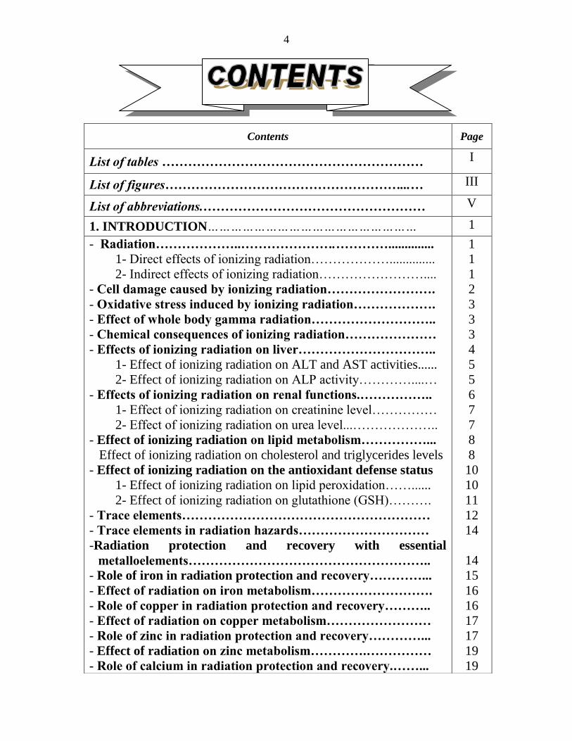
4
Contents Page
List of tables helliphelliphelliphelliphelliphelliphelliphelliphelliphelliphelliphelliphelliphelliphelliphelliphelliphelliphelliphellip I
List of figureshelliphelliphelliphelliphelliphelliphelliphelliphelliphelliphelliphelliphelliphelliphelliphelliphelliphelliphellip III
List of abbreviationshelliphelliphelliphelliphelliphelliphelliphelliphelliphelliphelliphelliphelliphelliphelliphelliphellip V
1 INTRODUCTIONhelliphelliphelliphelliphelliphelliphelliphelliphelliphelliphelliphelliphelliphelliphelliphelliphelliphellip 1
- Radiationhelliphelliphelliphelliphelliphelliphelliphelliphelliphelliphelliphelliphelliphelliphelliphelliphellip
1- Direct effects of ionizing radiationhelliphelliphelliphelliphelliphellip
2- Indirect effects of ionizing radiationhelliphelliphelliphelliphelliphelliphelliphellip
- Cell damage caused by ionizing radiationhelliphelliphelliphelliphelliphelliphelliphellip
- Oxidative stress induced by ionizing radiationhelliphelliphelliphelliphelliphellip
- Effect of whole body gamma radiationhelliphelliphelliphelliphelliphelliphelliphelliphellip
- Chemical consequences of ionizing radiationhelliphelliphelliphelliphelliphelliphellip
- Effects of ionizing radiation on liverhelliphelliphelliphelliphelliphelliphelliphelliphelliphellip
1- Effect of ionizing radiation on ALT and AST activities
2- Effect of ionizing radiation on ALP activityhelliphelliphelliphelliphellip
- Effects of ionizing radiation on renal functionshelliphelliphelliphelliphellip
1- Effect of ionizing radiation on creatinine levelhelliphelliphelliphelliphellip
2- Effect of ionizing radiation on urea levelhelliphelliphelliphelliphelliphellip
- Effect of ionizing radiation on lipid metabolismhelliphelliphelliphelliphellip
Effect of ionizing radiation on cholesterol and triglycerides levels
- Effect of ionizing radiation on the antioxidant defense status
1- Effect of ionizing radiation on lipid peroxidationhelliphellip
2- Effect of ionizing radiation on glutathione (GSH)helliphelliphellip
- Trace elementshelliphelliphelliphelliphelliphelliphelliphelliphelliphelliphelliphelliphelliphelliphelliphelliphelliphelliphellip
- Trace elements in radiation hazardshelliphelliphelliphelliphelliphelliphelliphelliphelliphellip
-Radiation protection and recovery with essential
metalloelementshelliphelliphelliphelliphelliphelliphelliphelliphelliphelliphelliphelliphelliphelliphelliphelliphelliphellip
- Role of iron in radiation protection and recoveryhelliphelliphelliphellip
- Effect of radiation on iron metabolismhelliphelliphelliphelliphelliphelliphelliphelliphellip
- Role of copper in radiation protection and recoveryhelliphelliphellip
- Effect of radiation on copper metabolismhelliphelliphelliphelliphelliphelliphelliphellip
- Role of zinc in radiation protection and recoveryhelliphelliphelliphellip
- Effect of radiation on zinc metabolismhelliphelliphelliphelliphelliphelliphelliphelliphellip
- Role of calcium in radiation protection and recoveryhelliphellip
1
1
1
2
3
3
3
4
5
5
6
7
7
8
8
10
10
11
12
14
14
15
16
16
17
17
19
19
5
- Effect of radiation on calcium metabolismhelliphelliphelliphelliphelliphelliphellip
- Role of magnesium in radiation protection and recovery
- Effect of radiation on magnesium metabolismhelliphelliphelliphelliphellip
- Role of selenium in radiation protection and recoveryhellip
- Effect of radiation on selenium metabolismhelliphelliphelliphelliphelliphellip
- Role of manganese in radiation protection and recoveryhellip
- Effect of radiation on manganese metabolismhelliphelliphelliphelliphellip
- Use of medicinal plants in radiation protection and recovery
- Green teahelliphelliphelliphelliphelliphelliphelliphelliphelliphelliphelliphelliphelliphelliphelliphelliphelliphelliphelliphelliphelliphellip
- Absorption metabolism and excretion of green teahelliphelliphelliphellip
- Mechanism of action of green teahelliphelliphelliphelliphelliphelliphelliphelliphelliphelliphelliphellip
- Biological efficiency of green teahelliphelliphelliphelliphelliphelliphelliphelliphelliphelliphelliphellip
- Radioprotective role of green teahelliphelliphelliphelliphelliphelliphelliphelliphelliphelliphelliphellip
- Green tea and trace elementshelliphelliphelliphelliphelliphelliphelliphelliphelliphelliphelliphelliphellip
- Vitamin Ehelliphelliphelliphelliphelliphelliphelliphelliphelliphelliphelliphelliphelliphelliphelliphelliphelliphelliphelliphelliphellip
20
20
21
21
22
23
23
24
25
27
28
29
31
32
33
2 AIM OF THE WORKhelliphelliphelliphelliphelliphelliphelliphelliphelliphelliphelliphellip 36
3 MATERIAL amp METHODShelliphelliphelliphelliphelliphelliphelliphelliphelliphellip 38
- Materialhelliphelliphelliphelliphelliphelliphelliphelliphelliphelliphelliphelliphelliphelliphelliphelliphelliphelliphelliphelliphellip
1- Experimental Animalshelliphelliphelliphelliphelliphelliphelliphelliphelliphelliphelliphelliphelliphellip
2- Therapeutic agentshelliphelliphelliphelliphelliphelliphelliphelliphelliphelliphelliphelliphelliphelliphellip
3- Chemicals and their sourceshelliphelliphelliphelliphelliphelliphelliphelliphelliphelliphellip
4- Instrumentshelliphelliphelliphelliphelliphelliphelliphelliphelliphelliphelliphelliphelliphelliphelliphelliphelliphellip
- Experimental designhelliphelliphelliphelliphelliphelliphelliphelliphelliphelliphelliphelliphelliphelliphelliphellip
- Methodshelliphelliphelliphelliphelliphelliphelliphelliphelliphelliphelliphelliphelliphelliphelliphelliphelliphelliphelliphelliphellip
- Irradiation of animalshelliphelliphelliphelliphelliphelliphelliphelliphelliphelliphelliphelliphelliphellip
- Samplinghelliphelliphelliphelliphelliphelliphelliphelliphelliphelliphelliphelliphelliphelliphelliphelliphelliphelliphelliphellip
- Measured parametershelliphelliphelliphelliphelliphelliphelliphelliphelliphelliphelliphelliphelliphellip
1- Parameters measured in serumhelliphelliphelliphelliphelliphelliphelliphelliphellip
A-Determination of serum alkaline phosphatase activityhelliphellip
B- Determination of alanine transaminase activity (ALT)helliphellip
C- Determination of aspartate transaminase activity (AST)hellip
D- Determination of serum urea levelhelliphelliphelliphelliphelliphelliphelliphelliphellip
E- Determination of serum creatinine levelhelliphelliphelliphelliphelliphelliphellip
F- Determination of serum cholesterol levelhelliphelliphelliphelliphelliphellip
G- Determination of serum triglycerides levelhelliphelliphellip helliphellip
2- Parameters measured in liver and kidney homogenate A- Determination of reduced glutathione (GSH) contenthelliphellip
38
38
38
38
39
40
40
40
40
41
41 41
42
44
45
46
47
48
49
49
6
B- Determination of lipid peroxidation helliphelliphelliphelliphelliphelliphelliphelliphellip
C- Determination of metallothioneins contenthelliphelliphelliphelliphelliphellip
3- Parameters measured in acid digest of some organshellip
- Microwave digestor technologyhelliphelliphelliphelliphelliphelliphelliphelliphelliphelliphellip
- Instrumentationhelliphelliphelliphelliphelliphelliphelliphelliphelliphelliphelliphelliphelliphelliphelliphelliphelliphellip
- Statistical analysishelliphelliphelliphelliphelliphelliphelliphelliphelliphelliphelliphelliphelliphelliphelliphelliphellip
51
52
54
54
54
55
4 RESULTS helliphelliphelliphelliphelliphelliphelliphelliphelliphelliphelliphelliphelliphelliphelliphelliphellip 56
5 DISCUSSION helliphelliphelliphelliphelliphelliphelliphelliphelliphelliphelliphelliphelliphelliphelliphelliphelliphellip 102
6 SUMMARY amp CONCLUSIONShelliphelliphelliphelliphelliphelliphelliphelliphellip 128
7 REFERENCES helliphelliphelliphelliphelliphelliphelliphelliphelliphelliphelliphelliphelliphelliphelliphelliphelliphellip 131
ARABIC SUMMARY helliphelliphelliphelliphelliphelliphelliphelliphelliphelliphelliphelliphelliphelliphelliphellip 1
7
Table Title Page
I Kits chemicals and their sources 38
1
Effect of green tea (300 mgkg) or vitamin E (40
mgkg) on liver function tests in normal and
irradiated rats
57
2
Effect of green tea (300 mgkg) or vitamin E (40
mgkg) on liver glutathione (GSH)
malondialdehyde (MDA) and metallothioneins
(MTs) contents in normal and irradiated rats
60
3
Effect of green tea (300 mgkg) or vitamin E (40
mgkg) on liver iron (Fe) copper (Cu) and zinc (Zn)
contents in normal and irradiated rats
63
4
Effect of green tea (300 mgkg) or vitamin E (40
mgkg) on liver calcium (Ca) and magnesium (Mg)
contents in normal and irradiated rats
66
5
Effect of green tea (300 mgkg) or vitamin E (40
mgkg) on liver manganese (Mn) and selenium (Se)
contents in normal and irradiated rats
69
6
Effect of green tea (300 mgkg) or vitamin E (40
mgkg) on serum cholesterol and triglycerides levels
in normal and irradiated rats
72
7
Effect of green tea (300 mgkg) or vitamin E (40
mgkg) on serum urea and creatinine levels in
normal and irradiated rats
75
8
Effect of green tea (300 mgkg) or vitamin E (40
mgkg) on kidney glutathione (GSH)
malondialdehyde (MDA) and metallothioneins
(MTs) contents in normal and irradiated rats
78
9
Effect of green tea (300 mgkg) or vitamin E (40
mgkg) on kidney iron (Fe) copper (Cu) and zinc
(Zn) contents in normal and irradiated rats
81
List of Tables 7
8
Table Title Page
10
Effect of green tea (300 mgkg) or vitamin E (40
mgkg) on kidney calcium (Ca) and magnesium
(Mg) contents in normal and irradiated rats
84
11
Effect of green tea (300 mgkg) or vitamin E (40
mgkg) on kidney manganese (Mn) and selenium
(Se) contents in normal and irradiated rats
87
12
Effect of green tea (300 mgkg) or vitamin E (40
mgkg) on spleen iron (Fe) copper (Cu) and zinc
(Zn) contents in normal and irradiated rats
90
13
Effect of green tea (300 mgkg) or vitamin E (40
mgkg) on spleen calcium (Ca) magnesium (Mg)
and selenium (Se) contents in normal and irradiated
rats
93
14
Effect of green tea (300 mgkg) or vitamin E (40
mgkg) on testis iron (Fe) copper (Cu) and zinc
(Zn) contents in normal and irradiated rats
96
15
Effect of green tea (300 mgkg) or vitamin E (40
mgkg) on testis calcium (Ca) magnesium (Mg) and
selenium (Se) contents in normal and irradiated rats
99
16
The concentrations of some metalloelements in
green tea plants and green tea extract represented as
(μgg) and (μgml) except for Se represented as
(ngg) and (ngml)
101
9
Figure Title Page
I Some mechanisms by which natural products render
radioprotection 24
II Chemical structures of EGCG EGC ECG and EC 26
III Summary of the formation of metabolites and
conjugates of flavonoids in humans 27
IV The chemical structure of alpha-tocopherol 33
V The standard curve of ALT 43
VI The standard curve of AST 45
VII The standard curve of MDA 52
1
Effect of green tea (300 mgkg) or vitamin E (40
mgkg) on liver function tests in normal and
irradiated rats
58
2
Effect of green tea (300 mgkg) or vitamin E (40
mgkg) on liver glutathione (GSH) malondialdehyde
(MDA) and metallothioneins (MTs) contents in
normal and irradiated rats
61
3
Effect of green tea (300 mgkg) or vitamin E (40
mgkg) on liver iron (Fe) copper (Cu) and zinc (Zn)
contents in normal and irradiated rats
64
4
Effect of green tea (300 mgkg) or vitamin E (40
mgkg) on liver calcium (Ca) and magnesium (Mg)
contents in normal and irradiated rats
67
5
Effect of green tea (300 mgkg) or vitamin E (40
mgkg) on liver manganese (Mn) and selenium (Se)
contents in normal and irradiated rats
70
6
Effect of green tea (300 mgkg) or vitamin E (40
mgkg) on serum cholesterol and triglycerides levels
in normal and irradiated rats
73
10
Figure Title Page
7
Effect of green tea (300 mgkg) or vitamin E (40
mgkg) on serum urea and creatinine levels in normal
and irradiated rats
76
8
Effect of green tea (300 mgkg) or vitamin E (40
mgkg) on kidney glutathione (GSH)
malondialdehyde (MDA) and metallothioneins (MTs)
contents in normal and irradiated rats
79
9
Effect of green tea (300 mgkg) or vitamin E (40
mgkg) on kidney iron (Fe) copper (Cu) and zinc
(Zn) contents in normal and irradiated rats
82
10
Effect of green tea (300 mgkg) or vitamin E (40
mgkg) on kidney calcium (Ca) and magnesium (Mg)
contents in normal and irradiated rats
85
11
Effect of green tea (300 mgkg) or vitamin E (40
mgkg) on kidney manganese (Mn) and selenium (Se)
contents in normal and irradiated rats
88
12
Effect of green tea (300 mgkg) or vitamin E (40
mgkg) on spleen iron (Fe) copper (Cu) and zinc (Zn)
contents in normal and irradiated rats
91
13
Effect of green tea (300 mgkg) or vitamin E (40
mgkg) on spleen calcium (Ca) magnesium (Mg) and
selenium (Se) contents in normal and irradiated rats
94
14
Effect of green tea (300 mgkg) or vitamin E (40
mgkg) on testis iron (Fe) copper (Cu) and zinc (Zn)
contents in normal and irradiated rats
97
15
Effect of green tea (300 mgkg) or vitamin E (40
mgkg) on testis calcium (Ca) magnesium (Mg) and
selenium (Se) contents in normal and irradiated rats
100
11
Adenosine diphosphate ADP
Alkaline phosphatase ALP
Alanine transaminase ALT
Analysis of variance ANOVA
Aspartate transaminase AST
Adenosine triphosphate ATP
Body weight bwt
Cyclic adenosine monophosphate cAMP
Catalase CAT
Cholecystokinin CCK
Cholesterol Ch
Central nervous system CNS
Catechol-O-methyl-transferase COMT
Dihydrofolate reductase DHFR
Diribonucleic acid DNA
55
dithiobis(2-nitrobenzoic acid) DTNB
Epicatechin EC
Epicatechin-3-gallate ECG
Ethylenediaminetetraacetic acid EDTA
Epigallocatechin EGC
Epigallocatechin-3-gallate EGCG
Epidermal growth factor receptor EGFR
Gallocatechin-gallate GCG
Glumerular filtration rate GFR
Reduced glutathione GSH
Glutathione peroxidase GSH-PX
Oxidized glutathione GSSG
Green tea GT
Green tea extract GTE
Green tea polyphenols GTP
Gray Gy
12
Hydrogen peroxide H2O2
High density lipoprotein HDL
Human immunodeficiency virus HIV
3- Hydroxyl - 3- methyl glutaryl coenzyme A HMG-COA
Interleukin-1 IL-1
Kilo base pair Kb
Kilo Dalton KDa
Lecithin cholesterol acyl transferase LCAT
Low density lipoprotein LDL
Malondialdehyde MDA
Messenger ribonucleic acid mRNA
Metallothioneins MTs
Nicotinamide adenine dinucleotide phosphate
hydrogen
NADPH
Norepinephrine NE
Nuclear magnetic resonance NMR
Nitric oxide NO
Superoxide radical O2-
Hydroxyl radical OH
Peroxynitrite ONOO-
Parts per million ppm
Red blood cells RBCs
Roentgen equivalent man Rem
Ribonucleic acid RNA
Reactive oxygen species ROS
Superoxide dismutase SOD
Triiodothyronine T3
Thyroxine T4
Thiobarbituric acid TBA
Thiobarbituric acid reactive substance TBARS
Trichloroacetic acid TCA
Triglyceride TG
Tumor necrosis factor TNF
Ultraviolet UV
Ultraviolet B UVB
Vascular endothelial growth factor receptor VEGFR
Very low density lipoprotein VLDL
13
14
Introduction
Radiation-
Radiation is defined as the emission and propagation of energy in the
form of waves or particles through space or matter (Zaider and Rossi
1986) Ionizing radiation is type of radiation having sufficient energy to
cause ion pairs to be formed in the medium through which it passes Ionizing
radiations consist of electromagnetic radiation (photons including X-rays
and gamma rays) and particulate radiation (such as electrons protons and
neutrons) (Cho and Glatstein 1998)
Radiation produces either direct or indirect chemical changes in
molecules Both the direct and indirect effects of ionizing radiation lead to
molecular damage which is translated to biochemical changes Exposure to
such radiation can induce alterations in the cellular macromolecules and
affect their functions (Roach et al 2009)
1-Direct effects of ionizing radiation Direct effects of radiation include
changes which appear as a result of the absorption of radiation energy by
biological materials (target molecules) which initiate a chain of reactions
leading to free radical formation (Michaels and Hunt 1978) Free radicals
are by definition species which contain a number of electrons they may be
positively charged negatively charged or neutral and all three types are
important A role for free radicals has been proposed in the toxicity diseases
(Kehrer and Lund 1994)
2-Indirect effects of ionizing radiation Indirect effects comprise the
changes occurring to the molecules in a solution induced by decomposition
products of water or other solutes and not by the radiant energy absorbed by
the molecule (Michaels and Hunt 1978)
The indirect effect of radiation in biological systems depends on the
effect of irradiation on water and the presence of oxygen in the tissue being
irradiated The end products of radiolysis of water without oxygen are γ-radiation
2H2O H + OH
+ H
+ + OH
-
H and OH
released by ionizing radiation are the most important free
radicals comprising 55 of the initial relative yield (Nair et al 2001)
15
In the presence of oxygen other radiolysis products also formed that
have oxidizing properties namely hydroperoxide radical (HOO) and
hydrogen peroxide (H2O2)
H + O2 rarr HOO
HOO
+ HOOrarr H2O2 + O2
Cell damage caused by ionizing radiation-
Ionizing radiation induces multiple biological effects through direct
interaction with DNA or production of activated free radical species from
water When tissues are exposed to ionizing radiation most of the energy
taken up is absorbed by the cell water largely because there is more water
than any other molecules thus creating two radicals a hydrogen radical (H)
and a hydroxyl radical (OH) The latter radical can attack and damage
almost every molecule found in living cells (Halliwell and Gutteridge
1999)
Ionizing radiation induces reactive oxygen species (ROS) in the form
of OH H
singlet oxygen and peroxyl radicals that follow a cascade of
events leading to DNA damage such as single or double strand breakages
base damage and DNA-protein cross-links These lesions cluster as complex
local multiply damage sites The DNA double strand breaks are considered
the most lethal events following ionizing radiation and have been found to
be the main target of cell killing by radiation (Jagetia 2007)
Mondelaers and Lahorte (2001) reported that the processes
leading to radiation damage are complex but can be considered to take place
in the following stages
The initial physical stage (Lasting for 10-13
second) in which
energy is deposited in the cell and caused ionization
The physicochemical stage (Lasting for 10-7
second) in which
the ions interact with other water molecules resulting in the
production of free radicals which are chemically highly reactive
due to the presence of an unpaired electron Another reaction
product is hydrogen peroxide which is a strong oxidizing agent
The chemical stage (Lasting for few minutes or hours) in which
the reaction products interact with the important organic
molecules of the cell
16
The biological stage In which the time scale varies from minutes
to tens of years and is depending on the type of the cell affected
Oxidative stress induced by ionizing radiation-
Oxidative stress is a state of imbalance between generation of (ROS)
and the levels of antioxidant defense system Antioxidant enzymes are part
of the endogenous system available for the removal or detoxification of free
radicals and their products formed by ionizing radiation (Bhatia and Jain
2004)
Oxidative stress has been linked to diseases including some allergic
and inflammatory skin diseases (Okayama 2005) neurodegeneration
(Moreira et al 2005) and atherosclerosis in diabetic patients (Lankin et
al 2005) As a defense mechanism the body produces a number of
endogenous antioxidants such as superoxide dismutase (SOD) catalase
(CAT) and glutathione peroxidase (GSH-PX) capable of scavenging harmful
ROS to maintain an optimal oxidantantioxidant balance thereby
maintaining normal cellular function and health (Droumlge 2002)
Effect of whole body gamma radiation
Factors that determine the biological effects of ionizing radiation
include the type of radiation the received dose the rate at which the
radiation dose is delivered nutritional factors the type of irradiated tissues
as well as the age and sex of the exposed person In addition whether the
dose was delivered in fractions or in a single exposure could determine the
biological effect (Beir 1990)
A single whole body exposure of mammals to ionizing radiation
results in a complex set of syndromes whose onset nature and severity are a
function of both total radiation dose and radiation quality At a cellular level
ionizing radiation can induce damage in biologically important
macromolecules such as DNA proteins lipids and carbohydrates in various
organs While some damage may be expressed early the other may be
expressed over a period of time depending upon cell kinetics and radiation
tolerance of the tissues (Baliga et al 2004)
Chemical consequences of ionizing radiation
17
The first consequence of ionizing radiation is ionization of water
Since water represents 70 of the chemical composition of the adult body
its chemical transformation by ionizing radiation merits serious
consideration Ionization of water is well understood and produces very
reactive aquated electrons monoatomic hydrogen atoms hydroxyl radicals
hydrogen peroxide and protonated water as well as superoxide and
hydroperoxyl radicals in the presence of oxygen Hydroperoxyl radical
hydroxyl radical monoatomic hydrogen and aquated electron have very
short half lives (10-1
to 10-3
sec) and consequently react rapidly with cellular
components in reduction oxidation initiation insertion propagation and
addition reactions causing loss of function and need for biochemical
replacement andor repair (Sorenson 2002) The second consequence of
ionizing radiation is its ability to impart sufficient energy to all biochemicals
to cause homolytic bond breaking and produce all conceivable organic
radicals in considering C-C C-N C-O C-H P-O S-O hellipetc bond
homolysis These radicals will undergo the reactions listed above causing
further destruction and requiring replacement andor repair (Droumlge 2002)
A third consequence of ionizing radiation is homolytic or heterolytic
bond breaking of coordinate-covalent bonded metalloelements These are the
weakest bonds in biochemical molecules and potential sites of the greatest
damage which may be most in need of replacement andor repair since
many repair enzymes are metalloelements-dependent as are the
metalloelement dependent protective SODs (Sorenson 2002)
Effects of ionizing radiation on liver
It was reported that ionizing radiation affects the liver function
(Feurgard et al 1998) Influence of stress on liver is of interest from the
clinical point of view because stress plays a potential role in aggravating
liver diseases in general and hepatic inflammation in particular probably
through generation of ROS (Zaidi et al 2005)
The serum transaminases activity is the most widely used parameter
as a measure of hepatic injury due to its ease of measurement and high
degree of sensitivity It is useful for the detection of early damage of hepatic
tissue and requires less effort than that for a histological analysis (Ray et al
2006) Serum elevation of alanine transaminase (ALT) activity is rarely
18
observed in condition other than parenchymal liver disease Moreover
elevation of ALT activity persists longer than does that of aspartate
transaminase (AST) activity (Tolman and Rej 1999) ALT is the enzyme
produced within the cells of the liver and its abnormality is increased in
conditions where cells of the liver have been inflamed or undergone cell
death Any form of hepatic cell damage can result in an elevation in ALT
activity which may or may not correlate with the degree of cell death or
inflammation ALT is the most sensitive marker for liver cell damage and
the most important test for recognition of acute and chronic hepatic failure
(Dufour et al 2000)
1-Effect of ionizing radiation on ALT and AST activities
AST and ALT are enzymes responsible for the catalization of the
transference of an amino group from α-amino acid to α-keto acid and they
are considered as indicators for liver injury caused by exposure to ionizing
radiation In view of the effect of radiation on transaminases many authors
reported that the activities of AST and ALT increased when mice or rats
exposed to gamma radiation at dose levels from 4 to 6 Gy (Bhatia et al
2007 Adaramoye 2010)
Roushdy et al (1984) showed that gamma irradiation at a dose level
of 6 Gy resulted in remarkable increases in the transaminases activities both
in serum and liver They indicated that the rise in the liver transaminases
activities may be due to the drastic physiological effects caused by
irradiation The increase in ALT activity may be related to extensive
breakdown of liver parenchyma with subsequent enzyme release or to
increase in permeability of the cell membrane that could enhance the
movement of enzymes from their sites of production (Manciluae et al
1978) Also Fahim et al (1991) suggested that the elevation in ALT and
AST activities in rats exposed to 75 Gy of gamma radiation may be due to
destruction of radio-sensitive cells of haematopoietic tissue and erythrocytes
haemolysis
2- Effect of ionizing radiation on ALP activity
Alkaline phosphatase (ALP) is a hydrolytic enzyme acting on
phosphoric esters with the liberation of inorganic phosphate from various
19
substrates In addition alkaline phosphatase is mainly involved in passive
transport mechanism (Verma and Nair 2001) It is well known that ALP
plays an important role in maintaining the cell membrane permeability
(Samarth and Kumar 2003) Magnesium and zinc ions are essential for
stability and maximum catalytic activity of ALP enzyme (Gowenlock et al
1988)
Exposure of rats or mice to radiation at dose levels range from 4 to
8Gy induced an increase in ALP activity that was recorded by many authors
(Sunila and Kuttan 2005 Adaramoye et al 2008 Pratheeshkumar and
kuttan 2011)
Abdel-Fattah et al (1999) stated that ALP activity in plasma of rats
increased significantly at 1 3 and 5 hours after exposure to single dose of 6
Gy gamma radiation They suggested that this increase could be considered
as a reflection of liver dysfunction in the acute radiation sickness Authors
also revealed that the increase in alkaline phosphatase activity may be due to
destruction of cell membrane or destruction of this enzyme inhibitor by
radiation
Furthermore Kafafy and Ashry (2001) found that whole body
gamma-irradiation affected liver structure and functions as indicated by
changes in the serum ALP activity which increased significantly along the
post-irradiation days where it reached its maximum at the tenth day
following exposure The authors deduced that this increase reflected
detectable changes in liver function due to the changes in tissue permeability
induced by irradiation which enhanced the movement of enzymes from their
subcellular sites of production to extracellular process and consequently into
the blood circulation
Effects of ionizing radiation on renal functions
It is well established that radiation exposure is known to impair the
biological integrity of living organisms It is also known that exposure to
acute radiation dose can cause substantial well detectable functional changes
in the organisms much earlier than morphological changes would develop
(Robbins and Bonsib 1995) Many authors reported that ionizing radiation
greatly affected renal function (Ramadan et al 1998 kafafy et al 2005)
Radiation-induced renal impairment occurs predictably after local kidney
20
irradiation or total body irradiation (Robbins and Bonsib 1995 Badr El-
Din 2004) Irradiation leads to progressive biochemical changes in the
irradiated animals The animals may suffer from continuous loss in body
weights which could be attributed to disturbance in nitrogen metabolism
usually recognized as negative nitrogen balance Accordingly it could be
expected that this may cause an increase in the urea ammonia and amino
acid levels in blood and urine due to great protein destruction induced by
irradiation that is an evidence of marked impairment of kidney function
(Robbins et al 1992)
1-Effect of ionizing radiation on creatinine level
It is well known that creatine is converted to creatine phosphate in the
muscle and that creatine phosphate is converted to creatinine before
excretion in the urine Ionizing radiation causes damage in muscle of
mammals which appears by increased excretion of nitrogenous metabolites
such as creatine (Gerber et al 1961)
Urinary output of creatinine may be taken as a sensitive parameter
indicating the degree of impaired tissue metabolism due to radiation effect
The kidney is relatively more resistant to ionizing radiation (Roushdy et al
1997 Cheng et al 2002)
Yildiz et al (1998) observed that serum creatinine level increased
when kidneys of male rats were irradiated with either 10 Gy single dose or
26Gy at a rate of 2 Gy per day and after 4 weeks of irradiation glomerular
and proximal tubular injury were observed Increased serum creatinine level
in the irradiated rats indicates development of nephritis and renal
dysfunction (Borg et al 2002) that may be attributed to impairment of
glomerular selective properties caused by irradiation (Berry et al 2001)
Studies of Hassan et al (1994) showed that serum creatinine level
was elevated when the rats were exposed to gamma-irradiation at
fractionated dose levels of 3 Gy to a cumulative dose of 9 Gy on the 2nd
hours 1st and 7
th days post-exposure They concluded that fractionated
exposure to gamma irradiation effectively altered the glomerular filtration
rate (GFR) in rats
21
Many authors observed significant increase in plasma level of
creatinine post whole body gamma irradiation with 65 Gy (Badr El-Din
2004) and 75 Gy (Omran et al 2009)
2-Effect of ionizing radiation on urea level
Most of ammonia formed by deamination of amino acids is converted
to urea The urea resulting from protein degradation is excreted by the
kidney so the level of urea in plasma of rats is an indicator for the effect of
radiation on kidney function (Kutchai 1993)
Studies of Geraci et al (1990) and Adaramoye (2010) showed that
an increase in serum urea level of animals is induced post-irradiation The
authors considered this increase as a reflection of deteriorating renal
performance
On the other hand Mahdy et al (1997) observed that whole body
gamma-irradiation of rats at 75 Gy (single dose) caused a significant
increase of urea level as recorded 7 10 and 14 days after irradiation The
authors suggested that elevation in serum urea level may be due to an
increased oxidative deamination of amino acids in the liver resulting in
excess urea formation
Badr El-Din (2004) declared that an increase in blood urea level has
been reported after exposure to radiation and secondary to renal damage
The elevation of urea may be attributed to an increase in nitrogen retention
or excessive protein breakdown Furthermore Omran et al (2009)
demonstrated that rats exposed to 75 Gy whole body gamma irradiation
showed significant increase in plasma urea level (50) at both time intervals
of 7 and 16 days
Effect of ionizing radiation on lipid metabolism
Lipid profile especially cholesterol has been representing a major
essential constituent for all animal cell membranes Plasma lipid levels are
affected by genetic and dietary factors medication and certain primary
disease states (Feldman and Kuske 1987) Hyperlipidemia occurring due
to exposure to ionizing radiation resulted in accumulation of cholesterol
22
triglycerides and phospholipids (Feurgard et al 1999) The accumulated
lipoproteins were susceptible to peroxidation process causing a shift and
imbalance in oxidative stress This imbalance manifested themselves
through exaggerated ROS production and cellular molecular damage
(Romero et al 1998)
Effect of ionizing radiation on cholesterol and triglycerides levels
Cholesterol is synthesized in the liver and its balance is maintained by
the livers ability to remove cholesterol from lipoproteins and use it to
produce bile acids and salts that excreted in the bile duct In obstructive
jaundice the bile can not be eliminated cholesterol and triacylglycerols may
accumulate in the blood In acute necrotic liver diseases triacylglycerols
may be elevated due to hepatic lipase deficiency In liver failure caused by
necrosis the livers ability to synthesize cholesterol is reduced and the blood
levels may be low (OacuteGrady et al 1993)
Free radical mediated oxidative damage induced by radiation is one of
the prime factors that increase the hepatic cholesterol and triglycerides levels
(Pote et al 2006) Radiation leads to hyperlipidemia through destruction of
cell membranes enhancement of lipid metabolism cholesterol release and
increased triglycerides synthesis (Bowden et al 1989)
Irradiation of rats induced increase in the total lipid synthesis in bone
marrow liver and blood that was attributed to the increase in stimulation of
the liver enzyme responsible for the biosynthesis of fatty acids and to the
mobilization of fat from adipose tissue to the blood stream leading to
hyperlipidemic state (Sedlakova et al 1988) Another explanation for this
hyperlipidemic state is the retention character caused by the diminished
utilization of circulating lipids by the damaged tissues (Abou Safi and
Ashry 2004 Kafafy 2004) Also some changes in the activities of hepatic
HMGCoA reductase (the rate-limiting enzyme for cholesterol synthesis) and
in hepatic cholesterol 7alpha-hydroxylase (the key enzyme involved in
degradation of cholesterol in the liver) were noted following radiation
exposure (Feurgard et al 1999)
Many authors concluded that whole body gamma-irradiation showed a
significant increase of serum cholesterol and triglycerides levels whether this
23
radiation is applied as a single dose (Feurgard et al 1998 Kafafy 2004
Baker et al 2009) or fractionated doses (Abou-Safi et al 2001)
Girgis et al (2000) showed that whole body gamma-irradiation of
rats at a dose level of 6 Gy significantly decreased the total cholesterol level
in plasma by 374 on the 1st day after irradiation as compared to the
control value However it increased by 4804 309 and 96 after 3 7
and 14 days from irradiation respectively as compared to the control value
The authors suggested that ionizing radiation by activating the cholesterol
esterase enzyme may play a role in the development of atherosclerosis in
experimental animals
The hypercholesterolemia induced by radiation was attributed to two
causes the first was the activation of cholesterologenesis in different cells of
tissue as an early reaction to harmful effect of the radiation for restoring the
cell membranes activity and the second was the decrease in the lecithin
cholesterol esterification where HDL cholesterol may be the vehicle for
reversed cholesterol transport and esterification (Abdel-Fattah et al 2003)
Effect of ionizing radiation on the antioxidant defense status
When cellular production of ROS overwhelms its antioxidant
capacity a state of oxidative stress is reached leading to serious cellular
injuries that contributes to the pathogenesis of several diseases (Gloire et
al 2006) The systemic damage observed following irradiation is
particularly due to the overproduction of ROS which disrupt the delicate
pro-oxidantanti-oxidant balance of tissues leading to proteins lipids and
DNA oxidation (Flora 2007) Free radicals are highly reactive and cause
tissue damage by reacting with poly unsaturated fatty acids found in cellular
membranes or by reacting with sulfhydryl bonds in proteins as reported by
Guney et al (2004)
The antioxidant defense system consists of numerous enzymes and
low molecular weight compounds that scavenge produced radicals and other
ROS and prevent production of more reactive radical species It also
removes lipid peroxides preventing further propagation (Sies 1993) This
antioxidant defense system is consisting of enzymes such as CAT SODs
GSH-PX and numerous non-enzymatic antioxidants including vitamins A E
and C glutathione (GSH) metallothioneins and flavonoids (Belviranli and
Goumlkbel 2006)
24
1-Effect of ionizing radiation on lipid peroxidation
ROS are relatively short lived molecules that exert local effects They
can attack poly unsaturated fatty acids and initiate lipid peroxidation within
the cell The process of lipid peroxidation is one of oxidative conversion of
poly unsaturated fatty acid to byproducts known as malondialdehyde (MDA)
or lipid peroxides which is the most studied biologically relevant free
radical reaction These byproducts can diffuse large distances from site of
their generation before mediating damage They are capable of inactivating
enzymes (Wilson et al 2003) Lipid peroxidation is a complex process
characterized by three distinct phases initiation propagation and
termination Radiation induced lipid peroxidation is initiated by direct or
indirect ionization or by free radical attack (Gupta et al 2000)
Lipid peroxidation is a chain reaction in which the interaction of the
lipid radical with another organic molecule results in conversion of that
molecule to the free radical state and propagation of damage Peroxidation
of membrane lipids can have numerous effects including increased
membrane rigidity decreased activity of membrane bound enzymes altered
activity of membrane receptors as well as altered permeability (Kamat et
al 2000) It was found that whole body gamma irradiation of male rats caused
changes in the antioxidant defense system of the organism which depend on
the intensity of lipid peroxidation level in the blood (Gatsko et al 1990)
Furthermore many authors deduced that irradiation of rats or mice at dose
range from 6-12 Gy either applied as single dose or fractionated doses
induced significant increase in liver and blood MDA levels (Baliga et al
2004 Samarth et al 2006 Kilciksiz et al 2008 Pratheeshkumar and
kuttan 2011)
Nunia et al (2007) noted a significant increase in blood level and
hepatic content of lipid peroxidation in mice after 75 Gy of gamma
irradiation They attributed this increase to the membrane damage caused by
ROS which may allow the entry of excess calcium into cells with sequential
biochemical and micro anatomical cellular degranulation and necrosis
2-Effect of ionizing radiation on glutathione (GSH)
25
GSH is a small molecule made up of three amino acids (tripeptide)
[glutamine ndash cysteine - glycine] whose antioxidant action is facilitated by the
sulfhydryl group of cysteine (Townsend et al 2003) GSH is the most
abundant non-protein thiol in mammalian cells It plays an important role in
regulation of cellular redox balance The most recognized function of GSH
is its role as a substrate for GSH-S-transferase and GSH-PX These enzymes
catalyze the antioxidation of ROS and free radicals (Weis et al 1993)
The presence of GSH is required to maintain the normal function of
the immune system It is essential for the activation of T-lymphocytes and
polymorphonuclear leukocytes as well as for cytokine production and
therefore for mounting successful immune responses (Townsend et al
2003)
GSH reacts directly with free radicals and can protect cells from
single oxygen radical (O) hydroxyl radical (OH
) and superoxide radical
(O2) (Cominacini et al 1996) GSH may stabilize membrane structure by
removing acyl peroxides formed by lipid peroxidation reactions (May et al
1998)
GSH with its sulfhydryl group functions in the maintenance of
sulfhydryl groups of other molecules (especially proteins) and as a catalyst
for disulfide exchange reactions It also functions in the detoxification of
foreign compounds hydrogen peroxide and free radicals When GSH acts as
reducing agent itrsquos SH becomes oxidized and forms a disulfide link with
other molecules of GSH (Manda et al 2007) The reduced GSH in
oxidationreduction cycling catalyzed by GSH-PX enzyme is critical in
reducing H2O2 thus breaks the chain reaction resulting from the superoxide
radical to the highly reactive hydroxyl radical (Hayes and Mclellan 1999)
GSH-PX
H2O2 + 2GSH GSSG + 2H2O
In addition to its action on H2O2 GSH-PX has the ability to use lipid
peroxides as substrate to convert them to inert compounds (Andersen et al
1997) GSH-PX
ROOH + 2GSH GSSG + ROH + H2O
26
Considerable evidence pointed to the fact that intracellular non-
protein sulfhydryl compounds play an important role in cellular response to
ionizing radiation (Bump and Brown 1990) In the same concern Jagetia
et al (2004) studied the effect of different doses of radiation in mice They
revealed that GSH content of mice livers decreased in a dose dependant
manner Also Inal et al (2002) observed that administration of GSH
appears to be useful approach to reduce radiation injury by reducing MDA
levels and increasing CAT activities
A lot of authors revealed that blood level and liver content of GSH
exhibited significant decrease after exposure of rats or mice to whole body
gamma radiation at dose levels of 6 Gy (Pratheeshkumar and kuttan
2011) 75 Gy (Nunia et al 2007) 8 and 10 Gy (Sharma and Kumar
2007)
Trace elements
Trace elements are elements that are present in the body at very low
amounts micro grams to milligrams but they are essential for certain
biochemical processes (Wada 2004) Trace elements act as essential
activators or cofactors for antioxidant enzymes to exert their action
(Ostrakhovitch and Cherian 2005)
An element is considered by Mertz (1970) to be essential if its
deficiency results in impairment of a function from optimal to suboptimal
Cotzais (1967) indicated that a trace element can be considered essential if it
meets the following criteria (1) it is present in all healthy tissues of all
living things (2) its concentration from one animal to the next is fairly
constant (3) its withdrawal from the body induces reproducibly the same
physiological and structural abnormalities regardless of the species studied
(4) its addition either reverses or prevents these abnormalities (5) the
abnormalities induced by deficiency are always accompanied by pertinent
and specific biochemical changes (6) these biochemical changes can be
prevented or cured when the deficiency is prevented or cured
Copper iron manganese and zinc are essential metalloelements
These essential metalloelements as well as essential amino acids essential
fatty acids and essential vitamins are required by all cells for normal
metabolic processes but can not be synthesized de novo and dietary intake
27
and absorption are required to obtain them Ionic forms of these
metalloelements have particularly high affinities for organic ligands found in
biological systems and rapidly undergo bonding interactions to form
complexes or chelates in biological systems Absorbed metalloelement
chelates undergo systemic circulation to all tissues and utilization by all cells
following ligand exchange with small molecular mass ligands apoproteins
and apoenzymes to form metalloproteins and metalloenzymes in de novo
synthesis The degree of radiation injury and nutritional state of health of an
individual may determine whether or not an individual will be able to
overcome metalloelement-dependent repairable radiation injury (Sorenson
2002)
The action of a very small amount of trace element is necessary for
optimal performance of a whole organism Lack of a small amount of a trace
element (eg iron) can result in disease (anemia) seemingly this
proportionate to the amount of element missing The bases for the
amplification of trace element action is that trace elements are constituents
ofor interact with enzymes or hormones that regulate the metabolism of
much larger amounts of biochemical substrates If the substrates are also
regulators the effect is even further amplified (Abdel-Mageed and Oehme
1990a)
Essential trace elements are specific for their in vivo functions They
cannot be effectively replaced by chemically similar elements Certain trace
elements are stable in more than one valence state (eg Fe Cu Mo)
allowing biochemical redox function while others are stable in only a single
state [eg Zn(II) Ni(II)] (Milne 2001) Specificity of trace element function
is also promoted by specific carrier and storage proteins such as transferrin
and ferritin for iron albumin and α-macroglobulin for zinc ceruplasmin for
copper transmanganin for manganese and nickeloplasmin for nickel These
carrier proteins recognize and bind specific metals and transport them toor
store them at specific site with the organism (Mensa et al 1995 Vivoli et
al 1995)
Interaction between metals may be important not only when one
metal is present in excess and the other is deficient but also when the lack of
one metal decreases the bioavailability of the other (Pallareacutes et al 1996)
Pallareacutes et al (1993) previously found that Fe deficiency affects Ca P and
Mg metabolism (at absorptive level) Also the addition of large amounts of
28
zinc to a diet interferes with the intestinal copper absorption system
resulting in copper deficiency (Mills 1981)
Changes in concentrations of essential trace elements in the body
associated with the progression of neoplastic diseases and have a profound
impact systemic metabolic activity (Siddiqui et al 2006) The deficiency of
trace elements may depress the antioxidant defense mechanisms (Kumar
and Shivakumar 1997) erythrocyte production (Morgan et al 1995)
enhance lipid abnormalities (Tajik and Nazifi 2010) While the toxicity of
trace elements may induce renal liver and erythropoietic abnormalities
(Chmielnicka et al 1993 Farinati et al 1995 Kadkhodaee and Gol
2004)
Trace elements in radiation hazards
Most of cellular alterations induced by ionizing radiation are indirect
and are mediated by the generation of free radicals and related reactive
species (Maurya et al 2007) Mammalian cells are equipped with both
enzymatic and non-enzymatic antioxidant mechanisms to minimize cellular
damage resulting from the interaction between cellular constituents and
ROS Ionizing radiation causes homolytic and heterolytic bond breaking of
covalent and coordinate covalent bonded metalloelements These are the
weakest bonds in biochemical molecules and potentially the sites of the
greatest damage so they are most in need of replacement andor repair
Many repair enzymes are metalloelements dependent as the metalloelement
dependent protective SODs (Sorenson 2002)
Radiation protection and recovery with essential metalloelements
Recognizing that loss of enzyme activity is dependent on essential
metalloelements may at least partially account for lethality of ionizing
radiation Cu Fe Mn and Zn dependent enzymes have roles in protecting
against accumulation of ROS as well as facilitating the repair (Sorenson
1978) which may explain the radiation protection and radiation recovery
activity of Cu Fe Mn and Zn compounds (Matsubara et al 1986) It is
suggested that the IL-1-mediated redistribution of essential metalloelements
may account for subsequent de novo synthesis of the metalloelement
dependent enzymes required for biochemical repair and replacement of
29
cellular and extracellular components needed for recovery from radiolytic
damage (Sorenson 1992)
De novo synthesis of metalloelements dependent enzymes is required
for utilization of oxygen and preventions of oxygen accumulation as well as
for tissue repair processes including metalloelement dependent DNA and
RNA repair This is the key to hypothesis that essential metalloelement
complexes prevent andor facilitate recovery from radiation-induced lesions
(Berg 1989)
Role of iron in radiation protection and recovery
Iron is the most important of the essential trace metals An appropriate
number of human diseases are related to iron deficiency or disorders of iron
metabolism (Kazi et al 2008) It is the oxygen carrier in hemoglobin and
myoglobin It also functions in the respiratory chain Iron in the body is
either functional or stored Functional iron is found in hemoglobin and
myoglobin whereas stored iron is found in association with transferrin
ferritin and hemosiderin The storage sites of ferritin and hemosiderin are the
liver spleen and bone marrow (McCarter and Holbrook 1992) Iron is
required in many biochemical processes ranging from oxidative metabolism
to DNA synthesis and cell division (Crowe and Morgan 1996) It has been
reported that iron and its complexes protect from ionizing radiation
(Sorenson et al 1990) play an important role in facilitation of iron
dependent enzymes required for tissue or cellular repair processes including
DNA repair (Ambroz et al 1998) and protect against radiation-induced
immunosupression (Tilbrook and Hider 1998)
The oxidative damage is thought to be a consequence of increased
free radical generation secondary to tissue iron accumulation The damage
may be also a consequence of the reduction in Zn or Cu dependent
antioxidizing processes as an increase in tissue iron was observed in Zn and
Cu deficiencies (Oteiza et al 1995)
ROS promote iron release from ferritin A free iron ion catalyzes
changes from relatively poor reactive O2 and H2O2 to highly reactive HO
(Fenton reaction) (Koike and Miyoshi 2006) In addition iron can catalyze
the decomposition of lipid hydroperoxides to form alkoxyl peroxyl and
other radicals (Halliwell and Gutteridge 1990)
30
Effect of radiation on iron metabolism
Exposure of rats to whole body gamma radiation with single dose of
6Gy and 4 Gy induced significant increase in liver content and serum level
of iron (Mansour et al 2006 Abdel-Gawad and Aiad 2008) In addition
an increase of iron content in liver and spleen of irradiated animals were
demonstrated by Nada et al (2008) The same increase in serum iron level
was demonstrated also in case of animalsrsquo exposure to fractionated 12 Gy
gamma rays (2 Gy weekly) (Ashry et al 2010)
Kotb et al (1990) reported that accumulation of iron in the spleen
after whole body gamma irradiation could be resulted from disturbances in
the biological function of RBCs including possible intravascular haemolysis
and subsequent storage of iron in the spleen Also Osman et al (2003) and
Harris (1995) attributed the increase of iron content in liver and spleen post
irradiation to the inhibition of ceruloplasmin which is essential for iron
metabolism and distribution
Role of copper in radiation protection and recovery
Cu is one of the essential trace elements in humans and disorders
associated with its deficiency and excess have been reported (Aoki 2004) It
is an integral component of many enzymes and proteins needed in a wide
range of metabolic processes (Ozcelik et al 2003) Copper in the divalent
state (Cu2+
) has the capacity to form complexes with many proteins These
metalloproteins form an important group of oxidase enzymes including
cytochrome C oxidase (in the mitochondrial electron transport chain) SOD
(part of the protection against ROS) and lysyl oxidase which is needed for
the cross-linking of collagen and elastin (Culotta and Gitlin 2000) Copper
also complexes with L-amino acids that facilitate its absorption from the
stomach and duodenum (Irato et al 1996) The importance of Cu in the
efficient use of iron makes it essential in hemoglobin synthesis (Han et al
2008)
It has been reported that Cu plays important role in the protection
from DNA damage induced by ionizing radiation (Cai et al 2001)
amelioration of oxidative stress induced by radiation (Abou Seif et al
31
2003) maintaining cellular homeostasis (Iakovleva et al 2002) and
enhancement of antioxidant defense mechanisms (Štarha et al 2009)
Chen et al (1995) studied the effect of severely depressed Cu
concentration on MTs induction in rats They found that Cu deficiency
induced MTs gene transcription selectively in the liver
Effect of radiation on copper metabolism
Kotb et al (1990) found that 24 hrs after irradiation disturbance in
Cu content was quite evident It was manifested as reduced content in
spleen heart and kidney Many authors found significant reduction in Cu
content of liver after whole body gamma irradiation at dose level of 4 Gy
and 65 Gy (Osman et al 2003 Nada et al 2008) In addition
Isoherranen et al (1997) stated that UVB irradiation reduced both the
enzymatic activity and the expression of the 07 and 09 Kb mRNA
transcripts of Cu Zn-SOD an antioxidant enzyme
Role of zinc in radiation protection and recovery
Zinc is known to have several biological actions Zn is known to serve
as the active center of many enzymes It protects various membranes system
from peroxidative damage induced by heavy metals and high oxygen tension
in addition to the stabilization of perturbation (Micheletti et al 2001) Zn is
an essential oligo element for cell growth and cell survival (Norii 2008)
The function of Zn can be categorized as catalytic (metalloenzymes)
structural (eg Zn finger domains of proteins) and regulatory (eg metal
response element of gene promoter) (Cousins 1996)
The protective effects of Zn against radiation hazards have been
reported in many investigations (Markant and Pallauf 1996 Morcillo et
al 2000) Zn ions can directly act as an antioxidant by stabilizing and
protecting sulfhydryl-containing proteins Zn can displace Fe and Cu from
cell membranes and proteins which can otherwise cause lipid peroxidation
and destruction of membrane protein lipid organization due to their ability to
promote the generation of hydroxyl ion from H2O2 and superoxide via the
Fenton reaction This is because Zn has only one oxidation state (II) and
therefore cannot undergo these redox reactions In addition Zn can accept a
32
spare pair of electrons from oxidants hence neutralizing their reactivity
(Truong-Tran et al 2001)
Floresheim and Floresheim (1986) concluded that Zn salts are class
of radioprotectors that might protect against radiation-induced tissue injury
The antioxidant role of Zn could be related to its ability to induce
metallothioneins (MTs) (Winum et al 2007) Metallothioneins are a family
of low molecular weight (about 67 KDa) Cystein rich (30) intracellular
proteins with high affinity for both essential (Zn and Cu) and non-essential
(Cd and Hg) metals (Krezel and Maret 2008) MTs are important
compounds on reducing the efficiency of zinc absorption at elevated zinc
intakes (Davis et al 1998) The major biological function of MTs is the
detoxification of potentially toxic heavy metals ions and regulation of the
homeostasis of essential trace elements
However there is increasing evidence that MTs can reduce toxic
effects of several types of free radicals including superoxide hydroxyl and
peroxyl radicals (Pierrel et al 2007) MTs play a protective role against the
toxic effects of free radicals and electerophiles produced by gamma
radiation (Liu et al 1999) The hepatic and renal MTs have been increased
after whole body X-irradiation (Shiraishi et al 1986) Furthermore the
whole body gamma-irradiation induced MTs-mRNA transcription protein
expression and accumulation in liver that implicates the organ specific
resistance to radiation-induced cellular damage (Koropatnick et al 1989)
MTs are involved in the protection of tissue against various forms of
oxidative injury including radiation lipid peroxidation and oxidative stress
(Kondoh and Sato 2002) Induction of MTs biosynthesis is involved in
protective mechanisms against radiation injuries (Azab et al 2004)
Nishiyma et al (1994) concluded that Zn may play a role in thyroid
hormone metabolism in low T3 patients and may in part contribute to
conversion of T4 to T3 in humans Sidhu et al (2005) studied the effects of
Zn treatment in conditions of protein deficiency on rat liver antioxidant
parameters which included CAT GSH-PX glutathione reductase SOD
GSH glutathione-S-transferase and the level of lipid peroxidation They
found significant elevation in the levels of GSH and SOD in protein
deficient animals treated with Zn Also it was reported that subcutaneous
injection of Zn pre-irradiation ameliorated and reduced the chromosomal
aberrations that occur by radiation hazards (El-Dawy and El-Sayed Aly
2004)
33
Effect of radiation on Zn metabolism
Kotb et al (1990) noticed that there was a significant reduction in
the content of Zn in kidney 24 hrs heart and spleen 3 days following
irradiation with doses between 10 and 25 rem This decrease was followed
up by a gradual increase of the element contents which exceeded the pre-
irradiation contents in most cases Also Ashry et al (2010) observed that
exposure of rats to fractionated 12 Gy γ-rays induced significant increase in
Zn serum level
A possible explanation for the increased MTs post-irradiation in liver
and kidney was suggested by Shiraishi et al (1986) where Zn accumulated
in these damaged tissues by irradiation thus stimulating the induction of
MTs synthesis Moreover Nada et al (2008) indicated that irradiation
andor 14 dioxane induced increases in Zn content of liver spleen lung
brain and intestine of irradiated rats
Role of calcium in radiation protection and recovery
Ca is the most common mineral in the human body About 99 of the
Ca in the body is found in bones and teeth while the other 1 is found in
the blood and soft tissue The physiological functions of Ca are so vital to
survival that the body will demineralize bone to maintain normal blood Ca
levels when Ca intake is inadequate (Weaver and Heaney 1999)
Ca is necessary to stabilize a number of proteins and enzymes
optimizing their activities The binding of Ca ion is required for the
activation of the seven vitamin K-dependent clotting factors in the
coagulation cascade (Olson 1999) Calcium also plays a role in mediating
the contraction and relaxation of blood vessels nerve impulse transmission
muscle contraction and the secretion of hormones like insulin (FNB 1997)
The binding of Ca to the protein calmodulin activates enzymes that break
down muscle glycogen to provide energy for muscle contraction A
chronically low Ca intake in growing individuals may prevent the attainment
of optimal peak bone mass Once peak bone mass is achieved inadequate Ca
intake may contribute to accelerated bone loss and ultimately to the
development of osteoporosis (Weaver and Heaney 1999)
34
Sorenson (2002) found that many calcium-channel blockers drugs act
as radioprotectors and radiorecovery prodrugs Also many investigators
found that nutrient extracts like propolis and rosemary which contain highly
contents of Ca Mg and Mn exert benefit protection against radiation injury
(Nada and Azab 2005 Nada 2008)
Effect of radiation on calcium metabolism
Cengiz et al (2003) exposed rats to 5 Gy of whole body γ-rays
Serum calcium level was studied 8 weeks after exposure and a significant
increase was recorded in its level While Ibrahim and Darwish (2009)
found that serum calcium level was decreased in pregnant rats subjected to a
dose level up to 15 Gy delivered as 3 fractionated doses of 05 Gy each
Kotb et al (1990) observed a reduction in calcium content of spleen
heart and kidney 24 hrs after irradiation In addition many authors noticed
that exposure of rats to whole body gamma radiation with single dose of 6 -
65 Gy induced significant increase in liver Ca content while a significant
decrease in kidney content was found (Mansour et al 2006 Nada et al
2008) Also a significant elevation in Ca content of spleen lung and brain
tissues post-irradiation was observed by Nada et al (2008)
Role of magnesium in radiation protection and recovery
Mg is the fourth most abundant mineral in the body and is essential to
good health Approximately 50 of total body Mg is found in bone The
other half is found predominantly inside cells of body tissues and organs
Only 1 of Mg is found in blood but the body works very hard to keep
blood levels of Mg constant (Rude 1998)
Mg is needed for more than 300 biochemical reactions in the body It
helps maintain normal muscle and nerve function keeps heart rhythm
steady supports a healthy immune system and keeps bones strong Mg also
helps regulate blood sugar level promotes normal blood pressure and is
known to be involved in energy metabolism and protein synthesis (Saris et
al 2000)
35
It is established that magnesium has two major priorities It can form
chelates with important intracellular anionic ligands notably adenosine
triphosphate (ATP) and it can compete with calcium for binding sites on
proteins and membranes (Jozanov-Stankov et al 2003) Severe Mg
deficiency can result in low levels of Ca in blood (hypocalcenomia) Mg
deficiency is also associated with low levels of K in the blood (hypokalemia)
(Rude 1998) Magnesium effects on the vasculature are opposite to Ca Mg
is found primarily intracellulary unlike Ca which is found extracellulary In
hypertention intracellular free Mg is deficient while Ca is elevated (Lim
and Herzog 1998)
Mg protects the cells against oxy-radical damage and assists
absorption and metabolism of B vitamins vitamin C and E which are
antioxidants important in cell protection Evidence suggests that vitamin E
enhances glutathione levels and may play a protective role in Mg deficiency-
induced cardiac lesions (Barbagallo et al 1999)
Effect of radiation on magnesium metabolism
Kotb et al (1990) found reduced magnesium content in heart kidney
and spleen 24 hours following irradiation doses between 10 and 25 rem
Meanwhile Cengiz et al (2003) stated that myocardium and lung contents
of magnesium did not show any significant change 8 weeks after whole
body irradiation of rats at dose level of 5 Gy in a single fraction
Salem (2007) revealed a significant elevation in plasma level and
liver content of Mg in groups of mice bearing tumor with or without
radiation exposure to fractionated dose (2times3 Gy) day after day In the same
concern Nada et al (2008) found that after whole body gamma irradiation
at 65 Gy the contents of Mg were insignificantly changed in liver brain
and intestine while significantly increased in spleen and lung and decreased
in kidney
Role of selenium in radiation protection and recovery
The role of Se as a biologic response modifier is thought to be
mediated by an antioxidative as well as immunomodulatory function (Ilbaumlck
et al 1998) The essential effects of Se in mammals are the result of several
36
biologically active Se compounds They include the family of GSH-PX (Sun
et al 1998)
It has been reported that Se plays important roles in the enhancement
of antioxidant defense system (Noaman et al 2002) increases the
resistance against ionizing radiation as well as fungal and viral infections
(Knizhnikov et al 1991) exerts marked amelioration in the biochemical
disorders (lipids cholesterol triglycerides GSH-PX SOD CAT T3 and
T4) induced by free radicals produced by ionizing radiation (El-Masry and
Saad 2005) protect mammalian cells against UV-induced DNA damage (Baliga et al 2007) protects kidney tissues from radiation damage
(Stevens et al 1989) and potentially affect cancer development through its
known effect on oxidative stress DNA methylation DNA repair
inflammation apoptosis cell proliferation carcinogen metabolism hormone
production and immune function (Taylor et al 2004) El-Nabarawy and
Abdel-Gawad (2001) reported that Se has protective effect against whole
body gamma irradiation induced-biochemical changes when given before
irradiation more than after
An important enzymatic function of Se was also identified when types
I II and III iodo thyronine deiodinases were identified as selenoenzymes
(Croteau et al 1995) The most recent selenoenzymes identified was
thioredoxin reductase
Se deficiency leads to variety of diseases in humans and experimental
animals such as coronary artery disease cardiomyopathy atherosclerosis
(Salonen et al 1988 Demirel-Yilmaz et al 1998) Se deficiency disturbs
the optimal functioning of several cellular mechanisms it generally impairs
immune function including the defense mechanisms that recognize and
eliminate infection agents and increase oxygen-induced tissue damage (Roy
et al 1993 Taylor et al 1994)
Effect of radiation on selenium metabolism
Studies of Borek et al (1986) and Stajn et al (1997) indicated that
Se and vitamin E act alone and in additive fashion as radioprotecting and
chemopreventing agents
37
Concerning the effect of gamma irradiation on Se metabolism Guumlney
et al (2006) reported that serum Se level of guinea pigs were not affected by
whole body gamma irradiation in doses of 8 Gy and 15 Gy 24 hours after
irradiation The authors explained that this period might not be enough to
influence serum selenium level Djujic et al (1992) found that radiation
induced a significant decrease in selenium content and distribution in liver
spleen heart and blood while an increase was observed in kidney testis and
brain at a single dose of 4 and 2 Gy Moreover Fahim (2008) demonstrated
that gamma irradiation of animals with fractionated dose of 6 Gy (6times1 Gy)
induced reduction in heart selenium content in 1st and 6
th days post-
irradiation
Role of manganese in radiation protection and recovery
Mn plays an important role in a number of physiologic processes as a
constituent of some enzymes and an activator of other enzymes (Nielsen
1999) Mn is a crucial component of the metalloenzyme manganese
superoxide dismutase (MnSOD) which is the principle antioxidant enzyme
of mitochondria because mitochondria consume over 90 of the oxygen
used by cells The superoxide radical is one of the (ROS) produced in
mitochondria during ATP synthesis MnSOD catalyzes the conversion of
superoxide radicals to hydrogen peroxide which can be reduced to water by
other antioxidant enzymes Arginase a manganese-containing enzyme is
required by liver for the urea cycle a process that detoxifies ammonia
generated during amino acid metabolism Pyruvate carboxylase and
phosphenol pyruvate carboxykinase another two manganese containing
enzymes play critical roles in gluconeogenesis ndash the production of glucose
from non-carbohydrate precursors (Leach and Harris 1997) Mn is a
cofactor for another number of enzymes including peptidase and glycosyl
transferases (Pierrel et al 2007)
Mn and its compounds were found to be effective in protecting from
CNS depression induced by ionizing radiation (Sorenson et al 1990)
protecting against riboflavin-mediated ultra violet phototoxicity (Ortel et
al 1990) radiorecovery agent from radiation-induced loss of body mass
(Irving et al 1996) radioprotective agent against increased lethality
(Sorenson et al 1990 Hosseinimehr et al 2007) and therapeutic agent in
treatment of neuropathies associated with oxidative stress and radiation
38
injury (Mackenzie et al 1999) Mn and its compounds were also reported
to inhibit radiation-induced apoptosis (Epperly et al 2002) enhance the
induction of MT synthesis (Shiraishi et al 1983) overcome inflammation
due to radiation injury (Booth et al 1999) and maintain cellular
homeostasis (Iakovleva et al 2002)
Effect of radiation on manganese metabolism
Studies of Nada and Azab (2005) indicated significant decrease in
brain and heart Mn content of irradiated rats after whole body gamma
irradiation (7 Gy) Meanwhile Cengiz et al (2003) found no change in
myocardium and lung Mn content after total body irradiation (5 Gy)
Use of medicinal plants in radiation protection and recovery
A large number of drugs have been screened for their radioprotective
efficacy however because of the inherent toxicity at useful concentrations
none of them could find clinical acceptance (Singh and Yadav 2005) No
ideal safe synthetic radioprotectors are available to date so the search for
alternative sources including plants has been on going for several decades
The use of plants is as old as the mankind Natural products are cheap and
claimed to be safe They are also suitable raw material for production of new
synthetic agents Medicinal plants play a key role in the human health care
About 80 of the world population relies on the use of traditional medicine
which is predominantly based on plant material A number of medicinal
plants have shown protective effects against ionizing radiation Plant
extracts eliciting radioprotective efficacy contain a variety of compounds
including antioxidants anti-inflammatory immunostimulants cell
proliferation stimulators and antimicrobial agents (Arora et al 2005)
Interest in polyphenols as antioxidants has been centered on a group
referred to as flavonoids which share a common molecular structure based
on diphenylpropane (Park et al 2002) Flavonoids are group of phenolic
compounds occurring abundantly in vegetables fruits and green plants that
had attracted special attention as they showed high antioxidant property The
major sources of flavonoids are apples onions mulberries and beverages
such as tea (Gupta et al 2008)
39
Figure (I) Some mechanisms by which natural products render
radioprotection (Arora et al 2005)
Green tea
Tea is a pleasant popular socially accepted economical and safe
drink that is enjoyed every day by hundreds of millions of people across all
continents All teas (green black and oolong) are derived from the same
plant Camellia sinensis Family Theaceae The difference is in how the
plucked leaves are prepared Green tea unlike black and oolong tea is not
fermented so the active constituents remain unaltered in the herb (Demeule
et al 2002)
The main green tea ingredients are polyphenols particularly catechins
It also contains proteins (15ndash20 dry weight) whose enzymes constitute an
important fraction aminoacids (1ndash4 dry weight) carbohydrates (5ndash7 dry
weight) lipids sterols vitamins (B C E) xanthic bases such as caffeine
and theophylline pigments as chlorophyll and carotenoids volatile
compounds as aldehydes alcohols esters lactones hydrocarbons etc
minerals and trace elements (5 dry weight) such as Ca Mg Cr Mn Fe
Cu Zn Mo Se Na P Co Sr Ni K F and Al Due to the great importance
of the mineral presence in tea many studies have been carried out to
determine their levels in green tea leaves and their infusions (Cabrera et al
2006)
40
Green tea is rich in flavonoids which are a large group of phenolic
products of plant metabolism with a variety of phenolic structures that have
unique biological properties and may be responsible for many of the health
benefits attributed to tea Depending on the structural features flavonoids
can be further subdivided into flavones flavonols isoflavones flavanone
and flavononols (Cook and Samman 1996) The flavanols particularly
catechin and catechin gallate ester family and the flavonols quercetin
kaempferal are the most abundant flavonoids in green and black tea
(Formica and Regelson 1995)
The major catehins found in GT are (-)-epicatechin (EC) 64 (-)-
epicatechin-3-gallate (ECG) 136 (-)- epigallocatechin (EGC) 19 and
(-)-epigallocatechin-3-gallate (EGCG) 59 of total catechins The later is
the most abundant component and has stronger physiological activities
compared to the other catechin compounds (Cabrera et al 2006)
Figure (II) Chemical structures of EGCG EGC ECG and EC
(Cabrera et al 2006)
41
Catechins represent up to one-third of green tea dry weight (Dufresne
and Farnworth 2001) Antioxidant activity of catechins is several folds
higher than that of vitamin C and E According to one study the total
equivalent antioxidant capacity of catechins increases from 099mmoll for
vitamin C and E to 240 250 301 382 475 and 493 mmoll for catechin
epicatechin gallic acid epigallocatechin epigallocatechin gallate and
epicatechin gallate respectively (Rice-Evans et al 1995)
Evidence suggests that catechins can prevent lipid hydroperoxide
formation and toxicity (Kaneko et al 1998) and scavenge superoxide and
other free radicals Intake of green tea extract also increases the activity of
superoxide dismutase (SOD) in serum and the expression of catalase in the
aorta which are enzymes implicated in cellular protection against reactive
oxygen species (Negishi et al 2004) Catechins were also shown to chelate
iron and copper thus preventing metal-catalyzed free radical formation
(Kashima 1999)
Absorption metabolism and excretion of green tea
Although flavanols such as catechin and epicatechin have long been
characterized as powerful antioxidants in vitro evidence suggests that these
compounds undergo significant metabolism and conjugation during
absorption in the small intestine and in the colon In the small intestine these
modifications lead primarily to the formation of glucuronide conjugates that
are more polar than the parent flavanol and are marked for renal excretion
Other phase II processes lead to the production of O-methylated forms that
have reduced antioxidant potential via the methylation of the
B-ring
catechol Significant modification of flavanols also occurs in the colon
where the resident microflora degrade them to smaller phenolic acids some
of which may be absorbed Remaining compounds derived from falvonoid
intake pass out in the feces Cell animal and human studies have confirmed
such metabolism by the detection of flavanol metabolites in the circulation
and tissues (Scalbert et al 2002 Spencer 2003)
42
Figure (III) Summary of the formation of metabolites and conjugates of
flavonoids in humans (Spencer 2003)
Mechanism of action of green tea
Recent human studies suggest that green tea may contribute to a
reduction in the risk of cardiovascular disease some forms of cancer oral
health and has other physiological functions that include anti-hypertensive
and anti-fibrotic properties body weight control antibacterial and antiviral
activity solar ultraviolet protection increases bone mineral density and
protects the nervous system (Hodgson et al 2000 Cabrera et al 2006)
Several studies have proved the effect of green tea as anticancer
EGCG plays the fundamental role as it inhibits many proteins and the
activity of many protein kinases involved in tumor cell proliferation and
survival These include the large multi-catalytic protease metalo-
proteionases involved in tumor survival and metastasis epidermal growth
factor receptor (EGFR) vascular endothelial growth factor receptor (VEGF)
platelete-derived growth factor receptor mitogen-activated protein kinase
and IĸB kinase (Kazi et al 2002) Furthermore it was found that EGCG
43
can inhibit dihydrofolate reductase (DHFR) activity which results in the
disruption of DNA biosynthesis This mechanism can explain why tea
extracts have been used as anticarcinogenicantibiotic agents or in the
treatment of conditions such as psoriasis (Navarro-Per n et al 2005)
Aqueous extracts of green tea posses marked antimutagenic potential
against a variety of important dietary and environmental mutagens Two
mechanisms appear to be responsible The first involves a direct interaction
between the reactive genotoxic species of the various promutagens and
nucleophilic tea components present in the aqueous extracts The second
mechanism involves inhibition of the cytochrome P450-dependant
bioactivation of the promutagens (Bu-Abbas et al 1994)
Also green tea can act as antimicrobial agent through direct binding
of tea catechins to peptide structure of bacterial components viruses and
enzymes (Shimamura et al 2007)
It was postulated that the action of catechins as hypocholesterolemic
is due to the formation of an insoluble complex between them and
cholesterol thereby decreasing intestinal absorption and increasing fecal
excretion (Elseweidy et al 2008)
The mechanism of action of green tea as a potent appetite suppressant
can be partly explained by the fact that it increases both norepinephrine (NE)
and dopamine (Dulloo et al 1999) but further mechanisms of action have
been hypothesized Specifically tea polyphenols have been known to elevate
levels of cholecystokinin (CCK) a hormone which depresses food intake
(Liao 2001)
Different explanations were suggested for the effect of green tea in
stimulating weight loss One of them is the inhibition of catechol-O-methyl-
transferase (COMT) by EGCG COMT is the enzyme that breaks down NE
one of the bodys most important lipolytic hormones Caffeine also plays a
synergistic role by inhibiting phosphdiesterases (enzymes that break down
cAMP which is further down the lipolytic pathway) Although EGCG is the
most responsible some flavonoids found in small amounts in green tea such
as quercetin and myricetin also inhibit COMT and may play a minor role in
the hypolipidemic effect (Dulloo et al 1999) Green tea also decreases the
digestibility of dietary fat The proposed mechanism of action is inhibition
of both gastric and pancreatic lipase the enzymes that play major role in the
44
digestion of fat so when they are inhibited a smaller proportion of fat is
absorbed and a greater proportion is excreted (Chantre and Lairon 2002)
Biological efficiency of green tea
Many of the ingredients in green tea are potent antioxidants In vitro
green tea andor EGCG prevent the development ofor directly quench a
variety of ROS including superoxide (Nakagawa and Yokozawa 2002)
peroxynitrite and hydroxyl radicals (Nagai et al 2002) Green tea was
found to be superior to both lipoic acid and melatonin in preventing lipid
peroxidation by hydrogen peroxide (Lee et al 2003) In humans acute
administration of 450ml of green tea (which would contain about 375mg
EGCG) significantly improves plasma antioxidant capacity causing an
increase of 127 after two hours (Sung et al 2000) In turn this enhanced
protection against oxidative stress offers a variety of health benefits
Green tea also exerts a protective effect in the liver acting in a
synergistic fashion with vitamin E (Cai et al 2002) as well as the digestive
organs It protects against or lessens liver damage caused by alcohol and
carbon tetrachloride in rats (Xiao et al 2002) or by chlorpyriphos pesticide
(Khan and Kour 2007) or by cyoproterone acetate which is a steroidal
antiandrogen (Barakat 2010)
Liu et al (2003) found that activities of large drug-metabolizing
enzymes of rats liver especially cytochrome P450 were significantly
improved after long-term consumption of tea polyphenols Also He et al
(2001) reported that green tea extract significantly suppressed
lipopolysaccharide ndash induced liver injury in D-galactoseamine-sensitized
rats They suggested that the protective effect of green tea was mainly
through the inhibition of TNF-α-induced apoptosis of hepatocytes rather
than through the suppression of TNF-α-production Skrzydlewska et al
(2002) studied the bioactive ingredients of green tea extract on rat liver they
found that an increase in the activity of GSH-PX glutathione reductase and
in the content of reduced glutathione as well as marked decrease in lipid
hydroperoxides and MDA
In a study about the activity and level of enzymatic and non-
enzymatic antioxidants and the level of markers of proteins and lipid
oxidation in the liver of aged rats intoxicated with ethanol Augustyniak et
45
al (2005) found that administration of green tea partly normalized the
activity of enzymes like SOD and CAT as well as the level of non-enzymatic
antioxidants like vitamins C E A and β-carotene It also decreased lipid and
protein oxidation The protective effect of green tea was confirmed by the
significantly lower activity of biomarkers of liver damage (AST and ALT)
Furthermore Ojo et al (2006) studied the inhibition of paracetamol-
induced oxidative stress in rats by green tea extract They revealed that the
extract produced significant antioxidant effect by inhibiting the elevation of
serum levels of MDA and CAT Moreover the extract was able to prevent
alteration to membrane lipids by preventing the increase in
cholesterolphospholipid ratio by paracetamol
In the gastrointestinal tract green tea reverses intestinal damage
induced by fasting in rats (Asfar et al 2003) and inhibits production of a
toxin (produced by helicopacter pyroli) associated with some gastric
diseases (Tombola et al 2003) Other preliminary studies indicate that
green tea may be useful in the treatment of arthritis (Meki et al 2009) and
cataracts (Gupta et al 2002)
Green tea also may aid in the prevention of insulin resistance in type
II diabetes which is often closely interrelated with other cardiovascular
conditions due to its ability to increase SOD and GSH levels In normal rats
green tea significantly increases glucose tolerance while in diabetic rats it
significantly reduces serum glucose (Sabu et al 2002) Also it was found
that green tea improved kidney function in diabetic rats (Rhee et al 2002)
and impedes dyslipidemia lipid peroxidation and protein glycation in the
heart of streptozotocin-diabetic rats (Babu et al 2006)
Another area in which the activity of green tea is particularly
important is in the brain Green tea protects against oxidative damage in the
brain (Nagai et al 2002) and improves brain recovery from ischemia-
reperfusion injury in rats (Hong et al 2000) It may also useful in
preventing Parkinsons disease through a fairly specific mechanism and this
has been an area of much study (Pan et al 2003)
Many in vivo and in vitro studies have been conducted on the effect of
green tea on cancer GTP especially EGCG may help to protect various
cells from chemical or physical damage that leads to carcinogenesis Tea
catechins could act as antitumorigenic agents and as immuno-modulators in
46
immuno-dysfunction caused by transplanted tumors or by carcinogen
treatment GTP has antiproliferative activity in hepatoma cells and
hypolipidemic activity in hepatoma-treated rats (Crespy and Williamson
2004)
Green tea polyphenols induce apoptosis of breast cancer cells
(Thangapazham et al 2007) Other in vitro data has found that green tea
inhibits the proliferation of cervical cancer (Ahn et al 2003) prostate
cancer (Adhami et al 2003) leukemia (Lung et al 2002) and pancreatic
carcinoma cells (Takada et al 2002)
Finally many cell culture studies have found that green tea may have
strong antiviral activity It has been tested successfully against influenza A
and B and has been found to inhibit their growth (Imanishi et al 2002) and
it may also decrease the chance of HIV infection (Weber et al 2003)
Radioprotective role of green tea
Concerning the radioprotective action of green tea Kafafy et al
(2005) studied the radioprotective antioxidative potential of two
concentrations of green tea extract (15 and 3) against fractionated 3 Gy
gamma radiation in pregnant rats They found that serum ALT (which was
significantly elevated by irradiation) was dropped approaching control level
with green tea extract 3 while AST (which was dropped by irradiation)
was normalized attaining control level Also Abu-Nour (2008) found that
daily oral administration of green tea for 30 days prior gamma radiation
exposure (6 Gy) to rats showed marked protection of the ultra-structure of
the liver and testis due to polyphenols that have the ability to inhibit lipid
peroxide formation which is the main toxic free radical that mediates liver
and testis damage
GTP protects normal salivary gland cells from the effect of gamma-
irradiation and the chemotherapy drug cis-platinum (II) diammine dichloride
(Yamamoto et al 2004) Also Green tea extract and EGCG protected
macrophages from ionizing radiation in patients with cancer of the head
neck and pelvic during radiotherapy (Pajonk et al 2006)
Green tea and trace elements
47
Metal complexes of all flavonoids were found to be considerably
more potent than parent flavonoids The advantage of their application could
be the strong increase in ROS scavenging ability and consequently a better
cell protection under the condition of cellular oxidative stress (Kostyuk et
al 2001)
Green tea shows pharmacological effects that include antioxidant and
iron chelating activities (Srichairatanakool et al 2006) The iron and
copper chelating ability of tea polyphenols may contribute to their
antioxidant activity by preventing redox ndash active transition metals from
catalyzing free radical formation (Record et al 1996 Rice-Evans et al
1997) These metal-chelating properties likely explain the ability of tea
polyphenols to inhibit copper-mediated LDL oxidation and other transition
metal-catalyzed oxidation in vitro (Brown et al 1998)
It has been suggested that tea consumption can disturb the
homeostasis of some trace elements particularly iron increasing the risk of
anemia in humans and animals Green tea consumption significantly reduced
the serum liver spleen kidney femur and heart iron stores (Greger and
Lyle 1988 Hamdaoui et al 1997 Hamdaoui et al 2005) As such
green tea could be relevant for the clinical management of iron overload and
oxidative stress (Srichairatanakool et al 2006)
Record et al (1996) examined the growth trace element status and
hematological parameters of weanling rats given either (1) water (2) 1
black tea (3) 1 green tea or (4) 02 crude green tea extract as their sole
drinking fluid while consuming diet containing either adequate or low
amounts of iron With the exception of manganese none of the trace
elements studied (iron copper zinc and manganese) or the hematological
indices measured were affected by the type of beverage supplied even
though the polyphenol extract was showed to chelate metals in vitro and all
the animals fed the low iron diet were showed to be anemic They also found
that lower level of brain manganese was associated with green tea
consumption and a higher level of this element in the kidney of animals fed
black tea
Hamadaoui et al (1997) reported that tea infusion significantly
increased copper levels in whole blood but not in liver Contrary to this
48
Greger and Lyle (1988) found that instant or black tea elevated liver copper
levels
Later Hamdaoui et al (2005) found that serum kidney heart and
femur levels of zinc in rats administrated with green tea increased in a dose-
dependant fashion In a recent study about the evaluation of trace metal
concentrations in some herbal teas Kara (2009) determined 16 trace
metallic analytes (Ba Ca Ce Co Cr Cu Fe K La Mg Mn Na Sr P and
Zn) in acid digest of 18 different herbal teas The results obtained showed
that black tea and green tea had got the highest concentration of Mn and also
higher concentration of Zn Cu Ni P and K comparatively
Vitamin E
Vitamin E is the major lipid soluble antioxidant It acts in adipose
tissue in plasma lipoproteins in membranes of mitochondria and cells
(Bjorneboe et al 1990)
Vitamin Es molecular formula is C29H50O2 its molecular weight is
4307 (Parfitt et al 1999) and its structural formula (Brigelius-Floheacute and
Traber 1999) is
Figure (IV) The chemical structure of alpha-tocopherol
Structural analysis of vitamin E have revealed that molecules having
vitamin E antioxidant activity include four tocopherols (α- β- γ- δ-)
(Brigelius-Floheacute and Traber 1999) One form α-tocopherol is the most
abundant form in nature (Sheppard et al 1993) and has the highest
biological activity (Weiser et al 1996)
Dietary vitamin E is absorbed in the intestine and carried by
lipoproteins to the liver In the liver the α-tocopherol fraction is
49
incorporated into very low density lipoprotein (VLDL) by α-tocopherol
transfer protein (Sato et al 1993) and then secreted into the blood stream
(Traber and Arai 1999) The control of the distribution and metabolism of
α-tocopherol throughout the body is closely linked to the complex
mechanisms that mediate and regulate cholesterol triglycerides and
lipoprotein metabolism (Mardones et al 2002)
α-tocopherol (vitamin E) has long been identified as constituting an
essential component of the cellular defense mechanisms against endogenous
and exogenous oxidants (Weiss and Landauer 2000 Kennedy et al
2001) In 2003 Mantovani et al reported that vitamin E with other
antioxidants was effective in reducing ROS levels Vitamin E
supplementation to diabetic rats augments the antioxidant defense
mechanism and provides evidence that vitamin E may have a therapeutic
role in free radical mediated diseases (Garg et al 2005)
Vitamin E plays an important protective role against radiation-induced
peroxidation of polyunsaturated fatty acids in vitro and erythrocyte damage
in vivo (Guumlney et al 2006) It is the primary chain breaking antioxidant in
membranes and reduces peroxyl hydroxyl supperoxide radical and singlet
oxygen (Mete et al 1999)
Vitamin E have been verified to be an effective modulator to GSH and
MDA disturbed levels in plasma and RBCs induced by fractionated and
acute single γ-irradiation at dose level of 9 Gy (Abou-Safi and Ashry
2004) Yet it had quenched the effect of γ-irradiation on plasma lipids
(Peker et al 2004) Its effect on lipid peroxidation is not only via direct
participation in free radical oxidation but also due to transformation of
biological membranes structure (Galkina 1984) Kagan and Packer (1993)
concluded that α-tocopherol is a potent inhibitor of lipid peroxidation in
microsomal and mitochondrial membranes and there is a strong correlation
between vitamin E content and the resistance to oxidative stress in
mitochondria and microsomes obtained from liver of rats fed diet enriched in
vitamin E
Abou-Safi et al (2005) evaluated the combined antioxidative
capacity of alpha tocopherol and N-acetyl-L-cystiene injected
intraperitoneally before gamma irradiation (2 Gy) to male rats They found
that triglycerides were decreased total cholesterol was dropped and liver
GSH was elevated while liver MDA was reduced
50
Prophylactic administration of α-tocopherol exerts an intense
antioxidant action by reducing lipid peroxidation and maintaining the
endogenous antioxidant defense against irradiation-induced injury
(Kotzampassi et al 2003) El-Nabarawy and Abdel-Gawad (2001) added
that vitamin E has protective effect against irradiation induced by chemical
changes when given before irradiation (55 Gy) more than after Also
Shaheen and Hassan (1991) recorded that administration of vitamin E
preceding gamma-irradiation (75 Gy) gave a significant radioprotection to
haematological levels Boerma et al (2008) reported that vitamin E
combined with pentoxifylline protected against radiation-induced heart
injury in rats when administered either before irradiation or after irradiation
during disease progression Vitamin E supplementation may play a role in
maintaining the integrity of cellular immunity which permits to continue
oxidative stress resistance to gamma radiation
Concerning the effect of vitamin E on disturbed kidney function
Haidara et al (2009) reported that vitamin E supplementation in addition to
insulin can have additive protective effects against deterioration of renal
function in streptozotocin-induced type 1 diabetes Also Moawad (2007)
investigated the prophylactic effect of vitamin E on renal toxicity induced by
CCl4 administration in albino rats Vitamin E was efficient to alleviate the
serum levels of urea and creatinine Also the attenuation in kidney content
of cholesterol triglycerides total lipids and MDA were obvious
Recently it was found that vitamin E supplementation modulates
endotoxin-induced liver damage by reducing the levels of MDA restoring
the levels of glutathione and decreasing the elevated activities of liver
function marker enzymes (ALP ALT and AST) (Bharrhan et al 2010)
51
52
Aim of the work
Ionizing radiation has been found to produce deleterious effects on the
biological system The cellular damage induced by ionizing radiation is
predominantly mediated through generation of ROS which when present in
excess can react with certain components of the cell and cause serious
systemic damage to various organs tissues cellular and sub-cellular
structures
Humans and animals are accomplished with antioxidant defense
system that scavenges and minimizes the formation of ROS Antioxidant
enzymes are part of this system available for removal and detoxification of
free radicals and their products formed by ionizing radiation Most of these
enzymes are affected by trace elements that act as essential activators or
cofactors for them to exert their action So any disturbance in trace element
level post-irradiation will in turn affect the activity of these enzymes
Ionizing radiation causes cell damage due to liberation of free
radicals This damage may be inhibited with exogenous antioxidant
supplementations Herbal drugs have been used by mankind to treat various
disorders and offer an alternative to the synthetic compounds as they have
been considered less toxic The radioprotective activity of plants and herbs
may be mediated through several mechanisms since they are complex
mixtures of many compounds Scavenging of radiation induced free radicals
and elevation of cellular antioxidant activity could be the leading
mechanisms of radioprotection by plants and herbs
Green tea (GT) is produced from freshly harvested leaves of the tea
plant Camellia sinensis The major polyphenols in GT are catechins which
constitute about one third of its total dry weight Recently green tea
catechins have received much attention as they can facilitate a number of
antioxidative mechanisms and improve health status
Evidence has been accumulating to show that animal consumption of
green tea and its polyphenols is associated with reduction of the incidence
and severity of many diseases The present study aimed to elucidate the
biochemical effects of whole body gamma irradiation (65 Gy) on male rats
and to investigate the possible protective role of Camellia sinensis against
the biochemical and trace element changes induced by irradiation In order
53
to achieve the goal of the present study transaminases alkaline phosphatase
cholesterol triglycerides urea and creatinine were measured in serum The
antioxidant status reduced glutathione and metallothioneins as well as the
content of thiobarbituric acid reactive substances were assayed in liver and
kidney tissues Also the present study was devoted to throw more light on
the essential trace elements (Fe Cu Zn Mg Ca Se and Mn) changes
induced by gamma radiation in different studied tissue organs (liver spleen
kidney and testis) and the possible ameliorating effect of green tea in the
modulation of these alterations induced by gamma irradiation Vitamin E
was selected and used as a reference standard
54
55
Material and methods
Material
1- Experimental animals
Adult male albino rats of Wistar strain weighing 120-150 g purchased
from the National Research Center (Giza Egypt) were used in this study
Animals were housed under appropriate conditions of controlled humidity
maintained at constant room temperature and were allowed free access to
water and standard chow diet ad-libitum The rats were left for an initial
adaptation period of at least one week before subjecting to the experimental
manipulations
2- Therapeutic agents
1- Green tea extract green tea (Isis company Egypt) was obtained
commercially from local market Green tea extract was prepared according
to Khan et al (2009) by adding 25g of green tea to 50ml boiling water and
steeped for 20 minutes The extract was cooled to room temperature then
filtered The tea leaves were extracted again with 50 ml of boiling water and
filtered then the two filtrates were combined to obtain 25 green tea
extract The extract was preserved in dark bottle and stored at 4 C Green
tea extract used in oral dose of 300mgkg (Arteel et al 2002)
2- Vitamin E (α-tocopherol) was obtained from Sigma-Aldrich
chemical Co St Louis MO USA Vitamin E was used in an oral dose of
40mgkg (Moawad 2007)
3- Chemicals and their sources
Table (I) kits chemicals and their sources
Item Source
Alanine aminotransferase kit
Alkaline phosphatase kit
Aspartate aminotransferase kit
Biodiagnostic Egypt
Biodiagnostic Egypt
Biodiagnostic Egypt
56
Cholesterol kit
Creatinine kit
Reduced glutathione kit
Triglycerides kit
Urea kit
Diethyl ether
Dipotassium hydrogen ortho-
phosphate (K2HPO4)
Disodium hydrogen phosphate
(Na2HPO4)
EDTA
Glycine
Hydrogen peroxide
N-butanol
Nitric acid
Potassium chloride (KCl)
Potassium dihydrogen phosphate
Silver nitrate (AgNO3)
Sodium chloride (NaCl)
Sodium hydroxide (NaOH)
Standard malondialdehyde
(1133tetrahydroxy propane)
Thiobarbituric acid
Trichloroacetic acid
Tris-hydrochloric acid
Biodiagnostic Egypt
Biodiagnostic Egypt
Biodiagnostic Egypt
Biodiagnostic Egypt
Biodiagnostic Egypt
ADWIC Egypt
ADWIC Egypt
ADWIC Egypt
Cambrian chemicals Beddington
England
ADWIC Egypt
Genlab Egypt
Merck Germany
Prolabo France
El-Nasr Egypt
El-Nasr Egypt
El-Nasr Egypt
ADWIC Egypt
ADWIC Egypt
Sigma-Aldrich St Louis MO USA
Sigma-Aldrich St Louis MO USA
Sigma-Aldrich St Louis MO USA
Sigma-Aldrich St Louis MO USA
4- Instruments
1- Animal balance Sartorius England
2- Analytical balance MITTLER-TOLEDO AB 104 Switzerland
3- Homogenizer Glas-Col USA
4- Ultra pure water station deionizer ELGA England
5- Atomic absorption spectrophotometer SOLAR System UNICAM
939 England
6- Spectrophotometer UNICAM 5625 UVVIS England
7- High performance microwave digestion unit Milestone MLS 1200
57
MEGA Italy
8- Water bath Green field Oldham England
9- Vortex VM-300 Taiwan
10- Centrifuge PLC-012 Taiwan
11- Cooling centrifuge Hettich MIKRO 22R Germany
12- Incubator Heraeus England
Experimental design
After adaptation period of one week animals were randomly located
in six groups each consisting of 8 rats and then categorized as follow
Effect of green tea or vitamin E on certain biochemical parameters in
normal rats
Group 1 normal rarr received saline once daily for 28 days
Group 2 green tea rarr received GTE once daily for 28 days
Group 3 vitamin E rarr received vitamin E once daily for 28 days
Effect of green tea or vitamin E on certain biochemical parameters in
irradiated rats
Group 4 irradiated control rarr received saline for 21 days then were
exposed to 65 Gy single dose whole body gamma radiation followed by
receiving saline for 7 days later
Group 5 irradiated + green tea rarr received GTE once daily for 21 days
and then were exposed to single dose whole body gamma radiation (65 Gy)
followed by treatment with GTE 7 days later to be 28 days as group 2
Group 6 Irradiated + vitamin E rarr received vitamin E once daily for 21
days and then were exposed to single dose whole body gamma radiation
(65Gy) followed by treatment with vitamin E 7 days later to be 28 days as
group 3
Rats were sacrificed after seven days of gamma irradiation
Methods
Irradiation of animals
58
Rats were exposed to whole body gamma radiation as acute single
dose of 65Gy delivered at a dose rate of 048Gymin The irradiation source
was cesium-137 irradiation unit (Gamma cell-40) produced by the Atomic
Energy of Canada Limited belonging to the National Center for Radiation
Research and Technology Cairo Egypt
Sampling
1- Blood samples at the end of the experiment animals were
subjected to diethyl ether light anesthesia and then were sacrificed by
decapitation Blood samples were collected left for 1 hr at 37C and
centrifuged at 3000 rpm for 15 minutes to separate serum for further
analysis
2- Tissue samples immediately after the animals were sacrificed
organs were removed as follow
i Parts of the liver and kidney of each animal were quickly excised
washed with normal saline and deionized water blotted with filter
paper weighed and were ready for homogenization for the
measurement of lipid peroxidation reduced glutathione and
metallothioneins
ii Another parts of the liver kidney spleen and testis of each animal
were quickly excised washed with deionized water weigthed and
digested in concentrated pure nitric acid (65) (specific gravity 142)
and hydrogen peroxide in 5 1 ratio (IAEA 1980) Sample digestion
was carried out at elevated temperature and pressure by using high
performance microwave digestion unit Milestone MLS 1200 MEGA
Italy Samples were converted to soluble matter in deionized water to
appropriate concentration level in which some elements (Fe Cu Zn
Ca Mg Se and Mn) were measured
Measured parameters
1- Parameters measured in serum
A Determination of serum alkaline phosphatase activity
59
Alkaline phasphatase activity was measured in serum according to the
method of Belfield and Goldberg (1971)
Principle
Alkaline phosphatase pH 10
Phenyl phosphate phenol + phosphate
The liberated phenol is measured colorimetrically in the presence of 4-
aminophenazone and potassium ferricyanide
Reagents
- R1 Standard phenol 159mmoll
- R2 Buffer-substrate buffer pH 10 50mmoll amp phenylphosphate
5mmoll
- R3 Enzyme inhibitor EDTA 100mmoll amp 4-aminophenazone
50mmoll
- R4 Color reagent potassium ferricyanide 200mmoll
Procedure
1 Test tubes were labeled (Tn) for test samples (S) for the reference
standard and (B) for the reagent blank
2 Standard (25μl) was pipetted in the tube (S)
3 25μl of each serum sample were pipetted in their tubes (Tn)
4 05ml of R2 was added to all tubes followed by incubation at 37C
exactly for 15 minutes
5 025ml of R3 was added to all tubes followed by well mixing
6 025 ml of R4 was added to all tubes followed by well mixing then
standing at room temperature in the dark for 10 minutes
7 The absorbance of samples (Asample) and the standard (Astandard) were
read against the blank at 510nm the color is stable for 1 hour
Calculation
Enzyme activity (IUl) = (Asample Astandard) x 100
B Determination of alanine transaminase activity (ALT)
60
Alanine amino transaminase activity was estimated according to the
method of Reitman and Frankel (1957) using a kit from Biodiagnostic
Company
Principle
ALT activity was determined colorimetrically according to the reaction
ALT
Alanine + α-ketoglutarate pyruvate + glutamate
The keto acid pyruvate formed was measured in its derivative form 24-
dinitrophenylhydrazone
Reagents
- R1 ALT buffer substrate formed of phosphate buffer (pH 75)
100mmoll + alanine 200mmoll + α-ketoglutarate 2mmoll
- R2 color reagent (24 dinitrophenylhydrazine 1mmoll)
- R3 standard pyruvate 2mmoll
- R4 sodium hydroxide 04 N
Procedure
1 Appropriate set of test tubes was labeled for each sample
2 05ml of R1 was added to each tube
3 All tubes were incubated at 37oC for exactly 5 minutes
4 01ml of serum was added to test tubes
5 All tubes were then incubated at 37oC for exactly 30 minutes
6 05ml of R2 was added to all tubes
7 Mix well and let tubes to stand for 20 minutes at 20 ndash 25oC
8 5ml of R4 was added to all tubes
9 All tubes were mixed by inversion were stood for 5 minutes then the
optical density was measured at 505 nm against distilled water
Calculation
The activity of ALT in the serum was obtained from the standard
curve
61
Figure (V) The standard curve of ALT
C Determination of aspartate transaminase activity (AST)
Aspartate aminotransferase activity was estimated according to the method
of Reitman and Frankel (1957) using a kit from Biodiagnostic Company
Principle
Colorimetric determination of AST according to the following reaction
AST
Aspartate + α-ketoglutarate oxaloacetate + glutamate
The keto acid oxaloacetate formed was measured in its derivative form 24
dinitrophenylhydrazone
Reagents
62
- R1 AST buffer substrate formed of phosphate buffer (pH 75)
100mmoll + aspartate 100mmoll + α-ketoglutarate 2mmoll
- R2 color reagent (24 dinitrophenylhydrazine 1mmoll)
- R3 standard pyruvate 2mmoll
- R4 sodium hydroxide 04 N
Procedure
1 Appropriate set of test tubes was labeled for each sample
2 05ml of R1 was added to each tube
3 All tubes were incubated at 37oC for exactly 5 minutes
4 01ml of serum was added to test tubes
5 All tubes were then incubated at 37oC for exactly 60 minutes
6 05ml of R2 was added to all tubes
7 Mix well and let tubes to stand for 20 minutes at 20 ndash 25oC
8 5ml of R4 was added to all tubes
9 All tubes were mixed by inversion were stood for 5 minutes then
the optical density was measured at 505 nm against distilled water
Calculation
The activity of AST in the serum was obtained from the standard
curve
63
Figure (VI) The standard curve of AST
D Determination of serum urea level
The urea level present in the samples were determined according to
the method of Fawcett and Scott (1960) using urea kit from Biodiagnostic
Company
Principle
The method is based on the following reaction
Urease
Urea + H2O 2NH3 + CO2
The ammonium ions formed were measured by the Berthelot reaction The
reaction product blue indophenol dye absorbs light between 530 and 560
nm proportional to initial urea concentration
Reagents
- R1 standard urea 50mgdl
- R2 buffer ndash enzyme contains 50mmoll phosphate buffer +
gt10000mmol urease
64
- R3 color reagent contains 100mmoll phenol + 02mmoll sodium
nitroprusside
- R4 alkaline reagent contains 150mmoll sodium hydroxide +
15mmoll sodium hypochlorite
Procedure
1 Appropriate set of test tubes was labeled for samples standard and
blank
2 Put 001 ml of R1 in standard tube
3 Put 001 ml of sample in sample tube
4 Add 02 ml of R2 in all tubes (standard sample and blank)
5 Mix and incubate for 10 minutes at 37oC
6 Add 1 ml of R3 in all tubes
7 Add 1 ml of R4 in all tubes
8 Mix and incubate for 15 minutes at 37oC
9 Measure the absorbance of the sample (Asample) and of standard
(Astandard) against the blank at 550nm
Calculation
Serum urea concentration (mgdl) = (A sample A standard) x standard
concentration
E Determination of serum creatinine level
The creatinine level presented in the sample was determined
according to the method of Schirmeister et al (1964) using a creatinine kit
from Biodiagnostic Company
Principle
Creatinine forms a colored complex with picrate in an alkaline
medium
Reagents
- R1 standard creatinine 2mgdl (177μmoll)
- R2 picric acid 20mmoll
65
- R3 sodium hydroxide 12mmoll
- R4 trichloroacetic acid 12moll
- R5 mix equal volumes of reagents R2 and R3 immediately before the
assay
Procedure
1- For deprotinization put 05 ml of R4 and 05 ml of serum in centrifuge
tube Mix well and wait 5 minutes then centrifuge for 10 minutes at
3000rpm and carefully pour the clear supernatant into dry test tube
2- Appropriate set of test tubes was labeled for samples standard and
blank
3- Put 025ml of distilled water in blank tube
4- Put 025ml of R1 in standard tube
5- Put 025ml of R4 in blank and standard tubes
6- Put 05ml of serum supernate in sample tube
7- Add 05ml of R5 in all tubes (blank standard and sample)
8- Mix and incubate for 5 minutes at 37oC
9- Measure the absorbance of the sample (Asample) and of standard
(Astandard) against the blank at 520nm
Calculation
Creatinine in serum (mgdl) = (A sample A standard) x standard concentration
F Determination of serum cholesterol level
Cholesterol was measured in serum according to the method of
Richmond (1973) and Allain et al (1974)
Principle
The cholesterol is determined after enzymatic hydrolysis and oxidation The
quinoneimine is formed from hydrogen peroxide and 4-aminoantipyrine in
the presence of phenol and peroxidase
Cholesterol
Cholestrol-ester + H2O cholesterol + fatty acid Esterase
66
Cholesterol
Cholesterol + O2 cholestene-3-one + H2O2
Oxidase
Peroxidase
2H2O2 + phenol + 4-aminoantipyrine quinoneimine + 4H2O
Reagents
- R1 Standard 200mgdl (517mmoll)
- R2 Buffer ndash chromogen (buffer 100mmoll phenol 20mmoll
surfactant)
- R3 Enzymes (cholesterol esterase gt170Ul cholesterol oxidase
gt270Ul peroxidase gt1000Ul 4-aminoantipyrine 06mmoll)
- R4 working reagents (mixture of equal volumes of R2 and R3
prepared immediately before assay)
Procedure
1 Test tubes were labeled (Tn) for test samples (S) for the reference
standard and (B) for the reagent blank
2 001ml of R1 was pipetted in its tube (S) and 001ml of each serum
sample was pipetted in its tube (Tn)
3 Add 1ml of R4 to all tubes
4 Mix well then incubate for 10 minutes at 37oC
5 Measure the absorbance of samples (A sample) and standard (A standard)
against the blank at 500nm The color intensity is stable for
30minutes
Calculation
Cholesterol in sample (mgdl) = (A sample A standard) x standard concentration
G Determination of serum triglycerides level
Triglycerides level was measured in serum according to the method of
Fossati and Prencipe (1982)
67
Principle
Lipase
Triglycerides glycerol + fatty acid
Glycerokinase
Glycerol + ATP glycerol-3-phosphate + ADP
Glycerol-3-phosphate
Glycerol-3-phosphate dihydroxyacetone
phosphate+H2O2 Oxidase
peroxidase 2H2O2 + 4-chlorophenol + 4-aminoantipyrine
Quinoneimine + 4H2O +
HCl
Reagents
- R1 Standard 200mgdl (229mmoll)
- R2 Buffer ndash chromogen (buffer pH 75 100mmoll 4-chlorophenol
3mmoll surfactant)
- R3 Enzymes (lipase gt1000Ul glycerokinase gt400Ul glycerol-3-
phosphate oxidase gt2000Ul peroxidase gt200Ul 4-aminoantipyrine
05mmoll ATP 05mmoll)
- R4 working reagents (mixture of equal volumes of R2 and R3
prepared immediately before assay)
Procedure
1 Test tubes were labeled (Tn) for test samples (S) for the reference
standard and (B) for the reagent blank
2 002ml of R1 was pipetted in its tube (S) and 002ml of each serum
sample was pipetted in its tube (Tn)
3 Add 1ml of R4 to all tubes
4 Mix well then incubate for 10 minutes at 37oC
68
5 Measure the absorbance of samples (Asample) and standard (Astandard)
against the blank at 500nm The color intensity is stable for
30minutes
Calculation
Triglycerides in sample (mgdl) = (Asample Astandard) x standard
concentration
2- Parameters measured in liver and kidney homogenate
A- Determination of reduced glutathione (GSH) content
Reduced glutathione content was measured in liver and kidney
according to the method of Beutler et al (1963)
Principle
The method is based on the reduction of 55 dithiobis(2-nitrobenzoic
acid) (DTNB) with glutathione (GSH) to produce a yellow compound The
reduced chromogen is directly proportional to GSH concentration and its
absorbance can be measured at 405nm
Reagents
- R1 Trichloroacetic acid (TCA) 500mmoll
- R2 Buffer 100mmoll
- R3 DTNB 1mmoll
Tissue homogenate preparation
1 Prior to dissection perfuse tissue with phosphate buffered saline
solution pH 74 containing 016mgml heparin to remove any blood
cells and clots
2 Homogenize the tissue in 5-10ml cold buffer (ie 50mM potassium
phosphate pH 75 1mM EDTA) per gram tissue
3 Centrifuge at 4000rpm for 15 minutes at 4oC
4 Remove the supernatant for assay and store in ice
69
Procedure
1 Test tubes were labeled (Tn) for test samples and (B) for blank
2 05ml of tissue homogenate was pipetted in Tn tubes And 05 ml of
distilled water was pipetted in B tube
3 Add 05ml of R1 to all tubes
4 Mix well and allow to stand for 5 minutes at room temperature then
centrifuge at 3000rpm for 15 minutes
5 Take 05ml of supernatant of all tubes (Tn and B) and add to each 1ml
of R2
6 To all tubes add 1ml of R3
7 Mix well and measure the absorbance of samples (Asample) after 5-10
minutes at 405nm against the blank
Calculation
GSH content in tissue (mgg tissue) = (Asample g tissue used) x 6666
B- Determination of lipid peroxidation
Lipid peroxidation in liver and kidney was determined by
malondialdehyde estimation using the method of Yoshioka et al (1979)
Principle
The coloremetric determination of thiobarbituric acid reactive
substance (TBARS) is based on the reaction of one molecule of
malondialdehyde (MDA) with two molecules of thiobarbituric acid (TBA) at
low pH (2-3) The resultant pink acid pigment product is extracted by n-
butanol and the absorbance is determined at 535nm
Reagents
- R1 025M sucrose
- R2 20 trichloroacetic acid (TCA)
- R3 067 thiobarbituric acid (TBA)
- R4 n-butyl alcohol
70
- R5 standard malonaldehyde (1133 tetraethoxy propane) Serial
dilutions of R5 (5-30 nmolml) were prepared to set up a standard
curve for lipid peroxidation
Procedure
Liver and kidney samples were weighed and perfused in saline then
rapidly removed and homogenized in four volumes of R1 The
homogenate was centrifuged at 3000rpm for 15 minutes at 4oC (Sarder
et al 1996)
1 05ml of supernatant was taken with 25 ml of R2 in 10 ml
centrifuge tube and the mixture was shaked
2 1 ml of R3 was added shaked and warmed for 30 minutes in a
boiling water bath followed by rapid cooling
3 4 ml of R4 was added and shaken The mixture was centrifuged at
3000 rpm for 10 minutes at 4oC
4 The resultant n-butyl alcohol was taken into separate tube and
TBARS content in samples was determined coloremetrically by
measuring the absorbance at 535nm against blank containing 05
ml distilled water instead of the sample
Calculation
TBARS in liver or kidney homogenate was estimated by first
calculation of TBARS as nmolml from the standard curve then converting it
to nmolg tissue according to the following equation
TBARS (nmolg tissue) = TBARS (nmolml) x dilution factor
71
Figure (VII) The standard curve of MDA
C- Determination of metallothioneins content
Metallothioneins content in liver and kidney was determined by Ag-
saturation hemolysate method according to Scheuhammer and Cherian
(1986) and Bienengraumlber et al (1995)
Principle
Ag demonstrates high affinity for the thiol groups of metallothioneins
When samples of perfused hepatic Zn-MTs or Cd-MTs were titrated with
Ag+ followed by hemolysate heat treatment it was found that saturation of
metallothioneins occurred at 17-18 g atom Ag+molecule protein which
indicated a probable metal to thiol ratio of 11 The rank order of potencies
of metals to displace Ag+ from
110Ag-labeled Ag-MTs was
Ag+gtCu
2+gtCd
2+gtHg
2+gtZn
2+ at pH 85 in 05 M glycine buffer The amount
of Ag+ was estimated by atomic absorption spectrometry
Reagents
y = 00893x - 04327 Rsup2 = 09037
0
05
1
15
2
25
3
0 5 10 15 20 25 30 35
Ab
so
rban
ce a
t 535 n
m
Concncentration of MDA n molml
72
- R1 025M sucrose
- R2 20 ppm Ag
- R3 05 M glycine-NaOH buffer pH 85 (freshly prepared)
Procedure
1 Liver and kidney samples were weighed and perfused in saline
rapidly removed and homogenized in four volumes of R1 The
homogenate was centrifuged at 3000rpm for 20 minutes at 4oC
2 After centrifugation 005ml of aliquot of the resulting supernatant
fraction was incubated with 05ml of R2 for 10 minutes at 20oC to
saturate the metal-binding sites of metallothioneins
3 The resulting Ag-MTs were incubated in 05ml of R3 for 5 minutes
4 Excess Ag will be removed by addition of 01ml rat RBCs
homogenate to the assay tube and shaked followed by heat treatment
in boiling water bath for 5 minutes The heat treatment caused
precipitation of Ag-band hemoglobin and other proteins except
metallothioneins which is heat stable and the denaturated proteins
were removed by centrifugation at 3000 rpm for 5 minutes at 4oC
5 The hemolysateheatcentrifugation steps (hem treatment) were
repeated 3 times to ensure the removal of unbound metal Ag
6 The amount of Ag+ in the final supernatant fraction was estimated by
atomic absorption spectrometry where it is proportional to the amount
of metallothioneins present (Irato et al 1996)
Rat RBCs hemolysate preparation
The method is according to Onosaka and Cherian (1982) and Irato
et al (1996)
Procedure
1 Control rat was anesthetized by diethyl ether then blood was collected
by heart puncture in heparinized tube
2 20ml of 115 KCL was added to 10 ml blood and mix well then
centrifuge at 3000 rpm for 5 minutes at 10oC
3 The pellete containing the RBCs was suspended in 30 ml of 115
KCL and centrifuged
4 The previous washing and centrifugation steps were repeated twice
73
5 The washed RBCs were resuspended in 20 ml of freshly prepared
30mM tris-HCl buffer at pH 8 and kept at room temperature for 10
minutes for hemolysis
6 The membrane fraction was removed by centrifugation at 3000 rpm
for 10 minutes at 20oC
7 The supernatant fraction was collected and used for hemolysate for
Ag-hem method
8 The hemolysate samples can be stored at 4oC for 2 to 3 weeks (until
they turned dark)
3- Parameters measured in acid digest of some organs
Some trace elements (Cu Mg Zn Ca Se Mn and Fe) were
determined in green tea plants green tea extract and some tissues (liver
kidney spleen and testis)
Microwave digestor technology
Microwave is electromagnetic energy Frequencies for microwave
heating are set by the Federal Communication Commission and International
Radio Regulations Microwave frequencies designed for industrial medical
and scientific uses The closed vessels technology included by microwave
heating gives rise to several advantages (1) Very fast heating rate (2)
temperature of an acid in a closed vessel is higher than the atmospheric
boiling point (3) reduction of digestion time (4) microwave heating raises
the temperature and vapor pressure of the solution (5) the reaction may also
generate gases further increasing the pressure inside the closed vessels This
approach significantly reduces overall sample preparation time and
eliminates the need for laboratories to invest in multiple conversional
apparatuses (vacuum drying oven digestion system and water or sanded
baths) (Kingston and Jassei 1988) (IAEA 1980)
Instrumentation
Some trace elements (Cu Mg Zn Ca Se and Fe) were determined in
plant extract and some tissue organs after digestion in concentrated pure
nitric acid and hydrogen peroxide in 51 ratio Sample digestion will be
carried out by using Microwave sample preparation Lab Station MLS-1200
MEGA Italy (IAEA 1980) The selected elements will be estimated by
using SOLAR system Unican 939 Atomic Absorption Spectrometer
74
England equipped with deuterium back ground corrections and supplied
with vapor system unit (hydride Kit) for the estimation of volatile
metals(Se) All solutions will be prepared with ultra pure water with specific
resistance of 18 Ω cm-1
obtained from ELGA Ultra pure water Station
Deionizer Feed water England The biochemical assay will be achieved by
using spectrometer Unican 5625 UV VIS England
The element concentration in the original sample could be determined from
the following equation
C1μg times D
C2μg g = ــــــــــــــــــــــــــــــــــــــــــــــــ (for solid sample)
Sample weight
Where
C1 = metal concentration in solution
C2 = metal concentration in sample
D = dilution factor
C1μg times D
C2μg g = ــــــــــــــــــــــــــــــــــــــــــــــــ (for liquid sample)
Sample volume
The samples were atomized under the instrumental condition shown
in the following list
Element Fe Cu Zn Mn Ca Mg Se
Wave length (nm)
Band pass (nm)
Lamb current (mA)
Integration period
Air flow rate (Lm)
Acetylene flow rate (Lm)
Sensitivity
Flame (mgL)
Furnace (pg)
2483
02
7-11
4 Sec
5
08-11
006
15
2139
05
2-4
4 Sec
5
08-11
0041
18
2139
05
4-7
4 Sec
5
09-12
0013
022
2795
02
6-9
4 Sec
5
09-12
0029
057
4227
05
5-7
4 Sec
5
4-44
0015
08
2855
05
2-3
4 Sec
5
09-12
0003
013
1960
05
15
4 Sec
5
384
029
74
Statistical analysis
Comparisons between different groups were carried out by one way
analysis variance (ANOVA) followed by Tukey-Kramer multiple
75
comparison test (Armitage and Berry 1987) The P value was set at P le
005 which mean significance (Dawson-Saunders and Trapp 1990)
Graph pad soft ware instant (version 2) was used to carry out these
statistical tests The figures were drawn using the excel program
76
77
1- Effect of green tea (300 mgkg) or vitamin E (40 mgkg) on liver
function tests in normal and irradiated rats
Results are shown in table (1) and illustrated in figure (1)
In normal rats serum aspartate transaminase (AST) alanine
transaminase (ALT) and Alkaline phosphatase (ALP) activities were 5313 plusmn
099 2963 plusmn 060 Uml and 8862 plusmn 142 IUI respectively The prolonged
administration of green tea extract (GTE) (300mgKg) or vitamin E (40
mgKg) for 28 consecutive days showed insignificant changes in serum
AST ALT and ALP activities in normal non-irradiated rats
Exposing rats to gamma-radiation (65 Gy) induced a significant
increase in serum AST ALT and ALP activities by about 37 32 and
35 respectively from normal value after 7 days of irradiation
Adminestration of GTE (300 mgkg) or vitamin E (40 mgkg) for 21
successive days before irradiation and 7 successive days after irradiation
induced significant decrease in serum AST activity by 17 and 26
respectively compared to irradiated control group Serum ALT was
ameliorated by green tea recording a percentage decrease of 14 while
vitamin E induced non significant change compared to irradiated control
group Concerning serum ALP activity significant decline was observed in
animals treated with GTE or vitamin E pre and post-irradiation recording
percentage change of 24 and 17 respectively compared to irradiated
control group
78
Table (1) Effect of green tea (300 mgkg) or vitamin E (40 mgkg) on
liver function tests in normal and irradiated rats
Parameter
Treatment
AST
(Uml)
of
normal
ALT
(Uml)
of
normal
ALP
(IUl)
of
normal
Normal 5313 plusmn 099 100 2963 plusmn 060 100 8862 plusmn 142 100
Green tea 5113 plusmn 081 96 2800 plusmn 065 94 8626 plusmn 176 97
Vitamin E 4925 plusmn 092 93 2725 plusmn 037 92 9452 plusmn 244 107
Irradiated
control
(a)
7300 plusmn 112 137
(a)
3913 plusmn 072 132
(a)
11990 plusmn 123 135
Irradiated
+
Green tea
(abc)
114
(abc)
114
(bc)
103 6075 plusmn 100 3375 plusmn 070 9167 plusmn 236
Irradiated
+
Vitamin E
(b)
102
(a)
124
(ab)
113 5425 plusmn 139 3663 plusmn 065 10010 plusmn 161
a significant difference from normal at P le 005
b significant difference from irradiated control at P le 005
c significant difference from irradiated + vitamin E at P le 005
n = 8 animals
All values are expressed as mean plusmn SE
Data were analyzed by one way ANOVA followed by Tukey-Kramer test
79
Fig (1) Effect of green tea (300 mgkg) or vitamin E (40 mgkg) on liver
function tests in normal and irradiated rats
a significant difference from normal at P le 005
b significant difference from irradiated control at P le 005
c significant difference from irradiated + vitamin E at P le 005
n = 8 animals
All values are expressed as mean plusmn SE
Data were analyzed by one way ANOVA followed by Tukey-Kramer test
0
20
40
60
80
100
120
140
160
AST ALT ALP
o
f n
orm
al v
alu
eNormal Green tea
Vitamin E Irradiated control
Irradiated + Green tea Irradiated + Vitamin E
a
abcb
a abc
aa
bcab
80
2- Effect of green tea (300 mgkg) or vitamin E (40 mgkg) on liver
glutathione (GSH) malondialdehyde (MDA) and metallothioneins
(MTs) contents in normal and irradiated rats
Results are shown in table (2) and illustrated in figure (2)
Obtained results demonstrated that normal control values of liver
glutathione (GSH) malondialdehyde (MDA) and metallothioneins (MTs)
contents were 3246plusmn108 mggtissue 19160plusmn208 nmolgtissue and
3018plusmn122 μggtissue respectively Compared to normal value
administration of GTE caused a significant decrease in liver MDA content
by 8 and a significant increase in liver MTs content by 15 Non
significant change was observed in liver GSH content On the other side
administration of vitamin E produced non-significant effect on liver content
of GSH MDA and MTs
Exposure of rats to whole body gamma radiation induced a significant
increase in liver MDA and MTs contents recording percent increase of 18
and 60 respectively while a significant decrease of GSH content (32)
was observed compared to normal value
GTE or vitamin E given to rats pre and post irradiation showed
marked modulation in liver MDA and MTs contents which were decreased
by 11 amp 10 respectively for GTE treated group and 6 amp 16
respectively for vitamin E treated group compared to irradiated control
group In addition treatment with GTE or vitamin E significantly increased
liver GSH content by 23 and 19 respectively compared to irradiated
control group
81
Table (2) Effect of green tea (300 mgkg) or vitamin E (40 mgkg) on
liver glutathione (GSH) malondialdehyde (MDA) and metallothioneins
(MTs) contents in normal and irradiated rats
Parameter
Treatment
Liver GSH
(mggtissue)
of
normal Liver MDA
(n molgtissue)
of
normal
liver MTs
(μggtissue)
of
normal
Normal 3246 plusmn 108 100 19160 plusmn 208 100 3018 plusmn 122 100
Green tea 3137 plusmn 071 97 (a)
17630 plusmn 147 92
(a)
3474 plusmn 102 115
Vitamin E 3185 plusmn 053 98 18970 plusmn 135 99 3148 plusmn 117 104
Irradiated
control
(a)
2213 plusmn 060 68
(a)
22640 plusmn 183 118
(a)
4840 plusmn 081 160
Irradiated
+
Green tea
(a b) 84
(a b c) 105
(a b) 144
2718 plusmn 063 20140 plusmn 207 4346 plusmn 103
Irradiated
+
Vitamin E
(a b) 81
(a b) 111
(a b) 134
2630 plusmn 057 21200 plusmn 160 4058 plusmn 071
a significant difference from normal at P le 005
b significant difference from irradiated control at P le 005
c significant difference from irradiated + vitamin E at P le 005
n = 8 animals
All values are expressed as mean plusmn SE
Data were analyzed by one way ANOVA followed by Tukey-Kramer test
82
Fig (2) Effect of green tea (300 mgkg) or vitamin E (40 mgkg) on liver
glutathione (GSH) malondialdehyde (MDA) and metallothioneins
(MTs) contents in normal and irradiated rats
a significant difference from normal at P le 005
b significant difference from irradiated control at P le 005
c significant difference from irradiated + vitamin E at P le 005
n = 8 animals
All values are expressed as mean plusmn SE
Data were analyzed by one way ANOVA followed by Tukey-Kramer test
0
20
40
60
80
100
120
140
160
180
Liver GSH Liver MDA liver MTs
o
f n
orm
al v
alu
eNormal Green tea
Vitamin E Irradiated control
Irradiated + Green tea Irradiated + Vitamin E
a
ab
ab
a
a
abc
ab
a
a
ab
ab
83
3- Effect of green tea (300 mgkg) or vitamin E (40 mgkg) on liver iron
(Fe) copper (Cu) and zinc (Zn) contents in normal and irradiated rats
Results are shown in table (3) and illustrated in figure (3)
In normal rats liver contents of Fe Cu and Zn were 11310plusmn260
361plusmn006 and 2664plusmn046 μgg respectively Rats received GTE recorded
remarkable percentage decrease of 18 in Fe liver content relative to normal
value On the other hand vitamin E did not produce any significant
alterations in liver Fe Cu and Zn contents
Regarding to results radiation induced significant increase of 64
and 36 in liver Fe and Zn contents respectively while Cu content was
decreased by 25 in comparison to normal content
Administration of GTE or vitamin E pre and post irradiation
significantly decreased hepatic content of Fe by 17 amp 13 respectively
They also decreased the elevation in hepatic Zn by 8 amp 19 respectively
while no change was observed on hepatic Cu content comparing with
irradiated control group
84
Table (3) Effect of green tea (300 mgkg) or vitamin E (40 mgkg) on
liver iron (Fe) copper (Cu) and zinc (Zn) contents in normal and
irradiated rats
Parameter
Treatment
Fe in liver
(μgg)
of
normal
Cu in liver
(μgg)
of
normal
Zn in liver
(μgg)
of
normal
Normal 11310 plusmn 260 100 361 plusmn 006 100 2664 plusmn 046 100
Green tea (a)
9296 plusmn 301 82 330 plusmn 010 91 2483 plusmn 056 93
Vitamin E 11630 plusmn 229 103 331 plusmn 011 92 2692 plusmn 055 101
Irradiated
control
(a)
18540 plusmn 458 164
(a)
269 plusmn 008 75
(a)
3611 plusmn 052 136
Irradiated
+
Green tea
(ab) 136
(a) 70
(abc) 124
15360 plusmn 377 254 plusmn 005 3309 plusmn 069
Irradiated
+
Vitamin E
(ab) 143
(a) 77
(ab) 109
16220 plusmn 331 277 plusmn 006 2914 plusmn 041
a significant difference from normal at P le 005
b significant difference from irradiated control at P le 005
c significant difference from irradiated + vitamin E at P le 005
n = 8 animals
All values are expressed as mean plusmn SE
Data were analyzed by one way ANOVA followed by Tukey-Kramer test
85
Fig (3) Effect of green tea (300 mgkg) or vitamin E (40 mgkg) on liver
iron (Fe) copper (Cu) and zinc (Zn) contents in normal and irradiated
rats
a significant difference from normal at P le 005
b significant difference from irradiated control at P le 005
c significant difference from irradiated + vitamin E at P le 005
n = 8 animals
All values are expressed as mean plusmn SE
Data were analyzed by one way ANOVA followed by Tukey-Kramer test
0
20
40
60
80
100
120
140
160
180
Fe in liver Cu in liver Zn in liver
o
f n
orm
al v
alu
eNormal Green tea
Vitamin E Irradiated control
Irradiated + Green tea Irradiated + Vitamin E
a
a
ab
ab
aa a
aabc
ab
86
4- Effect of green tea (300 mgkg) or vitamin E (40 mgkg) on liver
calcium (Ca) and magnesium (Mg) contents in normal and irradiated
rats
Results are shown in table (4) and illustrated in figure (4)
The normal values of liver Ca and Mg contents were 20910plusmn581 and
61420plusmn1033 μgg respectively Neither administration of GTE nor vitamin
E produced significant effect on liver Ca and Mg contents relative to normal
values
Irradiation of rats induced no significant effect on liver Mg content
while significant elevation in liver Ca content by 16 was observed in
comparison to normal content
Treatment with GTE or vitamin E pre and post irradiation
significantly decreased hepatic Ca content by 10 as compared to irradiated
control group
87
Table (4) Effect of green tea (300 mgkg) or vitamin E (40 mgkg) on
liver calcium (Ca) and magnesium (Mg) contents in normal and
irradiated rats
Parameter
Treatment
Ca in liver
(μgg)
of
normal
Mg in liver
(μgg)
of
normal
Normal 20910 plusmn 581 100 61420 plusmn 1033 100
Green tea 21130 plusmn 528 101 59940 plusmn 990 98
Vitamin E 21340 plusmn 317 102 57880 plusmn 1439 94
Irradiated
control
(a)
24340 plusmn 708 116 59780 plusmn 1603 97
Irradiated
+
Green tea
(b) 104
99
21830 plusmn 632 60760 plusmn 1007
Irradiated
+
Vitamin E
(b) 105
93
21980 plusmn 481 57290 plusmn 1408
a significant difference from normal at P le 005
b significant difference from irradiated control at P le 005
c significant difference from irradiated + vitamin E at P le 005
n = 8 animals
All values are expressed as mean plusmn SE
Data were analyzed by one way ANOVA followed by Tukey-Kramer test
88
Fig (4) Effect of green tea (300 mgkg) or vitamin E (40 mgkg) on liver
calcium (Ca) and magnesium (Mg) contents in normal and irradiated
rats
a significant difference from normal at P le 005
b significant difference from irradiated control at P le 005
c significant difference from irradiated + vitamin E at P le 005
n = 8 animals
All values are expressed as mean plusmn SE
Data were analyzed by one way ANOVA followed by Tukey-Kramer test
0
20
40
60
80
100
120
140
Ca in liver Mg in liver
o
f n
orm
al v
alu
eNormal Green tea
Vitamin E Irradiated control
Irradiated + Green tea Irradiated + Vitamin E
ab b
89
5- Effect of green tea (300 mgkg) or vitamin E (40 mgkg) on liver
manganese (Mn) and selenium (Se) contents in normal and irradiated
rats
Results are shown in table (5) and illustrated in figure (5)
The mean values of liver Mn and Se contents in normal rats were
249plusmn003 μgg and 19720plusmn723 ngg respectively It was found that rats
supplemented with GTE exhibited an increase of hepatic Se content by 20
while vitamin E induced significant decrease in hepatic Mn content by 6
as compared to normal group
Exposure of animals to whole body gamma-radiation (65 Gy)
significantly decreased liver Mn and Se contents by 25 and 24
respectively compared to normal rats
Administration of GTE pre and post irradiation significantly increased
liver Mn and Se contents by 11 amp 22 respectively as compared with
irradiated control group Treatment of rats with vitamin E pre and post
irradiation did not significantly change hepatic Mn and Se contents relative
to the corresponding irradiated control group content
90
Table (5) Effect of green tea (300 mgkg) or vitamin E (40 mgkg) on
liver manganese (Mn) and selenium (Se) contents in normal and
irradiated rats
Parameter
Treatment
Mn in liver
(μgg)
of
normal
Se in liver
(ngg)
of
normal
Normal 249 plusmn 003 100 19720 plusmn 723 100
Green tea 237 plusmn 003 95 (a)
23720 plusmn 858 120
Vitamin E (a)
233 plusmn 002 94 20150 plusmn 648 102
Irradiated
control
(a)
186 plusmn 004 75
(a)
14960 plusmn 467 76
Irradiated
+
Green tea
(abc) 83
(bc) 93
206 plusmn 005 18320 plusmn 530
Irradiated
+
Vitamin E
(a) 74
(a) 86
185 plusmn 002 16920 plusmn 423
a significant difference from normal at P le 005
b significant difference from irradiated control at P le 005
c significant difference from irradiated + vitamin E at P le 005
n = 8 animals
All values are expressed as mean plusmn SE
Data were analyzed by one way ANOVA followed by Tukey-Kramer test
91
Fig (5) Effect of green tea (300 mgkg) or vitamin E (40 mgkg) on liver
manganese (Mn) and selenium (Se) contents in normal and irradiated
rats
a significant difference from normal at P le 005
b significant difference from irradiated control at P le 005
c significant difference from irradiated + vitamin E at P le 005
n = 8 animals
All values are expressed as mean plusmn SE
Data were analyzed by one way ANOVA followed by Tukey-Kramer test
0
20
40
60
80
100
120
140
Mn in liver Se in liver
o
f n
orm
al v
alu
eNormal Green tea
Vitamin E Irradiated control
Irradiated + Green tea Irradiated + Vitamin E
a
a
abca
a
a
bc
a
92
6- Effect of green tea (300 mgkg) or vitamin E (40 mgkg) on serum
cholesterol and triglycerides levels in normal and irradiated rats
Results are shown in table (6) and illustrated in figure (6)
In normal rats serum cholesterol and triglycerides levels were
8761plusmn172 and 4278plusmn106 mgdl respectively The prolonged
administration of GTE for 28 consecutive days produced significant
decrease in serum cholesterol and triglycerides levels by 11 and 9
respectively from normal value However treatment with vitamin E did not
significantly change the cholesterol or triglycerides compared with normal
group
In irradiated rats there were observable elevations in serum
cholesterol and triglycerides levels by 34 and 62 respectively in
comparison with normal levels
Supplementation of rats with GTE produced decrease in cholesterol
and triglycerides levels by 13 amp 14 respectively Similarly vitamin E
significantly decreased cholesterol and triglycerides levels by 17 amp 20
respectively as compared to irradiated control rats
93
Table (6) Effect of green tea (300 mgkg) or vitamin E (40 mgkg) on
serum cholesterol and triglycerides levels in normal and irradiated rats
Parameter
Treatment
Cholesterol
(mgdl)
of
normal
Triglycerides
(mgdl)
of
normal
Normal 8761 plusmn 172 100 4278 plusmn 106 100
Green tea (a)
7794 plusmn 130 89 (a)
3875 plusmn 087 91
Vitamin E 8625 plusmn 168 98 4030 plusmn 066 94
Irradiated
control (a)
11710 plusmn 187 134
(a)
6948 plusmn 080 162
Irradiated
+
Green tea
(ab) 116
(abc) 140
10170 plusmn 135 5996 plusmn 088
Irradiated
+
Vitamin E
(ab) 111
(ab) 131
9705 plusmn 176 5592 plusmn 096
a significant difference from normal at P le 005
b significant difference from irradiated control at P le 005
c significant difference from irradiated + vitamin E at P le 005
n = 8 animals
All values are expressed as mean plusmn SE
Data were analyzed by one way ANOVA followed by Tukey-Kramer test
94
Fig (6) Effect of green tea (300 mgkg) or vitamin E (40 mgkg) on
serum cholesterol and triglycerides levels in normal and irradiated rats
a significant difference from normal at P le 005
b significant difference from irradiated control at P le 005
c significant difference from irradiated + vitamin E at P le 005
n = 8 animals
All values are expressed as mean plusmn SE
Data were analyzed by one way ANOVA followed by Tukey-Kramer test
0
20
40
60
80
100
120
140
160
180
Cholesterol Triglycerides
o
f n
orm
al v
alu
eNormal Green tea
Vitamin E Irradiated control
Irradiated + Green tea Irradiated + Vitamin E
a
aab
ab
a
a
abcab
95
7- Effect of green tea (300 mgkg) or vitamin E (40 mgkg) on serum
urea and creatinine levels in normal and irradiated rats
Results are shown in table (7) and illustrated in figure (7)
The mean values of serum urea and creatinine levels in normal rats
were 3910plusmn073 and 074plusmn001 mgdl respectively No changes were
recorded in serum urea and creatinine levels of groups supplemented with
GTE or vitamin E without irradiation in comparison with normal group
level
Exposure of the animals to whole body gamma-radiation (65 Gy)
significantly increased serum urea and creatinine levels by 59 and 50
respectively compared to normal level
Administration of GTE pre and post irradiation significantly
decreased serum urea and creatinine levels by 17 amp 16 respectively
Similarly vitamin E administration significantly decreased serum urea and
creatinine levels by 18 amp 15 respectively as compared with irradiated
control group level
96
Table (7) Effect of green tea (300 mgkg) or vitamin E (40 mgkg) on
serum urea and creatinine levels in normal and irradiated rats
Parameter
Treatment
Urea
(mgdl)
of
normal
Creatinine
(mgdl)
of
normal
Normal 3910 plusmn 073 100 074 plusmn 001 100
Green tea 3685 plusmn 080 94 072 plusmn 001 97
Vitamin E 3801 plusmn 103 97 074 plusmn 001 100
Irradiated
control
(a)
6209 plusmn 109 159
(a)
111 plusmn 002 150
Irradiated
+
Green tea
(ab) 132
(ab) 126
5154 plusmn 097 093 plusmn 001
Irradiated
+
Vitamin E
(ab) 130
(ab) 127
5099 plusmn 111 094 plusmn 002
a significant difference from normal at P le 005
b significant difference from irradiated control at P le 005
c significant difference from irradiated + vitamin E at P le 005
n = 8 animals
All values are expressed as mean plusmn SE
Data were analyzed by one way ANOVA followed by Tukey-Kramer test
97
Fig (7) Effect of green tea (300 mgkg) or vitamin E (40 mgkg) on
serum urea and creatinine levels in normal and irradiated rats
a significant difference from normal at P le 005
b significant difference from irradiated control at P le 005
c significant difference from irradiated + vitamin E at P le 005
n = 8 animals
All values are expressed as mean plusmn SE
Data were analyzed by one way ANOVA followed by Tukey-Kramer test
0
20
40
60
80
100
120
140
160
180
Urea Creatinine
o
f n
orm
al v
alu
eNormal Green tea
Vitamin E Irradiated control
Irradiated + Green tea Irradiated + Vitamin E
a
ab ab
a
ab ab
98
8- Effect of green tea (300 mgkg) or vitamin E (40 mgkg) on kidney
glutathione (GSH) malondialdehyde (MDA) and metallothioneins
(MTs) contents in normal and irradiated rats
Results are shown in table (8) and illustrated in figure (8)
Results demonstrated that normal values of kidney GSH MDA and
MTs contents were 2556plusmn067 mggtissue 5371plusmn099 nmolgtissue and
2365plusmn083 μggtissue and respectively As compared with normal group it
was found that administration of GTE induced a percentage decrease in
kidney MDA content by 7 and elevated MTs content by 35 Meanwhile
vitamin E administration induced no significant changes in kidney MDA and
MTs contents Kidney GSH content was not affected neither by GTE nor by
vitamin E administration
Exposing rats to whole body gamma-radiation induced marked
elevation in kidney MDA and MTs contents recording percentage of 20
and 64 respectively as compared to normal control group In contrast a
remarkable decrease in kidney GSH content was induced by radiation This
percent of decrease was 28 in comparison to normal control level
The supplementation of rats with GTE or vitamin E pre and post
exposure to whole body gamma radiation significantly decreased kidney
MDA content by 9 amp 8 respectively and also decreased kidney MTs
content by 13 for both of them as compared with irradiated control group
level On the other hand both GTE and vitamin E administration elevated
kidney GSH content by 29 and 27 respectively to reach its normal level
in normal rats
99
Table (8) Effect of green tea (300 mgkg) or vitamin E (40 mgkg) on
kidney glutathione (GSH) malondialdehyde (MDA) and
metallothioneins (MTs) contents in normal and irradiated rats
Parameter
Treatment
Kidney
GSH (mggtissue)
of
normal
Kidney
MDA (n molgtissue)
of
normal
Kidney
MTs
(μggtissue)
of
normal
Normal 2556 plusmn 067 100 5371 plusmn 099 100 2365 plusmn 083 100
Green tea 2622 plusmn 066 103 (a)
5006 plusmn 093 93
(a)
3183 plusmn 099 135
Vitamin E 2520 plusmn 048 99 5202 plusmn 086 97 2596 plusmn 076 110
Irradiated
control
(a)
1836 plusmn 069 72
(a)
6435 plusmn 099 120
(a)
3884 plusmn 060 164
Irradiated
+
Green tea
(b) 93
(ab) 109
(ab) 143
2374 plusmn 052 5852 plusmn 068 3387 plusmn 054
Irradiated
+
Vitamin E
(b) 91
(ab) 110
(ab) 143
2331 plusmn 045 5929 plusmn 064 3385 plusmn 071
a significant difference from normal at P le 005
b significant difference from irradiated control at P le 005
c significant difference from irradiated + vitamin E at P le 005
n = 8 animals
All values are expressed as mean plusmn SE
Data were analyzed by one way ANOVA followed by Tukey-Kramer test
100
Fig (8) Effect of green tea (300 mgkg) or vitamin E (40 mgkg) on
kidney glutathione (GSH) malondialdehyde (MDA) and
metallothioneins (MTs) contents in normal and irradiated rats
a significant difference from normal at P le 005
b significant difference from irradiated control at P le 005
c significant difference from irradiated + vitamin E at P le 005
n = 8 animals
All values are expressed as mean plusmn SE
Data were analyzed by one way ANOVA followed by Tukey-Kramer test
0
20
40
60
80
100
120
140
160
180
Kidney GSH Kidney MDA kidney MTs
o
f n
orm
al v
alu
eNormal Green tea
Vitamin E Irradiated control
Irradiated + Green tea Irradiated + Vitamin E
a
bb
a
a abab
a
a
ab
ab
101
9- Effect of green tea (300 mgkg) or vitamin E (40 mgkg) on kidney
iron (Fe) copper (Cu) and zinc (Zn) contents in normal and irradiated
rats
Results are shown in table (9) and illustrated in figure (9)
The normal values of kidney Fe Cu and Zn contents were
6492plusmn216 406plusmn009 and 2800plusmn065 μgg respectively No pronounced
effects were observed in kidney contents of Fe Cu and Zn due to
administration of GTE andor irradiation as compared with normal control
level Meanwhile treatment with vitamin E for normal as well as pre and
post irradiated animals significantly decreased kidney Cu content by 8
from normal level and 9 from irradiated control group level respectively
102
Table (9) Effect of green tea (300 mgkg) or vitamin E (40 mgkg) on
kidney iron (Fe) copper (Cu) and zinc (Zn) contents in normal and
irradiated rats
Parameter
Treatment
Fe in
kidney
(μgg)
of
normal
Cu in
kidney
(μgg)
of
normal
Zn in
kidney
(μgg)
of
normal
Normal 6492 plusmn 216 100 406 plusmn 009 100 2800 plusmn 065 100
Green tea 6026 plusmn299 93 394 plusmn 005 97 2760 plusmn 083 99
Vitamin E 6005 plusmn 083 92 (a)
375 plusmn 002 92 2701 plusmn 058 96
Irradiated
control 6843 plusmn 232 105 411 plusmn 009 101 2967 plusmn 035 106
Irradiated
+
Green tea
(b) 92
94
100
5982 plusmn 156 383 plusmn 007 2794 plusmn 084
Irradiated
+
Vitamin E
(b) 91
(ab) 93
99
5929 plusmn 101 376 plusmn 005 2768 plusmn 043
a significant difference from normal at P le 005
b significant difference from irradiated control at P le 005
c significant difference from irradiated + vitamin E at P le 005
n = 8 animals
All values are expressed as mean plusmn SE
Data were analyzed by one way ANOVA followed by Tukey-Kramer test
103
Fig (9) Effect of green tea (300 mgkg) or vitamin E (40 mgkg) on
kidney iron (Fe) copper (Cu) and zinc (Zn) contents in normal and
irradiated rats
a significant difference from normal at P le 005
b significant difference from irradiated control at P le 005
c significant difference from irradiated + vitamin E at P le 005
n = 8 animals
All values are expressed as mean plusmn SE
Data were analyzed by one way ANOVA followed by Tukey-Kramer test
80
85
90
95
100
105
110
Fe in kidney Cu in kidney Zn in kidney
o
f n
orm
al v
alu
eNormal Green tea
Vitamin E Irradiated control
Irradiated + Green tea Irradiated + Vitamin E
bb
a ab
104
10- Effect of green tea (300 mgkg) or vitamin E (40 mgkg) on kidney
calcium (Ca) and magnesium (Mg) contents in normal and irradiated
rats
Results are shown in table (10) and illustrated in figure (10)
It was found that normal control contents of Ca and Mg in kidney
were 35310plusmn588 and 69650plusmn1247 μgg respectively Treatment with GTE
or vitamin E did not significantly change kidney Ca content as compared to
normal animals Meanwhile administration of GTE or vitamin E for 28 days
significantly decreased Mg content by 12 and 26 respectively compared
with normal content
Rats submitted to sublethal dose (65 Gy) of gamma rays exhibited
significant decrease in kidney Ca and Mg contents by the same percent
change which was 20 for both when compared with normal value
The use of GTE or vitamin E as a protective therapy before and after
irradiation produced partial recovery to kidney content of Ca attending an
increase of 12 for the former and 14 for the later as compared with the
corresponding irradiated control group Concerning kidney Mg content both
of GTE and vitamin E produced further decrease of 5 and 11
respectively relative to the corresponding irradiated control group
105
Table (10) Effect of green tea (300 mgkg) or vitamin E (40 mgkg) on
kidney calcium (Ca) and magnesium (Mg) contents in normal and
irradiated rats
Parameter
Treatment
Ca in kidney
(μgg)
of
normal
Mg in kidney
(μgg)
of
normal
Normal 35310 plusmn 588 100 69650 plusmn 1247 100
Green tea 34010 plusmn 532 96 (a)
61270 plusmn 2415 88
Vitamin E 33910 plusmn 865 96 (a)
51560 plusmn 1243 74
Irradiated
control (a)
28150 plusmn 349 80 (a)
55580 plusmn 689 80
Irradiated
+
Green tea
(ab)
90
(a)
76 31610 plusmn 665 52800 plusmn 774
Irradiated
+
Vitamin E
(ab) 91
(ab) 71
32100 plusmn 1179 49490 plusmn 752
a significant difference from normal at P le 005
b significant difference from irradiated control at P le 005
c significant difference from irradiated + vitamin E at P le 005
n = 8 animals
All values are expressed as mean plusmn SE
Data were analyzed by one way ANOVA followed by Tukey-Kramer test
106
Fig (10) Effect of green tea (300 mgkg) or vitamin E (40 mgkg) on
kidney calcium (Ca) and magnesium (Mg) contents in normal and
irradiated rats
a significant difference from normal at P le 005
b significant difference from irradiated control at P le 005
c significant difference from irradiated + vitamin E at P le 005
n = 8 animals
All values are expressed as mean plusmn SE
Data were analyzed by one way ANOVA followed by Tukey-Kramer test
0
20
40
60
80
100
120
Ca in kidney Mg in kidney
o
f n
orm
al v
alu
eNormal Green tea
Vitamin E Irradiated control
Irradiated + Green tea Irradiated + Vitamin E
a
ab ab
a
a a a
ab
107
11- Effect of green tea (300 mgkg) or vitamin E (40 mgkg) on kidney
manganese (Mn) and selenium (Se) contents in normal and irradiated
rats
Results are shown in table (11) and illustrated in figure (11)
In normal rats the mean values of kidney Mn and Se contents were
146plusmn004 μgg and 52660plusmn716 ngg respectively Prolonged administration
of GTE or vitamin E showed insignificant changes in kidney Mn and Se
contents as compared with normal rats
In irradiated rats there was observable decline in kidney Mn and Se
contents by 22 and 17 respectively in comparison with normal group
Administration of GTE pre and post irradiation did not significantly
change kidney Mn content as compared with irradiated control group but it
normalized kidney Se content achieving a percent increase of 18 as
compared with irradiated control group level Treatment with vitamin E pre
and post irradiation of rats produced further decrease by 11 in kidney Mn
content and did not significantly change kidney Se content relative to the
corresponding irradiated control group content
108
Table (11) Effect of green tea (300 mgkg) or vitamin E (40 mgkg) on
kidney manganese (Mn) and selenium (Se) contents in normal and
irradiated rats
Parameter
Treatment
Mn in kidney
(μgg)
of
normal
Se in kidney
(ngg)
of
normal
Normal 146 plusmn 004 100 52660 plusmn 716 100
Green tea 144 plusmn 003 99 55850 plusmn 1089 106
Vitamin E 138 plusmn 002 95 52080 plusmn 671 99
Irradiated
control (a)
114 plusmn 002 78 (a)
43970 plusmn 667 83
Irradiated
+
Green tea
(ac) 77
(bc) 98
113 plusmn 003 51800 plusmn 981
Irradiated
+
Vitamin E
(ab)
69
(a)
87 101 plusmn 002 45860 plusmn 490
a significant difference from normal at P le 005
b significant difference from irradiated control at P le 005
c significant difference from irradiated + vitamin E at P le 005
n = 8 animals
All values are expressed as mean plusmn SE
Data were analyzed by one way ANOVA followed by Tukey-Kramer test
109
Fig (11) Effect of green tea (300 mgkg) or vitamin E (40 mgkg) on
kidney manganese (Mn) and selenium (Se) contents in normal and
irradiated rats
a significant difference from normal at P le 005
b significant difference from irradiated control at P le 005
c significant difference from irradiated + vitamin E at P le 005
n = 8 animals
All values are expressed as mean plusmn SE
Data were analyzed by one way ANOVA followed by Tukey-Kramer test
0
20
40
60
80
100
120
Mn in kidney Se in kidney
o
f n
orm
al v
alu
eNormal Green tea
Vitamin E Irradiated control
Irradiated + Green tea Irradiated + Vitamin E
a acab
a
bc
a
110
12- Effect of green tea (300 mgkg) or vitamin E (40 mgkg) on spleen
iron (Fe) copper (Cu) and zinc (Zn) contents in normal and irradiated
rats
Results are shown in table (12) and illustrated in figure (12)
The estimated normal values of spleen Fe Cu and Zn contents were
31700plusmn962 152plusmn008 and 2906plusmn061 μgg respectively It was found that
rats supplemented with GTE exhibited percent decrease of 23 amp 24 in
spleen contents of Fe and Zn respectively compared to normal group
Supplementation with vitamin E did not affect spleen Fe content but it
recorded a percent decrease of 33 from normal control in spleen Zn
content
Spleen content of Fe and Zn significantly increased after exposure to
radiation by 220 and 18 respectively as compared to normal control
group
Pronounced improvement were observed in spleen Fe content of rats
received GTE or vitamin E pre and post irradiation but GTE was superior
and recorded a decrease of 43 while vitamin E recorded a decrease of 10
as compared with corresponding irradiated control group Concerning spleen
Zn content both of GTE and vitamin E reduced significantly the elevation
induced by irradiation and recorded percent decrease of 33 amp 39
respectively from corresponding irradiated control group content
No significant changes were observed in Cu content of spleen due to
radiation exposure or due to administration of GTE or vitamin E with or
without irradiation as compared with normal group
111
Table (12) Effect of green tea (300 mgkg) or vitamin E (40 mgkg) on
spleen iron (Fe) copper (Cu) and zinc (Zn) contents in normal and
irradiated rats
Parameter
Treatment
Fe in spleen
(μgg)
of
normal
Cu in
spleen
(μgg)
of
normal
Zn in spleen
(μgg)
of
normal
Normal 31700 plusmn 962 100 152 plusmn 008 100 2906 plusmn 061 100
Green tea (a)
24560 plusmn 474 77 148 plusmn 003 97
(a)
2216 plusmn 044 76
Vitamin E 29530 plusmn 426 93 153 plusmn 004 101 (a)
1951 plusmn 032 67
Irradiated
control (a)
101500 plusmn 1900 320 141 plusmn 003 93 (a)
3415 plusmn 053 118
Irradiated
+
Green tea
(abc)
184
102
(ab)
79 58250 plusmn 1053 155 plusmn 004 2291 plusmn 059
Irradiated
+
Vitamin E
(ab) 287
105
(ab) 72
91050 plusmn 2086 159 plusmn 003 2079 plusmn 048
a significant difference from normal at P le 005
b significant difference from irradiated control at P le 005
c significant difference from irradiated + vitamin E at P le 005
n = 8 animals
All values are expressed as mean plusmn SE
Data were analyzed by one way ANOVA followed by Tukey-Kramer test
112
Fig (12) Effect of green tea (300 mgkg) or vitamin E (40 mgkg) on
spleen iron (Fe) copper (Cu) and zinc (Zn) contents in normal and
irradiated rats
a significant difference from normal at P le 005
b significant difference from irradiated control at P le 005
c significant difference from irradiated + vitamin E at P le 005
n = 8 animals
All values are expressed as mean plusmn SE
Data were analyzed by one way ANOVA followed by Tukey-Kramer test
0
50
100
150
200
250
300
350
Fe in spleen Cu in spleen Zn in spleen
o
f n
orm
al v
alu
eNormal Green tea
Vitamin E Irradiated control
Irradiated + Green tea Irradiated + Vitamin E
a
a
abc
ab
a
a
aab
ab
113
13- Effect of green tea (300 mgkg) or vitamin E (40 mgkg) on spleen
calcium (Ca) magnesium (Mg) and selenium (Se) contents in normal
and irradiated rats
Results are shown in table (13) and illustrated in figure (13)
It was found that normal contents of Ca Mg and Se in spleen were
32790plusmn688 63800plusmn1084 μgg and 15280plusmn282 ngg respectively
Administration of GTE did not significantly change spleen Ca content but it
caused significant decrease in spleen Mg content by 16 and significant
increase in spleen Se content by 35 as compared to normal animals
Administration of vitamin E did not significantly change Ca and Se contents
of spleen but it induced significant decrease in Mg content by 22 from
corresponding normal group
Rats submitted to sublethal dose (65 Gy) of gamma rays exhibited
significant increase in Ca Mg and Se contents of spleen by 50 56 and
100 respectively as compared to normal animals
The use of GTE or vitamin E as a protective therapy before and after
decreased spleen content of Ca by 9 for the former and 10 for the later as
compared with the corresponding irradiated control group level while both
of them normalized spleen Mg content achieving percent decrease of 38
for GTE and 41 for vitamin E when compared with irradiated control
group Concerning spleen Se content pre and post irradiation treatment with
vitamin E did not significantly decreased Se content while pre and post
irradiation treatment with GTE produced further increase in spleen Se
content by 54 relative to the corresponding irradiated control group
114
Table (13) Effect of green tea (300 mgkg) or vitamin E (40 mgkg) on
spleen calcium (Ca) magnesium (Mg) and selenium (Se) contents in
normal and irradiated rats
Parameter
Treatment
Ca in spleen
(μgg)
of
normal
Mg in spleen
(μgg)
of
normal
Se in spleen
(ngg)
of
normal
Normal 32790 plusmn 688 100 63800 plusmn 1084 100 15280 plusmn 282 100
Green tea 30550 plusmn 705 93 (a)
53870 plusmn 1280 84
(a)
20650 plusmn 533 135
Vitamin E 30340 plusmn 570 93 (a)
49660 plusmn 610 78 15660 plusmn 430 102
Irradiated
control
(a)
49200 plusmn 1154 150
(a)
99340 plusmn 3490 156
(a)
30550 plusmn 454 200
Irradiated
+
Green tea
(ab) 136
(b) 96
(abc) 307
44630 plusmn 980 61440 plusmn 1123 46970 plusmn 402
Irradiated
+
Vitamin E
(ab) 136
(b) 93
(a) 191
44480 plusmn 978 59050 plusmn 1479 29250 plusmn 535
a significant difference from normal at P le 005
b significant difference from irradiated control at P le 005
c significant difference from irradiated + vitamin E at P le 005
n = 8 animals
All values are expressed as mean plusmn SE
Data were analyzed by one way ANOVA followed by Tukey-Kramer test
115
Fig (13) Effect of green tea (300 mgkg) or vitamin E (40 mgkg) on
spleen calcium (Ca) magnesium (Mg) and selenium (Se) contents in
normal and irradiated rats
a significant difference from normal at P le 005
b significant difference from irradiated control at P le 005
c significant difference from irradiated + vitamin E at P le 005
n = 8 animals
All values are expressed as mean plusmn SE
Data were analyzed by one way ANOVA followed by Tukey-Kramer test
0
50
100
150
200
250
300
350
Ca in spleen Mg in spleen Se in spleen
o
f n
orm
al v
alu
eNormal Green tea
Vitamin E Irradiated control
Irradiated + Green tea Irradiated + Vitamin E
a abab
a a
a
bb
a
a
abc
a
116
14- Effect of green tea (300 mgkg) or vitamin E (40 mgkg) on testis
iron (Fe) copper (Cu) and zinc (Zn) contents in normal and irradiated
rats
Results are shown in table (14) and illustrated in figure (14)
Results indicated that normal contents of testis Fe Cu and Zn were
2641plusmn061 201plusmn003 and 3056plusmn066 μgg respectively Administration of
GTE or vitamin E for 28 consecutive days did not significantly change testis
Fe Cu and Zn contents as compared with normal values
Testis Fe and Zn contents exhibited significant increase after exposure
of rats to sublethal dose of 65 Gy by 68 amp 8 respectively relative to
normal group
Pre and post irradiation treatment with GTE or vitamin E normalized
both Fe and Zn contents of testis recording percentage decrease of 43 amp
12 respectively for GTE and 43 amp 11 for vitamin E as compared with
corresponding irradiated control group content
Concerning Cu content of testis No significant changes were
observed due to radiation exposure or due to administration of GTE or
vitamin E with or without irradiation
117
Table (14) Effect of green tea (300 mgkg) or vitamin E (40 mgkg) on
testis iron (Fe) copper (Cu) and zinc (Zn) contents in normal and
irradiated rats
Parameter
Treatment
Fe in testis
(μgg)
of
normal
Cu in testis
(μgg)
of
normal
Zn in testis
(μgg)
of
normal
Normal 2641 plusmn 061 100 201 plusmn 003 100 3056 plusmn 066 100
Green tea 2563 plusmn 069 97 203 plusmn 005 101 3121 plusmn 069 102
Vitamin E 2482 plusmn 053 94 211 plusmn 003 105 2862 plusmn 045 94
Irradiated
control (a)
4424 plusmn 122 168 201 plusmn 004 100 (a)
3302 plusmn 043 108
Irradiated
+
Green tea
(b) 95
(c) 93
(b) 95
2512 plusmn 038 186 plusmn 007 2891 plusmn 051
Irradiated
+
Vitamin E
(b)
95
105
(b)
96 2522 plusmn 035 212 plusmn 003 2928 plusmn 057
a significant difference from normal at P le 005
b significant difference from irradiated control at P le 005
c significant difference from irradiated + vitamin E at P le 005
n = 8 animals
All values are expressed as mean plusmn SE
Data were analyzed by one way ANOVA followed by Tukey-Kramer test
118
Fig (14) Effect of green tea (300 mgkg) or vitamin E (40 mgkg) on
testis iron (Fe) copper (Cu) and zinc (Zn) contents in normal and
irradiated rats
a significant difference from normal at P le 005
b significant difference from irradiated control at P le 005
c significant difference from irradiated + vitamin E at P le 005
n = 8 animals
All values are expressed as mean plusmn SE
Data were analyzed by one way ANOVA followed by Tukey-Kramer test
0
20
40
60
80
100
120
140
160
180
Fe in testis Cu in testis Zn in testis
o
f n
orm
al v
alu
eNormal Green tea
Vitamin E Irradiated control
Irradiated + Green tea Irradiated + Vitamin E
a
b b a
b bc
119
15- Effect of green tea (300 mgkg) or vitamin E (40 mgkg) on testis
calcium (Ca) magnesium (Mg) and selenium (Se) contents in normal
and irradiated rats
Results are shown in table (15) and illustrated in figure (15)
The estimated normal values of testis Ca Mg and Se contents were
19720plusmn450 44470plusmn341 μgg and 40640plusmn1144 ngg respectively No
pronounced effects were observed in testis content of Ca and Se due to
supplementation of rats with GTE or vitamin E while they induced percent
decrease of 12 and 6 respectively in testis Mg content relative to the
corresponding normal group content
Exposing rats to whole body gamma-radiation induced marked
elevation in Ca Mg and Se contents of testis by 59 48 and 18
respectively as compared with normal values
Partial improvement were observed in testis Ca content of rats
received GTE or vitamin E pre and post irradiation recording percentage
decrease of 7 and 15 respectively as compared with corresponding
irradiated control group level Also there was pronounced improvement in
testis Mg content due to pre and post irradiation treatment with GTE
recording a percentage decrease of 37 relative to the corresponding
irradiated control group content Meanwhile vitamin E could normalize Mg
content of testis achieving a percentage decrease of 31 relative to the
corresponding irradiated control group No changes were observed in testis
Se content due to supplementation with GTE or vitamin E pre and post
irradiation in comparison to irradiated control group
120
Table (15) Effect of green tea (300 mgkg) or vitamin E (40 mgkg) on
testis calcium (Ca) magnesium (Mg) and selenium (Se) contents in
normal and irradiated rats
Parameter
Treatment
Ca in testis
(μgg)
of
normal
Mg in testis
(μgg)
of
normal
Se in testis
(ngg)
of
normal
Normal 19720 plusmn 450 100 44470 plusmn 341 100 40640 plusmn 1144 100
Green tea 21350 plusmn 481 108 (a)
39000 plusmn 1202 88 40720 plusmn 1024 100
Vitamin E 19530 plusmn 313 99 (a)
41850 plusmn 359 94 40370 plusmn 731 99
Irradiated
control (a)
31260 plusmn 732 159 (a)
65980 plusmn 412 148 (a)
47980 plusmn 1228 118
Irradiated
+
Green tea
(abc) 148
(abc) 93
(a) 126
29110 plusmn 433 41330 plusmn 317 51280 plusmn 1534
Irradiated
+
Vitamin E
(ab) 134
(b) 102
(a) 124
26420 plusmn 407 45370 plusmn 264 50350 plusmn 1129
a significant difference from normal at P le 005
b significant difference from irradiated control at P le 005
c significant difference from irradiated + vitamin E at P le 005
n = 8 animals
All values are expressed as mean plusmn SE
Data were analyzed by one way ANOVA followed by Tukey-Kramer test
121
Fig (15) Effect of green tea (300 mgkg) or vitamin E (40 mgkg) on
testis calcium (Ca) magnesium (Mg) and selenium (Se) contents in
normal and irradiated rats
a significant difference from normal at P le 005
b significant difference from irradiated control at P le 005
c significant difference from irradiated + vitamin E at P le 005
n = 8 animals
All values are expressed as mean plusmn SE
Data were analyzed by one way ANOVA followed by Tukey-Kramer test
0
20
40
60
80
100
120
140
160
180
Ca in testis Mg in testis Se in testis
o
f n
orm
al v
alu
eNormal Green tea
Vitamin E Irradiated control
Irradiated + Green tea Irradiated + Vitamin E
aabc
ab
a a
abc
a
b
a a
a
122
Table (16) The concentrations of some metalloelements in green tea
plants and green tea extract represented as (μgg) and (μgml) except for
Se represented as (ngg) and (ngml)
Element Concentration in
green tea plants
Concentration in
green tea extract
Fe 2232 plusmn 1078 2195 plusmn 075
Cu 1594 plusmn 05467 468 plusmn 015
Zn 2728 plusmn 1067 688 plusmn 018
Ca 3679 plusmn 1886 4082 plusmn 786
Mg 99808 plusmn 7048 6128 plusmn 835
Mn 2931 plusmn 1044 2457 plusmn 865
Se 6583 plusmn 5455 1263 plusmn 060
All values are expressed as mean plusmn SE of 6 samples
123
124
Discussion
Exposure to ionizing radiation whether accidental or during
radiotherapy leads to serious systemic damage to various cellular and
subcellular structures and initiates a cascade of events that are based not
only on direct DNA damage (Moulder 2002) but also on other effects
including oxidative damage that leads to alteration of tissue physiological
functions (Ropenga et al 2004)
In the current study it was noticed that ionizing radiation at dose level
of 65 Gy produced a significant increase in serum ALT and AST activities
indicating liver damage The present results are in accordance with results of
Kafafy (2000) Pradeep et al (2008) and Adaramoye (2010) They
explained that changes in the enzymatic activities after irradiation is related
to either the release of enzymes from radio-sensitive tissues or to the
extensive breakdown of liver parenchyma Furthermore the change in
tissues permeability due to irradiation could enhance the release of
transaminase enzymes from their subcellular sites of production to
extracellular process and consequently to blood circulation (Saad and El
Masry 2005)
The results of the present study showed that whole body gamma
irradiation significantly increased ALP activity which is in agreement with
Sunila and Kuttan (2005) Adaramoye et al (2008) Pradeep et al (2008)
and Pratheeshkumar and kuttan (2011) It is well known that ALP plays
an important role in maintaining the cell membrane permeability (Samarth
and Kumar 2003) Radiation-exposure caused damage to the cell
membrane that increased the ALP activity This change in ALP activity also
might be due to the radiation-induced changes in the amino acid residues
and catalytic activity of ALP (Kumar et al 2003) or due to destruction of
the inhibitor of this enzyme by radiation (Abdel-Fattah et al 1999)
Ramadan et al (2001) attributed the higher activity of ALP to the
damage in the hematopiotic system and drastic dysfunction of liver cells by
irradiation Furthermore liver responds to hepatobiliary disease or injury by
synthesizing more enzymes which enter the circulation rising the enzyme
activity in serum (Moss et al 1987)
125
In addition the increase in ALP activity may be due to the increase in
Zn contents of liver spleen amp testis and Mg contents of spleen amp testis post-
irradiation as indicated in this study The enzyme requires these metal ions
both for preservation of its structure and for its enzymatic activity The
enzyme molecule contains one to two atoms of Mg beside two to four atoms
of Zn (Ahlers 1974) Since the activity of ALP in erythrocytes decreases as
a result of low Zn diet (Samman et al 1996) and since heat inactivation of
ALP decreases when Mg ions are in the assay it was suggested that Mg and
Zn ions are essential for stability and maximum catalytic activity of ALP
enzyme (Gowenlock et al 1988) So the increase in levels of these metals
will lead to an increase in ALP activity
Current study revealed elevation of serum creatinine level in response
to whole body gamma irradiation These results are in accordance with
results of Yildiz et al (1998) and Abou-Safi and Ashry (2004) They
reported that this elevation denoted renal damage or impairment In the
same sense Hassan et al (1994) concluded that elevation in serum
creatinine level post-irradiation may be due to the back-leakage of the
filtered creatinine which may occur through the damaged tubular epithelium
along the concentration gradient established by salt and water reabsorption
Moreover the present study showed that gamma irradiation induced a
significant increase in serum urea level Uremia has similarly been reported
in whole body gamma irradiated rats at dose level of 85 Gy (Konnova et
al 1991) 65 Gy (Mohamed 1997) 6 Gy (El-Gabry et al 2003) 5 Gy
(Adaramoye 2010) and fractionated doses of 9 Gy (6times15) (Gharib 2007)
Ammonia is either formed from the deamination of amino acids or in
the gastrointestinal tract by the action of intestinal bacteria on nitrogenous
substrate (Olde Damink et al 2002) Being toxic compound it is
transformed in the liver to urea The liver is probably the only site for urea
formation which excreted by the kidney So the elevation in urea level in the
serum may be due to an increased oxidative deamination of amino acids in
liver resulting in excess urea formation (Mahdy et al 1997) or due to the
disturbance in renal function after irradiation The impaired detoxification
function of the liver caused by irradiation could also contribute in the
increase of urea in the blood (Robbins et al 2001) Moreover
accumulating evidence suggested that carbamoyl phosphate synthetase
which initiate the controlling step in urea biosynthesis act in conjunction
with mitochondrial glutamate dehydrogenase to channel nitrogen from
126
glutamate into carbamoyl phosphate and thus into urea The activity of
glutamate dehydrogenase was shown to increase after radiation exposure
and this may increase carbamoyl phosphate synthetase activity leading to
increased urea level in blood (Ramadan et al 2001 Barakat et al 2011)
In the present study marked significant elevation was observed in
serum cholesterol and triglycerides of irradiated rats These results are in
agreement with results of Markevich and Kolomiĭtseva (1994) and Baker
et al (2009) They reported an increase in plasma lipids level of rats post-
irradiation They attributed the hypercholesterolemia conditions to the
stimulation of cholesterol synthesis in the liver after gamma irradiation
Also radiation-induced hypercholesterolemia could be attributed to
the decrease in lecithin cholesterol acyl transferase (LCAT) leading to
decrease in cholesterol esterification of rat plasma (Kafafy and Ashry
2001) or due to the increase in activation of β-hydroxy-3-methyl-gluyaryl
CoA (HMG-COA) reductase which is the key regulatory enzyme of reaction
process of cholesterol biosynthesis resulting in reduction of lipoprotein
catabolism (Abd El-Gawad and Aiad 2008)
Chaialo et al (1992) and Feurgard et al (1999) attributed the
increase of cholesterol and triglycerides levels after radiation exposure to the
degeneration effect on hepatic cell and biomembranes resulting in releasing
of structural phosphorlipids
The increase in serum triglycerides level after irradiation might result
from inhibition of lipoprotein lipase activity leading to reduction in uptake
of triacylglycerols (Sedlakova et al 1986) in addition to decreased fatty
acid oxidation (Clarke 2001) Also the stimulation of liver enzymes
responsible for the biosynthesis of fatty acids by gamma irradiation may be a
possible explanation for the hyperlipidemic state under the effect of gamma
irradiation (Kafafy 2004)
The deleterious effects of ionizing radiation could be related to free
radicals propagation as observed in the present study This was evidenced by
significant elevation in liver and kidney TBARS contents Similar increase
in lipid peroxidation was obtained previously after exposure to gamma
radiation at dose level of 5 Gy (Karslioglu et al 2004) 65 Gy (Abdel-
Fattah et al 2005) 8 Gy (Samarth et al 2006) and 6 Gy (Kilciksiz et al
2008 Pratheeshkumar and kuttan 2011)
127
Free radicals generated by irradiation react with unsaturated lipids
generating hydroperoxides which in turn can induce changes in lipid bilayer
thereby altering the membrane permeability and inducing lipid peroxidation
Lipid hydroperoxides or related peroxidative intermediates by-products may
trigger signal transduction pathways calling for either greater cytoprotection
through up-regulation of detoxifing and antioxidant enzymes or deliberate
termination to apoptotic or necrotic death (Suzuki et al 1997) Moreover
the increase of liver iron content in the present study post-irradiation can
further explain the increase in liver TBARS content as free iron facilitates
the decomposition of lipid hydroperoxides resulting in lipid peroxidation and
induces the generation of OH
radicals and also accelerates the non-
enzymatic oxidation of glutathione to form O2
radicals (Gavin et al 2004)
Excessive lipid peroxidation can cause increased glutathione
consumption (Manda and Bhatia 2003) GSH is the most abundant non
protein sulfhydryl-containing compound and constitutes the largest
component of the endogenous thiol buffer (Holmgren et al 2005)
Assessment of GSH in biological samples is essential for evaluation of the
redox homeostasis and detoxification status of cells in relation to its
protective role against oxidative and free radical-mediated cell injury (Rossi
et al 2006)
Significant depletion of liver and kidney GSH content was observed
in irradiated rats Likewise the decrease in GSH level post-gamma
irradiation was reported at dose level of 65 Gy (Abdel-Fattah et al 2005)
75 Gy (Nunia et al 2007) 8 and 10Gy (Sharma and Kumar 2007) and
6Gy (Kilciksiz et al 2008 Pratheeshkumar and kuttan 2011) This
decrease may be due to the inactivation of glutathione reductase and
peroxidase activities with subsequent production of GSSG (Savoureacute et al
1996) the deficiency of NADPH which is necessary to change oxidized
glutathione to its reduced form (Pulpanova et al 1982) or due to its
utilization by the enhanced production of reactive oxygen species
(Srinivasan et al 2007)
The inability of the cells to generate enough GSH due to severe
cellular damage and the greater utility in combating the oxidative stress is
another possible explanation for the decreased liver GSH content post-
irradiation (Bhartiya et al 2008) Reduced glutathione has been reported to
form either nucleophil-forming conjugates with the active metabolites or act
as a reductant for peroxides and free radicals (Moldeus and Quanguan
128
1987) which might explain its depletion The resultant reduction in GSH
level may thus increase susceptibility of the tissue to oxidative damage
including lipid peroxidation
The obtained data revealed significant increase of liver and kidney
metallothioneins (MTs) contents post-irradiation The mechanisms of MTs
induction by irradiation are unknown However MTs synthesis can be
induced by physical and chemical oxidative stress including free radicals
generators so it may be induced directly or indirectly by free radicals
induced from irradiation (Sato and Bremner 1993) especially in liver and
kidney which will bond Zn MTs synthesis can be induced by a wide variety
of metal ion including zinc cadmium copper mercury and cobalt (Sanders
1990) In accordance with previous studies (Shiraishi et al 1986 and Nada
et al 2008) and the present study gamma-irradiation led to marked
elevation of Zn content in liver tissues Alternatively the increased Zn
content in this tissue might be caused by an increased liberation of
interleukin (Weglicki et al 1992) which will lead to induction of MTs
(Davis and Cousins 2000) Additionally the increased Fe content in liver -
as present in the current study- may have induced the synthesis of MTs
which in turn bind Zn (Fleet et al 1990)
Also it was assumed by Matsubara et al (1987) that MTs can work
as the alternative of glutathione when cells are in need of glutathione They
speculated that zinc-copper-thionein has a function almost equivalent to that
of glutathione and seems to be a sort of energy protein which has a
protective role against radiation stress Since radiation induced depression in
glutathione (Nada and Azab 2005 and Noaman and Gharib 2005)
therefore elevation of MTs as a stimulated defense mechanism against
radiation damage could occur
Iron homeostasis has to be tightly controlled Free iron has the ability
to catalyze the generation of radicals which attack and damage cellular
macromolecules and promote cell death and tissue injury (Papanikolaou
and Pantopoulos 2005) Concerning the current study one can observe that
after gamma irradiation at dose level of 65 Gy iron content was
significantly increased in liver spleen and testis while almost no detectable
changes in its kidney content These results are in agreement with those of
Beregovskaia et al (1988) and Nada et al (2008) who reported an increase
of iron content in liver and spleen after whole body irradiation while in the
kidney the changes of iron were comparatively small According to
129
Hampton and Mayerson (1950) the kidney is capable of forming ferritin
from iron released from haemoglobin while in liver the oxidative stress
induced by radiation causes damage resulting in ferritin degeneration and
increases in the intracellular free iron content (Atkinson et al 2005) Iron
overload is associated with liver damage characterized by massive iron
deposition in hepatic parenchymal cells leading to fibrosis and eventually to
hepatic cirrhosis (Ashry et al 2010) Meanwhile the accumulation of iron
in the spleen may result from disturbance in the biological functions of red
blood cells including possible intravascular haemolysis and subsequent
storage of iron in the spleen (Kotb et al 1990) However Ludewing and
Chanutin (1951) attributed the increase in value of iron post-irradiation to
the inability of bone marrow to utilize the iron available in the diet and
released from destroyed red blood cells
Moreover the high accumulation of iron in liver and spleen due to
radiation is closely correlated with the inhibition of ceruloplasmin which is
essential for iron metabolism and distribution (Osman et al 2003 and
Harris 1995) The loss of feroxidase activity of ceruloplasmin resulted in
systemic iron deposition and tissue damage (Okamoto et al 1996)
In the course of the present work irradiation dose of 65 Gy induced
depression in liver copper content while non-significant changes in kidney
spleen and testis contents Similar observations were obtained by many
investigators (Kotb et al 1990 and Osman et al 2003) who recorded that
radiation induced a decrease in liver Cu content Cuproenzymes posses high
affinity for oxygen depending on the number of incorporated copper atoms
and are able to reduce oxygen to water or to hydrogen peroxide (Abdel
Mageed and Oehme 1990b) these may explain the decrease of copper due
to excess utilization of cuproenzymes after irradiation or may be due to de
novo synthesis of Cu-SODs and CAT which prevent the formation of O2 and
hydroxyl radical associated with irradiation (Sorenson 2002) Also it has
been reported that as a result of accumulation of lipid peroxidation hepatic
synthesis of ceruloplasmin (the major copper carrying protein in blood) is
decreased which resulted in a decreased content of copper in liver (Noaman
and El-Kabany 2002)
A significant inverse correlation between hepatic iron and copper
contents has been demonstrated in rats (Thomas and Oates 2003) In the
present study the copper depression may enhance the retention of iron in
130
many organs Both absence and excess of essential trace elements may
produce undesirable effects (Takacs and Tatar 1987)
Zinc is an essential component of many metalloenzymes In the
current study irradiation induced increases in zinc content of liver spleen
and testis Similar observations were obtained by many investigators (Nada
et al 2008 Ashry et al 2010) they found that whole body gamma-
irradiation induced an elevation of zinc in different organs During the cell
damage and inflammation liver cells take up more Zn to synthesize nucleic
acids proteins and enzymes related with zinc Aslo radiation exposure
produces alteration in the plasma protein and protein fractions which could
affect the transport of Zn (Noaman and El-Kabany 2002) However
Heggen et al (1958) reported that the most striking changes in irradiated
rats were found in spleen where iron and zinc contents were increased
shortly after irradiation Lymphoid organs as spleen lymph nodes and bone
marrow are extremely radiosensitive Zinc derived from these tissues that
were damaged by irradiation could be accumulated in liver thus stimulating
the induction of MTs (Okada 1970)
MTs are involved in the regulation of zinc metabolism Since
radiation exposure produces lipid peroxidation and increases in MTs
synthesis it was suggested that the redistribution of zinc after irradiation
may be a biological protection behavior against irradiation these may
include DNA repair protein synthesis and scavenging the toxic free radicals
Accordingly it was assumed that an increase in zinciron ratio in some
organs may confer protection from iron catalyzed free radicals-induced
damage as explained by Sorenson (2002) As essential metal zinc is
required for many cellular functions It has a major role in some
metalloenzymes like thymidine kinase and DNA amp RNA polymerase
(Powell 2000) It protects various membrane systems from peroxidation
damages induced by irradiation (Shiraishi et al 1983 Matsubara et al
1987) and stabilizes the membrane perturbation (Markant and Pallauf
1996 Morcillo et al 2000 Micheletti et al 2001)
Magnesium is clearly associated with calcium both in its functional
role and the homeostatic mechanisms Chemical and physiological
properties of calcium and magnesium show similarities which have led to
the correlations between the two divalent cations in human and other
animals (Brown 1986) The results of the present study for both elements
(Ca and Mg) showed significant increase of their contents in spleen and
131
testis while significant decrease in kidney The difference was in liver where
Ca content showed significant increase while Mg content displayed
insignificant change The increase of calcium content in liver spleen and
testis may be attributed to hypoxia induced by oxidative stress (Berna et al
2001) In addition during oxidative stress the inadequate generation of ATP
can cause malfunctioning of calcium ATPase pumps and an increase in
intracellular calcium (Heunks et al 1999) Irradiation causes ischemic cell
injury associated with rushed influx of calcium from extracellular into
intracellular compartment and such ischemia results from the damaged small
blood vessels (Alden and Frith 1991)
The current results are in accordance with the findings of Nada et al
(2008) who found that radiation induced significant increase of Ca and Mg
spleen contents while it induced significant decrease of their contents in
kidney Sarker et al (1982) recorded that lethal radiation dose increased
plasma calcium while Kotb et al (1990) observed reduction of Ca amp Mg
contents in kidney Also Jozanov-Stankov et al (2003) found that testis
from irradiated rats had a significantly higher content of Mg They explained
that Mg is concentrated with the purpose of protecting the homeostasis of
this reproductive organ
The disturbances of calcium and magnesium metabolism after
irradiation may be attributed to the insufficient renal function (Kotb et al
1990) It is interesting to note that various radioprotective agents are known
to influence calcium metabolism The redistribution of calcium and
magnesium in tissue organs may be responsible for the recovery from
radiation-induced pathology and for repairing the damage in biomembrane
to prevent irreversible cell damage (Nada et al 2008)
Selenium is a micronutrient essential for the immune system and can
also modulate radiation-induced reaction (Mckenzie 2000 Rafferty et al
2002) The results of the present study showed significant decrease of
selenium content in liver and kidney of irradiated group These results are in
agreement with the results that previously obtained by Djujic et al (1992)
and Fahim (2008) They recorded a decrease of Se concentration in many
organs after irradiation at doses of 42 Gy (one shot) and 6 Gy (fractionated)
respectively Previous results on animal experiments suggested that low
selenium concentration is a biological result of the acute-phase response of
pathological conditions (Maehira et al 2002) The decrease of selenium
might indirectly be contributed to the decrease of GSH content and its
132
related antioxidant enzymes namely glutathione peroxidase (Pigeolet et al
1990) This idea might be supported by the well known fact that Se is
present in the active site of the antioxidant enzyme GSH-PX (Rotruck et al
1973) and that Se deficiency decreased GSH-PX in response to radiation
(Savoureacute et al 1996)
Meanwhile results indicated that there was an increase in spleen and
testis Se contents of irradiated rats There are regulation mechanisms exist
for selenium distribution which in nutritional selenium deficiency cause
reduced excretion of the element and priority of supply to certain tissues
This in turn leads to a redistribution of selenium in the organism (Behne
and Houmlfer-Bosse 1984) These results are in agreement with results of
Djujic et al (1992) who found that ionizing radiation at dose level of 42 Gy
induced significant changes in Se content and distribution as it induced
significant decrease in some tissues like liver and blood while it induced
significant increase in other tissues like testis and adrenal glands of
irradiated rats Behne and Houmlfer-Bosse (1984) investigated the effect of the
low selenium status on the retention of 75
Se in different tissues The highest
retention factors were obtained for the testis and the adrenal then for thymus
and spleen ie the animals fed the selenium-deficient diet retained more
selenium in these tissues than the controls while the lowest retention factor
was for liver and erythrocytes In the testis and in the adrenal the two tissues
with the highest retention factors the decreases in the selenium content were
the lowest The priority supply of the element to the testis of rats with a low
selenium status was explained by hormone-controlled regulation
mechanisms with the help of which the organism strives to maintain the
selenium content in the male gonads at a certain level
Concerning Mn content current results showed a significant decrease
of Mn content in liver and kidney after irradiation These results are in
accordance with those of Nada and Azab (2005) who reported a significant
decrease in Mn content of liver and other organs post-irradiation This
decrease may be due to excess utilization in de novo synthesis of Mn
dependent enzymes required for utilization of oxygen and prevention of O
accumulation as well as tissue repair processes including metalloelement-
dependent DNA and RNA repair which are key to the hypothesis that
essential metalloelement chelates facilitate recovery from radiation-induced
pathology (Sorenson 2002) The decrease of Mn might indirectly contribute
to the decrease of many enzymes especially the antioxidant enzyme SOD
(Pigeolet et al 1990) This idea might be supported by the well Known fact
133
that Mn is present in the active site of the enzyme Mn-SOD It has been
reported that Mn and its compounds protect from CNS depression induced
by ionizing radiation (Sorenson et al 1990) increase metallothioneins
level as a protective mechanism against radiation (Matsubara et al 1987)
and inhibit radiation-induced apoptosis (Epperly et al 2002)
The present study revealed that long term pretreatment with green tea
extract for 21 days prior to irradiation then treatment with green tea extract
for 7 days post-irradiation attenuated the increase in transaminases (ALT amp
AST) and ALP activities induced by gamma radiation These results are in
accordance with those of Barakat (2010) who reported that GTE either
before or along with cyproterone acetate administration gave a high
hepatoprotective effect by suppressing the increment of serum ALT AST
ALP activities The observed decrease in these parameters showed that GTE
had liver injury preventative effect and preserved the structural integrity of
the liver from the toxic effects The hepatoprotective effect of green tea
polyphenols was confirmed also against ethanol (Augustyniak et al 2005
Balouchzadeh et al 2011) and chlorpyriphos in rats (Khan and Kour
2007) The protective effect of green tea polyphenols against radiation
induced AST ALT and ALP enzyme changes may be due to green tea
polyphenols antioxidant capacity to scavenge free radicals and their
intermediates that can inhibit biomembrane damage of subcellular structures
and reversed release of the enzymes (Kafafy et al 2005) In addition
Oyejide and Olushola (2005) suggested that tea may have a chemical
component that stabilizes the integrity of the cell membrane keeping the
membrane intact and the enzymes enclosed
GTE administration resulted in remarkable reduction in the radiation-
induced increases of serum urea and creatinine levels This ameliorative
effect may be due to the decrease in uremic toxin nitric oxide (NO)
production and increasing radical-scavenging enzyme activity thus
eliminating reactive oxygen and nitrogen species and chelating redox active
transition metal ions It was found that green tea could reduce the increases
of blood urea nitrogen and creatinine in rats with arginine-induced renal
failure (Yokozawa et al 2003) and gentamicin-induced nephrotoxicity
(Abdel-Raheem et al 2010) reflecting less damage to the kidney Also
EGCG was shown to have antioxidant effect on creatinine oxidation in rats
with chronic renal failure and thus inhibited methylguanidine production in
an adenine-induced renal failure model (Nakagawa et al 2004) Likewise
134
it is expected to decrease serum urea and creatinine increases induced by
gamma radiation
The obtained results indicated that GTE caused significant reduction
in serum cholesterol and triglycerides levels of normal rats and have
ameliorative effect against radiation-induced increase of their levels in
irradiated rats The results are in accordance with those of Lee et al (2008)
who found that GCG-rich tea catechins were effective in lowering
cholesterol and triglycerides levels in hyperlipidemic rats Similar
observations were obtained by Sayama et al (2000) who reported that
concentrations of total cholesterol in the liver triglycerides in serum amp liver
and serum non-esterified fatty acids from mice which were administered
green tea diet (1 2 and 4 green tea diets) were lower than those in the
control Also Hasegawa et al (2003) studied the effect of powdered green
tea on lipid metabolism in male Zucker rats fed high fat diet and found
lowered plasma total cholesterol and total lipid as well as triglycerides
levels They indicated that the hypocholesterolemic activity of powdered
green tea might be due to the inhibition of the synthesis of cholesterol in
liver
The decrease in cholesterol and triglycerides levels may be attributed
to the effect of tea polyphenols via their scavenging potency towards free
radicals leading to reduced oxidation of lipid molecules thus rendering
them easily catabolized via their receptors and in turn reduce their levels It
has been also suggested that green tea catechins may have a hypolipidemic
effect and their ingestion has been associated with decreased serum
triacylglycerols and cholesterol Possible mechanism of action include
downregulation of liver fatty acid synthase HMG-CoA-reductase ndash a key
enzyme in cholesterol synthesis ndash and cholesterol acyltransferase which is
believed to play an important role in intestinal cholesteryl esterification
before cholesterol is absorbed in the chylomicrons (Kono et al 1992
Chan et al 1999 Van Het Hof et al 1999)
In addition it was reported by Hasegawa and Mori (2000) that when
mature adipocytes were exposed to 01mgml of powdered green tea smaller
intracytoplasmic lipid droplets selectively disappeared There is some
evidence that catechins can influence the adipocyte triglycerides level
Catechins was found to inhibit triglycerides accumulation in 3T3-L1 cells by
inhibiting acetyl-CoA carboxylase activity (Watanabe et al 1998)
135
In vitro studies with green tea extracts containing 25 of catechins
have shown its capacity (in conditions similar to physiological ones) to
significantly inhibit the gastric lipase and to a
lower extent also the
pancreatic lipase (Juhel et al 2000) In vitro studies have also shown that
green tea extracts interfere in the fat emulsification process which occurs
before enzymes act and is indispensable for lipid intestinal absorption
(Juhel et al 2000 Chantre and Lairon 2002) Moreover Raederstorff
et al (2003) investigated the dose-response and the mechanism of action of
EGCG in rats which were fed a diet high in cholesterol and fat after 4 weeks
of treatment total cholesterol and LDL-cholesterol plasma levels were
significantly reduced in the group fed 1 EGCG when compared to the non-
treated group These authors suggested that one
of the underlying
mechanisms by which EGCG affects lipid metabolism is by interfering with
the micellar solubilization of cholesterol in the digestive tract which then in
turn decreases cholesterol absorption
In addition it was found that EGCG has the ability to inhibit COMT
(the enzyme that breaks down the lipolytic hormone NE) Caffeine also
plays a synergistic role by inhibiting phosphdiesterases (enzymes that break
down cAMP which is further down the lipolytic pathway) Although EGCG
is the most responsible some flavonoids found in small amounts in green tea
such as quercetin and myricetin also inhibit COMT and may play a minor
role in the hypolipidemic effect (Dulloo et al 1999) All the previous
mechanisms may explain the hypolipidemic effect of green tea extract
supplemented to irradiated rats
Results of current study indicated that administration of GTE to
normal control rats caused a marked decrease in liver and kidney MDA
contents These results are in accordance with the results that previously
obtained by Skrzydlewska et al (2002) who demonstrated that giving green
tea extract in drinking water to healthy young rats for five weeks lowered the
concentration of the lipid peroxidation products and increases the total
antioxidant potential of the liver serum and central nervous tissue The
present results also indicated that GTE administration reduced the increase
induced by irradiation of liver and kidney MDA contents that are in
agreement with the results of Wang et al (2003) who evaluated the
protective effects of green tea at concentrations of 12 25 and 5 on
mice with the irradiation damage They reported that compared with
irradiated control group the serum level of MDA decreased significantly in
all experimental groups Also it was found that liver MDA content
136
decreased significantly after treatment with GTE in cadmium chloride-
intoxicated rats (kumar et al 2010) or ethanol-intoxicated rats
(Balouchzadeh et al 2011)
The antioxidant activity of flavonoids may be attributed to the
scavenging of free radicals and other oxidizing intermediates as well as
chelating of iron or copper ions which are capable of catalyzing lipid
peroxidation Most antioxidant polyphenols interfere with the oxidation of
lipids and other molecules by rapid donation of a hydrogen atom to radicals
The phenoxy radical intermediates are relatively stable and also act as
terminators of the propagation route by reacting with other free radicals
(Ferguson 2001)
Furthermore Ahlenstiel et al (2003) reported that quercetin and
catechins attenuated the substantial loss of cell integrity significantly
enhanced survival and reduced lipid peroxidation The effects of
bioflavonoids were governed by the number and arrangement of hydroxyl
substitutes electron delocalization and lipophilicity of the basic skeleton
They further suggested that flavonoids were incorporated into membrane
lipid bilayers and interfere with membrane lipid peroxidation Tea
flavonoids serve as derivatives of conjugated ring structures and hydroxyl
groups that have the potential to function as in vitro antioxidants by
scavenging superoxide anion (Razali et al 2008) singlet oxygen (Almeida
et al 2008) lipid peroxy-radicals (Alejandro et al 2000) andor
stabilizing free radicals involved in oxidative processes through
hydrogenation or complexing with oxidizing species (Shahidi et al 1992)
Structure of polyphenols occurring in the green tea suggests that o-
dihydroxy or o-trihydroxyphenyl B-ring (catechol structure) is responsible
for the most effective property in inhibition of lipid peroxidation (Bors et
al 1990 Jovanovic et al 1996) Catechins react with peroxyl radicals in
phospholipid bilayers via a single electron transfer followed by
deprotonation (Jovanovic et al 1996) Previous studies proposed that the
B-ring in green tea catechins is finally the principal site of antioxidant
reactivity (Valcic et al 1999) Apart from scavenging of radicals green tea
polyphenols may also repair α-tocopherol radicals (Jovanovic et al 1996)
Green tea catechins (EGCG in particular) regenerate tocopherol radical to
tocopherol through the ability to release hydrogen atom Moreover
catechins having lower reducing potentials than oxygen free radicals may
prevent reduction of vitamin E concentration through scavenging oxygen
137
radicals such as hydroxyl radical superoxide anion peroxide and lipid
radicals which occurred in the presence of Cu2+
ions (Cherubini et al
1999) Catechins ability to scavenge radicals is also connected with its di- or
trihydroxyl structure of the phenyl ring which secures stability for radical
forms (Ostrowska and Skrzydlewska 2006) All previous data explain
why green tea was effective in minimization of liver and kidney MDA
contents post-irradiation
The present study demonstrated that administration of GTE before
and after irradiation caused a significant increase in liver and kidney GSH
contents Results are in accordance with earlier observations of Babu et al
(2006) who have reported that green tea by scavenging the free radicals
directly in rats may reduce the utilization of GSH and thereby exhibiting an
increase in heart GSH content of diabetic rats treated with green tea extract
Consumption of GTE prevented liver depletion of GSH in male rats induced
by cadmium chloride (kumar et al 2010) or induced by ethanol
administration (Skrzydlewska et al 2002)
Indeed polyphenols can inhibit the expression
of inducible NO
synthase and NO production (Wu and Meininger 2002) and hence prevent
or attenuate GSH depletion in cells because increase in NO production
causes γ-glutamylcysteine synthetase inhibition (the enzyme responsible for
GSH synthesis) and consequently GSH depletion (Canals et al 2003) By
this way it is confirmed that green tea was effective in the maintenance of
liver and kidney GSH contents which were depleted by gamma irradiation
In this study supplementation of GTE to non-irradiated rats induced
significant increase in liver and kidney MTs contents Green tea contains
about seventeen amino acids (Liang et al 1990) It was suggested by
Hamdaoui et al (2005) that green tea by providing important amounts of
amino acids such as cysteine can increase Se level Consequently the same
mechanism might be suggested as an explanation for the increase in liver
and kidney MTs which is dependent in its production on cysteine
Results also demonstrated that supplementation of GTE to irradiated
rats attenuated the increase in liver and kidney MTs contents induced by
irradiation It was found by Quesada et al (2011) that green tea flavonoid
EGCG can bind zinc cations in solution with higher affinity than the zinc-
specific chelator zinquin and dose-dependently prevent zinc-induced
toxicity in the human hepatocarcinoma cell line HepG2 Since radiation
138
increased Zn content in liver spleen and testis and consequently induced
MTs synthesis as obtained from the results of the current study also green
tea flavonoids can bind Zn and prevent Zn toxicity induced by irradiation
which in turn prevent Zn-induced MTs synthesis and consequently
decreased liver and kidney MTs contents
Regarding the main principal constituents of Camellia sinensis plants
considerable concentrations of essential trace elements were identified (Fe
Cu Zn Mg Ca Mn and Se) These essential trace elements are involved in
multiple biological processes as constituents of enzymes system Sorenson
(1992) has found that iron selenium manganese copper calcium
magnesium and Zn-complexes prevent death in lethally irradiated mice due
to facilitation of de novo synthesis of essentially metalloelements-dependent
enzymes especially metallothioneins
The results obtained in this work showed that green tea administration
to normal control rats significantly decreased Fe content in liver and spleen
This finding is supported by previous finding reported by Hamdaoui et al
(2005) who found that green tea decoction induced significant decrease in
serum liver spleen and femur Fe content Also Samman et al (2001)
reported in young women that the addition of green tea extracts to a meal
significantly decreased nonheme iron absorption by 265 These authors
concluded that phenolic-rich extracts used as antioxidants in foods reduced
the utilization of dietery Fe When Fe is absorbed it is transported by serum
transferring to the cells or to the bone marrow for erythropoiesis (Hamdaoui
et al 2005) An excess of absorbed iron is stored as ferritin or hemosiderin
particularly in liver intestine spleen and bone marrow Fe content in the
spleen is a good indicator for Fe metabolism because it indicates the level of
erythrocytes degradation which gives a rapid Fe release in the spleen
(Hurrell 1997) The principal tea compounds responsible for the inhibition
of Fe absorption are the polyphenols including catechins These compounds
are known to interfere with Fe by forming insoluble phenol iron complex in
the gastrointestinal lumen making the iron less available for absorption
(Disler et al 1975)
It is obvious from results that administration of GTE pre and post-
irradiation significantly decreased Fe content in all estimated tissues as
compared with irradiated control animals (which exhibited significant
increase in liver spleen and testis Fe contents) It normalized Fe content in
testis as compared with normal control animals Although green tea
139
polyphenols have negative effect on iron status evidence suggests that the
reduction of Fe absorption especially in patients with low Fe requirements
may protect tissues against damage caused by oxygen free radicals and ion-
dependent metal lipid peroxidation (Samman et al 2001) Indeed it has
been demonstrated that most of lipid peroxidation observed in vivo is
involved with Fe and sometimes Cu (Halliwell 1995) Iron ndash which already
increased post irradiation ndash participates in Fenton chemistry generating
hydroxyl radicals that are particularly reactive with lipids (Halliwell 1995
Grinberg et al 1997) So the cytoprotective effect of tea polyphenols
against lipid peroxidation arises not only from their antioxidant properties
including the scavenging of oxygen radicals and lipid radicals but also from
their iron-chelating activity that attenuate the accumulation of Fe after
irradiation Guo et al (1996) demonstrated that the ability of green tea
polyphenols EGCG ECG EGC and EC to protect synaptosomes against
damage from lipid peroxidation initiated by Fe2+
Fe3+
depends on the ratio of
these compounds to iron They showed that the inhibitory effects of those
compounds on TBAR materials from lipid peroxidation decreased in the
order of EGCG gt ECG gt EGC gt EC Furthermore Erba et al (1999)
showed that supplementation of the Jurkat T-cell line with green tea extract
significantly decreased malondialdehyde production and DNA damage after
Fe2+
oxidative treatment
Although supplementation of rats with GTE pre and post-irradiation
did not attenuate the decrease in liver Cu content induced by irradiation but
it is expected that the presence of considerable amount of Cu in it affected
its radioprotective role Cu is one of the essential trace elements in humans
and disorders associated with its deficiency and excess have been reported
(Aoki 2004) In a large number of cuproproteins in mammals Cu is part of
the molecule and hence is present as a fixed proportion of the molecular
structure These metalloproteins form an important group of oxidase
enzymes and include ceruloplasmin (ferroxidase) superoxide dismutase
cytochrome-C-oxidase lysyl oxidase dopamine beta-hydroxylase
tyrosinase uricase spermine oxidase benzylamine oxidase diamine oxidase
and tryptophan 2 3 dioxygenase (tryptophan pyrrolase) (Culotta and
Gitlin 2000) The importance of Cu in the efficient use of iron makes it
essential in hemoglobin synthesis (Han et al 2008) It has been reported
that Cu can protect from DNA damage induced by ionizing radiation (Cai et
al 2001) plays important role in the amelioration of oxidative stress
induced by radiation (Abou Seif et al 2003) maintaining cellular
140
homeostasis ((Iakovleva et al 2002) and enhancement of antioxidant
defense mechanisms (Štarha et al 2009)
The present results revealed that gamma irradiation induced
depression in copper content in liver Pan and Loo (2000) observed the
effect of Cu deficiency induced by high affinity Cu chelator on JurKat
lymphocytes They found that Cu deficient cells were significantly more
susceptible to hydrogen peroxide and this susceptibility could be prevented
by Cu supplementation The highly copper content in green tea (table 16)
may attenuate the depletion in cuproenzymes induced by irradiation It may
induce the proper function of copper dependant enzymes including
cytochrome-C-oxidase (energy production) tyrosinase (pigmentation)
dopamine hydroxylase (catecholamine production) lysyl oxidase (collagen
and elastin formation) and clotting factor V (blood clotting) (Solomons
1985) It may also induce the de novo synthesis of Cu-ZnSOD and catalase
which prevent the formation of free radicals associated with irradiation (Wei
et al 2001) and prevention of lipid peroxidation (Pan and Loo 2000) so
that it causes an enhancement of antioxidant defense mechanisms
Regarding to results consumption of GTE before and after irradiation
of rats minimized the increase in liver Zn content induced by irradiation and
normalized its content in testis while in spleen administration of GTE to
both normal and irradiated rats caused a significant decrease in Zn content
This decrease is consistent with other studies that found a decrease in Zn
content of male but not female guinea pig liver after receiving GTE
(Kilicalp et al 2009) Indeed it was found that green tea leaves and green
tea water extract decreased the apparent absorption of Zn in tibia and
cerebrum of old rats (Zeyuan et al 1998) It was demonstrated by Quesada
et al (2011) that green tea flavonoid EGCG can bind zinc cations in solution
with higher affinity than the zinc-specific chelator zinquin and dose-
dependently prevent zinc-induced toxicity in the human hepatocarcinoma
cell line HepG2 So it is expected that green tea flavonoids by binding Zn
could relieve Zn overload in some organs induced by gamma irradiation
Results demonstrated that normal rats supplemented with GTE had a
decreased Mg content in kidney spleen and testis This may be due to the
fact that tea hinders the absorption of Mg (Phyllis and Balch 2006) So
administration of green tea pre and post-irradiation brought Mg content
(which increased dramatically in spleen and testis due to radiation) to the
normal range in spleen and attenuated the increase in its content in testis
141
Also the results obtained indicated that GTE administration to irradiated rats
normalized liver Ca content In spleen and testis GTE reduced the increase
in Ca content post-irradiation while in kidney it could attenuate the decrease
in Ca content induced by radiation It is known that lipoperoxides lower the
membrane fluidity and disrupt the integrity of cell membrane thus
increasing the transmembrane inflow of Ca2+
(Rolo et al 2002) So
consumption of green tea ndashwhich is a potent antioxidant that interferes with
the oxidation of lipids and other molecules by rapid donation of a hydrogen
atom to radicalsndash decreased lipid peroxidation and returned Ca content to
normal range in liver while improving its concentration in kidney spleen
and testis
In the current study consumption of green tea in normal animals
increased Se content significantly in liver and spleen Meanwhile its
consumption to irradiated animals normalized Se content in liver amp kidney
(which was decreased due to irradiation) and induced further increase in
spleen Se content No effect was observed in testis Se content due to green
tea consumption pre and post-irradiation as compared with irradiated control
animals Green tea represents a source of selenium needed for the body The
present data are consistent with those of Borawska et al (2004) who
showed that regular tea consumption increased serum Se in subjects It was
found that green tea contains about seventeen amino acids (Liang et al
1990) Hamdaoui et al (2005) suggested that green tea can increase Se by
providing important amounts of amino acids such as cysteine serine and
methionine which have the potential to increase the solubility of ingested
sodium selenite and facilitate its absorption (Schrauzer 2000)
The heart kidney lung liver pancreas and muscle had very high
contents of selenium as a component of glutathione (Groff et al 1995
Burk and Levander 1999) Reduced glutathione is the first line of defense
against free radicals The glutathione system is the key in the coordination of
the water and lipid soluble antioxidant defense systems (Balakrishnan and
Anuradha 1998) The peroxidases use reduced glutathione to stop
peroxidation of cells by breaking down hydrogen peroxide (H2O2) and lipid
peroxides Adequate levels of the intracellular substrate reduced
glutathione are required in order for GSH-PX to exhibit antioxidant
properties (Ji 1995) The enzyme glutathione peroxidase (GSH-PX) is
dependent upon selenium Without selenium GSH-PX relinquishes the
ability to degrade H2O2 (Powers and Ji 1999) It has been reported that Se
plays important roles in the enhancement of antioxidant defense system
142
(Noaman et al 2002) exerts marked amelioration in the biochemical
disorders (lipids cholesterol triglyceroides GSH-PX SOD CAT T3 and
T4) induced by free radicals produced by ionizing radiation (El-Masry and
Saad 2005) and also protects kidney tissues from radiation damage
(Stevens et al 1989) Selenium involved in the deactivation of singlet
molecular oxygen and lipid peroxidation induced by oxidative stress
(Scurlock et al 1991 Pietschmann et al 1992) These may explain the
marked amelioration in the lipid metabolism noticeable enhancement in the
antioxidant GSH status in liver and kidney as well as the induction of MTs
in addition to the minimization of lipid peroxidation in some organs of
irradiated animals after supplementation with green tea before and after
whole body gamma irradiation
Results demonstrated that administration of GTE pre and post-
irradiation attenuated the decrease in liver Mn content due to exposure of
animals to gamma radiation Kara (2009) found that among 18 different
herbal teas black tea and green tea had got the highest concentration of Mn
Manganese is a constituent of three metalloenzymes (arginase pyruvate
carboxylase and Mn-superoxide dismutase) and it activates a large number
of enzymes such as glycosyl transferases involved in mucopolysaccharide
synthesis (Leach and Harris 1997) Manganese deficiency can cause
abnormalities in the metabolism of carbohydrates glycosaminoglycans and
cholesterol (Rude 2000) Also Mn has a role in enhancement the induction
of MTs synthesis (Shiraishi et al 1983)
Essential trace elements are involved in multiple biological processes
as constituents of enzyme system These metals increased the antioxidant
capacities and the induction of metalloelements dependent enzymes which
play an important role in preventing the accumulation of pathological
concentration of oxygen radicals or in repairing damage caused by
irradiation injury (Sorenson 1992) The highly content of essential trace
elements in Camellia sinensis plants may offer a medicinal chemistry
approach to overcoming radiation injury
Vitamin E is a well-known antioxidant and an effective primary
defense against lipid peroxidation of cell membrane (Niki et al 1989)
Vitamin E comprises 8 natural fat-soluble compounds including 4
tocopherols and 4 tocotrienols Among them α-tocopherol is the most
prevalent and the most active Due to its effective antioxidant property and
143
free radical scavenging capability administration of α-tocopherol has been
proposed as a potential radio-protectant
The present data demonstrated that pre and post irradiation treatment
with vitamin E normalized serum AST and afforded protection against
elevation in ALP activities These results are consistent with the study of
Zaidi et al (2005) who revealed that vitamin E can be given as a
prophylactic therapeutic supplement for combating free radicals generated in
liver tissue So it may reduce oxidative stress caused by diseases such as
cirrhosis Also Lavine (2000) have demonstrated that vitamin E could
reduce aminotransferases activities of obese children with nonalcoholic
steatohepatitis
It has been well established that pre-treatment with vitamin E has
been reported to confer protection against such changes of liver marker
enzymes in formaldehyde (Gulec et al 2006) monosodium glutamate
(Onyema et al 2006) and endotoxin (Bharrhan et al 2010) induced-
hepatotoxicity and oxidative stress in rats Also the esters of vitamin E and
synthetic vitamin E-like antioxidant have been found to reduce carbon
tetrachloride-induced liver injury (Campo et al 2001) Furthermore
vitamin E was found to be more effective in restoring the endogenous
antioxidant system than vitamin A The beneficial effects of vitamin E
treatment were reflected in reversion of altered aminotransferases activities
towards their control values (Zaidi et al 2005)
ALP is considered as an enzyme of the hepatocytes plasma
membrane thus an increase in its serum activity has been related to damage
of the liver cell membranes (Kaplan 1986) α-tocopherol form complexes
with membrane lipid components that have tendency to destabilize the
bilayer structure thereby countering their effects and rendering the
membrane more stable It also can maintain the balance between the
hydrophilic and hydrophobic clusters inside the cell membrane and suppress
the effect of hydrolyzed products that affect membrane stability (Wang and
Quinn 1999) and by this way it can keep the membrane intact and reduce
the release of ALP into blood circulation post-irradiation
In rat kidney the current results revealed that pre and post-irradiation
treatment with vitamin E afforded protection against elevation in serum urea
and creatinine levels Results are in accordance with previous studies which
revealed that vitamin E either alone or in combination with other
144
antioxidants was effective in reducing elevated urea and creatinine levels in
carbon tetra chloride-intoxicated rats (Moawad 2007) and diabetic aged
male rats (Oumlzkaya et al 2011) In addition vitamin E supplementation
normalized renal dysfunction regulated blood pressure and improved
glomerular filtration rate (GFR) in chronic cadmium-poisoned rats (Choi
and Rhee 2003) and in streptozotocin-induced type 1 diabetes rats
(Haidara et al 2009) Also it was found that excess vitamin E completely
prevented calcium oxalate deposition by preventing peroxidative injury and
restoring renal tissue antioxidants and glutathione redox balance
(Thamilselvan and Menon 2005) Vitamin E attenuates the chronic renal
injury scavenges free radicals (Diplock 1994) and attenuates redox-
sensitive mechanisms (Pryor 2000)
The present data indicated that pre and post-irradiation treatment with
vitamin E ameliorated the increase in serum levels of cholesterol and
triglycerides induced by irradiation In animal models of diet-induced
hypercholesterolemia α-tocopherol supplementation often decreases plasma
cholesterol (Ozer et al 1998 kurtoglu et al 2008) because
supplementation with antioxidant vitamin E lead to a significant rise in
plasma vitamin E level thus preventing or minimizing cholesterol oxidation
Changes in the plasma cholesterol level result from the effect of vitamin E
on liver cholesterol metabolism Hepatic cholesterol synthesis has been
found to be increased in vitamin E-deficient rabbits and the conversion of
cholesterol into bile acids was observed to be decreased (Eskelson et al
1973) Such increase in cholesterogenesis and decrease in cholesterol
catabolism are consistent with the increase in liver cholesterol content found
in the vitamin E-deficient rat (Kaseki et al 1986)
Pritchard et al (1986) found that high vitamin E supplementation in
the diets of streptozocin-induced diabetic rats returned the plasma
triglycerides towards normal level and increased the activity of lipoprotein
lipase They suggested that vitamin E increases the total hepatic triglyceride
lipase activity by increasing the lipoprotein lipase activity possibly by
protecting the membrane-bound lipase against peroxidative damage
The potential role of vitamin E to prevent radiation-induced lipid
peroxidation has been investigated in the present study Results
demonstrated that administration of α-tocopherol before and after irradiation
led to a significant diminution of liver and kidney MDA contents This effect
of vitamin E has been reported by several studies (Schmitt et al 1995
145
Kotzampassi et al 2003 Bharrhan et al 2010) It has been observed by
Ramadan and El-Ghazaly (1997) that administration of vitamin E before
exposure to radiation caused a reduction of MDA content in liver and spleen
homogenates as well as in plasma of irradiated rats at 1st 2
nd 7
th and 14
th day
post-irradiation
Also Schmitt et al (1995) showed that effective concentration of α-
tocopherol inhibited cellular lipid peroxidation induced by oxidized LDL in
cultured endothelial cells The principal role of vitamin E as an antioxidant
is to scavenge the lipid peroxyl radical before it is able to attack the target
lipid substrate (Wang and Quinn 1999) Scavenging of lipid peroxyl
radicals (LOO) by vitamin E through hydrogen atom transfer could be
represented by the following equation (Burton and Ingold 1986)
α-TOH + LOO
rarr α-TO
+ LOOH
The current concept is that the tocopheroxyl radical (α-TO) is
reduced back to α-tocopherol by ascorbate or other reducing systems As a
reducing agent vitamin C reacts with vitamin E radical to yield vitamin C
radical while regenerating vitamin E Vitamin C radical is not a reactive
species because its unpaired electron is energetically stable
Data obtained from the results indicated that vitamin E was effective
in reducing the increase in liver and kidney MDA contents induced by
radiation This effect may be explained by capability of vitamin E to make
strong physical interaction with polyunsaturated fatty acids in the cell
membrane (Lucy 1972) It can effectively protect the cell membranes
through its protection of polyunsaturated fatty acids against radiation-
induced peroxidation (Konings and Drijver 1979) The mode of interaction
of unsaturated fatty acids with α-tocopherol has been investigated by Urano
et al (1993) using fluorescence and NMR methods They showed that the
three methyl groups attached to the aromatic ring rather than the isoprenoid
side chain have the strongest affinity for unsaturated lipids Lipid radicals
react with vitamin E 1000 times more rapidly than they do with
polyunsaturated fatty acids (Packer 1984) Vitamin E provides easily
donated hydrogen to the lipid reaction and an antioxidant radical is created
(Halliwell and Chirico 1993) Then the new antioxidant radical combines
with other antioxidant radicals and becomes harmless or combines with
ascorbic acid and is converted back to α-tocopherol
146
Supplementation of rats with α-tocopherol before and after exposure
to gamma radiation in the current study attenuated GSH depletion induced
by radiation in liver and inhibited its depletion in kidney Results are in
accordance with the finding of previous studies (Schmitt et al 1995
Kotzampassi et al 2003 Bharrhan et al 2010) The compensation of
GSH depletion by α-tocopherol is consistent with the block of the cellular
oxidative process triggered by oxidized LDL (Schmitt et al 1995) The
cytoprotective mechanisms of vitamin E include quenching ROS and
maintaining normal levels of thiols (Pascoe et al 1987)
The current results also indicated that vitamin E administration before
and after exposure to radiation decreased the content of liver and kidney
MTs Induction of MTs biosynthesis is involved as a protective mechanism
against radiation injuries (Azab et al 2004) MTs are induced in response
to free radicals formed in tissues and lipid peroxidation So vitamin E by its
antioxidant action that decreases lipid peroxidation and scavenges free
radicals decreased liver and kidney MTs content
Administration of vitamin E before and after exposure to radiation
resulted in ameliorative effects in contents of most trace elements that
disturbed due to irradiation The recovery of these metals is attributed to the
enhancement of immune response and the powerful antioxidant action of
vitamin E Galan et al (1997) indicated that vitamin supplementation in
elderly subjects treated with relatively low amounts of antioxidant nutrients
lead to improvement in vitamin and mineral status Also Shahin et al
(2001) investigated the protective role of vitamin E supplementation on
some mineral levels (Fe Zn and Cu) in broilers under heat stress They
found that vitamin E reduced the abnormal effects induced by heat stress on
the levels of these minerals
Results indicated that vitamin E administration before and after
irradiation caused a significant decrease in liver and spleen Fe contents
(which were increased by irradiation) as well as normalization of its content
in testis These results are in accordance with those of Ibrahim and Chow
(2005) who found that dietary vitamin E dose dependently reduced the
contents of iron and lipid peroxidation products in liver kidney spleen and
skeletal muscle of male and female rats They suggested that dietary vitamin
E may protect against oxidative tissue damage by reducing the generation
andor level of superoxide which in turn attenuates the release of iron from
its protein complexes Vitamin E is the most potent liposoluble antioxidant
147
and has the potential to improve tolerance of iron supplementation and
prevent further tissue damage It was suggested by Omara and Blakley
(1993) that vitamin E is a useful antidote for iron toxicity and that iron-
induced depletion of vitamin E may play a role in the pathogenesis of iron
toxicity Also Carrier et al (2002) indicated that vitamin E significantly
reduced intestinal inflammation and disease activity produced by concurrent
iron supplementation This suggests that adding of vitamin E to oral iron
therapy may improve gastrointestinal tolerance in patients with
inflammatory bowel disease
It was observed that rats supplemented with vitamin E either non-
irradiated or irradiated suffered from significant decrease in kidney Cu
content Also vitamin E failed to improve liver Cu content that was
decreased due to irradiation These results are in agreement with Ewan
(1971) who found significant decrease in kidney Cu content of rats fed
vitamin E An opposite relation has been reported by many authors between
dietary Cu and serum or liver concentrations of vitamin E (Kutsky 1981
and Shahin et al 2001) In addition it was found that incubation of
hepatocytes from copper over-loaded rats with D-α-tocopheryl succinate
completely ameliorated the copper-induced changes in viability and lipid
peroxidation that was better than the specific Cu chelator 232 tetramine
(Sokol et al 1996)
Results demonstrated that administration of vitamin E to irradiated
rats could minimize the increase of Zn content induced by irradiation in liver
and normalized it in testis while administration of vitamin E to normal and
irradiated rats induced significant decrease in spleen Zn content Vitamin E
can increase the activity and synthesis of antioxidant enzymes such as SOD
(Bharrhan et al 2010) This may explain the decrease in Zn content upon
vitamin E supplementation due to excess utilization by SOD which is Zn-
dependent enzyme
Radiation induced disturbances in Ca concentration Results obtained
indicated that vitamin E administration to irradiated rats normalized liver Ca
content In spleen and testis vitamin E reduced the increase in Ca content
post-irradiation while in kidney it could attenuate the decrease in Ca content
induced by radiation These results are in accordance with the study of
Moawad (2007) who found that treatment of carbon tetra chloride-
intoxicated rats with vitamin E caused improvement in serum Ca level The
biological activity of the isoprene side chain of vitamin E in restoration of
148
normal mitochondrial enzymes activities has been reported by Weber et al
(1958) and the restoration of their activities could in turn improve the
intracellular Ca homeostatic mechanisms In the same concern Seyama et
al (1999) observed that vitamin E in a dose of (40 mgkg) inhibited the
accumulation of Ca in the aorta and the elastin fraction from the
arteriosclerotic rats It may be stated that this result can be applied on the
other organs where vitamin E administration minimized the increase in Ca
content induced by irradiation
Concerning Mg level current study indicated that vitamin E
supplementation to normal rats induced a significant decrease in kidney
spleen and testis Mg content Meanwhile vitamin E supplementation to
irradiated rats was effective in some organs like spleen and testis as it caused
a significant decline in Mg content which increased by irradiation while it
worsen the case in kidney where radiation caused a significant decrease in
Mg content and vitamin E induced further decrease The decrease in Mg
content was expected to be due to the fact that vitamin E hinders the
absorption of Mg (Phyllis and Balch 2006)
This study also demonstrated that vitamin E supplementation to
normal and irradiated rats did not significantly change Se content in any of
the tissues used as compared with normal and irradiated control rats
respectively These results are in agreement with those of Ewan (1971) who
found that supplementation with vitamin E had no significant effect on the
content of Se in muscle liver and kidney of young pigs
Present study indicated that vitamin E supplementation induced
significant decrease in liver Mn content of normal rats However in
irradiated rats it could not attenuate the decrease in liver Mn content and
induced further decrease in kidney Mn content In the same concern Koch
et al (2000) found significant increase in liver Mn content of vitamin E-
deficient and not vitamin E-supplemented group in response to chronic
alcoholism So it is expected the presence of inverse relation between
vitamin E supplementation and Mn content in these organs which may be
due to excess utilization by SOD enzyme
In conclusion it was found in this study that the effect of 300mgkg
green tea was nearly equivalent to that of 40mgkg vitamin E in reducing
irradiation damage The antioxidant mechanism of green tea may include
one or more of the following interactions scavenging or neutralizing of free
149
radicals (Shahidi et al 1992) interacting with oxidative cascade and
preventing its outcome by acting as terminators of the propagation route
(Ferguson 2001) oxygen quenching and making it less available for
oxidative reaction (Almeida et al 2008) alteration of the catalytic activity
of oxidative enzymes (De Groot and Rauen 1998) enhancement of
antioxidant status (Sung et al 2000) increasing the levels of enzymatic and
non-enzymatic antioxidants (Augustyniak et al 2005) as well as chelating
and disarming the oxidative properties of some metal ions (Kashima 1999)
Thus in this work green tea effectively modulated radiation-induced some
biochemical disorders by decreasing the oxidative stress enhancing the
antioxidant status and restoring some of the metalloelement contents in some
organs Such results lend further support to the reported antioxidant
properties of green tea
150
151
Summary and conclusions
The process of ionization occurring after radiation energy absorption
in atoms and molecules of biological matter results in biochemical
alterations which cause damage to cellular elements This damage is
mediated through generation of reactive oxygen species (ROS) that in turn
damage proteins lipids nucleic-acids and trace elements They also can
attack poly unsaturated fatty acids and initiate lipid peroxidation within the
cell
So the present study was constructed in order to assess the role of
green tea extract (GTE) (300 mgkg) to overcome the hazards of ionizing
radiation The parameters studied in the current work were serum AST ALT
and ALP activities as well as serum levels of cholesterol triglyceride urea
and creatinine Liver and kidney glutathione (GSH) lipid peroxidation
(TBARS) and metallothioneins (MTs) contents were also investigated In
addition contents of some trace elements (Fe Cu Zn Ca Mg Se and Mn)
in liver kidney spleen and testis tissues as well as the content of these trace
elements in green tea plant and green tea extract were also estimated
Vitamin E was selected and used at dose of 40 mgkg as reference standard
Male Wistar albino rats (48) were used weighing 120-150 g divided
into 6 groups each consists of 8 rats
Group (1) rarr received saline for 28 days and served as normal group
Group (2) rarr received GTE once daily for 28 days Group (3) rarr received
vitamin E once daily for 28 days Group 4 rarr received saline for 21 days
then were exposed to 65 Gy single dose whole body gamma irradiation
followed by receiving saline for 7 days later and served as irradiated
control Group (5) rarr received GTE once daily for 21 days and then were
exposed to single dose whole body gamma irradiation (65 Gy) followed by
treatment with GTE 7 days later to be 28 days as group 2 and Group (6) rarr
received vitamin E once daily for 21 days and then were exposed to single
dose whole body gamma irradiation (65 Gy) followed by treatment with
vitamin E 7 days later to be 28 days as group 3 Sacrifice of all animals was
performed at the end of the experiment and blood liver kidney spleen and
testis were obtained for determination of different biochemical parameters
152
The results of the present study can be summarized as follows
1- Rats exposed to gamma radiation exhibited a profound elevation of
serum aspartate transaminase (AST) alanine transaminase (ALT)
alkaline phosphatase activities urea creatinine and lipids levels
(cholesterol triglyceride) as well as an increase in lipid peroxidation
and metallothioneins contents of liver and kidney Noticeable drop in
liver and kidney glutathione content was found Moreover tissues
displayed some changes in trace element contents that are
summarized as increase in Fe Zn and Ca contents of liver spleen and
testis as well as Mg and Se contents of spleen and testis while there
was a decrease in Cu Se and Mn contents of liver and Ca Mg Se and
Mn contents of kidney comparing with normal rats
2- Normal rats that administered green tea extract exhibited little
decrease in serum cholesterol and triglycerides levels as well as liver
and kidney lipid peroxidation Some increase in liver and kidney
metallothioneins contents also was achieved Concerning tissues trace
element contents there was an increase in Se content of liver and
spleen On the other hand there was a decrease in Fe content of liver
and spleen Zn content of spleen as well as Mg content of kidney
spleen and testis comparing with normal rats
3- Normal rats that administered vitamin E also exhibited some changes
in tissues trace element contents which manifested as a decrease in
Cu content of kidney Zn content of spleen Mg content of kidney
spleen and testis as well as Mn content of liver comparing with
normal rats
4- Rats treated with green tea extract before and after whole body
gamma irradiation showed significant decrease in transaminases
alkaline phosphatase activities urea creatinine cholesterol and
triglycerides levels Concerning the level of antioxidants green tea
extract was effective in minimizing the radiation-induced increase in
lipid peroxidation and metallothioneins while increasing the depleted
glutathione contents of liver and kidney In addition green tea extract
decreased Fe contents of all estimated tissues Zn and Ca contents of
liver spleen and testis as well as Mg content of spleen and testis
Meanwhile it increased Mn content of liver Se contents of liver
153
kidney and spleen as well as Ca content of kidney comparing with
irradiated control rats
5- Treatment with vitamin E before and after whole body gamma
irradiation attenuated the increase in AST ALP activities urea
creatinine cholesterol triglycerides levels Vitamin E reduced liver
and kidney lipid peroxidation as well as metallothioneins contents and
increased the contents of liver and kidney glutathione In addition it
decreased Fe content of all estimated tissues Zn and Ca contents of
liver spleen and testis Mg content of kidney spleen and testis as
well as Cu and Mn contents of kidney while it achieved significant
increase in Ca content of kidney comparing with irradiated control
rats
It was found in this study that the effect of green tea was nearly
equivalent to that of vitamin E in reducing irradiation-induced damage It
could be concluded that green tea extract by its content of bioactive
compounds and trace elements might scavenge or neutralize free radicals
increase the levels of enzymatic and non-enzymatic antioxidants chelate and
disarm the oxidative properties of some metal ions Green tea can exert
beneficial protective potentials against many radiation-induced biochemical
perturbations and disturbed oxidative stress biomarkers Then green tea is
recommended as a promising approach for radioprotection
154
155
References
Abd El-Gawad EI and Aiad SK (2008) Antioxidant and hypolipidemic
effect of raw green snap (Phaseolus vulgaris) on aged male rats exposed to
γ-irradiation Egypt J Rad Sci Applic 21(1) 131-144
Abdel-Fattah KI El-Sayed NM Abou-Safi HM and Hussain AH (1999)
The early alterations in some enzymatic activity blood glucose and liver
glycogen levels induced by atropine injection and whole body irradiation of
rats Egypt J Rad Sci Applic 12(2) 37-50
Abdel-Fattah KI Abou-Safi HM and Haggag AM (2003) Radiation and
heat stress impact on plasma levels of thyroid hormones lipid fractions
glucose and liver glycogen in rats Egypt J Rad Sci Applic 16(1) 113-
126
Abdel-Fattah KI El-Gawish MA and Abou-Safi HM (2005) Protective
effect of exogenous dehydroepiandrosterone sulfate (DHEAS) on liver cells
organelles of whole body gamma irradiation rats Egypt J Rad Sci Applic
18(2) 277-293
Abdel-Mageed AB and Oehme FW (1990a) A review of the biochemical
roles toxicity and interactions of zinc copper and iron I zinc Vet Hum
Toxicol 32(1) 34-43
Abdel-Mageed AB and Oehme FW (1990b) A review of the biochemical
roles toxicity and interactions of zinc copper and iron II copper Vet Hum Toxicol 32 (3) 230-234
Abdel-Raheem IT El-Sherbiny GA and Taye A (2010) Green tea
ameliorates renal oxidative damage induced by gentamicin in rats Pak J Pharm Sci 23(1) 21-28
Abou-Safi HM and Ashry OM (2004) Modulation of radiation-induced
disorders in plasma and red blood cells of female rats by combined treatment
with vitamin E C and β -carotene Egypt J Rad Sci Applic 17(2) 251-
266
156
Abou-Safi HM Ashry OM and Kafafy YA (2005) Effect of N-acetyl-L-
cysteine and α-tocopherol administration on endogenous antioxidant
protection of liver DNA and RNA and plasma lipid profile in γ-irradiated
rats Egypt J Rad Sci Applic 18(1) 81-96
Abou-Safi HM Waer HF Abdel-Khalek LG and Bahgat MM (2001) Biochemical and histological studies on the biological role of selenium and
vitamin E as antioxidants on irradiated rats Egypt J Med Sci 22(1-2) 51-
63
Abou-Seif MA El-Naggar MM El-Far M Ramadan M and Salah N
(2003) Prevention of biochemical changes in gamma-irradiated rats by
some metal complexes Clin Chem Lab Med 41(7) 926-933
Abu Nour S (2008) Ultrastructural study on the possible protective role of
green tea against gamma radiation on liver and testis of male adult rats J Rad Res Appl Sci 1(1) 1-13
Adaramoye O (2010) Protective effect of kolaviron a biflavonoid from
Garcinia kola seeds in brain of wistar albino rats exposed to gamma-
radiation Biol Pharm Bull 33(2) 260-266
Adaramoye O Ogungbenro B Anyaegbu O and Fafunso M (2008)
Protective effects of extracts of Vernonia amygdalina Hibiscus sabdariffa
and vitamin C against radiation-induced liver damage in rats J Radiat Res 49 123-131
Adhami VM Ahmad N and Mukhtar H (2003) Molecular targets for
green tea in prostate cancer prevention J Nutr 133(7) 2417S-2424S
Ahlenstiel T Burkhardt G Kohler H and Kuhlmann MK (2003) Bioflavonoids attenuate renal proximal tubular cell injury during cold
preservation in Euro-Collins and University of Wisconsin solutions Kidney
Int 63(2) 554-563
Ahlers J (1974) Kinetics of alkaline phosphatase from pig kidney
mechanism of activation by magnesium ions Biochem J 141 257-263
Ahn WS Huh SW Bae SM Lee IP Lee JM Namkoong SE Kim CK
and Sin JI (2003) A major constituent of green tea EGCG inhibits the
growth of a human cervical cancer cell line CaSki cells through apoptosis
157
G(1) arrest and regulation of gene expression DNA Cell Biol 22(3) 217-
224
Alden CL and Frith CH (1991) Urinary system In Hand book of
toxicologic pathology Haschek WM Rousseaux CG (Eds) Academic Press
San Diego pp 315-387
Alejandro DB Andreo G and Susana P (2000) Effect of nitric oxide and
plant antioxidants on microsomal content of lipid radicaacuteis Biol Res 33 159-
165
Allain CC Poon LS Chan CSG Richmond W and Fu PC (1974)
Enzymatic determination of total serum cholesterol Clin Chem 20(4) 470-
475
Almeida IF Fernandes E Lima JL Costa PC and Bahia MF (2008)
Protective effect of Castanea sativa and Quercus robur leaf extracts against
oxygen and nitrogen reactive species J Photochem Photobiol B 91 87-
95
Ambroz HB Kemp TJ Kornacka EM and Przybytniak GK (1998) The
role of copper and iron ions in the gamma radiolysis of DNA Rad Phys
Chem 53 491-499
Andersen HR Nielsen JB Nielsen F and Grandjean P (1997) Antioxidative enzyme activities in human erythrocytes Clin Chem 43 562-
568
Aoki T (2004) Copper deficiency and the clinical practice JMAJ 47(8)
365-370
Armitage P and Berry G (1987) Comparison of several groups In
statistical methods in medical research (2nd edn) Armitage P Berry G
(Eds) Blackwel Science Oxford England pp 186-213
Arora R Gupta D Chawla R Sagar R Sharma A Kumar R Prasad J
Singh S Samanta N and Sharma RK (2005) Radioprotection by plant
products present status and future prospects J Phytother Res 19(1) 1-22
158
Arteel GE Uesugi T Bevan LN Gaumlbele E Wheeler MD McKim SE
and Thurman RG (2002) Green tea extract protects against early alcohol-
induced liver injury in rats Biol Chem 383 (3-4) 663ndash670
Asfar S Abdeen S Dashti H Khoursheed M Al-Sayer H Mathew T
and Al-Bader A (2003) Effect of green tea in the prevention and reversal of
fasting-induced intestinal mucosal damage Nutrition 19(6) 536-540
Ashry OM Kafafy YA and El-Tawil GA (2010) Influence of hesperidin
on lipid metabolism oxidative stress and related trace elements in γ-
irradiated rats Egypt J Rad Sci Applic 23(1) 15-28
Atkinson MJ Spanner MT Rosemann M Linzner V Muller WA and
Gossner W (2005) Intracellular sequestration of 223Ra by the iron-storage
protein ferritin Rad Res 164(2) 230-233
Augustyniak A Waszkiewicz E and Skrzydlewska E (2005) Preventive
action of green tea from changes in the liver antioxidant abilities of different
aged rats intoxicated with ethanol Nutrition 21(9) 925-932
Azab Kh Sh Zahran AM and Noaman E (2004) Role of zinc cysteine in
the regulation of metallothionein induction in whole body γ-irradiated rats
Egypt J Rad Sci Applic 17(2) 211-223
Babu PV Sabitha KE and Shyamaladevi CS (2006) Green tea impedes
dyslipidemia lipid peroxidation protein glycation and ameliorates Ca2+
-
ATPase and Na+K
+ -ATPase activity in the heart of streptozotocin diabetic
rats Chem Biol Interact 162(2) 157-164
Badr El-Din NK (2004) Influence of pretreatment with sanumgerman on
total body γndashirradiation induced kidney impairment in mice Egypt J Rad
Sci Applic 17(1) 61-74
Baker JE Fish BL Su J Haworth ST Strande JL Komorowski RA
Migrino RQ Doppalapudi A Harmann L Allen Li X Hopewell JW
and Moulder JE (2009) 10 Gy total body irradiation increases risk of
coronary sclerosis degeneration of heart structure and function in a rat
model Int J Radiat Biol 85(12)1089-1100
159
Balakrishnan SD and Anuradha CV (1998) Exercise depletion of
antioxidants and antioxidant manipulation Cell Biochem Funct 16(4) 269-
275
Baliga MS Jagetia GC Venkatesh P Reddy R and Ulloor JN (2004)
Radioprotective effect of abana a polyherbal drug following total body
irradiation Br J Radiol 77 1027-1035
Baliga MS Wang H Zhuo P Schwartz JL and Diamond AM (2007) Selenium and GPx-1 overexpression protect mammalian cells against UV-
induced DNA damage Biol Trace Elem Res 115(3) 227-241
Balouchzadeh A Rahimi HR Ebadollahi-Natanzi AR Minaei-Zangi B
and Sabzevari O (2011) Aqueous extract of Iranian green tea prevents lipid
peroxidation and chronic ethanol liver toxicity in rat J Pharmacol Toxicol
6 691-700
Barakat H (2010) Green tea extract ameliorate liver toxicity and immune
system dysfunction induced by cyproterone acetate in female rats J Amer
Sci 6(5) 179-185
Barakat IAH Abbas OA Ayad S and Hassan A (2011) Evaluation of
radio protective effects of wheat germ oil in male rats J Amer Sci 7(2)
664-673
Barbagallo M Dominguez LJ Tagliamonte MR Resnick LM and
Paolisso G (1999) Effects of vitamin E and glutathione on glucose
metabolism role of magnesium Hypertension 34 1002-1006
Behne D and Houmlfer-Bosse A (1984) Effects of a low selenium status on the
distribution and retention of selenium in the rat J Nutr 114 1289-1296
Beir V (1990) Health effects of exposure to low levels of ionizing radiation
committee on the biological effects of ionizing radiations National Research
Council National Academy Press Washington DC pp 161-241
Belfield A and Goldberg DM (1971) Revised assay for serum phenyl
phosphatase activity using 4-amino-antipyrine Enzyme 12(5) 561-586
160
Belviranli M and Goumlkbel H (2006) Acute exercise induced oxidative stress
and antioxidant changes Eur J Gen Med 3(3) 126-131
Beregovskaia NN Sidorik EP Udovichenko TV and Chebotarev EE
(1988) The state of the molecular electron carriers of the cell energy system
at early periods following irradiation Radiobiol 28(5) 588-590
Berg JM (1989) Zinc fingers The role of zinc (II) in transcription factor
IIIA and related proteins Met Ions in Biol Sys 25 235-254
Berna N Arnould T Remacle J and Michiels C (2001) Hypoxia-induced
increase in intracellular calcium concentration in endothelial cells role of
the Na+-glucose cotransporter J Cell Biochem 84(1) 115-131
Berry C Brosman MJ Fennell J Hamilton CA and Dominic AF (2001) Oxidative stress and vascular damage in hypertension Curr Opin Nephrol
Hypertens 10 247-255
Beutler E Duron O and Kelly MB (1963) Improved method for
determination of blood glutathione reduced J Lab Clin Med 61 882-888
Bharrhan S Chopra K and Rishi P (2010) Vitamin E supplementation
modulates endotoxin-induced liver damage in a rat model Am J Biomed
Sci 2(1) 51-62
Bhartiya US Raut YS Joseph LJ Hawaldar RW and Rao BS (2008) Evaluation of the radioprotective effect of turmeric extract and vitamin E in
mice exposed to therapeutic dose of radioiodine Ind J Clin Biochem
23(4) 382-386
Bhatia AL and Jain M (2004) Spinacia oleracea L protects against gamma
radiation a study on glutathione and lipid peroxidation in mouse liver
Phytomed 11(7-8) 607-615
Bhatia AL Sharma A Patni S and Sharma AL (2007) Prophylactic
effect of flaxseed oil against radiation-induced hepatotoxicity in mice
Phytother Res 21(9) 852-859
Bienengraumlber M Forderkunz S Klein D and Summer KH (1995)
Determination of Cu-containing metallothionein comparison of Ag
161
saturation assay thiomolybdate assay and enzyme-linked immunosorbent
assay Anal Biochem 228 69-73
Bjorneboe A Bjorneboe GE and Drevon CA (1990) Absorption
transport and distribution of vitamin E J Nutr 120(3) 233-242
Boerma M Roberto KA and Hauer-Jensen M (2008) Prevention and
treatment of functional and structural radiation injury in the rat heart by
pentoxifylline and alpha-tocopherol Int J Radiat Oncol Biol Phys 72
170ndash177
Booth BL Pitters E Mayer B and Sorenson JRJ (1999) Down-
regulation of porcine heart diaphorease reactivity by trimanganese hexakis
(35- Diisopropyl salisylate) Mn(3)(35- DIPS)(6) and down regulation of
nitric oxide synthesis reactivity by Mn(3)(35DIPS)(6) and Cu(II)(2)(35-
DIPS)(4) Met Based Drugs 6(2) 111-120
Borawska MH Witkowska AM Hukalowicz K and Markiewicz R
(2004) Influence of dietary habits on serum selenium concentration Ann
Nutr Metab 48 134-140
Borek C Ong A Mason H Donanue L and Biaglow JE (1986) Selenium
and vitamin E inhibit radiogenic and chemically induced transformation in
vitro via different mechanisms Proc Nalt Acad Sci USA 83(5) 1490-
1494
Borg M Hughes T Horrath N Rice M and Thomas A (2002) Renal
toxicity after total body irradiation Int J Radiat Oncol Biol Phys 54(4)
1165-1173
Bors W Heller W and Michel C (1990) Flavonoids as antioxidants
determination of radical-scavenging efficiencies Methods Enzymol 186
343-355
Bowden DA Nclean P and Steinmetz A (1989) Lipoprotein
apolipoprotein and lipolytic enzyme changes following estrogen
administration in postmenopausal women Lipid Res 30 1895-1906
Brigelius-Floh R and Traber MG (1999) Vitamin E function and
metabolism FASEB J 13 1145-1155
162
Brown IR (1986) More on the relation between magnesium and calcium in
serum and plasma Clin Chem 32(1Pt 1) 221-222
Brown JE Khodr H Hider RC and Rice-Evans CA (1998) Structural
dependence of flavonoid interactions with Cu2+
ions implications for their
antioxidant properties Biochem J 330 1173-1178
Bu-Abbas A Clifford MN Walker R and Ioannides C (1994) Marked
antimutagenic potential of aqueous green tea extracts mechanism of action
Mutagenesis 9(4) 325-331
Bump EA and Brown JM (1990) Role of glutathione in the radiation
response of mammalian cells in vitro and in vivo Pharmac Ther 47 117-
136
Burk RF and Levander OA (1999) Selenium In Modern nutrition in
health and disease (9th
edn) Shils M Olson J Shike M Ross AC (Eds)
Baltimore Williams amp Wilkins pp 265-276
Burton GW and Ingold KU (1986) Vitamin E application of principles of
physical organic chemistry to the exploration of structure and function Acc
Chem Res 19 194-201
Cabrera C Artacho R and Gimenez R (2006) Beneficial effects of green
tea A review J Am Coll Nutr 25 79-99
Cai L Koropatnick J and Cherian MGC (2001) Roles of vitamin C in
radiation induced DNA damage in presence and absence of copper Chem
Biol Interact 137(1) 75-88
Cai YJ Ma LP Hou LF Zhou B Yang L and Liu ZL (2002) Antioxidant effects of green tea polyphenols on free radical initiated
peroxidation of rat liver microsomes Chem Phys Lipids 120(1-2) 109-117
Campo GM Squadrito F Ceccarelli S Calo M Avenoso A Campo S
Squadrito G and Altavilla D (2001) Reduction of carbon tetrachloride-
induced rat liver injury by IRFI 042 a novel dual vitamin E-like antioxidant
Free Radical Res 34 379-393
163
Canals S Casarejos MJ De Bernardo S Rodriguez-Martin E and Mena
MA (2003) Nitric oxide triggers the toxicity due to glutathione depletion in
midbrain cultures through 12-lipoxygenase J Biol Chem 278 21542-
21549
Carrier J Aghdassi E Cullen J and Allard JP (2002) Iron
supplementation increases disease activity and vitamin E ameliorates the
effect in rats with dextran sulfate sodium-induced colitis J Nutr 132 3146-
3150
Cengiz M Gurkaynak M Vural H Aksoy N Cengiz B Yildiz F and
Atahan IL (2003) Tissue trace element change after total body irradiation
Nephron Exp Nephrol 94(1)e12-e16
Chaialo PP Liakhovchnk NN and Chovotprimeko GM (1992) Effect of a
sigle and fractionated gamma-irradiation on lipid content in rat blood and
tissues Ukr BioKhim Zh 64(5) 60-66
Chan PT Fong WP Cheung YL Huang Y Ho WK and Chen ZY
(1999) Jasimine green tea epicatechins are hypolipidemic in hamsters
(Mesocricetus auratus) fed a high fat diet J Nutr 129 1094-1101
Chantre P and Lairon D (2002) Recent findings of green tea extract AR25
(Exolise) and its activity for the treatment of obesity Phytomedicine 9 3-8
Chen Q Conner JR and Beard JL (1995) Brain iron transferrin and
ferritin concentrations are altered in developing iron deficient rats J Nutr 125(6) 1529-1535
Cheng JC Wu JK Huang CM Liu HS Huang DY Cheng SH Tsai SY
Jian JJ Lin YM Cheng TI Horng CF and Huang AT (2002) Radiation-
induced liver disease after three-dimensional conformal radiotherapy for
patients with hepatocellular carcinoma dosimetric analysis and implication
Int JRadiat Oncol Biol Phys 54(1) 156-162
Cherubini A Beal MF and Frei B (1999) Black tea increases the
resistance of human plasma to lipid peroxidation in vitro but not ex vivo
Free Radic Biol Med 27 381-387
164
Chmielnicka J Zareba G Polkowska-Kulesza E Najder M and
Korycka A (1993) Comparison of tin and lead toxic action on
erythropioetic system in blood and bone marrow of rabbits Biol Trace
Elem Res 36(1) 73-87
Cho LC and Glatstein E (1998) Radiation injury In Harrisons Principles
of Internal Medicine (14th edn) Fauci AS Braunwald E Isselbacher KJ
Hauser SL Wilson JD Martin J Kasper DL and Longo DL (Eds) New
York McGraw Hill pp 2259-2504
Choi JH and Rhee SJ (2003) Effects of vitamin E on renal dysfunction in
chronic cadmium-poisoned rats J Med Food 6(3) 209-215
Clarke SD (2001) Poly unsaturated fatty acid regulation of gene
transcription A molecular mechanism to improve the metabolic syndrome
J Nutr 131(4) 1129-1132
Cominacini L Garbin U and Cascio V (1996) The need for a Primefree radical
initiativePrime Diabetologia 39 364-366
Cook NC and Samman S (1996) Flavonoids-chemistry metabolism
cardioprotective effects and dietary sources Nutr Biochem 7 66-76
Cotzais GC (1967) Importance of trace substances in environmental health
as exemplified by manganese In Proc 1st Ann Conf Trace Substances in
Environmental Health Hemphill DD (Eds) University of Missouri
Columbia Mo pp 5-19
Cousins RJ (1996) Zinc In Present knowledge in nutrition (7th
edn) Filer
LJ Ziegler EE (Eds) International Life Sciences Institute Nutrition
Foundation Washington DC pp 293-306
Crespy V and Williamson G (2004) A review of the health effects of
green tea catechins in vivo animal models J Nutr 134(12 suppl) 3431S-
3440S
Croteau W Whittemore SL Schneider MJ and St-Germain DL (1995) Cloning and expression of a cDNA for a mammalian type III iodothyronine
deiodinase J Biol Chem 270(28) 16569-16575
165
Crowe A and Morgan EH (1996) Interactions between tissue uptake of
lead and iron in normal and iron-deficient rats during development Biol
Trace Elem Res 52(3) 249 -261
Culotta VC and Gitlin JD (2000) Disorders of copper transport In The
metabolic and molecular bases of inherited disease (8th
edn) Scriver CR Sly
WS (Eds) vol II Mc Graw Hill New York pp 3105-3126
Davis SR and Cousins RJ (2000) Metallothionein expression in animals a
physiological perspective on function J Nutr 130(5)1085-1088
Davis SR McMahon RJ and Cousins RJ (1998) Metallothionein
knochout and transgenic mice exhibit altered intestinal processing of zinc
with uniform zinc-dependent zinc trasporter-1 expression J Nutr
128(5)825-831
Dawson-Saunders B and Trapp RG (1990) Methods for analysis In
Basic and Clinical Biostatistics Dawson-Saunders B Trapp RG (Eds) East
Norwalk AppletonampLange pp 99-123 and 161-182
De Groot H and Rauen U (1998) Tissue injury by reactive oxygen species
and the protective effects of flavonoids Fundam Clin Pharmacol 12 249
ndash255
Demeule M Michaud-Levesque J Annabi B Gingras D Boivin D
Jodoin J Lamy S Bertrand Y and Beliveau R (2002) Green tea
catechins as novel antitumor and antiangiogenic compounds Curr Med
Chem Anticanc Agents 2(4) 441-463
Demirel-Yilmaz E Dincer D Yilmaz G and Turan B (1998) The effect
of selenium and vitamin E on microvascular permeability of rat organs Biol
Trace Elem Res 64 161-168
Diplock AT (1994) Antioxidants and disease prevention Mole Aspects
Med 15 293-376
Disler PB Lynch SR Torrance JD Sayers MH Bothwell TH and
Charlton RW (1975) The mechanism of the inhibition of iron absorption
by tea S Afr J Med Sci 40 109-116
166
Djujic IS Jozanov-Stankov O Mandic M Demajo M and Vrvic MM
(1992) Selenium content and distribution in rat tissues irradiated with
gamma rays Biol Trace Elem Res 33(1-3) 197-204
Droumlge W (2002) Free radicals in the physiological control of cell function
Physiol Rev 82(1) 47- 95
Dufour D Lott J Notle F Gretch D Koff R and Seeff L (2000) Diagnosis and monitoring of hepatic injury I performance characteristics of
laboratory tests Clin Chem 46(12) 2027-2049
Dufresne CJ and Farnworth ER (2001) A review of latest research
findings on the health promotion properties of tea J Nutr Biochem 12 404
ndash421
Dulloo AG Duret C Rohrer D Girardier L Mensi N Fathi M Chantre
P and Vandermander J (1999) Efficacy of a green tea extract rich in
catechin polyphenols and caffeine in increasing 24-h energy expenditure and
fat oxidation in humans Am J Clin Nutr 70(6) 1040-1045
El-Dawy HA and El-Sayed Aly SM (2004) Radioprotective role of zinc
administration pre-exposure to gamma irradiation in male albino mice
Egypt J Rad Sci Applic 17(1) 13-24
El-Gabry MS Abou-Safi HM El-Yamany NA and Abdel Hamid GR
(2003) Physiological studies on the efficacy of silymarin as antioxidant
against the disorders in some blood constituents induced by irradiation in
female rats Egypt J Hospital Med 11 1-14
El-Masry FS and Saad TM (2005) Roles of selenium and vitamin E in
modification of radiation disorders in male albino rats Isotope Rad Res
37(5) 1261-1273
El-Nabarawy SK and Abdel-Gawad II (2001) Electrophoretic protein
analysis and some biochemical studies on the effect of selenium and vitamin
E in rats subjected to whole body gamma irradiation Isotope Rad Res
33(3) 295-317
167
Elseweidy MM Abd El-Baky A and Mahmoud M (2008) Green tea
catechins as hypolipemic agents - probable mode of action JASMR 3 95-
101
Epperly MW Sikora CA Defilippi SJ Gretton JA Zhan Q Kufe DW
and Greenberger JS (2002) Manganese superoxide dismutase (SOD2)
inhibits radiation-induced apoptosis by stabilization of the mitochondrial
membrane Radiat Res 157(5) 568-577
Erba D Riso P Colombo A and Testolin G (1999) Supplementation of
Jurkat T cells with green tea extract decreases oxidative damage due to iron
treatment J Nutr 129 2130-2134
Eskelson CD Jacobi HP and Fitch DM (1973) Some effects of vitamin E
deficiency on in vitro cholesterol metabolism Physiol Chem Phys 5 319-
329
Ewan RC (1971) Effect of vitamin E and selenium on tissue composition of
young pigs J Anim Sci 32 883-887
Fahim FA Yousri RM Roushdy HM and Abady MI (1991) Some
biochemical aspects of the protective effect of Strontium chloride on gamma
irradiated rats Isotopenpraxis 27(7) 318-324
Fahim ThM (2008) Possible radio-protective efficiency of bee-pollen
against radiation induced cardiotoxicity in male rats Egypt J Rad Sci
Applic 21(2) 547-563
Farinati F Cardin R De Maria N Lecis PE Dilla Libera G Burra P
Marafin C Sturniolo GC and Naccarato R (1995) Zinc iron and
peroxidation in liver tissue Cumulative effects of alcohol consumption and
virus-mediated damage-a preliminary report Biol Trace Elem Res 47(1-3)
193- 199
Fawcett JK and Scott JE (1960) A rapid and precise method for
determination of urea J Clin Pathol 13 156-159
Feldman EB and Kuske TT (1987) Why what and how to implement
reduction of cardiovascular risk factors by diet J Am Coll Nutr 6 475-
484
168
Ferguson LR (2001) Role of plant polyphenols in genomic stability Mutat
Res 475(1-2) 89-111
Feurgard C Bayle D Gueacutezingar F Seacuterougne C Mazur A Lutton C
Aigueperse J Gourmelon P and Matheacute D (1998) Effects of ionizing
radiation (neutrongamma rays) on plasma lipids and lipoproteins in rats
Rad Res 150(1) 43- 51
Feurgard C Boehler N Ferezon J Serougne C Aigueperse J
Gourmelon P Lutton C and Math D (1999) Ionizing radiation alters
hepatic cholesterol metabolism and plasma lipoproteins in Syrian hamster
Int J Radiat Biol 75(6) 757-766
Fleet JC Andrews GK and McCormick CC (1990) Iron induced
metallothionein in chick liver A rapid route-dependent effect independent of
Zn status J Nutr 120 1214-1222
Floersheim GL and Floersheim P (1986) Protection against ionizing -
radiation and synergism with thiols by zinc aspartate Br J Radiol 59 597-
602
Flora SJ (2007) Role of free radicals and antioxidants in health and disease
Cell Mol Biol (Noisy-le-grand) 53(1) 1-2
FNB (1997) Food and Nutrition Board (FNB) Institute of Medicine
Dietary reference intakes for calcium phosphorus magnesium vitamin D
and fluoride Washington DC National Academy Press pp71-145
Formica JV and Regelson W (1995) Review of the biology of quercetin
and related bioflavonoids Food Chem Toxicol 33 1061-1080
Fossati P and Prencipe L (1982) Serum triglycerides determined
colorimetrically with an enzyme that produces hydrogen peroxide Clin Chem 28 2077-2080
Galan P Preziosi P Monget AL Richard MJ Arnaud J Lesourd B
Girodon F Munoz Alferez MJ Bourgeois C Keller H Favier A and
Hercberg S (1997) Effects of trace element andor vitamin supplementation
on vitamin and mineral status free radicals metabolism and immunological
169
markers in eldery long term hospitalized subjects Geriatric Network MIN
VIT AOX Int J Vitam Nutr Res 67 450-460
Galkina SI (1984) Effect of different forms of vitamin A and its
combination with vitamin E on the peroxidation of lipids Vopr Med Khim
30(4) 91-94
Garg MC Chaudhary DP and Bansal DD (2005) Effect of vitamin E
supplementation on diabetes induced oxidative stress in experimental
diabetes in rats Ind J Exp Biol 43(2) 177- 180
Gatsko GG Mazhul LM Shablinskaya OV and Volykhina VE (1990)
The effect of ionizing radiation on lipid peroxidation in rat blood Radiobiol
30(3) 413-415
Gavin Y Maria G Neelam K Taneya H Gres J Peter L Michael J
Peter H and Backx D (2004) Taurine supplementation reduces oxidative
stress and improves cardiovascular function in an iron-overloaded murine
model Circulation 109 1877- 1885
Geraci JP Jackson KL Mariano MS and Michieli BM (1990) Kidney
and lung injury in irradiated rats protected from acute death by partial-body
Shielding Rad Res 122(1) 95-100
Gerber GB Gentler P Altman KI and Hempelmann LH (1961) Dose
dependency of radiation induced creatine excretion in rat urine Rad Res
15 307-313
Gharib OA (2007) Does kombucha tea reduce the damage-induced by
radiation exposure Egypt J Rad Sci Applic 20(1) 141-157
Girgis RB Abdel-Fattah KI Khamis FI and Abu-Zeid NM (2000)
Changes in hormonal levels relationships of hypophyseal adrenal axis
induced by food deprivation and gamma irradiation and the protective role
of serotonin in rats J Egypt Ger Soc Zool pp 211-227
Gloire G Legrand-Poels S and Piette J (2006) NF-κB activation by
reactive oxygen species Fifteen years later Biochem Pharmacol 72(11)
1493-1505
170
Gowenlock AH McMurray JR and McLauchlan DM (1988) Varleys
practical clinical biochemistery (6th
edn) Heinemann Medical Books
London p 546
Greger JL and Lyle BJ (1988) Iron copper and zinc metabolism of rats
fed various levels and types of tea J Nutr 118 52-60
Grinberg LN Newmark H Kitrossky N Rahamim E Chevion M and
Rachmilewitz EA (1997) Protective effects of tea polyphenols against
oxidative damage to red blood cells Biochem Pharmacol 54 973-978
Groff JL Gropper SS and Hunt SM (1995) Microminerals In Advanced
nutrition and human metabolism West Publishing Company Minneapolis
pp 381-384
Gulec M Gurel A and Armutcu F (2006) Vitamin E protects against
oxidative damage caused by formaldehyde in the liver and plasma of rats
Mol Cell Biochem 290 61ndash67
Guney Y Bukan N Dizman A Hicsonmez A and Bilglhan A (2004) Effects of two different high doses of irradiation on antioxidant system in
the liver of ginea pigs Eksp Onkol 26(1) 71-74
Guumlney Y Tuumlrkcuuml UumlOuml Mertoğlu Ouml Bilgihan A Hicsoumlnmez A Andrieu
MN and Kurtman C (2006) Serum AOPP selenium and vitamin E levels
after irradiation Turk J Cancer 36(1) 19-22
Guo Q Zhao B Li M Shen S and Xin W (1996) Studies on protective
mechanisms of four components of green tea polyphenols against lipid
peroxidation in synaptosomes Biochem Biophys Acta 1304 210-222
Gupta SK Halder N Srivastava S Trivedi D Joshi S and Varma SD
(2002) Green tea (Camellia sinensis) protects against selenite-induced
oxidative stress in experimental cataractogenesis Ophthalmic Res 34(4)
258-263
Gupta S Reddy M and Harinath B (2000) Assessment of oxidative stress
and effect of antioxidant supplementation during radiotherapy in carcinoma
of upper digestive tract Ind J Clin Bioch 15(1) 52-55
171
Gupta J Siddique YH Ara G Beg T and Afzal M (2008) A review on
the beneficial effects of tea polyphenols on human health Int J Pharmaco 4(5) 314-338
Haidara MA Mikhailidis DP Rateb MA Ahmed ZA Yassin HZ
Ibrahim IM and Rashed LA (2009) Evaluation of the effect of oxidative
stress and vitamin E supplementation on renal function in rats with
streptozotocin-induced Type 1 diabetes J Diabetes Complications 23(2)
130-136
Halliwell B (1995) How to characterize an antioxidant An update
Biochem Soc Symp 61 73-101
Halliwell B and Chirico S (1993) Lipid peroxidation its mechanism
measurement and significance Am J Clin Nutr 57(5 suppl) 715S-725S
Halliwell B and Gutteridge JMC (1990) Role of free radicals and catalytic
metal ions in human disease an overview Methods Enzymol 186 1-85
Halliwell B and Gutteridge JMC (1999) Oxidative stress adaptation
damage repair and death In Free radicals in biology and medicine (3rd
edn) Oxford University Press Inc USA pp 246-350
Hamdaoui MH Chahed A Ellouze-Chabchoub S Marouani N Ben-
Abida Z and H dhili A (2005) Effect of green tea decoction on long-term
iron zinc and selenium status of rats Ann Nutr Metab 49 118-124
Hamdaoui M H dhili A Doghri T and Tritar B (1997) Effect of tea
decoction given to rats ad libitum for a relatively long time on body weight
gains and iron copper zinc magnesium concentrations in blood liver
duodenum and spleen Ann Nutr Metab 41 196-202
Hampton JKJr and Mayerson HS (1950) Hemoglobin iron as a stimulus
for the production of ferritin by the kidney Am J Physiol 160(1) 1-8
Han C Li J and Hui Q (2008) Determination of trace elements in jinqi a
traditional chenese medicine Biol Trace Elem Res 122(2) 122-126
Harris ED (1995) The iron-copper connection the link to ceruloplasmin
grows stronger Nutr Rev 53(6) 170-173
172
Hasegawa N and Mori M (2000) Effect of powdered green tea and the
caffeine content on lipogensis and lipolysis in 3T3-L1 cell J Health Sci 46
153-155
Hasegawa N Yamda N and Mori M (2003) Powdered green tea has
antilipogenic effect on Zuker rats fed a high-fat diet Phytother Res 17(5)
477-480
Hassan SHM Abu-Ghadeer ARM Osman SAA and Roushdy HM
(1994) Possible curative role of the antipsychotic drug Fluphenazine
against post-irradiation injury in rats Egypt J Rad Sci Applic 7(2) 181-
200
Hayes JD and McLellan LI (1999) Glutathione and glutathione-dependent
enzymes represent a co-ordinately regulated defense against oxidative stress
Free Radic Res 31 273-300
He P Noda Y and Sugiyama K (2001) Green tea suppresses
lipopolysaccharide-induced liver injury in D-galactosaminendashsensitized rats
J Nutr 131 1560-1567
Heggen GE Olson KB Edwards CF Clark LB and Maisal M (1958)
Effect of X-irradiation on trace element levels in rat tissues Rad Res 9(2)
285-290
Heunks LM Vina J Van Herwaarden CL Flogering HT Gimeno A
and Dekhuijzen PN (1999) Xanthine oxidase is involved in exercise-
induced oxidative stress in chronic obstructive pulmonary disease Am J Physiol 277 R1697-R1704
Hodgson JM Puddey IB Croft KD Burke V Mori TA Caccetta RA
and Beilin LJ (2000) Acute effects of ingestion of black and green tea on
lipoprotein oxidation Am J Clin Nutr 71 1103-1107
Holmgren A Johansson C Berndt C Lonn ME Hudemann C and
Lillig CH (2005) Thiol redox control via thioredoxin and glutaredoxin
systems Biochem Soc Trans 33 1375-1377
Hong JT Ryu SR Kim HJ Lee JK Lee SH Kim DB Yun YP Ryu JH
Lee BM and Kim PY (2000) Neuroprotective effect of green tea extract in
experimental ischemia-reperfusion brain injury Brain Res Bull 53 743-
173
749
Hosseinimehr SJ Emami S Taghdisi SM and Akhalaghpoor S (2007) 5
7-Dihydroxychromone-2-carboxylic acid and its transition-metal (Mn and Zn)
chelates as non-thiol radioprotective agents Eur J Med Chem 43(3) 557-
561
Hurrell RF (1997) Bioavailability of iron Eur J Clin Nutr 51 S4-S8
IAEA (1980) Elemental analysis of biological materials International
Atomic Energy Agency IAEA Veinna Technical Reports Series No 197
379
Iakovleva MN Sinelshchikova TA Perminova IN and Zasukhina GD
(2002) Role of superoxide dismutase in maintaining cellular homeostasis
upon exposure to gamma-rays and nickle sulfate Radiats Biol Radioecol
42(3) 299-301
Ibrahim MF and Darwish MM (2009) Impact of mono sodium glutamate
andor gamma irradiation on pregnant rats and their embryos Egypt J Rad
Sci Applic 22(1) 139-165
Ibrahim W and Chow CK (2005) Dietary vitamin E reduces labile iron in
rat tissues J Biochem Mol Toxicol 19 298-303
Ilbaumlck NG Fohlman J and Friman G (1998) Effects of selenium
supplementation on virus-induced inflammatory heart disease Biol Trace
Elem Res 63 51-62
Imanishi N Tuji Y Katada Y Maruhashi M Konosu S Mantani N
Terasawa K and Ochiai H (2002) Additional inhibitory effect of tea
extract on the growth of influenza A and B viruses in MDCK cells
Microbiol Immunol 46(7) 491-494
Inal ME Akgun A and Kahraman A (2002) Radioprotective effects of
exogenous glutathione against whole-body gamma-ray irradiation age-and
gender-related changes in malondialdehyde levels superoxide dismutase and
catalase activities in rat liver Methods Find Exp Clin Pharmacol 24(4)
209-212
174
Irato p Sturniolo G Giacon G Margo A DInca R Mestriner C and
Albergoni V (1996) Effect of zinc supplementation on metallothionein
copper and zinc concentration in various tissues of copper-loaded rats Biol Trace Elem Res 51(1) 87-96
Irving HJ Wear MA Simmons H Tipton LG Tipton JB Maddox KM
willingam WM and Sorenson JRJ (1996) An examination of the
radioprotective and radiorecovery activities of Fe(III)(35-
diisopropylsalicylate)3 and Mn(III)2(II)(μ3-O) (3 5 diisopropylsalicylate)6
Inflammopharmacol 4 309-321
Isoherranen K Peltola V Lauri-Kainen L Punnonen J Laihia J
Ahotupa M and Punnonen K (1997) Regulation of copperzinc and
manganese superoxide dismutase by UVB-irradiation oxidative stress and
cytokines J Photohem Photobiol B 40(3) 288-293
Jagetia GC (2007) Radioprotective potential of plants and herbs against the
effects of ionizing radiation J Clin Biochem Nutr 40(2) 74ndash81
Jagetia GC Venkatesh P and Baliga MS (2004) Fruit extract of Aegle
marmelos protects mice against radiation induced lethality Integr Cancer
Ther 3(4) 323-332
Ji LL (1995) Exercise and oxidative stress role of the cellular antioxidant
systems In Exercise and sport science reviews Holloszy J (Ed) Baltimore
Williams and Wilkins pp 135-166
Jovanovic SV Steenken S Hara Y and Simic MG (1996) Reduction
potential of flavonoid and model phenoxyl radicals Which ring in
flavonoids is responsible for antioxidant activity J Chem Soc Perkin
Trans 2 2497-2504
Jozanov-Stankov O Demajo M Djujic I and Mandic M (2003) Effect of
gamma irradiation on magnesium content in rat tissue Magnes Res 16(3)
176-182
Juhel C Armand M Pafumi Y Rosier C Vandermander J and Larson
D (2000) Green tea extract (AR25) inhibits lipolysis of triglycerides in
gastric and duodenal medium in vitro J Nutr Biochem 11 45 ndash51
175
Kadkhodaee M and Gol A (2004) The role of nitric oxide in iron-induced
rat renal injury Hum Exp Toxicol 23 (11) 533-536
Kafafy YA (2000) Protective effect of cysteine and vitamin E on gamma
irradiation injury in rats Egypt J Rad Sci Applic 13(2) 17-27
Kafafy YA (2004) Concomitant effects of caffeine and gamma irradiation
in female rats Egypt J Rad Sci Applic 17(2) 289-304
Kafafy YA and Ashry O (2001) Antioxidative potential of parsley on
gamma irradiated rats Egypt J Rad Sci Applic 14(1) 25-42
Kafafy YA Roushdy H Abdel-Haliem M Mossad M Ashry O and
Salam S (2005) Protective role of green tea administration against
radiation-induced changes in pregnant rats Egypt J Rad Sci Applic 18(2)
367-384
Kagan VE and Packer L (1993) Electron transport regenerates vitamin E
in mitochondria and microsomes via ubiquinone an antioxidant duet In
free radicals and antioxidants in nutrition Corongiu F Banni S Dessi MA
Rice-Evans C (Eds) Richelieu press London pp 27-36
Kamat JP Boloor KK Devasagyam TPP and Venkatachalm SR (2000)
Antioxidant properties of Asparaglus racemosus against damage induced by
gamma-radiation in rat liver mitochondria J Ethnopharmacol 71 425-435
Kaneko T Matsuo M and Baba N (1998) Inhibition of linoleic acid
hydroperoxide-induced toxicity in cultured human umbilical vein endothelial
cells by catechins Chem Biol Interact 114 109 ndash119
Kaplan MM (1986) Serum alkaline phosphatase-another piece is added to
the puzzle Hepatology 6(3) 526-528
Kara D (2009) Evaluation of trace metal concentrations in some herbs and
herbal teas by principal component analysis Food Chem 114(1) 347-354
Karslioglu I Ertekin MV Kocer I Taysi S Sezen O Gepdiremen A
and Balci E (2004) Protective role of intramuscularly administered vitamin
E on the levels of lipid peroxidation and the activities of antioxidant
enzymes in the lens of rats made cataractous with gamma-irradiation Eur J
Ophthalmol 14(6) 478-485
176
Kaseki H Kim EY Whisler RL and Cornwell DG (1986) Effect of an
oral dose of vitamin E on the vitamin E and cholesterol content of tissues of
the vitamin E-deficient rat J Nutr 116 1631-1639
Kashima M (1999) Effects of catechins on superoxide and hydroxyl
radical Chem Pharm Bull (Tokyo) 47 279 ndash283
Kazi A Smith DM Daniel K Zhong S Gupta P Bosley ME and Dou
QP (2002) Potential molecular targets of tea polyphenols in human tumor
cells significance in cancer prevention In Vivo 16(6) 397-403
Kazi TG Afridi HI Kazi N Jamali MK Arain MB Jalbani N and
Kandhro GA (2008) Copper chromium manganese iron nickel and zinc
levels in biological samples of diabetes mellitus patients Biol Trace Elem
Res 122(1) 1-8
Kehrer JP and Lund LG (1994) Cellular reducing equivalents and
oxidative stress Free Radic Biol Med 17 65-75
Kennedy M Bruninga K Mutlu EA Losurdo J Choudhary S and
Keshavarzian A (2001) Successful and sustained treatment of chronic
radiation proctitis with antioxidant vitamins E and C Am J Gastroenterol
96 1080-1084
Khan SM and Kour G (2007) Subacute oral toxicity of chlorpyriphos and
protective effect of green tea extract Pesticide Biochem Physiol 89 118-
123
Khan SA Priyamvada S Farooq N Khan S Khan M and Yusufi A
(2009) Protective effect of green tea extract on gentamicin-induced
nephrotoxicity and oxidative damage in rat kidney Pharmacol Res 59 254
-262
Kilciksiz S Demirel C Erdal N Guumlrguumll S Tamer L Ayaz L and Oumlrs Y
(2008) The effect of N-acetylcysteine on biomarkers for radiation-induced
oxidative damage in a rat model Acta Med Okayama 62(6) 403-409
Kilicalp D Dede S Deger Y and Aslan L (2009) Effects of green tea on
mineral levels of liver and testis of guinea pigs electromagnetic field emitted
by mobil phones Asian J Anim Vet Adv 4(2) 86-92
177
Kingston HM and Jassei LB (1988) Introduction to microwave sample
preparation ACS Washington DC pp 126-130
Knizhnikov VA Komleva VA Tutelian VA Novoselova GP Golubkina
NA Trushina EN Kumpulaĭnen I and Edelman K (1991) The effect of
an increased dietary intake of organic selenium on the resistance of rats to
ionizing radiation aflatoxin B1 and infection Vopr Pitan 4 52-55
Koch O Farreacute S De Leo ME Palozza P Palazzotti B Borrelo S
Palombini G Cravero A and Galeotti T (2000) Regulation of manganese
superoxide dismutase (MnSOD) in chronic experimental alcoholism effects
of vitamin E-supplemented and deficient diets Alcohol Alcohol 35 159-
163
Koike K and Miyoshi H (2006) Oxidative stress and hepatitis C viral
infection J Hepatol Res 34(2) 65-73
Kondoh M and Sato M (2002) Protective stress responsive protein
metallothionein Nippon Eiseigaku Zasshi 56(4) 634-640
Konings AW and Drijver EB (1979) Radiation effects on membranes
IVitamin E deficiency and lipid peroxidation Rad Res 80 494-501
Konnova L Konnov B and Komar V (1991) Indices of protein and amino
acid metabolism at early periods following whole body uniform gamma and
proton irradiation Radiobiol 31(3) 410-413
Kono S Shinchi K Ikeda N Yanai F and Imanishi K (1992) Green tea
consumption and serum lipid profiles a cross-sectional study in Northern
Kyushu Japan Prev Med 21 526-531
Koropatnick J Leibbrand M and cherian MG (1989) Organ-specific
metallothionein induction in mice by X-irradiation Rad Res 119 356-365
Kostyuk VA Potapovich AI Vladykovskaya EN Korkina LG and
Afanasprimeev IB (2001) Influence of metal ions on flavonoid protection
against asbestos-induced cell injury Arch Biochem Biophys 385 129-137
Kotb MA El-khatib AM Morsey AA Ramadan MIA and El-Bassiouni
EA (1990) Changes in mineral elements in some tissues of mice following
neutron irradiation Isotop-enpaxis 26(7) 297-300
178
Kotzampassi K Paramythiotis D Tsiakitzis K Kourounakis AP
Tzitzikas J Metaxas G Kourounakis PN Harlaftis N and Eleftheriadis
E (2003) The impact of α-tocopherol on radiation-induced liver injury
Nutr Res 23(1) 103-109
Krezel A and Maret W (2008) Thioneinmetallothionein control Zn (II)
availability and the activity of enzymes J Biol Inorg Chem 13(3) 401-
409
Kumar BP and Shivakumar K (1997) Depressed antioxidant defense in
rat heart in experimental magnesium deficiency Implications for the
pathogensis of myocardial lesions Biol Trace Elem Res 60(1-2)139-144
Kumar M Sharma M Saxena P and Kumar A (2003) Radioprotective
effect of panax gensing on the phosphatases and lipid peroxidation level in
testis of Swiss albino mice Biol Pharm Bull 26(3) 308-312
Kumar PV Bricey AA Selvi VVT Kumar CS and Ramesh N (2010)
Antioxidant effect of green tea extract in cadmium chloride intoxicated rats
Adv Appl Sci Res 1(2) 9-13
Kurtoglu F Kurtoglu V and Sivrikaya A (2008) Effects of vitamin E
supplementation on antioxidation and lipid profiles of rats on diets
supplemented with cholesterol and olive oil Asian-Australasian J Anim
Sci 21(6) 883-889
Kutchai HC (1993) The gastrointestinal system motility digestion and
absorption In Physiology (3rd
edn) Berne N Levy MD (Eds) Mosby Year
Book pp 615ndash685
Kutsky RJ (1981) Handbook of vitamins minerals and hormones (2nd
ed)
Van Nostrand Reinhold Co New York pp 157-207
Lankin VZ Lisina MO Arzamastseva NE Konovalova GG
Nedosugova LV Kaminnyi AI Tikhaze Ak Ageev FT Kukharchuk VV
and Belenkov YN (2005) Oxidative stress in atherosclerosis and diabetes
Bull Exp Biol Med 140 41-43
Lavine JE (2000) Vitamin E treatment of nonalcoholic steatohepatitis in
children a pilot study J Pediatr 136(6) 734-738
179
Leach RM and Harris ED (1997) Manganese In Handbook of
nutritionally essential minerals OacuteDell BL Sunde RA (eds) Marcel Dekker
Inc New York pp 335-355
Lee SM Kim CW Kim JK Shin HJ and Baik JH (2008) GCG-rich tea
catechins are effective in lowering cholesterol and triglyceride
concentrations in hyperlipidemic rats Lipids 43 419-429
Lee SR Im KJ Suh SI and Jung JG (2003) Protective effect of green tea
polyphenol (-)-epigallocatechin gallate and other antioxidants on lipid
peroxidation in gerbil brain homogenates Phytother Res 17(3) 206-209
Liang YR Liu ZS Xu YR and Hu YL (1990) A study on chemical
composition of two special green teas (Camellia sinensis) J Sci Food
Agric 53(4) 541ndash548
Liao S (2001) The medicinal action of androgens and green tea
epigallocatechin gallate Hong Kong Med J 7(4) 369-374
Lim R and Herzog W (1998) Magnesium for cardiac patients is it a
valuable treatment supplement Contemp Intern Med 10 6-9
Liu J Kimler BF Liu Y and Klaassen CD (1999) Metallothionein-I
transgenic mice are not protected from gamma-radiation Toxicol Lett
104(3) 183-187
Liu T Liang N Li Y Yang F Lu Y Meng Z and Zhang L (2003) Effects of long-term tea polyphenols consumption on hepatic microsomal
drug-metabolising enzymes and liver function in wistar rats World J
Gastroenterol 9(12) 2742-2744
Lucy JA (1972) Functional and structural aspects of biological membranes
a suggested structural role for vitamin E in the control of membrane
permeability and stability Ann N Y Acad Sci 203 4-11
Ludewing S and Chanutin A (1951) Effect of whole body X-irradiation on
the storage of iron in liver spleen and kidney of rats Am J Physiol 166(2)
384-386
180
Lung HL Ip WK Wong CK Mak NK Chen ZY and Leung KN (2002) Anti-proliferative and differentiation-inducing activities of the green tea
catechin epigallocatechin-3-gallate (EGCG) on the human eosinophilic
leukemia EoL-1 cell line Life sci 72(3) 257-268
Mackenzie A Filippini S and Martin W (1999) Effect of superoxide
dismutase mimetics on the activity of nitric oxide in rat aorta Br J
Pharmacol 127(5) 1159-1164
Maehira F Luyo GA Miyagi I Oshiro M Yamane N Kuba M and
Nakazato Y (2002) Alterations of serum selenium concentrations in the
acute phase of pathological conditions Clin Chem Acta 316 137-146
Mahdy AM Saada HN El-Naggar AM Abdel-Salam AM and Osama
ZS (1997) The combined effect of vitamin C and single or fractionated
gamma irradiation on serum contents of proteins and urea in albino rats
Isotope Rad Res 29(1 2) 31-37
Manciluae S Giurgea R and Ilyes I (1978) Biochemical changes in white
rats subjected to sublethal irradiation Radiobiol Radiother 19 331-336
Manda K Anzai K Kumari S and Bhatia AL (2007) Melatonin
attenuates radiation-induced learning deficit and brain oxidative stress in
mice Acta Neurobiol Exp 67 63-70
Manda K and Bhatia AL (2003) Role of β-carotene against
acetaminophen-induced hepatotoxicity in mice Nutr Res 23 1097-1103
Mansour SZ Aly SME and Ibrahim NK (2006) Antioxidant role of
inositol hexaphosphate on male rats after short term exposure to ionizing
radiation Egypt J Rad Sci Applic 19(1) 317-329
Mantovani G Maccio A Madeddu C Mura L Gramignano G Lusso
MR Murgia V Camboni P Ferreli L Mocci M and Massa E (2003)
The impact of different antioxidant agents alone or in combination on
reactive oxygen species antioxidant enzymes and cytokines in a series of
advanced cancer patients at different sites correlation with disease
progression Free Radic Res 37(2) 213-223
181
Mardones P Strobel P Miranda S Leighton F Quintildeones V Amigo L
Rosowski J Krieger M and Rigotti A (2002) Alpha-tocopherol
metabolism is abnormal in scavenger receptor class B type 1 (SR-BI)-
deficient mice J Nutr 132 443-449
Markant A and Pallauf J (1996) Metallothionein and zinc as potential
antioxidants in radical-induced lipid peroxidation in cultured hepatocytes J
Trace Elem Med Biol 10(2) 88-95
Markevich LN and Kolomiĭtseva IK (1994) Lipids from liver microsomes
and mitochonderia upon acute and chronic gamma irradiation of rats
Biokhimiia 59(7) 1027-1033
Matsubara J Ishioka K Shibata Y and Katoh K (1986) Risk analysis of
multiple environmental factors radiation zinc cadmium and calcium
Environ Res 40(2) 525-530
Matsubara J Tajima Y and Karasawa M (1987) Promotion of
radioresistance by metallothionein induction prior to irradiation Environ
Res 43(1) 66-74
Maurya DK Adhikari S Nair CK and Devasagayam PA (2007) DNA
protective properties of vanillin against γ-radiation under different
conditions Possible mechanisms Mutat Res 634 69-80
May MJ Vernoux T Leaver CJ Van Montagu M and Inzeacute D (1998)
Glutathione homeostasis in plants Implications for environmental sensing
and plant development J Exp Bot 49(321) 649-667
McCarter DN and Holbrook J (1992) Vitamins and minerals In Clinical
pharmacy and therpeutics (5th edn) Herfindal ET Gourley DR Hart LL
(Eds) Baltimore Williams and Wilkins pp 133-149
Mckenzie CK (2000) Selenium ultraviolet radiation and the skin Clin
Exp Dermatol 25 631-636
Meki AMA Hamed EA and Ezam KA (2009) Effect of green tea extract
and vitamin C on oxidant or antioxidant status of rheumatoid arthritis rat
model Ind J Clin Biochem 24 (3) 280-287
182
Mensa OJ Steffensen IL Hjertholm H and Andersen RA (1995) Accumulation of metallothionein and its multiple forms by zinc cadmium
and dexamethasone in human peripheral T and B lymphocytes and
monocytes Chemico-Biol Interact 94 225-242
Mertz W (1970) Some aspects of nutritional trace elements research Fed
Proc 29(4) 1482-1488
Mete N Isik B Erdinc L and Gurkan F (1999) The effect of fish oil on
liver and plasma MDA and antioxidant status of rats Turk J Med Sci 29
1-6
Michaels HB and Hunt JW (1978) A model for radiation damage in cells
by direct effect and indirect effect a radiation chemistry approach Rad Res
74 23-34
Micheletti A Rossi R and Rufini S (2001) Zinc status in athletes relation
to diet and exercise Sports Med 31(8) 577-582
Mills CF (1981) Interactions between elements in tissues studies in animal
models Fed Proc 40 2138-2143
Milne DB (2001) Trace elements In Tietz fundamentals of clinical
chemistery (5th edn) Burtis CA Ashwood ER (Eds) WB Saunders
Company Philadelphia New York pp 568-583
Moawad KM (2007) Possible prophylactic effects of vitamin E or lycopene
treatment on renal toxicity induced by CCl4 administration in albino rats
World J Zool 2(2) 19-28
Mohamed ET (1997) A study of certain physiological changes in male
albino rats under condition of exposure to gamma irradiation and
radioprotection M Sc Thesis Faculty of Science Cairo Univ
Moldeus P and Quanguan J (1987) Importance of GSH cycle in drug
metabolism Pharmacol Ther 33(3) 37-40
Mondelaers W and Lahorte P (2001) Radiation-induced bioradicals
physical chemical and biological aspects Pys Chem Bas Biotech 7 249-
276
183
Morcillo MA Rucandio MI and Snatamaria J (2000) Effect of gamma
radiation on liver metallothionein synthesis and lipid peroxidation in rats
Cell Mol Biol (Noisy- Le- Grand) 46(2) 435-444
Moreira PI Smith MA Zhu X Nunomura A Castellani RJ and Perry
G (2005) Oxidative stress and neurodegeneration Ann N Y Acad Sci
1043 545-552
Morgan PN Wehr CM Mac-Gregor JT Woodhouse LR and King JC
(1995) Zinc deficiency erythrocyte production and chromosomal damage in
pregnant rats and their fetuses Nutr Biochem 6 263-268
Moss DW Henderson AR and Kachmar JF (1987) Enzymes In
Fundamentals of clinical chemistry (3rd
edn) Tietz NW (Ed) Philadelphia
WB Saunders company pp 346-421
Moulder JE (2002) Report on the interagency workshop on the
radiobiology of nuclear terrorism Molecular and cellular biology of
moderate dose (1-10Sv) radiation and potential mechanisms of radiation
protection (Bethesda Maryland December17-18 2001) Rad Res 158
118-124
Nada AS (2008) Modulating efficacy of rosemary extracts in rats exposed
to oxidative stress Egypt J Rad Sci Applic 21(2) 499- 514
Nada AS and Azab KhSh (2005) Induction of cellular metallothionein in
irradiated rats supplemented with Egyptian proplis extract Egypt J Rad
Sci Applic 18(2) 351-365
Nada AS Gharib OA Noaman E and Amin NE (2008) Early sings of
trace element alterations induced by environmental pollutants and radiation
exposure in rats Egypt J Rad Sci Applic 21(2) 515-530
Nagai K Jiang MH Hada J Nagata T Yajima Y Yamamoto S and
Nishizaki T (2002) (-)-Epigallocatechin gallate protects against NO stress-
induced neuronal damage after ischemia by acting as an anti-oxidant Brain
Res 956(2) 319-322
Nair CK Parida DK and Nomura T (2001) Radioprotectors in
radiotherapy J Radiat Res 42 21-37
184
Nakagawa T and Yokozawa T (2002) Direct scavenging of nitric oxide
and superoxide by green tea Food Chem Toxicol 40(12) 1745-1750
Nakagawa T Yokozawa T Sano M Takeuchi S Kim M and Minamoto
S (2004) Activity of (-)-epigallocatechin 3-O-gallate against oxidative stress
in rats with adenine-induced renal failure J Agric Food Chem 52 2103-
2107
Navarro-Per n E Cabezas-Herrera J Garc a-C novas F Durrant MC
Thorneley RNF and Rodr guez-L pez JN (2005) The antifolate activity
of tea catechins Cancer Res 65 2059-2064
Negishi H Xu JW Ikeda K Njelekela M Nara Y and Yamori Y (2004) Black and green tea polyphenols attenuate blood pressure increases in
stroke-prone spontaneously hypertensive rats J Nutr 134 38-42
Nielsen FH (1999) Ultra trace minerals In Modern nutrition in health and
disease (9th edn) Shils ME Olson JA Shike M Ross AC (Eds) Baltimore
Williams and Wilkins pp 283-303
Niki E Yamamoto Y Takahashi M Komuro E and Miyama Y (1989) Inhibition of oxidation of biomembranes by tocopherol Ann N Y Acad
Sci 570 23-31
Nishiyama S Futagoishi-Suginohara Y Matsukura M Nakamura T
Higashi A Shinohara M and Matsuda I (1994) Zinc supplementation
alters thyroid hormone metabolism in disabled patients with zinc deficiency
J Am Coll Nutr 13(1) 62-67
Noaman E and El-Kabany H (2002) Effect of novel copper compound
(Cu-BISDO) and γ-irradiation on copper and zinc concentration
ceruloplasmin and superoxide dismutase activity in mice loaded with
Ehlirich tumor Egypt J Biomed Sci 9 209-223
Noaman E and Gharib OA (2005) Early signs of oxidative stress induced
by environmental pollutants and radiation Egypt J Rad Sci Applic 18
335-349
185
Noaman E Zahran AM Kamel AM and Omran MF (2002) Vitamin E
and selenium administration as a modulator of antioxidant defense system
Biol Trace Elem Res 86(1) 55-64
Norii T (2008) Growth of zinc-dificient rats during intra-gastric tube
feeding Biol Trace Elem Res 122(3) 266-275
Nunia V Sancheti G and Goyal PK (2007) Protection of Swiss albino
mice against whole-body gamma irradiation by diltiazem Brit J Radiol 80
77-84
OGrady JG Schalm SW and Williams R (1993) Acute liver failure
redefining the syndromes Lancet 342 (8866) 273ndash275
Ojo OO Kabutu FR Bello M and Babayo U (2006) Inhibition of
paracetamol-induced oxidative stress in rats by extracts of lemongrass
(Cymbropogon citratus) and green tea (Camellia sinensis) in rats Afr J
Biotechnol 5(12) 1227-1232
Okada S (1970) Radiation induced death In Radiation biochemistery
Altman KI Gerber GB Okada S (Eds) Academic Press New York pp
247-285
Okamoto N Wada S Oga T Kawabata Y Habu D Takada Z and
Wada Y (1996) Hereditary ceruloplasmin deficiency with hemosiderosis
Hum Genet 97(6) 755-758
Okayama Y (2005) Oxidative stress in allergic and inflammatory skin
diseases Curr Drug Targets Inflamm Allergy 4 517-519
Olde Damink S Deutz N Dejong C Soeters PB and Jalan R (2002) Interorgan ammonia metabolism in liver failure Neurochem Int 41 177-
188
Olson RE (1999) Vitamin K In Modern nutrition in health and disease (9th
edn) Shils M Olson JA Shike M Ross AC (Eds) Baltimore Lippincott
Williams amp Wilkins pp 363-380
Omara FO and Blakley BR (1993) Vitamin E is protective against iron
toxicity and iron-induced hepatic vitamin E depletion in mice J Nutr 123
1649-1655
186
Omran MF Abouelella AMK and Shahein YE (2009) Amifostine
(WR2721) drug controls radiation induced damage in rats Egypt J Rad
Sci Applic 22(2) 397-411
Onosaka S and Cherian MG (1982) The induced synthesis of
metallothionein in various tissues of rats in response to metals II Influence
of Zinc status and specific effect on pancreatic metallothionein Toxicol 23(1) 11-20
Onyema OO Farombi EO Emerole GO Ukoha AI and Onyeze GO
(2006) Effect of vitamin E on monosodium glutamate induced
hepatotoxicity and oxidative stress in rats Ind J Biochem Biophys 43 20-
24
Ortel B Gange RW and Hasan TJ (1990) Investigations of manganese-
containing mimic of superoxide dismutase in riboflavin phototoxicity in
human cells in vitro J Photochem Photobiol B 7(2-4) 261-276
Osman SA Abu Ghadeer AR Yousri RM Abdallah NM and El-Fatih
NM (2003) Protective role of 6 8 thioctic acid against metalloproteins
impairment induced by gamma irradiation in rats Egypt J Rad Sci Applic 16(1) 27-39
Ostrakhovitch EA and Cherian MG (2005) Role of p53 and reactive
oxygen species in apoptotic resonse to copper and zinc in epithelial breast
cancer cells Apoptosis 10(1) 111-121
Ostrowska J and Skrzydlewska E (2006) The comparison of effect of
catechins and green tea extract on oxidative modification of LDL in vitro
Adv Med Sci 51 298-303
Oteiza PI Olin KL Fraga CG and Keen CL (1995) Zinc deficiency
causes oxidative damage to proteins lipids and DNA in rat testes J Nutr
125 823-829
Oyejide OO and Olushola L (2005) Hepatoprotective and antioxidant
properties of extract of Carmellia sinensis (black tea) in rats Afr J
Biotechnol 4(11) 1432-1438
187
Ozcelik D Ozaras R Gurel Z Uzun H and Aydin S (2003) Copper-
mediated oxidative stress in rat liver Biol Trace Elem Res 96 209-215
Ozer NK Sirikci O Taha S San T Moser U and Azzi A (1998) Effect
of vitamin E and probucol on dietary cholesterol-induced atherosclerosis in
rabbits Free Rad Biol Med 24 226-233
Oumlzkaya D Nazıroğlu M Armağan A Demirel A Koumlroglu BK
Ccedilolakoğlu N Kuumlkner A and Soumlnmez TT (2011) Dietary vitamin C and E
modulates oxidative stress induced-kidney and lens injury in diabetic aged
male rats through modulating glucose homeostasis and antioxidant systems
Cell Biochem Funct 29(4) 287-293
Packer L (1984) Vitamin E physical exercise and tissue damage in
animals Med Biol 62 105-109
Pajonk F Riedisser A Henke M McBride WH and Fiebich B (2006)
The effects of tea extracts on proinflammatory signaling BMC Med 4 28
Pallar s I Lisbona F L pez-Aliaga I Barrionuevo M Alferez MJM
and Campos MS (1993) Effect of iron deficiency on the digestive
utilization of iron phosphorus calcium and magnesium in rats Br J Nutr
70 609-620
Pallar s I L pez-Aliaga I Lisbona F Moratalla A G mez-Ayala AE
Barrionuevo M Hartiti S Alferez MJM and Campos MS (1996) Effects
of iron replenishment on iron calcium phosphorus and magnesium
metabolism in iron-deficient rats Int J Vit Nutr Res 66 158-165
Pan T Fei J Zhou X Jankovic J and Le W (2003) Effects of green tea
polyphenols on dopamine uptake and on MPP+ -induced dopamine neuron
injury Life Sci 72(9) 1073-1083
Pan Y and Loo G (2000) Effect of copper deficiency on oxidative DNA
damage in Jurkat T-lymphocytes Free Radic Biol Med 28 (5) 824-830
Papanikolaou G and Pantopoulos K (2005) Iron metabolism and toxicity
Toxicol Appl Pharmacol 202 199-211
188
Pascoe G Olafsdottier F and Reed D (1987) Vitamin E protection against
chemical induced cell injury 1 Maintenance of cellular protein thiols as a
cytoprotective mechanism Arch Biochem Biophys 256 150-158
Parfitt K Sweetman SC Blake PS and Parsons AV (1999) Nutritional
Agents and Vitamins In Martindale The complete drug reference (32nd
edn) Pharmaceutical Press London pp 1338-1369
Park S Bok S Jeon S Park Y Lee S Jeong T and Choi M (2002) Effect
of rutin and tannic acid supplements on cholesterol metabolism in rats Nutr
Res 22(3) 283-295
Peker S Abacioglu U Sun I Konya D Yuumlksel M and Pamir NM (2004) Prophylactic effects of magnesium and vitamin E in rat spinal cord radiation
damage evaluation based on lipid peroxidation levels Life Sci 75(12)
1523-1530
Phyllis A and Balch CNC (2006) Minerals In Prescription for nutritional
healing (4th edn) Avery Trade p 67
Pierrel F Cobine PA and Winge DR (2007) Metal ion availability in
mitochondria Biometals 20(3-4) 675-682
Pietschmann A Kukliniski B and Otterstein A (1992) Protection from
UV-light induced oxidative stress by nutritional radical scavengers Z
Gesamate Inn Med 47(11) 518-522
Pigeolet E Corbisier P Houbion A Lambert D Michiels C Raes M
Zachary MD and Remacle J (1990) Glutathione peroxidase superoxide
dismutase and catalase inactivation by peroxides and oxygen derived free
radicals Mech Ageing Dev 51(3) 283-297
Pote MS Gandhi NM and Mishra KP (2006) Antiatherogenic and
radioprotective role of folic acid in whole body gamma-irradiated mice Mol
Cell Biochem 292(1-2) 19-25
Powell SR (2000) The antioxidant properties of zinc J Nutr 130 1447S-
1454S
189
Powers SK and Ji LL (1999) Exercise training-induced alterations in
skeletal muscle antioxidant capacity a brief review Med Sci Sports Exerc
31(7) 987-997
Pradeep K Park SH and Ko KC (2008) Hesperidin a flavanoglycone
protects against gamma-irradiation induced hepatocellular damage and
oxidative stress in SpraguendashDawley rats Eur J pharmacol 587(1-3) 273-
280
Pratheeshkumar P and Kuttan G (2011) Protective role of Vernonia
cinerea L against gamma radiation-induced immunosupression and
oxidative stress in mice Hum Exp Toxicol 30(8) 1022-1038
Pritchard KA Jr Patel ST Karpen CW Newman HA and
Panganamala RV (1986) Triglyceride-lowering effect of dietary vitamin E
in streptozocin-induced diabetic rats Increased lipoprotein lipase activity in
livers of diabetic rats fed high dietary vitamin E Diabetes 35(3) 278-281
Pryor WA (2000) Vitamin E and heart disease Basic science to clinical
intervention trials Free Radic Biol Med 28 141-164
Pulpanova J Kovarova H and Ledvina M (1982) Changes of the reduced
glutathione concentration and the activity of glutathione reductase in some
tissues of ratrsquos gamma irradiated after the administration of cystamine
Radiobiol Radiother 23(1) 50-56
Quesada IM Bustos M Blay M Pujadas G Ardegravevol A Salvadoacute
MJ Bladeacute C Arola L and Fernaacutendez-Larrea J (2011) Dietary catechins
and procyanidins modulate zinc homeostasis in human HepG2 cells J Nutr
Biochem 22(2) 153-163
Raederstorff DG Schlachter MF Elste V and Weber P (2003) Effect of
EGCG on lipid absorption and plasma lipid levels in rats J Nutr Biochem
14(6) 326 ndash332
Rafferty TS Walker C Hunter JA Beckett GJ and Mckenzie RC
(2002) Inhibition of ultraviolet B radiation-induced interleukin 10
expression in murine keratinocytes by selenium compounds Br J Dermatol 146 485-489
190
Ramadan LA and El-Ghazaly MA (1997) Effect of vitamin E on lipid
peroxidation in spleen liver and plasma of irradiated rats J Egypt Soc
Pharmacol Exp Ther 16(2) 711-728
Ramadan L El-Ghazaly MA Moussa L Saleh S El Denshary E and
Roushdy HM (1998) Radioprotective effect of thiola on radiation induced
functional and structural changes in rat intestine Egypt J Rad Sci Applic
10-11 39-58
Ramadan LA Shoman SA Sayed-Ahmed M and El-Habit OH (2001)
Modulation of radiation induced organ toxicity by Cremopher-el in
experimental animals Pharmacol Res 43(2) 185-191
Ray D Sharatchandra K and Thokchom I (2006) Antipyretic
antidiarrhoeal hypoglycemic and hepatoprotective activities of ethyl acetate
extract of Acacia catechu Willd in albino rats Ind J Pharmacol 38(6)
408-413
Razali N Razab R Junit SN and Aziz AA (2008) Radical scavenging and
reducing properties of extracts of cashew shoots (Anacardium occidentale)
Food Chem 111 38-44
Record IR McInerney JK and Dreosti IE (1996) Black tea green tea and
tea polyphenols Effects on trace element status in weanling rats Biol Trace Elem Res 53 27-43
Reitman S and Frankel S (1957) A colorimetric method for the
determination of serum glutamic oxalacetic and glutamic pyruvic
transaminases Am J Clin Path 28(1) 56-63
Rhee SJ Kim MJ and Kwag OG (2002) Effects of green tea catechin on
prostaglandin synthesis of renal glomerular and renal dysfunction in
streptozotocin-induced diabetic rats Asia Pac J Clin Nutr 11(3) 232-236
Rice-Evans CA Miller NJ Bolwell PG Bramley PM and Pridham JB
(1995) The relative antioxidant activities of plant-derived polyphenolic
flavonoids Free Radic Res 22 375 ndash383
Rice-Evans CA Miller NJ and Paganga G (1997) Antioxidant properties
of phenolic compounds Trends Plant Sci 2 152-159
191
Richmond W (1973) Preparation and properties of a cholesterol oxidase
from Nocardia sp and its application to the enzymatic assay of total
cholesterol in serum Clin Chem 19(12) 1350-1356
Roach M Waldman F and Pollack A (2009) Predictive models in
external beam radiotherapy for clinically localized prostate cancer Cancer 115(13 Suppl) 3112-3120
Robbins ME Barnes DW Hopewell JW Knowles JF Rezvani M and
Sansom JM (1992) Functional changes in the pig kidney following
irradiation with fractionated doses of fast neutrons (42 MeVd--gtBe) Br J Radiol 65(778) 910-917
Robbins ME and Bonsib SM (1995) Radiation nephropathy A review
Scanning Microsc 9 535-560
Robbins M OrsquoMalley Y Davis C and Bonsib S (2001) The role of
tubulointerstitium in radiation-induced renal fibrosis Rad Res 155 481-
489
Rolo AP Oliveira PJ Seica R Santos MS Moreno AJ and Palmeira
CM (2002) Disruption of mitochondrial calcium homeostasis after chronic
alpha-naphthylisothiocyanate administration relevance for cholestasis J
Investig Med 50 193-200
Romero FJ Morell FB Romero MJ Jareno EJ Romero B Marin N
and Roma J (1998) Lipid peroxidation products and antioxidants in human
diseases Environ Heal Persp 106(S5) 1229-1234
Ropenga A Chaple A Vandamme M and Griffiths N (2004) Use of
reference gene expression in rat distal colon after radiation exposure a
caveat Technical advance Rad Res 161 597-602
Rossi R Dalle-Donne I Milzani A and Giustarini D (2006) Oxidized
forms of glutathione in peripheral blood as biomarkers of oxidative stress
Clin Chem 52(7) 1406-1414
Rotruck JT Pope AL Ganther HE Swanson AB Hafeman DG and
Hoekstra WG (1973) Selenium biochemical role as a component of
glutathione peroxidase Science 179(73) 588-590
192
Roushdy HM Abdel-Hamid FM and Abu-Ghadeer AR (1997) Impairment of liver and kidney functions in gamma irradiated rats suffering
from pesticide toxicity Isotope Rad Res 29 11-21
Roushdy HM Ibrahim HA and Edrees GM (1984) Spleen homogenate
for biological treatment of radiation induced disorder in the activity of
certain serum and liver enzymes Egypt J Rad Sci Applic 1(2) 107-124
Roy M Kiremidjian-Schumacher L and Wishe HI (1993) Selenium
supplementation enhances the expression of interleukin 2 receptor subunits
and internalization of interleukin 2 Soc Exp Biol Med 202(3) 295-301
Rude RK (1998) Magnesium deficiency A cause of heterogeneous disease
in humans J Bone Miner Res 13 749-758
Rude R (2000) Minerals Magnesium In Biochemical and physiological
basis of human nutrition Stipanuk M (Ed) Orlando Saunders pp 671-685
Saad TMM and El Masry FS (2005) Antioxidant activity of lycopene
extracted from tomato pomace toward gamma irradiation hazards in male
albino rats Isotope Rad Res 37(4) 937-946
Sabu MC Smitha K and Kuttan R (2002) Anti-diabetic activity of green
tea polyphenols and their role in reducing oxidative stress in experimental
diabetes J Ethnopharmacol 83(1-2) 109-116
Salem S (2007) Possible protective role of certain flavonoids against ehrlich
carcinoma and γ-irradiation in tumor bearing animals M Sc Faculty of
scince Monoufia University
Salonen JT Salonen R Steppamen K Kantola M Parviainen M
Alfthan G Maenpaa P Taskinen E and Rauramaa R (1988) Relation of
serum selenium and antioxidants to plasma lipoproteins platelet
aggregabililty and prevalent ischaemic heart disease in Estern Finnish men
Atherosclerosis 70 155-160
Samarth RM and Kumar A (2003) Radioprotection of Swiss albino mice
by plant extract Meentha piperita (Linn) J Radiat Res 44(2) 101-109
Samarth RM Panwar M Kumar M and Kumar A (2006) Radioprotective influence of Mentha piperita (Linn) against gamma
193
irradiation in mice Antioxidant and radical scavenging activity Int J Radiat Biol 82 331-337
Samman S Sandstorm B Toft MB Bukhave K Jensen M Sorensen SS
and Hansen M (2001) Green tea or rosemary extract added to food reduces
nonheme-iron absorption Am J Clin Nutr 73 607-612
Samman S Soto C Cooke L Ahmad Z and Farmakalidis E (1996) Is
erythrocyte alkaline phosphatase activity a marker of zinc status in humans
Biol Trace Elem Res 51(3) 285-291
Sanders B (1990) Stress proteins potential as multitiered biomarkers In
Biomarkers of environmental contamination McCarthy JF Shugart LR
(Eds) Lewis publisher Chelsea pp 165-191
Sarder S Chakraborty A and Chatterjee M (1996) Comparative
effectiveness of vitamin D3 and dietary vitamin E on peroxidation of lipids
and enzymes of the hepatic antioxidant system in Sprague-Dawley rats Int J Vitam Nutr Res 66 39-45
Saris NE Mervaala E Karppanen H Khawaja JA and Lewenstam A
(2000) Magnesium an update on physiological clinical and analytical
aspects Clin Chim Acta 294 1-26
Sarker SR Singh R Uniyal BP and Chandhuri BN (1982) Effect of
whole-body gamma irradiation on some metal of rat plasma
Strahlentherapie 158(2) 125-128
Sato Y Arai H Miyata A Tokita S Yamamoto K Tanabe T and Inoue
K (1993) Primary structure of alpha-tocopherol transfer protein from rat
liver homology with cellular retinaldehyde-binding protein J Biol Chem
268 17705-17710
Sato M and Bremner I (1993) Oxygen free radicals and metallothionein
Free Radic Biol Med 14(3) 325-337
Savoureacute N Maudet M Nicol M Pelissier MA Albrecht R Briand G
and Combre A (1996) Modulation of ultraviolet light induced oxidative
stress in mice skin related to dietary vitamin A and selenium intake Int J
Vit Nutr Res 66(4) 306-315
194
Sayama K Lin S Zheng G and Oguni L (2000) Effects of green tea on
growth food utilization and lipid metabolism in mice In vivo 14 481-484
Scalbert A Morand C Manach C and Reacutemeacutesy C (2002) Absorption and
metabolism of polyphenols in the gut and impact on health Biomed
Pharmacother 56(6) 276-282
Scheuhammer AM and Cherian MG (1986) Quantification of
metallothioneins by a silver-saturation method Toxicol Appl Pharmacol 82 417-425
Schirmeister J Willmann H Kiefer H and Hallauer W (1964) For and
against the usefulness of endogenous creatinine clearance in functional
kidney diagnosis Dtsch Med Wochenschr 89 1640-1647
Schmitt A Salvayre R Delchambre J and Negre-Salvayre A (1995) Prevention by alpha-tocopherol and rutin of glutathione and ATP depletion
induced by oxidized LDL in cultured endothelial cells Br J Pharmacol
116(3) 1985-1990
Schrauzer GN (2000) Selenomethionine A review of its nutritional
significance metabolism and toxicity J Nutr 130 1653-1656
Scurlock R Rougeacutee M Bensasson RV Evers M and Dereu N (1991) Deactivation of singlet molecular oxygen by orango-selenium copmpounds
exhibiting glautathione peroxidase activity and by sulfur-containing
homologos Photochem Phostobiol 54(5) 733-736
Sedlakova A Borikova K and Ahler L (1986) Changes in lipoprotein
lipase activity in the adipose tissue and heart of non-lethally X-irradiated
rats Physiol Bohemoslov 35 400-405
Sedlakova A Paulikova E and Timko J (1988) Lipids in bone marrow
and thymus of continuously irradiated rats Radiobiol Radiother 29(2)
171-178
Seyama Y Hayashi M Takegami H and Usami E (1999) Comparative
effects of vitamin K2 and vitamin E on experimental arteriosclerosis Int J
Vitam Nutr Res 69(1) 23-26
195
Shaheen AA and Hassan SM (1991) Radioprotection of whole body
gamma-irradiation-induced alteration in some haematological parameters by
cysteine vitamin E and their combination in rats Strahlenther Onkolo
167(8) 498-501
Shahidi F Janitha PK and Wanasundara PD (1992) Phenolic
antioxidants Crit Rev Food Sci Nutr 32 67-103
Shahin K Shahin N Onderci M Yaralioglu S and Kucuk O (2001)
Protective role of supplemental vitamin E on lipid peroxidation vitamins E
A and some mineral concentrations of broilers reared under heat stress Vet Med 46(5) 140-144
Sharma M and Kumar M (2007) Radioprotection of Swiss albino mice by
Myristica fragrans houtt J Radiat Res 48(2) 135-141
Sheppard AJ Pennington JAT and Weihrauch JL (1993) Analysis and
distribution of vitamin E in vegetable oils and foods In Vitamin E in health
and disease Packer L Fuchs J (Eds) Marcel Dekker New York pp 9-31
Shimamura T Zhao W and Hu Z (2007) Mechanism of action and
potential for use of tea catechin as an antiinfective agent Anti-Infect Agents
Med Chem 6 57-62
Shiraishi N Aono K and Utsumi K (1983) Increased metallothionein
content in rat liver induced by x-irradiation and exposure to high oxygen
tension Rad Res 95(2) 298-302
Shiraishi N Yamamato H Takeda Y Kondoh S Hayashi H Hashimoto
K and Aono K (1986) Increased metallothionein content in rat liver and
kidney following X-irradiation Toxicol Appl Pharmacol 85(2) 128-134
Siddiqui MK Jyoti Singh S Mehrotra PK Singh K and Sarangi R
(2006) Comparison of some trace elements concentration in blood tumor
free breast and tumor tissue of women with benign and malignant breast
lesions an Indian study Environ Int 32(5) 630-637
Sidhu P Garg ML and Dhawan DK (2005) Protective effects of zinc on
oxidative stress enzymes in liver of protein-deficient rats J Drug Chem
Toxicol 28(2) 211-230
196
Sies H (1993) Strategies of antioxidant defense Eur J Biochem 215 213-
219
Singh VK and Yadav VS (2005) Role of cytokines and growth factors in
radioprotection Exp Mol Pathol 78 156-169
Skrzydlewska E Ostrowska J Farbiszewski R and Michalak K (2002) Protective effect of green tea against lipid peroxidation in the rat liver blood
serum and the brain Phytomedicine 9(3) 232-238
Sokol RJ McKim JM Jr and Devereaux MW (1996) [alpha]-Tocopherol ameliorates oxidant injury in isolated copper-
overloaded rat hepatocytes Pediatr Res 39(2) 259-263
Solomons NW (1985) Biochemical metabolic and clinical role of copper in
human nutrition J Am Coll Nutr 4 83-105
Sorenson JRJ (1978) An evaluation of altered copper iron magnesium
manganese and zinc concentrations in rheumatoid arthritis Inorg Perspect
Biol Med 2 1-26
Sorenson JRJ (1992) Essential metalloelement metabolism and radiation
protection and recovery Rad Res 132(1) 19-29
Sorenson JRJ (2002) Cu Fe Mn and Zn Chelates offer a medicinal
chemistry approach to overcoming radiation injury Curr Med Chem 9(6)
639-662
Sorenson JRJ Soderberg LSF Baker ML Barnett JB Chang LW
Salari H and Willingham WM (1990) Radiation recovery agents Cu (II)
Mn (II) Zn (II) or Fe (III) 3 5 diisopropylsalicylate complexes facilitates
recovery from ionizing radiation induced radical mediated tissue damage
Adv Exp Med Biol 264 69-77
Spencer JPE (2003) Metabolism of tea flavonoids in the gastrointestinal
tract J Nutr 133 3255S-3261S
Srichairatanakool S Ounjaijean S Thephinlap C Khansuwan U
Phisalpong C and Fucharoen S (2006) Iron-chelating and free-radical
scavenging activities of microwave-processed green tea in iron overload
Hemoglobin 30(2) 311-327
197
Srinivasan M Sudheer AR Pillai KR Kumar PR Sudhakaran PR and
Menon VP (2007) Modulatory effects of curcumin on gamma-radiation
induced cellular damage in primary culture of isolated rat hepatocytes
Envir Toxic Pharma 24 98-105
Stajn A Zikic RV Ognjanovic B Saicic ZS Pauloric SZ Kosti MM
and Petrovic VM (1997) Effect of cadmium and selenium on the
antioxidant defense system in rat kidneys Comp Biochem Physiol
117C(2) 167-172
Štarha P Tr vn ček Z Herchel R Popa I Suchyacute P and Vančo J (2009) Dinuclear copper(II) complexes containing 6-(benzylamino)purines as
bridging ligands synthesis characterization and in vitro and in vivo
antioxidant activities J Inorg Biochem 103(3) 432-440
Stevens GN Joiner MC Joiner B John H and Stratford MR (1989) Role of glutathione peroxidase in the radiation response of mouse kidney
Int J Radiat Oncol Biol Phys 16(5) 1213-1217
Sun Y Ha PC Butler JA Ou BR Yeh Jy and Whanger P (1998) Effect
of dietary selenium on selenoprotein W and glutathione peroxidase in 28
tissues of the rat J Nutr Biochem 9(1) 23-27
Sung H Nah J Chun S Park H Yang SE and Min WK (2000) In vivo
antioxidant effect of green tea Eur J Clin Nutr 54(7) 527-529
Sunila ES and Kuttan G (2005) Protective effect of Thuja occidentalis
against radiation induced toxicity in mice Integr Cancer Ther 4(4) 322-
328
Suzuki YJ Forman HF and Sevanian A (1997) Oxidants as stimulators
of signal transduction J Free Radic Biol Med 22(1-2) 269-285
Tajik J and Nazifi S (2010) Correlation of serum lipids and lipoproteins
with trace elements in water buffalo (Bubalus bubalis) Vet Res forum 1(2)
91-97
Takacs S and Tatar A (1987) Trace element in the environment and in
human organsI Methods and results Environ Res 42(2) 312-320
Takada M Nakamura Y Koizumi T Toyama H Kamigaki T Suzuki
Y Takeyama Y and Kuroda Y (2002) Suppression of human pancreatic
198
carcinoma cell growth and invasion by epigallocatechin-3-gallate Pancreas
25(1) 45-48
Taylor EW Rammathan CS Jalluri RK and Nadimpalli RG (1994) A
basis for new approaches to the chemotherapy of AIDS novel genes in HIV-
1 potentially encode selenoproteins exposed by ribosomal frameshifting and
termination suppression J Med Chem 37(17) 2637-2654
Taylor PR Parnes HL and Lippman SM (2004) Science peels the onion
of selenium effects on prostate carcinogenesis J Natl Cancer Inst 96 645-
647
Thamilselvan S and Menon M (2005) Vitamin E therapy prevents
hyperoxaluria-induced calcium oxalate crystal deposition in the kidney by
improving renal tissue antioxidant status BJU Int 96 117-126
Thangapazham RL Singh AK Sharma A Warren J Gaddipati JP and
Maheshwari RK (2007) Green tea polyphenols and its constituent
epigallocatechin gallate inhibits proliferation of human breast cancer cells in
vitro and in vivo Cancer Lett 245(1-2) 232-241
Thomas C and Oates PS (2003) Copper deficiency increases iron
absorption in the rat Am J Physiol Gastrointest Liver Physiol 285(5)
G789-G795
Tilbrook H and Hider L (1998) Iron chelators for clinical use In Metal
ions in biological systems Iron transport and storage in microorganisms
plants and animals Sigal A Sigal H (Eds) Marcel Dekker New York Vol
35 pp 691-730
Tolman K and Rej R (1999) Liver function In Tietz text book of clinical
chemistery (3rd
edn) Burtis CA Ashwood ER (Eds) WB Saunders Co
Philadelphia pp1125-1177
Tombola F Campello S De Luca L Ruggiero P Del Giudice G Papini
E and Zoratti M (2003) Plant polyphenols inhibit VacA a toxin secreted
by the gastric pathogen Helicobacter pylori FEBS Lett 543(1-3) 184-189
Townsend DM Tew KD and Tapiero H (2003) The importance of
glutathione in human disease Biomed Pharmacother 57 145-155
199
Traber MG and Arai H (1999) Molecular mechanisms of vitamin E
transport Annu Rev Nutr 19 343-355
Truong-Tran AQ Carter J Ruffin R and Zalewski PD (2001) New
insights into the role of zinc in the respiratory epithelium Immunol Cell
Biol 79 170ndash177
Urano S Matsuo M Sakanaka T Uemura I Koyama M Kumadaki I
and Fukuzawa K (1993) Mobility and molecular orientation of vitamin E
in liposomal membranes as determined by 19F NMR and fluorescence
polarization techniques Arch Biochem Biophys 303 10-14
Valcic S Muders A Jacobsen NE Liebler DC and Timmermann BN
(1999) Antioxidant chemistry of green tea catechins Identification of
products of the reaction of (-) epigallocatechin gallate with peroxyl radicals
Chem Res Toxicol 12 382-386
Van Het Hof KH Wiseman SA Yang CS and Tijburg LB (1999) Plasma and lipoprotein levels of tea catechins following repeated tea
consumption Proc Soc Exp Biol Med 220 203-209
Verma RJ and Nair A (2001) Vitamin E ameliorates aflatoxin-induced
biochemical changes in the testis of mice Asian J Androl 3 305-309
Vivoli G Bergomi M Rovesti S Pinotti M and Caselgrandi E (1995) Zinc copper and zinc- or copper-dependent enzymes in human
hypertension Biol Trace Elem Res 49(2-3) 97-106
Wada O (2004) What are trace elements Their deficiency and excess
states JMAJ 47(8) 351-358
Wang X and Quinn PJ (1999) Vitamin E and its function in membranes
Prog Lipid Res 38(4) 309-336
Wang Z Zeng L Xiao Y Lu S and Gao X (2003) Protective effects of
green tea on mice with the irradiating damage induced by gamma rays
Sichuan Da Xue Xue Bao Yi Xue Ban 34(2) 303-305
200
Watanabe J Kawabata J and Niki R (1998) Isolation and identification
of acetyl-CoA carboxylase inhibitors from green tea (Camellia sinesis)
Biosci Biotechnol Biochem 62 532ndash534
Weaver CM and Heaney RP (1999) Calcium In Modern nutrition in
health and disease (9th
edn) Shils M Olson JA Shike M Ross AC (Eds)
Baltimore Lippincott Williams amp Wilkins pp 141-155
Weber F Gloor U and Wiss O (1958) Mechanism of reactivation of
succinic cytochrome -C- reductase by vitamins E and K Helv Chim Acta
41 1038-1046
Weber JM Ruzindana-Umunyana A Imbeault L and Sircar S (2003) Inhibition of adenovirus infection and adenain by green tea catechins
Antiviral Res 58(2) 167-173
Weglicki WB Phillips TM Freedman AM Cassidy MM and Dickens
BF (1992) Magnesium-deficiency elevates circulating levels of
inflammatory cytokines and endothelin Mol Cell Biochem 110(2) 169-
173
Wei JP Srinivasan C Han H Valentine JS and Gralla EB (2001)
Evidence for a novel role of copper-zinc superoxide dismutase in zinc
metabolism J Biol Chem 276 44798-44803
Weis M Cotgreave IC Moore GA Norbeck K and Moldeus P (1993)
Accessibility of hepatocyte protein thiols to monobromobimane Biochim
Biophys Acta 1176 13-19
Weiser H Riss G and Kormann AW (1996) Bio-discrimination of the
eigth alpha-tocopherol stereoisomers results in preferential accumulation of
the four 2R forms in tissues and plasma of rats J Nutr 126 2539-2549
Weiss JF and Landauer MR (2000) Radioprotection by antioxidants Ann
NY Acad Sci 899 44-60
Wilson DM 3rd Sofinowski TM and McNeill DR (2003) Repair
mechanisms for oxidative DNA damage Front BioSci 8 d963-d981
201
Winum JY Scozzafava A Montero JL and Supuran CT (2007) Metal
binding functions in design of carbonic anhydrase inhibitors Curr Top Med Chem 7(9) 835-848
Wu G and Meininger CJ (2002) Regulation of nitric oxide synthesis by
dietary factors Annu Rev Nutr 22 61-86
Xiao J Lu R Shen X and Wu M (2002) Green tea extracts protected
against carbon tetrachloride-induced chronic liver damage and cirrhosis
Zhonghua Yu Fang Yi Xue Za Zhi 36(4) 243-246
Yamamoto T Stapies J Wataha J Lewis J Lockwood P Schoenlein P
Rao S Osaki T Dickinson D Kamatani T Schuster G and Hsu S
(2004) Protective effects of EGCG on salivary gland cells treated with
gamma-radiation or cis-platinum (II) diammine dichloride Anticancer Res
24(5A) 3065-3073
Yildiz F Atahan IL Tuncel M and Konan A (1998) The influence of
dose per fraction on the pathogenesis of radiation nephropathy Australas Radiol 42(4) 347-353
Yokozawa T Cho J and Nakagawa T (2003) Influence of green tea
polyphenol in rats with arginine induced renal failure J Agric Food Chem
51(8) 2421-2425
Yoshioka T Kawada K Shimada T and Mori M (1979) Lipid
peroxidation in maternal and cord blood protective mechanism against
activated-oxygen toxicity in the blood Am J Obstet Gynecol 135(3) 372-
376
Zaider M and Rossi HH (1986) Microdosimetry and its application to
biological processes In Radiation dosimetry Physical and biological
aspects Orton CG (Ed) Plenum Publishing New York pp 171-242
Zaidi SM Al-Qirim TM and Banu N (2005) Effects of antioxidant
vitamins on glutathione depletion and lipid peroxidation induced by restraint
stress in the rat liver Drugs RD 6(3) 157-165
Zeyuan D Bingying T Xiaolin L Jinming H and Yifeng C (1998) Effect
of green tea and black tea on the metabolisms of mineral elements in old
rats Biol Trace Elem Res 65(1) 75-86
202
203
ػ١ت اخأ٠ اخ ححذد بؼذ إخظبص اطبلت الإشؼبػ١ ف رساث حؤد
جض٠ئبث ابد اح١ إ حغ١١شاث ح٠١ ب ٠سبب حف ف اؼبطش اخ٠ زا
خ١ك شاسد حش )رساث أوسح١ خفبػ( حخف ابشح١بث حاخف ٠بذأ خلاي
ػبف إ بجت الأحبع ا١ب١ذاث الأحبع ا٠ اؼبطش اشح١ح ببلإ
اذ١ اغ١ش شبؼ حى اذ افق ؤوسذة داخ اخ١
زه فئ اذساس احب١ حذف إ حم١١ دس سخخض اشب الأخؼش بجشػ
إض٠بث ابل شبؽجوج ف اخغب ػ ػشس الأشؼ اؤ٠ لذ ح ل١بط 033
وزه سخ (ALP)٠ افسفبح١ض ام إضALT AST)) الأ١
ف إ ل١بط ػباىش٠بح١ ف ظ اذ ببلإ اب١ب اى١سخ١شي اذ ازلار١
)حخ اجحبر١ اخخضي ا١خبر١١( وزه بؼغ اذلالاث اؼبد لأوسذ
غ )ااد اخفبػ دساست اخغ١شاث اخ ححذد ف سخ اذ افق ؤوسذ
حمذ٠ش حخ بؼغ اؼبطش اشح١ححغ از١بسب١خ١سن( ف اىبذ اى غ
ف اىبذ اى )احذ٠ذ احبط اضه اىبس١ ابغس١ اج١ض اس١١١(
جوج 03لذ ح إخخ١بس ف١خب١ ـ وشجغ مبس بجشػت اطحبي اخظ١
( روس اجشرا اب١ؼبء اخ 04سخخذا ػذد )لذ حؼج ز اذساس إ
جػبث ححخ و جػ سختجشا لسج إ 150-120 ٠خشاح صب
(جشرا 4ػ )
اطب١ؼ١ اخ ح ؼبجخب بحي ح اجػ الأ جشرا اجػ
جشرا حج ؼبجخب ب١از اجػ ٠ ػ اخا ػ طش٠ك اف 84ذة
ػ ػ اخا٠ب 84ذة ش ١٠ب وج(ج 033) بسخخض اشب الأخؼش
وج( ش ج 03اجػت ازبز جشرا حج ؼبجخب بف١خب١ ـ )طش٠ك اف
ؼ اجػ اشاب اجػ ػ طش٠ك اف ػ اخا٠ب 84ذة ١٠ب
حؼشػب ٠ ر ح 82ؼبجخب بحي ح ذة جشرا حاشؼؼ اؼببط
ببحي اح جشا( ر ػجج شة أخش 56 أشؼت جبب ) فشد إ جشػ
بسخخض اشب الأخؼشجشرا حج ؼبجخب اخبس اجػ ا٠ب 7ذة
جشا( ر ٠56ب ر ح حؼشػب لأشؼت جبب ) 82ذة ش ١٠ب وج(ج 033)
٠ب )وب ف 84أ٠ب خى 7ذة بسخخض اشب الأخؼشػجج شة أخش
03) خب١ ـبف١جشرا حج ؼبجخب اجػ اسبدس (١ازب اجػ
جشا( ر ػجج ٠56ب ر ح حؼشػب لأشؼت جبب ) 82ذة ش ١٠ب وج(ج
204
ف (زازب ٠ب )وب ف اجػ 84 خى أ٠ب 7ذة بف١خب١ ـأخش ش
خؼ١١ ي اخظ١اذ اىبذ اى اطحب ظ أخزث ػ١بث ب٠ت اخجشب
اسبف روشب سببمب اخخف اخغ١شاث اب١و١١بئ١
٠ى حخ١ض خبئج ابحذ وبلاح
شبؽ سحفبػب ف إ جشا( 56 اخ حؼشػج لإشؼبع )اجشراأظشث 2
( (ALP إض٠ افسفبح١ض ام AST ALT)ابل الأ١ ) بثض٠إ
ف ظ اىش٠بح١ازلار١ اب١ب وزه سخ اى١سخ١شي اذ
اذ أ٠ؼب جذ اسحفبع ف حخ اىبذ اى اذ افق ؤوسذ
إخفغ غ حغ از١بسب١خ١سن( ا١خبر١١ ب١ب )ااد اخفبػ
بؼغ اخغ١١شاث ببلإػبف إخفبػب حظبإاجحبر١ حخاب
احذ٠ذ اضه حخسحفغ إح١ذ سج اؼبطش اشح١حف حخ الأ
٠ؼب ابغ١س١ أ و اىبذ اطحبي اخظ١اىبس١ ف
احبط إخفغ حخب١ب اس١١١ ف و اطحبي اخظ١
١ ٠ؼب اىبس١ ابغ١س١ اس١١أاس١١١ اج١ض ف اىبذ
اجػ اطب١ؼ١ اؼببط غ جشرا ض ف اى ببمبساج١
إخفغ سخ اى١سخ١شي اذ ازلار١ ف ظ اذ أ٠ؼب 8
حخ اىبذ اى اذ افق ؤوسذ ب١ب إسحفغ حخاب خفغإ
ا١خبر١١ ف اجشرا اخ حبج سخخض اشب الأخؼش د
اخؼشع لإشؼبع أب ببسب خغ١١شاث ف اؼبطش اشح١ح فمذ جذ
بع ف إسحفبع ف حخ اس١١١ بى اىبذ اطحبي ب١ب جذ إخف
خفبع حخ اضه ببطحبي إحخ احذ٠ذ بىلاب ببلاػبف ا
ببمبس غ جشرا حخ ابغ١س١ بى اى اطحبي اخظ١
اجػ اطب١ؼ١ اؼببط
أظشث اجشرا اخ حبج ف١خب١ ـ د اخؼشع لإشؼبع بؼغ 0
بطش اشح١ح إخفبع ف و اخغ١١شاث ف حخ الأسج اؼ
حخ احبط ف اى حخ اضه ف اطحبي حخ
ابغ١س١ ف اى اطحبي اخظ١ حخ اج١ض ف اىبذ ره
ببمبس غ اجػ اطب١ؼ١ اؼببط
أظشث اجشرا اخ ػجج بسخخض اشب الأخؼش لب بؼذ اخؼشع 0
(AST ALTشؼت جبب إخفبػب ف شبؽ إض٠بث ابل الأ١ )لأ
205
اى١سخ١شي اذ ازلار١ وزه سخإض٠ افسفبح١ض ام
ف ظ اذ أ٠ؼب أظش سخخض اشب الأخؼش اىش٠بح١اب١ب
ابحج فبػ١ ف حم١ إسحفبع حخ اذ افق ؤوسذ ا١خبر١١
ػ اخؼشع لأشؼ ب١ب إسحفغ حخ اجحبر١ اخخضي از حسببج
أشؼت جبب ف اخفبػ ره ف و اىبذ اى ببلإػبفت إ ره فمذ
جذ أ سخخض اشب الأخؼش امذس ػ حم١ و حخ احذ٠ذ
اىبس١ ف اىبذ ف و الأسج اخ ح ل١بس بب حخ اضه
اطحبي اخظ١ حخ ابغ١س١ ف اطحبي اخظ١ ب١ب
امذس ػ سفغ حخ اج١ض ف اىبذ حخ اس١١١ ف اىبذ
اى اطحبي حخ اىبس١ ف اى ب ٠حس ػغ ؼظ
ببمبس غ اجػ اؼبطش اخ حسببج أشؼت جبب ف اخ بب ره
اشؼؼ اؼببط
أظشث اجشرا اخ ػجج بف١خب١ ـ لب بؼذ اخؼشع لأشؼت جبب 6
ض٠ افسفبح١ض ام ( إASTإخفبػب ف شبؽ إض٠ ابل الأ١ )
ف ظ اىش٠بح١ اب١ب اى١سخ١شي اذ ازلار١ وزه سخ
أ٠ؼب ف١خب١ ـ احذ إسحفبع حخ اذ افق ؤوسذ اذ إسخطبع
ا١خبر١١ ف اىبذ اى أ٠ؼب احذ إخفبع حخ اجحبر١
اخخضي ف اىبذ اى ابحج ػ اخؼشع لأشؼ ػلا ػ ره فمذ
الأسج جذ أ ف١خب١ ـ امذس ػ حم١ و حخ احذ٠ذ ف و
اخ ح ل١بس بب حخ اضه اىبس١ ف اىبذ اطحبي اخظ١
حخ ابغ١س١ ف اى اطحبي اخظ١ حخ احبط
اح١ض ف اى ب١ب امذس ػ سفغ حخ اىبس١ ف اى ب
خ بب ره ٠حس ػغ بؼغ اؼبطش اخ حسببج أشؼت جبب ف ا
ببمبس غ اجػ اشؼؼ اؼببط
لذ جذ ف ز اذساس أ حأر١ش اشب الأخؼش ٠ؼبدي حمش٠بب حأر١ش ف١خب١ ـ
بك ٠ى إسخخلاص أ اشب ب سف حم١ اؼشس ابج ػ اخؼشع لإشؼبع
الأخؼش بب ٠حخ٠ ىبث فؼب ػبطش شح١ح ٠سخط١غ أ ٠ج أ ٠ؼبدي
اشاسد احش ٠شفغ سخ ؼبداث الأوسذ الإض١٠ اغ١ش إض١٠ ببخب
فئ اشب الأخؼش خظبئض فؼب ف احب٠ ػذ الأػشاس ابحج ػ اخؼشع
اؤ٠ لأشؼت جبب
206
اذس البئ احخ شب الأخؼش ػذ الإشؼبع احذد
رابؼغ اخغ١١شاث اب١و١١بئ١ت اؼبطش اشح١حت ف اجش
إ و١ت اظ١ذت سسبت مذت ndash جبؼت امبشة
)أد٠ت س( حظي ػ دسجت ابجسخ١ش ف اؼ اظ١ذ١ت
مراد عزيز حنامها الصيدلانيه
جبمعة القبهسه ndashالحبصله على بكبلوزيوس العلوم الصيدليه
ط١ذلا١ بمس ابحد اذائ١ الاشؼبػ١
اشوض ام بحد حىج١ب الإشؼبع
١ئت اطبلت ازس٠ت
تحت إشراف
عبد التواب عبد اللطيف هحكمأد أد عفاف عبد المنعم عين شوكة
الأدوية والسموم أستبذ الأدوية والسموم أستبذ
ndashو١ت اظ١ذت جبؼت امبشة و١ت اظ١ذت ndash جبؼت امبشة
محمدأمين أد نور الدين
الكيميبء البيولوجية أستبذ
اشوض ام بحد حىج١ب الإشؼبع
هيئة الطبقة الرزية
8328
5
- Effect of radiation on calcium metabolismhelliphelliphelliphelliphelliphelliphellip
- Role of magnesium in radiation protection and recovery
- Effect of radiation on magnesium metabolismhelliphelliphelliphelliphellip
- Role of selenium in radiation protection and recoveryhellip
- Effect of radiation on selenium metabolismhelliphelliphelliphelliphelliphellip
- Role of manganese in radiation protection and recoveryhellip
- Effect of radiation on manganese metabolismhelliphelliphelliphelliphellip
- Use of medicinal plants in radiation protection and recovery
- Green teahelliphelliphelliphelliphelliphelliphelliphelliphelliphelliphelliphelliphelliphelliphelliphelliphelliphelliphelliphelliphelliphellip
- Absorption metabolism and excretion of green teahelliphelliphelliphellip
- Mechanism of action of green teahelliphelliphelliphelliphelliphelliphelliphelliphelliphelliphelliphellip
- Biological efficiency of green teahelliphelliphelliphelliphelliphelliphelliphelliphelliphelliphelliphellip
- Radioprotective role of green teahelliphelliphelliphelliphelliphelliphelliphelliphelliphelliphelliphellip
- Green tea and trace elementshelliphelliphelliphelliphelliphelliphelliphelliphelliphelliphelliphelliphellip
- Vitamin Ehelliphelliphelliphelliphelliphelliphelliphelliphelliphelliphelliphelliphelliphelliphelliphelliphelliphelliphelliphelliphellip
20
20
21
21
22
23
23
24
25
27
28
29
31
32
33
2 AIM OF THE WORKhelliphelliphelliphelliphelliphelliphelliphelliphelliphelliphelliphellip 36
3 MATERIAL amp METHODShelliphelliphelliphelliphelliphelliphelliphelliphelliphellip 38
- Materialhelliphelliphelliphelliphelliphelliphelliphelliphelliphelliphelliphelliphelliphelliphelliphelliphelliphelliphelliphelliphellip
1- Experimental Animalshelliphelliphelliphelliphelliphelliphelliphelliphelliphelliphelliphelliphelliphellip
2- Therapeutic agentshelliphelliphelliphelliphelliphelliphelliphelliphelliphelliphelliphelliphelliphelliphellip
3- Chemicals and their sourceshelliphelliphelliphelliphelliphelliphelliphelliphelliphelliphellip
4- Instrumentshelliphelliphelliphelliphelliphelliphelliphelliphelliphelliphelliphelliphelliphelliphelliphelliphelliphellip
- Experimental designhelliphelliphelliphelliphelliphelliphelliphelliphelliphelliphelliphelliphelliphelliphelliphellip
- Methodshelliphelliphelliphelliphelliphelliphelliphelliphelliphelliphelliphelliphelliphelliphelliphelliphelliphelliphelliphelliphellip
- Irradiation of animalshelliphelliphelliphelliphelliphelliphelliphelliphelliphelliphelliphelliphelliphellip
- Samplinghelliphelliphelliphelliphelliphelliphelliphelliphelliphelliphelliphelliphelliphelliphelliphelliphelliphelliphelliphellip
- Measured parametershelliphelliphelliphelliphelliphelliphelliphelliphelliphelliphelliphelliphelliphellip
1- Parameters measured in serumhelliphelliphelliphelliphelliphelliphelliphelliphellip
A-Determination of serum alkaline phosphatase activityhelliphellip
B- Determination of alanine transaminase activity (ALT)helliphellip
C- Determination of aspartate transaminase activity (AST)hellip
D- Determination of serum urea levelhelliphelliphelliphelliphelliphelliphelliphelliphellip
E- Determination of serum creatinine levelhelliphelliphelliphelliphelliphelliphellip
F- Determination of serum cholesterol levelhelliphelliphelliphelliphelliphellip
G- Determination of serum triglycerides levelhelliphelliphellip helliphellip
2- Parameters measured in liver and kidney homogenate A- Determination of reduced glutathione (GSH) contenthelliphellip
38
38
38
38
39
40
40
40
40
41
41 41
42
44
45
46
47
48
49
49
6
B- Determination of lipid peroxidation helliphelliphelliphelliphelliphelliphelliphelliphellip
C- Determination of metallothioneins contenthelliphelliphelliphelliphelliphellip
3- Parameters measured in acid digest of some organshellip
- Microwave digestor technologyhelliphelliphelliphelliphelliphelliphelliphelliphelliphelliphellip
- Instrumentationhelliphelliphelliphelliphelliphelliphelliphelliphelliphelliphelliphelliphelliphelliphelliphelliphelliphellip
- Statistical analysishelliphelliphelliphelliphelliphelliphelliphelliphelliphelliphelliphelliphelliphelliphelliphelliphellip
51
52
54
54
54
55
4 RESULTS helliphelliphelliphelliphelliphelliphelliphelliphelliphelliphelliphelliphelliphelliphelliphelliphellip 56
5 DISCUSSION helliphelliphelliphelliphelliphelliphelliphelliphelliphelliphelliphelliphelliphelliphelliphelliphelliphellip 102
6 SUMMARY amp CONCLUSIONShelliphelliphelliphelliphelliphelliphelliphelliphellip 128
7 REFERENCES helliphelliphelliphelliphelliphelliphelliphelliphelliphelliphelliphelliphelliphelliphelliphelliphelliphellip 131
ARABIC SUMMARY helliphelliphelliphelliphelliphelliphelliphelliphelliphelliphelliphelliphelliphelliphelliphellip 1
7
Table Title Page
I Kits chemicals and their sources 38
1
Effect of green tea (300 mgkg) or vitamin E (40
mgkg) on liver function tests in normal and
irradiated rats
57
2
Effect of green tea (300 mgkg) or vitamin E (40
mgkg) on liver glutathione (GSH)
malondialdehyde (MDA) and metallothioneins
(MTs) contents in normal and irradiated rats
60
3
Effect of green tea (300 mgkg) or vitamin E (40
mgkg) on liver iron (Fe) copper (Cu) and zinc (Zn)
contents in normal and irradiated rats
63
4
Effect of green tea (300 mgkg) or vitamin E (40
mgkg) on liver calcium (Ca) and magnesium (Mg)
contents in normal and irradiated rats
66
5
Effect of green tea (300 mgkg) or vitamin E (40
mgkg) on liver manganese (Mn) and selenium (Se)
contents in normal and irradiated rats
69
6
Effect of green tea (300 mgkg) or vitamin E (40
mgkg) on serum cholesterol and triglycerides levels
in normal and irradiated rats
72
7
Effect of green tea (300 mgkg) or vitamin E (40
mgkg) on serum urea and creatinine levels in
normal and irradiated rats
75
8
Effect of green tea (300 mgkg) or vitamin E (40
mgkg) on kidney glutathione (GSH)
malondialdehyde (MDA) and metallothioneins
(MTs) contents in normal and irradiated rats
78
9
Effect of green tea (300 mgkg) or vitamin E (40
mgkg) on kidney iron (Fe) copper (Cu) and zinc
(Zn) contents in normal and irradiated rats
81
List of Tables 7
8
Table Title Page
10
Effect of green tea (300 mgkg) or vitamin E (40
mgkg) on kidney calcium (Ca) and magnesium
(Mg) contents in normal and irradiated rats
84
11
Effect of green tea (300 mgkg) or vitamin E (40
mgkg) on kidney manganese (Mn) and selenium
(Se) contents in normal and irradiated rats
87
12
Effect of green tea (300 mgkg) or vitamin E (40
mgkg) on spleen iron (Fe) copper (Cu) and zinc
(Zn) contents in normal and irradiated rats
90
13
Effect of green tea (300 mgkg) or vitamin E (40
mgkg) on spleen calcium (Ca) magnesium (Mg)
and selenium (Se) contents in normal and irradiated
rats
93
14
Effect of green tea (300 mgkg) or vitamin E (40
mgkg) on testis iron (Fe) copper (Cu) and zinc
(Zn) contents in normal and irradiated rats
96
15
Effect of green tea (300 mgkg) or vitamin E (40
mgkg) on testis calcium (Ca) magnesium (Mg) and
selenium (Se) contents in normal and irradiated rats
99
16
The concentrations of some metalloelements in
green tea plants and green tea extract represented as
(μgg) and (μgml) except for Se represented as
(ngg) and (ngml)
101
9
Figure Title Page
I Some mechanisms by which natural products render
radioprotection 24
II Chemical structures of EGCG EGC ECG and EC 26
III Summary of the formation of metabolites and
conjugates of flavonoids in humans 27
IV The chemical structure of alpha-tocopherol 33
V The standard curve of ALT 43
VI The standard curve of AST 45
VII The standard curve of MDA 52
1
Effect of green tea (300 mgkg) or vitamin E (40
mgkg) on liver function tests in normal and
irradiated rats
58
2
Effect of green tea (300 mgkg) or vitamin E (40
mgkg) on liver glutathione (GSH) malondialdehyde
(MDA) and metallothioneins (MTs) contents in
normal and irradiated rats
61
3
Effect of green tea (300 mgkg) or vitamin E (40
mgkg) on liver iron (Fe) copper (Cu) and zinc (Zn)
contents in normal and irradiated rats
64
4
Effect of green tea (300 mgkg) or vitamin E (40
mgkg) on liver calcium (Ca) and magnesium (Mg)
contents in normal and irradiated rats
67
5
Effect of green tea (300 mgkg) or vitamin E (40
mgkg) on liver manganese (Mn) and selenium (Se)
contents in normal and irradiated rats
70
6
Effect of green tea (300 mgkg) or vitamin E (40
mgkg) on serum cholesterol and triglycerides levels
in normal and irradiated rats
73
10
Figure Title Page
7
Effect of green tea (300 mgkg) or vitamin E (40
mgkg) on serum urea and creatinine levels in normal
and irradiated rats
76
8
Effect of green tea (300 mgkg) or vitamin E (40
mgkg) on kidney glutathione (GSH)
malondialdehyde (MDA) and metallothioneins (MTs)
contents in normal and irradiated rats
79
9
Effect of green tea (300 mgkg) or vitamin E (40
mgkg) on kidney iron (Fe) copper (Cu) and zinc
(Zn) contents in normal and irradiated rats
82
10
Effect of green tea (300 mgkg) or vitamin E (40
mgkg) on kidney calcium (Ca) and magnesium (Mg)
contents in normal and irradiated rats
85
11
Effect of green tea (300 mgkg) or vitamin E (40
mgkg) on kidney manganese (Mn) and selenium (Se)
contents in normal and irradiated rats
88
12
Effect of green tea (300 mgkg) or vitamin E (40
mgkg) on spleen iron (Fe) copper (Cu) and zinc (Zn)
contents in normal and irradiated rats
91
13
Effect of green tea (300 mgkg) or vitamin E (40
mgkg) on spleen calcium (Ca) magnesium (Mg) and
selenium (Se) contents in normal and irradiated rats
94
14
Effect of green tea (300 mgkg) or vitamin E (40
mgkg) on testis iron (Fe) copper (Cu) and zinc (Zn)
contents in normal and irradiated rats
97
15
Effect of green tea (300 mgkg) or vitamin E (40
mgkg) on testis calcium (Ca) magnesium (Mg) and
selenium (Se) contents in normal and irradiated rats
100
11
Adenosine diphosphate ADP
Alkaline phosphatase ALP
Alanine transaminase ALT
Analysis of variance ANOVA
Aspartate transaminase AST
Adenosine triphosphate ATP
Body weight bwt
Cyclic adenosine monophosphate cAMP
Catalase CAT
Cholecystokinin CCK
Cholesterol Ch
Central nervous system CNS
Catechol-O-methyl-transferase COMT
Dihydrofolate reductase DHFR
Diribonucleic acid DNA
55
dithiobis(2-nitrobenzoic acid) DTNB
Epicatechin EC
Epicatechin-3-gallate ECG
Ethylenediaminetetraacetic acid EDTA
Epigallocatechin EGC
Epigallocatechin-3-gallate EGCG
Epidermal growth factor receptor EGFR
Gallocatechin-gallate GCG
Glumerular filtration rate GFR
Reduced glutathione GSH
Glutathione peroxidase GSH-PX
Oxidized glutathione GSSG
Green tea GT
Green tea extract GTE
Green tea polyphenols GTP
Gray Gy
12
Hydrogen peroxide H2O2
High density lipoprotein HDL
Human immunodeficiency virus HIV
3- Hydroxyl - 3- methyl glutaryl coenzyme A HMG-COA
Interleukin-1 IL-1
Kilo base pair Kb
Kilo Dalton KDa
Lecithin cholesterol acyl transferase LCAT
Low density lipoprotein LDL
Malondialdehyde MDA
Messenger ribonucleic acid mRNA
Metallothioneins MTs
Nicotinamide adenine dinucleotide phosphate
hydrogen
NADPH
Norepinephrine NE
Nuclear magnetic resonance NMR
Nitric oxide NO
Superoxide radical O2-
Hydroxyl radical OH
Peroxynitrite ONOO-
Parts per million ppm
Red blood cells RBCs
Roentgen equivalent man Rem
Ribonucleic acid RNA
Reactive oxygen species ROS
Superoxide dismutase SOD
Triiodothyronine T3
Thyroxine T4
Thiobarbituric acid TBA
Thiobarbituric acid reactive substance TBARS
Trichloroacetic acid TCA
Triglyceride TG
Tumor necrosis factor TNF
Ultraviolet UV
Ultraviolet B UVB
Vascular endothelial growth factor receptor VEGFR
Very low density lipoprotein VLDL
13
14
Introduction
Radiation-
Radiation is defined as the emission and propagation of energy in the
form of waves or particles through space or matter (Zaider and Rossi
1986) Ionizing radiation is type of radiation having sufficient energy to
cause ion pairs to be formed in the medium through which it passes Ionizing
radiations consist of electromagnetic radiation (photons including X-rays
and gamma rays) and particulate radiation (such as electrons protons and
neutrons) (Cho and Glatstein 1998)
Radiation produces either direct or indirect chemical changes in
molecules Both the direct and indirect effects of ionizing radiation lead to
molecular damage which is translated to biochemical changes Exposure to
such radiation can induce alterations in the cellular macromolecules and
affect their functions (Roach et al 2009)
1-Direct effects of ionizing radiation Direct effects of radiation include
changes which appear as a result of the absorption of radiation energy by
biological materials (target molecules) which initiate a chain of reactions
leading to free radical formation (Michaels and Hunt 1978) Free radicals
are by definition species which contain a number of electrons they may be
positively charged negatively charged or neutral and all three types are
important A role for free radicals has been proposed in the toxicity diseases
(Kehrer and Lund 1994)
2-Indirect effects of ionizing radiation Indirect effects comprise the
changes occurring to the molecules in a solution induced by decomposition
products of water or other solutes and not by the radiant energy absorbed by
the molecule (Michaels and Hunt 1978)
The indirect effect of radiation in biological systems depends on the
effect of irradiation on water and the presence of oxygen in the tissue being
irradiated The end products of radiolysis of water without oxygen are γ-radiation
2H2O H + OH
+ H
+ + OH
-
H and OH
released by ionizing radiation are the most important free
radicals comprising 55 of the initial relative yield (Nair et al 2001)
15
In the presence of oxygen other radiolysis products also formed that
have oxidizing properties namely hydroperoxide radical (HOO) and
hydrogen peroxide (H2O2)
H + O2 rarr HOO
HOO
+ HOOrarr H2O2 + O2
Cell damage caused by ionizing radiation-
Ionizing radiation induces multiple biological effects through direct
interaction with DNA or production of activated free radical species from
water When tissues are exposed to ionizing radiation most of the energy
taken up is absorbed by the cell water largely because there is more water
than any other molecules thus creating two radicals a hydrogen radical (H)
and a hydroxyl radical (OH) The latter radical can attack and damage
almost every molecule found in living cells (Halliwell and Gutteridge
1999)
Ionizing radiation induces reactive oxygen species (ROS) in the form
of OH H
singlet oxygen and peroxyl radicals that follow a cascade of
events leading to DNA damage such as single or double strand breakages
base damage and DNA-protein cross-links These lesions cluster as complex
local multiply damage sites The DNA double strand breaks are considered
the most lethal events following ionizing radiation and have been found to
be the main target of cell killing by radiation (Jagetia 2007)
Mondelaers and Lahorte (2001) reported that the processes
leading to radiation damage are complex but can be considered to take place
in the following stages
The initial physical stage (Lasting for 10-13
second) in which
energy is deposited in the cell and caused ionization
The physicochemical stage (Lasting for 10-7
second) in which
the ions interact with other water molecules resulting in the
production of free radicals which are chemically highly reactive
due to the presence of an unpaired electron Another reaction
product is hydrogen peroxide which is a strong oxidizing agent
The chemical stage (Lasting for few minutes or hours) in which
the reaction products interact with the important organic
molecules of the cell
16
The biological stage In which the time scale varies from minutes
to tens of years and is depending on the type of the cell affected
Oxidative stress induced by ionizing radiation-
Oxidative stress is a state of imbalance between generation of (ROS)
and the levels of antioxidant defense system Antioxidant enzymes are part
of the endogenous system available for the removal or detoxification of free
radicals and their products formed by ionizing radiation (Bhatia and Jain
2004)
Oxidative stress has been linked to diseases including some allergic
and inflammatory skin diseases (Okayama 2005) neurodegeneration
(Moreira et al 2005) and atherosclerosis in diabetic patients (Lankin et
al 2005) As a defense mechanism the body produces a number of
endogenous antioxidants such as superoxide dismutase (SOD) catalase
(CAT) and glutathione peroxidase (GSH-PX) capable of scavenging harmful
ROS to maintain an optimal oxidantantioxidant balance thereby
maintaining normal cellular function and health (Droumlge 2002)
Effect of whole body gamma radiation
Factors that determine the biological effects of ionizing radiation
include the type of radiation the received dose the rate at which the
radiation dose is delivered nutritional factors the type of irradiated tissues
as well as the age and sex of the exposed person In addition whether the
dose was delivered in fractions or in a single exposure could determine the
biological effect (Beir 1990)
A single whole body exposure of mammals to ionizing radiation
results in a complex set of syndromes whose onset nature and severity are a
function of both total radiation dose and radiation quality At a cellular level
ionizing radiation can induce damage in biologically important
macromolecules such as DNA proteins lipids and carbohydrates in various
organs While some damage may be expressed early the other may be
expressed over a period of time depending upon cell kinetics and radiation
tolerance of the tissues (Baliga et al 2004)
Chemical consequences of ionizing radiation
17
The first consequence of ionizing radiation is ionization of water
Since water represents 70 of the chemical composition of the adult body
its chemical transformation by ionizing radiation merits serious
consideration Ionization of water is well understood and produces very
reactive aquated electrons monoatomic hydrogen atoms hydroxyl radicals
hydrogen peroxide and protonated water as well as superoxide and
hydroperoxyl radicals in the presence of oxygen Hydroperoxyl radical
hydroxyl radical monoatomic hydrogen and aquated electron have very
short half lives (10-1
to 10-3
sec) and consequently react rapidly with cellular
components in reduction oxidation initiation insertion propagation and
addition reactions causing loss of function and need for biochemical
replacement andor repair (Sorenson 2002) The second consequence of
ionizing radiation is its ability to impart sufficient energy to all biochemicals
to cause homolytic bond breaking and produce all conceivable organic
radicals in considering C-C C-N C-O C-H P-O S-O hellipetc bond
homolysis These radicals will undergo the reactions listed above causing
further destruction and requiring replacement andor repair (Droumlge 2002)
A third consequence of ionizing radiation is homolytic or heterolytic
bond breaking of coordinate-covalent bonded metalloelements These are the
weakest bonds in biochemical molecules and potential sites of the greatest
damage which may be most in need of replacement andor repair since
many repair enzymes are metalloelements-dependent as are the
metalloelement dependent protective SODs (Sorenson 2002)
Effects of ionizing radiation on liver
It was reported that ionizing radiation affects the liver function
(Feurgard et al 1998) Influence of stress on liver is of interest from the
clinical point of view because stress plays a potential role in aggravating
liver diseases in general and hepatic inflammation in particular probably
through generation of ROS (Zaidi et al 2005)
The serum transaminases activity is the most widely used parameter
as a measure of hepatic injury due to its ease of measurement and high
degree of sensitivity It is useful for the detection of early damage of hepatic
tissue and requires less effort than that for a histological analysis (Ray et al
2006) Serum elevation of alanine transaminase (ALT) activity is rarely
18
observed in condition other than parenchymal liver disease Moreover
elevation of ALT activity persists longer than does that of aspartate
transaminase (AST) activity (Tolman and Rej 1999) ALT is the enzyme
produced within the cells of the liver and its abnormality is increased in
conditions where cells of the liver have been inflamed or undergone cell
death Any form of hepatic cell damage can result in an elevation in ALT
activity which may or may not correlate with the degree of cell death or
inflammation ALT is the most sensitive marker for liver cell damage and
the most important test for recognition of acute and chronic hepatic failure
(Dufour et al 2000)
1-Effect of ionizing radiation on ALT and AST activities
AST and ALT are enzymes responsible for the catalization of the
transference of an amino group from α-amino acid to α-keto acid and they
are considered as indicators for liver injury caused by exposure to ionizing
radiation In view of the effect of radiation on transaminases many authors
reported that the activities of AST and ALT increased when mice or rats
exposed to gamma radiation at dose levels from 4 to 6 Gy (Bhatia et al
2007 Adaramoye 2010)
Roushdy et al (1984) showed that gamma irradiation at a dose level
of 6 Gy resulted in remarkable increases in the transaminases activities both
in serum and liver They indicated that the rise in the liver transaminases
activities may be due to the drastic physiological effects caused by
irradiation The increase in ALT activity may be related to extensive
breakdown of liver parenchyma with subsequent enzyme release or to
increase in permeability of the cell membrane that could enhance the
movement of enzymes from their sites of production (Manciluae et al
1978) Also Fahim et al (1991) suggested that the elevation in ALT and
AST activities in rats exposed to 75 Gy of gamma radiation may be due to
destruction of radio-sensitive cells of haematopoietic tissue and erythrocytes
haemolysis
2- Effect of ionizing radiation on ALP activity
Alkaline phosphatase (ALP) is a hydrolytic enzyme acting on
phosphoric esters with the liberation of inorganic phosphate from various
19
substrates In addition alkaline phosphatase is mainly involved in passive
transport mechanism (Verma and Nair 2001) It is well known that ALP
plays an important role in maintaining the cell membrane permeability
(Samarth and Kumar 2003) Magnesium and zinc ions are essential for
stability and maximum catalytic activity of ALP enzyme (Gowenlock et al
1988)
Exposure of rats or mice to radiation at dose levels range from 4 to
8Gy induced an increase in ALP activity that was recorded by many authors
(Sunila and Kuttan 2005 Adaramoye et al 2008 Pratheeshkumar and
kuttan 2011)
Abdel-Fattah et al (1999) stated that ALP activity in plasma of rats
increased significantly at 1 3 and 5 hours after exposure to single dose of 6
Gy gamma radiation They suggested that this increase could be considered
as a reflection of liver dysfunction in the acute radiation sickness Authors
also revealed that the increase in alkaline phosphatase activity may be due to
destruction of cell membrane or destruction of this enzyme inhibitor by
radiation
Furthermore Kafafy and Ashry (2001) found that whole body
gamma-irradiation affected liver structure and functions as indicated by
changes in the serum ALP activity which increased significantly along the
post-irradiation days where it reached its maximum at the tenth day
following exposure The authors deduced that this increase reflected
detectable changes in liver function due to the changes in tissue permeability
induced by irradiation which enhanced the movement of enzymes from their
subcellular sites of production to extracellular process and consequently into
the blood circulation
Effects of ionizing radiation on renal functions
It is well established that radiation exposure is known to impair the
biological integrity of living organisms It is also known that exposure to
acute radiation dose can cause substantial well detectable functional changes
in the organisms much earlier than morphological changes would develop
(Robbins and Bonsib 1995) Many authors reported that ionizing radiation
greatly affected renal function (Ramadan et al 1998 kafafy et al 2005)
Radiation-induced renal impairment occurs predictably after local kidney
20
irradiation or total body irradiation (Robbins and Bonsib 1995 Badr El-
Din 2004) Irradiation leads to progressive biochemical changes in the
irradiated animals The animals may suffer from continuous loss in body
weights which could be attributed to disturbance in nitrogen metabolism
usually recognized as negative nitrogen balance Accordingly it could be
expected that this may cause an increase in the urea ammonia and amino
acid levels in blood and urine due to great protein destruction induced by
irradiation that is an evidence of marked impairment of kidney function
(Robbins et al 1992)
1-Effect of ionizing radiation on creatinine level
It is well known that creatine is converted to creatine phosphate in the
muscle and that creatine phosphate is converted to creatinine before
excretion in the urine Ionizing radiation causes damage in muscle of
mammals which appears by increased excretion of nitrogenous metabolites
such as creatine (Gerber et al 1961)
Urinary output of creatinine may be taken as a sensitive parameter
indicating the degree of impaired tissue metabolism due to radiation effect
The kidney is relatively more resistant to ionizing radiation (Roushdy et al
1997 Cheng et al 2002)
Yildiz et al (1998) observed that serum creatinine level increased
when kidneys of male rats were irradiated with either 10 Gy single dose or
26Gy at a rate of 2 Gy per day and after 4 weeks of irradiation glomerular
and proximal tubular injury were observed Increased serum creatinine level
in the irradiated rats indicates development of nephritis and renal
dysfunction (Borg et al 2002) that may be attributed to impairment of
glomerular selective properties caused by irradiation (Berry et al 2001)
Studies of Hassan et al (1994) showed that serum creatinine level
was elevated when the rats were exposed to gamma-irradiation at
fractionated dose levels of 3 Gy to a cumulative dose of 9 Gy on the 2nd
hours 1st and 7
th days post-exposure They concluded that fractionated
exposure to gamma irradiation effectively altered the glomerular filtration
rate (GFR) in rats
21
Many authors observed significant increase in plasma level of
creatinine post whole body gamma irradiation with 65 Gy (Badr El-Din
2004) and 75 Gy (Omran et al 2009)
2-Effect of ionizing radiation on urea level
Most of ammonia formed by deamination of amino acids is converted
to urea The urea resulting from protein degradation is excreted by the
kidney so the level of urea in plasma of rats is an indicator for the effect of
radiation on kidney function (Kutchai 1993)
Studies of Geraci et al (1990) and Adaramoye (2010) showed that
an increase in serum urea level of animals is induced post-irradiation The
authors considered this increase as a reflection of deteriorating renal
performance
On the other hand Mahdy et al (1997) observed that whole body
gamma-irradiation of rats at 75 Gy (single dose) caused a significant
increase of urea level as recorded 7 10 and 14 days after irradiation The
authors suggested that elevation in serum urea level may be due to an
increased oxidative deamination of amino acids in the liver resulting in
excess urea formation
Badr El-Din (2004) declared that an increase in blood urea level has
been reported after exposure to radiation and secondary to renal damage
The elevation of urea may be attributed to an increase in nitrogen retention
or excessive protein breakdown Furthermore Omran et al (2009)
demonstrated that rats exposed to 75 Gy whole body gamma irradiation
showed significant increase in plasma urea level (50) at both time intervals
of 7 and 16 days
Effect of ionizing radiation on lipid metabolism
Lipid profile especially cholesterol has been representing a major
essential constituent for all animal cell membranes Plasma lipid levels are
affected by genetic and dietary factors medication and certain primary
disease states (Feldman and Kuske 1987) Hyperlipidemia occurring due
to exposure to ionizing radiation resulted in accumulation of cholesterol
22
triglycerides and phospholipids (Feurgard et al 1999) The accumulated
lipoproteins were susceptible to peroxidation process causing a shift and
imbalance in oxidative stress This imbalance manifested themselves
through exaggerated ROS production and cellular molecular damage
(Romero et al 1998)
Effect of ionizing radiation on cholesterol and triglycerides levels
Cholesterol is synthesized in the liver and its balance is maintained by
the livers ability to remove cholesterol from lipoproteins and use it to
produce bile acids and salts that excreted in the bile duct In obstructive
jaundice the bile can not be eliminated cholesterol and triacylglycerols may
accumulate in the blood In acute necrotic liver diseases triacylglycerols
may be elevated due to hepatic lipase deficiency In liver failure caused by
necrosis the livers ability to synthesize cholesterol is reduced and the blood
levels may be low (OacuteGrady et al 1993)
Free radical mediated oxidative damage induced by radiation is one of
the prime factors that increase the hepatic cholesterol and triglycerides levels
(Pote et al 2006) Radiation leads to hyperlipidemia through destruction of
cell membranes enhancement of lipid metabolism cholesterol release and
increased triglycerides synthesis (Bowden et al 1989)
Irradiation of rats induced increase in the total lipid synthesis in bone
marrow liver and blood that was attributed to the increase in stimulation of
the liver enzyme responsible for the biosynthesis of fatty acids and to the
mobilization of fat from adipose tissue to the blood stream leading to
hyperlipidemic state (Sedlakova et al 1988) Another explanation for this
hyperlipidemic state is the retention character caused by the diminished
utilization of circulating lipids by the damaged tissues (Abou Safi and
Ashry 2004 Kafafy 2004) Also some changes in the activities of hepatic
HMGCoA reductase (the rate-limiting enzyme for cholesterol synthesis) and
in hepatic cholesterol 7alpha-hydroxylase (the key enzyme involved in
degradation of cholesterol in the liver) were noted following radiation
exposure (Feurgard et al 1999)
Many authors concluded that whole body gamma-irradiation showed a
significant increase of serum cholesterol and triglycerides levels whether this
23
radiation is applied as a single dose (Feurgard et al 1998 Kafafy 2004
Baker et al 2009) or fractionated doses (Abou-Safi et al 2001)
Girgis et al (2000) showed that whole body gamma-irradiation of
rats at a dose level of 6 Gy significantly decreased the total cholesterol level
in plasma by 374 on the 1st day after irradiation as compared to the
control value However it increased by 4804 309 and 96 after 3 7
and 14 days from irradiation respectively as compared to the control value
The authors suggested that ionizing radiation by activating the cholesterol
esterase enzyme may play a role in the development of atherosclerosis in
experimental animals
The hypercholesterolemia induced by radiation was attributed to two
causes the first was the activation of cholesterologenesis in different cells of
tissue as an early reaction to harmful effect of the radiation for restoring the
cell membranes activity and the second was the decrease in the lecithin
cholesterol esterification where HDL cholesterol may be the vehicle for
reversed cholesterol transport and esterification (Abdel-Fattah et al 2003)
Effect of ionizing radiation on the antioxidant defense status
When cellular production of ROS overwhelms its antioxidant
capacity a state of oxidative stress is reached leading to serious cellular
injuries that contributes to the pathogenesis of several diseases (Gloire et
al 2006) The systemic damage observed following irradiation is
particularly due to the overproduction of ROS which disrupt the delicate
pro-oxidantanti-oxidant balance of tissues leading to proteins lipids and
DNA oxidation (Flora 2007) Free radicals are highly reactive and cause
tissue damage by reacting with poly unsaturated fatty acids found in cellular
membranes or by reacting with sulfhydryl bonds in proteins as reported by
Guney et al (2004)
The antioxidant defense system consists of numerous enzymes and
low molecular weight compounds that scavenge produced radicals and other
ROS and prevent production of more reactive radical species It also
removes lipid peroxides preventing further propagation (Sies 1993) This
antioxidant defense system is consisting of enzymes such as CAT SODs
GSH-PX and numerous non-enzymatic antioxidants including vitamins A E
and C glutathione (GSH) metallothioneins and flavonoids (Belviranli and
Goumlkbel 2006)
24
1-Effect of ionizing radiation on lipid peroxidation
ROS are relatively short lived molecules that exert local effects They
can attack poly unsaturated fatty acids and initiate lipid peroxidation within
the cell The process of lipid peroxidation is one of oxidative conversion of
poly unsaturated fatty acid to byproducts known as malondialdehyde (MDA)
or lipid peroxides which is the most studied biologically relevant free
radical reaction These byproducts can diffuse large distances from site of
their generation before mediating damage They are capable of inactivating
enzymes (Wilson et al 2003) Lipid peroxidation is a complex process
characterized by three distinct phases initiation propagation and
termination Radiation induced lipid peroxidation is initiated by direct or
indirect ionization or by free radical attack (Gupta et al 2000)
Lipid peroxidation is a chain reaction in which the interaction of the
lipid radical with another organic molecule results in conversion of that
molecule to the free radical state and propagation of damage Peroxidation
of membrane lipids can have numerous effects including increased
membrane rigidity decreased activity of membrane bound enzymes altered
activity of membrane receptors as well as altered permeability (Kamat et
al 2000) It was found that whole body gamma irradiation of male rats caused
changes in the antioxidant defense system of the organism which depend on
the intensity of lipid peroxidation level in the blood (Gatsko et al 1990)
Furthermore many authors deduced that irradiation of rats or mice at dose
range from 6-12 Gy either applied as single dose or fractionated doses
induced significant increase in liver and blood MDA levels (Baliga et al
2004 Samarth et al 2006 Kilciksiz et al 2008 Pratheeshkumar and
kuttan 2011)
Nunia et al (2007) noted a significant increase in blood level and
hepatic content of lipid peroxidation in mice after 75 Gy of gamma
irradiation They attributed this increase to the membrane damage caused by
ROS which may allow the entry of excess calcium into cells with sequential
biochemical and micro anatomical cellular degranulation and necrosis
2-Effect of ionizing radiation on glutathione (GSH)
25
GSH is a small molecule made up of three amino acids (tripeptide)
[glutamine ndash cysteine - glycine] whose antioxidant action is facilitated by the
sulfhydryl group of cysteine (Townsend et al 2003) GSH is the most
abundant non-protein thiol in mammalian cells It plays an important role in
regulation of cellular redox balance The most recognized function of GSH
is its role as a substrate for GSH-S-transferase and GSH-PX These enzymes
catalyze the antioxidation of ROS and free radicals (Weis et al 1993)
The presence of GSH is required to maintain the normal function of
the immune system It is essential for the activation of T-lymphocytes and
polymorphonuclear leukocytes as well as for cytokine production and
therefore for mounting successful immune responses (Townsend et al
2003)
GSH reacts directly with free radicals and can protect cells from
single oxygen radical (O) hydroxyl radical (OH
) and superoxide radical
(O2) (Cominacini et al 1996) GSH may stabilize membrane structure by
removing acyl peroxides formed by lipid peroxidation reactions (May et al
1998)
GSH with its sulfhydryl group functions in the maintenance of
sulfhydryl groups of other molecules (especially proteins) and as a catalyst
for disulfide exchange reactions It also functions in the detoxification of
foreign compounds hydrogen peroxide and free radicals When GSH acts as
reducing agent itrsquos SH becomes oxidized and forms a disulfide link with
other molecules of GSH (Manda et al 2007) The reduced GSH in
oxidationreduction cycling catalyzed by GSH-PX enzyme is critical in
reducing H2O2 thus breaks the chain reaction resulting from the superoxide
radical to the highly reactive hydroxyl radical (Hayes and Mclellan 1999)
GSH-PX
H2O2 + 2GSH GSSG + 2H2O
In addition to its action on H2O2 GSH-PX has the ability to use lipid
peroxides as substrate to convert them to inert compounds (Andersen et al
1997) GSH-PX
ROOH + 2GSH GSSG + ROH + H2O
26
Considerable evidence pointed to the fact that intracellular non-
protein sulfhydryl compounds play an important role in cellular response to
ionizing radiation (Bump and Brown 1990) In the same concern Jagetia
et al (2004) studied the effect of different doses of radiation in mice They
revealed that GSH content of mice livers decreased in a dose dependant
manner Also Inal et al (2002) observed that administration of GSH
appears to be useful approach to reduce radiation injury by reducing MDA
levels and increasing CAT activities
A lot of authors revealed that blood level and liver content of GSH
exhibited significant decrease after exposure of rats or mice to whole body
gamma radiation at dose levels of 6 Gy (Pratheeshkumar and kuttan
2011) 75 Gy (Nunia et al 2007) 8 and 10 Gy (Sharma and Kumar
2007)
Trace elements
Trace elements are elements that are present in the body at very low
amounts micro grams to milligrams but they are essential for certain
biochemical processes (Wada 2004) Trace elements act as essential
activators or cofactors for antioxidant enzymes to exert their action
(Ostrakhovitch and Cherian 2005)
An element is considered by Mertz (1970) to be essential if its
deficiency results in impairment of a function from optimal to suboptimal
Cotzais (1967) indicated that a trace element can be considered essential if it
meets the following criteria (1) it is present in all healthy tissues of all
living things (2) its concentration from one animal to the next is fairly
constant (3) its withdrawal from the body induces reproducibly the same
physiological and structural abnormalities regardless of the species studied
(4) its addition either reverses or prevents these abnormalities (5) the
abnormalities induced by deficiency are always accompanied by pertinent
and specific biochemical changes (6) these biochemical changes can be
prevented or cured when the deficiency is prevented or cured
Copper iron manganese and zinc are essential metalloelements
These essential metalloelements as well as essential amino acids essential
fatty acids and essential vitamins are required by all cells for normal
metabolic processes but can not be synthesized de novo and dietary intake
27
and absorption are required to obtain them Ionic forms of these
metalloelements have particularly high affinities for organic ligands found in
biological systems and rapidly undergo bonding interactions to form
complexes or chelates in biological systems Absorbed metalloelement
chelates undergo systemic circulation to all tissues and utilization by all cells
following ligand exchange with small molecular mass ligands apoproteins
and apoenzymes to form metalloproteins and metalloenzymes in de novo
synthesis The degree of radiation injury and nutritional state of health of an
individual may determine whether or not an individual will be able to
overcome metalloelement-dependent repairable radiation injury (Sorenson
2002)
The action of a very small amount of trace element is necessary for
optimal performance of a whole organism Lack of a small amount of a trace
element (eg iron) can result in disease (anemia) seemingly this
proportionate to the amount of element missing The bases for the
amplification of trace element action is that trace elements are constituents
ofor interact with enzymes or hormones that regulate the metabolism of
much larger amounts of biochemical substrates If the substrates are also
regulators the effect is even further amplified (Abdel-Mageed and Oehme
1990a)
Essential trace elements are specific for their in vivo functions They
cannot be effectively replaced by chemically similar elements Certain trace
elements are stable in more than one valence state (eg Fe Cu Mo)
allowing biochemical redox function while others are stable in only a single
state [eg Zn(II) Ni(II)] (Milne 2001) Specificity of trace element function
is also promoted by specific carrier and storage proteins such as transferrin
and ferritin for iron albumin and α-macroglobulin for zinc ceruplasmin for
copper transmanganin for manganese and nickeloplasmin for nickel These
carrier proteins recognize and bind specific metals and transport them toor
store them at specific site with the organism (Mensa et al 1995 Vivoli et
al 1995)
Interaction between metals may be important not only when one
metal is present in excess and the other is deficient but also when the lack of
one metal decreases the bioavailability of the other (Pallareacutes et al 1996)
Pallareacutes et al (1993) previously found that Fe deficiency affects Ca P and
Mg metabolism (at absorptive level) Also the addition of large amounts of
28
zinc to a diet interferes with the intestinal copper absorption system
resulting in copper deficiency (Mills 1981)
Changes in concentrations of essential trace elements in the body
associated with the progression of neoplastic diseases and have a profound
impact systemic metabolic activity (Siddiqui et al 2006) The deficiency of
trace elements may depress the antioxidant defense mechanisms (Kumar
and Shivakumar 1997) erythrocyte production (Morgan et al 1995)
enhance lipid abnormalities (Tajik and Nazifi 2010) While the toxicity of
trace elements may induce renal liver and erythropoietic abnormalities
(Chmielnicka et al 1993 Farinati et al 1995 Kadkhodaee and Gol
2004)
Trace elements in radiation hazards
Most of cellular alterations induced by ionizing radiation are indirect
and are mediated by the generation of free radicals and related reactive
species (Maurya et al 2007) Mammalian cells are equipped with both
enzymatic and non-enzymatic antioxidant mechanisms to minimize cellular
damage resulting from the interaction between cellular constituents and
ROS Ionizing radiation causes homolytic and heterolytic bond breaking of
covalent and coordinate covalent bonded metalloelements These are the
weakest bonds in biochemical molecules and potentially the sites of the
greatest damage so they are most in need of replacement andor repair
Many repair enzymes are metalloelements dependent as the metalloelement
dependent protective SODs (Sorenson 2002)
Radiation protection and recovery with essential metalloelements
Recognizing that loss of enzyme activity is dependent on essential
metalloelements may at least partially account for lethality of ionizing
radiation Cu Fe Mn and Zn dependent enzymes have roles in protecting
against accumulation of ROS as well as facilitating the repair (Sorenson
1978) which may explain the radiation protection and radiation recovery
activity of Cu Fe Mn and Zn compounds (Matsubara et al 1986) It is
suggested that the IL-1-mediated redistribution of essential metalloelements
may account for subsequent de novo synthesis of the metalloelement
dependent enzymes required for biochemical repair and replacement of
29
cellular and extracellular components needed for recovery from radiolytic
damage (Sorenson 1992)
De novo synthesis of metalloelements dependent enzymes is required
for utilization of oxygen and preventions of oxygen accumulation as well as
for tissue repair processes including metalloelement dependent DNA and
RNA repair This is the key to hypothesis that essential metalloelement
complexes prevent andor facilitate recovery from radiation-induced lesions
(Berg 1989)
Role of iron in radiation protection and recovery
Iron is the most important of the essential trace metals An appropriate
number of human diseases are related to iron deficiency or disorders of iron
metabolism (Kazi et al 2008) It is the oxygen carrier in hemoglobin and
myoglobin It also functions in the respiratory chain Iron in the body is
either functional or stored Functional iron is found in hemoglobin and
myoglobin whereas stored iron is found in association with transferrin
ferritin and hemosiderin The storage sites of ferritin and hemosiderin are the
liver spleen and bone marrow (McCarter and Holbrook 1992) Iron is
required in many biochemical processes ranging from oxidative metabolism
to DNA synthesis and cell division (Crowe and Morgan 1996) It has been
reported that iron and its complexes protect from ionizing radiation
(Sorenson et al 1990) play an important role in facilitation of iron
dependent enzymes required for tissue or cellular repair processes including
DNA repair (Ambroz et al 1998) and protect against radiation-induced
immunosupression (Tilbrook and Hider 1998)
The oxidative damage is thought to be a consequence of increased
free radical generation secondary to tissue iron accumulation The damage
may be also a consequence of the reduction in Zn or Cu dependent
antioxidizing processes as an increase in tissue iron was observed in Zn and
Cu deficiencies (Oteiza et al 1995)
ROS promote iron release from ferritin A free iron ion catalyzes
changes from relatively poor reactive O2 and H2O2 to highly reactive HO
(Fenton reaction) (Koike and Miyoshi 2006) In addition iron can catalyze
the decomposition of lipid hydroperoxides to form alkoxyl peroxyl and
other radicals (Halliwell and Gutteridge 1990)
30
Effect of radiation on iron metabolism
Exposure of rats to whole body gamma radiation with single dose of
6Gy and 4 Gy induced significant increase in liver content and serum level
of iron (Mansour et al 2006 Abdel-Gawad and Aiad 2008) In addition
an increase of iron content in liver and spleen of irradiated animals were
demonstrated by Nada et al (2008) The same increase in serum iron level
was demonstrated also in case of animalsrsquo exposure to fractionated 12 Gy
gamma rays (2 Gy weekly) (Ashry et al 2010)
Kotb et al (1990) reported that accumulation of iron in the spleen
after whole body gamma irradiation could be resulted from disturbances in
the biological function of RBCs including possible intravascular haemolysis
and subsequent storage of iron in the spleen Also Osman et al (2003) and
Harris (1995) attributed the increase of iron content in liver and spleen post
irradiation to the inhibition of ceruloplasmin which is essential for iron
metabolism and distribution
Role of copper in radiation protection and recovery
Cu is one of the essential trace elements in humans and disorders
associated with its deficiency and excess have been reported (Aoki 2004) It
is an integral component of many enzymes and proteins needed in a wide
range of metabolic processes (Ozcelik et al 2003) Copper in the divalent
state (Cu2+
) has the capacity to form complexes with many proteins These
metalloproteins form an important group of oxidase enzymes including
cytochrome C oxidase (in the mitochondrial electron transport chain) SOD
(part of the protection against ROS) and lysyl oxidase which is needed for
the cross-linking of collagen and elastin (Culotta and Gitlin 2000) Copper
also complexes with L-amino acids that facilitate its absorption from the
stomach and duodenum (Irato et al 1996) The importance of Cu in the
efficient use of iron makes it essential in hemoglobin synthesis (Han et al
2008)
It has been reported that Cu plays important role in the protection
from DNA damage induced by ionizing radiation (Cai et al 2001)
amelioration of oxidative stress induced by radiation (Abou Seif et al
31
2003) maintaining cellular homeostasis (Iakovleva et al 2002) and
enhancement of antioxidant defense mechanisms (Štarha et al 2009)
Chen et al (1995) studied the effect of severely depressed Cu
concentration on MTs induction in rats They found that Cu deficiency
induced MTs gene transcription selectively in the liver
Effect of radiation on copper metabolism
Kotb et al (1990) found that 24 hrs after irradiation disturbance in
Cu content was quite evident It was manifested as reduced content in
spleen heart and kidney Many authors found significant reduction in Cu
content of liver after whole body gamma irradiation at dose level of 4 Gy
and 65 Gy (Osman et al 2003 Nada et al 2008) In addition
Isoherranen et al (1997) stated that UVB irradiation reduced both the
enzymatic activity and the expression of the 07 and 09 Kb mRNA
transcripts of Cu Zn-SOD an antioxidant enzyme
Role of zinc in radiation protection and recovery
Zinc is known to have several biological actions Zn is known to serve
as the active center of many enzymes It protects various membranes system
from peroxidative damage induced by heavy metals and high oxygen tension
in addition to the stabilization of perturbation (Micheletti et al 2001) Zn is
an essential oligo element for cell growth and cell survival (Norii 2008)
The function of Zn can be categorized as catalytic (metalloenzymes)
structural (eg Zn finger domains of proteins) and regulatory (eg metal
response element of gene promoter) (Cousins 1996)
The protective effects of Zn against radiation hazards have been
reported in many investigations (Markant and Pallauf 1996 Morcillo et
al 2000) Zn ions can directly act as an antioxidant by stabilizing and
protecting sulfhydryl-containing proteins Zn can displace Fe and Cu from
cell membranes and proteins which can otherwise cause lipid peroxidation
and destruction of membrane protein lipid organization due to their ability to
promote the generation of hydroxyl ion from H2O2 and superoxide via the
Fenton reaction This is because Zn has only one oxidation state (II) and
therefore cannot undergo these redox reactions In addition Zn can accept a
32
spare pair of electrons from oxidants hence neutralizing their reactivity
(Truong-Tran et al 2001)
Floresheim and Floresheim (1986) concluded that Zn salts are class
of radioprotectors that might protect against radiation-induced tissue injury
The antioxidant role of Zn could be related to its ability to induce
metallothioneins (MTs) (Winum et al 2007) Metallothioneins are a family
of low molecular weight (about 67 KDa) Cystein rich (30) intracellular
proteins with high affinity for both essential (Zn and Cu) and non-essential
(Cd and Hg) metals (Krezel and Maret 2008) MTs are important
compounds on reducing the efficiency of zinc absorption at elevated zinc
intakes (Davis et al 1998) The major biological function of MTs is the
detoxification of potentially toxic heavy metals ions and regulation of the
homeostasis of essential trace elements
However there is increasing evidence that MTs can reduce toxic
effects of several types of free radicals including superoxide hydroxyl and
peroxyl radicals (Pierrel et al 2007) MTs play a protective role against the
toxic effects of free radicals and electerophiles produced by gamma
radiation (Liu et al 1999) The hepatic and renal MTs have been increased
after whole body X-irradiation (Shiraishi et al 1986) Furthermore the
whole body gamma-irradiation induced MTs-mRNA transcription protein
expression and accumulation in liver that implicates the organ specific
resistance to radiation-induced cellular damage (Koropatnick et al 1989)
MTs are involved in the protection of tissue against various forms of
oxidative injury including radiation lipid peroxidation and oxidative stress
(Kondoh and Sato 2002) Induction of MTs biosynthesis is involved in
protective mechanisms against radiation injuries (Azab et al 2004)
Nishiyma et al (1994) concluded that Zn may play a role in thyroid
hormone metabolism in low T3 patients and may in part contribute to
conversion of T4 to T3 in humans Sidhu et al (2005) studied the effects of
Zn treatment in conditions of protein deficiency on rat liver antioxidant
parameters which included CAT GSH-PX glutathione reductase SOD
GSH glutathione-S-transferase and the level of lipid peroxidation They
found significant elevation in the levels of GSH and SOD in protein
deficient animals treated with Zn Also it was reported that subcutaneous
injection of Zn pre-irradiation ameliorated and reduced the chromosomal
aberrations that occur by radiation hazards (El-Dawy and El-Sayed Aly
2004)
33
Effect of radiation on Zn metabolism
Kotb et al (1990) noticed that there was a significant reduction in
the content of Zn in kidney 24 hrs heart and spleen 3 days following
irradiation with doses between 10 and 25 rem This decrease was followed
up by a gradual increase of the element contents which exceeded the pre-
irradiation contents in most cases Also Ashry et al (2010) observed that
exposure of rats to fractionated 12 Gy γ-rays induced significant increase in
Zn serum level
A possible explanation for the increased MTs post-irradiation in liver
and kidney was suggested by Shiraishi et al (1986) where Zn accumulated
in these damaged tissues by irradiation thus stimulating the induction of
MTs synthesis Moreover Nada et al (2008) indicated that irradiation
andor 14 dioxane induced increases in Zn content of liver spleen lung
brain and intestine of irradiated rats
Role of calcium in radiation protection and recovery
Ca is the most common mineral in the human body About 99 of the
Ca in the body is found in bones and teeth while the other 1 is found in
the blood and soft tissue The physiological functions of Ca are so vital to
survival that the body will demineralize bone to maintain normal blood Ca
levels when Ca intake is inadequate (Weaver and Heaney 1999)
Ca is necessary to stabilize a number of proteins and enzymes
optimizing their activities The binding of Ca ion is required for the
activation of the seven vitamin K-dependent clotting factors in the
coagulation cascade (Olson 1999) Calcium also plays a role in mediating
the contraction and relaxation of blood vessels nerve impulse transmission
muscle contraction and the secretion of hormones like insulin (FNB 1997)
The binding of Ca to the protein calmodulin activates enzymes that break
down muscle glycogen to provide energy for muscle contraction A
chronically low Ca intake in growing individuals may prevent the attainment
of optimal peak bone mass Once peak bone mass is achieved inadequate Ca
intake may contribute to accelerated bone loss and ultimately to the
development of osteoporosis (Weaver and Heaney 1999)
34
Sorenson (2002) found that many calcium-channel blockers drugs act
as radioprotectors and radiorecovery prodrugs Also many investigators
found that nutrient extracts like propolis and rosemary which contain highly
contents of Ca Mg and Mn exert benefit protection against radiation injury
(Nada and Azab 2005 Nada 2008)
Effect of radiation on calcium metabolism
Cengiz et al (2003) exposed rats to 5 Gy of whole body γ-rays
Serum calcium level was studied 8 weeks after exposure and a significant
increase was recorded in its level While Ibrahim and Darwish (2009)
found that serum calcium level was decreased in pregnant rats subjected to a
dose level up to 15 Gy delivered as 3 fractionated doses of 05 Gy each
Kotb et al (1990) observed a reduction in calcium content of spleen
heart and kidney 24 hrs after irradiation In addition many authors noticed
that exposure of rats to whole body gamma radiation with single dose of 6 -
65 Gy induced significant increase in liver Ca content while a significant
decrease in kidney content was found (Mansour et al 2006 Nada et al
2008) Also a significant elevation in Ca content of spleen lung and brain
tissues post-irradiation was observed by Nada et al (2008)
Role of magnesium in radiation protection and recovery
Mg is the fourth most abundant mineral in the body and is essential to
good health Approximately 50 of total body Mg is found in bone The
other half is found predominantly inside cells of body tissues and organs
Only 1 of Mg is found in blood but the body works very hard to keep
blood levels of Mg constant (Rude 1998)
Mg is needed for more than 300 biochemical reactions in the body It
helps maintain normal muscle and nerve function keeps heart rhythm
steady supports a healthy immune system and keeps bones strong Mg also
helps regulate blood sugar level promotes normal blood pressure and is
known to be involved in energy metabolism and protein synthesis (Saris et
al 2000)
35
It is established that magnesium has two major priorities It can form
chelates with important intracellular anionic ligands notably adenosine
triphosphate (ATP) and it can compete with calcium for binding sites on
proteins and membranes (Jozanov-Stankov et al 2003) Severe Mg
deficiency can result in low levels of Ca in blood (hypocalcenomia) Mg
deficiency is also associated with low levels of K in the blood (hypokalemia)
(Rude 1998) Magnesium effects on the vasculature are opposite to Ca Mg
is found primarily intracellulary unlike Ca which is found extracellulary In
hypertention intracellular free Mg is deficient while Ca is elevated (Lim
and Herzog 1998)
Mg protects the cells against oxy-radical damage and assists
absorption and metabolism of B vitamins vitamin C and E which are
antioxidants important in cell protection Evidence suggests that vitamin E
enhances glutathione levels and may play a protective role in Mg deficiency-
induced cardiac lesions (Barbagallo et al 1999)
Effect of radiation on magnesium metabolism
Kotb et al (1990) found reduced magnesium content in heart kidney
and spleen 24 hours following irradiation doses between 10 and 25 rem
Meanwhile Cengiz et al (2003) stated that myocardium and lung contents
of magnesium did not show any significant change 8 weeks after whole
body irradiation of rats at dose level of 5 Gy in a single fraction
Salem (2007) revealed a significant elevation in plasma level and
liver content of Mg in groups of mice bearing tumor with or without
radiation exposure to fractionated dose (2times3 Gy) day after day In the same
concern Nada et al (2008) found that after whole body gamma irradiation
at 65 Gy the contents of Mg were insignificantly changed in liver brain
and intestine while significantly increased in spleen and lung and decreased
in kidney
Role of selenium in radiation protection and recovery
The role of Se as a biologic response modifier is thought to be
mediated by an antioxidative as well as immunomodulatory function (Ilbaumlck
et al 1998) The essential effects of Se in mammals are the result of several
36
biologically active Se compounds They include the family of GSH-PX (Sun
et al 1998)
It has been reported that Se plays important roles in the enhancement
of antioxidant defense system (Noaman et al 2002) increases the
resistance against ionizing radiation as well as fungal and viral infections
(Knizhnikov et al 1991) exerts marked amelioration in the biochemical
disorders (lipids cholesterol triglycerides GSH-PX SOD CAT T3 and
T4) induced by free radicals produced by ionizing radiation (El-Masry and
Saad 2005) protect mammalian cells against UV-induced DNA damage (Baliga et al 2007) protects kidney tissues from radiation damage
(Stevens et al 1989) and potentially affect cancer development through its
known effect on oxidative stress DNA methylation DNA repair
inflammation apoptosis cell proliferation carcinogen metabolism hormone
production and immune function (Taylor et al 2004) El-Nabarawy and
Abdel-Gawad (2001) reported that Se has protective effect against whole
body gamma irradiation induced-biochemical changes when given before
irradiation more than after
An important enzymatic function of Se was also identified when types
I II and III iodo thyronine deiodinases were identified as selenoenzymes
(Croteau et al 1995) The most recent selenoenzymes identified was
thioredoxin reductase
Se deficiency leads to variety of diseases in humans and experimental
animals such as coronary artery disease cardiomyopathy atherosclerosis
(Salonen et al 1988 Demirel-Yilmaz et al 1998) Se deficiency disturbs
the optimal functioning of several cellular mechanisms it generally impairs
immune function including the defense mechanisms that recognize and
eliminate infection agents and increase oxygen-induced tissue damage (Roy
et al 1993 Taylor et al 1994)
Effect of radiation on selenium metabolism
Studies of Borek et al (1986) and Stajn et al (1997) indicated that
Se and vitamin E act alone and in additive fashion as radioprotecting and
chemopreventing agents
37
Concerning the effect of gamma irradiation on Se metabolism Guumlney
et al (2006) reported that serum Se level of guinea pigs were not affected by
whole body gamma irradiation in doses of 8 Gy and 15 Gy 24 hours after
irradiation The authors explained that this period might not be enough to
influence serum selenium level Djujic et al (1992) found that radiation
induced a significant decrease in selenium content and distribution in liver
spleen heart and blood while an increase was observed in kidney testis and
brain at a single dose of 4 and 2 Gy Moreover Fahim (2008) demonstrated
that gamma irradiation of animals with fractionated dose of 6 Gy (6times1 Gy)
induced reduction in heart selenium content in 1st and 6
th days post-
irradiation
Role of manganese in radiation protection and recovery
Mn plays an important role in a number of physiologic processes as a
constituent of some enzymes and an activator of other enzymes (Nielsen
1999) Mn is a crucial component of the metalloenzyme manganese
superoxide dismutase (MnSOD) which is the principle antioxidant enzyme
of mitochondria because mitochondria consume over 90 of the oxygen
used by cells The superoxide radical is one of the (ROS) produced in
mitochondria during ATP synthesis MnSOD catalyzes the conversion of
superoxide radicals to hydrogen peroxide which can be reduced to water by
other antioxidant enzymes Arginase a manganese-containing enzyme is
required by liver for the urea cycle a process that detoxifies ammonia
generated during amino acid metabolism Pyruvate carboxylase and
phosphenol pyruvate carboxykinase another two manganese containing
enzymes play critical roles in gluconeogenesis ndash the production of glucose
from non-carbohydrate precursors (Leach and Harris 1997) Mn is a
cofactor for another number of enzymes including peptidase and glycosyl
transferases (Pierrel et al 2007)
Mn and its compounds were found to be effective in protecting from
CNS depression induced by ionizing radiation (Sorenson et al 1990)
protecting against riboflavin-mediated ultra violet phototoxicity (Ortel et
al 1990) radiorecovery agent from radiation-induced loss of body mass
(Irving et al 1996) radioprotective agent against increased lethality
(Sorenson et al 1990 Hosseinimehr et al 2007) and therapeutic agent in
treatment of neuropathies associated with oxidative stress and radiation
38
injury (Mackenzie et al 1999) Mn and its compounds were also reported
to inhibit radiation-induced apoptosis (Epperly et al 2002) enhance the
induction of MT synthesis (Shiraishi et al 1983) overcome inflammation
due to radiation injury (Booth et al 1999) and maintain cellular
homeostasis (Iakovleva et al 2002)
Effect of radiation on manganese metabolism
Studies of Nada and Azab (2005) indicated significant decrease in
brain and heart Mn content of irradiated rats after whole body gamma
irradiation (7 Gy) Meanwhile Cengiz et al (2003) found no change in
myocardium and lung Mn content after total body irradiation (5 Gy)
Use of medicinal plants in radiation protection and recovery
A large number of drugs have been screened for their radioprotective
efficacy however because of the inherent toxicity at useful concentrations
none of them could find clinical acceptance (Singh and Yadav 2005) No
ideal safe synthetic radioprotectors are available to date so the search for
alternative sources including plants has been on going for several decades
The use of plants is as old as the mankind Natural products are cheap and
claimed to be safe They are also suitable raw material for production of new
synthetic agents Medicinal plants play a key role in the human health care
About 80 of the world population relies on the use of traditional medicine
which is predominantly based on plant material A number of medicinal
plants have shown protective effects against ionizing radiation Plant
extracts eliciting radioprotective efficacy contain a variety of compounds
including antioxidants anti-inflammatory immunostimulants cell
proliferation stimulators and antimicrobial agents (Arora et al 2005)
Interest in polyphenols as antioxidants has been centered on a group
referred to as flavonoids which share a common molecular structure based
on diphenylpropane (Park et al 2002) Flavonoids are group of phenolic
compounds occurring abundantly in vegetables fruits and green plants that
had attracted special attention as they showed high antioxidant property The
major sources of flavonoids are apples onions mulberries and beverages
such as tea (Gupta et al 2008)
39
Figure (I) Some mechanisms by which natural products render
radioprotection (Arora et al 2005)
Green tea
Tea is a pleasant popular socially accepted economical and safe
drink that is enjoyed every day by hundreds of millions of people across all
continents All teas (green black and oolong) are derived from the same
plant Camellia sinensis Family Theaceae The difference is in how the
plucked leaves are prepared Green tea unlike black and oolong tea is not
fermented so the active constituents remain unaltered in the herb (Demeule
et al 2002)
The main green tea ingredients are polyphenols particularly catechins
It also contains proteins (15ndash20 dry weight) whose enzymes constitute an
important fraction aminoacids (1ndash4 dry weight) carbohydrates (5ndash7 dry
weight) lipids sterols vitamins (B C E) xanthic bases such as caffeine
and theophylline pigments as chlorophyll and carotenoids volatile
compounds as aldehydes alcohols esters lactones hydrocarbons etc
minerals and trace elements (5 dry weight) such as Ca Mg Cr Mn Fe
Cu Zn Mo Se Na P Co Sr Ni K F and Al Due to the great importance
of the mineral presence in tea many studies have been carried out to
determine their levels in green tea leaves and their infusions (Cabrera et al
2006)
40
Green tea is rich in flavonoids which are a large group of phenolic
products of plant metabolism with a variety of phenolic structures that have
unique biological properties and may be responsible for many of the health
benefits attributed to tea Depending on the structural features flavonoids
can be further subdivided into flavones flavonols isoflavones flavanone
and flavononols (Cook and Samman 1996) The flavanols particularly
catechin and catechin gallate ester family and the flavonols quercetin
kaempferal are the most abundant flavonoids in green and black tea
(Formica and Regelson 1995)
The major catehins found in GT are (-)-epicatechin (EC) 64 (-)-
epicatechin-3-gallate (ECG) 136 (-)- epigallocatechin (EGC) 19 and
(-)-epigallocatechin-3-gallate (EGCG) 59 of total catechins The later is
the most abundant component and has stronger physiological activities
compared to the other catechin compounds (Cabrera et al 2006)
Figure (II) Chemical structures of EGCG EGC ECG and EC
(Cabrera et al 2006)
41
Catechins represent up to one-third of green tea dry weight (Dufresne
and Farnworth 2001) Antioxidant activity of catechins is several folds
higher than that of vitamin C and E According to one study the total
equivalent antioxidant capacity of catechins increases from 099mmoll for
vitamin C and E to 240 250 301 382 475 and 493 mmoll for catechin
epicatechin gallic acid epigallocatechin epigallocatechin gallate and
epicatechin gallate respectively (Rice-Evans et al 1995)
Evidence suggests that catechins can prevent lipid hydroperoxide
formation and toxicity (Kaneko et al 1998) and scavenge superoxide and
other free radicals Intake of green tea extract also increases the activity of
superoxide dismutase (SOD) in serum and the expression of catalase in the
aorta which are enzymes implicated in cellular protection against reactive
oxygen species (Negishi et al 2004) Catechins were also shown to chelate
iron and copper thus preventing metal-catalyzed free radical formation
(Kashima 1999)
Absorption metabolism and excretion of green tea
Although flavanols such as catechin and epicatechin have long been
characterized as powerful antioxidants in vitro evidence suggests that these
compounds undergo significant metabolism and conjugation during
absorption in the small intestine and in the colon In the small intestine these
modifications lead primarily to the formation of glucuronide conjugates that
are more polar than the parent flavanol and are marked for renal excretion
Other phase II processes lead to the production of O-methylated forms that
have reduced antioxidant potential via the methylation of the
B-ring
catechol Significant modification of flavanols also occurs in the colon
where the resident microflora degrade them to smaller phenolic acids some
of which may be absorbed Remaining compounds derived from falvonoid
intake pass out in the feces Cell animal and human studies have confirmed
such metabolism by the detection of flavanol metabolites in the circulation
and tissues (Scalbert et al 2002 Spencer 2003)
42
Figure (III) Summary of the formation of metabolites and conjugates of
flavonoids in humans (Spencer 2003)
Mechanism of action of green tea
Recent human studies suggest that green tea may contribute to a
reduction in the risk of cardiovascular disease some forms of cancer oral
health and has other physiological functions that include anti-hypertensive
and anti-fibrotic properties body weight control antibacterial and antiviral
activity solar ultraviolet protection increases bone mineral density and
protects the nervous system (Hodgson et al 2000 Cabrera et al 2006)
Several studies have proved the effect of green tea as anticancer
EGCG plays the fundamental role as it inhibits many proteins and the
activity of many protein kinases involved in tumor cell proliferation and
survival These include the large multi-catalytic protease metalo-
proteionases involved in tumor survival and metastasis epidermal growth
factor receptor (EGFR) vascular endothelial growth factor receptor (VEGF)
platelete-derived growth factor receptor mitogen-activated protein kinase
and IĸB kinase (Kazi et al 2002) Furthermore it was found that EGCG
43
can inhibit dihydrofolate reductase (DHFR) activity which results in the
disruption of DNA biosynthesis This mechanism can explain why tea
extracts have been used as anticarcinogenicantibiotic agents or in the
treatment of conditions such as psoriasis (Navarro-Per n et al 2005)
Aqueous extracts of green tea posses marked antimutagenic potential
against a variety of important dietary and environmental mutagens Two
mechanisms appear to be responsible The first involves a direct interaction
between the reactive genotoxic species of the various promutagens and
nucleophilic tea components present in the aqueous extracts The second
mechanism involves inhibition of the cytochrome P450-dependant
bioactivation of the promutagens (Bu-Abbas et al 1994)
Also green tea can act as antimicrobial agent through direct binding
of tea catechins to peptide structure of bacterial components viruses and
enzymes (Shimamura et al 2007)
It was postulated that the action of catechins as hypocholesterolemic
is due to the formation of an insoluble complex between them and
cholesterol thereby decreasing intestinal absorption and increasing fecal
excretion (Elseweidy et al 2008)
The mechanism of action of green tea as a potent appetite suppressant
can be partly explained by the fact that it increases both norepinephrine (NE)
and dopamine (Dulloo et al 1999) but further mechanisms of action have
been hypothesized Specifically tea polyphenols have been known to elevate
levels of cholecystokinin (CCK) a hormone which depresses food intake
(Liao 2001)
Different explanations were suggested for the effect of green tea in
stimulating weight loss One of them is the inhibition of catechol-O-methyl-
transferase (COMT) by EGCG COMT is the enzyme that breaks down NE
one of the bodys most important lipolytic hormones Caffeine also plays a
synergistic role by inhibiting phosphdiesterases (enzymes that break down
cAMP which is further down the lipolytic pathway) Although EGCG is the
most responsible some flavonoids found in small amounts in green tea such
as quercetin and myricetin also inhibit COMT and may play a minor role in
the hypolipidemic effect (Dulloo et al 1999) Green tea also decreases the
digestibility of dietary fat The proposed mechanism of action is inhibition
of both gastric and pancreatic lipase the enzymes that play major role in the
44
digestion of fat so when they are inhibited a smaller proportion of fat is
absorbed and a greater proportion is excreted (Chantre and Lairon 2002)
Biological efficiency of green tea
Many of the ingredients in green tea are potent antioxidants In vitro
green tea andor EGCG prevent the development ofor directly quench a
variety of ROS including superoxide (Nakagawa and Yokozawa 2002)
peroxynitrite and hydroxyl radicals (Nagai et al 2002) Green tea was
found to be superior to both lipoic acid and melatonin in preventing lipid
peroxidation by hydrogen peroxide (Lee et al 2003) In humans acute
administration of 450ml of green tea (which would contain about 375mg
EGCG) significantly improves plasma antioxidant capacity causing an
increase of 127 after two hours (Sung et al 2000) In turn this enhanced
protection against oxidative stress offers a variety of health benefits
Green tea also exerts a protective effect in the liver acting in a
synergistic fashion with vitamin E (Cai et al 2002) as well as the digestive
organs It protects against or lessens liver damage caused by alcohol and
carbon tetrachloride in rats (Xiao et al 2002) or by chlorpyriphos pesticide
(Khan and Kour 2007) or by cyoproterone acetate which is a steroidal
antiandrogen (Barakat 2010)
Liu et al (2003) found that activities of large drug-metabolizing
enzymes of rats liver especially cytochrome P450 were significantly
improved after long-term consumption of tea polyphenols Also He et al
(2001) reported that green tea extract significantly suppressed
lipopolysaccharide ndash induced liver injury in D-galactoseamine-sensitized
rats They suggested that the protective effect of green tea was mainly
through the inhibition of TNF-α-induced apoptosis of hepatocytes rather
than through the suppression of TNF-α-production Skrzydlewska et al
(2002) studied the bioactive ingredients of green tea extract on rat liver they
found that an increase in the activity of GSH-PX glutathione reductase and
in the content of reduced glutathione as well as marked decrease in lipid
hydroperoxides and MDA
In a study about the activity and level of enzymatic and non-
enzymatic antioxidants and the level of markers of proteins and lipid
oxidation in the liver of aged rats intoxicated with ethanol Augustyniak et
45
al (2005) found that administration of green tea partly normalized the
activity of enzymes like SOD and CAT as well as the level of non-enzymatic
antioxidants like vitamins C E A and β-carotene It also decreased lipid and
protein oxidation The protective effect of green tea was confirmed by the
significantly lower activity of biomarkers of liver damage (AST and ALT)
Furthermore Ojo et al (2006) studied the inhibition of paracetamol-
induced oxidative stress in rats by green tea extract They revealed that the
extract produced significant antioxidant effect by inhibiting the elevation of
serum levels of MDA and CAT Moreover the extract was able to prevent
alteration to membrane lipids by preventing the increase in
cholesterolphospholipid ratio by paracetamol
In the gastrointestinal tract green tea reverses intestinal damage
induced by fasting in rats (Asfar et al 2003) and inhibits production of a
toxin (produced by helicopacter pyroli) associated with some gastric
diseases (Tombola et al 2003) Other preliminary studies indicate that
green tea may be useful in the treatment of arthritis (Meki et al 2009) and
cataracts (Gupta et al 2002)
Green tea also may aid in the prevention of insulin resistance in type
II diabetes which is often closely interrelated with other cardiovascular
conditions due to its ability to increase SOD and GSH levels In normal rats
green tea significantly increases glucose tolerance while in diabetic rats it
significantly reduces serum glucose (Sabu et al 2002) Also it was found
that green tea improved kidney function in diabetic rats (Rhee et al 2002)
and impedes dyslipidemia lipid peroxidation and protein glycation in the
heart of streptozotocin-diabetic rats (Babu et al 2006)
Another area in which the activity of green tea is particularly
important is in the brain Green tea protects against oxidative damage in the
brain (Nagai et al 2002) and improves brain recovery from ischemia-
reperfusion injury in rats (Hong et al 2000) It may also useful in
preventing Parkinsons disease through a fairly specific mechanism and this
has been an area of much study (Pan et al 2003)
Many in vivo and in vitro studies have been conducted on the effect of
green tea on cancer GTP especially EGCG may help to protect various
cells from chemical or physical damage that leads to carcinogenesis Tea
catechins could act as antitumorigenic agents and as immuno-modulators in
46
immuno-dysfunction caused by transplanted tumors or by carcinogen
treatment GTP has antiproliferative activity in hepatoma cells and
hypolipidemic activity in hepatoma-treated rats (Crespy and Williamson
2004)
Green tea polyphenols induce apoptosis of breast cancer cells
(Thangapazham et al 2007) Other in vitro data has found that green tea
inhibits the proliferation of cervical cancer (Ahn et al 2003) prostate
cancer (Adhami et al 2003) leukemia (Lung et al 2002) and pancreatic
carcinoma cells (Takada et al 2002)
Finally many cell culture studies have found that green tea may have
strong antiviral activity It has been tested successfully against influenza A
and B and has been found to inhibit their growth (Imanishi et al 2002) and
it may also decrease the chance of HIV infection (Weber et al 2003)
Radioprotective role of green tea
Concerning the radioprotective action of green tea Kafafy et al
(2005) studied the radioprotective antioxidative potential of two
concentrations of green tea extract (15 and 3) against fractionated 3 Gy
gamma radiation in pregnant rats They found that serum ALT (which was
significantly elevated by irradiation) was dropped approaching control level
with green tea extract 3 while AST (which was dropped by irradiation)
was normalized attaining control level Also Abu-Nour (2008) found that
daily oral administration of green tea for 30 days prior gamma radiation
exposure (6 Gy) to rats showed marked protection of the ultra-structure of
the liver and testis due to polyphenols that have the ability to inhibit lipid
peroxide formation which is the main toxic free radical that mediates liver
and testis damage
GTP protects normal salivary gland cells from the effect of gamma-
irradiation and the chemotherapy drug cis-platinum (II) diammine dichloride
(Yamamoto et al 2004) Also Green tea extract and EGCG protected
macrophages from ionizing radiation in patients with cancer of the head
neck and pelvic during radiotherapy (Pajonk et al 2006)
Green tea and trace elements
47
Metal complexes of all flavonoids were found to be considerably
more potent than parent flavonoids The advantage of their application could
be the strong increase in ROS scavenging ability and consequently a better
cell protection under the condition of cellular oxidative stress (Kostyuk et
al 2001)
Green tea shows pharmacological effects that include antioxidant and
iron chelating activities (Srichairatanakool et al 2006) The iron and
copper chelating ability of tea polyphenols may contribute to their
antioxidant activity by preventing redox ndash active transition metals from
catalyzing free radical formation (Record et al 1996 Rice-Evans et al
1997) These metal-chelating properties likely explain the ability of tea
polyphenols to inhibit copper-mediated LDL oxidation and other transition
metal-catalyzed oxidation in vitro (Brown et al 1998)
It has been suggested that tea consumption can disturb the
homeostasis of some trace elements particularly iron increasing the risk of
anemia in humans and animals Green tea consumption significantly reduced
the serum liver spleen kidney femur and heart iron stores (Greger and
Lyle 1988 Hamdaoui et al 1997 Hamdaoui et al 2005) As such
green tea could be relevant for the clinical management of iron overload and
oxidative stress (Srichairatanakool et al 2006)
Record et al (1996) examined the growth trace element status and
hematological parameters of weanling rats given either (1) water (2) 1
black tea (3) 1 green tea or (4) 02 crude green tea extract as their sole
drinking fluid while consuming diet containing either adequate or low
amounts of iron With the exception of manganese none of the trace
elements studied (iron copper zinc and manganese) or the hematological
indices measured were affected by the type of beverage supplied even
though the polyphenol extract was showed to chelate metals in vitro and all
the animals fed the low iron diet were showed to be anemic They also found
that lower level of brain manganese was associated with green tea
consumption and a higher level of this element in the kidney of animals fed
black tea
Hamadaoui et al (1997) reported that tea infusion significantly
increased copper levels in whole blood but not in liver Contrary to this
48
Greger and Lyle (1988) found that instant or black tea elevated liver copper
levels
Later Hamdaoui et al (2005) found that serum kidney heart and
femur levels of zinc in rats administrated with green tea increased in a dose-
dependant fashion In a recent study about the evaluation of trace metal
concentrations in some herbal teas Kara (2009) determined 16 trace
metallic analytes (Ba Ca Ce Co Cr Cu Fe K La Mg Mn Na Sr P and
Zn) in acid digest of 18 different herbal teas The results obtained showed
that black tea and green tea had got the highest concentration of Mn and also
higher concentration of Zn Cu Ni P and K comparatively
Vitamin E
Vitamin E is the major lipid soluble antioxidant It acts in adipose
tissue in plasma lipoproteins in membranes of mitochondria and cells
(Bjorneboe et al 1990)
Vitamin Es molecular formula is C29H50O2 its molecular weight is
4307 (Parfitt et al 1999) and its structural formula (Brigelius-Floheacute and
Traber 1999) is
Figure (IV) The chemical structure of alpha-tocopherol
Structural analysis of vitamin E have revealed that molecules having
vitamin E antioxidant activity include four tocopherols (α- β- γ- δ-)
(Brigelius-Floheacute and Traber 1999) One form α-tocopherol is the most
abundant form in nature (Sheppard et al 1993) and has the highest
biological activity (Weiser et al 1996)
Dietary vitamin E is absorbed in the intestine and carried by
lipoproteins to the liver In the liver the α-tocopherol fraction is
49
incorporated into very low density lipoprotein (VLDL) by α-tocopherol
transfer protein (Sato et al 1993) and then secreted into the blood stream
(Traber and Arai 1999) The control of the distribution and metabolism of
α-tocopherol throughout the body is closely linked to the complex
mechanisms that mediate and regulate cholesterol triglycerides and
lipoprotein metabolism (Mardones et al 2002)
α-tocopherol (vitamin E) has long been identified as constituting an
essential component of the cellular defense mechanisms against endogenous
and exogenous oxidants (Weiss and Landauer 2000 Kennedy et al
2001) In 2003 Mantovani et al reported that vitamin E with other
antioxidants was effective in reducing ROS levels Vitamin E
supplementation to diabetic rats augments the antioxidant defense
mechanism and provides evidence that vitamin E may have a therapeutic
role in free radical mediated diseases (Garg et al 2005)
Vitamin E plays an important protective role against radiation-induced
peroxidation of polyunsaturated fatty acids in vitro and erythrocyte damage
in vivo (Guumlney et al 2006) It is the primary chain breaking antioxidant in
membranes and reduces peroxyl hydroxyl supperoxide radical and singlet
oxygen (Mete et al 1999)
Vitamin E have been verified to be an effective modulator to GSH and
MDA disturbed levels in plasma and RBCs induced by fractionated and
acute single γ-irradiation at dose level of 9 Gy (Abou-Safi and Ashry
2004) Yet it had quenched the effect of γ-irradiation on plasma lipids
(Peker et al 2004) Its effect on lipid peroxidation is not only via direct
participation in free radical oxidation but also due to transformation of
biological membranes structure (Galkina 1984) Kagan and Packer (1993)
concluded that α-tocopherol is a potent inhibitor of lipid peroxidation in
microsomal and mitochondrial membranes and there is a strong correlation
between vitamin E content and the resistance to oxidative stress in
mitochondria and microsomes obtained from liver of rats fed diet enriched in
vitamin E
Abou-Safi et al (2005) evaluated the combined antioxidative
capacity of alpha tocopherol and N-acetyl-L-cystiene injected
intraperitoneally before gamma irradiation (2 Gy) to male rats They found
that triglycerides were decreased total cholesterol was dropped and liver
GSH was elevated while liver MDA was reduced
50
Prophylactic administration of α-tocopherol exerts an intense
antioxidant action by reducing lipid peroxidation and maintaining the
endogenous antioxidant defense against irradiation-induced injury
(Kotzampassi et al 2003) El-Nabarawy and Abdel-Gawad (2001) added
that vitamin E has protective effect against irradiation induced by chemical
changes when given before irradiation (55 Gy) more than after Also
Shaheen and Hassan (1991) recorded that administration of vitamin E
preceding gamma-irradiation (75 Gy) gave a significant radioprotection to
haematological levels Boerma et al (2008) reported that vitamin E
combined with pentoxifylline protected against radiation-induced heart
injury in rats when administered either before irradiation or after irradiation
during disease progression Vitamin E supplementation may play a role in
maintaining the integrity of cellular immunity which permits to continue
oxidative stress resistance to gamma radiation
Concerning the effect of vitamin E on disturbed kidney function
Haidara et al (2009) reported that vitamin E supplementation in addition to
insulin can have additive protective effects against deterioration of renal
function in streptozotocin-induced type 1 diabetes Also Moawad (2007)
investigated the prophylactic effect of vitamin E on renal toxicity induced by
CCl4 administration in albino rats Vitamin E was efficient to alleviate the
serum levels of urea and creatinine Also the attenuation in kidney content
of cholesterol triglycerides total lipids and MDA were obvious
Recently it was found that vitamin E supplementation modulates
endotoxin-induced liver damage by reducing the levels of MDA restoring
the levels of glutathione and decreasing the elevated activities of liver
function marker enzymes (ALP ALT and AST) (Bharrhan et al 2010)
51
52
Aim of the work
Ionizing radiation has been found to produce deleterious effects on the
biological system The cellular damage induced by ionizing radiation is
predominantly mediated through generation of ROS which when present in
excess can react with certain components of the cell and cause serious
systemic damage to various organs tissues cellular and sub-cellular
structures
Humans and animals are accomplished with antioxidant defense
system that scavenges and minimizes the formation of ROS Antioxidant
enzymes are part of this system available for removal and detoxification of
free radicals and their products formed by ionizing radiation Most of these
enzymes are affected by trace elements that act as essential activators or
cofactors for them to exert their action So any disturbance in trace element
level post-irradiation will in turn affect the activity of these enzymes
Ionizing radiation causes cell damage due to liberation of free
radicals This damage may be inhibited with exogenous antioxidant
supplementations Herbal drugs have been used by mankind to treat various
disorders and offer an alternative to the synthetic compounds as they have
been considered less toxic The radioprotective activity of plants and herbs
may be mediated through several mechanisms since they are complex
mixtures of many compounds Scavenging of radiation induced free radicals
and elevation of cellular antioxidant activity could be the leading
mechanisms of radioprotection by plants and herbs
Green tea (GT) is produced from freshly harvested leaves of the tea
plant Camellia sinensis The major polyphenols in GT are catechins which
constitute about one third of its total dry weight Recently green tea
catechins have received much attention as they can facilitate a number of
antioxidative mechanisms and improve health status
Evidence has been accumulating to show that animal consumption of
green tea and its polyphenols is associated with reduction of the incidence
and severity of many diseases The present study aimed to elucidate the
biochemical effects of whole body gamma irradiation (65 Gy) on male rats
and to investigate the possible protective role of Camellia sinensis against
the biochemical and trace element changes induced by irradiation In order
53
to achieve the goal of the present study transaminases alkaline phosphatase
cholesterol triglycerides urea and creatinine were measured in serum The
antioxidant status reduced glutathione and metallothioneins as well as the
content of thiobarbituric acid reactive substances were assayed in liver and
kidney tissues Also the present study was devoted to throw more light on
the essential trace elements (Fe Cu Zn Mg Ca Se and Mn) changes
induced by gamma radiation in different studied tissue organs (liver spleen
kidney and testis) and the possible ameliorating effect of green tea in the
modulation of these alterations induced by gamma irradiation Vitamin E
was selected and used as a reference standard
54
55
Material and methods
Material
1- Experimental animals
Adult male albino rats of Wistar strain weighing 120-150 g purchased
from the National Research Center (Giza Egypt) were used in this study
Animals were housed under appropriate conditions of controlled humidity
maintained at constant room temperature and were allowed free access to
water and standard chow diet ad-libitum The rats were left for an initial
adaptation period of at least one week before subjecting to the experimental
manipulations
2- Therapeutic agents
1- Green tea extract green tea (Isis company Egypt) was obtained
commercially from local market Green tea extract was prepared according
to Khan et al (2009) by adding 25g of green tea to 50ml boiling water and
steeped for 20 minutes The extract was cooled to room temperature then
filtered The tea leaves were extracted again with 50 ml of boiling water and
filtered then the two filtrates were combined to obtain 25 green tea
extract The extract was preserved in dark bottle and stored at 4 C Green
tea extract used in oral dose of 300mgkg (Arteel et al 2002)
2- Vitamin E (α-tocopherol) was obtained from Sigma-Aldrich
chemical Co St Louis MO USA Vitamin E was used in an oral dose of
40mgkg (Moawad 2007)
3- Chemicals and their sources
Table (I) kits chemicals and their sources
Item Source
Alanine aminotransferase kit
Alkaline phosphatase kit
Aspartate aminotransferase kit
Biodiagnostic Egypt
Biodiagnostic Egypt
Biodiagnostic Egypt
56
Cholesterol kit
Creatinine kit
Reduced glutathione kit
Triglycerides kit
Urea kit
Diethyl ether
Dipotassium hydrogen ortho-
phosphate (K2HPO4)
Disodium hydrogen phosphate
(Na2HPO4)
EDTA
Glycine
Hydrogen peroxide
N-butanol
Nitric acid
Potassium chloride (KCl)
Potassium dihydrogen phosphate
Silver nitrate (AgNO3)
Sodium chloride (NaCl)
Sodium hydroxide (NaOH)
Standard malondialdehyde
(1133tetrahydroxy propane)
Thiobarbituric acid
Trichloroacetic acid
Tris-hydrochloric acid
Biodiagnostic Egypt
Biodiagnostic Egypt
Biodiagnostic Egypt
Biodiagnostic Egypt
Biodiagnostic Egypt
ADWIC Egypt
ADWIC Egypt
ADWIC Egypt
Cambrian chemicals Beddington
England
ADWIC Egypt
Genlab Egypt
Merck Germany
Prolabo France
El-Nasr Egypt
El-Nasr Egypt
El-Nasr Egypt
ADWIC Egypt
ADWIC Egypt
Sigma-Aldrich St Louis MO USA
Sigma-Aldrich St Louis MO USA
Sigma-Aldrich St Louis MO USA
Sigma-Aldrich St Louis MO USA
4- Instruments
1- Animal balance Sartorius England
2- Analytical balance MITTLER-TOLEDO AB 104 Switzerland
3- Homogenizer Glas-Col USA
4- Ultra pure water station deionizer ELGA England
5- Atomic absorption spectrophotometer SOLAR System UNICAM
939 England
6- Spectrophotometer UNICAM 5625 UVVIS England
7- High performance microwave digestion unit Milestone MLS 1200
57
MEGA Italy
8- Water bath Green field Oldham England
9- Vortex VM-300 Taiwan
10- Centrifuge PLC-012 Taiwan
11- Cooling centrifuge Hettich MIKRO 22R Germany
12- Incubator Heraeus England
Experimental design
After adaptation period of one week animals were randomly located
in six groups each consisting of 8 rats and then categorized as follow
Effect of green tea or vitamin E on certain biochemical parameters in
normal rats
Group 1 normal rarr received saline once daily for 28 days
Group 2 green tea rarr received GTE once daily for 28 days
Group 3 vitamin E rarr received vitamin E once daily for 28 days
Effect of green tea or vitamin E on certain biochemical parameters in
irradiated rats
Group 4 irradiated control rarr received saline for 21 days then were
exposed to 65 Gy single dose whole body gamma radiation followed by
receiving saline for 7 days later
Group 5 irradiated + green tea rarr received GTE once daily for 21 days
and then were exposed to single dose whole body gamma radiation (65 Gy)
followed by treatment with GTE 7 days later to be 28 days as group 2
Group 6 Irradiated + vitamin E rarr received vitamin E once daily for 21
days and then were exposed to single dose whole body gamma radiation
(65Gy) followed by treatment with vitamin E 7 days later to be 28 days as
group 3
Rats were sacrificed after seven days of gamma irradiation
Methods
Irradiation of animals
58
Rats were exposed to whole body gamma radiation as acute single
dose of 65Gy delivered at a dose rate of 048Gymin The irradiation source
was cesium-137 irradiation unit (Gamma cell-40) produced by the Atomic
Energy of Canada Limited belonging to the National Center for Radiation
Research and Technology Cairo Egypt
Sampling
1- Blood samples at the end of the experiment animals were
subjected to diethyl ether light anesthesia and then were sacrificed by
decapitation Blood samples were collected left for 1 hr at 37C and
centrifuged at 3000 rpm for 15 minutes to separate serum for further
analysis
2- Tissue samples immediately after the animals were sacrificed
organs were removed as follow
i Parts of the liver and kidney of each animal were quickly excised
washed with normal saline and deionized water blotted with filter
paper weighed and were ready for homogenization for the
measurement of lipid peroxidation reduced glutathione and
metallothioneins
ii Another parts of the liver kidney spleen and testis of each animal
were quickly excised washed with deionized water weigthed and
digested in concentrated pure nitric acid (65) (specific gravity 142)
and hydrogen peroxide in 5 1 ratio (IAEA 1980) Sample digestion
was carried out at elevated temperature and pressure by using high
performance microwave digestion unit Milestone MLS 1200 MEGA
Italy Samples were converted to soluble matter in deionized water to
appropriate concentration level in which some elements (Fe Cu Zn
Ca Mg Se and Mn) were measured
Measured parameters
1- Parameters measured in serum
A Determination of serum alkaline phosphatase activity
59
Alkaline phasphatase activity was measured in serum according to the
method of Belfield and Goldberg (1971)
Principle
Alkaline phosphatase pH 10
Phenyl phosphate phenol + phosphate
The liberated phenol is measured colorimetrically in the presence of 4-
aminophenazone and potassium ferricyanide
Reagents
- R1 Standard phenol 159mmoll
- R2 Buffer-substrate buffer pH 10 50mmoll amp phenylphosphate
5mmoll
- R3 Enzyme inhibitor EDTA 100mmoll amp 4-aminophenazone
50mmoll
- R4 Color reagent potassium ferricyanide 200mmoll
Procedure
1 Test tubes were labeled (Tn) for test samples (S) for the reference
standard and (B) for the reagent blank
2 Standard (25μl) was pipetted in the tube (S)
3 25μl of each serum sample were pipetted in their tubes (Tn)
4 05ml of R2 was added to all tubes followed by incubation at 37C
exactly for 15 minutes
5 025ml of R3 was added to all tubes followed by well mixing
6 025 ml of R4 was added to all tubes followed by well mixing then
standing at room temperature in the dark for 10 minutes
7 The absorbance of samples (Asample) and the standard (Astandard) were
read against the blank at 510nm the color is stable for 1 hour
Calculation
Enzyme activity (IUl) = (Asample Astandard) x 100
B Determination of alanine transaminase activity (ALT)
60
Alanine amino transaminase activity was estimated according to the
method of Reitman and Frankel (1957) using a kit from Biodiagnostic
Company
Principle
ALT activity was determined colorimetrically according to the reaction
ALT
Alanine + α-ketoglutarate pyruvate + glutamate
The keto acid pyruvate formed was measured in its derivative form 24-
dinitrophenylhydrazone
Reagents
- R1 ALT buffer substrate formed of phosphate buffer (pH 75)
100mmoll + alanine 200mmoll + α-ketoglutarate 2mmoll
- R2 color reagent (24 dinitrophenylhydrazine 1mmoll)
- R3 standard pyruvate 2mmoll
- R4 sodium hydroxide 04 N
Procedure
1 Appropriate set of test tubes was labeled for each sample
2 05ml of R1 was added to each tube
3 All tubes were incubated at 37oC for exactly 5 minutes
4 01ml of serum was added to test tubes
5 All tubes were then incubated at 37oC for exactly 30 minutes
6 05ml of R2 was added to all tubes
7 Mix well and let tubes to stand for 20 minutes at 20 ndash 25oC
8 5ml of R4 was added to all tubes
9 All tubes were mixed by inversion were stood for 5 minutes then the
optical density was measured at 505 nm against distilled water
Calculation
The activity of ALT in the serum was obtained from the standard
curve
61
Figure (V) The standard curve of ALT
C Determination of aspartate transaminase activity (AST)
Aspartate aminotransferase activity was estimated according to the method
of Reitman and Frankel (1957) using a kit from Biodiagnostic Company
Principle
Colorimetric determination of AST according to the following reaction
AST
Aspartate + α-ketoglutarate oxaloacetate + glutamate
The keto acid oxaloacetate formed was measured in its derivative form 24
dinitrophenylhydrazone
Reagents
62
- R1 AST buffer substrate formed of phosphate buffer (pH 75)
100mmoll + aspartate 100mmoll + α-ketoglutarate 2mmoll
- R2 color reagent (24 dinitrophenylhydrazine 1mmoll)
- R3 standard pyruvate 2mmoll
- R4 sodium hydroxide 04 N
Procedure
1 Appropriate set of test tubes was labeled for each sample
2 05ml of R1 was added to each tube
3 All tubes were incubated at 37oC for exactly 5 minutes
4 01ml of serum was added to test tubes
5 All tubes were then incubated at 37oC for exactly 60 minutes
6 05ml of R2 was added to all tubes
7 Mix well and let tubes to stand for 20 minutes at 20 ndash 25oC
8 5ml of R4 was added to all tubes
9 All tubes were mixed by inversion were stood for 5 minutes then
the optical density was measured at 505 nm against distilled water
Calculation
The activity of AST in the serum was obtained from the standard
curve
63
Figure (VI) The standard curve of AST
D Determination of serum urea level
The urea level present in the samples were determined according to
the method of Fawcett and Scott (1960) using urea kit from Biodiagnostic
Company
Principle
The method is based on the following reaction
Urease
Urea + H2O 2NH3 + CO2
The ammonium ions formed were measured by the Berthelot reaction The
reaction product blue indophenol dye absorbs light between 530 and 560
nm proportional to initial urea concentration
Reagents
- R1 standard urea 50mgdl
- R2 buffer ndash enzyme contains 50mmoll phosphate buffer +
gt10000mmol urease
64
- R3 color reagent contains 100mmoll phenol + 02mmoll sodium
nitroprusside
- R4 alkaline reagent contains 150mmoll sodium hydroxide +
15mmoll sodium hypochlorite
Procedure
1 Appropriate set of test tubes was labeled for samples standard and
blank
2 Put 001 ml of R1 in standard tube
3 Put 001 ml of sample in sample tube
4 Add 02 ml of R2 in all tubes (standard sample and blank)
5 Mix and incubate for 10 minutes at 37oC
6 Add 1 ml of R3 in all tubes
7 Add 1 ml of R4 in all tubes
8 Mix and incubate for 15 minutes at 37oC
9 Measure the absorbance of the sample (Asample) and of standard
(Astandard) against the blank at 550nm
Calculation
Serum urea concentration (mgdl) = (A sample A standard) x standard
concentration
E Determination of serum creatinine level
The creatinine level presented in the sample was determined
according to the method of Schirmeister et al (1964) using a creatinine kit
from Biodiagnostic Company
Principle
Creatinine forms a colored complex with picrate in an alkaline
medium
Reagents
- R1 standard creatinine 2mgdl (177μmoll)
- R2 picric acid 20mmoll
65
- R3 sodium hydroxide 12mmoll
- R4 trichloroacetic acid 12moll
- R5 mix equal volumes of reagents R2 and R3 immediately before the
assay
Procedure
1- For deprotinization put 05 ml of R4 and 05 ml of serum in centrifuge
tube Mix well and wait 5 minutes then centrifuge for 10 minutes at
3000rpm and carefully pour the clear supernatant into dry test tube
2- Appropriate set of test tubes was labeled for samples standard and
blank
3- Put 025ml of distilled water in blank tube
4- Put 025ml of R1 in standard tube
5- Put 025ml of R4 in blank and standard tubes
6- Put 05ml of serum supernate in sample tube
7- Add 05ml of R5 in all tubes (blank standard and sample)
8- Mix and incubate for 5 minutes at 37oC
9- Measure the absorbance of the sample (Asample) and of standard
(Astandard) against the blank at 520nm
Calculation
Creatinine in serum (mgdl) = (A sample A standard) x standard concentration
F Determination of serum cholesterol level
Cholesterol was measured in serum according to the method of
Richmond (1973) and Allain et al (1974)
Principle
The cholesterol is determined after enzymatic hydrolysis and oxidation The
quinoneimine is formed from hydrogen peroxide and 4-aminoantipyrine in
the presence of phenol and peroxidase
Cholesterol
Cholestrol-ester + H2O cholesterol + fatty acid Esterase
66
Cholesterol
Cholesterol + O2 cholestene-3-one + H2O2
Oxidase
Peroxidase
2H2O2 + phenol + 4-aminoantipyrine quinoneimine + 4H2O
Reagents
- R1 Standard 200mgdl (517mmoll)
- R2 Buffer ndash chromogen (buffer 100mmoll phenol 20mmoll
surfactant)
- R3 Enzymes (cholesterol esterase gt170Ul cholesterol oxidase
gt270Ul peroxidase gt1000Ul 4-aminoantipyrine 06mmoll)
- R4 working reagents (mixture of equal volumes of R2 and R3
prepared immediately before assay)
Procedure
1 Test tubes were labeled (Tn) for test samples (S) for the reference
standard and (B) for the reagent blank
2 001ml of R1 was pipetted in its tube (S) and 001ml of each serum
sample was pipetted in its tube (Tn)
3 Add 1ml of R4 to all tubes
4 Mix well then incubate for 10 minutes at 37oC
5 Measure the absorbance of samples (A sample) and standard (A standard)
against the blank at 500nm The color intensity is stable for
30minutes
Calculation
Cholesterol in sample (mgdl) = (A sample A standard) x standard concentration
G Determination of serum triglycerides level
Triglycerides level was measured in serum according to the method of
Fossati and Prencipe (1982)
67
Principle
Lipase
Triglycerides glycerol + fatty acid
Glycerokinase
Glycerol + ATP glycerol-3-phosphate + ADP
Glycerol-3-phosphate
Glycerol-3-phosphate dihydroxyacetone
phosphate+H2O2 Oxidase
peroxidase 2H2O2 + 4-chlorophenol + 4-aminoantipyrine
Quinoneimine + 4H2O +
HCl
Reagents
- R1 Standard 200mgdl (229mmoll)
- R2 Buffer ndash chromogen (buffer pH 75 100mmoll 4-chlorophenol
3mmoll surfactant)
- R3 Enzymes (lipase gt1000Ul glycerokinase gt400Ul glycerol-3-
phosphate oxidase gt2000Ul peroxidase gt200Ul 4-aminoantipyrine
05mmoll ATP 05mmoll)
- R4 working reagents (mixture of equal volumes of R2 and R3
prepared immediately before assay)
Procedure
1 Test tubes were labeled (Tn) for test samples (S) for the reference
standard and (B) for the reagent blank
2 002ml of R1 was pipetted in its tube (S) and 002ml of each serum
sample was pipetted in its tube (Tn)
3 Add 1ml of R4 to all tubes
4 Mix well then incubate for 10 minutes at 37oC
68
5 Measure the absorbance of samples (Asample) and standard (Astandard)
against the blank at 500nm The color intensity is stable for
30minutes
Calculation
Triglycerides in sample (mgdl) = (Asample Astandard) x standard
concentration
2- Parameters measured in liver and kidney homogenate
A- Determination of reduced glutathione (GSH) content
Reduced glutathione content was measured in liver and kidney
according to the method of Beutler et al (1963)
Principle
The method is based on the reduction of 55 dithiobis(2-nitrobenzoic
acid) (DTNB) with glutathione (GSH) to produce a yellow compound The
reduced chromogen is directly proportional to GSH concentration and its
absorbance can be measured at 405nm
Reagents
- R1 Trichloroacetic acid (TCA) 500mmoll
- R2 Buffer 100mmoll
- R3 DTNB 1mmoll
Tissue homogenate preparation
1 Prior to dissection perfuse tissue with phosphate buffered saline
solution pH 74 containing 016mgml heparin to remove any blood
cells and clots
2 Homogenize the tissue in 5-10ml cold buffer (ie 50mM potassium
phosphate pH 75 1mM EDTA) per gram tissue
3 Centrifuge at 4000rpm for 15 minutes at 4oC
4 Remove the supernatant for assay and store in ice
69
Procedure
1 Test tubes were labeled (Tn) for test samples and (B) for blank
2 05ml of tissue homogenate was pipetted in Tn tubes And 05 ml of
distilled water was pipetted in B tube
3 Add 05ml of R1 to all tubes
4 Mix well and allow to stand for 5 minutes at room temperature then
centrifuge at 3000rpm for 15 minutes
5 Take 05ml of supernatant of all tubes (Tn and B) and add to each 1ml
of R2
6 To all tubes add 1ml of R3
7 Mix well and measure the absorbance of samples (Asample) after 5-10
minutes at 405nm against the blank
Calculation
GSH content in tissue (mgg tissue) = (Asample g tissue used) x 6666
B- Determination of lipid peroxidation
Lipid peroxidation in liver and kidney was determined by
malondialdehyde estimation using the method of Yoshioka et al (1979)
Principle
The coloremetric determination of thiobarbituric acid reactive
substance (TBARS) is based on the reaction of one molecule of
malondialdehyde (MDA) with two molecules of thiobarbituric acid (TBA) at
low pH (2-3) The resultant pink acid pigment product is extracted by n-
butanol and the absorbance is determined at 535nm
Reagents
- R1 025M sucrose
- R2 20 trichloroacetic acid (TCA)
- R3 067 thiobarbituric acid (TBA)
- R4 n-butyl alcohol
70
- R5 standard malonaldehyde (1133 tetraethoxy propane) Serial
dilutions of R5 (5-30 nmolml) were prepared to set up a standard
curve for lipid peroxidation
Procedure
Liver and kidney samples were weighed and perfused in saline then
rapidly removed and homogenized in four volumes of R1 The
homogenate was centrifuged at 3000rpm for 15 minutes at 4oC (Sarder
et al 1996)
1 05ml of supernatant was taken with 25 ml of R2 in 10 ml
centrifuge tube and the mixture was shaked
2 1 ml of R3 was added shaked and warmed for 30 minutes in a
boiling water bath followed by rapid cooling
3 4 ml of R4 was added and shaken The mixture was centrifuged at
3000 rpm for 10 minutes at 4oC
4 The resultant n-butyl alcohol was taken into separate tube and
TBARS content in samples was determined coloremetrically by
measuring the absorbance at 535nm against blank containing 05
ml distilled water instead of the sample
Calculation
TBARS in liver or kidney homogenate was estimated by first
calculation of TBARS as nmolml from the standard curve then converting it
to nmolg tissue according to the following equation
TBARS (nmolg tissue) = TBARS (nmolml) x dilution factor
71
Figure (VII) The standard curve of MDA
C- Determination of metallothioneins content
Metallothioneins content in liver and kidney was determined by Ag-
saturation hemolysate method according to Scheuhammer and Cherian
(1986) and Bienengraumlber et al (1995)
Principle
Ag demonstrates high affinity for the thiol groups of metallothioneins
When samples of perfused hepatic Zn-MTs or Cd-MTs were titrated with
Ag+ followed by hemolysate heat treatment it was found that saturation of
metallothioneins occurred at 17-18 g atom Ag+molecule protein which
indicated a probable metal to thiol ratio of 11 The rank order of potencies
of metals to displace Ag+ from
110Ag-labeled Ag-MTs was
Ag+gtCu
2+gtCd
2+gtHg
2+gtZn
2+ at pH 85 in 05 M glycine buffer The amount
of Ag+ was estimated by atomic absorption spectrometry
Reagents
y = 00893x - 04327 Rsup2 = 09037
0
05
1
15
2
25
3
0 5 10 15 20 25 30 35
Ab
so
rban
ce a
t 535 n
m
Concncentration of MDA n molml
72
- R1 025M sucrose
- R2 20 ppm Ag
- R3 05 M glycine-NaOH buffer pH 85 (freshly prepared)
Procedure
1 Liver and kidney samples were weighed and perfused in saline
rapidly removed and homogenized in four volumes of R1 The
homogenate was centrifuged at 3000rpm for 20 minutes at 4oC
2 After centrifugation 005ml of aliquot of the resulting supernatant
fraction was incubated with 05ml of R2 for 10 minutes at 20oC to
saturate the metal-binding sites of metallothioneins
3 The resulting Ag-MTs were incubated in 05ml of R3 for 5 minutes
4 Excess Ag will be removed by addition of 01ml rat RBCs
homogenate to the assay tube and shaked followed by heat treatment
in boiling water bath for 5 minutes The heat treatment caused
precipitation of Ag-band hemoglobin and other proteins except
metallothioneins which is heat stable and the denaturated proteins
were removed by centrifugation at 3000 rpm for 5 minutes at 4oC
5 The hemolysateheatcentrifugation steps (hem treatment) were
repeated 3 times to ensure the removal of unbound metal Ag
6 The amount of Ag+ in the final supernatant fraction was estimated by
atomic absorption spectrometry where it is proportional to the amount
of metallothioneins present (Irato et al 1996)
Rat RBCs hemolysate preparation
The method is according to Onosaka and Cherian (1982) and Irato
et al (1996)
Procedure
1 Control rat was anesthetized by diethyl ether then blood was collected
by heart puncture in heparinized tube
2 20ml of 115 KCL was added to 10 ml blood and mix well then
centrifuge at 3000 rpm for 5 minutes at 10oC
3 The pellete containing the RBCs was suspended in 30 ml of 115
KCL and centrifuged
4 The previous washing and centrifugation steps were repeated twice
73
5 The washed RBCs were resuspended in 20 ml of freshly prepared
30mM tris-HCl buffer at pH 8 and kept at room temperature for 10
minutes for hemolysis
6 The membrane fraction was removed by centrifugation at 3000 rpm
for 10 minutes at 20oC
7 The supernatant fraction was collected and used for hemolysate for
Ag-hem method
8 The hemolysate samples can be stored at 4oC for 2 to 3 weeks (until
they turned dark)
3- Parameters measured in acid digest of some organs
Some trace elements (Cu Mg Zn Ca Se Mn and Fe) were
determined in green tea plants green tea extract and some tissues (liver
kidney spleen and testis)
Microwave digestor technology
Microwave is electromagnetic energy Frequencies for microwave
heating are set by the Federal Communication Commission and International
Radio Regulations Microwave frequencies designed for industrial medical
and scientific uses The closed vessels technology included by microwave
heating gives rise to several advantages (1) Very fast heating rate (2)
temperature of an acid in a closed vessel is higher than the atmospheric
boiling point (3) reduction of digestion time (4) microwave heating raises
the temperature and vapor pressure of the solution (5) the reaction may also
generate gases further increasing the pressure inside the closed vessels This
approach significantly reduces overall sample preparation time and
eliminates the need for laboratories to invest in multiple conversional
apparatuses (vacuum drying oven digestion system and water or sanded
baths) (Kingston and Jassei 1988) (IAEA 1980)
Instrumentation
Some trace elements (Cu Mg Zn Ca Se and Fe) were determined in
plant extract and some tissue organs after digestion in concentrated pure
nitric acid and hydrogen peroxide in 51 ratio Sample digestion will be
carried out by using Microwave sample preparation Lab Station MLS-1200
MEGA Italy (IAEA 1980) The selected elements will be estimated by
using SOLAR system Unican 939 Atomic Absorption Spectrometer
74
England equipped with deuterium back ground corrections and supplied
with vapor system unit (hydride Kit) for the estimation of volatile
metals(Se) All solutions will be prepared with ultra pure water with specific
resistance of 18 Ω cm-1
obtained from ELGA Ultra pure water Station
Deionizer Feed water England The biochemical assay will be achieved by
using spectrometer Unican 5625 UV VIS England
The element concentration in the original sample could be determined from
the following equation
C1μg times D
C2μg g = ــــــــــــــــــــــــــــــــــــــــــــــــ (for solid sample)
Sample weight
Where
C1 = metal concentration in solution
C2 = metal concentration in sample
D = dilution factor
C1μg times D
C2μg g = ــــــــــــــــــــــــــــــــــــــــــــــــ (for liquid sample)
Sample volume
The samples were atomized under the instrumental condition shown
in the following list
Element Fe Cu Zn Mn Ca Mg Se
Wave length (nm)
Band pass (nm)
Lamb current (mA)
Integration period
Air flow rate (Lm)
Acetylene flow rate (Lm)
Sensitivity
Flame (mgL)
Furnace (pg)
2483
02
7-11
4 Sec
5
08-11
006
15
2139
05
2-4
4 Sec
5
08-11
0041
18
2139
05
4-7
4 Sec
5
09-12
0013
022
2795
02
6-9
4 Sec
5
09-12
0029
057
4227
05
5-7
4 Sec
5
4-44
0015
08
2855
05
2-3
4 Sec
5
09-12
0003
013
1960
05
15
4 Sec
5
384
029
74
Statistical analysis
Comparisons between different groups were carried out by one way
analysis variance (ANOVA) followed by Tukey-Kramer multiple
75
comparison test (Armitage and Berry 1987) The P value was set at P le
005 which mean significance (Dawson-Saunders and Trapp 1990)
Graph pad soft ware instant (version 2) was used to carry out these
statistical tests The figures were drawn using the excel program
76
77
1- Effect of green tea (300 mgkg) or vitamin E (40 mgkg) on liver
function tests in normal and irradiated rats
Results are shown in table (1) and illustrated in figure (1)
In normal rats serum aspartate transaminase (AST) alanine
transaminase (ALT) and Alkaline phosphatase (ALP) activities were 5313 plusmn
099 2963 plusmn 060 Uml and 8862 plusmn 142 IUI respectively The prolonged
administration of green tea extract (GTE) (300mgKg) or vitamin E (40
mgKg) for 28 consecutive days showed insignificant changes in serum
AST ALT and ALP activities in normal non-irradiated rats
Exposing rats to gamma-radiation (65 Gy) induced a significant
increase in serum AST ALT and ALP activities by about 37 32 and
35 respectively from normal value after 7 days of irradiation
Adminestration of GTE (300 mgkg) or vitamin E (40 mgkg) for 21
successive days before irradiation and 7 successive days after irradiation
induced significant decrease in serum AST activity by 17 and 26
respectively compared to irradiated control group Serum ALT was
ameliorated by green tea recording a percentage decrease of 14 while
vitamin E induced non significant change compared to irradiated control
group Concerning serum ALP activity significant decline was observed in
animals treated with GTE or vitamin E pre and post-irradiation recording
percentage change of 24 and 17 respectively compared to irradiated
control group
78
Table (1) Effect of green tea (300 mgkg) or vitamin E (40 mgkg) on
liver function tests in normal and irradiated rats
Parameter
Treatment
AST
(Uml)
of
normal
ALT
(Uml)
of
normal
ALP
(IUl)
of
normal
Normal 5313 plusmn 099 100 2963 plusmn 060 100 8862 plusmn 142 100
Green tea 5113 plusmn 081 96 2800 plusmn 065 94 8626 plusmn 176 97
Vitamin E 4925 plusmn 092 93 2725 plusmn 037 92 9452 plusmn 244 107
Irradiated
control
(a)
7300 plusmn 112 137
(a)
3913 plusmn 072 132
(a)
11990 plusmn 123 135
Irradiated
+
Green tea
(abc)
114
(abc)
114
(bc)
103 6075 plusmn 100 3375 plusmn 070 9167 plusmn 236
Irradiated
+
Vitamin E
(b)
102
(a)
124
(ab)
113 5425 plusmn 139 3663 plusmn 065 10010 plusmn 161
a significant difference from normal at P le 005
b significant difference from irradiated control at P le 005
c significant difference from irradiated + vitamin E at P le 005
n = 8 animals
All values are expressed as mean plusmn SE
Data were analyzed by one way ANOVA followed by Tukey-Kramer test
79
Fig (1) Effect of green tea (300 mgkg) or vitamin E (40 mgkg) on liver
function tests in normal and irradiated rats
a significant difference from normal at P le 005
b significant difference from irradiated control at P le 005
c significant difference from irradiated + vitamin E at P le 005
n = 8 animals
All values are expressed as mean plusmn SE
Data were analyzed by one way ANOVA followed by Tukey-Kramer test
0
20
40
60
80
100
120
140
160
AST ALT ALP
o
f n
orm
al v
alu
eNormal Green tea
Vitamin E Irradiated control
Irradiated + Green tea Irradiated + Vitamin E
a
abcb
a abc
aa
bcab
80
2- Effect of green tea (300 mgkg) or vitamin E (40 mgkg) on liver
glutathione (GSH) malondialdehyde (MDA) and metallothioneins
(MTs) contents in normal and irradiated rats
Results are shown in table (2) and illustrated in figure (2)
Obtained results demonstrated that normal control values of liver
glutathione (GSH) malondialdehyde (MDA) and metallothioneins (MTs)
contents were 3246plusmn108 mggtissue 19160plusmn208 nmolgtissue and
3018plusmn122 μggtissue respectively Compared to normal value
administration of GTE caused a significant decrease in liver MDA content
by 8 and a significant increase in liver MTs content by 15 Non
significant change was observed in liver GSH content On the other side
administration of vitamin E produced non-significant effect on liver content
of GSH MDA and MTs
Exposure of rats to whole body gamma radiation induced a significant
increase in liver MDA and MTs contents recording percent increase of 18
and 60 respectively while a significant decrease of GSH content (32)
was observed compared to normal value
GTE or vitamin E given to rats pre and post irradiation showed
marked modulation in liver MDA and MTs contents which were decreased
by 11 amp 10 respectively for GTE treated group and 6 amp 16
respectively for vitamin E treated group compared to irradiated control
group In addition treatment with GTE or vitamin E significantly increased
liver GSH content by 23 and 19 respectively compared to irradiated
control group
81
Table (2) Effect of green tea (300 mgkg) or vitamin E (40 mgkg) on
liver glutathione (GSH) malondialdehyde (MDA) and metallothioneins
(MTs) contents in normal and irradiated rats
Parameter
Treatment
Liver GSH
(mggtissue)
of
normal Liver MDA
(n molgtissue)
of
normal
liver MTs
(μggtissue)
of
normal
Normal 3246 plusmn 108 100 19160 plusmn 208 100 3018 plusmn 122 100
Green tea 3137 plusmn 071 97 (a)
17630 plusmn 147 92
(a)
3474 plusmn 102 115
Vitamin E 3185 plusmn 053 98 18970 plusmn 135 99 3148 plusmn 117 104
Irradiated
control
(a)
2213 plusmn 060 68
(a)
22640 plusmn 183 118
(a)
4840 plusmn 081 160
Irradiated
+
Green tea
(a b) 84
(a b c) 105
(a b) 144
2718 plusmn 063 20140 plusmn 207 4346 plusmn 103
Irradiated
+
Vitamin E
(a b) 81
(a b) 111
(a b) 134
2630 plusmn 057 21200 plusmn 160 4058 plusmn 071
a significant difference from normal at P le 005
b significant difference from irradiated control at P le 005
c significant difference from irradiated + vitamin E at P le 005
n = 8 animals
All values are expressed as mean plusmn SE
Data were analyzed by one way ANOVA followed by Tukey-Kramer test
82
Fig (2) Effect of green tea (300 mgkg) or vitamin E (40 mgkg) on liver
glutathione (GSH) malondialdehyde (MDA) and metallothioneins
(MTs) contents in normal and irradiated rats
a significant difference from normal at P le 005
b significant difference from irradiated control at P le 005
c significant difference from irradiated + vitamin E at P le 005
n = 8 animals
All values are expressed as mean plusmn SE
Data were analyzed by one way ANOVA followed by Tukey-Kramer test
0
20
40
60
80
100
120
140
160
180
Liver GSH Liver MDA liver MTs
o
f n
orm
al v
alu
eNormal Green tea
Vitamin E Irradiated control
Irradiated + Green tea Irradiated + Vitamin E
a
ab
ab
a
a
abc
ab
a
a
ab
ab
83
3- Effect of green tea (300 mgkg) or vitamin E (40 mgkg) on liver iron
(Fe) copper (Cu) and zinc (Zn) contents in normal and irradiated rats
Results are shown in table (3) and illustrated in figure (3)
In normal rats liver contents of Fe Cu and Zn were 11310plusmn260
361plusmn006 and 2664plusmn046 μgg respectively Rats received GTE recorded
remarkable percentage decrease of 18 in Fe liver content relative to normal
value On the other hand vitamin E did not produce any significant
alterations in liver Fe Cu and Zn contents
Regarding to results radiation induced significant increase of 64
and 36 in liver Fe and Zn contents respectively while Cu content was
decreased by 25 in comparison to normal content
Administration of GTE or vitamin E pre and post irradiation
significantly decreased hepatic content of Fe by 17 amp 13 respectively
They also decreased the elevation in hepatic Zn by 8 amp 19 respectively
while no change was observed on hepatic Cu content comparing with
irradiated control group
84
Table (3) Effect of green tea (300 mgkg) or vitamin E (40 mgkg) on
liver iron (Fe) copper (Cu) and zinc (Zn) contents in normal and
irradiated rats
Parameter
Treatment
Fe in liver
(μgg)
of
normal
Cu in liver
(μgg)
of
normal
Zn in liver
(μgg)
of
normal
Normal 11310 plusmn 260 100 361 plusmn 006 100 2664 plusmn 046 100
Green tea (a)
9296 plusmn 301 82 330 plusmn 010 91 2483 plusmn 056 93
Vitamin E 11630 plusmn 229 103 331 plusmn 011 92 2692 plusmn 055 101
Irradiated
control
(a)
18540 plusmn 458 164
(a)
269 plusmn 008 75
(a)
3611 plusmn 052 136
Irradiated
+
Green tea
(ab) 136
(a) 70
(abc) 124
15360 plusmn 377 254 plusmn 005 3309 plusmn 069
Irradiated
+
Vitamin E
(ab) 143
(a) 77
(ab) 109
16220 plusmn 331 277 plusmn 006 2914 plusmn 041
a significant difference from normal at P le 005
b significant difference from irradiated control at P le 005
c significant difference from irradiated + vitamin E at P le 005
n = 8 animals
All values are expressed as mean plusmn SE
Data were analyzed by one way ANOVA followed by Tukey-Kramer test
85
Fig (3) Effect of green tea (300 mgkg) or vitamin E (40 mgkg) on liver
iron (Fe) copper (Cu) and zinc (Zn) contents in normal and irradiated
rats
a significant difference from normal at P le 005
b significant difference from irradiated control at P le 005
c significant difference from irradiated + vitamin E at P le 005
n = 8 animals
All values are expressed as mean plusmn SE
Data were analyzed by one way ANOVA followed by Tukey-Kramer test
0
20
40
60
80
100
120
140
160
180
Fe in liver Cu in liver Zn in liver
o
f n
orm
al v
alu
eNormal Green tea
Vitamin E Irradiated control
Irradiated + Green tea Irradiated + Vitamin E
a
a
ab
ab
aa a
aabc
ab
86
4- Effect of green tea (300 mgkg) or vitamin E (40 mgkg) on liver
calcium (Ca) and magnesium (Mg) contents in normal and irradiated
rats
Results are shown in table (4) and illustrated in figure (4)
The normal values of liver Ca and Mg contents were 20910plusmn581 and
61420plusmn1033 μgg respectively Neither administration of GTE nor vitamin
E produced significant effect on liver Ca and Mg contents relative to normal
values
Irradiation of rats induced no significant effect on liver Mg content
while significant elevation in liver Ca content by 16 was observed in
comparison to normal content
Treatment with GTE or vitamin E pre and post irradiation
significantly decreased hepatic Ca content by 10 as compared to irradiated
control group
87
Table (4) Effect of green tea (300 mgkg) or vitamin E (40 mgkg) on
liver calcium (Ca) and magnesium (Mg) contents in normal and
irradiated rats
Parameter
Treatment
Ca in liver
(μgg)
of
normal
Mg in liver
(μgg)
of
normal
Normal 20910 plusmn 581 100 61420 plusmn 1033 100
Green tea 21130 plusmn 528 101 59940 plusmn 990 98
Vitamin E 21340 plusmn 317 102 57880 plusmn 1439 94
Irradiated
control
(a)
24340 plusmn 708 116 59780 plusmn 1603 97
Irradiated
+
Green tea
(b) 104
99
21830 plusmn 632 60760 plusmn 1007
Irradiated
+
Vitamin E
(b) 105
93
21980 plusmn 481 57290 plusmn 1408
a significant difference from normal at P le 005
b significant difference from irradiated control at P le 005
c significant difference from irradiated + vitamin E at P le 005
n = 8 animals
All values are expressed as mean plusmn SE
Data were analyzed by one way ANOVA followed by Tukey-Kramer test
88
Fig (4) Effect of green tea (300 mgkg) or vitamin E (40 mgkg) on liver
calcium (Ca) and magnesium (Mg) contents in normal and irradiated
rats
a significant difference from normal at P le 005
b significant difference from irradiated control at P le 005
c significant difference from irradiated + vitamin E at P le 005
n = 8 animals
All values are expressed as mean plusmn SE
Data were analyzed by one way ANOVA followed by Tukey-Kramer test
0
20
40
60
80
100
120
140
Ca in liver Mg in liver
o
f n
orm
al v
alu
eNormal Green tea
Vitamin E Irradiated control
Irradiated + Green tea Irradiated + Vitamin E
ab b
89
5- Effect of green tea (300 mgkg) or vitamin E (40 mgkg) on liver
manganese (Mn) and selenium (Se) contents in normal and irradiated
rats
Results are shown in table (5) and illustrated in figure (5)
The mean values of liver Mn and Se contents in normal rats were
249plusmn003 μgg and 19720plusmn723 ngg respectively It was found that rats
supplemented with GTE exhibited an increase of hepatic Se content by 20
while vitamin E induced significant decrease in hepatic Mn content by 6
as compared to normal group
Exposure of animals to whole body gamma-radiation (65 Gy)
significantly decreased liver Mn and Se contents by 25 and 24
respectively compared to normal rats
Administration of GTE pre and post irradiation significantly increased
liver Mn and Se contents by 11 amp 22 respectively as compared with
irradiated control group Treatment of rats with vitamin E pre and post
irradiation did not significantly change hepatic Mn and Se contents relative
to the corresponding irradiated control group content
90
Table (5) Effect of green tea (300 mgkg) or vitamin E (40 mgkg) on
liver manganese (Mn) and selenium (Se) contents in normal and
irradiated rats
Parameter
Treatment
Mn in liver
(μgg)
of
normal
Se in liver
(ngg)
of
normal
Normal 249 plusmn 003 100 19720 plusmn 723 100
Green tea 237 plusmn 003 95 (a)
23720 plusmn 858 120
Vitamin E (a)
233 plusmn 002 94 20150 plusmn 648 102
Irradiated
control
(a)
186 plusmn 004 75
(a)
14960 plusmn 467 76
Irradiated
+
Green tea
(abc) 83
(bc) 93
206 plusmn 005 18320 plusmn 530
Irradiated
+
Vitamin E
(a) 74
(a) 86
185 plusmn 002 16920 plusmn 423
a significant difference from normal at P le 005
b significant difference from irradiated control at P le 005
c significant difference from irradiated + vitamin E at P le 005
n = 8 animals
All values are expressed as mean plusmn SE
Data were analyzed by one way ANOVA followed by Tukey-Kramer test
91
Fig (5) Effect of green tea (300 mgkg) or vitamin E (40 mgkg) on liver
manganese (Mn) and selenium (Se) contents in normal and irradiated
rats
a significant difference from normal at P le 005
b significant difference from irradiated control at P le 005
c significant difference from irradiated + vitamin E at P le 005
n = 8 animals
All values are expressed as mean plusmn SE
Data were analyzed by one way ANOVA followed by Tukey-Kramer test
0
20
40
60
80
100
120
140
Mn in liver Se in liver
o
f n
orm
al v
alu
eNormal Green tea
Vitamin E Irradiated control
Irradiated + Green tea Irradiated + Vitamin E
a
a
abca
a
a
bc
a
92
6- Effect of green tea (300 mgkg) or vitamin E (40 mgkg) on serum
cholesterol and triglycerides levels in normal and irradiated rats
Results are shown in table (6) and illustrated in figure (6)
In normal rats serum cholesterol and triglycerides levels were
8761plusmn172 and 4278plusmn106 mgdl respectively The prolonged
administration of GTE for 28 consecutive days produced significant
decrease in serum cholesterol and triglycerides levels by 11 and 9
respectively from normal value However treatment with vitamin E did not
significantly change the cholesterol or triglycerides compared with normal
group
In irradiated rats there were observable elevations in serum
cholesterol and triglycerides levels by 34 and 62 respectively in
comparison with normal levels
Supplementation of rats with GTE produced decrease in cholesterol
and triglycerides levels by 13 amp 14 respectively Similarly vitamin E
significantly decreased cholesterol and triglycerides levels by 17 amp 20
respectively as compared to irradiated control rats
93
Table (6) Effect of green tea (300 mgkg) or vitamin E (40 mgkg) on
serum cholesterol and triglycerides levels in normal and irradiated rats
Parameter
Treatment
Cholesterol
(mgdl)
of
normal
Triglycerides
(mgdl)
of
normal
Normal 8761 plusmn 172 100 4278 plusmn 106 100
Green tea (a)
7794 plusmn 130 89 (a)
3875 plusmn 087 91
Vitamin E 8625 plusmn 168 98 4030 plusmn 066 94
Irradiated
control (a)
11710 plusmn 187 134
(a)
6948 plusmn 080 162
Irradiated
+
Green tea
(ab) 116
(abc) 140
10170 plusmn 135 5996 plusmn 088
Irradiated
+
Vitamin E
(ab) 111
(ab) 131
9705 plusmn 176 5592 plusmn 096
a significant difference from normal at P le 005
b significant difference from irradiated control at P le 005
c significant difference from irradiated + vitamin E at P le 005
n = 8 animals
All values are expressed as mean plusmn SE
Data were analyzed by one way ANOVA followed by Tukey-Kramer test
94
Fig (6) Effect of green tea (300 mgkg) or vitamin E (40 mgkg) on
serum cholesterol and triglycerides levels in normal and irradiated rats
a significant difference from normal at P le 005
b significant difference from irradiated control at P le 005
c significant difference from irradiated + vitamin E at P le 005
n = 8 animals
All values are expressed as mean plusmn SE
Data were analyzed by one way ANOVA followed by Tukey-Kramer test
0
20
40
60
80
100
120
140
160
180
Cholesterol Triglycerides
o
f n
orm
al v
alu
eNormal Green tea
Vitamin E Irradiated control
Irradiated + Green tea Irradiated + Vitamin E
a
aab
ab
a
a
abcab
95
7- Effect of green tea (300 mgkg) or vitamin E (40 mgkg) on serum
urea and creatinine levels in normal and irradiated rats
Results are shown in table (7) and illustrated in figure (7)
The mean values of serum urea and creatinine levels in normal rats
were 3910plusmn073 and 074plusmn001 mgdl respectively No changes were
recorded in serum urea and creatinine levels of groups supplemented with
GTE or vitamin E without irradiation in comparison with normal group
level
Exposure of the animals to whole body gamma-radiation (65 Gy)
significantly increased serum urea and creatinine levels by 59 and 50
respectively compared to normal level
Administration of GTE pre and post irradiation significantly
decreased serum urea and creatinine levels by 17 amp 16 respectively
Similarly vitamin E administration significantly decreased serum urea and
creatinine levels by 18 amp 15 respectively as compared with irradiated
control group level
96
Table (7) Effect of green tea (300 mgkg) or vitamin E (40 mgkg) on
serum urea and creatinine levels in normal and irradiated rats
Parameter
Treatment
Urea
(mgdl)
of
normal
Creatinine
(mgdl)
of
normal
Normal 3910 plusmn 073 100 074 plusmn 001 100
Green tea 3685 plusmn 080 94 072 plusmn 001 97
Vitamin E 3801 plusmn 103 97 074 plusmn 001 100
Irradiated
control
(a)
6209 plusmn 109 159
(a)
111 plusmn 002 150
Irradiated
+
Green tea
(ab) 132
(ab) 126
5154 plusmn 097 093 plusmn 001
Irradiated
+
Vitamin E
(ab) 130
(ab) 127
5099 plusmn 111 094 plusmn 002
a significant difference from normal at P le 005
b significant difference from irradiated control at P le 005
c significant difference from irradiated + vitamin E at P le 005
n = 8 animals
All values are expressed as mean plusmn SE
Data were analyzed by one way ANOVA followed by Tukey-Kramer test
97
Fig (7) Effect of green tea (300 mgkg) or vitamin E (40 mgkg) on
serum urea and creatinine levels in normal and irradiated rats
a significant difference from normal at P le 005
b significant difference from irradiated control at P le 005
c significant difference from irradiated + vitamin E at P le 005
n = 8 animals
All values are expressed as mean plusmn SE
Data were analyzed by one way ANOVA followed by Tukey-Kramer test
0
20
40
60
80
100
120
140
160
180
Urea Creatinine
o
f n
orm
al v
alu
eNormal Green tea
Vitamin E Irradiated control
Irradiated + Green tea Irradiated + Vitamin E
a
ab ab
a
ab ab
98
8- Effect of green tea (300 mgkg) or vitamin E (40 mgkg) on kidney
glutathione (GSH) malondialdehyde (MDA) and metallothioneins
(MTs) contents in normal and irradiated rats
Results are shown in table (8) and illustrated in figure (8)
Results demonstrated that normal values of kidney GSH MDA and
MTs contents were 2556plusmn067 mggtissue 5371plusmn099 nmolgtissue and
2365plusmn083 μggtissue and respectively As compared with normal group it
was found that administration of GTE induced a percentage decrease in
kidney MDA content by 7 and elevated MTs content by 35 Meanwhile
vitamin E administration induced no significant changes in kidney MDA and
MTs contents Kidney GSH content was not affected neither by GTE nor by
vitamin E administration
Exposing rats to whole body gamma-radiation induced marked
elevation in kidney MDA and MTs contents recording percentage of 20
and 64 respectively as compared to normal control group In contrast a
remarkable decrease in kidney GSH content was induced by radiation This
percent of decrease was 28 in comparison to normal control level
The supplementation of rats with GTE or vitamin E pre and post
exposure to whole body gamma radiation significantly decreased kidney
MDA content by 9 amp 8 respectively and also decreased kidney MTs
content by 13 for both of them as compared with irradiated control group
level On the other hand both GTE and vitamin E administration elevated
kidney GSH content by 29 and 27 respectively to reach its normal level
in normal rats
99
Table (8) Effect of green tea (300 mgkg) or vitamin E (40 mgkg) on
kidney glutathione (GSH) malondialdehyde (MDA) and
metallothioneins (MTs) contents in normal and irradiated rats
Parameter
Treatment
Kidney
GSH (mggtissue)
of
normal
Kidney
MDA (n molgtissue)
of
normal
Kidney
MTs
(μggtissue)
of
normal
Normal 2556 plusmn 067 100 5371 plusmn 099 100 2365 plusmn 083 100
Green tea 2622 plusmn 066 103 (a)
5006 plusmn 093 93
(a)
3183 plusmn 099 135
Vitamin E 2520 plusmn 048 99 5202 plusmn 086 97 2596 plusmn 076 110
Irradiated
control
(a)
1836 plusmn 069 72
(a)
6435 plusmn 099 120
(a)
3884 plusmn 060 164
Irradiated
+
Green tea
(b) 93
(ab) 109
(ab) 143
2374 plusmn 052 5852 plusmn 068 3387 plusmn 054
Irradiated
+
Vitamin E
(b) 91
(ab) 110
(ab) 143
2331 plusmn 045 5929 plusmn 064 3385 plusmn 071
a significant difference from normal at P le 005
b significant difference from irradiated control at P le 005
c significant difference from irradiated + vitamin E at P le 005
n = 8 animals
All values are expressed as mean plusmn SE
Data were analyzed by one way ANOVA followed by Tukey-Kramer test
100
Fig (8) Effect of green tea (300 mgkg) or vitamin E (40 mgkg) on
kidney glutathione (GSH) malondialdehyde (MDA) and
metallothioneins (MTs) contents in normal and irradiated rats
a significant difference from normal at P le 005
b significant difference from irradiated control at P le 005
c significant difference from irradiated + vitamin E at P le 005
n = 8 animals
All values are expressed as mean plusmn SE
Data were analyzed by one way ANOVA followed by Tukey-Kramer test
0
20
40
60
80
100
120
140
160
180
Kidney GSH Kidney MDA kidney MTs
o
f n
orm
al v
alu
eNormal Green tea
Vitamin E Irradiated control
Irradiated + Green tea Irradiated + Vitamin E
a
bb
a
a abab
a
a
ab
ab
101
9- Effect of green tea (300 mgkg) or vitamin E (40 mgkg) on kidney
iron (Fe) copper (Cu) and zinc (Zn) contents in normal and irradiated
rats
Results are shown in table (9) and illustrated in figure (9)
The normal values of kidney Fe Cu and Zn contents were
6492plusmn216 406plusmn009 and 2800plusmn065 μgg respectively No pronounced
effects were observed in kidney contents of Fe Cu and Zn due to
administration of GTE andor irradiation as compared with normal control
level Meanwhile treatment with vitamin E for normal as well as pre and
post irradiated animals significantly decreased kidney Cu content by 8
from normal level and 9 from irradiated control group level respectively
102
Table (9) Effect of green tea (300 mgkg) or vitamin E (40 mgkg) on
kidney iron (Fe) copper (Cu) and zinc (Zn) contents in normal and
irradiated rats
Parameter
Treatment
Fe in
kidney
(μgg)
of
normal
Cu in
kidney
(μgg)
of
normal
Zn in
kidney
(μgg)
of
normal
Normal 6492 plusmn 216 100 406 plusmn 009 100 2800 plusmn 065 100
Green tea 6026 plusmn299 93 394 plusmn 005 97 2760 plusmn 083 99
Vitamin E 6005 plusmn 083 92 (a)
375 plusmn 002 92 2701 plusmn 058 96
Irradiated
control 6843 plusmn 232 105 411 plusmn 009 101 2967 plusmn 035 106
Irradiated
+
Green tea
(b) 92
94
100
5982 plusmn 156 383 plusmn 007 2794 plusmn 084
Irradiated
+
Vitamin E
(b) 91
(ab) 93
99
5929 plusmn 101 376 plusmn 005 2768 plusmn 043
a significant difference from normal at P le 005
b significant difference from irradiated control at P le 005
c significant difference from irradiated + vitamin E at P le 005
n = 8 animals
All values are expressed as mean plusmn SE
Data were analyzed by one way ANOVA followed by Tukey-Kramer test
103
Fig (9) Effect of green tea (300 mgkg) or vitamin E (40 mgkg) on
kidney iron (Fe) copper (Cu) and zinc (Zn) contents in normal and
irradiated rats
a significant difference from normal at P le 005
b significant difference from irradiated control at P le 005
c significant difference from irradiated + vitamin E at P le 005
n = 8 animals
All values are expressed as mean plusmn SE
Data were analyzed by one way ANOVA followed by Tukey-Kramer test
80
85
90
95
100
105
110
Fe in kidney Cu in kidney Zn in kidney
o
f n
orm
al v
alu
eNormal Green tea
Vitamin E Irradiated control
Irradiated + Green tea Irradiated + Vitamin E
bb
a ab
104
10- Effect of green tea (300 mgkg) or vitamin E (40 mgkg) on kidney
calcium (Ca) and magnesium (Mg) contents in normal and irradiated
rats
Results are shown in table (10) and illustrated in figure (10)
It was found that normal control contents of Ca and Mg in kidney
were 35310plusmn588 and 69650plusmn1247 μgg respectively Treatment with GTE
or vitamin E did not significantly change kidney Ca content as compared to
normal animals Meanwhile administration of GTE or vitamin E for 28 days
significantly decreased Mg content by 12 and 26 respectively compared
with normal content
Rats submitted to sublethal dose (65 Gy) of gamma rays exhibited
significant decrease in kidney Ca and Mg contents by the same percent
change which was 20 for both when compared with normal value
The use of GTE or vitamin E as a protective therapy before and after
irradiation produced partial recovery to kidney content of Ca attending an
increase of 12 for the former and 14 for the later as compared with the
corresponding irradiated control group Concerning kidney Mg content both
of GTE and vitamin E produced further decrease of 5 and 11
respectively relative to the corresponding irradiated control group
105
Table (10) Effect of green tea (300 mgkg) or vitamin E (40 mgkg) on
kidney calcium (Ca) and magnesium (Mg) contents in normal and
irradiated rats
Parameter
Treatment
Ca in kidney
(μgg)
of
normal
Mg in kidney
(μgg)
of
normal
Normal 35310 plusmn 588 100 69650 plusmn 1247 100
Green tea 34010 plusmn 532 96 (a)
61270 plusmn 2415 88
Vitamin E 33910 plusmn 865 96 (a)
51560 plusmn 1243 74
Irradiated
control (a)
28150 plusmn 349 80 (a)
55580 plusmn 689 80
Irradiated
+
Green tea
(ab)
90
(a)
76 31610 plusmn 665 52800 plusmn 774
Irradiated
+
Vitamin E
(ab) 91
(ab) 71
32100 plusmn 1179 49490 plusmn 752
a significant difference from normal at P le 005
b significant difference from irradiated control at P le 005
c significant difference from irradiated + vitamin E at P le 005
n = 8 animals
All values are expressed as mean plusmn SE
Data were analyzed by one way ANOVA followed by Tukey-Kramer test
106
Fig (10) Effect of green tea (300 mgkg) or vitamin E (40 mgkg) on
kidney calcium (Ca) and magnesium (Mg) contents in normal and
irradiated rats
a significant difference from normal at P le 005
b significant difference from irradiated control at P le 005
c significant difference from irradiated + vitamin E at P le 005
n = 8 animals
All values are expressed as mean plusmn SE
Data were analyzed by one way ANOVA followed by Tukey-Kramer test
0
20
40
60
80
100
120
Ca in kidney Mg in kidney
o
f n
orm
al v
alu
eNormal Green tea
Vitamin E Irradiated control
Irradiated + Green tea Irradiated + Vitamin E
a
ab ab
a
a a a
ab
107
11- Effect of green tea (300 mgkg) or vitamin E (40 mgkg) on kidney
manganese (Mn) and selenium (Se) contents in normal and irradiated
rats
Results are shown in table (11) and illustrated in figure (11)
In normal rats the mean values of kidney Mn and Se contents were
146plusmn004 μgg and 52660plusmn716 ngg respectively Prolonged administration
of GTE or vitamin E showed insignificant changes in kidney Mn and Se
contents as compared with normal rats
In irradiated rats there was observable decline in kidney Mn and Se
contents by 22 and 17 respectively in comparison with normal group
Administration of GTE pre and post irradiation did not significantly
change kidney Mn content as compared with irradiated control group but it
normalized kidney Se content achieving a percent increase of 18 as
compared with irradiated control group level Treatment with vitamin E pre
and post irradiation of rats produced further decrease by 11 in kidney Mn
content and did not significantly change kidney Se content relative to the
corresponding irradiated control group content
108
Table (11) Effect of green tea (300 mgkg) or vitamin E (40 mgkg) on
kidney manganese (Mn) and selenium (Se) contents in normal and
irradiated rats
Parameter
Treatment
Mn in kidney
(μgg)
of
normal
Se in kidney
(ngg)
of
normal
Normal 146 plusmn 004 100 52660 plusmn 716 100
Green tea 144 plusmn 003 99 55850 plusmn 1089 106
Vitamin E 138 plusmn 002 95 52080 plusmn 671 99
Irradiated
control (a)
114 plusmn 002 78 (a)
43970 plusmn 667 83
Irradiated
+
Green tea
(ac) 77
(bc) 98
113 plusmn 003 51800 plusmn 981
Irradiated
+
Vitamin E
(ab)
69
(a)
87 101 plusmn 002 45860 plusmn 490
a significant difference from normal at P le 005
b significant difference from irradiated control at P le 005
c significant difference from irradiated + vitamin E at P le 005
n = 8 animals
All values are expressed as mean plusmn SE
Data were analyzed by one way ANOVA followed by Tukey-Kramer test
109
Fig (11) Effect of green tea (300 mgkg) or vitamin E (40 mgkg) on
kidney manganese (Mn) and selenium (Se) contents in normal and
irradiated rats
a significant difference from normal at P le 005
b significant difference from irradiated control at P le 005
c significant difference from irradiated + vitamin E at P le 005
n = 8 animals
All values are expressed as mean plusmn SE
Data were analyzed by one way ANOVA followed by Tukey-Kramer test
0
20
40
60
80
100
120
Mn in kidney Se in kidney
o
f n
orm
al v
alu
eNormal Green tea
Vitamin E Irradiated control
Irradiated + Green tea Irradiated + Vitamin E
a acab
a
bc
a
110
12- Effect of green tea (300 mgkg) or vitamin E (40 mgkg) on spleen
iron (Fe) copper (Cu) and zinc (Zn) contents in normal and irradiated
rats
Results are shown in table (12) and illustrated in figure (12)
The estimated normal values of spleen Fe Cu and Zn contents were
31700plusmn962 152plusmn008 and 2906plusmn061 μgg respectively It was found that
rats supplemented with GTE exhibited percent decrease of 23 amp 24 in
spleen contents of Fe and Zn respectively compared to normal group
Supplementation with vitamin E did not affect spleen Fe content but it
recorded a percent decrease of 33 from normal control in spleen Zn
content
Spleen content of Fe and Zn significantly increased after exposure to
radiation by 220 and 18 respectively as compared to normal control
group
Pronounced improvement were observed in spleen Fe content of rats
received GTE or vitamin E pre and post irradiation but GTE was superior
and recorded a decrease of 43 while vitamin E recorded a decrease of 10
as compared with corresponding irradiated control group Concerning spleen
Zn content both of GTE and vitamin E reduced significantly the elevation
induced by irradiation and recorded percent decrease of 33 amp 39
respectively from corresponding irradiated control group content
No significant changes were observed in Cu content of spleen due to
radiation exposure or due to administration of GTE or vitamin E with or
without irradiation as compared with normal group
111
Table (12) Effect of green tea (300 mgkg) or vitamin E (40 mgkg) on
spleen iron (Fe) copper (Cu) and zinc (Zn) contents in normal and
irradiated rats
Parameter
Treatment
Fe in spleen
(μgg)
of
normal
Cu in
spleen
(μgg)
of
normal
Zn in spleen
(μgg)
of
normal
Normal 31700 plusmn 962 100 152 plusmn 008 100 2906 plusmn 061 100
Green tea (a)
24560 plusmn 474 77 148 plusmn 003 97
(a)
2216 plusmn 044 76
Vitamin E 29530 plusmn 426 93 153 plusmn 004 101 (a)
1951 plusmn 032 67
Irradiated
control (a)
101500 plusmn 1900 320 141 plusmn 003 93 (a)
3415 plusmn 053 118
Irradiated
+
Green tea
(abc)
184
102
(ab)
79 58250 plusmn 1053 155 plusmn 004 2291 plusmn 059
Irradiated
+
Vitamin E
(ab) 287
105
(ab) 72
91050 plusmn 2086 159 plusmn 003 2079 plusmn 048
a significant difference from normal at P le 005
b significant difference from irradiated control at P le 005
c significant difference from irradiated + vitamin E at P le 005
n = 8 animals
All values are expressed as mean plusmn SE
Data were analyzed by one way ANOVA followed by Tukey-Kramer test
112
Fig (12) Effect of green tea (300 mgkg) or vitamin E (40 mgkg) on
spleen iron (Fe) copper (Cu) and zinc (Zn) contents in normal and
irradiated rats
a significant difference from normal at P le 005
b significant difference from irradiated control at P le 005
c significant difference from irradiated + vitamin E at P le 005
n = 8 animals
All values are expressed as mean plusmn SE
Data were analyzed by one way ANOVA followed by Tukey-Kramer test
0
50
100
150
200
250
300
350
Fe in spleen Cu in spleen Zn in spleen
o
f n
orm
al v
alu
eNormal Green tea
Vitamin E Irradiated control
Irradiated + Green tea Irradiated + Vitamin E
a
a
abc
ab
a
a
aab
ab
113
13- Effect of green tea (300 mgkg) or vitamin E (40 mgkg) on spleen
calcium (Ca) magnesium (Mg) and selenium (Se) contents in normal
and irradiated rats
Results are shown in table (13) and illustrated in figure (13)
It was found that normal contents of Ca Mg and Se in spleen were
32790plusmn688 63800plusmn1084 μgg and 15280plusmn282 ngg respectively
Administration of GTE did not significantly change spleen Ca content but it
caused significant decrease in spleen Mg content by 16 and significant
increase in spleen Se content by 35 as compared to normal animals
Administration of vitamin E did not significantly change Ca and Se contents
of spleen but it induced significant decrease in Mg content by 22 from
corresponding normal group
Rats submitted to sublethal dose (65 Gy) of gamma rays exhibited
significant increase in Ca Mg and Se contents of spleen by 50 56 and
100 respectively as compared to normal animals
The use of GTE or vitamin E as a protective therapy before and after
decreased spleen content of Ca by 9 for the former and 10 for the later as
compared with the corresponding irradiated control group level while both
of them normalized spleen Mg content achieving percent decrease of 38
for GTE and 41 for vitamin E when compared with irradiated control
group Concerning spleen Se content pre and post irradiation treatment with
vitamin E did not significantly decreased Se content while pre and post
irradiation treatment with GTE produced further increase in spleen Se
content by 54 relative to the corresponding irradiated control group
114
Table (13) Effect of green tea (300 mgkg) or vitamin E (40 mgkg) on
spleen calcium (Ca) magnesium (Mg) and selenium (Se) contents in
normal and irradiated rats
Parameter
Treatment
Ca in spleen
(μgg)
of
normal
Mg in spleen
(μgg)
of
normal
Se in spleen
(ngg)
of
normal
Normal 32790 plusmn 688 100 63800 plusmn 1084 100 15280 plusmn 282 100
Green tea 30550 plusmn 705 93 (a)
53870 plusmn 1280 84
(a)
20650 plusmn 533 135
Vitamin E 30340 plusmn 570 93 (a)
49660 plusmn 610 78 15660 plusmn 430 102
Irradiated
control
(a)
49200 plusmn 1154 150
(a)
99340 plusmn 3490 156
(a)
30550 plusmn 454 200
Irradiated
+
Green tea
(ab) 136
(b) 96
(abc) 307
44630 plusmn 980 61440 plusmn 1123 46970 plusmn 402
Irradiated
+
Vitamin E
(ab) 136
(b) 93
(a) 191
44480 plusmn 978 59050 plusmn 1479 29250 plusmn 535
a significant difference from normal at P le 005
b significant difference from irradiated control at P le 005
c significant difference from irradiated + vitamin E at P le 005
n = 8 animals
All values are expressed as mean plusmn SE
Data were analyzed by one way ANOVA followed by Tukey-Kramer test
115
Fig (13) Effect of green tea (300 mgkg) or vitamin E (40 mgkg) on
spleen calcium (Ca) magnesium (Mg) and selenium (Se) contents in
normal and irradiated rats
a significant difference from normal at P le 005
b significant difference from irradiated control at P le 005
c significant difference from irradiated + vitamin E at P le 005
n = 8 animals
All values are expressed as mean plusmn SE
Data were analyzed by one way ANOVA followed by Tukey-Kramer test
0
50
100
150
200
250
300
350
Ca in spleen Mg in spleen Se in spleen
o
f n
orm
al v
alu
eNormal Green tea
Vitamin E Irradiated control
Irradiated + Green tea Irradiated + Vitamin E
a abab
a a
a
bb
a
a
abc
a
116
14- Effect of green tea (300 mgkg) or vitamin E (40 mgkg) on testis
iron (Fe) copper (Cu) and zinc (Zn) contents in normal and irradiated
rats
Results are shown in table (14) and illustrated in figure (14)
Results indicated that normal contents of testis Fe Cu and Zn were
2641plusmn061 201plusmn003 and 3056plusmn066 μgg respectively Administration of
GTE or vitamin E for 28 consecutive days did not significantly change testis
Fe Cu and Zn contents as compared with normal values
Testis Fe and Zn contents exhibited significant increase after exposure
of rats to sublethal dose of 65 Gy by 68 amp 8 respectively relative to
normal group
Pre and post irradiation treatment with GTE or vitamin E normalized
both Fe and Zn contents of testis recording percentage decrease of 43 amp
12 respectively for GTE and 43 amp 11 for vitamin E as compared with
corresponding irradiated control group content
Concerning Cu content of testis No significant changes were
observed due to radiation exposure or due to administration of GTE or
vitamin E with or without irradiation
117
Table (14) Effect of green tea (300 mgkg) or vitamin E (40 mgkg) on
testis iron (Fe) copper (Cu) and zinc (Zn) contents in normal and
irradiated rats
Parameter
Treatment
Fe in testis
(μgg)
of
normal
Cu in testis
(μgg)
of
normal
Zn in testis
(μgg)
of
normal
Normal 2641 plusmn 061 100 201 plusmn 003 100 3056 plusmn 066 100
Green tea 2563 plusmn 069 97 203 plusmn 005 101 3121 plusmn 069 102
Vitamin E 2482 plusmn 053 94 211 plusmn 003 105 2862 plusmn 045 94
Irradiated
control (a)
4424 plusmn 122 168 201 plusmn 004 100 (a)
3302 plusmn 043 108
Irradiated
+
Green tea
(b) 95
(c) 93
(b) 95
2512 plusmn 038 186 plusmn 007 2891 plusmn 051
Irradiated
+
Vitamin E
(b)
95
105
(b)
96 2522 plusmn 035 212 plusmn 003 2928 plusmn 057
a significant difference from normal at P le 005
b significant difference from irradiated control at P le 005
c significant difference from irradiated + vitamin E at P le 005
n = 8 animals
All values are expressed as mean plusmn SE
Data were analyzed by one way ANOVA followed by Tukey-Kramer test
118
Fig (14) Effect of green tea (300 mgkg) or vitamin E (40 mgkg) on
testis iron (Fe) copper (Cu) and zinc (Zn) contents in normal and
irradiated rats
a significant difference from normal at P le 005
b significant difference from irradiated control at P le 005
c significant difference from irradiated + vitamin E at P le 005
n = 8 animals
All values are expressed as mean plusmn SE
Data were analyzed by one way ANOVA followed by Tukey-Kramer test
0
20
40
60
80
100
120
140
160
180
Fe in testis Cu in testis Zn in testis
o
f n
orm
al v
alu
eNormal Green tea
Vitamin E Irradiated control
Irradiated + Green tea Irradiated + Vitamin E
a
b b a
b bc
119
15- Effect of green tea (300 mgkg) or vitamin E (40 mgkg) on testis
calcium (Ca) magnesium (Mg) and selenium (Se) contents in normal
and irradiated rats
Results are shown in table (15) and illustrated in figure (15)
The estimated normal values of testis Ca Mg and Se contents were
19720plusmn450 44470plusmn341 μgg and 40640plusmn1144 ngg respectively No
pronounced effects were observed in testis content of Ca and Se due to
supplementation of rats with GTE or vitamin E while they induced percent
decrease of 12 and 6 respectively in testis Mg content relative to the
corresponding normal group content
Exposing rats to whole body gamma-radiation induced marked
elevation in Ca Mg and Se contents of testis by 59 48 and 18
respectively as compared with normal values
Partial improvement were observed in testis Ca content of rats
received GTE or vitamin E pre and post irradiation recording percentage
decrease of 7 and 15 respectively as compared with corresponding
irradiated control group level Also there was pronounced improvement in
testis Mg content due to pre and post irradiation treatment with GTE
recording a percentage decrease of 37 relative to the corresponding
irradiated control group content Meanwhile vitamin E could normalize Mg
content of testis achieving a percentage decrease of 31 relative to the
corresponding irradiated control group No changes were observed in testis
Se content due to supplementation with GTE or vitamin E pre and post
irradiation in comparison to irradiated control group
120
Table (15) Effect of green tea (300 mgkg) or vitamin E (40 mgkg) on
testis calcium (Ca) magnesium (Mg) and selenium (Se) contents in
normal and irradiated rats
Parameter
Treatment
Ca in testis
(μgg)
of
normal
Mg in testis
(μgg)
of
normal
Se in testis
(ngg)
of
normal
Normal 19720 plusmn 450 100 44470 plusmn 341 100 40640 plusmn 1144 100
Green tea 21350 plusmn 481 108 (a)
39000 plusmn 1202 88 40720 plusmn 1024 100
Vitamin E 19530 plusmn 313 99 (a)
41850 plusmn 359 94 40370 plusmn 731 99
Irradiated
control (a)
31260 plusmn 732 159 (a)
65980 plusmn 412 148 (a)
47980 plusmn 1228 118
Irradiated
+
Green tea
(abc) 148
(abc) 93
(a) 126
29110 plusmn 433 41330 plusmn 317 51280 plusmn 1534
Irradiated
+
Vitamin E
(ab) 134
(b) 102
(a) 124
26420 plusmn 407 45370 plusmn 264 50350 plusmn 1129
a significant difference from normal at P le 005
b significant difference from irradiated control at P le 005
c significant difference from irradiated + vitamin E at P le 005
n = 8 animals
All values are expressed as mean plusmn SE
Data were analyzed by one way ANOVA followed by Tukey-Kramer test
121
Fig (15) Effect of green tea (300 mgkg) or vitamin E (40 mgkg) on
testis calcium (Ca) magnesium (Mg) and selenium (Se) contents in
normal and irradiated rats
a significant difference from normal at P le 005
b significant difference from irradiated control at P le 005
c significant difference from irradiated + vitamin E at P le 005
n = 8 animals
All values are expressed as mean plusmn SE
Data were analyzed by one way ANOVA followed by Tukey-Kramer test
0
20
40
60
80
100
120
140
160
180
Ca in testis Mg in testis Se in testis
o
f n
orm
al v
alu
eNormal Green tea
Vitamin E Irradiated control
Irradiated + Green tea Irradiated + Vitamin E
aabc
ab
a a
abc
a
b
a a
a
122
Table (16) The concentrations of some metalloelements in green tea
plants and green tea extract represented as (μgg) and (μgml) except for
Se represented as (ngg) and (ngml)
Element Concentration in
green tea plants
Concentration in
green tea extract
Fe 2232 plusmn 1078 2195 plusmn 075
Cu 1594 plusmn 05467 468 plusmn 015
Zn 2728 plusmn 1067 688 plusmn 018
Ca 3679 plusmn 1886 4082 plusmn 786
Mg 99808 plusmn 7048 6128 plusmn 835
Mn 2931 plusmn 1044 2457 plusmn 865
Se 6583 plusmn 5455 1263 plusmn 060
All values are expressed as mean plusmn SE of 6 samples
123
124
Discussion
Exposure to ionizing radiation whether accidental or during
radiotherapy leads to serious systemic damage to various cellular and
subcellular structures and initiates a cascade of events that are based not
only on direct DNA damage (Moulder 2002) but also on other effects
including oxidative damage that leads to alteration of tissue physiological
functions (Ropenga et al 2004)
In the current study it was noticed that ionizing radiation at dose level
of 65 Gy produced a significant increase in serum ALT and AST activities
indicating liver damage The present results are in accordance with results of
Kafafy (2000) Pradeep et al (2008) and Adaramoye (2010) They
explained that changes in the enzymatic activities after irradiation is related
to either the release of enzymes from radio-sensitive tissues or to the
extensive breakdown of liver parenchyma Furthermore the change in
tissues permeability due to irradiation could enhance the release of
transaminase enzymes from their subcellular sites of production to
extracellular process and consequently to blood circulation (Saad and El
Masry 2005)
The results of the present study showed that whole body gamma
irradiation significantly increased ALP activity which is in agreement with
Sunila and Kuttan (2005) Adaramoye et al (2008) Pradeep et al (2008)
and Pratheeshkumar and kuttan (2011) It is well known that ALP plays
an important role in maintaining the cell membrane permeability (Samarth
and Kumar 2003) Radiation-exposure caused damage to the cell
membrane that increased the ALP activity This change in ALP activity also
might be due to the radiation-induced changes in the amino acid residues
and catalytic activity of ALP (Kumar et al 2003) or due to destruction of
the inhibitor of this enzyme by radiation (Abdel-Fattah et al 1999)
Ramadan et al (2001) attributed the higher activity of ALP to the
damage in the hematopiotic system and drastic dysfunction of liver cells by
irradiation Furthermore liver responds to hepatobiliary disease or injury by
synthesizing more enzymes which enter the circulation rising the enzyme
activity in serum (Moss et al 1987)
125
In addition the increase in ALP activity may be due to the increase in
Zn contents of liver spleen amp testis and Mg contents of spleen amp testis post-
irradiation as indicated in this study The enzyme requires these metal ions
both for preservation of its structure and for its enzymatic activity The
enzyme molecule contains one to two atoms of Mg beside two to four atoms
of Zn (Ahlers 1974) Since the activity of ALP in erythrocytes decreases as
a result of low Zn diet (Samman et al 1996) and since heat inactivation of
ALP decreases when Mg ions are in the assay it was suggested that Mg and
Zn ions are essential for stability and maximum catalytic activity of ALP
enzyme (Gowenlock et al 1988) So the increase in levels of these metals
will lead to an increase in ALP activity
Current study revealed elevation of serum creatinine level in response
to whole body gamma irradiation These results are in accordance with
results of Yildiz et al (1998) and Abou-Safi and Ashry (2004) They
reported that this elevation denoted renal damage or impairment In the
same sense Hassan et al (1994) concluded that elevation in serum
creatinine level post-irradiation may be due to the back-leakage of the
filtered creatinine which may occur through the damaged tubular epithelium
along the concentration gradient established by salt and water reabsorption
Moreover the present study showed that gamma irradiation induced a
significant increase in serum urea level Uremia has similarly been reported
in whole body gamma irradiated rats at dose level of 85 Gy (Konnova et
al 1991) 65 Gy (Mohamed 1997) 6 Gy (El-Gabry et al 2003) 5 Gy
(Adaramoye 2010) and fractionated doses of 9 Gy (6times15) (Gharib 2007)
Ammonia is either formed from the deamination of amino acids or in
the gastrointestinal tract by the action of intestinal bacteria on nitrogenous
substrate (Olde Damink et al 2002) Being toxic compound it is
transformed in the liver to urea The liver is probably the only site for urea
formation which excreted by the kidney So the elevation in urea level in the
serum may be due to an increased oxidative deamination of amino acids in
liver resulting in excess urea formation (Mahdy et al 1997) or due to the
disturbance in renal function after irradiation The impaired detoxification
function of the liver caused by irradiation could also contribute in the
increase of urea in the blood (Robbins et al 2001) Moreover
accumulating evidence suggested that carbamoyl phosphate synthetase
which initiate the controlling step in urea biosynthesis act in conjunction
with mitochondrial glutamate dehydrogenase to channel nitrogen from
126
glutamate into carbamoyl phosphate and thus into urea The activity of
glutamate dehydrogenase was shown to increase after radiation exposure
and this may increase carbamoyl phosphate synthetase activity leading to
increased urea level in blood (Ramadan et al 2001 Barakat et al 2011)
In the present study marked significant elevation was observed in
serum cholesterol and triglycerides of irradiated rats These results are in
agreement with results of Markevich and Kolomiĭtseva (1994) and Baker
et al (2009) They reported an increase in plasma lipids level of rats post-
irradiation They attributed the hypercholesterolemia conditions to the
stimulation of cholesterol synthesis in the liver after gamma irradiation
Also radiation-induced hypercholesterolemia could be attributed to
the decrease in lecithin cholesterol acyl transferase (LCAT) leading to
decrease in cholesterol esterification of rat plasma (Kafafy and Ashry
2001) or due to the increase in activation of β-hydroxy-3-methyl-gluyaryl
CoA (HMG-COA) reductase which is the key regulatory enzyme of reaction
process of cholesterol biosynthesis resulting in reduction of lipoprotein
catabolism (Abd El-Gawad and Aiad 2008)
Chaialo et al (1992) and Feurgard et al (1999) attributed the
increase of cholesterol and triglycerides levels after radiation exposure to the
degeneration effect on hepatic cell and biomembranes resulting in releasing
of structural phosphorlipids
The increase in serum triglycerides level after irradiation might result
from inhibition of lipoprotein lipase activity leading to reduction in uptake
of triacylglycerols (Sedlakova et al 1986) in addition to decreased fatty
acid oxidation (Clarke 2001) Also the stimulation of liver enzymes
responsible for the biosynthesis of fatty acids by gamma irradiation may be a
possible explanation for the hyperlipidemic state under the effect of gamma
irradiation (Kafafy 2004)
The deleterious effects of ionizing radiation could be related to free
radicals propagation as observed in the present study This was evidenced by
significant elevation in liver and kidney TBARS contents Similar increase
in lipid peroxidation was obtained previously after exposure to gamma
radiation at dose level of 5 Gy (Karslioglu et al 2004) 65 Gy (Abdel-
Fattah et al 2005) 8 Gy (Samarth et al 2006) and 6 Gy (Kilciksiz et al
2008 Pratheeshkumar and kuttan 2011)
127
Free radicals generated by irradiation react with unsaturated lipids
generating hydroperoxides which in turn can induce changes in lipid bilayer
thereby altering the membrane permeability and inducing lipid peroxidation
Lipid hydroperoxides or related peroxidative intermediates by-products may
trigger signal transduction pathways calling for either greater cytoprotection
through up-regulation of detoxifing and antioxidant enzymes or deliberate
termination to apoptotic or necrotic death (Suzuki et al 1997) Moreover
the increase of liver iron content in the present study post-irradiation can
further explain the increase in liver TBARS content as free iron facilitates
the decomposition of lipid hydroperoxides resulting in lipid peroxidation and
induces the generation of OH
radicals and also accelerates the non-
enzymatic oxidation of glutathione to form O2
radicals (Gavin et al 2004)
Excessive lipid peroxidation can cause increased glutathione
consumption (Manda and Bhatia 2003) GSH is the most abundant non
protein sulfhydryl-containing compound and constitutes the largest
component of the endogenous thiol buffer (Holmgren et al 2005)
Assessment of GSH in biological samples is essential for evaluation of the
redox homeostasis and detoxification status of cells in relation to its
protective role against oxidative and free radical-mediated cell injury (Rossi
et al 2006)
Significant depletion of liver and kidney GSH content was observed
in irradiated rats Likewise the decrease in GSH level post-gamma
irradiation was reported at dose level of 65 Gy (Abdel-Fattah et al 2005)
75 Gy (Nunia et al 2007) 8 and 10Gy (Sharma and Kumar 2007) and
6Gy (Kilciksiz et al 2008 Pratheeshkumar and kuttan 2011) This
decrease may be due to the inactivation of glutathione reductase and
peroxidase activities with subsequent production of GSSG (Savoureacute et al
1996) the deficiency of NADPH which is necessary to change oxidized
glutathione to its reduced form (Pulpanova et al 1982) or due to its
utilization by the enhanced production of reactive oxygen species
(Srinivasan et al 2007)
The inability of the cells to generate enough GSH due to severe
cellular damage and the greater utility in combating the oxidative stress is
another possible explanation for the decreased liver GSH content post-
irradiation (Bhartiya et al 2008) Reduced glutathione has been reported to
form either nucleophil-forming conjugates with the active metabolites or act
as a reductant for peroxides and free radicals (Moldeus and Quanguan
128
1987) which might explain its depletion The resultant reduction in GSH
level may thus increase susceptibility of the tissue to oxidative damage
including lipid peroxidation
The obtained data revealed significant increase of liver and kidney
metallothioneins (MTs) contents post-irradiation The mechanisms of MTs
induction by irradiation are unknown However MTs synthesis can be
induced by physical and chemical oxidative stress including free radicals
generators so it may be induced directly or indirectly by free radicals
induced from irradiation (Sato and Bremner 1993) especially in liver and
kidney which will bond Zn MTs synthesis can be induced by a wide variety
of metal ion including zinc cadmium copper mercury and cobalt (Sanders
1990) In accordance with previous studies (Shiraishi et al 1986 and Nada
et al 2008) and the present study gamma-irradiation led to marked
elevation of Zn content in liver tissues Alternatively the increased Zn
content in this tissue might be caused by an increased liberation of
interleukin (Weglicki et al 1992) which will lead to induction of MTs
(Davis and Cousins 2000) Additionally the increased Fe content in liver -
as present in the current study- may have induced the synthesis of MTs
which in turn bind Zn (Fleet et al 1990)
Also it was assumed by Matsubara et al (1987) that MTs can work
as the alternative of glutathione when cells are in need of glutathione They
speculated that zinc-copper-thionein has a function almost equivalent to that
of glutathione and seems to be a sort of energy protein which has a
protective role against radiation stress Since radiation induced depression in
glutathione (Nada and Azab 2005 and Noaman and Gharib 2005)
therefore elevation of MTs as a stimulated defense mechanism against
radiation damage could occur
Iron homeostasis has to be tightly controlled Free iron has the ability
to catalyze the generation of radicals which attack and damage cellular
macromolecules and promote cell death and tissue injury (Papanikolaou
and Pantopoulos 2005) Concerning the current study one can observe that
after gamma irradiation at dose level of 65 Gy iron content was
significantly increased in liver spleen and testis while almost no detectable
changes in its kidney content These results are in agreement with those of
Beregovskaia et al (1988) and Nada et al (2008) who reported an increase
of iron content in liver and spleen after whole body irradiation while in the
kidney the changes of iron were comparatively small According to
129
Hampton and Mayerson (1950) the kidney is capable of forming ferritin
from iron released from haemoglobin while in liver the oxidative stress
induced by radiation causes damage resulting in ferritin degeneration and
increases in the intracellular free iron content (Atkinson et al 2005) Iron
overload is associated with liver damage characterized by massive iron
deposition in hepatic parenchymal cells leading to fibrosis and eventually to
hepatic cirrhosis (Ashry et al 2010) Meanwhile the accumulation of iron
in the spleen may result from disturbance in the biological functions of red
blood cells including possible intravascular haemolysis and subsequent
storage of iron in the spleen (Kotb et al 1990) However Ludewing and
Chanutin (1951) attributed the increase in value of iron post-irradiation to
the inability of bone marrow to utilize the iron available in the diet and
released from destroyed red blood cells
Moreover the high accumulation of iron in liver and spleen due to
radiation is closely correlated with the inhibition of ceruloplasmin which is
essential for iron metabolism and distribution (Osman et al 2003 and
Harris 1995) The loss of feroxidase activity of ceruloplasmin resulted in
systemic iron deposition and tissue damage (Okamoto et al 1996)
In the course of the present work irradiation dose of 65 Gy induced
depression in liver copper content while non-significant changes in kidney
spleen and testis contents Similar observations were obtained by many
investigators (Kotb et al 1990 and Osman et al 2003) who recorded that
radiation induced a decrease in liver Cu content Cuproenzymes posses high
affinity for oxygen depending on the number of incorporated copper atoms
and are able to reduce oxygen to water or to hydrogen peroxide (Abdel
Mageed and Oehme 1990b) these may explain the decrease of copper due
to excess utilization of cuproenzymes after irradiation or may be due to de
novo synthesis of Cu-SODs and CAT which prevent the formation of O2 and
hydroxyl radical associated with irradiation (Sorenson 2002) Also it has
been reported that as a result of accumulation of lipid peroxidation hepatic
synthesis of ceruloplasmin (the major copper carrying protein in blood) is
decreased which resulted in a decreased content of copper in liver (Noaman
and El-Kabany 2002)
A significant inverse correlation between hepatic iron and copper
contents has been demonstrated in rats (Thomas and Oates 2003) In the
present study the copper depression may enhance the retention of iron in
130
many organs Both absence and excess of essential trace elements may
produce undesirable effects (Takacs and Tatar 1987)
Zinc is an essential component of many metalloenzymes In the
current study irradiation induced increases in zinc content of liver spleen
and testis Similar observations were obtained by many investigators (Nada
et al 2008 Ashry et al 2010) they found that whole body gamma-
irradiation induced an elevation of zinc in different organs During the cell
damage and inflammation liver cells take up more Zn to synthesize nucleic
acids proteins and enzymes related with zinc Aslo radiation exposure
produces alteration in the plasma protein and protein fractions which could
affect the transport of Zn (Noaman and El-Kabany 2002) However
Heggen et al (1958) reported that the most striking changes in irradiated
rats were found in spleen where iron and zinc contents were increased
shortly after irradiation Lymphoid organs as spleen lymph nodes and bone
marrow are extremely radiosensitive Zinc derived from these tissues that
were damaged by irradiation could be accumulated in liver thus stimulating
the induction of MTs (Okada 1970)
MTs are involved in the regulation of zinc metabolism Since
radiation exposure produces lipid peroxidation and increases in MTs
synthesis it was suggested that the redistribution of zinc after irradiation
may be a biological protection behavior against irradiation these may
include DNA repair protein synthesis and scavenging the toxic free radicals
Accordingly it was assumed that an increase in zinciron ratio in some
organs may confer protection from iron catalyzed free radicals-induced
damage as explained by Sorenson (2002) As essential metal zinc is
required for many cellular functions It has a major role in some
metalloenzymes like thymidine kinase and DNA amp RNA polymerase
(Powell 2000) It protects various membrane systems from peroxidation
damages induced by irradiation (Shiraishi et al 1983 Matsubara et al
1987) and stabilizes the membrane perturbation (Markant and Pallauf
1996 Morcillo et al 2000 Micheletti et al 2001)
Magnesium is clearly associated with calcium both in its functional
role and the homeostatic mechanisms Chemical and physiological
properties of calcium and magnesium show similarities which have led to
the correlations between the two divalent cations in human and other
animals (Brown 1986) The results of the present study for both elements
(Ca and Mg) showed significant increase of their contents in spleen and
131
testis while significant decrease in kidney The difference was in liver where
Ca content showed significant increase while Mg content displayed
insignificant change The increase of calcium content in liver spleen and
testis may be attributed to hypoxia induced by oxidative stress (Berna et al
2001) In addition during oxidative stress the inadequate generation of ATP
can cause malfunctioning of calcium ATPase pumps and an increase in
intracellular calcium (Heunks et al 1999) Irradiation causes ischemic cell
injury associated with rushed influx of calcium from extracellular into
intracellular compartment and such ischemia results from the damaged small
blood vessels (Alden and Frith 1991)
The current results are in accordance with the findings of Nada et al
(2008) who found that radiation induced significant increase of Ca and Mg
spleen contents while it induced significant decrease of their contents in
kidney Sarker et al (1982) recorded that lethal radiation dose increased
plasma calcium while Kotb et al (1990) observed reduction of Ca amp Mg
contents in kidney Also Jozanov-Stankov et al (2003) found that testis
from irradiated rats had a significantly higher content of Mg They explained
that Mg is concentrated with the purpose of protecting the homeostasis of
this reproductive organ
The disturbances of calcium and magnesium metabolism after
irradiation may be attributed to the insufficient renal function (Kotb et al
1990) It is interesting to note that various radioprotective agents are known
to influence calcium metabolism The redistribution of calcium and
magnesium in tissue organs may be responsible for the recovery from
radiation-induced pathology and for repairing the damage in biomembrane
to prevent irreversible cell damage (Nada et al 2008)
Selenium is a micronutrient essential for the immune system and can
also modulate radiation-induced reaction (Mckenzie 2000 Rafferty et al
2002) The results of the present study showed significant decrease of
selenium content in liver and kidney of irradiated group These results are in
agreement with the results that previously obtained by Djujic et al (1992)
and Fahim (2008) They recorded a decrease of Se concentration in many
organs after irradiation at doses of 42 Gy (one shot) and 6 Gy (fractionated)
respectively Previous results on animal experiments suggested that low
selenium concentration is a biological result of the acute-phase response of
pathological conditions (Maehira et al 2002) The decrease of selenium
might indirectly be contributed to the decrease of GSH content and its
132
related antioxidant enzymes namely glutathione peroxidase (Pigeolet et al
1990) This idea might be supported by the well known fact that Se is
present in the active site of the antioxidant enzyme GSH-PX (Rotruck et al
1973) and that Se deficiency decreased GSH-PX in response to radiation
(Savoureacute et al 1996)
Meanwhile results indicated that there was an increase in spleen and
testis Se contents of irradiated rats There are regulation mechanisms exist
for selenium distribution which in nutritional selenium deficiency cause
reduced excretion of the element and priority of supply to certain tissues
This in turn leads to a redistribution of selenium in the organism (Behne
and Houmlfer-Bosse 1984) These results are in agreement with results of
Djujic et al (1992) who found that ionizing radiation at dose level of 42 Gy
induced significant changes in Se content and distribution as it induced
significant decrease in some tissues like liver and blood while it induced
significant increase in other tissues like testis and adrenal glands of
irradiated rats Behne and Houmlfer-Bosse (1984) investigated the effect of the
low selenium status on the retention of 75
Se in different tissues The highest
retention factors were obtained for the testis and the adrenal then for thymus
and spleen ie the animals fed the selenium-deficient diet retained more
selenium in these tissues than the controls while the lowest retention factor
was for liver and erythrocytes In the testis and in the adrenal the two tissues
with the highest retention factors the decreases in the selenium content were
the lowest The priority supply of the element to the testis of rats with a low
selenium status was explained by hormone-controlled regulation
mechanisms with the help of which the organism strives to maintain the
selenium content in the male gonads at a certain level
Concerning Mn content current results showed a significant decrease
of Mn content in liver and kidney after irradiation These results are in
accordance with those of Nada and Azab (2005) who reported a significant
decrease in Mn content of liver and other organs post-irradiation This
decrease may be due to excess utilization in de novo synthesis of Mn
dependent enzymes required for utilization of oxygen and prevention of O
accumulation as well as tissue repair processes including metalloelement-
dependent DNA and RNA repair which are key to the hypothesis that
essential metalloelement chelates facilitate recovery from radiation-induced
pathology (Sorenson 2002) The decrease of Mn might indirectly contribute
to the decrease of many enzymes especially the antioxidant enzyme SOD
(Pigeolet et al 1990) This idea might be supported by the well Known fact
133
that Mn is present in the active site of the enzyme Mn-SOD It has been
reported that Mn and its compounds protect from CNS depression induced
by ionizing radiation (Sorenson et al 1990) increase metallothioneins
level as a protective mechanism against radiation (Matsubara et al 1987)
and inhibit radiation-induced apoptosis (Epperly et al 2002)
The present study revealed that long term pretreatment with green tea
extract for 21 days prior to irradiation then treatment with green tea extract
for 7 days post-irradiation attenuated the increase in transaminases (ALT amp
AST) and ALP activities induced by gamma radiation These results are in
accordance with those of Barakat (2010) who reported that GTE either
before or along with cyproterone acetate administration gave a high
hepatoprotective effect by suppressing the increment of serum ALT AST
ALP activities The observed decrease in these parameters showed that GTE
had liver injury preventative effect and preserved the structural integrity of
the liver from the toxic effects The hepatoprotective effect of green tea
polyphenols was confirmed also against ethanol (Augustyniak et al 2005
Balouchzadeh et al 2011) and chlorpyriphos in rats (Khan and Kour
2007) The protective effect of green tea polyphenols against radiation
induced AST ALT and ALP enzyme changes may be due to green tea
polyphenols antioxidant capacity to scavenge free radicals and their
intermediates that can inhibit biomembrane damage of subcellular structures
and reversed release of the enzymes (Kafafy et al 2005) In addition
Oyejide and Olushola (2005) suggested that tea may have a chemical
component that stabilizes the integrity of the cell membrane keeping the
membrane intact and the enzymes enclosed
GTE administration resulted in remarkable reduction in the radiation-
induced increases of serum urea and creatinine levels This ameliorative
effect may be due to the decrease in uremic toxin nitric oxide (NO)
production and increasing radical-scavenging enzyme activity thus
eliminating reactive oxygen and nitrogen species and chelating redox active
transition metal ions It was found that green tea could reduce the increases
of blood urea nitrogen and creatinine in rats with arginine-induced renal
failure (Yokozawa et al 2003) and gentamicin-induced nephrotoxicity
(Abdel-Raheem et al 2010) reflecting less damage to the kidney Also
EGCG was shown to have antioxidant effect on creatinine oxidation in rats
with chronic renal failure and thus inhibited methylguanidine production in
an adenine-induced renal failure model (Nakagawa et al 2004) Likewise
134
it is expected to decrease serum urea and creatinine increases induced by
gamma radiation
The obtained results indicated that GTE caused significant reduction
in serum cholesterol and triglycerides levels of normal rats and have
ameliorative effect against radiation-induced increase of their levels in
irradiated rats The results are in accordance with those of Lee et al (2008)
who found that GCG-rich tea catechins were effective in lowering
cholesterol and triglycerides levels in hyperlipidemic rats Similar
observations were obtained by Sayama et al (2000) who reported that
concentrations of total cholesterol in the liver triglycerides in serum amp liver
and serum non-esterified fatty acids from mice which were administered
green tea diet (1 2 and 4 green tea diets) were lower than those in the
control Also Hasegawa et al (2003) studied the effect of powdered green
tea on lipid metabolism in male Zucker rats fed high fat diet and found
lowered plasma total cholesterol and total lipid as well as triglycerides
levels They indicated that the hypocholesterolemic activity of powdered
green tea might be due to the inhibition of the synthesis of cholesterol in
liver
The decrease in cholesterol and triglycerides levels may be attributed
to the effect of tea polyphenols via their scavenging potency towards free
radicals leading to reduced oxidation of lipid molecules thus rendering
them easily catabolized via their receptors and in turn reduce their levels It
has been also suggested that green tea catechins may have a hypolipidemic
effect and their ingestion has been associated with decreased serum
triacylglycerols and cholesterol Possible mechanism of action include
downregulation of liver fatty acid synthase HMG-CoA-reductase ndash a key
enzyme in cholesterol synthesis ndash and cholesterol acyltransferase which is
believed to play an important role in intestinal cholesteryl esterification
before cholesterol is absorbed in the chylomicrons (Kono et al 1992
Chan et al 1999 Van Het Hof et al 1999)
In addition it was reported by Hasegawa and Mori (2000) that when
mature adipocytes were exposed to 01mgml of powdered green tea smaller
intracytoplasmic lipid droplets selectively disappeared There is some
evidence that catechins can influence the adipocyte triglycerides level
Catechins was found to inhibit triglycerides accumulation in 3T3-L1 cells by
inhibiting acetyl-CoA carboxylase activity (Watanabe et al 1998)
135
In vitro studies with green tea extracts containing 25 of catechins
have shown its capacity (in conditions similar to physiological ones) to
significantly inhibit the gastric lipase and to a
lower extent also the
pancreatic lipase (Juhel et al 2000) In vitro studies have also shown that
green tea extracts interfere in the fat emulsification process which occurs
before enzymes act and is indispensable for lipid intestinal absorption
(Juhel et al 2000 Chantre and Lairon 2002) Moreover Raederstorff
et al (2003) investigated the dose-response and the mechanism of action of
EGCG in rats which were fed a diet high in cholesterol and fat after 4 weeks
of treatment total cholesterol and LDL-cholesterol plasma levels were
significantly reduced in the group fed 1 EGCG when compared to the non-
treated group These authors suggested that one
of the underlying
mechanisms by which EGCG affects lipid metabolism is by interfering with
the micellar solubilization of cholesterol in the digestive tract which then in
turn decreases cholesterol absorption
In addition it was found that EGCG has the ability to inhibit COMT
(the enzyme that breaks down the lipolytic hormone NE) Caffeine also
plays a synergistic role by inhibiting phosphdiesterases (enzymes that break
down cAMP which is further down the lipolytic pathway) Although EGCG
is the most responsible some flavonoids found in small amounts in green tea
such as quercetin and myricetin also inhibit COMT and may play a minor
role in the hypolipidemic effect (Dulloo et al 1999) All the previous
mechanisms may explain the hypolipidemic effect of green tea extract
supplemented to irradiated rats
Results of current study indicated that administration of GTE to
normal control rats caused a marked decrease in liver and kidney MDA
contents These results are in accordance with the results that previously
obtained by Skrzydlewska et al (2002) who demonstrated that giving green
tea extract in drinking water to healthy young rats for five weeks lowered the
concentration of the lipid peroxidation products and increases the total
antioxidant potential of the liver serum and central nervous tissue The
present results also indicated that GTE administration reduced the increase
induced by irradiation of liver and kidney MDA contents that are in
agreement with the results of Wang et al (2003) who evaluated the
protective effects of green tea at concentrations of 12 25 and 5 on
mice with the irradiation damage They reported that compared with
irradiated control group the serum level of MDA decreased significantly in
all experimental groups Also it was found that liver MDA content
136
decreased significantly after treatment with GTE in cadmium chloride-
intoxicated rats (kumar et al 2010) or ethanol-intoxicated rats
(Balouchzadeh et al 2011)
The antioxidant activity of flavonoids may be attributed to the
scavenging of free radicals and other oxidizing intermediates as well as
chelating of iron or copper ions which are capable of catalyzing lipid
peroxidation Most antioxidant polyphenols interfere with the oxidation of
lipids and other molecules by rapid donation of a hydrogen atom to radicals
The phenoxy radical intermediates are relatively stable and also act as
terminators of the propagation route by reacting with other free radicals
(Ferguson 2001)
Furthermore Ahlenstiel et al (2003) reported that quercetin and
catechins attenuated the substantial loss of cell integrity significantly
enhanced survival and reduced lipid peroxidation The effects of
bioflavonoids were governed by the number and arrangement of hydroxyl
substitutes electron delocalization and lipophilicity of the basic skeleton
They further suggested that flavonoids were incorporated into membrane
lipid bilayers and interfere with membrane lipid peroxidation Tea
flavonoids serve as derivatives of conjugated ring structures and hydroxyl
groups that have the potential to function as in vitro antioxidants by
scavenging superoxide anion (Razali et al 2008) singlet oxygen (Almeida
et al 2008) lipid peroxy-radicals (Alejandro et al 2000) andor
stabilizing free radicals involved in oxidative processes through
hydrogenation or complexing with oxidizing species (Shahidi et al 1992)
Structure of polyphenols occurring in the green tea suggests that o-
dihydroxy or o-trihydroxyphenyl B-ring (catechol structure) is responsible
for the most effective property in inhibition of lipid peroxidation (Bors et
al 1990 Jovanovic et al 1996) Catechins react with peroxyl radicals in
phospholipid bilayers via a single electron transfer followed by
deprotonation (Jovanovic et al 1996) Previous studies proposed that the
B-ring in green tea catechins is finally the principal site of antioxidant
reactivity (Valcic et al 1999) Apart from scavenging of radicals green tea
polyphenols may also repair α-tocopherol radicals (Jovanovic et al 1996)
Green tea catechins (EGCG in particular) regenerate tocopherol radical to
tocopherol through the ability to release hydrogen atom Moreover
catechins having lower reducing potentials than oxygen free radicals may
prevent reduction of vitamin E concentration through scavenging oxygen
137
radicals such as hydroxyl radical superoxide anion peroxide and lipid
radicals which occurred in the presence of Cu2+
ions (Cherubini et al
1999) Catechins ability to scavenge radicals is also connected with its di- or
trihydroxyl structure of the phenyl ring which secures stability for radical
forms (Ostrowska and Skrzydlewska 2006) All previous data explain
why green tea was effective in minimization of liver and kidney MDA
contents post-irradiation
The present study demonstrated that administration of GTE before
and after irradiation caused a significant increase in liver and kidney GSH
contents Results are in accordance with earlier observations of Babu et al
(2006) who have reported that green tea by scavenging the free radicals
directly in rats may reduce the utilization of GSH and thereby exhibiting an
increase in heart GSH content of diabetic rats treated with green tea extract
Consumption of GTE prevented liver depletion of GSH in male rats induced
by cadmium chloride (kumar et al 2010) or induced by ethanol
administration (Skrzydlewska et al 2002)
Indeed polyphenols can inhibit the expression
of inducible NO
synthase and NO production (Wu and Meininger 2002) and hence prevent
or attenuate GSH depletion in cells because increase in NO production
causes γ-glutamylcysteine synthetase inhibition (the enzyme responsible for
GSH synthesis) and consequently GSH depletion (Canals et al 2003) By
this way it is confirmed that green tea was effective in the maintenance of
liver and kidney GSH contents which were depleted by gamma irradiation
In this study supplementation of GTE to non-irradiated rats induced
significant increase in liver and kidney MTs contents Green tea contains
about seventeen amino acids (Liang et al 1990) It was suggested by
Hamdaoui et al (2005) that green tea by providing important amounts of
amino acids such as cysteine can increase Se level Consequently the same
mechanism might be suggested as an explanation for the increase in liver
and kidney MTs which is dependent in its production on cysteine
Results also demonstrated that supplementation of GTE to irradiated
rats attenuated the increase in liver and kidney MTs contents induced by
irradiation It was found by Quesada et al (2011) that green tea flavonoid
EGCG can bind zinc cations in solution with higher affinity than the zinc-
specific chelator zinquin and dose-dependently prevent zinc-induced
toxicity in the human hepatocarcinoma cell line HepG2 Since radiation
138
increased Zn content in liver spleen and testis and consequently induced
MTs synthesis as obtained from the results of the current study also green
tea flavonoids can bind Zn and prevent Zn toxicity induced by irradiation
which in turn prevent Zn-induced MTs synthesis and consequently
decreased liver and kidney MTs contents
Regarding the main principal constituents of Camellia sinensis plants
considerable concentrations of essential trace elements were identified (Fe
Cu Zn Mg Ca Mn and Se) These essential trace elements are involved in
multiple biological processes as constituents of enzymes system Sorenson
(1992) has found that iron selenium manganese copper calcium
magnesium and Zn-complexes prevent death in lethally irradiated mice due
to facilitation of de novo synthesis of essentially metalloelements-dependent
enzymes especially metallothioneins
The results obtained in this work showed that green tea administration
to normal control rats significantly decreased Fe content in liver and spleen
This finding is supported by previous finding reported by Hamdaoui et al
(2005) who found that green tea decoction induced significant decrease in
serum liver spleen and femur Fe content Also Samman et al (2001)
reported in young women that the addition of green tea extracts to a meal
significantly decreased nonheme iron absorption by 265 These authors
concluded that phenolic-rich extracts used as antioxidants in foods reduced
the utilization of dietery Fe When Fe is absorbed it is transported by serum
transferring to the cells or to the bone marrow for erythropoiesis (Hamdaoui
et al 2005) An excess of absorbed iron is stored as ferritin or hemosiderin
particularly in liver intestine spleen and bone marrow Fe content in the
spleen is a good indicator for Fe metabolism because it indicates the level of
erythrocytes degradation which gives a rapid Fe release in the spleen
(Hurrell 1997) The principal tea compounds responsible for the inhibition
of Fe absorption are the polyphenols including catechins These compounds
are known to interfere with Fe by forming insoluble phenol iron complex in
the gastrointestinal lumen making the iron less available for absorption
(Disler et al 1975)
It is obvious from results that administration of GTE pre and post-
irradiation significantly decreased Fe content in all estimated tissues as
compared with irradiated control animals (which exhibited significant
increase in liver spleen and testis Fe contents) It normalized Fe content in
testis as compared with normal control animals Although green tea
139
polyphenols have negative effect on iron status evidence suggests that the
reduction of Fe absorption especially in patients with low Fe requirements
may protect tissues against damage caused by oxygen free radicals and ion-
dependent metal lipid peroxidation (Samman et al 2001) Indeed it has
been demonstrated that most of lipid peroxidation observed in vivo is
involved with Fe and sometimes Cu (Halliwell 1995) Iron ndash which already
increased post irradiation ndash participates in Fenton chemistry generating
hydroxyl radicals that are particularly reactive with lipids (Halliwell 1995
Grinberg et al 1997) So the cytoprotective effect of tea polyphenols
against lipid peroxidation arises not only from their antioxidant properties
including the scavenging of oxygen radicals and lipid radicals but also from
their iron-chelating activity that attenuate the accumulation of Fe after
irradiation Guo et al (1996) demonstrated that the ability of green tea
polyphenols EGCG ECG EGC and EC to protect synaptosomes against
damage from lipid peroxidation initiated by Fe2+
Fe3+
depends on the ratio of
these compounds to iron They showed that the inhibitory effects of those
compounds on TBAR materials from lipid peroxidation decreased in the
order of EGCG gt ECG gt EGC gt EC Furthermore Erba et al (1999)
showed that supplementation of the Jurkat T-cell line with green tea extract
significantly decreased malondialdehyde production and DNA damage after
Fe2+
oxidative treatment
Although supplementation of rats with GTE pre and post-irradiation
did not attenuate the decrease in liver Cu content induced by irradiation but
it is expected that the presence of considerable amount of Cu in it affected
its radioprotective role Cu is one of the essential trace elements in humans
and disorders associated with its deficiency and excess have been reported
(Aoki 2004) In a large number of cuproproteins in mammals Cu is part of
the molecule and hence is present as a fixed proportion of the molecular
structure These metalloproteins form an important group of oxidase
enzymes and include ceruloplasmin (ferroxidase) superoxide dismutase
cytochrome-C-oxidase lysyl oxidase dopamine beta-hydroxylase
tyrosinase uricase spermine oxidase benzylamine oxidase diamine oxidase
and tryptophan 2 3 dioxygenase (tryptophan pyrrolase) (Culotta and
Gitlin 2000) The importance of Cu in the efficient use of iron makes it
essential in hemoglobin synthesis (Han et al 2008) It has been reported
that Cu can protect from DNA damage induced by ionizing radiation (Cai et
al 2001) plays important role in the amelioration of oxidative stress
induced by radiation (Abou Seif et al 2003) maintaining cellular
140
homeostasis ((Iakovleva et al 2002) and enhancement of antioxidant
defense mechanisms (Štarha et al 2009)
The present results revealed that gamma irradiation induced
depression in copper content in liver Pan and Loo (2000) observed the
effect of Cu deficiency induced by high affinity Cu chelator on JurKat
lymphocytes They found that Cu deficient cells were significantly more
susceptible to hydrogen peroxide and this susceptibility could be prevented
by Cu supplementation The highly copper content in green tea (table 16)
may attenuate the depletion in cuproenzymes induced by irradiation It may
induce the proper function of copper dependant enzymes including
cytochrome-C-oxidase (energy production) tyrosinase (pigmentation)
dopamine hydroxylase (catecholamine production) lysyl oxidase (collagen
and elastin formation) and clotting factor V (blood clotting) (Solomons
1985) It may also induce the de novo synthesis of Cu-ZnSOD and catalase
which prevent the formation of free radicals associated with irradiation (Wei
et al 2001) and prevention of lipid peroxidation (Pan and Loo 2000) so
that it causes an enhancement of antioxidant defense mechanisms
Regarding to results consumption of GTE before and after irradiation
of rats minimized the increase in liver Zn content induced by irradiation and
normalized its content in testis while in spleen administration of GTE to
both normal and irradiated rats caused a significant decrease in Zn content
This decrease is consistent with other studies that found a decrease in Zn
content of male but not female guinea pig liver after receiving GTE
(Kilicalp et al 2009) Indeed it was found that green tea leaves and green
tea water extract decreased the apparent absorption of Zn in tibia and
cerebrum of old rats (Zeyuan et al 1998) It was demonstrated by Quesada
et al (2011) that green tea flavonoid EGCG can bind zinc cations in solution
with higher affinity than the zinc-specific chelator zinquin and dose-
dependently prevent zinc-induced toxicity in the human hepatocarcinoma
cell line HepG2 So it is expected that green tea flavonoids by binding Zn
could relieve Zn overload in some organs induced by gamma irradiation
Results demonstrated that normal rats supplemented with GTE had a
decreased Mg content in kidney spleen and testis This may be due to the
fact that tea hinders the absorption of Mg (Phyllis and Balch 2006) So
administration of green tea pre and post-irradiation brought Mg content
(which increased dramatically in spleen and testis due to radiation) to the
normal range in spleen and attenuated the increase in its content in testis
141
Also the results obtained indicated that GTE administration to irradiated rats
normalized liver Ca content In spleen and testis GTE reduced the increase
in Ca content post-irradiation while in kidney it could attenuate the decrease
in Ca content induced by radiation It is known that lipoperoxides lower the
membrane fluidity and disrupt the integrity of cell membrane thus
increasing the transmembrane inflow of Ca2+
(Rolo et al 2002) So
consumption of green tea ndashwhich is a potent antioxidant that interferes with
the oxidation of lipids and other molecules by rapid donation of a hydrogen
atom to radicalsndash decreased lipid peroxidation and returned Ca content to
normal range in liver while improving its concentration in kidney spleen
and testis
In the current study consumption of green tea in normal animals
increased Se content significantly in liver and spleen Meanwhile its
consumption to irradiated animals normalized Se content in liver amp kidney
(which was decreased due to irradiation) and induced further increase in
spleen Se content No effect was observed in testis Se content due to green
tea consumption pre and post-irradiation as compared with irradiated control
animals Green tea represents a source of selenium needed for the body The
present data are consistent with those of Borawska et al (2004) who
showed that regular tea consumption increased serum Se in subjects It was
found that green tea contains about seventeen amino acids (Liang et al
1990) Hamdaoui et al (2005) suggested that green tea can increase Se by
providing important amounts of amino acids such as cysteine serine and
methionine which have the potential to increase the solubility of ingested
sodium selenite and facilitate its absorption (Schrauzer 2000)
The heart kidney lung liver pancreas and muscle had very high
contents of selenium as a component of glutathione (Groff et al 1995
Burk and Levander 1999) Reduced glutathione is the first line of defense
against free radicals The glutathione system is the key in the coordination of
the water and lipid soluble antioxidant defense systems (Balakrishnan and
Anuradha 1998) The peroxidases use reduced glutathione to stop
peroxidation of cells by breaking down hydrogen peroxide (H2O2) and lipid
peroxides Adequate levels of the intracellular substrate reduced
glutathione are required in order for GSH-PX to exhibit antioxidant
properties (Ji 1995) The enzyme glutathione peroxidase (GSH-PX) is
dependent upon selenium Without selenium GSH-PX relinquishes the
ability to degrade H2O2 (Powers and Ji 1999) It has been reported that Se
plays important roles in the enhancement of antioxidant defense system
142
(Noaman et al 2002) exerts marked amelioration in the biochemical
disorders (lipids cholesterol triglyceroides GSH-PX SOD CAT T3 and
T4) induced by free radicals produced by ionizing radiation (El-Masry and
Saad 2005) and also protects kidney tissues from radiation damage
(Stevens et al 1989) Selenium involved in the deactivation of singlet
molecular oxygen and lipid peroxidation induced by oxidative stress
(Scurlock et al 1991 Pietschmann et al 1992) These may explain the
marked amelioration in the lipid metabolism noticeable enhancement in the
antioxidant GSH status in liver and kidney as well as the induction of MTs
in addition to the minimization of lipid peroxidation in some organs of
irradiated animals after supplementation with green tea before and after
whole body gamma irradiation
Results demonstrated that administration of GTE pre and post-
irradiation attenuated the decrease in liver Mn content due to exposure of
animals to gamma radiation Kara (2009) found that among 18 different
herbal teas black tea and green tea had got the highest concentration of Mn
Manganese is a constituent of three metalloenzymes (arginase pyruvate
carboxylase and Mn-superoxide dismutase) and it activates a large number
of enzymes such as glycosyl transferases involved in mucopolysaccharide
synthesis (Leach and Harris 1997) Manganese deficiency can cause
abnormalities in the metabolism of carbohydrates glycosaminoglycans and
cholesterol (Rude 2000) Also Mn has a role in enhancement the induction
of MTs synthesis (Shiraishi et al 1983)
Essential trace elements are involved in multiple biological processes
as constituents of enzyme system These metals increased the antioxidant
capacities and the induction of metalloelements dependent enzymes which
play an important role in preventing the accumulation of pathological
concentration of oxygen radicals or in repairing damage caused by
irradiation injury (Sorenson 1992) The highly content of essential trace
elements in Camellia sinensis plants may offer a medicinal chemistry
approach to overcoming radiation injury
Vitamin E is a well-known antioxidant and an effective primary
defense against lipid peroxidation of cell membrane (Niki et al 1989)
Vitamin E comprises 8 natural fat-soluble compounds including 4
tocopherols and 4 tocotrienols Among them α-tocopherol is the most
prevalent and the most active Due to its effective antioxidant property and
143
free radical scavenging capability administration of α-tocopherol has been
proposed as a potential radio-protectant
The present data demonstrated that pre and post irradiation treatment
with vitamin E normalized serum AST and afforded protection against
elevation in ALP activities These results are consistent with the study of
Zaidi et al (2005) who revealed that vitamin E can be given as a
prophylactic therapeutic supplement for combating free radicals generated in
liver tissue So it may reduce oxidative stress caused by diseases such as
cirrhosis Also Lavine (2000) have demonstrated that vitamin E could
reduce aminotransferases activities of obese children with nonalcoholic
steatohepatitis
It has been well established that pre-treatment with vitamin E has
been reported to confer protection against such changes of liver marker
enzymes in formaldehyde (Gulec et al 2006) monosodium glutamate
(Onyema et al 2006) and endotoxin (Bharrhan et al 2010) induced-
hepatotoxicity and oxidative stress in rats Also the esters of vitamin E and
synthetic vitamin E-like antioxidant have been found to reduce carbon
tetrachloride-induced liver injury (Campo et al 2001) Furthermore
vitamin E was found to be more effective in restoring the endogenous
antioxidant system than vitamin A The beneficial effects of vitamin E
treatment were reflected in reversion of altered aminotransferases activities
towards their control values (Zaidi et al 2005)
ALP is considered as an enzyme of the hepatocytes plasma
membrane thus an increase in its serum activity has been related to damage
of the liver cell membranes (Kaplan 1986) α-tocopherol form complexes
with membrane lipid components that have tendency to destabilize the
bilayer structure thereby countering their effects and rendering the
membrane more stable It also can maintain the balance between the
hydrophilic and hydrophobic clusters inside the cell membrane and suppress
the effect of hydrolyzed products that affect membrane stability (Wang and
Quinn 1999) and by this way it can keep the membrane intact and reduce
the release of ALP into blood circulation post-irradiation
In rat kidney the current results revealed that pre and post-irradiation
treatment with vitamin E afforded protection against elevation in serum urea
and creatinine levels Results are in accordance with previous studies which
revealed that vitamin E either alone or in combination with other
144
antioxidants was effective in reducing elevated urea and creatinine levels in
carbon tetra chloride-intoxicated rats (Moawad 2007) and diabetic aged
male rats (Oumlzkaya et al 2011) In addition vitamin E supplementation
normalized renal dysfunction regulated blood pressure and improved
glomerular filtration rate (GFR) in chronic cadmium-poisoned rats (Choi
and Rhee 2003) and in streptozotocin-induced type 1 diabetes rats
(Haidara et al 2009) Also it was found that excess vitamin E completely
prevented calcium oxalate deposition by preventing peroxidative injury and
restoring renal tissue antioxidants and glutathione redox balance
(Thamilselvan and Menon 2005) Vitamin E attenuates the chronic renal
injury scavenges free radicals (Diplock 1994) and attenuates redox-
sensitive mechanisms (Pryor 2000)
The present data indicated that pre and post-irradiation treatment with
vitamin E ameliorated the increase in serum levels of cholesterol and
triglycerides induced by irradiation In animal models of diet-induced
hypercholesterolemia α-tocopherol supplementation often decreases plasma
cholesterol (Ozer et al 1998 kurtoglu et al 2008) because
supplementation with antioxidant vitamin E lead to a significant rise in
plasma vitamin E level thus preventing or minimizing cholesterol oxidation
Changes in the plasma cholesterol level result from the effect of vitamin E
on liver cholesterol metabolism Hepatic cholesterol synthesis has been
found to be increased in vitamin E-deficient rabbits and the conversion of
cholesterol into bile acids was observed to be decreased (Eskelson et al
1973) Such increase in cholesterogenesis and decrease in cholesterol
catabolism are consistent with the increase in liver cholesterol content found
in the vitamin E-deficient rat (Kaseki et al 1986)
Pritchard et al (1986) found that high vitamin E supplementation in
the diets of streptozocin-induced diabetic rats returned the plasma
triglycerides towards normal level and increased the activity of lipoprotein
lipase They suggested that vitamin E increases the total hepatic triglyceride
lipase activity by increasing the lipoprotein lipase activity possibly by
protecting the membrane-bound lipase against peroxidative damage
The potential role of vitamin E to prevent radiation-induced lipid
peroxidation has been investigated in the present study Results
demonstrated that administration of α-tocopherol before and after irradiation
led to a significant diminution of liver and kidney MDA contents This effect
of vitamin E has been reported by several studies (Schmitt et al 1995
145
Kotzampassi et al 2003 Bharrhan et al 2010) It has been observed by
Ramadan and El-Ghazaly (1997) that administration of vitamin E before
exposure to radiation caused a reduction of MDA content in liver and spleen
homogenates as well as in plasma of irradiated rats at 1st 2
nd 7
th and 14
th day
post-irradiation
Also Schmitt et al (1995) showed that effective concentration of α-
tocopherol inhibited cellular lipid peroxidation induced by oxidized LDL in
cultured endothelial cells The principal role of vitamin E as an antioxidant
is to scavenge the lipid peroxyl radical before it is able to attack the target
lipid substrate (Wang and Quinn 1999) Scavenging of lipid peroxyl
radicals (LOO) by vitamin E through hydrogen atom transfer could be
represented by the following equation (Burton and Ingold 1986)
α-TOH + LOO
rarr α-TO
+ LOOH
The current concept is that the tocopheroxyl radical (α-TO) is
reduced back to α-tocopherol by ascorbate or other reducing systems As a
reducing agent vitamin C reacts with vitamin E radical to yield vitamin C
radical while regenerating vitamin E Vitamin C radical is not a reactive
species because its unpaired electron is energetically stable
Data obtained from the results indicated that vitamin E was effective
in reducing the increase in liver and kidney MDA contents induced by
radiation This effect may be explained by capability of vitamin E to make
strong physical interaction with polyunsaturated fatty acids in the cell
membrane (Lucy 1972) It can effectively protect the cell membranes
through its protection of polyunsaturated fatty acids against radiation-
induced peroxidation (Konings and Drijver 1979) The mode of interaction
of unsaturated fatty acids with α-tocopherol has been investigated by Urano
et al (1993) using fluorescence and NMR methods They showed that the
three methyl groups attached to the aromatic ring rather than the isoprenoid
side chain have the strongest affinity for unsaturated lipids Lipid radicals
react with vitamin E 1000 times more rapidly than they do with
polyunsaturated fatty acids (Packer 1984) Vitamin E provides easily
donated hydrogen to the lipid reaction and an antioxidant radical is created
(Halliwell and Chirico 1993) Then the new antioxidant radical combines
with other antioxidant radicals and becomes harmless or combines with
ascorbic acid and is converted back to α-tocopherol
146
Supplementation of rats with α-tocopherol before and after exposure
to gamma radiation in the current study attenuated GSH depletion induced
by radiation in liver and inhibited its depletion in kidney Results are in
accordance with the finding of previous studies (Schmitt et al 1995
Kotzampassi et al 2003 Bharrhan et al 2010) The compensation of
GSH depletion by α-tocopherol is consistent with the block of the cellular
oxidative process triggered by oxidized LDL (Schmitt et al 1995) The
cytoprotective mechanisms of vitamin E include quenching ROS and
maintaining normal levels of thiols (Pascoe et al 1987)
The current results also indicated that vitamin E administration before
and after exposure to radiation decreased the content of liver and kidney
MTs Induction of MTs biosynthesis is involved as a protective mechanism
against radiation injuries (Azab et al 2004) MTs are induced in response
to free radicals formed in tissues and lipid peroxidation So vitamin E by its
antioxidant action that decreases lipid peroxidation and scavenges free
radicals decreased liver and kidney MTs content
Administration of vitamin E before and after exposure to radiation
resulted in ameliorative effects in contents of most trace elements that
disturbed due to irradiation The recovery of these metals is attributed to the
enhancement of immune response and the powerful antioxidant action of
vitamin E Galan et al (1997) indicated that vitamin supplementation in
elderly subjects treated with relatively low amounts of antioxidant nutrients
lead to improvement in vitamin and mineral status Also Shahin et al
(2001) investigated the protective role of vitamin E supplementation on
some mineral levels (Fe Zn and Cu) in broilers under heat stress They
found that vitamin E reduced the abnormal effects induced by heat stress on
the levels of these minerals
Results indicated that vitamin E administration before and after
irradiation caused a significant decrease in liver and spleen Fe contents
(which were increased by irradiation) as well as normalization of its content
in testis These results are in accordance with those of Ibrahim and Chow
(2005) who found that dietary vitamin E dose dependently reduced the
contents of iron and lipid peroxidation products in liver kidney spleen and
skeletal muscle of male and female rats They suggested that dietary vitamin
E may protect against oxidative tissue damage by reducing the generation
andor level of superoxide which in turn attenuates the release of iron from
its protein complexes Vitamin E is the most potent liposoluble antioxidant
147
and has the potential to improve tolerance of iron supplementation and
prevent further tissue damage It was suggested by Omara and Blakley
(1993) that vitamin E is a useful antidote for iron toxicity and that iron-
induced depletion of vitamin E may play a role in the pathogenesis of iron
toxicity Also Carrier et al (2002) indicated that vitamin E significantly
reduced intestinal inflammation and disease activity produced by concurrent
iron supplementation This suggests that adding of vitamin E to oral iron
therapy may improve gastrointestinal tolerance in patients with
inflammatory bowel disease
It was observed that rats supplemented with vitamin E either non-
irradiated or irradiated suffered from significant decrease in kidney Cu
content Also vitamin E failed to improve liver Cu content that was
decreased due to irradiation These results are in agreement with Ewan
(1971) who found significant decrease in kidney Cu content of rats fed
vitamin E An opposite relation has been reported by many authors between
dietary Cu and serum or liver concentrations of vitamin E (Kutsky 1981
and Shahin et al 2001) In addition it was found that incubation of
hepatocytes from copper over-loaded rats with D-α-tocopheryl succinate
completely ameliorated the copper-induced changes in viability and lipid
peroxidation that was better than the specific Cu chelator 232 tetramine
(Sokol et al 1996)
Results demonstrated that administration of vitamin E to irradiated
rats could minimize the increase of Zn content induced by irradiation in liver
and normalized it in testis while administration of vitamin E to normal and
irradiated rats induced significant decrease in spleen Zn content Vitamin E
can increase the activity and synthesis of antioxidant enzymes such as SOD
(Bharrhan et al 2010) This may explain the decrease in Zn content upon
vitamin E supplementation due to excess utilization by SOD which is Zn-
dependent enzyme
Radiation induced disturbances in Ca concentration Results obtained
indicated that vitamin E administration to irradiated rats normalized liver Ca
content In spleen and testis vitamin E reduced the increase in Ca content
post-irradiation while in kidney it could attenuate the decrease in Ca content
induced by radiation These results are in accordance with the study of
Moawad (2007) who found that treatment of carbon tetra chloride-
intoxicated rats with vitamin E caused improvement in serum Ca level The
biological activity of the isoprene side chain of vitamin E in restoration of
148
normal mitochondrial enzymes activities has been reported by Weber et al
(1958) and the restoration of their activities could in turn improve the
intracellular Ca homeostatic mechanisms In the same concern Seyama et
al (1999) observed that vitamin E in a dose of (40 mgkg) inhibited the
accumulation of Ca in the aorta and the elastin fraction from the
arteriosclerotic rats It may be stated that this result can be applied on the
other organs where vitamin E administration minimized the increase in Ca
content induced by irradiation
Concerning Mg level current study indicated that vitamin E
supplementation to normal rats induced a significant decrease in kidney
spleen and testis Mg content Meanwhile vitamin E supplementation to
irradiated rats was effective in some organs like spleen and testis as it caused
a significant decline in Mg content which increased by irradiation while it
worsen the case in kidney where radiation caused a significant decrease in
Mg content and vitamin E induced further decrease The decrease in Mg
content was expected to be due to the fact that vitamin E hinders the
absorption of Mg (Phyllis and Balch 2006)
This study also demonstrated that vitamin E supplementation to
normal and irradiated rats did not significantly change Se content in any of
the tissues used as compared with normal and irradiated control rats
respectively These results are in agreement with those of Ewan (1971) who
found that supplementation with vitamin E had no significant effect on the
content of Se in muscle liver and kidney of young pigs
Present study indicated that vitamin E supplementation induced
significant decrease in liver Mn content of normal rats However in
irradiated rats it could not attenuate the decrease in liver Mn content and
induced further decrease in kidney Mn content In the same concern Koch
et al (2000) found significant increase in liver Mn content of vitamin E-
deficient and not vitamin E-supplemented group in response to chronic
alcoholism So it is expected the presence of inverse relation between
vitamin E supplementation and Mn content in these organs which may be
due to excess utilization by SOD enzyme
In conclusion it was found in this study that the effect of 300mgkg
green tea was nearly equivalent to that of 40mgkg vitamin E in reducing
irradiation damage The antioxidant mechanism of green tea may include
one or more of the following interactions scavenging or neutralizing of free
149
radicals (Shahidi et al 1992) interacting with oxidative cascade and
preventing its outcome by acting as terminators of the propagation route
(Ferguson 2001) oxygen quenching and making it less available for
oxidative reaction (Almeida et al 2008) alteration of the catalytic activity
of oxidative enzymes (De Groot and Rauen 1998) enhancement of
antioxidant status (Sung et al 2000) increasing the levels of enzymatic and
non-enzymatic antioxidants (Augustyniak et al 2005) as well as chelating
and disarming the oxidative properties of some metal ions (Kashima 1999)
Thus in this work green tea effectively modulated radiation-induced some
biochemical disorders by decreasing the oxidative stress enhancing the
antioxidant status and restoring some of the metalloelement contents in some
organs Such results lend further support to the reported antioxidant
properties of green tea
150
151
Summary and conclusions
The process of ionization occurring after radiation energy absorption
in atoms and molecules of biological matter results in biochemical
alterations which cause damage to cellular elements This damage is
mediated through generation of reactive oxygen species (ROS) that in turn
damage proteins lipids nucleic-acids and trace elements They also can
attack poly unsaturated fatty acids and initiate lipid peroxidation within the
cell
So the present study was constructed in order to assess the role of
green tea extract (GTE) (300 mgkg) to overcome the hazards of ionizing
radiation The parameters studied in the current work were serum AST ALT
and ALP activities as well as serum levels of cholesterol triglyceride urea
and creatinine Liver and kidney glutathione (GSH) lipid peroxidation
(TBARS) and metallothioneins (MTs) contents were also investigated In
addition contents of some trace elements (Fe Cu Zn Ca Mg Se and Mn)
in liver kidney spleen and testis tissues as well as the content of these trace
elements in green tea plant and green tea extract were also estimated
Vitamin E was selected and used at dose of 40 mgkg as reference standard
Male Wistar albino rats (48) were used weighing 120-150 g divided
into 6 groups each consists of 8 rats
Group (1) rarr received saline for 28 days and served as normal group
Group (2) rarr received GTE once daily for 28 days Group (3) rarr received
vitamin E once daily for 28 days Group 4 rarr received saline for 21 days
then were exposed to 65 Gy single dose whole body gamma irradiation
followed by receiving saline for 7 days later and served as irradiated
control Group (5) rarr received GTE once daily for 21 days and then were
exposed to single dose whole body gamma irradiation (65 Gy) followed by
treatment with GTE 7 days later to be 28 days as group 2 and Group (6) rarr
received vitamin E once daily for 21 days and then were exposed to single
dose whole body gamma irradiation (65 Gy) followed by treatment with
vitamin E 7 days later to be 28 days as group 3 Sacrifice of all animals was
performed at the end of the experiment and blood liver kidney spleen and
testis were obtained for determination of different biochemical parameters
152
The results of the present study can be summarized as follows
1- Rats exposed to gamma radiation exhibited a profound elevation of
serum aspartate transaminase (AST) alanine transaminase (ALT)
alkaline phosphatase activities urea creatinine and lipids levels
(cholesterol triglyceride) as well as an increase in lipid peroxidation
and metallothioneins contents of liver and kidney Noticeable drop in
liver and kidney glutathione content was found Moreover tissues
displayed some changes in trace element contents that are
summarized as increase in Fe Zn and Ca contents of liver spleen and
testis as well as Mg and Se contents of spleen and testis while there
was a decrease in Cu Se and Mn contents of liver and Ca Mg Se and
Mn contents of kidney comparing with normal rats
2- Normal rats that administered green tea extract exhibited little
decrease in serum cholesterol and triglycerides levels as well as liver
and kidney lipid peroxidation Some increase in liver and kidney
metallothioneins contents also was achieved Concerning tissues trace
element contents there was an increase in Se content of liver and
spleen On the other hand there was a decrease in Fe content of liver
and spleen Zn content of spleen as well as Mg content of kidney
spleen and testis comparing with normal rats
3- Normal rats that administered vitamin E also exhibited some changes
in tissues trace element contents which manifested as a decrease in
Cu content of kidney Zn content of spleen Mg content of kidney
spleen and testis as well as Mn content of liver comparing with
normal rats
4- Rats treated with green tea extract before and after whole body
gamma irradiation showed significant decrease in transaminases
alkaline phosphatase activities urea creatinine cholesterol and
triglycerides levels Concerning the level of antioxidants green tea
extract was effective in minimizing the radiation-induced increase in
lipid peroxidation and metallothioneins while increasing the depleted
glutathione contents of liver and kidney In addition green tea extract
decreased Fe contents of all estimated tissues Zn and Ca contents of
liver spleen and testis as well as Mg content of spleen and testis
Meanwhile it increased Mn content of liver Se contents of liver
153
kidney and spleen as well as Ca content of kidney comparing with
irradiated control rats
5- Treatment with vitamin E before and after whole body gamma
irradiation attenuated the increase in AST ALP activities urea
creatinine cholesterol triglycerides levels Vitamin E reduced liver
and kidney lipid peroxidation as well as metallothioneins contents and
increased the contents of liver and kidney glutathione In addition it
decreased Fe content of all estimated tissues Zn and Ca contents of
liver spleen and testis Mg content of kidney spleen and testis as
well as Cu and Mn contents of kidney while it achieved significant
increase in Ca content of kidney comparing with irradiated control
rats
It was found in this study that the effect of green tea was nearly
equivalent to that of vitamin E in reducing irradiation-induced damage It
could be concluded that green tea extract by its content of bioactive
compounds and trace elements might scavenge or neutralize free radicals
increase the levels of enzymatic and non-enzymatic antioxidants chelate and
disarm the oxidative properties of some metal ions Green tea can exert
beneficial protective potentials against many radiation-induced biochemical
perturbations and disturbed oxidative stress biomarkers Then green tea is
recommended as a promising approach for radioprotection
154
155
References
Abd El-Gawad EI and Aiad SK (2008) Antioxidant and hypolipidemic
effect of raw green snap (Phaseolus vulgaris) on aged male rats exposed to
γ-irradiation Egypt J Rad Sci Applic 21(1) 131-144
Abdel-Fattah KI El-Sayed NM Abou-Safi HM and Hussain AH (1999)
The early alterations in some enzymatic activity blood glucose and liver
glycogen levels induced by atropine injection and whole body irradiation of
rats Egypt J Rad Sci Applic 12(2) 37-50
Abdel-Fattah KI Abou-Safi HM and Haggag AM (2003) Radiation and
heat stress impact on plasma levels of thyroid hormones lipid fractions
glucose and liver glycogen in rats Egypt J Rad Sci Applic 16(1) 113-
126
Abdel-Fattah KI El-Gawish MA and Abou-Safi HM (2005) Protective
effect of exogenous dehydroepiandrosterone sulfate (DHEAS) on liver cells
organelles of whole body gamma irradiation rats Egypt J Rad Sci Applic
18(2) 277-293
Abdel-Mageed AB and Oehme FW (1990a) A review of the biochemical
roles toxicity and interactions of zinc copper and iron I zinc Vet Hum
Toxicol 32(1) 34-43
Abdel-Mageed AB and Oehme FW (1990b) A review of the biochemical
roles toxicity and interactions of zinc copper and iron II copper Vet Hum Toxicol 32 (3) 230-234
Abdel-Raheem IT El-Sherbiny GA and Taye A (2010) Green tea
ameliorates renal oxidative damage induced by gentamicin in rats Pak J Pharm Sci 23(1) 21-28
Abou-Safi HM and Ashry OM (2004) Modulation of radiation-induced
disorders in plasma and red blood cells of female rats by combined treatment
with vitamin E C and β -carotene Egypt J Rad Sci Applic 17(2) 251-
266
156
Abou-Safi HM Ashry OM and Kafafy YA (2005) Effect of N-acetyl-L-
cysteine and α-tocopherol administration on endogenous antioxidant
protection of liver DNA and RNA and plasma lipid profile in γ-irradiated
rats Egypt J Rad Sci Applic 18(1) 81-96
Abou-Safi HM Waer HF Abdel-Khalek LG and Bahgat MM (2001) Biochemical and histological studies on the biological role of selenium and
vitamin E as antioxidants on irradiated rats Egypt J Med Sci 22(1-2) 51-
63
Abou-Seif MA El-Naggar MM El-Far M Ramadan M and Salah N
(2003) Prevention of biochemical changes in gamma-irradiated rats by
some metal complexes Clin Chem Lab Med 41(7) 926-933
Abu Nour S (2008) Ultrastructural study on the possible protective role of
green tea against gamma radiation on liver and testis of male adult rats J Rad Res Appl Sci 1(1) 1-13
Adaramoye O (2010) Protective effect of kolaviron a biflavonoid from
Garcinia kola seeds in brain of wistar albino rats exposed to gamma-
radiation Biol Pharm Bull 33(2) 260-266
Adaramoye O Ogungbenro B Anyaegbu O and Fafunso M (2008)
Protective effects of extracts of Vernonia amygdalina Hibiscus sabdariffa
and vitamin C against radiation-induced liver damage in rats J Radiat Res 49 123-131
Adhami VM Ahmad N and Mukhtar H (2003) Molecular targets for
green tea in prostate cancer prevention J Nutr 133(7) 2417S-2424S
Ahlenstiel T Burkhardt G Kohler H and Kuhlmann MK (2003) Bioflavonoids attenuate renal proximal tubular cell injury during cold
preservation in Euro-Collins and University of Wisconsin solutions Kidney
Int 63(2) 554-563
Ahlers J (1974) Kinetics of alkaline phosphatase from pig kidney
mechanism of activation by magnesium ions Biochem J 141 257-263
Ahn WS Huh SW Bae SM Lee IP Lee JM Namkoong SE Kim CK
and Sin JI (2003) A major constituent of green tea EGCG inhibits the
growth of a human cervical cancer cell line CaSki cells through apoptosis
157
G(1) arrest and regulation of gene expression DNA Cell Biol 22(3) 217-
224
Alden CL and Frith CH (1991) Urinary system In Hand book of
toxicologic pathology Haschek WM Rousseaux CG (Eds) Academic Press
San Diego pp 315-387
Alejandro DB Andreo G and Susana P (2000) Effect of nitric oxide and
plant antioxidants on microsomal content of lipid radicaacuteis Biol Res 33 159-
165
Allain CC Poon LS Chan CSG Richmond W and Fu PC (1974)
Enzymatic determination of total serum cholesterol Clin Chem 20(4) 470-
475
Almeida IF Fernandes E Lima JL Costa PC and Bahia MF (2008)
Protective effect of Castanea sativa and Quercus robur leaf extracts against
oxygen and nitrogen reactive species J Photochem Photobiol B 91 87-
95
Ambroz HB Kemp TJ Kornacka EM and Przybytniak GK (1998) The
role of copper and iron ions in the gamma radiolysis of DNA Rad Phys
Chem 53 491-499
Andersen HR Nielsen JB Nielsen F and Grandjean P (1997) Antioxidative enzyme activities in human erythrocytes Clin Chem 43 562-
568
Aoki T (2004) Copper deficiency and the clinical practice JMAJ 47(8)
365-370
Armitage P and Berry G (1987) Comparison of several groups In
statistical methods in medical research (2nd edn) Armitage P Berry G
(Eds) Blackwel Science Oxford England pp 186-213
Arora R Gupta D Chawla R Sagar R Sharma A Kumar R Prasad J
Singh S Samanta N and Sharma RK (2005) Radioprotection by plant
products present status and future prospects J Phytother Res 19(1) 1-22
158
Arteel GE Uesugi T Bevan LN Gaumlbele E Wheeler MD McKim SE
and Thurman RG (2002) Green tea extract protects against early alcohol-
induced liver injury in rats Biol Chem 383 (3-4) 663ndash670
Asfar S Abdeen S Dashti H Khoursheed M Al-Sayer H Mathew T
and Al-Bader A (2003) Effect of green tea in the prevention and reversal of
fasting-induced intestinal mucosal damage Nutrition 19(6) 536-540
Ashry OM Kafafy YA and El-Tawil GA (2010) Influence of hesperidin
on lipid metabolism oxidative stress and related trace elements in γ-
irradiated rats Egypt J Rad Sci Applic 23(1) 15-28
Atkinson MJ Spanner MT Rosemann M Linzner V Muller WA and
Gossner W (2005) Intracellular sequestration of 223Ra by the iron-storage
protein ferritin Rad Res 164(2) 230-233
Augustyniak A Waszkiewicz E and Skrzydlewska E (2005) Preventive
action of green tea from changes in the liver antioxidant abilities of different
aged rats intoxicated with ethanol Nutrition 21(9) 925-932
Azab Kh Sh Zahran AM and Noaman E (2004) Role of zinc cysteine in
the regulation of metallothionein induction in whole body γ-irradiated rats
Egypt J Rad Sci Applic 17(2) 211-223
Babu PV Sabitha KE and Shyamaladevi CS (2006) Green tea impedes
dyslipidemia lipid peroxidation protein glycation and ameliorates Ca2+
-
ATPase and Na+K
+ -ATPase activity in the heart of streptozotocin diabetic
rats Chem Biol Interact 162(2) 157-164
Badr El-Din NK (2004) Influence of pretreatment with sanumgerman on
total body γndashirradiation induced kidney impairment in mice Egypt J Rad
Sci Applic 17(1) 61-74
Baker JE Fish BL Su J Haworth ST Strande JL Komorowski RA
Migrino RQ Doppalapudi A Harmann L Allen Li X Hopewell JW
and Moulder JE (2009) 10 Gy total body irradiation increases risk of
coronary sclerosis degeneration of heart structure and function in a rat
model Int J Radiat Biol 85(12)1089-1100
159
Balakrishnan SD and Anuradha CV (1998) Exercise depletion of
antioxidants and antioxidant manipulation Cell Biochem Funct 16(4) 269-
275
Baliga MS Jagetia GC Venkatesh P Reddy R and Ulloor JN (2004)
Radioprotective effect of abana a polyherbal drug following total body
irradiation Br J Radiol 77 1027-1035
Baliga MS Wang H Zhuo P Schwartz JL and Diamond AM (2007) Selenium and GPx-1 overexpression protect mammalian cells against UV-
induced DNA damage Biol Trace Elem Res 115(3) 227-241
Balouchzadeh A Rahimi HR Ebadollahi-Natanzi AR Minaei-Zangi B
and Sabzevari O (2011) Aqueous extract of Iranian green tea prevents lipid
peroxidation and chronic ethanol liver toxicity in rat J Pharmacol Toxicol
6 691-700
Barakat H (2010) Green tea extract ameliorate liver toxicity and immune
system dysfunction induced by cyproterone acetate in female rats J Amer
Sci 6(5) 179-185
Barakat IAH Abbas OA Ayad S and Hassan A (2011) Evaluation of
radio protective effects of wheat germ oil in male rats J Amer Sci 7(2)
664-673
Barbagallo M Dominguez LJ Tagliamonte MR Resnick LM and
Paolisso G (1999) Effects of vitamin E and glutathione on glucose
metabolism role of magnesium Hypertension 34 1002-1006
Behne D and Houmlfer-Bosse A (1984) Effects of a low selenium status on the
distribution and retention of selenium in the rat J Nutr 114 1289-1296
Beir V (1990) Health effects of exposure to low levels of ionizing radiation
committee on the biological effects of ionizing radiations National Research
Council National Academy Press Washington DC pp 161-241
Belfield A and Goldberg DM (1971) Revised assay for serum phenyl
phosphatase activity using 4-amino-antipyrine Enzyme 12(5) 561-586
160
Belviranli M and Goumlkbel H (2006) Acute exercise induced oxidative stress
and antioxidant changes Eur J Gen Med 3(3) 126-131
Beregovskaia NN Sidorik EP Udovichenko TV and Chebotarev EE
(1988) The state of the molecular electron carriers of the cell energy system
at early periods following irradiation Radiobiol 28(5) 588-590
Berg JM (1989) Zinc fingers The role of zinc (II) in transcription factor
IIIA and related proteins Met Ions in Biol Sys 25 235-254
Berna N Arnould T Remacle J and Michiels C (2001) Hypoxia-induced
increase in intracellular calcium concentration in endothelial cells role of
the Na+-glucose cotransporter J Cell Biochem 84(1) 115-131
Berry C Brosman MJ Fennell J Hamilton CA and Dominic AF (2001) Oxidative stress and vascular damage in hypertension Curr Opin Nephrol
Hypertens 10 247-255
Beutler E Duron O and Kelly MB (1963) Improved method for
determination of blood glutathione reduced J Lab Clin Med 61 882-888
Bharrhan S Chopra K and Rishi P (2010) Vitamin E supplementation
modulates endotoxin-induced liver damage in a rat model Am J Biomed
Sci 2(1) 51-62
Bhartiya US Raut YS Joseph LJ Hawaldar RW and Rao BS (2008) Evaluation of the radioprotective effect of turmeric extract and vitamin E in
mice exposed to therapeutic dose of radioiodine Ind J Clin Biochem
23(4) 382-386
Bhatia AL and Jain M (2004) Spinacia oleracea L protects against gamma
radiation a study on glutathione and lipid peroxidation in mouse liver
Phytomed 11(7-8) 607-615
Bhatia AL Sharma A Patni S and Sharma AL (2007) Prophylactic
effect of flaxseed oil against radiation-induced hepatotoxicity in mice
Phytother Res 21(9) 852-859
Bienengraumlber M Forderkunz S Klein D and Summer KH (1995)
Determination of Cu-containing metallothionein comparison of Ag
161
saturation assay thiomolybdate assay and enzyme-linked immunosorbent
assay Anal Biochem 228 69-73
Bjorneboe A Bjorneboe GE and Drevon CA (1990) Absorption
transport and distribution of vitamin E J Nutr 120(3) 233-242
Boerma M Roberto KA and Hauer-Jensen M (2008) Prevention and
treatment of functional and structural radiation injury in the rat heart by
pentoxifylline and alpha-tocopherol Int J Radiat Oncol Biol Phys 72
170ndash177
Booth BL Pitters E Mayer B and Sorenson JRJ (1999) Down-
regulation of porcine heart diaphorease reactivity by trimanganese hexakis
(35- Diisopropyl salisylate) Mn(3)(35- DIPS)(6) and down regulation of
nitric oxide synthesis reactivity by Mn(3)(35DIPS)(6) and Cu(II)(2)(35-
DIPS)(4) Met Based Drugs 6(2) 111-120
Borawska MH Witkowska AM Hukalowicz K and Markiewicz R
(2004) Influence of dietary habits on serum selenium concentration Ann
Nutr Metab 48 134-140
Borek C Ong A Mason H Donanue L and Biaglow JE (1986) Selenium
and vitamin E inhibit radiogenic and chemically induced transformation in
vitro via different mechanisms Proc Nalt Acad Sci USA 83(5) 1490-
1494
Borg M Hughes T Horrath N Rice M and Thomas A (2002) Renal
toxicity after total body irradiation Int J Radiat Oncol Biol Phys 54(4)
1165-1173
Bors W Heller W and Michel C (1990) Flavonoids as antioxidants
determination of radical-scavenging efficiencies Methods Enzymol 186
343-355
Bowden DA Nclean P and Steinmetz A (1989) Lipoprotein
apolipoprotein and lipolytic enzyme changes following estrogen
administration in postmenopausal women Lipid Res 30 1895-1906
Brigelius-Floh R and Traber MG (1999) Vitamin E function and
metabolism FASEB J 13 1145-1155
162
Brown IR (1986) More on the relation between magnesium and calcium in
serum and plasma Clin Chem 32(1Pt 1) 221-222
Brown JE Khodr H Hider RC and Rice-Evans CA (1998) Structural
dependence of flavonoid interactions with Cu2+
ions implications for their
antioxidant properties Biochem J 330 1173-1178
Bu-Abbas A Clifford MN Walker R and Ioannides C (1994) Marked
antimutagenic potential of aqueous green tea extracts mechanism of action
Mutagenesis 9(4) 325-331
Bump EA and Brown JM (1990) Role of glutathione in the radiation
response of mammalian cells in vitro and in vivo Pharmac Ther 47 117-
136
Burk RF and Levander OA (1999) Selenium In Modern nutrition in
health and disease (9th
edn) Shils M Olson J Shike M Ross AC (Eds)
Baltimore Williams amp Wilkins pp 265-276
Burton GW and Ingold KU (1986) Vitamin E application of principles of
physical organic chemistry to the exploration of structure and function Acc
Chem Res 19 194-201
Cabrera C Artacho R and Gimenez R (2006) Beneficial effects of green
tea A review J Am Coll Nutr 25 79-99
Cai L Koropatnick J and Cherian MGC (2001) Roles of vitamin C in
radiation induced DNA damage in presence and absence of copper Chem
Biol Interact 137(1) 75-88
Cai YJ Ma LP Hou LF Zhou B Yang L and Liu ZL (2002) Antioxidant effects of green tea polyphenols on free radical initiated
peroxidation of rat liver microsomes Chem Phys Lipids 120(1-2) 109-117
Campo GM Squadrito F Ceccarelli S Calo M Avenoso A Campo S
Squadrito G and Altavilla D (2001) Reduction of carbon tetrachloride-
induced rat liver injury by IRFI 042 a novel dual vitamin E-like antioxidant
Free Radical Res 34 379-393
163
Canals S Casarejos MJ De Bernardo S Rodriguez-Martin E and Mena
MA (2003) Nitric oxide triggers the toxicity due to glutathione depletion in
midbrain cultures through 12-lipoxygenase J Biol Chem 278 21542-
21549
Carrier J Aghdassi E Cullen J and Allard JP (2002) Iron
supplementation increases disease activity and vitamin E ameliorates the
effect in rats with dextran sulfate sodium-induced colitis J Nutr 132 3146-
3150
Cengiz M Gurkaynak M Vural H Aksoy N Cengiz B Yildiz F and
Atahan IL (2003) Tissue trace element change after total body irradiation
Nephron Exp Nephrol 94(1)e12-e16
Chaialo PP Liakhovchnk NN and Chovotprimeko GM (1992) Effect of a
sigle and fractionated gamma-irradiation on lipid content in rat blood and
tissues Ukr BioKhim Zh 64(5) 60-66
Chan PT Fong WP Cheung YL Huang Y Ho WK and Chen ZY
(1999) Jasimine green tea epicatechins are hypolipidemic in hamsters
(Mesocricetus auratus) fed a high fat diet J Nutr 129 1094-1101
Chantre P and Lairon D (2002) Recent findings of green tea extract AR25
(Exolise) and its activity for the treatment of obesity Phytomedicine 9 3-8
Chen Q Conner JR and Beard JL (1995) Brain iron transferrin and
ferritin concentrations are altered in developing iron deficient rats J Nutr 125(6) 1529-1535
Cheng JC Wu JK Huang CM Liu HS Huang DY Cheng SH Tsai SY
Jian JJ Lin YM Cheng TI Horng CF and Huang AT (2002) Radiation-
induced liver disease after three-dimensional conformal radiotherapy for
patients with hepatocellular carcinoma dosimetric analysis and implication
Int JRadiat Oncol Biol Phys 54(1) 156-162
Cherubini A Beal MF and Frei B (1999) Black tea increases the
resistance of human plasma to lipid peroxidation in vitro but not ex vivo
Free Radic Biol Med 27 381-387
164
Chmielnicka J Zareba G Polkowska-Kulesza E Najder M and
Korycka A (1993) Comparison of tin and lead toxic action on
erythropioetic system in blood and bone marrow of rabbits Biol Trace
Elem Res 36(1) 73-87
Cho LC and Glatstein E (1998) Radiation injury In Harrisons Principles
of Internal Medicine (14th edn) Fauci AS Braunwald E Isselbacher KJ
Hauser SL Wilson JD Martin J Kasper DL and Longo DL (Eds) New
York McGraw Hill pp 2259-2504
Choi JH and Rhee SJ (2003) Effects of vitamin E on renal dysfunction in
chronic cadmium-poisoned rats J Med Food 6(3) 209-215
Clarke SD (2001) Poly unsaturated fatty acid regulation of gene
transcription A molecular mechanism to improve the metabolic syndrome
J Nutr 131(4) 1129-1132
Cominacini L Garbin U and Cascio V (1996) The need for a Primefree radical
initiativePrime Diabetologia 39 364-366
Cook NC and Samman S (1996) Flavonoids-chemistry metabolism
cardioprotective effects and dietary sources Nutr Biochem 7 66-76
Cotzais GC (1967) Importance of trace substances in environmental health
as exemplified by manganese In Proc 1st Ann Conf Trace Substances in
Environmental Health Hemphill DD (Eds) University of Missouri
Columbia Mo pp 5-19
Cousins RJ (1996) Zinc In Present knowledge in nutrition (7th
edn) Filer
LJ Ziegler EE (Eds) International Life Sciences Institute Nutrition
Foundation Washington DC pp 293-306
Crespy V and Williamson G (2004) A review of the health effects of
green tea catechins in vivo animal models J Nutr 134(12 suppl) 3431S-
3440S
Croteau W Whittemore SL Schneider MJ and St-Germain DL (1995) Cloning and expression of a cDNA for a mammalian type III iodothyronine
deiodinase J Biol Chem 270(28) 16569-16575
165
Crowe A and Morgan EH (1996) Interactions between tissue uptake of
lead and iron in normal and iron-deficient rats during development Biol
Trace Elem Res 52(3) 249 -261
Culotta VC and Gitlin JD (2000) Disorders of copper transport In The
metabolic and molecular bases of inherited disease (8th
edn) Scriver CR Sly
WS (Eds) vol II Mc Graw Hill New York pp 3105-3126
Davis SR and Cousins RJ (2000) Metallothionein expression in animals a
physiological perspective on function J Nutr 130(5)1085-1088
Davis SR McMahon RJ and Cousins RJ (1998) Metallothionein
knochout and transgenic mice exhibit altered intestinal processing of zinc
with uniform zinc-dependent zinc trasporter-1 expression J Nutr
128(5)825-831
Dawson-Saunders B and Trapp RG (1990) Methods for analysis In
Basic and Clinical Biostatistics Dawson-Saunders B Trapp RG (Eds) East
Norwalk AppletonampLange pp 99-123 and 161-182
De Groot H and Rauen U (1998) Tissue injury by reactive oxygen species
and the protective effects of flavonoids Fundam Clin Pharmacol 12 249
ndash255
Demeule M Michaud-Levesque J Annabi B Gingras D Boivin D
Jodoin J Lamy S Bertrand Y and Beliveau R (2002) Green tea
catechins as novel antitumor and antiangiogenic compounds Curr Med
Chem Anticanc Agents 2(4) 441-463
Demirel-Yilmaz E Dincer D Yilmaz G and Turan B (1998) The effect
of selenium and vitamin E on microvascular permeability of rat organs Biol
Trace Elem Res 64 161-168
Diplock AT (1994) Antioxidants and disease prevention Mole Aspects
Med 15 293-376
Disler PB Lynch SR Torrance JD Sayers MH Bothwell TH and
Charlton RW (1975) The mechanism of the inhibition of iron absorption
by tea S Afr J Med Sci 40 109-116
166
Djujic IS Jozanov-Stankov O Mandic M Demajo M and Vrvic MM
(1992) Selenium content and distribution in rat tissues irradiated with
gamma rays Biol Trace Elem Res 33(1-3) 197-204
Droumlge W (2002) Free radicals in the physiological control of cell function
Physiol Rev 82(1) 47- 95
Dufour D Lott J Notle F Gretch D Koff R and Seeff L (2000) Diagnosis and monitoring of hepatic injury I performance characteristics of
laboratory tests Clin Chem 46(12) 2027-2049
Dufresne CJ and Farnworth ER (2001) A review of latest research
findings on the health promotion properties of tea J Nutr Biochem 12 404
ndash421
Dulloo AG Duret C Rohrer D Girardier L Mensi N Fathi M Chantre
P and Vandermander J (1999) Efficacy of a green tea extract rich in
catechin polyphenols and caffeine in increasing 24-h energy expenditure and
fat oxidation in humans Am J Clin Nutr 70(6) 1040-1045
El-Dawy HA and El-Sayed Aly SM (2004) Radioprotective role of zinc
administration pre-exposure to gamma irradiation in male albino mice
Egypt J Rad Sci Applic 17(1) 13-24
El-Gabry MS Abou-Safi HM El-Yamany NA and Abdel Hamid GR
(2003) Physiological studies on the efficacy of silymarin as antioxidant
against the disorders in some blood constituents induced by irradiation in
female rats Egypt J Hospital Med 11 1-14
El-Masry FS and Saad TM (2005) Roles of selenium and vitamin E in
modification of radiation disorders in male albino rats Isotope Rad Res
37(5) 1261-1273
El-Nabarawy SK and Abdel-Gawad II (2001) Electrophoretic protein
analysis and some biochemical studies on the effect of selenium and vitamin
E in rats subjected to whole body gamma irradiation Isotope Rad Res
33(3) 295-317
167
Elseweidy MM Abd El-Baky A and Mahmoud M (2008) Green tea
catechins as hypolipemic agents - probable mode of action JASMR 3 95-
101
Epperly MW Sikora CA Defilippi SJ Gretton JA Zhan Q Kufe DW
and Greenberger JS (2002) Manganese superoxide dismutase (SOD2)
inhibits radiation-induced apoptosis by stabilization of the mitochondrial
membrane Radiat Res 157(5) 568-577
Erba D Riso P Colombo A and Testolin G (1999) Supplementation of
Jurkat T cells with green tea extract decreases oxidative damage due to iron
treatment J Nutr 129 2130-2134
Eskelson CD Jacobi HP and Fitch DM (1973) Some effects of vitamin E
deficiency on in vitro cholesterol metabolism Physiol Chem Phys 5 319-
329
Ewan RC (1971) Effect of vitamin E and selenium on tissue composition of
young pigs J Anim Sci 32 883-887
Fahim FA Yousri RM Roushdy HM and Abady MI (1991) Some
biochemical aspects of the protective effect of Strontium chloride on gamma
irradiated rats Isotopenpraxis 27(7) 318-324
Fahim ThM (2008) Possible radio-protective efficiency of bee-pollen
against radiation induced cardiotoxicity in male rats Egypt J Rad Sci
Applic 21(2) 547-563
Farinati F Cardin R De Maria N Lecis PE Dilla Libera G Burra P
Marafin C Sturniolo GC and Naccarato R (1995) Zinc iron and
peroxidation in liver tissue Cumulative effects of alcohol consumption and
virus-mediated damage-a preliminary report Biol Trace Elem Res 47(1-3)
193- 199
Fawcett JK and Scott JE (1960) A rapid and precise method for
determination of urea J Clin Pathol 13 156-159
Feldman EB and Kuske TT (1987) Why what and how to implement
reduction of cardiovascular risk factors by diet J Am Coll Nutr 6 475-
484
168
Ferguson LR (2001) Role of plant polyphenols in genomic stability Mutat
Res 475(1-2) 89-111
Feurgard C Bayle D Gueacutezingar F Seacuterougne C Mazur A Lutton C
Aigueperse J Gourmelon P and Matheacute D (1998) Effects of ionizing
radiation (neutrongamma rays) on plasma lipids and lipoproteins in rats
Rad Res 150(1) 43- 51
Feurgard C Boehler N Ferezon J Serougne C Aigueperse J
Gourmelon P Lutton C and Math D (1999) Ionizing radiation alters
hepatic cholesterol metabolism and plasma lipoproteins in Syrian hamster
Int J Radiat Biol 75(6) 757-766
Fleet JC Andrews GK and McCormick CC (1990) Iron induced
metallothionein in chick liver A rapid route-dependent effect independent of
Zn status J Nutr 120 1214-1222
Floersheim GL and Floersheim P (1986) Protection against ionizing -
radiation and synergism with thiols by zinc aspartate Br J Radiol 59 597-
602
Flora SJ (2007) Role of free radicals and antioxidants in health and disease
Cell Mol Biol (Noisy-le-grand) 53(1) 1-2
FNB (1997) Food and Nutrition Board (FNB) Institute of Medicine
Dietary reference intakes for calcium phosphorus magnesium vitamin D
and fluoride Washington DC National Academy Press pp71-145
Formica JV and Regelson W (1995) Review of the biology of quercetin
and related bioflavonoids Food Chem Toxicol 33 1061-1080
Fossati P and Prencipe L (1982) Serum triglycerides determined
colorimetrically with an enzyme that produces hydrogen peroxide Clin Chem 28 2077-2080
Galan P Preziosi P Monget AL Richard MJ Arnaud J Lesourd B
Girodon F Munoz Alferez MJ Bourgeois C Keller H Favier A and
Hercberg S (1997) Effects of trace element andor vitamin supplementation
on vitamin and mineral status free radicals metabolism and immunological
169
markers in eldery long term hospitalized subjects Geriatric Network MIN
VIT AOX Int J Vitam Nutr Res 67 450-460
Galkina SI (1984) Effect of different forms of vitamin A and its
combination with vitamin E on the peroxidation of lipids Vopr Med Khim
30(4) 91-94
Garg MC Chaudhary DP and Bansal DD (2005) Effect of vitamin E
supplementation on diabetes induced oxidative stress in experimental
diabetes in rats Ind J Exp Biol 43(2) 177- 180
Gatsko GG Mazhul LM Shablinskaya OV and Volykhina VE (1990)
The effect of ionizing radiation on lipid peroxidation in rat blood Radiobiol
30(3) 413-415
Gavin Y Maria G Neelam K Taneya H Gres J Peter L Michael J
Peter H and Backx D (2004) Taurine supplementation reduces oxidative
stress and improves cardiovascular function in an iron-overloaded murine
model Circulation 109 1877- 1885
Geraci JP Jackson KL Mariano MS and Michieli BM (1990) Kidney
and lung injury in irradiated rats protected from acute death by partial-body
Shielding Rad Res 122(1) 95-100
Gerber GB Gentler P Altman KI and Hempelmann LH (1961) Dose
dependency of radiation induced creatine excretion in rat urine Rad Res
15 307-313
Gharib OA (2007) Does kombucha tea reduce the damage-induced by
radiation exposure Egypt J Rad Sci Applic 20(1) 141-157
Girgis RB Abdel-Fattah KI Khamis FI and Abu-Zeid NM (2000)
Changes in hormonal levels relationships of hypophyseal adrenal axis
induced by food deprivation and gamma irradiation and the protective role
of serotonin in rats J Egypt Ger Soc Zool pp 211-227
Gloire G Legrand-Poels S and Piette J (2006) NF-κB activation by
reactive oxygen species Fifteen years later Biochem Pharmacol 72(11)
1493-1505
170
Gowenlock AH McMurray JR and McLauchlan DM (1988) Varleys
practical clinical biochemistery (6th
edn) Heinemann Medical Books
London p 546
Greger JL and Lyle BJ (1988) Iron copper and zinc metabolism of rats
fed various levels and types of tea J Nutr 118 52-60
Grinberg LN Newmark H Kitrossky N Rahamim E Chevion M and
Rachmilewitz EA (1997) Protective effects of tea polyphenols against
oxidative damage to red blood cells Biochem Pharmacol 54 973-978
Groff JL Gropper SS and Hunt SM (1995) Microminerals In Advanced
nutrition and human metabolism West Publishing Company Minneapolis
pp 381-384
Gulec M Gurel A and Armutcu F (2006) Vitamin E protects against
oxidative damage caused by formaldehyde in the liver and plasma of rats
Mol Cell Biochem 290 61ndash67
Guney Y Bukan N Dizman A Hicsonmez A and Bilglhan A (2004) Effects of two different high doses of irradiation on antioxidant system in
the liver of ginea pigs Eksp Onkol 26(1) 71-74
Guumlney Y Tuumlrkcuuml UumlOuml Mertoğlu Ouml Bilgihan A Hicsoumlnmez A Andrieu
MN and Kurtman C (2006) Serum AOPP selenium and vitamin E levels
after irradiation Turk J Cancer 36(1) 19-22
Guo Q Zhao B Li M Shen S and Xin W (1996) Studies on protective
mechanisms of four components of green tea polyphenols against lipid
peroxidation in synaptosomes Biochem Biophys Acta 1304 210-222
Gupta SK Halder N Srivastava S Trivedi D Joshi S and Varma SD
(2002) Green tea (Camellia sinensis) protects against selenite-induced
oxidative stress in experimental cataractogenesis Ophthalmic Res 34(4)
258-263
Gupta S Reddy M and Harinath B (2000) Assessment of oxidative stress
and effect of antioxidant supplementation during radiotherapy in carcinoma
of upper digestive tract Ind J Clin Bioch 15(1) 52-55
171
Gupta J Siddique YH Ara G Beg T and Afzal M (2008) A review on
the beneficial effects of tea polyphenols on human health Int J Pharmaco 4(5) 314-338
Haidara MA Mikhailidis DP Rateb MA Ahmed ZA Yassin HZ
Ibrahim IM and Rashed LA (2009) Evaluation of the effect of oxidative
stress and vitamin E supplementation on renal function in rats with
streptozotocin-induced Type 1 diabetes J Diabetes Complications 23(2)
130-136
Halliwell B (1995) How to characterize an antioxidant An update
Biochem Soc Symp 61 73-101
Halliwell B and Chirico S (1993) Lipid peroxidation its mechanism
measurement and significance Am J Clin Nutr 57(5 suppl) 715S-725S
Halliwell B and Gutteridge JMC (1990) Role of free radicals and catalytic
metal ions in human disease an overview Methods Enzymol 186 1-85
Halliwell B and Gutteridge JMC (1999) Oxidative stress adaptation
damage repair and death In Free radicals in biology and medicine (3rd
edn) Oxford University Press Inc USA pp 246-350
Hamdaoui MH Chahed A Ellouze-Chabchoub S Marouani N Ben-
Abida Z and H dhili A (2005) Effect of green tea decoction on long-term
iron zinc and selenium status of rats Ann Nutr Metab 49 118-124
Hamdaoui M H dhili A Doghri T and Tritar B (1997) Effect of tea
decoction given to rats ad libitum for a relatively long time on body weight
gains and iron copper zinc magnesium concentrations in blood liver
duodenum and spleen Ann Nutr Metab 41 196-202
Hampton JKJr and Mayerson HS (1950) Hemoglobin iron as a stimulus
for the production of ferritin by the kidney Am J Physiol 160(1) 1-8
Han C Li J and Hui Q (2008) Determination of trace elements in jinqi a
traditional chenese medicine Biol Trace Elem Res 122(2) 122-126
Harris ED (1995) The iron-copper connection the link to ceruloplasmin
grows stronger Nutr Rev 53(6) 170-173
172
Hasegawa N and Mori M (2000) Effect of powdered green tea and the
caffeine content on lipogensis and lipolysis in 3T3-L1 cell J Health Sci 46
153-155
Hasegawa N Yamda N and Mori M (2003) Powdered green tea has
antilipogenic effect on Zuker rats fed a high-fat diet Phytother Res 17(5)
477-480
Hassan SHM Abu-Ghadeer ARM Osman SAA and Roushdy HM
(1994) Possible curative role of the antipsychotic drug Fluphenazine
against post-irradiation injury in rats Egypt J Rad Sci Applic 7(2) 181-
200
Hayes JD and McLellan LI (1999) Glutathione and glutathione-dependent
enzymes represent a co-ordinately regulated defense against oxidative stress
Free Radic Res 31 273-300
He P Noda Y and Sugiyama K (2001) Green tea suppresses
lipopolysaccharide-induced liver injury in D-galactosaminendashsensitized rats
J Nutr 131 1560-1567
Heggen GE Olson KB Edwards CF Clark LB and Maisal M (1958)
Effect of X-irradiation on trace element levels in rat tissues Rad Res 9(2)
285-290
Heunks LM Vina J Van Herwaarden CL Flogering HT Gimeno A
and Dekhuijzen PN (1999) Xanthine oxidase is involved in exercise-
induced oxidative stress in chronic obstructive pulmonary disease Am J Physiol 277 R1697-R1704
Hodgson JM Puddey IB Croft KD Burke V Mori TA Caccetta RA
and Beilin LJ (2000) Acute effects of ingestion of black and green tea on
lipoprotein oxidation Am J Clin Nutr 71 1103-1107
Holmgren A Johansson C Berndt C Lonn ME Hudemann C and
Lillig CH (2005) Thiol redox control via thioredoxin and glutaredoxin
systems Biochem Soc Trans 33 1375-1377
Hong JT Ryu SR Kim HJ Lee JK Lee SH Kim DB Yun YP Ryu JH
Lee BM and Kim PY (2000) Neuroprotective effect of green tea extract in
experimental ischemia-reperfusion brain injury Brain Res Bull 53 743-
173
749
Hosseinimehr SJ Emami S Taghdisi SM and Akhalaghpoor S (2007) 5
7-Dihydroxychromone-2-carboxylic acid and its transition-metal (Mn and Zn)
chelates as non-thiol radioprotective agents Eur J Med Chem 43(3) 557-
561
Hurrell RF (1997) Bioavailability of iron Eur J Clin Nutr 51 S4-S8
IAEA (1980) Elemental analysis of biological materials International
Atomic Energy Agency IAEA Veinna Technical Reports Series No 197
379
Iakovleva MN Sinelshchikova TA Perminova IN and Zasukhina GD
(2002) Role of superoxide dismutase in maintaining cellular homeostasis
upon exposure to gamma-rays and nickle sulfate Radiats Biol Radioecol
42(3) 299-301
Ibrahim MF and Darwish MM (2009) Impact of mono sodium glutamate
andor gamma irradiation on pregnant rats and their embryos Egypt J Rad
Sci Applic 22(1) 139-165
Ibrahim W and Chow CK (2005) Dietary vitamin E reduces labile iron in
rat tissues J Biochem Mol Toxicol 19 298-303
Ilbaumlck NG Fohlman J and Friman G (1998) Effects of selenium
supplementation on virus-induced inflammatory heart disease Biol Trace
Elem Res 63 51-62
Imanishi N Tuji Y Katada Y Maruhashi M Konosu S Mantani N
Terasawa K and Ochiai H (2002) Additional inhibitory effect of tea
extract on the growth of influenza A and B viruses in MDCK cells
Microbiol Immunol 46(7) 491-494
Inal ME Akgun A and Kahraman A (2002) Radioprotective effects of
exogenous glutathione against whole-body gamma-ray irradiation age-and
gender-related changes in malondialdehyde levels superoxide dismutase and
catalase activities in rat liver Methods Find Exp Clin Pharmacol 24(4)
209-212
174
Irato p Sturniolo G Giacon G Margo A DInca R Mestriner C and
Albergoni V (1996) Effect of zinc supplementation on metallothionein
copper and zinc concentration in various tissues of copper-loaded rats Biol Trace Elem Res 51(1) 87-96
Irving HJ Wear MA Simmons H Tipton LG Tipton JB Maddox KM
willingam WM and Sorenson JRJ (1996) An examination of the
radioprotective and radiorecovery activities of Fe(III)(35-
diisopropylsalicylate)3 and Mn(III)2(II)(μ3-O) (3 5 diisopropylsalicylate)6
Inflammopharmacol 4 309-321
Isoherranen K Peltola V Lauri-Kainen L Punnonen J Laihia J
Ahotupa M and Punnonen K (1997) Regulation of copperzinc and
manganese superoxide dismutase by UVB-irradiation oxidative stress and
cytokines J Photohem Photobiol B 40(3) 288-293
Jagetia GC (2007) Radioprotective potential of plants and herbs against the
effects of ionizing radiation J Clin Biochem Nutr 40(2) 74ndash81
Jagetia GC Venkatesh P and Baliga MS (2004) Fruit extract of Aegle
marmelos protects mice against radiation induced lethality Integr Cancer
Ther 3(4) 323-332
Ji LL (1995) Exercise and oxidative stress role of the cellular antioxidant
systems In Exercise and sport science reviews Holloszy J (Ed) Baltimore
Williams and Wilkins pp 135-166
Jovanovic SV Steenken S Hara Y and Simic MG (1996) Reduction
potential of flavonoid and model phenoxyl radicals Which ring in
flavonoids is responsible for antioxidant activity J Chem Soc Perkin
Trans 2 2497-2504
Jozanov-Stankov O Demajo M Djujic I and Mandic M (2003) Effect of
gamma irradiation on magnesium content in rat tissue Magnes Res 16(3)
176-182
Juhel C Armand M Pafumi Y Rosier C Vandermander J and Larson
D (2000) Green tea extract (AR25) inhibits lipolysis of triglycerides in
gastric and duodenal medium in vitro J Nutr Biochem 11 45 ndash51
175
Kadkhodaee M and Gol A (2004) The role of nitric oxide in iron-induced
rat renal injury Hum Exp Toxicol 23 (11) 533-536
Kafafy YA (2000) Protective effect of cysteine and vitamin E on gamma
irradiation injury in rats Egypt J Rad Sci Applic 13(2) 17-27
Kafafy YA (2004) Concomitant effects of caffeine and gamma irradiation
in female rats Egypt J Rad Sci Applic 17(2) 289-304
Kafafy YA and Ashry O (2001) Antioxidative potential of parsley on
gamma irradiated rats Egypt J Rad Sci Applic 14(1) 25-42
Kafafy YA Roushdy H Abdel-Haliem M Mossad M Ashry O and
Salam S (2005) Protective role of green tea administration against
radiation-induced changes in pregnant rats Egypt J Rad Sci Applic 18(2)
367-384
Kagan VE and Packer L (1993) Electron transport regenerates vitamin E
in mitochondria and microsomes via ubiquinone an antioxidant duet In
free radicals and antioxidants in nutrition Corongiu F Banni S Dessi MA
Rice-Evans C (Eds) Richelieu press London pp 27-36
Kamat JP Boloor KK Devasagyam TPP and Venkatachalm SR (2000)
Antioxidant properties of Asparaglus racemosus against damage induced by
gamma-radiation in rat liver mitochondria J Ethnopharmacol 71 425-435
Kaneko T Matsuo M and Baba N (1998) Inhibition of linoleic acid
hydroperoxide-induced toxicity in cultured human umbilical vein endothelial
cells by catechins Chem Biol Interact 114 109 ndash119
Kaplan MM (1986) Serum alkaline phosphatase-another piece is added to
the puzzle Hepatology 6(3) 526-528
Kara D (2009) Evaluation of trace metal concentrations in some herbs and
herbal teas by principal component analysis Food Chem 114(1) 347-354
Karslioglu I Ertekin MV Kocer I Taysi S Sezen O Gepdiremen A
and Balci E (2004) Protective role of intramuscularly administered vitamin
E on the levels of lipid peroxidation and the activities of antioxidant
enzymes in the lens of rats made cataractous with gamma-irradiation Eur J
Ophthalmol 14(6) 478-485
176
Kaseki H Kim EY Whisler RL and Cornwell DG (1986) Effect of an
oral dose of vitamin E on the vitamin E and cholesterol content of tissues of
the vitamin E-deficient rat J Nutr 116 1631-1639
Kashima M (1999) Effects of catechins on superoxide and hydroxyl
radical Chem Pharm Bull (Tokyo) 47 279 ndash283
Kazi A Smith DM Daniel K Zhong S Gupta P Bosley ME and Dou
QP (2002) Potential molecular targets of tea polyphenols in human tumor
cells significance in cancer prevention In Vivo 16(6) 397-403
Kazi TG Afridi HI Kazi N Jamali MK Arain MB Jalbani N and
Kandhro GA (2008) Copper chromium manganese iron nickel and zinc
levels in biological samples of diabetes mellitus patients Biol Trace Elem
Res 122(1) 1-8
Kehrer JP and Lund LG (1994) Cellular reducing equivalents and
oxidative stress Free Radic Biol Med 17 65-75
Kennedy M Bruninga K Mutlu EA Losurdo J Choudhary S and
Keshavarzian A (2001) Successful and sustained treatment of chronic
radiation proctitis with antioxidant vitamins E and C Am J Gastroenterol
96 1080-1084
Khan SM and Kour G (2007) Subacute oral toxicity of chlorpyriphos and
protective effect of green tea extract Pesticide Biochem Physiol 89 118-
123
Khan SA Priyamvada S Farooq N Khan S Khan M and Yusufi A
(2009) Protective effect of green tea extract on gentamicin-induced
nephrotoxicity and oxidative damage in rat kidney Pharmacol Res 59 254
-262
Kilciksiz S Demirel C Erdal N Guumlrguumll S Tamer L Ayaz L and Oumlrs Y
(2008) The effect of N-acetylcysteine on biomarkers for radiation-induced
oxidative damage in a rat model Acta Med Okayama 62(6) 403-409
Kilicalp D Dede S Deger Y and Aslan L (2009) Effects of green tea on
mineral levels of liver and testis of guinea pigs electromagnetic field emitted
by mobil phones Asian J Anim Vet Adv 4(2) 86-92
177
Kingston HM and Jassei LB (1988) Introduction to microwave sample
preparation ACS Washington DC pp 126-130
Knizhnikov VA Komleva VA Tutelian VA Novoselova GP Golubkina
NA Trushina EN Kumpulaĭnen I and Edelman K (1991) The effect of
an increased dietary intake of organic selenium on the resistance of rats to
ionizing radiation aflatoxin B1 and infection Vopr Pitan 4 52-55
Koch O Farreacute S De Leo ME Palozza P Palazzotti B Borrelo S
Palombini G Cravero A and Galeotti T (2000) Regulation of manganese
superoxide dismutase (MnSOD) in chronic experimental alcoholism effects
of vitamin E-supplemented and deficient diets Alcohol Alcohol 35 159-
163
Koike K and Miyoshi H (2006) Oxidative stress and hepatitis C viral
infection J Hepatol Res 34(2) 65-73
Kondoh M and Sato M (2002) Protective stress responsive protein
metallothionein Nippon Eiseigaku Zasshi 56(4) 634-640
Konings AW and Drijver EB (1979) Radiation effects on membranes
IVitamin E deficiency and lipid peroxidation Rad Res 80 494-501
Konnova L Konnov B and Komar V (1991) Indices of protein and amino
acid metabolism at early periods following whole body uniform gamma and
proton irradiation Radiobiol 31(3) 410-413
Kono S Shinchi K Ikeda N Yanai F and Imanishi K (1992) Green tea
consumption and serum lipid profiles a cross-sectional study in Northern
Kyushu Japan Prev Med 21 526-531
Koropatnick J Leibbrand M and cherian MG (1989) Organ-specific
metallothionein induction in mice by X-irradiation Rad Res 119 356-365
Kostyuk VA Potapovich AI Vladykovskaya EN Korkina LG and
Afanasprimeev IB (2001) Influence of metal ions on flavonoid protection
against asbestos-induced cell injury Arch Biochem Biophys 385 129-137
Kotb MA El-khatib AM Morsey AA Ramadan MIA and El-Bassiouni
EA (1990) Changes in mineral elements in some tissues of mice following
neutron irradiation Isotop-enpaxis 26(7) 297-300
178
Kotzampassi K Paramythiotis D Tsiakitzis K Kourounakis AP
Tzitzikas J Metaxas G Kourounakis PN Harlaftis N and Eleftheriadis
E (2003) The impact of α-tocopherol on radiation-induced liver injury
Nutr Res 23(1) 103-109
Krezel A and Maret W (2008) Thioneinmetallothionein control Zn (II)
availability and the activity of enzymes J Biol Inorg Chem 13(3) 401-
409
Kumar BP and Shivakumar K (1997) Depressed antioxidant defense in
rat heart in experimental magnesium deficiency Implications for the
pathogensis of myocardial lesions Biol Trace Elem Res 60(1-2)139-144
Kumar M Sharma M Saxena P and Kumar A (2003) Radioprotective
effect of panax gensing on the phosphatases and lipid peroxidation level in
testis of Swiss albino mice Biol Pharm Bull 26(3) 308-312
Kumar PV Bricey AA Selvi VVT Kumar CS and Ramesh N (2010)
Antioxidant effect of green tea extract in cadmium chloride intoxicated rats
Adv Appl Sci Res 1(2) 9-13
Kurtoglu F Kurtoglu V and Sivrikaya A (2008) Effects of vitamin E
supplementation on antioxidation and lipid profiles of rats on diets
supplemented with cholesterol and olive oil Asian-Australasian J Anim
Sci 21(6) 883-889
Kutchai HC (1993) The gastrointestinal system motility digestion and
absorption In Physiology (3rd
edn) Berne N Levy MD (Eds) Mosby Year
Book pp 615ndash685
Kutsky RJ (1981) Handbook of vitamins minerals and hormones (2nd
ed)
Van Nostrand Reinhold Co New York pp 157-207
Lankin VZ Lisina MO Arzamastseva NE Konovalova GG
Nedosugova LV Kaminnyi AI Tikhaze Ak Ageev FT Kukharchuk VV
and Belenkov YN (2005) Oxidative stress in atherosclerosis and diabetes
Bull Exp Biol Med 140 41-43
Lavine JE (2000) Vitamin E treatment of nonalcoholic steatohepatitis in
children a pilot study J Pediatr 136(6) 734-738
179
Leach RM and Harris ED (1997) Manganese In Handbook of
nutritionally essential minerals OacuteDell BL Sunde RA (eds) Marcel Dekker
Inc New York pp 335-355
Lee SM Kim CW Kim JK Shin HJ and Baik JH (2008) GCG-rich tea
catechins are effective in lowering cholesterol and triglyceride
concentrations in hyperlipidemic rats Lipids 43 419-429
Lee SR Im KJ Suh SI and Jung JG (2003) Protective effect of green tea
polyphenol (-)-epigallocatechin gallate and other antioxidants on lipid
peroxidation in gerbil brain homogenates Phytother Res 17(3) 206-209
Liang YR Liu ZS Xu YR and Hu YL (1990) A study on chemical
composition of two special green teas (Camellia sinensis) J Sci Food
Agric 53(4) 541ndash548
Liao S (2001) The medicinal action of androgens and green tea
epigallocatechin gallate Hong Kong Med J 7(4) 369-374
Lim R and Herzog W (1998) Magnesium for cardiac patients is it a
valuable treatment supplement Contemp Intern Med 10 6-9
Liu J Kimler BF Liu Y and Klaassen CD (1999) Metallothionein-I
transgenic mice are not protected from gamma-radiation Toxicol Lett
104(3) 183-187
Liu T Liang N Li Y Yang F Lu Y Meng Z and Zhang L (2003) Effects of long-term tea polyphenols consumption on hepatic microsomal
drug-metabolising enzymes and liver function in wistar rats World J
Gastroenterol 9(12) 2742-2744
Lucy JA (1972) Functional and structural aspects of biological membranes
a suggested structural role for vitamin E in the control of membrane
permeability and stability Ann N Y Acad Sci 203 4-11
Ludewing S and Chanutin A (1951) Effect of whole body X-irradiation on
the storage of iron in liver spleen and kidney of rats Am J Physiol 166(2)
384-386
180
Lung HL Ip WK Wong CK Mak NK Chen ZY and Leung KN (2002) Anti-proliferative and differentiation-inducing activities of the green tea
catechin epigallocatechin-3-gallate (EGCG) on the human eosinophilic
leukemia EoL-1 cell line Life sci 72(3) 257-268
Mackenzie A Filippini S and Martin W (1999) Effect of superoxide
dismutase mimetics on the activity of nitric oxide in rat aorta Br J
Pharmacol 127(5) 1159-1164
Maehira F Luyo GA Miyagi I Oshiro M Yamane N Kuba M and
Nakazato Y (2002) Alterations of serum selenium concentrations in the
acute phase of pathological conditions Clin Chem Acta 316 137-146
Mahdy AM Saada HN El-Naggar AM Abdel-Salam AM and Osama
ZS (1997) The combined effect of vitamin C and single or fractionated
gamma irradiation on serum contents of proteins and urea in albino rats
Isotope Rad Res 29(1 2) 31-37
Manciluae S Giurgea R and Ilyes I (1978) Biochemical changes in white
rats subjected to sublethal irradiation Radiobiol Radiother 19 331-336
Manda K Anzai K Kumari S and Bhatia AL (2007) Melatonin
attenuates radiation-induced learning deficit and brain oxidative stress in
mice Acta Neurobiol Exp 67 63-70
Manda K and Bhatia AL (2003) Role of β-carotene against
acetaminophen-induced hepatotoxicity in mice Nutr Res 23 1097-1103
Mansour SZ Aly SME and Ibrahim NK (2006) Antioxidant role of
inositol hexaphosphate on male rats after short term exposure to ionizing
radiation Egypt J Rad Sci Applic 19(1) 317-329
Mantovani G Maccio A Madeddu C Mura L Gramignano G Lusso
MR Murgia V Camboni P Ferreli L Mocci M and Massa E (2003)
The impact of different antioxidant agents alone or in combination on
reactive oxygen species antioxidant enzymes and cytokines in a series of
advanced cancer patients at different sites correlation with disease
progression Free Radic Res 37(2) 213-223
181
Mardones P Strobel P Miranda S Leighton F Quintildeones V Amigo L
Rosowski J Krieger M and Rigotti A (2002) Alpha-tocopherol
metabolism is abnormal in scavenger receptor class B type 1 (SR-BI)-
deficient mice J Nutr 132 443-449
Markant A and Pallauf J (1996) Metallothionein and zinc as potential
antioxidants in radical-induced lipid peroxidation in cultured hepatocytes J
Trace Elem Med Biol 10(2) 88-95
Markevich LN and Kolomiĭtseva IK (1994) Lipids from liver microsomes
and mitochonderia upon acute and chronic gamma irradiation of rats
Biokhimiia 59(7) 1027-1033
Matsubara J Ishioka K Shibata Y and Katoh K (1986) Risk analysis of
multiple environmental factors radiation zinc cadmium and calcium
Environ Res 40(2) 525-530
Matsubara J Tajima Y and Karasawa M (1987) Promotion of
radioresistance by metallothionein induction prior to irradiation Environ
Res 43(1) 66-74
Maurya DK Adhikari S Nair CK and Devasagayam PA (2007) DNA
protective properties of vanillin against γ-radiation under different
conditions Possible mechanisms Mutat Res 634 69-80
May MJ Vernoux T Leaver CJ Van Montagu M and Inzeacute D (1998)
Glutathione homeostasis in plants Implications for environmental sensing
and plant development J Exp Bot 49(321) 649-667
McCarter DN and Holbrook J (1992) Vitamins and minerals In Clinical
pharmacy and therpeutics (5th edn) Herfindal ET Gourley DR Hart LL
(Eds) Baltimore Williams and Wilkins pp 133-149
Mckenzie CK (2000) Selenium ultraviolet radiation and the skin Clin
Exp Dermatol 25 631-636
Meki AMA Hamed EA and Ezam KA (2009) Effect of green tea extract
and vitamin C on oxidant or antioxidant status of rheumatoid arthritis rat
model Ind J Clin Biochem 24 (3) 280-287
182
Mensa OJ Steffensen IL Hjertholm H and Andersen RA (1995) Accumulation of metallothionein and its multiple forms by zinc cadmium
and dexamethasone in human peripheral T and B lymphocytes and
monocytes Chemico-Biol Interact 94 225-242
Mertz W (1970) Some aspects of nutritional trace elements research Fed
Proc 29(4) 1482-1488
Mete N Isik B Erdinc L and Gurkan F (1999) The effect of fish oil on
liver and plasma MDA and antioxidant status of rats Turk J Med Sci 29
1-6
Michaels HB and Hunt JW (1978) A model for radiation damage in cells
by direct effect and indirect effect a radiation chemistry approach Rad Res
74 23-34
Micheletti A Rossi R and Rufini S (2001) Zinc status in athletes relation
to diet and exercise Sports Med 31(8) 577-582
Mills CF (1981) Interactions between elements in tissues studies in animal
models Fed Proc 40 2138-2143
Milne DB (2001) Trace elements In Tietz fundamentals of clinical
chemistery (5th edn) Burtis CA Ashwood ER (Eds) WB Saunders
Company Philadelphia New York pp 568-583
Moawad KM (2007) Possible prophylactic effects of vitamin E or lycopene
treatment on renal toxicity induced by CCl4 administration in albino rats
World J Zool 2(2) 19-28
Mohamed ET (1997) A study of certain physiological changes in male
albino rats under condition of exposure to gamma irradiation and
radioprotection M Sc Thesis Faculty of Science Cairo Univ
Moldeus P and Quanguan J (1987) Importance of GSH cycle in drug
metabolism Pharmacol Ther 33(3) 37-40
Mondelaers W and Lahorte P (2001) Radiation-induced bioradicals
physical chemical and biological aspects Pys Chem Bas Biotech 7 249-
276
183
Morcillo MA Rucandio MI and Snatamaria J (2000) Effect of gamma
radiation on liver metallothionein synthesis and lipid peroxidation in rats
Cell Mol Biol (Noisy- Le- Grand) 46(2) 435-444
Moreira PI Smith MA Zhu X Nunomura A Castellani RJ and Perry
G (2005) Oxidative stress and neurodegeneration Ann N Y Acad Sci
1043 545-552
Morgan PN Wehr CM Mac-Gregor JT Woodhouse LR and King JC
(1995) Zinc deficiency erythrocyte production and chromosomal damage in
pregnant rats and their fetuses Nutr Biochem 6 263-268
Moss DW Henderson AR and Kachmar JF (1987) Enzymes In
Fundamentals of clinical chemistry (3rd
edn) Tietz NW (Ed) Philadelphia
WB Saunders company pp 346-421
Moulder JE (2002) Report on the interagency workshop on the
radiobiology of nuclear terrorism Molecular and cellular biology of
moderate dose (1-10Sv) radiation and potential mechanisms of radiation
protection (Bethesda Maryland December17-18 2001) Rad Res 158
118-124
Nada AS (2008) Modulating efficacy of rosemary extracts in rats exposed
to oxidative stress Egypt J Rad Sci Applic 21(2) 499- 514
Nada AS and Azab KhSh (2005) Induction of cellular metallothionein in
irradiated rats supplemented with Egyptian proplis extract Egypt J Rad
Sci Applic 18(2) 351-365
Nada AS Gharib OA Noaman E and Amin NE (2008) Early sings of
trace element alterations induced by environmental pollutants and radiation
exposure in rats Egypt J Rad Sci Applic 21(2) 515-530
Nagai K Jiang MH Hada J Nagata T Yajima Y Yamamoto S and
Nishizaki T (2002) (-)-Epigallocatechin gallate protects against NO stress-
induced neuronal damage after ischemia by acting as an anti-oxidant Brain
Res 956(2) 319-322
Nair CK Parida DK and Nomura T (2001) Radioprotectors in
radiotherapy J Radiat Res 42 21-37
184
Nakagawa T and Yokozawa T (2002) Direct scavenging of nitric oxide
and superoxide by green tea Food Chem Toxicol 40(12) 1745-1750
Nakagawa T Yokozawa T Sano M Takeuchi S Kim M and Minamoto
S (2004) Activity of (-)-epigallocatechin 3-O-gallate against oxidative stress
in rats with adenine-induced renal failure J Agric Food Chem 52 2103-
2107
Navarro-Per n E Cabezas-Herrera J Garc a-C novas F Durrant MC
Thorneley RNF and Rodr guez-L pez JN (2005) The antifolate activity
of tea catechins Cancer Res 65 2059-2064
Negishi H Xu JW Ikeda K Njelekela M Nara Y and Yamori Y (2004) Black and green tea polyphenols attenuate blood pressure increases in
stroke-prone spontaneously hypertensive rats J Nutr 134 38-42
Nielsen FH (1999) Ultra trace minerals In Modern nutrition in health and
disease (9th edn) Shils ME Olson JA Shike M Ross AC (Eds) Baltimore
Williams and Wilkins pp 283-303
Niki E Yamamoto Y Takahashi M Komuro E and Miyama Y (1989) Inhibition of oxidation of biomembranes by tocopherol Ann N Y Acad
Sci 570 23-31
Nishiyama S Futagoishi-Suginohara Y Matsukura M Nakamura T
Higashi A Shinohara M and Matsuda I (1994) Zinc supplementation
alters thyroid hormone metabolism in disabled patients with zinc deficiency
J Am Coll Nutr 13(1) 62-67
Noaman E and El-Kabany H (2002) Effect of novel copper compound
(Cu-BISDO) and γ-irradiation on copper and zinc concentration
ceruloplasmin and superoxide dismutase activity in mice loaded with
Ehlirich tumor Egypt J Biomed Sci 9 209-223
Noaman E and Gharib OA (2005) Early signs of oxidative stress induced
by environmental pollutants and radiation Egypt J Rad Sci Applic 18
335-349
185
Noaman E Zahran AM Kamel AM and Omran MF (2002) Vitamin E
and selenium administration as a modulator of antioxidant defense system
Biol Trace Elem Res 86(1) 55-64
Norii T (2008) Growth of zinc-dificient rats during intra-gastric tube
feeding Biol Trace Elem Res 122(3) 266-275
Nunia V Sancheti G and Goyal PK (2007) Protection of Swiss albino
mice against whole-body gamma irradiation by diltiazem Brit J Radiol 80
77-84
OGrady JG Schalm SW and Williams R (1993) Acute liver failure
redefining the syndromes Lancet 342 (8866) 273ndash275
Ojo OO Kabutu FR Bello M and Babayo U (2006) Inhibition of
paracetamol-induced oxidative stress in rats by extracts of lemongrass
(Cymbropogon citratus) and green tea (Camellia sinensis) in rats Afr J
Biotechnol 5(12) 1227-1232
Okada S (1970) Radiation induced death In Radiation biochemistery
Altman KI Gerber GB Okada S (Eds) Academic Press New York pp
247-285
Okamoto N Wada S Oga T Kawabata Y Habu D Takada Z and
Wada Y (1996) Hereditary ceruloplasmin deficiency with hemosiderosis
Hum Genet 97(6) 755-758
Okayama Y (2005) Oxidative stress in allergic and inflammatory skin
diseases Curr Drug Targets Inflamm Allergy 4 517-519
Olde Damink S Deutz N Dejong C Soeters PB and Jalan R (2002) Interorgan ammonia metabolism in liver failure Neurochem Int 41 177-
188
Olson RE (1999) Vitamin K In Modern nutrition in health and disease (9th
edn) Shils M Olson JA Shike M Ross AC (Eds) Baltimore Lippincott
Williams amp Wilkins pp 363-380
Omara FO and Blakley BR (1993) Vitamin E is protective against iron
toxicity and iron-induced hepatic vitamin E depletion in mice J Nutr 123
1649-1655
186
Omran MF Abouelella AMK and Shahein YE (2009) Amifostine
(WR2721) drug controls radiation induced damage in rats Egypt J Rad
Sci Applic 22(2) 397-411
Onosaka S and Cherian MG (1982) The induced synthesis of
metallothionein in various tissues of rats in response to metals II Influence
of Zinc status and specific effect on pancreatic metallothionein Toxicol 23(1) 11-20
Onyema OO Farombi EO Emerole GO Ukoha AI and Onyeze GO
(2006) Effect of vitamin E on monosodium glutamate induced
hepatotoxicity and oxidative stress in rats Ind J Biochem Biophys 43 20-
24
Ortel B Gange RW and Hasan TJ (1990) Investigations of manganese-
containing mimic of superoxide dismutase in riboflavin phototoxicity in
human cells in vitro J Photochem Photobiol B 7(2-4) 261-276
Osman SA Abu Ghadeer AR Yousri RM Abdallah NM and El-Fatih
NM (2003) Protective role of 6 8 thioctic acid against metalloproteins
impairment induced by gamma irradiation in rats Egypt J Rad Sci Applic 16(1) 27-39
Ostrakhovitch EA and Cherian MG (2005) Role of p53 and reactive
oxygen species in apoptotic resonse to copper and zinc in epithelial breast
cancer cells Apoptosis 10(1) 111-121
Ostrowska J and Skrzydlewska E (2006) The comparison of effect of
catechins and green tea extract on oxidative modification of LDL in vitro
Adv Med Sci 51 298-303
Oteiza PI Olin KL Fraga CG and Keen CL (1995) Zinc deficiency
causes oxidative damage to proteins lipids and DNA in rat testes J Nutr
125 823-829
Oyejide OO and Olushola L (2005) Hepatoprotective and antioxidant
properties of extract of Carmellia sinensis (black tea) in rats Afr J
Biotechnol 4(11) 1432-1438
187
Ozcelik D Ozaras R Gurel Z Uzun H and Aydin S (2003) Copper-
mediated oxidative stress in rat liver Biol Trace Elem Res 96 209-215
Ozer NK Sirikci O Taha S San T Moser U and Azzi A (1998) Effect
of vitamin E and probucol on dietary cholesterol-induced atherosclerosis in
rabbits Free Rad Biol Med 24 226-233
Oumlzkaya D Nazıroğlu M Armağan A Demirel A Koumlroglu BK
Ccedilolakoğlu N Kuumlkner A and Soumlnmez TT (2011) Dietary vitamin C and E
modulates oxidative stress induced-kidney and lens injury in diabetic aged
male rats through modulating glucose homeostasis and antioxidant systems
Cell Biochem Funct 29(4) 287-293
Packer L (1984) Vitamin E physical exercise and tissue damage in
animals Med Biol 62 105-109
Pajonk F Riedisser A Henke M McBride WH and Fiebich B (2006)
The effects of tea extracts on proinflammatory signaling BMC Med 4 28
Pallar s I Lisbona F L pez-Aliaga I Barrionuevo M Alferez MJM
and Campos MS (1993) Effect of iron deficiency on the digestive
utilization of iron phosphorus calcium and magnesium in rats Br J Nutr
70 609-620
Pallar s I L pez-Aliaga I Lisbona F Moratalla A G mez-Ayala AE
Barrionuevo M Hartiti S Alferez MJM and Campos MS (1996) Effects
of iron replenishment on iron calcium phosphorus and magnesium
metabolism in iron-deficient rats Int J Vit Nutr Res 66 158-165
Pan T Fei J Zhou X Jankovic J and Le W (2003) Effects of green tea
polyphenols on dopamine uptake and on MPP+ -induced dopamine neuron
injury Life Sci 72(9) 1073-1083
Pan Y and Loo G (2000) Effect of copper deficiency on oxidative DNA
damage in Jurkat T-lymphocytes Free Radic Biol Med 28 (5) 824-830
Papanikolaou G and Pantopoulos K (2005) Iron metabolism and toxicity
Toxicol Appl Pharmacol 202 199-211
188
Pascoe G Olafsdottier F and Reed D (1987) Vitamin E protection against
chemical induced cell injury 1 Maintenance of cellular protein thiols as a
cytoprotective mechanism Arch Biochem Biophys 256 150-158
Parfitt K Sweetman SC Blake PS and Parsons AV (1999) Nutritional
Agents and Vitamins In Martindale The complete drug reference (32nd
edn) Pharmaceutical Press London pp 1338-1369
Park S Bok S Jeon S Park Y Lee S Jeong T and Choi M (2002) Effect
of rutin and tannic acid supplements on cholesterol metabolism in rats Nutr
Res 22(3) 283-295
Peker S Abacioglu U Sun I Konya D Yuumlksel M and Pamir NM (2004) Prophylactic effects of magnesium and vitamin E in rat spinal cord radiation
damage evaluation based on lipid peroxidation levels Life Sci 75(12)
1523-1530
Phyllis A and Balch CNC (2006) Minerals In Prescription for nutritional
healing (4th edn) Avery Trade p 67
Pierrel F Cobine PA and Winge DR (2007) Metal ion availability in
mitochondria Biometals 20(3-4) 675-682
Pietschmann A Kukliniski B and Otterstein A (1992) Protection from
UV-light induced oxidative stress by nutritional radical scavengers Z
Gesamate Inn Med 47(11) 518-522
Pigeolet E Corbisier P Houbion A Lambert D Michiels C Raes M
Zachary MD and Remacle J (1990) Glutathione peroxidase superoxide
dismutase and catalase inactivation by peroxides and oxygen derived free
radicals Mech Ageing Dev 51(3) 283-297
Pote MS Gandhi NM and Mishra KP (2006) Antiatherogenic and
radioprotective role of folic acid in whole body gamma-irradiated mice Mol
Cell Biochem 292(1-2) 19-25
Powell SR (2000) The antioxidant properties of zinc J Nutr 130 1447S-
1454S
189
Powers SK and Ji LL (1999) Exercise training-induced alterations in
skeletal muscle antioxidant capacity a brief review Med Sci Sports Exerc
31(7) 987-997
Pradeep K Park SH and Ko KC (2008) Hesperidin a flavanoglycone
protects against gamma-irradiation induced hepatocellular damage and
oxidative stress in SpraguendashDawley rats Eur J pharmacol 587(1-3) 273-
280
Pratheeshkumar P and Kuttan G (2011) Protective role of Vernonia
cinerea L against gamma radiation-induced immunosupression and
oxidative stress in mice Hum Exp Toxicol 30(8) 1022-1038
Pritchard KA Jr Patel ST Karpen CW Newman HA and
Panganamala RV (1986) Triglyceride-lowering effect of dietary vitamin E
in streptozocin-induced diabetic rats Increased lipoprotein lipase activity in
livers of diabetic rats fed high dietary vitamin E Diabetes 35(3) 278-281
Pryor WA (2000) Vitamin E and heart disease Basic science to clinical
intervention trials Free Radic Biol Med 28 141-164
Pulpanova J Kovarova H and Ledvina M (1982) Changes of the reduced
glutathione concentration and the activity of glutathione reductase in some
tissues of ratrsquos gamma irradiated after the administration of cystamine
Radiobiol Radiother 23(1) 50-56
Quesada IM Bustos M Blay M Pujadas G Ardegravevol A Salvadoacute
MJ Bladeacute C Arola L and Fernaacutendez-Larrea J (2011) Dietary catechins
and procyanidins modulate zinc homeostasis in human HepG2 cells J Nutr
Biochem 22(2) 153-163
Raederstorff DG Schlachter MF Elste V and Weber P (2003) Effect of
EGCG on lipid absorption and plasma lipid levels in rats J Nutr Biochem
14(6) 326 ndash332
Rafferty TS Walker C Hunter JA Beckett GJ and Mckenzie RC
(2002) Inhibition of ultraviolet B radiation-induced interleukin 10
expression in murine keratinocytes by selenium compounds Br J Dermatol 146 485-489
190
Ramadan LA and El-Ghazaly MA (1997) Effect of vitamin E on lipid
peroxidation in spleen liver and plasma of irradiated rats J Egypt Soc
Pharmacol Exp Ther 16(2) 711-728
Ramadan L El-Ghazaly MA Moussa L Saleh S El Denshary E and
Roushdy HM (1998) Radioprotective effect of thiola on radiation induced
functional and structural changes in rat intestine Egypt J Rad Sci Applic
10-11 39-58
Ramadan LA Shoman SA Sayed-Ahmed M and El-Habit OH (2001)
Modulation of radiation induced organ toxicity by Cremopher-el in
experimental animals Pharmacol Res 43(2) 185-191
Ray D Sharatchandra K and Thokchom I (2006) Antipyretic
antidiarrhoeal hypoglycemic and hepatoprotective activities of ethyl acetate
extract of Acacia catechu Willd in albino rats Ind J Pharmacol 38(6)
408-413
Razali N Razab R Junit SN and Aziz AA (2008) Radical scavenging and
reducing properties of extracts of cashew shoots (Anacardium occidentale)
Food Chem 111 38-44
Record IR McInerney JK and Dreosti IE (1996) Black tea green tea and
tea polyphenols Effects on trace element status in weanling rats Biol Trace Elem Res 53 27-43
Reitman S and Frankel S (1957) A colorimetric method for the
determination of serum glutamic oxalacetic and glutamic pyruvic
transaminases Am J Clin Path 28(1) 56-63
Rhee SJ Kim MJ and Kwag OG (2002) Effects of green tea catechin on
prostaglandin synthesis of renal glomerular and renal dysfunction in
streptozotocin-induced diabetic rats Asia Pac J Clin Nutr 11(3) 232-236
Rice-Evans CA Miller NJ Bolwell PG Bramley PM and Pridham JB
(1995) The relative antioxidant activities of plant-derived polyphenolic
flavonoids Free Radic Res 22 375 ndash383
Rice-Evans CA Miller NJ and Paganga G (1997) Antioxidant properties
of phenolic compounds Trends Plant Sci 2 152-159
191
Richmond W (1973) Preparation and properties of a cholesterol oxidase
from Nocardia sp and its application to the enzymatic assay of total
cholesterol in serum Clin Chem 19(12) 1350-1356
Roach M Waldman F and Pollack A (2009) Predictive models in
external beam radiotherapy for clinically localized prostate cancer Cancer 115(13 Suppl) 3112-3120
Robbins ME Barnes DW Hopewell JW Knowles JF Rezvani M and
Sansom JM (1992) Functional changes in the pig kidney following
irradiation with fractionated doses of fast neutrons (42 MeVd--gtBe) Br J Radiol 65(778) 910-917
Robbins ME and Bonsib SM (1995) Radiation nephropathy A review
Scanning Microsc 9 535-560
Robbins M OrsquoMalley Y Davis C and Bonsib S (2001) The role of
tubulointerstitium in radiation-induced renal fibrosis Rad Res 155 481-
489
Rolo AP Oliveira PJ Seica R Santos MS Moreno AJ and Palmeira
CM (2002) Disruption of mitochondrial calcium homeostasis after chronic
alpha-naphthylisothiocyanate administration relevance for cholestasis J
Investig Med 50 193-200
Romero FJ Morell FB Romero MJ Jareno EJ Romero B Marin N
and Roma J (1998) Lipid peroxidation products and antioxidants in human
diseases Environ Heal Persp 106(S5) 1229-1234
Ropenga A Chaple A Vandamme M and Griffiths N (2004) Use of
reference gene expression in rat distal colon after radiation exposure a
caveat Technical advance Rad Res 161 597-602
Rossi R Dalle-Donne I Milzani A and Giustarini D (2006) Oxidized
forms of glutathione in peripheral blood as biomarkers of oxidative stress
Clin Chem 52(7) 1406-1414
Rotruck JT Pope AL Ganther HE Swanson AB Hafeman DG and
Hoekstra WG (1973) Selenium biochemical role as a component of
glutathione peroxidase Science 179(73) 588-590
192
Roushdy HM Abdel-Hamid FM and Abu-Ghadeer AR (1997) Impairment of liver and kidney functions in gamma irradiated rats suffering
from pesticide toxicity Isotope Rad Res 29 11-21
Roushdy HM Ibrahim HA and Edrees GM (1984) Spleen homogenate
for biological treatment of radiation induced disorder in the activity of
certain serum and liver enzymes Egypt J Rad Sci Applic 1(2) 107-124
Roy M Kiremidjian-Schumacher L and Wishe HI (1993) Selenium
supplementation enhances the expression of interleukin 2 receptor subunits
and internalization of interleukin 2 Soc Exp Biol Med 202(3) 295-301
Rude RK (1998) Magnesium deficiency A cause of heterogeneous disease
in humans J Bone Miner Res 13 749-758
Rude R (2000) Minerals Magnesium In Biochemical and physiological
basis of human nutrition Stipanuk M (Ed) Orlando Saunders pp 671-685
Saad TMM and El Masry FS (2005) Antioxidant activity of lycopene
extracted from tomato pomace toward gamma irradiation hazards in male
albino rats Isotope Rad Res 37(4) 937-946
Sabu MC Smitha K and Kuttan R (2002) Anti-diabetic activity of green
tea polyphenols and their role in reducing oxidative stress in experimental
diabetes J Ethnopharmacol 83(1-2) 109-116
Salem S (2007) Possible protective role of certain flavonoids against ehrlich
carcinoma and γ-irradiation in tumor bearing animals M Sc Faculty of
scince Monoufia University
Salonen JT Salonen R Steppamen K Kantola M Parviainen M
Alfthan G Maenpaa P Taskinen E and Rauramaa R (1988) Relation of
serum selenium and antioxidants to plasma lipoproteins platelet
aggregabililty and prevalent ischaemic heart disease in Estern Finnish men
Atherosclerosis 70 155-160
Samarth RM and Kumar A (2003) Radioprotection of Swiss albino mice
by plant extract Meentha piperita (Linn) J Radiat Res 44(2) 101-109
Samarth RM Panwar M Kumar M and Kumar A (2006) Radioprotective influence of Mentha piperita (Linn) against gamma
193
irradiation in mice Antioxidant and radical scavenging activity Int J Radiat Biol 82 331-337
Samman S Sandstorm B Toft MB Bukhave K Jensen M Sorensen SS
and Hansen M (2001) Green tea or rosemary extract added to food reduces
nonheme-iron absorption Am J Clin Nutr 73 607-612
Samman S Soto C Cooke L Ahmad Z and Farmakalidis E (1996) Is
erythrocyte alkaline phosphatase activity a marker of zinc status in humans
Biol Trace Elem Res 51(3) 285-291
Sanders B (1990) Stress proteins potential as multitiered biomarkers In
Biomarkers of environmental contamination McCarthy JF Shugart LR
(Eds) Lewis publisher Chelsea pp 165-191
Sarder S Chakraborty A and Chatterjee M (1996) Comparative
effectiveness of vitamin D3 and dietary vitamin E on peroxidation of lipids
and enzymes of the hepatic antioxidant system in Sprague-Dawley rats Int J Vitam Nutr Res 66 39-45
Saris NE Mervaala E Karppanen H Khawaja JA and Lewenstam A
(2000) Magnesium an update on physiological clinical and analytical
aspects Clin Chim Acta 294 1-26
Sarker SR Singh R Uniyal BP and Chandhuri BN (1982) Effect of
whole-body gamma irradiation on some metal of rat plasma
Strahlentherapie 158(2) 125-128
Sato Y Arai H Miyata A Tokita S Yamamoto K Tanabe T and Inoue
K (1993) Primary structure of alpha-tocopherol transfer protein from rat
liver homology with cellular retinaldehyde-binding protein J Biol Chem
268 17705-17710
Sato M and Bremner I (1993) Oxygen free radicals and metallothionein
Free Radic Biol Med 14(3) 325-337
Savoureacute N Maudet M Nicol M Pelissier MA Albrecht R Briand G
and Combre A (1996) Modulation of ultraviolet light induced oxidative
stress in mice skin related to dietary vitamin A and selenium intake Int J
Vit Nutr Res 66(4) 306-315
194
Sayama K Lin S Zheng G and Oguni L (2000) Effects of green tea on
growth food utilization and lipid metabolism in mice In vivo 14 481-484
Scalbert A Morand C Manach C and Reacutemeacutesy C (2002) Absorption and
metabolism of polyphenols in the gut and impact on health Biomed
Pharmacother 56(6) 276-282
Scheuhammer AM and Cherian MG (1986) Quantification of
metallothioneins by a silver-saturation method Toxicol Appl Pharmacol 82 417-425
Schirmeister J Willmann H Kiefer H and Hallauer W (1964) For and
against the usefulness of endogenous creatinine clearance in functional
kidney diagnosis Dtsch Med Wochenschr 89 1640-1647
Schmitt A Salvayre R Delchambre J and Negre-Salvayre A (1995) Prevention by alpha-tocopherol and rutin of glutathione and ATP depletion
induced by oxidized LDL in cultured endothelial cells Br J Pharmacol
116(3) 1985-1990
Schrauzer GN (2000) Selenomethionine A review of its nutritional
significance metabolism and toxicity J Nutr 130 1653-1656
Scurlock R Rougeacutee M Bensasson RV Evers M and Dereu N (1991) Deactivation of singlet molecular oxygen by orango-selenium copmpounds
exhibiting glautathione peroxidase activity and by sulfur-containing
homologos Photochem Phostobiol 54(5) 733-736
Sedlakova A Borikova K and Ahler L (1986) Changes in lipoprotein
lipase activity in the adipose tissue and heart of non-lethally X-irradiated
rats Physiol Bohemoslov 35 400-405
Sedlakova A Paulikova E and Timko J (1988) Lipids in bone marrow
and thymus of continuously irradiated rats Radiobiol Radiother 29(2)
171-178
Seyama Y Hayashi M Takegami H and Usami E (1999) Comparative
effects of vitamin K2 and vitamin E on experimental arteriosclerosis Int J
Vitam Nutr Res 69(1) 23-26
195
Shaheen AA and Hassan SM (1991) Radioprotection of whole body
gamma-irradiation-induced alteration in some haematological parameters by
cysteine vitamin E and their combination in rats Strahlenther Onkolo
167(8) 498-501
Shahidi F Janitha PK and Wanasundara PD (1992) Phenolic
antioxidants Crit Rev Food Sci Nutr 32 67-103
Shahin K Shahin N Onderci M Yaralioglu S and Kucuk O (2001)
Protective role of supplemental vitamin E on lipid peroxidation vitamins E
A and some mineral concentrations of broilers reared under heat stress Vet Med 46(5) 140-144
Sharma M and Kumar M (2007) Radioprotection of Swiss albino mice by
Myristica fragrans houtt J Radiat Res 48(2) 135-141
Sheppard AJ Pennington JAT and Weihrauch JL (1993) Analysis and
distribution of vitamin E in vegetable oils and foods In Vitamin E in health
and disease Packer L Fuchs J (Eds) Marcel Dekker New York pp 9-31
Shimamura T Zhao W and Hu Z (2007) Mechanism of action and
potential for use of tea catechin as an antiinfective agent Anti-Infect Agents
Med Chem 6 57-62
Shiraishi N Aono K and Utsumi K (1983) Increased metallothionein
content in rat liver induced by x-irradiation and exposure to high oxygen
tension Rad Res 95(2) 298-302
Shiraishi N Yamamato H Takeda Y Kondoh S Hayashi H Hashimoto
K and Aono K (1986) Increased metallothionein content in rat liver and
kidney following X-irradiation Toxicol Appl Pharmacol 85(2) 128-134
Siddiqui MK Jyoti Singh S Mehrotra PK Singh K and Sarangi R
(2006) Comparison of some trace elements concentration in blood tumor
free breast and tumor tissue of women with benign and malignant breast
lesions an Indian study Environ Int 32(5) 630-637
Sidhu P Garg ML and Dhawan DK (2005) Protective effects of zinc on
oxidative stress enzymes in liver of protein-deficient rats J Drug Chem
Toxicol 28(2) 211-230
196
Sies H (1993) Strategies of antioxidant defense Eur J Biochem 215 213-
219
Singh VK and Yadav VS (2005) Role of cytokines and growth factors in
radioprotection Exp Mol Pathol 78 156-169
Skrzydlewska E Ostrowska J Farbiszewski R and Michalak K (2002) Protective effect of green tea against lipid peroxidation in the rat liver blood
serum and the brain Phytomedicine 9(3) 232-238
Sokol RJ McKim JM Jr and Devereaux MW (1996) [alpha]-Tocopherol ameliorates oxidant injury in isolated copper-
overloaded rat hepatocytes Pediatr Res 39(2) 259-263
Solomons NW (1985) Biochemical metabolic and clinical role of copper in
human nutrition J Am Coll Nutr 4 83-105
Sorenson JRJ (1978) An evaluation of altered copper iron magnesium
manganese and zinc concentrations in rheumatoid arthritis Inorg Perspect
Biol Med 2 1-26
Sorenson JRJ (1992) Essential metalloelement metabolism and radiation
protection and recovery Rad Res 132(1) 19-29
Sorenson JRJ (2002) Cu Fe Mn and Zn Chelates offer a medicinal
chemistry approach to overcoming radiation injury Curr Med Chem 9(6)
639-662
Sorenson JRJ Soderberg LSF Baker ML Barnett JB Chang LW
Salari H and Willingham WM (1990) Radiation recovery agents Cu (II)
Mn (II) Zn (II) or Fe (III) 3 5 diisopropylsalicylate complexes facilitates
recovery from ionizing radiation induced radical mediated tissue damage
Adv Exp Med Biol 264 69-77
Spencer JPE (2003) Metabolism of tea flavonoids in the gastrointestinal
tract J Nutr 133 3255S-3261S
Srichairatanakool S Ounjaijean S Thephinlap C Khansuwan U
Phisalpong C and Fucharoen S (2006) Iron-chelating and free-radical
scavenging activities of microwave-processed green tea in iron overload
Hemoglobin 30(2) 311-327
197
Srinivasan M Sudheer AR Pillai KR Kumar PR Sudhakaran PR and
Menon VP (2007) Modulatory effects of curcumin on gamma-radiation
induced cellular damage in primary culture of isolated rat hepatocytes
Envir Toxic Pharma 24 98-105
Stajn A Zikic RV Ognjanovic B Saicic ZS Pauloric SZ Kosti MM
and Petrovic VM (1997) Effect of cadmium and selenium on the
antioxidant defense system in rat kidneys Comp Biochem Physiol
117C(2) 167-172
Štarha P Tr vn ček Z Herchel R Popa I Suchyacute P and Vančo J (2009) Dinuclear copper(II) complexes containing 6-(benzylamino)purines as
bridging ligands synthesis characterization and in vitro and in vivo
antioxidant activities J Inorg Biochem 103(3) 432-440
Stevens GN Joiner MC Joiner B John H and Stratford MR (1989) Role of glutathione peroxidase in the radiation response of mouse kidney
Int J Radiat Oncol Biol Phys 16(5) 1213-1217
Sun Y Ha PC Butler JA Ou BR Yeh Jy and Whanger P (1998) Effect
of dietary selenium on selenoprotein W and glutathione peroxidase in 28
tissues of the rat J Nutr Biochem 9(1) 23-27
Sung H Nah J Chun S Park H Yang SE and Min WK (2000) In vivo
antioxidant effect of green tea Eur J Clin Nutr 54(7) 527-529
Sunila ES and Kuttan G (2005) Protective effect of Thuja occidentalis
against radiation induced toxicity in mice Integr Cancer Ther 4(4) 322-
328
Suzuki YJ Forman HF and Sevanian A (1997) Oxidants as stimulators
of signal transduction J Free Radic Biol Med 22(1-2) 269-285
Tajik J and Nazifi S (2010) Correlation of serum lipids and lipoproteins
with trace elements in water buffalo (Bubalus bubalis) Vet Res forum 1(2)
91-97
Takacs S and Tatar A (1987) Trace element in the environment and in
human organsI Methods and results Environ Res 42(2) 312-320
Takada M Nakamura Y Koizumi T Toyama H Kamigaki T Suzuki
Y Takeyama Y and Kuroda Y (2002) Suppression of human pancreatic
198
carcinoma cell growth and invasion by epigallocatechin-3-gallate Pancreas
25(1) 45-48
Taylor EW Rammathan CS Jalluri RK and Nadimpalli RG (1994) A
basis for new approaches to the chemotherapy of AIDS novel genes in HIV-
1 potentially encode selenoproteins exposed by ribosomal frameshifting and
termination suppression J Med Chem 37(17) 2637-2654
Taylor PR Parnes HL and Lippman SM (2004) Science peels the onion
of selenium effects on prostate carcinogenesis J Natl Cancer Inst 96 645-
647
Thamilselvan S and Menon M (2005) Vitamin E therapy prevents
hyperoxaluria-induced calcium oxalate crystal deposition in the kidney by
improving renal tissue antioxidant status BJU Int 96 117-126
Thangapazham RL Singh AK Sharma A Warren J Gaddipati JP and
Maheshwari RK (2007) Green tea polyphenols and its constituent
epigallocatechin gallate inhibits proliferation of human breast cancer cells in
vitro and in vivo Cancer Lett 245(1-2) 232-241
Thomas C and Oates PS (2003) Copper deficiency increases iron
absorption in the rat Am J Physiol Gastrointest Liver Physiol 285(5)
G789-G795
Tilbrook H and Hider L (1998) Iron chelators for clinical use In Metal
ions in biological systems Iron transport and storage in microorganisms
plants and animals Sigal A Sigal H (Eds) Marcel Dekker New York Vol
35 pp 691-730
Tolman K and Rej R (1999) Liver function In Tietz text book of clinical
chemistery (3rd
edn) Burtis CA Ashwood ER (Eds) WB Saunders Co
Philadelphia pp1125-1177
Tombola F Campello S De Luca L Ruggiero P Del Giudice G Papini
E and Zoratti M (2003) Plant polyphenols inhibit VacA a toxin secreted
by the gastric pathogen Helicobacter pylori FEBS Lett 543(1-3) 184-189
Townsend DM Tew KD and Tapiero H (2003) The importance of
glutathione in human disease Biomed Pharmacother 57 145-155
199
Traber MG and Arai H (1999) Molecular mechanisms of vitamin E
transport Annu Rev Nutr 19 343-355
Truong-Tran AQ Carter J Ruffin R and Zalewski PD (2001) New
insights into the role of zinc in the respiratory epithelium Immunol Cell
Biol 79 170ndash177
Urano S Matsuo M Sakanaka T Uemura I Koyama M Kumadaki I
and Fukuzawa K (1993) Mobility and molecular orientation of vitamin E
in liposomal membranes as determined by 19F NMR and fluorescence
polarization techniques Arch Biochem Biophys 303 10-14
Valcic S Muders A Jacobsen NE Liebler DC and Timmermann BN
(1999) Antioxidant chemistry of green tea catechins Identification of
products of the reaction of (-) epigallocatechin gallate with peroxyl radicals
Chem Res Toxicol 12 382-386
Van Het Hof KH Wiseman SA Yang CS and Tijburg LB (1999) Plasma and lipoprotein levels of tea catechins following repeated tea
consumption Proc Soc Exp Biol Med 220 203-209
Verma RJ and Nair A (2001) Vitamin E ameliorates aflatoxin-induced
biochemical changes in the testis of mice Asian J Androl 3 305-309
Vivoli G Bergomi M Rovesti S Pinotti M and Caselgrandi E (1995) Zinc copper and zinc- or copper-dependent enzymes in human
hypertension Biol Trace Elem Res 49(2-3) 97-106
Wada O (2004) What are trace elements Their deficiency and excess
states JMAJ 47(8) 351-358
Wang X and Quinn PJ (1999) Vitamin E and its function in membranes
Prog Lipid Res 38(4) 309-336
Wang Z Zeng L Xiao Y Lu S and Gao X (2003) Protective effects of
green tea on mice with the irradiating damage induced by gamma rays
Sichuan Da Xue Xue Bao Yi Xue Ban 34(2) 303-305
200
Watanabe J Kawabata J and Niki R (1998) Isolation and identification
of acetyl-CoA carboxylase inhibitors from green tea (Camellia sinesis)
Biosci Biotechnol Biochem 62 532ndash534
Weaver CM and Heaney RP (1999) Calcium In Modern nutrition in
health and disease (9th
edn) Shils M Olson JA Shike M Ross AC (Eds)
Baltimore Lippincott Williams amp Wilkins pp 141-155
Weber F Gloor U and Wiss O (1958) Mechanism of reactivation of
succinic cytochrome -C- reductase by vitamins E and K Helv Chim Acta
41 1038-1046
Weber JM Ruzindana-Umunyana A Imbeault L and Sircar S (2003) Inhibition of adenovirus infection and adenain by green tea catechins
Antiviral Res 58(2) 167-173
Weglicki WB Phillips TM Freedman AM Cassidy MM and Dickens
BF (1992) Magnesium-deficiency elevates circulating levels of
inflammatory cytokines and endothelin Mol Cell Biochem 110(2) 169-
173
Wei JP Srinivasan C Han H Valentine JS and Gralla EB (2001)
Evidence for a novel role of copper-zinc superoxide dismutase in zinc
metabolism J Biol Chem 276 44798-44803
Weis M Cotgreave IC Moore GA Norbeck K and Moldeus P (1993)
Accessibility of hepatocyte protein thiols to monobromobimane Biochim
Biophys Acta 1176 13-19
Weiser H Riss G and Kormann AW (1996) Bio-discrimination of the
eigth alpha-tocopherol stereoisomers results in preferential accumulation of
the four 2R forms in tissues and plasma of rats J Nutr 126 2539-2549
Weiss JF and Landauer MR (2000) Radioprotection by antioxidants Ann
NY Acad Sci 899 44-60
Wilson DM 3rd Sofinowski TM and McNeill DR (2003) Repair
mechanisms for oxidative DNA damage Front BioSci 8 d963-d981
201
Winum JY Scozzafava A Montero JL and Supuran CT (2007) Metal
binding functions in design of carbonic anhydrase inhibitors Curr Top Med Chem 7(9) 835-848
Wu G and Meininger CJ (2002) Regulation of nitric oxide synthesis by
dietary factors Annu Rev Nutr 22 61-86
Xiao J Lu R Shen X and Wu M (2002) Green tea extracts protected
against carbon tetrachloride-induced chronic liver damage and cirrhosis
Zhonghua Yu Fang Yi Xue Za Zhi 36(4) 243-246
Yamamoto T Stapies J Wataha J Lewis J Lockwood P Schoenlein P
Rao S Osaki T Dickinson D Kamatani T Schuster G and Hsu S
(2004) Protective effects of EGCG on salivary gland cells treated with
gamma-radiation or cis-platinum (II) diammine dichloride Anticancer Res
24(5A) 3065-3073
Yildiz F Atahan IL Tuncel M and Konan A (1998) The influence of
dose per fraction on the pathogenesis of radiation nephropathy Australas Radiol 42(4) 347-353
Yokozawa T Cho J and Nakagawa T (2003) Influence of green tea
polyphenol in rats with arginine induced renal failure J Agric Food Chem
51(8) 2421-2425
Yoshioka T Kawada K Shimada T and Mori M (1979) Lipid
peroxidation in maternal and cord blood protective mechanism against
activated-oxygen toxicity in the blood Am J Obstet Gynecol 135(3) 372-
376
Zaider M and Rossi HH (1986) Microdosimetry and its application to
biological processes In Radiation dosimetry Physical and biological
aspects Orton CG (Ed) Plenum Publishing New York pp 171-242
Zaidi SM Al-Qirim TM and Banu N (2005) Effects of antioxidant
vitamins on glutathione depletion and lipid peroxidation induced by restraint
stress in the rat liver Drugs RD 6(3) 157-165
Zeyuan D Bingying T Xiaolin L Jinming H and Yifeng C (1998) Effect
of green tea and black tea on the metabolisms of mineral elements in old
rats Biol Trace Elem Res 65(1) 75-86
202
203
ػ١ت اخأ٠ اخ ححذد بؼذ إخظبص اطبلت الإشؼبػ١ ف رساث حؤد
جض٠ئبث ابد اح١ إ حغ١١شاث ح٠١ ب ٠سبب حف ف اؼبطش اخ٠ زا
خ١ك شاسد حش )رساث أوسح١ خفبػ( حخف ابشح١بث حاخف ٠بذأ خلاي
ػبف إ بجت الأحبع ا١ب١ذاث الأحبع ا٠ اؼبطش اشح١ح ببلإ
اذ١ اغ١ش شبؼ حى اذ افق ؤوسذة داخ اخ١
زه فئ اذساس احب١ حذف إ حم١١ دس سخخض اشب الأخؼش بجشػ
إض٠بث ابل شبؽجوج ف اخغب ػ ػشس الأشؼ اؤ٠ لذ ح ل١بط 033
وزه سخ (ALP)٠ افسفبح١ض ام إضALT AST)) الأ١
ف إ ل١بط ػباىش٠بح١ ف ظ اذ ببلإ اب١ب اى١سخ١شي اذ ازلار١
)حخ اجحبر١ اخخضي ا١خبر١١( وزه بؼغ اذلالاث اؼبد لأوسذ
غ )ااد اخفبػ دساست اخغ١شاث اخ ححذد ف سخ اذ افق ؤوسذ
حمذ٠ش حخ بؼغ اؼبطش اشح١ححغ از١بسب١خ١سن( ف اىبذ اى غ
ف اىبذ اى )احذ٠ذ احبط اضه اىبس١ ابغس١ اج١ض اس١١١(
جوج 03لذ ح إخخ١بس ف١خب١ ـ وشجغ مبس بجشػت اطحبي اخظ١
( روس اجشرا اب١ؼبء اخ 04سخخذا ػذد )لذ حؼج ز اذساس إ
جػبث ححخ و جػ سختجشا لسج إ 150-120 ٠خشاح صب
(جشرا 4ػ )
اطب١ؼ١ اخ ح ؼبجخب بحي ح اجػ الأ جشرا اجػ
جشرا حج ؼبجخب ب١از اجػ ٠ ػ اخا ػ طش٠ك اف 84ذة
ػ ػ اخا٠ب 84ذة ش ١٠ب وج(ج 033) بسخخض اشب الأخؼش
وج( ش ج 03اجػت ازبز جشرا حج ؼبجخب بف١خب١ ـ )طش٠ك اف
ؼ اجػ اشاب اجػ ػ طش٠ك اف ػ اخا٠ب 84ذة ١٠ب
حؼشػب ٠ ر ح 82ؼبجخب بحي ح ذة جشرا حاشؼؼ اؼببط
ببحي اح جشا( ر ػجج شة أخش 56 أشؼت جبب ) فشد إ جشػ
بسخخض اشب الأخؼشجشرا حج ؼبجخب اخبس اجػ ا٠ب 7ذة
جشا( ر ٠56ب ر ح حؼشػب لأشؼت جبب ) 82ذة ش ١٠ب وج(ج 033)
٠ب )وب ف 84أ٠ب خى 7ذة بسخخض اشب الأخؼشػجج شة أخش
03) خب١ ـبف١جشرا حج ؼبجخب اجػ اسبدس (١ازب اجػ
جشا( ر ػجج ٠56ب ر ح حؼشػب لأشؼت جبب ) 82ذة ش ١٠ب وج(ج
204
ف (زازب ٠ب )وب ف اجػ 84 خى أ٠ب 7ذة بف١خب١ ـأخش ش
خؼ١١ ي اخظ١اذ اىبذ اى اطحب ظ أخزث ػ١بث ب٠ت اخجشب
اسبف روشب سببمب اخخف اخغ١شاث اب١و١١بئ١
٠ى حخ١ض خبئج ابحذ وبلاح
شبؽ سحفبػب ف إ جشا( 56 اخ حؼشػج لإشؼبع )اجشراأظشث 2
( (ALP إض٠ افسفبح١ض ام AST ALT)ابل الأ١ ) بثض٠إ
ف ظ اىش٠بح١ازلار١ اب١ب وزه سخ اى١سخ١شي اذ
اذ أ٠ؼب جذ اسحفبع ف حخ اىبذ اى اذ افق ؤوسذ
إخفغ غ حغ از١بسب١خ١سن( ا١خبر١١ ب١ب )ااد اخفبػ
بؼغ اخغ١١شاث ببلإػبف إخفبػب حظبإاجحبر١ حخاب
احذ٠ذ اضه حخسحفغ إح١ذ سج اؼبطش اشح١حف حخ الأ
٠ؼب ابغ١س١ أ و اىبذ اطحبي اخظ١اىبس١ ف
احبط إخفغ حخب١ب اس١١١ ف و اطحبي اخظ١
١ ٠ؼب اىبس١ ابغ١س١ اس١١أاس١١١ اج١ض ف اىبذ
اجػ اطب١ؼ١ اؼببط غ جشرا ض ف اى ببمبساج١
إخفغ سخ اى١سخ١شي اذ ازلار١ ف ظ اذ أ٠ؼب 8
حخ اىبذ اى اذ افق ؤوسذ ب١ب إسحفغ حخاب خفغإ
ا١خبر١١ ف اجشرا اخ حبج سخخض اشب الأخؼش د
اخؼشع لإشؼبع أب ببسب خغ١١شاث ف اؼبطش اشح١ح فمذ جذ
بع ف إسحفبع ف حخ اس١١١ بى اىبذ اطحبي ب١ب جذ إخف
خفبع حخ اضه ببطحبي إحخ احذ٠ذ بىلاب ببلاػبف ا
ببمبس غ جشرا حخ ابغ١س١ بى اى اطحبي اخظ١
اجػ اطب١ؼ١ اؼببط
أظشث اجشرا اخ حبج ف١خب١ ـ د اخؼشع لإشؼبع بؼغ 0
بطش اشح١ح إخفبع ف و اخغ١١شاث ف حخ الأسج اؼ
حخ احبط ف اى حخ اضه ف اطحبي حخ
ابغ١س١ ف اى اطحبي اخظ١ حخ اج١ض ف اىبذ ره
ببمبس غ اجػ اطب١ؼ١ اؼببط
أظشث اجشرا اخ ػجج بسخخض اشب الأخؼش لب بؼذ اخؼشع 0
(AST ALTشؼت جبب إخفبػب ف شبؽ إض٠بث ابل الأ١ )لأ
205
اى١سخ١شي اذ ازلار١ وزه سخإض٠ افسفبح١ض ام
ف ظ اذ أ٠ؼب أظش سخخض اشب الأخؼش اىش٠بح١اب١ب
ابحج فبػ١ ف حم١ إسحفبع حخ اذ افق ؤوسذ ا١خبر١١
ػ اخؼشع لأشؼ ب١ب إسحفغ حخ اجحبر١ اخخضي از حسببج
أشؼت جبب ف اخفبػ ره ف و اىبذ اى ببلإػبفت إ ره فمذ
جذ أ سخخض اشب الأخؼش امذس ػ حم١ و حخ احذ٠ذ
اىبس١ ف اىبذ ف و الأسج اخ ح ل١بس بب حخ اضه
اطحبي اخظ١ حخ ابغ١س١ ف اطحبي اخظ١ ب١ب
امذس ػ سفغ حخ اج١ض ف اىبذ حخ اس١١١ ف اىبذ
اى اطحبي حخ اىبس١ ف اى ب ٠حس ػغ ؼظ
ببمبس غ اجػ اؼبطش اخ حسببج أشؼت جبب ف اخ بب ره
اشؼؼ اؼببط
أظشث اجشرا اخ ػجج بف١خب١ ـ لب بؼذ اخؼشع لأشؼت جبب 6
ض٠ افسفبح١ض ام ( إASTإخفبػب ف شبؽ إض٠ ابل الأ١ )
ف ظ اىش٠بح١ اب١ب اى١سخ١شي اذ ازلار١ وزه سخ
أ٠ؼب ف١خب١ ـ احذ إسحفبع حخ اذ افق ؤوسذ اذ إسخطبع
ا١خبر١١ ف اىبذ اى أ٠ؼب احذ إخفبع حخ اجحبر١
اخخضي ف اىبذ اى ابحج ػ اخؼشع لأشؼ ػلا ػ ره فمذ
الأسج جذ أ ف١خب١ ـ امذس ػ حم١ و حخ احذ٠ذ ف و
اخ ح ل١بس بب حخ اضه اىبس١ ف اىبذ اطحبي اخظ١
حخ ابغ١س١ ف اى اطحبي اخظ١ حخ احبط
اح١ض ف اى ب١ب امذس ػ سفغ حخ اىبس١ ف اى ب
خ بب ره ٠حس ػغ بؼغ اؼبطش اخ حسببج أشؼت جبب ف ا
ببمبس غ اجػ اشؼؼ اؼببط
لذ جذ ف ز اذساس أ حأر١ش اشب الأخؼش ٠ؼبدي حمش٠بب حأر١ش ف١خب١ ـ
بك ٠ى إسخخلاص أ اشب ب سف حم١ اؼشس ابج ػ اخؼشع لإشؼبع
الأخؼش بب ٠حخ٠ ىبث فؼب ػبطش شح١ح ٠سخط١غ أ ٠ج أ ٠ؼبدي
اشاسد احش ٠شفغ سخ ؼبداث الأوسذ الإض١٠ اغ١ش إض١٠ ببخب
فئ اشب الأخؼش خظبئض فؼب ف احب٠ ػذ الأػشاس ابحج ػ اخؼشع
اؤ٠ لأشؼت جبب
206
اذس البئ احخ شب الأخؼش ػذ الإشؼبع احذد
رابؼغ اخغ١١شاث اب١و١١بئ١ت اؼبطش اشح١حت ف اجش
إ و١ت اظ١ذت سسبت مذت ndash جبؼت امبشة
)أد٠ت س( حظي ػ دسجت ابجسخ١ش ف اؼ اظ١ذ١ت
مراد عزيز حنامها الصيدلانيه
جبمعة القبهسه ndashالحبصله على بكبلوزيوس العلوم الصيدليه
ط١ذلا١ بمس ابحد اذائ١ الاشؼبػ١
اشوض ام بحد حىج١ب الإشؼبع
١ئت اطبلت ازس٠ت
تحت إشراف
عبد التواب عبد اللطيف هحكمأد أد عفاف عبد المنعم عين شوكة
الأدوية والسموم أستبذ الأدوية والسموم أستبذ
ndashو١ت اظ١ذت جبؼت امبشة و١ت اظ١ذت ndash جبؼت امبشة
محمدأمين أد نور الدين
الكيميبء البيولوجية أستبذ
اشوض ام بحد حىج١ب الإشؼبع
هيئة الطبقة الرزية
8328
6
B- Determination of lipid peroxidation helliphelliphelliphelliphelliphelliphelliphelliphellip
C- Determination of metallothioneins contenthelliphelliphelliphelliphelliphellip
3- Parameters measured in acid digest of some organshellip
- Microwave digestor technologyhelliphelliphelliphelliphelliphelliphelliphelliphelliphelliphellip
- Instrumentationhelliphelliphelliphelliphelliphelliphelliphelliphelliphelliphelliphelliphelliphelliphelliphelliphelliphellip
- Statistical analysishelliphelliphelliphelliphelliphelliphelliphelliphelliphelliphelliphelliphelliphelliphelliphelliphellip
51
52
54
54
54
55
4 RESULTS helliphelliphelliphelliphelliphelliphelliphelliphelliphelliphelliphelliphelliphelliphelliphelliphellip 56
5 DISCUSSION helliphelliphelliphelliphelliphelliphelliphelliphelliphelliphelliphelliphelliphelliphelliphelliphelliphellip 102
6 SUMMARY amp CONCLUSIONShelliphelliphelliphelliphelliphelliphelliphelliphellip 128
7 REFERENCES helliphelliphelliphelliphelliphelliphelliphelliphelliphelliphelliphelliphelliphelliphelliphelliphelliphellip 131
ARABIC SUMMARY helliphelliphelliphelliphelliphelliphelliphelliphelliphelliphelliphelliphelliphelliphelliphellip 1
7
Table Title Page
I Kits chemicals and their sources 38
1
Effect of green tea (300 mgkg) or vitamin E (40
mgkg) on liver function tests in normal and
irradiated rats
57
2
Effect of green tea (300 mgkg) or vitamin E (40
mgkg) on liver glutathione (GSH)
malondialdehyde (MDA) and metallothioneins
(MTs) contents in normal and irradiated rats
60
3
Effect of green tea (300 mgkg) or vitamin E (40
mgkg) on liver iron (Fe) copper (Cu) and zinc (Zn)
contents in normal and irradiated rats
63
4
Effect of green tea (300 mgkg) or vitamin E (40
mgkg) on liver calcium (Ca) and magnesium (Mg)
contents in normal and irradiated rats
66
5
Effect of green tea (300 mgkg) or vitamin E (40
mgkg) on liver manganese (Mn) and selenium (Se)
contents in normal and irradiated rats
69
6
Effect of green tea (300 mgkg) or vitamin E (40
mgkg) on serum cholesterol and triglycerides levels
in normal and irradiated rats
72
7
Effect of green tea (300 mgkg) or vitamin E (40
mgkg) on serum urea and creatinine levels in
normal and irradiated rats
75
8
Effect of green tea (300 mgkg) or vitamin E (40
mgkg) on kidney glutathione (GSH)
malondialdehyde (MDA) and metallothioneins
(MTs) contents in normal and irradiated rats
78
9
Effect of green tea (300 mgkg) or vitamin E (40
mgkg) on kidney iron (Fe) copper (Cu) and zinc
(Zn) contents in normal and irradiated rats
81
List of Tables 7
8
Table Title Page
10
Effect of green tea (300 mgkg) or vitamin E (40
mgkg) on kidney calcium (Ca) and magnesium
(Mg) contents in normal and irradiated rats
84
11
Effect of green tea (300 mgkg) or vitamin E (40
mgkg) on kidney manganese (Mn) and selenium
(Se) contents in normal and irradiated rats
87
12
Effect of green tea (300 mgkg) or vitamin E (40
mgkg) on spleen iron (Fe) copper (Cu) and zinc
(Zn) contents in normal and irradiated rats
90
13
Effect of green tea (300 mgkg) or vitamin E (40
mgkg) on spleen calcium (Ca) magnesium (Mg)
and selenium (Se) contents in normal and irradiated
rats
93
14
Effect of green tea (300 mgkg) or vitamin E (40
mgkg) on testis iron (Fe) copper (Cu) and zinc
(Zn) contents in normal and irradiated rats
96
15
Effect of green tea (300 mgkg) or vitamin E (40
mgkg) on testis calcium (Ca) magnesium (Mg) and
selenium (Se) contents in normal and irradiated rats
99
16
The concentrations of some metalloelements in
green tea plants and green tea extract represented as
(μgg) and (μgml) except for Se represented as
(ngg) and (ngml)
101
9
Figure Title Page
I Some mechanisms by which natural products render
radioprotection 24
II Chemical structures of EGCG EGC ECG and EC 26
III Summary of the formation of metabolites and
conjugates of flavonoids in humans 27
IV The chemical structure of alpha-tocopherol 33
V The standard curve of ALT 43
VI The standard curve of AST 45
VII The standard curve of MDA 52
1
Effect of green tea (300 mgkg) or vitamin E (40
mgkg) on liver function tests in normal and
irradiated rats
58
2
Effect of green tea (300 mgkg) or vitamin E (40
mgkg) on liver glutathione (GSH) malondialdehyde
(MDA) and metallothioneins (MTs) contents in
normal and irradiated rats
61
3
Effect of green tea (300 mgkg) or vitamin E (40
mgkg) on liver iron (Fe) copper (Cu) and zinc (Zn)
contents in normal and irradiated rats
64
4
Effect of green tea (300 mgkg) or vitamin E (40
mgkg) on liver calcium (Ca) and magnesium (Mg)
contents in normal and irradiated rats
67
5
Effect of green tea (300 mgkg) or vitamin E (40
mgkg) on liver manganese (Mn) and selenium (Se)
contents in normal and irradiated rats
70
6
Effect of green tea (300 mgkg) or vitamin E (40
mgkg) on serum cholesterol and triglycerides levels
in normal and irradiated rats
73
10
Figure Title Page
7
Effect of green tea (300 mgkg) or vitamin E (40
mgkg) on serum urea and creatinine levels in normal
and irradiated rats
76
8
Effect of green tea (300 mgkg) or vitamin E (40
mgkg) on kidney glutathione (GSH)
malondialdehyde (MDA) and metallothioneins (MTs)
contents in normal and irradiated rats
79
9
Effect of green tea (300 mgkg) or vitamin E (40
mgkg) on kidney iron (Fe) copper (Cu) and zinc
(Zn) contents in normal and irradiated rats
82
10
Effect of green tea (300 mgkg) or vitamin E (40
mgkg) on kidney calcium (Ca) and magnesium (Mg)
contents in normal and irradiated rats
85
11
Effect of green tea (300 mgkg) or vitamin E (40
mgkg) on kidney manganese (Mn) and selenium (Se)
contents in normal and irradiated rats
88
12
Effect of green tea (300 mgkg) or vitamin E (40
mgkg) on spleen iron (Fe) copper (Cu) and zinc (Zn)
contents in normal and irradiated rats
91
13
Effect of green tea (300 mgkg) or vitamin E (40
mgkg) on spleen calcium (Ca) magnesium (Mg) and
selenium (Se) contents in normal and irradiated rats
94
14
Effect of green tea (300 mgkg) or vitamin E (40
mgkg) on testis iron (Fe) copper (Cu) and zinc (Zn)
contents in normal and irradiated rats
97
15
Effect of green tea (300 mgkg) or vitamin E (40
mgkg) on testis calcium (Ca) magnesium (Mg) and
selenium (Se) contents in normal and irradiated rats
100
11
Adenosine diphosphate ADP
Alkaline phosphatase ALP
Alanine transaminase ALT
Analysis of variance ANOVA
Aspartate transaminase AST
Adenosine triphosphate ATP
Body weight bwt
Cyclic adenosine monophosphate cAMP
Catalase CAT
Cholecystokinin CCK
Cholesterol Ch
Central nervous system CNS
Catechol-O-methyl-transferase COMT
Dihydrofolate reductase DHFR
Diribonucleic acid DNA
55
dithiobis(2-nitrobenzoic acid) DTNB
Epicatechin EC
Epicatechin-3-gallate ECG
Ethylenediaminetetraacetic acid EDTA
Epigallocatechin EGC
Epigallocatechin-3-gallate EGCG
Epidermal growth factor receptor EGFR
Gallocatechin-gallate GCG
Glumerular filtration rate GFR
Reduced glutathione GSH
Glutathione peroxidase GSH-PX
Oxidized glutathione GSSG
Green tea GT
Green tea extract GTE
Green tea polyphenols GTP
Gray Gy
12
Hydrogen peroxide H2O2
High density lipoprotein HDL
Human immunodeficiency virus HIV
3- Hydroxyl - 3- methyl glutaryl coenzyme A HMG-COA
Interleukin-1 IL-1
Kilo base pair Kb
Kilo Dalton KDa
Lecithin cholesterol acyl transferase LCAT
Low density lipoprotein LDL
Malondialdehyde MDA
Messenger ribonucleic acid mRNA
Metallothioneins MTs
Nicotinamide adenine dinucleotide phosphate
hydrogen
NADPH
Norepinephrine NE
Nuclear magnetic resonance NMR
Nitric oxide NO
Superoxide radical O2-
Hydroxyl radical OH
Peroxynitrite ONOO-
Parts per million ppm
Red blood cells RBCs
Roentgen equivalent man Rem
Ribonucleic acid RNA
Reactive oxygen species ROS
Superoxide dismutase SOD
Triiodothyronine T3
Thyroxine T4
Thiobarbituric acid TBA
Thiobarbituric acid reactive substance TBARS
Trichloroacetic acid TCA
Triglyceride TG
Tumor necrosis factor TNF
Ultraviolet UV
Ultraviolet B UVB
Vascular endothelial growth factor receptor VEGFR
Very low density lipoprotein VLDL
13
14
Introduction
Radiation-
Radiation is defined as the emission and propagation of energy in the
form of waves or particles through space or matter (Zaider and Rossi
1986) Ionizing radiation is type of radiation having sufficient energy to
cause ion pairs to be formed in the medium through which it passes Ionizing
radiations consist of electromagnetic radiation (photons including X-rays
and gamma rays) and particulate radiation (such as electrons protons and
neutrons) (Cho and Glatstein 1998)
Radiation produces either direct or indirect chemical changes in
molecules Both the direct and indirect effects of ionizing radiation lead to
molecular damage which is translated to biochemical changes Exposure to
such radiation can induce alterations in the cellular macromolecules and
affect their functions (Roach et al 2009)
1-Direct effects of ionizing radiation Direct effects of radiation include
changes which appear as a result of the absorption of radiation energy by
biological materials (target molecules) which initiate a chain of reactions
leading to free radical formation (Michaels and Hunt 1978) Free radicals
are by definition species which contain a number of electrons they may be
positively charged negatively charged or neutral and all three types are
important A role for free radicals has been proposed in the toxicity diseases
(Kehrer and Lund 1994)
2-Indirect effects of ionizing radiation Indirect effects comprise the
changes occurring to the molecules in a solution induced by decomposition
products of water or other solutes and not by the radiant energy absorbed by
the molecule (Michaels and Hunt 1978)
The indirect effect of radiation in biological systems depends on the
effect of irradiation on water and the presence of oxygen in the tissue being
irradiated The end products of radiolysis of water without oxygen are γ-radiation
2H2O H + OH
+ H
+ + OH
-
H and OH
released by ionizing radiation are the most important free
radicals comprising 55 of the initial relative yield (Nair et al 2001)
15
In the presence of oxygen other radiolysis products also formed that
have oxidizing properties namely hydroperoxide radical (HOO) and
hydrogen peroxide (H2O2)
H + O2 rarr HOO
HOO
+ HOOrarr H2O2 + O2
Cell damage caused by ionizing radiation-
Ionizing radiation induces multiple biological effects through direct
interaction with DNA or production of activated free radical species from
water When tissues are exposed to ionizing radiation most of the energy
taken up is absorbed by the cell water largely because there is more water
than any other molecules thus creating two radicals a hydrogen radical (H)
and a hydroxyl radical (OH) The latter radical can attack and damage
almost every molecule found in living cells (Halliwell and Gutteridge
1999)
Ionizing radiation induces reactive oxygen species (ROS) in the form
of OH H
singlet oxygen and peroxyl radicals that follow a cascade of
events leading to DNA damage such as single or double strand breakages
base damage and DNA-protein cross-links These lesions cluster as complex
local multiply damage sites The DNA double strand breaks are considered
the most lethal events following ionizing radiation and have been found to
be the main target of cell killing by radiation (Jagetia 2007)
Mondelaers and Lahorte (2001) reported that the processes
leading to radiation damage are complex but can be considered to take place
in the following stages
The initial physical stage (Lasting for 10-13
second) in which
energy is deposited in the cell and caused ionization
The physicochemical stage (Lasting for 10-7
second) in which
the ions interact with other water molecules resulting in the
production of free radicals which are chemically highly reactive
due to the presence of an unpaired electron Another reaction
product is hydrogen peroxide which is a strong oxidizing agent
The chemical stage (Lasting for few minutes or hours) in which
the reaction products interact with the important organic
molecules of the cell
16
The biological stage In which the time scale varies from minutes
to tens of years and is depending on the type of the cell affected
Oxidative stress induced by ionizing radiation-
Oxidative stress is a state of imbalance between generation of (ROS)
and the levels of antioxidant defense system Antioxidant enzymes are part
of the endogenous system available for the removal or detoxification of free
radicals and their products formed by ionizing radiation (Bhatia and Jain
2004)
Oxidative stress has been linked to diseases including some allergic
and inflammatory skin diseases (Okayama 2005) neurodegeneration
(Moreira et al 2005) and atherosclerosis in diabetic patients (Lankin et
al 2005) As a defense mechanism the body produces a number of
endogenous antioxidants such as superoxide dismutase (SOD) catalase
(CAT) and glutathione peroxidase (GSH-PX) capable of scavenging harmful
ROS to maintain an optimal oxidantantioxidant balance thereby
maintaining normal cellular function and health (Droumlge 2002)
Effect of whole body gamma radiation
Factors that determine the biological effects of ionizing radiation
include the type of radiation the received dose the rate at which the
radiation dose is delivered nutritional factors the type of irradiated tissues
as well as the age and sex of the exposed person In addition whether the
dose was delivered in fractions or in a single exposure could determine the
biological effect (Beir 1990)
A single whole body exposure of mammals to ionizing radiation
results in a complex set of syndromes whose onset nature and severity are a
function of both total radiation dose and radiation quality At a cellular level
ionizing radiation can induce damage in biologically important
macromolecules such as DNA proteins lipids and carbohydrates in various
organs While some damage may be expressed early the other may be
expressed over a period of time depending upon cell kinetics and radiation
tolerance of the tissues (Baliga et al 2004)
Chemical consequences of ionizing radiation
17
The first consequence of ionizing radiation is ionization of water
Since water represents 70 of the chemical composition of the adult body
its chemical transformation by ionizing radiation merits serious
consideration Ionization of water is well understood and produces very
reactive aquated electrons monoatomic hydrogen atoms hydroxyl radicals
hydrogen peroxide and protonated water as well as superoxide and
hydroperoxyl radicals in the presence of oxygen Hydroperoxyl radical
hydroxyl radical monoatomic hydrogen and aquated electron have very
short half lives (10-1
to 10-3
sec) and consequently react rapidly with cellular
components in reduction oxidation initiation insertion propagation and
addition reactions causing loss of function and need for biochemical
replacement andor repair (Sorenson 2002) The second consequence of
ionizing radiation is its ability to impart sufficient energy to all biochemicals
to cause homolytic bond breaking and produce all conceivable organic
radicals in considering C-C C-N C-O C-H P-O S-O hellipetc bond
homolysis These radicals will undergo the reactions listed above causing
further destruction and requiring replacement andor repair (Droumlge 2002)
A third consequence of ionizing radiation is homolytic or heterolytic
bond breaking of coordinate-covalent bonded metalloelements These are the
weakest bonds in biochemical molecules and potential sites of the greatest
damage which may be most in need of replacement andor repair since
many repair enzymes are metalloelements-dependent as are the
metalloelement dependent protective SODs (Sorenson 2002)
Effects of ionizing radiation on liver
It was reported that ionizing radiation affects the liver function
(Feurgard et al 1998) Influence of stress on liver is of interest from the
clinical point of view because stress plays a potential role in aggravating
liver diseases in general and hepatic inflammation in particular probably
through generation of ROS (Zaidi et al 2005)
The serum transaminases activity is the most widely used parameter
as a measure of hepatic injury due to its ease of measurement and high
degree of sensitivity It is useful for the detection of early damage of hepatic
tissue and requires less effort than that for a histological analysis (Ray et al
2006) Serum elevation of alanine transaminase (ALT) activity is rarely
18
observed in condition other than parenchymal liver disease Moreover
elevation of ALT activity persists longer than does that of aspartate
transaminase (AST) activity (Tolman and Rej 1999) ALT is the enzyme
produced within the cells of the liver and its abnormality is increased in
conditions where cells of the liver have been inflamed or undergone cell
death Any form of hepatic cell damage can result in an elevation in ALT
activity which may or may not correlate with the degree of cell death or
inflammation ALT is the most sensitive marker for liver cell damage and
the most important test for recognition of acute and chronic hepatic failure
(Dufour et al 2000)
1-Effect of ionizing radiation on ALT and AST activities
AST and ALT are enzymes responsible for the catalization of the
transference of an amino group from α-amino acid to α-keto acid and they
are considered as indicators for liver injury caused by exposure to ionizing
radiation In view of the effect of radiation on transaminases many authors
reported that the activities of AST and ALT increased when mice or rats
exposed to gamma radiation at dose levels from 4 to 6 Gy (Bhatia et al
2007 Adaramoye 2010)
Roushdy et al (1984) showed that gamma irradiation at a dose level
of 6 Gy resulted in remarkable increases in the transaminases activities both
in serum and liver They indicated that the rise in the liver transaminases
activities may be due to the drastic physiological effects caused by
irradiation The increase in ALT activity may be related to extensive
breakdown of liver parenchyma with subsequent enzyme release or to
increase in permeability of the cell membrane that could enhance the
movement of enzymes from their sites of production (Manciluae et al
1978) Also Fahim et al (1991) suggested that the elevation in ALT and
AST activities in rats exposed to 75 Gy of gamma radiation may be due to
destruction of radio-sensitive cells of haematopoietic tissue and erythrocytes
haemolysis
2- Effect of ionizing radiation on ALP activity
Alkaline phosphatase (ALP) is a hydrolytic enzyme acting on
phosphoric esters with the liberation of inorganic phosphate from various
19
substrates In addition alkaline phosphatase is mainly involved in passive
transport mechanism (Verma and Nair 2001) It is well known that ALP
plays an important role in maintaining the cell membrane permeability
(Samarth and Kumar 2003) Magnesium and zinc ions are essential for
stability and maximum catalytic activity of ALP enzyme (Gowenlock et al
1988)
Exposure of rats or mice to radiation at dose levels range from 4 to
8Gy induced an increase in ALP activity that was recorded by many authors
(Sunila and Kuttan 2005 Adaramoye et al 2008 Pratheeshkumar and
kuttan 2011)
Abdel-Fattah et al (1999) stated that ALP activity in plasma of rats
increased significantly at 1 3 and 5 hours after exposure to single dose of 6
Gy gamma radiation They suggested that this increase could be considered
as a reflection of liver dysfunction in the acute radiation sickness Authors
also revealed that the increase in alkaline phosphatase activity may be due to
destruction of cell membrane or destruction of this enzyme inhibitor by
radiation
Furthermore Kafafy and Ashry (2001) found that whole body
gamma-irradiation affected liver structure and functions as indicated by
changes in the serum ALP activity which increased significantly along the
post-irradiation days where it reached its maximum at the tenth day
following exposure The authors deduced that this increase reflected
detectable changes in liver function due to the changes in tissue permeability
induced by irradiation which enhanced the movement of enzymes from their
subcellular sites of production to extracellular process and consequently into
the blood circulation
Effects of ionizing radiation on renal functions
It is well established that radiation exposure is known to impair the
biological integrity of living organisms It is also known that exposure to
acute radiation dose can cause substantial well detectable functional changes
in the organisms much earlier than morphological changes would develop
(Robbins and Bonsib 1995) Many authors reported that ionizing radiation
greatly affected renal function (Ramadan et al 1998 kafafy et al 2005)
Radiation-induced renal impairment occurs predictably after local kidney
20
irradiation or total body irradiation (Robbins and Bonsib 1995 Badr El-
Din 2004) Irradiation leads to progressive biochemical changes in the
irradiated animals The animals may suffer from continuous loss in body
weights which could be attributed to disturbance in nitrogen metabolism
usually recognized as negative nitrogen balance Accordingly it could be
expected that this may cause an increase in the urea ammonia and amino
acid levels in blood and urine due to great protein destruction induced by
irradiation that is an evidence of marked impairment of kidney function
(Robbins et al 1992)
1-Effect of ionizing radiation on creatinine level
It is well known that creatine is converted to creatine phosphate in the
muscle and that creatine phosphate is converted to creatinine before
excretion in the urine Ionizing radiation causes damage in muscle of
mammals which appears by increased excretion of nitrogenous metabolites
such as creatine (Gerber et al 1961)
Urinary output of creatinine may be taken as a sensitive parameter
indicating the degree of impaired tissue metabolism due to radiation effect
The kidney is relatively more resistant to ionizing radiation (Roushdy et al
1997 Cheng et al 2002)
Yildiz et al (1998) observed that serum creatinine level increased
when kidneys of male rats were irradiated with either 10 Gy single dose or
26Gy at a rate of 2 Gy per day and after 4 weeks of irradiation glomerular
and proximal tubular injury were observed Increased serum creatinine level
in the irradiated rats indicates development of nephritis and renal
dysfunction (Borg et al 2002) that may be attributed to impairment of
glomerular selective properties caused by irradiation (Berry et al 2001)
Studies of Hassan et al (1994) showed that serum creatinine level
was elevated when the rats were exposed to gamma-irradiation at
fractionated dose levels of 3 Gy to a cumulative dose of 9 Gy on the 2nd
hours 1st and 7
th days post-exposure They concluded that fractionated
exposure to gamma irradiation effectively altered the glomerular filtration
rate (GFR) in rats
21
Many authors observed significant increase in plasma level of
creatinine post whole body gamma irradiation with 65 Gy (Badr El-Din
2004) and 75 Gy (Omran et al 2009)
2-Effect of ionizing radiation on urea level
Most of ammonia formed by deamination of amino acids is converted
to urea The urea resulting from protein degradation is excreted by the
kidney so the level of urea in plasma of rats is an indicator for the effect of
radiation on kidney function (Kutchai 1993)
Studies of Geraci et al (1990) and Adaramoye (2010) showed that
an increase in serum urea level of animals is induced post-irradiation The
authors considered this increase as a reflection of deteriorating renal
performance
On the other hand Mahdy et al (1997) observed that whole body
gamma-irradiation of rats at 75 Gy (single dose) caused a significant
increase of urea level as recorded 7 10 and 14 days after irradiation The
authors suggested that elevation in serum urea level may be due to an
increased oxidative deamination of amino acids in the liver resulting in
excess urea formation
Badr El-Din (2004) declared that an increase in blood urea level has
been reported after exposure to radiation and secondary to renal damage
The elevation of urea may be attributed to an increase in nitrogen retention
or excessive protein breakdown Furthermore Omran et al (2009)
demonstrated that rats exposed to 75 Gy whole body gamma irradiation
showed significant increase in plasma urea level (50) at both time intervals
of 7 and 16 days
Effect of ionizing radiation on lipid metabolism
Lipid profile especially cholesterol has been representing a major
essential constituent for all animal cell membranes Plasma lipid levels are
affected by genetic and dietary factors medication and certain primary
disease states (Feldman and Kuske 1987) Hyperlipidemia occurring due
to exposure to ionizing radiation resulted in accumulation of cholesterol
22
triglycerides and phospholipids (Feurgard et al 1999) The accumulated
lipoproteins were susceptible to peroxidation process causing a shift and
imbalance in oxidative stress This imbalance manifested themselves
through exaggerated ROS production and cellular molecular damage
(Romero et al 1998)
Effect of ionizing radiation on cholesterol and triglycerides levels
Cholesterol is synthesized in the liver and its balance is maintained by
the livers ability to remove cholesterol from lipoproteins and use it to
produce bile acids and salts that excreted in the bile duct In obstructive
jaundice the bile can not be eliminated cholesterol and triacylglycerols may
accumulate in the blood In acute necrotic liver diseases triacylglycerols
may be elevated due to hepatic lipase deficiency In liver failure caused by
necrosis the livers ability to synthesize cholesterol is reduced and the blood
levels may be low (OacuteGrady et al 1993)
Free radical mediated oxidative damage induced by radiation is one of
the prime factors that increase the hepatic cholesterol and triglycerides levels
(Pote et al 2006) Radiation leads to hyperlipidemia through destruction of
cell membranes enhancement of lipid metabolism cholesterol release and
increased triglycerides synthesis (Bowden et al 1989)
Irradiation of rats induced increase in the total lipid synthesis in bone
marrow liver and blood that was attributed to the increase in stimulation of
the liver enzyme responsible for the biosynthesis of fatty acids and to the
mobilization of fat from adipose tissue to the blood stream leading to
hyperlipidemic state (Sedlakova et al 1988) Another explanation for this
hyperlipidemic state is the retention character caused by the diminished
utilization of circulating lipids by the damaged tissues (Abou Safi and
Ashry 2004 Kafafy 2004) Also some changes in the activities of hepatic
HMGCoA reductase (the rate-limiting enzyme for cholesterol synthesis) and
in hepatic cholesterol 7alpha-hydroxylase (the key enzyme involved in
degradation of cholesterol in the liver) were noted following radiation
exposure (Feurgard et al 1999)
Many authors concluded that whole body gamma-irradiation showed a
significant increase of serum cholesterol and triglycerides levels whether this
23
radiation is applied as a single dose (Feurgard et al 1998 Kafafy 2004
Baker et al 2009) or fractionated doses (Abou-Safi et al 2001)
Girgis et al (2000) showed that whole body gamma-irradiation of
rats at a dose level of 6 Gy significantly decreased the total cholesterol level
in plasma by 374 on the 1st day after irradiation as compared to the
control value However it increased by 4804 309 and 96 after 3 7
and 14 days from irradiation respectively as compared to the control value
The authors suggested that ionizing radiation by activating the cholesterol
esterase enzyme may play a role in the development of atherosclerosis in
experimental animals
The hypercholesterolemia induced by radiation was attributed to two
causes the first was the activation of cholesterologenesis in different cells of
tissue as an early reaction to harmful effect of the radiation for restoring the
cell membranes activity and the second was the decrease in the lecithin
cholesterol esterification where HDL cholesterol may be the vehicle for
reversed cholesterol transport and esterification (Abdel-Fattah et al 2003)
Effect of ionizing radiation on the antioxidant defense status
When cellular production of ROS overwhelms its antioxidant
capacity a state of oxidative stress is reached leading to serious cellular
injuries that contributes to the pathogenesis of several diseases (Gloire et
al 2006) The systemic damage observed following irradiation is
particularly due to the overproduction of ROS which disrupt the delicate
pro-oxidantanti-oxidant balance of tissues leading to proteins lipids and
DNA oxidation (Flora 2007) Free radicals are highly reactive and cause
tissue damage by reacting with poly unsaturated fatty acids found in cellular
membranes or by reacting with sulfhydryl bonds in proteins as reported by
Guney et al (2004)
The antioxidant defense system consists of numerous enzymes and
low molecular weight compounds that scavenge produced radicals and other
ROS and prevent production of more reactive radical species It also
removes lipid peroxides preventing further propagation (Sies 1993) This
antioxidant defense system is consisting of enzymes such as CAT SODs
GSH-PX and numerous non-enzymatic antioxidants including vitamins A E
and C glutathione (GSH) metallothioneins and flavonoids (Belviranli and
Goumlkbel 2006)
24
1-Effect of ionizing radiation on lipid peroxidation
ROS are relatively short lived molecules that exert local effects They
can attack poly unsaturated fatty acids and initiate lipid peroxidation within
the cell The process of lipid peroxidation is one of oxidative conversion of
poly unsaturated fatty acid to byproducts known as malondialdehyde (MDA)
or lipid peroxides which is the most studied biologically relevant free
radical reaction These byproducts can diffuse large distances from site of
their generation before mediating damage They are capable of inactivating
enzymes (Wilson et al 2003) Lipid peroxidation is a complex process
characterized by three distinct phases initiation propagation and
termination Radiation induced lipid peroxidation is initiated by direct or
indirect ionization or by free radical attack (Gupta et al 2000)
Lipid peroxidation is a chain reaction in which the interaction of the
lipid radical with another organic molecule results in conversion of that
molecule to the free radical state and propagation of damage Peroxidation
of membrane lipids can have numerous effects including increased
membrane rigidity decreased activity of membrane bound enzymes altered
activity of membrane receptors as well as altered permeability (Kamat et
al 2000) It was found that whole body gamma irradiation of male rats caused
changes in the antioxidant defense system of the organism which depend on
the intensity of lipid peroxidation level in the blood (Gatsko et al 1990)
Furthermore many authors deduced that irradiation of rats or mice at dose
range from 6-12 Gy either applied as single dose or fractionated doses
induced significant increase in liver and blood MDA levels (Baliga et al
2004 Samarth et al 2006 Kilciksiz et al 2008 Pratheeshkumar and
kuttan 2011)
Nunia et al (2007) noted a significant increase in blood level and
hepatic content of lipid peroxidation in mice after 75 Gy of gamma
irradiation They attributed this increase to the membrane damage caused by
ROS which may allow the entry of excess calcium into cells with sequential
biochemical and micro anatomical cellular degranulation and necrosis
2-Effect of ionizing radiation on glutathione (GSH)
25
GSH is a small molecule made up of three amino acids (tripeptide)
[glutamine ndash cysteine - glycine] whose antioxidant action is facilitated by the
sulfhydryl group of cysteine (Townsend et al 2003) GSH is the most
abundant non-protein thiol in mammalian cells It plays an important role in
regulation of cellular redox balance The most recognized function of GSH
is its role as a substrate for GSH-S-transferase and GSH-PX These enzymes
catalyze the antioxidation of ROS and free radicals (Weis et al 1993)
The presence of GSH is required to maintain the normal function of
the immune system It is essential for the activation of T-lymphocytes and
polymorphonuclear leukocytes as well as for cytokine production and
therefore for mounting successful immune responses (Townsend et al
2003)
GSH reacts directly with free radicals and can protect cells from
single oxygen radical (O) hydroxyl radical (OH
) and superoxide radical
(O2) (Cominacini et al 1996) GSH may stabilize membrane structure by
removing acyl peroxides formed by lipid peroxidation reactions (May et al
1998)
GSH with its sulfhydryl group functions in the maintenance of
sulfhydryl groups of other molecules (especially proteins) and as a catalyst
for disulfide exchange reactions It also functions in the detoxification of
foreign compounds hydrogen peroxide and free radicals When GSH acts as
reducing agent itrsquos SH becomes oxidized and forms a disulfide link with
other molecules of GSH (Manda et al 2007) The reduced GSH in
oxidationreduction cycling catalyzed by GSH-PX enzyme is critical in
reducing H2O2 thus breaks the chain reaction resulting from the superoxide
radical to the highly reactive hydroxyl radical (Hayes and Mclellan 1999)
GSH-PX
H2O2 + 2GSH GSSG + 2H2O
In addition to its action on H2O2 GSH-PX has the ability to use lipid
peroxides as substrate to convert them to inert compounds (Andersen et al
1997) GSH-PX
ROOH + 2GSH GSSG + ROH + H2O
26
Considerable evidence pointed to the fact that intracellular non-
protein sulfhydryl compounds play an important role in cellular response to
ionizing radiation (Bump and Brown 1990) In the same concern Jagetia
et al (2004) studied the effect of different doses of radiation in mice They
revealed that GSH content of mice livers decreased in a dose dependant
manner Also Inal et al (2002) observed that administration of GSH
appears to be useful approach to reduce radiation injury by reducing MDA
levels and increasing CAT activities
A lot of authors revealed that blood level and liver content of GSH
exhibited significant decrease after exposure of rats or mice to whole body
gamma radiation at dose levels of 6 Gy (Pratheeshkumar and kuttan
2011) 75 Gy (Nunia et al 2007) 8 and 10 Gy (Sharma and Kumar
2007)
Trace elements
Trace elements are elements that are present in the body at very low
amounts micro grams to milligrams but they are essential for certain
biochemical processes (Wada 2004) Trace elements act as essential
activators or cofactors for antioxidant enzymes to exert their action
(Ostrakhovitch and Cherian 2005)
An element is considered by Mertz (1970) to be essential if its
deficiency results in impairment of a function from optimal to suboptimal
Cotzais (1967) indicated that a trace element can be considered essential if it
meets the following criteria (1) it is present in all healthy tissues of all
living things (2) its concentration from one animal to the next is fairly
constant (3) its withdrawal from the body induces reproducibly the same
physiological and structural abnormalities regardless of the species studied
(4) its addition either reverses or prevents these abnormalities (5) the
abnormalities induced by deficiency are always accompanied by pertinent
and specific biochemical changes (6) these biochemical changes can be
prevented or cured when the deficiency is prevented or cured
Copper iron manganese and zinc are essential metalloelements
These essential metalloelements as well as essential amino acids essential
fatty acids and essential vitamins are required by all cells for normal
metabolic processes but can not be synthesized de novo and dietary intake
27
and absorption are required to obtain them Ionic forms of these
metalloelements have particularly high affinities for organic ligands found in
biological systems and rapidly undergo bonding interactions to form
complexes or chelates in biological systems Absorbed metalloelement
chelates undergo systemic circulation to all tissues and utilization by all cells
following ligand exchange with small molecular mass ligands apoproteins
and apoenzymes to form metalloproteins and metalloenzymes in de novo
synthesis The degree of radiation injury and nutritional state of health of an
individual may determine whether or not an individual will be able to
overcome metalloelement-dependent repairable radiation injury (Sorenson
2002)
The action of a very small amount of trace element is necessary for
optimal performance of a whole organism Lack of a small amount of a trace
element (eg iron) can result in disease (anemia) seemingly this
proportionate to the amount of element missing The bases for the
amplification of trace element action is that trace elements are constituents
ofor interact with enzymes or hormones that regulate the metabolism of
much larger amounts of biochemical substrates If the substrates are also
regulators the effect is even further amplified (Abdel-Mageed and Oehme
1990a)
Essential trace elements are specific for their in vivo functions They
cannot be effectively replaced by chemically similar elements Certain trace
elements are stable in more than one valence state (eg Fe Cu Mo)
allowing biochemical redox function while others are stable in only a single
state [eg Zn(II) Ni(II)] (Milne 2001) Specificity of trace element function
is also promoted by specific carrier and storage proteins such as transferrin
and ferritin for iron albumin and α-macroglobulin for zinc ceruplasmin for
copper transmanganin for manganese and nickeloplasmin for nickel These
carrier proteins recognize and bind specific metals and transport them toor
store them at specific site with the organism (Mensa et al 1995 Vivoli et
al 1995)
Interaction between metals may be important not only when one
metal is present in excess and the other is deficient but also when the lack of
one metal decreases the bioavailability of the other (Pallareacutes et al 1996)
Pallareacutes et al (1993) previously found that Fe deficiency affects Ca P and
Mg metabolism (at absorptive level) Also the addition of large amounts of
28
zinc to a diet interferes with the intestinal copper absorption system
resulting in copper deficiency (Mills 1981)
Changes in concentrations of essential trace elements in the body
associated with the progression of neoplastic diseases and have a profound
impact systemic metabolic activity (Siddiqui et al 2006) The deficiency of
trace elements may depress the antioxidant defense mechanisms (Kumar
and Shivakumar 1997) erythrocyte production (Morgan et al 1995)
enhance lipid abnormalities (Tajik and Nazifi 2010) While the toxicity of
trace elements may induce renal liver and erythropoietic abnormalities
(Chmielnicka et al 1993 Farinati et al 1995 Kadkhodaee and Gol
2004)
Trace elements in radiation hazards
Most of cellular alterations induced by ionizing radiation are indirect
and are mediated by the generation of free radicals and related reactive
species (Maurya et al 2007) Mammalian cells are equipped with both
enzymatic and non-enzymatic antioxidant mechanisms to minimize cellular
damage resulting from the interaction between cellular constituents and
ROS Ionizing radiation causes homolytic and heterolytic bond breaking of
covalent and coordinate covalent bonded metalloelements These are the
weakest bonds in biochemical molecules and potentially the sites of the
greatest damage so they are most in need of replacement andor repair
Many repair enzymes are metalloelements dependent as the metalloelement
dependent protective SODs (Sorenson 2002)
Radiation protection and recovery with essential metalloelements
Recognizing that loss of enzyme activity is dependent on essential
metalloelements may at least partially account for lethality of ionizing
radiation Cu Fe Mn and Zn dependent enzymes have roles in protecting
against accumulation of ROS as well as facilitating the repair (Sorenson
1978) which may explain the radiation protection and radiation recovery
activity of Cu Fe Mn and Zn compounds (Matsubara et al 1986) It is
suggested that the IL-1-mediated redistribution of essential metalloelements
may account for subsequent de novo synthesis of the metalloelement
dependent enzymes required for biochemical repair and replacement of
29
cellular and extracellular components needed for recovery from radiolytic
damage (Sorenson 1992)
De novo synthesis of metalloelements dependent enzymes is required
for utilization of oxygen and preventions of oxygen accumulation as well as
for tissue repair processes including metalloelement dependent DNA and
RNA repair This is the key to hypothesis that essential metalloelement
complexes prevent andor facilitate recovery from radiation-induced lesions
(Berg 1989)
Role of iron in radiation protection and recovery
Iron is the most important of the essential trace metals An appropriate
number of human diseases are related to iron deficiency or disorders of iron
metabolism (Kazi et al 2008) It is the oxygen carrier in hemoglobin and
myoglobin It also functions in the respiratory chain Iron in the body is
either functional or stored Functional iron is found in hemoglobin and
myoglobin whereas stored iron is found in association with transferrin
ferritin and hemosiderin The storage sites of ferritin and hemosiderin are the
liver spleen and bone marrow (McCarter and Holbrook 1992) Iron is
required in many biochemical processes ranging from oxidative metabolism
to DNA synthesis and cell division (Crowe and Morgan 1996) It has been
reported that iron and its complexes protect from ionizing radiation
(Sorenson et al 1990) play an important role in facilitation of iron
dependent enzymes required for tissue or cellular repair processes including
DNA repair (Ambroz et al 1998) and protect against radiation-induced
immunosupression (Tilbrook and Hider 1998)
The oxidative damage is thought to be a consequence of increased
free radical generation secondary to tissue iron accumulation The damage
may be also a consequence of the reduction in Zn or Cu dependent
antioxidizing processes as an increase in tissue iron was observed in Zn and
Cu deficiencies (Oteiza et al 1995)
ROS promote iron release from ferritin A free iron ion catalyzes
changes from relatively poor reactive O2 and H2O2 to highly reactive HO
(Fenton reaction) (Koike and Miyoshi 2006) In addition iron can catalyze
the decomposition of lipid hydroperoxides to form alkoxyl peroxyl and
other radicals (Halliwell and Gutteridge 1990)
30
Effect of radiation on iron metabolism
Exposure of rats to whole body gamma radiation with single dose of
6Gy and 4 Gy induced significant increase in liver content and serum level
of iron (Mansour et al 2006 Abdel-Gawad and Aiad 2008) In addition
an increase of iron content in liver and spleen of irradiated animals were
demonstrated by Nada et al (2008) The same increase in serum iron level
was demonstrated also in case of animalsrsquo exposure to fractionated 12 Gy
gamma rays (2 Gy weekly) (Ashry et al 2010)
Kotb et al (1990) reported that accumulation of iron in the spleen
after whole body gamma irradiation could be resulted from disturbances in
the biological function of RBCs including possible intravascular haemolysis
and subsequent storage of iron in the spleen Also Osman et al (2003) and
Harris (1995) attributed the increase of iron content in liver and spleen post
irradiation to the inhibition of ceruloplasmin which is essential for iron
metabolism and distribution
Role of copper in radiation protection and recovery
Cu is one of the essential trace elements in humans and disorders
associated with its deficiency and excess have been reported (Aoki 2004) It
is an integral component of many enzymes and proteins needed in a wide
range of metabolic processes (Ozcelik et al 2003) Copper in the divalent
state (Cu2+
) has the capacity to form complexes with many proteins These
metalloproteins form an important group of oxidase enzymes including
cytochrome C oxidase (in the mitochondrial electron transport chain) SOD
(part of the protection against ROS) and lysyl oxidase which is needed for
the cross-linking of collagen and elastin (Culotta and Gitlin 2000) Copper
also complexes with L-amino acids that facilitate its absorption from the
stomach and duodenum (Irato et al 1996) The importance of Cu in the
efficient use of iron makes it essential in hemoglobin synthesis (Han et al
2008)
It has been reported that Cu plays important role in the protection
from DNA damage induced by ionizing radiation (Cai et al 2001)
amelioration of oxidative stress induced by radiation (Abou Seif et al
31
2003) maintaining cellular homeostasis (Iakovleva et al 2002) and
enhancement of antioxidant defense mechanisms (Štarha et al 2009)
Chen et al (1995) studied the effect of severely depressed Cu
concentration on MTs induction in rats They found that Cu deficiency
induced MTs gene transcription selectively in the liver
Effect of radiation on copper metabolism
Kotb et al (1990) found that 24 hrs after irradiation disturbance in
Cu content was quite evident It was manifested as reduced content in
spleen heart and kidney Many authors found significant reduction in Cu
content of liver after whole body gamma irradiation at dose level of 4 Gy
and 65 Gy (Osman et al 2003 Nada et al 2008) In addition
Isoherranen et al (1997) stated that UVB irradiation reduced both the
enzymatic activity and the expression of the 07 and 09 Kb mRNA
transcripts of Cu Zn-SOD an antioxidant enzyme
Role of zinc in radiation protection and recovery
Zinc is known to have several biological actions Zn is known to serve
as the active center of many enzymes It protects various membranes system
from peroxidative damage induced by heavy metals and high oxygen tension
in addition to the stabilization of perturbation (Micheletti et al 2001) Zn is
an essential oligo element for cell growth and cell survival (Norii 2008)
The function of Zn can be categorized as catalytic (metalloenzymes)
structural (eg Zn finger domains of proteins) and regulatory (eg metal
response element of gene promoter) (Cousins 1996)
The protective effects of Zn against radiation hazards have been
reported in many investigations (Markant and Pallauf 1996 Morcillo et
al 2000) Zn ions can directly act as an antioxidant by stabilizing and
protecting sulfhydryl-containing proteins Zn can displace Fe and Cu from
cell membranes and proteins which can otherwise cause lipid peroxidation
and destruction of membrane protein lipid organization due to their ability to
promote the generation of hydroxyl ion from H2O2 and superoxide via the
Fenton reaction This is because Zn has only one oxidation state (II) and
therefore cannot undergo these redox reactions In addition Zn can accept a
32
spare pair of electrons from oxidants hence neutralizing their reactivity
(Truong-Tran et al 2001)
Floresheim and Floresheim (1986) concluded that Zn salts are class
of radioprotectors that might protect against radiation-induced tissue injury
The antioxidant role of Zn could be related to its ability to induce
metallothioneins (MTs) (Winum et al 2007) Metallothioneins are a family
of low molecular weight (about 67 KDa) Cystein rich (30) intracellular
proteins with high affinity for both essential (Zn and Cu) and non-essential
(Cd and Hg) metals (Krezel and Maret 2008) MTs are important
compounds on reducing the efficiency of zinc absorption at elevated zinc
intakes (Davis et al 1998) The major biological function of MTs is the
detoxification of potentially toxic heavy metals ions and regulation of the
homeostasis of essential trace elements
However there is increasing evidence that MTs can reduce toxic
effects of several types of free radicals including superoxide hydroxyl and
peroxyl radicals (Pierrel et al 2007) MTs play a protective role against the
toxic effects of free radicals and electerophiles produced by gamma
radiation (Liu et al 1999) The hepatic and renal MTs have been increased
after whole body X-irradiation (Shiraishi et al 1986) Furthermore the
whole body gamma-irradiation induced MTs-mRNA transcription protein
expression and accumulation in liver that implicates the organ specific
resistance to radiation-induced cellular damage (Koropatnick et al 1989)
MTs are involved in the protection of tissue against various forms of
oxidative injury including radiation lipid peroxidation and oxidative stress
(Kondoh and Sato 2002) Induction of MTs biosynthesis is involved in
protective mechanisms against radiation injuries (Azab et al 2004)
Nishiyma et al (1994) concluded that Zn may play a role in thyroid
hormone metabolism in low T3 patients and may in part contribute to
conversion of T4 to T3 in humans Sidhu et al (2005) studied the effects of
Zn treatment in conditions of protein deficiency on rat liver antioxidant
parameters which included CAT GSH-PX glutathione reductase SOD
GSH glutathione-S-transferase and the level of lipid peroxidation They
found significant elevation in the levels of GSH and SOD in protein
deficient animals treated with Zn Also it was reported that subcutaneous
injection of Zn pre-irradiation ameliorated and reduced the chromosomal
aberrations that occur by radiation hazards (El-Dawy and El-Sayed Aly
2004)
33
Effect of radiation on Zn metabolism
Kotb et al (1990) noticed that there was a significant reduction in
the content of Zn in kidney 24 hrs heart and spleen 3 days following
irradiation with doses between 10 and 25 rem This decrease was followed
up by a gradual increase of the element contents which exceeded the pre-
irradiation contents in most cases Also Ashry et al (2010) observed that
exposure of rats to fractionated 12 Gy γ-rays induced significant increase in
Zn serum level
A possible explanation for the increased MTs post-irradiation in liver
and kidney was suggested by Shiraishi et al (1986) where Zn accumulated
in these damaged tissues by irradiation thus stimulating the induction of
MTs synthesis Moreover Nada et al (2008) indicated that irradiation
andor 14 dioxane induced increases in Zn content of liver spleen lung
brain and intestine of irradiated rats
Role of calcium in radiation protection and recovery
Ca is the most common mineral in the human body About 99 of the
Ca in the body is found in bones and teeth while the other 1 is found in
the blood and soft tissue The physiological functions of Ca are so vital to
survival that the body will demineralize bone to maintain normal blood Ca
levels when Ca intake is inadequate (Weaver and Heaney 1999)
Ca is necessary to stabilize a number of proteins and enzymes
optimizing their activities The binding of Ca ion is required for the
activation of the seven vitamin K-dependent clotting factors in the
coagulation cascade (Olson 1999) Calcium also plays a role in mediating
the contraction and relaxation of blood vessels nerve impulse transmission
muscle contraction and the secretion of hormones like insulin (FNB 1997)
The binding of Ca to the protein calmodulin activates enzymes that break
down muscle glycogen to provide energy for muscle contraction A
chronically low Ca intake in growing individuals may prevent the attainment
of optimal peak bone mass Once peak bone mass is achieved inadequate Ca
intake may contribute to accelerated bone loss and ultimately to the
development of osteoporosis (Weaver and Heaney 1999)
34
Sorenson (2002) found that many calcium-channel blockers drugs act
as radioprotectors and radiorecovery prodrugs Also many investigators
found that nutrient extracts like propolis and rosemary which contain highly
contents of Ca Mg and Mn exert benefit protection against radiation injury
(Nada and Azab 2005 Nada 2008)
Effect of radiation on calcium metabolism
Cengiz et al (2003) exposed rats to 5 Gy of whole body γ-rays
Serum calcium level was studied 8 weeks after exposure and a significant
increase was recorded in its level While Ibrahim and Darwish (2009)
found that serum calcium level was decreased in pregnant rats subjected to a
dose level up to 15 Gy delivered as 3 fractionated doses of 05 Gy each
Kotb et al (1990) observed a reduction in calcium content of spleen
heart and kidney 24 hrs after irradiation In addition many authors noticed
that exposure of rats to whole body gamma radiation with single dose of 6 -
65 Gy induced significant increase in liver Ca content while a significant
decrease in kidney content was found (Mansour et al 2006 Nada et al
2008) Also a significant elevation in Ca content of spleen lung and brain
tissues post-irradiation was observed by Nada et al (2008)
Role of magnesium in radiation protection and recovery
Mg is the fourth most abundant mineral in the body and is essential to
good health Approximately 50 of total body Mg is found in bone The
other half is found predominantly inside cells of body tissues and organs
Only 1 of Mg is found in blood but the body works very hard to keep
blood levels of Mg constant (Rude 1998)
Mg is needed for more than 300 biochemical reactions in the body It
helps maintain normal muscle and nerve function keeps heart rhythm
steady supports a healthy immune system and keeps bones strong Mg also
helps regulate blood sugar level promotes normal blood pressure and is
known to be involved in energy metabolism and protein synthesis (Saris et
al 2000)
35
It is established that magnesium has two major priorities It can form
chelates with important intracellular anionic ligands notably adenosine
triphosphate (ATP) and it can compete with calcium for binding sites on
proteins and membranes (Jozanov-Stankov et al 2003) Severe Mg
deficiency can result in low levels of Ca in blood (hypocalcenomia) Mg
deficiency is also associated with low levels of K in the blood (hypokalemia)
(Rude 1998) Magnesium effects on the vasculature are opposite to Ca Mg
is found primarily intracellulary unlike Ca which is found extracellulary In
hypertention intracellular free Mg is deficient while Ca is elevated (Lim
and Herzog 1998)
Mg protects the cells against oxy-radical damage and assists
absorption and metabolism of B vitamins vitamin C and E which are
antioxidants important in cell protection Evidence suggests that vitamin E
enhances glutathione levels and may play a protective role in Mg deficiency-
induced cardiac lesions (Barbagallo et al 1999)
Effect of radiation on magnesium metabolism
Kotb et al (1990) found reduced magnesium content in heart kidney
and spleen 24 hours following irradiation doses between 10 and 25 rem
Meanwhile Cengiz et al (2003) stated that myocardium and lung contents
of magnesium did not show any significant change 8 weeks after whole
body irradiation of rats at dose level of 5 Gy in a single fraction
Salem (2007) revealed a significant elevation in plasma level and
liver content of Mg in groups of mice bearing tumor with or without
radiation exposure to fractionated dose (2times3 Gy) day after day In the same
concern Nada et al (2008) found that after whole body gamma irradiation
at 65 Gy the contents of Mg were insignificantly changed in liver brain
and intestine while significantly increased in spleen and lung and decreased
in kidney
Role of selenium in radiation protection and recovery
The role of Se as a biologic response modifier is thought to be
mediated by an antioxidative as well as immunomodulatory function (Ilbaumlck
et al 1998) The essential effects of Se in mammals are the result of several
36
biologically active Se compounds They include the family of GSH-PX (Sun
et al 1998)
It has been reported that Se plays important roles in the enhancement
of antioxidant defense system (Noaman et al 2002) increases the
resistance against ionizing radiation as well as fungal and viral infections
(Knizhnikov et al 1991) exerts marked amelioration in the biochemical
disorders (lipids cholesterol triglycerides GSH-PX SOD CAT T3 and
T4) induced by free radicals produced by ionizing radiation (El-Masry and
Saad 2005) protect mammalian cells against UV-induced DNA damage (Baliga et al 2007) protects kidney tissues from radiation damage
(Stevens et al 1989) and potentially affect cancer development through its
known effect on oxidative stress DNA methylation DNA repair
inflammation apoptosis cell proliferation carcinogen metabolism hormone
production and immune function (Taylor et al 2004) El-Nabarawy and
Abdel-Gawad (2001) reported that Se has protective effect against whole
body gamma irradiation induced-biochemical changes when given before
irradiation more than after
An important enzymatic function of Se was also identified when types
I II and III iodo thyronine deiodinases were identified as selenoenzymes
(Croteau et al 1995) The most recent selenoenzymes identified was
thioredoxin reductase
Se deficiency leads to variety of diseases in humans and experimental
animals such as coronary artery disease cardiomyopathy atherosclerosis
(Salonen et al 1988 Demirel-Yilmaz et al 1998) Se deficiency disturbs
the optimal functioning of several cellular mechanisms it generally impairs
immune function including the defense mechanisms that recognize and
eliminate infection agents and increase oxygen-induced tissue damage (Roy
et al 1993 Taylor et al 1994)
Effect of radiation on selenium metabolism
Studies of Borek et al (1986) and Stajn et al (1997) indicated that
Se and vitamin E act alone and in additive fashion as radioprotecting and
chemopreventing agents
37
Concerning the effect of gamma irradiation on Se metabolism Guumlney
et al (2006) reported that serum Se level of guinea pigs were not affected by
whole body gamma irradiation in doses of 8 Gy and 15 Gy 24 hours after
irradiation The authors explained that this period might not be enough to
influence serum selenium level Djujic et al (1992) found that radiation
induced a significant decrease in selenium content and distribution in liver
spleen heart and blood while an increase was observed in kidney testis and
brain at a single dose of 4 and 2 Gy Moreover Fahim (2008) demonstrated
that gamma irradiation of animals with fractionated dose of 6 Gy (6times1 Gy)
induced reduction in heart selenium content in 1st and 6
th days post-
irradiation
Role of manganese in radiation protection and recovery
Mn plays an important role in a number of physiologic processes as a
constituent of some enzymes and an activator of other enzymes (Nielsen
1999) Mn is a crucial component of the metalloenzyme manganese
superoxide dismutase (MnSOD) which is the principle antioxidant enzyme
of mitochondria because mitochondria consume over 90 of the oxygen
used by cells The superoxide radical is one of the (ROS) produced in
mitochondria during ATP synthesis MnSOD catalyzes the conversion of
superoxide radicals to hydrogen peroxide which can be reduced to water by
other antioxidant enzymes Arginase a manganese-containing enzyme is
required by liver for the urea cycle a process that detoxifies ammonia
generated during amino acid metabolism Pyruvate carboxylase and
phosphenol pyruvate carboxykinase another two manganese containing
enzymes play critical roles in gluconeogenesis ndash the production of glucose
from non-carbohydrate precursors (Leach and Harris 1997) Mn is a
cofactor for another number of enzymes including peptidase and glycosyl
transferases (Pierrel et al 2007)
Mn and its compounds were found to be effective in protecting from
CNS depression induced by ionizing radiation (Sorenson et al 1990)
protecting against riboflavin-mediated ultra violet phototoxicity (Ortel et
al 1990) radiorecovery agent from radiation-induced loss of body mass
(Irving et al 1996) radioprotective agent against increased lethality
(Sorenson et al 1990 Hosseinimehr et al 2007) and therapeutic agent in
treatment of neuropathies associated with oxidative stress and radiation
38
injury (Mackenzie et al 1999) Mn and its compounds were also reported
to inhibit radiation-induced apoptosis (Epperly et al 2002) enhance the
induction of MT synthesis (Shiraishi et al 1983) overcome inflammation
due to radiation injury (Booth et al 1999) and maintain cellular
homeostasis (Iakovleva et al 2002)
Effect of radiation on manganese metabolism
Studies of Nada and Azab (2005) indicated significant decrease in
brain and heart Mn content of irradiated rats after whole body gamma
irradiation (7 Gy) Meanwhile Cengiz et al (2003) found no change in
myocardium and lung Mn content after total body irradiation (5 Gy)
Use of medicinal plants in radiation protection and recovery
A large number of drugs have been screened for their radioprotective
efficacy however because of the inherent toxicity at useful concentrations
none of them could find clinical acceptance (Singh and Yadav 2005) No
ideal safe synthetic radioprotectors are available to date so the search for
alternative sources including plants has been on going for several decades
The use of plants is as old as the mankind Natural products are cheap and
claimed to be safe They are also suitable raw material for production of new
synthetic agents Medicinal plants play a key role in the human health care
About 80 of the world population relies on the use of traditional medicine
which is predominantly based on plant material A number of medicinal
plants have shown protective effects against ionizing radiation Plant
extracts eliciting radioprotective efficacy contain a variety of compounds
including antioxidants anti-inflammatory immunostimulants cell
proliferation stimulators and antimicrobial agents (Arora et al 2005)
Interest in polyphenols as antioxidants has been centered on a group
referred to as flavonoids which share a common molecular structure based
on diphenylpropane (Park et al 2002) Flavonoids are group of phenolic
compounds occurring abundantly in vegetables fruits and green plants that
had attracted special attention as they showed high antioxidant property The
major sources of flavonoids are apples onions mulberries and beverages
such as tea (Gupta et al 2008)
39
Figure (I) Some mechanisms by which natural products render
radioprotection (Arora et al 2005)
Green tea
Tea is a pleasant popular socially accepted economical and safe
drink that is enjoyed every day by hundreds of millions of people across all
continents All teas (green black and oolong) are derived from the same
plant Camellia sinensis Family Theaceae The difference is in how the
plucked leaves are prepared Green tea unlike black and oolong tea is not
fermented so the active constituents remain unaltered in the herb (Demeule
et al 2002)
The main green tea ingredients are polyphenols particularly catechins
It also contains proteins (15ndash20 dry weight) whose enzymes constitute an
important fraction aminoacids (1ndash4 dry weight) carbohydrates (5ndash7 dry
weight) lipids sterols vitamins (B C E) xanthic bases such as caffeine
and theophylline pigments as chlorophyll and carotenoids volatile
compounds as aldehydes alcohols esters lactones hydrocarbons etc
minerals and trace elements (5 dry weight) such as Ca Mg Cr Mn Fe
Cu Zn Mo Se Na P Co Sr Ni K F and Al Due to the great importance
of the mineral presence in tea many studies have been carried out to
determine their levels in green tea leaves and their infusions (Cabrera et al
2006)
40
Green tea is rich in flavonoids which are a large group of phenolic
products of plant metabolism with a variety of phenolic structures that have
unique biological properties and may be responsible for many of the health
benefits attributed to tea Depending on the structural features flavonoids
can be further subdivided into flavones flavonols isoflavones flavanone
and flavononols (Cook and Samman 1996) The flavanols particularly
catechin and catechin gallate ester family and the flavonols quercetin
kaempferal are the most abundant flavonoids in green and black tea
(Formica and Regelson 1995)
The major catehins found in GT are (-)-epicatechin (EC) 64 (-)-
epicatechin-3-gallate (ECG) 136 (-)- epigallocatechin (EGC) 19 and
(-)-epigallocatechin-3-gallate (EGCG) 59 of total catechins The later is
the most abundant component and has stronger physiological activities
compared to the other catechin compounds (Cabrera et al 2006)
Figure (II) Chemical structures of EGCG EGC ECG and EC
(Cabrera et al 2006)
41
Catechins represent up to one-third of green tea dry weight (Dufresne
and Farnworth 2001) Antioxidant activity of catechins is several folds
higher than that of vitamin C and E According to one study the total
equivalent antioxidant capacity of catechins increases from 099mmoll for
vitamin C and E to 240 250 301 382 475 and 493 mmoll for catechin
epicatechin gallic acid epigallocatechin epigallocatechin gallate and
epicatechin gallate respectively (Rice-Evans et al 1995)
Evidence suggests that catechins can prevent lipid hydroperoxide
formation and toxicity (Kaneko et al 1998) and scavenge superoxide and
other free radicals Intake of green tea extract also increases the activity of
superoxide dismutase (SOD) in serum and the expression of catalase in the
aorta which are enzymes implicated in cellular protection against reactive
oxygen species (Negishi et al 2004) Catechins were also shown to chelate
iron and copper thus preventing metal-catalyzed free radical formation
(Kashima 1999)
Absorption metabolism and excretion of green tea
Although flavanols such as catechin and epicatechin have long been
characterized as powerful antioxidants in vitro evidence suggests that these
compounds undergo significant metabolism and conjugation during
absorption in the small intestine and in the colon In the small intestine these
modifications lead primarily to the formation of glucuronide conjugates that
are more polar than the parent flavanol and are marked for renal excretion
Other phase II processes lead to the production of O-methylated forms that
have reduced antioxidant potential via the methylation of the
B-ring
catechol Significant modification of flavanols also occurs in the colon
where the resident microflora degrade them to smaller phenolic acids some
of which may be absorbed Remaining compounds derived from falvonoid
intake pass out in the feces Cell animal and human studies have confirmed
such metabolism by the detection of flavanol metabolites in the circulation
and tissues (Scalbert et al 2002 Spencer 2003)
42
Figure (III) Summary of the formation of metabolites and conjugates of
flavonoids in humans (Spencer 2003)
Mechanism of action of green tea
Recent human studies suggest that green tea may contribute to a
reduction in the risk of cardiovascular disease some forms of cancer oral
health and has other physiological functions that include anti-hypertensive
and anti-fibrotic properties body weight control antibacterial and antiviral
activity solar ultraviolet protection increases bone mineral density and
protects the nervous system (Hodgson et al 2000 Cabrera et al 2006)
Several studies have proved the effect of green tea as anticancer
EGCG plays the fundamental role as it inhibits many proteins and the
activity of many protein kinases involved in tumor cell proliferation and
survival These include the large multi-catalytic protease metalo-
proteionases involved in tumor survival and metastasis epidermal growth
factor receptor (EGFR) vascular endothelial growth factor receptor (VEGF)
platelete-derived growth factor receptor mitogen-activated protein kinase
and IĸB kinase (Kazi et al 2002) Furthermore it was found that EGCG
43
can inhibit dihydrofolate reductase (DHFR) activity which results in the
disruption of DNA biosynthesis This mechanism can explain why tea
extracts have been used as anticarcinogenicantibiotic agents or in the
treatment of conditions such as psoriasis (Navarro-Per n et al 2005)
Aqueous extracts of green tea posses marked antimutagenic potential
against a variety of important dietary and environmental mutagens Two
mechanisms appear to be responsible The first involves a direct interaction
between the reactive genotoxic species of the various promutagens and
nucleophilic tea components present in the aqueous extracts The second
mechanism involves inhibition of the cytochrome P450-dependant
bioactivation of the promutagens (Bu-Abbas et al 1994)
Also green tea can act as antimicrobial agent through direct binding
of tea catechins to peptide structure of bacterial components viruses and
enzymes (Shimamura et al 2007)
It was postulated that the action of catechins as hypocholesterolemic
is due to the formation of an insoluble complex between them and
cholesterol thereby decreasing intestinal absorption and increasing fecal
excretion (Elseweidy et al 2008)
The mechanism of action of green tea as a potent appetite suppressant
can be partly explained by the fact that it increases both norepinephrine (NE)
and dopamine (Dulloo et al 1999) but further mechanisms of action have
been hypothesized Specifically tea polyphenols have been known to elevate
levels of cholecystokinin (CCK) a hormone which depresses food intake
(Liao 2001)
Different explanations were suggested for the effect of green tea in
stimulating weight loss One of them is the inhibition of catechol-O-methyl-
transferase (COMT) by EGCG COMT is the enzyme that breaks down NE
one of the bodys most important lipolytic hormones Caffeine also plays a
synergistic role by inhibiting phosphdiesterases (enzymes that break down
cAMP which is further down the lipolytic pathway) Although EGCG is the
most responsible some flavonoids found in small amounts in green tea such
as quercetin and myricetin also inhibit COMT and may play a minor role in
the hypolipidemic effect (Dulloo et al 1999) Green tea also decreases the
digestibility of dietary fat The proposed mechanism of action is inhibition
of both gastric and pancreatic lipase the enzymes that play major role in the
44
digestion of fat so when they are inhibited a smaller proportion of fat is
absorbed and a greater proportion is excreted (Chantre and Lairon 2002)
Biological efficiency of green tea
Many of the ingredients in green tea are potent antioxidants In vitro
green tea andor EGCG prevent the development ofor directly quench a
variety of ROS including superoxide (Nakagawa and Yokozawa 2002)
peroxynitrite and hydroxyl radicals (Nagai et al 2002) Green tea was
found to be superior to both lipoic acid and melatonin in preventing lipid
peroxidation by hydrogen peroxide (Lee et al 2003) In humans acute
administration of 450ml of green tea (which would contain about 375mg
EGCG) significantly improves plasma antioxidant capacity causing an
increase of 127 after two hours (Sung et al 2000) In turn this enhanced
protection against oxidative stress offers a variety of health benefits
Green tea also exerts a protective effect in the liver acting in a
synergistic fashion with vitamin E (Cai et al 2002) as well as the digestive
organs It protects against or lessens liver damage caused by alcohol and
carbon tetrachloride in rats (Xiao et al 2002) or by chlorpyriphos pesticide
(Khan and Kour 2007) or by cyoproterone acetate which is a steroidal
antiandrogen (Barakat 2010)
Liu et al (2003) found that activities of large drug-metabolizing
enzymes of rats liver especially cytochrome P450 were significantly
improved after long-term consumption of tea polyphenols Also He et al
(2001) reported that green tea extract significantly suppressed
lipopolysaccharide ndash induced liver injury in D-galactoseamine-sensitized
rats They suggested that the protective effect of green tea was mainly
through the inhibition of TNF-α-induced apoptosis of hepatocytes rather
than through the suppression of TNF-α-production Skrzydlewska et al
(2002) studied the bioactive ingredients of green tea extract on rat liver they
found that an increase in the activity of GSH-PX glutathione reductase and
in the content of reduced glutathione as well as marked decrease in lipid
hydroperoxides and MDA
In a study about the activity and level of enzymatic and non-
enzymatic antioxidants and the level of markers of proteins and lipid
oxidation in the liver of aged rats intoxicated with ethanol Augustyniak et
45
al (2005) found that administration of green tea partly normalized the
activity of enzymes like SOD and CAT as well as the level of non-enzymatic
antioxidants like vitamins C E A and β-carotene It also decreased lipid and
protein oxidation The protective effect of green tea was confirmed by the
significantly lower activity of biomarkers of liver damage (AST and ALT)
Furthermore Ojo et al (2006) studied the inhibition of paracetamol-
induced oxidative stress in rats by green tea extract They revealed that the
extract produced significant antioxidant effect by inhibiting the elevation of
serum levels of MDA and CAT Moreover the extract was able to prevent
alteration to membrane lipids by preventing the increase in
cholesterolphospholipid ratio by paracetamol
In the gastrointestinal tract green tea reverses intestinal damage
induced by fasting in rats (Asfar et al 2003) and inhibits production of a
toxin (produced by helicopacter pyroli) associated with some gastric
diseases (Tombola et al 2003) Other preliminary studies indicate that
green tea may be useful in the treatment of arthritis (Meki et al 2009) and
cataracts (Gupta et al 2002)
Green tea also may aid in the prevention of insulin resistance in type
II diabetes which is often closely interrelated with other cardiovascular
conditions due to its ability to increase SOD and GSH levels In normal rats
green tea significantly increases glucose tolerance while in diabetic rats it
significantly reduces serum glucose (Sabu et al 2002) Also it was found
that green tea improved kidney function in diabetic rats (Rhee et al 2002)
and impedes dyslipidemia lipid peroxidation and protein glycation in the
heart of streptozotocin-diabetic rats (Babu et al 2006)
Another area in which the activity of green tea is particularly
important is in the brain Green tea protects against oxidative damage in the
brain (Nagai et al 2002) and improves brain recovery from ischemia-
reperfusion injury in rats (Hong et al 2000) It may also useful in
preventing Parkinsons disease through a fairly specific mechanism and this
has been an area of much study (Pan et al 2003)
Many in vivo and in vitro studies have been conducted on the effect of
green tea on cancer GTP especially EGCG may help to protect various
cells from chemical or physical damage that leads to carcinogenesis Tea
catechins could act as antitumorigenic agents and as immuno-modulators in
46
immuno-dysfunction caused by transplanted tumors or by carcinogen
treatment GTP has antiproliferative activity in hepatoma cells and
hypolipidemic activity in hepatoma-treated rats (Crespy and Williamson
2004)
Green tea polyphenols induce apoptosis of breast cancer cells
(Thangapazham et al 2007) Other in vitro data has found that green tea
inhibits the proliferation of cervical cancer (Ahn et al 2003) prostate
cancer (Adhami et al 2003) leukemia (Lung et al 2002) and pancreatic
carcinoma cells (Takada et al 2002)
Finally many cell culture studies have found that green tea may have
strong antiviral activity It has been tested successfully against influenza A
and B and has been found to inhibit their growth (Imanishi et al 2002) and
it may also decrease the chance of HIV infection (Weber et al 2003)
Radioprotective role of green tea
Concerning the radioprotective action of green tea Kafafy et al
(2005) studied the radioprotective antioxidative potential of two
concentrations of green tea extract (15 and 3) against fractionated 3 Gy
gamma radiation in pregnant rats They found that serum ALT (which was
significantly elevated by irradiation) was dropped approaching control level
with green tea extract 3 while AST (which was dropped by irradiation)
was normalized attaining control level Also Abu-Nour (2008) found that
daily oral administration of green tea for 30 days prior gamma radiation
exposure (6 Gy) to rats showed marked protection of the ultra-structure of
the liver and testis due to polyphenols that have the ability to inhibit lipid
peroxide formation which is the main toxic free radical that mediates liver
and testis damage
GTP protects normal salivary gland cells from the effect of gamma-
irradiation and the chemotherapy drug cis-platinum (II) diammine dichloride
(Yamamoto et al 2004) Also Green tea extract and EGCG protected
macrophages from ionizing radiation in patients with cancer of the head
neck and pelvic during radiotherapy (Pajonk et al 2006)
Green tea and trace elements
47
Metal complexes of all flavonoids were found to be considerably
more potent than parent flavonoids The advantage of their application could
be the strong increase in ROS scavenging ability and consequently a better
cell protection under the condition of cellular oxidative stress (Kostyuk et
al 2001)
Green tea shows pharmacological effects that include antioxidant and
iron chelating activities (Srichairatanakool et al 2006) The iron and
copper chelating ability of tea polyphenols may contribute to their
antioxidant activity by preventing redox ndash active transition metals from
catalyzing free radical formation (Record et al 1996 Rice-Evans et al
1997) These metal-chelating properties likely explain the ability of tea
polyphenols to inhibit copper-mediated LDL oxidation and other transition
metal-catalyzed oxidation in vitro (Brown et al 1998)
It has been suggested that tea consumption can disturb the
homeostasis of some trace elements particularly iron increasing the risk of
anemia in humans and animals Green tea consumption significantly reduced
the serum liver spleen kidney femur and heart iron stores (Greger and
Lyle 1988 Hamdaoui et al 1997 Hamdaoui et al 2005) As such
green tea could be relevant for the clinical management of iron overload and
oxidative stress (Srichairatanakool et al 2006)
Record et al (1996) examined the growth trace element status and
hematological parameters of weanling rats given either (1) water (2) 1
black tea (3) 1 green tea or (4) 02 crude green tea extract as their sole
drinking fluid while consuming diet containing either adequate or low
amounts of iron With the exception of manganese none of the trace
elements studied (iron copper zinc and manganese) or the hematological
indices measured were affected by the type of beverage supplied even
though the polyphenol extract was showed to chelate metals in vitro and all
the animals fed the low iron diet were showed to be anemic They also found
that lower level of brain manganese was associated with green tea
consumption and a higher level of this element in the kidney of animals fed
black tea
Hamadaoui et al (1997) reported that tea infusion significantly
increased copper levels in whole blood but not in liver Contrary to this
48
Greger and Lyle (1988) found that instant or black tea elevated liver copper
levels
Later Hamdaoui et al (2005) found that serum kidney heart and
femur levels of zinc in rats administrated with green tea increased in a dose-
dependant fashion In a recent study about the evaluation of trace metal
concentrations in some herbal teas Kara (2009) determined 16 trace
metallic analytes (Ba Ca Ce Co Cr Cu Fe K La Mg Mn Na Sr P and
Zn) in acid digest of 18 different herbal teas The results obtained showed
that black tea and green tea had got the highest concentration of Mn and also
higher concentration of Zn Cu Ni P and K comparatively
Vitamin E
Vitamin E is the major lipid soluble antioxidant It acts in adipose
tissue in plasma lipoproteins in membranes of mitochondria and cells
(Bjorneboe et al 1990)
Vitamin Es molecular formula is C29H50O2 its molecular weight is
4307 (Parfitt et al 1999) and its structural formula (Brigelius-Floheacute and
Traber 1999) is
Figure (IV) The chemical structure of alpha-tocopherol
Structural analysis of vitamin E have revealed that molecules having
vitamin E antioxidant activity include four tocopherols (α- β- γ- δ-)
(Brigelius-Floheacute and Traber 1999) One form α-tocopherol is the most
abundant form in nature (Sheppard et al 1993) and has the highest
biological activity (Weiser et al 1996)
Dietary vitamin E is absorbed in the intestine and carried by
lipoproteins to the liver In the liver the α-tocopherol fraction is
49
incorporated into very low density lipoprotein (VLDL) by α-tocopherol
transfer protein (Sato et al 1993) and then secreted into the blood stream
(Traber and Arai 1999) The control of the distribution and metabolism of
α-tocopherol throughout the body is closely linked to the complex
mechanisms that mediate and regulate cholesterol triglycerides and
lipoprotein metabolism (Mardones et al 2002)
α-tocopherol (vitamin E) has long been identified as constituting an
essential component of the cellular defense mechanisms against endogenous
and exogenous oxidants (Weiss and Landauer 2000 Kennedy et al
2001) In 2003 Mantovani et al reported that vitamin E with other
antioxidants was effective in reducing ROS levels Vitamin E
supplementation to diabetic rats augments the antioxidant defense
mechanism and provides evidence that vitamin E may have a therapeutic
role in free radical mediated diseases (Garg et al 2005)
Vitamin E plays an important protective role against radiation-induced
peroxidation of polyunsaturated fatty acids in vitro and erythrocyte damage
in vivo (Guumlney et al 2006) It is the primary chain breaking antioxidant in
membranes and reduces peroxyl hydroxyl supperoxide radical and singlet
oxygen (Mete et al 1999)
Vitamin E have been verified to be an effective modulator to GSH and
MDA disturbed levels in plasma and RBCs induced by fractionated and
acute single γ-irradiation at dose level of 9 Gy (Abou-Safi and Ashry
2004) Yet it had quenched the effect of γ-irradiation on plasma lipids
(Peker et al 2004) Its effect on lipid peroxidation is not only via direct
participation in free radical oxidation but also due to transformation of
biological membranes structure (Galkina 1984) Kagan and Packer (1993)
concluded that α-tocopherol is a potent inhibitor of lipid peroxidation in
microsomal and mitochondrial membranes and there is a strong correlation
between vitamin E content and the resistance to oxidative stress in
mitochondria and microsomes obtained from liver of rats fed diet enriched in
vitamin E
Abou-Safi et al (2005) evaluated the combined antioxidative
capacity of alpha tocopherol and N-acetyl-L-cystiene injected
intraperitoneally before gamma irradiation (2 Gy) to male rats They found
that triglycerides were decreased total cholesterol was dropped and liver
GSH was elevated while liver MDA was reduced
50
Prophylactic administration of α-tocopherol exerts an intense
antioxidant action by reducing lipid peroxidation and maintaining the
endogenous antioxidant defense against irradiation-induced injury
(Kotzampassi et al 2003) El-Nabarawy and Abdel-Gawad (2001) added
that vitamin E has protective effect against irradiation induced by chemical
changes when given before irradiation (55 Gy) more than after Also
Shaheen and Hassan (1991) recorded that administration of vitamin E
preceding gamma-irradiation (75 Gy) gave a significant radioprotection to
haematological levels Boerma et al (2008) reported that vitamin E
combined with pentoxifylline protected against radiation-induced heart
injury in rats when administered either before irradiation or after irradiation
during disease progression Vitamin E supplementation may play a role in
maintaining the integrity of cellular immunity which permits to continue
oxidative stress resistance to gamma radiation
Concerning the effect of vitamin E on disturbed kidney function
Haidara et al (2009) reported that vitamin E supplementation in addition to
insulin can have additive protective effects against deterioration of renal
function in streptozotocin-induced type 1 diabetes Also Moawad (2007)
investigated the prophylactic effect of vitamin E on renal toxicity induced by
CCl4 administration in albino rats Vitamin E was efficient to alleviate the
serum levels of urea and creatinine Also the attenuation in kidney content
of cholesterol triglycerides total lipids and MDA were obvious
Recently it was found that vitamin E supplementation modulates
endotoxin-induced liver damage by reducing the levels of MDA restoring
the levels of glutathione and decreasing the elevated activities of liver
function marker enzymes (ALP ALT and AST) (Bharrhan et al 2010)
51
52
Aim of the work
Ionizing radiation has been found to produce deleterious effects on the
biological system The cellular damage induced by ionizing radiation is
predominantly mediated through generation of ROS which when present in
excess can react with certain components of the cell and cause serious
systemic damage to various organs tissues cellular and sub-cellular
structures
Humans and animals are accomplished with antioxidant defense
system that scavenges and minimizes the formation of ROS Antioxidant
enzymes are part of this system available for removal and detoxification of
free radicals and their products formed by ionizing radiation Most of these
enzymes are affected by trace elements that act as essential activators or
cofactors for them to exert their action So any disturbance in trace element
level post-irradiation will in turn affect the activity of these enzymes
Ionizing radiation causes cell damage due to liberation of free
radicals This damage may be inhibited with exogenous antioxidant
supplementations Herbal drugs have been used by mankind to treat various
disorders and offer an alternative to the synthetic compounds as they have
been considered less toxic The radioprotective activity of plants and herbs
may be mediated through several mechanisms since they are complex
mixtures of many compounds Scavenging of radiation induced free radicals
and elevation of cellular antioxidant activity could be the leading
mechanisms of radioprotection by plants and herbs
Green tea (GT) is produced from freshly harvested leaves of the tea
plant Camellia sinensis The major polyphenols in GT are catechins which
constitute about one third of its total dry weight Recently green tea
catechins have received much attention as they can facilitate a number of
antioxidative mechanisms and improve health status
Evidence has been accumulating to show that animal consumption of
green tea and its polyphenols is associated with reduction of the incidence
and severity of many diseases The present study aimed to elucidate the
biochemical effects of whole body gamma irradiation (65 Gy) on male rats
and to investigate the possible protective role of Camellia sinensis against
the biochemical and trace element changes induced by irradiation In order
53
to achieve the goal of the present study transaminases alkaline phosphatase
cholesterol triglycerides urea and creatinine were measured in serum The
antioxidant status reduced glutathione and metallothioneins as well as the
content of thiobarbituric acid reactive substances were assayed in liver and
kidney tissues Also the present study was devoted to throw more light on
the essential trace elements (Fe Cu Zn Mg Ca Se and Mn) changes
induced by gamma radiation in different studied tissue organs (liver spleen
kidney and testis) and the possible ameliorating effect of green tea in the
modulation of these alterations induced by gamma irradiation Vitamin E
was selected and used as a reference standard
54
55
Material and methods
Material
1- Experimental animals
Adult male albino rats of Wistar strain weighing 120-150 g purchased
from the National Research Center (Giza Egypt) were used in this study
Animals were housed under appropriate conditions of controlled humidity
maintained at constant room temperature and were allowed free access to
water and standard chow diet ad-libitum The rats were left for an initial
adaptation period of at least one week before subjecting to the experimental
manipulations
2- Therapeutic agents
1- Green tea extract green tea (Isis company Egypt) was obtained
commercially from local market Green tea extract was prepared according
to Khan et al (2009) by adding 25g of green tea to 50ml boiling water and
steeped for 20 minutes The extract was cooled to room temperature then
filtered The tea leaves were extracted again with 50 ml of boiling water and
filtered then the two filtrates were combined to obtain 25 green tea
extract The extract was preserved in dark bottle and stored at 4 C Green
tea extract used in oral dose of 300mgkg (Arteel et al 2002)
2- Vitamin E (α-tocopherol) was obtained from Sigma-Aldrich
chemical Co St Louis MO USA Vitamin E was used in an oral dose of
40mgkg (Moawad 2007)
3- Chemicals and their sources
Table (I) kits chemicals and their sources
Item Source
Alanine aminotransferase kit
Alkaline phosphatase kit
Aspartate aminotransferase kit
Biodiagnostic Egypt
Biodiagnostic Egypt
Biodiagnostic Egypt
56
Cholesterol kit
Creatinine kit
Reduced glutathione kit
Triglycerides kit
Urea kit
Diethyl ether
Dipotassium hydrogen ortho-
phosphate (K2HPO4)
Disodium hydrogen phosphate
(Na2HPO4)
EDTA
Glycine
Hydrogen peroxide
N-butanol
Nitric acid
Potassium chloride (KCl)
Potassium dihydrogen phosphate
Silver nitrate (AgNO3)
Sodium chloride (NaCl)
Sodium hydroxide (NaOH)
Standard malondialdehyde
(1133tetrahydroxy propane)
Thiobarbituric acid
Trichloroacetic acid
Tris-hydrochloric acid
Biodiagnostic Egypt
Biodiagnostic Egypt
Biodiagnostic Egypt
Biodiagnostic Egypt
Biodiagnostic Egypt
ADWIC Egypt
ADWIC Egypt
ADWIC Egypt
Cambrian chemicals Beddington
England
ADWIC Egypt
Genlab Egypt
Merck Germany
Prolabo France
El-Nasr Egypt
El-Nasr Egypt
El-Nasr Egypt
ADWIC Egypt
ADWIC Egypt
Sigma-Aldrich St Louis MO USA
Sigma-Aldrich St Louis MO USA
Sigma-Aldrich St Louis MO USA
Sigma-Aldrich St Louis MO USA
4- Instruments
1- Animal balance Sartorius England
2- Analytical balance MITTLER-TOLEDO AB 104 Switzerland
3- Homogenizer Glas-Col USA
4- Ultra pure water station deionizer ELGA England
5- Atomic absorption spectrophotometer SOLAR System UNICAM
939 England
6- Spectrophotometer UNICAM 5625 UVVIS England
7- High performance microwave digestion unit Milestone MLS 1200
57
MEGA Italy
8- Water bath Green field Oldham England
9- Vortex VM-300 Taiwan
10- Centrifuge PLC-012 Taiwan
11- Cooling centrifuge Hettich MIKRO 22R Germany
12- Incubator Heraeus England
Experimental design
After adaptation period of one week animals were randomly located
in six groups each consisting of 8 rats and then categorized as follow
Effect of green tea or vitamin E on certain biochemical parameters in
normal rats
Group 1 normal rarr received saline once daily for 28 days
Group 2 green tea rarr received GTE once daily for 28 days
Group 3 vitamin E rarr received vitamin E once daily for 28 days
Effect of green tea or vitamin E on certain biochemical parameters in
irradiated rats
Group 4 irradiated control rarr received saline for 21 days then were
exposed to 65 Gy single dose whole body gamma radiation followed by
receiving saline for 7 days later
Group 5 irradiated + green tea rarr received GTE once daily for 21 days
and then were exposed to single dose whole body gamma radiation (65 Gy)
followed by treatment with GTE 7 days later to be 28 days as group 2
Group 6 Irradiated + vitamin E rarr received vitamin E once daily for 21
days and then were exposed to single dose whole body gamma radiation
(65Gy) followed by treatment with vitamin E 7 days later to be 28 days as
group 3
Rats were sacrificed after seven days of gamma irradiation
Methods
Irradiation of animals
58
Rats were exposed to whole body gamma radiation as acute single
dose of 65Gy delivered at a dose rate of 048Gymin The irradiation source
was cesium-137 irradiation unit (Gamma cell-40) produced by the Atomic
Energy of Canada Limited belonging to the National Center for Radiation
Research and Technology Cairo Egypt
Sampling
1- Blood samples at the end of the experiment animals were
subjected to diethyl ether light anesthesia and then were sacrificed by
decapitation Blood samples were collected left for 1 hr at 37C and
centrifuged at 3000 rpm for 15 minutes to separate serum for further
analysis
2- Tissue samples immediately after the animals were sacrificed
organs were removed as follow
i Parts of the liver and kidney of each animal were quickly excised
washed with normal saline and deionized water blotted with filter
paper weighed and were ready for homogenization for the
measurement of lipid peroxidation reduced glutathione and
metallothioneins
ii Another parts of the liver kidney spleen and testis of each animal
were quickly excised washed with deionized water weigthed and
digested in concentrated pure nitric acid (65) (specific gravity 142)
and hydrogen peroxide in 5 1 ratio (IAEA 1980) Sample digestion
was carried out at elevated temperature and pressure by using high
performance microwave digestion unit Milestone MLS 1200 MEGA
Italy Samples were converted to soluble matter in deionized water to
appropriate concentration level in which some elements (Fe Cu Zn
Ca Mg Se and Mn) were measured
Measured parameters
1- Parameters measured in serum
A Determination of serum alkaline phosphatase activity
59
Alkaline phasphatase activity was measured in serum according to the
method of Belfield and Goldberg (1971)
Principle
Alkaline phosphatase pH 10
Phenyl phosphate phenol + phosphate
The liberated phenol is measured colorimetrically in the presence of 4-
aminophenazone and potassium ferricyanide
Reagents
- R1 Standard phenol 159mmoll
- R2 Buffer-substrate buffer pH 10 50mmoll amp phenylphosphate
5mmoll
- R3 Enzyme inhibitor EDTA 100mmoll amp 4-aminophenazone
50mmoll
- R4 Color reagent potassium ferricyanide 200mmoll
Procedure
1 Test tubes were labeled (Tn) for test samples (S) for the reference
standard and (B) for the reagent blank
2 Standard (25μl) was pipetted in the tube (S)
3 25μl of each serum sample were pipetted in their tubes (Tn)
4 05ml of R2 was added to all tubes followed by incubation at 37C
exactly for 15 minutes
5 025ml of R3 was added to all tubes followed by well mixing
6 025 ml of R4 was added to all tubes followed by well mixing then
standing at room temperature in the dark for 10 minutes
7 The absorbance of samples (Asample) and the standard (Astandard) were
read against the blank at 510nm the color is stable for 1 hour
Calculation
Enzyme activity (IUl) = (Asample Astandard) x 100
B Determination of alanine transaminase activity (ALT)
60
Alanine amino transaminase activity was estimated according to the
method of Reitman and Frankel (1957) using a kit from Biodiagnostic
Company
Principle
ALT activity was determined colorimetrically according to the reaction
ALT
Alanine + α-ketoglutarate pyruvate + glutamate
The keto acid pyruvate formed was measured in its derivative form 24-
dinitrophenylhydrazone
Reagents
- R1 ALT buffer substrate formed of phosphate buffer (pH 75)
100mmoll + alanine 200mmoll + α-ketoglutarate 2mmoll
- R2 color reagent (24 dinitrophenylhydrazine 1mmoll)
- R3 standard pyruvate 2mmoll
- R4 sodium hydroxide 04 N
Procedure
1 Appropriate set of test tubes was labeled for each sample
2 05ml of R1 was added to each tube
3 All tubes were incubated at 37oC for exactly 5 minutes
4 01ml of serum was added to test tubes
5 All tubes were then incubated at 37oC for exactly 30 minutes
6 05ml of R2 was added to all tubes
7 Mix well and let tubes to stand for 20 minutes at 20 ndash 25oC
8 5ml of R4 was added to all tubes
9 All tubes were mixed by inversion were stood for 5 minutes then the
optical density was measured at 505 nm against distilled water
Calculation
The activity of ALT in the serum was obtained from the standard
curve
61
Figure (V) The standard curve of ALT
C Determination of aspartate transaminase activity (AST)
Aspartate aminotransferase activity was estimated according to the method
of Reitman and Frankel (1957) using a kit from Biodiagnostic Company
Principle
Colorimetric determination of AST according to the following reaction
AST
Aspartate + α-ketoglutarate oxaloacetate + glutamate
The keto acid oxaloacetate formed was measured in its derivative form 24
dinitrophenylhydrazone
Reagents
62
- R1 AST buffer substrate formed of phosphate buffer (pH 75)
100mmoll + aspartate 100mmoll + α-ketoglutarate 2mmoll
- R2 color reagent (24 dinitrophenylhydrazine 1mmoll)
- R3 standard pyruvate 2mmoll
- R4 sodium hydroxide 04 N
Procedure
1 Appropriate set of test tubes was labeled for each sample
2 05ml of R1 was added to each tube
3 All tubes were incubated at 37oC for exactly 5 minutes
4 01ml of serum was added to test tubes
5 All tubes were then incubated at 37oC for exactly 60 minutes
6 05ml of R2 was added to all tubes
7 Mix well and let tubes to stand for 20 minutes at 20 ndash 25oC
8 5ml of R4 was added to all tubes
9 All tubes were mixed by inversion were stood for 5 minutes then
the optical density was measured at 505 nm against distilled water
Calculation
The activity of AST in the serum was obtained from the standard
curve
63
Figure (VI) The standard curve of AST
D Determination of serum urea level
The urea level present in the samples were determined according to
the method of Fawcett and Scott (1960) using urea kit from Biodiagnostic
Company
Principle
The method is based on the following reaction
Urease
Urea + H2O 2NH3 + CO2
The ammonium ions formed were measured by the Berthelot reaction The
reaction product blue indophenol dye absorbs light between 530 and 560
nm proportional to initial urea concentration
Reagents
- R1 standard urea 50mgdl
- R2 buffer ndash enzyme contains 50mmoll phosphate buffer +
gt10000mmol urease
64
- R3 color reagent contains 100mmoll phenol + 02mmoll sodium
nitroprusside
- R4 alkaline reagent contains 150mmoll sodium hydroxide +
15mmoll sodium hypochlorite
Procedure
1 Appropriate set of test tubes was labeled for samples standard and
blank
2 Put 001 ml of R1 in standard tube
3 Put 001 ml of sample in sample tube
4 Add 02 ml of R2 in all tubes (standard sample and blank)
5 Mix and incubate for 10 minutes at 37oC
6 Add 1 ml of R3 in all tubes
7 Add 1 ml of R4 in all tubes
8 Mix and incubate for 15 minutes at 37oC
9 Measure the absorbance of the sample (Asample) and of standard
(Astandard) against the blank at 550nm
Calculation
Serum urea concentration (mgdl) = (A sample A standard) x standard
concentration
E Determination of serum creatinine level
The creatinine level presented in the sample was determined
according to the method of Schirmeister et al (1964) using a creatinine kit
from Biodiagnostic Company
Principle
Creatinine forms a colored complex with picrate in an alkaline
medium
Reagents
- R1 standard creatinine 2mgdl (177μmoll)
- R2 picric acid 20mmoll
65
- R3 sodium hydroxide 12mmoll
- R4 trichloroacetic acid 12moll
- R5 mix equal volumes of reagents R2 and R3 immediately before the
assay
Procedure
1- For deprotinization put 05 ml of R4 and 05 ml of serum in centrifuge
tube Mix well and wait 5 minutes then centrifuge for 10 minutes at
3000rpm and carefully pour the clear supernatant into dry test tube
2- Appropriate set of test tubes was labeled for samples standard and
blank
3- Put 025ml of distilled water in blank tube
4- Put 025ml of R1 in standard tube
5- Put 025ml of R4 in blank and standard tubes
6- Put 05ml of serum supernate in sample tube
7- Add 05ml of R5 in all tubes (blank standard and sample)
8- Mix and incubate for 5 minutes at 37oC
9- Measure the absorbance of the sample (Asample) and of standard
(Astandard) against the blank at 520nm
Calculation
Creatinine in serum (mgdl) = (A sample A standard) x standard concentration
F Determination of serum cholesterol level
Cholesterol was measured in serum according to the method of
Richmond (1973) and Allain et al (1974)
Principle
The cholesterol is determined after enzymatic hydrolysis and oxidation The
quinoneimine is formed from hydrogen peroxide and 4-aminoantipyrine in
the presence of phenol and peroxidase
Cholesterol
Cholestrol-ester + H2O cholesterol + fatty acid Esterase
66
Cholesterol
Cholesterol + O2 cholestene-3-one + H2O2
Oxidase
Peroxidase
2H2O2 + phenol + 4-aminoantipyrine quinoneimine + 4H2O
Reagents
- R1 Standard 200mgdl (517mmoll)
- R2 Buffer ndash chromogen (buffer 100mmoll phenol 20mmoll
surfactant)
- R3 Enzymes (cholesterol esterase gt170Ul cholesterol oxidase
gt270Ul peroxidase gt1000Ul 4-aminoantipyrine 06mmoll)
- R4 working reagents (mixture of equal volumes of R2 and R3
prepared immediately before assay)
Procedure
1 Test tubes were labeled (Tn) for test samples (S) for the reference
standard and (B) for the reagent blank
2 001ml of R1 was pipetted in its tube (S) and 001ml of each serum
sample was pipetted in its tube (Tn)
3 Add 1ml of R4 to all tubes
4 Mix well then incubate for 10 minutes at 37oC
5 Measure the absorbance of samples (A sample) and standard (A standard)
against the blank at 500nm The color intensity is stable for
30minutes
Calculation
Cholesterol in sample (mgdl) = (A sample A standard) x standard concentration
G Determination of serum triglycerides level
Triglycerides level was measured in serum according to the method of
Fossati and Prencipe (1982)
67
Principle
Lipase
Triglycerides glycerol + fatty acid
Glycerokinase
Glycerol + ATP glycerol-3-phosphate + ADP
Glycerol-3-phosphate
Glycerol-3-phosphate dihydroxyacetone
phosphate+H2O2 Oxidase
peroxidase 2H2O2 + 4-chlorophenol + 4-aminoantipyrine
Quinoneimine + 4H2O +
HCl
Reagents
- R1 Standard 200mgdl (229mmoll)
- R2 Buffer ndash chromogen (buffer pH 75 100mmoll 4-chlorophenol
3mmoll surfactant)
- R3 Enzymes (lipase gt1000Ul glycerokinase gt400Ul glycerol-3-
phosphate oxidase gt2000Ul peroxidase gt200Ul 4-aminoantipyrine
05mmoll ATP 05mmoll)
- R4 working reagents (mixture of equal volumes of R2 and R3
prepared immediately before assay)
Procedure
1 Test tubes were labeled (Tn) for test samples (S) for the reference
standard and (B) for the reagent blank
2 002ml of R1 was pipetted in its tube (S) and 002ml of each serum
sample was pipetted in its tube (Tn)
3 Add 1ml of R4 to all tubes
4 Mix well then incubate for 10 minutes at 37oC
68
5 Measure the absorbance of samples (Asample) and standard (Astandard)
against the blank at 500nm The color intensity is stable for
30minutes
Calculation
Triglycerides in sample (mgdl) = (Asample Astandard) x standard
concentration
2- Parameters measured in liver and kidney homogenate
A- Determination of reduced glutathione (GSH) content
Reduced glutathione content was measured in liver and kidney
according to the method of Beutler et al (1963)
Principle
The method is based on the reduction of 55 dithiobis(2-nitrobenzoic
acid) (DTNB) with glutathione (GSH) to produce a yellow compound The
reduced chromogen is directly proportional to GSH concentration and its
absorbance can be measured at 405nm
Reagents
- R1 Trichloroacetic acid (TCA) 500mmoll
- R2 Buffer 100mmoll
- R3 DTNB 1mmoll
Tissue homogenate preparation
1 Prior to dissection perfuse tissue with phosphate buffered saline
solution pH 74 containing 016mgml heparin to remove any blood
cells and clots
2 Homogenize the tissue in 5-10ml cold buffer (ie 50mM potassium
phosphate pH 75 1mM EDTA) per gram tissue
3 Centrifuge at 4000rpm for 15 minutes at 4oC
4 Remove the supernatant for assay and store in ice
69
Procedure
1 Test tubes were labeled (Tn) for test samples and (B) for blank
2 05ml of tissue homogenate was pipetted in Tn tubes And 05 ml of
distilled water was pipetted in B tube
3 Add 05ml of R1 to all tubes
4 Mix well and allow to stand for 5 minutes at room temperature then
centrifuge at 3000rpm for 15 minutes
5 Take 05ml of supernatant of all tubes (Tn and B) and add to each 1ml
of R2
6 To all tubes add 1ml of R3
7 Mix well and measure the absorbance of samples (Asample) after 5-10
minutes at 405nm against the blank
Calculation
GSH content in tissue (mgg tissue) = (Asample g tissue used) x 6666
B- Determination of lipid peroxidation
Lipid peroxidation in liver and kidney was determined by
malondialdehyde estimation using the method of Yoshioka et al (1979)
Principle
The coloremetric determination of thiobarbituric acid reactive
substance (TBARS) is based on the reaction of one molecule of
malondialdehyde (MDA) with two molecules of thiobarbituric acid (TBA) at
low pH (2-3) The resultant pink acid pigment product is extracted by n-
butanol and the absorbance is determined at 535nm
Reagents
- R1 025M sucrose
- R2 20 trichloroacetic acid (TCA)
- R3 067 thiobarbituric acid (TBA)
- R4 n-butyl alcohol
70
- R5 standard malonaldehyde (1133 tetraethoxy propane) Serial
dilutions of R5 (5-30 nmolml) were prepared to set up a standard
curve for lipid peroxidation
Procedure
Liver and kidney samples were weighed and perfused in saline then
rapidly removed and homogenized in four volumes of R1 The
homogenate was centrifuged at 3000rpm for 15 minutes at 4oC (Sarder
et al 1996)
1 05ml of supernatant was taken with 25 ml of R2 in 10 ml
centrifuge tube and the mixture was shaked
2 1 ml of R3 was added shaked and warmed for 30 minutes in a
boiling water bath followed by rapid cooling
3 4 ml of R4 was added and shaken The mixture was centrifuged at
3000 rpm for 10 minutes at 4oC
4 The resultant n-butyl alcohol was taken into separate tube and
TBARS content in samples was determined coloremetrically by
measuring the absorbance at 535nm against blank containing 05
ml distilled water instead of the sample
Calculation
TBARS in liver or kidney homogenate was estimated by first
calculation of TBARS as nmolml from the standard curve then converting it
to nmolg tissue according to the following equation
TBARS (nmolg tissue) = TBARS (nmolml) x dilution factor
71
Figure (VII) The standard curve of MDA
C- Determination of metallothioneins content
Metallothioneins content in liver and kidney was determined by Ag-
saturation hemolysate method according to Scheuhammer and Cherian
(1986) and Bienengraumlber et al (1995)
Principle
Ag demonstrates high affinity for the thiol groups of metallothioneins
When samples of perfused hepatic Zn-MTs or Cd-MTs were titrated with
Ag+ followed by hemolysate heat treatment it was found that saturation of
metallothioneins occurred at 17-18 g atom Ag+molecule protein which
indicated a probable metal to thiol ratio of 11 The rank order of potencies
of metals to displace Ag+ from
110Ag-labeled Ag-MTs was
Ag+gtCu
2+gtCd
2+gtHg
2+gtZn
2+ at pH 85 in 05 M glycine buffer The amount
of Ag+ was estimated by atomic absorption spectrometry
Reagents
y = 00893x - 04327 Rsup2 = 09037
0
05
1
15
2
25
3
0 5 10 15 20 25 30 35
Ab
so
rban
ce a
t 535 n
m
Concncentration of MDA n molml
72
- R1 025M sucrose
- R2 20 ppm Ag
- R3 05 M glycine-NaOH buffer pH 85 (freshly prepared)
Procedure
1 Liver and kidney samples were weighed and perfused in saline
rapidly removed and homogenized in four volumes of R1 The
homogenate was centrifuged at 3000rpm for 20 minutes at 4oC
2 After centrifugation 005ml of aliquot of the resulting supernatant
fraction was incubated with 05ml of R2 for 10 minutes at 20oC to
saturate the metal-binding sites of metallothioneins
3 The resulting Ag-MTs were incubated in 05ml of R3 for 5 minutes
4 Excess Ag will be removed by addition of 01ml rat RBCs
homogenate to the assay tube and shaked followed by heat treatment
in boiling water bath for 5 minutes The heat treatment caused
precipitation of Ag-band hemoglobin and other proteins except
metallothioneins which is heat stable and the denaturated proteins
were removed by centrifugation at 3000 rpm for 5 minutes at 4oC
5 The hemolysateheatcentrifugation steps (hem treatment) were
repeated 3 times to ensure the removal of unbound metal Ag
6 The amount of Ag+ in the final supernatant fraction was estimated by
atomic absorption spectrometry where it is proportional to the amount
of metallothioneins present (Irato et al 1996)
Rat RBCs hemolysate preparation
The method is according to Onosaka and Cherian (1982) and Irato
et al (1996)
Procedure
1 Control rat was anesthetized by diethyl ether then blood was collected
by heart puncture in heparinized tube
2 20ml of 115 KCL was added to 10 ml blood and mix well then
centrifuge at 3000 rpm for 5 minutes at 10oC
3 The pellete containing the RBCs was suspended in 30 ml of 115
KCL and centrifuged
4 The previous washing and centrifugation steps were repeated twice
73
5 The washed RBCs were resuspended in 20 ml of freshly prepared
30mM tris-HCl buffer at pH 8 and kept at room temperature for 10
minutes for hemolysis
6 The membrane fraction was removed by centrifugation at 3000 rpm
for 10 minutes at 20oC
7 The supernatant fraction was collected and used for hemolysate for
Ag-hem method
8 The hemolysate samples can be stored at 4oC for 2 to 3 weeks (until
they turned dark)
3- Parameters measured in acid digest of some organs
Some trace elements (Cu Mg Zn Ca Se Mn and Fe) were
determined in green tea plants green tea extract and some tissues (liver
kidney spleen and testis)
Microwave digestor technology
Microwave is electromagnetic energy Frequencies for microwave
heating are set by the Federal Communication Commission and International
Radio Regulations Microwave frequencies designed for industrial medical
and scientific uses The closed vessels technology included by microwave
heating gives rise to several advantages (1) Very fast heating rate (2)
temperature of an acid in a closed vessel is higher than the atmospheric
boiling point (3) reduction of digestion time (4) microwave heating raises
the temperature and vapor pressure of the solution (5) the reaction may also
generate gases further increasing the pressure inside the closed vessels This
approach significantly reduces overall sample preparation time and
eliminates the need for laboratories to invest in multiple conversional
apparatuses (vacuum drying oven digestion system and water or sanded
baths) (Kingston and Jassei 1988) (IAEA 1980)
Instrumentation
Some trace elements (Cu Mg Zn Ca Se and Fe) were determined in
plant extract and some tissue organs after digestion in concentrated pure
nitric acid and hydrogen peroxide in 51 ratio Sample digestion will be
carried out by using Microwave sample preparation Lab Station MLS-1200
MEGA Italy (IAEA 1980) The selected elements will be estimated by
using SOLAR system Unican 939 Atomic Absorption Spectrometer
74
England equipped with deuterium back ground corrections and supplied
with vapor system unit (hydride Kit) for the estimation of volatile
metals(Se) All solutions will be prepared with ultra pure water with specific
resistance of 18 Ω cm-1
obtained from ELGA Ultra pure water Station
Deionizer Feed water England The biochemical assay will be achieved by
using spectrometer Unican 5625 UV VIS England
The element concentration in the original sample could be determined from
the following equation
C1μg times D
C2μg g = ــــــــــــــــــــــــــــــــــــــــــــــــ (for solid sample)
Sample weight
Where
C1 = metal concentration in solution
C2 = metal concentration in sample
D = dilution factor
C1μg times D
C2μg g = ــــــــــــــــــــــــــــــــــــــــــــــــ (for liquid sample)
Sample volume
The samples were atomized under the instrumental condition shown
in the following list
Element Fe Cu Zn Mn Ca Mg Se
Wave length (nm)
Band pass (nm)
Lamb current (mA)
Integration period
Air flow rate (Lm)
Acetylene flow rate (Lm)
Sensitivity
Flame (mgL)
Furnace (pg)
2483
02
7-11
4 Sec
5
08-11
006
15
2139
05
2-4
4 Sec
5
08-11
0041
18
2139
05
4-7
4 Sec
5
09-12
0013
022
2795
02
6-9
4 Sec
5
09-12
0029
057
4227
05
5-7
4 Sec
5
4-44
0015
08
2855
05
2-3
4 Sec
5
09-12
0003
013
1960
05
15
4 Sec
5
384
029
74
Statistical analysis
Comparisons between different groups were carried out by one way
analysis variance (ANOVA) followed by Tukey-Kramer multiple
75
comparison test (Armitage and Berry 1987) The P value was set at P le
005 which mean significance (Dawson-Saunders and Trapp 1990)
Graph pad soft ware instant (version 2) was used to carry out these
statistical tests The figures were drawn using the excel program
76
77
1- Effect of green tea (300 mgkg) or vitamin E (40 mgkg) on liver
function tests in normal and irradiated rats
Results are shown in table (1) and illustrated in figure (1)
In normal rats serum aspartate transaminase (AST) alanine
transaminase (ALT) and Alkaline phosphatase (ALP) activities were 5313 plusmn
099 2963 plusmn 060 Uml and 8862 plusmn 142 IUI respectively The prolonged
administration of green tea extract (GTE) (300mgKg) or vitamin E (40
mgKg) for 28 consecutive days showed insignificant changes in serum
AST ALT and ALP activities in normal non-irradiated rats
Exposing rats to gamma-radiation (65 Gy) induced a significant
increase in serum AST ALT and ALP activities by about 37 32 and
35 respectively from normal value after 7 days of irradiation
Adminestration of GTE (300 mgkg) or vitamin E (40 mgkg) for 21
successive days before irradiation and 7 successive days after irradiation
induced significant decrease in serum AST activity by 17 and 26
respectively compared to irradiated control group Serum ALT was
ameliorated by green tea recording a percentage decrease of 14 while
vitamin E induced non significant change compared to irradiated control
group Concerning serum ALP activity significant decline was observed in
animals treated with GTE or vitamin E pre and post-irradiation recording
percentage change of 24 and 17 respectively compared to irradiated
control group
78
Table (1) Effect of green tea (300 mgkg) or vitamin E (40 mgkg) on
liver function tests in normal and irradiated rats
Parameter
Treatment
AST
(Uml)
of
normal
ALT
(Uml)
of
normal
ALP
(IUl)
of
normal
Normal 5313 plusmn 099 100 2963 plusmn 060 100 8862 plusmn 142 100
Green tea 5113 plusmn 081 96 2800 plusmn 065 94 8626 plusmn 176 97
Vitamin E 4925 plusmn 092 93 2725 plusmn 037 92 9452 plusmn 244 107
Irradiated
control
(a)
7300 plusmn 112 137
(a)
3913 plusmn 072 132
(a)
11990 plusmn 123 135
Irradiated
+
Green tea
(abc)
114
(abc)
114
(bc)
103 6075 plusmn 100 3375 plusmn 070 9167 plusmn 236
Irradiated
+
Vitamin E
(b)
102
(a)
124
(ab)
113 5425 plusmn 139 3663 plusmn 065 10010 plusmn 161
a significant difference from normal at P le 005
b significant difference from irradiated control at P le 005
c significant difference from irradiated + vitamin E at P le 005
n = 8 animals
All values are expressed as mean plusmn SE
Data were analyzed by one way ANOVA followed by Tukey-Kramer test
79
Fig (1) Effect of green tea (300 mgkg) or vitamin E (40 mgkg) on liver
function tests in normal and irradiated rats
a significant difference from normal at P le 005
b significant difference from irradiated control at P le 005
c significant difference from irradiated + vitamin E at P le 005
n = 8 animals
All values are expressed as mean plusmn SE
Data were analyzed by one way ANOVA followed by Tukey-Kramer test
0
20
40
60
80
100
120
140
160
AST ALT ALP
o
f n
orm
al v
alu
eNormal Green tea
Vitamin E Irradiated control
Irradiated + Green tea Irradiated + Vitamin E
a
abcb
a abc
aa
bcab
80
2- Effect of green tea (300 mgkg) or vitamin E (40 mgkg) on liver
glutathione (GSH) malondialdehyde (MDA) and metallothioneins
(MTs) contents in normal and irradiated rats
Results are shown in table (2) and illustrated in figure (2)
Obtained results demonstrated that normal control values of liver
glutathione (GSH) malondialdehyde (MDA) and metallothioneins (MTs)
contents were 3246plusmn108 mggtissue 19160plusmn208 nmolgtissue and
3018plusmn122 μggtissue respectively Compared to normal value
administration of GTE caused a significant decrease in liver MDA content
by 8 and a significant increase in liver MTs content by 15 Non
significant change was observed in liver GSH content On the other side
administration of vitamin E produced non-significant effect on liver content
of GSH MDA and MTs
Exposure of rats to whole body gamma radiation induced a significant
increase in liver MDA and MTs contents recording percent increase of 18
and 60 respectively while a significant decrease of GSH content (32)
was observed compared to normal value
GTE or vitamin E given to rats pre and post irradiation showed
marked modulation in liver MDA and MTs contents which were decreased
by 11 amp 10 respectively for GTE treated group and 6 amp 16
respectively for vitamin E treated group compared to irradiated control
group In addition treatment with GTE or vitamin E significantly increased
liver GSH content by 23 and 19 respectively compared to irradiated
control group
81
Table (2) Effect of green tea (300 mgkg) or vitamin E (40 mgkg) on
liver glutathione (GSH) malondialdehyde (MDA) and metallothioneins
(MTs) contents in normal and irradiated rats
Parameter
Treatment
Liver GSH
(mggtissue)
of
normal Liver MDA
(n molgtissue)
of
normal
liver MTs
(μggtissue)
of
normal
Normal 3246 plusmn 108 100 19160 plusmn 208 100 3018 plusmn 122 100
Green tea 3137 plusmn 071 97 (a)
17630 plusmn 147 92
(a)
3474 plusmn 102 115
Vitamin E 3185 plusmn 053 98 18970 plusmn 135 99 3148 plusmn 117 104
Irradiated
control
(a)
2213 plusmn 060 68
(a)
22640 plusmn 183 118
(a)
4840 plusmn 081 160
Irradiated
+
Green tea
(a b) 84
(a b c) 105
(a b) 144
2718 plusmn 063 20140 plusmn 207 4346 plusmn 103
Irradiated
+
Vitamin E
(a b) 81
(a b) 111
(a b) 134
2630 plusmn 057 21200 plusmn 160 4058 plusmn 071
a significant difference from normal at P le 005
b significant difference from irradiated control at P le 005
c significant difference from irradiated + vitamin E at P le 005
n = 8 animals
All values are expressed as mean plusmn SE
Data were analyzed by one way ANOVA followed by Tukey-Kramer test
82
Fig (2) Effect of green tea (300 mgkg) or vitamin E (40 mgkg) on liver
glutathione (GSH) malondialdehyde (MDA) and metallothioneins
(MTs) contents in normal and irradiated rats
a significant difference from normal at P le 005
b significant difference from irradiated control at P le 005
c significant difference from irradiated + vitamin E at P le 005
n = 8 animals
All values are expressed as mean plusmn SE
Data were analyzed by one way ANOVA followed by Tukey-Kramer test
0
20
40
60
80
100
120
140
160
180
Liver GSH Liver MDA liver MTs
o
f n
orm
al v
alu
eNormal Green tea
Vitamin E Irradiated control
Irradiated + Green tea Irradiated + Vitamin E
a
ab
ab
a
a
abc
ab
a
a
ab
ab
83
3- Effect of green tea (300 mgkg) or vitamin E (40 mgkg) on liver iron
(Fe) copper (Cu) and zinc (Zn) contents in normal and irradiated rats
Results are shown in table (3) and illustrated in figure (3)
In normal rats liver contents of Fe Cu and Zn were 11310plusmn260
361plusmn006 and 2664plusmn046 μgg respectively Rats received GTE recorded
remarkable percentage decrease of 18 in Fe liver content relative to normal
value On the other hand vitamin E did not produce any significant
alterations in liver Fe Cu and Zn contents
Regarding to results radiation induced significant increase of 64
and 36 in liver Fe and Zn contents respectively while Cu content was
decreased by 25 in comparison to normal content
Administration of GTE or vitamin E pre and post irradiation
significantly decreased hepatic content of Fe by 17 amp 13 respectively
They also decreased the elevation in hepatic Zn by 8 amp 19 respectively
while no change was observed on hepatic Cu content comparing with
irradiated control group
84
Table (3) Effect of green tea (300 mgkg) or vitamin E (40 mgkg) on
liver iron (Fe) copper (Cu) and zinc (Zn) contents in normal and
irradiated rats
Parameter
Treatment
Fe in liver
(μgg)
of
normal
Cu in liver
(μgg)
of
normal
Zn in liver
(μgg)
of
normal
Normal 11310 plusmn 260 100 361 plusmn 006 100 2664 plusmn 046 100
Green tea (a)
9296 plusmn 301 82 330 plusmn 010 91 2483 plusmn 056 93
Vitamin E 11630 plusmn 229 103 331 plusmn 011 92 2692 plusmn 055 101
Irradiated
control
(a)
18540 plusmn 458 164
(a)
269 plusmn 008 75
(a)
3611 plusmn 052 136
Irradiated
+
Green tea
(ab) 136
(a) 70
(abc) 124
15360 plusmn 377 254 plusmn 005 3309 plusmn 069
Irradiated
+
Vitamin E
(ab) 143
(a) 77
(ab) 109
16220 plusmn 331 277 plusmn 006 2914 plusmn 041
a significant difference from normal at P le 005
b significant difference from irradiated control at P le 005
c significant difference from irradiated + vitamin E at P le 005
n = 8 animals
All values are expressed as mean plusmn SE
Data were analyzed by one way ANOVA followed by Tukey-Kramer test
85
Fig (3) Effect of green tea (300 mgkg) or vitamin E (40 mgkg) on liver
iron (Fe) copper (Cu) and zinc (Zn) contents in normal and irradiated
rats
a significant difference from normal at P le 005
b significant difference from irradiated control at P le 005
c significant difference from irradiated + vitamin E at P le 005
n = 8 animals
All values are expressed as mean plusmn SE
Data were analyzed by one way ANOVA followed by Tukey-Kramer test
0
20
40
60
80
100
120
140
160
180
Fe in liver Cu in liver Zn in liver
o
f n
orm
al v
alu
eNormal Green tea
Vitamin E Irradiated control
Irradiated + Green tea Irradiated + Vitamin E
a
a
ab
ab
aa a
aabc
ab
86
4- Effect of green tea (300 mgkg) or vitamin E (40 mgkg) on liver
calcium (Ca) and magnesium (Mg) contents in normal and irradiated
rats
Results are shown in table (4) and illustrated in figure (4)
The normal values of liver Ca and Mg contents were 20910plusmn581 and
61420plusmn1033 μgg respectively Neither administration of GTE nor vitamin
E produced significant effect on liver Ca and Mg contents relative to normal
values
Irradiation of rats induced no significant effect on liver Mg content
while significant elevation in liver Ca content by 16 was observed in
comparison to normal content
Treatment with GTE or vitamin E pre and post irradiation
significantly decreased hepatic Ca content by 10 as compared to irradiated
control group
87
Table (4) Effect of green tea (300 mgkg) or vitamin E (40 mgkg) on
liver calcium (Ca) and magnesium (Mg) contents in normal and
irradiated rats
Parameter
Treatment
Ca in liver
(μgg)
of
normal
Mg in liver
(μgg)
of
normal
Normal 20910 plusmn 581 100 61420 plusmn 1033 100
Green tea 21130 plusmn 528 101 59940 plusmn 990 98
Vitamin E 21340 plusmn 317 102 57880 plusmn 1439 94
Irradiated
control
(a)
24340 plusmn 708 116 59780 plusmn 1603 97
Irradiated
+
Green tea
(b) 104
99
21830 plusmn 632 60760 plusmn 1007
Irradiated
+
Vitamin E
(b) 105
93
21980 plusmn 481 57290 plusmn 1408
a significant difference from normal at P le 005
b significant difference from irradiated control at P le 005
c significant difference from irradiated + vitamin E at P le 005
n = 8 animals
All values are expressed as mean plusmn SE
Data were analyzed by one way ANOVA followed by Tukey-Kramer test
88
Fig (4) Effect of green tea (300 mgkg) or vitamin E (40 mgkg) on liver
calcium (Ca) and magnesium (Mg) contents in normal and irradiated
rats
a significant difference from normal at P le 005
b significant difference from irradiated control at P le 005
c significant difference from irradiated + vitamin E at P le 005
n = 8 animals
All values are expressed as mean plusmn SE
Data were analyzed by one way ANOVA followed by Tukey-Kramer test
0
20
40
60
80
100
120
140
Ca in liver Mg in liver
o
f n
orm
al v
alu
eNormal Green tea
Vitamin E Irradiated control
Irradiated + Green tea Irradiated + Vitamin E
ab b
89
5- Effect of green tea (300 mgkg) or vitamin E (40 mgkg) on liver
manganese (Mn) and selenium (Se) contents in normal and irradiated
rats
Results are shown in table (5) and illustrated in figure (5)
The mean values of liver Mn and Se contents in normal rats were
249plusmn003 μgg and 19720plusmn723 ngg respectively It was found that rats
supplemented with GTE exhibited an increase of hepatic Se content by 20
while vitamin E induced significant decrease in hepatic Mn content by 6
as compared to normal group
Exposure of animals to whole body gamma-radiation (65 Gy)
significantly decreased liver Mn and Se contents by 25 and 24
respectively compared to normal rats
Administration of GTE pre and post irradiation significantly increased
liver Mn and Se contents by 11 amp 22 respectively as compared with
irradiated control group Treatment of rats with vitamin E pre and post
irradiation did not significantly change hepatic Mn and Se contents relative
to the corresponding irradiated control group content
90
Table (5) Effect of green tea (300 mgkg) or vitamin E (40 mgkg) on
liver manganese (Mn) and selenium (Se) contents in normal and
irradiated rats
Parameter
Treatment
Mn in liver
(μgg)
of
normal
Se in liver
(ngg)
of
normal
Normal 249 plusmn 003 100 19720 plusmn 723 100
Green tea 237 plusmn 003 95 (a)
23720 plusmn 858 120
Vitamin E (a)
233 plusmn 002 94 20150 plusmn 648 102
Irradiated
control
(a)
186 plusmn 004 75
(a)
14960 plusmn 467 76
Irradiated
+
Green tea
(abc) 83
(bc) 93
206 plusmn 005 18320 plusmn 530
Irradiated
+
Vitamin E
(a) 74
(a) 86
185 plusmn 002 16920 plusmn 423
a significant difference from normal at P le 005
b significant difference from irradiated control at P le 005
c significant difference from irradiated + vitamin E at P le 005
n = 8 animals
All values are expressed as mean plusmn SE
Data were analyzed by one way ANOVA followed by Tukey-Kramer test
91
Fig (5) Effect of green tea (300 mgkg) or vitamin E (40 mgkg) on liver
manganese (Mn) and selenium (Se) contents in normal and irradiated
rats
a significant difference from normal at P le 005
b significant difference from irradiated control at P le 005
c significant difference from irradiated + vitamin E at P le 005
n = 8 animals
All values are expressed as mean plusmn SE
Data were analyzed by one way ANOVA followed by Tukey-Kramer test
0
20
40
60
80
100
120
140
Mn in liver Se in liver
o
f n
orm
al v
alu
eNormal Green tea
Vitamin E Irradiated control
Irradiated + Green tea Irradiated + Vitamin E
a
a
abca
a
a
bc
a
92
6- Effect of green tea (300 mgkg) or vitamin E (40 mgkg) on serum
cholesterol and triglycerides levels in normal and irradiated rats
Results are shown in table (6) and illustrated in figure (6)
In normal rats serum cholesterol and triglycerides levels were
8761plusmn172 and 4278plusmn106 mgdl respectively The prolonged
administration of GTE for 28 consecutive days produced significant
decrease in serum cholesterol and triglycerides levels by 11 and 9
respectively from normal value However treatment with vitamin E did not
significantly change the cholesterol or triglycerides compared with normal
group
In irradiated rats there were observable elevations in serum
cholesterol and triglycerides levels by 34 and 62 respectively in
comparison with normal levels
Supplementation of rats with GTE produced decrease in cholesterol
and triglycerides levels by 13 amp 14 respectively Similarly vitamin E
significantly decreased cholesterol and triglycerides levels by 17 amp 20
respectively as compared to irradiated control rats
93
Table (6) Effect of green tea (300 mgkg) or vitamin E (40 mgkg) on
serum cholesterol and triglycerides levels in normal and irradiated rats
Parameter
Treatment
Cholesterol
(mgdl)
of
normal
Triglycerides
(mgdl)
of
normal
Normal 8761 plusmn 172 100 4278 plusmn 106 100
Green tea (a)
7794 plusmn 130 89 (a)
3875 plusmn 087 91
Vitamin E 8625 plusmn 168 98 4030 plusmn 066 94
Irradiated
control (a)
11710 plusmn 187 134
(a)
6948 plusmn 080 162
Irradiated
+
Green tea
(ab) 116
(abc) 140
10170 plusmn 135 5996 plusmn 088
Irradiated
+
Vitamin E
(ab) 111
(ab) 131
9705 plusmn 176 5592 plusmn 096
a significant difference from normal at P le 005
b significant difference from irradiated control at P le 005
c significant difference from irradiated + vitamin E at P le 005
n = 8 animals
All values are expressed as mean plusmn SE
Data were analyzed by one way ANOVA followed by Tukey-Kramer test
94
Fig (6) Effect of green tea (300 mgkg) or vitamin E (40 mgkg) on
serum cholesterol and triglycerides levels in normal and irradiated rats
a significant difference from normal at P le 005
b significant difference from irradiated control at P le 005
c significant difference from irradiated + vitamin E at P le 005
n = 8 animals
All values are expressed as mean plusmn SE
Data were analyzed by one way ANOVA followed by Tukey-Kramer test
0
20
40
60
80
100
120
140
160
180
Cholesterol Triglycerides
o
f n
orm
al v
alu
eNormal Green tea
Vitamin E Irradiated control
Irradiated + Green tea Irradiated + Vitamin E
a
aab
ab
a
a
abcab
95
7- Effect of green tea (300 mgkg) or vitamin E (40 mgkg) on serum
urea and creatinine levels in normal and irradiated rats
Results are shown in table (7) and illustrated in figure (7)
The mean values of serum urea and creatinine levels in normal rats
were 3910plusmn073 and 074plusmn001 mgdl respectively No changes were
recorded in serum urea and creatinine levels of groups supplemented with
GTE or vitamin E without irradiation in comparison with normal group
level
Exposure of the animals to whole body gamma-radiation (65 Gy)
significantly increased serum urea and creatinine levels by 59 and 50
respectively compared to normal level
Administration of GTE pre and post irradiation significantly
decreased serum urea and creatinine levels by 17 amp 16 respectively
Similarly vitamin E administration significantly decreased serum urea and
creatinine levels by 18 amp 15 respectively as compared with irradiated
control group level
96
Table (7) Effect of green tea (300 mgkg) or vitamin E (40 mgkg) on
serum urea and creatinine levels in normal and irradiated rats
Parameter
Treatment
Urea
(mgdl)
of
normal
Creatinine
(mgdl)
of
normal
Normal 3910 plusmn 073 100 074 plusmn 001 100
Green tea 3685 plusmn 080 94 072 plusmn 001 97
Vitamin E 3801 plusmn 103 97 074 plusmn 001 100
Irradiated
control
(a)
6209 plusmn 109 159
(a)
111 plusmn 002 150
Irradiated
+
Green tea
(ab) 132
(ab) 126
5154 plusmn 097 093 plusmn 001
Irradiated
+
Vitamin E
(ab) 130
(ab) 127
5099 plusmn 111 094 plusmn 002
a significant difference from normal at P le 005
b significant difference from irradiated control at P le 005
c significant difference from irradiated + vitamin E at P le 005
n = 8 animals
All values are expressed as mean plusmn SE
Data were analyzed by one way ANOVA followed by Tukey-Kramer test
97
Fig (7) Effect of green tea (300 mgkg) or vitamin E (40 mgkg) on
serum urea and creatinine levels in normal and irradiated rats
a significant difference from normal at P le 005
b significant difference from irradiated control at P le 005
c significant difference from irradiated + vitamin E at P le 005
n = 8 animals
All values are expressed as mean plusmn SE
Data were analyzed by one way ANOVA followed by Tukey-Kramer test
0
20
40
60
80
100
120
140
160
180
Urea Creatinine
o
f n
orm
al v
alu
eNormal Green tea
Vitamin E Irradiated control
Irradiated + Green tea Irradiated + Vitamin E
a
ab ab
a
ab ab
98
8- Effect of green tea (300 mgkg) or vitamin E (40 mgkg) on kidney
glutathione (GSH) malondialdehyde (MDA) and metallothioneins
(MTs) contents in normal and irradiated rats
Results are shown in table (8) and illustrated in figure (8)
Results demonstrated that normal values of kidney GSH MDA and
MTs contents were 2556plusmn067 mggtissue 5371plusmn099 nmolgtissue and
2365plusmn083 μggtissue and respectively As compared with normal group it
was found that administration of GTE induced a percentage decrease in
kidney MDA content by 7 and elevated MTs content by 35 Meanwhile
vitamin E administration induced no significant changes in kidney MDA and
MTs contents Kidney GSH content was not affected neither by GTE nor by
vitamin E administration
Exposing rats to whole body gamma-radiation induced marked
elevation in kidney MDA and MTs contents recording percentage of 20
and 64 respectively as compared to normal control group In contrast a
remarkable decrease in kidney GSH content was induced by radiation This
percent of decrease was 28 in comparison to normal control level
The supplementation of rats with GTE or vitamin E pre and post
exposure to whole body gamma radiation significantly decreased kidney
MDA content by 9 amp 8 respectively and also decreased kidney MTs
content by 13 for both of them as compared with irradiated control group
level On the other hand both GTE and vitamin E administration elevated
kidney GSH content by 29 and 27 respectively to reach its normal level
in normal rats
99
Table (8) Effect of green tea (300 mgkg) or vitamin E (40 mgkg) on
kidney glutathione (GSH) malondialdehyde (MDA) and
metallothioneins (MTs) contents in normal and irradiated rats
Parameter
Treatment
Kidney
GSH (mggtissue)
of
normal
Kidney
MDA (n molgtissue)
of
normal
Kidney
MTs
(μggtissue)
of
normal
Normal 2556 plusmn 067 100 5371 plusmn 099 100 2365 plusmn 083 100
Green tea 2622 plusmn 066 103 (a)
5006 plusmn 093 93
(a)
3183 plusmn 099 135
Vitamin E 2520 plusmn 048 99 5202 plusmn 086 97 2596 plusmn 076 110
Irradiated
control
(a)
1836 plusmn 069 72
(a)
6435 plusmn 099 120
(a)
3884 plusmn 060 164
Irradiated
+
Green tea
(b) 93
(ab) 109
(ab) 143
2374 plusmn 052 5852 plusmn 068 3387 plusmn 054
Irradiated
+
Vitamin E
(b) 91
(ab) 110
(ab) 143
2331 plusmn 045 5929 plusmn 064 3385 plusmn 071
a significant difference from normal at P le 005
b significant difference from irradiated control at P le 005
c significant difference from irradiated + vitamin E at P le 005
n = 8 animals
All values are expressed as mean plusmn SE
Data were analyzed by one way ANOVA followed by Tukey-Kramer test
100
Fig (8) Effect of green tea (300 mgkg) or vitamin E (40 mgkg) on
kidney glutathione (GSH) malondialdehyde (MDA) and
metallothioneins (MTs) contents in normal and irradiated rats
a significant difference from normal at P le 005
b significant difference from irradiated control at P le 005
c significant difference from irradiated + vitamin E at P le 005
n = 8 animals
All values are expressed as mean plusmn SE
Data were analyzed by one way ANOVA followed by Tukey-Kramer test
0
20
40
60
80
100
120
140
160
180
Kidney GSH Kidney MDA kidney MTs
o
f n
orm
al v
alu
eNormal Green tea
Vitamin E Irradiated control
Irradiated + Green tea Irradiated + Vitamin E
a
bb
a
a abab
a
a
ab
ab
101
9- Effect of green tea (300 mgkg) or vitamin E (40 mgkg) on kidney
iron (Fe) copper (Cu) and zinc (Zn) contents in normal and irradiated
rats
Results are shown in table (9) and illustrated in figure (9)
The normal values of kidney Fe Cu and Zn contents were
6492plusmn216 406plusmn009 and 2800plusmn065 μgg respectively No pronounced
effects were observed in kidney contents of Fe Cu and Zn due to
administration of GTE andor irradiation as compared with normal control
level Meanwhile treatment with vitamin E for normal as well as pre and
post irradiated animals significantly decreased kidney Cu content by 8
from normal level and 9 from irradiated control group level respectively
102
Table (9) Effect of green tea (300 mgkg) or vitamin E (40 mgkg) on
kidney iron (Fe) copper (Cu) and zinc (Zn) contents in normal and
irradiated rats
Parameter
Treatment
Fe in
kidney
(μgg)
of
normal
Cu in
kidney
(μgg)
of
normal
Zn in
kidney
(μgg)
of
normal
Normal 6492 plusmn 216 100 406 plusmn 009 100 2800 plusmn 065 100
Green tea 6026 plusmn299 93 394 plusmn 005 97 2760 plusmn 083 99
Vitamin E 6005 plusmn 083 92 (a)
375 plusmn 002 92 2701 plusmn 058 96
Irradiated
control 6843 plusmn 232 105 411 plusmn 009 101 2967 plusmn 035 106
Irradiated
+
Green tea
(b) 92
94
100
5982 plusmn 156 383 plusmn 007 2794 plusmn 084
Irradiated
+
Vitamin E
(b) 91
(ab) 93
99
5929 plusmn 101 376 plusmn 005 2768 plusmn 043
a significant difference from normal at P le 005
b significant difference from irradiated control at P le 005
c significant difference from irradiated + vitamin E at P le 005
n = 8 animals
All values are expressed as mean plusmn SE
Data were analyzed by one way ANOVA followed by Tukey-Kramer test
103
Fig (9) Effect of green tea (300 mgkg) or vitamin E (40 mgkg) on
kidney iron (Fe) copper (Cu) and zinc (Zn) contents in normal and
irradiated rats
a significant difference from normal at P le 005
b significant difference from irradiated control at P le 005
c significant difference from irradiated + vitamin E at P le 005
n = 8 animals
All values are expressed as mean plusmn SE
Data were analyzed by one way ANOVA followed by Tukey-Kramer test
80
85
90
95
100
105
110
Fe in kidney Cu in kidney Zn in kidney
o
f n
orm
al v
alu
eNormal Green tea
Vitamin E Irradiated control
Irradiated + Green tea Irradiated + Vitamin E
bb
a ab
104
10- Effect of green tea (300 mgkg) or vitamin E (40 mgkg) on kidney
calcium (Ca) and magnesium (Mg) contents in normal and irradiated
rats
Results are shown in table (10) and illustrated in figure (10)
It was found that normal control contents of Ca and Mg in kidney
were 35310plusmn588 and 69650plusmn1247 μgg respectively Treatment with GTE
or vitamin E did not significantly change kidney Ca content as compared to
normal animals Meanwhile administration of GTE or vitamin E for 28 days
significantly decreased Mg content by 12 and 26 respectively compared
with normal content
Rats submitted to sublethal dose (65 Gy) of gamma rays exhibited
significant decrease in kidney Ca and Mg contents by the same percent
change which was 20 for both when compared with normal value
The use of GTE or vitamin E as a protective therapy before and after
irradiation produced partial recovery to kidney content of Ca attending an
increase of 12 for the former and 14 for the later as compared with the
corresponding irradiated control group Concerning kidney Mg content both
of GTE and vitamin E produced further decrease of 5 and 11
respectively relative to the corresponding irradiated control group
105
Table (10) Effect of green tea (300 mgkg) or vitamin E (40 mgkg) on
kidney calcium (Ca) and magnesium (Mg) contents in normal and
irradiated rats
Parameter
Treatment
Ca in kidney
(μgg)
of
normal
Mg in kidney
(μgg)
of
normal
Normal 35310 plusmn 588 100 69650 plusmn 1247 100
Green tea 34010 plusmn 532 96 (a)
61270 plusmn 2415 88
Vitamin E 33910 plusmn 865 96 (a)
51560 plusmn 1243 74
Irradiated
control (a)
28150 plusmn 349 80 (a)
55580 plusmn 689 80
Irradiated
+
Green tea
(ab)
90
(a)
76 31610 plusmn 665 52800 plusmn 774
Irradiated
+
Vitamin E
(ab) 91
(ab) 71
32100 plusmn 1179 49490 plusmn 752
a significant difference from normal at P le 005
b significant difference from irradiated control at P le 005
c significant difference from irradiated + vitamin E at P le 005
n = 8 animals
All values are expressed as mean plusmn SE
Data were analyzed by one way ANOVA followed by Tukey-Kramer test
106
Fig (10) Effect of green tea (300 mgkg) or vitamin E (40 mgkg) on
kidney calcium (Ca) and magnesium (Mg) contents in normal and
irradiated rats
a significant difference from normal at P le 005
b significant difference from irradiated control at P le 005
c significant difference from irradiated + vitamin E at P le 005
n = 8 animals
All values are expressed as mean plusmn SE
Data were analyzed by one way ANOVA followed by Tukey-Kramer test
0
20
40
60
80
100
120
Ca in kidney Mg in kidney
o
f n
orm
al v
alu
eNormal Green tea
Vitamin E Irradiated control
Irradiated + Green tea Irradiated + Vitamin E
a
ab ab
a
a a a
ab
107
11- Effect of green tea (300 mgkg) or vitamin E (40 mgkg) on kidney
manganese (Mn) and selenium (Se) contents in normal and irradiated
rats
Results are shown in table (11) and illustrated in figure (11)
In normal rats the mean values of kidney Mn and Se contents were
146plusmn004 μgg and 52660plusmn716 ngg respectively Prolonged administration
of GTE or vitamin E showed insignificant changes in kidney Mn and Se
contents as compared with normal rats
In irradiated rats there was observable decline in kidney Mn and Se
contents by 22 and 17 respectively in comparison with normal group
Administration of GTE pre and post irradiation did not significantly
change kidney Mn content as compared with irradiated control group but it
normalized kidney Se content achieving a percent increase of 18 as
compared with irradiated control group level Treatment with vitamin E pre
and post irradiation of rats produced further decrease by 11 in kidney Mn
content and did not significantly change kidney Se content relative to the
corresponding irradiated control group content
108
Table (11) Effect of green tea (300 mgkg) or vitamin E (40 mgkg) on
kidney manganese (Mn) and selenium (Se) contents in normal and
irradiated rats
Parameter
Treatment
Mn in kidney
(μgg)
of
normal
Se in kidney
(ngg)
of
normal
Normal 146 plusmn 004 100 52660 plusmn 716 100
Green tea 144 plusmn 003 99 55850 plusmn 1089 106
Vitamin E 138 plusmn 002 95 52080 plusmn 671 99
Irradiated
control (a)
114 plusmn 002 78 (a)
43970 plusmn 667 83
Irradiated
+
Green tea
(ac) 77
(bc) 98
113 plusmn 003 51800 plusmn 981
Irradiated
+
Vitamin E
(ab)
69
(a)
87 101 plusmn 002 45860 plusmn 490
a significant difference from normal at P le 005
b significant difference from irradiated control at P le 005
c significant difference from irradiated + vitamin E at P le 005
n = 8 animals
All values are expressed as mean plusmn SE
Data were analyzed by one way ANOVA followed by Tukey-Kramer test
109
Fig (11) Effect of green tea (300 mgkg) or vitamin E (40 mgkg) on
kidney manganese (Mn) and selenium (Se) contents in normal and
irradiated rats
a significant difference from normal at P le 005
b significant difference from irradiated control at P le 005
c significant difference from irradiated + vitamin E at P le 005
n = 8 animals
All values are expressed as mean plusmn SE
Data were analyzed by one way ANOVA followed by Tukey-Kramer test
0
20
40
60
80
100
120
Mn in kidney Se in kidney
o
f n
orm
al v
alu
eNormal Green tea
Vitamin E Irradiated control
Irradiated + Green tea Irradiated + Vitamin E
a acab
a
bc
a
110
12- Effect of green tea (300 mgkg) or vitamin E (40 mgkg) on spleen
iron (Fe) copper (Cu) and zinc (Zn) contents in normal and irradiated
rats
Results are shown in table (12) and illustrated in figure (12)
The estimated normal values of spleen Fe Cu and Zn contents were
31700plusmn962 152plusmn008 and 2906plusmn061 μgg respectively It was found that
rats supplemented with GTE exhibited percent decrease of 23 amp 24 in
spleen contents of Fe and Zn respectively compared to normal group
Supplementation with vitamin E did not affect spleen Fe content but it
recorded a percent decrease of 33 from normal control in spleen Zn
content
Spleen content of Fe and Zn significantly increased after exposure to
radiation by 220 and 18 respectively as compared to normal control
group
Pronounced improvement were observed in spleen Fe content of rats
received GTE or vitamin E pre and post irradiation but GTE was superior
and recorded a decrease of 43 while vitamin E recorded a decrease of 10
as compared with corresponding irradiated control group Concerning spleen
Zn content both of GTE and vitamin E reduced significantly the elevation
induced by irradiation and recorded percent decrease of 33 amp 39
respectively from corresponding irradiated control group content
No significant changes were observed in Cu content of spleen due to
radiation exposure or due to administration of GTE or vitamin E with or
without irradiation as compared with normal group
111
Table (12) Effect of green tea (300 mgkg) or vitamin E (40 mgkg) on
spleen iron (Fe) copper (Cu) and zinc (Zn) contents in normal and
irradiated rats
Parameter
Treatment
Fe in spleen
(μgg)
of
normal
Cu in
spleen
(μgg)
of
normal
Zn in spleen
(μgg)
of
normal
Normal 31700 plusmn 962 100 152 plusmn 008 100 2906 plusmn 061 100
Green tea (a)
24560 plusmn 474 77 148 plusmn 003 97
(a)
2216 plusmn 044 76
Vitamin E 29530 plusmn 426 93 153 plusmn 004 101 (a)
1951 plusmn 032 67
Irradiated
control (a)
101500 plusmn 1900 320 141 plusmn 003 93 (a)
3415 plusmn 053 118
Irradiated
+
Green tea
(abc)
184
102
(ab)
79 58250 plusmn 1053 155 plusmn 004 2291 plusmn 059
Irradiated
+
Vitamin E
(ab) 287
105
(ab) 72
91050 plusmn 2086 159 plusmn 003 2079 plusmn 048
a significant difference from normal at P le 005
b significant difference from irradiated control at P le 005
c significant difference from irradiated + vitamin E at P le 005
n = 8 animals
All values are expressed as mean plusmn SE
Data were analyzed by one way ANOVA followed by Tukey-Kramer test
112
Fig (12) Effect of green tea (300 mgkg) or vitamin E (40 mgkg) on
spleen iron (Fe) copper (Cu) and zinc (Zn) contents in normal and
irradiated rats
a significant difference from normal at P le 005
b significant difference from irradiated control at P le 005
c significant difference from irradiated + vitamin E at P le 005
n = 8 animals
All values are expressed as mean plusmn SE
Data were analyzed by one way ANOVA followed by Tukey-Kramer test
0
50
100
150
200
250
300
350
Fe in spleen Cu in spleen Zn in spleen
o
f n
orm
al v
alu
eNormal Green tea
Vitamin E Irradiated control
Irradiated + Green tea Irradiated + Vitamin E
a
a
abc
ab
a
a
aab
ab
113
13- Effect of green tea (300 mgkg) or vitamin E (40 mgkg) on spleen
calcium (Ca) magnesium (Mg) and selenium (Se) contents in normal
and irradiated rats
Results are shown in table (13) and illustrated in figure (13)
It was found that normal contents of Ca Mg and Se in spleen were
32790plusmn688 63800plusmn1084 μgg and 15280plusmn282 ngg respectively
Administration of GTE did not significantly change spleen Ca content but it
caused significant decrease in spleen Mg content by 16 and significant
increase in spleen Se content by 35 as compared to normal animals
Administration of vitamin E did not significantly change Ca and Se contents
of spleen but it induced significant decrease in Mg content by 22 from
corresponding normal group
Rats submitted to sublethal dose (65 Gy) of gamma rays exhibited
significant increase in Ca Mg and Se contents of spleen by 50 56 and
100 respectively as compared to normal animals
The use of GTE or vitamin E as a protective therapy before and after
decreased spleen content of Ca by 9 for the former and 10 for the later as
compared with the corresponding irradiated control group level while both
of them normalized spleen Mg content achieving percent decrease of 38
for GTE and 41 for vitamin E when compared with irradiated control
group Concerning spleen Se content pre and post irradiation treatment with
vitamin E did not significantly decreased Se content while pre and post
irradiation treatment with GTE produced further increase in spleen Se
content by 54 relative to the corresponding irradiated control group
114
Table (13) Effect of green tea (300 mgkg) or vitamin E (40 mgkg) on
spleen calcium (Ca) magnesium (Mg) and selenium (Se) contents in
normal and irradiated rats
Parameter
Treatment
Ca in spleen
(μgg)
of
normal
Mg in spleen
(μgg)
of
normal
Se in spleen
(ngg)
of
normal
Normal 32790 plusmn 688 100 63800 plusmn 1084 100 15280 plusmn 282 100
Green tea 30550 plusmn 705 93 (a)
53870 plusmn 1280 84
(a)
20650 plusmn 533 135
Vitamin E 30340 plusmn 570 93 (a)
49660 plusmn 610 78 15660 plusmn 430 102
Irradiated
control
(a)
49200 plusmn 1154 150
(a)
99340 plusmn 3490 156
(a)
30550 plusmn 454 200
Irradiated
+
Green tea
(ab) 136
(b) 96
(abc) 307
44630 plusmn 980 61440 plusmn 1123 46970 plusmn 402
Irradiated
+
Vitamin E
(ab) 136
(b) 93
(a) 191
44480 plusmn 978 59050 plusmn 1479 29250 plusmn 535
a significant difference from normal at P le 005
b significant difference from irradiated control at P le 005
c significant difference from irradiated + vitamin E at P le 005
n = 8 animals
All values are expressed as mean plusmn SE
Data were analyzed by one way ANOVA followed by Tukey-Kramer test
115
Fig (13) Effect of green tea (300 mgkg) or vitamin E (40 mgkg) on
spleen calcium (Ca) magnesium (Mg) and selenium (Se) contents in
normal and irradiated rats
a significant difference from normal at P le 005
b significant difference from irradiated control at P le 005
c significant difference from irradiated + vitamin E at P le 005
n = 8 animals
All values are expressed as mean plusmn SE
Data were analyzed by one way ANOVA followed by Tukey-Kramer test
0
50
100
150
200
250
300
350
Ca in spleen Mg in spleen Se in spleen
o
f n
orm
al v
alu
eNormal Green tea
Vitamin E Irradiated control
Irradiated + Green tea Irradiated + Vitamin E
a abab
a a
a
bb
a
a
abc
a
116
14- Effect of green tea (300 mgkg) or vitamin E (40 mgkg) on testis
iron (Fe) copper (Cu) and zinc (Zn) contents in normal and irradiated
rats
Results are shown in table (14) and illustrated in figure (14)
Results indicated that normal contents of testis Fe Cu and Zn were
2641plusmn061 201plusmn003 and 3056plusmn066 μgg respectively Administration of
GTE or vitamin E for 28 consecutive days did not significantly change testis
Fe Cu and Zn contents as compared with normal values
Testis Fe and Zn contents exhibited significant increase after exposure
of rats to sublethal dose of 65 Gy by 68 amp 8 respectively relative to
normal group
Pre and post irradiation treatment with GTE or vitamin E normalized
both Fe and Zn contents of testis recording percentage decrease of 43 amp
12 respectively for GTE and 43 amp 11 for vitamin E as compared with
corresponding irradiated control group content
Concerning Cu content of testis No significant changes were
observed due to radiation exposure or due to administration of GTE or
vitamin E with or without irradiation
117
Table (14) Effect of green tea (300 mgkg) or vitamin E (40 mgkg) on
testis iron (Fe) copper (Cu) and zinc (Zn) contents in normal and
irradiated rats
Parameter
Treatment
Fe in testis
(μgg)
of
normal
Cu in testis
(μgg)
of
normal
Zn in testis
(μgg)
of
normal
Normal 2641 plusmn 061 100 201 plusmn 003 100 3056 plusmn 066 100
Green tea 2563 plusmn 069 97 203 plusmn 005 101 3121 plusmn 069 102
Vitamin E 2482 plusmn 053 94 211 plusmn 003 105 2862 plusmn 045 94
Irradiated
control (a)
4424 plusmn 122 168 201 plusmn 004 100 (a)
3302 plusmn 043 108
Irradiated
+
Green tea
(b) 95
(c) 93
(b) 95
2512 plusmn 038 186 plusmn 007 2891 plusmn 051
Irradiated
+
Vitamin E
(b)
95
105
(b)
96 2522 plusmn 035 212 plusmn 003 2928 plusmn 057
a significant difference from normal at P le 005
b significant difference from irradiated control at P le 005
c significant difference from irradiated + vitamin E at P le 005
n = 8 animals
All values are expressed as mean plusmn SE
Data were analyzed by one way ANOVA followed by Tukey-Kramer test
118
Fig (14) Effect of green tea (300 mgkg) or vitamin E (40 mgkg) on
testis iron (Fe) copper (Cu) and zinc (Zn) contents in normal and
irradiated rats
a significant difference from normal at P le 005
b significant difference from irradiated control at P le 005
c significant difference from irradiated + vitamin E at P le 005
n = 8 animals
All values are expressed as mean plusmn SE
Data were analyzed by one way ANOVA followed by Tukey-Kramer test
0
20
40
60
80
100
120
140
160
180
Fe in testis Cu in testis Zn in testis
o
f n
orm
al v
alu
eNormal Green tea
Vitamin E Irradiated control
Irradiated + Green tea Irradiated + Vitamin E
a
b b a
b bc
119
15- Effect of green tea (300 mgkg) or vitamin E (40 mgkg) on testis
calcium (Ca) magnesium (Mg) and selenium (Se) contents in normal
and irradiated rats
Results are shown in table (15) and illustrated in figure (15)
The estimated normal values of testis Ca Mg and Se contents were
19720plusmn450 44470plusmn341 μgg and 40640plusmn1144 ngg respectively No
pronounced effects were observed in testis content of Ca and Se due to
supplementation of rats with GTE or vitamin E while they induced percent
decrease of 12 and 6 respectively in testis Mg content relative to the
corresponding normal group content
Exposing rats to whole body gamma-radiation induced marked
elevation in Ca Mg and Se contents of testis by 59 48 and 18
respectively as compared with normal values
Partial improvement were observed in testis Ca content of rats
received GTE or vitamin E pre and post irradiation recording percentage
decrease of 7 and 15 respectively as compared with corresponding
irradiated control group level Also there was pronounced improvement in
testis Mg content due to pre and post irradiation treatment with GTE
recording a percentage decrease of 37 relative to the corresponding
irradiated control group content Meanwhile vitamin E could normalize Mg
content of testis achieving a percentage decrease of 31 relative to the
corresponding irradiated control group No changes were observed in testis
Se content due to supplementation with GTE or vitamin E pre and post
irradiation in comparison to irradiated control group
120
Table (15) Effect of green tea (300 mgkg) or vitamin E (40 mgkg) on
testis calcium (Ca) magnesium (Mg) and selenium (Se) contents in
normal and irradiated rats
Parameter
Treatment
Ca in testis
(μgg)
of
normal
Mg in testis
(μgg)
of
normal
Se in testis
(ngg)
of
normal
Normal 19720 plusmn 450 100 44470 plusmn 341 100 40640 plusmn 1144 100
Green tea 21350 plusmn 481 108 (a)
39000 plusmn 1202 88 40720 plusmn 1024 100
Vitamin E 19530 plusmn 313 99 (a)
41850 plusmn 359 94 40370 plusmn 731 99
Irradiated
control (a)
31260 plusmn 732 159 (a)
65980 plusmn 412 148 (a)
47980 plusmn 1228 118
Irradiated
+
Green tea
(abc) 148
(abc) 93
(a) 126
29110 plusmn 433 41330 plusmn 317 51280 plusmn 1534
Irradiated
+
Vitamin E
(ab) 134
(b) 102
(a) 124
26420 plusmn 407 45370 plusmn 264 50350 plusmn 1129
a significant difference from normal at P le 005
b significant difference from irradiated control at P le 005
c significant difference from irradiated + vitamin E at P le 005
n = 8 animals
All values are expressed as mean plusmn SE
Data were analyzed by one way ANOVA followed by Tukey-Kramer test
121
Fig (15) Effect of green tea (300 mgkg) or vitamin E (40 mgkg) on
testis calcium (Ca) magnesium (Mg) and selenium (Se) contents in
normal and irradiated rats
a significant difference from normal at P le 005
b significant difference from irradiated control at P le 005
c significant difference from irradiated + vitamin E at P le 005
n = 8 animals
All values are expressed as mean plusmn SE
Data were analyzed by one way ANOVA followed by Tukey-Kramer test
0
20
40
60
80
100
120
140
160
180
Ca in testis Mg in testis Se in testis
o
f n
orm
al v
alu
eNormal Green tea
Vitamin E Irradiated control
Irradiated + Green tea Irradiated + Vitamin E
aabc
ab
a a
abc
a
b
a a
a
122
Table (16) The concentrations of some metalloelements in green tea
plants and green tea extract represented as (μgg) and (μgml) except for
Se represented as (ngg) and (ngml)
Element Concentration in
green tea plants
Concentration in
green tea extract
Fe 2232 plusmn 1078 2195 plusmn 075
Cu 1594 plusmn 05467 468 plusmn 015
Zn 2728 plusmn 1067 688 plusmn 018
Ca 3679 plusmn 1886 4082 plusmn 786
Mg 99808 plusmn 7048 6128 plusmn 835
Mn 2931 plusmn 1044 2457 plusmn 865
Se 6583 plusmn 5455 1263 plusmn 060
All values are expressed as mean plusmn SE of 6 samples
123
124
Discussion
Exposure to ionizing radiation whether accidental or during
radiotherapy leads to serious systemic damage to various cellular and
subcellular structures and initiates a cascade of events that are based not
only on direct DNA damage (Moulder 2002) but also on other effects
including oxidative damage that leads to alteration of tissue physiological
functions (Ropenga et al 2004)
In the current study it was noticed that ionizing radiation at dose level
of 65 Gy produced a significant increase in serum ALT and AST activities
indicating liver damage The present results are in accordance with results of
Kafafy (2000) Pradeep et al (2008) and Adaramoye (2010) They
explained that changes in the enzymatic activities after irradiation is related
to either the release of enzymes from radio-sensitive tissues or to the
extensive breakdown of liver parenchyma Furthermore the change in
tissues permeability due to irradiation could enhance the release of
transaminase enzymes from their subcellular sites of production to
extracellular process and consequently to blood circulation (Saad and El
Masry 2005)
The results of the present study showed that whole body gamma
irradiation significantly increased ALP activity which is in agreement with
Sunila and Kuttan (2005) Adaramoye et al (2008) Pradeep et al (2008)
and Pratheeshkumar and kuttan (2011) It is well known that ALP plays
an important role in maintaining the cell membrane permeability (Samarth
and Kumar 2003) Radiation-exposure caused damage to the cell
membrane that increased the ALP activity This change in ALP activity also
might be due to the radiation-induced changes in the amino acid residues
and catalytic activity of ALP (Kumar et al 2003) or due to destruction of
the inhibitor of this enzyme by radiation (Abdel-Fattah et al 1999)
Ramadan et al (2001) attributed the higher activity of ALP to the
damage in the hematopiotic system and drastic dysfunction of liver cells by
irradiation Furthermore liver responds to hepatobiliary disease or injury by
synthesizing more enzymes which enter the circulation rising the enzyme
activity in serum (Moss et al 1987)
125
In addition the increase in ALP activity may be due to the increase in
Zn contents of liver spleen amp testis and Mg contents of spleen amp testis post-
irradiation as indicated in this study The enzyme requires these metal ions
both for preservation of its structure and for its enzymatic activity The
enzyme molecule contains one to two atoms of Mg beside two to four atoms
of Zn (Ahlers 1974) Since the activity of ALP in erythrocytes decreases as
a result of low Zn diet (Samman et al 1996) and since heat inactivation of
ALP decreases when Mg ions are in the assay it was suggested that Mg and
Zn ions are essential for stability and maximum catalytic activity of ALP
enzyme (Gowenlock et al 1988) So the increase in levels of these metals
will lead to an increase in ALP activity
Current study revealed elevation of serum creatinine level in response
to whole body gamma irradiation These results are in accordance with
results of Yildiz et al (1998) and Abou-Safi and Ashry (2004) They
reported that this elevation denoted renal damage or impairment In the
same sense Hassan et al (1994) concluded that elevation in serum
creatinine level post-irradiation may be due to the back-leakage of the
filtered creatinine which may occur through the damaged tubular epithelium
along the concentration gradient established by salt and water reabsorption
Moreover the present study showed that gamma irradiation induced a
significant increase in serum urea level Uremia has similarly been reported
in whole body gamma irradiated rats at dose level of 85 Gy (Konnova et
al 1991) 65 Gy (Mohamed 1997) 6 Gy (El-Gabry et al 2003) 5 Gy
(Adaramoye 2010) and fractionated doses of 9 Gy (6times15) (Gharib 2007)
Ammonia is either formed from the deamination of amino acids or in
the gastrointestinal tract by the action of intestinal bacteria on nitrogenous
substrate (Olde Damink et al 2002) Being toxic compound it is
transformed in the liver to urea The liver is probably the only site for urea
formation which excreted by the kidney So the elevation in urea level in the
serum may be due to an increased oxidative deamination of amino acids in
liver resulting in excess urea formation (Mahdy et al 1997) or due to the
disturbance in renal function after irradiation The impaired detoxification
function of the liver caused by irradiation could also contribute in the
increase of urea in the blood (Robbins et al 2001) Moreover
accumulating evidence suggested that carbamoyl phosphate synthetase
which initiate the controlling step in urea biosynthesis act in conjunction
with mitochondrial glutamate dehydrogenase to channel nitrogen from
126
glutamate into carbamoyl phosphate and thus into urea The activity of
glutamate dehydrogenase was shown to increase after radiation exposure
and this may increase carbamoyl phosphate synthetase activity leading to
increased urea level in blood (Ramadan et al 2001 Barakat et al 2011)
In the present study marked significant elevation was observed in
serum cholesterol and triglycerides of irradiated rats These results are in
agreement with results of Markevich and Kolomiĭtseva (1994) and Baker
et al (2009) They reported an increase in plasma lipids level of rats post-
irradiation They attributed the hypercholesterolemia conditions to the
stimulation of cholesterol synthesis in the liver after gamma irradiation
Also radiation-induced hypercholesterolemia could be attributed to
the decrease in lecithin cholesterol acyl transferase (LCAT) leading to
decrease in cholesterol esterification of rat plasma (Kafafy and Ashry
2001) or due to the increase in activation of β-hydroxy-3-methyl-gluyaryl
CoA (HMG-COA) reductase which is the key regulatory enzyme of reaction
process of cholesterol biosynthesis resulting in reduction of lipoprotein
catabolism (Abd El-Gawad and Aiad 2008)
Chaialo et al (1992) and Feurgard et al (1999) attributed the
increase of cholesterol and triglycerides levels after radiation exposure to the
degeneration effect on hepatic cell and biomembranes resulting in releasing
of structural phosphorlipids
The increase in serum triglycerides level after irradiation might result
from inhibition of lipoprotein lipase activity leading to reduction in uptake
of triacylglycerols (Sedlakova et al 1986) in addition to decreased fatty
acid oxidation (Clarke 2001) Also the stimulation of liver enzymes
responsible for the biosynthesis of fatty acids by gamma irradiation may be a
possible explanation for the hyperlipidemic state under the effect of gamma
irradiation (Kafafy 2004)
The deleterious effects of ionizing radiation could be related to free
radicals propagation as observed in the present study This was evidenced by
significant elevation in liver and kidney TBARS contents Similar increase
in lipid peroxidation was obtained previously after exposure to gamma
radiation at dose level of 5 Gy (Karslioglu et al 2004) 65 Gy (Abdel-
Fattah et al 2005) 8 Gy (Samarth et al 2006) and 6 Gy (Kilciksiz et al
2008 Pratheeshkumar and kuttan 2011)
127
Free radicals generated by irradiation react with unsaturated lipids
generating hydroperoxides which in turn can induce changes in lipid bilayer
thereby altering the membrane permeability and inducing lipid peroxidation
Lipid hydroperoxides or related peroxidative intermediates by-products may
trigger signal transduction pathways calling for either greater cytoprotection
through up-regulation of detoxifing and antioxidant enzymes or deliberate
termination to apoptotic or necrotic death (Suzuki et al 1997) Moreover
the increase of liver iron content in the present study post-irradiation can
further explain the increase in liver TBARS content as free iron facilitates
the decomposition of lipid hydroperoxides resulting in lipid peroxidation and
induces the generation of OH
radicals and also accelerates the non-
enzymatic oxidation of glutathione to form O2
radicals (Gavin et al 2004)
Excessive lipid peroxidation can cause increased glutathione
consumption (Manda and Bhatia 2003) GSH is the most abundant non
protein sulfhydryl-containing compound and constitutes the largest
component of the endogenous thiol buffer (Holmgren et al 2005)
Assessment of GSH in biological samples is essential for evaluation of the
redox homeostasis and detoxification status of cells in relation to its
protective role against oxidative and free radical-mediated cell injury (Rossi
et al 2006)
Significant depletion of liver and kidney GSH content was observed
in irradiated rats Likewise the decrease in GSH level post-gamma
irradiation was reported at dose level of 65 Gy (Abdel-Fattah et al 2005)
75 Gy (Nunia et al 2007) 8 and 10Gy (Sharma and Kumar 2007) and
6Gy (Kilciksiz et al 2008 Pratheeshkumar and kuttan 2011) This
decrease may be due to the inactivation of glutathione reductase and
peroxidase activities with subsequent production of GSSG (Savoureacute et al
1996) the deficiency of NADPH which is necessary to change oxidized
glutathione to its reduced form (Pulpanova et al 1982) or due to its
utilization by the enhanced production of reactive oxygen species
(Srinivasan et al 2007)
The inability of the cells to generate enough GSH due to severe
cellular damage and the greater utility in combating the oxidative stress is
another possible explanation for the decreased liver GSH content post-
irradiation (Bhartiya et al 2008) Reduced glutathione has been reported to
form either nucleophil-forming conjugates with the active metabolites or act
as a reductant for peroxides and free radicals (Moldeus and Quanguan
128
1987) which might explain its depletion The resultant reduction in GSH
level may thus increase susceptibility of the tissue to oxidative damage
including lipid peroxidation
The obtained data revealed significant increase of liver and kidney
metallothioneins (MTs) contents post-irradiation The mechanisms of MTs
induction by irradiation are unknown However MTs synthesis can be
induced by physical and chemical oxidative stress including free radicals
generators so it may be induced directly or indirectly by free radicals
induced from irradiation (Sato and Bremner 1993) especially in liver and
kidney which will bond Zn MTs synthesis can be induced by a wide variety
of metal ion including zinc cadmium copper mercury and cobalt (Sanders
1990) In accordance with previous studies (Shiraishi et al 1986 and Nada
et al 2008) and the present study gamma-irradiation led to marked
elevation of Zn content in liver tissues Alternatively the increased Zn
content in this tissue might be caused by an increased liberation of
interleukin (Weglicki et al 1992) which will lead to induction of MTs
(Davis and Cousins 2000) Additionally the increased Fe content in liver -
as present in the current study- may have induced the synthesis of MTs
which in turn bind Zn (Fleet et al 1990)
Also it was assumed by Matsubara et al (1987) that MTs can work
as the alternative of glutathione when cells are in need of glutathione They
speculated that zinc-copper-thionein has a function almost equivalent to that
of glutathione and seems to be a sort of energy protein which has a
protective role against radiation stress Since radiation induced depression in
glutathione (Nada and Azab 2005 and Noaman and Gharib 2005)
therefore elevation of MTs as a stimulated defense mechanism against
radiation damage could occur
Iron homeostasis has to be tightly controlled Free iron has the ability
to catalyze the generation of radicals which attack and damage cellular
macromolecules and promote cell death and tissue injury (Papanikolaou
and Pantopoulos 2005) Concerning the current study one can observe that
after gamma irradiation at dose level of 65 Gy iron content was
significantly increased in liver spleen and testis while almost no detectable
changes in its kidney content These results are in agreement with those of
Beregovskaia et al (1988) and Nada et al (2008) who reported an increase
of iron content in liver and spleen after whole body irradiation while in the
kidney the changes of iron were comparatively small According to
129
Hampton and Mayerson (1950) the kidney is capable of forming ferritin
from iron released from haemoglobin while in liver the oxidative stress
induced by radiation causes damage resulting in ferritin degeneration and
increases in the intracellular free iron content (Atkinson et al 2005) Iron
overload is associated with liver damage characterized by massive iron
deposition in hepatic parenchymal cells leading to fibrosis and eventually to
hepatic cirrhosis (Ashry et al 2010) Meanwhile the accumulation of iron
in the spleen may result from disturbance in the biological functions of red
blood cells including possible intravascular haemolysis and subsequent
storage of iron in the spleen (Kotb et al 1990) However Ludewing and
Chanutin (1951) attributed the increase in value of iron post-irradiation to
the inability of bone marrow to utilize the iron available in the diet and
released from destroyed red blood cells
Moreover the high accumulation of iron in liver and spleen due to
radiation is closely correlated with the inhibition of ceruloplasmin which is
essential for iron metabolism and distribution (Osman et al 2003 and
Harris 1995) The loss of feroxidase activity of ceruloplasmin resulted in
systemic iron deposition and tissue damage (Okamoto et al 1996)
In the course of the present work irradiation dose of 65 Gy induced
depression in liver copper content while non-significant changes in kidney
spleen and testis contents Similar observations were obtained by many
investigators (Kotb et al 1990 and Osman et al 2003) who recorded that
radiation induced a decrease in liver Cu content Cuproenzymes posses high
affinity for oxygen depending on the number of incorporated copper atoms
and are able to reduce oxygen to water or to hydrogen peroxide (Abdel
Mageed and Oehme 1990b) these may explain the decrease of copper due
to excess utilization of cuproenzymes after irradiation or may be due to de
novo synthesis of Cu-SODs and CAT which prevent the formation of O2 and
hydroxyl radical associated with irradiation (Sorenson 2002) Also it has
been reported that as a result of accumulation of lipid peroxidation hepatic
synthesis of ceruloplasmin (the major copper carrying protein in blood) is
decreased which resulted in a decreased content of copper in liver (Noaman
and El-Kabany 2002)
A significant inverse correlation between hepatic iron and copper
contents has been demonstrated in rats (Thomas and Oates 2003) In the
present study the copper depression may enhance the retention of iron in
130
many organs Both absence and excess of essential trace elements may
produce undesirable effects (Takacs and Tatar 1987)
Zinc is an essential component of many metalloenzymes In the
current study irradiation induced increases in zinc content of liver spleen
and testis Similar observations were obtained by many investigators (Nada
et al 2008 Ashry et al 2010) they found that whole body gamma-
irradiation induced an elevation of zinc in different organs During the cell
damage and inflammation liver cells take up more Zn to synthesize nucleic
acids proteins and enzymes related with zinc Aslo radiation exposure
produces alteration in the plasma protein and protein fractions which could
affect the transport of Zn (Noaman and El-Kabany 2002) However
Heggen et al (1958) reported that the most striking changes in irradiated
rats were found in spleen where iron and zinc contents were increased
shortly after irradiation Lymphoid organs as spleen lymph nodes and bone
marrow are extremely radiosensitive Zinc derived from these tissues that
were damaged by irradiation could be accumulated in liver thus stimulating
the induction of MTs (Okada 1970)
MTs are involved in the regulation of zinc metabolism Since
radiation exposure produces lipid peroxidation and increases in MTs
synthesis it was suggested that the redistribution of zinc after irradiation
may be a biological protection behavior against irradiation these may
include DNA repair protein synthesis and scavenging the toxic free radicals
Accordingly it was assumed that an increase in zinciron ratio in some
organs may confer protection from iron catalyzed free radicals-induced
damage as explained by Sorenson (2002) As essential metal zinc is
required for many cellular functions It has a major role in some
metalloenzymes like thymidine kinase and DNA amp RNA polymerase
(Powell 2000) It protects various membrane systems from peroxidation
damages induced by irradiation (Shiraishi et al 1983 Matsubara et al
1987) and stabilizes the membrane perturbation (Markant and Pallauf
1996 Morcillo et al 2000 Micheletti et al 2001)
Magnesium is clearly associated with calcium both in its functional
role and the homeostatic mechanisms Chemical and physiological
properties of calcium and magnesium show similarities which have led to
the correlations between the two divalent cations in human and other
animals (Brown 1986) The results of the present study for both elements
(Ca and Mg) showed significant increase of their contents in spleen and
131
testis while significant decrease in kidney The difference was in liver where
Ca content showed significant increase while Mg content displayed
insignificant change The increase of calcium content in liver spleen and
testis may be attributed to hypoxia induced by oxidative stress (Berna et al
2001) In addition during oxidative stress the inadequate generation of ATP
can cause malfunctioning of calcium ATPase pumps and an increase in
intracellular calcium (Heunks et al 1999) Irradiation causes ischemic cell
injury associated with rushed influx of calcium from extracellular into
intracellular compartment and such ischemia results from the damaged small
blood vessels (Alden and Frith 1991)
The current results are in accordance with the findings of Nada et al
(2008) who found that radiation induced significant increase of Ca and Mg
spleen contents while it induced significant decrease of their contents in
kidney Sarker et al (1982) recorded that lethal radiation dose increased
plasma calcium while Kotb et al (1990) observed reduction of Ca amp Mg
contents in kidney Also Jozanov-Stankov et al (2003) found that testis
from irradiated rats had a significantly higher content of Mg They explained
that Mg is concentrated with the purpose of protecting the homeostasis of
this reproductive organ
The disturbances of calcium and magnesium metabolism after
irradiation may be attributed to the insufficient renal function (Kotb et al
1990) It is interesting to note that various radioprotective agents are known
to influence calcium metabolism The redistribution of calcium and
magnesium in tissue organs may be responsible for the recovery from
radiation-induced pathology and for repairing the damage in biomembrane
to prevent irreversible cell damage (Nada et al 2008)
Selenium is a micronutrient essential for the immune system and can
also modulate radiation-induced reaction (Mckenzie 2000 Rafferty et al
2002) The results of the present study showed significant decrease of
selenium content in liver and kidney of irradiated group These results are in
agreement with the results that previously obtained by Djujic et al (1992)
and Fahim (2008) They recorded a decrease of Se concentration in many
organs after irradiation at doses of 42 Gy (one shot) and 6 Gy (fractionated)
respectively Previous results on animal experiments suggested that low
selenium concentration is a biological result of the acute-phase response of
pathological conditions (Maehira et al 2002) The decrease of selenium
might indirectly be contributed to the decrease of GSH content and its
132
related antioxidant enzymes namely glutathione peroxidase (Pigeolet et al
1990) This idea might be supported by the well known fact that Se is
present in the active site of the antioxidant enzyme GSH-PX (Rotruck et al
1973) and that Se deficiency decreased GSH-PX in response to radiation
(Savoureacute et al 1996)
Meanwhile results indicated that there was an increase in spleen and
testis Se contents of irradiated rats There are regulation mechanisms exist
for selenium distribution which in nutritional selenium deficiency cause
reduced excretion of the element and priority of supply to certain tissues
This in turn leads to a redistribution of selenium in the organism (Behne
and Houmlfer-Bosse 1984) These results are in agreement with results of
Djujic et al (1992) who found that ionizing radiation at dose level of 42 Gy
induced significant changes in Se content and distribution as it induced
significant decrease in some tissues like liver and blood while it induced
significant increase in other tissues like testis and adrenal glands of
irradiated rats Behne and Houmlfer-Bosse (1984) investigated the effect of the
low selenium status on the retention of 75
Se in different tissues The highest
retention factors were obtained for the testis and the adrenal then for thymus
and spleen ie the animals fed the selenium-deficient diet retained more
selenium in these tissues than the controls while the lowest retention factor
was for liver and erythrocytes In the testis and in the adrenal the two tissues
with the highest retention factors the decreases in the selenium content were
the lowest The priority supply of the element to the testis of rats with a low
selenium status was explained by hormone-controlled regulation
mechanisms with the help of which the organism strives to maintain the
selenium content in the male gonads at a certain level
Concerning Mn content current results showed a significant decrease
of Mn content in liver and kidney after irradiation These results are in
accordance with those of Nada and Azab (2005) who reported a significant
decrease in Mn content of liver and other organs post-irradiation This
decrease may be due to excess utilization in de novo synthesis of Mn
dependent enzymes required for utilization of oxygen and prevention of O
accumulation as well as tissue repair processes including metalloelement-
dependent DNA and RNA repair which are key to the hypothesis that
essential metalloelement chelates facilitate recovery from radiation-induced
pathology (Sorenson 2002) The decrease of Mn might indirectly contribute
to the decrease of many enzymes especially the antioxidant enzyme SOD
(Pigeolet et al 1990) This idea might be supported by the well Known fact
133
that Mn is present in the active site of the enzyme Mn-SOD It has been
reported that Mn and its compounds protect from CNS depression induced
by ionizing radiation (Sorenson et al 1990) increase metallothioneins
level as a protective mechanism against radiation (Matsubara et al 1987)
and inhibit radiation-induced apoptosis (Epperly et al 2002)
The present study revealed that long term pretreatment with green tea
extract for 21 days prior to irradiation then treatment with green tea extract
for 7 days post-irradiation attenuated the increase in transaminases (ALT amp
AST) and ALP activities induced by gamma radiation These results are in
accordance with those of Barakat (2010) who reported that GTE either
before or along with cyproterone acetate administration gave a high
hepatoprotective effect by suppressing the increment of serum ALT AST
ALP activities The observed decrease in these parameters showed that GTE
had liver injury preventative effect and preserved the structural integrity of
the liver from the toxic effects The hepatoprotective effect of green tea
polyphenols was confirmed also against ethanol (Augustyniak et al 2005
Balouchzadeh et al 2011) and chlorpyriphos in rats (Khan and Kour
2007) The protective effect of green tea polyphenols against radiation
induced AST ALT and ALP enzyme changes may be due to green tea
polyphenols antioxidant capacity to scavenge free radicals and their
intermediates that can inhibit biomembrane damage of subcellular structures
and reversed release of the enzymes (Kafafy et al 2005) In addition
Oyejide and Olushola (2005) suggested that tea may have a chemical
component that stabilizes the integrity of the cell membrane keeping the
membrane intact and the enzymes enclosed
GTE administration resulted in remarkable reduction in the radiation-
induced increases of serum urea and creatinine levels This ameliorative
effect may be due to the decrease in uremic toxin nitric oxide (NO)
production and increasing radical-scavenging enzyme activity thus
eliminating reactive oxygen and nitrogen species and chelating redox active
transition metal ions It was found that green tea could reduce the increases
of blood urea nitrogen and creatinine in rats with arginine-induced renal
failure (Yokozawa et al 2003) and gentamicin-induced nephrotoxicity
(Abdel-Raheem et al 2010) reflecting less damage to the kidney Also
EGCG was shown to have antioxidant effect on creatinine oxidation in rats
with chronic renal failure and thus inhibited methylguanidine production in
an adenine-induced renal failure model (Nakagawa et al 2004) Likewise
134
it is expected to decrease serum urea and creatinine increases induced by
gamma radiation
The obtained results indicated that GTE caused significant reduction
in serum cholesterol and triglycerides levels of normal rats and have
ameliorative effect against radiation-induced increase of their levels in
irradiated rats The results are in accordance with those of Lee et al (2008)
who found that GCG-rich tea catechins were effective in lowering
cholesterol and triglycerides levels in hyperlipidemic rats Similar
observations were obtained by Sayama et al (2000) who reported that
concentrations of total cholesterol in the liver triglycerides in serum amp liver
and serum non-esterified fatty acids from mice which were administered
green tea diet (1 2 and 4 green tea diets) were lower than those in the
control Also Hasegawa et al (2003) studied the effect of powdered green
tea on lipid metabolism in male Zucker rats fed high fat diet and found
lowered plasma total cholesterol and total lipid as well as triglycerides
levels They indicated that the hypocholesterolemic activity of powdered
green tea might be due to the inhibition of the synthesis of cholesterol in
liver
The decrease in cholesterol and triglycerides levels may be attributed
to the effect of tea polyphenols via their scavenging potency towards free
radicals leading to reduced oxidation of lipid molecules thus rendering
them easily catabolized via their receptors and in turn reduce their levels It
has been also suggested that green tea catechins may have a hypolipidemic
effect and their ingestion has been associated with decreased serum
triacylglycerols and cholesterol Possible mechanism of action include
downregulation of liver fatty acid synthase HMG-CoA-reductase ndash a key
enzyme in cholesterol synthesis ndash and cholesterol acyltransferase which is
believed to play an important role in intestinal cholesteryl esterification
before cholesterol is absorbed in the chylomicrons (Kono et al 1992
Chan et al 1999 Van Het Hof et al 1999)
In addition it was reported by Hasegawa and Mori (2000) that when
mature adipocytes were exposed to 01mgml of powdered green tea smaller
intracytoplasmic lipid droplets selectively disappeared There is some
evidence that catechins can influence the adipocyte triglycerides level
Catechins was found to inhibit triglycerides accumulation in 3T3-L1 cells by
inhibiting acetyl-CoA carboxylase activity (Watanabe et al 1998)
135
In vitro studies with green tea extracts containing 25 of catechins
have shown its capacity (in conditions similar to physiological ones) to
significantly inhibit the gastric lipase and to a
lower extent also the
pancreatic lipase (Juhel et al 2000) In vitro studies have also shown that
green tea extracts interfere in the fat emulsification process which occurs
before enzymes act and is indispensable for lipid intestinal absorption
(Juhel et al 2000 Chantre and Lairon 2002) Moreover Raederstorff
et al (2003) investigated the dose-response and the mechanism of action of
EGCG in rats which were fed a diet high in cholesterol and fat after 4 weeks
of treatment total cholesterol and LDL-cholesterol plasma levels were
significantly reduced in the group fed 1 EGCG when compared to the non-
treated group These authors suggested that one
of the underlying
mechanisms by which EGCG affects lipid metabolism is by interfering with
the micellar solubilization of cholesterol in the digestive tract which then in
turn decreases cholesterol absorption
In addition it was found that EGCG has the ability to inhibit COMT
(the enzyme that breaks down the lipolytic hormone NE) Caffeine also
plays a synergistic role by inhibiting phosphdiesterases (enzymes that break
down cAMP which is further down the lipolytic pathway) Although EGCG
is the most responsible some flavonoids found in small amounts in green tea
such as quercetin and myricetin also inhibit COMT and may play a minor
role in the hypolipidemic effect (Dulloo et al 1999) All the previous
mechanisms may explain the hypolipidemic effect of green tea extract
supplemented to irradiated rats
Results of current study indicated that administration of GTE to
normal control rats caused a marked decrease in liver and kidney MDA
contents These results are in accordance with the results that previously
obtained by Skrzydlewska et al (2002) who demonstrated that giving green
tea extract in drinking water to healthy young rats for five weeks lowered the
concentration of the lipid peroxidation products and increases the total
antioxidant potential of the liver serum and central nervous tissue The
present results also indicated that GTE administration reduced the increase
induced by irradiation of liver and kidney MDA contents that are in
agreement with the results of Wang et al (2003) who evaluated the
protective effects of green tea at concentrations of 12 25 and 5 on
mice with the irradiation damage They reported that compared with
irradiated control group the serum level of MDA decreased significantly in
all experimental groups Also it was found that liver MDA content
136
decreased significantly after treatment with GTE in cadmium chloride-
intoxicated rats (kumar et al 2010) or ethanol-intoxicated rats
(Balouchzadeh et al 2011)
The antioxidant activity of flavonoids may be attributed to the
scavenging of free radicals and other oxidizing intermediates as well as
chelating of iron or copper ions which are capable of catalyzing lipid
peroxidation Most antioxidant polyphenols interfere with the oxidation of
lipids and other molecules by rapid donation of a hydrogen atom to radicals
The phenoxy radical intermediates are relatively stable and also act as
terminators of the propagation route by reacting with other free radicals
(Ferguson 2001)
Furthermore Ahlenstiel et al (2003) reported that quercetin and
catechins attenuated the substantial loss of cell integrity significantly
enhanced survival and reduced lipid peroxidation The effects of
bioflavonoids were governed by the number and arrangement of hydroxyl
substitutes electron delocalization and lipophilicity of the basic skeleton
They further suggested that flavonoids were incorporated into membrane
lipid bilayers and interfere with membrane lipid peroxidation Tea
flavonoids serve as derivatives of conjugated ring structures and hydroxyl
groups that have the potential to function as in vitro antioxidants by
scavenging superoxide anion (Razali et al 2008) singlet oxygen (Almeida
et al 2008) lipid peroxy-radicals (Alejandro et al 2000) andor
stabilizing free radicals involved in oxidative processes through
hydrogenation or complexing with oxidizing species (Shahidi et al 1992)
Structure of polyphenols occurring in the green tea suggests that o-
dihydroxy or o-trihydroxyphenyl B-ring (catechol structure) is responsible
for the most effective property in inhibition of lipid peroxidation (Bors et
al 1990 Jovanovic et al 1996) Catechins react with peroxyl radicals in
phospholipid bilayers via a single electron transfer followed by
deprotonation (Jovanovic et al 1996) Previous studies proposed that the
B-ring in green tea catechins is finally the principal site of antioxidant
reactivity (Valcic et al 1999) Apart from scavenging of radicals green tea
polyphenols may also repair α-tocopherol radicals (Jovanovic et al 1996)
Green tea catechins (EGCG in particular) regenerate tocopherol radical to
tocopherol through the ability to release hydrogen atom Moreover
catechins having lower reducing potentials than oxygen free radicals may
prevent reduction of vitamin E concentration through scavenging oxygen
137
radicals such as hydroxyl radical superoxide anion peroxide and lipid
radicals which occurred in the presence of Cu2+
ions (Cherubini et al
1999) Catechins ability to scavenge radicals is also connected with its di- or
trihydroxyl structure of the phenyl ring which secures stability for radical
forms (Ostrowska and Skrzydlewska 2006) All previous data explain
why green tea was effective in minimization of liver and kidney MDA
contents post-irradiation
The present study demonstrated that administration of GTE before
and after irradiation caused a significant increase in liver and kidney GSH
contents Results are in accordance with earlier observations of Babu et al
(2006) who have reported that green tea by scavenging the free radicals
directly in rats may reduce the utilization of GSH and thereby exhibiting an
increase in heart GSH content of diabetic rats treated with green tea extract
Consumption of GTE prevented liver depletion of GSH in male rats induced
by cadmium chloride (kumar et al 2010) or induced by ethanol
administration (Skrzydlewska et al 2002)
Indeed polyphenols can inhibit the expression
of inducible NO
synthase and NO production (Wu and Meininger 2002) and hence prevent
or attenuate GSH depletion in cells because increase in NO production
causes γ-glutamylcysteine synthetase inhibition (the enzyme responsible for
GSH synthesis) and consequently GSH depletion (Canals et al 2003) By
this way it is confirmed that green tea was effective in the maintenance of
liver and kidney GSH contents which were depleted by gamma irradiation
In this study supplementation of GTE to non-irradiated rats induced
significant increase in liver and kidney MTs contents Green tea contains
about seventeen amino acids (Liang et al 1990) It was suggested by
Hamdaoui et al (2005) that green tea by providing important amounts of
amino acids such as cysteine can increase Se level Consequently the same
mechanism might be suggested as an explanation for the increase in liver
and kidney MTs which is dependent in its production on cysteine
Results also demonstrated that supplementation of GTE to irradiated
rats attenuated the increase in liver and kidney MTs contents induced by
irradiation It was found by Quesada et al (2011) that green tea flavonoid
EGCG can bind zinc cations in solution with higher affinity than the zinc-
specific chelator zinquin and dose-dependently prevent zinc-induced
toxicity in the human hepatocarcinoma cell line HepG2 Since radiation
138
increased Zn content in liver spleen and testis and consequently induced
MTs synthesis as obtained from the results of the current study also green
tea flavonoids can bind Zn and prevent Zn toxicity induced by irradiation
which in turn prevent Zn-induced MTs synthesis and consequently
decreased liver and kidney MTs contents
Regarding the main principal constituents of Camellia sinensis plants
considerable concentrations of essential trace elements were identified (Fe
Cu Zn Mg Ca Mn and Se) These essential trace elements are involved in
multiple biological processes as constituents of enzymes system Sorenson
(1992) has found that iron selenium manganese copper calcium
magnesium and Zn-complexes prevent death in lethally irradiated mice due
to facilitation of de novo synthesis of essentially metalloelements-dependent
enzymes especially metallothioneins
The results obtained in this work showed that green tea administration
to normal control rats significantly decreased Fe content in liver and spleen
This finding is supported by previous finding reported by Hamdaoui et al
(2005) who found that green tea decoction induced significant decrease in
serum liver spleen and femur Fe content Also Samman et al (2001)
reported in young women that the addition of green tea extracts to a meal
significantly decreased nonheme iron absorption by 265 These authors
concluded that phenolic-rich extracts used as antioxidants in foods reduced
the utilization of dietery Fe When Fe is absorbed it is transported by serum
transferring to the cells or to the bone marrow for erythropoiesis (Hamdaoui
et al 2005) An excess of absorbed iron is stored as ferritin or hemosiderin
particularly in liver intestine spleen and bone marrow Fe content in the
spleen is a good indicator for Fe metabolism because it indicates the level of
erythrocytes degradation which gives a rapid Fe release in the spleen
(Hurrell 1997) The principal tea compounds responsible for the inhibition
of Fe absorption are the polyphenols including catechins These compounds
are known to interfere with Fe by forming insoluble phenol iron complex in
the gastrointestinal lumen making the iron less available for absorption
(Disler et al 1975)
It is obvious from results that administration of GTE pre and post-
irradiation significantly decreased Fe content in all estimated tissues as
compared with irradiated control animals (which exhibited significant
increase in liver spleen and testis Fe contents) It normalized Fe content in
testis as compared with normal control animals Although green tea
139
polyphenols have negative effect on iron status evidence suggests that the
reduction of Fe absorption especially in patients with low Fe requirements
may protect tissues against damage caused by oxygen free radicals and ion-
dependent metal lipid peroxidation (Samman et al 2001) Indeed it has
been demonstrated that most of lipid peroxidation observed in vivo is
involved with Fe and sometimes Cu (Halliwell 1995) Iron ndash which already
increased post irradiation ndash participates in Fenton chemistry generating
hydroxyl radicals that are particularly reactive with lipids (Halliwell 1995
Grinberg et al 1997) So the cytoprotective effect of tea polyphenols
against lipid peroxidation arises not only from their antioxidant properties
including the scavenging of oxygen radicals and lipid radicals but also from
their iron-chelating activity that attenuate the accumulation of Fe after
irradiation Guo et al (1996) demonstrated that the ability of green tea
polyphenols EGCG ECG EGC and EC to protect synaptosomes against
damage from lipid peroxidation initiated by Fe2+
Fe3+
depends on the ratio of
these compounds to iron They showed that the inhibitory effects of those
compounds on TBAR materials from lipid peroxidation decreased in the
order of EGCG gt ECG gt EGC gt EC Furthermore Erba et al (1999)
showed that supplementation of the Jurkat T-cell line with green tea extract
significantly decreased malondialdehyde production and DNA damage after
Fe2+
oxidative treatment
Although supplementation of rats with GTE pre and post-irradiation
did not attenuate the decrease in liver Cu content induced by irradiation but
it is expected that the presence of considerable amount of Cu in it affected
its radioprotective role Cu is one of the essential trace elements in humans
and disorders associated with its deficiency and excess have been reported
(Aoki 2004) In a large number of cuproproteins in mammals Cu is part of
the molecule and hence is present as a fixed proportion of the molecular
structure These metalloproteins form an important group of oxidase
enzymes and include ceruloplasmin (ferroxidase) superoxide dismutase
cytochrome-C-oxidase lysyl oxidase dopamine beta-hydroxylase
tyrosinase uricase spermine oxidase benzylamine oxidase diamine oxidase
and tryptophan 2 3 dioxygenase (tryptophan pyrrolase) (Culotta and
Gitlin 2000) The importance of Cu in the efficient use of iron makes it
essential in hemoglobin synthesis (Han et al 2008) It has been reported
that Cu can protect from DNA damage induced by ionizing radiation (Cai et
al 2001) plays important role in the amelioration of oxidative stress
induced by radiation (Abou Seif et al 2003) maintaining cellular
140
homeostasis ((Iakovleva et al 2002) and enhancement of antioxidant
defense mechanisms (Štarha et al 2009)
The present results revealed that gamma irradiation induced
depression in copper content in liver Pan and Loo (2000) observed the
effect of Cu deficiency induced by high affinity Cu chelator on JurKat
lymphocytes They found that Cu deficient cells were significantly more
susceptible to hydrogen peroxide and this susceptibility could be prevented
by Cu supplementation The highly copper content in green tea (table 16)
may attenuate the depletion in cuproenzymes induced by irradiation It may
induce the proper function of copper dependant enzymes including
cytochrome-C-oxidase (energy production) tyrosinase (pigmentation)
dopamine hydroxylase (catecholamine production) lysyl oxidase (collagen
and elastin formation) and clotting factor V (blood clotting) (Solomons
1985) It may also induce the de novo synthesis of Cu-ZnSOD and catalase
which prevent the formation of free radicals associated with irradiation (Wei
et al 2001) and prevention of lipid peroxidation (Pan and Loo 2000) so
that it causes an enhancement of antioxidant defense mechanisms
Regarding to results consumption of GTE before and after irradiation
of rats minimized the increase in liver Zn content induced by irradiation and
normalized its content in testis while in spleen administration of GTE to
both normal and irradiated rats caused a significant decrease in Zn content
This decrease is consistent with other studies that found a decrease in Zn
content of male but not female guinea pig liver after receiving GTE
(Kilicalp et al 2009) Indeed it was found that green tea leaves and green
tea water extract decreased the apparent absorption of Zn in tibia and
cerebrum of old rats (Zeyuan et al 1998) It was demonstrated by Quesada
et al (2011) that green tea flavonoid EGCG can bind zinc cations in solution
with higher affinity than the zinc-specific chelator zinquin and dose-
dependently prevent zinc-induced toxicity in the human hepatocarcinoma
cell line HepG2 So it is expected that green tea flavonoids by binding Zn
could relieve Zn overload in some organs induced by gamma irradiation
Results demonstrated that normal rats supplemented with GTE had a
decreased Mg content in kidney spleen and testis This may be due to the
fact that tea hinders the absorption of Mg (Phyllis and Balch 2006) So
administration of green tea pre and post-irradiation brought Mg content
(which increased dramatically in spleen and testis due to radiation) to the
normal range in spleen and attenuated the increase in its content in testis
141
Also the results obtained indicated that GTE administration to irradiated rats
normalized liver Ca content In spleen and testis GTE reduced the increase
in Ca content post-irradiation while in kidney it could attenuate the decrease
in Ca content induced by radiation It is known that lipoperoxides lower the
membrane fluidity and disrupt the integrity of cell membrane thus
increasing the transmembrane inflow of Ca2+
(Rolo et al 2002) So
consumption of green tea ndashwhich is a potent antioxidant that interferes with
the oxidation of lipids and other molecules by rapid donation of a hydrogen
atom to radicalsndash decreased lipid peroxidation and returned Ca content to
normal range in liver while improving its concentration in kidney spleen
and testis
In the current study consumption of green tea in normal animals
increased Se content significantly in liver and spleen Meanwhile its
consumption to irradiated animals normalized Se content in liver amp kidney
(which was decreased due to irradiation) and induced further increase in
spleen Se content No effect was observed in testis Se content due to green
tea consumption pre and post-irradiation as compared with irradiated control
animals Green tea represents a source of selenium needed for the body The
present data are consistent with those of Borawska et al (2004) who
showed that regular tea consumption increased serum Se in subjects It was
found that green tea contains about seventeen amino acids (Liang et al
1990) Hamdaoui et al (2005) suggested that green tea can increase Se by
providing important amounts of amino acids such as cysteine serine and
methionine which have the potential to increase the solubility of ingested
sodium selenite and facilitate its absorption (Schrauzer 2000)
The heart kidney lung liver pancreas and muscle had very high
contents of selenium as a component of glutathione (Groff et al 1995
Burk and Levander 1999) Reduced glutathione is the first line of defense
against free radicals The glutathione system is the key in the coordination of
the water and lipid soluble antioxidant defense systems (Balakrishnan and
Anuradha 1998) The peroxidases use reduced glutathione to stop
peroxidation of cells by breaking down hydrogen peroxide (H2O2) and lipid
peroxides Adequate levels of the intracellular substrate reduced
glutathione are required in order for GSH-PX to exhibit antioxidant
properties (Ji 1995) The enzyme glutathione peroxidase (GSH-PX) is
dependent upon selenium Without selenium GSH-PX relinquishes the
ability to degrade H2O2 (Powers and Ji 1999) It has been reported that Se
plays important roles in the enhancement of antioxidant defense system
142
(Noaman et al 2002) exerts marked amelioration in the biochemical
disorders (lipids cholesterol triglyceroides GSH-PX SOD CAT T3 and
T4) induced by free radicals produced by ionizing radiation (El-Masry and
Saad 2005) and also protects kidney tissues from radiation damage
(Stevens et al 1989) Selenium involved in the deactivation of singlet
molecular oxygen and lipid peroxidation induced by oxidative stress
(Scurlock et al 1991 Pietschmann et al 1992) These may explain the
marked amelioration in the lipid metabolism noticeable enhancement in the
antioxidant GSH status in liver and kidney as well as the induction of MTs
in addition to the minimization of lipid peroxidation in some organs of
irradiated animals after supplementation with green tea before and after
whole body gamma irradiation
Results demonstrated that administration of GTE pre and post-
irradiation attenuated the decrease in liver Mn content due to exposure of
animals to gamma radiation Kara (2009) found that among 18 different
herbal teas black tea and green tea had got the highest concentration of Mn
Manganese is a constituent of three metalloenzymes (arginase pyruvate
carboxylase and Mn-superoxide dismutase) and it activates a large number
of enzymes such as glycosyl transferases involved in mucopolysaccharide
synthesis (Leach and Harris 1997) Manganese deficiency can cause
abnormalities in the metabolism of carbohydrates glycosaminoglycans and
cholesterol (Rude 2000) Also Mn has a role in enhancement the induction
of MTs synthesis (Shiraishi et al 1983)
Essential trace elements are involved in multiple biological processes
as constituents of enzyme system These metals increased the antioxidant
capacities and the induction of metalloelements dependent enzymes which
play an important role in preventing the accumulation of pathological
concentration of oxygen radicals or in repairing damage caused by
irradiation injury (Sorenson 1992) The highly content of essential trace
elements in Camellia sinensis plants may offer a medicinal chemistry
approach to overcoming radiation injury
Vitamin E is a well-known antioxidant and an effective primary
defense against lipid peroxidation of cell membrane (Niki et al 1989)
Vitamin E comprises 8 natural fat-soluble compounds including 4
tocopherols and 4 tocotrienols Among them α-tocopherol is the most
prevalent and the most active Due to its effective antioxidant property and
143
free radical scavenging capability administration of α-tocopherol has been
proposed as a potential radio-protectant
The present data demonstrated that pre and post irradiation treatment
with vitamin E normalized serum AST and afforded protection against
elevation in ALP activities These results are consistent with the study of
Zaidi et al (2005) who revealed that vitamin E can be given as a
prophylactic therapeutic supplement for combating free radicals generated in
liver tissue So it may reduce oxidative stress caused by diseases such as
cirrhosis Also Lavine (2000) have demonstrated that vitamin E could
reduce aminotransferases activities of obese children with nonalcoholic
steatohepatitis
It has been well established that pre-treatment with vitamin E has
been reported to confer protection against such changes of liver marker
enzymes in formaldehyde (Gulec et al 2006) monosodium glutamate
(Onyema et al 2006) and endotoxin (Bharrhan et al 2010) induced-
hepatotoxicity and oxidative stress in rats Also the esters of vitamin E and
synthetic vitamin E-like antioxidant have been found to reduce carbon
tetrachloride-induced liver injury (Campo et al 2001) Furthermore
vitamin E was found to be more effective in restoring the endogenous
antioxidant system than vitamin A The beneficial effects of vitamin E
treatment were reflected in reversion of altered aminotransferases activities
towards their control values (Zaidi et al 2005)
ALP is considered as an enzyme of the hepatocytes plasma
membrane thus an increase in its serum activity has been related to damage
of the liver cell membranes (Kaplan 1986) α-tocopherol form complexes
with membrane lipid components that have tendency to destabilize the
bilayer structure thereby countering their effects and rendering the
membrane more stable It also can maintain the balance between the
hydrophilic and hydrophobic clusters inside the cell membrane and suppress
the effect of hydrolyzed products that affect membrane stability (Wang and
Quinn 1999) and by this way it can keep the membrane intact and reduce
the release of ALP into blood circulation post-irradiation
In rat kidney the current results revealed that pre and post-irradiation
treatment with vitamin E afforded protection against elevation in serum urea
and creatinine levels Results are in accordance with previous studies which
revealed that vitamin E either alone or in combination with other
144
antioxidants was effective in reducing elevated urea and creatinine levels in
carbon tetra chloride-intoxicated rats (Moawad 2007) and diabetic aged
male rats (Oumlzkaya et al 2011) In addition vitamin E supplementation
normalized renal dysfunction regulated blood pressure and improved
glomerular filtration rate (GFR) in chronic cadmium-poisoned rats (Choi
and Rhee 2003) and in streptozotocin-induced type 1 diabetes rats
(Haidara et al 2009) Also it was found that excess vitamin E completely
prevented calcium oxalate deposition by preventing peroxidative injury and
restoring renal tissue antioxidants and glutathione redox balance
(Thamilselvan and Menon 2005) Vitamin E attenuates the chronic renal
injury scavenges free radicals (Diplock 1994) and attenuates redox-
sensitive mechanisms (Pryor 2000)
The present data indicated that pre and post-irradiation treatment with
vitamin E ameliorated the increase in serum levels of cholesterol and
triglycerides induced by irradiation In animal models of diet-induced
hypercholesterolemia α-tocopherol supplementation often decreases plasma
cholesterol (Ozer et al 1998 kurtoglu et al 2008) because
supplementation with antioxidant vitamin E lead to a significant rise in
plasma vitamin E level thus preventing or minimizing cholesterol oxidation
Changes in the plasma cholesterol level result from the effect of vitamin E
on liver cholesterol metabolism Hepatic cholesterol synthesis has been
found to be increased in vitamin E-deficient rabbits and the conversion of
cholesterol into bile acids was observed to be decreased (Eskelson et al
1973) Such increase in cholesterogenesis and decrease in cholesterol
catabolism are consistent with the increase in liver cholesterol content found
in the vitamin E-deficient rat (Kaseki et al 1986)
Pritchard et al (1986) found that high vitamin E supplementation in
the diets of streptozocin-induced diabetic rats returned the plasma
triglycerides towards normal level and increased the activity of lipoprotein
lipase They suggested that vitamin E increases the total hepatic triglyceride
lipase activity by increasing the lipoprotein lipase activity possibly by
protecting the membrane-bound lipase against peroxidative damage
The potential role of vitamin E to prevent radiation-induced lipid
peroxidation has been investigated in the present study Results
demonstrated that administration of α-tocopherol before and after irradiation
led to a significant diminution of liver and kidney MDA contents This effect
of vitamin E has been reported by several studies (Schmitt et al 1995
145
Kotzampassi et al 2003 Bharrhan et al 2010) It has been observed by
Ramadan and El-Ghazaly (1997) that administration of vitamin E before
exposure to radiation caused a reduction of MDA content in liver and spleen
homogenates as well as in plasma of irradiated rats at 1st 2
nd 7
th and 14
th day
post-irradiation
Also Schmitt et al (1995) showed that effective concentration of α-
tocopherol inhibited cellular lipid peroxidation induced by oxidized LDL in
cultured endothelial cells The principal role of vitamin E as an antioxidant
is to scavenge the lipid peroxyl radical before it is able to attack the target
lipid substrate (Wang and Quinn 1999) Scavenging of lipid peroxyl
radicals (LOO) by vitamin E through hydrogen atom transfer could be
represented by the following equation (Burton and Ingold 1986)
α-TOH + LOO
rarr α-TO
+ LOOH
The current concept is that the tocopheroxyl radical (α-TO) is
reduced back to α-tocopherol by ascorbate or other reducing systems As a
reducing agent vitamin C reacts with vitamin E radical to yield vitamin C
radical while regenerating vitamin E Vitamin C radical is not a reactive
species because its unpaired electron is energetically stable
Data obtained from the results indicated that vitamin E was effective
in reducing the increase in liver and kidney MDA contents induced by
radiation This effect may be explained by capability of vitamin E to make
strong physical interaction with polyunsaturated fatty acids in the cell
membrane (Lucy 1972) It can effectively protect the cell membranes
through its protection of polyunsaturated fatty acids against radiation-
induced peroxidation (Konings and Drijver 1979) The mode of interaction
of unsaturated fatty acids with α-tocopherol has been investigated by Urano
et al (1993) using fluorescence and NMR methods They showed that the
three methyl groups attached to the aromatic ring rather than the isoprenoid
side chain have the strongest affinity for unsaturated lipids Lipid radicals
react with vitamin E 1000 times more rapidly than they do with
polyunsaturated fatty acids (Packer 1984) Vitamin E provides easily
donated hydrogen to the lipid reaction and an antioxidant radical is created
(Halliwell and Chirico 1993) Then the new antioxidant radical combines
with other antioxidant radicals and becomes harmless or combines with
ascorbic acid and is converted back to α-tocopherol
146
Supplementation of rats with α-tocopherol before and after exposure
to gamma radiation in the current study attenuated GSH depletion induced
by radiation in liver and inhibited its depletion in kidney Results are in
accordance with the finding of previous studies (Schmitt et al 1995
Kotzampassi et al 2003 Bharrhan et al 2010) The compensation of
GSH depletion by α-tocopherol is consistent with the block of the cellular
oxidative process triggered by oxidized LDL (Schmitt et al 1995) The
cytoprotective mechanisms of vitamin E include quenching ROS and
maintaining normal levels of thiols (Pascoe et al 1987)
The current results also indicated that vitamin E administration before
and after exposure to radiation decreased the content of liver and kidney
MTs Induction of MTs biosynthesis is involved as a protective mechanism
against radiation injuries (Azab et al 2004) MTs are induced in response
to free radicals formed in tissues and lipid peroxidation So vitamin E by its
antioxidant action that decreases lipid peroxidation and scavenges free
radicals decreased liver and kidney MTs content
Administration of vitamin E before and after exposure to radiation
resulted in ameliorative effects in contents of most trace elements that
disturbed due to irradiation The recovery of these metals is attributed to the
enhancement of immune response and the powerful antioxidant action of
vitamin E Galan et al (1997) indicated that vitamin supplementation in
elderly subjects treated with relatively low amounts of antioxidant nutrients
lead to improvement in vitamin and mineral status Also Shahin et al
(2001) investigated the protective role of vitamin E supplementation on
some mineral levels (Fe Zn and Cu) in broilers under heat stress They
found that vitamin E reduced the abnormal effects induced by heat stress on
the levels of these minerals
Results indicated that vitamin E administration before and after
irradiation caused a significant decrease in liver and spleen Fe contents
(which were increased by irradiation) as well as normalization of its content
in testis These results are in accordance with those of Ibrahim and Chow
(2005) who found that dietary vitamin E dose dependently reduced the
contents of iron and lipid peroxidation products in liver kidney spleen and
skeletal muscle of male and female rats They suggested that dietary vitamin
E may protect against oxidative tissue damage by reducing the generation
andor level of superoxide which in turn attenuates the release of iron from
its protein complexes Vitamin E is the most potent liposoluble antioxidant
147
and has the potential to improve tolerance of iron supplementation and
prevent further tissue damage It was suggested by Omara and Blakley
(1993) that vitamin E is a useful antidote for iron toxicity and that iron-
induced depletion of vitamin E may play a role in the pathogenesis of iron
toxicity Also Carrier et al (2002) indicated that vitamin E significantly
reduced intestinal inflammation and disease activity produced by concurrent
iron supplementation This suggests that adding of vitamin E to oral iron
therapy may improve gastrointestinal tolerance in patients with
inflammatory bowel disease
It was observed that rats supplemented with vitamin E either non-
irradiated or irradiated suffered from significant decrease in kidney Cu
content Also vitamin E failed to improve liver Cu content that was
decreased due to irradiation These results are in agreement with Ewan
(1971) who found significant decrease in kidney Cu content of rats fed
vitamin E An opposite relation has been reported by many authors between
dietary Cu and serum or liver concentrations of vitamin E (Kutsky 1981
and Shahin et al 2001) In addition it was found that incubation of
hepatocytes from copper over-loaded rats with D-α-tocopheryl succinate
completely ameliorated the copper-induced changes in viability and lipid
peroxidation that was better than the specific Cu chelator 232 tetramine
(Sokol et al 1996)
Results demonstrated that administration of vitamin E to irradiated
rats could minimize the increase of Zn content induced by irradiation in liver
and normalized it in testis while administration of vitamin E to normal and
irradiated rats induced significant decrease in spleen Zn content Vitamin E
can increase the activity and synthesis of antioxidant enzymes such as SOD
(Bharrhan et al 2010) This may explain the decrease in Zn content upon
vitamin E supplementation due to excess utilization by SOD which is Zn-
dependent enzyme
Radiation induced disturbances in Ca concentration Results obtained
indicated that vitamin E administration to irradiated rats normalized liver Ca
content In spleen and testis vitamin E reduced the increase in Ca content
post-irradiation while in kidney it could attenuate the decrease in Ca content
induced by radiation These results are in accordance with the study of
Moawad (2007) who found that treatment of carbon tetra chloride-
intoxicated rats with vitamin E caused improvement in serum Ca level The
biological activity of the isoprene side chain of vitamin E in restoration of
148
normal mitochondrial enzymes activities has been reported by Weber et al
(1958) and the restoration of their activities could in turn improve the
intracellular Ca homeostatic mechanisms In the same concern Seyama et
al (1999) observed that vitamin E in a dose of (40 mgkg) inhibited the
accumulation of Ca in the aorta and the elastin fraction from the
arteriosclerotic rats It may be stated that this result can be applied on the
other organs where vitamin E administration minimized the increase in Ca
content induced by irradiation
Concerning Mg level current study indicated that vitamin E
supplementation to normal rats induced a significant decrease in kidney
spleen and testis Mg content Meanwhile vitamin E supplementation to
irradiated rats was effective in some organs like spleen and testis as it caused
a significant decline in Mg content which increased by irradiation while it
worsen the case in kidney where radiation caused a significant decrease in
Mg content and vitamin E induced further decrease The decrease in Mg
content was expected to be due to the fact that vitamin E hinders the
absorption of Mg (Phyllis and Balch 2006)
This study also demonstrated that vitamin E supplementation to
normal and irradiated rats did not significantly change Se content in any of
the tissues used as compared with normal and irradiated control rats
respectively These results are in agreement with those of Ewan (1971) who
found that supplementation with vitamin E had no significant effect on the
content of Se in muscle liver and kidney of young pigs
Present study indicated that vitamin E supplementation induced
significant decrease in liver Mn content of normal rats However in
irradiated rats it could not attenuate the decrease in liver Mn content and
induced further decrease in kidney Mn content In the same concern Koch
et al (2000) found significant increase in liver Mn content of vitamin E-
deficient and not vitamin E-supplemented group in response to chronic
alcoholism So it is expected the presence of inverse relation between
vitamin E supplementation and Mn content in these organs which may be
due to excess utilization by SOD enzyme
In conclusion it was found in this study that the effect of 300mgkg
green tea was nearly equivalent to that of 40mgkg vitamin E in reducing
irradiation damage The antioxidant mechanism of green tea may include
one or more of the following interactions scavenging or neutralizing of free
149
radicals (Shahidi et al 1992) interacting with oxidative cascade and
preventing its outcome by acting as terminators of the propagation route
(Ferguson 2001) oxygen quenching and making it less available for
oxidative reaction (Almeida et al 2008) alteration of the catalytic activity
of oxidative enzymes (De Groot and Rauen 1998) enhancement of
antioxidant status (Sung et al 2000) increasing the levels of enzymatic and
non-enzymatic antioxidants (Augustyniak et al 2005) as well as chelating
and disarming the oxidative properties of some metal ions (Kashima 1999)
Thus in this work green tea effectively modulated radiation-induced some
biochemical disorders by decreasing the oxidative stress enhancing the
antioxidant status and restoring some of the metalloelement contents in some
organs Such results lend further support to the reported antioxidant
properties of green tea
150
151
Summary and conclusions
The process of ionization occurring after radiation energy absorption
in atoms and molecules of biological matter results in biochemical
alterations which cause damage to cellular elements This damage is
mediated through generation of reactive oxygen species (ROS) that in turn
damage proteins lipids nucleic-acids and trace elements They also can
attack poly unsaturated fatty acids and initiate lipid peroxidation within the
cell
So the present study was constructed in order to assess the role of
green tea extract (GTE) (300 mgkg) to overcome the hazards of ionizing
radiation The parameters studied in the current work were serum AST ALT
and ALP activities as well as serum levels of cholesterol triglyceride urea
and creatinine Liver and kidney glutathione (GSH) lipid peroxidation
(TBARS) and metallothioneins (MTs) contents were also investigated In
addition contents of some trace elements (Fe Cu Zn Ca Mg Se and Mn)
in liver kidney spleen and testis tissues as well as the content of these trace
elements in green tea plant and green tea extract were also estimated
Vitamin E was selected and used at dose of 40 mgkg as reference standard
Male Wistar albino rats (48) were used weighing 120-150 g divided
into 6 groups each consists of 8 rats
Group (1) rarr received saline for 28 days and served as normal group
Group (2) rarr received GTE once daily for 28 days Group (3) rarr received
vitamin E once daily for 28 days Group 4 rarr received saline for 21 days
then were exposed to 65 Gy single dose whole body gamma irradiation
followed by receiving saline for 7 days later and served as irradiated
control Group (5) rarr received GTE once daily for 21 days and then were
exposed to single dose whole body gamma irradiation (65 Gy) followed by
treatment with GTE 7 days later to be 28 days as group 2 and Group (6) rarr
received vitamin E once daily for 21 days and then were exposed to single
dose whole body gamma irradiation (65 Gy) followed by treatment with
vitamin E 7 days later to be 28 days as group 3 Sacrifice of all animals was
performed at the end of the experiment and blood liver kidney spleen and
testis were obtained for determination of different biochemical parameters
152
The results of the present study can be summarized as follows
1- Rats exposed to gamma radiation exhibited a profound elevation of
serum aspartate transaminase (AST) alanine transaminase (ALT)
alkaline phosphatase activities urea creatinine and lipids levels
(cholesterol triglyceride) as well as an increase in lipid peroxidation
and metallothioneins contents of liver and kidney Noticeable drop in
liver and kidney glutathione content was found Moreover tissues
displayed some changes in trace element contents that are
summarized as increase in Fe Zn and Ca contents of liver spleen and
testis as well as Mg and Se contents of spleen and testis while there
was a decrease in Cu Se and Mn contents of liver and Ca Mg Se and
Mn contents of kidney comparing with normal rats
2- Normal rats that administered green tea extract exhibited little
decrease in serum cholesterol and triglycerides levels as well as liver
and kidney lipid peroxidation Some increase in liver and kidney
metallothioneins contents also was achieved Concerning tissues trace
element contents there was an increase in Se content of liver and
spleen On the other hand there was a decrease in Fe content of liver
and spleen Zn content of spleen as well as Mg content of kidney
spleen and testis comparing with normal rats
3- Normal rats that administered vitamin E also exhibited some changes
in tissues trace element contents which manifested as a decrease in
Cu content of kidney Zn content of spleen Mg content of kidney
spleen and testis as well as Mn content of liver comparing with
normal rats
4- Rats treated with green tea extract before and after whole body
gamma irradiation showed significant decrease in transaminases
alkaline phosphatase activities urea creatinine cholesterol and
triglycerides levels Concerning the level of antioxidants green tea
extract was effective in minimizing the radiation-induced increase in
lipid peroxidation and metallothioneins while increasing the depleted
glutathione contents of liver and kidney In addition green tea extract
decreased Fe contents of all estimated tissues Zn and Ca contents of
liver spleen and testis as well as Mg content of spleen and testis
Meanwhile it increased Mn content of liver Se contents of liver
153
kidney and spleen as well as Ca content of kidney comparing with
irradiated control rats
5- Treatment with vitamin E before and after whole body gamma
irradiation attenuated the increase in AST ALP activities urea
creatinine cholesterol triglycerides levels Vitamin E reduced liver
and kidney lipid peroxidation as well as metallothioneins contents and
increased the contents of liver and kidney glutathione In addition it
decreased Fe content of all estimated tissues Zn and Ca contents of
liver spleen and testis Mg content of kidney spleen and testis as
well as Cu and Mn contents of kidney while it achieved significant
increase in Ca content of kidney comparing with irradiated control
rats
It was found in this study that the effect of green tea was nearly
equivalent to that of vitamin E in reducing irradiation-induced damage It
could be concluded that green tea extract by its content of bioactive
compounds and trace elements might scavenge or neutralize free radicals
increase the levels of enzymatic and non-enzymatic antioxidants chelate and
disarm the oxidative properties of some metal ions Green tea can exert
beneficial protective potentials against many radiation-induced biochemical
perturbations and disturbed oxidative stress biomarkers Then green tea is
recommended as a promising approach for radioprotection
154
155
References
Abd El-Gawad EI and Aiad SK (2008) Antioxidant and hypolipidemic
effect of raw green snap (Phaseolus vulgaris) on aged male rats exposed to
γ-irradiation Egypt J Rad Sci Applic 21(1) 131-144
Abdel-Fattah KI El-Sayed NM Abou-Safi HM and Hussain AH (1999)
The early alterations in some enzymatic activity blood glucose and liver
glycogen levels induced by atropine injection and whole body irradiation of
rats Egypt J Rad Sci Applic 12(2) 37-50
Abdel-Fattah KI Abou-Safi HM and Haggag AM (2003) Radiation and
heat stress impact on plasma levels of thyroid hormones lipid fractions
glucose and liver glycogen in rats Egypt J Rad Sci Applic 16(1) 113-
126
Abdel-Fattah KI El-Gawish MA and Abou-Safi HM (2005) Protective
effect of exogenous dehydroepiandrosterone sulfate (DHEAS) on liver cells
organelles of whole body gamma irradiation rats Egypt J Rad Sci Applic
18(2) 277-293
Abdel-Mageed AB and Oehme FW (1990a) A review of the biochemical
roles toxicity and interactions of zinc copper and iron I zinc Vet Hum
Toxicol 32(1) 34-43
Abdel-Mageed AB and Oehme FW (1990b) A review of the biochemical
roles toxicity and interactions of zinc copper and iron II copper Vet Hum Toxicol 32 (3) 230-234
Abdel-Raheem IT El-Sherbiny GA and Taye A (2010) Green tea
ameliorates renal oxidative damage induced by gentamicin in rats Pak J Pharm Sci 23(1) 21-28
Abou-Safi HM and Ashry OM (2004) Modulation of radiation-induced
disorders in plasma and red blood cells of female rats by combined treatment
with vitamin E C and β -carotene Egypt J Rad Sci Applic 17(2) 251-
266
156
Abou-Safi HM Ashry OM and Kafafy YA (2005) Effect of N-acetyl-L-
cysteine and α-tocopherol administration on endogenous antioxidant
protection of liver DNA and RNA and plasma lipid profile in γ-irradiated
rats Egypt J Rad Sci Applic 18(1) 81-96
Abou-Safi HM Waer HF Abdel-Khalek LG and Bahgat MM (2001) Biochemical and histological studies on the biological role of selenium and
vitamin E as antioxidants on irradiated rats Egypt J Med Sci 22(1-2) 51-
63
Abou-Seif MA El-Naggar MM El-Far M Ramadan M and Salah N
(2003) Prevention of biochemical changes in gamma-irradiated rats by
some metal complexes Clin Chem Lab Med 41(7) 926-933
Abu Nour S (2008) Ultrastructural study on the possible protective role of
green tea against gamma radiation on liver and testis of male adult rats J Rad Res Appl Sci 1(1) 1-13
Adaramoye O (2010) Protective effect of kolaviron a biflavonoid from
Garcinia kola seeds in brain of wistar albino rats exposed to gamma-
radiation Biol Pharm Bull 33(2) 260-266
Adaramoye O Ogungbenro B Anyaegbu O and Fafunso M (2008)
Protective effects of extracts of Vernonia amygdalina Hibiscus sabdariffa
and vitamin C against radiation-induced liver damage in rats J Radiat Res 49 123-131
Adhami VM Ahmad N and Mukhtar H (2003) Molecular targets for
green tea in prostate cancer prevention J Nutr 133(7) 2417S-2424S
Ahlenstiel T Burkhardt G Kohler H and Kuhlmann MK (2003) Bioflavonoids attenuate renal proximal tubular cell injury during cold
preservation in Euro-Collins and University of Wisconsin solutions Kidney
Int 63(2) 554-563
Ahlers J (1974) Kinetics of alkaline phosphatase from pig kidney
mechanism of activation by magnesium ions Biochem J 141 257-263
Ahn WS Huh SW Bae SM Lee IP Lee JM Namkoong SE Kim CK
and Sin JI (2003) A major constituent of green tea EGCG inhibits the
growth of a human cervical cancer cell line CaSki cells through apoptosis
157
G(1) arrest and regulation of gene expression DNA Cell Biol 22(3) 217-
224
Alden CL and Frith CH (1991) Urinary system In Hand book of
toxicologic pathology Haschek WM Rousseaux CG (Eds) Academic Press
San Diego pp 315-387
Alejandro DB Andreo G and Susana P (2000) Effect of nitric oxide and
plant antioxidants on microsomal content of lipid radicaacuteis Biol Res 33 159-
165
Allain CC Poon LS Chan CSG Richmond W and Fu PC (1974)
Enzymatic determination of total serum cholesterol Clin Chem 20(4) 470-
475
Almeida IF Fernandes E Lima JL Costa PC and Bahia MF (2008)
Protective effect of Castanea sativa and Quercus robur leaf extracts against
oxygen and nitrogen reactive species J Photochem Photobiol B 91 87-
95
Ambroz HB Kemp TJ Kornacka EM and Przybytniak GK (1998) The
role of copper and iron ions in the gamma radiolysis of DNA Rad Phys
Chem 53 491-499
Andersen HR Nielsen JB Nielsen F and Grandjean P (1997) Antioxidative enzyme activities in human erythrocytes Clin Chem 43 562-
568
Aoki T (2004) Copper deficiency and the clinical practice JMAJ 47(8)
365-370
Armitage P and Berry G (1987) Comparison of several groups In
statistical methods in medical research (2nd edn) Armitage P Berry G
(Eds) Blackwel Science Oxford England pp 186-213
Arora R Gupta D Chawla R Sagar R Sharma A Kumar R Prasad J
Singh S Samanta N and Sharma RK (2005) Radioprotection by plant
products present status and future prospects J Phytother Res 19(1) 1-22
158
Arteel GE Uesugi T Bevan LN Gaumlbele E Wheeler MD McKim SE
and Thurman RG (2002) Green tea extract protects against early alcohol-
induced liver injury in rats Biol Chem 383 (3-4) 663ndash670
Asfar S Abdeen S Dashti H Khoursheed M Al-Sayer H Mathew T
and Al-Bader A (2003) Effect of green tea in the prevention and reversal of
fasting-induced intestinal mucosal damage Nutrition 19(6) 536-540
Ashry OM Kafafy YA and El-Tawil GA (2010) Influence of hesperidin
on lipid metabolism oxidative stress and related trace elements in γ-
irradiated rats Egypt J Rad Sci Applic 23(1) 15-28
Atkinson MJ Spanner MT Rosemann M Linzner V Muller WA and
Gossner W (2005) Intracellular sequestration of 223Ra by the iron-storage
protein ferritin Rad Res 164(2) 230-233
Augustyniak A Waszkiewicz E and Skrzydlewska E (2005) Preventive
action of green tea from changes in the liver antioxidant abilities of different
aged rats intoxicated with ethanol Nutrition 21(9) 925-932
Azab Kh Sh Zahran AM and Noaman E (2004) Role of zinc cysteine in
the regulation of metallothionein induction in whole body γ-irradiated rats
Egypt J Rad Sci Applic 17(2) 211-223
Babu PV Sabitha KE and Shyamaladevi CS (2006) Green tea impedes
dyslipidemia lipid peroxidation protein glycation and ameliorates Ca2+
-
ATPase and Na+K
+ -ATPase activity in the heart of streptozotocin diabetic
rats Chem Biol Interact 162(2) 157-164
Badr El-Din NK (2004) Influence of pretreatment with sanumgerman on
total body γndashirradiation induced kidney impairment in mice Egypt J Rad
Sci Applic 17(1) 61-74
Baker JE Fish BL Su J Haworth ST Strande JL Komorowski RA
Migrino RQ Doppalapudi A Harmann L Allen Li X Hopewell JW
and Moulder JE (2009) 10 Gy total body irradiation increases risk of
coronary sclerosis degeneration of heart structure and function in a rat
model Int J Radiat Biol 85(12)1089-1100
159
Balakrishnan SD and Anuradha CV (1998) Exercise depletion of
antioxidants and antioxidant manipulation Cell Biochem Funct 16(4) 269-
275
Baliga MS Jagetia GC Venkatesh P Reddy R and Ulloor JN (2004)
Radioprotective effect of abana a polyherbal drug following total body
irradiation Br J Radiol 77 1027-1035
Baliga MS Wang H Zhuo P Schwartz JL and Diamond AM (2007) Selenium and GPx-1 overexpression protect mammalian cells against UV-
induced DNA damage Biol Trace Elem Res 115(3) 227-241
Balouchzadeh A Rahimi HR Ebadollahi-Natanzi AR Minaei-Zangi B
and Sabzevari O (2011) Aqueous extract of Iranian green tea prevents lipid
peroxidation and chronic ethanol liver toxicity in rat J Pharmacol Toxicol
6 691-700
Barakat H (2010) Green tea extract ameliorate liver toxicity and immune
system dysfunction induced by cyproterone acetate in female rats J Amer
Sci 6(5) 179-185
Barakat IAH Abbas OA Ayad S and Hassan A (2011) Evaluation of
radio protective effects of wheat germ oil in male rats J Amer Sci 7(2)
664-673
Barbagallo M Dominguez LJ Tagliamonte MR Resnick LM and
Paolisso G (1999) Effects of vitamin E and glutathione on glucose
metabolism role of magnesium Hypertension 34 1002-1006
Behne D and Houmlfer-Bosse A (1984) Effects of a low selenium status on the
distribution and retention of selenium in the rat J Nutr 114 1289-1296
Beir V (1990) Health effects of exposure to low levels of ionizing radiation
committee on the biological effects of ionizing radiations National Research
Council National Academy Press Washington DC pp 161-241
Belfield A and Goldberg DM (1971) Revised assay for serum phenyl
phosphatase activity using 4-amino-antipyrine Enzyme 12(5) 561-586
160
Belviranli M and Goumlkbel H (2006) Acute exercise induced oxidative stress
and antioxidant changes Eur J Gen Med 3(3) 126-131
Beregovskaia NN Sidorik EP Udovichenko TV and Chebotarev EE
(1988) The state of the molecular electron carriers of the cell energy system
at early periods following irradiation Radiobiol 28(5) 588-590
Berg JM (1989) Zinc fingers The role of zinc (II) in transcription factor
IIIA and related proteins Met Ions in Biol Sys 25 235-254
Berna N Arnould T Remacle J and Michiels C (2001) Hypoxia-induced
increase in intracellular calcium concentration in endothelial cells role of
the Na+-glucose cotransporter J Cell Biochem 84(1) 115-131
Berry C Brosman MJ Fennell J Hamilton CA and Dominic AF (2001) Oxidative stress and vascular damage in hypertension Curr Opin Nephrol
Hypertens 10 247-255
Beutler E Duron O and Kelly MB (1963) Improved method for
determination of blood glutathione reduced J Lab Clin Med 61 882-888
Bharrhan S Chopra K and Rishi P (2010) Vitamin E supplementation
modulates endotoxin-induced liver damage in a rat model Am J Biomed
Sci 2(1) 51-62
Bhartiya US Raut YS Joseph LJ Hawaldar RW and Rao BS (2008) Evaluation of the radioprotective effect of turmeric extract and vitamin E in
mice exposed to therapeutic dose of radioiodine Ind J Clin Biochem
23(4) 382-386
Bhatia AL and Jain M (2004) Spinacia oleracea L protects against gamma
radiation a study on glutathione and lipid peroxidation in mouse liver
Phytomed 11(7-8) 607-615
Bhatia AL Sharma A Patni S and Sharma AL (2007) Prophylactic
effect of flaxseed oil against radiation-induced hepatotoxicity in mice
Phytother Res 21(9) 852-859
Bienengraumlber M Forderkunz S Klein D and Summer KH (1995)
Determination of Cu-containing metallothionein comparison of Ag
161
saturation assay thiomolybdate assay and enzyme-linked immunosorbent
assay Anal Biochem 228 69-73
Bjorneboe A Bjorneboe GE and Drevon CA (1990) Absorption
transport and distribution of vitamin E J Nutr 120(3) 233-242
Boerma M Roberto KA and Hauer-Jensen M (2008) Prevention and
treatment of functional and structural radiation injury in the rat heart by
pentoxifylline and alpha-tocopherol Int J Radiat Oncol Biol Phys 72
170ndash177
Booth BL Pitters E Mayer B and Sorenson JRJ (1999) Down-
regulation of porcine heart diaphorease reactivity by trimanganese hexakis
(35- Diisopropyl salisylate) Mn(3)(35- DIPS)(6) and down regulation of
nitric oxide synthesis reactivity by Mn(3)(35DIPS)(6) and Cu(II)(2)(35-
DIPS)(4) Met Based Drugs 6(2) 111-120
Borawska MH Witkowska AM Hukalowicz K and Markiewicz R
(2004) Influence of dietary habits on serum selenium concentration Ann
Nutr Metab 48 134-140
Borek C Ong A Mason H Donanue L and Biaglow JE (1986) Selenium
and vitamin E inhibit radiogenic and chemically induced transformation in
vitro via different mechanisms Proc Nalt Acad Sci USA 83(5) 1490-
1494
Borg M Hughes T Horrath N Rice M and Thomas A (2002) Renal
toxicity after total body irradiation Int J Radiat Oncol Biol Phys 54(4)
1165-1173
Bors W Heller W and Michel C (1990) Flavonoids as antioxidants
determination of radical-scavenging efficiencies Methods Enzymol 186
343-355
Bowden DA Nclean P and Steinmetz A (1989) Lipoprotein
apolipoprotein and lipolytic enzyme changes following estrogen
administration in postmenopausal women Lipid Res 30 1895-1906
Brigelius-Floh R and Traber MG (1999) Vitamin E function and
metabolism FASEB J 13 1145-1155
162
Brown IR (1986) More on the relation between magnesium and calcium in
serum and plasma Clin Chem 32(1Pt 1) 221-222
Brown JE Khodr H Hider RC and Rice-Evans CA (1998) Structural
dependence of flavonoid interactions with Cu2+
ions implications for their
antioxidant properties Biochem J 330 1173-1178
Bu-Abbas A Clifford MN Walker R and Ioannides C (1994) Marked
antimutagenic potential of aqueous green tea extracts mechanism of action
Mutagenesis 9(4) 325-331
Bump EA and Brown JM (1990) Role of glutathione in the radiation
response of mammalian cells in vitro and in vivo Pharmac Ther 47 117-
136
Burk RF and Levander OA (1999) Selenium In Modern nutrition in
health and disease (9th
edn) Shils M Olson J Shike M Ross AC (Eds)
Baltimore Williams amp Wilkins pp 265-276
Burton GW and Ingold KU (1986) Vitamin E application of principles of
physical organic chemistry to the exploration of structure and function Acc
Chem Res 19 194-201
Cabrera C Artacho R and Gimenez R (2006) Beneficial effects of green
tea A review J Am Coll Nutr 25 79-99
Cai L Koropatnick J and Cherian MGC (2001) Roles of vitamin C in
radiation induced DNA damage in presence and absence of copper Chem
Biol Interact 137(1) 75-88
Cai YJ Ma LP Hou LF Zhou B Yang L and Liu ZL (2002) Antioxidant effects of green tea polyphenols on free radical initiated
peroxidation of rat liver microsomes Chem Phys Lipids 120(1-2) 109-117
Campo GM Squadrito F Ceccarelli S Calo M Avenoso A Campo S
Squadrito G and Altavilla D (2001) Reduction of carbon tetrachloride-
induced rat liver injury by IRFI 042 a novel dual vitamin E-like antioxidant
Free Radical Res 34 379-393
163
Canals S Casarejos MJ De Bernardo S Rodriguez-Martin E and Mena
MA (2003) Nitric oxide triggers the toxicity due to glutathione depletion in
midbrain cultures through 12-lipoxygenase J Biol Chem 278 21542-
21549
Carrier J Aghdassi E Cullen J and Allard JP (2002) Iron
supplementation increases disease activity and vitamin E ameliorates the
effect in rats with dextran sulfate sodium-induced colitis J Nutr 132 3146-
3150
Cengiz M Gurkaynak M Vural H Aksoy N Cengiz B Yildiz F and
Atahan IL (2003) Tissue trace element change after total body irradiation
Nephron Exp Nephrol 94(1)e12-e16
Chaialo PP Liakhovchnk NN and Chovotprimeko GM (1992) Effect of a
sigle and fractionated gamma-irradiation on lipid content in rat blood and
tissues Ukr BioKhim Zh 64(5) 60-66
Chan PT Fong WP Cheung YL Huang Y Ho WK and Chen ZY
(1999) Jasimine green tea epicatechins are hypolipidemic in hamsters
(Mesocricetus auratus) fed a high fat diet J Nutr 129 1094-1101
Chantre P and Lairon D (2002) Recent findings of green tea extract AR25
(Exolise) and its activity for the treatment of obesity Phytomedicine 9 3-8
Chen Q Conner JR and Beard JL (1995) Brain iron transferrin and
ferritin concentrations are altered in developing iron deficient rats J Nutr 125(6) 1529-1535
Cheng JC Wu JK Huang CM Liu HS Huang DY Cheng SH Tsai SY
Jian JJ Lin YM Cheng TI Horng CF and Huang AT (2002) Radiation-
induced liver disease after three-dimensional conformal radiotherapy for
patients with hepatocellular carcinoma dosimetric analysis and implication
Int JRadiat Oncol Biol Phys 54(1) 156-162
Cherubini A Beal MF and Frei B (1999) Black tea increases the
resistance of human plasma to lipid peroxidation in vitro but not ex vivo
Free Radic Biol Med 27 381-387
164
Chmielnicka J Zareba G Polkowska-Kulesza E Najder M and
Korycka A (1993) Comparison of tin and lead toxic action on
erythropioetic system in blood and bone marrow of rabbits Biol Trace
Elem Res 36(1) 73-87
Cho LC and Glatstein E (1998) Radiation injury In Harrisons Principles
of Internal Medicine (14th edn) Fauci AS Braunwald E Isselbacher KJ
Hauser SL Wilson JD Martin J Kasper DL and Longo DL (Eds) New
York McGraw Hill pp 2259-2504
Choi JH and Rhee SJ (2003) Effects of vitamin E on renal dysfunction in
chronic cadmium-poisoned rats J Med Food 6(3) 209-215
Clarke SD (2001) Poly unsaturated fatty acid regulation of gene
transcription A molecular mechanism to improve the metabolic syndrome
J Nutr 131(4) 1129-1132
Cominacini L Garbin U and Cascio V (1996) The need for a Primefree radical
initiativePrime Diabetologia 39 364-366
Cook NC and Samman S (1996) Flavonoids-chemistry metabolism
cardioprotective effects and dietary sources Nutr Biochem 7 66-76
Cotzais GC (1967) Importance of trace substances in environmental health
as exemplified by manganese In Proc 1st Ann Conf Trace Substances in
Environmental Health Hemphill DD (Eds) University of Missouri
Columbia Mo pp 5-19
Cousins RJ (1996) Zinc In Present knowledge in nutrition (7th
edn) Filer
LJ Ziegler EE (Eds) International Life Sciences Institute Nutrition
Foundation Washington DC pp 293-306
Crespy V and Williamson G (2004) A review of the health effects of
green tea catechins in vivo animal models J Nutr 134(12 suppl) 3431S-
3440S
Croteau W Whittemore SL Schneider MJ and St-Germain DL (1995) Cloning and expression of a cDNA for a mammalian type III iodothyronine
deiodinase J Biol Chem 270(28) 16569-16575
165
Crowe A and Morgan EH (1996) Interactions between tissue uptake of
lead and iron in normal and iron-deficient rats during development Biol
Trace Elem Res 52(3) 249 -261
Culotta VC and Gitlin JD (2000) Disorders of copper transport In The
metabolic and molecular bases of inherited disease (8th
edn) Scriver CR Sly
WS (Eds) vol II Mc Graw Hill New York pp 3105-3126
Davis SR and Cousins RJ (2000) Metallothionein expression in animals a
physiological perspective on function J Nutr 130(5)1085-1088
Davis SR McMahon RJ and Cousins RJ (1998) Metallothionein
knochout and transgenic mice exhibit altered intestinal processing of zinc
with uniform zinc-dependent zinc trasporter-1 expression J Nutr
128(5)825-831
Dawson-Saunders B and Trapp RG (1990) Methods for analysis In
Basic and Clinical Biostatistics Dawson-Saunders B Trapp RG (Eds) East
Norwalk AppletonampLange pp 99-123 and 161-182
De Groot H and Rauen U (1998) Tissue injury by reactive oxygen species
and the protective effects of flavonoids Fundam Clin Pharmacol 12 249
ndash255
Demeule M Michaud-Levesque J Annabi B Gingras D Boivin D
Jodoin J Lamy S Bertrand Y and Beliveau R (2002) Green tea
catechins as novel antitumor and antiangiogenic compounds Curr Med
Chem Anticanc Agents 2(4) 441-463
Demirel-Yilmaz E Dincer D Yilmaz G and Turan B (1998) The effect
of selenium and vitamin E on microvascular permeability of rat organs Biol
Trace Elem Res 64 161-168
Diplock AT (1994) Antioxidants and disease prevention Mole Aspects
Med 15 293-376
Disler PB Lynch SR Torrance JD Sayers MH Bothwell TH and
Charlton RW (1975) The mechanism of the inhibition of iron absorption
by tea S Afr J Med Sci 40 109-116
166
Djujic IS Jozanov-Stankov O Mandic M Demajo M and Vrvic MM
(1992) Selenium content and distribution in rat tissues irradiated with
gamma rays Biol Trace Elem Res 33(1-3) 197-204
Droumlge W (2002) Free radicals in the physiological control of cell function
Physiol Rev 82(1) 47- 95
Dufour D Lott J Notle F Gretch D Koff R and Seeff L (2000) Diagnosis and monitoring of hepatic injury I performance characteristics of
laboratory tests Clin Chem 46(12) 2027-2049
Dufresne CJ and Farnworth ER (2001) A review of latest research
findings on the health promotion properties of tea J Nutr Biochem 12 404
ndash421
Dulloo AG Duret C Rohrer D Girardier L Mensi N Fathi M Chantre
P and Vandermander J (1999) Efficacy of a green tea extract rich in
catechin polyphenols and caffeine in increasing 24-h energy expenditure and
fat oxidation in humans Am J Clin Nutr 70(6) 1040-1045
El-Dawy HA and El-Sayed Aly SM (2004) Radioprotective role of zinc
administration pre-exposure to gamma irradiation in male albino mice
Egypt J Rad Sci Applic 17(1) 13-24
El-Gabry MS Abou-Safi HM El-Yamany NA and Abdel Hamid GR
(2003) Physiological studies on the efficacy of silymarin as antioxidant
against the disorders in some blood constituents induced by irradiation in
female rats Egypt J Hospital Med 11 1-14
El-Masry FS and Saad TM (2005) Roles of selenium and vitamin E in
modification of radiation disorders in male albino rats Isotope Rad Res
37(5) 1261-1273
El-Nabarawy SK and Abdel-Gawad II (2001) Electrophoretic protein
analysis and some biochemical studies on the effect of selenium and vitamin
E in rats subjected to whole body gamma irradiation Isotope Rad Res
33(3) 295-317
167
Elseweidy MM Abd El-Baky A and Mahmoud M (2008) Green tea
catechins as hypolipemic agents - probable mode of action JASMR 3 95-
101
Epperly MW Sikora CA Defilippi SJ Gretton JA Zhan Q Kufe DW
and Greenberger JS (2002) Manganese superoxide dismutase (SOD2)
inhibits radiation-induced apoptosis by stabilization of the mitochondrial
membrane Radiat Res 157(5) 568-577
Erba D Riso P Colombo A and Testolin G (1999) Supplementation of
Jurkat T cells with green tea extract decreases oxidative damage due to iron
treatment J Nutr 129 2130-2134
Eskelson CD Jacobi HP and Fitch DM (1973) Some effects of vitamin E
deficiency on in vitro cholesterol metabolism Physiol Chem Phys 5 319-
329
Ewan RC (1971) Effect of vitamin E and selenium on tissue composition of
young pigs J Anim Sci 32 883-887
Fahim FA Yousri RM Roushdy HM and Abady MI (1991) Some
biochemical aspects of the protective effect of Strontium chloride on gamma
irradiated rats Isotopenpraxis 27(7) 318-324
Fahim ThM (2008) Possible radio-protective efficiency of bee-pollen
against radiation induced cardiotoxicity in male rats Egypt J Rad Sci
Applic 21(2) 547-563
Farinati F Cardin R De Maria N Lecis PE Dilla Libera G Burra P
Marafin C Sturniolo GC and Naccarato R (1995) Zinc iron and
peroxidation in liver tissue Cumulative effects of alcohol consumption and
virus-mediated damage-a preliminary report Biol Trace Elem Res 47(1-3)
193- 199
Fawcett JK and Scott JE (1960) A rapid and precise method for
determination of urea J Clin Pathol 13 156-159
Feldman EB and Kuske TT (1987) Why what and how to implement
reduction of cardiovascular risk factors by diet J Am Coll Nutr 6 475-
484
168
Ferguson LR (2001) Role of plant polyphenols in genomic stability Mutat
Res 475(1-2) 89-111
Feurgard C Bayle D Gueacutezingar F Seacuterougne C Mazur A Lutton C
Aigueperse J Gourmelon P and Matheacute D (1998) Effects of ionizing
radiation (neutrongamma rays) on plasma lipids and lipoproteins in rats
Rad Res 150(1) 43- 51
Feurgard C Boehler N Ferezon J Serougne C Aigueperse J
Gourmelon P Lutton C and Math D (1999) Ionizing radiation alters
hepatic cholesterol metabolism and plasma lipoproteins in Syrian hamster
Int J Radiat Biol 75(6) 757-766
Fleet JC Andrews GK and McCormick CC (1990) Iron induced
metallothionein in chick liver A rapid route-dependent effect independent of
Zn status J Nutr 120 1214-1222
Floersheim GL and Floersheim P (1986) Protection against ionizing -
radiation and synergism with thiols by zinc aspartate Br J Radiol 59 597-
602
Flora SJ (2007) Role of free radicals and antioxidants in health and disease
Cell Mol Biol (Noisy-le-grand) 53(1) 1-2
FNB (1997) Food and Nutrition Board (FNB) Institute of Medicine
Dietary reference intakes for calcium phosphorus magnesium vitamin D
and fluoride Washington DC National Academy Press pp71-145
Formica JV and Regelson W (1995) Review of the biology of quercetin
and related bioflavonoids Food Chem Toxicol 33 1061-1080
Fossati P and Prencipe L (1982) Serum triglycerides determined
colorimetrically with an enzyme that produces hydrogen peroxide Clin Chem 28 2077-2080
Galan P Preziosi P Monget AL Richard MJ Arnaud J Lesourd B
Girodon F Munoz Alferez MJ Bourgeois C Keller H Favier A and
Hercberg S (1997) Effects of trace element andor vitamin supplementation
on vitamin and mineral status free radicals metabolism and immunological
169
markers in eldery long term hospitalized subjects Geriatric Network MIN
VIT AOX Int J Vitam Nutr Res 67 450-460
Galkina SI (1984) Effect of different forms of vitamin A and its
combination with vitamin E on the peroxidation of lipids Vopr Med Khim
30(4) 91-94
Garg MC Chaudhary DP and Bansal DD (2005) Effect of vitamin E
supplementation on diabetes induced oxidative stress in experimental
diabetes in rats Ind J Exp Biol 43(2) 177- 180
Gatsko GG Mazhul LM Shablinskaya OV and Volykhina VE (1990)
The effect of ionizing radiation on lipid peroxidation in rat blood Radiobiol
30(3) 413-415
Gavin Y Maria G Neelam K Taneya H Gres J Peter L Michael J
Peter H and Backx D (2004) Taurine supplementation reduces oxidative
stress and improves cardiovascular function in an iron-overloaded murine
model Circulation 109 1877- 1885
Geraci JP Jackson KL Mariano MS and Michieli BM (1990) Kidney
and lung injury in irradiated rats protected from acute death by partial-body
Shielding Rad Res 122(1) 95-100
Gerber GB Gentler P Altman KI and Hempelmann LH (1961) Dose
dependency of radiation induced creatine excretion in rat urine Rad Res
15 307-313
Gharib OA (2007) Does kombucha tea reduce the damage-induced by
radiation exposure Egypt J Rad Sci Applic 20(1) 141-157
Girgis RB Abdel-Fattah KI Khamis FI and Abu-Zeid NM (2000)
Changes in hormonal levels relationships of hypophyseal adrenal axis
induced by food deprivation and gamma irradiation and the protective role
of serotonin in rats J Egypt Ger Soc Zool pp 211-227
Gloire G Legrand-Poels S and Piette J (2006) NF-κB activation by
reactive oxygen species Fifteen years later Biochem Pharmacol 72(11)
1493-1505
170
Gowenlock AH McMurray JR and McLauchlan DM (1988) Varleys
practical clinical biochemistery (6th
edn) Heinemann Medical Books
London p 546
Greger JL and Lyle BJ (1988) Iron copper and zinc metabolism of rats
fed various levels and types of tea J Nutr 118 52-60
Grinberg LN Newmark H Kitrossky N Rahamim E Chevion M and
Rachmilewitz EA (1997) Protective effects of tea polyphenols against
oxidative damage to red blood cells Biochem Pharmacol 54 973-978
Groff JL Gropper SS and Hunt SM (1995) Microminerals In Advanced
nutrition and human metabolism West Publishing Company Minneapolis
pp 381-384
Gulec M Gurel A and Armutcu F (2006) Vitamin E protects against
oxidative damage caused by formaldehyde in the liver and plasma of rats
Mol Cell Biochem 290 61ndash67
Guney Y Bukan N Dizman A Hicsonmez A and Bilglhan A (2004) Effects of two different high doses of irradiation on antioxidant system in
the liver of ginea pigs Eksp Onkol 26(1) 71-74
Guumlney Y Tuumlrkcuuml UumlOuml Mertoğlu Ouml Bilgihan A Hicsoumlnmez A Andrieu
MN and Kurtman C (2006) Serum AOPP selenium and vitamin E levels
after irradiation Turk J Cancer 36(1) 19-22
Guo Q Zhao B Li M Shen S and Xin W (1996) Studies on protective
mechanisms of four components of green tea polyphenols against lipid
peroxidation in synaptosomes Biochem Biophys Acta 1304 210-222
Gupta SK Halder N Srivastava S Trivedi D Joshi S and Varma SD
(2002) Green tea (Camellia sinensis) protects against selenite-induced
oxidative stress in experimental cataractogenesis Ophthalmic Res 34(4)
258-263
Gupta S Reddy M and Harinath B (2000) Assessment of oxidative stress
and effect of antioxidant supplementation during radiotherapy in carcinoma
of upper digestive tract Ind J Clin Bioch 15(1) 52-55
171
Gupta J Siddique YH Ara G Beg T and Afzal M (2008) A review on
the beneficial effects of tea polyphenols on human health Int J Pharmaco 4(5) 314-338
Haidara MA Mikhailidis DP Rateb MA Ahmed ZA Yassin HZ
Ibrahim IM and Rashed LA (2009) Evaluation of the effect of oxidative
stress and vitamin E supplementation on renal function in rats with
streptozotocin-induced Type 1 diabetes J Diabetes Complications 23(2)
130-136
Halliwell B (1995) How to characterize an antioxidant An update
Biochem Soc Symp 61 73-101
Halliwell B and Chirico S (1993) Lipid peroxidation its mechanism
measurement and significance Am J Clin Nutr 57(5 suppl) 715S-725S
Halliwell B and Gutteridge JMC (1990) Role of free radicals and catalytic
metal ions in human disease an overview Methods Enzymol 186 1-85
Halliwell B and Gutteridge JMC (1999) Oxidative stress adaptation
damage repair and death In Free radicals in biology and medicine (3rd
edn) Oxford University Press Inc USA pp 246-350
Hamdaoui MH Chahed A Ellouze-Chabchoub S Marouani N Ben-
Abida Z and H dhili A (2005) Effect of green tea decoction on long-term
iron zinc and selenium status of rats Ann Nutr Metab 49 118-124
Hamdaoui M H dhili A Doghri T and Tritar B (1997) Effect of tea
decoction given to rats ad libitum for a relatively long time on body weight
gains and iron copper zinc magnesium concentrations in blood liver
duodenum and spleen Ann Nutr Metab 41 196-202
Hampton JKJr and Mayerson HS (1950) Hemoglobin iron as a stimulus
for the production of ferritin by the kidney Am J Physiol 160(1) 1-8
Han C Li J and Hui Q (2008) Determination of trace elements in jinqi a
traditional chenese medicine Biol Trace Elem Res 122(2) 122-126
Harris ED (1995) The iron-copper connection the link to ceruloplasmin
grows stronger Nutr Rev 53(6) 170-173
172
Hasegawa N and Mori M (2000) Effect of powdered green tea and the
caffeine content on lipogensis and lipolysis in 3T3-L1 cell J Health Sci 46
153-155
Hasegawa N Yamda N and Mori M (2003) Powdered green tea has
antilipogenic effect on Zuker rats fed a high-fat diet Phytother Res 17(5)
477-480
Hassan SHM Abu-Ghadeer ARM Osman SAA and Roushdy HM
(1994) Possible curative role of the antipsychotic drug Fluphenazine
against post-irradiation injury in rats Egypt J Rad Sci Applic 7(2) 181-
200
Hayes JD and McLellan LI (1999) Glutathione and glutathione-dependent
enzymes represent a co-ordinately regulated defense against oxidative stress
Free Radic Res 31 273-300
He P Noda Y and Sugiyama K (2001) Green tea suppresses
lipopolysaccharide-induced liver injury in D-galactosaminendashsensitized rats
J Nutr 131 1560-1567
Heggen GE Olson KB Edwards CF Clark LB and Maisal M (1958)
Effect of X-irradiation on trace element levels in rat tissues Rad Res 9(2)
285-290
Heunks LM Vina J Van Herwaarden CL Flogering HT Gimeno A
and Dekhuijzen PN (1999) Xanthine oxidase is involved in exercise-
induced oxidative stress in chronic obstructive pulmonary disease Am J Physiol 277 R1697-R1704
Hodgson JM Puddey IB Croft KD Burke V Mori TA Caccetta RA
and Beilin LJ (2000) Acute effects of ingestion of black and green tea on
lipoprotein oxidation Am J Clin Nutr 71 1103-1107
Holmgren A Johansson C Berndt C Lonn ME Hudemann C and
Lillig CH (2005) Thiol redox control via thioredoxin and glutaredoxin
systems Biochem Soc Trans 33 1375-1377
Hong JT Ryu SR Kim HJ Lee JK Lee SH Kim DB Yun YP Ryu JH
Lee BM and Kim PY (2000) Neuroprotective effect of green tea extract in
experimental ischemia-reperfusion brain injury Brain Res Bull 53 743-
173
749
Hosseinimehr SJ Emami S Taghdisi SM and Akhalaghpoor S (2007) 5
7-Dihydroxychromone-2-carboxylic acid and its transition-metal (Mn and Zn)
chelates as non-thiol radioprotective agents Eur J Med Chem 43(3) 557-
561
Hurrell RF (1997) Bioavailability of iron Eur J Clin Nutr 51 S4-S8
IAEA (1980) Elemental analysis of biological materials International
Atomic Energy Agency IAEA Veinna Technical Reports Series No 197
379
Iakovleva MN Sinelshchikova TA Perminova IN and Zasukhina GD
(2002) Role of superoxide dismutase in maintaining cellular homeostasis
upon exposure to gamma-rays and nickle sulfate Radiats Biol Radioecol
42(3) 299-301
Ibrahim MF and Darwish MM (2009) Impact of mono sodium glutamate
andor gamma irradiation on pregnant rats and their embryos Egypt J Rad
Sci Applic 22(1) 139-165
Ibrahim W and Chow CK (2005) Dietary vitamin E reduces labile iron in
rat tissues J Biochem Mol Toxicol 19 298-303
Ilbaumlck NG Fohlman J and Friman G (1998) Effects of selenium
supplementation on virus-induced inflammatory heart disease Biol Trace
Elem Res 63 51-62
Imanishi N Tuji Y Katada Y Maruhashi M Konosu S Mantani N
Terasawa K and Ochiai H (2002) Additional inhibitory effect of tea
extract on the growth of influenza A and B viruses in MDCK cells
Microbiol Immunol 46(7) 491-494
Inal ME Akgun A and Kahraman A (2002) Radioprotective effects of
exogenous glutathione against whole-body gamma-ray irradiation age-and
gender-related changes in malondialdehyde levels superoxide dismutase and
catalase activities in rat liver Methods Find Exp Clin Pharmacol 24(4)
209-212
174
Irato p Sturniolo G Giacon G Margo A DInca R Mestriner C and
Albergoni V (1996) Effect of zinc supplementation on metallothionein
copper and zinc concentration in various tissues of copper-loaded rats Biol Trace Elem Res 51(1) 87-96
Irving HJ Wear MA Simmons H Tipton LG Tipton JB Maddox KM
willingam WM and Sorenson JRJ (1996) An examination of the
radioprotective and radiorecovery activities of Fe(III)(35-
diisopropylsalicylate)3 and Mn(III)2(II)(μ3-O) (3 5 diisopropylsalicylate)6
Inflammopharmacol 4 309-321
Isoherranen K Peltola V Lauri-Kainen L Punnonen J Laihia J
Ahotupa M and Punnonen K (1997) Regulation of copperzinc and
manganese superoxide dismutase by UVB-irradiation oxidative stress and
cytokines J Photohem Photobiol B 40(3) 288-293
Jagetia GC (2007) Radioprotective potential of plants and herbs against the
effects of ionizing radiation J Clin Biochem Nutr 40(2) 74ndash81
Jagetia GC Venkatesh P and Baliga MS (2004) Fruit extract of Aegle
marmelos protects mice against radiation induced lethality Integr Cancer
Ther 3(4) 323-332
Ji LL (1995) Exercise and oxidative stress role of the cellular antioxidant
systems In Exercise and sport science reviews Holloszy J (Ed) Baltimore
Williams and Wilkins pp 135-166
Jovanovic SV Steenken S Hara Y and Simic MG (1996) Reduction
potential of flavonoid and model phenoxyl radicals Which ring in
flavonoids is responsible for antioxidant activity J Chem Soc Perkin
Trans 2 2497-2504
Jozanov-Stankov O Demajo M Djujic I and Mandic M (2003) Effect of
gamma irradiation on magnesium content in rat tissue Magnes Res 16(3)
176-182
Juhel C Armand M Pafumi Y Rosier C Vandermander J and Larson
D (2000) Green tea extract (AR25) inhibits lipolysis of triglycerides in
gastric and duodenal medium in vitro J Nutr Biochem 11 45 ndash51
175
Kadkhodaee M and Gol A (2004) The role of nitric oxide in iron-induced
rat renal injury Hum Exp Toxicol 23 (11) 533-536
Kafafy YA (2000) Protective effect of cysteine and vitamin E on gamma
irradiation injury in rats Egypt J Rad Sci Applic 13(2) 17-27
Kafafy YA (2004) Concomitant effects of caffeine and gamma irradiation
in female rats Egypt J Rad Sci Applic 17(2) 289-304
Kafafy YA and Ashry O (2001) Antioxidative potential of parsley on
gamma irradiated rats Egypt J Rad Sci Applic 14(1) 25-42
Kafafy YA Roushdy H Abdel-Haliem M Mossad M Ashry O and
Salam S (2005) Protective role of green tea administration against
radiation-induced changes in pregnant rats Egypt J Rad Sci Applic 18(2)
367-384
Kagan VE and Packer L (1993) Electron transport regenerates vitamin E
in mitochondria and microsomes via ubiquinone an antioxidant duet In
free radicals and antioxidants in nutrition Corongiu F Banni S Dessi MA
Rice-Evans C (Eds) Richelieu press London pp 27-36
Kamat JP Boloor KK Devasagyam TPP and Venkatachalm SR (2000)
Antioxidant properties of Asparaglus racemosus against damage induced by
gamma-radiation in rat liver mitochondria J Ethnopharmacol 71 425-435
Kaneko T Matsuo M and Baba N (1998) Inhibition of linoleic acid
hydroperoxide-induced toxicity in cultured human umbilical vein endothelial
cells by catechins Chem Biol Interact 114 109 ndash119
Kaplan MM (1986) Serum alkaline phosphatase-another piece is added to
the puzzle Hepatology 6(3) 526-528
Kara D (2009) Evaluation of trace metal concentrations in some herbs and
herbal teas by principal component analysis Food Chem 114(1) 347-354
Karslioglu I Ertekin MV Kocer I Taysi S Sezen O Gepdiremen A
and Balci E (2004) Protective role of intramuscularly administered vitamin
E on the levels of lipid peroxidation and the activities of antioxidant
enzymes in the lens of rats made cataractous with gamma-irradiation Eur J
Ophthalmol 14(6) 478-485
176
Kaseki H Kim EY Whisler RL and Cornwell DG (1986) Effect of an
oral dose of vitamin E on the vitamin E and cholesterol content of tissues of
the vitamin E-deficient rat J Nutr 116 1631-1639
Kashima M (1999) Effects of catechins on superoxide and hydroxyl
radical Chem Pharm Bull (Tokyo) 47 279 ndash283
Kazi A Smith DM Daniel K Zhong S Gupta P Bosley ME and Dou
QP (2002) Potential molecular targets of tea polyphenols in human tumor
cells significance in cancer prevention In Vivo 16(6) 397-403
Kazi TG Afridi HI Kazi N Jamali MK Arain MB Jalbani N and
Kandhro GA (2008) Copper chromium manganese iron nickel and zinc
levels in biological samples of diabetes mellitus patients Biol Trace Elem
Res 122(1) 1-8
Kehrer JP and Lund LG (1994) Cellular reducing equivalents and
oxidative stress Free Radic Biol Med 17 65-75
Kennedy M Bruninga K Mutlu EA Losurdo J Choudhary S and
Keshavarzian A (2001) Successful and sustained treatment of chronic
radiation proctitis with antioxidant vitamins E and C Am J Gastroenterol
96 1080-1084
Khan SM and Kour G (2007) Subacute oral toxicity of chlorpyriphos and
protective effect of green tea extract Pesticide Biochem Physiol 89 118-
123
Khan SA Priyamvada S Farooq N Khan S Khan M and Yusufi A
(2009) Protective effect of green tea extract on gentamicin-induced
nephrotoxicity and oxidative damage in rat kidney Pharmacol Res 59 254
-262
Kilciksiz S Demirel C Erdal N Guumlrguumll S Tamer L Ayaz L and Oumlrs Y
(2008) The effect of N-acetylcysteine on biomarkers for radiation-induced
oxidative damage in a rat model Acta Med Okayama 62(6) 403-409
Kilicalp D Dede S Deger Y and Aslan L (2009) Effects of green tea on
mineral levels of liver and testis of guinea pigs electromagnetic field emitted
by mobil phones Asian J Anim Vet Adv 4(2) 86-92
177
Kingston HM and Jassei LB (1988) Introduction to microwave sample
preparation ACS Washington DC pp 126-130
Knizhnikov VA Komleva VA Tutelian VA Novoselova GP Golubkina
NA Trushina EN Kumpulaĭnen I and Edelman K (1991) The effect of
an increased dietary intake of organic selenium on the resistance of rats to
ionizing radiation aflatoxin B1 and infection Vopr Pitan 4 52-55
Koch O Farreacute S De Leo ME Palozza P Palazzotti B Borrelo S
Palombini G Cravero A and Galeotti T (2000) Regulation of manganese
superoxide dismutase (MnSOD) in chronic experimental alcoholism effects
of vitamin E-supplemented and deficient diets Alcohol Alcohol 35 159-
163
Koike K and Miyoshi H (2006) Oxidative stress and hepatitis C viral
infection J Hepatol Res 34(2) 65-73
Kondoh M and Sato M (2002) Protective stress responsive protein
metallothionein Nippon Eiseigaku Zasshi 56(4) 634-640
Konings AW and Drijver EB (1979) Radiation effects on membranes
IVitamin E deficiency and lipid peroxidation Rad Res 80 494-501
Konnova L Konnov B and Komar V (1991) Indices of protein and amino
acid metabolism at early periods following whole body uniform gamma and
proton irradiation Radiobiol 31(3) 410-413
Kono S Shinchi K Ikeda N Yanai F and Imanishi K (1992) Green tea
consumption and serum lipid profiles a cross-sectional study in Northern
Kyushu Japan Prev Med 21 526-531
Koropatnick J Leibbrand M and cherian MG (1989) Organ-specific
metallothionein induction in mice by X-irradiation Rad Res 119 356-365
Kostyuk VA Potapovich AI Vladykovskaya EN Korkina LG and
Afanasprimeev IB (2001) Influence of metal ions on flavonoid protection
against asbestos-induced cell injury Arch Biochem Biophys 385 129-137
Kotb MA El-khatib AM Morsey AA Ramadan MIA and El-Bassiouni
EA (1990) Changes in mineral elements in some tissues of mice following
neutron irradiation Isotop-enpaxis 26(7) 297-300
178
Kotzampassi K Paramythiotis D Tsiakitzis K Kourounakis AP
Tzitzikas J Metaxas G Kourounakis PN Harlaftis N and Eleftheriadis
E (2003) The impact of α-tocopherol on radiation-induced liver injury
Nutr Res 23(1) 103-109
Krezel A and Maret W (2008) Thioneinmetallothionein control Zn (II)
availability and the activity of enzymes J Biol Inorg Chem 13(3) 401-
409
Kumar BP and Shivakumar K (1997) Depressed antioxidant defense in
rat heart in experimental magnesium deficiency Implications for the
pathogensis of myocardial lesions Biol Trace Elem Res 60(1-2)139-144
Kumar M Sharma M Saxena P and Kumar A (2003) Radioprotective
effect of panax gensing on the phosphatases and lipid peroxidation level in
testis of Swiss albino mice Biol Pharm Bull 26(3) 308-312
Kumar PV Bricey AA Selvi VVT Kumar CS and Ramesh N (2010)
Antioxidant effect of green tea extract in cadmium chloride intoxicated rats
Adv Appl Sci Res 1(2) 9-13
Kurtoglu F Kurtoglu V and Sivrikaya A (2008) Effects of vitamin E
supplementation on antioxidation and lipid profiles of rats on diets
supplemented with cholesterol and olive oil Asian-Australasian J Anim
Sci 21(6) 883-889
Kutchai HC (1993) The gastrointestinal system motility digestion and
absorption In Physiology (3rd
edn) Berne N Levy MD (Eds) Mosby Year
Book pp 615ndash685
Kutsky RJ (1981) Handbook of vitamins minerals and hormones (2nd
ed)
Van Nostrand Reinhold Co New York pp 157-207
Lankin VZ Lisina MO Arzamastseva NE Konovalova GG
Nedosugova LV Kaminnyi AI Tikhaze Ak Ageev FT Kukharchuk VV
and Belenkov YN (2005) Oxidative stress in atherosclerosis and diabetes
Bull Exp Biol Med 140 41-43
Lavine JE (2000) Vitamin E treatment of nonalcoholic steatohepatitis in
children a pilot study J Pediatr 136(6) 734-738
179
Leach RM and Harris ED (1997) Manganese In Handbook of
nutritionally essential minerals OacuteDell BL Sunde RA (eds) Marcel Dekker
Inc New York pp 335-355
Lee SM Kim CW Kim JK Shin HJ and Baik JH (2008) GCG-rich tea
catechins are effective in lowering cholesterol and triglyceride
concentrations in hyperlipidemic rats Lipids 43 419-429
Lee SR Im KJ Suh SI and Jung JG (2003) Protective effect of green tea
polyphenol (-)-epigallocatechin gallate and other antioxidants on lipid
peroxidation in gerbil brain homogenates Phytother Res 17(3) 206-209
Liang YR Liu ZS Xu YR and Hu YL (1990) A study on chemical
composition of two special green teas (Camellia sinensis) J Sci Food
Agric 53(4) 541ndash548
Liao S (2001) The medicinal action of androgens and green tea
epigallocatechin gallate Hong Kong Med J 7(4) 369-374
Lim R and Herzog W (1998) Magnesium for cardiac patients is it a
valuable treatment supplement Contemp Intern Med 10 6-9
Liu J Kimler BF Liu Y and Klaassen CD (1999) Metallothionein-I
transgenic mice are not protected from gamma-radiation Toxicol Lett
104(3) 183-187
Liu T Liang N Li Y Yang F Lu Y Meng Z and Zhang L (2003) Effects of long-term tea polyphenols consumption on hepatic microsomal
drug-metabolising enzymes and liver function in wistar rats World J
Gastroenterol 9(12) 2742-2744
Lucy JA (1972) Functional and structural aspects of biological membranes
a suggested structural role for vitamin E in the control of membrane
permeability and stability Ann N Y Acad Sci 203 4-11
Ludewing S and Chanutin A (1951) Effect of whole body X-irradiation on
the storage of iron in liver spleen and kidney of rats Am J Physiol 166(2)
384-386
180
Lung HL Ip WK Wong CK Mak NK Chen ZY and Leung KN (2002) Anti-proliferative and differentiation-inducing activities of the green tea
catechin epigallocatechin-3-gallate (EGCG) on the human eosinophilic
leukemia EoL-1 cell line Life sci 72(3) 257-268
Mackenzie A Filippini S and Martin W (1999) Effect of superoxide
dismutase mimetics on the activity of nitric oxide in rat aorta Br J
Pharmacol 127(5) 1159-1164
Maehira F Luyo GA Miyagi I Oshiro M Yamane N Kuba M and
Nakazato Y (2002) Alterations of serum selenium concentrations in the
acute phase of pathological conditions Clin Chem Acta 316 137-146
Mahdy AM Saada HN El-Naggar AM Abdel-Salam AM and Osama
ZS (1997) The combined effect of vitamin C and single or fractionated
gamma irradiation on serum contents of proteins and urea in albino rats
Isotope Rad Res 29(1 2) 31-37
Manciluae S Giurgea R and Ilyes I (1978) Biochemical changes in white
rats subjected to sublethal irradiation Radiobiol Radiother 19 331-336
Manda K Anzai K Kumari S and Bhatia AL (2007) Melatonin
attenuates radiation-induced learning deficit and brain oxidative stress in
mice Acta Neurobiol Exp 67 63-70
Manda K and Bhatia AL (2003) Role of β-carotene against
acetaminophen-induced hepatotoxicity in mice Nutr Res 23 1097-1103
Mansour SZ Aly SME and Ibrahim NK (2006) Antioxidant role of
inositol hexaphosphate on male rats after short term exposure to ionizing
radiation Egypt J Rad Sci Applic 19(1) 317-329
Mantovani G Maccio A Madeddu C Mura L Gramignano G Lusso
MR Murgia V Camboni P Ferreli L Mocci M and Massa E (2003)
The impact of different antioxidant agents alone or in combination on
reactive oxygen species antioxidant enzymes and cytokines in a series of
advanced cancer patients at different sites correlation with disease
progression Free Radic Res 37(2) 213-223
181
Mardones P Strobel P Miranda S Leighton F Quintildeones V Amigo L
Rosowski J Krieger M and Rigotti A (2002) Alpha-tocopherol
metabolism is abnormal in scavenger receptor class B type 1 (SR-BI)-
deficient mice J Nutr 132 443-449
Markant A and Pallauf J (1996) Metallothionein and zinc as potential
antioxidants in radical-induced lipid peroxidation in cultured hepatocytes J
Trace Elem Med Biol 10(2) 88-95
Markevich LN and Kolomiĭtseva IK (1994) Lipids from liver microsomes
and mitochonderia upon acute and chronic gamma irradiation of rats
Biokhimiia 59(7) 1027-1033
Matsubara J Ishioka K Shibata Y and Katoh K (1986) Risk analysis of
multiple environmental factors radiation zinc cadmium and calcium
Environ Res 40(2) 525-530
Matsubara J Tajima Y and Karasawa M (1987) Promotion of
radioresistance by metallothionein induction prior to irradiation Environ
Res 43(1) 66-74
Maurya DK Adhikari S Nair CK and Devasagayam PA (2007) DNA
protective properties of vanillin against γ-radiation under different
conditions Possible mechanisms Mutat Res 634 69-80
May MJ Vernoux T Leaver CJ Van Montagu M and Inzeacute D (1998)
Glutathione homeostasis in plants Implications for environmental sensing
and plant development J Exp Bot 49(321) 649-667
McCarter DN and Holbrook J (1992) Vitamins and minerals In Clinical
pharmacy and therpeutics (5th edn) Herfindal ET Gourley DR Hart LL
(Eds) Baltimore Williams and Wilkins pp 133-149
Mckenzie CK (2000) Selenium ultraviolet radiation and the skin Clin
Exp Dermatol 25 631-636
Meki AMA Hamed EA and Ezam KA (2009) Effect of green tea extract
and vitamin C on oxidant or antioxidant status of rheumatoid arthritis rat
model Ind J Clin Biochem 24 (3) 280-287
182
Mensa OJ Steffensen IL Hjertholm H and Andersen RA (1995) Accumulation of metallothionein and its multiple forms by zinc cadmium
and dexamethasone in human peripheral T and B lymphocytes and
monocytes Chemico-Biol Interact 94 225-242
Mertz W (1970) Some aspects of nutritional trace elements research Fed
Proc 29(4) 1482-1488
Mete N Isik B Erdinc L and Gurkan F (1999) The effect of fish oil on
liver and plasma MDA and antioxidant status of rats Turk J Med Sci 29
1-6
Michaels HB and Hunt JW (1978) A model for radiation damage in cells
by direct effect and indirect effect a radiation chemistry approach Rad Res
74 23-34
Micheletti A Rossi R and Rufini S (2001) Zinc status in athletes relation
to diet and exercise Sports Med 31(8) 577-582
Mills CF (1981) Interactions between elements in tissues studies in animal
models Fed Proc 40 2138-2143
Milne DB (2001) Trace elements In Tietz fundamentals of clinical
chemistery (5th edn) Burtis CA Ashwood ER (Eds) WB Saunders
Company Philadelphia New York pp 568-583
Moawad KM (2007) Possible prophylactic effects of vitamin E or lycopene
treatment on renal toxicity induced by CCl4 administration in albino rats
World J Zool 2(2) 19-28
Mohamed ET (1997) A study of certain physiological changes in male
albino rats under condition of exposure to gamma irradiation and
radioprotection M Sc Thesis Faculty of Science Cairo Univ
Moldeus P and Quanguan J (1987) Importance of GSH cycle in drug
metabolism Pharmacol Ther 33(3) 37-40
Mondelaers W and Lahorte P (2001) Radiation-induced bioradicals
physical chemical and biological aspects Pys Chem Bas Biotech 7 249-
276
183
Morcillo MA Rucandio MI and Snatamaria J (2000) Effect of gamma
radiation on liver metallothionein synthesis and lipid peroxidation in rats
Cell Mol Biol (Noisy- Le- Grand) 46(2) 435-444
Moreira PI Smith MA Zhu X Nunomura A Castellani RJ and Perry
G (2005) Oxidative stress and neurodegeneration Ann N Y Acad Sci
1043 545-552
Morgan PN Wehr CM Mac-Gregor JT Woodhouse LR and King JC
(1995) Zinc deficiency erythrocyte production and chromosomal damage in
pregnant rats and their fetuses Nutr Biochem 6 263-268
Moss DW Henderson AR and Kachmar JF (1987) Enzymes In
Fundamentals of clinical chemistry (3rd
edn) Tietz NW (Ed) Philadelphia
WB Saunders company pp 346-421
Moulder JE (2002) Report on the interagency workshop on the
radiobiology of nuclear terrorism Molecular and cellular biology of
moderate dose (1-10Sv) radiation and potential mechanisms of radiation
protection (Bethesda Maryland December17-18 2001) Rad Res 158
118-124
Nada AS (2008) Modulating efficacy of rosemary extracts in rats exposed
to oxidative stress Egypt J Rad Sci Applic 21(2) 499- 514
Nada AS and Azab KhSh (2005) Induction of cellular metallothionein in
irradiated rats supplemented with Egyptian proplis extract Egypt J Rad
Sci Applic 18(2) 351-365
Nada AS Gharib OA Noaman E and Amin NE (2008) Early sings of
trace element alterations induced by environmental pollutants and radiation
exposure in rats Egypt J Rad Sci Applic 21(2) 515-530
Nagai K Jiang MH Hada J Nagata T Yajima Y Yamamoto S and
Nishizaki T (2002) (-)-Epigallocatechin gallate protects against NO stress-
induced neuronal damage after ischemia by acting as an anti-oxidant Brain
Res 956(2) 319-322
Nair CK Parida DK and Nomura T (2001) Radioprotectors in
radiotherapy J Radiat Res 42 21-37
184
Nakagawa T and Yokozawa T (2002) Direct scavenging of nitric oxide
and superoxide by green tea Food Chem Toxicol 40(12) 1745-1750
Nakagawa T Yokozawa T Sano M Takeuchi S Kim M and Minamoto
S (2004) Activity of (-)-epigallocatechin 3-O-gallate against oxidative stress
in rats with adenine-induced renal failure J Agric Food Chem 52 2103-
2107
Navarro-Per n E Cabezas-Herrera J Garc a-C novas F Durrant MC
Thorneley RNF and Rodr guez-L pez JN (2005) The antifolate activity
of tea catechins Cancer Res 65 2059-2064
Negishi H Xu JW Ikeda K Njelekela M Nara Y and Yamori Y (2004) Black and green tea polyphenols attenuate blood pressure increases in
stroke-prone spontaneously hypertensive rats J Nutr 134 38-42
Nielsen FH (1999) Ultra trace minerals In Modern nutrition in health and
disease (9th edn) Shils ME Olson JA Shike M Ross AC (Eds) Baltimore
Williams and Wilkins pp 283-303
Niki E Yamamoto Y Takahashi M Komuro E and Miyama Y (1989) Inhibition of oxidation of biomembranes by tocopherol Ann N Y Acad
Sci 570 23-31
Nishiyama S Futagoishi-Suginohara Y Matsukura M Nakamura T
Higashi A Shinohara M and Matsuda I (1994) Zinc supplementation
alters thyroid hormone metabolism in disabled patients with zinc deficiency
J Am Coll Nutr 13(1) 62-67
Noaman E and El-Kabany H (2002) Effect of novel copper compound
(Cu-BISDO) and γ-irradiation on copper and zinc concentration
ceruloplasmin and superoxide dismutase activity in mice loaded with
Ehlirich tumor Egypt J Biomed Sci 9 209-223
Noaman E and Gharib OA (2005) Early signs of oxidative stress induced
by environmental pollutants and radiation Egypt J Rad Sci Applic 18
335-349
185
Noaman E Zahran AM Kamel AM and Omran MF (2002) Vitamin E
and selenium administration as a modulator of antioxidant defense system
Biol Trace Elem Res 86(1) 55-64
Norii T (2008) Growth of zinc-dificient rats during intra-gastric tube
feeding Biol Trace Elem Res 122(3) 266-275
Nunia V Sancheti G and Goyal PK (2007) Protection of Swiss albino
mice against whole-body gamma irradiation by diltiazem Brit J Radiol 80
77-84
OGrady JG Schalm SW and Williams R (1993) Acute liver failure
redefining the syndromes Lancet 342 (8866) 273ndash275
Ojo OO Kabutu FR Bello M and Babayo U (2006) Inhibition of
paracetamol-induced oxidative stress in rats by extracts of lemongrass
(Cymbropogon citratus) and green tea (Camellia sinensis) in rats Afr J
Biotechnol 5(12) 1227-1232
Okada S (1970) Radiation induced death In Radiation biochemistery
Altman KI Gerber GB Okada S (Eds) Academic Press New York pp
247-285
Okamoto N Wada S Oga T Kawabata Y Habu D Takada Z and
Wada Y (1996) Hereditary ceruloplasmin deficiency with hemosiderosis
Hum Genet 97(6) 755-758
Okayama Y (2005) Oxidative stress in allergic and inflammatory skin
diseases Curr Drug Targets Inflamm Allergy 4 517-519
Olde Damink S Deutz N Dejong C Soeters PB and Jalan R (2002) Interorgan ammonia metabolism in liver failure Neurochem Int 41 177-
188
Olson RE (1999) Vitamin K In Modern nutrition in health and disease (9th
edn) Shils M Olson JA Shike M Ross AC (Eds) Baltimore Lippincott
Williams amp Wilkins pp 363-380
Omara FO and Blakley BR (1993) Vitamin E is protective against iron
toxicity and iron-induced hepatic vitamin E depletion in mice J Nutr 123
1649-1655
186
Omran MF Abouelella AMK and Shahein YE (2009) Amifostine
(WR2721) drug controls radiation induced damage in rats Egypt J Rad
Sci Applic 22(2) 397-411
Onosaka S and Cherian MG (1982) The induced synthesis of
metallothionein in various tissues of rats in response to metals II Influence
of Zinc status and specific effect on pancreatic metallothionein Toxicol 23(1) 11-20
Onyema OO Farombi EO Emerole GO Ukoha AI and Onyeze GO
(2006) Effect of vitamin E on monosodium glutamate induced
hepatotoxicity and oxidative stress in rats Ind J Biochem Biophys 43 20-
24
Ortel B Gange RW and Hasan TJ (1990) Investigations of manganese-
containing mimic of superoxide dismutase in riboflavin phototoxicity in
human cells in vitro J Photochem Photobiol B 7(2-4) 261-276
Osman SA Abu Ghadeer AR Yousri RM Abdallah NM and El-Fatih
NM (2003) Protective role of 6 8 thioctic acid against metalloproteins
impairment induced by gamma irradiation in rats Egypt J Rad Sci Applic 16(1) 27-39
Ostrakhovitch EA and Cherian MG (2005) Role of p53 and reactive
oxygen species in apoptotic resonse to copper and zinc in epithelial breast
cancer cells Apoptosis 10(1) 111-121
Ostrowska J and Skrzydlewska E (2006) The comparison of effect of
catechins and green tea extract on oxidative modification of LDL in vitro
Adv Med Sci 51 298-303
Oteiza PI Olin KL Fraga CG and Keen CL (1995) Zinc deficiency
causes oxidative damage to proteins lipids and DNA in rat testes J Nutr
125 823-829
Oyejide OO and Olushola L (2005) Hepatoprotective and antioxidant
properties of extract of Carmellia sinensis (black tea) in rats Afr J
Biotechnol 4(11) 1432-1438
187
Ozcelik D Ozaras R Gurel Z Uzun H and Aydin S (2003) Copper-
mediated oxidative stress in rat liver Biol Trace Elem Res 96 209-215
Ozer NK Sirikci O Taha S San T Moser U and Azzi A (1998) Effect
of vitamin E and probucol on dietary cholesterol-induced atherosclerosis in
rabbits Free Rad Biol Med 24 226-233
Oumlzkaya D Nazıroğlu M Armağan A Demirel A Koumlroglu BK
Ccedilolakoğlu N Kuumlkner A and Soumlnmez TT (2011) Dietary vitamin C and E
modulates oxidative stress induced-kidney and lens injury in diabetic aged
male rats through modulating glucose homeostasis and antioxidant systems
Cell Biochem Funct 29(4) 287-293
Packer L (1984) Vitamin E physical exercise and tissue damage in
animals Med Biol 62 105-109
Pajonk F Riedisser A Henke M McBride WH and Fiebich B (2006)
The effects of tea extracts on proinflammatory signaling BMC Med 4 28
Pallar s I Lisbona F L pez-Aliaga I Barrionuevo M Alferez MJM
and Campos MS (1993) Effect of iron deficiency on the digestive
utilization of iron phosphorus calcium and magnesium in rats Br J Nutr
70 609-620
Pallar s I L pez-Aliaga I Lisbona F Moratalla A G mez-Ayala AE
Barrionuevo M Hartiti S Alferez MJM and Campos MS (1996) Effects
of iron replenishment on iron calcium phosphorus and magnesium
metabolism in iron-deficient rats Int J Vit Nutr Res 66 158-165
Pan T Fei J Zhou X Jankovic J and Le W (2003) Effects of green tea
polyphenols on dopamine uptake and on MPP+ -induced dopamine neuron
injury Life Sci 72(9) 1073-1083
Pan Y and Loo G (2000) Effect of copper deficiency on oxidative DNA
damage in Jurkat T-lymphocytes Free Radic Biol Med 28 (5) 824-830
Papanikolaou G and Pantopoulos K (2005) Iron metabolism and toxicity
Toxicol Appl Pharmacol 202 199-211
188
Pascoe G Olafsdottier F and Reed D (1987) Vitamin E protection against
chemical induced cell injury 1 Maintenance of cellular protein thiols as a
cytoprotective mechanism Arch Biochem Biophys 256 150-158
Parfitt K Sweetman SC Blake PS and Parsons AV (1999) Nutritional
Agents and Vitamins In Martindale The complete drug reference (32nd
edn) Pharmaceutical Press London pp 1338-1369
Park S Bok S Jeon S Park Y Lee S Jeong T and Choi M (2002) Effect
of rutin and tannic acid supplements on cholesterol metabolism in rats Nutr
Res 22(3) 283-295
Peker S Abacioglu U Sun I Konya D Yuumlksel M and Pamir NM (2004) Prophylactic effects of magnesium and vitamin E in rat spinal cord radiation
damage evaluation based on lipid peroxidation levels Life Sci 75(12)
1523-1530
Phyllis A and Balch CNC (2006) Minerals In Prescription for nutritional
healing (4th edn) Avery Trade p 67
Pierrel F Cobine PA and Winge DR (2007) Metal ion availability in
mitochondria Biometals 20(3-4) 675-682
Pietschmann A Kukliniski B and Otterstein A (1992) Protection from
UV-light induced oxidative stress by nutritional radical scavengers Z
Gesamate Inn Med 47(11) 518-522
Pigeolet E Corbisier P Houbion A Lambert D Michiels C Raes M
Zachary MD and Remacle J (1990) Glutathione peroxidase superoxide
dismutase and catalase inactivation by peroxides and oxygen derived free
radicals Mech Ageing Dev 51(3) 283-297
Pote MS Gandhi NM and Mishra KP (2006) Antiatherogenic and
radioprotective role of folic acid in whole body gamma-irradiated mice Mol
Cell Biochem 292(1-2) 19-25
Powell SR (2000) The antioxidant properties of zinc J Nutr 130 1447S-
1454S
189
Powers SK and Ji LL (1999) Exercise training-induced alterations in
skeletal muscle antioxidant capacity a brief review Med Sci Sports Exerc
31(7) 987-997
Pradeep K Park SH and Ko KC (2008) Hesperidin a flavanoglycone
protects against gamma-irradiation induced hepatocellular damage and
oxidative stress in SpraguendashDawley rats Eur J pharmacol 587(1-3) 273-
280
Pratheeshkumar P and Kuttan G (2011) Protective role of Vernonia
cinerea L against gamma radiation-induced immunosupression and
oxidative stress in mice Hum Exp Toxicol 30(8) 1022-1038
Pritchard KA Jr Patel ST Karpen CW Newman HA and
Panganamala RV (1986) Triglyceride-lowering effect of dietary vitamin E
in streptozocin-induced diabetic rats Increased lipoprotein lipase activity in
livers of diabetic rats fed high dietary vitamin E Diabetes 35(3) 278-281
Pryor WA (2000) Vitamin E and heart disease Basic science to clinical
intervention trials Free Radic Biol Med 28 141-164
Pulpanova J Kovarova H and Ledvina M (1982) Changes of the reduced
glutathione concentration and the activity of glutathione reductase in some
tissues of ratrsquos gamma irradiated after the administration of cystamine
Radiobiol Radiother 23(1) 50-56
Quesada IM Bustos M Blay M Pujadas G Ardegravevol A Salvadoacute
MJ Bladeacute C Arola L and Fernaacutendez-Larrea J (2011) Dietary catechins
and procyanidins modulate zinc homeostasis in human HepG2 cells J Nutr
Biochem 22(2) 153-163
Raederstorff DG Schlachter MF Elste V and Weber P (2003) Effect of
EGCG on lipid absorption and plasma lipid levels in rats J Nutr Biochem
14(6) 326 ndash332
Rafferty TS Walker C Hunter JA Beckett GJ and Mckenzie RC
(2002) Inhibition of ultraviolet B radiation-induced interleukin 10
expression in murine keratinocytes by selenium compounds Br J Dermatol 146 485-489
190
Ramadan LA and El-Ghazaly MA (1997) Effect of vitamin E on lipid
peroxidation in spleen liver and plasma of irradiated rats J Egypt Soc
Pharmacol Exp Ther 16(2) 711-728
Ramadan L El-Ghazaly MA Moussa L Saleh S El Denshary E and
Roushdy HM (1998) Radioprotective effect of thiola on radiation induced
functional and structural changes in rat intestine Egypt J Rad Sci Applic
10-11 39-58
Ramadan LA Shoman SA Sayed-Ahmed M and El-Habit OH (2001)
Modulation of radiation induced organ toxicity by Cremopher-el in
experimental animals Pharmacol Res 43(2) 185-191
Ray D Sharatchandra K and Thokchom I (2006) Antipyretic
antidiarrhoeal hypoglycemic and hepatoprotective activities of ethyl acetate
extract of Acacia catechu Willd in albino rats Ind J Pharmacol 38(6)
408-413
Razali N Razab R Junit SN and Aziz AA (2008) Radical scavenging and
reducing properties of extracts of cashew shoots (Anacardium occidentale)
Food Chem 111 38-44
Record IR McInerney JK and Dreosti IE (1996) Black tea green tea and
tea polyphenols Effects on trace element status in weanling rats Biol Trace Elem Res 53 27-43
Reitman S and Frankel S (1957) A colorimetric method for the
determination of serum glutamic oxalacetic and glutamic pyruvic
transaminases Am J Clin Path 28(1) 56-63
Rhee SJ Kim MJ and Kwag OG (2002) Effects of green tea catechin on
prostaglandin synthesis of renal glomerular and renal dysfunction in
streptozotocin-induced diabetic rats Asia Pac J Clin Nutr 11(3) 232-236
Rice-Evans CA Miller NJ Bolwell PG Bramley PM and Pridham JB
(1995) The relative antioxidant activities of plant-derived polyphenolic
flavonoids Free Radic Res 22 375 ndash383
Rice-Evans CA Miller NJ and Paganga G (1997) Antioxidant properties
of phenolic compounds Trends Plant Sci 2 152-159
191
Richmond W (1973) Preparation and properties of a cholesterol oxidase
from Nocardia sp and its application to the enzymatic assay of total
cholesterol in serum Clin Chem 19(12) 1350-1356
Roach M Waldman F and Pollack A (2009) Predictive models in
external beam radiotherapy for clinically localized prostate cancer Cancer 115(13 Suppl) 3112-3120
Robbins ME Barnes DW Hopewell JW Knowles JF Rezvani M and
Sansom JM (1992) Functional changes in the pig kidney following
irradiation with fractionated doses of fast neutrons (42 MeVd--gtBe) Br J Radiol 65(778) 910-917
Robbins ME and Bonsib SM (1995) Radiation nephropathy A review
Scanning Microsc 9 535-560
Robbins M OrsquoMalley Y Davis C and Bonsib S (2001) The role of
tubulointerstitium in radiation-induced renal fibrosis Rad Res 155 481-
489
Rolo AP Oliveira PJ Seica R Santos MS Moreno AJ and Palmeira
CM (2002) Disruption of mitochondrial calcium homeostasis after chronic
alpha-naphthylisothiocyanate administration relevance for cholestasis J
Investig Med 50 193-200
Romero FJ Morell FB Romero MJ Jareno EJ Romero B Marin N
and Roma J (1998) Lipid peroxidation products and antioxidants in human
diseases Environ Heal Persp 106(S5) 1229-1234
Ropenga A Chaple A Vandamme M and Griffiths N (2004) Use of
reference gene expression in rat distal colon after radiation exposure a
caveat Technical advance Rad Res 161 597-602
Rossi R Dalle-Donne I Milzani A and Giustarini D (2006) Oxidized
forms of glutathione in peripheral blood as biomarkers of oxidative stress
Clin Chem 52(7) 1406-1414
Rotruck JT Pope AL Ganther HE Swanson AB Hafeman DG and
Hoekstra WG (1973) Selenium biochemical role as a component of
glutathione peroxidase Science 179(73) 588-590
192
Roushdy HM Abdel-Hamid FM and Abu-Ghadeer AR (1997) Impairment of liver and kidney functions in gamma irradiated rats suffering
from pesticide toxicity Isotope Rad Res 29 11-21
Roushdy HM Ibrahim HA and Edrees GM (1984) Spleen homogenate
for biological treatment of radiation induced disorder in the activity of
certain serum and liver enzymes Egypt J Rad Sci Applic 1(2) 107-124
Roy M Kiremidjian-Schumacher L and Wishe HI (1993) Selenium
supplementation enhances the expression of interleukin 2 receptor subunits
and internalization of interleukin 2 Soc Exp Biol Med 202(3) 295-301
Rude RK (1998) Magnesium deficiency A cause of heterogeneous disease
in humans J Bone Miner Res 13 749-758
Rude R (2000) Minerals Magnesium In Biochemical and physiological
basis of human nutrition Stipanuk M (Ed) Orlando Saunders pp 671-685
Saad TMM and El Masry FS (2005) Antioxidant activity of lycopene
extracted from tomato pomace toward gamma irradiation hazards in male
albino rats Isotope Rad Res 37(4) 937-946
Sabu MC Smitha K and Kuttan R (2002) Anti-diabetic activity of green
tea polyphenols and their role in reducing oxidative stress in experimental
diabetes J Ethnopharmacol 83(1-2) 109-116
Salem S (2007) Possible protective role of certain flavonoids against ehrlich
carcinoma and γ-irradiation in tumor bearing animals M Sc Faculty of
scince Monoufia University
Salonen JT Salonen R Steppamen K Kantola M Parviainen M
Alfthan G Maenpaa P Taskinen E and Rauramaa R (1988) Relation of
serum selenium and antioxidants to plasma lipoproteins platelet
aggregabililty and prevalent ischaemic heart disease in Estern Finnish men
Atherosclerosis 70 155-160
Samarth RM and Kumar A (2003) Radioprotection of Swiss albino mice
by plant extract Meentha piperita (Linn) J Radiat Res 44(2) 101-109
Samarth RM Panwar M Kumar M and Kumar A (2006) Radioprotective influence of Mentha piperita (Linn) against gamma
193
irradiation in mice Antioxidant and radical scavenging activity Int J Radiat Biol 82 331-337
Samman S Sandstorm B Toft MB Bukhave K Jensen M Sorensen SS
and Hansen M (2001) Green tea or rosemary extract added to food reduces
nonheme-iron absorption Am J Clin Nutr 73 607-612
Samman S Soto C Cooke L Ahmad Z and Farmakalidis E (1996) Is
erythrocyte alkaline phosphatase activity a marker of zinc status in humans
Biol Trace Elem Res 51(3) 285-291
Sanders B (1990) Stress proteins potential as multitiered biomarkers In
Biomarkers of environmental contamination McCarthy JF Shugart LR
(Eds) Lewis publisher Chelsea pp 165-191
Sarder S Chakraborty A and Chatterjee M (1996) Comparative
effectiveness of vitamin D3 and dietary vitamin E on peroxidation of lipids
and enzymes of the hepatic antioxidant system in Sprague-Dawley rats Int J Vitam Nutr Res 66 39-45
Saris NE Mervaala E Karppanen H Khawaja JA and Lewenstam A
(2000) Magnesium an update on physiological clinical and analytical
aspects Clin Chim Acta 294 1-26
Sarker SR Singh R Uniyal BP and Chandhuri BN (1982) Effect of
whole-body gamma irradiation on some metal of rat plasma
Strahlentherapie 158(2) 125-128
Sato Y Arai H Miyata A Tokita S Yamamoto K Tanabe T and Inoue
K (1993) Primary structure of alpha-tocopherol transfer protein from rat
liver homology with cellular retinaldehyde-binding protein J Biol Chem
268 17705-17710
Sato M and Bremner I (1993) Oxygen free radicals and metallothionein
Free Radic Biol Med 14(3) 325-337
Savoureacute N Maudet M Nicol M Pelissier MA Albrecht R Briand G
and Combre A (1996) Modulation of ultraviolet light induced oxidative
stress in mice skin related to dietary vitamin A and selenium intake Int J
Vit Nutr Res 66(4) 306-315
194
Sayama K Lin S Zheng G and Oguni L (2000) Effects of green tea on
growth food utilization and lipid metabolism in mice In vivo 14 481-484
Scalbert A Morand C Manach C and Reacutemeacutesy C (2002) Absorption and
metabolism of polyphenols in the gut and impact on health Biomed
Pharmacother 56(6) 276-282
Scheuhammer AM and Cherian MG (1986) Quantification of
metallothioneins by a silver-saturation method Toxicol Appl Pharmacol 82 417-425
Schirmeister J Willmann H Kiefer H and Hallauer W (1964) For and
against the usefulness of endogenous creatinine clearance in functional
kidney diagnosis Dtsch Med Wochenschr 89 1640-1647
Schmitt A Salvayre R Delchambre J and Negre-Salvayre A (1995) Prevention by alpha-tocopherol and rutin of glutathione and ATP depletion
induced by oxidized LDL in cultured endothelial cells Br J Pharmacol
116(3) 1985-1990
Schrauzer GN (2000) Selenomethionine A review of its nutritional
significance metabolism and toxicity J Nutr 130 1653-1656
Scurlock R Rougeacutee M Bensasson RV Evers M and Dereu N (1991) Deactivation of singlet molecular oxygen by orango-selenium copmpounds
exhibiting glautathione peroxidase activity and by sulfur-containing
homologos Photochem Phostobiol 54(5) 733-736
Sedlakova A Borikova K and Ahler L (1986) Changes in lipoprotein
lipase activity in the adipose tissue and heart of non-lethally X-irradiated
rats Physiol Bohemoslov 35 400-405
Sedlakova A Paulikova E and Timko J (1988) Lipids in bone marrow
and thymus of continuously irradiated rats Radiobiol Radiother 29(2)
171-178
Seyama Y Hayashi M Takegami H and Usami E (1999) Comparative
effects of vitamin K2 and vitamin E on experimental arteriosclerosis Int J
Vitam Nutr Res 69(1) 23-26
195
Shaheen AA and Hassan SM (1991) Radioprotection of whole body
gamma-irradiation-induced alteration in some haematological parameters by
cysteine vitamin E and their combination in rats Strahlenther Onkolo
167(8) 498-501
Shahidi F Janitha PK and Wanasundara PD (1992) Phenolic
antioxidants Crit Rev Food Sci Nutr 32 67-103
Shahin K Shahin N Onderci M Yaralioglu S and Kucuk O (2001)
Protective role of supplemental vitamin E on lipid peroxidation vitamins E
A and some mineral concentrations of broilers reared under heat stress Vet Med 46(5) 140-144
Sharma M and Kumar M (2007) Radioprotection of Swiss albino mice by
Myristica fragrans houtt J Radiat Res 48(2) 135-141
Sheppard AJ Pennington JAT and Weihrauch JL (1993) Analysis and
distribution of vitamin E in vegetable oils and foods In Vitamin E in health
and disease Packer L Fuchs J (Eds) Marcel Dekker New York pp 9-31
Shimamura T Zhao W and Hu Z (2007) Mechanism of action and
potential for use of tea catechin as an antiinfective agent Anti-Infect Agents
Med Chem 6 57-62
Shiraishi N Aono K and Utsumi K (1983) Increased metallothionein
content in rat liver induced by x-irradiation and exposure to high oxygen
tension Rad Res 95(2) 298-302
Shiraishi N Yamamato H Takeda Y Kondoh S Hayashi H Hashimoto
K and Aono K (1986) Increased metallothionein content in rat liver and
kidney following X-irradiation Toxicol Appl Pharmacol 85(2) 128-134
Siddiqui MK Jyoti Singh S Mehrotra PK Singh K and Sarangi R
(2006) Comparison of some trace elements concentration in blood tumor
free breast and tumor tissue of women with benign and malignant breast
lesions an Indian study Environ Int 32(5) 630-637
Sidhu P Garg ML and Dhawan DK (2005) Protective effects of zinc on
oxidative stress enzymes in liver of protein-deficient rats J Drug Chem
Toxicol 28(2) 211-230
196
Sies H (1993) Strategies of antioxidant defense Eur J Biochem 215 213-
219
Singh VK and Yadav VS (2005) Role of cytokines and growth factors in
radioprotection Exp Mol Pathol 78 156-169
Skrzydlewska E Ostrowska J Farbiszewski R and Michalak K (2002) Protective effect of green tea against lipid peroxidation in the rat liver blood
serum and the brain Phytomedicine 9(3) 232-238
Sokol RJ McKim JM Jr and Devereaux MW (1996) [alpha]-Tocopherol ameliorates oxidant injury in isolated copper-
overloaded rat hepatocytes Pediatr Res 39(2) 259-263
Solomons NW (1985) Biochemical metabolic and clinical role of copper in
human nutrition J Am Coll Nutr 4 83-105
Sorenson JRJ (1978) An evaluation of altered copper iron magnesium
manganese and zinc concentrations in rheumatoid arthritis Inorg Perspect
Biol Med 2 1-26
Sorenson JRJ (1992) Essential metalloelement metabolism and radiation
protection and recovery Rad Res 132(1) 19-29
Sorenson JRJ (2002) Cu Fe Mn and Zn Chelates offer a medicinal
chemistry approach to overcoming radiation injury Curr Med Chem 9(6)
639-662
Sorenson JRJ Soderberg LSF Baker ML Barnett JB Chang LW
Salari H and Willingham WM (1990) Radiation recovery agents Cu (II)
Mn (II) Zn (II) or Fe (III) 3 5 diisopropylsalicylate complexes facilitates
recovery from ionizing radiation induced radical mediated tissue damage
Adv Exp Med Biol 264 69-77
Spencer JPE (2003) Metabolism of tea flavonoids in the gastrointestinal
tract J Nutr 133 3255S-3261S
Srichairatanakool S Ounjaijean S Thephinlap C Khansuwan U
Phisalpong C and Fucharoen S (2006) Iron-chelating and free-radical
scavenging activities of microwave-processed green tea in iron overload
Hemoglobin 30(2) 311-327
197
Srinivasan M Sudheer AR Pillai KR Kumar PR Sudhakaran PR and
Menon VP (2007) Modulatory effects of curcumin on gamma-radiation
induced cellular damage in primary culture of isolated rat hepatocytes
Envir Toxic Pharma 24 98-105
Stajn A Zikic RV Ognjanovic B Saicic ZS Pauloric SZ Kosti MM
and Petrovic VM (1997) Effect of cadmium and selenium on the
antioxidant defense system in rat kidneys Comp Biochem Physiol
117C(2) 167-172
Štarha P Tr vn ček Z Herchel R Popa I Suchyacute P and Vančo J (2009) Dinuclear copper(II) complexes containing 6-(benzylamino)purines as
bridging ligands synthesis characterization and in vitro and in vivo
antioxidant activities J Inorg Biochem 103(3) 432-440
Stevens GN Joiner MC Joiner B John H and Stratford MR (1989) Role of glutathione peroxidase in the radiation response of mouse kidney
Int J Radiat Oncol Biol Phys 16(5) 1213-1217
Sun Y Ha PC Butler JA Ou BR Yeh Jy and Whanger P (1998) Effect
of dietary selenium on selenoprotein W and glutathione peroxidase in 28
tissues of the rat J Nutr Biochem 9(1) 23-27
Sung H Nah J Chun S Park H Yang SE and Min WK (2000) In vivo
antioxidant effect of green tea Eur J Clin Nutr 54(7) 527-529
Sunila ES and Kuttan G (2005) Protective effect of Thuja occidentalis
against radiation induced toxicity in mice Integr Cancer Ther 4(4) 322-
328
Suzuki YJ Forman HF and Sevanian A (1997) Oxidants as stimulators
of signal transduction J Free Radic Biol Med 22(1-2) 269-285
Tajik J and Nazifi S (2010) Correlation of serum lipids and lipoproteins
with trace elements in water buffalo (Bubalus bubalis) Vet Res forum 1(2)
91-97
Takacs S and Tatar A (1987) Trace element in the environment and in
human organsI Methods and results Environ Res 42(2) 312-320
Takada M Nakamura Y Koizumi T Toyama H Kamigaki T Suzuki
Y Takeyama Y and Kuroda Y (2002) Suppression of human pancreatic
198
carcinoma cell growth and invasion by epigallocatechin-3-gallate Pancreas
25(1) 45-48
Taylor EW Rammathan CS Jalluri RK and Nadimpalli RG (1994) A
basis for new approaches to the chemotherapy of AIDS novel genes in HIV-
1 potentially encode selenoproteins exposed by ribosomal frameshifting and
termination suppression J Med Chem 37(17) 2637-2654
Taylor PR Parnes HL and Lippman SM (2004) Science peels the onion
of selenium effects on prostate carcinogenesis J Natl Cancer Inst 96 645-
647
Thamilselvan S and Menon M (2005) Vitamin E therapy prevents
hyperoxaluria-induced calcium oxalate crystal deposition in the kidney by
improving renal tissue antioxidant status BJU Int 96 117-126
Thangapazham RL Singh AK Sharma A Warren J Gaddipati JP and
Maheshwari RK (2007) Green tea polyphenols and its constituent
epigallocatechin gallate inhibits proliferation of human breast cancer cells in
vitro and in vivo Cancer Lett 245(1-2) 232-241
Thomas C and Oates PS (2003) Copper deficiency increases iron
absorption in the rat Am J Physiol Gastrointest Liver Physiol 285(5)
G789-G795
Tilbrook H and Hider L (1998) Iron chelators for clinical use In Metal
ions in biological systems Iron transport and storage in microorganisms
plants and animals Sigal A Sigal H (Eds) Marcel Dekker New York Vol
35 pp 691-730
Tolman K and Rej R (1999) Liver function In Tietz text book of clinical
chemistery (3rd
edn) Burtis CA Ashwood ER (Eds) WB Saunders Co
Philadelphia pp1125-1177
Tombola F Campello S De Luca L Ruggiero P Del Giudice G Papini
E and Zoratti M (2003) Plant polyphenols inhibit VacA a toxin secreted
by the gastric pathogen Helicobacter pylori FEBS Lett 543(1-3) 184-189
Townsend DM Tew KD and Tapiero H (2003) The importance of
glutathione in human disease Biomed Pharmacother 57 145-155
199
Traber MG and Arai H (1999) Molecular mechanisms of vitamin E
transport Annu Rev Nutr 19 343-355
Truong-Tran AQ Carter J Ruffin R and Zalewski PD (2001) New
insights into the role of zinc in the respiratory epithelium Immunol Cell
Biol 79 170ndash177
Urano S Matsuo M Sakanaka T Uemura I Koyama M Kumadaki I
and Fukuzawa K (1993) Mobility and molecular orientation of vitamin E
in liposomal membranes as determined by 19F NMR and fluorescence
polarization techniques Arch Biochem Biophys 303 10-14
Valcic S Muders A Jacobsen NE Liebler DC and Timmermann BN
(1999) Antioxidant chemistry of green tea catechins Identification of
products of the reaction of (-) epigallocatechin gallate with peroxyl radicals
Chem Res Toxicol 12 382-386
Van Het Hof KH Wiseman SA Yang CS and Tijburg LB (1999) Plasma and lipoprotein levels of tea catechins following repeated tea
consumption Proc Soc Exp Biol Med 220 203-209
Verma RJ and Nair A (2001) Vitamin E ameliorates aflatoxin-induced
biochemical changes in the testis of mice Asian J Androl 3 305-309
Vivoli G Bergomi M Rovesti S Pinotti M and Caselgrandi E (1995) Zinc copper and zinc- or copper-dependent enzymes in human
hypertension Biol Trace Elem Res 49(2-3) 97-106
Wada O (2004) What are trace elements Their deficiency and excess
states JMAJ 47(8) 351-358
Wang X and Quinn PJ (1999) Vitamin E and its function in membranes
Prog Lipid Res 38(4) 309-336
Wang Z Zeng L Xiao Y Lu S and Gao X (2003) Protective effects of
green tea on mice with the irradiating damage induced by gamma rays
Sichuan Da Xue Xue Bao Yi Xue Ban 34(2) 303-305
200
Watanabe J Kawabata J and Niki R (1998) Isolation and identification
of acetyl-CoA carboxylase inhibitors from green tea (Camellia sinesis)
Biosci Biotechnol Biochem 62 532ndash534
Weaver CM and Heaney RP (1999) Calcium In Modern nutrition in
health and disease (9th
edn) Shils M Olson JA Shike M Ross AC (Eds)
Baltimore Lippincott Williams amp Wilkins pp 141-155
Weber F Gloor U and Wiss O (1958) Mechanism of reactivation of
succinic cytochrome -C- reductase by vitamins E and K Helv Chim Acta
41 1038-1046
Weber JM Ruzindana-Umunyana A Imbeault L and Sircar S (2003) Inhibition of adenovirus infection and adenain by green tea catechins
Antiviral Res 58(2) 167-173
Weglicki WB Phillips TM Freedman AM Cassidy MM and Dickens
BF (1992) Magnesium-deficiency elevates circulating levels of
inflammatory cytokines and endothelin Mol Cell Biochem 110(2) 169-
173
Wei JP Srinivasan C Han H Valentine JS and Gralla EB (2001)
Evidence for a novel role of copper-zinc superoxide dismutase in zinc
metabolism J Biol Chem 276 44798-44803
Weis M Cotgreave IC Moore GA Norbeck K and Moldeus P (1993)
Accessibility of hepatocyte protein thiols to monobromobimane Biochim
Biophys Acta 1176 13-19
Weiser H Riss G and Kormann AW (1996) Bio-discrimination of the
eigth alpha-tocopherol stereoisomers results in preferential accumulation of
the four 2R forms in tissues and plasma of rats J Nutr 126 2539-2549
Weiss JF and Landauer MR (2000) Radioprotection by antioxidants Ann
NY Acad Sci 899 44-60
Wilson DM 3rd Sofinowski TM and McNeill DR (2003) Repair
mechanisms for oxidative DNA damage Front BioSci 8 d963-d981
201
Winum JY Scozzafava A Montero JL and Supuran CT (2007) Metal
binding functions in design of carbonic anhydrase inhibitors Curr Top Med Chem 7(9) 835-848
Wu G and Meininger CJ (2002) Regulation of nitric oxide synthesis by
dietary factors Annu Rev Nutr 22 61-86
Xiao J Lu R Shen X and Wu M (2002) Green tea extracts protected
against carbon tetrachloride-induced chronic liver damage and cirrhosis
Zhonghua Yu Fang Yi Xue Za Zhi 36(4) 243-246
Yamamoto T Stapies J Wataha J Lewis J Lockwood P Schoenlein P
Rao S Osaki T Dickinson D Kamatani T Schuster G and Hsu S
(2004) Protective effects of EGCG on salivary gland cells treated with
gamma-radiation or cis-platinum (II) diammine dichloride Anticancer Res
24(5A) 3065-3073
Yildiz F Atahan IL Tuncel M and Konan A (1998) The influence of
dose per fraction on the pathogenesis of radiation nephropathy Australas Radiol 42(4) 347-353
Yokozawa T Cho J and Nakagawa T (2003) Influence of green tea
polyphenol in rats with arginine induced renal failure J Agric Food Chem
51(8) 2421-2425
Yoshioka T Kawada K Shimada T and Mori M (1979) Lipid
peroxidation in maternal and cord blood protective mechanism against
activated-oxygen toxicity in the blood Am J Obstet Gynecol 135(3) 372-
376
Zaider M and Rossi HH (1986) Microdosimetry and its application to
biological processes In Radiation dosimetry Physical and biological
aspects Orton CG (Ed) Plenum Publishing New York pp 171-242
Zaidi SM Al-Qirim TM and Banu N (2005) Effects of antioxidant
vitamins on glutathione depletion and lipid peroxidation induced by restraint
stress in the rat liver Drugs RD 6(3) 157-165
Zeyuan D Bingying T Xiaolin L Jinming H and Yifeng C (1998) Effect
of green tea and black tea on the metabolisms of mineral elements in old
rats Biol Trace Elem Res 65(1) 75-86
202
203
ػ١ت اخأ٠ اخ ححذد بؼذ إخظبص اطبلت الإشؼبػ١ ف رساث حؤد
جض٠ئبث ابد اح١ إ حغ١١شاث ح٠١ ب ٠سبب حف ف اؼبطش اخ٠ زا
خ١ك شاسد حش )رساث أوسح١ خفبػ( حخف ابشح١بث حاخف ٠بذأ خلاي
ػبف إ بجت الأحبع ا١ب١ذاث الأحبع ا٠ اؼبطش اشح١ح ببلإ
اذ١ اغ١ش شبؼ حى اذ افق ؤوسذة داخ اخ١
زه فئ اذساس احب١ حذف إ حم١١ دس سخخض اشب الأخؼش بجشػ
إض٠بث ابل شبؽجوج ف اخغب ػ ػشس الأشؼ اؤ٠ لذ ح ل١بط 033
وزه سخ (ALP)٠ افسفبح١ض ام إضALT AST)) الأ١
ف إ ل١بط ػباىش٠بح١ ف ظ اذ ببلإ اب١ب اى١سخ١شي اذ ازلار١
)حخ اجحبر١ اخخضي ا١خبر١١( وزه بؼغ اذلالاث اؼبد لأوسذ
غ )ااد اخفبػ دساست اخغ١شاث اخ ححذد ف سخ اذ افق ؤوسذ
حمذ٠ش حخ بؼغ اؼبطش اشح١ححغ از١بسب١خ١سن( ف اىبذ اى غ
ف اىبذ اى )احذ٠ذ احبط اضه اىبس١ ابغس١ اج١ض اس١١١(
جوج 03لذ ح إخخ١بس ف١خب١ ـ وشجغ مبس بجشػت اطحبي اخظ١
( روس اجشرا اب١ؼبء اخ 04سخخذا ػذد )لذ حؼج ز اذساس إ
جػبث ححخ و جػ سختجشا لسج إ 150-120 ٠خشاح صب
(جشرا 4ػ )
اطب١ؼ١ اخ ح ؼبجخب بحي ح اجػ الأ جشرا اجػ
جشرا حج ؼبجخب ب١از اجػ ٠ ػ اخا ػ طش٠ك اف 84ذة
ػ ػ اخا٠ب 84ذة ش ١٠ب وج(ج 033) بسخخض اشب الأخؼش
وج( ش ج 03اجػت ازبز جشرا حج ؼبجخب بف١خب١ ـ )طش٠ك اف
ؼ اجػ اشاب اجػ ػ طش٠ك اف ػ اخا٠ب 84ذة ١٠ب
حؼشػب ٠ ر ح 82ؼبجخب بحي ح ذة جشرا حاشؼؼ اؼببط
ببحي اح جشا( ر ػجج شة أخش 56 أشؼت جبب ) فشد إ جشػ
بسخخض اشب الأخؼشجشرا حج ؼبجخب اخبس اجػ ا٠ب 7ذة
جشا( ر ٠56ب ر ح حؼشػب لأشؼت جبب ) 82ذة ش ١٠ب وج(ج 033)
٠ب )وب ف 84أ٠ب خى 7ذة بسخخض اشب الأخؼشػجج شة أخش
03) خب١ ـبف١جشرا حج ؼبجخب اجػ اسبدس (١ازب اجػ
جشا( ر ػجج ٠56ب ر ح حؼشػب لأشؼت جبب ) 82ذة ش ١٠ب وج(ج
204
ف (زازب ٠ب )وب ف اجػ 84 خى أ٠ب 7ذة بف١خب١ ـأخش ش
خؼ١١ ي اخظ١اذ اىبذ اى اطحب ظ أخزث ػ١بث ب٠ت اخجشب
اسبف روشب سببمب اخخف اخغ١شاث اب١و١١بئ١
٠ى حخ١ض خبئج ابحذ وبلاح
شبؽ سحفبػب ف إ جشا( 56 اخ حؼشػج لإشؼبع )اجشراأظشث 2
( (ALP إض٠ افسفبح١ض ام AST ALT)ابل الأ١ ) بثض٠إ
ف ظ اىش٠بح١ازلار١ اب١ب وزه سخ اى١سخ١شي اذ
اذ أ٠ؼب جذ اسحفبع ف حخ اىبذ اى اذ افق ؤوسذ
إخفغ غ حغ از١بسب١خ١سن( ا١خبر١١ ب١ب )ااد اخفبػ
بؼغ اخغ١١شاث ببلإػبف إخفبػب حظبإاجحبر١ حخاب
احذ٠ذ اضه حخسحفغ إح١ذ سج اؼبطش اشح١حف حخ الأ
٠ؼب ابغ١س١ أ و اىبذ اطحبي اخظ١اىبس١ ف
احبط إخفغ حخب١ب اس١١١ ف و اطحبي اخظ١
١ ٠ؼب اىبس١ ابغ١س١ اس١١أاس١١١ اج١ض ف اىبذ
اجػ اطب١ؼ١ اؼببط غ جشرا ض ف اى ببمبساج١
إخفغ سخ اى١سخ١شي اذ ازلار١ ف ظ اذ أ٠ؼب 8
حخ اىبذ اى اذ افق ؤوسذ ب١ب إسحفغ حخاب خفغإ
ا١خبر١١ ف اجشرا اخ حبج سخخض اشب الأخؼش د
اخؼشع لإشؼبع أب ببسب خغ١١شاث ف اؼبطش اشح١ح فمذ جذ
بع ف إسحفبع ف حخ اس١١١ بى اىبذ اطحبي ب١ب جذ إخف
خفبع حخ اضه ببطحبي إحخ احذ٠ذ بىلاب ببلاػبف ا
ببمبس غ جشرا حخ ابغ١س١ بى اى اطحبي اخظ١
اجػ اطب١ؼ١ اؼببط
أظشث اجشرا اخ حبج ف١خب١ ـ د اخؼشع لإشؼبع بؼغ 0
بطش اشح١ح إخفبع ف و اخغ١١شاث ف حخ الأسج اؼ
حخ احبط ف اى حخ اضه ف اطحبي حخ
ابغ١س١ ف اى اطحبي اخظ١ حخ اج١ض ف اىبذ ره
ببمبس غ اجػ اطب١ؼ١ اؼببط
أظشث اجشرا اخ ػجج بسخخض اشب الأخؼش لب بؼذ اخؼشع 0
(AST ALTشؼت جبب إخفبػب ف شبؽ إض٠بث ابل الأ١ )لأ
205
اى١سخ١شي اذ ازلار١ وزه سخإض٠ افسفبح١ض ام
ف ظ اذ أ٠ؼب أظش سخخض اشب الأخؼش اىش٠بح١اب١ب
ابحج فبػ١ ف حم١ إسحفبع حخ اذ افق ؤوسذ ا١خبر١١
ػ اخؼشع لأشؼ ب١ب إسحفغ حخ اجحبر١ اخخضي از حسببج
أشؼت جبب ف اخفبػ ره ف و اىبذ اى ببلإػبفت إ ره فمذ
جذ أ سخخض اشب الأخؼش امذس ػ حم١ و حخ احذ٠ذ
اىبس١ ف اىبذ ف و الأسج اخ ح ل١بس بب حخ اضه
اطحبي اخظ١ حخ ابغ١س١ ف اطحبي اخظ١ ب١ب
امذس ػ سفغ حخ اج١ض ف اىبذ حخ اس١١١ ف اىبذ
اى اطحبي حخ اىبس١ ف اى ب ٠حس ػغ ؼظ
ببمبس غ اجػ اؼبطش اخ حسببج أشؼت جبب ف اخ بب ره
اشؼؼ اؼببط
أظشث اجشرا اخ ػجج بف١خب١ ـ لب بؼذ اخؼشع لأشؼت جبب 6
ض٠ افسفبح١ض ام ( إASTإخفبػب ف شبؽ إض٠ ابل الأ١ )
ف ظ اىش٠بح١ اب١ب اى١سخ١شي اذ ازلار١ وزه سخ
أ٠ؼب ف١خب١ ـ احذ إسحفبع حخ اذ افق ؤوسذ اذ إسخطبع
ا١خبر١١ ف اىبذ اى أ٠ؼب احذ إخفبع حخ اجحبر١
اخخضي ف اىبذ اى ابحج ػ اخؼشع لأشؼ ػلا ػ ره فمذ
الأسج جذ أ ف١خب١ ـ امذس ػ حم١ و حخ احذ٠ذ ف و
اخ ح ل١بس بب حخ اضه اىبس١ ف اىبذ اطحبي اخظ١
حخ ابغ١س١ ف اى اطحبي اخظ١ حخ احبط
اح١ض ف اى ب١ب امذس ػ سفغ حخ اىبس١ ف اى ب
خ بب ره ٠حس ػغ بؼغ اؼبطش اخ حسببج أشؼت جبب ف ا
ببمبس غ اجػ اشؼؼ اؼببط
لذ جذ ف ز اذساس أ حأر١ش اشب الأخؼش ٠ؼبدي حمش٠بب حأر١ش ف١خب١ ـ
بك ٠ى إسخخلاص أ اشب ب سف حم١ اؼشس ابج ػ اخؼشع لإشؼبع
الأخؼش بب ٠حخ٠ ىبث فؼب ػبطش شح١ح ٠سخط١غ أ ٠ج أ ٠ؼبدي
اشاسد احش ٠شفغ سخ ؼبداث الأوسذ الإض١٠ اغ١ش إض١٠ ببخب
فئ اشب الأخؼش خظبئض فؼب ف احب٠ ػذ الأػشاس ابحج ػ اخؼشع
اؤ٠ لأشؼت جبب
206
اذس البئ احخ شب الأخؼش ػذ الإشؼبع احذد
رابؼغ اخغ١١شاث اب١و١١بئ١ت اؼبطش اشح١حت ف اجش
إ و١ت اظ١ذت سسبت مذت ndash جبؼت امبشة
)أد٠ت س( حظي ػ دسجت ابجسخ١ش ف اؼ اظ١ذ١ت
مراد عزيز حنامها الصيدلانيه
جبمعة القبهسه ndashالحبصله على بكبلوزيوس العلوم الصيدليه
ط١ذلا١ بمس ابحد اذائ١ الاشؼبػ١
اشوض ام بحد حىج١ب الإشؼبع
١ئت اطبلت ازس٠ت
تحت إشراف
عبد التواب عبد اللطيف هحكمأد أد عفاف عبد المنعم عين شوكة
الأدوية والسموم أستبذ الأدوية والسموم أستبذ
ndashو١ت اظ١ذت جبؼت امبشة و١ت اظ١ذت ndash جبؼت امبشة
محمدأمين أد نور الدين
الكيميبء البيولوجية أستبذ
اشوض ام بحد حىج١ب الإشؼبع
هيئة الطبقة الرزية
8328


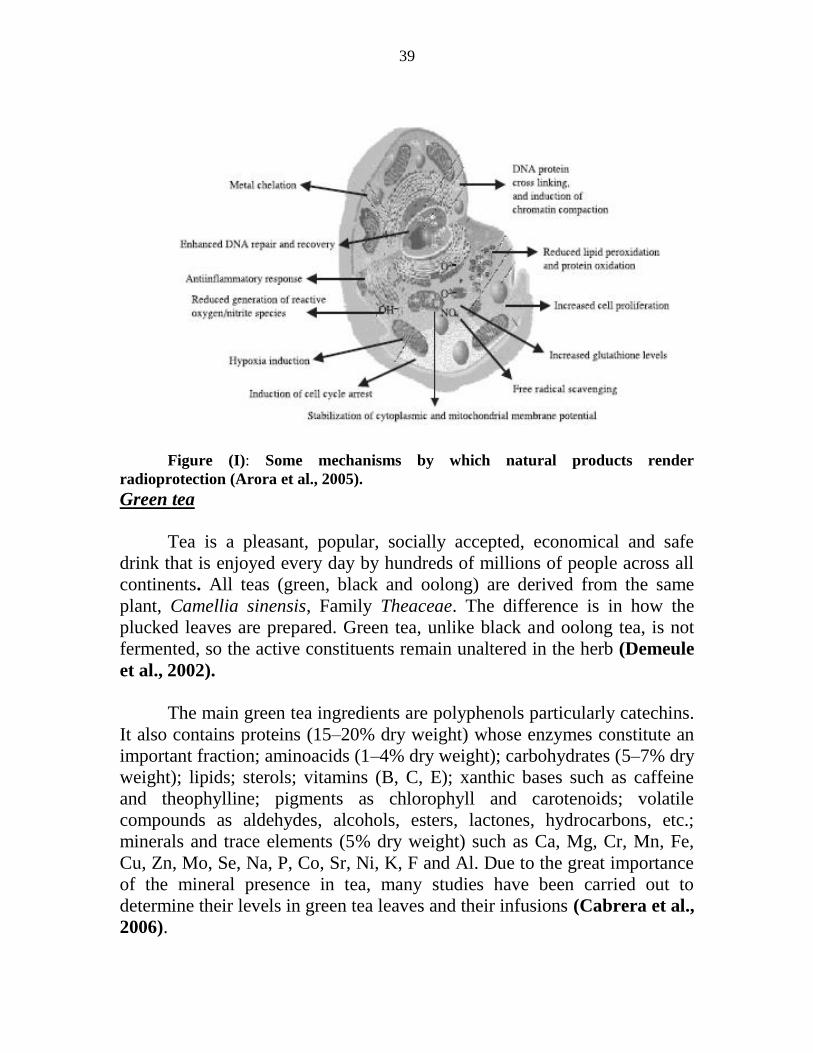
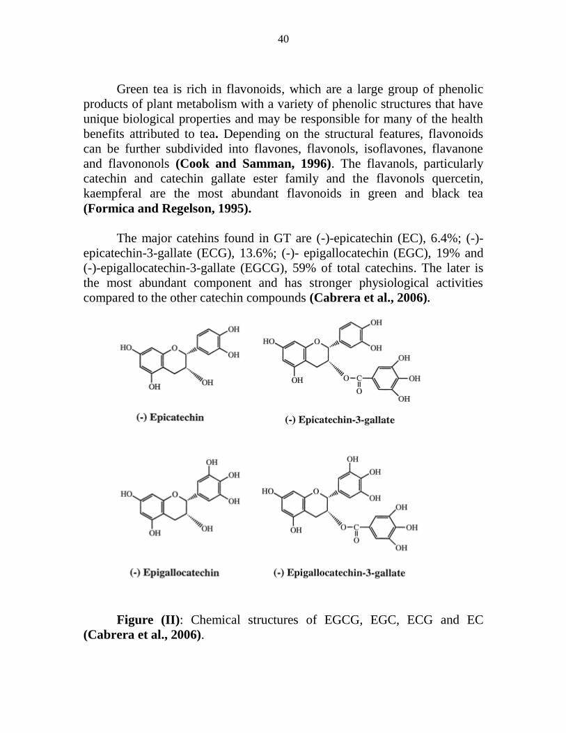
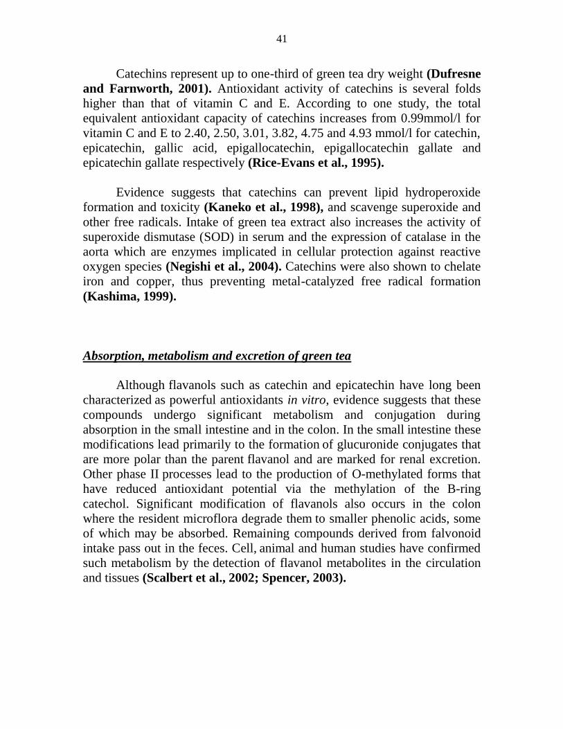
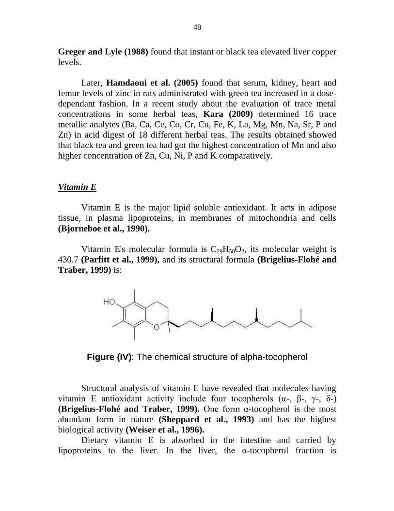

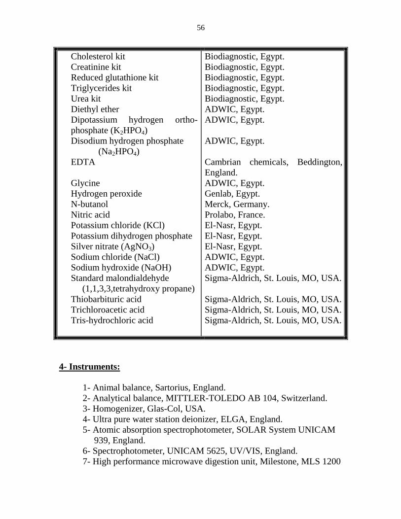
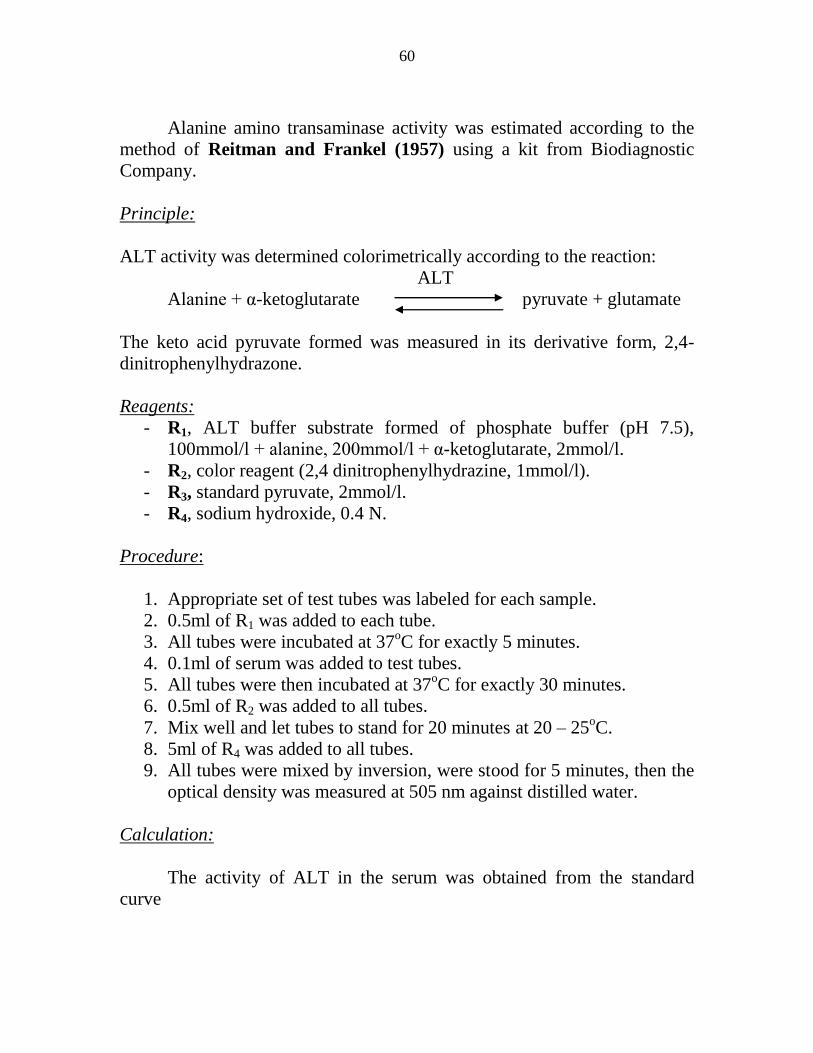
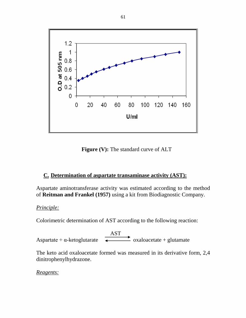
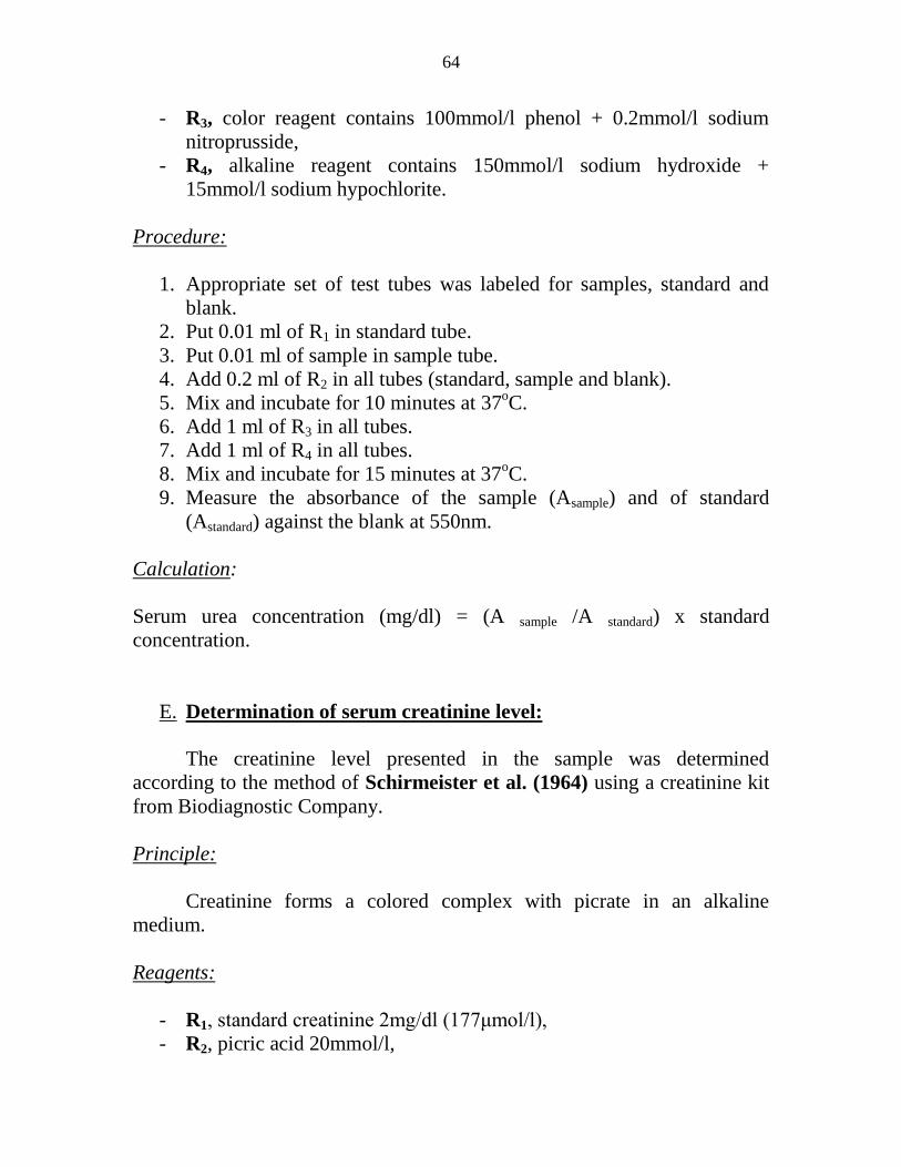
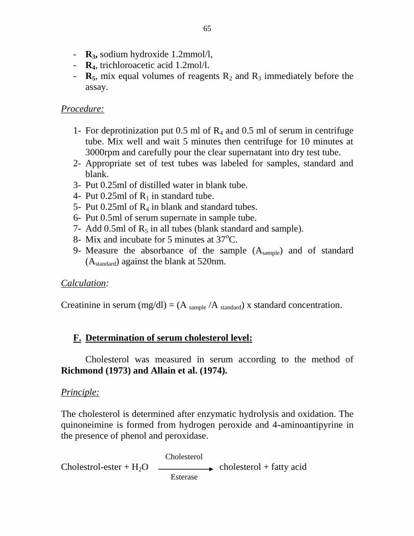
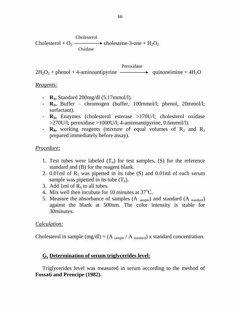
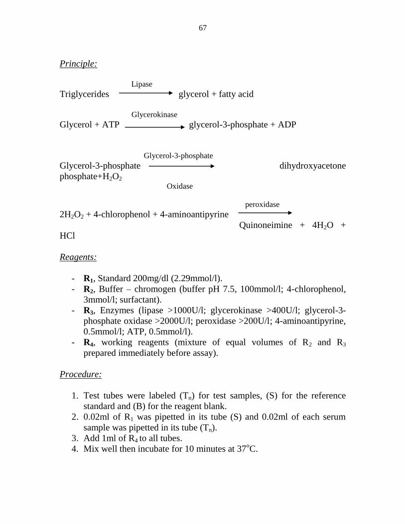
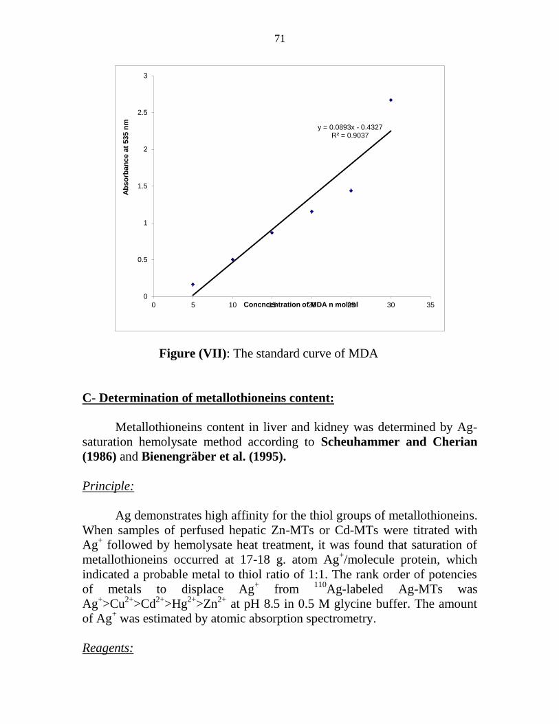
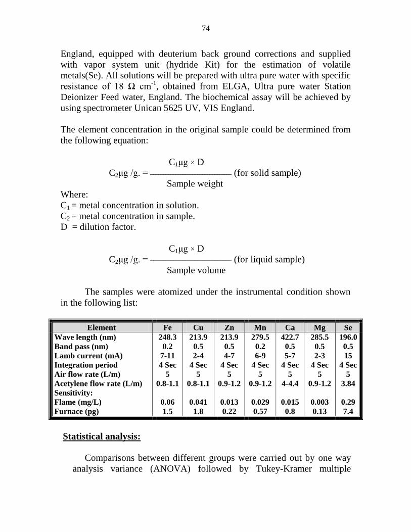

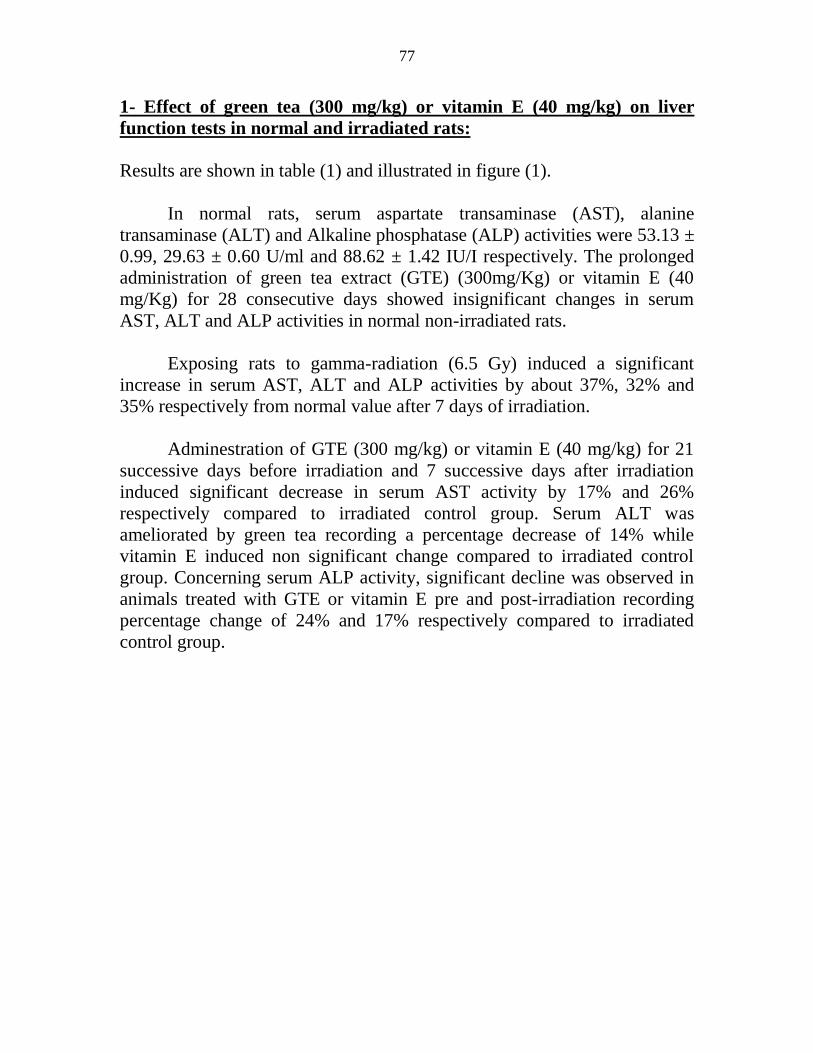
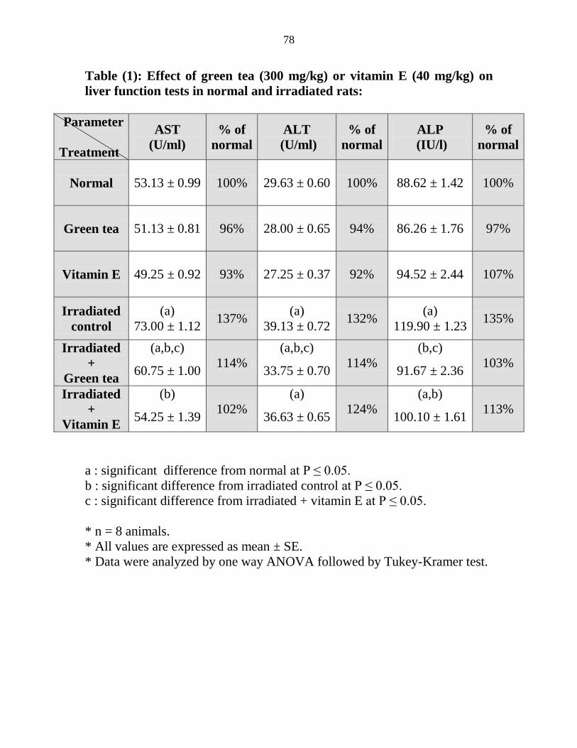
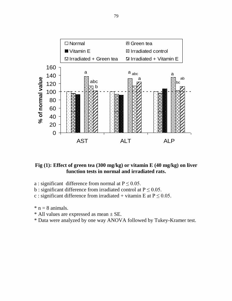
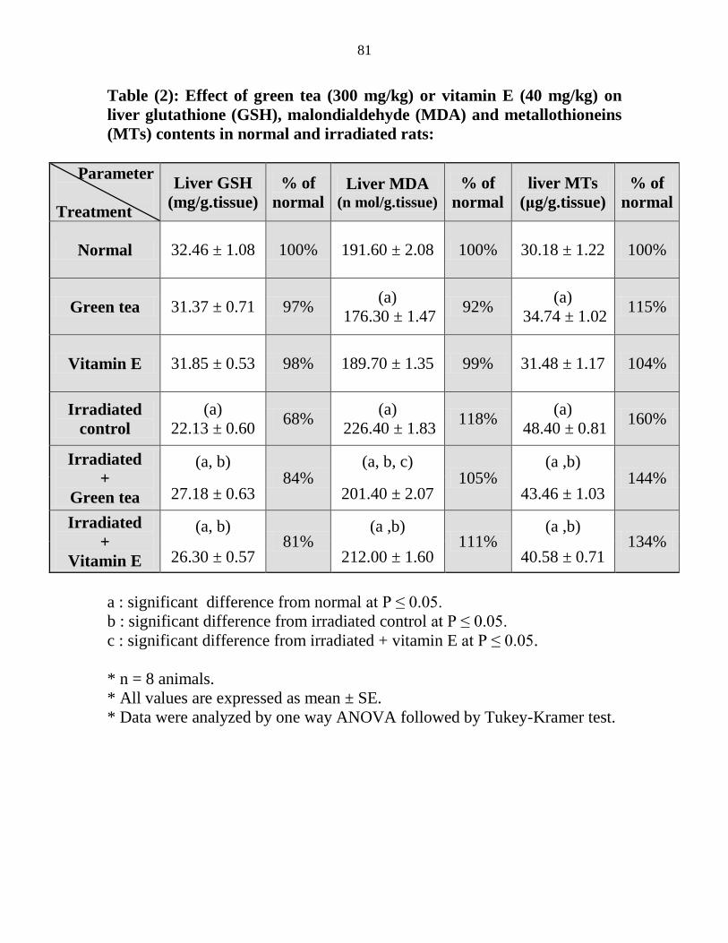
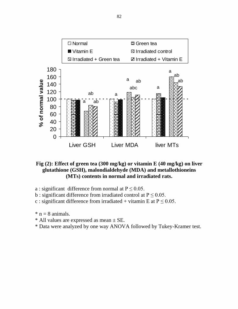
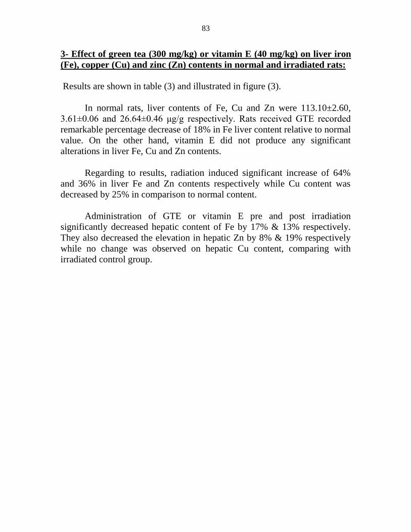
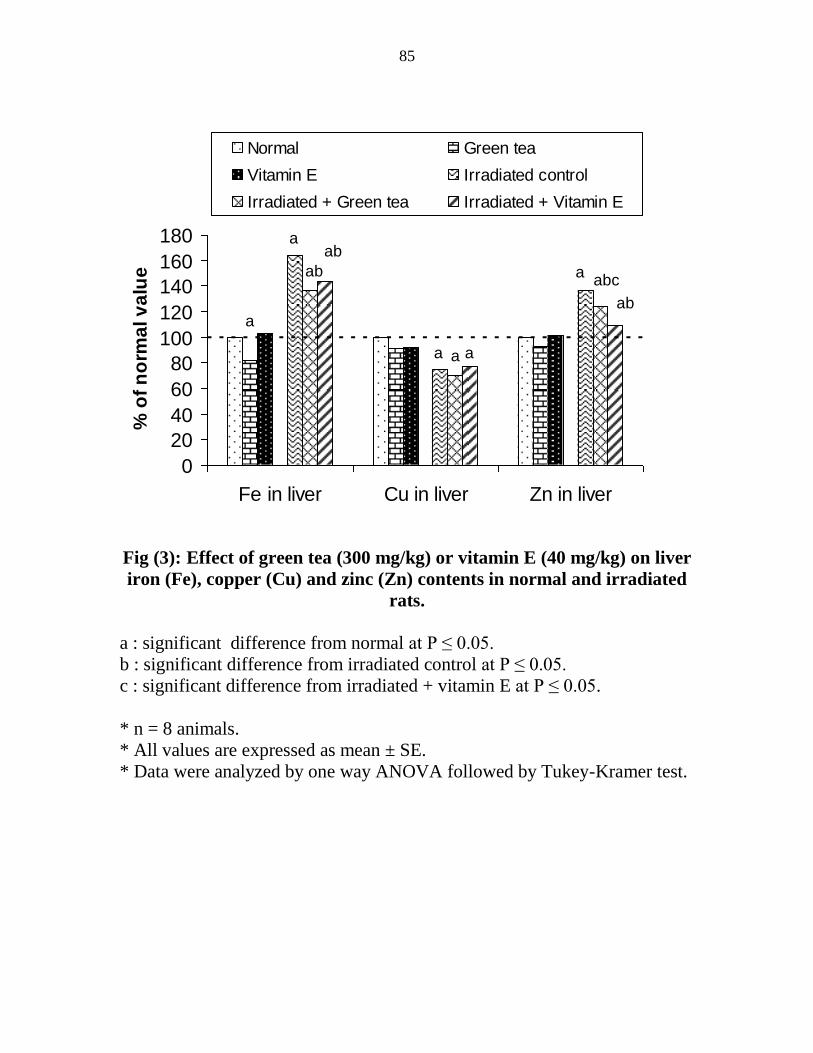
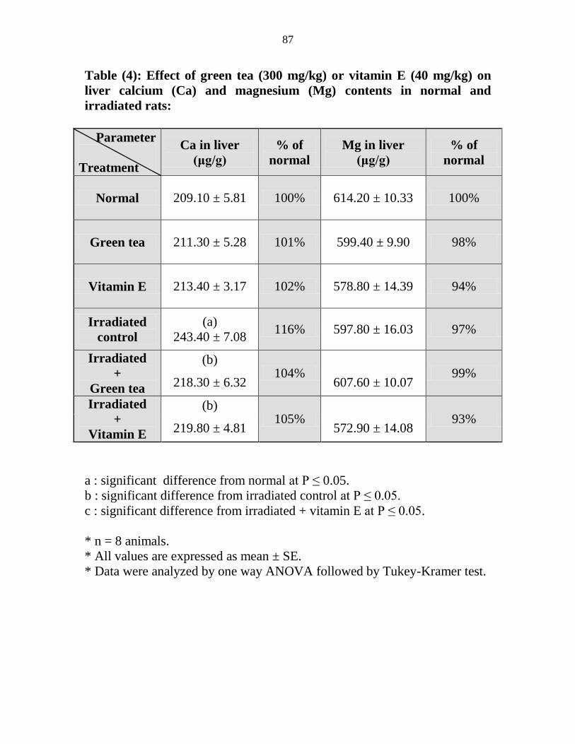
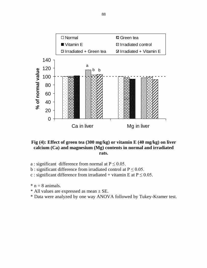
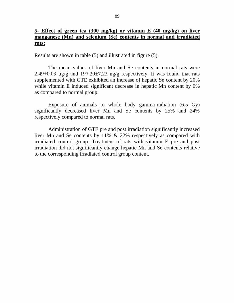
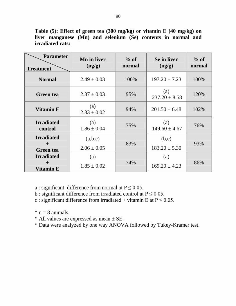
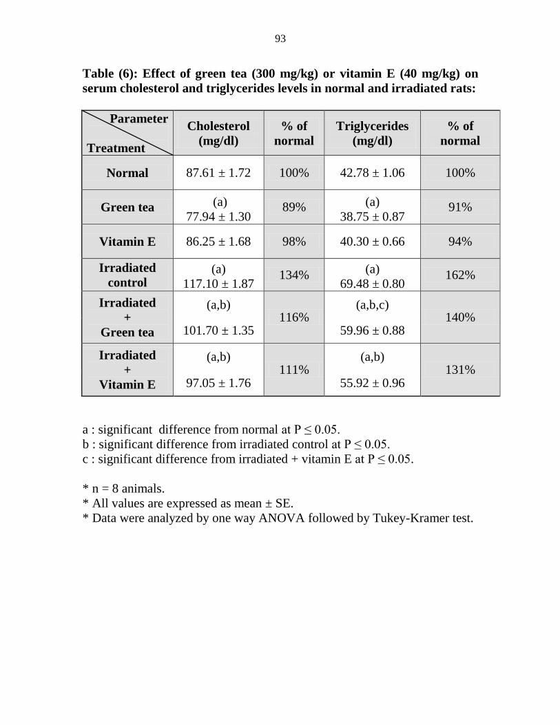
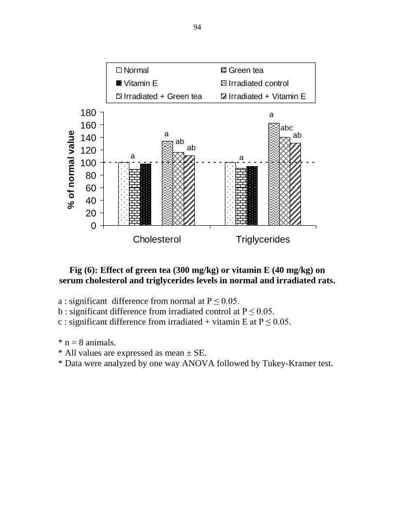
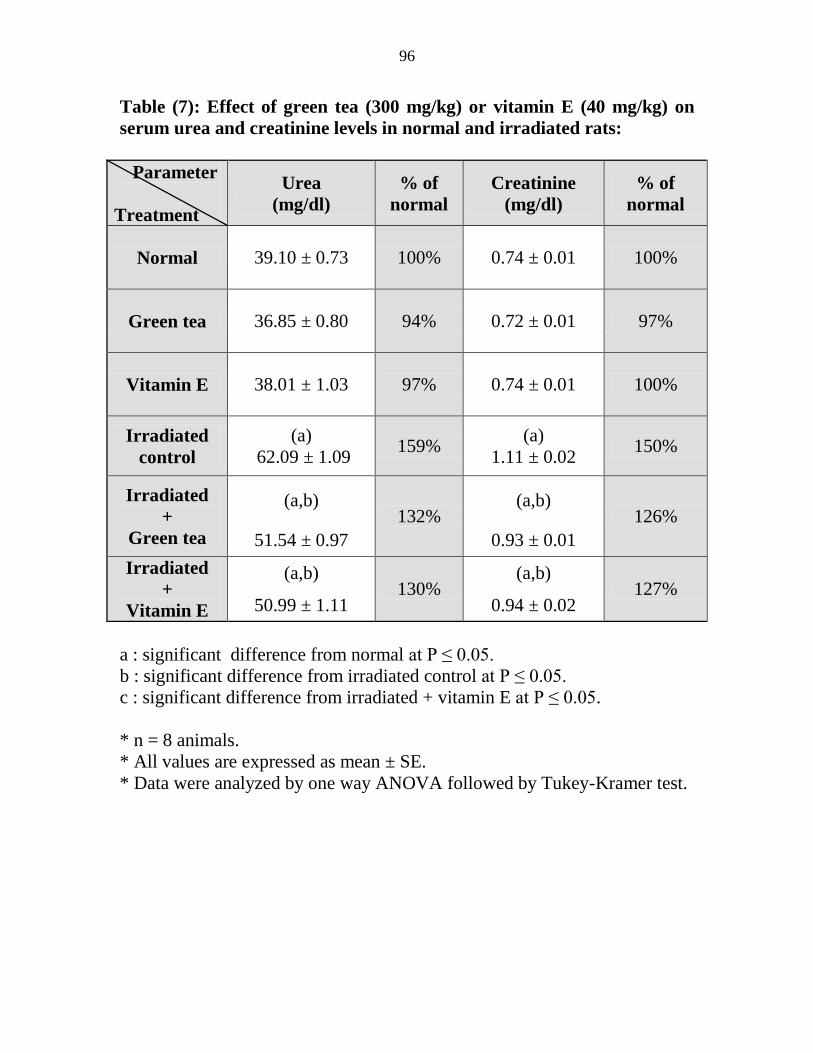
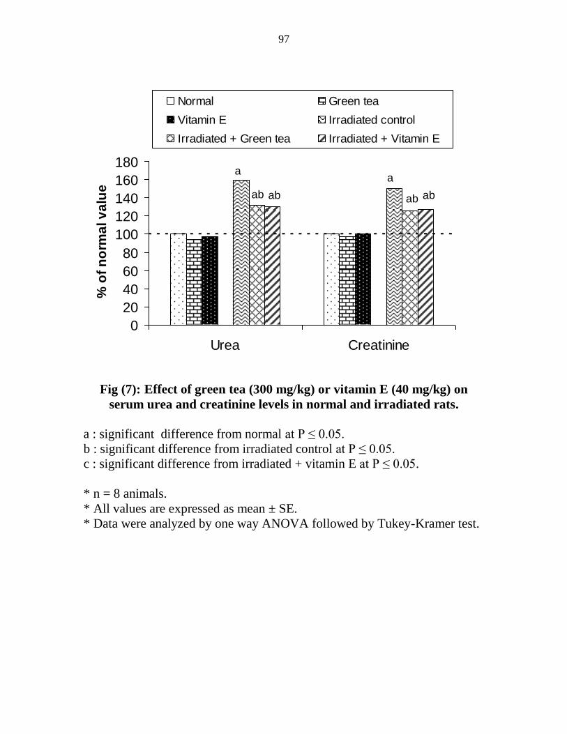
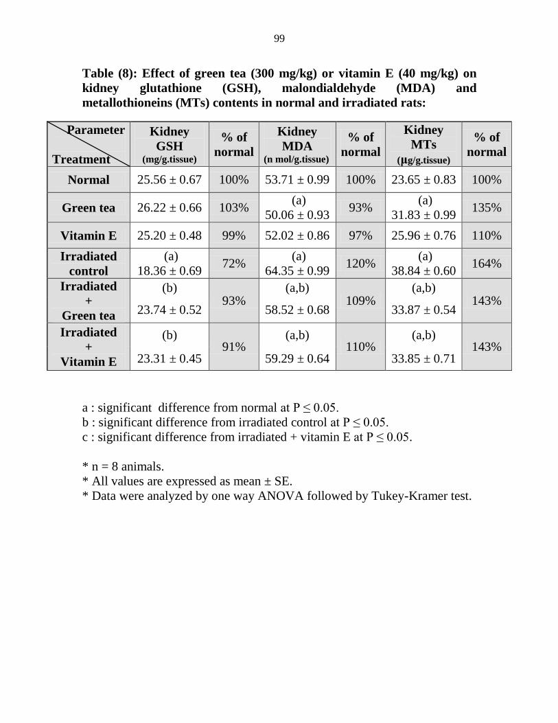
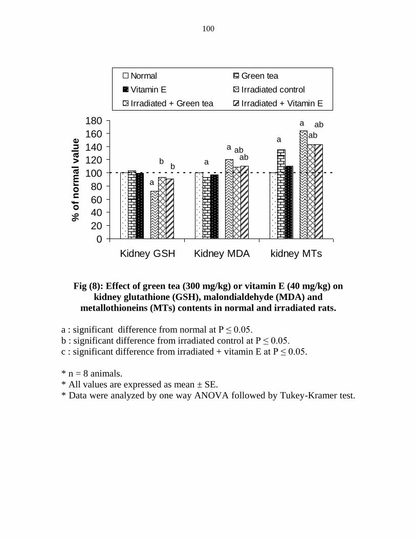
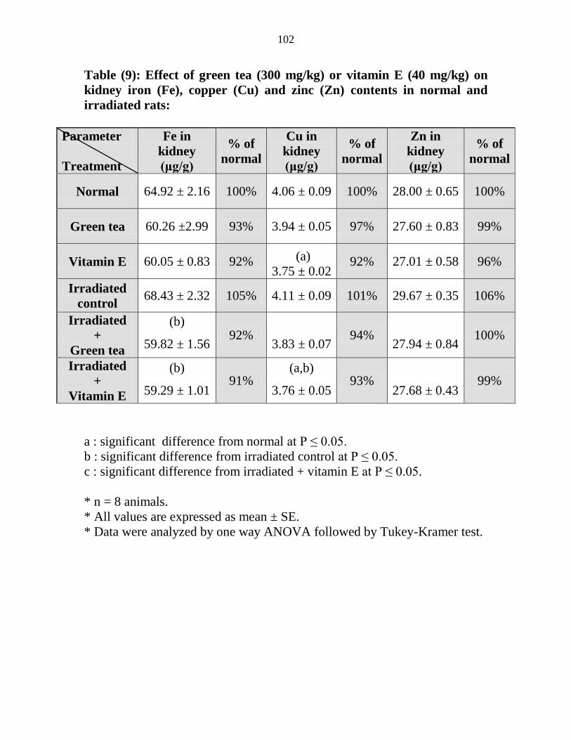
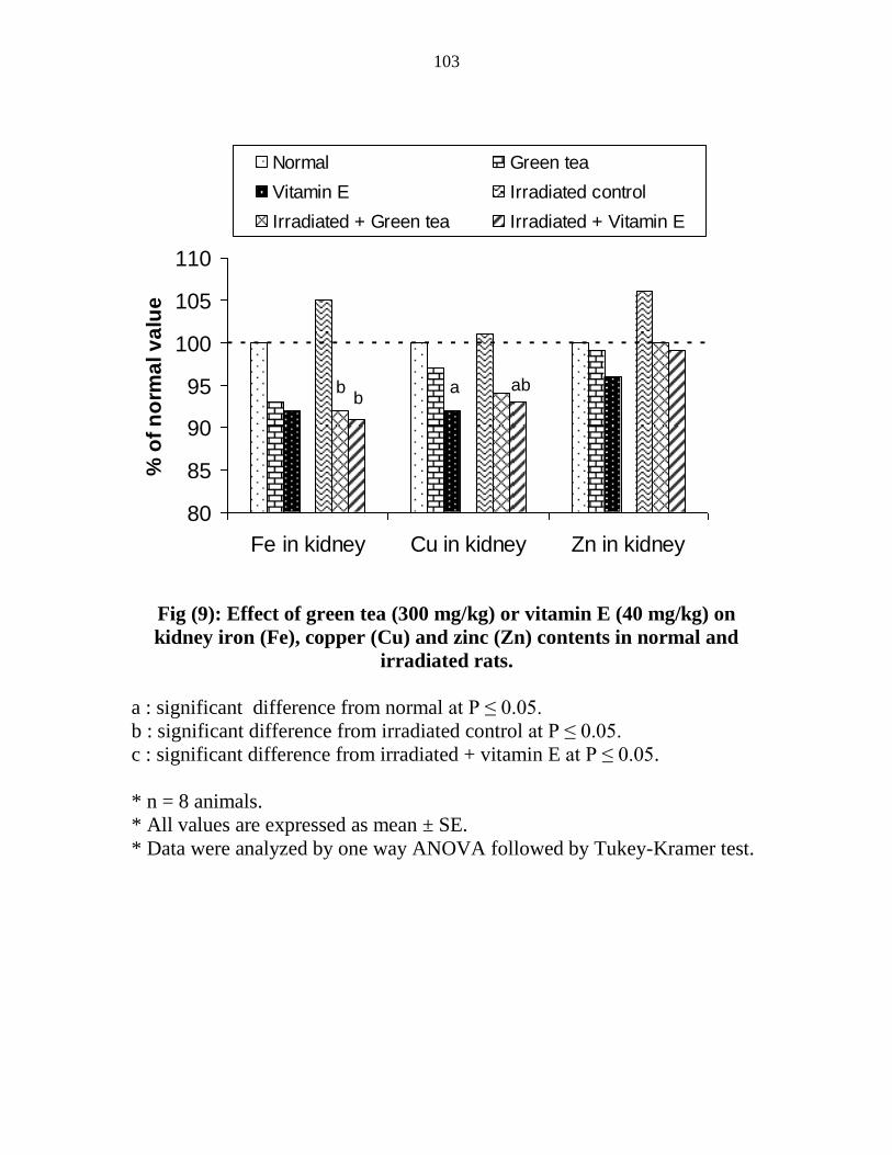
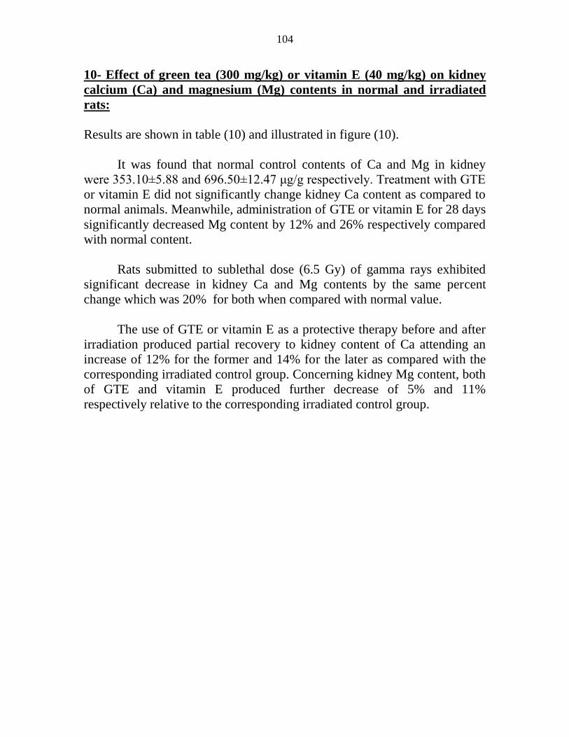
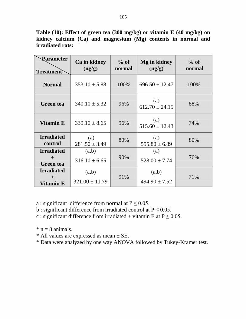
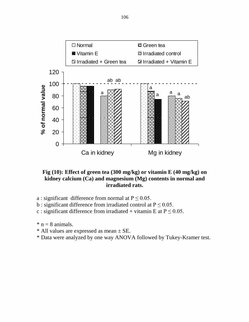
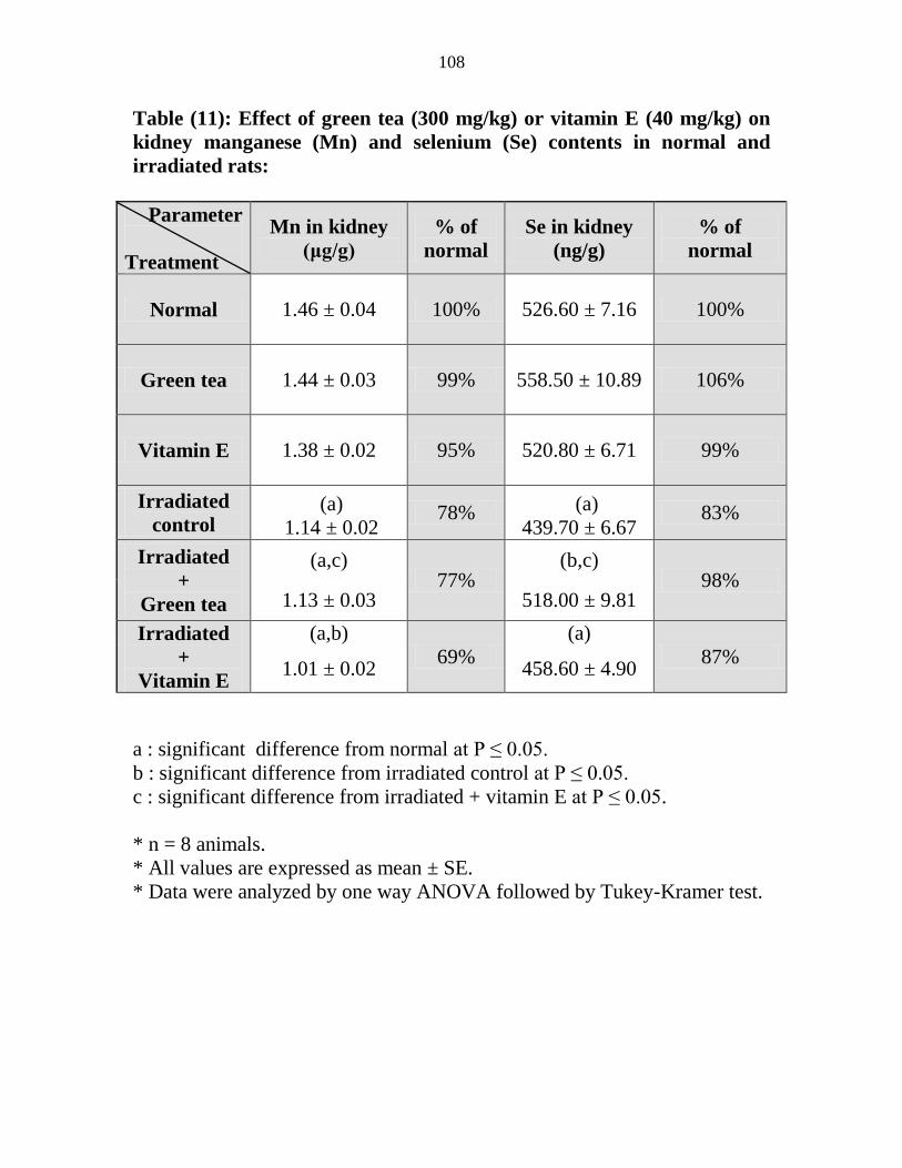
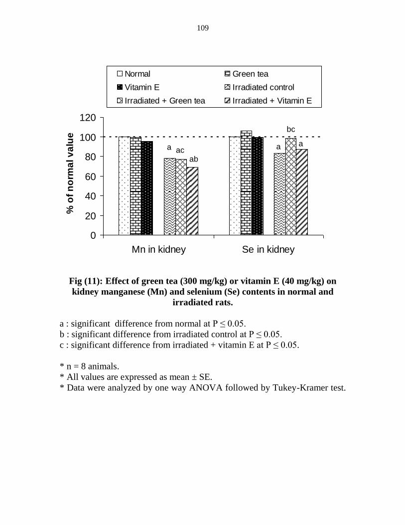
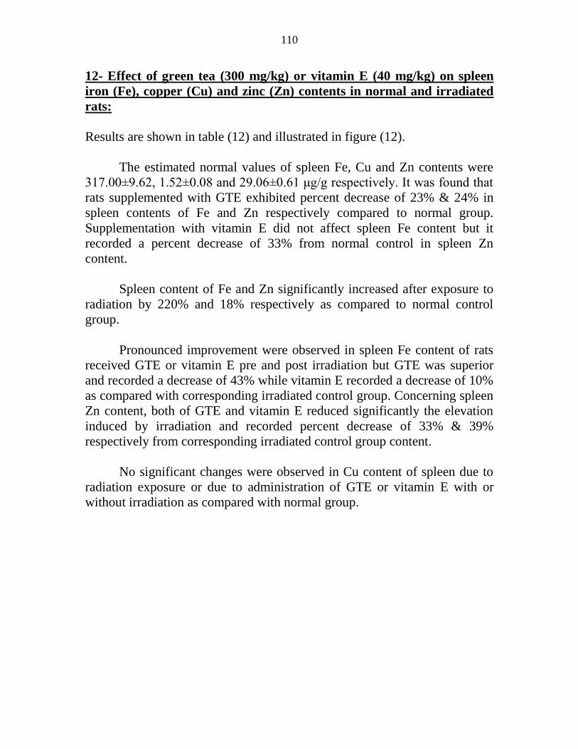
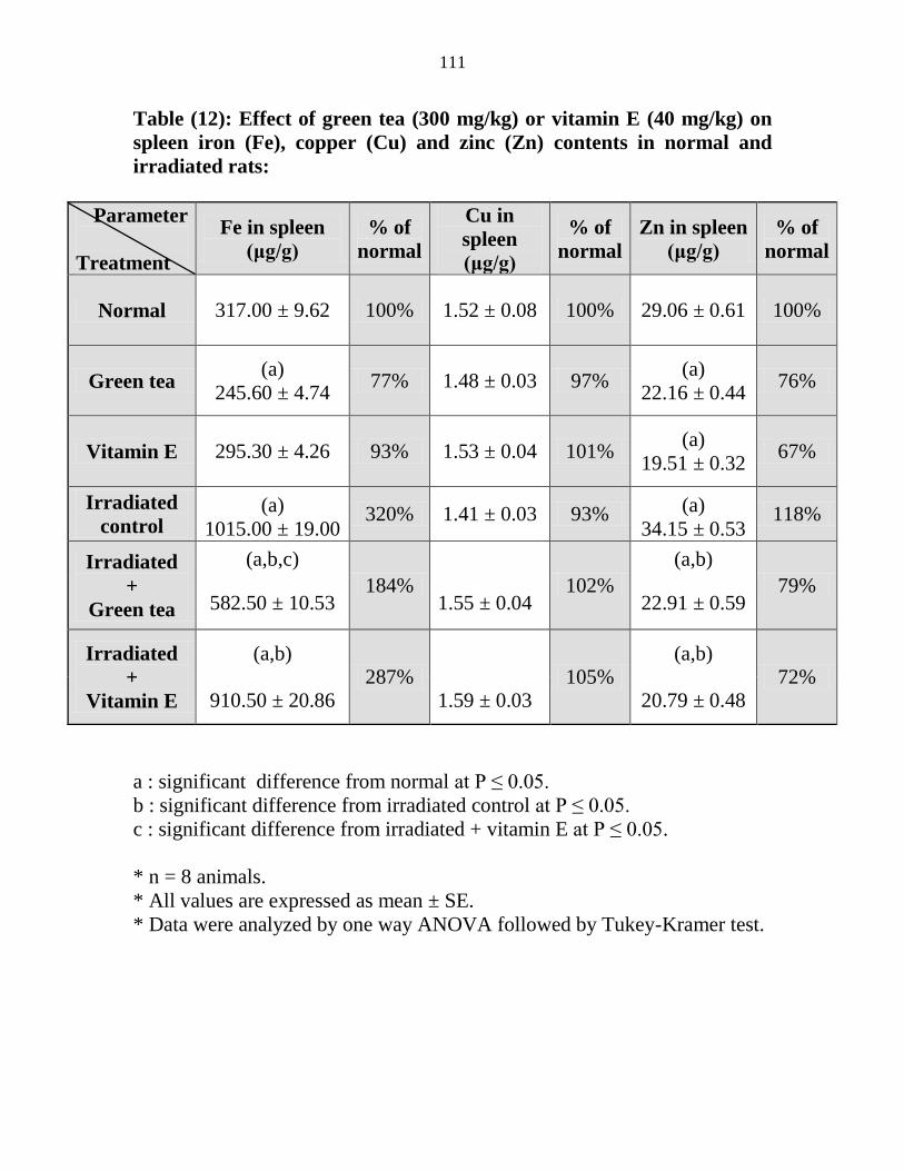
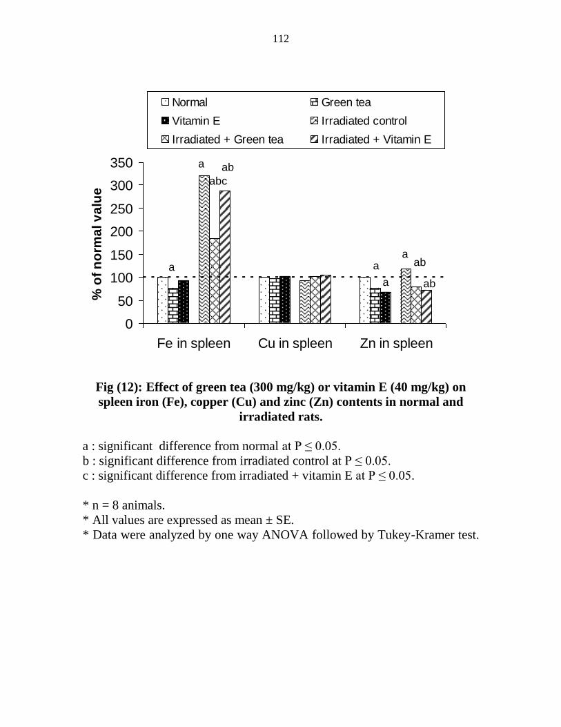
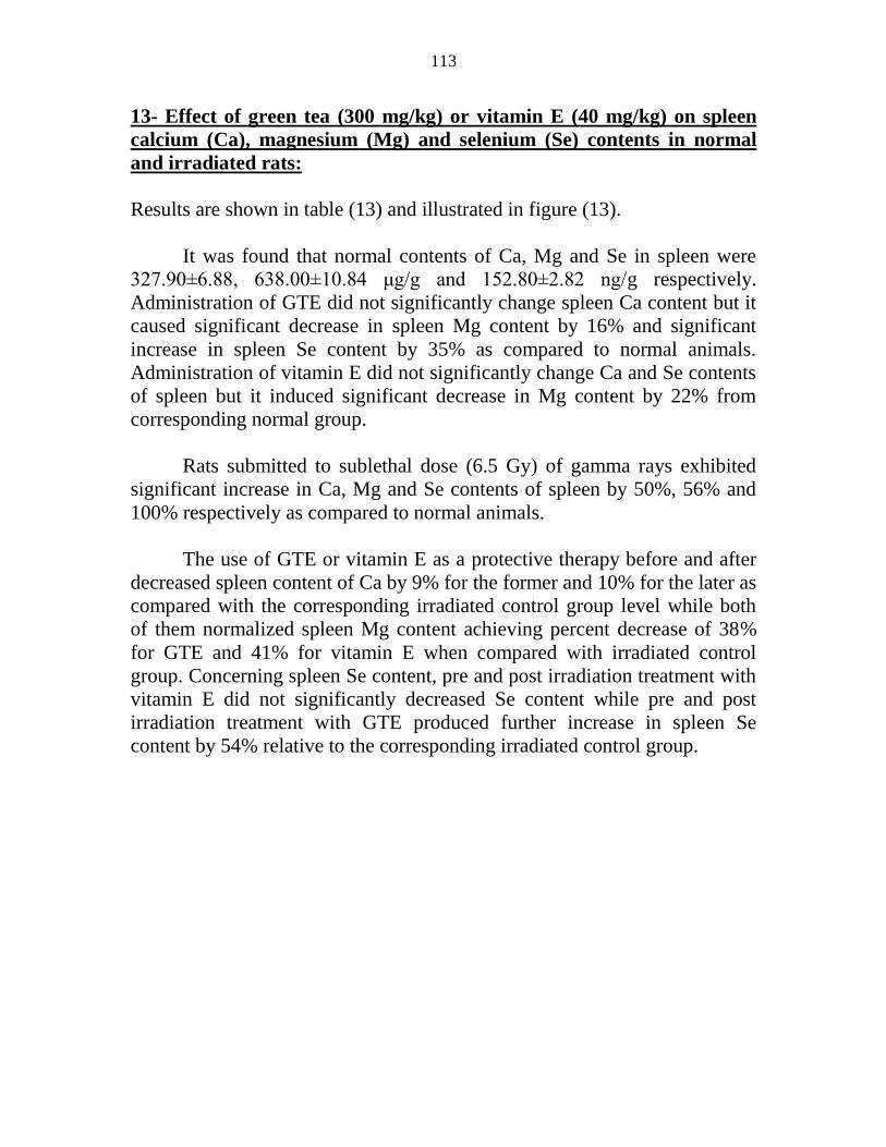

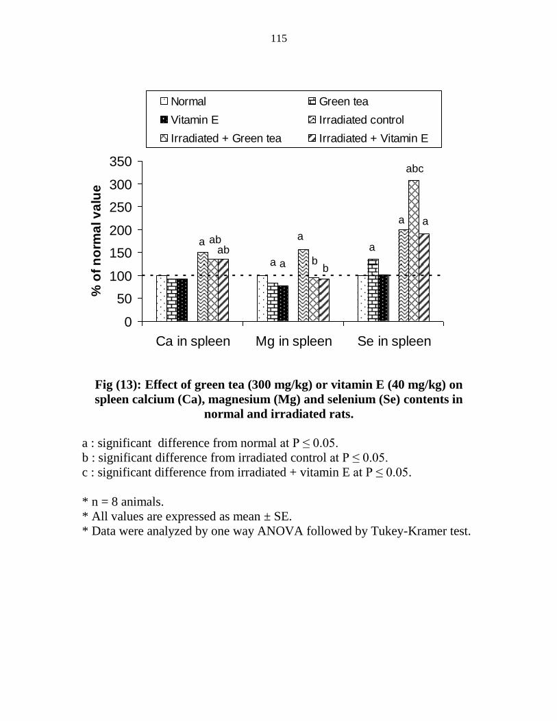

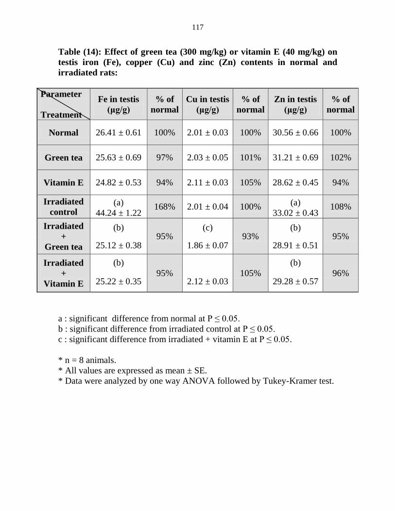
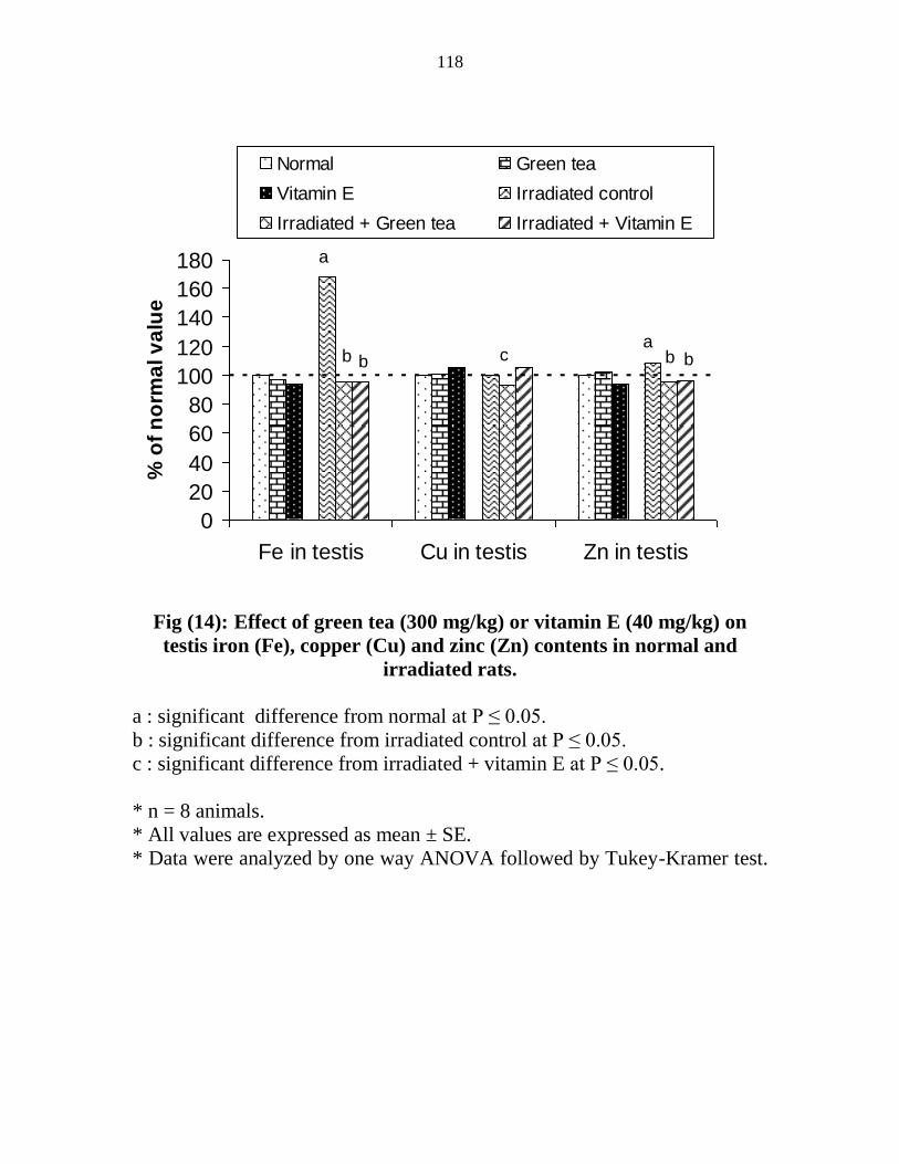
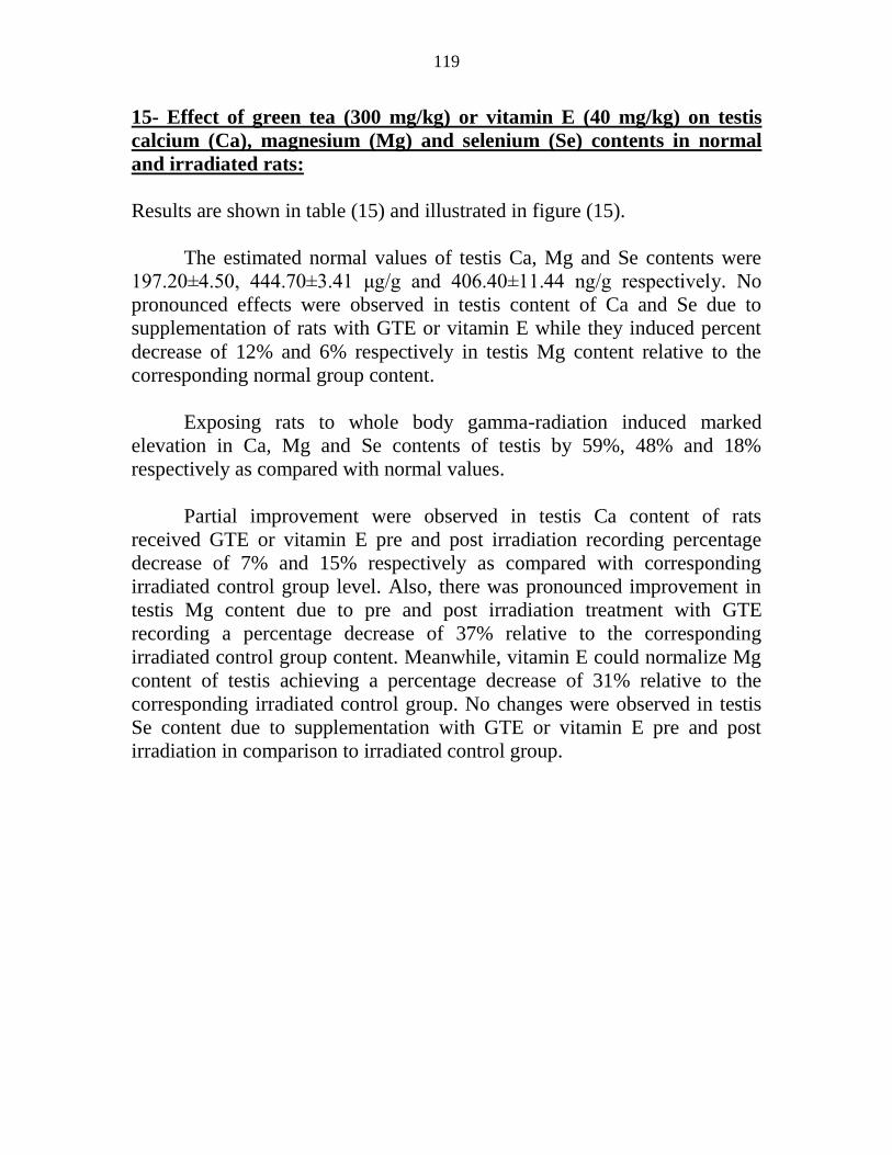

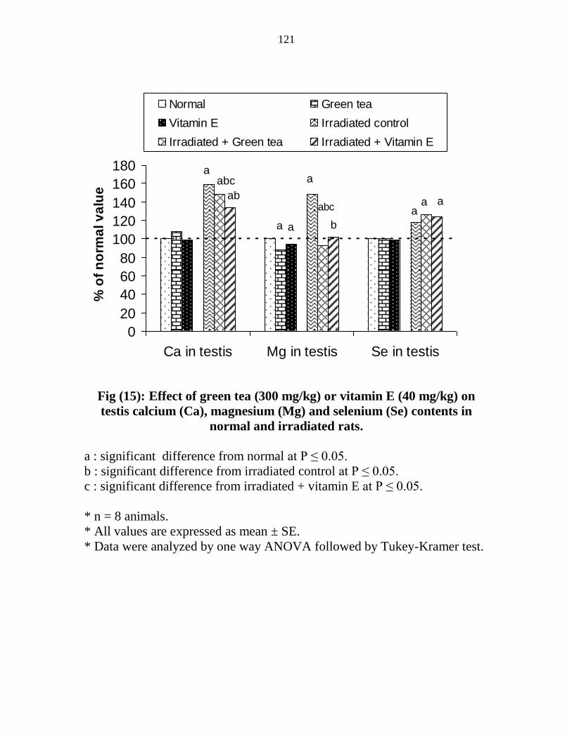
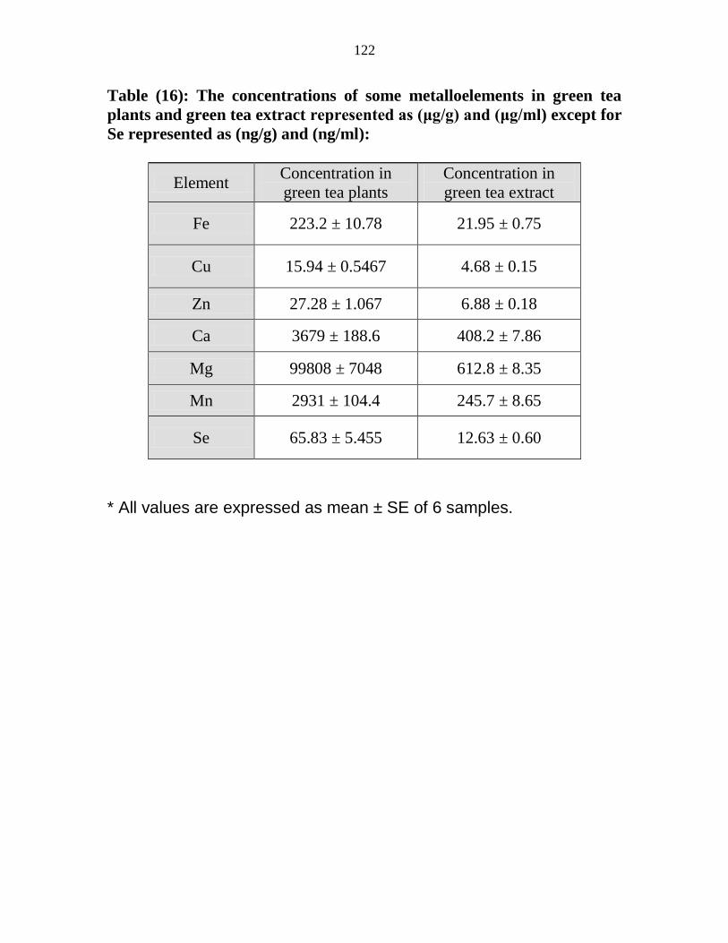


Recommended