Embed Size (px)
Citation preview
975
Personal non-commercial use only. EJH copyright © 2020. All rights served DOI: 10.21608/ejh.2020.25027.1258
Original Article
The Possible Protective Role of Pumpkin Seed Oil in Ameliorating Tongue Mucosal Damage Induced by Orlistat in Adult Male Albino Rats: A Light and Scanning Electron Microscopic Study
Amira Adly Kassab, Khalid Ahmed Ahmed Moustafa and Amal A.A. Abd-El-Hafez
Department of Histology and Cell Biology, Faculty of Medicine, Tanta University, Egypt
ABSTRACTBackground: Orlistat is an effective anti-obesity drug by reducing fat absorption. The oral cavity and its associated structures are target organs for many abnormalities that develop from orlistat. Pumpkin seed oil (PSO) is a valuable nutritional and health protective agent with prominent pharmacological properties.Aim: Evaluation of the possible protective role of pumpkin seed oil in ameliorating tongue mucosal damage induced by orlistat in adult male albino rats.Materials and Methods: Fifty adult male albino rats were used as a control group, an orlistat group and an orlistat-pumpkin group. Both orlistat (32mg/kg) and PSO (1.5ml/kg) were given orally once daily for four weeks. Specimens of the tongue were processed for light and scanning electron microscopic studies. Immunohistochemical study was performed using anti-proliferating cell nuclear antigen (PCNA) antibodies. Results: Specimens of the orlistat group showed an obvious distortion in the filiform and the fungiform papillae and in the covering epithelium of the tongue mucosa. There was a focal loss of the papillae and the epithelial ridges. The epithelial cells showed vacuolated cytoplasm and nuclear alteration. The lamina propria contained congested blood vessels and severe inflammatory cellular infiltration. There was a statistically significant decrease in the epithelial thickness, papillae height, papillae width and in the PCNA-immunoreaction of the epithelium. Scanning electron microscopy showed disfigurement and a focal atrophy of the filiform and the fungiform papillae. In contrast, minimal changes appeared in orlistat-pumpkin group that received PSO before orlistat. Conclusion: Orlistat induced significant structural alterations in the tongue mucosa of albino rats. PSO attenuated these effects and preserved the structure of the tongue mucosa.
Received: 02 March 2020, Accepted: 12 March 2020
Key Words: Orlistat, PCNA, pumpkin, scanning electron microscopy, tongue mucosa. Corresponding Author: Amira A. Kassab, MD, Department of Histology and Cell Biology, Faculty of Medicine, Tanta University, 31527 Tanta, Egypt, Tel.: +20 10 16697635, E-mail: [email protected]: 1110-0559, Vol. 43, No.4
INTRODUCTION
Orlistat (tetrahydrolipstin) is a weight-loss marketed drug approved by the Food and Drug Administration (FDA). It is a saturated derivative of lipstatin, a natural product isolated from Streptomyces toxytricini. It has been proved as a highly selective inhibitor of gastric and pancreatic lipases enzymes whose main function is digesting dietary fat. In the digestive tract, orlistat can bind to the lipase enzyme preventing hydrolysis of triglycerides into free fatty acids, which are absorbable for the cells and the undigested triglycerides are then excreted in feces. Thus the inhibition of lipases by orlistat will lead to reduced caloric intake. At present, orlistat is used for clinical treatment of obesity-related type 2 diabetes and cardiovascular diseases. Orlistat may have other clinical applications as in the treatment of chylous ascites which mostly is associated with cirrhosis[1,2].
However, although orlistat's effect is highly selective, some serious side effects had been reported in many studies. The most common gastrointestinal adverse effects of orlistat are diarrhea, flatulence, abdominal pain, bloating
and dyspepsia. It is metabolized in the gastrointestinal tract with direct damaging effect on the intestinal villi. In addition, malabsorption of fat soluble vitamins (ADEK)leading to their deficiency. Orlistat has been shown to inhibit carboxylesterase-2, a major detoxification enzyme, predisposing to severe liver, pancreatic and kidney damage[2,3,4]. The oral cavity is also a target organ for a number of various abnormalities that develop from side effects of this anti-obesity drug[5].
Nutrient deficiencies as a side effect for anti-obesity drugs can affect the function of the oral cavity that includes taste, mastication, salivation and swallowing food. An appropriate diet containing vitamins and trace elements plays an essential role in maintaining normal health of the oral structures. Their deficiencies can cause oral diseases such as oral mucosal diseases and periodontal diseases. The most commonly affected oral structures are soft tissues like gingiva, tongue and lining mucosa. Deficiency of the antioxidant vitamins (E&A) can lead to increased oxidative stress and subsequently increase in oral mucosal lesions such as oral leukoplakias and cancers[6].

976
PUMPKIN ROLE IN ORLISTAT INDUCED-TONGUE MUCOSAL DAMAGE
Recently, a special interest has been directed to the use of the antioxidants in the treatment due to their important role in maintaining the oral and general health. The pumpkin is a valuable vegetable belonging to the cucurbitaceae family. Pumpkin contains biologically active compounds that include polysaccharides, para-aminobenzoic acid, sterol, fixed oils, peptides and proteins. Consumption of pumpkin helps to prevent skin diseases, eye disorders and cancer. It improves the immune function and reduces cell damage in the body. Recently, it has obtained considerable attention due to the nutritional and health protective values of the seeds. The seeds have wide pharmacological activities such as anti-diabetic, antibacterial, antifungal, anti-inflammation and antioxidant effects. It can complement staples in food by supplying vitamins and indispensable minerals that may not be present in staple diets. Different researchers reported its medicinal uses such as antidiabetic, antihypertension, antitumor and immunomodulation[7,8].
The above-mentioned data make us much interested to carry out this research that aimed to study the effect of orlistat on tongue mucosa of adult male albino rats and to evaluate the potential protective role of pumpkin seed oil using different histological and immunohistochemical methods.
MATERIALS AND METHODS
Ethical ApprovalThis study was carried out at the Histology Department,
Faculty of Medicine of Tanta University, Egypt. The animal work was done according to the guidelines for use of animals in research approved by the Local Ethics Committee of the Faculty of Medicine, Tanta University, Tanta, Egypt (Approval code: 32682/11/18).
Chemicals1. Orlistat or tetrahydrolipstin is the active ingredient
which is marketed under the trade name Orlistat. It was a solid and white substance that was available in the form of hard gelatin capsules (each contains 120 milligrams of orlistat). It was manufactured by SIGMA Pharmaceutical Industries for October Pharma S.A.E., 6 October City, Egypt. (M.O.H. Reg. No.: 254461/2008).
2. Pumpkin seed oil: a glass bottle contains 30 ml of pumpkin seed oil in liquid form which is ready to be administered to rats as oil. It was manufactured by El Captain Company for extracting natural oils, plants and cosmetics (Cap Pharm, Egypt).
Study DesignFifty adult male albino rats weighing 220-250 grams
were used in the study. They were kept on a standard 12-h light/12-h dark cycle in clean properly ventilated cages before the experiment and throughout the study period with access to a balanced laboratory diet and water ad libitum. The rats were divided into three groups:
1. Group I (control group): included 30 rats that were subdivided into 3 equal subgroups: the first (subgroup IA) received no treatment, the second (subgroup IB) received 1ml saline orally once daily for 4 weeks and the third (subgroup IC) received pumpkin seed oil (PSO) 1.5 ml/kg/day orally by a gastric tube once daily for 4 weeks[9].
2. Group II (orlistat group): included 10 rats that received orlistat at a dose of 32 mg/kg/day which was suspended in 1ml saline. The suspension was well mixed and was administered to rats orally by a gastric tube once daily for 4 weeks[4].
3. Group III (orlistat-pumpkin group): included 10 rats that received PSO at 1.5 ml/kg/day, one hour before orlistat dose of 32 mg/kg/day orally by the gastric tube once daily for 4 weeks.
Samples CollectionAt the end of the experiment, all rats were anesthetized
by an intraperitoneal injection of pentobarbital (50 mg/ kg body weight)[10]. Then, the whole tongue was removed from oral cavity of all animals and dissected from the midline into 2 halves. Specimens from the anterior two thirds of the right halves were processed for light microscopic examination while the left ones were processed for scanning electron microscopic examination.
For Light Microscope (L/M)
The collected specimens of the right halves of tongues were immediately fixed in 10% neutral-buffered formalin, washed, dehydrated, cleared and then embedded in paraffin. Subsequently, serial sections of 5μm thickness were stained with haematoxylin and eosin (H&E) and were examined by the light microscope (L/M)[11].
For Immunohistochemistry
Sections (5μm thickness) were dewaxed, rehydrated, and washed with phosphate buffered saline (PBS). The sections were incubated in a humid chamber with the primary anti- proliferating cell nuclear antigen (PCNA) antibody (Mouse monoclonal antibody, 1:200 dilution, Ab-1 (Clone PC10), CAT. # MS-106-R7, Lab Vision Corporation, USA) in PBS overnight at 4°C. Thereafter, it was washed in PBS buffer, and co-incubated with biotinylated secondary antibody (Dako North America, Inc., CA, USA) for one hour at room temperature. Streptavidin peroxidase was added for ten minutes and rinsed three times in PBS. The immunoreactivity was visualized using 3, 3’diaminobenzidine (DAB)-hydrogen peroxide as a chromogen. Finally, the sections were counterstained using Mayer's haematoxylin. The negative control sections were processed without primary antibodies addition[12]. Positive control was tonsil, lymph node, or small intestine. All immunostained slides were assessed in triplicates in order to confirm the accuracy of the obtained results. The epithelial cells with brown nuclear staining were considered as PCNA-immunopositive cells.

977
Kassab et. al
For Scanning Electron Microscope (SEM)Specimens from left halves of the tongues were
fixed (2.5% glutaraldehyde and postfixed in 1% osmium tetroxide), dehydrated in Alcohol, dried, mounted on stubs and coated with gold using a sputter coater. It converts electrically non-conductive samples into conductive ones enabling a tightly focused electron beam to be scanned across the sample surface by scanning electron microscope (JEOL JSM-636 OLA at an accelerating voltage of 15kv)at Electron Microscopic Unit, Faculty of Medicine, Tanta University, Egypt[13].
Morphometric StudyA Leica microscope (DM3000; Leica Microsystems,
Wetzlar, Germany) coupled to a CCD camera (DFC-290; Leica, Heerbrugg, Switzerland) was used to obtain the images. Immunohistochemical evaluation was performed using an image analysis computer system (Leica Q 500 MC program) at Central Research Lab., Tanta Faculty of Medicine, Tanta University. Area percentage (area %) of PCNA positive immunoreaction of the epithelium was measured in ten selected different fields in each DAB-stained slide (X400). Moreover, thickness of the epithelium of the dorsal and ventral mucosal surfaces of the tongue and the height and width of the lingual papillae (the filiform and fungiform) were measured in ten selected fields for each H&E-stained slide (X400). The papillae height was obtained by measuring the distance from tip of the papilla to its base. The papillae width was obtained by measuring the base diameter at the widest points extending from the external epithelial wall to the other external epithelial wall. The measurement sites of the height and width of the papillae were illustrated in (Figures i and ii) according to Takahashi et al., 2019[14].
Figs. i & ii: Measurement sites for height and width of the filiform (i) and fungiform papillae (ii).
Statistical AnalysisThe obtained data of the morphometric study was
statistically analyzed by one-way analysis of variance (ANOVA) followed by Tukey’s procedure for comparison
between the different study’s groups by using statistical package for social sciences statistical analysis software (version 11.5; SPSS Inc., Chicago, Illinois, USA). The mean values and their standard deviation values (Mean ± SD) for each group were obtained. Probability values (P values) < 0.05 were considered as significant[15].
RESULTS
All experimental rats survived until the final experimental period.
Light Microscopic Findings
Group I (control group)Examination of H&E stained sections obtained from all
control subgroups showed the same histological features of the mucosa of rat tongue. The mucosa covering the dorsal surface of the anterior two thirds (2/3) of the tongue showed characteristic long projections or lingual papillae. Each lingual papilla was composed of a lamina propria or connective tissue core and was covered by a keratinized stratified squamous epithelium resting on a basement membrane and had many epithelial ridges. The most numerous filiform papillae appeared conical in shape with characteristic tapering tips and regular distribution and orientation. The epithelium appeared with its four layers; basal cell layer, spinosum cell layer, granulosum cell layer and superficial corneum layer (Figures 1 and 2). The mushroom shaped fungiform papillae were less in number and scattered between the filiform ones having a few barrel shaped taste buds on its upper surface (Figure 3). The underlying lamina propria core was very thin and was continuous with that of the lingual muscles and contained a few collagen fibers as well as small blood vessels (Figures 1 and 3). The mucous membrane of the ventral surface of the tongue appeared without lingual papillae and had a thin keratin layer over the stratified squamous epithelial cover (Figure 4).
Group II (orlistat group)Examination of H&E stained sections of orlistat treated
rats revealed an obvious distortion in the lingual papillae of the dorsal surface and in the covering epithelium of the dorsal and ventral surfaces of the anterior 2/3 of the tongue. There was an apparent decrease in the height of the filiform papillae. Many areas showed filiform papillae having blunt ends while others showed ill-defined papillae. Some specimens showed disorganized epithelial cells with deeply stained pyknotic nuclei which were surrounded by the vacuolated cytoplasm (Figures 5 and 6). Moreover, an apparent reduction in the thickness of the epithelial cover and the keratin layer with completely lost papillae was noticed in some focal areas. The basement membrane of the epithelium appeared almost straight with loss of the epithelial ridges in some areas (Figure 7). The underlying lamina propria core contained congested blood vessels and severe inflammatory cellular infiltration (Figure 6). There was also distortion in the fungiform papillae that showed deeply stained nuclei and cytoplasmic vacuoles of their epithelial cell cover and in cells of their taste buds (Figure 8). The epithelial cover of the

978
PUMPKIN ROLE IN ORLISTAT INDUCED-TONGUE MUCOSAL DAMAGE
ventral surface showed an apparent decrease in its thickness and the keratin layer appeared in some regions discontinuous and/or separated (Figure 9). Moreover, a focal loss of the covering epithelium was also seen (Figure 10).
Group III (orlistat-pumpkin group)The examination revealed partially preserved histological
features of the mucosa on the dorsal and ventral surfaces of the anterior 2/3 of the rat tongue. Most of the lingual papillae exhibited nearly normal architecture with restoration of the normal epithelial cover and the keratin layer and nearly normal pattern of the epithelial ridges comparable to that of group I (control group). Nevertheless, a few disfigured papillae were observed in a few areas of the dorsal mucosal surface (Figures 11, 12 and 13).
Immunohistochemical FindingsPCNA antigen immunostaining: In PCNA-
immunostained sections of the control group (group I), many epithelial cells in the basal and parabasal layers of the tongue mucosa exhibited a positive nuclear immunoreaction for PCNA (Figure 14). In the orlistat group (group II), a few epithelial cells of the tongue mucosa expressed a positive nuclear immunoreaction for PCNA (Figure 15). Whereas in the orlistat-pumpkin group (group III), a positive nuclear immunoreaction for PCNA was detected in many epithelial cells of the tongue mucosa (Figure 16).
Scanning Electron Microscopic Findings
Group I (control group) Examination of the dorsal mucosal surface of the anterior
2/3 of the rat tongue of all control subgroups revealed numerous, elongated and regularly arranged conical shaped filiform lingual papillae having intact tapering tips that pointed into the same direction. A few fungiform lingual papillae were seen sporadically between the filiform ones. They appeared short, broad and dome shaped having flattened upper surfaces with taste pores in their centers (Figures 17 and 18).
Group II (orlistat group)Examination of this group revealed obvious changes in
the lingual papilla on the dorsal mucosal surface. Some areas showed disfigured or thin atrophied filiform papillae. Other papillae were widely separated and irregularly arranged in different directions. Short thin desquamated and/or damaged filiform papillae were also observed. Scattered fungiform papillae with rough upper surface and ill-defined taste pores could be seen on the dorsal mucosal surface of the tongue (Figures 19, 20, 21 and 22).
Group III (orlistat-pumpkin group)This group appeared similar to group I (control group)
and showed restoration of almost regular arrangement and shape of the filiform papillae on the dorsal mucosal surface with regular distribution of normal fungiform papillae between the filiform ones. Nevertheless, a few disfigured filiform papillae were observed in a few areas of the dorsal mucosal surface (Figures 23 and 24).Morphometric and Statistical Results
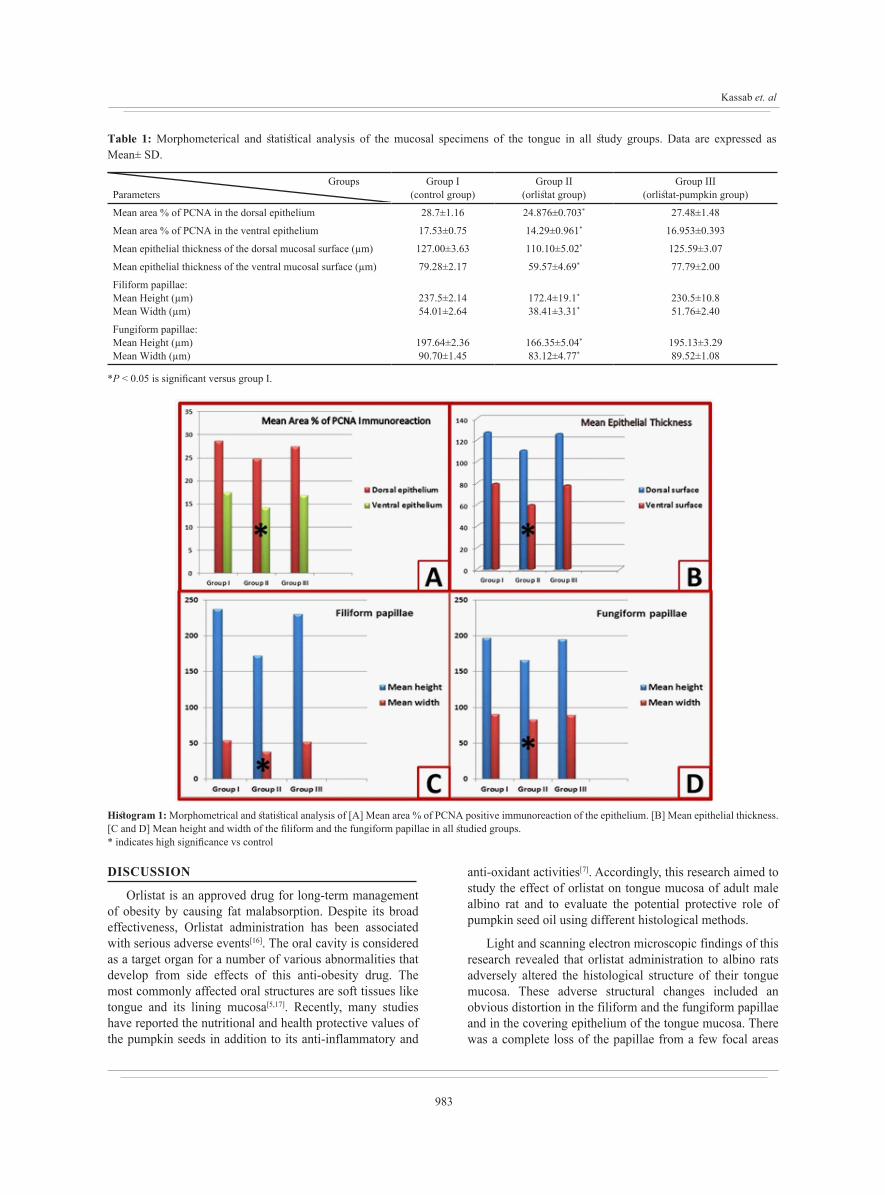
The mean area percentage of PCNA positive immunoreaction on the dorsal & ventral epithelium of the tongue showed a statistically significant decrease in the orlistat group (group II) compared to the control group. Moreover, the orlistat-pumpkin group (group III) showed a non-significant decrease compared to the control group (Table 1 and Histogram 1A).
The mean epithelial thickness of the dorsal & ventral mucosal surfaces of the tongue showed a significant decrease in the orlistat group (group II) compared to the control group. Moreover, orlistat-pumpkin group (group III) showed a non-significant decrease compared to the control (Table 1 and Histogram 1B).
The mean height and width of both filiform and fungiform papillae of the dorsal mucosal surface of the tongue showed a significant decrease in the orlistat group (group II) compared to the control group. Moreover, orlistat-pumpkin group (group III) showed a non-significant decrease compared to the control (Table 1 and Histogram 1C&1D).
Fig. 1: A photomicrograph of the dorsal mucosal surface of the anterior 2/3 of the rat’s tongue from group I [control group] showing regular distribution and orientation of the filiform papillae with characteristic tapering ends [arrow]. Each papilla shows a connective tissue core [C] with small blood vessels [wavy arrow] and is covered by a keratinized stratified squamous epithelium [E] resting on a basement membrane with many epithelial ridges [arrow head]. [H&E X 200].
979
Kassab et. al
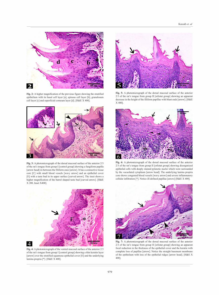
Fig. 2: A higher magnification of the previous figure showing the stratified epithelium with its basal cell layer [a], spinous cell layer [b], granulosum cell layer [c] and superficial corneum layer [d]. [H&E X 400].
Fig. 3: A photomicrograph of the dorsal mucosal surface of the anterior 2/3 of the rat’s tongue from group I [control group] showing a fungiform papilla [arrow head] in-between the filiform ones [arrow]. It has a connective tissue core [C] with small blood vessels [wavy arrow] and an epithelial cover [E] with a taste bud in its upper surface [curved arrow]. The inset shows a higher magnification of the barrel shaped taste bud [curved arrow]. [H&E X 200, Inset X400].
Fig. 4: A photomicrograph of the ventral mucosal surface of the anterior 2/3 of the rat’s tongue from group I [control group] showing a thin keratin layer [arrow] over the stratified squamous epithelial cover [E] and the underlying lamina propria [*]. [H&E X 400].
Fig. 5: A photomicrograph of the dorsal mucosal surface of the anterior 2/3 of the rat’s tongue from group II [orlistat group] showing an apparent decrease in the height of the filiform papillae with blunt ends [arrow]. [H&E X 400].
Fig. 6: A photomicrograph of the dorsal mucosal surface of the anterior 2/3 of the rat’s tongue from group II [orlistat group] showing disorganized epithelial cells with deeply stained pyknotic nuclei which were surrounded by the vacuolated cytoplasm [arrow head]. The underlying lamina propria core shows congested blood vessels [wavy arrow] and severe inflammatory cellular infiltration [*]. Notice ill-defined papillae [arrow] [H&E X 400].
Fig. 7: A photomicrograph of the dorsal mucosal surface of the anterior 2/3 of the rat’s tongue from group II [orlistat group] showing an apparent focal reduction in the thickness of the epithelial cover and the keratin with complete loss of papillae [arrow]. Notice the straight basement membrane of the epithelium with loss of the epithelial ridges [arrow head]. [H&E X 400].
980
PUMPKIN ROLE IN ORLISTAT INDUCED-TONGUE MUCOSAL DAMAGE
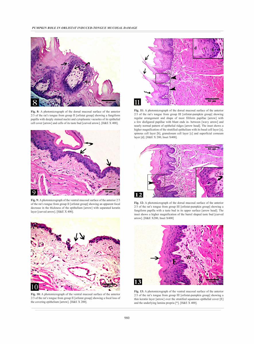
Fig. 8: A photomicrograph of the dorsal mucosal surface of the anterior 2/3 of the rat’s tongue from group II [orlistat group] showing a fungiform papilla with deeply stained nuclei and cytoplasmic vacuoles of its epithelial cell cover [arrow] and cells of its taste bud [curved arrow]. [H&E X 400].
Fig. 9: A photomicrograph of the ventral mucosal surface of the anterior 2/3 of the rat’s tongue from group II [orlistat group] showing an apparent focal decrease in the thickness of the epithelium [arrow] with separated keratin layer [curved arrow]. [H&E X 400].
Fig. 10: A photomicrograph of the ventral mucosal surface of the anterior 2/3 of the rat’s tongue from group II [orlistat group] showing a focal loss of the covering epithelium [arrow]. [H&E X 200].
Fig. 11: A photomicrograph of the dorsal mucosal surface of the anterior 2/3 of the rat’s tongue from group III [orlistat-pumpkin group] showing regular arrangement and shape of most filiform papillae [arrow] with a few disfigured papillae with blunt ends in- between [wavy arrow] and nearly normal pattern of epithelial ridges [arrow head]. The inset shows a higher magnification of the stratified epithelium with its basal cell layer [a], spinous cell layer [b], granulosum cell layer [c] and superficial corneum layer [d]. [H&E X 200, Inset X400].
Fig. 12: A photomicrograph of the dorsal mucosal surface of the anterior 2/3 of the rat’s tongue from group III [orlistat-pumpkin group] showing a fungiform papilla with a taste bud in its upper surface [arrow head]. The inset shows a higher magnification of the barrel shaped taste bud [curved arrow]. [H&E X200, Inset X400]
Fig. 13: A photomicrograph of the ventral mucosal surface of the anterior 2/3 of the rat’s tongue from group III [orlistat-pumpkin group] showing a thin keratin layer [arrow] over the stratified squamous epithelial cover [E] and the underlying lamina propria [*]. [H&E X 400].
981
Kassab et. al
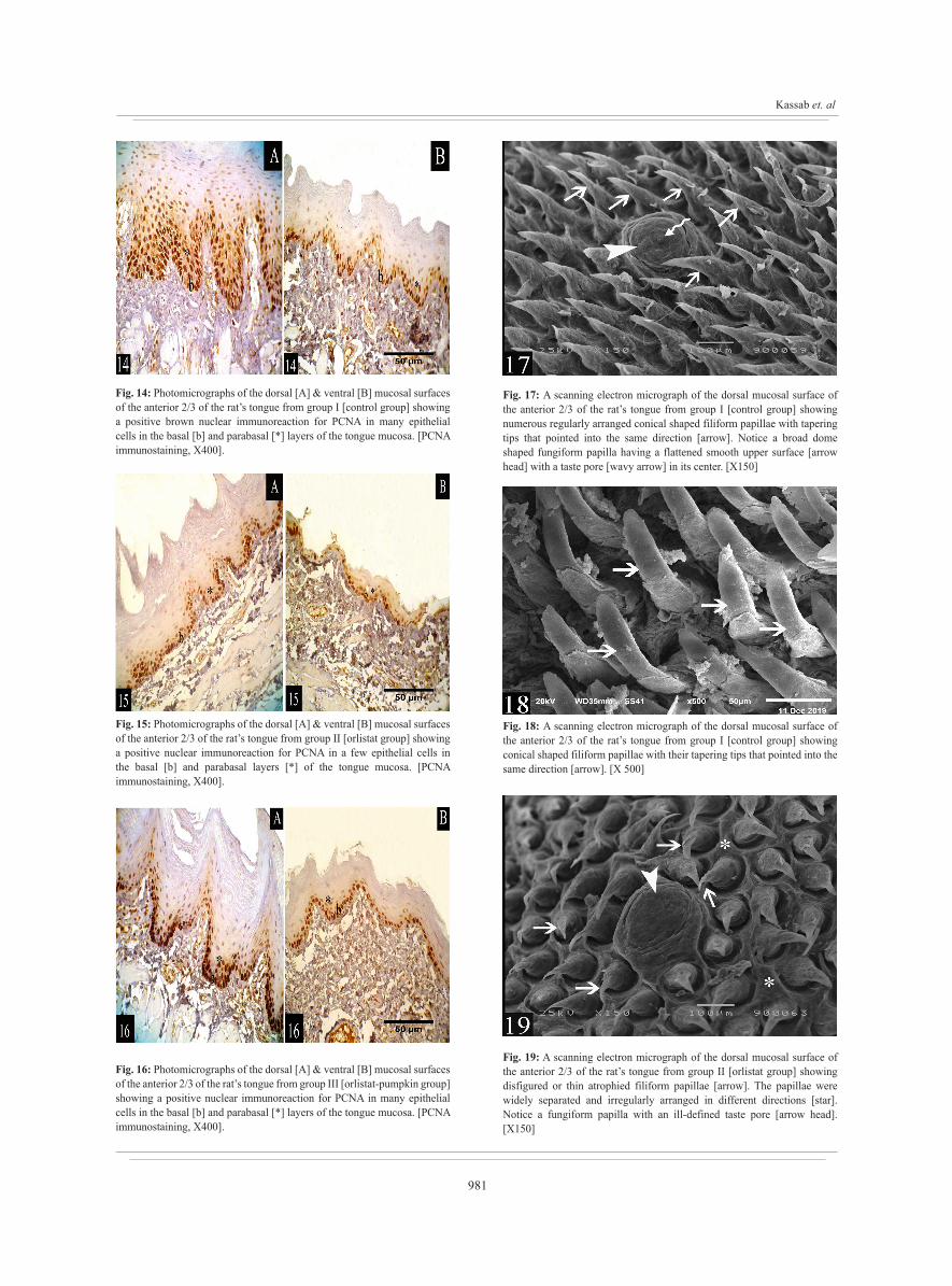
Fig. 14: Photomicrographs of the dorsal [A] & ventral [B] mucosal surfaces of the anterior 2/3 of the rat’s tongue from group I [control group] showing a positive brown nuclear immunoreaction for PCNA in many epithelial cells in the basal [b] and parabasal [*] layers of the tongue mucosa. [PCNA immunostaining, X400].
Fig. 15: Photomicrographs of the dorsal [A] & ventral [B] mucosal surfaces of the anterior 2/3 of the rat’s tongue from group II [orlistat group] showing a positive nuclear immunoreaction for PCNA in a few epithelial cells in the basal [b] and parabasal layers [*] of the tongue mucosa. [PCNA immunostaining, X400].
Fig. 16: Photomicrographs of the dorsal [A] & ventral [B] mucosal surfaces of the anterior 2/3 of the rat’s tongue from group III [orlistat-pumpkin group] showing a positive nuclear immunoreaction for PCNA in many epithelial cells in the basal [b] and parabasal [*] layers of the tongue mucosa. [PCNA immunostaining, X400].
Fig. 17: A scanning electron micrograph of the dorsal mucosal surface of the anterior 2/3 of the rat’s tongue from group I [control group] showing numerous regularly arranged conical shaped filiform papillae with tapering tips that pointed into the same direction [arrow]. Notice a broad dome shaped fungiform papilla having a flattened smooth upper surface [arrow head] with a taste pore [wavy arrow] in its center. [X150]
Fig. 18: A scanning electron micrograph of the dorsal mucosal surface of the anterior 2/3 of the rat’s tongue from group I [control group] showing conical shaped filiform papillae with their tapering tips that pointed into the same direction [arrow]. [X 500]
Fig. 19: A scanning electron micrograph of the dorsal mucosal surface of the anterior 2/3 of the rat’s tongue from group II [orlistat group] showing disfigured or thin atrophied filiform papillae [arrow]. The papillae were widely separated and irregularly arranged in different directions [star]. Notice a fungiform papilla with an ill-defined taste pore [arrow head]. [X150]
982
PUMPKIN ROLE IN ORLISTAT INDUCED-TONGUE MUCOSAL DAMAGE
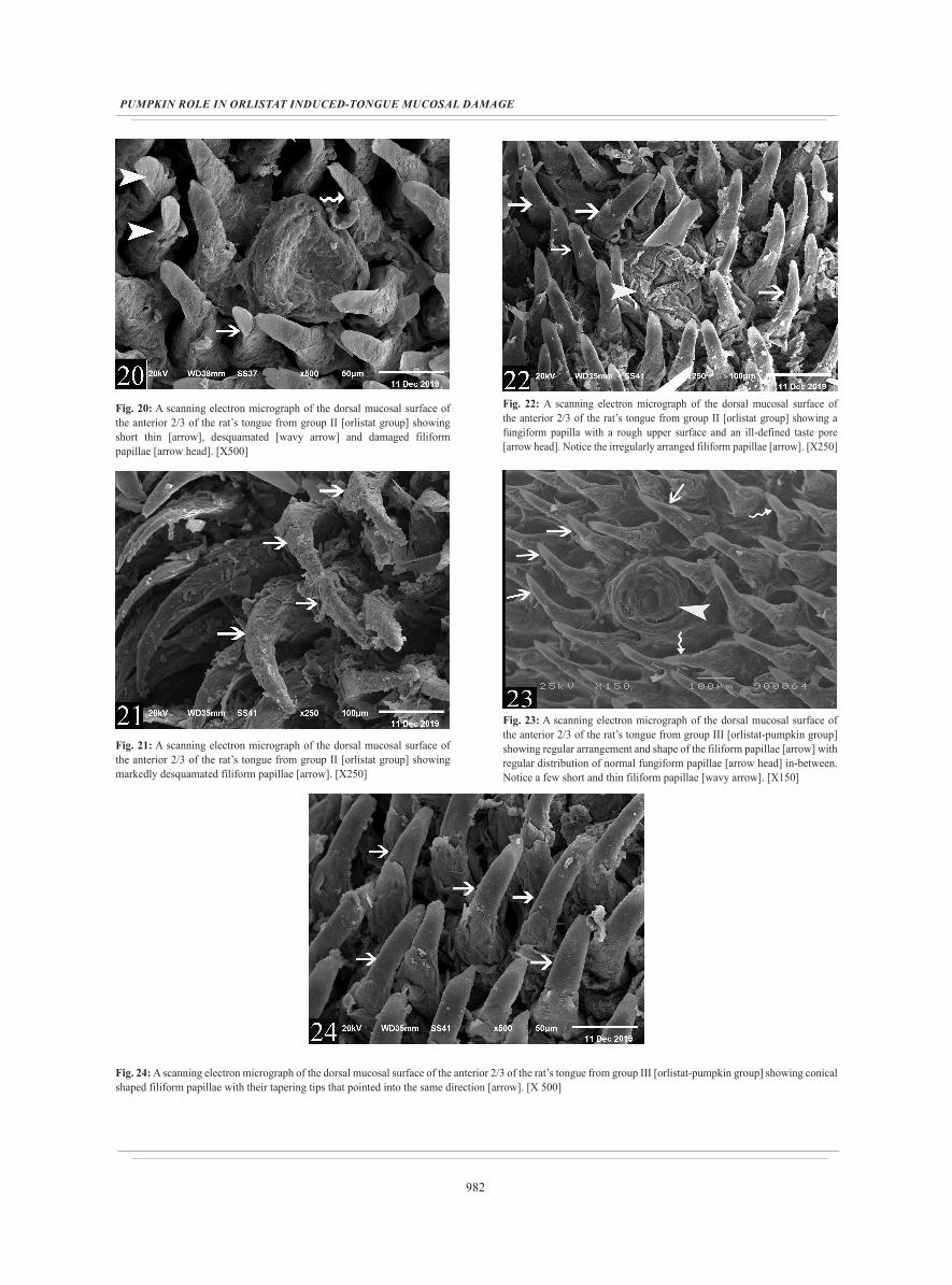
Fig. 20: A scanning electron micrograph of the dorsal mucosal surface of the anterior 2/3 of the rat’s tongue from group II [orlistat group] showing short thin [arrow], desquamated [wavy arrow] and damaged filiform papillae [arrow head]. [X500]
Fig. 21: A scanning electron micrograph of the dorsal mucosal surface of the anterior 2/3 of the rat’s tongue from group II [orlistat group] showing markedly desquamated filiform papillae [arrow]. [X250]
Fig. 22: A scanning electron micrograph of the dorsal mucosal surface of the anterior 2/3 of the rat’s tongue from group II [orlistat group] showing a fungiform papilla with a rough upper surface and an ill-defined taste pore [arrow head]. Notice the irregularly arranged filiform papillae [arrow]. [X250]
Fig. 23: A scanning electron micrograph of the dorsal mucosal surface of the anterior 2/3 of the rat’s tongue from group III [orlistat-pumpkin group] showing regular arrangement and shape of the filiform papillae [arrow] with regular distribution of normal fungiform papillae [arrow head] in-between. Notice a few short and thin filiform papillae [wavy arrow]. [X150]
Fig. 24: A scanning electron micrograph of the dorsal mucosal surface of the anterior 2/3 of the rat’s tongue from group III [orlistat-pumpkin group] showing conical shaped filiform papillae with their tapering tips that pointed into the same direction [arrow]. [X 500]
983
Kassab et. al
DISCUSSION
Orlistat is an approved drug for long-term management of obesity by causing fat malabsorption. Despite its broad effectiveness, Orlistat administration has been associated with serious adverse events[16]. The oral cavity is considered as a target organ for a number of various abnormalities that develop from side effects of this anti-obesity drug. The most commonly affected oral structures are soft tissues like tongue and its lining mucosa[5,17]. Recently, many studies have reported the nutritional and health protective values of the pumpkin seeds in addition to its anti-inflammatory and
anti-oxidant activities[7]. Accordingly, this research aimed to study the effect of orlistat on tongue mucosa of adult male albino rat and to evaluate the potential protective role of pumpkin seed oil using different histological methods.
Light and scanning electron microscopic findings of this research revealed that orlistat administration to albino rats adversely altered the histological structure of their tongue mucosa. These adverse structural changes included an obvious distortion in the filiform and the fungiform papillae and in the covering epithelium of the tongue mucosa. There was a complete loss of the papillae from a few focal areas
Table 1: Morphometerical and statistical analysis of the mucosal specimens of the tongue in all study groups. Data are expressed as Mean± SD.
Groups Parameters
Group I(control group)
Group II(orlistat group)
Group III(orlistat-pumpkin group)
Mean area % of PCNA in the dorsal epithelium 28.7±1.16 24.876±0.703* 27.48±1.48
Mean area % of PCNA in the ventral epithelium 17.53±0.75 14.29±0.961* 16.953±0.393
Mean epithelial thickness of the dorsal mucosal surface (µm) 127.00±3.63 110.10±5.02* 125.59±3.07
Mean epithelial thickness of the ventral mucosal surface (µm) 79.28±2.17 59.57±4.69* 77.79±2.00
Filiform papillae:Mean Height (µm)Mean Width (µm)
237.5±2.1454.01±2.64
172.4±19.1*
38.41±3.31*230.5±10.851.76±2.40
Fungiform papillae:Mean Height (µm)Mean Width (µm)
197.64±2.3690.70±1.45
166.35±5.04*
83.12±4.77*195.13±3.2989.52±1.08
*P < 0.05 is significant versus group I.
Histogram 1: Morphometrical and statistical analysis of [A] Mean area % of PCNA positive immunoreaction of the epithelium. [B] Mean epithelial thickness. [C and D] Mean height and width of the filiform and the fungiform papillae in all studied groups.* indicates high significance vs control
984
PUMPKIN ROLE IN ORLISTAT INDUCED-TONGUE MUCOSAL DAMAGE
on the tongue mucosa. Moreover, there was a significant decrease in the epithelial thickness, papillae height and width and in the PCNA-immunoreaction of the epithelium. Scanning electron microscopy showed signs of atrophy of the filiform and the fungiform papillae. These adverse effects of orlistat on the mucosa were compatible with the findings of other researchers who found that orlistat exerted damaging effects on tongue mucosa and colonic mucosa of rats[5,18]. These results matched also the findings of other researchers who reported development of oral ulcers with orlistat therapy[19]. Similarly, it was reported that orlistat induced disturbance of the normal architecture of the pancreatic acini causing acute pancreatitis with elevated levels of the pancreatic enzymes[20,21]. Also, other studies reported that orlistat causes gastrointestinal problems and fat soluble vitamins deficiencies (A,D,E&K)[22,23] in addition to serious liver and kidney damage limiting the patient compliance for orlistat[4,24].
The mechanism involved in orlistat induced tongue mucosal damage may be attributed to nutritional deficiency as a side effect of this anti-obesity drug. Several studies reported fat soluble vitamins deficiency (A,E,D) with orlistat therapy due to deficient intestinal absorption of these vitamins[25]. Vitamin A is essential for protection and regeneration of the mucous membranes. Its deficiency may lead to defective healing and desquamation of the mucosa[26,27]. Moreover, vitamin A plays an important role in regulating cellular differentiation[28]. Vitamin E is an important anti-oxidant vitamin to protect and maintain health of the epithelial cells of the mucous membranes. Oxidative stress resulted from vitamin E deficiency may play a role in orlistat induced tongue mucosal damage. Vitamin E protects the biological cell membranes against the harmful effects of molecular oxygen. Moreover, it preserves the functions of intracellular organelles and ensures the integrity of the cell membranes[29,30]. Recently, it was proved that vitamin D plays an essential role in the protection of the mucous membranes through its anti-inflammatory and anti-infective activities. This occurs through down-regulation of the expression of the pro-inflammatory mediators and the NF-κB pathway. It also has a function in regulating tight junctions and pathogen invasion[31]. So, deficiency of these vitamins due to orlistat therapy may play a role in the pathogenesis of orlistat induced tongue mucosal damage.
The structural changes of the tongue in the current study were considered as a manifestation for atrophic glossitis. The tongue is considered as a mirror reflecting the general health and the nutritional status of the body. The nutritional deficiency is considered as the principal aetiological factor for atrophy of the tongue mucosa. Atrophic glossitis due to a vitamin deficiency occurs by two principal mechanisms of action: firstly, a malabsorption of the nutrient; secondly, defective metabolization of a specific vitamin. In addition, there are other mechanisms leading to an atrophic condition such as the fungal infection or candidiasis[32,33].
The atrophic influence of orlistat on tongue papillae might be secondary to another adverse effect reported in
other study which stated that orlistat could induce macrocytic anemia and thrombocytopenia[34]. This consecutively could affect tongue architecture resulting in atrophy of tongue papillae[35].
The light microscopic findings of this research were identical to those of previous study on the effect of orlistat on the small intestinal mucosa. Orlistat induced mucosal damage may be attributed to enhanced nitric oxide production by inducible nitric oxide synthase (iNOS) in the tissue. This effect was diminished by using iNOS inhibitor[36].
The scanning electron microscopic findings of the current study supported the light microscopic one and they were in agreement with other investigators who attributed these findings to free radical attack to the cells due to increased oxidative stress[4].
The immunohistochemical results of this study revealed an obvious reduction in PCNA immunoexpression in orlistat group compared to the control group and statistical results showed that this reduction was significant. Moreover, there was a significant decrease in the epithelial thickness and in the height and width of the papillae. Proliferating cell nuclear antigen (PCNA) is localized in the nucleus of cells and is associated with cell proliferation. It is necessary for DNA replication. Elevated levels of PCNA occur at the G1/S phase transition in cells undergoing division[37]. Orlistat decreased the proliferative capacity of the epithelial cells leading to reduced epithelial thickness. This effect may be attributed to deficiency of the fat soluble vitamins A & D. Vitamin A has an important role in regulating cell growth and differentiation[28]. Recently, it was proved that vitamin D stimulates proliferation and migration of the epithelial cells[38].
The results of this study demonstrated the protective role of PSO against orlistat induced tongue mucosal damage. This finding is supported by previous reports which stated that PSO exerted a protective effect through its antioxidant and anti-inflammatory activities. PSO is an excellent source for minerals, vitamins and antioxidants[39,40]. PSO significantly attenuates the severity of lipid peroxidation and enhances the antioxidant enzyme activity[41]. This antioxidant effect was attributed mainly to its high content of α-tocopherol (vitamin E), vitamin A and iron. It protects the polyunsaturated fatty acids of the cell membrane phospholipids. It is also a potent peroxyl radical scavenger that can protect the biological cell membranes from the harmful free radical effects. It neutralizes the elevated reactive oxygen species production and prevents DNA oxidative damage. Moreover, the anti-inflammatory activity of PSO was attributed to its high content of beta-carotene and the promising proportions of ω-6 and ω-9 unsaturated fatty acids[4,42,43].
Our study showed that PSO improved the proliferative capacity of the epithelium. This was evident in this study by the morphometric and statistical results. This effect was attributed to the synergistic action of its active constituents of tocopherols, fatty acids and phytosterols. These bioactive components are responsible for the regenerative capacity of

985
Kassab et. al
the oil helping in full re-epithelization and migration of the fibroblasts by providing connective tissue matrix. Vitamin E content of the oil helps in this regenerative effect by preventing cell damage and promotion of DNA synthesis by its antioxidant property[44].
CONCLUSION
The current study showed that orlistat administration to albino rats adversely altered the histological structure of their tongue mucosa. The study suggests that pumpkin seed oil might be beneficial in minimizing the tongue mucosa structural changes induced by orlistat in rats most probably through its anti-oxidant and anti-inflammatory activities in addition to its regenerative capacity. Therefore, Pumpkin seed oil could be a promising protective agent for patients receiving orlistat to reduce its complications on tongue mucosa.
CONFLICTS OF INTEREST
There are no conflicts of interest.
FUNDING SOURCE
There are no funding source to declare.
REFERENCES
1. Amin HM, Tawfek NS, Abo-El Hussein BK, Abd El-Ghany MS: Anti-Obesity Potential of Orlistat and Amphetamine in Rats Fed on High Fat Diet. Middle East Journal of Applied Sciences (2015); 05 (02): 453-461.
2. Qi X: Review of the Clinical Effect of Orlistat. IOP Conf. Series: Materials Science and Engineering (2018); 301: 1-8.
3. Nwobodo NN: Toxicity and Safety Concerns in Orlistat Therapy for Obesity: A Critical Evaluation. Asian Journal of Biomedical and Pharmaceutical Sciences (2015); 5(47): 01-04.
4. Youssef S: Light and Electron Microscopic Study of the Effect of Orlistat on the Liver of Adult Male Albino Rats and the Possible Protective Role of β-Carotene. Forensic Medicine and Anatomy Research (2018); 6: 20-36
5. Ezzat AM and Labah DA: Comparative Study of the Effects of Orlistat and Green Coffee Bean Extract on Tongue Mucosa in Obese Rat Model. Life Science Journal (2017);14(8): 11-18
6. Shaik PS and Pachava S: The role of vitamins and trace elements on oral health: a systematic review. Int J Med Rev (2017); 4(1):22-31
7. Dar AH, Sofi SA, Rafiq S: Pumpkin the functional and therapeutic ingredient: A review. International Journal of Food Science and Nutrition (2017); 2(6): 165-170
8. Habib A, Biswas S, Siddique AH, Manirujjaman M, Uddin B, Hasan S, Khan MMH, Uddin M, Islam M, Hasan M, Rahman M, Asaduzzaman M, Sohanur RM, Khatun M, Islam MA, Rahman M: Nutritional and Lipid Composition Analysis of Pumpkin Seed (Cucurbita maxima Linn.). J Nutr Food Sci (2015); 5: 374
9. Ali DM and Abdelzaher WY: Possible Protective Effect of Pumpkin Seed Oil against Sodium Nitrite in Rats; A Biochemical and Genetic Study. Int J Clin Pharmacol Toxicol. 2017; 6(2), 262-269.
10. Gaertner DJ, Hallman TM, Hankenson FC, Batcherder MA: Anesthesia and analgesia for laboratory rodents. Anesthesia and analgesia in laboratory animals.2 nd edition. Academic press, San Diego, CA. Boston. (2008); 239 -240
11. Bancroft JD and Gamble M: Theory and practice of histological techniques. 6th ed. Philadelphia: Churchill Livingstone: Elsevier Health Science. (2008); 126–127
12. Ramos-Vara JA, Kiupel M, Baszler T, Bliven L, Brodersen B, Chelack B, Czub S, Del Piero F, Dial S, Ehrhart EJ, Graham T, Manning L, Paulsen D, Valli VE, West K: Suggested guidelines for immunohistochemical techniques in veterinary diagnostic laboratories. J Vet Diagn Invest (2008); 20: 393–413.
13. Piroeva S, Atanassova-Vladimirova L, Dimowa H, Sbirkova G, Radoslavov P, Hristov BLS: A simple and rapid scanning electron microscope preparative technique for observation of biological samples: application on bacteria and DNA samples. Bulgarian Chemical Communications (2013); 45 (4): 510–515.
14. Takahashi Y, Takahashi H, Stern PL, Kirita T, Tsuboi A: Expression of Oncofetal Antigen 5T4 in Murine Taste Papillae. Front. Cell Neurosci. 2019; 13(343):1-13.
15. Dawson-Saunders B and Trapp R. Basic and clinical biostatics. 3rd ed., Lang Medical Book, McGrow Hill Medical Publishing Division. 2001; 161-218.
16. Priyadharshini A, Ahalya SP, Vaishnavi P, Pavithra S, Rakesh Rosario A: A review on benefits and toxicity of orlistat therapy. Drug Invention Today (2019) 12(3): 550-553.
17. Abdollahi M, Rahimi R, Radfar M. Current opinion on drug-induced oral reactions: A comprehensive review. J Contemp Dent Pract 2008; 3:001-015.
18. Nairooz S, Ibrahim SH, Omar SM, Affan M. Structural changes of the colonic mucosa induced by orlistat: Experimental study. Egypt. J. Histol. 2010; 33: 635– 648.

986
PUMPKIN ROLE IN ORLISTAT INDUCED-TONGUE MUCOSAL DAMAGE
19. Taha M, Ghosn S, Zeitoun A. Oral aphthous ulcers associated with orlistat. AJHP. 2012; 69: 1462-1464.
20. Kose M, Emet S, Akpınar TS, Ilhan M, Gok AF, Dadashov M, Tukek T: An Unexpected Result of Obesity Treatment: Orlistat- Related Acute Pancreatitis. Case reports in Gastroentrol (2015) 9 : 152- 155.
21. Abdel Wahab SA, Ali AH, Mahmoud AS, Abdelhaleem AH. Effect of orlistat on the pancreas of the female albino rat: Histological and Histochemical study. Journal of Medical Histology (2017) 1(1);1-14.
22. Kaila B and Raman M. Obesity: a review of pathogenesis and management strategies. Can J Gastroenterol. 2008; 22:61-68.
23. Filippatos TD, Derdemezis CS, Gazi IF, Nakou ES, Mikhailidis DP, Elisaf MS. Orlistat-Associated Adverse Effects and Drug Interactions. A Critical Review. Drug Safety (2008); 31 (1): 53-65.
24. Tousson E, Massoud A, Salem A, Fatoh SA. Nephrotoxicity associated with Orlistat in normal and obese female rats. Journal of Bioscience and Applied Research (2018) 4 (3), 193-198.
25. McDuffie JR, Calis KA, Booth SL,Uwaifo GI,Yanovski JA: Effect of orlistat on fat-soluble vitamins in obese adolescent. Pharmacotherapy (2002) 22(7):814-822.
26. Philipone E and Yoon JA: Mucosal Manifestations of Nutritional Deficiencies. Oral Pathology in the Pediatric Patient (2017): 121-123. DOI 10.1007/978-3-319-44640-0_8
27. Maccabee MS, Trune DR, Hwang P: paranasal sinus mucosal regeneration: the effect of topical retinoic acid. American Journal of Rhinology (2003) 17(3): 133-137.
28. Sun H and Kawaguchi R: The Membrane Receptor for Plasma Retinol Binding Protein, a New Type of Cell-Surface Receptor. Int Rev Cell Mol Biol. 2011; 288: 1–41.
29. Osman HI, Abd El Razik N, Koura AS: Histological changes of rat tongue papillae due to chromium toxicity and the protective role of vitamin E. Egyptian Dental Journal (2006) 52: 193-200.
30. Ghosh A, Pallavi SK, Nagpal B, Hegde U, Archana S, Nagpal J: Role of Vitamins in Oral Health & Disease: An Overview. Indian Journal of Applied Research (2015); 5 (12): 292-295.
31. Sun J: Vitamin D and mucosal immune function. Curr Opin Gastroenterol. 2010; 26(6): 591–595.
32. Erriu M, Pili FMG, Cadoni S, Garau V: Diagnosis of Lingual Atrophic Conditions: Associations with Local and Systemic Factors. A Descriptive Review. The Open Dentistry Journal, 2016, 10, 619-635.
33. Niimi N and Mori N. Papillary atrophy of the tongue. Clin Case Rep. 2018; 6: 2283-2284.
34. Martinez DP, Alvarez JC, Santamaria NM, Ruiz OP, Garcia AR, Alonso RS. Macrocytic anemia and thrombocytopenia induced by orlistat. Int J Endocrinol Metab. 2013; 11(4); e6721: 1-5.
35. Gaddey HL: Oral manifestations of systemic disease. Gen Dent 2017; 65(6):23-29.
36. Caner, M., Dogruman, H., Taskin, E., Kandil, A., Demirci, C. Effects of Orlistat and Its Relationship with Nitric Oxide in the Small Intestinal Mucosa. Chinese Journal of Physiology 2005; 48(4): 217-222.
37. Aboushady IM, Mubarak RT, El-mougy SAF, Rashed LA, El-desouky AA: The Effect of Transplanted Bone Marrow Stem Cells on the Tongue of Irradiated Rats (Histological and Immunohistochemical study). J Am Sci 2012; 8(11): 553-561.
38. Nazzal A, Tipton DA, Karydis A, Slominski A, Stein SH. Vitamin D Stimulates Epithelial Cell Proliferation and Facilitates Wound Closure via A Cathelicidin Independent Pathway In Vitro. Periodon Prosthodon. 2016; 2(2): 1-8.
39. Sharquie KE, Noaimi AA, Latif TM: Treatment of Recurrent Aphthous Stomatitis by 100% Topical Pumpkin Seed Oil. Journal of Cosmetics, Dermatological Sciences and Applications (2017), 7, 324-335.
40. Karanja JK, Mugendi BJ, Khamis FM, Muchugi AN: Nutritional Composition of the Pumpkin (Cucurbita spp.) Seed Cultivated from Selected Regions in Kenya. Journal of Horticulture Letters 2013, 3(1): 17-22.
41. Galaly SR, Hozayen WG, Amin KA, Ramadan SM: Effects of orlistat and herbal mixture extract on brain, testes functions and oxidative stress biomarkers in a rat model of high fat diet. Beni Suef University Journal of Basic and Applied Sciences (2014); 3: 93-105.
42. Montesano D, Blasi F, Simonetti MS, Santini A, Cossignani L: Chemical and nuctritional characterization of seed oil from Cucurbita maxima L. Foods 2018; 7(30): 1-14.
43. Syed QA, Akram M, Shukat R: Nutritional and therapeutic importance of the pumpkin seeds. Biomed Sci & Tech Res. 2019; 21(2):15798-15803.
44. Bardaa S, Ben Halima N, Aloui F, Ben Mansour R, Jabeur H, Bouaziz M, Sahnoun Z: Oil from pumpkin (Cucurbita pepo L.) seeds: evaluation of its functional properties on wound healing in rats. Lipids in Health and Disease 2016; 15: 73.

987
Kassab et. al
الملخص العربى
الدور الوقائى المحتمل لزيت بذور اليقطين فى تخفيف تلف الغشاء المخاطى للسان المحدث بالأورليستات فى ذكور الجرذان البيضاء البالغة: دراسة بالمجهر الضوئى
والالكترونى الماسح
أميرة عدلى كساب، خالد أحمد أحمد مصطفى، أمل على عبد الحافظ
قسم الهستولوجيا وبيولوجيا الخلايا، كلية الطب، جامعة طنطا، مصر
المقدمة: أورليستات هو دواء فعال ضد السمنة عن طريق تقليل امتصاص الدهون. تجويف الفم والتركيبات المرتبطة
به هي أعضاء مستهدفة لمختلف التشوهات التي قد تنشأ من العلاج بالأورليستات. زيت بذور اليقطين (PSO) هو عامل
وقائي غذائي وصحي ذو قيمة مع خصائص دوائية بارزة.
الهدف من البحث: تقييم الدور الوقائى المحتمل لزيت بذور اليقطين في تخفيف تلف الغشاء المخاطي للسان المحدث
بالأورليستات في ذكور الجرذان البيضاء البالغة.
,ومجموعة كمجموعة ضابطة البالغة البيضاء الجرذان ذكور من جرذاً خمسون استخدام تم البحث: وطرق مواد
وزيت كجم) / مجم 32) أورليستات من كل إعطاء تم اليقطين. بذور زيت و أورليستات ,ومجموعة أورليستات
اللسان من عينات تجهيز تم ، أسابيع. أربعة لمدة يومياً واحدة مرة الفم كجم) عن طريق / (1.5مل اليقطين بذور
للدراسة بالمجهر الضوئى والالكترونى الماسح, وقد أجريت دراسة هستوكيميائية مناعية باستخدام الأجسام المضادة لل
.(PCNA)
النتائج: أظهرت عينات من مجموعة أورليستات تشويهًا واضحًا في الحليمات الخيطية والفطرية وفي الخلايا الطلائية
تجاويف ظهرت الظهارية.كما والتلال الحليمات فى فقدان أيضا هناك كان للسان. المخاطي الغشاء تغطي التى
بالسيتوبلازم وتغيرات بأنوية الخلايا الطلائية. كما ظهر تسلل خلوى التهابي شديد مع وجود أوعية دموية محتقنة. وكان
PCNA هناك انخفاض ذات دلالة احصائية فى سمك الظهارية ,وارتفاع وعرض الحليمات وفي التفاعل المناعى لل
في الخلايا الظهارية. أظهر الفحص المجهري الالكترونى الماسح تشوهًا و ضمورًا بؤرياً سطحياً في الحليمات الخيطية
والفطرية. وفي المقابل ، ظهرت تغييرات طفيفة في مجموعة أورليستات و زيت بذور اليقطين التي تلقت PSOقبل
الأورليستات.
الاستنتاج: أورليستات سبب تغييرات تركيبية في الغشاء المخاطى للسان الجرذان البيضاء. زيت بذور اليقطين خفف
من هذه الآثار وحافظ على تركيب الغشاء المخاطى للسان.


![Ultrasound-Assisted Extraction (UAE) and Solvent ...€¦ · watermelon seed oil (59.6%) [20] and pumpkin seed oil (55.6%) [21,22]. Both olive oil and papaya seed oil are rich sources](https://img.pdfslide.us/doc/110x75/605e65ab44493b44880d978b/ultrasound-assisted-extraction-uae-and-solvent-watermelon-seed-oil-596.jpg)
![SS19 Josper Pizza 9.07 - Off Peak Luxury · SWEET POTATO, AVOCADO & WHIPPED FETA HASH [GF] [V].....8.50 Spinach, chilli, toasted pumpkin seeds, kale & pumpkin seed pesto, poached](https://img.pdfslide.us/doc/110x75/5ec33b7be031ca47c408c4c9/ss19-josper-pizza-907-off-peak-luxury-sweet-potato-avocado-whipped-feta.jpg)