The biochemistry and medical significance of the flavonoids
Bent H. Havsteen*
Department of Biochemistry, University of Kiel, Olshausenstrasse 40, D-24098 Kiel, Germany
Abstract
Flavonoids are plant pigments that are synthesised from phenylalanine, generally display marvelous colors known from flower petals,
mostly emit brilliant fluorescence when they are excited by UV light, and are ubiquitous to green plant cells. The flavonoids are used by
botanists for taxonomical classification. They regulate plant growth by inhibition of the exocytosis of the auxin indolyl acetic acid, as well as
by induction of gene expression, and they influence other biological cells in numerous ways. Flavonoids inhibit or kill many bacterial strains,
inhibit important viral enzymes, such as reverse transcriptase and protease, and destroy some pathogenic protozoans. Yet, their toxicity to
animal cells is low. Flavonoids are major functional components of many herbal and insect preparations for medical use, e.g., propolis (bee’s
glue) and honey, which have been used since ancient times. The daily intake of flavonoids with normal food, especially fruit and vegetables,
is 1–2 g. Modern authorised physicians are increasing their use of pure flavonoids to treat many important common diseases, due to their
proven ability to inhibit specific enzymes, to simulate some hormones and neurotransmitters, and to scavenge free radicals.
D 2002 Elsevier Science Inc. All rights reserved.
Keywords: Flavonoids; Benzopyrones; Heat shock proteins; Gene expression; Enzyme inhibition
Abbreviations: Ab, b-amyloid; AC, adenylate cyclase; ACTH, adrenocorticotrophic hormone; AD, Alzheimer’s disease; AIDS, acquired immunodeficiency
syndrome; APC, antigen-presenting cell; cAMP, cyclic AMP; CAT, chloramphenicol acetyltransferase; cGMP, cyclic GMP; CoA, coenzyme A; COX, cyclo-
oxygenase; CSF, colony stimulating factor; DAG, diacylglycerol; ER, estrogen receptor; FA, fatty acid; GABA, g-aminobutyric acid; GC-MS, gas
chromatography-mass spectrometry; GSH, glutathione; HIV, human immunodeficiency virus; HMG, 3-hydroxy-3-methyl-glutaryl; HSE, heat shock regulatory
element; HSF, heat shock factor; HSP, heat shock protein; HTLV, human T-lymphocyte-associated virus; IAA, indolyl acetic acid; ICE, interconverting
enzyme; IFN, interferon; Ig, immunoglobulin; IL, interleukin; LDL, low-density lipoprotein; MHC, major histocompatibility complex; NK-T-Ly, natural killer
T-lymphocyte; NO, nitric oxide; PDE, phosphodiesterase; PG, prostaglandin; PGI2, prostacyclin; PIL, phosphatidylinositol lipase; PKC, protein kinase C; PL,
phospholipase; PRR, proton relaxation rate; Pyr-P, pyridoxal phosphate; R, receptor; RA, rheumatoid arthritis; SIV, Simian immunodeficiency virus; SOD,
superoxide dismutase; THF, tetrahydrofolate; TIMP, tissue inhibitor of matrix metalloproteinase; TNF, tumor necrosis factor; Tx, thromboxane; XO, xanthine
oxidase.
Contents
1. Preface . . . . . . . . . . . . . . . . . . . . . . . . . . . . . . . . . . . . . . . . . . . . . . . 69
2. Introduction . . . . . . . . . . . . . . . . . . . . . . . . . . . . . . . . . . . . . . . . . . . . 70
3. The chemistry of flavonoids . . . . . . . . . . . . . . . . . . . . . . . . . . . . . . . . . . . . 71
3.1. Structure and nomenclature. . . . . . . . . . . . . . . . . . . . . . . . . . . . . . . . . 71
3.2. The oxidation-reduction potential of flavonoids . . . . . . . . . . . . . . . . . . . . . . 71
3.3. Acid-base properties . . . . . . . . . . . . . . . . . . . . . . . . . . . . . . . . . . . . 72
3.3.1. The tautomery of anthocyanin . . . . . . . . . . . . . . . . . . . . . . . . . . . 72
3.4. Absorption and fluorescence spectra of flavonoids . . . . . . . . . . . . . . . . . . . . . 74
3.5. Optical activity of flavonoids. . . . . . . . . . . . . . . . . . . . . . . . . . . . . . . . 76
3.6. Radical scavenging by flavonoids . . . . . . . . . . . . . . . . . . . . . . . . . . . . . 77
3.7. Linear free-energy relationships applied to the flavonoids . . . . . . . . . . . . . . . . . 80
3.7.1. The nature of the problem. . . . . . . . . . . . . . . . . . . . . . . . . . . . . 80
3.7.2. Linear free-energy relationships . . . . . . . . . . . . . . . . . . . . . . . . . . 80
0163-7258/02/$ – see front matter D 2002 Elsevier Science Inc. All rights reserved.
PII: S0163 -7258 (02 )00298 -X
* Current Address: Abildgaardsvej 49, DK-2830 Virum, Denmark. Tel.: +49-0431-880-3214.
E-mail address: [email protected] (B.H. Havsteen).
Pharmacology & Therapeutics 96 (2002) 67–202
4. The occurrence of flavonoids . . . . . . . . . . . . . . . . . . . . . . . . . . . . . . . . . . . 81
4.1. Distribution in nature . . . . . . . . . . . . . . . . . . . . . . . . . . . . . . . . . . . . 81
5. Identification of flavonoids . . . . . . . . . . . . . . . . . . . . . . . . . . . . . . . . . . . . 82
5.1. Magnetic resonance spectrometry of flavonoids . . . . . . . . . . . . . . . . . . . . . . 83
5.1.1. Introduction . . . . . . . . . . . . . . . . . . . . . . . . . . . . . . . . . . . . 83
5.1.2. Information available from proton relaxation rates . . . . . . . . . . . . . . . . 83
5.1.3. The theory of pulsed nuclear magnetic resonance . . . . . . . . . . . . . . . . . 83
5.1.4. The measurement of relaxation times . . . . . . . . . . . . . . . . . . . . . . . 86
5.1.5. Applications of proton resonance relaxation. . . . . . . . . . . . . . . . . . . . 88
5.1.6. Concluding remarks on nuclear magnetic resonance . . . . . . . . . . . . . . . 88
5.2. Identification of flavonoids by gas chromatography-mass spectrometry . . . . . . . . . . 89
5.2.1. Scope . . . . . . . . . . . . . . . . . . . . . . . . . . . . . . . . . . . . . . . 89
5.2.2. Analysis of propolis by gas chromatography-mass spectrometry . . . . . . . . . 89
5.3. Analysis of propolis by high performance liquid chromatography . . . . . . . . . . . . . 89
5.3.1. Scope . . . . . . . . . . . . . . . . . . . . . . . . . . . . . . . . . . . . . . . 89
5.3.2. The analytical procedure. . . . . . . . . . . . . . . . . . . . . . . . . . . . . . 89
6. The biosynthesis of flavonoids. . . . . . . . . . . . . . . . . . . . . . . . . . . . . . . . . . . 90
6.1. Anabolism . . . . . . . . . . . . . . . . . . . . . . . . . . . . . . . . . . . . . . . . . 90
6.2. The genetics of flavonoids . . . . . . . . . . . . . . . . . . . . . . . . . . . . . . . . . 92
7. The role of the flavonoids in plant physiology . . . . . . . . . . . . . . . . . . . . . . . . . . 93
7.1. Flavonoids as signals of symbiosis . . . . . . . . . . . . . . . . . . . . . . . . . . . . . 95
8. The pharmacology of flavonoids in animals . . . . . . . . . . . . . . . . . . . . . . . . . . . . 95
8.1. Pharmacodynamics . . . . . . . . . . . . . . . . . . . . . . . . . . . . . . . . . . . . . 96
8.2. Acute toxicity of flavonoids . . . . . . . . . . . . . . . . . . . . . . . . . . . . . . . . 97
8.3. Long-term effects of flavonoids . . . . . . . . . . . . . . . . . . . . . . . . . . . . . . 97
8.4. The catabolism of flavonoids . . . . . . . . . . . . . . . . . . . . . . . . . . . . . . . . 97
9. The immunology of the flavonoids . . . . . . . . . . . . . . . . . . . . . . . . . . . . . . . . 99
9.1. The flavonoids as antigens . . . . . . . . . . . . . . . . . . . . . . . . . . . . . . . . 100
9.2. Flavonoids as immune modulators . . . . . . . . . . . . . . . . . . . . . . . . . . . . 100
10. Scavenging of free radicals by flavonoids . . . . . . . . . . . . . . . . . . . . . . . . . . . . 101
11. The electron transfer catalysis by flavonoids . . . . . . . . . . . . . . . . . . . . . . . . . . 103
12. The flavonoids as enzyme inhibitors. . . . . . . . . . . . . . . . . . . . . . . . . . . . . . . 104
12.1. Hydrolases . . . . . . . . . . . . . . . . . . . . . . . . . . . . . . . . . . . . . . . 104
12.2. Oxidoreductases . . . . . . . . . . . . . . . . . . . . . . . . . . . . . . . . . . . . . 106
12.3. Kinases . . . . . . . . . . . . . . . . . . . . . . . . . . . . . . . . . . . . . . . . . 108
12.4. Isomerases . . . . . . . . . . . . . . . . . . . . . . . . . . . . . . . . . . . . . . . 108
12.5. Transferases . . . . . . . . . . . . . . . . . . . . . . . . . . . . . . . . . . . . . . . 108
12.6. Ligases and lyases . . . . . . . . . . . . . . . . . . . . . . . . . . . . . . . . . . . 108
13. The hormone action of flavonoids . . . . . . . . . . . . . . . . . . . . . . . . . . . . . . . . 108
14. The mutagenic potential of flavonoids . . . . . . . . . . . . . . . . . . . . . . . . . . . . . . 108
15. The influence of the flavonoids on the sensory system . . . . . . . . . . . . . . . . . . . . . 109
15.1. The olfactory system . . . . . . . . . . . . . . . . . . . . . . . . . . . . . . . . . . 109
15.2. The neurostimulatory effect of flavonoids . . . . . . . . . . . . . . . . . . . . . . . 110
15.3. The analgesic effect of flavonoids . . . . . . . . . . . . . . . . . . . . . . . . . . . 110
16. Complexes of flavonoids with heavy metal ions. . . . . . . . . . . . . . . . . . . . . . . . . 110
17. Medical, technical, gastronomic, and other applications of flavonoids . . . . . . . . . . . . . 111
17.1. Hypertension and microbleeding . . . . . . . . . . . . . . . . . . . . . . . . . . . . 112
17.2. Inflammation . . . . . . . . . . . . . . . . . . . . . . . . . . . . . . . . . . . . . . 114
17.3. The effect of flavonoids on the condition of diabetes mellitus patients . . . . . . . . . 121
17.4. Local anaesthesia by flavonoids . . . . . . . . . . . . . . . . . . . . . . . . . . . . 125
17.5. Protein-rich oedema . . . . . . . . . . . . . . . . . . . . . . . . . . . . . . . . . . . 126
17.6. Loosening of connective tissue . . . . . . . . . . . . . . . . . . . . . . . . . . . . . 127
17.7. The effect of flavonoids on allergy and asthma. . . . . . . . . . . . . . . . . . . . . 128
17.8. The influence of flavonoids on cancer . . . . . . . . . . . . . . . . . . . . . . . . . 130
17.8.1. The biology of cancer . . . . . . . . . . . . . . . . . . . . . . . . . . . . 130
17.8.2. The treatment of cancer by flavonoids . . . . . . . . . . . . . . . . . . . . 133
17.8.3. Biochemical processes of cancer influenced by flavonoids . . . . . . . . . 134
17.8.4. Stress response . . . . . . . . . . . . . . . . . . . . . . . . . . . . . . . . 136
17.9. The influence of flavonoids on cardiovascular diseases. . . . . . . . . . . . . . . . . 140
17.9.1. The genetic disposition. . . . . . . . . . . . . . . . . . . . . . . . . . . . 140
17.9.2. The role of flavonoids in the dietary component of cardiovascular stress . . 141
B.H. Havsteen / Pharmacology & Therapeutics 96 (2002) 67–20268
17.9.3. Flavonoids in the management of ischaemia/reperfusion damage . . . . . . 142
17.10. The effect of flavonoids on gastrointestinal ulcers . . . . . . . . . . . . . . . . . . . 143
17.11. The effect of flavonoids on rheumatic diseases . . . . . . . . . . . . . . . . . . . . . 144
17.12. Bacterial infection. . . . . . . . . . . . . . . . . . . . . . . . . . . . . . . . . . . . 145
17.13. The antiviral properties of flavonoids . . . . . . . . . . . . . . . . . . . . . . . . . . 147
17.14. Morbus alzheimer’s . . . . . . . . . . . . . . . . . . . . . . . . . . . . . . . . . . . 152
17.15. Wound healing . . . . . . . . . . . . . . . . . . . . . . . . . . . . . . . . . . . . . 154
17.15.1. Cellular reactions to damage . . . . . . . . . . . . . . . . . . . . . . . . 154
17.15.2. Peroxidation of lipids by free radicals . . . . . . . . . . . . . . . . . . . 154
17.15.3. Metabolic changes caused by organic solvents . . . . . . . . . . . . . . . 156
17.15.4. Hypoxic cell damage . . . . . . . . . . . . . . . . . . . . . . . . . . . . 157
17.15.5. Tissue regeneration . . . . . . . . . . . . . . . . . . . . . . . . . . . . . 158
17.15.6. The anabolism . . . . . . . . . . . . . . . . . . . . . . . . . . . . . . . 158
17.16. Heavy metal detoxification . . . . . . . . . . . . . . . . . . . . . . . . . . . . . . . 162
17.17. Hypercholesterolemia . . . . . . . . . . . . . . . . . . . . . . . . . . . . . . . . . . 162
17.17.1. Treatment of hypercholesterolemia . . . . . . . . . . . . . . . . . . . . . 164
17.17.2. Sites of flavonoid action in cholesterol metabolism. . . . . . . . . . . . . 165
17.18. Stimulation of the immune system by flavonoids. . . . . . . . . . . . . . . . . . . . 165
17.19. The potential of flavonoids in the acquired immunodeficiency syndrome prophylaxis
and therapy . . . . . . . . . . . . . . . . . . . . . . . . . . . . . . . . . . . . . . . 166
17.19.1. Introduction . . . . . . . . . . . . . . . . . . . . . . . . . . . . . . . . . 166
17.19.2. The origin of the acquired immunodeficiency syndrome . . . . . . . . . . 167
17.19.3. The human immunodeficiency virus gene . . . . . . . . . . . . . . . . . 167
17.19.4. Possible targets of antiviral drugs . . . . . . . . . . . . . . . . . . . . . . 168
17.20. The use of flavonoids in birth control (fertility control) . . . . . . . . . . . . . . . . 170
18. Interaction of flavonoids with other drugs . . . . . . . . . . . . . . . . . . . . . . . . . . . . 170
19. Prospects of further applications of flavonoids . . . . . . . . . . . . . . . . . . . . . . . . . . 171
Acknowledgements . . . . . . . . . . . . . . . . . . . . . . . . . . . . . . . . . . . . . . . . . . . 171
References . . . . . . . . . . . . . . . . . . . . . . . . . . . . . . . . . . . . . . . . . . . . . . . 172
1. Preface
Humans have gathered food and medical herbs ever since
their arrival on earth. We were guided then by instinct,
followed by experience, and more recently, also by rational
thought. For millions of years, mankind has fared quite well
using this approach, but after the development of science and
technology, many people felt that the current state of affairs
was quite satisfactory and, hence, they failed to support
research and education adequately. Yet, the activities of
humans on this clod evidently interact effectively with other
evolving systems of nature, with consequences that may
become very harmful to higher life soon. Therefore, it is time
to examine more closely what we are eating, how diseases
can be treated more rationally, and how we can more
effectively conserve our natural resources. Although the
analyses of such problems at the moment are neither suffi-
ciently diversified nor adequately penetrant, the feeling that
such work is urgent has become widespread (Geissman,
1963; Harborne, 1988a, 1988b; Harnaj, 1975; Dixon et al.,
1998; Montanari et al., 1998); many living species of all
biological kingdoms become extinct before their significance
to the ecology has been ascertained. Reasons for this are
based on the laws of nature and the increasingly aggressive
and thoughtless exploitation of nature by humans. One of our
natural resources is the plants in remote forests, some of
which undoubtedly contain compounds of potential medical
use. The first medical treatment was performed with natural
products, and later the pharmaceutical sciences developed
from these roots. Practitioners of lay medicine still use herbs
in lone localities, where scientifically trained medical staff is
not readily available, or where the latter have lost the
confidence of the patients. The lay medical practitioners rely
on experience handed down through the generations and on
common sense. Although such persons may cause a few
medical accidents, which might also happen to medical
doctors, especially of past generations, in some cases, the
lay treatment can be effective and, therefore, deserves an
examination with the methods of modern science.
The flavonoids appear to have played a major role in the
successful medical treatments of ancient times, and their use
has persevered up to now. The recent interest in the prop-
erties of the flavonoids has several converging explanations.
(1) Since flavonoids are pigments, which are ubiquitous to
green plant cells and are highly diversified, as well as
easily separable with modern chromatographic equip-
ment, botanists have long used the pattern of occurrence
of these compounds for taxonomical studies. This
approach is a substitute for full sequencing of the
genome and only an indirect reflection of the hereditary
traits, but the procedure is quick, easy, and useful.
B.H. Havsteen / Pharmacology & Therapeutics 96 (2002) 67–202 69
(2) Another reason for the increasing interest in the
flavonoids is that the pharmaceutical industry, true to
its tradition, is always searching for new medical herbs,
the functional compounds of which can serve as a
starting point for the development of optimal deriva-
tives. During such scanning procedures, flavonoids
possessing interesting properties were discovered.
(3) A third reason for the growing activity in the field of
flavonoid biochemistry is the persistent claim by many
lay medical practitioners of the beneficial effects of
treatment with natural products, which proved to be rich
in flavonoids. Some biochemists from scientifically
recognized laboratories felt compelled to text some of
the seemingly exaggerated claims made by laymen and
confirmed the existence of many interesting effects of
the flavonoids (e.g., Havsteen, 1983).
During the past 2–3 decades, the literature on flavonoids
in highly rated scientific journals has swelled enormously.
More than 1000 substantial articles have been recorded.
Accordingly, the need for reviews and monographs on the
subject has to be satisfied. So far, only a few such pub-
lications have appeared. Those that emerged mainly dealt
with the isolation, identification, and synthesis of the
flavonoids, whereas the physiological properties, with a
few notable exceptions (Das, 1989; Bentsath et al., 1936;
Kubota et al., 1992), were neglected. Since flavonoids are
produced by plants, the existing reviews mainly deal with
the role of these compounds in plant physiology. From a
medical point of view, the treatment of the effects of
flavonoids on animal biochemistry, therefore, is due. The
author hopes that this review will contribute to the fulfill-
ment of this need.
2. Introduction
The flavonoids are members of a class of natural com-
pounds that recently has been the subject of considerable
scientific and therapeutic interest. The flavonoids are ubi-
quitous to green plant cells and, therefore, could be
expected to participate in the photosynthetic process
(Mukohata et al., 1978). However, so far, no evidence of
a direct involvement of these compounds in photosynthesis
has been found. In contrast, detailed evidence of the role of
flavonoids in gene regulation and growth metabolism is
known. The mutagenic role of flavonoids is of particular
interest to botanical taxonomists and a reminder to medical
practitioners of the potential dangers of the consumption of
natural products. Nutritionists estimate the average intake of
flavonoids by humans on a normal diet is 1–2 g per day
(see Table 3 and de Vries et al., 1997). Such a high
consumption of relatively unknown compounds is a good
reason for contemplations about a revision of the research
effort in the fields of toxicology and nutrition, since so far,
much attention has been given to highly toxic compounds
in low concentration, but little attention has been given to
the massive intake of weak toxins. However, in spite of the
substantial daily exposure of our bodies to flavonoids, the
fact that this state of affairs has existed since the arrival of
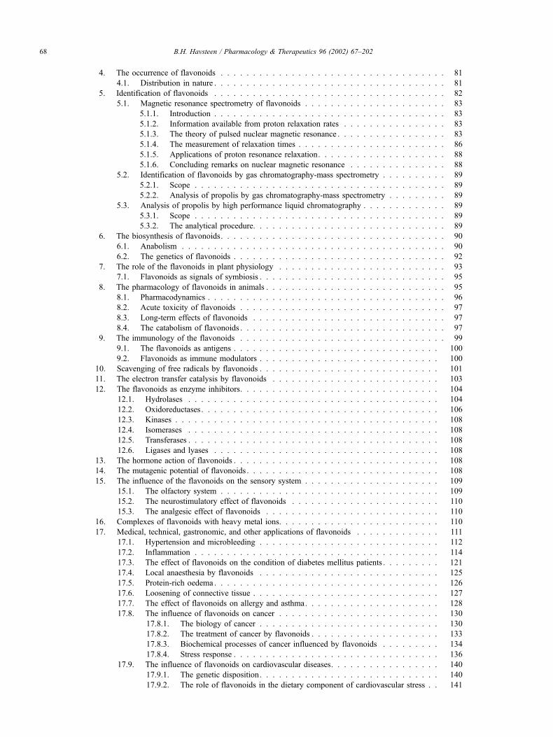
Fig. 1. Structure of benzo-g-pyrone. Note the numbering of the atoms of the
ring structure, which is essential to the nomenclature of the derivatives.
Examples: pelargonidin, R =H; R0=OH, R00=OH; cyanidin, R =OH;
R0 =OH, R00 =H; delphinidin, R =OH; R0 =OH, R00 =OH; peonidin,
R =OCH3; R0=OH, R00 =H; and malvidin, R =OCH3; R
0 =OH, R00 =OCH3.
Fig. 2. Structure, tautomerism, and mesomerism of anthocyanidines.
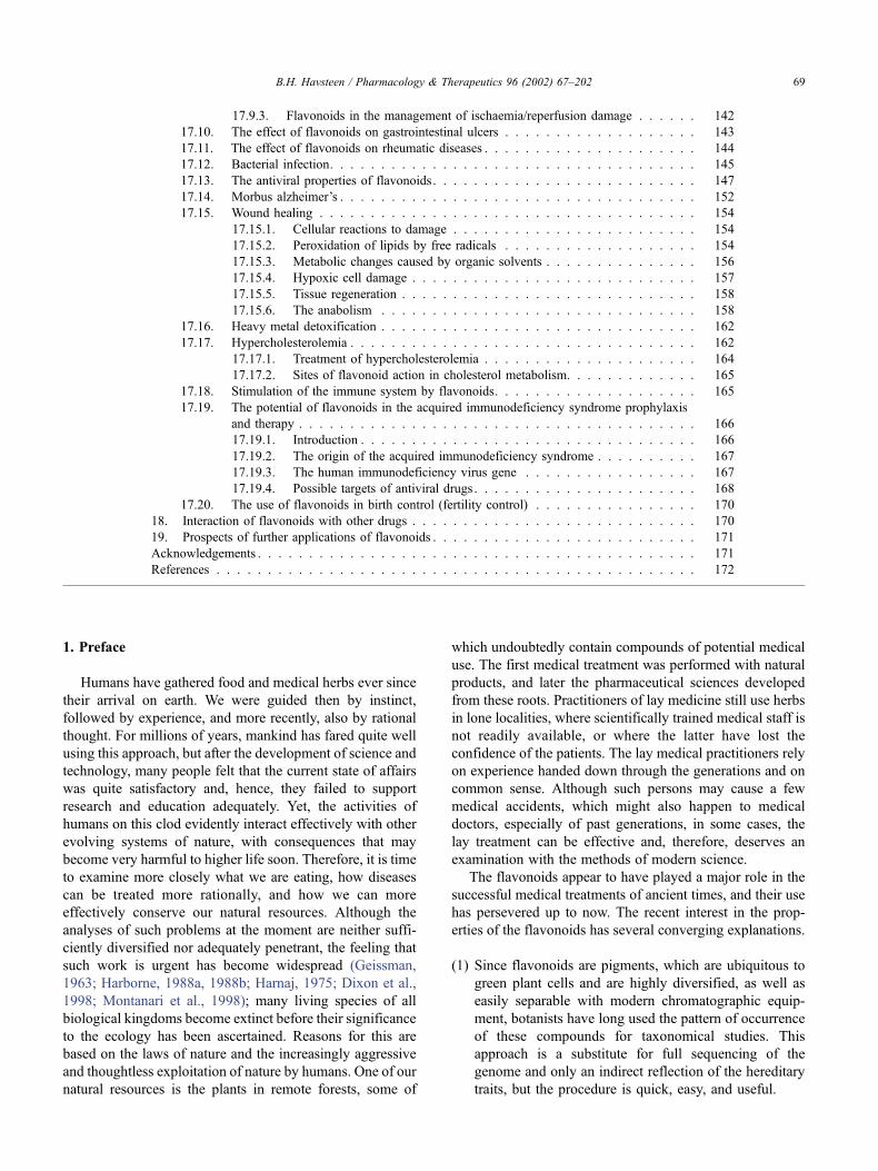
Fig. 3. Structure of flavonoles. Examples: kaempherol, R =H; R0 =OH;
quercetin, R =OH; R0 =OH.
B.H. Havsteen / Pharmacology & Therapeutics 96 (2002) 67–20270
mankind seems to indicate that there is no reason for great
alarm. On the other hand, we need to improve our know-
ledge of the effects of the food we eat. The evidence given
below shows that they are far from trivial. Detailed books
on flavonoids have been published, which impress by their
comprehensiveness in the description of the structures,
procedures of isolation, and approaches to the organic
synthesis of flavonoids. However, the wealth of detail is
likely to deter readers seeking clarity, basic principles, and
applications. Hence, there seems to be a need for a review
with a different emphasis.
3. The chemistry of flavonoids
3.1. Structure and nomenclature
The term flavonoids is a collective noun for plant pig-
ments, mostly derived from benzo-g-pyrone, which is
synonymous with chromone (Hassig et al., 1999; Harborne,
1964, 1967; Croft, 1998) (Fig. 1).
The group comprises anthocyanidines, hydroxyl-4-dihy-
droflavonoles; anthocyanides, glycosides of anthocyani-
dines (Fig. 2); flavonoles, 2-phenyl-3-hydroxy-chromones
(Fig. 3); iso-flavonoles, 3-phenyl-2-hydroxy-chromones
(Fig. 4); flavones, 2-phenyl-chromones (Fig. 5); iso-fla-
vones, 3-phenyl-chromones (Fig. 6); flavanes 2-phenyl-3-
dihydro-chromones, 2-phenyl-flavanones (Fig. 7); iso-fla-
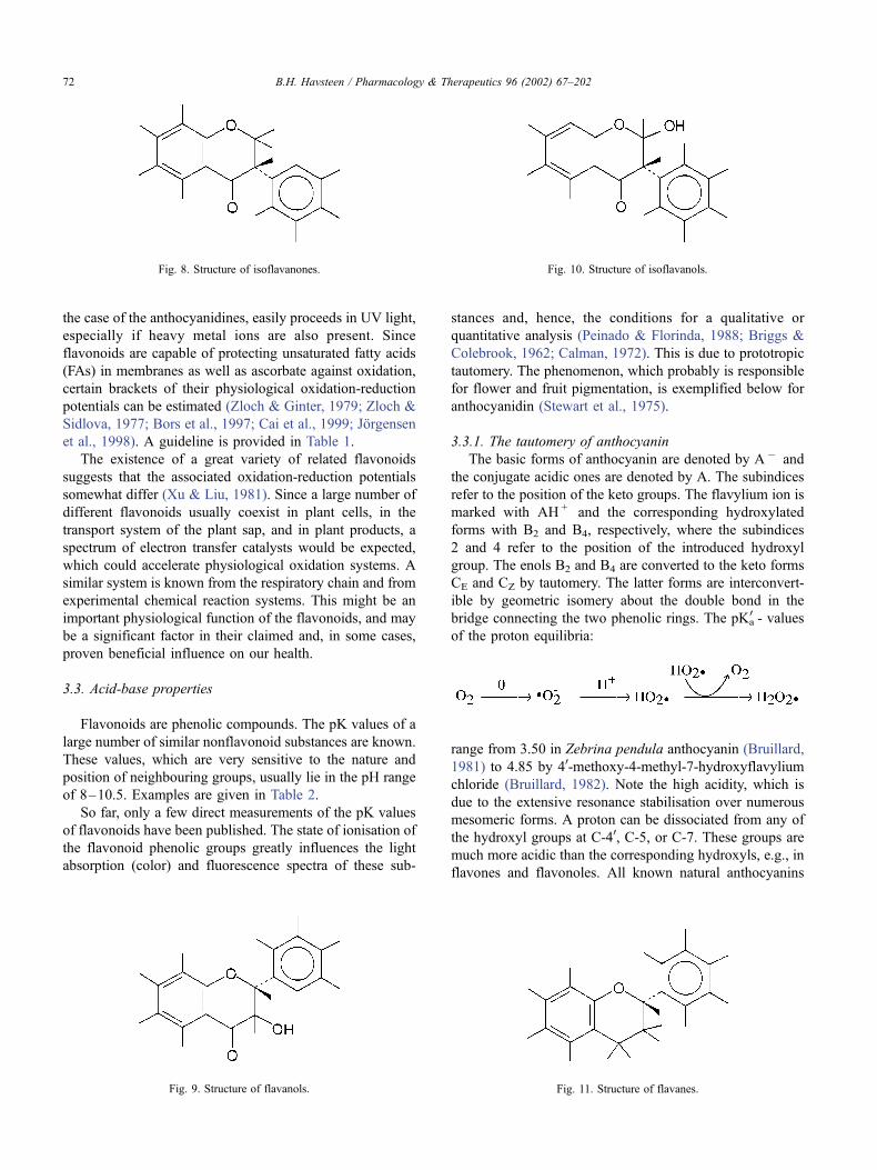
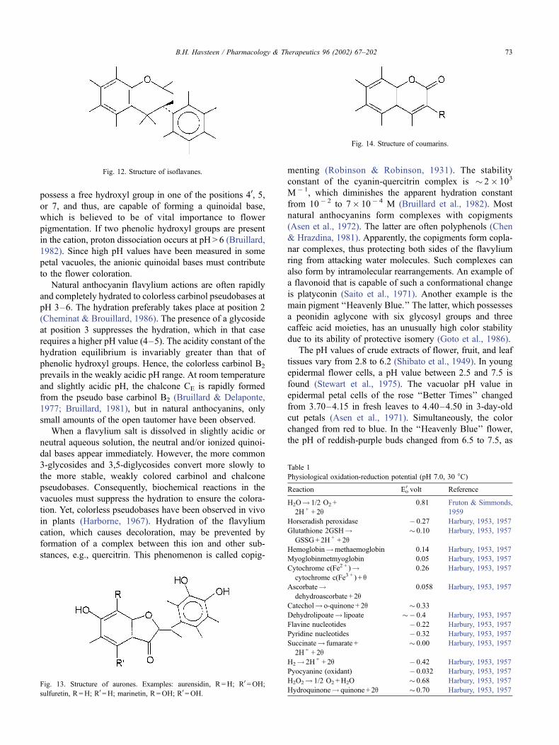
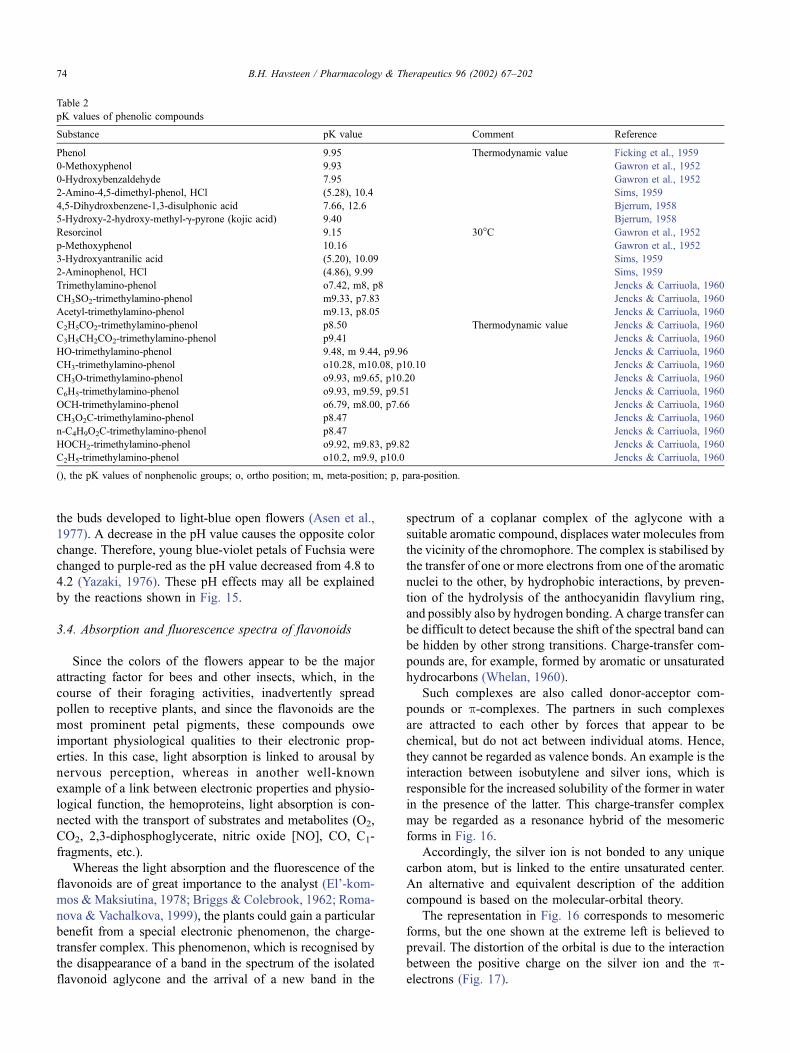
vones, 3-phenyl-2-dihydro-chromones (Fig. 8); flavanols,
2-phenyl-3-hydro-3-hydroxy-chromones (catechins) (Fig.
9); iso-flavanols, 2-hydro-2-hydroxy-3-phenyl-chromones
(Fig. 10); flavanes, 2-phenyl-di-hydro-benzo-g-pyranes
(Fig. 11); iso-flavanes, 3-phenyl-di-hydro-g-benzo-pyranes
(Fig. 12); aurones, benzo-furones (Fig. 13); and coumarins,
benzo-g-pyron derivatives (Fig. 14).
Reviews are found in Fruton and Simmonds (1959), Cody
et al. (1986a, 1986b), and Lahann and Purucker (1975).
Separate genes control the production of 40-hydroxylated
aglycones (e.g., pelargonidin, apigenin, and kaempferol) and
of 30,40-dihydroxylated aglycones (e.g., cyanidin, luteolin,
and quercetin) (Jorgensen & Geissman, 1955; Geissman &
Harborne, 1955; Geissman, 1962). The number and position
of hydroxyl groups attached to the A-ring are also controlled
by different genes, and the nature and position of the
carbohydrate units in the glycosides are determined by still
other genetic factors.
The color production is one of the most explored areas in
the study of the genetics of higher plants (Laurence & Price,
1940; Brouillard & Cheminat, 1988). The biosynthesis of the
plant pigments has been reviewed by Seshadri (1951) and
Peach (1955). Examples of the chemical synthesis of flavo-
noids are given by Baker and Robinson (1928), Dunne et al.
(1950), Mozingo and Atkins (1938), as well as by Tatsuda
(1947).
3.2. The oxidation-reduction potential of flavonoids
The flavonoids are phenolic compounds and, therefore,
are prone to oxidation to quinones. The process, which can
be accompanied with a ring opening at C1, which occurs in
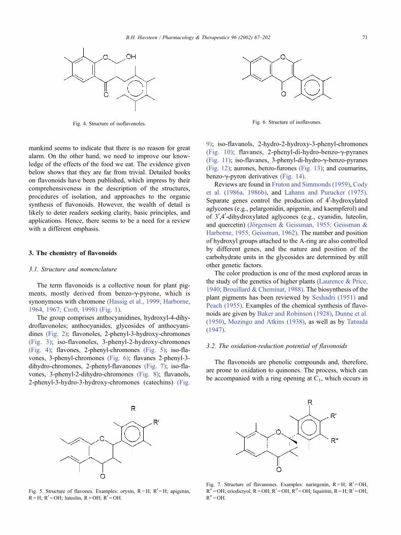
Fig. 4. Structure of isoflavonoles.
Fig. 5. Structure of flavones. Examples: orysin, R =H; R0=H; apigenin,
R =H; R0 =OH; luteolin, R =OH; R0=OH.
Fig. 6. Structure of isoflavones.
Fig. 7. Structure of flavanones. Examples: naringenin, R =H; R0=OH,
R00 =OH; eriodictyol, R =OH; R0=OH, R00=OH; liquiritin, R =H; R0 =OH,
R00 =OH.
B.H. Havsteen / Pharmacology & Therapeutics 96 (2002) 67–202 71
the case of the anthocyanidines, easily proceeds in UV light,
especially if heavy metal ions are also present. Since
flavonoids are capable of protecting unsaturated fatty acids
(FAs) in membranes as well as ascorbate against oxidation,
certain brackets of their physiological oxidation-reduction
potentials can be estimated (Zloch & Ginter, 1979; Zloch &
Sidlova, 1977; Bors et al., 1997; Cai et al., 1999; Jorgensen
et al., 1998). A guideline is provided in Table 1.
The existence of a great variety of related flavonoids
suggests that the associated oxidation-reduction potentials
somewhat differ (Xu & Liu, 1981). Since a large number of
different flavonoids usually coexist in plant cells, in the
transport system of the plant sap, and in plant products, a
spectrum of electron transfer catalysts would be expected,
which could accelerate physiological oxidation systems. A
similar system is known from the respiratory chain and from
experimental chemical reaction systems. This might be an
important physiological function of the flavonoids, and may
be a significant factor in their claimed and, in some cases,
proven beneficial influence on our health.
3.3. Acid-base properties
Flavonoids are phenolic compounds. The pK values of a
large number of similar nonflavonoid substances are known.
These values, which are very sensitive to the nature and
position of neighbouring groups, usually lie in the pH range
of 8–10.5. Examples are given in Table 2.
So far, only a few direct measurements of the pK values
of flavonoids have been published. The state of ionisation of
the flavonoid phenolic groups greatly influences the light
absorption (color) and fluorescence spectra of these sub-
stances and, hence, the conditions for a qualitative or
quantitative analysis (Peinado & Florinda, 1988; Briggs &
Colebrook, 1962; Calman, 1972). This is due to prototropic
tautomery. The phenomenon, which probably is responsible
for flower and fruit pigmentation, is exemplified below for
anthocyanidin (Stewart et al., 1975).
3.3.1. The tautomery of anthocyanin
The basic forms of anthocyanin are denoted by A � and
the conjugate acidic ones are denoted by A. The subindices
refer to the position of the keto groups. The flavylium ion is
marked with AH + and the corresponding hydroxylated
forms with B2 and B4, respectively, where the subindices
2 and 4 refer to the position of the introduced hydroxyl
group. The enols B2 and B4 are converted to the keto forms
CE and CZ by tautomery. The latter forms are interconvert-
ible by geometric isomery about the double bond in the
bridge connecting the two phenolic rings. The pKa0 - values
of the proton equilibria:
range from 3.50 in Zebrina pendula anthocyanin (Bruillard,
1981) to 4.85 by 40-methoxy-4-methyl-7-hydroxyflavylium
chloride (Bruillard, 1982). Note the high acidity, which is
due to the extensive resonance stabilisation over numerous
mesomeric forms. A proton can be dissociated from any of
the hydroxyl groups at C-40, C-5, or C-7. These groups are
much more acidic than the corresponding hydroxyls, e.g., in
flavones and flavonoles. All known natural anthocyanins
Fig. 8. Structure of isoflavanones.
Fig. 9. Structure of flavanols.
Fig. 10. Structure of isoflavanols.
Fig. 11. Structure of flavanes.
B.H. Havsteen / Pharmacology & Therapeutics 96 (2002) 67–20272
possess a free hydroxyl group in one of the positions 40, 5,
or 7, and thus, are capable of forming a quinoidal base,
which is believed to be of vital importance to flower
pigmentation. If two phenolic hydroxyl groups are present
in the cation, proton dissociation occurs at pH > 6 (Bruillard,
1982). Since high pH values have been measured in some
petal vacuoles, the anionic quinoidal bases must contribute
to the flower coloration.
Natural anthocyanin flavylium actions are often rapidly
and completely hydrated to colorless carbinol pseudobases at
pH 3–6. The hydration preferably takes place at position 2
(Cheminat & Brouillard, 1986). The presence of a glycoside
at position 3 suppresses the hydration, which in that case
requires a higher pH value (4–5). The acidity constant of the
hydration equilibrium is invariably greater than that of
phenolic hydroxyl groups. Hence, the colorless carbinol B2
prevails in the weakly acidic pH range. At room temperature
and slightly acidic pH, the chalcone CE is rapidly formed
from the pseudo base carbinol B2 (Bruillard & Delaponte,
1977; Bruillard, 1981), but in natural anthocyanins, only
small amounts of the open tautomer have been observed.
When a flavylium salt is dissolved in slightly acidic or
neutral aqueous solution, the neutral and/or ionized quinoi-
dal bases appear immediately. However, the more common
3-glycosides and 3,5-diglycosides convert more slowly to
the more stable, weakly colored carbinol and chalcone
pseudobases. Consequently, biochemical reactions in the
vacuoles must suppress the hydration to ensure the colora-
tion. Yet, colorless pseudobases have been observed in vivo
in plants (Harborne, 1967). Hydration of the flavylium
cation, which causes decoloration, may be prevented by
formation of a complex between this ion and other sub-
stances, e.g., quercitrin. This phenomenon is called copig-
menting (Robinson & Robinson, 1931). The stability
constant of the cyanin-quercitrin complex is � 2� 103
M � 1, which diminishes the apparent hydration constant
from 10� 2 to 7� 10� 4 M (Bruillard et al., 1982). Most
natural anthocyanins form complexes with copigments
(Asen et al., 1972). The latter are often polyphenols (Chen
& Hrazdina, 1981). Apparently, the copigments form copla-
nar complexes, thus protecting both sides of the flavylium
ring from attacking water molecules. Such complexes can
also form by intramolecular rearrangements. An example of
a flavonoid that is capable of such a conformational change
is platyconin (Saito et al., 1971). Another example is the
main pigment ‘‘Heavenly Blue.’’ The latter, which possesses
a peonidin aglycone with six glycosyl groups and three
caffeic acid moieties, has an unusually high color stability
due to its ability of protective isomery (Goto et al., 1986).
The pH values of crude extracts of flower, fruit, and leaf
tissues vary from 2.8 to 6.2 (Shibato et al., 1949). In young
epidermal flower cells, a pH value between 2.5 and 7.5 is
found (Stewart et al., 1975). The vacuolar pH value in
epidermal petal cells of the rose ‘‘Better Times’’ changed
from 3.70–4.15 in fresh leaves to 4.40–4.50 in 3-day-old
cut petals (Asen et al., 1971). Simultaneously, the color
changed from red to blue. In the ‘‘Heavenly Blue’’ flower,
the pH of reddish-purple buds changed from 6.5 to 7.5, as
Table 1
Physiological oxidation-reduction potential (pH 7.0, 30 �C)
Reaction Eo0 volt Reference
H2O! 1/2 O2 +
2H + + 2q0.81 Fruton & Simmonds,
1959
Horseradish peroxidase � 0.27 Harbury, 1953, 1957
Glutathione 2GSH!GSSG+ 2H + + 2q
� 0.10 Harbury, 1953, 1957
Hemoglobin!methaemoglobin 0.14 Harbury, 1953, 1957
Myoglobinmetmyoglobin 0.05 Harbury, 1953, 1957
Cytochrome c(Fe2 + )!cytochrome c(Fe3 + ) + q
0.26 Harbury, 1953, 1957
Ascorbate!dehydroascorbate + 2q
0.058 Harbury, 1953, 1957
Catechol! o-quinone + 2q � 0.33
Dehydrolipoate! lipoate �� 0.4 Harbury, 1953, 1957
Flavine nucleotides � 0.22 Harbury, 1953, 1957
Pyridine nucleotides � 0.32 Harbury, 1953, 1957
Succinate! fumarate +
2H + + 2q� 0.00 Harbury, 1953, 1957
H2! 2H + + 2q � 0.42 Harbury, 1953, 1957
Pyocyanine (oxidant) � 0.032 Harbury, 1953, 1957
H2O2! 1/2 O2 +H2O � 0.68 Harbury, 1953, 1957
Hydroquinone! quinone + 2q � 0.70 Harbury, 1953, 1957
Fig. 14. Structure of coumarins.
Fig. 12. Structure of isoflavanes.
Fig. 13. Structure of aurones. Examples: aurensidin, R =H; R0=OH;
sulfuretin, R =H; R0=H; marinetin, R =OH; R0=OH.
B.H. Havsteen / Pharmacology & Therapeutics 96 (2002) 67–202 73
the buds developed to light-blue open flowers (Asen et al.,
1977). A decrease in the pH value causes the opposite color
change. Therefore, young blue-violet petals of Fuchsia were
changed to purple-red as the pH value decreased from 4.8 to
4.2 (Yazaki, 1976). These pH effects may all be explained
by the reactions shown in Fig. 15.
3.4. Absorption and fluorescence spectra of flavonoids
Since the colors of the flowers appear to be the major
attracting factor for bees and other insects, which, in the
course of their foraging activities, inadvertently spread
pollen to receptive plants, and since the flavonoids are the
most prominent petal pigments, these compounds owe
important physiological qualities to their electronic prop-
erties. In this case, light absorption is linked to arousal by
nervous perception, whereas in another well-known
example of a link between electronic properties and physio-
logical function, the hemoproteins, light absorption is con-
nected with the transport of substrates and metabolites (O2,
CO2, 2,3-diphosphoglycerate, nitric oxide [NO], CO, C1-
fragments, etc.).
Whereas the light absorption and the fluorescence of the
flavonoids are of great importance to the analyst (El’-kom-
mos &Maksiutina, 1978; Briggs & Colebrook, 1962; Roma-
nova & Vachalkova, 1999), the plants could gain a particular
benefit from a special electronic phenomenon, the charge-
transfer complex. This phenomenon, which is recognised by
the disappearance of a band in the spectrum of the isolated
flavonoid aglycone and the arrival of a new band in the
spectrum of a coplanar complex of the aglycone with a
suitable aromatic compound, displaces water molecules from
the vicinity of the chromophore. The complex is stabilised by
the transfer of one or more electrons from one of the aromatic
nuclei to the other, by hydrophobic interactions, by preven-
tion of the hydrolysis of the anthocyanidin flavylium ring,
and possibly also by hydrogen bonding. A charge transfer can
be difficult to detect because the shift of the spectral band can
be hidden by other strong transitions. Charge-transfer com-
pounds are, for example, formed by aromatic or unsaturated
hydrocarbons (Whelan, 1960).
Such complexes are also called donor-acceptor com-
pounds or p-complexes. The partners in such complexes
are attracted to each other by forces that appear to be
chemical, but do not act between individual atoms. Hence,
they cannot be regarded as valence bonds. An example is the
interaction between isobutylene and silver ions, which is
responsible for the increased solubility of the former in water
in the presence of the latter. This charge-transfer complex
may be regarded as a resonance hybrid of the mesomeric
forms in Fig. 16.
Accordingly, the silver ion is not bonded to any unique
carbon atom, but is linked to the entire unsaturated center.
An alternative and equivalent description of the addition
compound is based on the molecular-orbital theory.
The representation in Fig. 16 corresponds to mesomeric
forms, but the one shown at the extreme left is believed to
prevail. The distortion of the orbital is due to the interaction
between the positive charge on the silver ion and the p-electrons (Fig. 17).
Table 2
pK values of phenolic compounds
Substance pK value Comment Reference
Phenol 9.95 Thermodynamic value Ficking et al., 1959
0-Methoxyphenol 9.93 Gawron et al., 1952
0-Hydroxybenzaldehyde 7.95 Gawron et al., 1952
2-Amino-4,5-dimethyl-phenol, HCl (5.28), 10.4 Sims, 1959
4,5-Dihydroxbenzene-1,3-disulphonic acid 7.66, 12.6 Bjerrum, 1958
5-Hydroxy-2-hydroxy-methyl-g-pyrone (kojic acid) 9.40 Bjerrum, 1958
Resorcinol 9.15 30�C Gawron et al., 1952
p-Methoxyphenol 10.16 Gawron et al., 1952
3-Hydroxyantranilic acid (5.20), 10.09 Sims, 1959
2-Aminophenol, HCl (4.86), 9.99 Sims, 1959
Trimethylamino-phenol o7.42, m8, p8 Jencks & Carriuola, 1960
CH3SO2-trimethylamino-phenol m9.33, p7.83 Jencks & Carriuola, 1960
Acetyl-trimethylamino-phenol m9.13, p8.05 Jencks & Carriuola, 1960
C2H5CO2-trimethylamino-phenol p8.50 Thermodynamic value Jencks & Carriuola, 1960
C3H5CH2CO2-trimethylamino-phenol p9.41 Jencks & Carriuola, 1960
HO-trimethylamino-phenol 9.48, m 9.44, p9.96 Jencks & Carriuola, 1960
CH3-trimethylamino-phenol o10.28, m10.08, p10.10 Jencks & Carriuola, 1960
CH3O-trimethylamino-phenol o9.93, m9.65, p10.20 Jencks & Carriuola, 1960
C6H5-trimethylamino-phenol o9.93, m9.59, p9.51 Jencks & Carriuola, 1960
OCH-trimethylamino-phenol o6.79, m8.00, p7.66 Jencks & Carriuola, 1960
CH3O2C-trimethylamino-phenol p8.47 Jencks & Carriuola, 1960
n-C4H9O2C-trimethylamino-phenol p8.47 Jencks & Carriuola, 1960
HOCH2-trimethylamino-phenol o9.92, m9.83, p9.82 Jencks & Carriuola, 1960
C2H5-trimethylamino-phenol o10.2, m9.9, p10.0 Jencks & Carriuola, 1960
(), the pK values of nonphenolic groups; o, ortho position; m, meta-position; p, para-position.
B.H. Havsteen / Pharmacology & Therapeutics 96 (2002) 67–20274
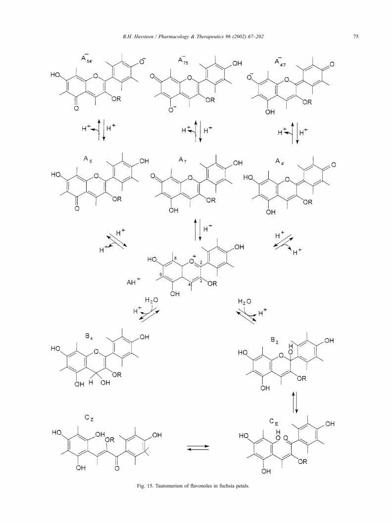
Fig. 15. Tautomerism of flavonoles in fuchsia petals.
B.H. Havsteen / Pharmacology & Therapeutics 96 (2002) 67–202 75
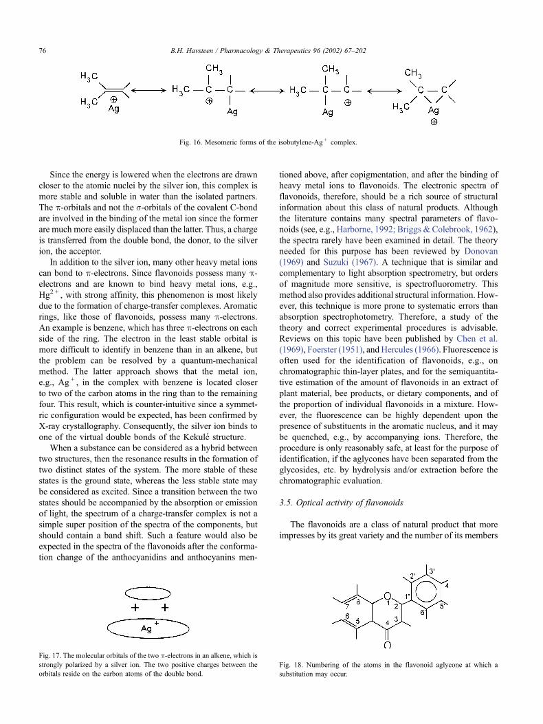
Since the energy is lowered when the electrons are drawn
closer to the atomic nuclei by the silver ion, this complex is
more stable and soluble in water than the isolated partners.
The p-orbitals and not the s-orbitals of the covalent C-bondare involved in the binding of the metal ion since the former
are much more easily displaced than the latter. Thus, a charge
is transferred from the double bond, the donor, to the silver
ion, the acceptor.
In addition to the silver ion, many other heavy metal ions
can bond to p-electrons. Since flavonoids possess many p-electrons and are known to bind heavy metal ions, e.g.,
Hg2 + , with strong affinity, this phenomenon is most likely
due to the formation of charge-transfer complexes. Aromatic
rings, like those of flavonoids, possess many p-electrons.An example is benzene, which has three p-electrons on eachside of the ring. The electron in the least stable orbital is
more difficult to identify in benzene than in an alkene, but
the problem can be resolved by a quantum-mechanical
method. The latter approach shows that the metal ion,
e.g., Ag + , in the complex with benzene is located closer
to two of the carbon atoms in the ring than to the remaining
four. This result, which is counter-intuitive since a symmet-
ric configuration would be expected, has been confirmed by
X-ray crystallography. Consequently, the silver ion binds to
one of the virtual double bonds of the Kekule structure.
When a substance can be considered as a hybrid between
two structures, then the resonance results in the formation of
two distinct states of the system. The more stable of these
states is the ground state, whereas the less stable state may
be considered as excited. Since a transition between the two
states should be accompanied by the absorption or emission
of light, the spectrum of a charge-transfer complex is not a
simple super position of the spectra of the components, but
should contain a band shift. Such a feature would also be
expected in the spectra of the flavonoids after the conforma-
tion change of the anthocyanidins and anthocyanins men-
tioned above, after copigmentation, and after the binding of
heavy metal ions to flavonoids. The electronic spectra of
flavonoids, therefore, should be a rich source of structural
information about this class of natural products. Although
the literature contains many spectral parameters of flavo-
noids (see, e.g., Harborne, 1992; Briggs & Colebrook, 1962),
the spectra rarely have been examined in detail. The theory
needed for this purpose has been reviewed by Donovan
(1969) and Suzuki (1967). A technique that is similar and
complementary to light absorption spectrometry, but orders
of magnitude more sensitive, is spectrofluorometry. This
method also provides additional structural information. How-
ever, this technique is more prone to systematic errors than
absorption spectrophotometry. Therefore, a study of the
theory and correct experimental procedures is advisable.
Reviews on this topic have been published by Chen et al.
(1969), Foerster (1951), and Hercules (1966). Fluorescence is
often used for the identification of flavonoids, e.g., on
chromatographic thin-layer plates, and for the semiquantita-
tive estimation of the amount of flavonoids in an extract of
plant material, bee products, or dietary components, and of
the proportion of individual flavonoids in a mixture. How-
ever, the fluorescence can be highly dependent upon the
presence of substituents in the aromatic nucleus, and it may
be quenched, e.g., by accompanying ions. Therefore, the
procedure is only reasonably safe, at least for the purpose of
identification, if the aglycones have been separated from the
glycosides, etc. by hydrolysis and/or extraction before the
chromatographic evaluation.
3.5. Optical activity of flavonoids
The flavonoids are a class of natural product that more
impresses by its great variety and the number of its members
Fig. 16. Mesomeric forms of the isobutylene-Ag + complex.
Fig. 17. The molecular orbitals of the two p-electrons in an alkene, which isstrongly polarized by a silver ion. The two positive charges between the
orbitals reside on the carbon atoms of the double bond.
Fig. 18. Numbering of the atoms in the flavonoid aglycone at which a
substitution may occur.
B.H. Havsteen / Pharmacology & Therapeutics 96 (2002) 67–20276
than by the complexity of the constituents in the structure. If
the positions in the characteristic flavonoid nucleus, at
which derivatives most commonly are formed by hydrox-
ylation, methylation, acetylation, glycosylation, isoprenyla-
tion, etc. (Fig. 18), then it is possible by simple permutation
to estimate the number of individual flavonoids that we can
expect to find in nature.
The flavonoids are often hydroxylated in positions 3, 5,
7, 30, 40, and 50. Some of these hydroxyl groups are
frequently methylated, acetylated, or sulphated. When gly-
cosides are formed, the glycosidic linkage is normally
located in position 3 or 7, and the carbohydrates are
commonly L-rhamnose, D-glucose, glucorhamnose, galac-
tose, or arabinose (Kuhnau, 1976). Prenylation usually
occurs directly at a carbon atom in the aromatic rings, but
0-prenylation has also been found.
These features alone can account for � 3� 105 members
of the flavonoid class, but the latter also includes a large
number of more exotic forms, which have been omitted here
for the sake of simplicity. The actual number of flavonoids
that have been found so far and for which the structure has
been completely elucidated is large, but probably does not
exceed 1% of the theoretical number of possible variants.
The discovery of a large number of additional, naturally
occurring flavonoids, therefore, must be expected. This
abundance of variants is further augmented by the chirality
of the subunits and their connections. Since many stereo-
isomers do not differ significantly in their electronic or
fluorescence spectra, the optical activity of the species is
often a very useful analytical parameter. Incident linearly or
circularly polarised electromagnetic waves sensitively inter-
act with the electrons of the substance examined, thus
shifting the phase of the former and producing a change in
the optical rotation. This effect is wavelength-dependent, but
particularly sensitive in the UV range. Accordingly, optical
rotatory dispersion spectra or their correlate, circular dichro-
ism spectra, often are very useful to distinguish between
stereoisomers, to identify the absolute configuration of the
structure, and to recognise centers of chirality. The theory,
applications and experimental techniques of optical rotatory
dispersion and circular dichroism, have been reviewed by
Imahori and Nicola (1973), Djerassi (1960), Tinoco (1970),
and Gaffield (1970), as well as by Sears and Beychok
(1973).
3.6. Radical scavenging by flavonoids
One of the prominent and medically most useful prop-
erties of many flavonoids is their ability to scavenge free
radicals (Agarwal & Nagaratnam, 1981; Wang & Zheng,
1992; Robak & Gryglewski, 1988; Gyorgy et al., 1992; van
Acker et al., 1995, 1996; Ubeda et al., 1995; Clemetson &
Andersen, 1966). These highly reactive species arise in the
course of many physiological processes, especially in the
respiratory chain and in oxidations catalyzed by oxy-
genases. These reactions are very common since molecular
oxygen is a very good electron acceptor. Its high affinity for
electrons provides the thermodynamic driving force, but in
contrast to other electron acceptors, dioxygen reacts only
slowly in the absence of a catalyst. The transfer of four
electrons to dioxygen generates two molecules of water,
i.e., a safe product, but partial reduction leads to the
formation of highly toxic compounds, e.g., the superoxide
anion �O2� :
O2 þ q� ! �O�2
This species is, e.g., formed by macrophages in the first
line of defence against invading foreign cells or virus
particles. This reaction is desirable, but excess superoxide
anion must be removed quickly before it has the opportunity
to destroy too many essential, unsaturated liquids in the
membranes, as well as sulfhydryl groups, e.g., in the active
sites of key enzymes.
Normally, the release of partly reduced intermediates in
the reaction with dioxygen is prevented. In the case of
cytochrome oxidase, this reaction is mediated by metal ions.
The dioxygen molecule is suspended at first between the
Fe2 + ions and the Cu + ions of the a3-CuB center in this
enzyme (Stryer, 1988). Each metal ion then donates an
electron to O2, thus converting it to a dianion. Subsequently,
an electron is donated from the cyt a-CuA center, which
produces an intermediate ferryl ion. After the uptake of two
protons from the medium, a water molecule is formed and
the transfer of a second electron leaves a hydroxyl ion
bound to Fe3 + . A third electron reduces Cu2 + to Cu + , and
the uptake of a proton produces a second water molecule
(Fig. 19).
If superoxide, e.g., due to denaturation of the enzyme,
escapes the heme protein before its full reduction, this free
radical starts a chain reaction that may involve nucleic acid
bases and many other vital cellular compounds, and results
in mutation, metabolic derailment, and possibly cancer.
Protonation of the superoxide anion yields the hydroper-
oxide radical HO2�, which spontaneously reacts with a
second of these anions to produce H2O2:
Another common source of free radicals is radiation, e.g.,
X-rays or g-rays. The main target is water, due to its
ubiquity and high concentration in living organisms. Upon
irradiation, this molecule produces hydroxyl radicals �OH,which, apart from the above mentioned targets, also attacks
other free radicals, e.g., NO� and superoxide, thus forming
peroxynitrite, H2O2, nitrous acid, etc. The production of
peroxynitrite is suppressed by flavonoids (Haenen et al.,
1997). Naturally, the organism has developed a defence
against such toxic substances. An important mechanism is
B.H. Havsteen / Pharmacology & Therapeutics 96 (2002) 67–202 77
catalyzed by the enzyme superoxide dismutase (SOD),
which converts two superoxide anions to H2O2 and O2:
�O�2 þ �O�
2!2Hþ
SODH2O2 þ O2
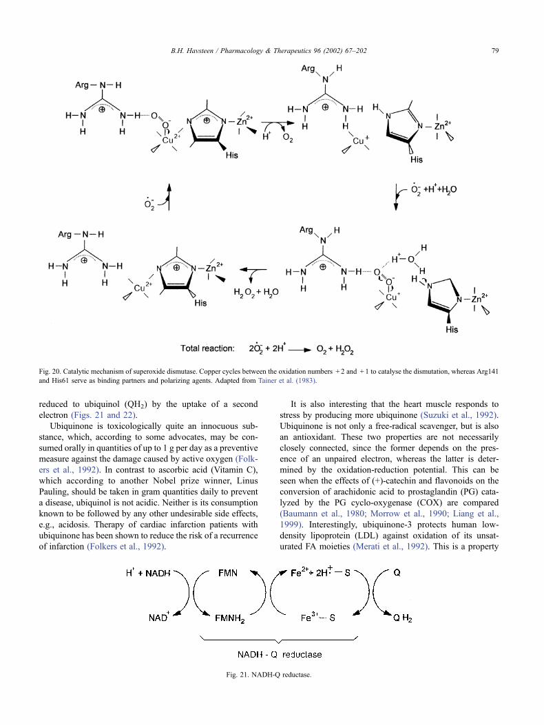
The active site of the cytosolic, eucaryotic SOD contains a
copper ion and a zinc ion coordinated to the imidazole
moiety of a histidine residue. The negatively charged super-
oxide is guided electrostatically to a positively charged
catalytic site at the bottom of a crevice. The superoxide
anion binds to Cu2 + and the guanidino group of an arginine
residue. An electron is transferred to Cu2 + from �O2� to form
Cu + and O2. The latter molecule is then released. Then a
second superoxide anion enters the cavity to bind to Cu + ,
arginine, and H3O+ . An electron is transferred from Cu + ,
and two protons are delivered from the two other binding
partners to form H2O2 and to regenerate Cu2 + (Fig. 20).
SOD is a relatively small enzyme that can be injected
into the blood stream without much danger of immuno-
logical complications. It is used to scavenge free radicals in
the reperfusion phase after ischemic heart stop, e.g., during
heart transplantation (Gulati et al., 1992; Fritz-Niggli,
1968). Another molecule, which is used for the same
purpose, is ubiquinone, coenzyme Q. This compound is of
particular interest since its properties resemble those of the
flavonoids. Ubiquinone (Q) is an active participant in the
respiratory chain (Fig. 21). Like cytochrome C, ubiquinone
is a soluble substance and, hence, diffusible, but unlike
cytochrome C, it can also traverse many biological lipid
membranes. Therefore, it is easy to administer to support the
respiratory chain, as well as the associated oxidative phos-
phorylation, and to scavenge free radicals.
During the operation of the respiratory chain, electrons
flow from iron-sulfur clusters of the NADH-Q reductase to
ubiquinone. The latter compound is a quinone derivative
with an isoprenoid tail, the length of which in mammals
usually is 10 isoprene units (Q10). As in the case of the
cytochromes, but different from the pyridine nucleotides
and the flavonucleotides, a single electron is transferred to
ubiquinone, which reduces it to an intermediate free-radical
semiquinone. This intermediate avidly scavenges other free
radicals that may be present, and this effect accounts in part
for the protective effect of ubiquinone, e.g., against active
oxygen species. In the respiratory chain, the semiquinone is
Fig. 19. The four electron reduction of O2 by cytochrome oxidase. Resp. chain, respiratory chain. Adapted from Stryer (1988).
B.H. Havsteen / Pharmacology & Therapeutics 96 (2002) 67–20278
reduced to ubiquinol (QH2) by the uptake of a second
electron (Figs. 21 and 22).
Ubiquinone is toxicologically quite an innocuous sub-
stance, which, according to some advocates, may be con-
sumed orally in quantities of up to 1 g per day as a preventive
measure against the damage caused by active oxygen (Folk-
ers et al., 1992). In contrast to ascorbic acid (Vitamin C),
which according to another Nobel prize winner, Linus
Pauling, should be taken in gram quantities daily to prevent
a disease, ubiquinol is not acidic. Neither is its consumption
known to be followed by any other undesirable side effects,
e.g., acidosis. Therapy of cardiac infarction patients with
ubiquinone has been shown to reduce the risk of a recurrence
of infarction (Folkers et al., 1992).
It is also interesting that the heart muscle responds to
stress by producing more ubiquinone (Suzuki et al., 1992).
Ubiquinone is not only a free-radical scavenger, but is also
an antioxidant. These two properties are not necessarily
closely connected, since the former depends on the pres-
ence of an unpaired electron, whereas the latter is deter-
mined by the oxidation-reduction potential. This can be
seen when the effects of (+)-catechin and flavonoids on the
conversion of arachidonic acid to prostaglandin (PG) cata-
lyzed by the PG cyclo-oxygenase (COX) are compared
(Baumann et al., 1980; Morrow et al., 1990; Liang et al.,
1999). Interestingly, ubiquinone-3 protects human low-
density lipoprotein (LDL) against oxidation of its unsat-
urated FA moieties (Merati et al., 1992). This is a property
Fig. 20. Catalytic mechanism of superoxide dismutase. Copper cycles between the oxidation numbers + 2 and + 1 to catalyse the dismutation, whereas Arg141
and His61 serve as binding partners and polarizing agents. Adapted from Tainer et al. (1983).
Fig. 21. NADH-Q reductase.
B.H. Havsteen / Pharmacology & Therapeutics 96 (2002) 67–202 79
also ascribed to flavonoids, i.e., another point of resemb-
lance (Brown & Rice-Evans, 1998).
A key enzyme in the biosynthesis of cholesterol, 3-
hydroxy-3-methyl-glutaryl-coenzyme A reductase (HMG-
CoA reductase) is inhibited by ubiquinone. As a result, the
cholesterol concentration in blood serum in the case of a
hypercholesterolemia can be reduced by the oral intake of
coenzyme Q (Sharma, 1979). The same is claimed for the
flavonoids (Chai et al., 1981; Oganesyan et al., 1989).
A substance that, for example, is important to the stability
of erythrocytes is glutathione (GSH). The oxidation of GSH
to GSSG by superoxide leads to hemolysis. The GSH thiol
free radical can be eliminated by reaction of GSSG with a
semiquinone free radical, e.g., of coenzyme Q or a flavonoid
(Iio et al., 1993; Galati et al., 1999; Kaneko & Baba, 1999).
However, some flavonoids exist that inhibit GSH reductase
(Elliott et al., 1992; Khushbaktova et al., 1991). Hence, the
oxidised form of GSH, GSSG, in that case can no longer be
reduced. The result will be an enhancement of hemolysis.
Thus, the effect of different flavonoids in a mixture can
antagonise each other. To the therapist, this means that it is
advisable to analyse a natural product for its flavonoids
before its use, or to apply a pure flavonoid. Flavonoids can
also inhibit GSH S-transferase, which can compromise the
transport of amino acids across membranes (Frohlich et al.,
1989; Yokozawa et al., 1999).
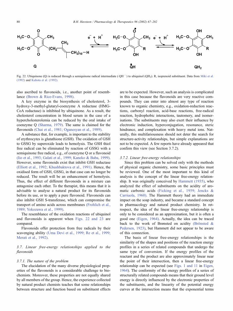
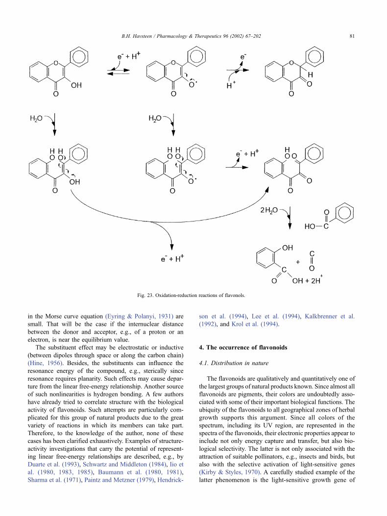
The resemblance of the oxidation reactions of ubiquinol
and flavonoids is apparent when Figs. 22 and 23 are
compared.
Flavonoids offer protection from free radicals by their
scavenging ability (Uma Devi et al., 1999; Re et al., 1999;
Merati et al., 1992).
3.7. Linear free-energy relationships applied to the
flavonoids
3.7.1. The nature of the problem
The elucidation of the many diverse physiological prop-
erties of the flavonoids is a considerable challenge to bio-
chemists. Moreover, these properties are not equally shared
by all members of the group. Hence, the experience collected
by natural product chemists teaches that some relationships
between structure and function based on substituent effects
are to be expected. However, such an analysis is complicated
in this case because the flavonoids are very reactive com-
pounds. They can enter into almost any type of reaction
known to organic chemistry, e.g., oxidation-reduction reac-
tions, carbonyl reaction, acid-base reactions, free-radical
reaction, hydrophobic interactions, tautomery, and isomer-
isations. The substituents may also exert their influence by
electronic induction, hyperconjugation, resonance, steric
hindrance, and complexation with heavy metal ions. Nat-
urally, this multifariousness should not deter the search for
structure-activity relationships, but simple explanations are
not to be expected. A few reports have already appeared that
confirm this view (see Section 3.7.2).
3.7.2. Linear free-energy relationships
Since this problem can be solved only with the methods
of physical organic chemistry, some basic principles must
be reviewed. One of the most important to this kind of
analysis is the concept of the linear free-energy relation-
ship. It was originally conceived by Hammett (1935), who
analyzed the effect of substituents on the acidity of aro-
matic carbonic acids (Ficking et al., 1959; Jencks &
Carriuola, 1960). The Hammett theory had an immediate
impact on the soap industry, and became a standard concept
in pharmacology and natural product chemistry. In ret-
rospect, the idea of the linear free-energy relationship is
only to be considered as an approximation, but it is often a
good one (Eigen, 1964). Actually, the idea can be traced
back to the work of Brønsted on acidity (Bronsted &
Pedersen, 1923), but Hammett did not appear to be aware
of this connection.
The basis of linear free-energy relationships is the
similarity of the shapes and positions of the reaction energy
profiles in a series of related compounds that undergo the
same type of conversion. If the energy profiles of the
reactant and the product are also approximately linear near
the point of their intersection, then a linear free-energy
relationship can be expected (see Figs. 1 and 11 in Eigen,
1964). The conformity of the energy profiles of a series of
structurally related compounds means that their ground level
energy is directly influenced by the electronic properties of
the substituents, and the linearity of the potential energy
curves at the intersection means that the exponential terms
Fig. 22. Ubiquinone (Q) is reduced through a semiquinone radical intermediate (�QH� ) to ubiquinol (QH2). R, isoprenoid substituent. Data from Miki et al.
(1992) and Kubota et al. (1992).
B.H. Havsteen / Pharmacology & Therapeutics 96 (2002) 67–20280
in the Morse curve equation (Eyring & Polanyi, 1931) are
small. That will be the case if the internuclear distance
between the donor and acceptor, e.g., of a proton or an
electron, is near the equilibrium value.
The substituent effect may be electrostatic or inductive
(between dipoles through space or along the carbon chain)
(Hine, 1956). Besides, the substituents can influence the
resonance energy of the compound, e.g., sterically since
resonance requires planarity. Such effects may cause depar-
ture from the linear free-energy relationship. Another source
of such nonlinearities is hydrogen bonding. A few authors
have already tried to correlate structure with the biological
activity of flavonoids. Such attempts are particularly com-
plicated for this group of natural products due to the great
variety of reactions in which its members can take part.
Therefore, to the knowledge of the author, none of these
cases has been clarified exhaustively. Examples of structure-
activity investigations that carry the potential of represent-
ing linear free-energy relationships are described, e.g., by
Duarte et al. (1993), Schwartz and Middleton (1984), Iio et
al. (1980, 1983, 1985), Baumann et al. (1980, 1981),
Sharma et al. (1971), Paintz and Metzner (1979), Hendrick-
son et al. (1994), Lee et al. (1994), Kalkbrenner et al.
(1992), and Krol et al. (1994).
4. The occurrence of flavonoids
4.1. Distribution in nature
The flavonoids are qualitatively and quantitatively one of
the largest groups of natural products known. Since almost all
flavonoids are pigments, their colors are undoubtedly asso-
ciated with some of their important biological functions. The
ubiquity of the flavonoids to all geographical zones of herbal
growth supports this argument. Since all colors of the
spectrum, including its UV region, are represented in the
spectra of the flavonoids, their electronic properties appear to
include not only energy capture and transfer, but also bio-
logical selectivity. The latter is not only associated with the
attraction of suitable pollinators, e.g., insects and birds, but
also with the selective activation of light-sensitive genes
(Kirby & Styles, 1970). A carefully studied example of the
latter phenomenon is the light-sensitive growth gene of
Fig. 23. Oxidation-reduction reactions of flavonols.
B.H. Havsteen / Pharmacology & Therapeutics 96 (2002) 67–202 81
barley. Although there is strong light absorption by the
flavonoids and they are present in all plant cells containing
plastids, no evidence of a participation of the flavonoids in the
primary photosynthetic process is known. Evidently, plants
are using light not only as a source of energy, but also for gene
regulation.
Another striking electronic property of the flavonoids is
their fluorescence. It remains yet to be proven whether this
property is used physiologically. However, such a function is
conceivable, since fluorescence can transfer small amounts
of energy, which may suffice to activate pigments associated
with light-sensitive genes.
The ubiquity and great diversity of the flavonoids render
these pigments suitable for a taxonomical classification.
Their usefulness for this purpose is enhanced by the close
association of the flavonoids with vital genes, especially
those involved in growth regulation. Such genes would be
expected to be particularly sensitive to environmental cues
and, hence, mirror both the nature of the biotope and the
competitive strength of the species.
The basis of the great variability of the flavonoids is:
1) differences in the ring structure of the aglycone and in its
state of oxidation/reduction;
2) differences in the extent of hydroxylation of the aglycone
and in the positions of the hydroxyl groups;
3) differences in the derivatisation of the hydroxyl groups,
e.g., with methyl groups, carbohydrates, or isoprenoids.
A permutation of these sources of variability reveals that
theoretically, more than 2� 106 different flavonoid species
can occur. So far, more than 2� 103 different flavonoids
have been identified, and their number is growing rapidly
(Bilyk & Sapers, 1985; Farkas et al., 1986; Cizmarik &
Matel, 1970, 1973; DuBois & Sneden, 1995). Since flavo-
noid family members are closely related structurally, it is
difficult to separate them (Hostettmann & Hostettmann,
1982). Besides, they are easily decomposed. Due to the
relatively high molecular weight and the complicated struc-
ture of these compounds, their identification and chemical
synthesis represent a challenge to the organic chemists, even
if they possess modern equipment.
Since the flavonoids, depending on their content of glyco-
sides, isoprenoids, and aliphatic ethers, can acquire almost
any polarity, a range of solvents from water to ethyl ether
must be used for their extraction from a complex mixture,
e.g., in propolis (bees glue), honey, wax, syrup, or plant
tissue. The extracts are often subfractionated on hydroxyla-
patite before a final separation is carried out by capillary
electrophoresis (Cancalon, 1999) or HPLC (Galensa & Herr-
mann, 1979; Garcia-Viguera et al., 1993; Greenaway et al.,
1987, 1991; Watson & Pitt, 1998; Watson & Oliveira, 1999;
Ishii et al., 1996; Gawron et al., 1952).
Several publications specializing in the identification of
flavonoids have appeared. Prominent examples are Mabry et
al. (1970), Markham (1982), Harborne (1988a, 1988b),
Bankova et al. (1982, 1987), Pangarova et al. (1980, 1986),
Garcia-Viguera et al. (1993), and Bonvehi et al. (1994).
5. Identification of flavonoids
The complete analysis of the absolute structure and
configuration of a flavonoid is usually a complicated task,
which requires the application of advanced techniques, e.g.,
[1H]- and [13C]-NMR-spectrometry, [1H-1H]-correlated
spectroscopy, circular dichroism, optical rotatory dispersion,
mass spectrometry, and X-ray diffraction. Since only a few
laboratories are equipped and staffed to make all of these
expensive methods available, simpler approaches to the
characterisation of flavonoids are desired. Modern chro-
matographic techniques like HPLC have become standard
equipment in biochemistry laboratories, and often yield not
only an excellent resolution, but also retention times that can
be very useful in the identification of a flavonoid. A much
less expensive method to acquire an impression of the
nature and amounts of individual flavonoids in an extract
is a combination of thin-layer chromatography and fluor-
escence (Jay et al., 1975; Ghisalberti, 1979; Nikolov et al.,
1976; Hladon et al., 1980; Lavie, 1978; Chi et al., 1994;
Glencross et al., 1972; McMurrough et al., 1985; Issaq,
1997; Karting & Hiermann, 1978).
The preparation of a sample for analysis can present a
problem since flavonoid glycosides are predominantly
polar structures and, hence, water-soluble, whereas the
aglycones are nonpolar (Calman, 1972). The latter, there-
fore, must be extracted by nonpolar solvents. Methanol is
often a useful compromise that permits the extraction of the
majority of the flavonoids. A particularly mild and efficient
extraction procedure for lipophilic flavonoids is triple-point
extraction with CO2. This procedure is rapidly gaining
acceptance. If a primitive method must be applied, a
sample of 50 mg of solid material may be extracted with
1 mL of methanol or amyl alcohol at room temperature in
15 min with shaking. A standard mixture of known
flavonoids may be used as references. The positions of
the flavonoids can be observed in UV light from a hand
lamp. The characteristic colors emitted by individual fla-
vonoids in a mixture, when exposed to UV light, aids in
their identification (see Section 4.1). Jay et al. (1975) have
published an extensive table of the mobilities in various
solvents and the fluorescence colors of flavonoids (� 175).
In addition, the main medical uses of some of the prom-
inent flavonoids are listed.
If more information that just the nature and relative
amounts of the flavonoids in a sample is required, then
each component must be isolated in amounts (>10 mg)
sufficient for an organic chemical analysis: elementary
composition (C, H, O), melting point, UV-, IR-, NMR-
and mass spectra. Guides for the systematic identification of
flavonoids have been published by Mabry et al. (1984) and
Markham (1982).
B.H. Havsteen / Pharmacology & Therapeutics 96 (2002) 67–20282
Examples of the content of flavonoids and their catabo-
lites in propolis and one of its important sources, poplar
exudate, are given in Table 3. More details are found in a
series of articles by Greenaway et al. (1987).
5.1. Magnetic resonance spectrometry of flavonoids
5.1.1. Introduction
In modern biochemistry, magnetic resonance spectro-
scopy has proven to be a particularly useful method, e.g.,
for the determination of intramolecular distances between
atoms or functional groups, for the determination of the
orientation of substituents about chiral centers, and to
assess molecular motion. In the field of the flavonoids,
NMR spectroscopy has prevailed over ESR methods, in
spite of the relatively small signals and higher instrumental
costs of the former technique. The reason is the universal
applicability of NMR techniques to organic substances and
the greater variety of accessible experimental parameters
that NMR spectrometry offers. A distinct advantage of the
NMR approach is that measurements can be made on the
native molecule without any introduction of foreign iso-
topes or reporter groups that might disturb the structure,
thus giving rise to artifacts. The atomic nuclei, which in
flavonoids most often are used for NMR experiments, are1H and 13C. Both of these isotopes are stable and occur
naturally. Therefore, complications due to radioactive
decay and protective measures against ionizing radiation
are avoided. The natural abundancies of 1H and 13C are
99.98 and 1.11%, respectively, if hydrogen is used as a
standard. Each of these nuclei must be measured with a
specific transmitter. The one for 1H (100 MHz at a field of
23.49 kG) is more expensive, but it also yields the highest
sensitivity of all nuclei. The 13C-sender has a frequency of
25 MHz (for a field of 23.49 kG), but the sensitivity of the
measurement is only 1.6% that of 1H. Since both nuclei
supply useful structural information on almost any com-
pound, a comparison of the two kinds of NMR spectra is
desirable. The principle of NMR spectroscopy is to meas-
ure the energy of a radiofrequency wave required to alter
the direction of the spin of a given type of nucleus. At first,
the sample is placed in a strong static magnetic field, which
orients the spin of all nuclei in its direction. Then, a
radiofrequency wave is radiated into the sample from a
direction perpendicular to the static field. The interaction
between the fields, especially the static, the radiofrequent,
and the one created by the rotating nucleus, results in a
change in the direction of the spin of the latter. As soon as
the radiofrequent wave is shut off, the nuclear spin relaxes
to its previous direction, the one of the static field. Then
the static field is scanned through a range sufficient to
switch the nuclear spin to a new direction allowed by the
laws of quantum chemistry. This process requires energy
that is taken from the radiofrequent wave. Hence, a
measurement of the amount of energy absorbed by the
sample as a function of the strength of the static field will
show a peak at the field strength characteristic of the
change of the nuclear spin. This phenomenon is also called
resonance. Since this condition depends on the envi-
ronment, i.e., on the fields of neighbouring nuclei, informa-
tion on the molecular structure is obtained. This envi-
ronmental information is expressed as a shift in the field
strength called the chemical shift, relative to the resonance
field strength of a standard substance (Bloembergen et al.,
1948; Bovey, 1969; Lee, M. W., 1994; Luz & Meiboom,
1964; McConnell et al., 1955).
5.1.2. Information available from proton relaxation rates
(1) Evidence of specific binding of ligand to the para-
magnetic probe may be obtained.
(2) At least three types of ligand-probe complexes have
been found and may be distinguished.
(3) Binding constants and the number of binding sites can
be obtained. The values found have usually compared
well with those obtained by other methods. The proton
relaxation rate (PRR) approach offers the advantage of
being fast.
(4) Even small conformational changes may be detected by
PRR.
(5) Changes in the state of oxidation may be detected by
PRR.
The limitations of the PRR studies are that a paramag-
netic species must be present and that the concentrations
required are rather large. However, a small volume may be
used, 10–100 mL may suffice. Precautions must be taken to
remove any chelating agents, which may interfere with a
paramagnetic metal ion probe.
5.1.3. The theory of pulsed nuclear magnetic resonance
Although the concepts of quantum mechanics are
required for a rigorous treatment of the relaxations from
pulse perturbations of atomic nuclei in a magnetic field, the
classical theory of mechanics suffices to explain the prin-
ciples and the experimental procedures of PRR (Kowalsky
& Cohn, 1964). The discussion is further restricted to nuclei
of the spin angular momentum quantum number I = 1/2.
Such nuclei are partitioned between two energy levels when
they are exposed to a static magnetic field, Ho, which is
applied in the Z-direction. The equilibrium distribution of
nuclei aligned parallel or anti-parallel to Ho has a small
excess of the former population, which creates a net
macroscopic magnetic moment, Mo, in the direction of
Ho. This equilibrium distribution may be disturbed by the
irradiation of the nuclei with an electromagnetic wave of a
frequency, no, corresponding to the energy difference
between the two populations, DE = h � no, where h is
Planck’s constant.
Classical mechanics predicts that an isolated nucleus
exposed to a magnetic field of the strength Ho, is subjected
to a mechanical torque m�Ho, where m is the magnetic
B.H. Havsteen / Pharmacology & Therapeutics 96 (2002) 67–202 83
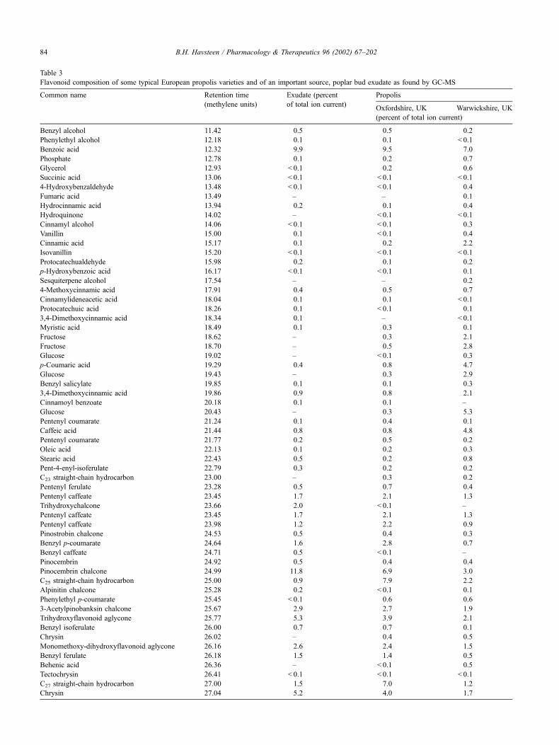
Table 3
Flavonoid composition of some typical European propolis varieties and of an important source, poplar bud exudate as found by GC-MS
Common name Retention time Exudate (percent Propolis
(methylene units) of total ion current)Oxfordshire, UK Warwickshire, UK
(percent of total ion current)
Benzyl alcohol 11.42 0.5 0.5 0.2
Phenylethyl alcohol 12.18 0.1 0.1 < 0.1
Benzoic acid 12.32 9.9 9.5 7.0
Phosphate 12.78 0.1 0.2 0.7
Glycerol 12.93 < 0.1 0.2 0.6
Succinic acid 13.06 < 0.1 < 0.1 < 0.1
4-Hydroxybenzaldehyde 13.48 < 0.1 < 0.1 0.4
Fumaric acid 13.49 – – 0.1
Hydrocinnamic acid 13.94 0.2 0.1 0.4
Hydroquinone 14.02 – < 0.1 < 0.1
Cinnamyl alcohol 14.06 < 0.1 < 0.1 0.3
Vanillin 15.00 0.1 < 0.1 0.4
Cinnamic acid 15.17 0.1 0.2 2.2
Isovanillin 15.20 < 0.1 < 0.1 < 0.1
Protocatechualdehyde 15.98 0.2 0.1 0.2
p-Hydroxybenzoic acid 16.17 < 0.1 < 0.1 0.1
Sesquiterpene alcohol 17.54 – – 0.2
4-Methoxycinnamic acid 17.91 0.4 0.5 0.7
Cinnamylideneacetic acid 18.04 0.1 0.1 < 0.1
Protocatechuic acid 18.26 0.1 < 0.1 0.1
3,4-Dimethoxycinnamic acid 18.34 0.1 – < 0.1
Myristic acid 18.49 0.1 0.3 0.1
Fructose 18.62 – 0.3 2.1
Fructose 18.70 – 0.5 2.8
Glucose 19.02 – < 0.1 0.3
p-Coumaric acid 19.29 0.4 0.8 4.7
Glucose 19.43 – 0.3 2.9
Benzyl salicylate 19.85 0.1 0.1 0.3
3,4-Dimethoxycinnamic acid 19.86 0.9 0.8 2.1
Cinnamoyl benzoate 20.18 0.1 0.1 –
Glucose 20.43 – 0.3 5.3
Pentenyl coumarate 21.24 0.1 0.4 0.1
Caffeic acid 21.44 0.8 0.8 4.8
Pentenyl coumarate 21.77 0.2 0.5 0.2
Oleic acid 22.13 0.1 0.2 0.3
Stearic acid 22.43 0.5 0.2 0.8
Pent-4-enyl-isoferulate 22.79 0.3 0.2 0.2
C23 straight-chain hydrocarbon 23.00 – 0.3 0.2
Pentenyl ferulate 23.28 0.5 0.7 0.4
Pentenyl caffeate 23.45 1.7 2.1 1.3
Trihydroxychalcone 23.66 2.0 < 0.1 –
Pentenyl caffeate 23.45 1.7 2.1 1.3
Pentenyl caffeate 23.98 1.2 2.2 0.9
Pinostrobin chalcone 24.53 0.5 0.4 0.3
Benzyl p-coumarate 24.64 1.6 2.8 0.7
Benzyl caffeate 24.71 0.5 < 0.1 –
Pinocembrin 24.92 0.5 0.4 0.4
Pinocembrin chalcone 24.99 11.8 6.9 3.0
C25 straight-chain hydrocarbon 25.00 0.9 7.9 2.2
Alpinitin chalcone 25.28 0.2 < 0.1 0.1
Phenylethyl p-coumarate 25.45 < 0.1 0.6 0.6
3-Acetylpinobanksin chalcone 25.67 2.9 2.7 1.9
Trihydroxyflavonoid aglycone 25.77 5.3 3.9 2.1
Benzyl isoferulate 26.00 0.7 0.7 0.1
Chrysin 26.02 – 0.4 0.5
Monomethoxy-dihydroxyflavonoid aglycone 26.16 2.6 2.4 1.5
Benzyl ferulate 26.18 1.5 1.4 0.5
Behenic acid 26.36 – < 0.1 0.5
Tectochrysin 26.41 < 0.1 < 0.1 < 0.1
C27 straight-chain hydrocarbon 27.00 1.5 7.0 1.2
Chrysin 27.04 5.2 4.0 1.7
B.H. Havsteen / Pharmacology & Therapeutics 96 (2002) 67–20284
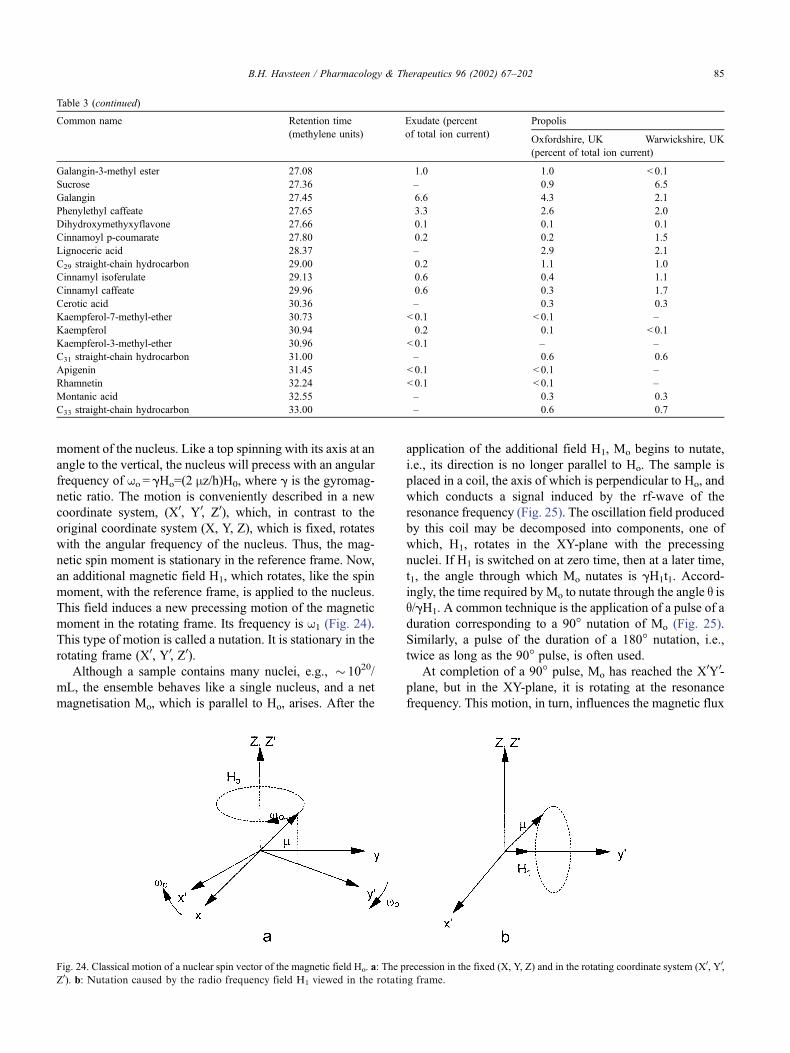
moment of the nucleus. Like a top spinning with its axis at an
angle to the vertical, the nucleus will precess with an angular
frequency of wo = gHo=(2 mz/h)H0, where g is the gyromag-
netic ratio. The motion is conveniently described in a new
coordinate system, (X0, Y0, Z0), which, in contrast to the
original coordinate system (X, Y, Z), which is fixed, rotates
with the angular frequency of the nucleus. Thus, the mag-
netic spin moment is stationary in the reference frame. Now,
an additional magnetic field H1, which rotates, like the spin
moment, with the reference frame, is applied to the nucleus.
This field induces a new precessing motion of the magnetic
moment in the rotating frame. Its frequency is w1 (Fig. 24).
This type of motion is called a nutation. It is stationary in the
rotating frame (X0, Y0, Z0).
Although a sample contains many nuclei, e.g., � 1020/
mL, the ensemble behaves like a single nucleus, and a net
magnetisation Mo, which is parallel to Ho, arises. After the
application of the additional field H1, Mo begins to nutate,
i.e., its direction is no longer parallel to Ho. The sample is
placed in a coil, the axis of which is perpendicular to Ho, and
which conducts a signal induced by the rf-wave of the
resonance frequency (Fig. 25). The oscillation field produced
by this coil may be decomposed into components, one of
which, H1, rotates in the XY-plane with the precessing
nuclei. If H1 is switched on at zero time, then at a later time,
t1, the angle through which Mo nutates is gH1t1. Accord-
ingly, the time required by Mo to nutate through the angle q isq/gH1. A common technique is the application of a pulse of a
duration corresponding to a 90� nutation of Mo (Fig. 25).
Similarly, a pulse of the duration of a 180� nutation, i.e.,
twice as long as the 90� pulse, is often used.
At completion of a 90� pulse, Mo has reached the X0Y0-
plane, but in the XY-plane, it is rotating at the resonance
frequency. This motion, in turn, influences the magnetic flux
Table 3 (continued)
Common name Retention time Exudate (percent Propolis
(methylene units) of total ion current)Oxfordshire, UK Warwickshire, UK
(percent of total ion current)
Galangin-3-methyl ester 27.08 1.0 1.0 < 0.1
Sucrose 27.36 – 0.9 6.5
Galangin 27.45 6.6 4.3 2.1
Phenylethyl caffeate 27.65 3.3 2.6 2.0
Dihydroxymethyxyflavone 27.66 0.1 0.1 0.1
Cinnamoyl p-coumarate 27.80 0.2 0.2 1.5
Lignoceric acid 28.37 – 2.9 2.1
C29 straight-chain hydrocarbon 29.00 0.2 1.1 1.0
Cinnamyl isoferulate 29.13 0.6 0.4 1.1
Cinnamyl caffeate 29.96 0.6 0.3 1.7
Cerotic acid 30.36 – 0.3 0.3
Kaempferol-7-methyl-ether 30.73 < 0.1 < 0.1 –
Kaempferol 30.94 0.2 0.1 < 0.1
Kaempferol-3-methyl-ether 30.96 < 0.1 – –
C31 straight-chain hydrocarbon 31.00 – 0.6 0.6
Apigenin 31.45 < 0.1 < 0.1 –
Rhamnetin 32.24 < 0.1 < 0.1 –
Montanic acid 32.55 – 0.3 0.3
C33 straight-chain hydrocarbon 33.00 – 0.6 0.7
Fig. 24. Classical motion of a nuclear spin vector of the magnetic field Ho. a: The precession in the fixed (X, Y, Z) and in the rotating coordinate system (X0, Y0,
Z0). b: Nutation caused by the radio frequency field H1 viewed in the rotating frame.
B.H. Havsteen / Pharmacology & Therapeutics 96 (2002) 67–202 85
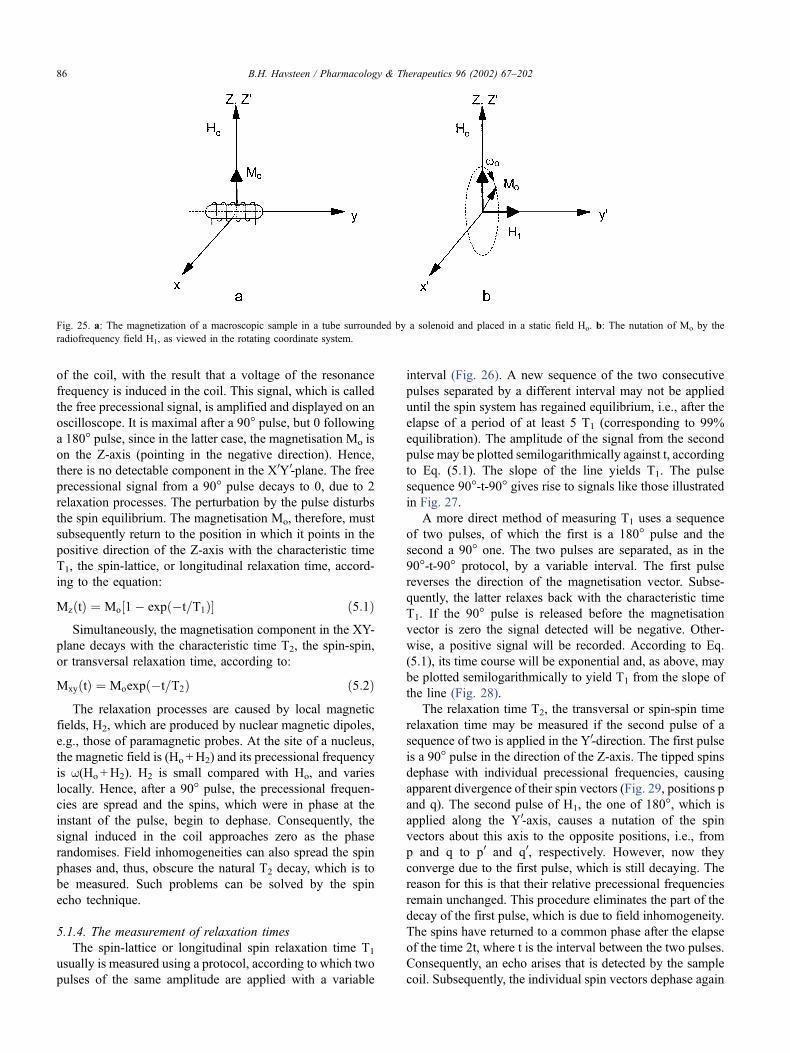
of the coil, with the result that a voltage of the resonance
frequency is induced in the coil. This signal, which is called
the free precessional signal, is amplified and displayed on an
oscilloscope. It is maximal after a 90� pulse, but 0 following
a 180� pulse, since in the latter case, the magnetisation Mo is
on the Z-axis (pointing in the negative direction). Hence,
there is no detectable component in the X0Y0-plane. The free
precessional signal from a 90� pulse decays to 0, due to 2
relaxation processes. The perturbation by the pulse disturbs
the spin equilibrium. The magnetisation Mo, therefore, must
subsequently return to the position in which it points in the
positive direction of the Z-axis with the characteristic time
T1, the spin-lattice, or longitudinal relaxation time, accord-
ing to the equation:
MzðtÞ ¼ Mo½1� expð�t=T1Þ� ð5:1ÞSimultaneously, the magnetisation component in the XY-
plane decays with the characteristic time T2, the spin-spin,
or transversal relaxation time, according to:
MxyðtÞ ¼ Moexpð�t=T2Þ ð5:2ÞThe relaxation processes are caused by local magnetic
fields, H2, which are produced by nuclear magnetic dipoles,
e.g., those of paramagnetic probes. At the site of a nucleus,
the magnetic field is (Ho +H2) and its precessional frequency
is w(Ho +H2). H2 is small compared with Ho, and varies
locally. Hence, after a 90� pulse, the precessional frequen-
cies are spread and the spins, which were in phase at the
instant of the pulse, begin to dephase. Consequently, the
signal induced in the coil approaches zero as the phase
randomises. Field inhomogeneities can also spread the spin
phases and, thus, obscure the natural T2 decay, which is to
be measured. Such problems can be solved by the spin
echo technique.
5.1.4. The measurement of relaxation times
The spin-lattice or longitudinal spin relaxation time T1
usually is measured using a protocol, according to which two
pulses of the same amplitude are applied with a variable
interval (Fig. 26). A new sequence of the two consecutive
pulses separated by a different interval may not be applied
until the spin system has regained equilibrium, i.e., after the
elapse of a period of at least 5 T1 (corresponding to 99%
equilibration). The amplitude of the signal from the second
pulse may be plotted semilogarithmically against t, according
to Eq. (5.1). The slope of the line yields T1. The pulse
sequence 90�-t-90� gives rise to signals like those illustrated
in Fig. 27.
A more direct method of measuring T1 uses a sequence
of two pulses, of which the first is a 180� pulse and the
second a 90� one. The two pulses are separated, as in the
90�-t-90� protocol, by a variable interval. The first pulse
reverses the direction of the magnetisation vector. Subse-
quently, the latter relaxes back with the characteristic time
T1. If the 90� pulse is released before the magnetisation
vector is zero the signal detected will be negative. Other-
wise, a positive signal will be recorded. According to Eq.
(5.1), its time course will be exponential and, as above, may
be plotted semilogarithmically to yield T1 from the slope of
the line (Fig. 28).
The relaxation time T2, the transversal or spin-spin time
relaxation time may be measured if the second pulse of a
sequence of two is applied in the Y0-direction. The first pulse
is a 90� pulse in the direction of the Z-axis. The tipped spins
dephase with individual precessional frequencies, causing
apparent divergence of their spin vectors (Fig. 29, positions p
and q). The second pulse of H1, the one of 180�, which is
applied along the Y0-axis, causes a nutation of the spin
vectors about this axis to the opposite positions, i.e., from
p and q to p0 and q0, respectively. However, now they
converge due to the first pulse, which is still decaying. The
reason for this is that their relative precessional frequencies
remain unchanged. This procedure eliminates the part of the
decay of the first pulse, which is due to field inhomogeneity.
The spins have returned to a common phase after the elapse
of the time 2t, where t is the interval between the two pulses.
Consequently, an echo arises that is detected by the sample
coil. Subsequently, the individual spin vectors dephase again
Fig. 25. a: The magnetization of a macroscopic sample in a tube surrounded by a solenoid and placed in a static field Ho. b: The nutation of Mo by the
radiofrequency field H1, as viewed in the rotating coordinate system.
B.H. Havsteen / Pharmacology & Therapeutics 96 (2002) 67–20286
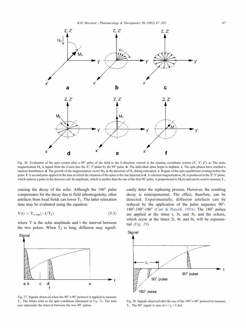
causing the decay of the echo. Although the 180� pulse
compensates for the decay due to field inhomogeneity, other
artefacts from local fields can lower T2. The latter relaxation
time may be evaluated using the equation:
YðtÞ ¼ Yo expð�t=T2Þ ð5:3Þ
where Y is the echo amplitude and t the interval between
the two pulses. When T2 is long, diffusion may signifi-
cantly deter the rephasing process. However, the resulting
decay is nonexponential. The effect, therefore, can be
detected. Experimentally, diffusion artefacts can be
reduced by the application of the pulse sequence 90�-180�-180�-180� (Carr & Purcell, 1954). The 180� pulses
are applied at the times t, 3t, and 5t, and the echoes,
which occur at the times 2t, 4t, and 6t, will be exponen-
tial (Fig. 29).
Fig. 28. Signals observed after the use of the 180�-t-90� protocol to measure
T1. The 90� signal is zero at t = tn = T1ln2.
Fig. 27. Signals observed when the 90�-t-90� protocol is applied to measure
T1. The letters refer to the spin conditions illustrated in Fig. 26. The time
axis represents the interval between the two 90� pulses.
Fig. 26. Evaluation of the spin system after a 90� pulse of the field in the Z-direction viewed in the rotating coordinate system (X0, Y0, Z0). a: The static
magnetization Mo is tipped from the Z-axis into the X0, Y0-plane by the 90� pulse. b: The individual spins begin to dephase. c: The spin phases have reached a
random distribution. d: The growth of the magnetization vector MZ in the direction of Ho during relaxation. e: Regain of the spin equilibrium existing before the
pulse. f: A second pulse applied at the time at which the situation of the spins is the one depicted in d. A coherent magnetization,M, is produced in the X0,Y0-plane,
which induces a pulse in the detector coil. Its amplitude, which is smaller than the one of the first 90� pulse, is proportional toMZ(t) and can be used to measure T1.
B.H. Havsteen / Pharmacology & Therapeutics 96 (2002) 67–202 87
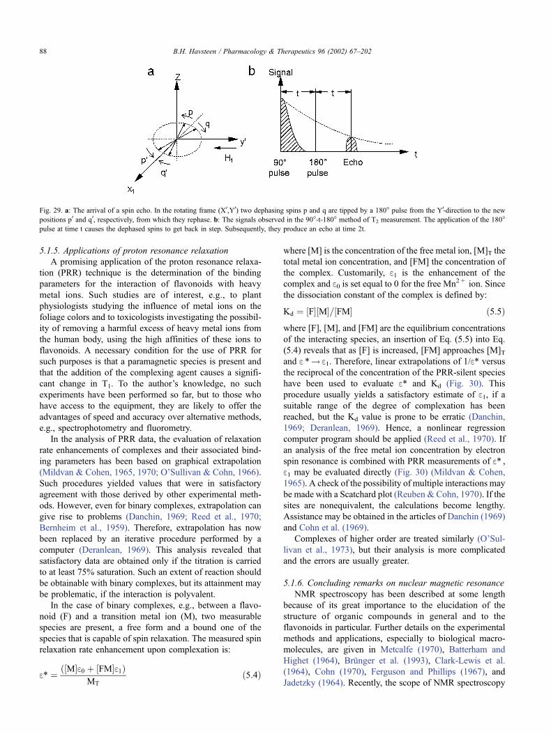
5.1.5. Applications of proton resonance relaxation
A promising application of the proton resonance relaxa-
tion (PRR) technique is the determination of the binding
parameters for the interaction of flavonoids with heavy
metal ions. Such studies are of interest, e.g., to plant
physiologists studying the influence of metal ions on the
foliage colors and to toxicologists investigating the possibil-
ity of removing a harmful excess of heavy metal ions from
the human body, using the high affinities of these ions to
flavonoids. A necessary condition for the use of PRR for
such purposes is that a paramagnetic species is present and
that the addition of the complexing agent causes a signifi-
cant change in T1. To the author’s knowledge, no such
experiments have been performed so far, but to those who
have access to the equipment, they are likely to offer the
advantages of speed and accuracy over alternative methods,
e.g., spectrophotometry and fluorometry.
In the analysis of PRR data, the evaluation of relaxation
rate enhancements of complexes and their associated bind-
ing parameters has been based on graphical extrapolation
(Mildvan & Cohen, 1965, 1970; O’Sullivan & Cohn, 1966).
Such procedures yielded values that were in satisfactory
agreement with those derived by other experimental meth-
ods. However, even for binary complexes, extrapolation can
give rise to problems (Danchin, 1969; Reed et al., 1970;
Bernheim et al., 1959). Therefore, extrapolation has now
been replaced by an iterative procedure performed by a
computer (Deranlean, 1969). This analysis revealed that
satisfactory data are obtained only if the titration is carried
to at least 75% saturation. Such an extent of reaction should
be obtainable with binary complexes, but its attainment may
be problematic, if the interaction is polyvalent.
In the case of binary complexes, e.g., between a flavo-
noid (F) and a transition metal ion (M), two measurable
species are present, a free form and a bound one of the
species that is capable of spin relaxation. The measured spin
relaxation rate enhancement upon complexation is:
e* ¼ ð½M�e0 þ ½FM�e1ÞMT
ð5:4Þ
where [M] is the concentration of the free metal ion, [M]T the
total metal ion concentration, and [FM] the concentration of
the complex. Customarily, e1 is the enhancement of the
complex and e0 is set equal to 0 for the free Mn2 + ion. Since
the dissociation constant of the complex is defined by:
Kd ¼ ½F�½M�=½FM� ð5:5Þwhere [F], [M], and [FM] are the equilibrium concentrations
of the interacting species, an insertion of Eq. (5.5) into Eq.
(5.4) reveals that as [F] is increased, [FM] approaches [M]Tand e *! e1. Therefore, linear extrapolations of 1/e* versus
the reciprocal of the concentration of the PRR-silent species
have been used to evaluate e* and Kd (Fig. 30). This
procedure usually yields a satisfactory estimate of e1, if asuitable range of the degree of complexation has been
reached, but the Kd value is prone to be erratic (Danchin,
1969; Deranlean, 1969). Hence, a nonlinear regression
computer program should be applied (Reed et al., 1970). If
an analysis of the free metal ion concentration by electron
spin resonance is combined with PRR measurements of e* ,e1 may be evaluated directly (Fig. 30) (Mildvan & Cohen,
1965). A check of the possibility of multiple interactions may
be made with a Scatchard plot (Reuben & Cohn, 1970). If the
sites are nonequivalent, the calculations become lengthy.
Assistance may be obtained in the articles of Danchin (1969)
and Cohn et al. (1969).
Complexes of higher order are treated similarly (O’Sul-
livan et al., 1973), but their analysis is more complicated
and the errors are usually greater.
5.1.6. Concluding remarks on nuclear magnetic resonance
NMR spectroscopy has been described at some length
because of its great importance to the elucidation of the
structure of organic compounds in general and to the
flavonoids in particular. Further details on the experimental
methods and applications, especially to biological macro-
molecules, are given in Metcalfe (1970), Batterham and
Highet (1964), Brunger et al. (1993), Clark-Lewis et al.
(1964), Cohn (1970), Ferguson and Phillips (1967), and
Jadetzky (1964). Recently, the scope of NMR spectroscopy
Fig. 29. a: The arrival of a spin echo. In the rotating frame (X0,Y0) two dephasing spins p and q are tipped by a 180� pulse from the Y0-direction to the new
positions p0 and q0, respectively, from which they rephase. b: The signals observed in the 90�-t-180� method of T2 measurement. The application of the 180�pulse at time t causes the dephased spins to get back in step. Subsequently, they produce an echo at time 2t.
B.H. Havsteen / Pharmacology & Therapeutics 96 (2002) 67–20288
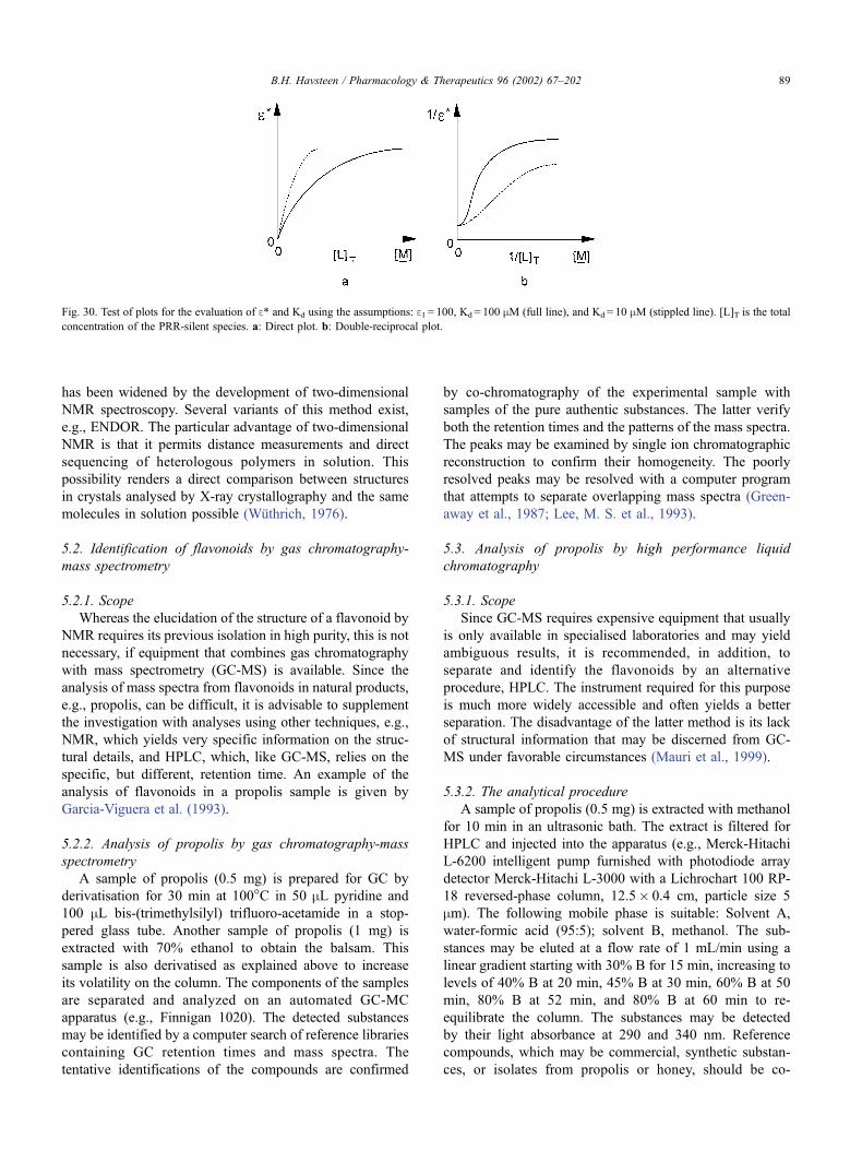
has been widened by the development of two-dimensional
NMR spectroscopy. Several variants of this method exist,
e.g., ENDOR. The particular advantage of two-dimensional
NMR is that it permits distance measurements and direct
sequencing of heterologous polymers in solution. This
possibility renders a direct comparison between structures
in crystals analysed by X-ray crystallography and the same
molecules in solution possible (Wuthrich, 1976).
5.2. Identification of flavonoids by gas chromatography-
mass spectrometry
5.2.1. Scope
Whereas the elucidation of the structure of a flavonoid by
NMR requires its previous isolation in high purity, this is not
necessary, if equipment that combines gas chromatography
with mass spectrometry (GC-MS) is available. Since the
analysis of mass spectra from flavonoids in natural products,
e.g., propolis, can be difficult, it is advisable to supplement
the investigation with analyses using other techniques, e.g.,
NMR, which yields very specific information on the struc-
tural details, and HPLC, which, like GC-MS, relies on the
specific, but different, retention time. An example of the
analysis of flavonoids in a propolis sample is given by
Garcia-Viguera et al. (1993).
5.2.2. Analysis of propolis by gas chromatography-mass
spectrometry
A sample of propolis (0.5 mg) is prepared for GC by
derivatisation for 30 min at 100�C in 50 mL pyridine and
100 mL bis-(trimethylsilyl) trifluoro-acetamide in a stop-
pered glass tube. Another sample of propolis (1 mg) is
extracted with 70% ethanol to obtain the balsam. This
sample is also derivatised as explained above to increase
its volatility on the column. The components of the samples
are separated and analyzed on an automated GC-MC
apparatus (e.g., Finnigan 1020). The detected substances
may be identified by a computer search of reference libraries
containing GC retention times and mass spectra. The
tentative identifications of the compounds are confirmed
by co-chromatography of the experimental sample with
samples of the pure authentic substances. The latter verify
both the retention times and the patterns of the mass spectra.
The peaks may be examined by single ion chromatographic
reconstruction to confirm their homogeneity. The poorly
resolved peaks may be resolved with a computer program
that attempts to separate overlapping mass spectra (Green-
away et al., 1987; Lee, M. S. et al., 1993).
5.3. Analysis of propolis by high performance liquid
chromatography
5.3.1. Scope
Since GC-MS requires expensive equipment that usually
is only available in specialised laboratories and may yield
ambiguous results, it is recommended, in addition, to
separate and identify the flavonoids by an alternative
procedure, HPLC. The instrument required for this purpose
is much more widely accessible and often yields a better
separation. The disadvantage of the latter method is its lack
of structural information that may be discerned from GC-
MS under favorable circumstances (Mauri et al., 1999).
5.3.2. The analytical procedure
A sample of propolis (0.5 mg) is extracted with methanol
for 10 min in an ultrasonic bath. The extract is filtered for
HPLC and injected into the apparatus (e.g., Merck-Hitachi
L-6200 intelligent pump furnished with photodiode array
detector Merck-Hitachi L-3000 with a Lichrochart 100 RP-
18 reversed-phase column, 12.5� 0.4 cm, particle size 5
mm). The following mobile phase is suitable: Solvent A,
water-formic acid (95:5); solvent B, methanol. The sub-
stances may be eluted at a flow rate of 1 mL/min using a
linear gradient starting with 30% B for 15 min, increasing to
levels of 40% B at 20 min, 45% B at 30 min, 60% B at 50
min, 80% B at 52 min, and 80% B at 60 min to re-
equilibrate the column. The substances may be detected
by their light absorbance at 290 and 340 nm. Reference
compounds, which may be commercial, synthetic substan-
ces, or isolates from propolis or honey, should be co-
Fig. 30. Test of plots for the evaluation of e* and Kd using the assumptions: e1 = 100, Kd = 100 mM (full line), and Kd = 10 mM (stippled line). [L]T is the total
concentration of the PRR-silent species. a: Direct plot. b: Double-reciprocal plot.
B.H. Havsteen / Pharmacology & Therapeutics 96 (2002) 67–202 89
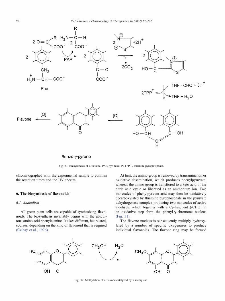
chromatographed with the experimental sample to confirm
the retention times and the UV spectra.
6. The biosynthesis of flavonoids
6.1. Anabolism
All green plant cells are capable of synthesizing flavo-
noids. The biosynthesis invariably begins with the ubiqui-
tous amino acid phenylalanine. It takes different, but related,
courses, depending on the kind of flavonoid that is required
(Czihay et al., 1976).
At first, the amino group is removed by transamination or
oxidative desamination, which produces phenylpyruvate,
whereas the amino group is transferred to a keto acid of the
citric acid cycle or liberated as an ammonium ion. Two
molecules of phenylpyruvic acid may then be oxidatively
decarboxylated by thiamine pyrophosphate in the pyruvate
dehydrogenase complex producing two molecules of active
aldehyde, which together with a C1-fragment (-CHO) in
an oxidative step form the phenyl-g-chromone nucleus
(Fig. 31).
The flavone nucleus is subsequently multiply hydroxy-
lated by a number of specific oxygenases to produce
individual flavonoids. The flavone ring may be formed
Fig. 31. Biosynthesis of a flavone. PAP, pyridoxal-P; TPP + , thiamine pyrophosphate.
Fig. 32. Methylation of a flavone catalyzed by a methylase.
B.H. Havsteen / Pharmacology & Therapeutics 96 (2002) 67–20290
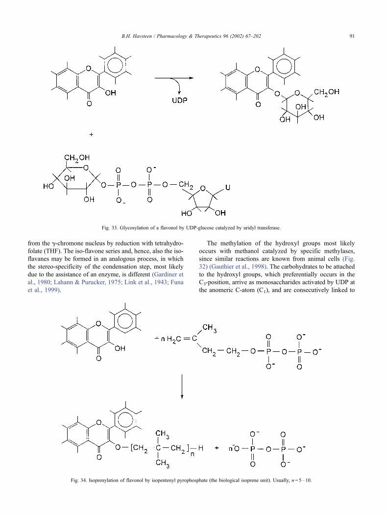
from the g-chromone nucleus by reduction with tetrahydro-
folate (THF). The iso-flavone series and, hence, also the iso-
flavanes may be formed in an analogous process, in which
the stereo-specificity of the condensation step, most likely
due to the assistance of an enzyme, is different (Gardiner et
al., 1980; Lahann & Purucker, 1975; Link et al., 1943; Funa
et al., 1999).
The methylation of the hydroxyl groups most likely
occurs with methanol catalyzed by specific methylases,
since similar reactions are known from animal cells (Fig.
32) (Gauthier et al., 1998). The carbohydrates to be attached
to the hydroxyl groups, which preferentially occurs in the
C3-position, arrive as monosaccharides activated by UDP at
the anomeric C-atom (C1), and are consecutively linked to
Fig. 33. Glycosylation of a flavonol by UDP-glucose catalyzed by uridyl transferase.
Fig. 34. Isoprenylation of flavonol by isopentenyl pyrophosphate (the biological isoprene unit). Usually, n= 5–10.
B.H. Havsteen / Pharmacology & Therapeutics 96 (2002) 67–202 91
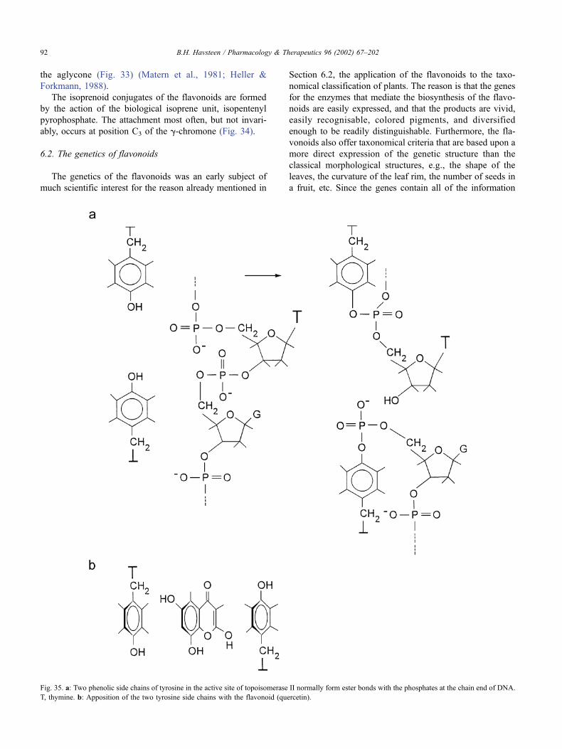
the aglycone (Fig. 33) (Matern et al., 1981; Heller &
Forkmann, 1988).
The isoprenoid conjugates of the flavonoids are formed
by the action of the biological isoprene unit, isopentenyl
pyrophosphate. The attachment most often, but not invari-
ably, occurs at position C3 of the g-chromone (Fig. 34).
6.2. The genetics of flavonoids
The genetics of the flavonoids was an early subject of
much scientific interest for the reason already mentioned in
Section 6.2, the application of the flavonoids to the taxo-
nomical classification of plants. The reason is that the genes
for the enzymes that mediate the biosynthesis of the flavo-
noids are easily expressed, and that the products are vivid,
easily recognisable, colored pigments, and diversified
enough to be readily distinguishable. Furthermore, the fla-
vonoids also offer taxonomical criteria that are based upon a
more direct expression of the genetic structure than the
classical morphological structures, e.g., the shape of the
leaves, the curvature of the leaf rim, the number of seeds in
a fruit, etc. Since the genes contain all of the information
Fig. 35. a: Two phenolic side chains of tyrosine in the active site of topoisomerase II normally form ester bonds with the phosphates at the chain end of DNA.
T, thymine. b: Apposition of the two tyrosine side chains with the flavonoid (quercetin).
B.H. Havsteen / Pharmacology & Therapeutics 96 (2002) 67–20292
about an individual specimen, the genetic approach to tax-
onomy appears to be the most fundamental basis available.
Moreover, flavonoids are not only produced in response to
many external stimuli, but also by metabolic feedback
reactions capable of inducing other genetic activities. The
result may be gene activation, mutation, or repair of DNA
(Zahringer et al., 1978; Cea et al., 1983; Peters et al., 1986;
Disseling, 1996; Fekermann et al., 1998; Ching & Baguley,
1987; Chin, 1999).
The biochemical basis of the activity of the flavonoids on
genes recently has been studied from a nutritional view point
(Singleton, 1981). Normal human nutrition has always con-
tained much vegetable material. It has been estimated that an
average healthy individual consumes 1–2 g of flavonoids
daily. Considering the chemical reactivity of the flavonoids,
this is an amount that gives rise to some concern. Evidently,
humans have fared rather well on such a diet since their
appearance on earth, but even a slight toxicity, at such an
extensive exposure, might give rise to some disease or
malfunction (Ritov et al., 1995). It was recognised that some
individuals actually are sensitive to flavonoids because
antibodies to these compounds were found in human blood.
It was also discovered that about 3–5% of the population is
allergic to flavonoids. Such a fraction is hardly alarming,
considering the great number of commonly occurring aller-
gens. Red wine, which has a high content of flavonoids, has
been recommended by prominent nutritionists for its favor-
able influence on cardiovascular health. This effect was
ascribed to the antioxidative effect of the flavonoids. Pathol-
ogists support this observation by noting that the vascular
walls of alcoholics at dissection are found to be in remark-
ably good condition, i.e., smooth and free of atherosclerosis
(Donovan et al., 1999; da Luz et al., 1999; Lairon & Amiot,
1999; Cishek et al., 1997; Miyagi et al., 1997). The proven
mutagenic activity of some flavonoids, a prominent example
is quercetin, gave reason for concern because some muta-
tions ultimately lead to cancer. However, the mutagenic
activity of the flavonoids in the Ames test, in which a
histidine-requiring mutant supplemented with mammalian
mitochondria is mutated back to the wild-type, was not
higher than that of similar compounds, which also are
indigenous to our nutrition (Ames et al., 1975; MacGregor
& Jurd, 1978; Maruta et al., 1979; Cea et al., 1983; Sahu et
al., 1981). Besides, no connection between flavonoids and
human cancer has been found. Some flavonoids possess an
antimutagenic effect (Choi et al., 1994; Agullo et al., 1996,
1997; Anderson et al., 1997, 1998).
However, one biochemical mechanism of the action of
flavonoids on DNA was found, since quercetin inhibits top-
oisomerases II (Duthie & Dobson, 1999) and IV (Bernard et
al., 1997). This enzyme cleaves the DNA chain in the super-
helix form to create additional turns or to unroll some such
superhelical turns. Then, it normally joins DNA ends again,
but this final step is competitively inhibited by quercetin. The
result is a single-strand breaks, which may suffice to cause
double-strand breaks that result in the loss of genetic informa-
tion. A further consequence of single-strand breaks is the
initiation of a repair mechanism. If successful, the latter
delivers an intact gene, but on a rare occasion, it is faulty,
and in that case, amutationmay occur. The twoDNA ends are
transiently linked to two tyrosine side chains positioned
opposite to each other in the active site of the topoisomerase.
Quercetin structurally resembles the phenol ring of tyrosine.
Hence, this flavonoid may intercalate between the two
phenol rings, e.g., forming a charge transfer complex or by
hydrophobic interactions. Thus, it may sterically hinder the
points to which the DNA chain ends must attach to become
rejoined (Fig. 35) (Howard et al., 1994; Leteurtre et al.,
1994; Freudenreich & Kreuzer, 1994).
Flavonoid-sensitive genes are associated with modu-
lation, longitudinal growth, stress response, and petal col-
oration. Only few details about the mechanisms are known,
but some information will be discussed in Section 19. The
stress response mediated by the flavonoids may well
involve the inhibition of DNA topoisomerase II described
in Section 7.
7. The role of the flavonoids in plant physiology
The flavonoids are essential constituents of the cells of
all higher plants. They resemble in their regulatory prop-
erties most of the lipid-soluble vitamins, but serve in
addition, due to their color and odor, as communicators
with the environment (Middleton & Teramura, 1993; Har-
borne et al., 1976; Brouillard & Cheminat, 1988; Harborne,
1986, 1988a, 1988b). They are recognised by pollinators,
e.g., insects, birds, and animals, which contribute to the
dispersion of seeds. The growth regulation of plants by
flavonoids has attracted considerable scientific interest
because it is important to plant breeding, agricultural eco-
nomics, and world health (Moyano et al., 1996). Besides,
the mechanism is sufficiently similar to growth-regulating
processes in animals to invite the suspicion that flavonoids
might also influence the growth metabolism of animal cells,
including those of humans (Groteweld et al., 1994; Jiang
et al., 1999; Ceriani et al., 1999; Ghosal & Jaiswal, 1980;
Fragner, 1964; Albrecht et al., 1999). Investigations of such
aspects are impeded by the difficulty of composing a long-
term diet that is sufficient for sustenance, but absolutely free
of flavonoids.
The effect of flavonoids on plant growth, which is
known, is at least partly indirect and associated with the
action of the auxins. The prominent representative of this
group, indolyl acetic acid (IAA), is formed from tryptophan
by pyridoxal phosphate (Pyr-P)-mediated transamination or
desamination followed by decarboxylation (Fig. 36).
This process occurs in the cytoplasm, but the growth
hormone may leave the cell to disperse in the plant via the
vascular system. Two routes of exit from the cell of origin
are conceivable: either directly through a cell membrane
channel, which is permeable to aromatic compounds, or,
B.H. Havsteen / Pharmacology & Therapeutics 96 (2002) 67–202 93
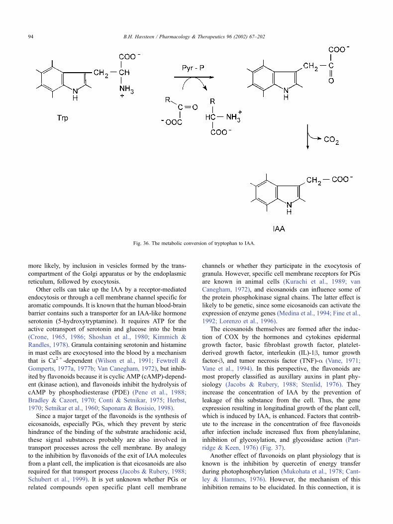
more likely, by inclusion in vesicles formed by the trans-
compartment of the Golgi apparatus or by the endoplasmic
reticulum, followed by exocytosis.
Other cells can take up the IAA by a receptor-mediated
endocytosis or through a cell membrane channel specific for
aromatic compounds. It is known that the human blood-brain
barrier contains such a transporter for an IAA-like hormone
serotonin (5-hydroxytryptamine). It requires ATP for the
active cotransport of serotonin and glucose into the brain
(Crone, 1965, 1986; Shoshan et al., 1980; Kimmich &
Randles, 1978). Granula containing serotonin and histamine
in mast cells are exocytosed into the blood by a mechanism
that is Ca2 + -dependent (Wilson et al., 1991; Fewtrell &
Gomperts, 1977a, 1977b; Van Canegham, 1972), but inhib-
ited by flavonoids because it is cyclic AMP (cAMP)-depend-
ent (kinase action), and flavonoids inhibit the hydrolysis of
cAMP by phosphodiesterase (PDE) (Pene et al., 1988;
Bradley & Cazort, 1970; Conti & Setnikar, 1975; Herbst,
1970; Setnikar et al., 1960; Saponara & Bosisio, 1998).
Since a major target of the flavonoids is the synthesis of
eicosanoids, especially PGs, which they prevent by steric
hindrance of the binding of the substrate arachidonic acid,
these signal substances probably are also involved in
transport processes across the cell membrane. By analogy
to the inhibition by flavonoids of the exit of IAA molecules
from a plant cell, the implication is that eicosanoids are also
required for that transport process (Jacobs & Rubery, 1988;
Schubert et al., 1999). It is yet unknown whether PGs or
related compounds open specific plant cell membrane
channels or whether they participate in the exocytosis of
granula. However, specific cell membrane receptors for PGs
are known in animal cells (Kurachi et al., 1989; van
Canegham, 1972), and eicosanoids can influence some of
the protein phosphokinase signal chains. The latter effect is
likely to be genetic, since some eicosanoids can activate the
expression of enzyme genes (Medina et al., 1994; Fine et al.,
1992; Lorenzo et al., 1996).
The eicosanoids themselves are formed after the induc-
tion of COX by the hormones and cytokines epidermal
growth factor, basic fibroblast growth factor, platelet-
derived growth factor, interleukin (IL)-1b, tumor growth
factor-b, and tumor necrosis factor (TNF)-a (Vane, 1971;
Vane et al., 1994). In this perspective, the flavonoids are
most properly classified as auxillary auxins in plant phy-
siology (Jacobs & Rubery, 1988; Stenlid, 1976). They
increase the concentration of IAA by the prevention of
leakage of this substance from the cell. Thus, the gene
expression resulting in longitudinal growth of the plant cell,
which is induced by IAA, is enhanced. Factors that contrib-
ute to the increase in the concentration of free flavonoids
after infection include increased flux from phenylalanine,
inhibition of glycosylation, and glycosidase action (Part-
ridge & Keen, 1976) (Fig. 37).
Another effect of flavonoids on plant physiology that is
known is the inhibition by quercetin of energy transfer
during photophosphorylation (Mukohata et al., 1978; Cant-
ley & Hammes, 1976). However, the mechanism of this
inhibition remains to be elucidated. In this connection, it is
Fig. 36. The metabolic conversion of tryptophan to IAA.
B.H. Havsteen / Pharmacology & Therapeutics 96 (2002) 67–20294
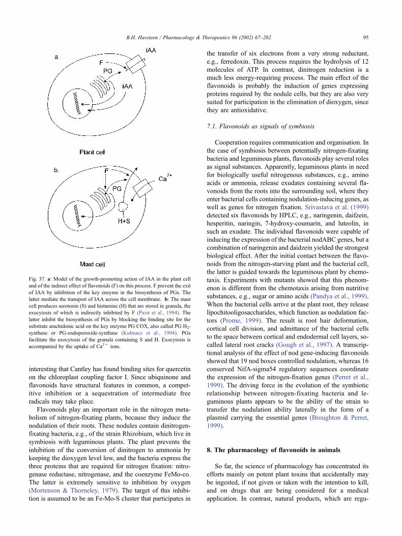
interesting that Cantley has found binding sites for quercetin
on the chloroplast coupling factor I. Since ubiquinone and
flavonoids have structural features in common, a compet-
itive inhibition or a sequestration of intermediate free
radicals may take place.
Flavonoids play an important role in the nitrogen meta-
bolism of nitrogen-fixating plants, because they induce the
nodulation of their roots. These nodules contain dinitrogen-
fixating bacteria, e.g., of the strain Rhizobium, which live in
symbiosis with leguminous plants. The plant prevents the
inhibition of the conversion of dinitrogen to ammonia by
keeping the dioxygen level low, and the bacteria express the
three proteins that are required for nitrogen fixation: nitro-
genase reductase, nitrogenase, and the coenzyme FeMo-co.
The latter is extremely sensitive to inhibition by oxygen
(Mortenson & Thorneley, 1979). The target of this inhibi-
tion is assumed to be an Fe-Mo-S cluster that participates in
the transfer of six electrons from a very strong reductant,
e.g., ferredoxin. This process requires the hydrolysis of 12
molecules of ATP. In contrast, dinitrogen reduction is a
much less energy-requiring process. The main effect of the
flavonoids is probably the induction of genes expressing
proteins required by the nodule cells, but they are also very
suited for participation in the elimination of dioxygen, since
they are antioxidative.
7.1. Flavonoids as signals of symbiosis
Cooperation requires communication and organisation. In
the case of symbiosis between potentially nitrogen-fixating
bacteria and leguminous plants, flavonoids play several roles
as signal substances. Apparently, leguminous plants in need
for biologically useful nitrogenous substances, e.g., amino
acids or ammonia, release exudates containing several fla-
vonoids from the roots into the surrounding soil, where they
enter bacterial cells containing nodulation-inducing genes, as
well as genes for nitrogen fixation. Srivastava et al. (1999)
detected six flavonoids by HPLC, e.g., naringenin, daifzein,
hesperitin, naringin, 7-hydroxy-coumarin, and luteolin, in
such an exudate. The individual flavonoids were capable of
inducing the expression of the bacterial nodABC genes, but a
combination of naringenin and daidzein yielded the strongest
biological effect. After the initial contact between the flavo-
noids from the nitrogen-starving plant and the bacterial cell,
the latter is guided towards the leguminous plant by chemo-
taxis. Experiments with mutants showed that this phenom-
enon is different from the chemotaxis arising from nutritive
substances, e.g., sugar or amino acids (Pandya et al., 1999).
When the bacterial cells arrive at the plant root, they release
lipochitooligosaccharides, which function as nodulation fac-
tors (Prome, 1999). The result is root hair deformation,
cortical cell division, and admittance of the bacterial cells
to the space between cortical and endodermal cell layers, so-
called lateral root cracks (Gough et al., 1997). A transcrip-
tional analysis of the effect of nod gene-inducing flavonoids
showed that 19 nod boxes controlled nodulation, whereas 16
conserved NifA-sigma54 regulatory sequences coordinate
the expression of the nitrogen-fixation genes (Perret et al.,
1999). The driving force in the evolution of the symbiotic
relationship between nitrogen-fixating bacteria and le-
guminous plants appears to be the ability of the strain to
transfer the nodulation ability laterally in the form of a
plasmid carrying the essential genes (Broughton & Perret,
1999).
8. The pharmacology of flavonoids in animals
So far, the science of pharmacology has concentrated its
efforts mainly on potent plant toxins that accidentally may
be ingested, if not given or taken with the intention to kill,
and on drugs that are being considered for a medical
application. In contrast, natural products, which are regu-
Fig. 37. a: Model of the growth-promoting action of IAA in the plant cell
and of the indirect effect of flavonoids (F) on this process. F prevent the exit
of IAA by inhibition of the key enzyme in the biosynthesis of PGs. The
latter mediate the transport of IAA across the cell membrane. b: The mast
cell produces serotonin (S) and histamine (H) that are stored in granula, the
exocytosis of which is indirectly inhibited by F (Picot et al., 1994). The
latter inhibit the biosynthesis of PGs by blocking the binding site for the
substrate arachidonic acid on the key enzyme PG COX, also called PG H2-
synthase or PG-endoperoxide-synthase (Kulmacz et al., 1994). PGs
facilitate the exocytosis of the granula containing S and H. Exocytosis is
accompanied by the uptake of Ca2 + ions.
B.H. Havsteen / Pharmacology & Therapeutics 96 (2002) 67–202 95

larly ingested in high amounts as components of a normal
human diet, but which are only slightly toxic, have almost
been ignored (Hughes & Wilson, 1977; Gabor, 1981). The
flavonoids belong to the latter category. The justification of
this policy is the high cost of a full-scale pharmacological
investigation and the moral obligation to develop a defence
against accidental or criminal, potentially fatal acute intox-
ication. However, since the long-term effects of the inges-
tion of compounds of low acute toxicity may impair health,
e.g., by accumulation in major organs, especially the liver,
or by initiation of immune disorders, an awareness of the
need for attention to this class of substances is rising. The
flavonoids recently have been included in such investi-
gations (Chipault et al., 1952; Di Carlo et al., 1999; Wada
et al., 1999). Another concern is that dietary components
such as flavonoids can influence the metabolism of drugs
(Conney et al., 1981). Important reasons for the low
toxicity of the flavonoids are the low solubility of the
aglycone in water and the rapid catabolism of the pyrone
nucleus in the liver. The low solubility of the flavonoids in
water often presents a problem for medical applications of
these substances. Hence, the development of semi-syn-
thetic, water-soluble flavonoids, e.g., for the treatment of
hypertension and microbleeding, has been an important
advance. Examples of such flavonoids are the hydroxye-
thylrutosides (see Section 17.1) and inositol-2-phosphate-
quercetin (Calias et al., 1996).
8.1. Pharmacodynamics
Nutritional flavonoids are absorbed from the gastrointes-
tinal tract (Crespy et al., 1999; Pforte et al., 1999), whereas
medical flavonoids are administered directly to the diseased
tissue, if it is accessible, e.g., in the skin or the throat, or
along a route leading immediately to the target, e.g., the
nasal or the vascular systems (Metzner et al., 1979; Heil-
mann & Merfort, 1998; Masquelier et al., 1979; Spilkova &
Hubik, 1986, 1988; Spilkova & Dusek, 1996; Vinson, 1998;
Zloch & Sidlova, 1977; Zloch, 1977; Hollman & Katan,
1997, 1998; Hollman et al., 1996; Hollman, 1997; Rice-
Evans & Miller, 1996; Maxwell & Thorpe, 1996; Di Carlo
et al., 1999; Balentine et al., 1997; Gabor, 1988; Graham et
al., 1978; Gugler et al., 1975; Booth et al., 1956; Cheng et
al., 1969; de Eds, 1968; Griffiths & Barrow, 1972; Griffiths
& Smith, 1972a, 1972b; Griffiths, 1975; Herrmann, 1976;
Honohan et al., 1976; Murray et al., 1954; Petrakis et al.,
1959; Piller, 1977; Simpson et al., 1969; Struckmann,
1999). After liberation of the glycosides from the aglycone
by bacterial enzymes in the intestine, about 15% of the
flavonoid aglycones are absorbed with bile micelles into the
epithelial cells and passed on to the lymph (Day et al., 1998;
Spencer et al., 1999). An important factor determining the
efficiency of the absorption of flavonoid glycosides from the
intestine is the sugar moiety. Hollman and colleagues (Holl-
man & Katan, 1999; Hollman et al., 1999a) showed that
quercetin glycosides from onions were absorbed better
(52%) than the pure aglycone (24%).
Some flavonoids inhibit the non-Na + -dependent facili-
tated diffusion of monosaccharides in intestinal epithelial
cells (Kimmich & Randles, 1978). Consequently, the par-
allel concentrative Na + -dependent transport ATPase for
monosaccharides gains efficiency (Sharma et al., 1981).
The remainder of the flavonoids are excreted with the faeces
and some in the urine (Choudhury et al., 1999). The lymph
carrying the flavonoids enters the portal blood near the liver,
and the majority (� 80%) probably is absorbed in the first
pass (Casley-Smith & Casley-Smith, 1986). Part of the
flavonoids probably are attached to serum albumin (Pod-
hajcer et al., 1980). Another part is found in conjugates that
have retained their antioxidative properties (Manach et al.,
1998). The hepatocytes transfer the flavonoids to the Golgi
apparatus and possibly also to the peroxisomes, in which
they are oxidatively degraded (Griffiths & Smith, 1972a,
1972b). Some oxidative degradation of flavonoid aglycones
also takes place in the intestine because some bacterial
enzymes can open the C-ring of the flavonoid skeleton
(Winter et al., 1989).
The products are secreted by organic acid transporters
into the blood and subsequently excreted through the
kidneys (Graefe et al., 1999; Bourne & Rice-Evans,
1999). The half-life of a typical flavonoid in the body has
been measured to be 1–2 hr, but data of sufficient accuracy
for a compartmental analysis have not been published. Since
very little information on the rates of transportation of
flavonoids and their decomposition products across mem-
branes are known, the proposition of a complete, realistic
dynamic model is premature (Honcha et al., 1995; Ueno
et al., 1983; Griffiths & Barrow, 1972; Tesi & Forssmann,
1971; Lenne-Gouverneur et al., 1999). However, the last
step, the active secretion of organic acids in the kidney into
the urine, has been studied in the isolated rat kidney (Moller,
1969). That excretion curve can be simulated with two
exponential terms, i.e., the process can be explained by a
two-compartment model (Fig. 38). The assignment of these
compartments to the morphological structures has not been
possible yet.
Since flavonoids are not accumulated in the liver and
their decomposition products (caffeic and cinnamic acids, as
Fig. 38. Minimal model of the pharmacodynamics of flavonoids in higher animals. Passage of flavonoids from 1, intestinal epithelial cell; over blood and
lymphs to 2, hepatocyte; 3, Golgi apparatus or peroxisome; again over blood to 4, renal tubulus cell.
B.H. Havsteen / Pharmacology & Therapeutics 96 (2002) 67–20296
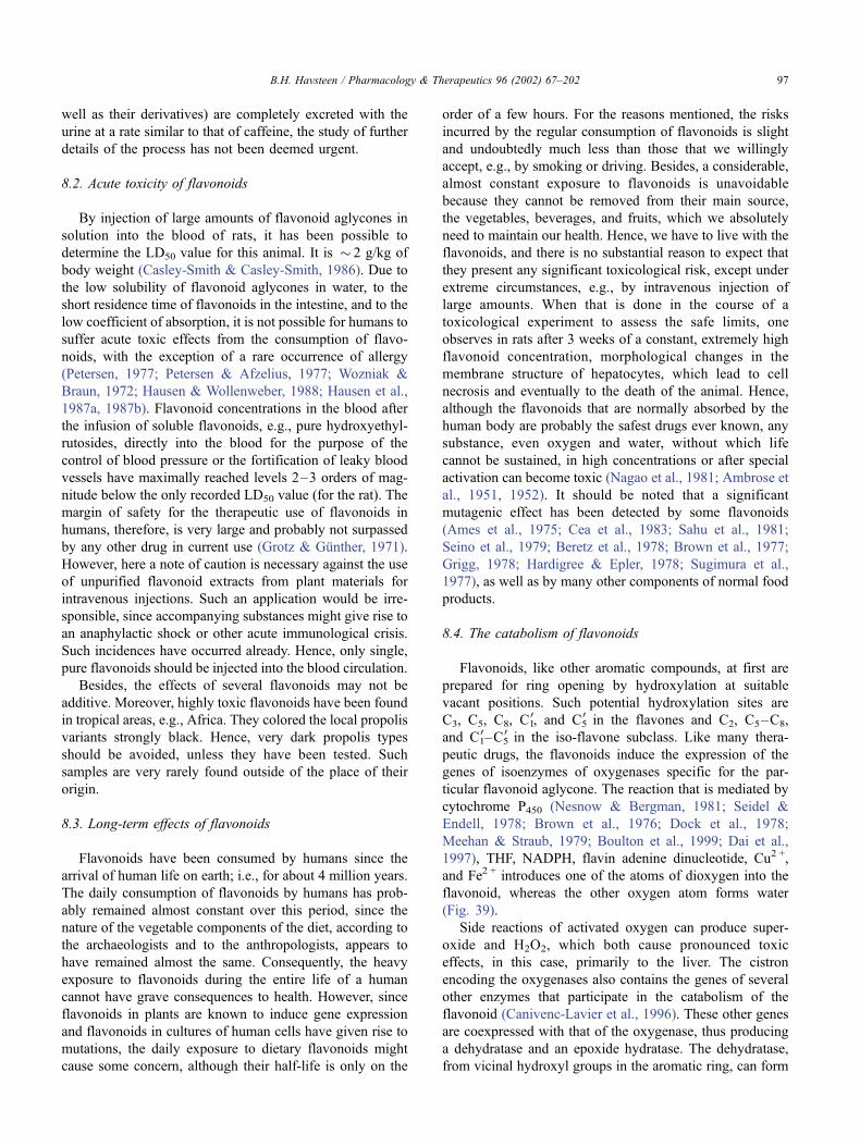
well as their derivatives) are completely excreted with the
urine at a rate similar to that of caffeine, the study of further
details of the process has not been deemed urgent.
8.2. Acute toxicity of flavonoids
By injection of large amounts of flavonoid aglycones in
solution into the blood of rats, it has been possible to
determine the LD50 value for this animal. It is � 2 g/kg of
body weight (Casley-Smith & Casley-Smith, 1986). Due to
the low solubility of flavonoid aglycones in water, to the
short residence time of flavonoids in the intestine, and to the
low coefficient of absorption, it is not possible for humans to
suffer acute toxic effects from the consumption of flavo-
noids, with the exception of a rare occurrence of allergy
(Petersen, 1977; Petersen & Afzelius, 1977; Wozniak &
Braun, 1972; Hausen & Wollenweber, 1988; Hausen et al.,
1987a, 1987b). Flavonoid concentrations in the blood after
the infusion of soluble flavonoids, e.g., pure hydroxyethyl-
rutosides, directly into the blood for the purpose of the
control of blood pressure or the fortification of leaky blood
vessels have maximally reached levels 2–3 orders of mag-
nitude below the only recorded LD50 value (for the rat). The
margin of safety for the therapeutic use of flavonoids in
humans, therefore, is very large and probably not surpassed
by any other drug in current use (Grotz & Gunther, 1971).
However, here a note of caution is necessary against the use
of unpurified flavonoid extracts from plant materials for
intravenous injections. Such an application would be irre-
sponsible, since accompanying substances might give rise to
an anaphylactic shock or other acute immunological crisis.
Such incidences have occurred already. Hence, only single,
pure flavonoids should be injected into the blood circulation.
Besides, the effects of several flavonoids may not be
additive. Moreover, highly toxic flavonoids have been found
in tropical areas, e.g., Africa. They colored the local propolis
variants strongly black. Hence, very dark propolis types
should be avoided, unless they have been tested. Such
samples are very rarely found outside of the place of their
origin.
8.3. Long-term effects of flavonoids
Flavonoids have been consumed by humans since the
arrival of human life on earth; i.e., for about 4 million years.
The daily consumption of flavonoids by humans has prob-
ably remained almost constant over this period, since the
nature of the vegetable components of the diet, according to
the archaeologists and to the anthropologists, appears to
have remained almost the same. Consequently, the heavy
exposure to flavonoids during the entire life of a human
cannot have grave consequences to health. However, since
flavonoids in plants are known to induce gene expression
and flavonoids in cultures of human cells have given rise to
mutations, the daily exposure to dietary flavonoids might
cause some concern, although their half-life is only on the
order of a few hours. For the reasons mentioned, the risks
incurred by the regular consumption of flavonoids is slight
and undoubtedly much less than those that we willingly
accept, e.g., by smoking or driving. Besides, a considerable,
almost constant exposure to flavonoids is unavoidable
because they cannot be removed from their main source,
the vegetables, beverages, and fruits, which we absolutely
need to maintain our health. Hence, we have to live with the
flavonoids, and there is no substantial reason to expect that
they present any significant toxicological risk, except under
extreme circumstances, e.g., by intravenous injection of
large amounts. When that is done in the course of a
toxicological experiment to assess the safe limits, one
observes in rats after 3 weeks of a constant, extremely high
flavonoid concentration, morphological changes in the
membrane structure of hepatocytes, which lead to cell
necrosis and eventually to the death of the animal. Hence,
although the flavonoids that are normally absorbed by the
human body are probably the safest drugs ever known, any
substance, even oxygen and water, without which life
cannot be sustained, in high concentrations or after special
activation can become toxic (Nagao et al., 1981; Ambrose et
al., 1951, 1952). It should be noted that a significant
mutagenic effect has been detected by some flavonoids
(Ames et al., 1975; Cea et al., 1983; Sahu et al., 1981;
Seino et al., 1979; Beretz et al., 1978; Brown et al., 1977;
Grigg, 1978; Hardigree & Epler, 1978; Sugimura et al.,
1977), as well as by many other components of normal food
products.
8.4. The catabolism of flavonoids
Flavonoids, like other aromatic compounds, at first are
prepared for ring opening by hydroxylation at suitable
vacant positions. Such potential hydroxylation sites are
C3, C5, C8, C10, and C5
0 in the flavones and C2, C5–C8,
and C10–C5
0 in the iso-flavone subclass. Like many thera-
peutic drugs, the flavonoids induce the expression of the
genes of isoenzymes of oxygenases specific for the par-
ticular flavonoid aglycone. The reaction that is mediated by
cytochrome P450 (Nesnow & Bergman, 1981; Seidel &
Endell, 1978; Brown et al., 1976; Dock et al., 1978;
Meehan & Straub, 1979; Boulton et al., 1999; Dai et al.,
1997), THF, NADPH, flavin adenine dinucleotide, Cu2 +,
and Fe2 + introduces one of the atoms of dioxygen into the
flavonoid, whereas the other oxygen atom forms water
(Fig. 39).
Side reactions of activated oxygen can produce super-
oxide and H2O2, which both cause pronounced toxic
effects, in this case, primarily to the liver. The cistron
encoding the oxygenases also contains the genes of several
other enzymes that participate in the catabolism of the
flavonoid (Canivenc-Lavier et al., 1996). These other genes
are coexpressed with that of the oxygenase, thus producing
a dehydratase and an epoxide hydratase. The dehydratase,
from vicinal hydroxyl groups in the aromatic ring, can form
B.H. Havsteen / Pharmacology & Therapeutics 96 (2002) 67–202 97
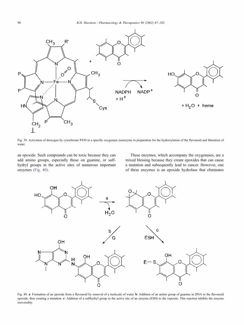
an epoxide. Such compounds can be toxic because they can
add amino groups, especially those on guanine, or sulf-
hydryl groups in the active sites of numerous important
enzymes (Fig. 40).
These enzymes, which accompany the oxygenases, are a
mixed blessing because they create epoxides that can cause
a mutation and subsequently lead to cancer. However, one
of these enzymes is an epoxide hydrolase that eliminates
Fig. 39. Activation of dioxygen by cytochrome P450 in a specific oxygenase isoenzyme in preparation for the hydroxylation of the flavonoid and liberation of
water.
Fig. 40. a: Formation of an epoxide from a flavonoid by removal of a molecule of water. b: Addition of an amino group of guanine in DNA to the flavonoid
epoxide, thus creating a mutation. c: Addition of a sulfhydryl group in the active site of an enzyme (ESH) to the exposite. This reaction inhibits the enzyme
irreversibly.
B.H. Havsteen / Pharmacology & Therapeutics 96 (2002) 67–20298
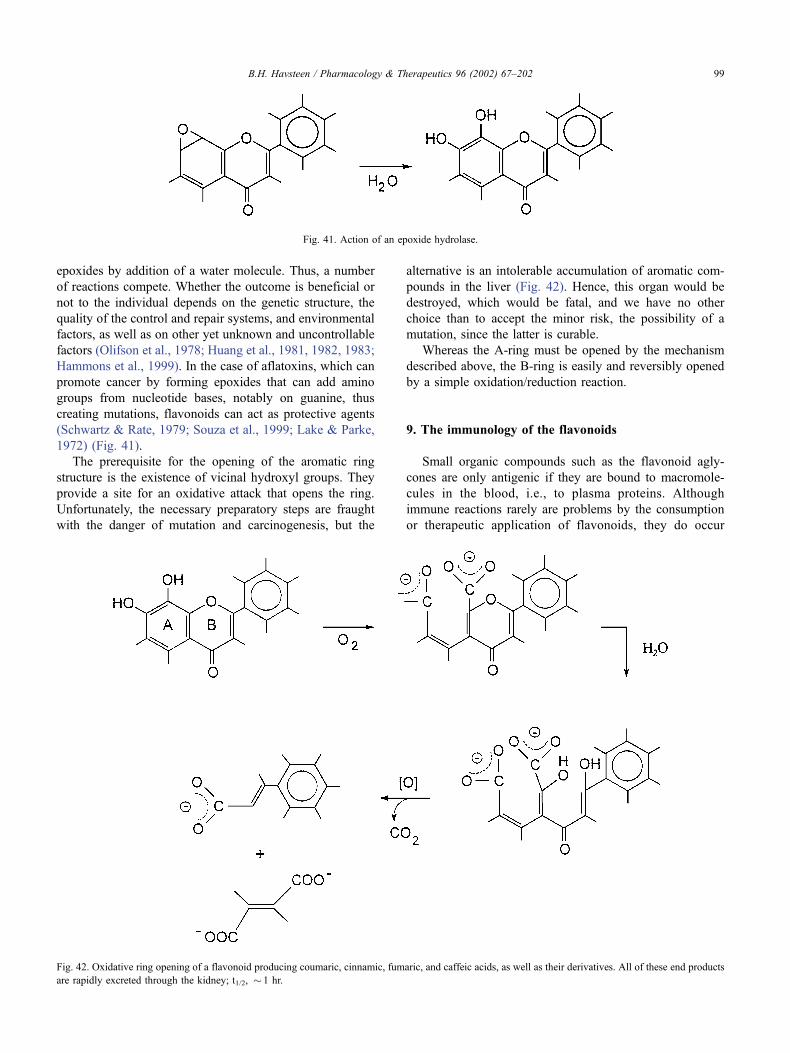
epoxides by addition of a water molecule. Thus, a number
of reactions compete. Whether the outcome is beneficial or
not to the individual depends on the genetic structure, the
quality of the control and repair systems, and environmental
factors, as well as on other yet unknown and uncontrollable
factors (Olifson et al., 1978; Huang et al., 1981, 1982, 1983;
Hammons et al., 1999). In the case of aflatoxins, which can
promote cancer by forming epoxides that can add amino
groups from nucleotide bases, notably on guanine, thus
creating mutations, flavonoids can act as protective agents
(Schwartz & Rate, 1979; Souza et al., 1999; Lake & Parke,
1972) (Fig. 41).
The prerequisite for the opening of the aromatic ring
structure is the existence of vicinal hydroxyl groups. They
provide a site for an oxidative attack that opens the ring.
Unfortunately, the necessary preparatory steps are fraught
with the danger of mutation and carcinogenesis, but the
alternative is an intolerable accumulation of aromatic com-
pounds in the liver (Fig. 42). Hence, this organ would be
destroyed, which would be fatal, and we have no other
choice than to accept the minor risk, the possibility of a
mutation, since the latter is curable.
Whereas the A-ring must be opened by the mechanism
described above, the B-ring is easily and reversibly opened
by a simple oxidation/reduction reaction.
9. The immunology of the flavonoids
Small organic compounds such as the flavonoid agly-
cones are only antigenic if they are bound to macromole-
cules in the blood, i.e., to plasma proteins. Although
immune reactions rarely are problems by the consumption
or therapeutic application of flavonoids, they do occur
Fig. 41. Action of an epoxide hydrolase.
Fig. 42. Oxidative ring opening of a flavonoid producing coumaric, cinnamic, fumaric, and caffeic acids, as well as their derivatives. All of these end products
are rapidly excreted through the kidney; t1/2, � 1 hr.
B.H. Havsteen / Pharmacology & Therapeutics 96 (2002) 67–202 99
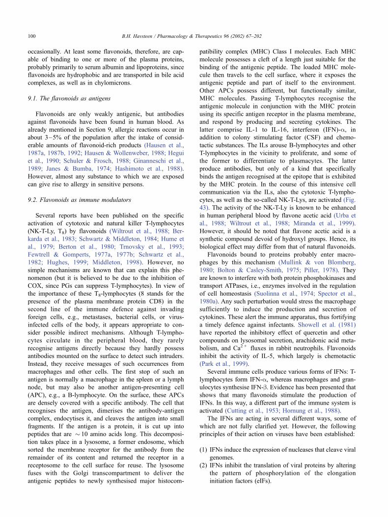
occasionally. At least some flavonoids, therefore, are cap-
able of binding to one or more of the plasma proteins,
probably primarily to serum albumin and lipoproteins, since
flavonoids are hydrophobic and are transported in bile acid
complexes, as well as in chylomicrons.
9.1. The flavonoids as antigens
Flavonoids are only weakly antigenic, but antibodies
against flavonoids have been found in human blood. As
already mentioned in Section 9, allergic reactions occur in
about 3–5% of the population after the intake of consid-
erable amounts of flavonoid-rich products (Hausen et al.,
1987a, 1987b, 1992; Hausen & Wollenweber, 1988; Hegui
et al., 1990; Schuler & Frosch, 1988; Ginanneschi et al.,
1989; Janes & Bumba, 1974; Hashimoto et al., 1988).
However, almost any substance to which we are exposed
can give rise to allergy in sensitive persons.
9.2. Flavonoids as immune modulators
Several reports have been published on the specific
activation of cytotoxic and natural killer T-lymphocytes
(NK-T-Ly, T8) by flavonoids (Wiltrout et al., 1988; Ber-
karda et al., 1983; Schwartz & Middleton, 1984; Hume et
al., 1979; Berton et al., 1980; Trnovsky et al., 1993;
Fewtrell & Gomperts, 1977a, 1977b; Schwartz et al.,
1982; Hughes, 1999; Middleton, 1998). However, no
simple mechanisms are known that can explain this phe-
nomenon (but it is believed to be due to the inhibition of
COX, since PGs can suppress T-lymphocytes). In view of
the importance of these T8-lymphocytes (8 stands for the
presence of the plasma membrane protein CD8) in the
second line of the immune defence against invading
foreign cells, e.g., metastases, bacterial cells, or virus-
infected cells of the body, it appears appropriate to con-
sider possible indirect mechanisms. Although T-lympho-
cytes circulate in the peripheral blood, they rarely
recognise antigens directly because they hardly possess
antibodies mounted on the surface to detect such intruders.
Instead, they receive messages of such occurrences from
macrophages and other cells. The first stop of such an
antigen is normally a macrophage in the spleen or a lymph
node, but may also be another antigen-presenting cell
(APC), e.g., a B-lymphocyte. On the surface, these APCs
are densely covered with a specific antibody. The cell that
recognises the antigen, dimerises the antibody-antigen
complex, endocytises it, and cleaves the antigen into small
fragments. If the antigen is a protein, it is cut up into
peptides that are � 10 amino acids long. This decomposi-
tion takes place in a lysosome, a former endosome, which
sorted the membrane receptor for the antibody from the
remainder of its content and returned the receptor in a
receptosome to the cell surface for reuse. The lysosome
fuses with the Golgi transcompartment to deliver the
antigenic peptides to newly synthesised major histocom-
patibility complex (MHC) Class I molecules. Each MHC
molecule possesses a cleft of a length just suitable for the
binding of the antigenic peptide. The loaded MHC mole-
cule then travels to the cell surface, where it exposes the
antigenic peptide and part of itself to the environment.
Other APCs possess different, but functionally similar,
MHC molecules. Passing T-lymphocytes recognise the
antigenic molecule in conjunction with the MHC protein
using its specific antigen receptor in the plasma membrane,
and respond by producing and secreting cytokines. The
latter comprise IL-1 to IL-16, interferon (IFN)-a, in
addition to colony stimulating factor (CSF) and chemo-
tactic substances. The ILs arouse B-lymphocytes and other
T-lymphocytes in the vicinity to proliferate, and some of
the former to differentiate to plasmacytes. The latter
produce antibodies, but only of a kind that specifically
binds the antigen recognised at the epitope that is exhibited
by the MHC protein. In the course of this intensive cell
communication via the ILs, also the cytotoxic T-lympho-
cytes, as well as the so-called NK-T-Lys, are activated (Fig.
43). The activity of the NK-T-Ly is known to be enhanced
in human peripheral blood by flavone acetic acid (Urba et
al., 1988; Wiltrout et al., 1988; Miranda et al., 1999).
However, it should be noted that flavone acetic acid is a
synthetic compound devoid of hydroxyl groups. Hence, its
biological effect may differ from that of natural flavonoids.
Flavonoids bound to proteins probably enter macro-
phages by this mechanism (Mullink & von Blomberg,
1980; Bolton & Casley-Smith, 1975; Piller, 1978). They
are known to interfere with both protein phosphokinases and
transport ATPases, i.e., enzymes involved in the regulation
of cell homeostasis (Suolinna et al., 1974; Spector et al.,
1980a). Any such perturbation would stress the macrophage
sufficiently to induce the production and secretion of
cytokines. These alert the immune apparatus, thus fortifying
a timely defence against infectants. Showell et al. (1981)
have reported the inhibitory effect of quercetin and other
compounds on lysosomal secretion, arachidonic acid meta-
bolism, and Ca2 + fluxes in rabbit neutrophils. Flavonoids
inhibit the activity of IL-5, which largely is chemotactic
(Park et al., 1999).
Several immune cells produce various forms of IFNs: T-
lymphocytes form IFN-a, whereas macrophages and gran-
ulocytes synthesise IFN-b. Evidence has been presented that
shows that many flavonoids stimulate the production of
IFNs. In this way, a different part of the immune system is
activated (Cutting et al., 1953; Hornung et al., 1988).
The IFNs are acting in several different ways, some of
which are not fully clarified yet. However, the following
principles of their action on viruses have been established:
(1) IFNs induce the expression of nucleases that cleave viral
genomes.
(2) IFNs inhibit the translation of viral proteins by altering
the pattern of phosphorylation of the elongation
initiation factors (eIFs).
B.H. Havsteen / Pharmacology & Therapeutics 96 (2002) 67–202100
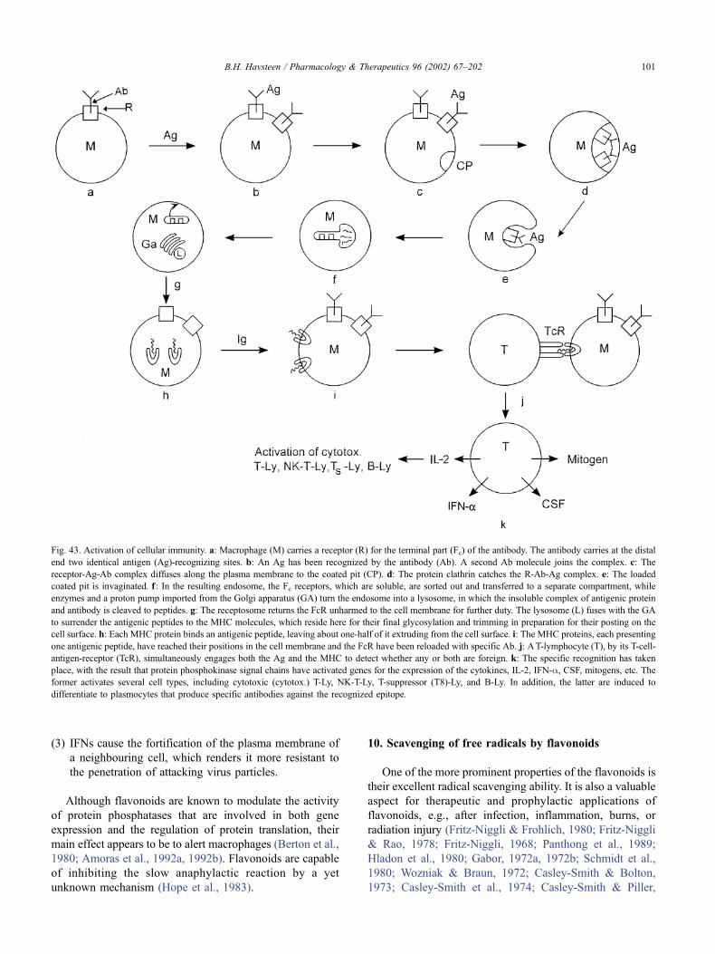
(3) IFNs cause the fortification of the plasma membrane of
a neighbouring cell, which renders it more resistant to
the penetration of attacking virus particles.
Although flavonoids are known to modulate the activity
of protein phosphatases that are involved in both gene
expression and the regulation of protein translation, their
main effect appears to be to alert macrophages (Berton et al.,
1980; Amoras et al., 1992a, 1992b). Flavonoids are capable
of inhibiting the slow anaphylactic reaction by a yet
unknown mechanism (Hope et al., 1983).
10. Scavenging of free radicals by flavonoids
One of the more prominent properties of the flavonoids is
their excellent radical scavenging ability. It is also a valuable
aspect for therapeutic and prophylactic applications of
flavonoids, e.g., after infection, inflammation, burns, or
radiation injury (Fritz-Niggli & Frohlich, 1980; Fritz-Niggli
& Rao, 1978; Fritz-Niggli, 1968; Panthong et al., 1989;
Hladon et al., 1980; Gabor, 1972a, 1972b; Schmidt et al.,
1980; Wozniak & Braun, 1972; Casley-Smith & Bolton,
1973; Casley-Smith et al., 1974; Casley-Smith & Piller,
Fig. 43. Activation of cellular immunity. a: Macrophage (M) carries a receptor (R) for the terminal part (Fc) of the antibody. The antibody carries at the distal
end two identical antigen (Ag)-recognizing sites. b: An Ag has been recognized by the antibody (Ab). A second Ab molecule joins the complex. c: The
receptor-Ag-Ab complex diffuses along the plasma membrane to the coated pit (CP). d: The protein clathrin catches the R-Ab-Ag complex. e: The loaded
coated pit is invaginated. f: In the resulting endosome, the Fc receptors, which are soluble, are sorted out and transferred to a separate compartment, while
enzymes and a proton pump imported from the Golgi apparatus (GA) turn the endosome into a lysosome, in which the insoluble complex of antigenic protein
and antibody is cleaved to peptides. g: The receptosome returns the FcR unharmed to the cell membrane for further duty. The lysosome (L) fuses with the GA
to surrender the antigenic peptides to the MHC molecules, which reside here for their final glycosylation and trimming in preparation for their posting on the
cell surface. h: Each MHC protein binds an antigenic peptide, leaving about one-half of it extruding from the cell surface. i: The MHC proteins, each presenting
one antigenic peptide, have reached their positions in the cell membrane and the FcR have been reloaded with specific Ab. j: AT-lymphocyte (T), by its T-cell-
antigen-receptor (TcR), simultaneously engages both the Ag and the MHC to detect whether any or both are foreign. k: The specific recognition has taken
place, with the result that protein phosphokinase signal chains have activated genes for the expression of the cytokines, IL-2, IFN-a, CSF, mitogens, etc. The
former activates several cell types, including cytotoxic (cytotox.) T-Ly, NK-T-Ly, T-suppressor (T8)-Ly, and B-Ly. In addition, the latter are induced to
differentiate to plasmocytes that produce specific antibodies against the recognized epitope.
B.H. Havsteen / Pharmacology & Therapeutics 96 (2002) 67–202 101
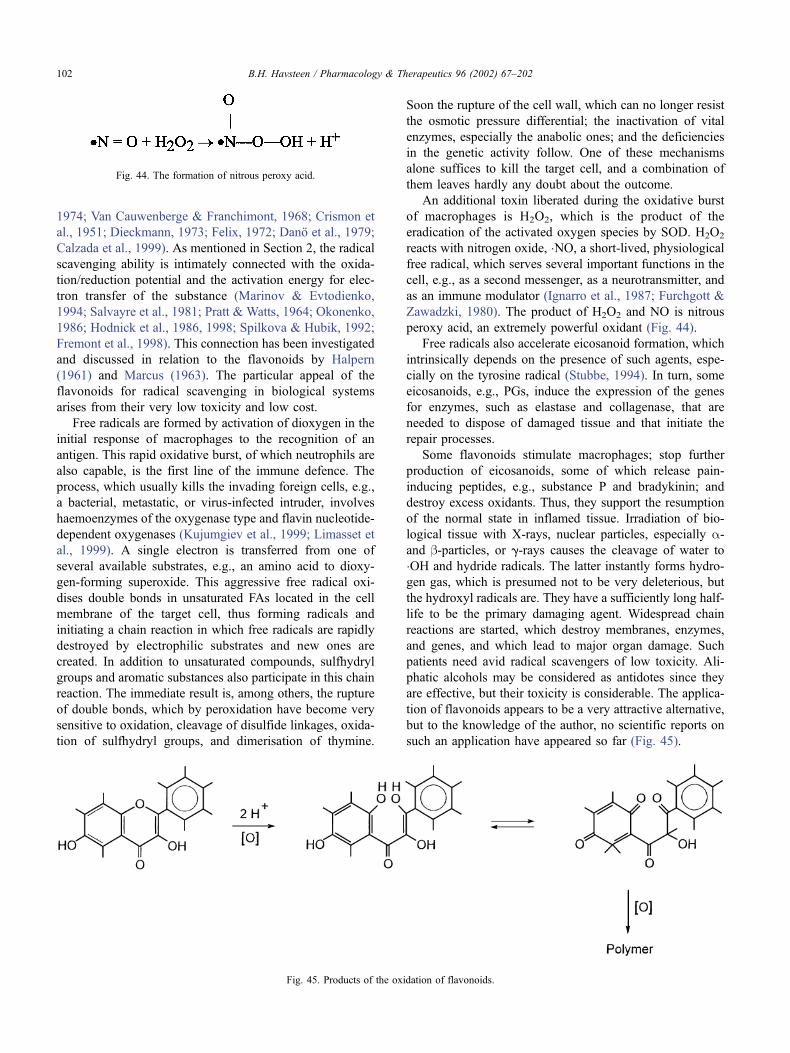
1974; Van Cauwenberge & Franchimont, 1968; Crismon et
al., 1951; Dieckmann, 1973; Felix, 1972; Dano et al., 1979;
Calzada et al., 1999). As mentioned in Section 2, the radical
scavenging ability is intimately connected with the oxida-
tion/reduction potential and the activation energy for elec-
tron transfer of the substance (Marinov & Evtodienko,
1994; Salvayre et al., 1981; Pratt & Watts, 1964; Okonenko,
1986; Hodnick et al., 1986, 1998; Spilkova & Hubik, 1992;
Fremont et al., 1998). This connection has been investigated
and discussed in relation to the flavonoids by Halpern
(1961) and Marcus (1963). The particular appeal of the
flavonoids for radical scavenging in biological systems
arises from their very low toxicity and low cost.
Free radicals are formed by activation of dioxygen in the
initial response of macrophages to the recognition of an
antigen. This rapid oxidative burst, of which neutrophils are
also capable, is the first line of the immune defence. The
process, which usually kills the invading foreign cells, e.g.,
a bacterial, metastatic, or virus-infected intruder, involves
haemoenzymes of the oxygenase type and flavin nucleotide-
dependent oxygenases (Kujumgiev et al., 1999; Limasset et
al., 1999). A single electron is transferred from one of
several available substrates, e.g., an amino acid to dioxy-
gen-forming superoxide. This aggressive free radical oxi-
dises double bonds in unsaturated FAs located in the cell
membrane of the target cell, thus forming radicals and
initiating a chain reaction in which free radicals are rapidly
destroyed by electrophilic substrates and new ones are
created. In addition to unsaturated compounds, sulfhydryl
groups and aromatic substances also participate in this chain
reaction. The immediate result is, among others, the rupture
of double bonds, which by peroxidation have become very
sensitive to oxidation, cleavage of disulfide linkages, oxida-
tion of sulfhydryl groups, and dimerisation of thymine.
Soon the rupture of the cell wall, which can no longer resist
the osmotic pressure differential; the inactivation of vital
enzymes, especially the anabolic ones; and the deficiencies
in the genetic activity follow. One of these mechanisms
alone suffices to kill the target cell, and a combination of
them leaves hardly any doubt about the outcome.
An additional toxin liberated during the oxidative burst
of macrophages is H2O2, which is the product of the
eradication of the activated oxygen species by SOD. H2O2
reacts with nitrogen oxide, �NO, a short-lived, physiologicalfree radical, which serves several important functions in the
cell, e.g., as a second messenger, as a neurotransmitter, and
as an immune modulator (Ignarro et al., 1987; Furchgott &
Zawadzki, 1980). The product of H2O2 and NO is nitrous
peroxy acid, an extremely powerful oxidant (Fig. 44).
Free radicals also accelerate eicosanoid formation, which
intrinsically depends on the presence of such agents, espe-
cially on the tyrosine radical (Stubbe, 1994). In turn, some
eicosanoids, e.g., PGs, induce the expression of the genes
for enzymes, such as elastase and collagenase, that are
needed to dispose of damaged tissue and that initiate the
repair processes.
Some flavonoids stimulate macrophages; stop further
production of eicosanoids, some of which release pain-
inducing peptides, e.g., substance P and bradykinin; and
destroy excess oxidants. Thus, they support the resumption
of the normal state in inflamed tissue. Irradiation of bio-
logical tissue with X-rays, nuclear particles, especially a-and b-particles, or g-rays causes the cleavage of water to
�OH and hydride radicals. The latter instantly forms hydro-
gen gas, which is presumed not to be very deleterious, but
the hydroxyl radicals are. They have a sufficiently long half-
life to be the primary damaging agent. Widespread chain
reactions are started, which destroy membranes, enzymes,
and genes, and which lead to major organ damage. Such
patients need avid radical scavengers of low toxicity. Ali-
phatic alcohols may be considered as antidotes since they
are effective, but their toxicity is considerable. The applica-
tion of flavonoids appears to be a very attractive alternative,
but to the knowledge of the author, no scientific reports on
such an application have appeared so far (Fig. 45).
Fig. 44. The formation of nitrous peroxy acid.
Fig. 45. Products of the oxidation of flavonoids.
B.H. Havsteen / Pharmacology & Therapeutics 96 (2002) 67–202102
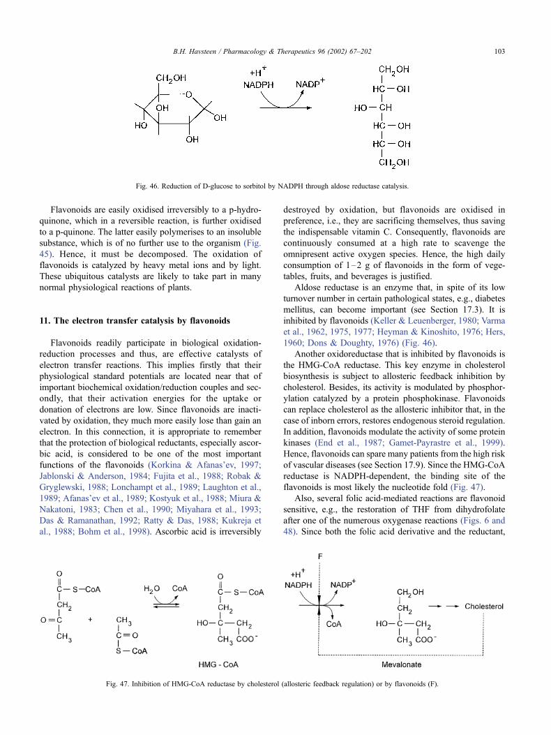
Flavonoids are easily oxidised irreversibly to a p-hydro-
quinone, which in a reversible reaction, is further oxidised
to a p-quinone. The latter easily polymerises to an insoluble
substance, which is of no further use to the organism (Fig.
45). Hence, it must be decomposed. The oxidation of
flavonoids is catalyzed by heavy metal ions and by light.
These ubiquitous catalysts are likely to take part in many
normal physiological reactions of plants.
11. The electron transfer catalysis by flavonoids
Flavonoids readily participate in biological oxidation-
reduction processes and thus, are effective catalysts of
electron transfer reactions. This implies firstly that their
physiological standard potentials are located near that of
important biochemical oxidation/reduction couples and sec-
ondly, that their activation energies for the uptake or
donation of electrons are low. Since flavonoids are inacti-
vated by oxidation, they much more easily lose than gain an
electron. In this connection, it is appropriate to remember
that the protection of biological reductants, especially ascor-
bic acid, is considered to be one of the most important
functions of the flavonoids (Korkina & Afanas’ev, 1997;
Jablonski & Anderson, 1984; Fujita et al., 1988; Robak &
Gryglewski, 1988; Lonchampt et al., 1989; Laughton et al.,
1989; Afanas’ev et al., 1989; Kostyuk et al., 1988; Miura &
Nakatoni, 1983; Chen et al., 1990; Miyahara et al., 1993;
Das & Ramanathan, 1992; Ratty & Das, 1988; Kukreja et
al., 1988; Bohm et al., 1998). Ascorbic acid is irreversibly
destroyed by oxidation, but flavonoids are oxidised in
preference, i.e., they are sacrificing themselves, thus saving
the indispensable vitamin C. Consequently, flavonoids are
continuously consumed at a high rate to scavenge the
omnipresent active oxygen species. Hence, the high daily
consumption of 1–2 g of flavonoids in the form of vege-
tables, fruits, and beverages is justified.
Aldose reductase is an enzyme that, in spite of its low
turnover number in certain pathological states, e.g., diabetes
mellitus, can become important (see Section 17.3). It is
inhibited by flavonoids (Keller & Leuenberger, 1980; Varma
et al., 1962, 1975, 1977; Heyman & Kinoshito, 1976; Hers,
1960; Dons & Doughty, 1976) (Fig. 46).
Another oxidoreductase that is inhibited by flavonoids is
the HMG-CoA reductase. This key enzyme in cholesterol
biosynthesis is subject to allosteric feedback inhibition by
cholesterol. Besides, its activity is modulated by phosphor-
ylation catalyzed by a protein phosphokinase. Flavonoids
can replace cholesterol as the allosteric inhibitor that, in the
case of inborn errors, restores endogenous steroid regulation.
In addition, flavonoids modulate the activity of some protein
kinases (End et al., 1987; Gamet-Payrastre et al., 1999).
Hence, flavonoids can spare many patients from the high risk
of vascular diseases (see Section 17.9). Since the HMG-CoA
reductase is NADPH-dependent, the binding site of the
flavonoids is most likely the nucleotide fold (Fig. 47).
Also, several folic acid-mediated reactions are flavonoid
sensitive, e.g., the restoration of THF from dihydrofolate
after one of the numerous oxygenase reactions (Figs. 6 and
48). Since both the folic acid derivative and the reductant,
Fig. 46. Reduction of D-glucose to sorbitol by NADPH through aldose reductase catalysis.
Fig. 47. Inhibition of HMG-CoA reductase by cholesterol (allosteric feedback regulation) or by flavonoids (F).
B.H. Havsteen / Pharmacology & Therapeutics 96 (2002) 67–202 103
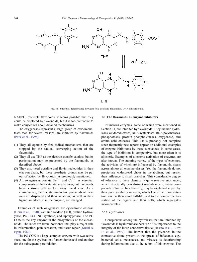
NADPH, resemble flavonoids, it seems possible that they
could be displaced by flavonoids, but it is too premature to
make conjectures about detailed mechanisms.
The oxygenases represent a large group of oxidoreduc-
tases that, for several reasons, are inhibited by flavonoids
(Park et al., 1998):
(1) They all operate by free radical mechanisms that are
stopped by the radical scavenging action of the
flavonoids.
(2) They all use THF as the electron transfer catalyst, but its
participation may be prevented by the flavonoids, as
described above.
(3) They also need pyridine and flavin nucleotides in their
electron chain, but these prosthetic groups may be put
out of action by flavonoids, as previously mentioned.
(4) All oxygenases contain Fe2 + and Cu2 + as essential
components of their catalytic mechanism, but flavonoids
have a strong affinity for heavy metal ions. As a
consequence, the oxidation/reduction potentials of these
ions are displaced and their locations, as well as their
ligand architecture in the enzyme, are changed.
Examples of such oxygenases are cytochrome oxidase
(Horn et al., 1970), xanthine oxidase (XO), proline hydrox-
ylase, PG COX, NO synthase, and lipoxygenase. The PG
COX is the key enzyme in the biosynthesis of the eicosa-
noids. The latter are tissue hormones that play a major role
in inflammation, pain sensation, and tissue repair (Kuehl &
Egan, 1980).
The PG COX is a large, complex enzyme with two active
sites, one for the cyclisation of arachidonic acid and another
for the subsequent peroxidation.
12. The flavonoids as enzyme inhibitors
Numerous enzymes, some of which were mentioned in
Section 11, are inhibited by flavonoids. They include hydro-
lases, oxidoreductases, DNA synthetases, RNA polymerases,
phosphatases, protein phosphokinases, oxygenase, and
amino acid oxidases. This list is probably not complete
since frequently new reports appear on additional examples
of enzyme inhibitions by these substances. In some cases,
the type of inhibition is competitive, but more often it is
allosteric. Examples of allosteric activation of enzymes are
also known. The stunning variety of the types of enzymes,
the activities of which are influenced by flavonoids, spans
across almost all enzyme classes. Yet, the flavonoids do not
precipitate widespread chaos in metabolism, but restrict
their influence to small branches. This considerable degree
of tolerance to these chemically quite reactive substances,
which structurally bear distinct resemblance to many com-
pounds of human biochemistry, may be explained in part by
their poor solubility in water, which keeps their concentra-
tion low; to their short half-life; and to the compartmental-
isation of the organs and their cells, which segregates
incompatibles.
12.1. Hydrolases
Conspicuous among the hydrolases that are inhibited by
flavonoids is hyaluronidase because of its importance to the
integrity of the loose connective tissue (Hasato et al., 1979;
Li et al., 1997). The barrier that the glycones in the
connective tissue present to the spread of infectants, e.g.,
bacterial cells, metastases, and viruses, is deteriorating
during inflammation due to the action of this enzyme. The
Fig. 48. Structural resemblance between folic acid and flavonoids. DHF, dihydrofolate.
B.H. Havsteen / Pharmacology & Therapeutics 96 (2002) 67–202104
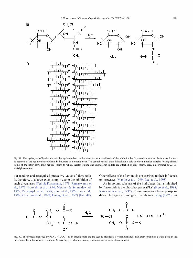
outstanding and recognised protective value of flavonoids
is, therefore, to a large extent simply due to the inhibition of
such glycanases (Tesi & Forssmann, 1971; Ramaswamy et
al., 1972; Bonvehi et al., 1994; Metzner & Schneidewind,
1978; Pepeljnjak et al., 1985; Shub et al., 1978; Lee et al.,
1997; Cecchini et al., 1997; Huang et al., 1997) (Fig. 49).
Other effects of the flavonoids are ascribed to their influence
on proteases (Mantle et al., 1999; Lee et al., 1998).
An important subclass of the hydrolases that is inhibited
by flavonoids is the phospholipases (PLs) (Kyo et al., 1998;
Kawaguchi et al., 1997). These enzymes cleave phospho-
diester linkages in biological membranes. Ring (1976) has
Fig. 49. The hydrolysis of hyaluronic acid by hyaluronidase. In this case, the structural basis of the inhibition by flavonoids is neither obvious nor known.
a: Segment of the hyaluronic acid chain. b: Structure of a proteoglycan. The central vertical chain is hyaluronic acid to which globular proteins (black) adhere.
Some of the latter carry long peptide chains to which keratan sulfate and chondroitin sulfate are attached as side chains. glcu, glucoronate; NAG, N-
acetylglucosamine.
Fig. 50. The process catalyzed by PLA2. R0-COO� is an arachidonate and the second product is a lysophosphatide. The latter constitutes a weak point in the
membrane that often causes its rupture. X may be, e.g., choline, serine, ethanolamine, or inositol (phosphate).
B.H. Havsteen / Pharmacology & Therapeutics 96 (2002) 67–202 105
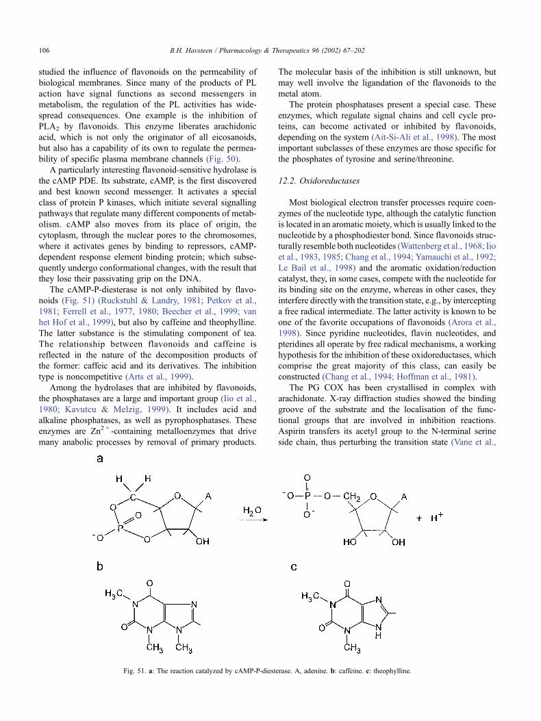
studied the influence of flavonoids on the permeability of
biological membranes. Since many of the products of PL
action have signal functions as second messengers in
metabolism, the regulation of the PL activities has wide-
spread consequences. One example is the inhibition of
PLA2 by flavonoids. This enzyme liberates arachidonic
acid, which is not only the originator of all eicosanoids,
but also has a capability of its own to regulate the permea-
bility of specific plasma membrane channels (Fig. 50).
A particularly interesting flavonoid-sensitive hydrolase is
the cAMP PDE. Its substrate, cAMP, is the first discovered
and best known second messenger. It activates a special
class of protein P kinases, which initiate several signalling
pathways that regulate many different components of metab-
olism. cAMP also moves from its place of origin, the
cytoplasm, through the nuclear pores to the chromosomes,
where it activates genes by binding to repressors, cAMP-
dependent response element binding protein; which subse-
quently undergo conformational changes, with the result that
they lose their passivating grip on the DNA.
The cAMP-P-diesterase is not only inhibited by flavo-
noids (Fig. 51) (Ruckstuhl & Landry, 1981; Petkov et al.,
1981; Ferrell et al., 1977, 1980; Beecher et al., 1999; van
het Hof et al., 1999), but also by caffeine and theophylline.
The latter substance is the stimulating component of tea.
The relationship between flavonoids and caffeine is
reflected in the nature of the decomposition products of
the former: caffeic acid and its derivatives. The inhibition
type is noncompetitive (Arts et al., 1999).
Among the hydrolases that are inhibited by flavonoids,
the phosphatases are a large and important group (Iio et al.,
1980; Kavutcu & Melzig, 1999). It includes acid and
alkaline phosphatases, as well as pyrophosphatases. These
enzymes are Zn2 + -containing metalloenzymes that drive
many anabolic processes by removal of primary products.
The molecular basis of the inhibition is still unknown, but
may well involve the ligandation of the flavonoids to the
metal atom.
The protein phosphatases present a special case. These
enzymes, which regulate signal chains and cell cycle pro-
teins, can become activated or inhibited by flavonoids,
depending on the system (Ait-Si-Ali et al., 1998). The most
important subclasses of these enzymes are those specific for
the phosphates of tyrosine and serine/threonine.
12.2. Oxidoreductases
Most biological electron transfer processes require coen-
zymes of the nucleotide type, although the catalytic function
is located in an aromatic moiety, which is usually linked to the
nucleotide by a phosphodiester bond. Since flavonoids struc-
turally resemble both nucleotides (Wattenberg et al., 1968; Iio
et al., 1983, 1985; Chang et al., 1994; Yamauchi et al., 1992;
Le Bail et al., 1998) and the aromatic oxidation/reduction
catalyst, they, in some cases, compete with the nucleotide for
its binding site on the enzyme, whereas in other cases, they
interfere directly with the transition state, e.g., by intercepting
a free radical intermediate. The latter activity is known to be
one of the favorite occupations of flavonoids (Arora et al.,
1998). Since pyridine nucleotides, flavin nucleotides, and
pteridines all operate by free radical mechanisms, a working
hypothesis for the inhibition of these oxidoreductases, which
comprise the great majority of this class, can easily be
constructed (Chang et al., 1994; Hoffman et al., 1981).
The PG COX has been crystallised in complex with
arachidonate. X-ray diffraction studies showed the binding
groove of the substrate and the localisation of the func-
tional groups that are involved in inhibition reactions.
Aspirin transfers its acetyl group to the N-terminal serine
side chain, thus perturbing the transition state (Vane et al.,
Fig. 51. a: The reaction catalyzed by cAMP-P-diesterase. A, adenine. b: caffeine. c: theophylline.
B.H. Havsteen / Pharmacology & Therapeutics 96 (2002) 67–202106
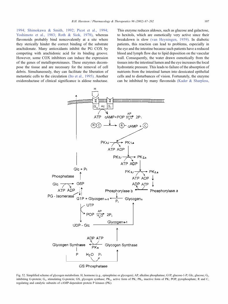
1994; Shimokawa & Smith, 1992; Picot et al., 1994;
Yoshimoto et al., 1983; Roth & Siok, 1978), whereas
flavonoids probably bind noncovalently at a site where
they sterically hinder the correct binding of the substrate
arachidonate. Many antioxidants inhibit the PG COX by
competing with arachidonic acid for its binding groove.
However, some COX inhibitors can induce the expression
of the genes of metalloproteinases. These enzymes decom-
pose the tissue and are necessary for the removal of cell
debris. Simultaneously, they can facilitate the liberation of
metastatic cells to the circulation (Ito et al., 1995). Another
oxidoreductase of clinical significance is aldose reductase.
This enzyme reduces aldoses, such as glucose and galactose,
to hexitols, which are osmotically very active since their
breakdown is slow (van Heyningen, 1959). In diabetic
patients, this reaction can lead to problems, especially in
the eye and the intestine because such patients have a reduced
blood and lymph flow due to lipid deposition on the vascular
wall. Consequently, the water drawn osmotically from the
tissues into the intestinal lumen and the eye increases the local
hydrostatic pressure. This leads to failure of the absorption of
nutrients from the intestinal lumen into dessicated epithelial
cells and to disturbances of vision. Fortunately, the enzyme
can be inhibited by many flavonoids (Kader & Sharpless,
Fig. 52. Simplified scheme of glycogen metabolism. H, hormone (e.g., epinephrine or glycogen); AP, alkaline phosphatase; G1P, glucose-1-P; Glc, glucose; Gi,
inhibiting G-protein; Gs, stimulating G-protein; GS, glycogen synthase; PKa, active form of PK; PKi, inactive form of PK; POP, pyrophosphate; R and C,
regulating and catalytic subunits of cAMP-dependent protein P kinases (PK).
B.H. Havsteen / Pharmacology & Therapeutics 96 (2002) 67–202 107
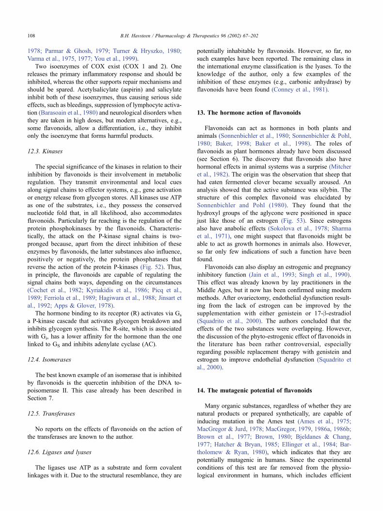
1978; Parmar & Ghosh, 1979; Turner & Hryszko, 1980;
Varma et al., 1975, 1977; You et al., 1999).
Two isoenzymes of COX exist (COX 1 and 2). One
releases the primary inflammatory response and should be
inhibited, whereas the other supports repair mechanisms and
should be spared. Acetylsalicylate (aspirin) and salicylate
inhibit both of these isoenzymes, thus causing serious side
effects, such as bleedings, suppression of lymphocyte activa-
tion (Barasoain et al., 1980) and neurological disorders when
they are taken in high doses, but modern alternatives, e.g.,
some flavonoids, allow a differentiation, i.e., they inhibit
only the isoenzyme that forms harmful products.
12.3. Kinases
The special significance of the kinases in relation to their
inhibition by flavonoids is their involvement in metabolic
regulation. They transmit environmental and local cues
along signal chains to effector systems, e.g., gene activation
or energy release from glycogen stores. All kinases use ATP
as one of the substrates, i.e., they possess the conserved
nucleotide fold that, in all likelihood, also accommodates
flavonoids. Particularly far reaching is the regulation of the
protein phosphokinases by the flavonoids. Characteris-
tically, the attack on the P-kinase signal chains is two-
pronged because, apart from the direct inhibition of these
enzymes by flavonoids, the latter substances also influence,
positively or negatively, the protein phosphatases that
reverse the action of the protein P-kinases (Fig. 52). Thus,
in principle, the flavonoids are capable of regulating the
signal chains both ways, depending on the circumstances
(Cochet et al., 1982; Kyriakidis et al., 1986; Picq et al.,
1989; Ferriola et al., 1989; Hagiwara et al., 1988; Jinsart et
al., 1992; Apps & Glover, 1978).
The hormone binding to its receptor (R) activates via Gs
a P-kinase cascade that activates glycogen breakdown and
inhibits glycogen synthesis. The R-site, which is associated
with Gi, has a lower affinity for the hormone than the one
linked to GS and inhibits adenylate cyclase (AC).
12.4. Isomerases
The best known example of an isomerase that is inhibited
by flavonoids is the quercetin inhibition of the DNA to-
poisomerase II. This case already has been described in
Section 7.
12.5. Transferases
No reports on the effects of flavonoids on the action of
the transferases are known to the author.
12.6. Ligases and lyases
The ligases use ATP as a substrate and form covalent
linkages with it. Due to the structural resemblance, they are
potentially inhabitable by flavonoids. However, so far, no
such examples have been reported. The remaining class in
the international enzyme classification is the lyases. To the
knowledge of the author, only a few examples of the
inhibition of these enzymes (e.g., carbonic anhydrase) by
flavonoids have been found (Conney et al., 1981).
13. The hormone action of flavonoids
Flavonoids can act as hormones in both plants and
animals (Sonnenbichler et al., 1980; Sonnenbichler & Pohl,
1980; Baker, 1998; Baker et al., 1998). The roles of
flavonoids as plant hormones already have been discussed
(see Section 6). The discovery that flavonoids also have
hormonal effects in animal systems was a surprise (Mitcher
et al., 1982). The origin was the observation that sheep that
had eaten fermented clover became sexually aroused. An
analysis showed that the active substance was silybin. The
structure of this complex flavonoid was elucidated by
Sonnenbichler and Pohl (1980). They found that the
hydroxyl groups of the aglycone were positioned in space
just like those of an estrogen (Fig. 53). Since estrogens
also have anabolic effects (Sokolova et al., 1978; Sharma
et al., 1971), one might suspect that flavonoids might be
able to act as growth hormones in animals also. However,
so far only few indications of such a function have been
found.
Flavonoids can also display an estrogenic and pregnancy
inhibitory function (Jain et al., 1993; Singh et al., 1990).
This effect was already known by lay practitioners in the
Middle Ages, but it now has been confirmed using modern
methods. After ovariectomy, endothelial dysfunction result-
ing from the lack of estrogen can be improved by the
supplementation with either genistein or 17-b-estradiol(Squadrito et al., 2000). The authors concluded that the
effects of the two substances were overlapping. However,
the discussion of the phyto-estrogenic effect of flavonoids in
the literature has been rather controversial, especially
regarding possible replacement therapy with genistein and
estrogen to improve endothelial dysfunction (Squadrito et
al., 2000).
14. The mutagenic potential of flavonoids
Many organic substances, regardless of whether they are
natural products or prepared synthetically, are capable of
inducing mutation in the Ames test (Ames et al., 1975;
MacGregor & Jurd, 1978; MacGregor, 1979, 1986a, 1986b;
Brown et al., 1977; Brown, 1980; Bjeldanes & Chang,
1977; Hatcher & Bryan, 1985; Ellinger et al., 1984; Bar-
tholomew & Ryan, 1980), which indicates that they are
potentially mutagenic in humans. Since the experimental
conditions of this test are far removed from the physio-
logical environment in humans, which includes efficient
B.H. Havsteen / Pharmacology & Therapeutics 96 (2002) 67–202108
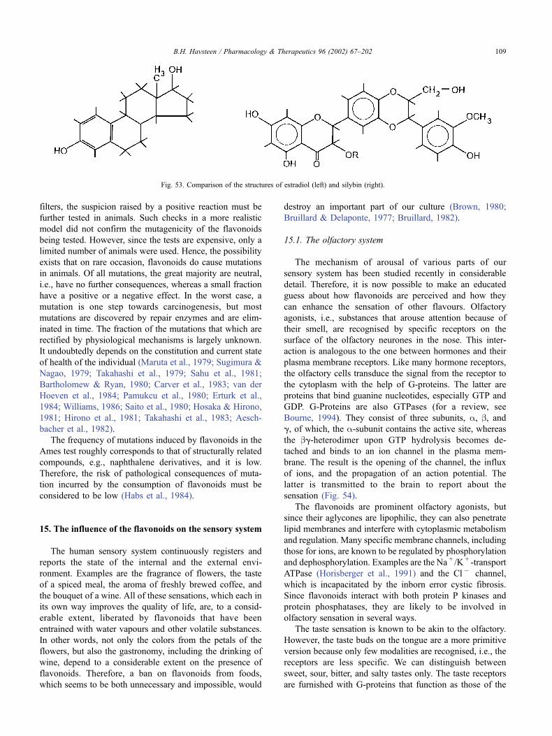
filters, the suspicion raised by a positive reaction must be
further tested in animals. Such checks in a more realistic
model did not confirm the mutagenicity of the flavonoids
being tested. However, since the tests are expensive, only a
limited number of animals were used. Hence, the possibility
exists that on rare occasion, flavonoids do cause mutations
in animals. Of all mutations, the great majority are neutral,
i.e., have no further consequences, whereas a small fraction
have a positive or a negative effect. In the worst case, a
mutation is one step towards carcinogenesis, but most
mutations are discovered by repair enzymes and are elim-
inated in time. The fraction of the mutations that which are
rectified by physiological mechanisms is largely unknown.
It undoubtedly depends on the constitution and current state
of health of the individual (Maruta et al., 1979; Sugimura &
Nagao, 1979; Takahashi et al., 1979; Sahu et al., 1981;
Bartholomew & Ryan, 1980; Carver et al., 1983; van der
Hoeven et al., 1984; Pamukcu et al., 1980; Erturk et al.,
1984; Williams, 1986; Saito et al., 1980; Hosaka & Hirono,
1981; Hirono et al., 1981; Takahashi et al., 1983; Aesch-
bacher et al., 1982).
The frequency of mutations induced by flavonoids in the
Ames test roughly corresponds to that of structurally related
compounds, e.g., naphthalene derivatives, and it is low.
Therefore, the risk of pathological consequences of muta-
tion incurred by the consumption of flavonoids must be
considered to be low (Habs et al., 1984).
15. The influence of the flavonoids on the sensory system
The human sensory system continuously registers and
reports the state of the internal and the external envi-
ronment. Examples are the fragrance of flowers, the taste
of a spiced meal, the aroma of freshly brewed coffee, and
the bouquet of a wine. All of these sensations, which each in
its own way improves the quality of life, are, to a consid-
erable extent, liberated by flavonoids that have been
entrained with water vapours and other volatile substances.
In other words, not only the colors from the petals of the
flowers, but also the gastronomy, including the drinking of
wine, depend to a considerable extent on the presence of
flavonoids. Therefore, a ban on flavonoids from foods,
which seems to be both unnecessary and impossible, would
destroy an important part of our culture (Brown, 1980;
Bruillard & Delaponte, 1977; Bruillard, 1982).
15.1. The olfactory system
The mechanism of arousal of various parts of our
sensory system has been studied recently in considerable
detail. Therefore, it is now possible to make an educated
guess about how flavonoids are perceived and how they
can enhance the sensation of other flavours. Olfactory
agonists, i.e., substances that arouse attention because of
their smell, are recognised by specific receptors on the
surface of the olfactory neurones in the nose. This inter-
action is analogous to the one between hormones and their
plasma membrane receptors. Like many hormone receptors,
the olfactory cells transduce the signal from the receptor to
the cytoplasm with the help of G-proteins. The latter are
proteins that bind guanine nucleotides, especially GTP and
GDP. G-Proteins are also GTPases (for a review, see
Bourne, 1994). They consist of three subunits, a, b, andg, of which, the a-subunit contains the active site, whereas
the bg-heterodimer upon GTP hydrolysis becomes de-
tached and binds to an ion channel in the plasma mem-
brane. The result is the opening of the channel, the influx
of ions, and the propagation of an action potential. The
latter is transmitted to the brain to report about the
sensation (Fig. 54).
The flavonoids are prominent olfactory agonists, but
since their aglycones are lipophilic, they can also penetrate
lipid membranes and interfere with cytoplasmic metabolism
and regulation. Many specific membrane channels, including
those for ions, are known to be regulated by phosphorylation
and dephosphorylation. Examples are the Na + /K + -transport
ATPase (Horisberger et al., 1991) and the Cl � channel,
which is incapacitated by the inborn error cystic fibrosis.
Since flavonoids interact with both protein P kinases and
protein phosphatases, they are likely to be involved in
olfactory sensation in several ways.
The taste sensation is known to be akin to the olfactory.
However, the taste buds on the tongue are a more primitive
version because only few modalities are recognised, i.e., the
receptors are less specific. We can distinguish between
sweet, sour, bitter, and salty tastes only. The taste receptors
are furnished with G-proteins that function as those of the
Fig. 53. Comparison of the structures of estradiol (left) and silybin (right).
B.H. Havsteen / Pharmacology & Therapeutics 96 (2002) 67–202 109
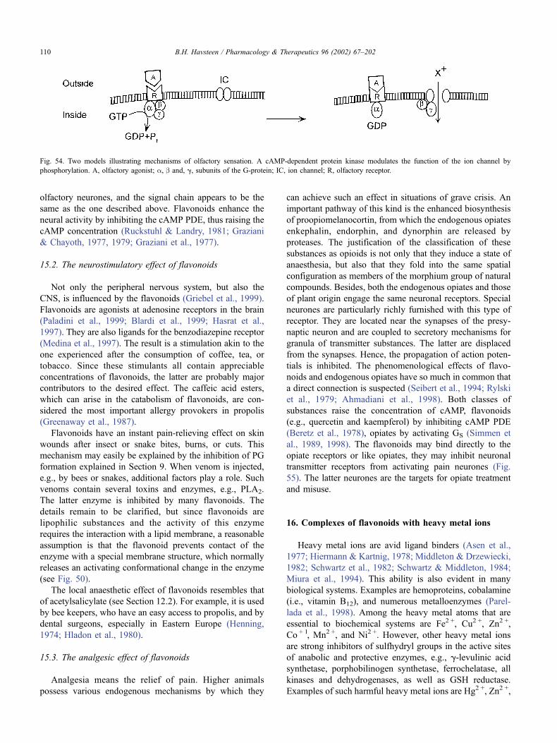
olfactory neurones, and the signal chain appears to be the
same as the one described above. Flavonoids enhance the
neural activity by inhibiting the cAMP PDE, thus raising the
cAMP concentration (Ruckstuhl & Landry, 1981; Graziani
& Chayoth, 1977, 1979; Graziani et al., 1977).
15.2. The neurostimulatory effect of flavonoids
Not only the peripheral nervous system, but also the
CNS, is influenced by the flavonoids (Griebel et al., 1999).
Flavonoids are agonists at adenosine receptors in the brain
(Paladini et al., 1999; Blardi et al., 1999; Hasrat et al.,
1997). They are also ligands for the benzodiazepine receptor
(Medina et al., 1997). The result is a stimulation akin to the
one experienced after the consumption of coffee, tea, or
tobacco. Since these stimulants all contain appreciable
concentrations of flavonoids, the latter are probably major
contributors to the desired effect. The caffeic acid esters,
which can arise in the catabolism of flavonoids, are con-
sidered the most important allergy provokers in propolis
(Greenaway et al., 1987).
Flavonoids have an instant pain-relieving effect on skin
wounds after insect or snake bites, burns, or cuts. This
mechanism may easily be explained by the inhibition of PG
formation explained in Section 9. When venom is injected,
e.g., by bees or snakes, additional factors play a role. Such
venoms contain several toxins and enzymes, e.g., PLA2.
The latter enzyme is inhibited by many flavonoids. The
details remain to be clarified, but since flavonoids are
lipophilic substances and the activity of this enzyme
requires the interaction with a lipid membrane, a reasonable
assumption is that the flavonoid prevents contact of the
enzyme with a special membrane structure, which normally
releases an activating conformational change in the enzyme
(see Fig. 50).
The local anaesthetic effect of flavonoids resembles that
of acetylsalicylate (see Section 12.2). For example, it is used
by bee keepers, who have an easy access to propolis, and by
dental surgeons, especially in Eastern Europe (Henning,
1974; Hladon et al., 1980).
15.3. The analgesic effect of flavonoids
Analgesia means the relief of pain. Higher animals
possess various endogenous mechanisms by which they
can achieve such an effect in situations of grave crisis. An
important pathway of this kind is the enhanced biosynthesis
of proopiomelanocortin, from which the endogenous opiates
enkephalin, endorphin, and dynorphin are released by
proteases. The justification of the classification of these
substances as opioids is not only that they induce a state of
anaesthesia, but also that they fold into the same spatial
configuration as members of the morphium group of natural
compounds. Besides, both the endogenous opiates and those
of plant origin engage the same neuronal receptors. Special
neurones are particularly richly furnished with this type of
receptor. They are located near the synapses of the presy-
naptic neuron and are coupled to secretory mechanisms for
granula of transmitter substances. The latter are displaced
from the synapses. Hence, the propagation of action poten-
tials is inhibited. The phenomenological effects of flavo-
noids and endogenous opiates have so much in common that
a direct connection is suspected (Seibert et al., 1994; Rylski
et al., 1979; Ahmadiani et al., 1998). Both classes of
substances raise the concentration of cAMP, flavonoids
(e.g., quercetin and kaempferol) by inhibiting cAMP PDE
(Beretz et al., 1978), opiates by activating GS (Simmen et
al., 1989, 1998). The flavonoids may bind directly to the
opiate receptors or like opiates, they may inhibit neuronal
transmitter receptors from activating pain neurones (Fig.
55). The latter neurones are the targets for opiate treatment
and misuse.
16. Complexes of flavonoids with heavy metal ions
Heavy metal ions are avid ligand binders (Asen et al.,
1977; Hiermann & Kartnig, 1978; Middleton & Drzewiecki,
1982; Schwartz et al., 1982; Schwartz & Middleton, 1984;
Miura et al., 1994). This ability is also evident in many
biological systems. Examples are hemoproteins, cobalamine
(i.e., vitamin B12), and numerous metalloenzymes (Parel-
lada et al., 1998). Among the heavy metal atoms that are
essential to biochemical systems are Fe2 +, Cu2 +, Zn2 +,
Co + 1, Mn2 +, and Ni2 +. However, other heavy metal ions
are strong inhibitors of sulfhydryl groups in the active sites
of anabolic and protective enzymes, e.g., g-levulinic acid
synthetase, porphobilinogen synthetase, ferrochelatase, all
kinases and dehydrogenases, as well as GSH reductase.
Examples of such harmful heavy metal ions are Hg2 +, Zn2 +,
Fig. 54. Two models illustrating mechanisms of olfactory sensation. A cAMP-dependent protein kinase modulates the function of the ion channel by
phosphorylation. A, olfactory agonist; a, b and, g, subunits of the G-protein; IC, ion channel; R, olfactory receptor.
B.H. Havsteen / Pharmacology & Therapeutics 96 (2002) 67–202110
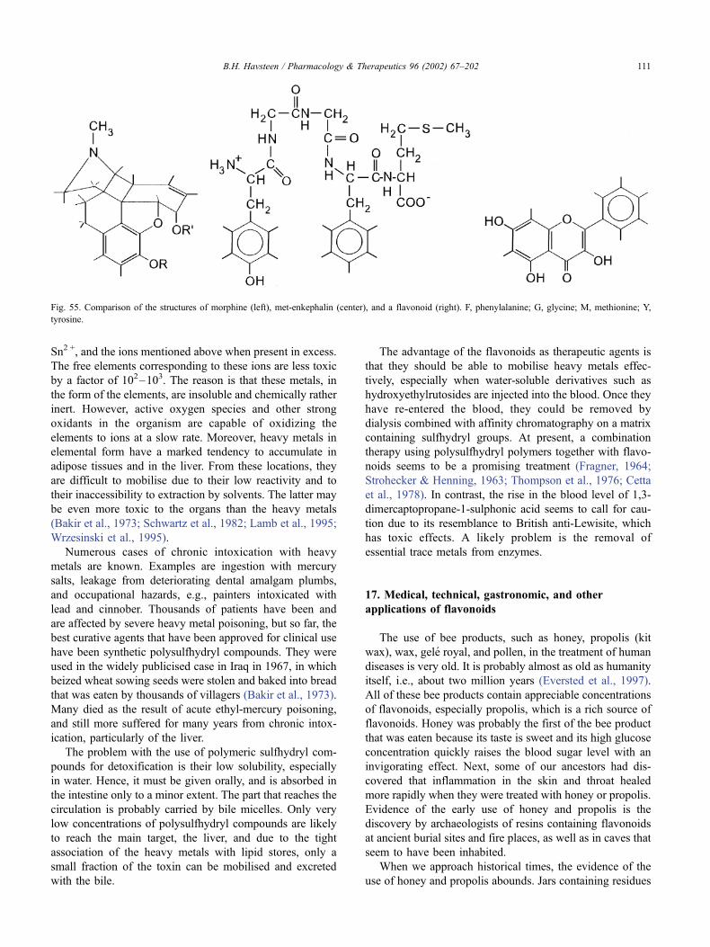
Sn2 +, and the ions mentioned above when present in excess.
The free elements corresponding to these ions are less toxic
by a factor of 102–103. The reason is that these metals, in
the form of the elements, are insoluble and chemically rather
inert. However, active oxygen species and other strong
oxidants in the organism are capable of oxidizing the
elements to ions at a slow rate. Moreover, heavy metals in
elemental form have a marked tendency to accumulate in
adipose tissues and in the liver. From these locations, they
are difficult to mobilise due to their low reactivity and to
their inaccessibility to extraction by solvents. The latter may
be even more toxic to the organs than the heavy metals
(Bakir et al., 1973; Schwartz et al., 1982; Lamb et al., 1995;
Wrzesinski et al., 1995).
Numerous cases of chronic intoxication with heavy
metals are known. Examples are ingestion with mercury
salts, leakage from deteriorating dental amalgam plumbs,
and occupational hazards, e.g., painters intoxicated with
lead and cinnober. Thousands of patients have been and
are affected by severe heavy metal poisoning, but so far, the
best curative agents that have been approved for clinical use
have been synthetic polysulfhydryl compounds. They were
used in the widely publicised case in Iraq in 1967, in which
beized wheat sowing seeds were stolen and baked into bread
that was eaten by thousands of villagers (Bakir et al., 1973).
Many died as the result of acute ethyl-mercury poisoning,
and still more suffered for many years from chronic intox-
ication, particularly of the liver.
The problem with the use of polymeric sulfhydryl com-
pounds for detoxification is their low solubility, especially
in water. Hence, it must be given orally, and is absorbed in
the intestine only to a minor extent. The part that reaches the
circulation is probably carried by bile micelles. Only very
low concentrations of polysulfhydryl compounds are likely
to reach the main target, the liver, and due to the tight
association of the heavy metals with lipid stores, only a
small fraction of the toxin can be mobilised and excreted
with the bile.
The advantage of the flavonoids as therapeutic agents is
that they should be able to mobilise heavy metals effec-
tively, especially when water-soluble derivatives such as
hydroxyethylrutosides are injected into the blood. Once they
have re-entered the blood, they could be removed by
dialysis combined with affinity chromatography on a matrix
containing sulfhydryl groups. At present, a combination
therapy using polysulfhydryl polymers together with flavo-
noids seems to be a promising treatment (Fragner, 1964;
Strohecker & Henning, 1963; Thompson et al., 1976; Cetta
et al., 1978). In contrast, the rise in the blood level of 1,3-
dimercaptopropane-1-sulphonic acid seems to call for cau-
tion due to its resemblance to British anti-Lewisite, which
has toxic effects. A likely problem is the removal of
essential trace metals from enzymes.
17. Medical, technical, gastronomic, and other
applications of flavonoids
The use of bee products, such as honey, propolis (kit
wax), wax, gele royal, and pollen, in the treatment of human
diseases is very old. It is probably almost as old as humanity
itself, i.e., about two million years (Eversted et al., 1997).
All of these bee products contain appreciable concentrations
of flavonoids, especially propolis, which is a rich source of
flavonoids. Honey was probably the first of the bee product
that was eaten because its taste is sweet and its high glucose
concentration quickly raises the blood sugar level with an
invigorating effect. Next, some of our ancestors had dis-
covered that inflammation in the skin and throat healed
more rapidly when they were treated with honey or propolis.
Evidence of the early use of honey and propolis is the
discovery by archaeologists of resins containing flavonoids
at ancient burial sites and fire places, as well as in caves that
seem to have been inhabited.
When we approach historical times, the evidence of the
use of honey and propolis abounds. Jars containing residues
Fig. 55. Comparison of the structures of morphine (left), met-enkephalin (center), and a flavonoid (right). F, phenylalanine; G, glycine; M, methionine; Y,
tyrosine.
B.H. Havsteen / Pharmacology & Therapeutics 96 (2002) 67–202 111
of high flavonoid content have been found in Egypt on the
tombs of the Pharaohs. Besides, propolis, due to its steriliz-
ing and tanning effect, in some cases has been used for
balsaming of mummies. Due to oxidation, these mummies
acquired a black surface colour, which at first was ascribed
to tar or asphalt.
Whereas the Bible only alludes to propolis, the Koran
explicitly mentions this substance (Muhammed, Ca. 500). It
specifically recommends the believers to keep a supply of
propolis in the house for use in the case of illness. The name
propolis also originates from the Near East, ‘‘pro’’ in Latin
means ‘‘in front of’’ and ‘‘polis’’ in Greek means ‘‘city’’.
This name alludes to the practice by bees of smearing the
landing platform in front of the entrance to the hive with
propolis glue, which the young working bees have enzy-
matically modified by chewing the resinous material that the
experienced collecting bees have gathered in trees and
shrubs. Their saliva contains an amylase that liberates the
flavonoid aglycones. Soldier bees on guard at the entrance
oversee that all arriving bees sterilise their feet in the
propolis glue before they enter the hive. The bees also
covered the walls of the latter with a layer of propolis glue
in order to render the hive almost sterile (Metzner et al.,
1979; Bankova et al., 1983; Hladon et al., 1980; Kujumgiev
et al., 1999) and to exclude draught and humidity. All
invading foreign insects and rodents, e.g., mice, snails, or
spiders, are instantly killed with stings and mummified with
propolis, if the carcasses are too heavy to be removed from
the hives.
Propolis has had numerous non-medical applications, a
few of which will be mentioned here to indicate the wide
appreciation of this substance in our culture (Lejeune et al.,
1984).
Today, propolis is mostly used by lay medical practi-
tioners, but in Eastern Europe, it is widely used by author-
ised medical doctors. It is used, for example, in cremes and
ointments for use against skin lesions, as repositories against
vaginal infections, and extracts of propolis, especially in
ethanol, are given per os to treat a variety of diseases, some
of which are discussed in the following sections. Flavonoids
can also be used as sweeteners (Brown et al., 1979).
In the following sections, some of the authorised uses of
flavonoids by the medical profession in Western civilisation
will be described.
17.1. Hypertension and microbleeding
Hypertension is a widespread disease, and in part can be
hereditary in nature. The condition can become aggravated,
for example, by psychological stress, tobacco smoking, and
alcohol consumption. These characteristics indicate the
participation of the part of the CNS that controls the state
of contraction of vascular smooth muscles. An understand-
ing of this complex syndrome requires a brief review of the
regulation of the water equilibria in the body (called water
homeostasis).
In response to a nervous signal from the formato retic-
ularis in the brain stem, prorenin is produced and secreted
into the blood. Biosynthesis primarily takes place in the
renal cortex, but, surprisingly, to a certain extent also in the
maxillary gland. Prorenin is activated, i.e., converted to
renin, by proteolytic liberation of a peptide. Renin, which
like pepsin belongs to the acidic type of proteases, activates
angiotensinogen (also called hypertensinogen), a prohor-
mone formed by the liver, to angiotensin (hypertensin). The
latter is shortened by a few amino acids by the angiotensin-
converting enzyme to angiotensin II, which binds to plasma
membrane receptors on a variety of cells, especially renal
tubular cells and vascular endothelial cells (Cushman &
Ondetti, 1991; Bettini et al., 1978a, 1978b, 1978c, 1978d;
Kimura et al., 1997). On the cytoplasmic side, the receptor
is in contact with G-proteins that activate AC. The latter
produces cAMP, which activates a protein phosphokinase
that opens a water channel by phosphorylation (Fig. 56).
Flavonoids can influence this mechanism in a variety of
ways (Bettini et al., 1978a, 1978b, 1978c, 1978d; Gerdin &
Svensjo, 1983; Casley-Smith, 1976; Ambrose & de Eds,
1949; Arturson, 1972; Bohr et al., 1949; Harper, 1966;
Klemm, 1966, 1967; Lockett & Jarman, 1958; Paris &
Moury, 1964; Paris & Vairel, 1949; Schiller, 1951; Svensjo
et al., 1977; van Cauwenberge & Franchimont, 1967;
Hodgson et al., 1999; Itoigawa et al., 1999; van Acker et
al., 1997). The most obvious is the inhibition of the PDE
that destroys cAMP by opening its ring (see Section 11).
The result of this inhibition is an increase in the flow of
water from blood into the tubular cell, from which it can
escape into the urine. The removal of water lowers the blood
pressure. This effect has long been claimed by lay medical
practitioners, but can now be confirmed by evidence from
recent biochemical research. The flavonoid class that most
frequently is used to lower blood pressure is a group of
water-soluble, semisynthetic compounds called hydrox-
yethyl-rutosides (Voelter & Doughty, 1978; Rose, 1970;
Tschopp et al., 1970).
The transport of water through lipid membranes is also
regulated by another mechanism, which is controlled by the
nonapeptide vasopressin (adiuretin or the antidiuretic hor-
mone). Vasopressin is produced in the hypothalamus at the
base of the brain in the nucleus paraventricularis. It is
formed as a prohormone linked to a larger protein neuro-
physin, which carries it along the axon of a long neuron,
terminating in the posterior part of the hypophysis, the
neurohypophysis, a gland located in the middle of the head
behind the nose. In the hypophysis, vasopressin is liberated
by proteolytic cleavage and stored until use. The hormone is
secreted into the blood upon receipt of a neurotransmitter
that binds to a surface receptor. The blood carries the
vasopressin to the kidneys, where it binds to a specific
receptor in the plasma membrane of tubular cells. This
receptor is a transmembrane protein, which, on the cyto-
plasmic side, is noncovalently linked to a G-protein by a
conformational change in the vasopressin-receptor complex.
B.H. Havsteen / Pharmacology & Therapeutics 96 (2002) 67–202112
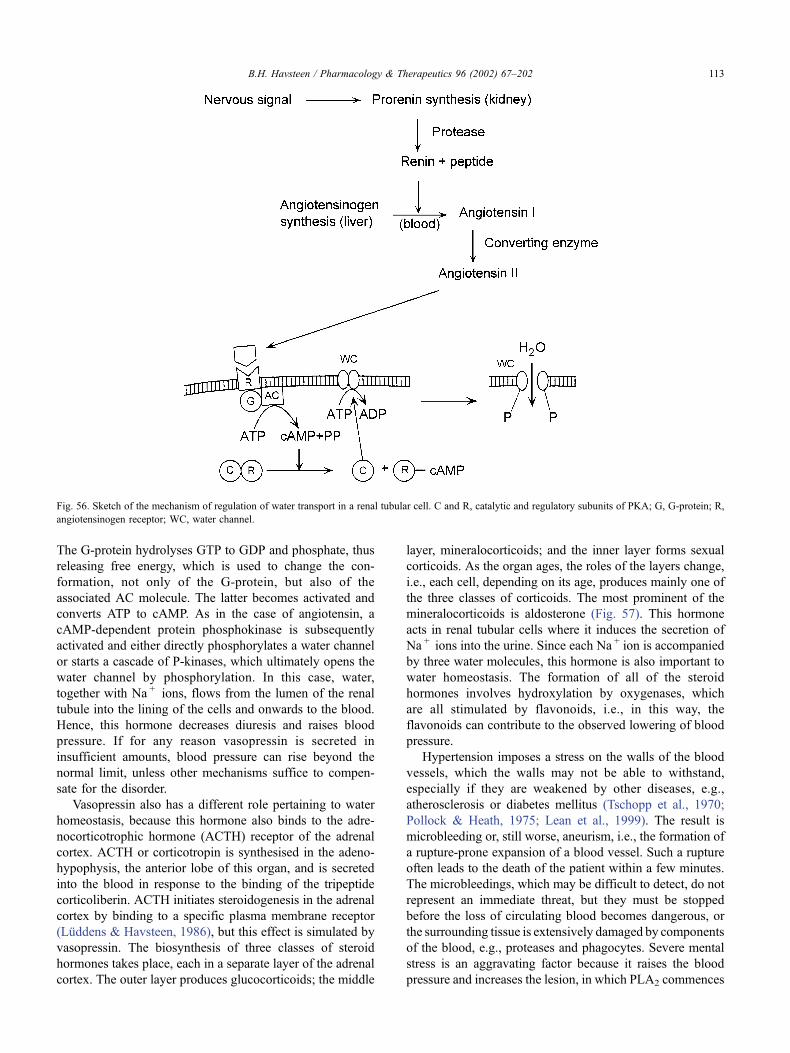
The G-protein hydrolyses GTP to GDP and phosphate, thus
releasing free energy, which is used to change the con-
formation, not only of the G-protein, but also of the
associated AC molecule. The latter becomes activated and
converts ATP to cAMP. As in the case of angiotensin, a
cAMP-dependent protein phosphokinase is subsequently
activated and either directly phosphorylates a water channel
or starts a cascade of P-kinases, which ultimately opens the
water channel by phosphorylation. In this case, water,
together with Na + ions, flows from the lumen of the renal
tubule into the lining of the cells and onwards to the blood.
Hence, this hormone decreases diuresis and raises blood
pressure. If for any reason vasopressin is secreted in
insufficient amounts, blood pressure can rise beyond the
normal limit, unless other mechanisms suffice to compen-
sate for the disorder.
Vasopressin also has a different role pertaining to water
homeostasis, because this hormone also binds to the adre-
nocorticotrophic hormone (ACTH) receptor of the adrenal
cortex. ACTH or corticotropin is synthesised in the adeno-
hypophysis, the anterior lobe of this organ, and is secreted
into the blood in response to the binding of the tripeptide
corticoliberin. ACTH initiates steroidogenesis in the adrenal
cortex by binding to a specific plasma membrane receptor
(Luddens & Havsteen, 1986), but this effect is simulated by
vasopressin. The biosynthesis of three classes of steroid
hormones takes place, each in a separate layer of the adrenal
cortex. The outer layer produces glucocorticoids; the middle
layer, mineralocorticoids; and the inner layer forms sexual
corticoids. As the organ ages, the roles of the layers change,
i.e., each cell, depending on its age, produces mainly one of
the three classes of corticoids. The most prominent of the
mineralocorticoids is aldosterone (Fig. 57). This hormone
acts in renal tubular cells where it induces the secretion of
Na + ions into the urine. Since each Na + ion is accompanied
by three water molecules, this hormone is also important to
water homeostasis. The formation of all of the steroid
hormones involves hydroxylation by oxygenases, which
are all stimulated by flavonoids, i.e., in this way, the
flavonoids can contribute to the observed lowering of blood
pressure.
Hypertension imposes a stress on the walls of the blood
vessels, which the walls may not be able to withstand,
especially if they are weakened by other diseases, e.g.,
atherosclerosis or diabetes mellitus (Tschopp et al., 1970;
Pollock & Heath, 1975; Lean et al., 1999). The result is
microbleeding or, still worse, aneurism, i.e., the formation of
a rupture-prone expansion of a blood vessel. Such a rupture
often leads to the death of the patient within a few minutes.
The microbleedings, which may be difficult to detect, do not
represent an immediate threat, but they must be stopped
before the loss of circulating blood becomes dangerous, or
the surrounding tissue is extensively damaged by components
of the blood, e.g., proteases and phagocytes. Severe mental
stress is an aggravating factor because it raises the blood
pressure and increases the lesion, in which PLA2 commences
Fig. 56. Sketch of the mechanism of regulation of water transport in a renal tubular cell. C and R, catalytic and regulatory subunits of PKA; G, G-protein; R,
angiotensinogen receptor; WC, water channel.
B.H. Havsteen / Pharmacology & Therapeutics 96 (2002) 67–202 113
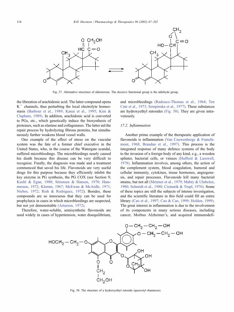
the liberation of arachidonic acid. The latter compound opens
K + channels, thus perturbing the local electrolyte homeo-
stasis (Barbour et al., 1989; Kanai et al., 1995; Kim &
Clapham, 1989). In addition, arachidonic acid is converted
to PGs, etc., which genetically induce the biosynthesis of
proteases, such as elastase and collagenases. The latter aid the
repair process by hydrolyzing fibrous proteins, but simulta-
neously further weakens blood vessel walls.
One example of the effect of stress on the vascular
system was the fate of a former chief executive in the
United States, who, in the course of the Watergate scandal,
suffered microbleedings. The microbleedings nearly caused
his death because this disease can be very difficult to
recognise. Finally, the diagnosis was made and a treatment
commenced that saved his life. Flavonoids are very useful
drugs for this purpose because they efficiently inhibit the
key enzyme in PG synthesis, the PG COX (see Section 9;
Kuehl & Egan, 1980; Sorensen & Hansen, 1970; Ham-
mersen, 1972; Klemm, 1967; McEwan & McArdle, 1971;
Niebes, 1972; Rish & Rodriguez, 1972). Besides, these
compounds are so innocuous that they can be used for
prophylaxis in cases in which microbleedings are suspected,
but not yet demonstrable (Arturson, 1972).
Therefore, water-soluble, semisynthetic flavonoids are
used widely in cases of hypertension, water disequilibrium,
and microbleedings (Radouco-Thomas et al., 1964; Ten
Cate et al., 1973; Sempinska et al., 1977). These substances
are hydroxyethyl rutosides (Fig. 58). They are given intra-
venously.
17.2. Inflammation
Another prime example of the therapeutic application of
flavonoids is inflammation (Van Cauwenberge & Franchi-
mont, 1968; Brandao et al., 1997). This process is the
integrated response of many defence systems of the body
to the invasion of a foreign body of any kind, e.g., a wooden
splinter, bacterial cells, or viruses (Hufford & Lasswell,
1978). Inflammation involves, among others, the action of
the complement system, blood coagulation, humoral and
cellular immunity, cytokines, tissue hormones, angiogene-
sis, and repair processes. Flavonoids kill many bacterial
strains, but not all (Metzner et al., 1979; Mabry & Ulubelen,
1980; Schmidt et al., 1980; Cizmarik & Trupl, 1976). Some
of these topics are still the subjects of intense investigation,
and the scientific literature in this field could fill an entire
library (Cao et al., 1997; Cao & Cao, 1999; Holden, 1999).
The great interest in inflammation is due to the involvement
of its components in many serious diseases, including
cancer, Morbus Alzheimer’s, and acquired immunodefi-
Fig. 57. Alternative structures of aldosterone. The decisive functional group is the aldehyde group.
Fig. 58. The structure of a hydroxyethyl rutoside (quercetyl rhamnose).
B.H. Havsteen / Pharmacology & Therapeutics 96 (2002) 67–202114
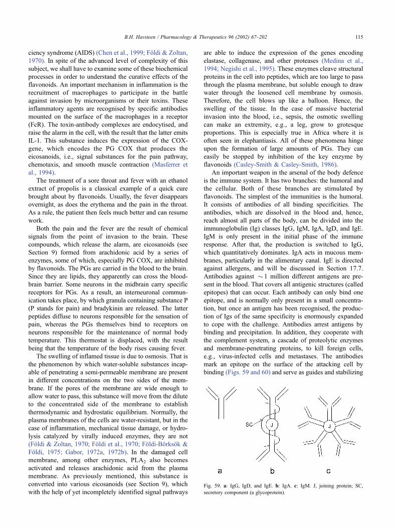
ciency syndrome (AIDS) (Chen et al., 1999; Foldi & Zoltan,
1970). In spite of the advanced level of complexity of this
subject, we shall have to examine some of these biochemical
processes in order to understand the curative effects of the
flavonoids. An important mechanism in inflammation is the
recruitment of macrophages to participate in the battle
against invasion by microorganisms or their toxins. These
inflammatory agents are recognised by specific antibodies
mounted on the surface of the macrophages in a receptor
(FcR). The toxin-antibody complexes are endocytised, and
raise the alarm in the cell, with the result that the latter emits
IL-1. This substance induces the expression of the COX-
gene, which encodes the PG COX that produces the
eicosanoids, i.e., signal substances for the pain pathway,
chemotaxis, and smooth muscle contraction (Masferrer et
al., 1994).
The treatment of a sore throat and fever with an ethanol
extract of propolis is a classical example of a quick cure
brought about by flavonoids. Usually, the fever disappears
overnight, as does the erythema and the pain in the throat.
As a rule, the patient then feels much better and can resume
work.
Both the pain and the fever are the result of chemical
signals from the point of invasion to the brain. These
compounds, which release the alarm, are eicosanoids (see
Section 9) formed from arachidonic acid by a series of
enzymes, some of which, especially PG COX, are inhibited
by flavonoids. The PGs are carried in the blood to the brain.
Since they are lipids, they apparently can cross the blood-
brain barrier. Some neurons in the midbrain carry specific
receptors for PGs. As a result, an interneuronal commun-
ication takes place, by which granula containing substance P
(P stands for pain) and bradykinin are released. The latter
peptides diffuse to neurons responsible for the sensation of
pain, whereas the PGs themselves bind to receptors on
neurons responsible for the maintenance of normal body
temperature. This thermostat is displaced, with the result
being that the temperature of the body rises causing fever.
The swelling of inflamed tissue is due to osmosis. That is
the phenomenon by which water-soluble substances incap-
able of penetrating a semi-permeable membrane are present
in different concentrations on the two sides of the mem-
brane. If the pores of the membrane are wide enough to
allow water to pass, this substance will move from the dilute
to the concentrated side of the membrane to establish
thermodynamic and hydrostatic equilibrium. Normally, the
plasma membranes of the cells are water-resistant, but in the
case of inflammation, mechanical tissue damage, or hydro-
lysis catalyzed by virally induced enzymes, they are not
(Foldi & Zoltan, 1970; Foldi et al., 1970; Foldi-Borksok &
Foldi, 1975; Gabor, 1972a, 1972b). In the damaged cell
membrane, among other enzymes, PLA2 also becomes
activated and releases arachidonic acid from the plasma
membrane. As previously mentioned, this substance is
converted into various eicosanoids (see Section 9), which
with the help of yet incompletely identified signal pathways
are able to induce the expression of the genes encoding
elastase, collagenase, and other proteases (Medina et al.,
1994; Negishi et al., 1995). These enzymes cleave structural
proteins in the cell into peptides, which are too large to pass
through the plasma membrane, but soluble enough to draw
water through the loosened cell membrane by osmosis.
Therefore, the cell blows up like a balloon. Hence, the
swelling of the tissue. In the case of massive bacterial
invasion into the blood, i.e., sepsis, the osmotic swelling
can make an extremity, e.g., a leg, grow to grotesque
proportions. This is especially true in Africa where it is
often seen in elephantiasis. All of these phenomena hinge
upon the formation of large amounts of PGs. They can
easily be stopped by inhibition of the key enzyme by
flavonoids (Casley-Smith & Casley-Smith, 1986).
An important weapon in the arsenal of the body defence
is the immune system. It has two branches: the humoral and
the cellular. Both of these branches are stimulated by
flavonoids. The simplest of the immunities is the humoral.
It consists of antibodies of all binding specificities. The
antibodies, which are dissolved in the blood and, hence,
reach almost all parts of the body, can be divided into the
immunoglobulin (Ig) classes IgG, IgM, IgA, IgD, and IgE.
IgM is only present in the initial phase of the immune
response. After that, the production is switched to IgG,
which quantitatively dominates. IgA acts in mucous mem-
branes, particularly in the alimentary canal. IgE is directed
against allergens, and will be discussed in Section 17.7.
Antibodies against � 1 million different antigens are pre-
sent in the blood. That covers all antigenic structures (called
epitopes) that can occur. Each antibody can only bind one
epitope, and is normally only present in a small concentra-
tion, but once an antigen has been recognised, the produc-
tion of Igs of the same specificity is enormously expanded
to cope with the challenge. Antibodies arrest antigens by
binding and precipitation. In addition, they cooperate with
the complement system, a cascade of proteolytic enzymes
and membrane-penetrating proteins, to kill foreign cells,
e.g., virus-infected cells and metastases. The antibodies
mark an epitope on the surface of the attacking cell by
binding (Figs. 59 and 60) and serve as guides and stabilizing
Fig. 59. a: IgG, IgD, and IgE. b: IgA. c: IgM. J, joining protein; SC,
secretory component (a glycoprotein).
B.H. Havsteen / Pharmacology & Therapeutics 96 (2002) 67–202 115
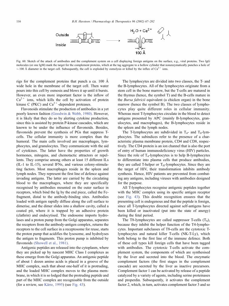
rigs for the complement proteins that punch a ca. 100 A
wide hole in the membrane of the target cell. Then water
pours into this cell by osmosis and blows it up until it bursts.
However, an even more important factor is the inflow of
Ca2 + ions, which kills the cell by activation of protein
kinase C (PKC) and Ca2 + -dependent proteases.
Flavonoids stimulate the production of antibodies in a yet
poorly known fashion (Goodwin & Webb, 1980). However,
it is likely that they do so by alerting cytokine production,
since this is assisted by protein P-kinase cascades, which are
known to be under the influence of flavonoids. Besides,
flavonoids prevent the synthesis of PGs that suppress T-
cells. The cellular immunity is more complex than the
humoral. The main cells involved are macrophages, lym-
phocytes, and granulocytes. They communicate with the aid
of cytokines. The latter have the properties of growth
hormones, mitogens, and chemotactic attractors or repel-
lents. They comprise among others at least 15 different ILs
(IL-1 to IL-15), several IFNs, and various colony-stimula-
ting factors. Most macrophages reside in the spleen and
lymph nodes. They represent the first line of defence against
invading antigens. The latter are carried by the circulating
blood to the macrophages, where they are specifically
recognised by antibodies mounted on the outer surface in
receptors, which bind the Ig by the end piece, called the Fc-
fragment, distal to the antibody-binding sites. Antibodies
loaded with antigen rapidly diffuse along the cell surface to
dimerise, and the dimer slides into a shallow cavity, called a
coated pit, where it is trapped by an adhesive protein
(clathrin) and endocytised. The endosome imports hydro-
lases and a proton pump from the Golgi apparatus, separates
the receptors from the antibody-antigen complex, returns the
receptors to the cell surface in a receptosome for reuse, starts
the proton pump that acidifies the lysosome, and hydrolyses
the antigen to fragments. This proton pump is inhibited by
flavonoids (Showell et al., 1981).
Antigenic peptides are released into the cytoplasm, where
they are picked up by nascent MHC Class I complexes as
these emerge from the Golgi apparatus. An antigenic peptide
of about 1 dozen amino acids is placed in a groove of the
MHC complex, such that about one-half of it is protruding,
and the loaded MHC complex moves to the plasma mem-
brane, in which it is so lodged that the protruding peptide and
part of the MHC complex are recognisable from the outside
(for a review, see Karre, 1995) (see Fig. 43).
The lymphocytes are divided into two classes, the T- and
the B-lymphocytes. All of the lymphocytes originate from a
stem cell in the bone marrow, but the T-cells are matured in
the thymus (hence, the symbol T) and the B-cells mature in
the Bursa fabricii equivalent (a chicken organ) in the bone
marrow (hence the symbol B). The two classes of lympho-
cytes play quite different roles in cellular immunity.
Whereas most T-lymphocytes circulate in the blood to detect
antigens presented by APC (mainly B-lymphocytes, gran-
ulocytes, and macrophages), the B-lymphocytes reside in
the spleen and the lymph nodes.
The T-lymphocytes are subdivided in T4- and T8-lym-
phocytes. The subindices refer to the presence of a char-
acteristic plasma membrane protein, CD4 and CD8, respec-
tively. The CD4 protein is an ion channel that is also the port
of entry of human immunodeficiency virus (HIV) particles.
Since the role of T4-lymphocytes is to help B-lymphocytes
to differentiate into plasma cells that produce antibodies,
they are called T-helper or TH-lymphocytes. Since they are
the target of HIV, their transformation inhibits antibody
synthesis. Hence, HIV patients are prevented from combat-
ing any antigens, including viruses with antibodies designed
for the purpose.
All T-lymphocytes recognise antigenic peptides together
with the MHC complex using its specific antigen receptor
(see Fig. 43). This double recognition ensures that the
presenting cell is endogenous and that the peptide is foreign,
since all T-lymphocytes directed against self-antigens have
been killed or inactivated (put into the state of anergy)
during the fetal period.
The T8-lymphocytes are called suppressor T-cells (TS),
because they inhibit the helper function of the T4-lympho-
cytes. Important subclasses of T8-cells are the cytotoxic T-
lymphocytes and natural killer T-cells (NK-T-Ly), which
both belong to the first line of the immune defence. Both
of these cell types kill foreign cells that have been tagged
with antibodies. The cytotoxic T-cells activate the com-
plement system, the components of which are synthesised
by the liver and secreted into the blood. The enzymatic
complement factors (the first stages in the complement
cascade) are secreted by the liver as inactive precursors.
Complement factor 1 can be activated by release of a peptide
catalyzed by a variety of agents, including serine proteinases
and properdin. Subsequently, it activates the complement
factor 2, which, in turn, activates complement factor 3 and so
Fig. 60. Sketch of the attack of antibodies and the complement system on a cell displaying foreign antigens on the surface, e.g., viral proteins. Two IgG
molecules (or one IgM) mark the target for the complement proteins, which at the tag aggregate to a hollow cylinder that nonenzymatically punches a hole of
� 100 A diameter in the target cell. Subsequently, the cell is exploded by osmolysis or killed by the influx of Ca2 + ions.
B.H. Havsteen / Pharmacology & Therapeutics 96 (2002) 67–202116
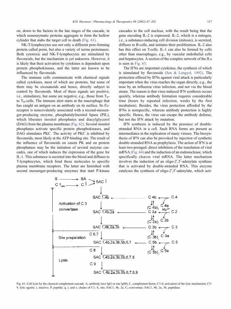
on, down to the factors in the late stages of the cascade, in
which nonenzymatic proteins aggregate to form the hollow
cylinder that stabs the target cell to death (Fig. 61).
NK-T-lymphocytes use not only a different pore-forming
protein called porin, but also a variety of serine proteinases.
Both cytotoxic and NK-T-lymphocytes are stimulated by
flavonoids, but the mechanism is yet unknown. However, it
is likely that their activation by cytokines is dependent upon
protein phosphokinases, and the latter are known to be
influenced by flavonoids.
The immune cells communicate with chemical signals
called cytokines, most of which are proteins, but some of
them may be eicosanoids and hence, directly subject to
control by flavonoids. Most of these signals are positive,
i.e., stimulatory, but some are negative, e.g., those from TS-
to TH-cells. The immune alert starts at the macrophage that
has caught an antigen on an antibody on its surface. Its Fc-
receptor is noncovalently associated with a second-messen-
ger-producing enzyme, phosphatidylinositol lipase (PIL),
which liberates inositol phosphates and diacylglycerol
(DAG) from the plasma membrane (Fig. 62). Several inositol
phosphates activate specific protein phosphokinases, and
DAG stimulates PKC. The activity of PKC is inhibited by
flavonoids, most likely at the ATP-binding site. The result of
the influence of flavonoids on casein PK and on protein
phosphatase may be the initiation of several enzyme cas-
cades, one of which induces the expression of the gene for
IL-1. This substance is secreted into the blood and diffuses to
T-lymphocytes, which bind these molecules to specific
plasma membrane receptors. The latter are furnished with
second messenger-producing enzymes that start P-kinase
cascades to the cell nucleus, with the result being that the
gene encoding IL-2 is expressed. IL-2, which is a mitogen,
i.e., a substance-inducing cell division (mitosis), is secreted,
diffuses to B-cells, and initiates their proliferation. IL-2 also
has this effect on T-cells. IL-1 can also be formed by cells
other than macrophages, e.g., by vascular endothelial cells
and hepatocytes. A section of the complex network of the ILs
is seen in Fig. 63.
The IFNs are important cytokines, the synthesis of which
is stimulated by flavonoids (Sen & Lengyel, 1992). The
protection offered by IFNs against viral attack is particularly
important when the virus reaches the organ directly, e.g., the
nose by an influenza virus infection, and not via the blood
steam. The reason is that virus-induced IFN synthesis occurs
quickly, whereas antibody formation requires considerable
time (hours by repeated infection, weeks by the first
incubation). Besides, the virus protection afforded by the
IFNs is nonspecific, whereas antibody protection is highly
specific. Hence, the virus can escape the antibody defense,
but not the IFN attack by mutation.
IFN synthesis is induced by the presence of double-
stranded RNA in a cell. Such RNA forms are present as
intermediates in the replication of many viruses. The biosyn-
thesis of IFN can also be provoked by injection of synthetic
double-stranded RNA as prophylaxis. The action of IFN is at
least two-pronged: direct inhibition of the translation of viral
mRNA (Fig. 64) and the induction of an endonuclease, which
specifically cleaves viral mRNA. The latter mechanism
involves the induction of an oligo-20,50-adenylate synthase
that is activated by double-stranded RNA. This enzyme
catalyses the synthesis of oligo-20,50-adenylate, which acti-
Fig. 61. Cell lysis by the classical complement cascade. A, antibody (two IgG or one IgM); C, complement factor; C1-4, activation of the lytic mechanism; C5-
9, lytic agents; i, inactive; P, peptide; q, r, and s, chains of C1; S, site; SAC1, 4b, 2a, C1-convertase; SAC1, 4b, 2a, 3b, peptidase.
B.H. Havsteen / Pharmacology & Therapeutics 96 (2002) 67–202 117
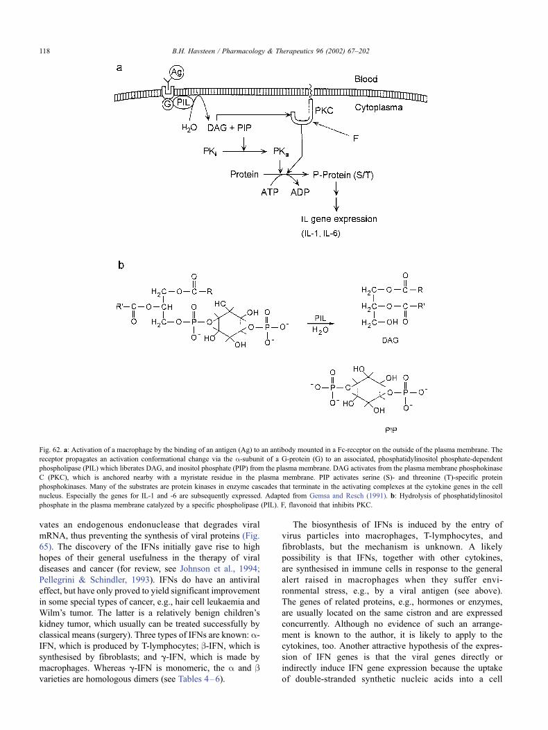
vates an endogenous endonuclease that degrades viral
mRNA, thus preventing the synthesis of viral proteins (Fig.
65). The discovery of the IFNs initially gave rise to high
hopes of their general usefulness in the therapy of viral
diseases and cancer (for review, see Johnson et al., 1994;
Pellegrini & Schindler, 1993). IFNs do have an antiviral
effect, but have only proved to yield significant improvement
in some special types of cancer, e.g., hair cell leukaemia and
Wilm’s tumor. The latter is a relatively benign children’s
kidney tumor, which usually can be treated successfully by
classical means (surgery). Three types of IFNs are known: a-IFN, which is produced by T-lymphocytes; b-IFN, which is
synthesised by fibroblasts; and g-IFN, which is made by
macrophages. Whereas g-IFN is monomeric, the a and bvarieties are homologous dimers (see Tables 4–6).
The biosynthesis of IFNs is induced by the entry of
virus particles into macrophages, T-lymphocytes, and
fibroblasts, but the mechanism is unknown. A likely
possibility is that IFNs, together with other cytokines,
are synthesised in immune cells in response to the general
alert raised in macrophages when they suffer envi-
ronmental stress, e.g., by a viral antigen (see above).
The genes of related proteins, e.g., hormones or enzymes,
are usually located on the same cistron and are expressed
concurrently. Although no evidence of such an arrange-
ment is known to the author, it is likely to apply to the
cytokines, too. Another attractive hypothesis of the expres-
sion of IFN genes is that the viral genes directly or
indirectly induce IFN gene expression because the uptake
of double-stranded synthetic nucleic acids into a cell
Fig. 62. a: Activation of a macrophage by the binding of an antigen (Ag) to an antibody mounted in a Fc-receptor on the outside of the plasma membrane. The
receptor propagates an activation conformational change via the a-subunit of a G-protein (G) to an associated, phosphatidylinositol phosphate-dependent
phospholipase (PIL) which liberates DAG, and inositol phosphate (PIP) from the plasma membrane. DAG activates from the plasma membrane phosphokinase
C (PKC), which is anchored nearby with a myristate residue in the plasma membrane. PIP activates serine (S)- and threonine (T)-specific protein
phosphokinases. Many of the substrates are protein kinases in enzyme cascades that terminate in the activating complexes at the cytokine genes in the cell
nucleus. Especially the genes for IL-1 and -6 are subsequently expressed. Adapted from Gemsa and Resch (1991). b: Hydrolysis of phosphatidylinositol
phosphate in the plasma membrane catalyzed by a specific phospholipase (PIL). F, flavonoid that inhibits PKC.
B.H. Havsteen / Pharmacology & Therapeutics 96 (2002) 67–202118
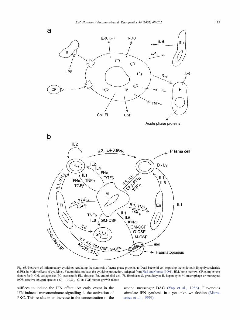
suffices to induce the IFN effect. An early event in the
IFN-induced transmembrane signalling is the activation of
PKC. This results in an increase in the concentration of the
second messenger DAG (Yap et al., 1986). Flavonoids
stimulate IFN synthesis in a yet unknown fashion (Mitro-
cotsa et al., 1999).
Fig. 63. Network of inflammatory cytokines regulating the synthesis of acute phase proteins. a: Dead bacterial cell exposing the endotoxin lipopolysaccharide
(LPS). b: Major effects of cytokines. Flavonoid stimulates the cytokine production. Adapted from Flad and Gemsa (1991). BM, bone marrow; CF, complement
factors 3a-9; Col, collagenase; EC, ecosanoid; EL, elastase; En, endothelial cell; Fi, fibroblast; G, granulocyte; H, hepatocyte; M, macrophage or monocyte;
ROS, reactive oxygen species (�O2� , H2O2, �OH); TGF, tumor growth factor.
B.H. Havsteen / Pharmacology & Therapeutics 96 (2002) 67–202 119
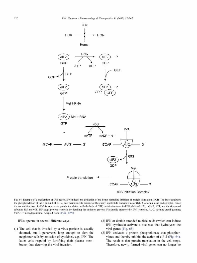
IFNs operate in several different ways:
(1) The cell that is invaded by a virus particle is usually
doomed, but it perseveres long enough to alert the
neighbour cells by emission of cytokines, e.g., IFN. The
latter cells respond by fortifying their plasma mem-
brane, thus deterring the viral invasion.
(2) IFN or double-stranded nucleic acids (which can induce
IFN synthesis) activate a nuclease that hydrolyses the
viral genes (Fig. 65).
(3) IFN activates a protein phosphokinase that phosphor-
ylates and thereby inhibits the action of eIF-2 (Fig. 64).
The result is that protein translation in the cell stops.
Therefore, newly formed viral genes can no longer be
Fig. 64. Example of a mechanism of IFN action. IFN induces the activation of the heme-controlled inhibitor of protein translation (HCI). The latter catalyzes
the phosphorylation of the a-subunit of eIF-2, thus permitting its binding of the guanyl nucleotide exchange factor (GEF) to form a dead end complex. Since
the normal function of eIF-2 is to promote protein translation with the help of GTP, methionine-transfer-RNA (Met-t-RNA), mRNA, ATP, and the ribosomal
subunits 40S and 60S, IFN stops protein synthesis by derailing the initiation process. Flavonoids promote the IFN synthesis. AUG, adenine-uracil-guanine;
50CAP, 7-methylguanosine. Adapted from Stryer (1995).
B.H. Havsteen / Pharmacology & Therapeutics 96 (2002) 67–202120
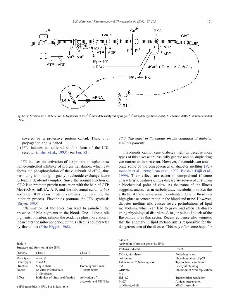
covered by a protective protein capsid. Thus, viral
propagation and is halted.
(4) IFN induces an antiviral soluble form of the LDL
receptor (Fisher et al., 1993) (see Fig. 83).
IFN induces the activation of the protein phosphokinase
home-controlled inhibitor of protein translation, which cat-
alyzes the phosphorylation of the a-subunit of eIF-2, thuspermitting its binding of guanyl nucleotide exchange factor
to form a dead-end complex. Since the normal function of
eIF-2 is to promote protein translation with the help of GTP,
Met-t-RNA, mRNA, ATP, and the ribosomal subunits 40S
and 60S, IFN stops protein synthesis by derailing the
initiation process. Flavonoids promote the IFN synthesis
(Stryer, 1995).
Inflammation of the liver can lead to jaundice, the
presence of bile pigments in the blood. One of these bile
pigments, bilirubin, inhibits the oxidative phosphorylation if
it can enter the mitochondrion, but this effect is counteracted
by flavonoids (Fritz-Niggli, 1968).
17.3. The effect of flavonoids on the condition of diabetes
mellitus patients
Flavonoids cannot cure diabetes mellitus because most
types of this disease are basically genetic and no single drug
can correct an inborn error. However, flavonoids can ameli-
orate some of the consequences of diabetes mellitus (Ver-
tounnen et al., 1994; Lean et al., 1999; Blostein-Fujii et al.,
1999). Their effects are easier to comprehend if some
characteristic features of this disease are reviewed first from
a biochemical point of view. As the name of the illness
suggests, anomalies in carbohydrate metabolism strikes the
afflicted if the disease remains untreated. One of these is a
high-glucose concentration in the blood and urine. However,
diabetes mellitus also causes severe perturbations of lipid
metabolism, which can lead to grave and often life-threat-
ening physiological disorders. A major point of attack of the
flavonoids is in this sector. Recent evidence also suggests
that the anomaly in lipid metabolism is responsible for the
dangerous turn of the disease. This may offer some hope for
Fig. 65. a: Mechanism of IFN action. b: Synthesis of tri-20,50-adenylate catalyzed by oligo-20,50-adenylate synthase (oAS). A, adenine; dsRNA, double-stranded
RNA.
Table 4
Structure and function of the IFNs
Property Class I Class II
Main types a and b g
Other types t and V –
Structure Single chain Homologous dimer
Source a: virus-infected cells T-lymphocytes
b: fibroblastsEffect Inhibition of virus proliferation Activation of
cytotoxic and NK-T-lys
t-IFN resembles a-IFN, but is less toxic.
Table 5
Activation of protein genes by IFNs
Proteins induced Effect
20-50-An-Synthase Polyadenylation
p68 kinase Phosphorylation of p68
Indoleamine 2,3-dioxygenase Tryptophan degradation
p56 Guanylate binding
GBP/p67 Inhibition of viral replication
Mx 1
IRF 1,2 Transcription regulation
MHC Antigen presentation
b2-Microglobulin MHC I assembly
B.H. Havsteen / Pharmacology & Therapeutics 96 (2002) 67–202 121
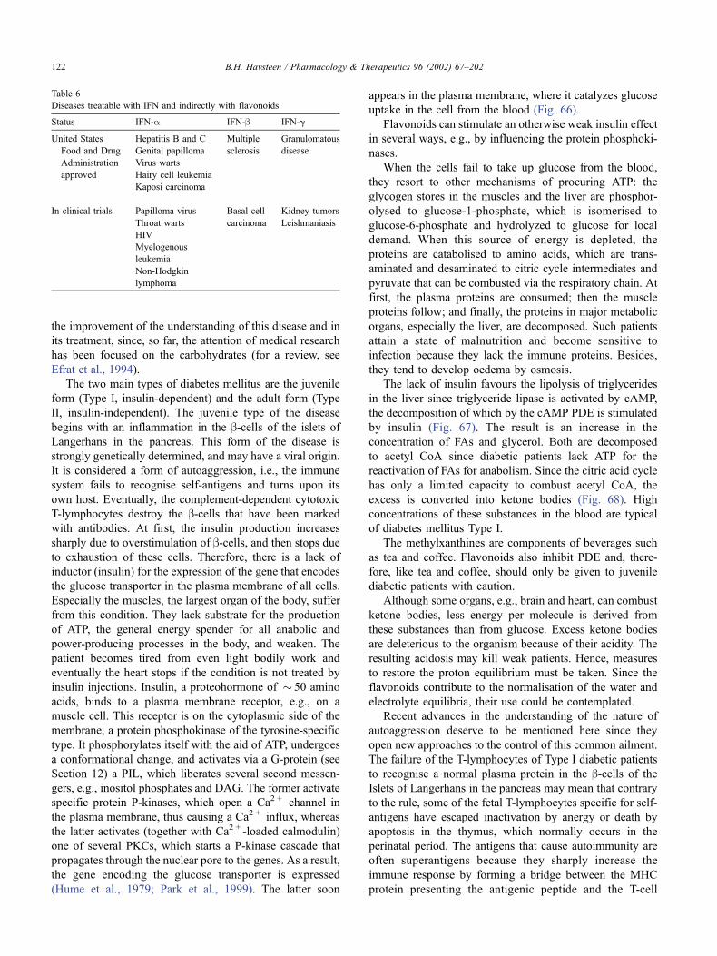
the improvement of the understanding of this disease and in
its treatment, since, so far, the attention of medical research
has been focused on the carbohydrates (for a review, see
Efrat et al., 1994).
The two main types of diabetes mellitus are the juvenile
form (Type I, insulin-dependent) and the adult form (Type
II, insulin-independent). The juvenile type of the disease
begins with an inflammation in the b-cells of the islets of
Langerhans in the pancreas. This form of the disease is
strongly genetically determined, and may have a viral origin.
It is considered a form of autoaggression, i.e., the immune
system fails to recognise self-antigens and turns upon its
own host. Eventually, the complement-dependent cytotoxic
T-lymphocytes destroy the b-cells that have been marked
with antibodies. At first, the insulin production increases
sharply due to overstimulation of b-cells, and then stops due
to exhaustion of these cells. Therefore, there is a lack of
inductor (insulin) for the expression of the gene that encodes
the glucose transporter in the plasma membrane of all cells.
Especially the muscles, the largest organ of the body, suffer
from this condition. They lack substrate for the production
of ATP, the general energy spender for all anabolic and
power-producing processes in the body, and weaken. The
patient becomes tired from even light bodily work and
eventually the heart stops if the condition is not treated by
insulin injections. Insulin, a proteohormone of � 50 amino
acids, binds to a plasma membrane receptor, e.g., on a
muscle cell. This receptor is on the cytoplasmic side of the
membrane, a protein phosphokinase of the tyrosine-specific
type. It phosphorylates itself with the aid of ATP, undergoes
a conformational change, and activates via a G-protein (see
Section 12) a PIL, which liberates several second messen-
gers, e.g., inositol phosphates and DAG. The former activate
specific protein P-kinases, which open a Ca2 + channel in
the plasma membrane, thus causing a Ca2 + influx, whereas
the latter activates (together with Ca2 + -loaded calmodulin)
one of several PKCs, which starts a P-kinase cascade that
propagates through the nuclear pore to the genes. As a result,
the gene encoding the glucose transporter is expressed
(Hume et al., 1979; Park et al., 1999). The latter soon
appears in the plasma membrane, where it catalyzes glucose
uptake in the cell from the blood (Fig. 66).
Flavonoids can stimulate an otherwise weak insulin effect
in several ways, e.g., by influencing the protein phosphoki-
nases.
When the cells fail to take up glucose from the blood,
they resort to other mechanisms of procuring ATP: the
glycogen stores in the muscles and the liver are phosphor-
olysed to glucose-1-phosphate, which is isomerised to
glucose-6-phosphate and hydrolyzed to glucose for local
demand. When this source of energy is depleted, the
proteins are catabolised to amino acids, which are trans-
aminated and desaminated to citric cycle intermediates and
pyruvate that can be combusted via the respiratory chain. At
first, the plasma proteins are consumed; then the muscle
proteins follow; and finally, the proteins in major metabolic
organs, especially the liver, are decomposed. Such patients
attain a state of malnutrition and become sensitive to
infection because they lack the immune proteins. Besides,
they tend to develop oedema by osmosis.
The lack of insulin favours the lipolysis of triglycerides
in the liver since triglyceride lipase is activated by cAMP,
the decomposition of which by the cAMP PDE is stimulated
by insulin (Fig. 67). The result is an increase in the
concentration of FAs and glycerol. Both are decomposed
to acetyl CoA since diabetic patients lack ATP for the
reactivation of FAs for anabolism. Since the citric acid cycle
has only a limited capacity to combust acetyl CoA, the
excess is converted into ketone bodies (Fig. 68). High
concentrations of these substances in the blood are typical
of diabetes mellitus Type I.
The methylxanthines are components of beverages such
as tea and coffee. Flavonoids also inhibit PDE and, there-
fore, like tea and coffee, should only be given to juvenile
diabetic patients with caution.
Although some organs, e.g., brain and heart, can combust
ketone bodies, less energy per molecule is derived from
these substances than from glucose. Excess ketone bodies
are deleterious to the organism because of their acidity. The
resulting acidosis may kill weak patients. Hence, measures
to restore the proton equilibrium must be taken. Since the
flavonoids contribute to the normalisation of the water and
electrolyte equilibria, their use could be contemplated.
Recent advances in the understanding of the nature of
autoaggression deserve to be mentioned here since they
open new approaches to the control of this common ailment.
The failure of the T-lymphocytes of Type I diabetic patients
to recognise a normal plasma protein in the b-cells of the
Islets of Langerhans in the pancreas may mean that contrary
to the rule, some of the fetal T-lymphocytes specific for self-
antigens have escaped inactivation by anergy or death by
apoptosis in the thymus, which normally occurs in the
perinatal period. The antigens that cause autoimmunity are
often superantigens because they sharply increase the
immune response by forming a bridge between the MHC
protein presenting the antigenic peptide and the T-cell
Table 6
Diseases treatable with IFN and indirectly with flavonoids
Status IFN-a IFN-b IFN-g
United States Hepatitis B and C Multiple Granulomatous
Food and Drug Genital papilloma sclerosis disease
Administration Virus warts
approved Hairy cell leukemia
Kaposi carcinoma
In clinical trials Papilloma virus Basal cell Kidney tumors
Throat warts carcinoma Leishmaniasis
HIV
Myelogenous
leukemia
Non-Hodgkin
lymphoma
B.H. Havsteen / Pharmacology & Therapeutics 96 (2002) 67–202122
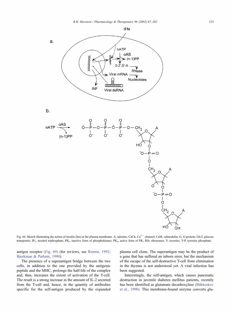
antigen receptor (Fig. 69) (for reviews, see Rennie, 1992;
Bjorkman & Parham, 1990).
The presence of a superantigen bridge between the two
cells, in addition to the one provided by the antigenic
peptide and the MHC, prolongs the half-life of the complex
and, thus, increases the extent of activation of the T-cell.
The result is a strong increase in the amount of IL-2 secreted
from the T-cell and, hence, in the quantity of antibodies
specific for the self-antigen produced by the expanded
plasma cell clone. The superantigen may be the product of
a gene that has suffered an inborn error, but the mechanism
of the escape of the self-destructive T-cell from elimination
in the thymus is not understood yet. A viral infection has
been suggested.
Interestingly, the self-antigen, which causes pancreatic
destruction in juvenile diabetes mellitus patients, recently
has been identified as glutamate decarboxylase (Bakkeskov
et al., 1990). This membrane-bound enzyme converts glu-
Fig. 66. Sketch illustrating the action of insulin (Ins) at the plasma membrane. A, adenine; CaCh, Ca2 + channel; CaM, calmodulin; G, G-protein; GlcT, glucose
transporter; IP3, inositol triphosphate; PKi, inactive form of phosphokinase; PKa, active form of PK; Rib, ribosomes; Y, tyrosine; Y-P, tyrosine phosphate.
B.H. Havsteen / Pharmacology & Therapeutics 96 (2002) 67–202 123
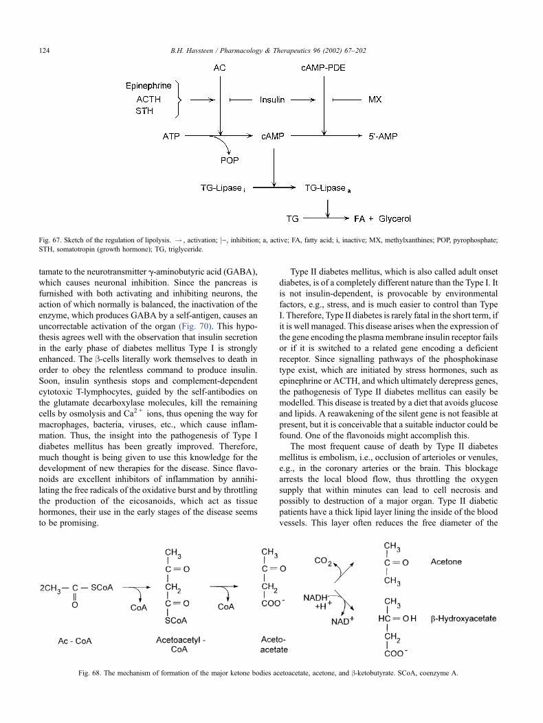
tamate to the neurotransmitter g-aminobutyric acid (GABA),
which causes neuronal inhibition. Since the pancreas is
furnished with both activating and inhibiting neurons, the
action of which normally is balanced, the inactivation of the
enzyme, which produces GABA by a self-antigen, causes an
uncorrectable activation of the organ (Fig. 70). This hypo-
thesis agrees well with the observation that insulin secretion
in the early phase of diabetes mellitus Type I is strongly
enhanced. The b-cells literally work themselves to death in
order to obey the relentless command to produce insulin.
Soon, insulin synthesis stops and complement-dependent
cytotoxic T-lymphocytes, guided by the self-antibodies on
the glutamate decarboxylase molecules, kill the remaining
cells by osmolysis and Ca2 + ions, thus opening the way for
macrophages, bacteria, viruses, etc., which cause inflam-
mation. Thus, the insight into the pathogenesis of Type I
diabetes mellitus has been greatly improved. Therefore,
much thought is being given to use this knowledge for the
development of new therapies for the disease. Since flavo-
noids are excellent inhibitors of inflammation by annihi-
lating the free radicals of the oxidative burst and by throttling
the production of the eicosanoids, which act as tissue
hormones, their use in the early stages of the disease seems
to be promising.
Type II diabetes mellitus, which is also called adult onset
diabetes, is of a completely different nature than the Type I. It
is not insulin-dependent, is provocable by environmental
factors, e.g., stress, and is much easier to control than Type
I. Therefore, Type II diabetes is rarely fatal in the short term, if
it is well managed. This disease arises when the expression of
the gene encoding the plasmamembrane insulin receptor fails
or if it is switched to a related gene encoding a deficient
receptor. Since signalling pathways of the phosphokinase
type exist, which are initiated by stress hormones, such as
epinephrine or ACTH, and which ultimately derepress genes,
the pathogenesis of Type II diabetes mellitus can easily be
modelled. This disease is treated by a diet that avoids glucose
and lipids. A reawakening of the silent gene is not feasible at
present, but it is conceivable that a suitable inductor could be
found. One of the flavonoids might accomplish this.
The most frequent cause of death by Type II diabetes
mellitus is embolism, i.e., occlusion of arterioles or venules,
e.g., in the coronary arteries or the brain. This blockage
arrests the local blood flow, thus throttling the oxygen
supply that within minutes can lead to cell necrosis and
possibly to destruction of a major organ. Type II diabetic
patients have a thick lipid layer lining the inside of the blood
vessels. This layer often reduces the free diameter of the
Fig. 67. Sketch of the regulation of lipolysis. ! , activation; j), inhibition; a, active; FA, fatty acid; i, inactive; MX, methylxanthines; POP, pyrophosphate;
STH, somatotropin (growth hormone); TG, triglyceride.
Fig. 68. The mechanism of formation of the major ketone bodies acetoacetate, acetone, and b-ketobutyrate. SCoA, coenzyme A.
B.H. Havsteen / Pharmacology & Therapeutics 96 (2002) 67–202124
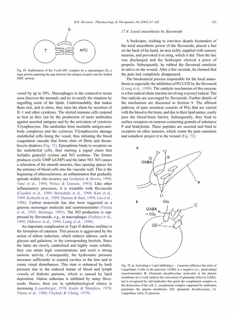
vessel by up to 50%. Macrophages in the connective tissue
soon discover the anomaly and try to rectify the situation by
engulfing some of the lipids. Understandably, that makes
them sick, and in stress, they raise the alarm by secretion of
IL-1 and other cytokines. The alerted immune cells respond
as best as they can by the production of more antibodies
against assorted antigens and by the activation of cytotoxic
T-lymphocytes. The antibodies form insoluble antigen-anti-
body complexes and the cytotoxic T-lymphocytes damage
endothelial cells lining the vessel, thus initiating the blood
coagulation cascade that forms clots of fibrin and throm-
bocyte shadows (Fig. 71). Epinephrine binds to receptors on
the endothelial cells, thus starting a signal chain that
includes guanylyl cyclase and NO synthase. The former
produces cyclic GMP (cGMP) and the latter NO. NO causes
a relaxation of the smooth muscles, thus opening spaces for
the entrance of blood cells into the vascular wall. This is the
beginning of atherosclerosis, an inflammation that gradually
spreads widely (for reviews, see Goldstein & Brown, 1990;
Vane et al., 1994; Prince & Gunson, 1993). Like other
inflammatory processes, it is treatable with flavonoids
(Casalini et al., 1999; Borradaile et al., 1999; Kim et al.,
1999; Kobuchi et al., 1999; Haenen & Bast, 1999; Lin et al.,
1996). Carbon monoxide has also been suggested as a
gaseous messenger molecule and neurotransmitter (Verma
et al., 1993; Barinaga, 1993). The NO production is sup-
pressed by flavonoids, e.g., in macrophages (Fushiya et al.,
1999; Oldreive et al., 1998; Liang et al., 1999).
An important complication in Type II diabetes mellitus is
the formation of cataracts. This process is aggravated by the
action of aldose reductase, which reduces aldoses, such as
glucose and galactose, to the corresponding hexitols. Since
the latter are slowly catabolised and highly water soluble,
they can attain high concentrations and exert a strong
osmotic activity. Consequently, the hydrostatic pressure
increases sufficiently to expand cavities in the lens and to
create visual disturbances. This state is enhanced by back
pressure due to the reduced lumen of blood and lymph
vessels of diabetic patients, which is caused by lipid
deposition. Aldose reductase is inhibited by many flavo-
noids. Hence, their use in ophthalmological clinics is
increasing (Leuenberger, 1978; Kader & Sharpless, 1978;
Varma et al., 1980; Chylack & Cheng, 1979).
17.4. Local anaesthesia by flavonoids
A beekeeper, wishing to convince skeptic bystanders of
the local anaesthetic power of the flavonoids, placed a bee
on the back of his hand, an area richly supplied with sensory
neurons, and provoked it to sting, which it did. Then the bee
was discharged and the beekeeper chewed a piece of
propolis. Subsequently, he rubbed the flavonoid emulsion
in saliva on the wound. After a few seconds, he claimed that
the pain had completely disappeared.
The biochemical process responsible for the local anaes-
thesia is especially the inhibition of PGCOX by the flavonoid
(Liang et al., 1999). The catalytic mechanism of this enzyme
is a free radical chain reaction involving a tyrosyl radical. The
free radicals are scavenged by flavonoids. Further details of
the mechanism are discussed in Section 9. The afferent
pathway of pain sensation consists of PGs that are carried
with the blood to the brain, and due to their lipid nature, easily
pass the blood-brain barrier. Subsequently, they bind to
surface receptors on neurons containing granula of substance
P and bradykinin. These peptides are secreted and bind to
receptors on other neurons, which create the pain sensation
and somehow project it to the wound (Fig. 72).
Fig. 69. Stabilization of the T-cell-APC complex by a superantigen (S), a
large protein spanning the gap between the antigen receptor and the loaded
MHC protein.
Fig. 70. a: Activating (+) and inhibiting (� ) neurons influence the islets of
Langerhans b-cells in the pancreas. GABA is a negative (i.e., passivating)
neurotransmitter. b: Glutamate decarboxylase molecules in the plasma
membrane in a b-cell catalyze the conversion of glutamate (Glu) to GABA,
but is recognized by self-antibodies that guide the complement complex to
the destruction of the cell. C, complement complex supported by antibodies
penetrates the plasma membrane; GD, glutamate decarboxylase; LI,
Langerhans islets; P, pancreas.
B.H. Havsteen / Pharmacology & Therapeutics 96 (2002) 67–202 125
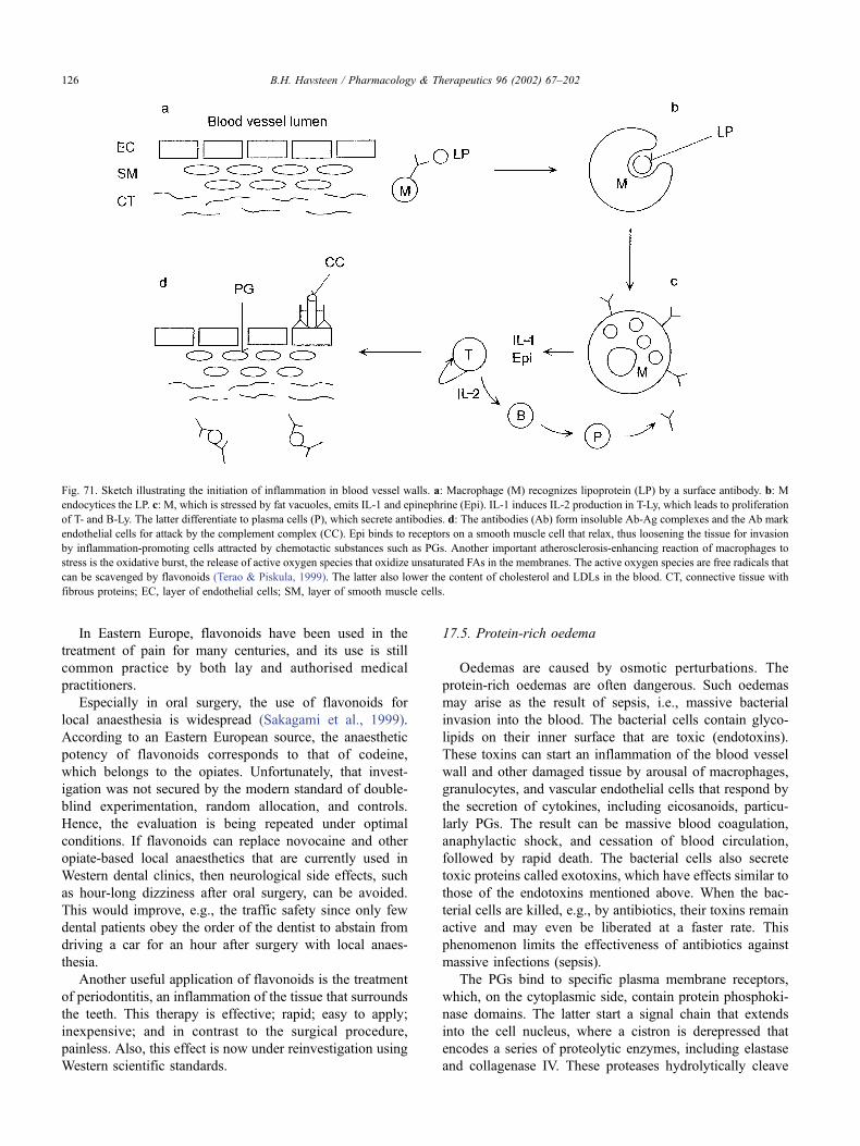
In Eastern Europe, flavonoids have been used in the
treatment of pain for many centuries, and its use is still
common practice by both lay and authorised medical
practitioners.
Especially in oral surgery, the use of flavonoids for
local anaesthesia is widespread (Sakagami et al., 1999).
According to an Eastern European source, the anaesthetic
potency of flavonoids corresponds to that of codeine,
which belongs to the opiates. Unfortunately, that invest-
igation was not secured by the modern standard of double-
blind experimentation, random allocation, and controls.
Hence, the evaluation is being repeated under optimal
conditions. If flavonoids can replace novocaine and other
opiate-based local anaesthetics that are currently used in
Western dental clinics, then neurological side effects, such
as hour-long dizziness after oral surgery, can be avoided.
This would improve, e.g., the traffic safety since only few
dental patients obey the order of the dentist to abstain from
driving a car for an hour after surgery with local anaes-
thesia.
Another useful application of flavonoids is the treatment
of periodontitis, an inflammation of the tissue that surrounds
the teeth. This therapy is effective; rapid; easy to apply;
inexpensive; and in contrast to the surgical procedure,
painless. Also, this effect is now under reinvestigation using
Western scientific standards.
17.5. Protein-rich oedema
Oedemas are caused by osmotic perturbations. The
protein-rich oedemas are often dangerous. Such oedemas
may arise as the result of sepsis, i.e., massive bacterial
invasion into the blood. The bacterial cells contain glyco-
lipids on their inner surface that are toxic (endotoxins).
These toxins can start an inflammation of the blood vessel
wall and other damaged tissue by arousal of macrophages,
granulocytes, and vascular endothelial cells that respond by
the secretion of cytokines, including eicosanoids, particu-
larly PGs. The result can be massive blood coagulation,
anaphylactic shock, and cessation of blood circulation,
followed by rapid death. The bacterial cells also secrete
toxic proteins called exotoxins, which have effects similar to
those of the endotoxins mentioned above. When the bac-
terial cells are killed, e.g., by antibiotics, their toxins remain
active and may even be liberated at a faster rate. This
phenomenon limits the effectiveness of antibiotics against
massive infections (sepsis).
The PGs bind to specific plasma membrane receptors,
which, on the cytoplasmic side, contain protein phosphoki-
nase domains. The latter start a signal chain that extends
into the cell nucleus, where a cistron is derepressed that
encodes a series of proteolytic enzymes, including elastase
and collagenase IV. These proteases hydrolytically cleave
Fig. 71. Sketch illustrating the initiation of inflammation in blood vessel walls. a: Macrophage (M) recognizes lipoprotein (LP) by a surface antibody. b: M
endocytices the LP. c: M, which is stressed by fat vacuoles, emits IL-1 and epinephrine (Epi). IL-1 induces IL-2 production in T-Ly, which leads to proliferation
of T- and B-Ly. The latter differentiate to plasma cells (P), which secrete antibodies. d: The antibodies (Ab) form insoluble Ab-Ag complexes and the Ab mark
endothelial cells for attack by the complement complex (CC). Epi binds to receptors on a smooth muscle cell that relax, thus loosening the tissue for invasion
by inflammation-promoting cells attracted by chemotactic substances such as PGs. Another important atherosclerosis-enhancing reaction of macrophages to
stress is the oxidative burst, the release of active oxygen species that oxidize unsaturated FAs in the membranes. The active oxygen species are free radicals that
can be scavenged by flavonoids (Terao & Piskula, 1999). The latter also lower the content of cholesterol and LDLs in the blood. CT, connective tissue with
fibrous proteins; EC, layer of endothelial cells; SM, layer of smooth muscle cells.
B.H. Havsteen / Pharmacology & Therapeutics 96 (2002) 67–202126
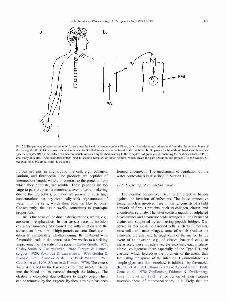
fibrous proteins in and around the cell, e.g., collagen,
laminin, and fibronectin. The products are peptides of
intermediate length, which, in contrast to the proteins from
which they originate, are soluble. These peptides are too
large to pass the plasma membrane, even after its lockering
due to the proteolysis, but they are present in such high
concentrations that they osmotically suck large amounts of
water into the cells, which then blow up like balloons.
Consequently, the tissue swells, sometimes to grotesque
proportions.
This is the basis of the drastic disfigurations, which, e.g.,
are seen in elephantiasis. In that case, a parasitic invasion
(by a trypanosome) has caused the inflammation and the
subsequent formation of high-protein oedema. Such a con-
dition is immediately life-threatening. Its treatment with
flavonoids leads in the course of a few weeks to a striking
improvement of the state of the patient (Casley-Smith, 1976;
Casley-Smith & Casley-Smith, 1986; Tarayre & Laures-
sergues, 1980; Sokolova & Liubartseva, 1979; Gerdin &
Svensjo, 1983; Ambrose & de Eds, 1974; Wismer, 1963;
Courbat et al., 1966; Sorensen & Hansen, 1970). The excess
water is drained by the flavonoids from the swollen tissues
into the blood and is excreted through the kidneys. The
elastically expanded skin collapses to empty bags, which
can be removed by the surgeon. By then, new skin has been
formed underneath. The mechanism of regulation of the
water homeostasis is described in Section 17.1.
17.6. Loosening of connective tissue
The healthy connective tissue is an effective barrier
against the invasion of infectants. The loose connective
tissue, which is involved here primarily, consists of a tight
network of fibrous proteins, such as collagen, elastin, and
chondroitin sulphate. The latter consists mainly of sulphated
hexosamines and hexuronic acids arranged in long branched
chains and supported by connecting peptide bridges. Dis-
persed in this mesh lie assorted cells, such as fibroblasts,
mast cells, and macrophages, some of which produce the
elements, proteins, and heteroglycans of the matrix. In the
event of an invasion, e.g., of viruses, bacterial cells, or
metastases, these intruders secrete enzymes, e.g., hyaluro-
nidase, collagenase (here especially of the Type III), and
elastase, which hydrolyse the polymers of the mesh, thus
facilitating the spread of the infection. Hyaluronidase is a
simple glycanase that somehow is inhibited by flavonoids
(Kuttan et al., 1981; Blumenkrantz & Asboe-Hansen, 1980;
Cetta et al., 1978; Zwillenberg-Fridman & Zwillenberg,
1972; Fine et al., 1992). Since certain of their features
resemble those of monosaccharides, it is likely that the
Fig. 72. The pathway of pain sensation. a: A bee stings the hand. Its venom contains PLA2, which hydrolyses arachidonic acid from the plasma membrane of
the damaged cell. PG COX converts arachidonic acid to PGs that are carried in the blood to the midbrain. b: PG passes the blood-brain barrier and binds to a
specific receptor (R) on the surface of a neuron, which initiates a signal chain leading to the exocytosis of granula (G) containing the peptides substance P (P)
and bradykinin (B). These neurotransmitters bind to specific receptors on other neurons, which create the pain sensation and project it to the wound. O,
occipital lobe; SC, spinal cord; T, thalamus.
B.H. Havsteen / Pharmacology & Therapeutics 96 (2002) 67–202 127
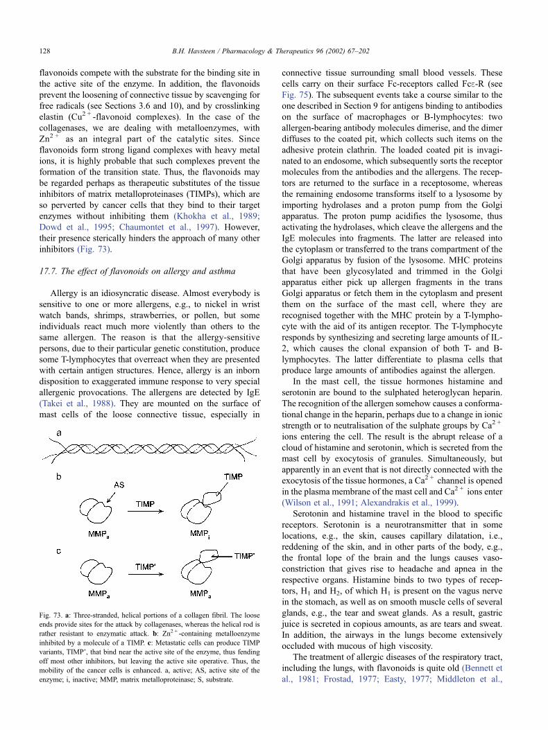
flavonoids compete with the substrate for the binding site in
the active site of the enzyme. In addition, the flavonoids
prevent the loosening of connective tissue by scavenging for
free radicals (see Sections 3.6 and 10), and by crosslinking
elastin (Cu2 + -flavonoid complexes). In the case of the
collagenases, we are dealing with metalloenzymes, with
Zn2 + as an integral part of the catalytic sites. Since
flavonoids form strong ligand complexes with heavy metal
ions, it is highly probable that such complexes prevent the
formation of the transition state. Thus, the flavonoids may
be regarded perhaps as therapeutic substitutes of the tissue
inhibitors of matrix metalloproteinases (TIMPs), which are
so perverted by cancer cells that they bind to their target
enzymes without inhibiting them (Khokha et al., 1989;
Dowd et al., 1995; Chaumontet et al., 1997). However,
their presence sterically hinders the approach of many other
inhibitors (Fig. 73).
17.7. The effect of flavonoids on allergy and asthma
Allergy is an idiosyncratic disease. Almost everybody is
sensitive to one or more allergens, e.g., to nickel in wrist
watch bands, shrimps, strawberries, or pollen, but some
individuals react much more violently than others to the
same allergen. The reason is that the allergy-sensitive
persons, due to their particular genetic constitution, produce
some T-lymphocytes that overreact when they are presented
with certain antigen structures. Hence, allergy is an inborn
disposition to exaggerated immune response to very special
allergenic provocations. The allergens are detected by IgE
(Takei et al., 1988). They are mounted on the surface of
mast cells of the loose connective tissue, especially in
connective tissue surrounding small blood vessels. These
cells carry on their surface Fc-receptors called Fce-R (see
Fig. 75). The subsequent events take a course similar to the
one described in Section 9 for antigens binding to antibodies
on the surface of macrophages or B-lymphocytes: two
allergen-bearing antibody molecules dimerise, and the dimer
diffuses to the coated pit, which collects such items on the
adhesive protein clathrin. The loaded coated pit is invagi-
nated to an endosome, which subsequently sorts the receptor
molecules from the antibodies and the allergens. The recep-
tors are returned to the surface in a receptosome, whereas
the remaining endosome transforms itself to a lysosome by
importing hydrolases and a proton pump from the Golgi
apparatus. The proton pump acidifies the lysosome, thus
activating the hydrolases, which cleave the allergens and the
IgE molecules into fragments. The latter are released into
the cytoplasm or transferred to the trans compartment of the
Golgi apparatus by fusion of the lysosome. MHC proteins
that have been glycosylated and trimmed in the Golgi
apparatus either pick up allergen fragments in the trans
Golgi apparatus or fetch them in the cytoplasm and present
them on the surface of the mast cell, where they are
recognised together with the MHC protein by a T-lympho-
cyte with the aid of its antigen receptor. The T-lymphocyte
responds by synthesizing and secreting large amounts of IL-
2, which causes the clonal expansion of both T- and B-
lymphocytes. The latter differentiate to plasma cells that
produce large amounts of antibodies against the allergen.
In the mast cell, the tissue hormones histamine and
serotonin are bound to the sulphated heteroglycan heparin.
The recognition of the allergen somehow causes a conforma-
tional change in the heparin, perhaps due to a change in ionic
strength or to neutralisation of the sulphate groups by Ca2 +
ions entering the cell. The result is the abrupt release of a
cloud of histamine and serotonin, which is secreted from the
mast cell by exocytosis of granules. Simultaneously, but
apparently in an event that is not directly connected with the
exocytosis of the tissue hormones, a Ca2 + channel is opened
in the plasma membrane of the mast cell and Ca2 + ions enter
(Wilson et al., 1991; Alexandrakis et al., 1999).
Serotonin and histamine travel in the blood to specific
receptors. Serotonin is a neurotransmitter that in some
locations, e.g., the skin, causes capillary dilatation, i.e.,
reddening of the skin, and in other parts of the body, e.g.,
the frontal lope of the brain and the lungs causes vaso-
constriction that gives rise to headache and apnea in the
respective organs. Histamine binds to two types of recep-
tors, H1 and H2, of which H1 is present on the vagus nerve
in the stomach, as well as on smooth muscle cells of several
glands, e.g., the tear and sweat glands. As a result, gastric
juice is secreted in copious amounts, as are tears and sweat.
In addition, the airways in the lungs become extensively
occluded with mucous of high viscosity.
The treatment of allergic diseases of the respiratory tract,
including the lungs, with flavonoids is quite old (Bennett et
al., 1981; Frostad, 1977; Easty, 1977; Middleton et al.,
Fig. 73. a: Three-stranded, helical portions of a collagen fibril. The loose
ends provide sites for the attack by collagenases, whereas the helical rod is
rather resistant to enzymatic attack. b: Zn2 + -containing metalloenzyme
inhibited by a molecule of a TIMP. c: Metastatic cells can produce TIMP
variants, TIMP’, that bind near the active site of the enzyme, thus fending
off most other inhibitors, but leaving the active site operative. Thus, the
mobility of the cancer cells is enhanced. a, active; AS, active site of the
enzyme; i, inactive; MMP, matrix metalloproteinase; S, substrate.
B.H. Havsteen / Pharmacology & Therapeutics 96 (2002) 67–202128
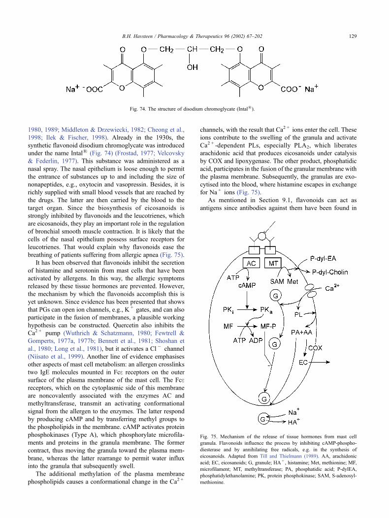
1980, 1989; Middleton & Drzewiecki, 1982; Cheong et al.,
1998; Ilek & Fischer, 1998). Already in the 1930s, the
synthetic flavonoid disodium chromoglycate was introduced
under the name Intal1 (Fig. 74) (Frostad, 1977; Velcovsky
& Federlin, 1977). This substance was administered as a
nasal spray. The nasal epithelium is loose enough to permit
the entrance of substances up to and including the size of
nonapeptides, e.g., oxytocin and vasopressin. Besides, it is
richly supplied with small blood vessels that are reached by
the drugs. The latter are then carried by the blood to the
target organ. Since the biosynthesis of eicosanoids is
strongly inhibited by flavonoids and the leucotrienes, which
are eicosanoids, they play an important role in the regulation
of bronchial smooth muscle contraction. It is likely that the
cells of the nasal epithelium possess surface receptors for
leucotrienes. That would explain why flavonoids ease the
breathing of patients suffering from allergic apnea (Fig. 75).
It has been observed that flavonoids inhibit the secretion
of histamine and serotonin from mast cells that have been
activated by allergens. In this way, the allergic symptoms
released by these tissue hormones are prevented. However,
the mechanism by which the flavonoids accomplish this is
yet unknown. Since evidence has been presented that shows
that PGs can open ion channels, e.g., K + gates, and can also
participate in the fusion of membranes, a plausible working
hypothesis can be constructed. Quercetin also inhibits the
Ca2 + pump (Wuthrich & Schatzmann, 1980; Fewtrell &
Gomperts, 1977a, 1977b; Bennett et al., 1981; Shoshan et
al., 1980; Long et al., 1981), but it activates a Cl � channel
(Niisato et al., 1999). Another line of evidence emphasises
other aspects of mast cell metabolism: an allergen crosslinks
two IgE molecules mounted in Fce receptors on the outer
surface of the plasma membrane of the mast cell. The Fcereceptors, which on the cytoplasmic side of this membrane
are noncovalently associated with the enzymes AC and
methyltransferase, transmit an activating conformational
signal from the allergen to the enzymes. The latter respond
by producing cAMP and by transferring methyl groups to
the phospholipids in the membrane. cAMP activates protein
phosphokinases (Type A), which phosphorylate microfila-
ments and proteins in the granula membrane. The former
contract, thus moving the granula toward the plasma mem-
brane, whereas the latter rearrange to permit water influx
into the granula that subsequently swell.
The additional methylation of the plasma membrane
phospholipids causes a conformational change in the Ca2 +
channels, with the result that Ca2 + ions enter the cell. These
ions contribute to the swelling of the granula and activate
Ca2 + -dependent PLs, especially PLA2, which liberates
arachidonic acid that produces eicosanoids under catalysis
by COX and lipoxygenase. The other product, phosphatidic
acid, participates in the fusion of the granular membrane with
the plasma membrane. Subsequently, the granulas are exo-
cytised into the blood, where histamine escapes in exchange
for Na + ions (Fig. 75).
As mentioned in Section 9.1, flavonoids can act as
antigens since antibodies against them have been found in
Fig. 74. The structure of disodium chromoglycate (Intal1).
Fig. 75. Mechanism of the release of tissue hormones from mast cell
granula. Flavonoids influence the process by inhibiting cAMP-phospho-
diesterase and by annihilating free radicals, e.g. in the synthesis of
eicosanoids. Adapted from Till and Thielmann (1989). AA, arachidonic
acid; EC, eicosanoids; G, granule; HA + , histamine; Met, methionine; MF,
microfilament; MT, methyltransferase; PA, phosphatidic acid; P-dylEA,
phosphatidylethanolamine; PK, protein phosphokinase; SAM, S-adenosyl-
methionine.
B.H. Havsteen / Pharmacology & Therapeutics 96 (2002) 67–202 129
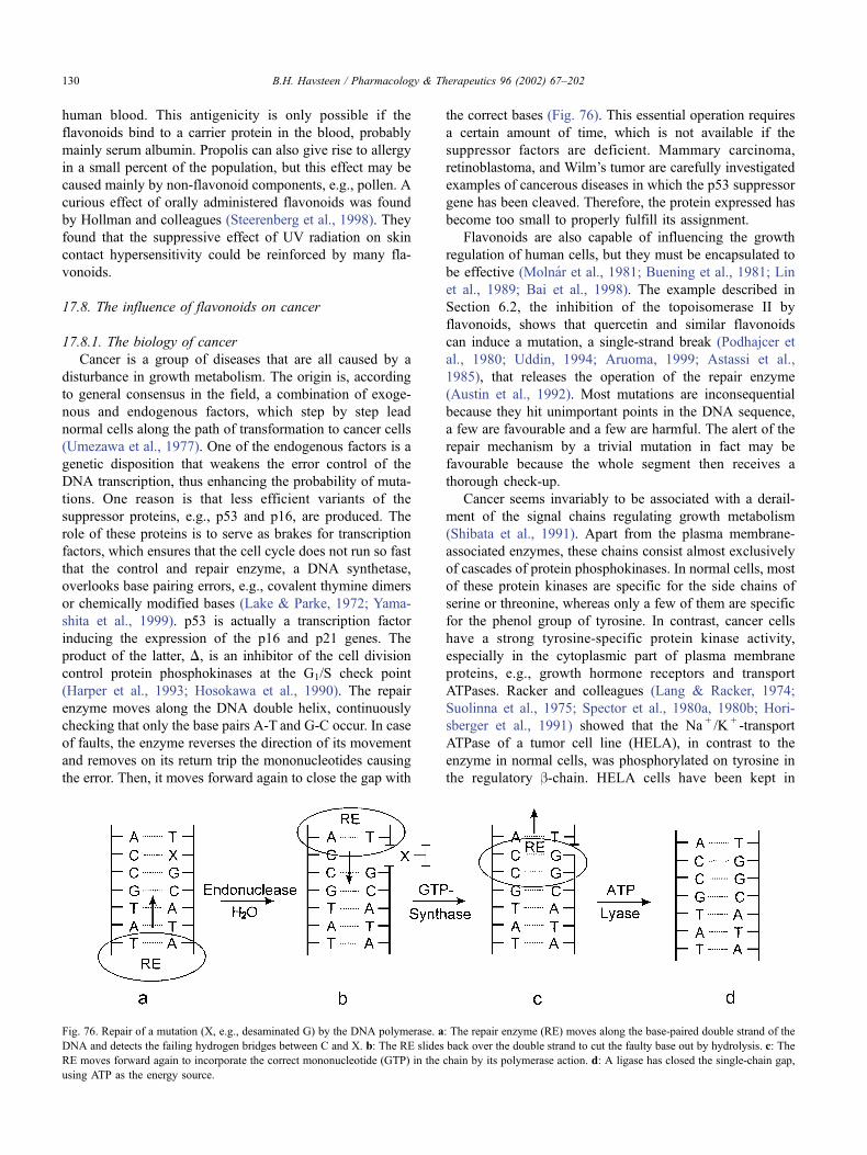
human blood. This antigenicity is only possible if the
flavonoids bind to a carrier protein in the blood, probably
mainly serum albumin. Propolis can also give rise to allergy
in a small percent of the population, but this effect may be
caused mainly by non-flavonoid components, e.g., pollen. A
curious effect of orally administered flavonoids was found
by Hollman and colleagues (Steerenberg et al., 1998). They
found that the suppressive effect of UV radiation on skin
contact hypersensitivity could be reinforced by many fla-
vonoids.
17.8. The influence of flavonoids on cancer
17.8.1. The biology of cancer
Cancer is a group of diseases that are all caused by a
disturbance in growth metabolism. The origin is, according
to general consensus in the field, a combination of exoge-
nous and endogenous factors, which step by step lead
normal cells along the path of transformation to cancer cells
(Umezawa et al., 1977). One of the endogenous factors is a
genetic disposition that weakens the error control of the
DNA transcription, thus enhancing the probability of muta-
tions. One reason is that less efficient variants of the
suppressor proteins, e.g., p53 and p16, are produced. The
role of these proteins is to serve as brakes for transcription
factors, which ensures that the cell cycle does not run so fast
that the control and repair enzyme, a DNA synthetase,
overlooks base pairing errors, e.g., covalent thymine dimers
or chemically modified bases (Lake & Parke, 1972; Yama-
shita et al., 1999). p53 is actually a transcription factor
inducing the expression of the p16 and p21 genes. The
product of the latter, D, is an inhibitor of the cell division
control protein phosphokinases at the G1/S check point
(Harper et al., 1993; Hosokawa et al., 1990). The repair
enzyme moves along the DNA double helix, continuously
checking that only the base pairs A-T and G-C occur. In case
of faults, the enzyme reverses the direction of its movement
and removes on its return trip the mononucleotides causing
the error. Then, it moves forward again to close the gap with
the correct bases (Fig. 76). This essential operation requires
a certain amount of time, which is not available if the
suppressor factors are deficient. Mammary carcinoma,
retinoblastoma, and Wilm’s tumor are carefully investigated
examples of cancerous diseases in which the p53 suppressor
gene has been cleaved. Therefore, the protein expressed has
become too small to properly fulfill its assignment.
Flavonoids are also capable of influencing the growth
regulation of human cells, but they must be encapsulated to
be effective (Molnar et al., 1981; Buening et al., 1981; Lin
et al., 1989; Bai et al., 1998). The example described in
Section 6.2, the inhibition of the topoisomerase II by
flavonoids, shows that quercetin and similar flavonoids
can induce a mutation, a single-strand break (Podhajcer et
al., 1980; Uddin, 1994; Aruoma, 1999; Astassi et al.,
1985), that releases the operation of the repair enzyme
(Austin et al., 1992). Most mutations are inconsequential
because they hit unimportant points in the DNA sequence,
a few are favourable and a few are harmful. The alert of the
repair mechanism by a trivial mutation in fact may be
favourable because the whole segment then receives a
thorough check-up.
Cancer seems invariably to be associated with a derail-
ment of the signal chains regulating growth metabolism
(Shibata et al., 1991). Apart from the plasma membrane-
associated enzymes, these chains consist almost exclusively
of cascades of protein phosphokinases. In normal cells, most
of these protein kinases are specific for the side chains of
serine or threonine, whereas only a few of them are specific
for the phenol group of tyrosine. In contrast, cancer cells
have a strong tyrosine-specific protein kinase activity,
especially in the cytoplasmic part of plasma membrane
proteins, e.g., growth hormone receptors and transport
ATPases. Racker and colleagues (Lang & Racker, 1974;
Suolinna et al., 1975; Spector et al., 1980a, 1980b; Hori-
sberger et al., 1991) showed that the Na + /K + -transport
ATPase of a tumor cell line (HELA), in contrast to the
enzyme in normal cells, was phosphorylated on tyrosine in
the regulatory b-chain. HELA cells have been kept in
Fig. 76. Repair of a mutation (X, e.g., desaminated G) by the DNA polymerase. a: The repair enzyme (RE) moves along the base-paired double strand of the
DNA and detects the failing hydrogen bridges between C and X. b: The RE slides back over the double strand to cut the faulty base out by hydrolysis. c: The
RE moves forward again to incorporate the correct mononucleotide (GTP) in the chain by its polymerase action. d: A ligase has closed the single-chain gap,
using ATP as the energy source.
B.H. Havsteen / Pharmacology & Therapeutics 96 (2002) 67–202130
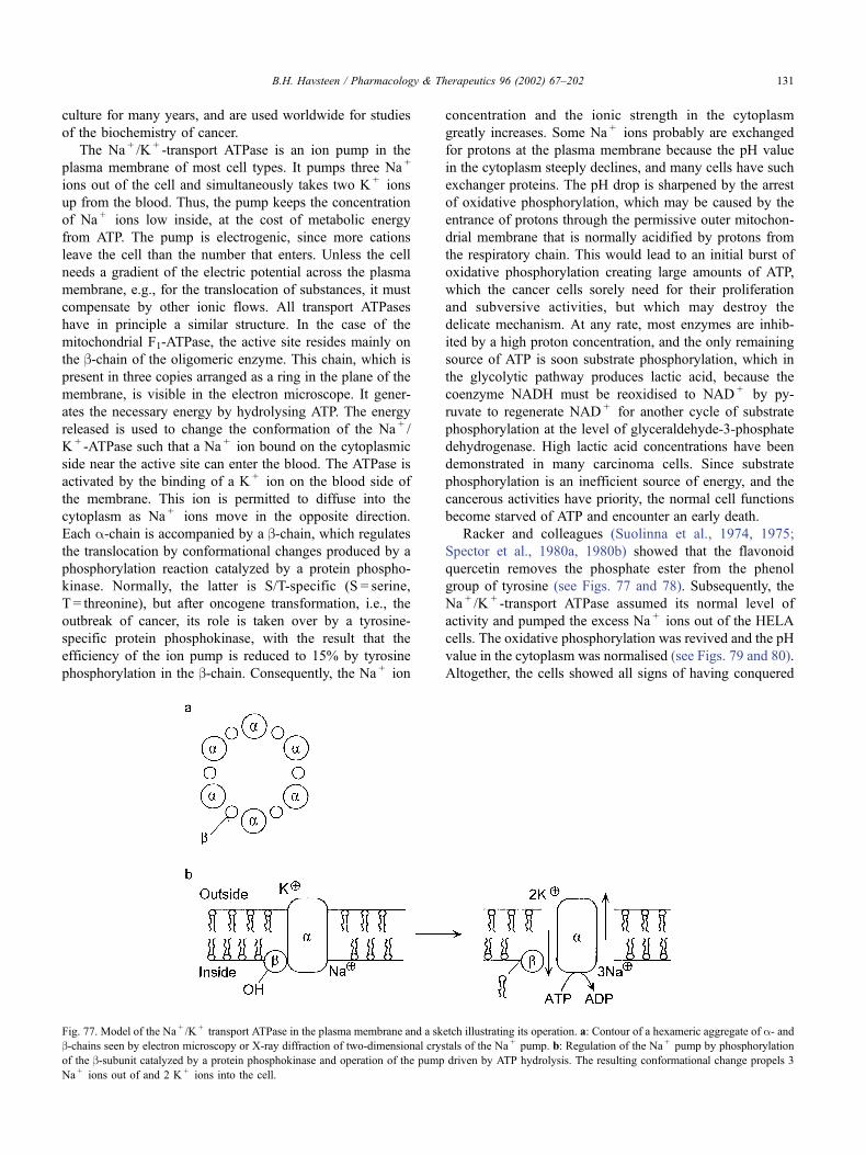
culture for many years, and are used worldwide for studies
of the biochemistry of cancer.
The Na + /K + -transport ATPase is an ion pump in the
plasma membrane of most cell types. It pumps three Na +
ions out of the cell and simultaneously takes two K + ions
up from the blood. Thus, the pump keeps the concentration
of Na + ions low inside, at the cost of metabolic energy
from ATP. The pump is electrogenic, since more cations
leave the cell than the number that enters. Unless the cell
needs a gradient of the electric potential across the plasma
membrane, e.g., for the translocation of substances, it must
compensate by other ionic flows. All transport ATPases
have in principle a similar structure. In the case of the
mitochondrial F1-ATPase, the active site resides mainly on
the b-chain of the oligomeric enzyme. This chain, which is
present in three copies arranged as a ring in the plane of the
membrane, is visible in the electron microscope. It gener-
ates the necessary energy by hydrolysing ATP. The energy
released is used to change the conformation of the Na + /
K + -ATPase such that a Na + ion bound on the cytoplasmic
side near the active site can enter the blood. The ATPase is
activated by the binding of a K + ion on the blood side of
the membrane. This ion is permitted to diffuse into the
cytoplasm as Na + ions move in the opposite direction.
Each a-chain is accompanied by a b-chain, which regulates
the translocation by conformational changes produced by a
phosphorylation reaction catalyzed by a protein phospho-
kinase. Normally, the latter is S/T-specific (S = serine,
T = threonine), but after oncogene transformation, i.e., the
outbreak of cancer, its role is taken over by a tyrosine-
specific protein phosphokinase, with the result that the
efficiency of the ion pump is reduced to 15% by tyrosine
phosphorylation in the b-chain. Consequently, the Na + ion
concentration and the ionic strength in the cytoplasm
greatly increases. Some Na + ions probably are exchanged
for protons at the plasma membrane because the pH value
in the cytoplasm steeply declines, and many cells have such
exchanger proteins. The pH drop is sharpened by the arrest
of oxidative phosphorylation, which may be caused by the
entrance of protons through the permissive outer mitochon-
drial membrane that is normally acidified by protons from
the respiratory chain. This would lead to an initial burst of
oxidative phosphorylation creating large amounts of ATP,
which the cancer cells sorely need for their proliferation
and subversive activities, but which may destroy the
delicate mechanism. At any rate, most enzymes are inhib-
ited by a high proton concentration, and the only remaining
source of ATP is soon substrate phosphorylation, which in
the glycolytic pathway produces lactic acid, because the
coenzyme NADH must be reoxidised to NAD + by py-
ruvate to regenerate NAD + for another cycle of substrate
phosphorylation at the level of glyceraldehyde-3-phosphate
dehydrogenase. High lactic acid concentrations have been
demonstrated in many carcinoma cells. Since substrate
phosphorylation is an inefficient source of energy, and the
cancerous activities have priority, the normal cell functions
become starved of ATP and encounter an early death.
Racker and colleagues (Suolinna et al., 1974, 1975;
Spector et al., 1980a, 1980b) showed that the flavonoid
quercetin removes the phosphate ester from the phenol
group of tyrosine (see Figs. 77 and 78). Subsequently, the
Na + /K + -transport ATPase assumed its normal level of
activity and pumped the excess Na + ions out of the HELA
cells. The oxidative phosphorylation was revived and the pH
value in the cytoplasm was normalised (see Figs. 79 and 80).
Altogether, the cells showed all signs of having conquered
Fig. 77. Model of the Na + /K + transport ATPase in the plasma membrane and a sketch illustrating its operation. a: Contour of a hexameric aggregate of a- andb-chains seen by electron microscopy or X-ray diffraction of two-dimensional crystals of the Na + pump. b: Regulation of the Na + pump by phosphorylation
of the b-subunit catalyzed by a protein phosphokinase and operation of the pump driven by ATP hydrolysis. The resulting conformational change propels 3
Na + ions out of and 2 K + ions into the cell.
B.H. Havsteen / Pharmacology & Therapeutics 96 (2002) 67–202 131
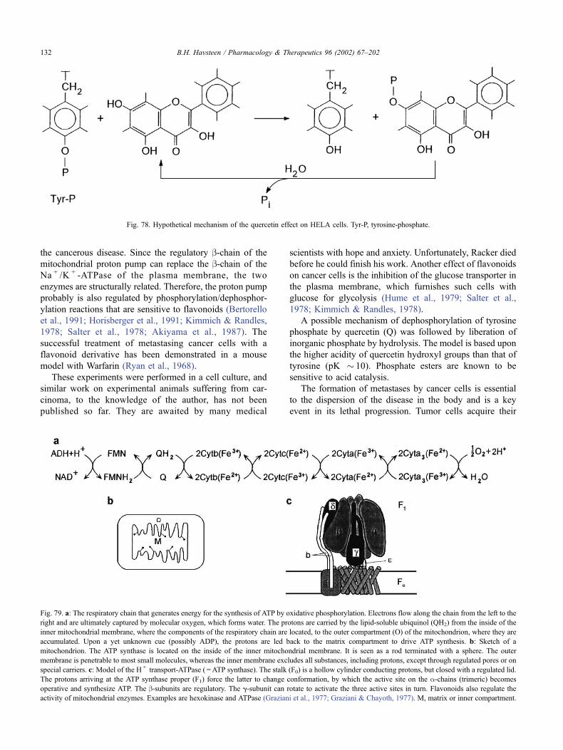
the cancerous disease. Since the regulatory b-chain of the
mitochondrial proton pump can replace the b-chain of the
Na + /K + -ATPase of the plasma membrane, the two
enzymes are structurally related. Therefore, the proton pump
probably is also regulated by phosphorylation/dephosphor-
ylation reactions that are sensitive to flavonoids (Bertorello
et al., 1991; Horisberger et al., 1991; Kimmich & Randles,
1978; Salter et al., 1978; Akiyama et al., 1987). The
successful treatment of metastasing cancer cells with a
flavonoid derivative has been demonstrated in a mouse
model with Warfarin (Ryan et al., 1968).
These experiments were performed in a cell culture, and
similar work on experimental animals suffering from car-
cinoma, to the knowledge of the author, has not been
published so far. They are awaited by many medical
scientists with hope and anxiety. Unfortunately, Racker died
before he could finish his work. Another effect of flavonoids
on cancer cells is the inhibition of the glucose transporter in
the plasma membrane, which furnishes such cells with
glucose for glycolysis (Hume et al., 1979; Salter et al.,
1978; Kimmich & Randles, 1978).
A possible mechanism of dephosphorylation of tyrosine
phosphate by quercetin (Q) was followed by liberation of
inorganic phosphate by hydrolysis. The model is based upon
the higher acidity of quercetin hydroxyl groups than that of
tyrosine (pK � 10). Phosphate esters are known to be
sensitive to acid catalysis.
The formation of metastases by cancer cells is essential
to the dispersion of the disease in the body and is a key
event in its lethal progression. Tumor cells acquire their
Fig. 78. Hypothetical mechanism of the quercetin effect on HELA cells. Tyr-P, tyrosine-phosphate.
Fig. 79. a: The respiratory chain that generates energy for the synthesis of ATP by oxidative phosphorylation. Electrons flow along the chain from the left to the
right and are ultimately captured by molecular oxygen, which forms water. The protons are carried by the lipid-soluble ubiquinol (QH2) from the inside of the
inner mitochondrial membrane, where the components of the respiratory chain are located, to the outer compartment (O) of the mitochondrion, where they are
accumulated. Upon a yet unknown cue (possibly ADP), the protons are led back to the matrix compartment to drive ATP synthesis. b: Sketch of a
mitochondrion. The ATP synthase is located on the inside of the inner mitochondrial membrane. It is seen as a rod terminated with a sphere. The outer
membrane is penetrable to most small molecules, whereas the inner membrane excludes all substances, including protons, except through regulated pores or on
special carriers. c: Model of the H + transport-ATPase ( =ATP synthase). The stalk (F0) is a hollow cylinder conducting protons, but closed with a regulated lid.
The protons arriving at the ATP synthase proper (F1) force the latter to change conformation, by which the active site on the a-chains (trimeric) becomes
operative and synthesize ATP. The b-subunits are regulatory. The g-subunit can rotate to activate the three active sites in turn. Flavonoids also regulate the
activity of mitochondrial enzymes. Examples are hexokinase and ATPase (Graziani et al., 1977; Graziani & Chayoth, 1977). M, matrix or inner compartment.
B.H. Havsteen / Pharmacology & Therapeutics 96 (2002) 67–202132
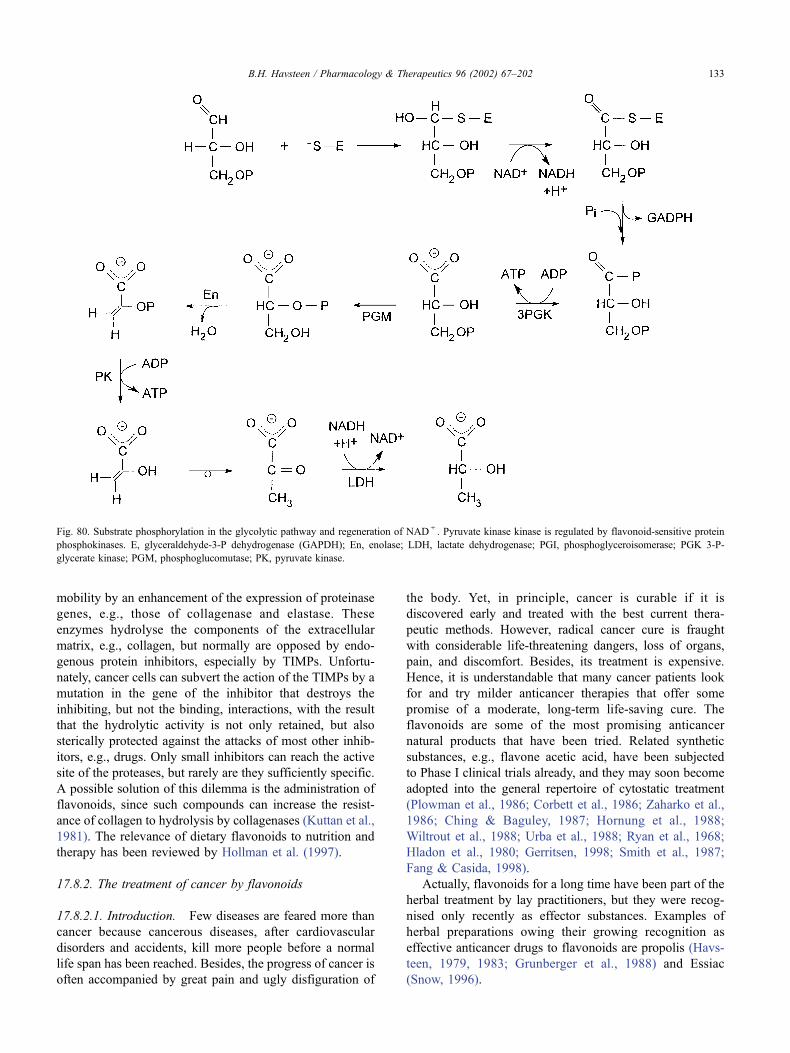
mobility by an enhancement of the expression of proteinase
genes, e.g., those of collagenase and elastase. These
enzymes hydrolyse the components of the extracellular
matrix, e.g., collagen, but normally are opposed by endo-
genous protein inhibitors, especially by TIMPs. Unfortu-
nately, cancer cells can subvert the action of the TIMPs by a
mutation in the gene of the inhibitor that destroys the
inhibiting, but not the binding, interactions, with the result
that the hydrolytic activity is not only retained, but also
sterically protected against the attacks of most other inhib-
itors, e.g., drugs. Only small inhibitors can reach the active
site of the proteases, but rarely are they sufficiently specific.
A possible solution of this dilemma is the administration of
flavonoids, since such compounds can increase the resist-
ance of collagen to hydrolysis by collagenases (Kuttan et al.,
1981). The relevance of dietary flavonoids to nutrition and
therapy has been reviewed by Hollman et al. (1997).
17.8.2. The treatment of cancer by flavonoids
17.8.2.1. Introduction. Few diseases are feared more than
cancer because cancerous diseases, after cardiovascular
disorders and accidents, kill more people before a normal
life span has been reached. Besides, the progress of cancer is
often accompanied by great pain and ugly disfiguration of
the body. Yet, in principle, cancer is curable if it is
discovered early and treated with the best current thera-
peutic methods. However, radical cancer cure is fraught
with considerable life-threatening dangers, loss of organs,
pain, and discomfort. Besides, its treatment is expensive.
Hence, it is understandable that many cancer patients look
for and try milder anticancer therapies that offer some
promise of a moderate, long-term life-saving cure. The
flavonoids are some of the most promising anticancer
natural products that have been tried. Related synthetic
substances, e.g., flavone acetic acid, have been subjected
to Phase I clinical trials already, and they may soon become
adopted into the general repertoire of cytostatic treatment
(Plowman et al., 1986; Corbett et al., 1986; Zaharko et al.,
1986; Ching & Baguley, 1987; Hornung et al., 1988;
Wiltrout et al., 1988; Urba et al., 1988; Ryan et al., 1968;
Hladon et al., 1980; Gerritsen, 1998; Smith et al., 1987;
Fang & Casida, 1998).
Actually, flavonoids for a long time have been part of the
herbal treatment by lay practitioners, but they were recog-
nised only recently as effector substances. Examples of
herbal preparations owing their growing recognition as
effective anticancer drugs to flavonoids are propolis (Havs-
teen, 1979, 1983; Grunberger et al., 1988) and Essiac
(Snow, 1996).
Fig. 80. Substrate phosphorylation in the glycolytic pathway and regeneration of NAD + . Pyruvate kinase kinase is regulated by flavonoid-sensitive protein
phosphokinases. E, glyceraldehyde-3-P dehydrogenase (GAPDH); En, enolase; LDH, lactate dehydrogenase; PGI, phosphoglyceroisomerase; PGK 3-P-
glycerate kinase; PGM, phosphoglucomutase; PK, pyruvate kinase.
B.H. Havsteen / Pharmacology & Therapeutics 96 (2002) 67–202 133
17.8.2.2. Types of cancerous diseases susceptible to
flavonoid inhibition. The inhibition of the growth of
cancer cells, both in vitro and in vivo, by flavonoids has
been reported in many articles, e.g., by Graziani et al.
(1983), Edwards et al. (1979), Kuriki and Racker (1976),
Kandaswami et al., 1991, as well as Scambia et al. (1992).
These observations pertained to both experimental cancer-
ous diseases in animals induced by chemical mutagens
(Markaverich et al., 1990) or viral transformation and to
spontaneous human tumors (Ranelletti et al., 1992; Zhu &
Liehr, 1994). Since the stepwise path of oncogenesis may
depend on the cell type, the cancerous diseases, which
proved to be inhibitable by flavonoids, are here in part
classified according to the germinal layer from which the
normal cells were formed during ontogenesis (Avila et al.,
1994; Kupchan et al., 1978; Kupchan & Bauerschmidt,
1971).
17.8.2.2.1. Carcinomas largely consisting of cells of ecto-
dermal origin. Carcinomas largely consisting of cells of
ectodermal origin include the skin (Chang et al., 1985;
Lama et al., 1998), breasts (Markaverich et al., 1990; Varma
et al., 1977; Scambia et al., 1991; Adlercreutz, 1984;
Makela et al. 1998), oral epithelium (Makita et al., 1996),
the urogenital tract (Malaveille et al., 1998), and lungs
(Malaveille et al., 1996).
17.8.2.2.2. Carcinomas largely consisting of cells of
entodermal origin. Carcinomas largely consisting of cells
of entodermal origin include the gastrointestinal tract
(Ranelletti et al., 1992; Hosokawa et al., 1990; Yoshida
et al., 1990; Adlercreutz, 1984; Garcia-Closas et al.,
1999), mammary glands (Guthrie & Carroll, 1998), the
gonads (Scambia et al., 1990a, 1990b, 1992), the uterus
(Markaverich & Clark, 1979; Markaverich et al., 1981,
1983, 1988, 1989, 1990, 1992), lungs (Garcia-Closas et
al., 1998; De Stefani et al., 1999; Knekt et al., 1997), the
prostata (Griffiths et al., 1998, 1999; Denis et al., 1999),
the colon (Deschner et al., 1993; Kuo, 1996), the rectum
(Graham et al., 1978), and mesenchyme (Makino et al.,
1998).
17.8.2.2.3. Tumors of mesodermal origin. Tumors of mes-
odermal origin include blood cells (Scambia et al., 1990a,
1990b; Varma et al., 1977; Larocca et al., 1990; Hoffman et
al., 1989; Molnar et al., 1981; Graziani et al., 1983), bone
(Shoshan & MacLennan, 1981; Bissery et al., 1988), and
muscle (Shoshan et al., 1980).
17.8.2.2.4. Tumors formed by oncogenic viruses. Tumors
formed by oncogenic viruses (Graziani et al., 1981, 1983,
1987). Although this survey cannot be complete, due to
the large number of publications on the subject, which are
not easily accessible, it is surprising that major classes of
tumors, e.g., those of the liver, kidney, bladder, and brain,
are not represented. The reason probably is not that such
tumors are not susceptible to treatment with flavonoids,
but rather, that the cancerous tissues are more difficult to
keep in culture and to develop in animal models. Experi-
mental therapy of human cancer patients with flavonoids
has just begun (Bissery et al., 1988; Hofmann et al., 1988,
1990).
17.8.3. Biochemical processes of cancer influenced by
flavonoids
17.8.3.1. Introduction. The plethora of effects of flavo-
noids on the metabolism of cancer cells is difficult to
rationalise to a few basic, specific mechanisms. The flavo-
noids interfere with a large number of regulatory pathways,
e.g., those of growth, energy metabolism, apoptosis, cell
division, transcription, gene repair, neuronal transmission,
inflammation, and stress response (Koh & Willoughby,
1979; Khayyal et al., 1993; Richter et al., 1999; Barinaga,
1996; Edwards et al., 1998; Habtemariam, 1997; Harper et
al., 1993; Jager et al., 1998). They can act as antioxidants,
free-radical scavengers, enzyme inhibitors, hormones (in-
cluding neurotransmitters), antihormones, or inducers of
gene expression.
These effects can be divided into two classes, the
electronic and the steric. The high mobility of the electrons
in the benzenoid nucleus of flavonoids accounts for both
their antioxidant and free-radical scavenging properties,
whereas the structural resemblance between the flavonoid
aglycone and many substances inherent to the biochemistry
of normal biological cells, e.g., nucleic acid bases, coen-
zymes, steroid hormones, and neurotransmitters, explains
their inhibition of enzymes, cytoplasmic/nuclear hormone
receptors, and neurotransmitters, as well as gene induction.
An example of the latter is the induction of protease genes
by coumarin during high-protein oedema (Piller, 1979;
Radouco-Thomas et al., 1964; Casley-Smith, 1976; Abate
et al., 1990). The high affinity of flavonoids for heavy metal
ions provides additional opportunities for interference with
the action of enzymes and Zn2 + fingers in DNA-binding
proteins.
The biochemical pathways, which are influenced by
flavonoids, can roughly be classified according to their
sensitivities. Generally, the actions mediated by the family
of cytoplasmic/nuclear hormone receptors are highly sen-
sitive to flavonoids, whereas enzyme inhibition and similar
processes, e.g., at synaptic membranes, require higher con-
centrations of flavonoids (Reiners et al., 1999).
17.8.3.2. Cytoplasmic/nuclear hormone receptors. The
superfamily of cytoplasmic hormone receptors plays a
central role in the transmission of steroid hormones and
hormones of a similar structure and function to the genes.
The hormones, which are carried by this superfamily of
proteins from the cytoplasm into the cell nucleus, include
steroids, thyroid hormones, vitamin D3, and retinoic acid.
Most of these hormones stimulate growth processes, but
some of them also control specialised functions, e.g.,
electrolyte homeostasis, carbohydrate metabolism, sexual
maturation, oxidative phosphorylation, and morphogenesis.
All cytoplasmic hormone receptors are single-chain pro-
B.H. Havsteen / Pharmacology & Therapeutics 96 (2002) 67–202134
teins, which are folded into three domains. The N-terminal
domain has a trans-activating function, a small centrally
located domain binds to DNA, and the C-terminal hydro-
phobic domain binds the hormone. The structure of the
flavonoids suggests that flavonoids also bind to the latter
domain. This would explain the ability of various flavo-
noids, e.g., silymarin (Sonnenbichler et al., 1980; Sonnen-
bichler & Pohl, 1980; Conseil et al., 1998; Das et al., 1987a,
1987b; Markaverich & Clark, 1979; Kitaoka et al., 1998), to
simulate the action of certain hormones.
Since the ligand affinity of the hormone receptors is very
high, it is understandable that flavonoids, which are exerting
their effect via cytoplasmic hormone receptors, have a much
more sensitive influence on the metabolism of the cell than
those that are low-affinity inhibitors of enzymes and plasma
membrane receptors. The inhibitory effect of flavonoids on
the growth of human thyroid cancer cell lines has been
demonstrated by Yin et al. (1999).
17.8.3.3. Enzymes. A large number of enzymes involved
in oncogenesis are influenced by flavonoids. Most of these
enzymes are inhibited by flavonoids, but there are also
examples of enzymatic processes that are enhanced by these
substances, e.g., the reactions of some phosphatases and
oxygenases (see Section 12).
In the case of the phosphatases, the activation seems to
be due to the binding of the flavonoid to a heavy metal
cofactor, e.g., Zn2 + , whereas the effect on oxygenases
probably is indirect, e.g., caused by the reduction of a
coenzyme, especially folic acid (to tetrahydrofolic acid).
Since the pathological effects of flavonoids are insignificant,
these substances seem to inhibit pathological enzyme var-
iants preferentially or to favour repair and defense mecha-
nisms. For the purpose of discussing the role of flavonoids
in oncogenesis, it is convenient to pay special attention to
three branches of metabolism: growth, energy, and detoxi-
fication.
17.8.3.4. Growth regulation. The derailment of growth
metabolism is a common feature of all types of cancer.
The two known pathways for the transmission of envi-
ronmental signals to the genes are the steroid path via the
cytoplasmic receptor (discussed in Section 13) and the
protein kinase cascades. The latter are typically started by
the binding of protein hormones and mitogens to receptors
in the plasma membrane. The events immediately following
the binding of the hormone to the receptor include a
conformational change across the membrane-activating
PLs, GTPases, adenylyl cyclases, and protein phosphoki-
nases. The role of the GTPases is to activate cyclases and
PILs, as well as to regulate ion channels. The role of
adenylyl cyclase is to produce cAMP, which, in turn,
activates a class of protein phosphokinases. The protein
kinases phosphorylate either the phenol group of tyrosine
(tyrosine phosphate is a high-energy compound) or the
alcoholic amino acid side chains of serine or threonine.
Tyrosine phosphorylation is especially important to
growth metabolism. It is particularly extensive in oncogen-
esis. The protein phosphorylation cascades terminate at the
transcription complex on the genes and cause an activation of
gene expression. Several enzymes, transcription factors, and
cell cycle components are activated by phosphorylation,
including DNA polymerase II, topoisomerase, and the cell
division control protein kinase at the G1-S check point.
Some flavonoids inhibit tyrosine-specific protein kinases
(Levy et al., 1984; Srivastava et al., 1998; Gansauge et al.,
1997; Huang et al., 1996), topoisomerases I and II (see
Section 12), as well as the cell division control protein
kinases. The result is that growth metabolism, including cell
division, is slowed. This effect supports the claim of the
slow tumor ablating effect of flavonoids (Grunberger et al.,
1988). Such a moderate cancer treatment does not seem to
agree well with the acute threat to the life of the patient, but
it has the distinct advantage that the concentrations of the
toxic catabolites of the tumor do not exceed the capacity of
the hepatic detoxification mechanisms.
Many flavonoids also inhibit the cAMP and cGMP PDEs
(Beretz et al., 1978, 1986; Ruckstuhl et al., 1979). The result
is that the cAMP level is raised. Thus, the normal protein
kinase chains, which are enhanced by cAMP, may be
favoured over the pathological signal chains that, to a large
extent, operate via tyrosine phosphorylation. An important
enzyme in several protein kinase chains is PKC. This
enzyme, which in the resting state is bound to the plasma
membrane, is liberated by a Ca2 + -activated protease, cal-
pactin. An enzymatically active fragment of PKC, PKM,
moves into the cell nucleus to participate in gene activation
by phosphorylation of transcription factors. PKC is inhibited
by several flavonoids, e.g., quercetin.
Hence, cell growth is slowed. Quercetin neither competes
with Ca2 + calmodulin nor with 12-O-tetradecanoyl-phor-
bol-13-acetate for their binding site on PKC, but rather with
ATP, since the latter interaction is known from other kinases
(Gschwendt et al., 1983; Nakadate et al., 1984; Kato et al.,
1985; Chang et al., 1985).
17.8.3.5. Energy metabolism. The demand for ATP by a
rapidly growing tumor is high and soon exceeds the supply,
which the transformed cell can provide. The major producer
of ATP, the respiratory chain, soon fails, possibly due to lack
of oxygen, replacement proteins, or citric cycle intermedi-
ates. However, glycolysis seems to be more responsive to
the oncogenic demands because the metabolic flux through
this pathway is increased after regulatory phosphorylation of
some of the key glycolytic enzymes. The resulting high rate
of production of lactic acid causes a considerable pH drop,
which favours lysosomal digestion and inhibits many
enzymes. In addition, the oncogenic protein kinases phos-
phorylate the regulatory b-chain of the Na + /K + -ATPase in
the plasma membrane, with the result that its activity is
drastically lowered (Kuriki & Racker, 1976; Shoshan &
MacLennan, 1981; Umarova et al., 1998). Hence, the Na +
B.H. Havsteen / Pharmacology & Therapeutics 96 (2002) 67–202 135
ion concentration in the cytoplasm rises and the ADP
concentration drops. Thus, the uptake of glucose, which is
driven by a Na + ion concentration gradient, is diminished
and the efficiency of substrate phosphorylation is reduced.
Racker and colleagues (Spector et al., 1980a, 1980b) showed
that the addition of a flavonoid, e.g., quercetin, caused the
removal of the inhibiting phosphate residue on the b-chain ofthe Na + /K + pump and the restoration of normal cell func-
tion. This effect may be the result of the activation of a protein
phosphatase, e.g., by the interaction of the flavonoid with a
heavy metal ion cofactor or of the inhibition of a protein
phosphokinase (Bohm & Lamprecht, 1959). Most of these
experiments were carried out in a cell culture of cervical
carcinoma cells, but deserve to be repeated on tumors of
living animals. The gastric H + /K + -ATPase can also be
inhibited by flavonoids (Murakami et al., 1999).
17.8.3.6. Detoxification. Environmental toxins with onco-
genic activity, e.g., aromatic hydrocarbons and free radicals,
are detoxified by oxidation. The hydrophobic toxins are
mostly taken up by lipoproteins and carried to the liver,
where they are endocytised. The hepatocytes have a marked
ability to express catabolic enzymes in response to the uptake
of xenogenic substances. The aromatic carcinogens, exem-
plified by benzo(a)pyrene, possess a structure suggesting
that they can bind to cytoplasmic receptors and be trans-
ported into the cell nucleus, where they are likely to induce
transcription, e.g., of cytochrome P450 isoenzymes. The
latter, and other oxygenases, begin the attack on the aromatic
nucleus of the toxin by hydroxylation. These free radical
reactions pass through an intermediate epoxide stage in
which the toxin can add amino groups of nucleic acid bases,
especially the one of guanine. Such alkylation products of
the bases suffice to deter the reactions of chromosomal DNA
to the extent that the repair mechanisms are overpowered.
The result is a mutation that is often followed by the
oncogenic transformation of the cell. Flavonoids are known
to inhibit neoplastic transformation (Franke et al., 1998).
Flavonoids are structurally sufficiently similar to the
hydroxylated aromates to induce the oxidative enzymes,
but cause, in addition, the expression of other enzymes, e.g.,
epoxide hydrolases (see Section 12). The latter enzymes
hydrolyse the aromatic epoxides to vicinal diols. Such
structures are suitable points of attack of ring opening
enzymes. Thus, the flavonoids can facilitate the breakdown
of the dangerous polycyclic aromates to relatively safe small
decomposition products, e.g., aromatic carbonic acids, that
can be excreted with the urine (Bresnick & Birt, 1988;
Lasker et al., 1984; Wargovich et al., 1985; Das et al.,
1987a, 1987b; Brown et al., 1998). Therefore, smokers need
a particularly high content of flavonoids in their diet.
17.8.4. Stress response
17.8.4.1. Mechanisms of stress response. Humans experi-
ence a wide variety of physical and psychological stresses
from the environment. Some of the more common physical
and chemical stress factors are temperature; physical insult;
gas concentrations (e.g., O2 and CO2); electromagnetic
radiation; noise; antigens (e.g., pollen); and dietary compo-
nents that are irritating due to their acidity, alkalinity, or
specific interactions with receptors. Stress, e.g., hyperther-
mia, is also used in cancer therapy because some types of
cancer cells are especially vulnerable to heating (e.g., locally
to 45�C for 1 hr), particularly when the treatment is
combined with ionizing radiation and cytostatic drugs
(Amstad et al., 1991; Applegate et al., 1991; Crawford et
al., 1988; Kuntz et al., 1999).
The stress response occurs along several interconnecting
pathways, including nervous induction of the catecholamine
synthesis, as well as the release of corticoliberin and
subsequently of ACTH, which stimulates the production
of corticoids in the adrenal cortex. These steroid hormones
induce the expression of a variety of genes, including heat
shock proteins (HSPs). The latter consist, among others, of
chaperonines, inhibitors of steroid binding to cytoplasmic
receptors, and glycolytic enzymes. The function of HSPs is
to support cell survival by facilitating refolding of denatured
proteins, by preventing the overshoot of stress responses, by
stimulating lysosomal hydrolysis, and by increasing ATP
synthesis. In spite of their name, the HSPs are not only
produced in response to thermal stress, but should rather be
regarded as a general mechanism of protection against
stress. Some of the more prominent HSPs are HSP-90,
HSP-70, HSP-117, HSP-40, and HSP-27. The following
biochemical functions have been assigned to these proteins:
HSP-70 and HSP-90 form a heterodimer that binds cortisol
in competition with the cytoplasmic steroid receptor, thus
modulating the stress response.
HSP-70 stimulates lysosomal hydrolysis. HSP-90 binds
the tyrosine-specific protein kinase, with the result that the
cell cycle is slowed and the thermal sensitivity is decreased.
Thus, HSP-90 protects cancer cells against hyperthermic
therapy.
Since flavonoids resemble catecholamines structurally,
they could be expected to interfere with the biosynthesis of
these typical stress hormones. Flavonoids actually influence
this process, but indirectly, because important enzymes of
this pathway are hydroxylases (Hosokawa et al., 1992).
Such enzymes need THF as a coenzyme, and THF must be
kept in the reduced form by ascorbic acid, which, in turn,
is protected from oxidation, e.g., from reactive oxygen
species by flavonoids (see Section 10 and Clemetson &
Andersen, 1966). In addition, the activity of key enzymes
in catechol anabolism is regulated by phosphorylation/
dephosphorylation reactions catalyzed by protein kinases
and protein phosphatases. Members of both of these
enzyme classes are regulated by the binding of flavonoids
(see Section 12.3).
17.8.4.2. The inhibition of heat shock protein genes by
flavonoids. The specific cytotoxic effect of flavonoids on
B.H. Havsteen / Pharmacology & Therapeutics 96 (2002) 67–202136
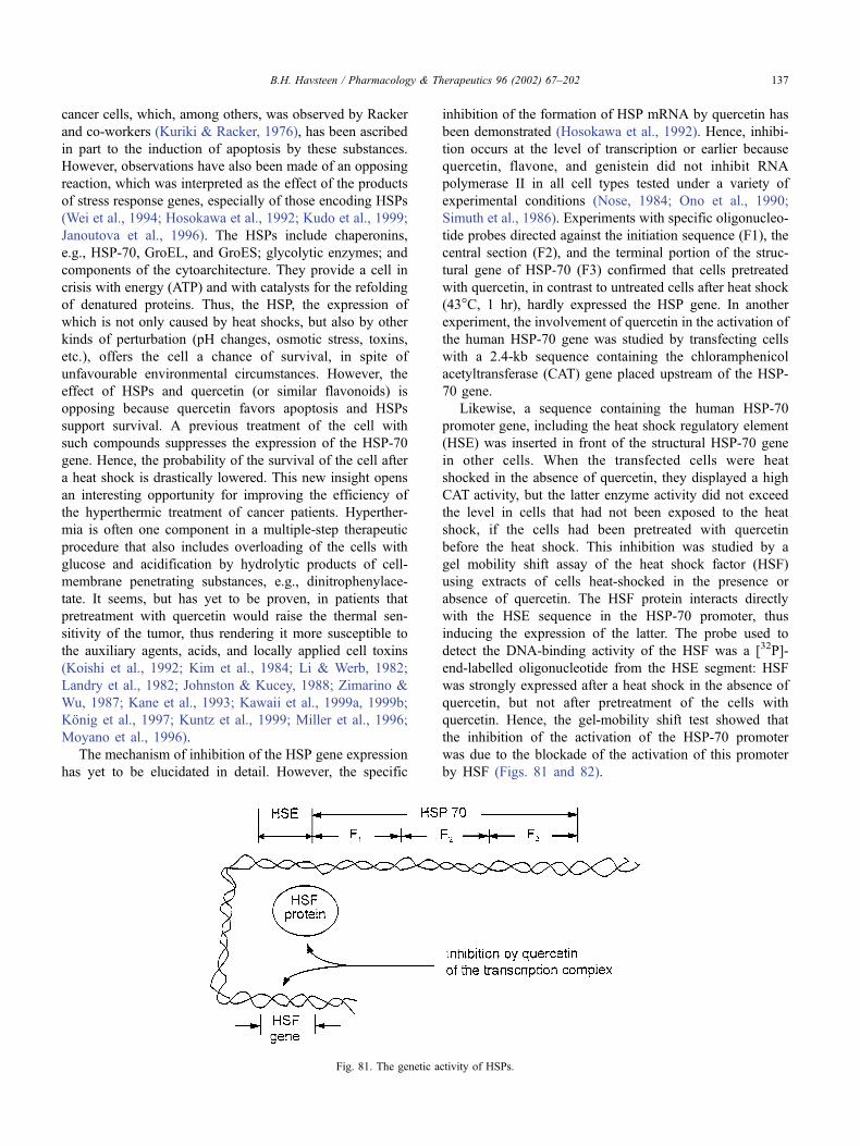
cancer cells, which, among others, was observed by Racker
and co-workers (Kuriki & Racker, 1976), has been ascribed
in part to the induction of apoptosis by these substances.
However, observations have also been made of an opposing
reaction, which was interpreted as the effect of the products
of stress response genes, especially of those encoding HSPs
(Wei et al., 1994; Hosokawa et al., 1992; Kudo et al., 1999;
Janoutova et al., 1996). The HSPs include chaperonins,
e.g., HSP-70, GroEL, and GroES; glycolytic enzymes; and
components of the cytoarchitecture. They provide a cell in
crisis with energy (ATP) and with catalysts for the refolding
of denatured proteins. Thus, the HSP, the expression of
which is not only caused by heat shocks, but also by other
kinds of perturbation (pH changes, osmotic stress, toxins,
etc.), offers the cell a chance of survival, in spite of
unfavourable environmental circumstances. However, the
effect of HSPs and quercetin (or similar flavonoids) is
opposing because quercetin favors apoptosis and HSPs
support survival. A previous treatment of the cell with
such compounds suppresses the expression of the HSP-70
gene. Hence, the probability of the survival of the cell after
a heat shock is drastically lowered. This new insight opens
an interesting opportunity for improving the efficiency of
the hyperthermic treatment of cancer patients. Hyperther-
mia is often one component in a multiple-step therapeutic
procedure that also includes overloading of the cells with
glucose and acidification by hydrolytic products of cell-
membrane penetrating substances, e.g., dinitrophenylace-
tate. It seems, but has yet to be proven, in patients that
pretreatment with quercetin would raise the thermal sen-
sitivity of the tumor, thus rendering it more susceptible to
the auxiliary agents, acids, and locally applied cell toxins
(Koishi et al., 1992; Kim et al., 1984; Li & Werb, 1982;
Landry et al., 1982; Johnston & Kucey, 1988; Zimarino &
Wu, 1987; Kane et al., 1993; Kawaii et al., 1999a, 1999b;
Konig et al., 1997; Kuntz et al., 1999; Miller et al., 1996;
Moyano et al., 1996).
The mechanism of inhibition of the HSP gene expression
has yet to be elucidated in detail. However, the specific
inhibition of the formation of HSP mRNA by quercetin has
been demonstrated (Hosokawa et al., 1992). Hence, inhibi-
tion occurs at the level of transcription or earlier because
quercetin, flavone, and genistein did not inhibit RNA
polymerase II in all cell types tested under a variety of
experimental conditions (Nose, 1984; Ono et al., 1990;
Simuth et al., 1986). Experiments with specific oligonucleo-
tide probes directed against the initiation sequence (F1), the
central section (F2), and the terminal portion of the struc-
tural gene of HSP-70 (F3) confirmed that cells pretreated
with quercetin, in contrast to untreated cells after heat shock
(43�C, 1 hr), hardly expressed the HSP gene. In another
experiment, the involvement of quercetin in the activation of
the human HSP-70 gene was studied by transfecting cells
with a 2.4-kb sequence containing the chloramphenicol
acetyltransferase (CAT) gene placed upstream of the HSP-
70 gene.
Likewise, a sequence containing the human HSP-70
promoter gene, including the heat shock regulatory element
(HSE) was inserted in front of the structural HSP-70 gene
in other cells. When the transfected cells were heat
shocked in the absence of quercetin, they displayed a high
CAT activity, but the latter enzyme activity did not exceed
the level in cells that had not been exposed to the heat
shock, if the cells had been pretreated with quercetin
before the heat shock. This inhibition was studied by a
gel mobility shift assay of the heat shock factor (HSF)
using extracts of cells heat-shocked in the presence or
absence of quercetin. The HSF protein interacts directly
with the HSE sequence in the HSP-70 promoter, thus
inducing the expression of the latter. The probe used to
detect the DNA-binding activity of the HSF was a [32P]-
end-labelled oligonucleotide from the HSE segment: HSF
was strongly expressed after a heat shock in the absence of
quercetin, but not after pretreatment of the cells with
quercetin. Hence, the gel-mobility shift test showed that
the inhibition of the activation of the HSP-70 promoter
was due to the blockade of the activation of this promoter
by HSF (Figs. 81 and 82).
Fig. 81. The genetic activity of HSPs.
B.H. Havsteen / Pharmacology & Therapeutics 96 (2002) 67–202 137
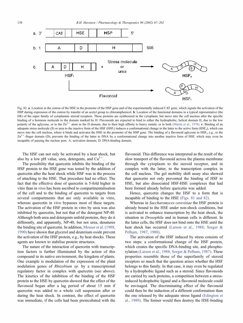
The HSF can not only be activated by a heat shock, but
also by a low pH value, urea, detergents, and Ca2 + .
The possibility that quercetin inhibits the binding of the
HSF protein to the HSE gene was tested by the addition of
quercetin after the heat shock while HSF was in the process
of attaching to the HSE. That procedure had no effect. The
fact that the effective dose of quercetin is 5-fold higher in
vitro than in vivo has been ascribed to compartmentalisation
of the cell and to the binding of quercetin to targets from
several compartments that are only available in vitro,
whereas quercetin in vivo bypasses most of these targets.
The activation of the HSF protein in vitro by urea was also
inhibited by quercetin, but not that of the detergent NP-40.
Although both urea and detergents unfold proteins, they do it
differently, and apparently, NP-40, but not urea, denatures
the binding site of quercetin. In addition, Mosser et al. (1988,
1990) have shown that glycerol and deuterium oxide prevent
the activation of the HSF protein, e.g., by heat shocks. These
agents are known to stabilise protein structures.
The nature of the interaction of quercetin with transcrip-
tion factors is further illuminated by the action of this
compound in its native environment, the kingdom of plants.
One example is modulation of the expression of the plant
nodulation genes of Rhizobium sp. by a transcriptional
regulatory factor in complex with quercetin (see above).
The kinetics of the inhibition of the binding of the HSF
protein to the HSE by quercetin showed that the effect of the
flavonoid began after a lag period of about 15 min if
quercetin was added to a whole cell suspension after or
during the heat shock. In contrast, the effect of quercetin
was immediate, if the cells had been preincubated with the
flavonoid. This difference was interpreted as the result of the
slow transport of the flavonoid across the plasma membrane
through the cytoplasm to the steroid receptor, and in
complex with the latter, to the transcription complex in
the cell nucleus. The gel mobility shift assay also showed
that quercetin not only prevented the binding of HSF to
HSE, but also dissociated HSF-HSE complexes that had
been formed already before quercetin was added.
Hence, quercetin changes the HSF to a form that is
incapable of binding to the HSE (Figs. 81 and 82).
Whereas in Saccharomyces cerevisiae the HSF protein is
already bound to the HSE under non-shock conditions, but
is activated to enhance transcription by the heat shock, the
situation in Drosophila and in human cells is different. In
the latter cells, the HSF does not bind onto the HSE until the
heat shock has occurred (Larson et al., 1988; Sorger &
Pelham, 1987, 1988).
The activation of the HSF induced by stress consists of
two steps: a conformational change of the HSF protein,
which creates the specific DNA-binding site, and phospho-
rylation (Larson et al., 1988; Sorger & Pelham, 1987). These
properties resemble those of the superfamily of steroid
receptors so much that the question arises whether the HSF
belongs to this family. In that case, it may even be regulated
by a hydrophobic ligand such as a steroid. Since flavonoids
are carried by such proteins, a competition between a stress-
induced hydrophobic ligand and a flavonoid molecule could
be envisaged. The discriminating effect of the flavonoid
could then be the induction of a different conformation than
the one released by the adequate stress ligand (Edington et
al., 1989). The former would then destroy the HSE-binding
Fig. 82. a: Location in the cistron of the HSE in the promoter of the HSP gene and of the experimentally induced CAT gene, which signals the activation of the
HSP during expression of the cistron by transfer of an acetyl group to chloramphenicol. b: Location of the functional domains in a typical representative (the
ER) of the super family of cytoplasmic steroid receptors. These proteins are synthesized in the cytoplasm, but move into the cell nucleus after the specific
binding of a hormone molecule to the domain marked by H. Flavonoids are expected to bind to either the hydrophobic, helical domain H, due to the low
polarity of the aglycone, or to the Zn2 + atom in the D domain, due to their high affinity to heavy metals, or to both (Martin et al., 1978). c: Binding of an
adequate stress molecule (S) or urea to the inactive form of the HSF (HSFi) induces a conformational change in the latter to the active form (HSFa), which can
move into the cell nucleus, where it binds and activates the HSE in the promoter of the HSP gene. The binding of a flavonoid aglycone to HSFi, e.g., to the
Zn2 + -finger domain (D), prevents the binding of the latter to DNA by a conformational change into another inactive form of HSF, which may even be
incapable of passing the nuclear pore. A, activation domain; D, DNA-binding domain.
B.H. Havsteen / Pharmacology & Therapeutics 96 (2002) 67–202138
fold, whereas the latter would form it. Since the geometric
restrictions by specific DNA-protein interactions are severe,
the two kinds of conformational changes may be quite
similar and the binding sites of the regulating ligands may
be partially overlapping.
If this model is correct, the reaction to stress may be self-
sustaining, i.e., autocatalytic. The reason is that both the HSF
and the product of the gene, which is activated via the HSE,
belong to the same protein superfamily. Their mutual support
in that case must be interrupted by a different mechanism
before the reaction is exaggerated to the detriment of the cell.
In the case of cancer, one could envisage the therapeutic
application of this flavonoid-assisted pathway to cell death.
Flavonoids are known to kill cancer cells selectively by slow
apoptosis (see Section 17.9), but the point of their attack has
not been identified yet. Alternatively, or in addition, they
may kill the cancer cells via the HSE, as described above.
The advantage of such a procedure is that it is slow enough to
commensurate with the time scale of detoxification of the
debris (especially of biogenic amines) in the liver.
Another aspect of the perturbation of the HSF structure
by the binding of a flavonoid is the susceptibility of the
former to proteolysis. It is known that the cell nucleus
contains a protease that digests cyclin molecules when they
have been marked by phosphorylation. Since HSF is phos-
phorylated, the possibility exists that the HSF is also
decomposed and that the binding of the flavonoid influences
the susceptibility of the HSF to digestion. The simplest way
to answer such questions is to clone the HSF and to express
sufficient amounts of this protein to conduct chemical and
crystallisation experiments. Besides quercetin, the flavonoid
genistein also inhibited the activation of HSF, but not by
preventing its binding to DNA (Price & Calderwood, 1991).
Instead, genistein influences a second stage of HSF activa-
tion, the phosphorylation, because genistein inhibits the
activity of protein phosphokinases (Akiyama et al., 1987;
Osada et al., 1988).
Since HSPs are involved in many cellular processes, e.g.,
the cell cycle, differentiation, apoptosis (Wei et al., 1994),
development, and oncogenic transformation, studies of the
regulation of HSF activity are important. The inhibition of
this process by flavonoids is the only known specific
reaction yet. Flavonoids, therefore, can be used to analyze
the processes mentioned. Besides, the flavonoids have
practical applications, e.g., as enhancers of the thermal
sensitivity of cancer cells prior to hyperthermic therapy.
The role of HSPs as antagonists to the induction of
apoptosis by quercetin is complex.
When the cancer cells were treated with an antisense
oligodesoxynucleotide to the HSP-70 initiation codon and 4
downstream exons, HSP-70 was not formed, and the sub-
sequent addition of quercetin increased the fraction of
apoptotic cells (hypodiploid cells from 20 to 80%). In a
control sample containing the corresponding sense oligonu-
cleotide, the fraction of apoptotic cells was comparable with
the effect of quercetin alone, 20%. The sense oligonucleo-
tide alone only produced an insignificant fraction of apop-
totic cells ( < 2%). Hence, HSP-70 antagonises the apoptotic
effect of quercetin on cancer cells. Since HSP-70 aggregates
in the nucleoplasma, the nucleolus, and the cytoplasm
following a heat shock, but is prevented from doing so by
a pretreatment with quercetin, the apoptosis initiated by
quercetin may be accelerated by the failure of the chaper-
onine function of HSP-70. Thus, proteins that must trans-
locate intracellularly to reach their sites of action, e.g., in the
mitochondria, would fail to refold correctly after unfolding
to pass a membrane. Consequently, the mitochondrion
would lack the chromosomally encoded proteins that are
imported normally, and vital functions, e.g., oxidative
phosphorylation, would cease.
Quercetin also cooperates with cytostatic agents and
inhibitors of protein biosynthesis in the induction of apop-
tosis. Cycloheximide, an inhibitor of protein translation,
increased the fraction of apoptotic cells from 40 ± 2%, the
level ascribable to quercetin alone, to 76 ± 2%, whereas
actinomycin D, an inhibitor of transcription, raised the
fraction of apoptotic cells to 91 ± 5%. In the control
samples, the cells in medium alone contain 2 ± 1% apop-
totic cells, whereas the cells with cycloheximide contained
14 ± 2% of such cells. Actinomycin D alone produced
19 ± 3% apoptotic cells. Hence, the effect of quercetin
synergises with that of protein inhibitors (Ferriola et al.,
1989; Hockenbery et al., 1993). In contrast, agents modu-
lating the activity of PKC., e.g., the phorbol ester 12-O-
tetradecanoyl-phorbol-13-acetate, the PKC inhibitor H-7,
and the Ca2 + chelator EGTA, failed to increase the influ-
ence on the apoptotic index. However, cis-diaminedichloro-
platinum (II) and nitrogen mustard enhanced the antiproli-
ferative effect of the inhibition of PKC (Hofmann et al.,
1988; Scambia et al., 1990a).
Interestingly, the endonuclease inhibitor aurintricarbox-
ylic acid also failed to synergise with the apoptotic effect of
quercetin. Hence, effects of quercetin lie in the early phases
of apoptosis and not in the final committed steps, including
nucleolysis between nucleosomes (Fig. 80) and proteolysis
catalyzed by enzymes of the interconverting enzyme (ICE)
type (caspases).
17.8.4.3. The influence of flavonoids on mammary
carcinoma cells. Some tumor cells of the female breast
are characterised by their ability to bind estrogens cir-
culating in the blood to specific plasma membrane recep-
tors and to respond by performing some of the steps of
differentiation, which have been cancelled or reversed in
the course of oncogenic transformation. Since this process
reduces the malignancy of such cells, or even restores
them to normality, the ability of mammary carcinoma
cells to bind estrogens specifically has been regarded as
a property indicating a favorable prognosis. This sensitiv-
ity to sexual steroids does not seem to be shared by other
epithelial tumors, not even by carcinomas of other female
reproductive organs. Thus, the mechanism appears to be
B.H. Havsteen / Pharmacology & Therapeutics 96 (2002) 67–202 139
part of a normal physiological process that has been
interrupted by the oncogenic transformation. The epithelial
cells of the female breast can express at least two estro-
gen receptors (ERs), EBS-I and -II, which possess the
same binding specificity, but differ in affinity for the
ligand. The lower-affinity receptor (Kd = 10–20 nM),
EBS-II, is inducible by flavonoids (Scambia et al.,
1993; Verma et al., 1988; Ranelletti et al., 1988). Quer-
cetin is especially efficient in inducing EBS-II (Markaver-
ich et al., 1988; Astassi et al., 1985; Bracke et al., 1999;
Rodgers & Grant, 1998; Guthrie & Carroll, 1998; Calta-
girone et al., 1997).
The estrogenic effect of some flavonoids originally was
discovered by observing the behavior of sheep that had
eaten fermented clover containing silybin (Sonnenbichler et
al., 1980; Sonnenbichler & Pohl, 1980). It was ascribed to
the fact that the steric positions of the hydroxyl group in this
flavonoid are almost identical to those of estrogens. Flavo-
noids, due to their nonpolarity or in complex with serum
albumin, can pass the plasma membrane and can attach to
the cytoplasmic steroid receptor. Accordingly, flavonoids
are carried into the cell nucleus to the transcription complex
at the genes controlling the expression of ERs and perhaps
also of other proteins participating in the growth and
function of the mammary gland.
The ability of quercetin to induce the EBS-II receptors in
a dose-dependent fashion was found in cell lines of both
ER-positive and ER-negative tumors. The latter observation
is particularly interesting since the prognosis for the type of
tumor lacking EBS-II is poor. The time course of receptor
induction after quercetin treatment, at least in the two cell
lines examined, is biphasic because the initial appearance of
the receptor, which peaks after 24 hr, is followed by its
disappearance after 72 hr and its reappearance and lasting
expression. This kinetics of expression, which is difficult to
interpret, has also been found for other hormone receptors of
the plasma membrane of reproductive organ cells (Ferran-
dina et al., 1992; Bagavandoss & Midgley, 1988; Scambia et
al., 1991).
The analysis of Scatchard plots revealed that the specific
binding affinity of the quercetin-induced EBS-II receptors
did not differ from that of the constitutively expressed
receptors, but the average number of such receptors per cell
doubled.
The induction of EBS-II receptors by quercetin required
gene transcription and protein synthesis, since the effect was
prevented by actinomycin D and cycloheximide. The inhibi-
tion by quercetin of the proliferation of breast cancer cells
appears to occur at several levels of metabolism and by
different mechanisms (Kawaii et al., 1999a, 1999b). When
the cells are exposed to low concentrations of quercetin, cell
proliferation is inhibited to a greater extent than when the
cells were pretreated with this substance, but at higher
concentrations, a pretreatment of the cells with quercetin
does not affect their sensitivity to the agent. Thus, exposure
of the cells to low concentrations of flavonoids ( < 1 mM)
appears to restore the normal growth metabolism by induc-
ing EBS-II receptors, whereas high quercetin concentrations
inhibit a variety of cellular enzymes (see Section 12). The
EBS-II receptor-inducing effect of quercetin is shared by a
number of other flavonoids, but for most of them, is less
pronounced. Thus, both the ability to increase the number of
EBS-II receptors and the receptor affinity varied in the
following order:
Quercetin ¼ 30; 40; 7� trimethoxy�quercetin
> ipriflavone
whereas rutin and hesperidin failed to show any effect. The
reason seems to be steric hindrance caused by the glycoside
in position 3 of the benzopyrone ring system.
17.9. The influence of flavonoids on cardiovascular diseases
Cardiovascular disease is the most frequent cause of
death in developed countries. Advances in the treatment
of such diseases, therefore, have the potential of yielding a
high return on the research effort. Recent evidence shows
that flavonoids have an important effect on the intricate
regulatory processes, which are derailed by these diseases,
and that they can markedly improve the condition of
patients suffering from various stages of this progressive
illness (Hollman et al., 1999b; Hertog et al., 1993; Brouil-
lard et al., 1997).
A common denominator of these diseases is an inflam-
mation of the vascular wall that is called atherosclerosis, but
the tendency to develop the illness is strongly dependent on
the individual genetic disposition and upon environmental
factors, such as the composition of the diet, smoking,
alcohol consumption, and psychological stress. Flavonoids
interfere at several points in the pathogenesis of cardiovas-
cular disease, and each will be discussed separately.
The general course of inflammatory processes has been
described in Section 17.2. The discussion of the reactions
will only be expanded if they are of particular importance to
cardiovascular diseases and their response to flavonoid
treatment (Yochum et al., 1999; van Acker et al., 1997).
17.9.1. The genetic disposition
Some individuals are born with resistant vascular walls,
but others with weak vascular walls. That is probably an
important reason for the surprising observation that some
people can smoke heavily throughout most of their lives and
age without developing cardiovascular diseases, whereas
others are very sensitive to smoke. For the same reason, it is
very difficult to define general criteria for safety limits, e.g.,
the cholesterol concentration in the blood. A good starting
point for the evaluation of the vascular health of a patient,
therefore, is the family record. If the parents of the patient
and their ancestors reached an advanced age before they
died, then the vascular walls of the patient are probably
fairly robust and the normal upper limit of 250 mg choles-
B.H. Havsteen / Pharmacology & Therapeutics 96 (2002) 67–202140
terol/100 mL blood may be applied. Conversely, if the
family record indicates that caution is necessary, then
dietary restrictions and other measures must be introduced
much sooner. One of the indications, which requires special
attention to avoid cardiovascular crises, is the existence of a
familial coagulopathy, especially one that enhances the
tendency for blood coagulation.
Other inborn dispositions that increase the risk of car-
diovascular diseases are morphological anomalies in the
heart valves. They often require surgery, and the reaction of
the patient to this intervention sharply increases the stress on
the vascular system.
Other risk factors, e.g., alcohol consumption and heavy
smoking, are probably partly determined by the genetic
constitution and are partly conditioned by the social envir-
onment (Hladovec, 1979a, 1979b). The inborn error seems
to cause the partial inactivation of the enzyme, which
liberates the endogenous opiate enkephalin. Since ethanol
improves the activity of this enzyme, presumably by caus-
ing an activating conformational change, the consumption
of alcoholic beverages calms such patients. However, alco-
hol also has an effect on vascular tone, the mechanism of
which is yet unclear, but which increases the risk of vascular
occlusion, i.e., embolism. Alcohol seems to activate a
converting enzyme (ICE) of angiotensin by inducing a
conformational change, and a relative to this ICE initiates
the final (fatal) phase of apoptosis (see Section 17.1).
17.9.2. The role of flavonoids in the dietary component of
cardiovascular stress
Experience teaches that a high cholesterol level and a high
ratio of saturated and monounsaturated to polyunsaturated
FAs in the blood predisposes patients to vascular diseases,
whereas a high dietary content of vegetables and fruits has
the opposite effect (Bok et al., 1999; Fotsis et al., 1997). The
lipids of marine animals, which contain many wC3-unsat-urated FAs, are especially effective in protecting against
cardiovascular crises. The latter seldom occur in people
living in Arctic countries, e.g., Inuits. A possible reason is
that the distribution of double bonds, the first of which
occurs at the third carbon atom from the hydrophobic end,
confers a high resistance against the peroxidation of these
FAs. Moreover and more importantly, wC3-unsaturated FAs
are transformed by COX to a thromboxane (Tx), Tx3, which
is different from the usual form, Tx2. Tx3 has a lower
propensity than Tx2 for the induction of the formation of
the white clot (aggregate of thrombocyte shadows).
The influence of the flavonoids on the endogenous
regulation of cholesterol biosynthesis has already been dealt
with in Section 17.3. However, another critical aspect of
cholesterol homeostasis is the uptake of lipoproteins con-
taining dietary phospholipids, triglycerides, and cholesterol
esters into cells, especially hepatocytes. The lipoproteins
contain a protein surface structure that is recognised by a
specific plasma membrane receptor (Fig. 83). However, both
the apolipoprotein and its receptor can be defective due to an
inborn error, with the result that the efficiency of the trans-
membrane transport of the lipoprotein is drastically reduced.
Hence the half-life of the lipoprotein in the blood is greatly
increased in that case. Finally, lipoproteins are engulfed by
macrophages or collapse due to proteolytic attack, thus
emptying the lipids into the blood and precipitating insoluble
steroids. The unsaturated FAs, both in the liposomes and in
the plasma membranes, are oxidised to peroxides by active
oxygen species from macrophages in stress caused by the
ingested lipids. Thus, the receptors are incapacitated.
Whereas the flavonoids are hardly able to correct the genetic
faults, they efficiently destroy the aggressive oxygen forms,
as previously described in Section 10. Since vegetables and
fruits are rich in flavonoids, their anti-atherosclerotic effects
Fig. 83. a: Lipoprotein docking with the specific recognition structure (S) of the apoprotein (A) in a receptor (R) on the outside of the plasma membrane. The
bag-shaped apoprotein contains triglycerides (T), phospholipids (P), and cholesterol esters (C). b: The cis- and trans-configurations of a double bond. c:
Cholesterol ester.
B.H. Havsteen / Pharmacology & Therapeutics 96 (2002) 67–202 141
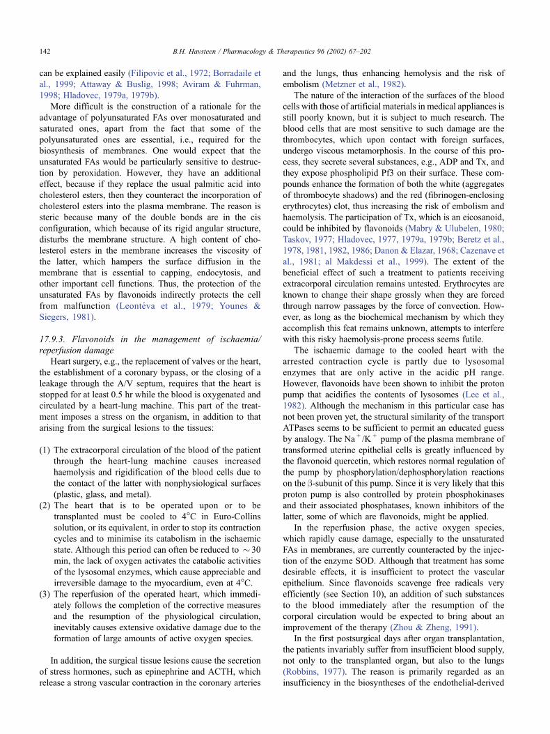
can be explained easily (Filipovic et al., 1972; Borradaile et
al., 1999; Attaway & Buslig, 1998; Aviram & Fuhrman,
1998; Hladovec, 1979a, 1979b).
More difficult is the construction of a rationale for the
advantage of polyunsaturated FAs over monosaturated and
saturated ones, apart from the fact that some of the
polyunsaturated ones are essential, i.e., required for the
biosynthesis of membranes. One would expect that the
unsaturated FAs would be particularly sensitive to destruc-
tion by peroxidation. However, they have an additional
effect, because if they replace the usual palmitic acid into
cholesterol esters, then they counteract the incorporation of
cholesterol esters into the plasma membrane. The reason is
steric because many of the double bonds are in the cis
configuration, which because of its rigid angular structure,
disturbs the membrane structure. A high content of cho-
lesterol esters in the membrane increases the viscosity of
the latter, which hampers the surface diffusion in the
membrane that is essential to capping, endocytosis, and
other important cell functions. Thus, the protection of the
unsaturated FAs by flavonoids indirectly protects the cell
from malfunction (Leonteva et al., 1979; Younes &
Siegers, 1981).
17.9.3. Flavonoids in the management of ischaemia/
reperfusion damage
Heart surgery, e.g., the replacement of valves or the heart,
the establishment of a coronary bypass, or the closing of a
leakage through the A/V septum, requires that the heart is
stopped for at least 0.5 hr while the blood is oxygenated and
circulated by a heart-lung machine. This part of the treat-
ment imposes a stress on the organism, in addition to that
arising from the surgical lesions to the tissues:
(1) The extracorporal circulation of the blood of the patient
through the heart-lung machine causes increased
haemolysis and rigidification of the blood cells due to
the contact of the latter with nonphysiological surfaces
(plastic, glass, and metal).
(2) The heart that is to be operated upon or to be
transplanted must be cooled to 4�C in Euro-Collins
solution, or its equivalent, in order to stop its contraction
cycles and to minimise its catabolism in the ischaemic
state. Although this period can often be reduced to � 30
min, the lack of oxygen activates the catabolic activities
of the lysosomal enzymes, which cause appreciable and
irreversible damage to the myocardium, even at 4�C.(3) The reperfusion of the operated heart, which immedi-
ately follows the completion of the corrective measures
and the resumption of the physiological circulation,
inevitably causes extensive oxidative damage due to the
formation of large amounts of active oxygen species.
In addition, the surgical tissue lesions cause the secretion
of stress hormones, such as epinephrine and ACTH, which
release a strong vascular contraction in the coronary arteries
and the lungs, thus enhancing hemolysis and the risk of
embolism (Metzner et al., 1982).
The nature of the interaction of the surfaces of the blood
cells with those of artificial materials in medical appliances is
still poorly known, but it is subject to much research. The
blood cells that are most sensitive to such damage are the
thrombocytes, which upon contact with foreign surfaces,
undergo viscous metamorphosis. In the course of this pro-
cess, they secrete several substances, e.g., ADP and Tx, and
they expose phospholipid Pf3 on their surface. These com-
pounds enhance the formation of both the white (aggregates
of thrombocyte shadows) and the red (fibrinogen-enclosing
erythrocytes) clot, thus increasing the risk of embolism and
haemolysis. The participation of Tx, which is an eicosanoid,
could be inhibited by flavonoids (Mabry & Ulubelen, 1980;
Taskov, 1977; Hladovec, 1977, 1979a, 1979b; Beretz et al.,
1978, 1981, 1982, 1986; Danon & Elazar, 1968; Cazenave et
al., 1981; al Makdessi et al., 1999). The extent of the
beneficial effect of such a treatment to patients receiving
extracorporal circulation remains untested. Erythrocytes are
known to change their shape grossly when they are forced
through narrow passages by the force of convection. How-
ever, as long as the biochemical mechanism by which they
accomplish this feat remains unknown, attempts to interfere
with this risky haemolysis-prone process seems futile.
The ischaemic damage to the cooled heart with the
arrested contraction cycle is partly due to lysosomal
enzymes that are only active in the acidic pH range.
However, flavonoids have been shown to inhibit the proton
pump that acidifies the contents of lysosomes (Lee et al.,
1982). Although the mechanism in this particular case has
not been proven yet, the structural similarity of the transport
ATPases seems to be sufficient to permit an educated guess
by analogy. The Na + /K + pump of the plasma membrane of
transformed uterine epithelial cells is greatly influenced by
the flavonoid quercetin, which restores normal regulation of
the pump by phosphorylation/dephosphorylation reactions
on the b-subunit of this pump. Since it is very likely that this
proton pump is also controlled by protein phosphokinases
and their associated phosphatases, known inhibitors of the
latter, some of which are flavonoids, might be applied.
In the reperfusion phase, the active oxygen species,
which rapidly cause damage, especially to the unsaturated
FAs in membranes, are currently counteracted by the injec-
tion of the enzyme SOD. Although that treatment has some
desirable effects, it is insufficient to protect the vascular
epithelium. Since flavonoids scavenge free radicals very
efficiently (see Section 10), an addition of such substances
to the blood immediately after the resumption of the
corporal circulation would be expected to bring about an
improvement of the therapy (Zhou & Zheng, 1991).
In the first postsurgical days after organ transplantation,
the patients invariably suffer from insufficient blood supply,
not only to the transplanted organ, but also to the lungs
(Robbins, 1977). The reason is primarily regarded as an
insufficiency in the biosyntheses of the endothelial-derived
B.H. Havsteen / Pharmacology & Therapeutics 96 (2002) 67–202142
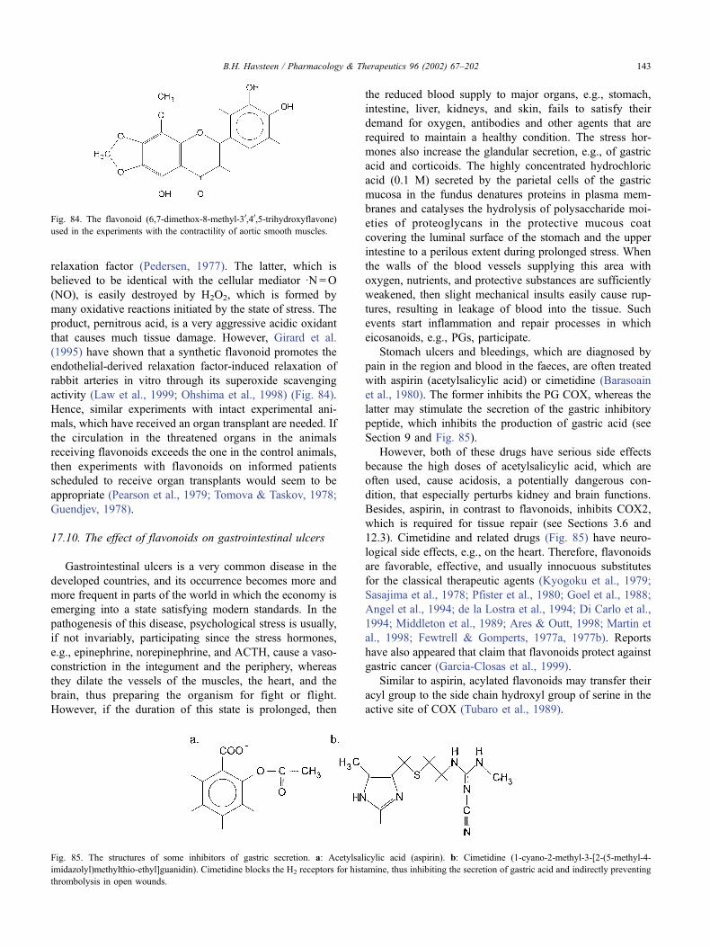
relaxation factor (Pedersen, 1977). The latter, which is
believed to be identical with the cellular mediator �N=O
(NO), is easily destroyed by H2O2, which is formed by
many oxidative reactions initiated by the state of stress. The
product, pernitrous acid, is a very aggressive acidic oxidant
that causes much tissue damage. However, Girard et al.
(1995) have shown that a synthetic flavonoid promotes the
endothelial-derived relaxation factor-induced relaxation of
rabbit arteries in vitro through its superoxide scavenging
activity (Law et al., 1999; Ohshima et al., 1998) (Fig. 84).
Hence, similar experiments with intact experimental ani-
mals, which have received an organ transplant are needed. If
the circulation in the threatened organs in the animals
receiving flavonoids exceeds the one in the control animals,
then experiments with flavonoids on informed patients
scheduled to receive organ transplants would seem to be
appropriate (Pearson et al., 1979; Tomova & Taskov, 1978;
Guendjev, 1978).
17.10. The effect of flavonoids on gastrointestinal ulcers
Gastrointestinal ulcers is a very common disease in the
developed countries, and its occurrence becomes more and
more frequent in parts of the world in which the economy is
emerging into a state satisfying modern standards. In the
pathogenesis of this disease, psychological stress is usually,
if not invariably, participating since the stress hormones,
e.g., epinephrine, norepinephrine, and ACTH, cause a vaso-
constriction in the integument and the periphery, whereas
they dilate the vessels of the muscles, the heart, and the
brain, thus preparing the organism for fight or flight.
However, if the duration of this state is prolonged, then
the reduced blood supply to major organs, e.g., stomach,
intestine, liver, kidneys, and skin, fails to satisfy their
demand for oxygen, antibodies and other agents that are
required to maintain a healthy condition. The stress hor-
mones also increase the glandular secretion, e.g., of gastric
acid and corticoids. The highly concentrated hydrochloric
acid (0.1 M) secreted by the parietal cells of the gastric
mucosa in the fundus denatures proteins in plasma mem-
branes and catalyses the hydrolysis of polysaccharide moi-
eties of proteoglycans in the protective mucous coat
covering the luminal surface of the stomach and the upper
intestine to a perilous extent during prolonged stress. When
the walls of the blood vessels supplying this area with
oxygen, nutrients, and protective substances are sufficiently
weakened, then slight mechanical insults easily cause rup-
tures, resulting in leakage of blood into the tissue. Such
events start inflammation and repair processes in which
eicosanoids, e.g., PGs, participate.
Stomach ulcers and bleedings, which are diagnosed by
pain in the region and blood in the faeces, are often treated
with aspirin (acetylsalicylic acid) or cimetidine (Barasoain
et al., 1980). The former inhibits the PG COX, whereas the
latter may stimulate the secretion of the gastric inhibitory
peptide, which inhibits the production of gastric acid (see
Section 9 and Fig. 85).
However, both of these drugs have serious side effects
because the high doses of acetylsalicylic acid, which are
often used, cause acidosis, a potentially dangerous con-
dition, that especially perturbs kidney and brain functions.
Besides, aspirin, in contrast to flavonoids, inhibits COX2,
which is required for tissue repair (see Sections 3.6 and
12.3). Cimetidine and related drugs (Fig. 85) have neuro-
logical side effects, e.g., on the heart. Therefore, flavonoids
are favorable, effective, and usually innocuous substitutes
for the classical therapeutic agents (Kyogoku et al., 1979;
Sasajima et al., 1978; Pfister et al., 1980; Goel et al., 1988;
Angel et al., 1994; de la Lostra et al., 1994; Di Carlo et al.,
1994; Middleton et al., 1989; Ares & Outt, 1998; Martin et
al., 1998; Fewtrell & Gomperts, 1977a, 1977b). Reports
have also appeared that claim that flavonoids protect against
gastric cancer (Garcia-Closas et al., 1999).
Similar to aspirin, acylated flavonoids may transfer their
acyl group to the side chain hydroxyl group of serine in the
active site of COX (Tubaro et al., 1989).
Fig. 84. The flavonoid (6,7-dimethox-8-methyl-30,40,5-trihydroxyflavone)
used in the experiments with the contractility of aortic smooth muscles.
Fig. 85. The structures of some inhibitors of gastric secretion. a: Acetylsalicylic acid (aspirin). b: Cimetidine (1-cyano-2-methyl-3-[2-(5-methyl-4-
imidazolyl)methylthio-ethyl]guanidin). Cimetidine blocks the H2 receptors for histamine, thus inhibiting the secretion of gastric acid and indirectly preventing
thrombolysis in open wounds.
B.H. Havsteen / Pharmacology & Therapeutics 96 (2002) 67–202 143
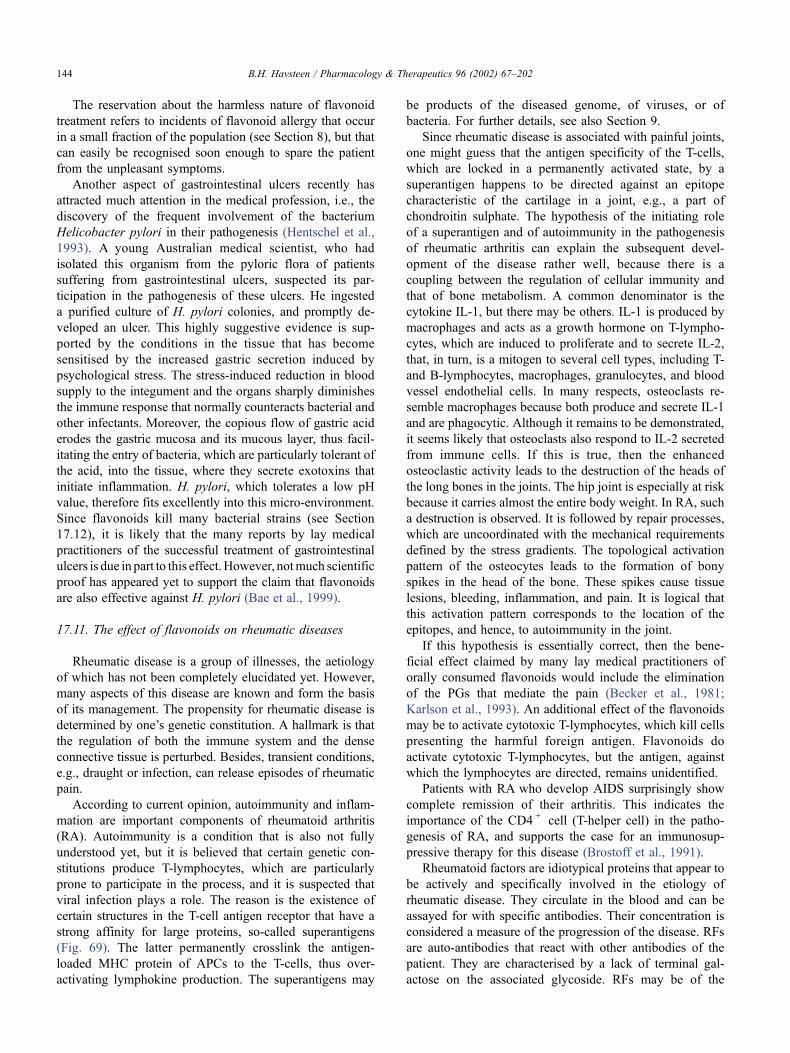
The reservation about the harmless nature of flavonoid
treatment refers to incidents of flavonoid allergy that occur
in a small fraction of the population (see Section 8), but that
can easily be recognised soon enough to spare the patient
from the unpleasant symptoms.
Another aspect of gastrointestinal ulcers recently has
attracted much attention in the medical profession, i.e., the
discovery of the frequent involvement of the bacterium
Helicobacter pylori in their pathogenesis (Hentschel et al.,
1993). A young Australian medical scientist, who had
isolated this organism from the pyloric flora of patients
suffering from gastrointestinal ulcers, suspected its par-
ticipation in the pathogenesis of these ulcers. He ingested
a purified culture of H. pylori colonies, and promptly de-
veloped an ulcer. This highly suggestive evidence is sup-
ported by the conditions in the tissue that has become
sensitised by the increased gastric secretion induced by
psychological stress. The stress-induced reduction in blood
supply to the integument and the organs sharply diminishes
the immune response that normally counteracts bacterial and
other infectants. Moreover, the copious flow of gastric acid
erodes the gastric mucosa and its mucous layer, thus facil-
itating the entry of bacteria, which are particularly tolerant of
the acid, into the tissue, where they secrete exotoxins that
initiate inflammation. H. pylori, which tolerates a low pH
value, therefore fits excellently into this micro-environment.
Since flavonoids kill many bacterial strains (see Section
17.12), it is likely that the many reports by lay medical
practitioners of the successful treatment of gastrointestinal
ulcers is due in part to this effect.However, notmuch scientific
proof has appeared yet to support the claim that flavonoids
are also effective against H. pylori (Bae et al., 1999).
17.11. The effect of flavonoids on rheumatic diseases
Rheumatic disease is a group of illnesses, the aetiology
of which has not been completely elucidated yet. However,
many aspects of this disease are known and form the basis
of its management. The propensity for rheumatic disease is
determined by one’s genetic constitution. A hallmark is that
the regulation of both the immune system and the dense
connective tissue is perturbed. Besides, transient conditions,
e.g., draught or infection, can release episodes of rheumatic
pain.
According to current opinion, autoimmunity and inflam-
mation are important components of rheumatoid arthritis
(RA). Autoimmunity is a condition that is also not fully
understood yet, but it is believed that certain genetic con-
stitutions produce T-lymphocytes, which are particularly
prone to participate in the process, and it is suspected that
viral infection plays a role. The reason is the existence of
certain structures in the T-cell antigen receptor that have a
strong affinity for large proteins, so-called superantigens
(Fig. 69). The latter permanently crosslink the antigen-
loaded MHC protein of APCs to the T-cells, thus over-
activating lymphokine production. The superantigens may
be products of the diseased genome, of viruses, or of
bacteria. For further details, see also Section 9.
Since rheumatic disease is associated with painful joints,
one might guess that the antigen specificity of the T-cells,
which are locked in a permanently activated state, by a
superantigen happens to be directed against an epitope
characteristic of the cartilage in a joint, e.g., a part of
chondroitin sulphate. The hypothesis of the initiating role
of a superantigen and of autoimmunity in the pathogenesis
of rheumatic arthritis can explain the subsequent devel-
opment of the disease rather well, because there is a
coupling between the regulation of cellular immunity and
that of bone metabolism. A common denominator is the
cytokine IL-1, but there may be others. IL-1 is produced by
macrophages and acts as a growth hormone on T-lympho-
cytes, which are induced to proliferate and to secrete IL-2,
that, in turn, is a mitogen to several cell types, including T-
and B-lymphocytes, macrophages, granulocytes, and blood
vessel endothelial cells. In many respects, osteoclasts re-
semble macrophages because both produce and secrete IL-1
and are phagocytic. Although it remains to be demonstrated,
it seems likely that osteoclasts also respond to IL-2 secreted
from immune cells. If this is true, then the enhanced
osteoclastic activity leads to the destruction of the heads of
the long bones in the joints. The hip joint is especially at risk
because it carries almost the entire body weight. In RA, such
a destruction is observed. It is followed by repair processes,
which are uncoordinated with the mechanical requirements
defined by the stress gradients. The topological activation
pattern of the osteocytes leads to the formation of bony
spikes in the head of the bone. These spikes cause tissue
lesions, bleeding, inflammation, and pain. It is logical that
this activation pattern corresponds to the location of the
epitopes, and hence, to autoimmunity in the joint.
If this hypothesis is essentially correct, then the bene-
ficial effect claimed by many lay medical practitioners of
orally consumed flavonoids would include the elimination
of the PGs that mediate the pain (Becker et al., 1981;
Karlson et al., 1993). An additional effect of the flavonoids
may be to activate cytotoxic T-lymphocytes, which kill cells
presenting the harmful foreign antigen. Flavonoids do
activate cytotoxic T-lymphocytes, but the antigen, against
which the lymphocytes are directed, remains unidentified.
Patients with RA who develop AIDS surprisingly show
complete remission of their arthritis. This indicates the
importance of the CD4 + cell (T-helper cell) in the patho-
genesis of RA, and supports the case for an immunosup-
pressive therapy for this disease (Brostoff et al., 1991).
Rheumatoid factors are idiotypical proteins that appear to
be actively and specifically involved in the etiology of
rheumatic disease. They circulate in the blood and can be
assayed for with specific antibodies. Their concentration is
considered a measure of the progression of the disease. RFs
are auto-antibodies that react with other antibodies of the
patient. They are characterised by a lack of terminal gal-
actose on the associated glycoside. RFs may be of the
B.H. Havsteen / Pharmacology & Therapeutics 96 (2002) 67–202144
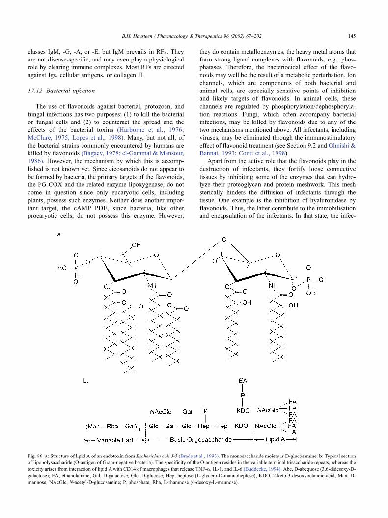
classes IgM, -G, -A, or -E, but IgM prevails in RFs. They
are not disease-specific, and may even play a physiological
role by clearing immune complexes. Most RFs are directed
against Igs, cellular antigens, or collagen II.
17.12. Bacterial infection
The use of flavonoids against bacterial, protozoan, and
fungal infections has two purposes: (1) to kill the bacterial
or fungal cells and (2) to counteract the spread and the
effects of the bacterial toxins (Harborne et al., 1976;
McClure, 1975; Lopes et al., 1998). Many, but not all, of
the bacterial strains commonly encountered by humans are
killed by flavonoids (Bagaev, 1978; el-Gammal & Mansour,
1986). However, the mechanism by which this is accomp-
lished is not known yet. Since eicosanoids do not appear to
be formed by bacteria, the primary targets of the flavonoids,
the PG COX and the related enzyme lipoxygenase, do not
come in question since only eucaryotic cells, including
plants, possess such enzymes. Neither does another impor-
tant target, the cAMP PDE, since bacteria, like other
procaryotic cells, do not possess this enzyme. However,
they do contain metalloenzymes, the heavy metal atoms that
form strong ligand complexes with flavonoids, e.g., phos-
phatases. Therefore, the bacteriocidal effect of the flavo-
noids may well be the result of a metabolic perturbation. Ion
channels, which are components of both bacterial and
animal cells, are especially sensitive points of inhibition
and likely targets of flavonoids. In animal cells, these
channels are regulated by phosphorylation/dephosphoryla-
tion reactions. Fungi, which often accompany bacterial
infections, may be killed by flavonoids due to any of the
two mechanisms mentioned above. All infectants, including
viruses, may be eliminated through the immunostimulatory
effect of flavonoid treatment (see Section 9.2 and Ohnishi &
Bannai, 1993; Conti et al., 1998).
Apart from the active role that the flavonoids play in the
destruction of infectants, they fortify loose connective
tissues by inhibiting some of the enzymes that can hydro-
lyze their proteoglycan and protein meshwork. This mesh
sterically hinders the diffusion of infectants through the
tissue. One example is the inhibition of hyaluronidase by
flavonoids. Thus, the latter contribute to the immobilisation
and encapsulation of the infectants. In that state, the infec-
Fig. 86. a: Structure of lipid A of an endotoxin from Escherichia coli J-5 (Brade et al., 1993). The monosaccharide moiety is D-glucosamine. b: Typical section
of lipopolysaccharide (O-antigen of Gram-negative bacteria). The specificity of the O-antigen resides in the variable terminal trisaccharide repeats, whereas the
toxicity arises from interaction of lipid Awith CD14 of macrophages that release TNF-a, IL-1, and IL-6 (Buddecke, 1994). Abe, D-abequose (3,6-didesoxy-D-galactose); EA, ethanolamine; Gal, D-galactose; Glc, D-glucose; Hep, heptose (L-glycero-D-mannoheptose); KDO, 2-keto-3-desoxyoctanoic acid; Man, D-
mannose; NAcGlc, N-acetyl-D-glucosamine; P, phosphate; Rha, L-rhamnose (6-desoxy-L-mannose).
B.H. Havsteen / Pharmacology & Therapeutics 96 (2002) 67–202 145
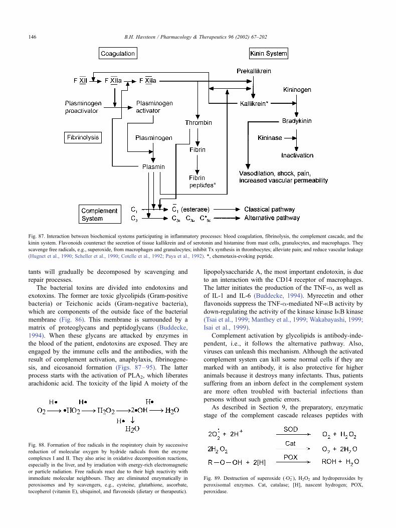
tants will gradually be decomposed by scavenging and
repair processes.
The bacterial toxins are divided into endotoxins and
exotoxins. The former are toxic glycolipids (Gram-positive
bacteria) or Teichonic acids (Gram-negative bacteria),
which are components of the outside face of the bacterial
membrane (Fig. 86). This membrane is surrounded by a
matrix of proteoglycans and peptidoglycans (Buddecke,
1994). When these glycans are attacked by enzymes in
the blood of the patient, endotoxins are exposed. They are
engaged by the immune cells and the antibodies, with the
result of complement activation, anaphylaxis, fibrinogene-
sis, and eicosanoid formation (Figs. 87–95). The latter
process starts with the activation of PLA2, which liberates
arachidonic acid. The toxicity of the lipid A moiety of the
lipopolysaccharide A, the most important endotoxin, is due
to an interaction with the CD14 receptor of macrophages.
The latter initiates the production of the TNF-a, as well asof IL-1 and IL-6 (Buddecke, 1994). Myrecetin and other
flavonoids suppress the TNF-a-mediated NF-kB activity by
down-regulating the activity of the kinase kinase IkB kinase
(Tsai et al., 1999; Manthey et al., 1999; Wakabayashi, 1999;
Isai et al., 1999).
Complement activation by glycolipids is antibody-inde-
pendent, i.e., it follows the alternative pathway. Also,
viruses can unleash this mechanism. Although the activated
complement system can kill some normal cells if they are
marked with an antibody, it is also protective for higher
animals because it destroys many infectants. Thus, patients
suffering from an inborn defect in the complement system
are more often troubled with bacterial infections than
persons without such genetic errors.
As described in Section 9, the preparatory, enzymatic
stage of the complement cascade releases peptides with
Fig. 87. Interaction between biochemical systems participating in inflammatory processes: blood coagulation, fibrinolysis, the complement cascade, and the
kinin system. Flavonoids counteract the secretion of tissue kallikrein and of serotonin and histamine from mast cells, granulocytes, and macrophages. They
scavenge free radicals, e.g., superoxide, from macrophages and granulocytes; inhibit Tx synthesis in thrombocytes; alleviate pain; and reduce vascular leakage
(Hugnet et al., 1990; Scheller et al., 1990; Cotelle et al., 1992; Paya et al., 1992). *, chemotaxis-evoking peptide.
Fig. 88. Formation of free radicals in the respiratory chain by successive
reduction of molecular oxygen by hydride radicals from the enzyme
complexes I and II. They also arise in oxidative decomposition reactions,
especially in the liver, and by irradiation with energy-rich electromagnetic
or particle radiation. Free radicals react due to their high reactivity with
immediate molecular neighbours. They are eliminated enzymatically in
peroxisomes and by scavengers, e.g., cysteine, glutathione, ascorbate,
tocopherol (vitamin E), ubiquinol, and flavonoids (dietary or therapeutic).
Fig. 89. Destruction of superoxide (�O2�), H2O2 and hydroperoxides by
peroxisomal enzymes. Cat, catalase; [H], nascent hydrogen; POX,
peroxidase.
B.H. Havsteen / Pharmacology & Therapeutics 96 (2002) 67–202146
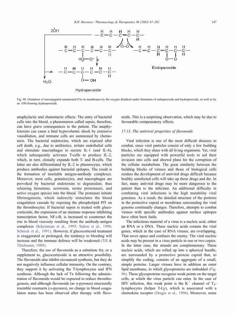
anaphylactic and chemotactic effects. The entry of bacterial
cells into the blood, a phenomenon called sepsis, therefore,
can have grave consequences to the patient. The anaphy-
lotoxins can cause a fatal hypovolumic shock by extensive
vasodilation, and immune cells are summoned by chemo-
taxis. The bacterial endotoxins, which are exposed after
cell death, e.g., due to antibiotics, irritate endothelial cells
and stimulate macrophages to secrete IL-1 (and IL-6),
which subsequently activates T-cells to produce IL-2,
which, in turn, clonally expands both T- and B-cells. The
latter are also differentiated by IL-2 to plasmocytes, which
produce antibodies against bacterial epitopes. The result is
the formation of insoluble antigen-antibody complexes.
Moreover, most cells, granulocytes, and macrophages are
provoked by bacterial endotoxins to degranulate, thus
releasing histamine, serotonin, serine proteinases, and
active oxygen species into the blood. The proteases initiate
fibrinogenesis, which indirectly stimulates the blood
coagulation cascade by exposing the phospholipid Pf3 on
the thrombocytes. If bacterial sepsis is treated with gluco-
corticoids, the expression of an immune response inhibiting
transcription factor, NF-kB, is increased to counteract the
rise in blood viscosity resulting from the antibody-antigen
complexes (Scheinman et al., 1995; Saliou et al., 1998;
Schreck et al., 1991). However, if glucocorticoid treatment
is exaggerated or prolonged, the tendency to bleeding will
increase and the immune defence will be weakened (Till &
Thielmann, 1989).
Therefore, the use of flavonoids as a substitute for, or a
supplement to, glucocorticoids is an attractive possibility.
The flavonoids also inhibit eicosanoid synthesis, but they do
not negatively influence cellular immunity. On the contrary,
they support it by activating the T-lymphocytes and IFN
synthesis. Although the lack of Tx following the adminis-
tration of flavonoids would be expected to reduce thrombo-
genesis, and although flavonoids (as g-pyrones) structurally
resemble coumarin (a-pyrones), no change in blood coagu-
lation status has been observed after therapy with flavo-
noids. This is a surprising observation, which may be due to
favourable compensatory effects.
17.13. The antiviral properties of flavonoids
Viral infection is one of the most difficult diseases to
combat, since viral particles consist of only a few building
blocks, which they share with all living organisms. Yet, viral
particles are equipped with powerful tools to aid their
invasion into cells and shrewd plans for the corruption of
the cellular metabolism. The great similarity between the
building blocks of viruses and those of biological cells
renders the development of antiviral drugs difficult because
healthy uninfected cells will take up these drugs and die. In
fact, many antiviral drugs may be more dangerous to the
patient than to the infectant. An additional difficulty in
combating viral infections is the high mutability viral
genomes. As a result, the detailed structure of the proteins
in the protective capsid or membrane surrounding the viral
genome continually changes. Therefore, attempts to combat
viruses with specific antibodies against surface epitopes
have often been futile.
The infectious material of a virus is a nucleic acid, either
an RNA or a DNA. These nucleic acids contain the viral
genes, which in the case of RNA viruses, are overlapping.
That saves space and confuses the enemy. The viral nucleic
acids may be present in a virus particle in one or two copies.
In the latter case, the strands are complementary. These
nucleic acids, which are rolled up into a spherical bundle,
are surrounded by a protective protein capsid that, to
simplify the coding, consists of an aggregate of a small,
simple proteins. Larger viruses have in addition an outer
lipid membrane, in which glycoproteins are imbedded (Fig.
96). These glycoproteins recognise weak points on the target
cells, at which the virus particle can enter. In the case of
HIV infection, this weak point is the K + channel of T4-
lymphocytes (helper T-Ly), which is associated with a
chemokine receptor (Dragic et al., 1996). Moreover, some
Fig. 90. Oxidation of unconjugated unsaturated FAs in membranes by the oxygen diradical under formation of endoperoxide and hydroperoxide, as well as by
an �OH-forming hydroperoxide.
B.H. Havsteen / Pharmacology & Therapeutics 96 (2002) 67–202 147
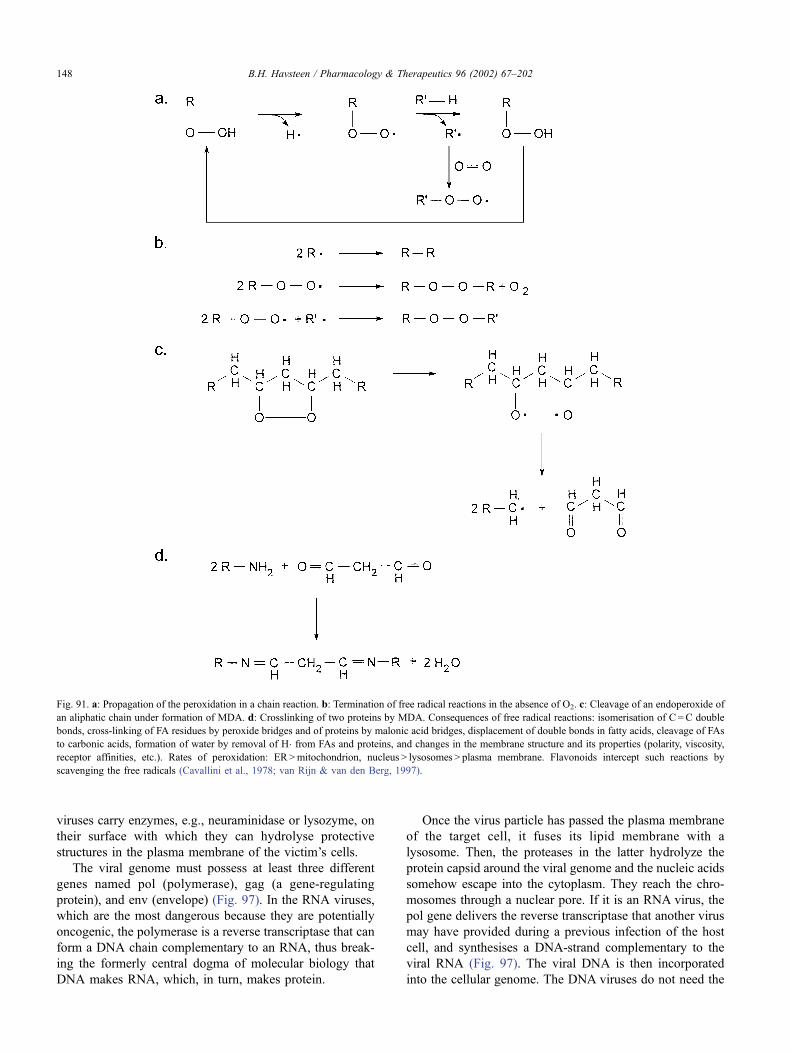
viruses carry enzymes, e.g., neuraminidase or lysozyme, on
their surface with which they can hydrolyse protective
structures in the plasma membrane of the victim’s cells.
The viral genome must possess at least three different
genes named pol (polymerase), gag (a gene-regulating
protein), and env (envelope) (Fig. 97). In the RNA viruses,
which are the most dangerous because they are potentially
oncogenic, the polymerase is a reverse transcriptase that can
form a DNA chain complementary to an RNA, thus break-
ing the formerly central dogma of molecular biology that
DNA makes RNA, which, in turn, makes protein.
Once the virus particle has passed the plasma membrane
of the target cell, it fuses its lipid membrane with a
lysosome. Then, the proteases in the latter hydrolyze the
protein capsid around the viral genome and the nucleic acids
somehow escape into the cytoplasm. They reach the chro-
mosomes through a nuclear pore. If it is an RNA virus, the
pol gene delivers the reverse transcriptase that another virus
may have provided during a previous infection of the host
cell, and synthesises a DNA-strand complementary to the
viral RNA (Fig. 97). The viral DNA is then incorporated
into the cellular genome. The DNA viruses do not need the
Fig. 91. a: Propagation of the peroxidation in a chain reaction. b: Termination of free radical reactions in the absence of O2. c: Cleavage of an endoperoxide of
an aliphatic chain under formation of MDA. d: Crosslinking of two proteins by MDA. Consequences of free radical reactions: isomerisation of C =C double
bonds, cross-linking of FA residues by peroxide bridges and of proteins by malonic acid bridges, displacement of double bonds in fatty acids, cleavage of FAs
to carbonic acids, formation of water by removal of H� from FAs and proteins, and changes in the membrane structure and its properties (polarity, viscosity,
receptor affinities, etc.). Rates of peroxidation: ER>mitochondrion, nucleus > lysosomes > plasma membrane. Flavonoids intercept such reactions by
scavenging the free radicals (Cavallini et al., 1978; van Rijn & van den Berg, 1997).
B.H. Havsteen / Pharmacology & Therapeutics 96 (2002) 67–202148
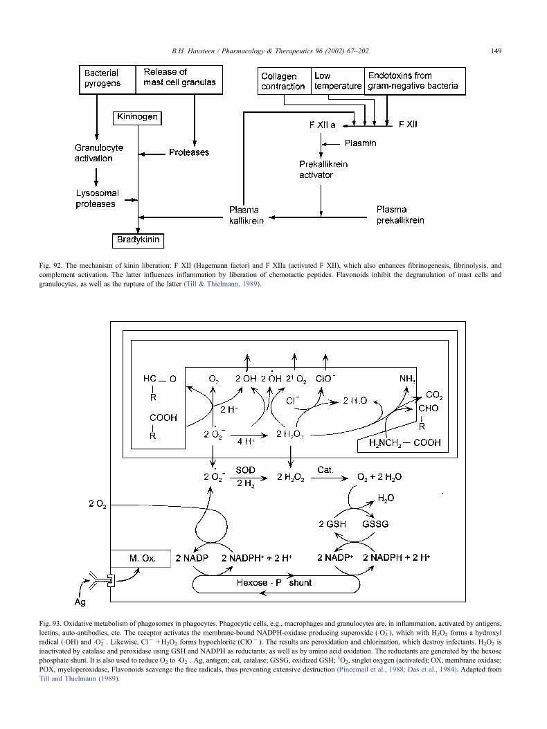
Fig. 92. The mechanism of kinin liberation: F XII (Hagemann factor) and F XIIa (activated F XII), which also enhances fibrinogenesis, fibrinolysis, and
complement activation. The latter influences inflammation by liberation of chemotactic peptides. Flavonoids inhibit the degranulation of mast cells and
granulocytes, as well as the rupture of the latter (Till & Thielmann, 1989).
Fig. 93. Oxidative metabolism of phagosomes in phagocytes. Phagocytic cells, e.g., macrophages and granulocytes are, in inflammation, activated by antigens,
lectins, auto-antibodies, etc. The receptor activates the membrane-bound NADPH-oxidase producing superoxide (�O2�), which with H2O2 forms a hydroxyl
radical (�OH) and �O2� . Likewise, Cl � +H2O2 forms hypochlorite (ClO� ). The results are peroxidation and chlorination, which destroy infectants. H2O2 is
inactivated by catalase and peroxidase using GSH and NADPH as reductants, as well as by amino acid oxidation. The reductants are generated by the hexose
phosphate shunt. It is also used to reduce O2 to �O2� . Ag, antigen; cat, catalase; GSSG, oxidized GSH; 1O2, singlet oxygen (activated); OX, membrane oxidase;
POX, myeloperoxidase, Flavonoids scavenge the free radicals, thus preventing extensive destruction (Pincemail et al., 1988; Das et al., 1984). Adapted from
Till and Thielmann (1989).
B.H. Havsteen / Pharmacology & Therapeutics 96 (2002) 67–202 149
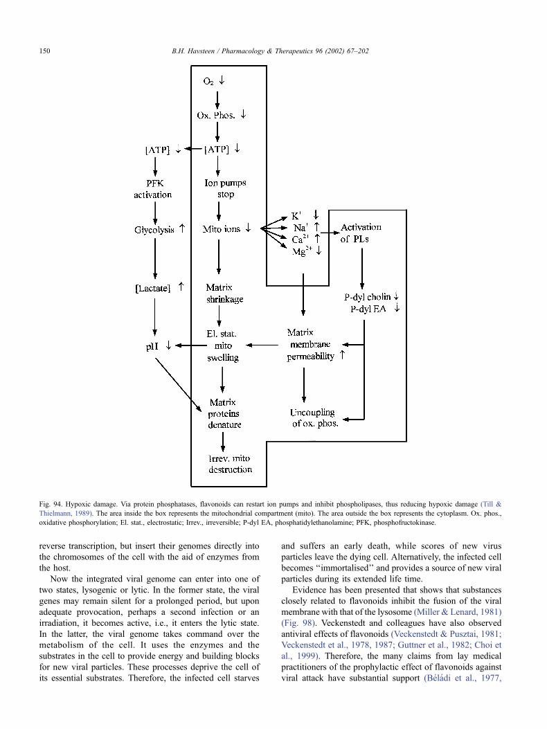
reverse transcription, but insert their genomes directly into
the chromosomes of the cell with the aid of enzymes from
the host.
Now the integrated viral genome can enter into one of
two states, lysogenic or lytic. In the former state, the viral
genes may remain silent for a prolonged period, but upon
adequate provocation, perhaps a second infection or an
irradiation, it becomes active, i.e., it enters the lytic state.
In the latter, the viral genome takes command over the
metabolism of the cell. It uses the enzymes and the
substrates in the cell to provide energy and building blocks
for new viral particles. These processes deprive the cell of
its essential substrates. Therefore, the infected cell starves
and suffers an early death, while scores of new virus
particles leave the dying cell. Alternatively, the infected cell
becomes ‘‘immortalised’’ and provides a source of new viral
particles during its extended life time.
Evidence has been presented that shows that substances
closely related to flavonoids inhibit the fusion of the viral
membrane with that of the lysosome (Miller & Lenard, 1981)
(Fig. 98). Veckenstedt and colleagues have also observed
antiviral effects of flavonoids (Veckenstedt & Pusztai, 1981;
Veckenstedt et al., 1978, 1987; Guttner et al., 1982; Choi et
al., 1999). Therefore, the many claims from lay medical
practitioners of the prophylactic effect of flavonoids against
viral attack have substantial support (Beladi et al., 1977,
Fig. 94. Hypoxic damage. Via protein phosphatases, flavonoids can restart ion pumps and inhibit phospholipases, thus reducing hypoxic damage (Till &
Thielmann, 1989). The area inside the box represents the mitochondrial compartment (mito). The area outside the box represents the cytoplasm. Ox. phos.,
oxidative phosphorylation; El. stat., electrostatic; Irrev., irreversible; P-dyl EA, phosphatidylethanolamine; PFK, phosphofructokinase.
B.H. Havsteen / Pharmacology & Therapeutics 96 (2002) 67–202150
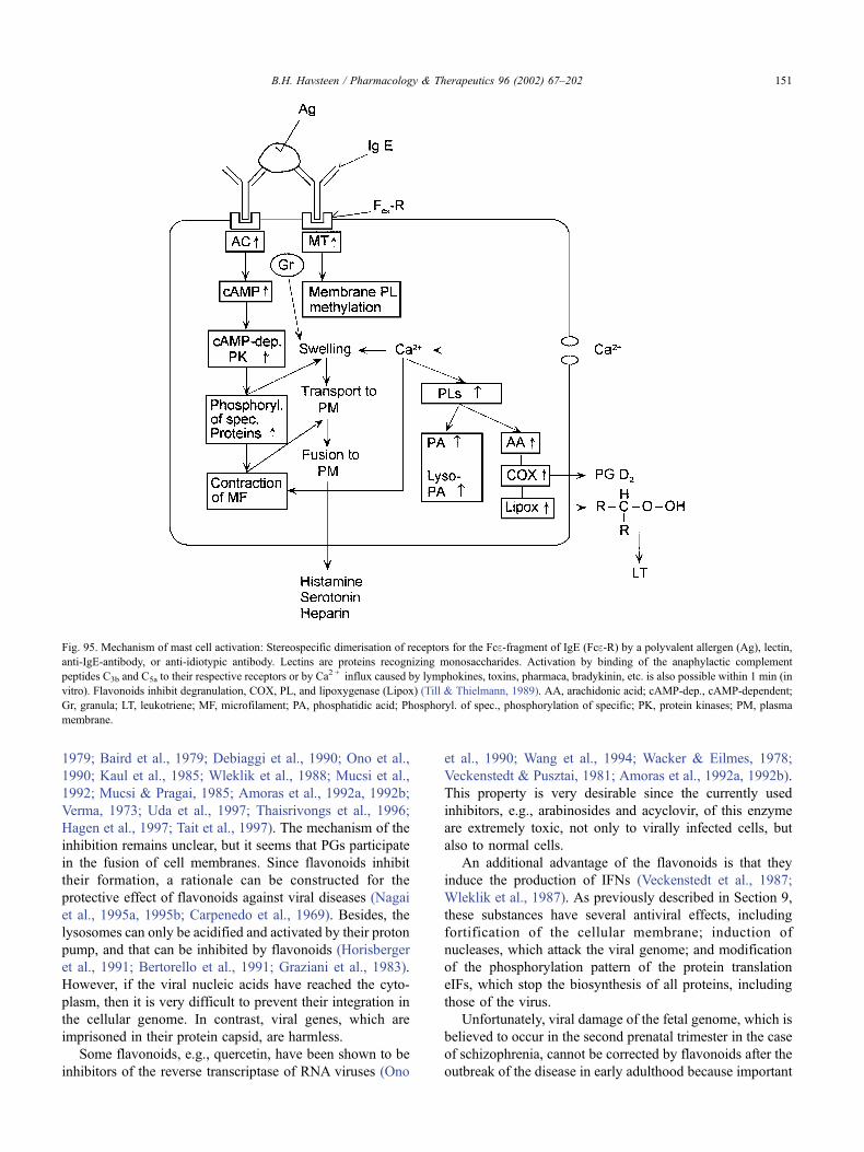
1979; Baird et al., 1979; Debiaggi et al., 1990; Ono et al.,
1990; Kaul et al., 1985; Wleklik et al., 1988; Mucsi et al.,
1992; Mucsi & Pragai, 1985; Amoras et al., 1992a, 1992b;
Verma, 1973; Uda et al., 1997; Thaisrivongs et al., 1996;
Hagen et al., 1997; Tait et al., 1997). The mechanism of the
inhibition remains unclear, but it seems that PGs participate
in the fusion of cell membranes. Since flavonoids inhibit
their formation, a rationale can be constructed for the
protective effect of flavonoids against viral diseases (Nagai
et al., 1995a, 1995b; Carpenedo et al., 1969). Besides, the
lysosomes can only be acidified and activated by their proton
pump, and that can be inhibited by flavonoids (Horisberger
et al., 1991; Bertorello et al., 1991; Graziani et al., 1983).
However, if the viral nucleic acids have reached the cyto-
plasm, then it is very difficult to prevent their integration in
the cellular genome. In contrast, viral genes, which are
imprisoned in their protein capsid, are harmless.
Some flavonoids, e.g., quercetin, have been shown to be
inhibitors of the reverse transcriptase of RNA viruses (Ono
et al., 1990; Wang et al., 1994; Wacker & Eilmes, 1978;
Veckenstedt & Pusztai, 1981; Amoras et al., 1992a, 1992b).
This property is very desirable since the currently used
inhibitors, e.g., arabinosides and acyclovir, of this enzyme
are extremely toxic, not only to virally infected cells, but
also to normal cells.
An additional advantage of the flavonoids is that they
induce the production of IFNs (Veckenstedt et al., 1987;
Wleklik et al., 1987). As previously described in Section 9,
these substances have several antiviral effects, including
fortification of the cellular membrane; induction of
nucleases, which attack the viral genome; and modification
of the phosphorylation pattern of the protein translation
eIFs, which stop the biosynthesis of all proteins, including
those of the virus.
Unfortunately, viral damage of the fetal genome, which is
believed to occur in the second prenatal trimester in the case
of schizophrenia, cannot be corrected by flavonoids after the
outbreak of the disease in early adulthood because important
Fig. 95. Mechanism of mast cell activation: Stereospecific dimerisation of receptors for the Fce-fragment of IgE (Fce-R) by a polyvalent allergen (Ag), lectin,
anti-IgE-antibody, or anti-idiotypic antibody. Lectins are proteins recognizing monosaccharides. Activation by binding of the anaphylactic complement
peptides C3b and C5a to their respective receptors or by Ca2 + influx caused by lymphokines, toxins, pharmaca, bradykinin, etc. is also possible within 1 min (in
vitro). Flavonoids inhibit degranulation, COX, PL, and lipoxygenase (Lipox) (Till & Thielmann, 1989). AA, arachidonic acid; cAMP-dep., cAMP-dependent;
Gr, granula; LT, leukotriene; MF, microfilament; PA, phosphatidic acid; Phosphoryl. of spec., phosphorylation of specific; PK, protein kinases; PM, plasma
membrane.
B.H. Havsteen / Pharmacology & Therapeutics 96 (2002) 67–202 151
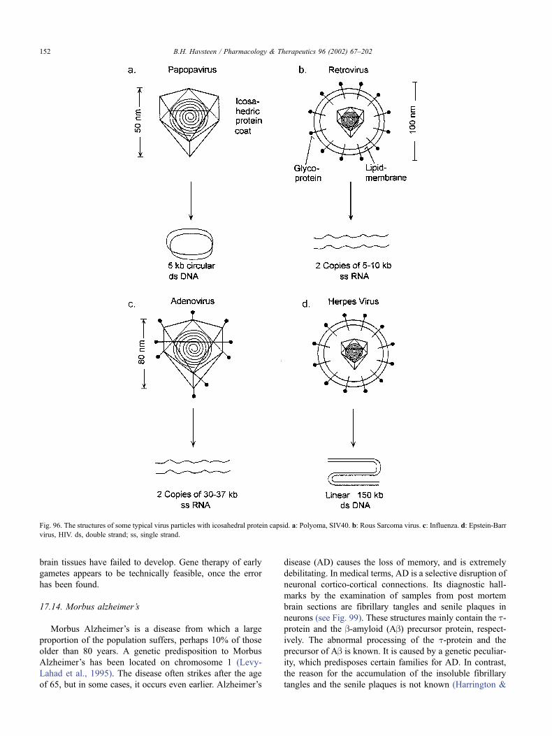
brain tissues have failed to develop. Gene therapy of early
gametes appears to be technically feasible, once the error
has been found.
17.14. Morbus alzheimer’s
Morbus Alzheimer’s is a disease from which a large
proportion of the population suffers, perhaps 10% of those
older than 80 years. A genetic predisposition to Morbus
Alzheimer’s has been located on chromosome 1 (Levy-
Lahad et al., 1995). The disease often strikes after the age
of 65, but in some cases, it occurs even earlier. Alzheimer’s
disease (AD) causes the loss of memory, and is extremely
debilitating. In medical terms, AD is a selective disruption of
neuronal cortico-cortical connections. Its diagnostic hall-
marks by the examination of samples from post mortem
brain sections are fibrillary tangles and senile plaques in
neurons (see Fig. 99). These structures mainly contain the t-protein and the b-amyloid (Ab) precursor protein, respect-ively. The abnormal processing of the t-protein and the
precursor of Ab is known. It is caused by a genetic peculiar-
ity, which predisposes certain families for AD. In contrast,
the reason for the accumulation of the insoluble fibrillary
tangles and the senile plaques is not known (Harrington &
Fig. 96. The structures of some typical virus particles with icosahedral protein capsid. a: Polyoma, SIV40. b: Rous Sarcoma virus. c: Influenza. d: Epstein-Barr
virus, HIV. ds, double strand; ss, single strand.
B.H. Havsteen / Pharmacology & Therapeutics 96 (2002) 67–202152
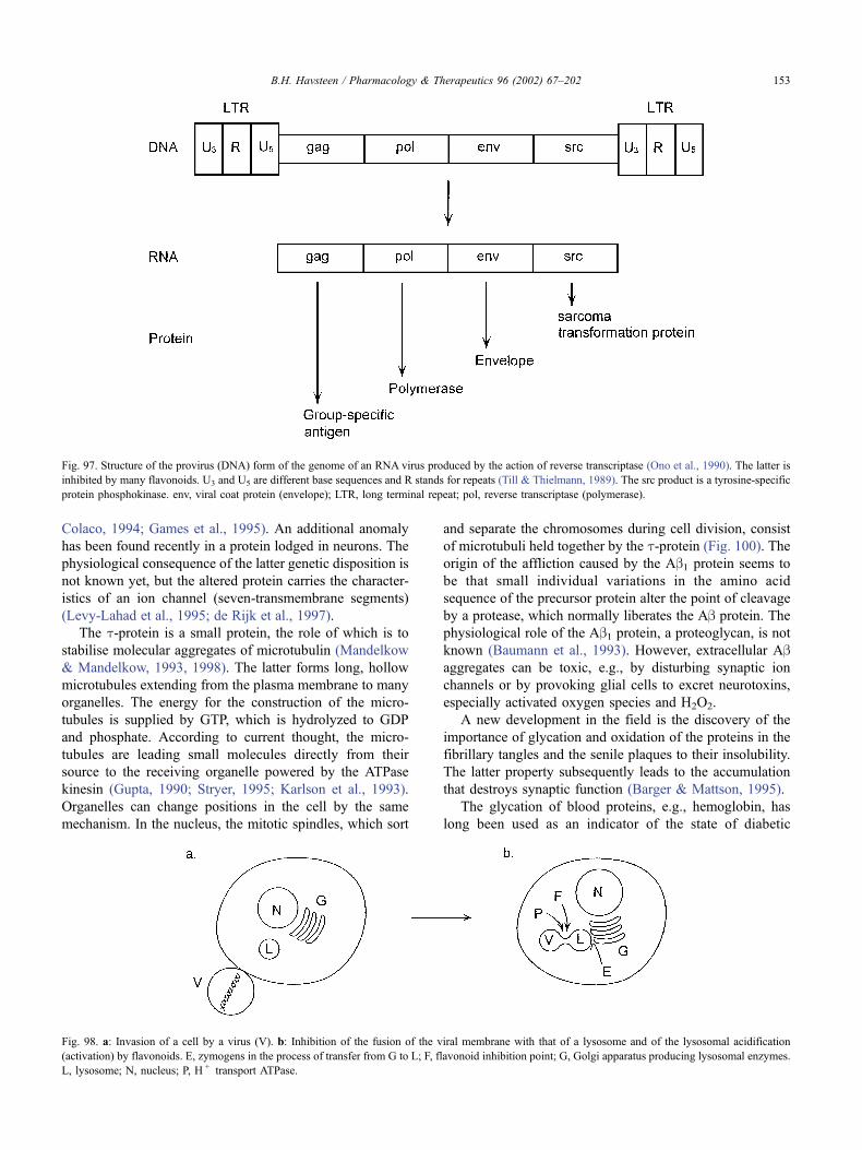
Colaco, 1994; Games et al., 1995). An additional anomaly
has been found recently in a protein lodged in neurons. The
physiological consequence of the latter genetic disposition is
not known yet, but the altered protein carries the character-
istics of an ion channel (seven-transmembrane segments)
(Levy-Lahad et al., 1995; de Rijk et al., 1997).
The t-protein is a small protein, the role of which is to
stabilise molecular aggregates of microtubulin (Mandelkow
& Mandelkow, 1993, 1998). The latter forms long, hollow
microtubules extending from the plasma membrane to many
organelles. The energy for the construction of the micro-
tubules is supplied by GTP, which is hydrolyzed to GDP
and phosphate. According to current thought, the micro-
tubules are leading small molecules directly from their
source to the receiving organelle powered by the ATPase
kinesin (Gupta, 1990; Stryer, 1995; Karlson et al., 1993).
Organelles can change positions in the cell by the same
mechanism. In the nucleus, the mitotic spindles, which sort
and separate the chromosomes during cell division, consist
of microtubuli held together by the t-protein (Fig. 100). Theorigin of the affliction caused by the Ab1 protein seems to
be that small individual variations in the amino acid
sequence of the precursor protein alter the point of cleavage
by a protease, which normally liberates the Ab protein. The
physiological role of the Ab1 protein, a proteoglycan, is notknown (Baumann et al., 1993). However, extracellular Abaggregates can be toxic, e.g., by disturbing synaptic ion
channels or by provoking glial cells to excret neurotoxins,
especially activated oxygen species and H2O2.
A new development in the field is the discovery of the
importance of glycation and oxidation of the proteins in the
fibrillary tangles and the senile plaques to their insolubility.
The latter property subsequently leads to the accumulation
that destroys synaptic function (Barger & Mattson, 1995).
The glycation of blood proteins, e.g., hemoglobin, has
long been used as an indicator of the state of diabetic
Fig. 97. Structure of the provirus (DNA) form of the genome of an RNA virus produced by the action of reverse transcriptase (Ono et al., 1990). The latter is
inhibited by many flavonoids. U3 and U5 are different base sequences and R stands for repeats (Till & Thielmann, 1989). The src product is a tyrosine-specific
protein phosphokinase. env, viral coat protein (envelope); LTR, long terminal repeat; pol, reverse transcriptase (polymerase).
Fig. 98. a: Invasion of a cell by a virus (V). b: Inhibition of the fusion of the viral membrane with that of a lysosome and of the lysosomal acidification
(activation) by flavonoids. E, zymogens in the process of transfer from G to L; F, flavonoid inhibition point; G, Golgi apparatus producing lysosomal enzymes.
L, lysosome; N, nucleus; P, H + transport ATPase.
B.H. Havsteen / Pharmacology & Therapeutics 96 (2002) 67–202 153
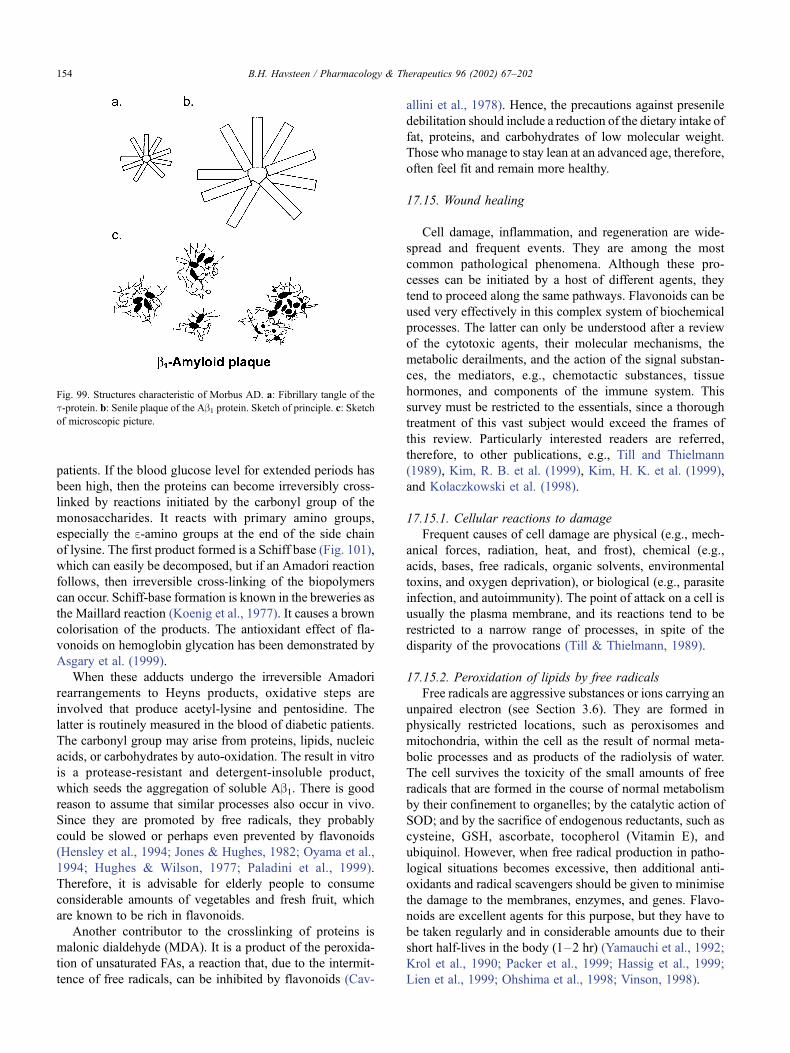
patients. If the blood glucose level for extended periods has
been high, then the proteins can become irreversibly cross-
linked by reactions initiated by the carbonyl group of the
monosaccharides. It reacts with primary amino groups,
especially the e-amino groups at the end of the side chain
of lysine. The first product formed is a Schiff base (Fig. 101),
which can easily be decomposed, but if an Amadori reaction
follows, then irreversible cross-linking of the biopolymers
can occur. Schiff-base formation is known in the breweries as
the Maillard reaction (Koenig et al., 1977). It causes a brown
colorisation of the products. The antioxidant effect of fla-
vonoids on hemoglobin glycation has been demonstrated by
Asgary et al. (1999).
When these adducts undergo the irreversible Amadori
rearrangements to Heyns products, oxidative steps are
involved that produce acetyl-lysine and pentosidine. The
latter is routinely measured in the blood of diabetic patients.
The carbonyl group may arise from proteins, lipids, nucleic
acids, or carbohydrates by auto-oxidation. The result in vitro
is a protease-resistant and detergent-insoluble product,
which seeds the aggregation of soluble Ab1. There is good
reason to assume that similar processes also occur in vivo.
Since they are promoted by free radicals, they probably
could be slowed or perhaps even prevented by flavonoids
(Hensley et al., 1994; Jones & Hughes, 1982; Oyama et al.,
1994; Hughes & Wilson, 1977; Paladini et al., 1999).
Therefore, it is advisable for elderly people to consume
considerable amounts of vegetables and fresh fruit, which
are known to be rich in flavonoids.
Another contributor to the crosslinking of proteins is
malonic dialdehyde (MDA). It is a product of the peroxida-
tion of unsaturated FAs, a reaction that, due to the intermit-
tence of free radicals, can be inhibited by flavonoids (Cav-
allini et al., 1978). Hence, the precautions against presenile
debilitation should include a reduction of the dietary intake of
fat, proteins, and carbohydrates of low molecular weight.
Those who manage to stay lean at an advanced age, therefore,
often feel fit and remain more healthy.
17.15. Wound healing
Cell damage, inflammation, and regeneration are wide-
spread and frequent events. They are among the most
common pathological phenomena. Although these pro-
cesses can be initiated by a host of different agents, they
tend to proceed along the same pathways. Flavonoids can be
used very effectively in this complex system of biochemical
processes. The latter can only be understood after a review
of the cytotoxic agents, their molecular mechanisms, the
metabolic derailments, and the action of the signal substan-
ces, the mediators, e.g., chemotactic substances, tissue
hormones, and components of the immune system. This
survey must be restricted to the essentials, since a thorough
treatment of this vast subject would exceed the frames of
this review. Particularly interested readers are referred,
therefore, to other publications, e.g., Till and Thielmann
(1989), Kim, R. B. et al. (1999), Kim, H. K. et al. (1999),
and Kolaczkowski et al. (1998).
17.15.1. Cellular reactions to damage
Frequent causes of cell damage are physical (e.g., mech-
anical forces, radiation, heat, and frost), chemical (e.g.,
acids, bases, free radicals, organic solvents, environmental
toxins, and oxygen deprivation), or biological (e.g., parasite
infection, and autoimmunity). The point of attack on a cell is
usually the plasma membrane, and its reactions tend to be
restricted to a narrow range of processes, in spite of the
disparity of the provocations (Till & Thielmann, 1989).
17.15.2. Peroxidation of lipids by free radicals
Free radicals are aggressive substances or ions carrying an
unpaired electron (see Section 3.6). They are formed in
physically restricted locations, such as peroxisomes and
mitochondria, within the cell as the result of normal meta-
bolic processes and as products of the radiolysis of water.
The cell survives the toxicity of the small amounts of free
radicals that are formed in the course of normal metabolism
by their confinement to organelles; by the catalytic action of
SOD; and by the sacrifice of endogenous reductants, such as
cysteine, GSH, ascorbate, tocopherol (Vitamin E), and
ubiquinol. However, when free radical production in patho-
logical situations becomes excessive, then additional anti-
oxidants and radical scavengers should be given to minimise
the damage to the membranes, enzymes, and genes. Flavo-
noids are excellent agents for this purpose, but they have to
be taken regularly and in considerable amounts due to their
short half-lives in the body (1–2 hr) (Yamauchi et al., 1992;
Krol et al., 1990; Packer et al., 1999; Hassig et al., 1999;
Lien et al., 1999; Ohshima et al., 1998; Vinson, 1998).
Fig. 99. Structures characteristic of Morbus AD. a: Fibrillary tangle of the
t-protein. b: Senile plaque of the Ab1 protein. Sketch of principle. c: Sketch
of microscopic picture.
B.H. Havsteen / Pharmacology & Therapeutics 96 (2002) 67–202154
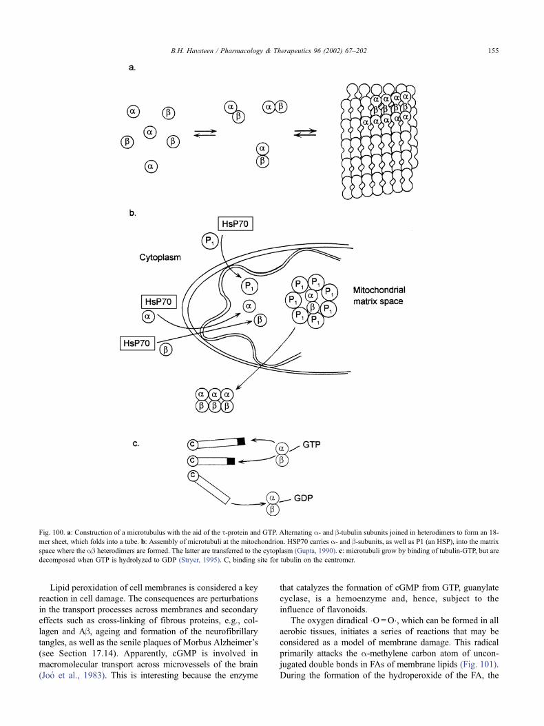
Lipid peroxidation of cell membranes is considered a key
reaction in cell damage. The consequences are perturbations
in the transport processes across membranes and secondary
effects such as cross-linking of fibrous proteins, e.g., col-
lagen and Ab, ageing and formation of the neurofibrillary
tangles, as well as the senile plaques of Morbus Alzheimer’s
(see Section 17.14). Apparently, cGMP is involved in
macromolecular transport across microvessels of the brain
(Joo et al., 1983). This is interesting because the enzyme
that catalyzes the formation of cGMP from GTP, guanylate
cyclase, is a hemoenzyme and, hence, subject to the
influence of flavonoids.
The oxygen diradical �O=O�, which can be formed in all
aerobic tissues, initiates a series of reactions that may be
considered as a model of membrane damage. This radical
primarily attacks the a-methylene carbon atom of uncon-
jugated double bonds in FAs of membrane lipids (Fig. 101).
During the formation of the hydroperoxide of the FA, the
Fig. 100. a: Construction of a microtubulus with the aid of the t-protein and GTP. Alternating a- and b-tubulin subunits joined in heterodimers to form an 18-
mer sheet, which folds into a tube. b: Assembly of microtubuli at the mitochondrion. HSP70 carries a- and b-subunits, as well as P1 (an HSP), into the matrix
space where the ab heterodimers are formed. The latter are transferred to the cytoplasm (Gupta, 1990). c: microtubuli grow by binding of tubulin-GTP, but are
decomposed when GTP is hydrolyzed to GDP (Stryer, 1995). C, binding site for tubulin on the centromer.
B.H. Havsteen / Pharmacology & Therapeutics 96 (2002) 67–202 155
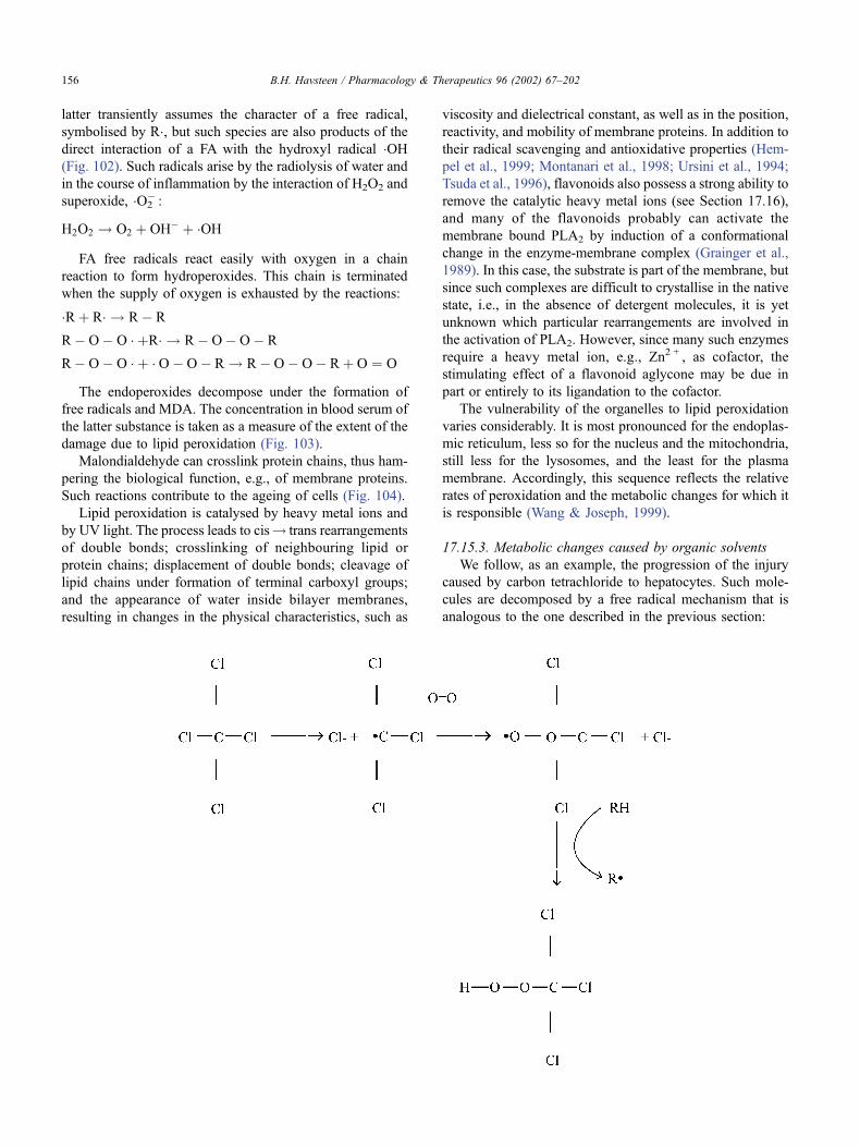
latter transiently assumes the character of a free radical,
symbolised by R�, but such species are also products of the
direct interaction of a FA with the hydroxyl radical �OH(Fig. 102). Such radicals arise by the radiolysis of water and
in the course of inflammation by the interaction of H2O2 and
superoxide, �O2� :
H2O2 ! O2 þ OH� þ �OH
FA free radicals react easily with oxygen in a chain
reaction to form hydroperoxides. This chain is terminated
when the supply of oxygen is exhausted by the reactions:
�Rþ R� ! R� R
R� O� O � þR� ! R� O� O� R
R� O� O � þ � O� O� R ! R� O� O� Rþ O ¼ O
The endoperoxides decompose under the formation of
free radicals and MDA. The concentration in blood serum of
the latter substance is taken as a measure of the extent of the
damage due to lipid peroxidation (Fig. 103).
Malondialdehyde can crosslink protein chains, thus ham-
pering the biological function, e.g., of membrane proteins.
Such reactions contribute to the ageing of cells (Fig. 104).
Lipid peroxidation is catalysed by heavy metal ions and
by UV light. The process leads to cis! trans rearrangements
of double bonds; crosslinking of neighbouring lipid or
protein chains; displacement of double bonds; cleavage of
lipid chains under formation of terminal carboxyl groups;
and the appearance of water inside bilayer membranes,
resulting in changes in the physical characteristics, such as
viscosity and dielectrical constant, as well as in the position,
reactivity, and mobility of membrane proteins. In addition to
their radical scavenging and antioxidative properties (Hem-
pel et al., 1999; Montanari et al., 1998; Ursini et al., 1994;
Tsuda et al., 1996), flavonoids also possess a strong ability to
remove the catalytic heavy metal ions (see Section 17.16),
and many of the flavonoids probably can activate the
membrane bound PLA2 by induction of a conformational
change in the enzyme-membrane complex (Grainger et al.,
1989). In this case, the substrate is part of the membrane, but
since such complexes are difficult to crystallise in the native
state, i.e., in the absence of detergent molecules, it is yet
unknown which particular rearrangements are involved in
the activation of PLA2. However, since many such enzymes
require a heavy metal ion, e.g., Zn2 + , as cofactor, the
stimulating effect of a flavonoid aglycone may be due in
part or entirely to its ligandation to the cofactor.
The vulnerability of the organelles to lipid peroxidation
varies considerably. It is most pronounced for the endoplas-
mic reticulum, less so for the nucleus and the mitochondria,
still less for the lysosomes, and the least for the plasma
membrane. Accordingly, this sequence reflects the relative
rates of peroxidation and the metabolic changes for which it
is responsible (Wang & Joseph, 1999).
17.15.3. Metabolic changes caused by organic solvents
We follow, as an example, the progression of the injury
caused by carbon tetrachloride to hepatocytes. Such mole-
cules are decomposed by a free radical mechanism that is
analogous to the one described in the previous section:
B.H. Havsteen / Pharmacology & Therapeutics 96 (2002) 67–202156
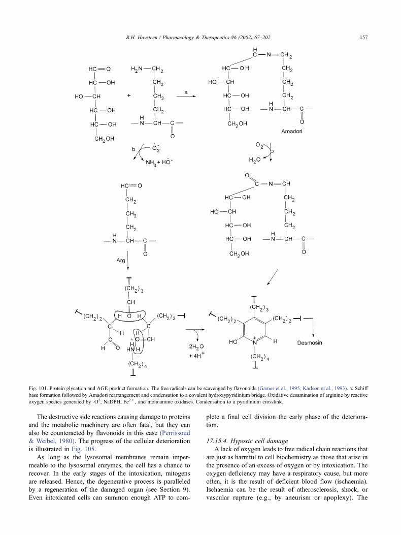
The destructive side reactions causing damage to proteins
and the metabolic machinery are often fatal, but they can
also be counteracted by flavonoids in this case (Perrissoud
& Weibel, 1980). The progress of the cellular deterioration
is illustrated in Fig. 105.
As long as the lysosomal membranes remain imper-
meable to the lysosomal enzymes, the cell has a chance to
recover. In the early stages of the intoxication, mitogens
are released. Hence, the degenerative process is paralleled
by a regeneration of the damaged organ (see Section 9).
Even intoxicated cells can summon enough ATP to com-
plete a final cell division the early phase of the deteriora-
tion.
17.15.4. Hypoxic cell damage
A lack of oxygen leads to free radical chain reactions that
are just as harmful to cell biochemistry as those that arise in
the presence of an excess of oxygen or by intoxication. The
oxygen deficiency may have a respiratory cause, but more
often, it is the result of deficient blood flow (ischaemia).
Ischaemia can be the result of atherosclerosis, shock, or
vascular rupture (e.g., by aneurism or apoplexy). The
Fig. 101. Protein glycation and AGE product formation. The free radicals can be scavenged by flavonoids (Games et al., 1995; Karlson et al., 1993). a: Schiff
base formation followed by Amadori rearrangement and condensation to a covalent hydroxypyridinium bridge. Oxidative desamination of arginine by reactive
oxygen species generated by �O2, NaDPH, Fe2 + , and monoamine oxidases. Condensation to a pyridinium crosslink.
B.H. Havsteen / Pharmacology & Therapeutics 96 (2002) 67–202 157
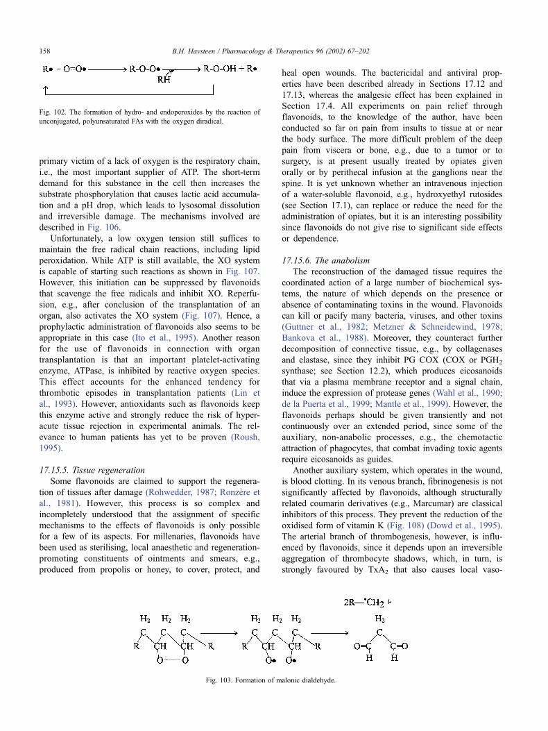
primary victim of a lack of oxygen is the respiratory chain,
i.e., the most important supplier of ATP. The short-term
demand for this substance in the cell then increases the
substrate phosphorylation that causes lactic acid accumula-
tion and a pH drop, which leads to lysosomal dissolution
and irreversible damage. The mechanisms involved are
described in Fig. 106.
Unfortunately, a low oxygen tension still suffices to
maintain the free radical chain reactions, including lipid
peroxidation. While ATP is still available, the XO system
is capable of starting such reactions as shown in Fig. 107.
However, this initiation can be suppressed by flavonoids
that scavenge the free radicals and inhibit XO. Reperfu-
sion, e.g., after conclusion of the transplantation of an
organ, also activates the XO system (Fig. 107). Hence, a
prophylactic administration of flavonoids also seems to be
appropriate in this case (Ito et al., 1995). Another reason
for the use of flavonoids in connection with organ
transplantation is that an important platelet-activating
enzyme, ATPase, is inhibited by reactive oxygen species.
This effect accounts for the enhanced tendency for
thrombotic episodes in transplantation patients (Lin et
al., 1993). However, antioxidants such as flavonoids keep
this enzyme active and strongly reduce the risk of hyper-
acute tissue rejection in experimental animals. The rel-
evance to human patients has yet to be proven (Roush,
1995).
17.15.5. Tissue regeneration
Some flavonoids are claimed to support the regenera-
tion of tissues after damage (Rohwedder, 1987; Ronzere et
al., 1981). However, this process is so complex and
incompletely understood that the assignment of specific
mechanisms to the effects of flavonoids is only possible
for a few of its aspects. For millenaries, flavonoids have
been used as sterilising, local anaesthetic and regeneration-
promoting constituents of ointments and smears, e.g.,
produced from propolis or honey, to cover, protect, and
heal open wounds. The bactericidal and antiviral prop-
erties have been described already in Sections 17.12 and
17.13, whereas the analgesic effect has been explained in
Section 17.4. All experiments on pain relief through
flavonoids, to the knowledge of the author, have been
conducted so far on pain from insults to tissue at or near
the body surface. The more difficult problem of the deep
pain from viscera or bone, e.g., due to a tumor or to
surgery, is at present usually treated by opiates given
orally or by perithecal infusion at the ganglions near the
spine. It is yet unknown whether an intravenous injection
of a water-soluble flavonoid, e.g., hydroxyethyl rutosides
(see Section 17.1), can replace or reduce the need for the
administration of opiates, but it is an interesting possibility
since flavonoids do not give rise to significant side effects
or dependence.
17.15.6. The anabolism
The reconstruction of the damaged tissue requires the
coordinated action of a large number of biochemical sys-
tems, the nature of which depends on the presence or
absence of contaminating toxins in the wound. Flavonoids
can kill or pacify many bacteria, viruses, and other toxins
(Guttner et al., 1982; Metzner & Schneidewind, 1978;
Bankova et al., 1988). Moreover, they counteract further
decomposition of connective tissue, e.g., by collagenases
and elastase, since they inhibit PG COX (COX or PGH2
synthase; see Section 12.2), which produces eicosanoids
that via a plasma membrane receptor and a signal chain,
induce the expression of protease genes (Wahl et al., 1990;
de la Puerta et al., 1999; Mantle et al., 1999). However, the
flavonoids perhaps should be given transiently and not
continuously over an extended period, since some of the
auxiliary, non-anabolic processes, e.g., the chemotactic
attraction of phagocytes, that combat invading toxic agents
require eicosanoids as guides.
Another auxiliary system, which operates in the wound,
is blood clotting. In its venous branch, fibrinogenesis is not
significantly affected by flavonoids, although structurally
related coumarin derivatives (e.g., Marcumar) are classical
inhibitors of this process. They prevent the reduction of the
oxidised form of vitamin K (Fig. 108) (Dowd et al., 1995).
The arterial branch of thrombogenesis, however, is influ-
enced by flavonoids, since it depends upon an irreversible
aggregation of thrombocyte shadows, which, in turn, is
strongly favoured by TxA2 that also causes local vaso-
Fig. 102. The formation of hydro- and endoperoxides by the reaction of
unconjugated, polyunsaturated FAs with the oxygen diradical.
Fig. 103. Formation of malonic dialdehyde.
B.H. Havsteen / Pharmacology & Therapeutics 96 (2002) 67–202158
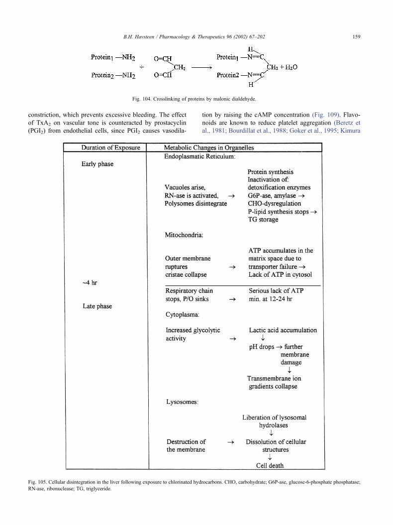
constriction, which prevents excessive bleeding. The effect
of TxA2 on vascular tone is counteracted by prostacyclin
(PGI2) from endothelial cells, since PGI2 causes vasodila-
tion by raising the cAMP concentration (Fig. 109). Flavo-
noids are known to reduce platelet aggregation (Beretz et
al., 1981; Bourdillat et al., 1988; Goker et al., 1995; Kimura
Fig. 104. Crosslinking of proteins by malonic dialdehyde.
Fig. 105. Cellular disintegration in the liver following exposure to chlorinated hydrocarbons. CHO, carbohydrate; G6P-ase, glucose-6-phosphate phosphatase;
RN-ase, ribonuclease; TG, triglyceride.
B.H. Havsteen / Pharmacology & Therapeutics 96 (2002) 67–202 159
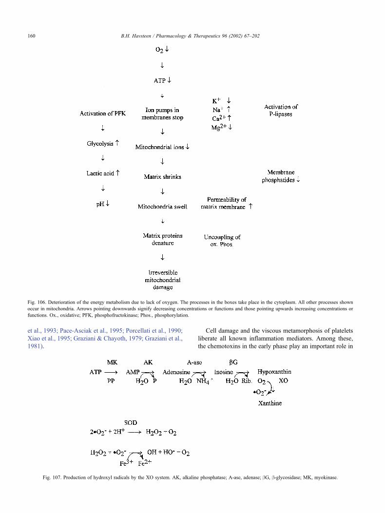
et al., 1993; Pace-Asciak et al., 1995; Porcellati et al., 1990;
Xiao et al., 1995; Graziani & Chayoth, 1979; Graziani et al.,
1981).
Cell damage and the viscous metamorphosis of platelets
liberate all known inflammation mediators. Among these,
the chemotoxins in the early phase play an important role in
Fig. 106. Deterioration of the energy metabolism due to lack of oxygen. The processes in the boxes take place in the cytoplasm. All other processes shown
occur in mitochondria. Arrows pointing downwards signify decreasing concentrations or functions and those pointing upwards increasing concentrations or
functions. Ox., oxidative; PFK, phosphofructokinase; Phos., phosphorylation.
Fig. 107. Production of hydroxyl radicals by the XO system. AK, alkaline phosphatase; A-ase, adenase; bG, b-glycosidase; MK, myokinase.
B.H. Havsteen / Pharmacology & Therapeutics 96 (2002) 67–202160
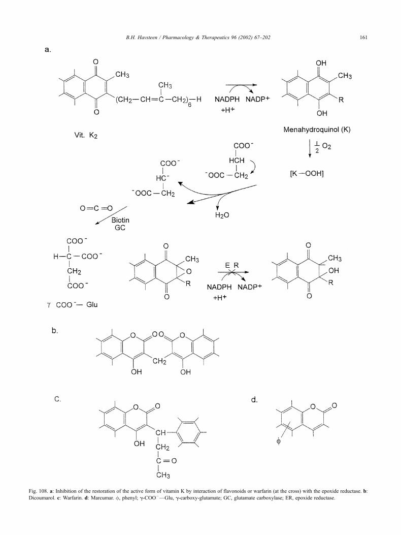
Fig. 108. a: Inhibition of the restoration of the active form of vitamin K by interaction of flavonoids or warfarin (at the cross) with the epoxide reductase. b:
Dicoumarol. c: Warfarin. d: Marcumar. f, phenyl; g-COO–—Glu, g-carboxy-glutamate; GC, glutamate carboxylase; ER, epoxide reductase.
B.H. Havsteen / Pharmacology & Therapeutics 96 (2002) 67–202 161
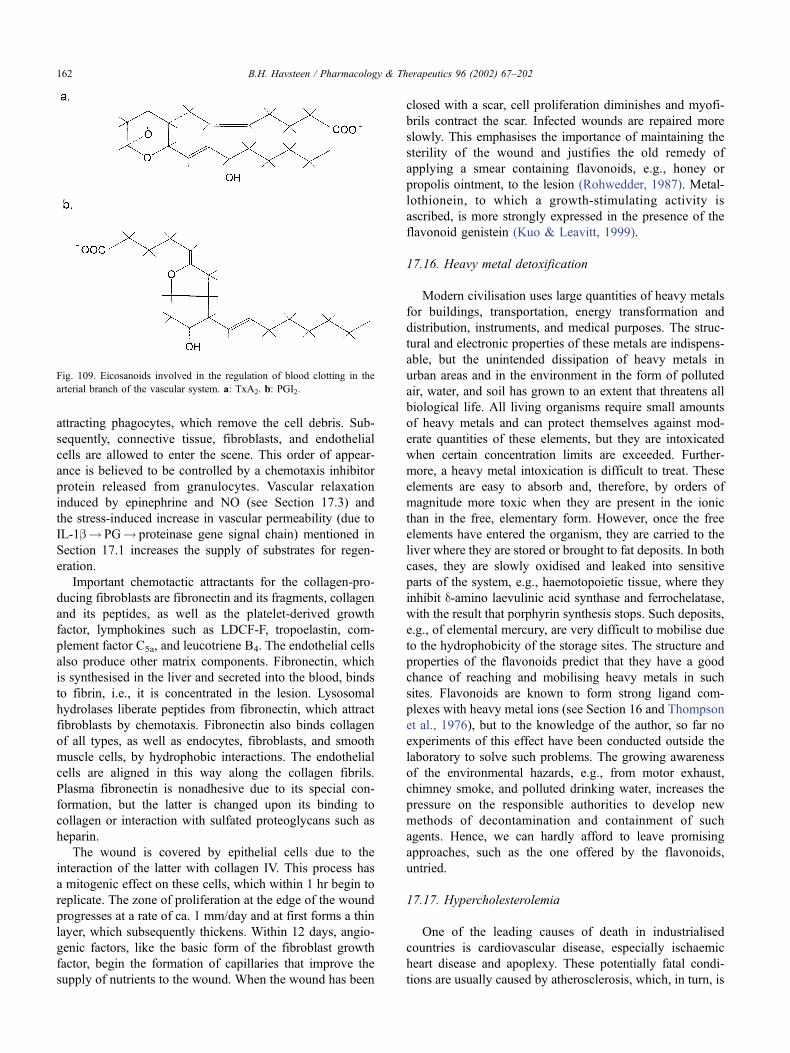
attracting phagocytes, which remove the cell debris. Sub-
sequently, connective tissue, fibroblasts, and endothelial
cells are allowed to enter the scene. This order of appear-
ance is believed to be controlled by a chemotaxis inhibitor
protein released from granulocytes. Vascular relaxation
induced by epinephrine and NO (see Section 17.3) and
the stress-induced increase in vascular permeability (due to
IL-1b! PG! proteinase gene signal chain) mentioned in
Section 17.1 increases the supply of substrates for regen-
eration.
Important chemotactic attractants for the collagen-pro-
ducing fibroblasts are fibronectin and its fragments, collagen
and its peptides, as well as the platelet-derived growth
factor, lymphokines such as LDCF-F, tropoelastin, com-
plement factor C5a, and leucotriene B4. The endothelial cells
also produce other matrix components. Fibronectin, which
is synthesised in the liver and secreted into the blood, binds
to fibrin, i.e., it is concentrated in the lesion. Lysosomal
hydrolases liberate peptides from fibronectin, which attract
fibroblasts by chemotaxis. Fibronectin also binds collagen
of all types, as well as endocytes, fibroblasts, and smooth
muscle cells, by hydrophobic interactions. The endothelial
cells are aligned in this way along the collagen fibrils.
Plasma fibronectin is nonadhesive due to its special con-
formation, but the latter is changed upon its binding to
collagen or interaction with sulfated proteoglycans such as
heparin.
The wound is covered by epithelial cells due to the
interaction of the latter with collagen IV. This process has
a mitogenic effect on these cells, which within 1 hr begin to
replicate. The zone of proliferation at the edge of the wound
progresses at a rate of ca. 1 mm/day and at first forms a thin
layer, which subsequently thickens. Within 12 days, angio-
genic factors, like the basic form of the fibroblast growth
factor, begin the formation of capillaries that improve the
supply of nutrients to the wound. When the wound has been
closed with a scar, cell proliferation diminishes and myofi-
brils contract the scar. Infected wounds are repaired more
slowly. This emphasises the importance of maintaining the
sterility of the wound and justifies the old remedy of
applying a smear containing flavonoids, e.g., honey or
propolis ointment, to the lesion (Rohwedder, 1987). Metal-
lothionein, to which a growth-stimulating activity is
ascribed, is more strongly expressed in the presence of the
flavonoid genistein (Kuo & Leavitt, 1999).
17.16. Heavy metal detoxification
Modern civilisation uses large quantities of heavy metals
for buildings, transportation, energy transformation and
distribution, instruments, and medical purposes. The struc-
tural and electronic properties of these metals are indispens-
able, but the unintended dissipation of heavy metals in
urban areas and in the environment in the form of polluted
air, water, and soil has grown to an extent that threatens all
biological life. All living organisms require small amounts
of heavy metals and can protect themselves against mod-
erate quantities of these elements, but they are intoxicated
when certain concentration limits are exceeded. Further-
more, a heavy metal intoxication is difficult to treat. These
elements are easy to absorb and, therefore, by orders of
magnitude more toxic when they are present in the ionic
than in the free, elementary form. However, once the free
elements have entered the organism, they are carried to the
liver where they are stored or brought to fat deposits. In both
cases, they are slowly oxidised and leaked into sensitive
parts of the system, e.g., haemotopoietic tissue, where they
inhibit d-amino laevulinic acid synthase and ferrochelatase,
with the result that porphyrin synthesis stops. Such deposits,
e.g., of elemental mercury, are very difficult to mobilise due
to the hydrophobicity of the storage sites. The structure and
properties of the flavonoids predict that they have a good
chance of reaching and mobilising heavy metals in such
sites. Flavonoids are known to form strong ligand com-
plexes with heavy metal ions (see Section 16 and Thompson
et al., 1976), but to the knowledge of the author, so far no
experiments of this effect have been conducted outside the
laboratory to solve such problems. The growing awareness
of the environmental hazards, e.g., from motor exhaust,
chimney smoke, and polluted drinking water, increases the
pressure on the responsible authorities to develop new
methods of decontamination and containment of such
agents. Hence, we can hardly afford to leave promising
approaches, such as the one offered by the flavonoids,
untried.
17.17. Hypercholesterolemia
One of the leading causes of death in industrialised
countries is cardiovascular disease, especially ischaemic
heart disease and apoplexy. These potentially fatal condi-
tions are usually caused by atherosclerosis, which, in turn, is
Fig. 109. Eicosanoids involved in the regulation of blood clotting in the
arterial branch of the vascular system. a: TxA2. b: PGI2.
B.H. Havsteen / Pharmacology & Therapeutics 96 (2002) 67–202162
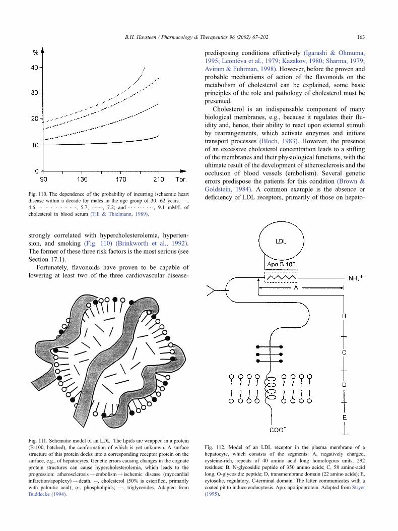
strongly correlated with hypercholesterolemia, hyperten-
sion, and smoking (Fig. 110) (Brinkworth et al., 1992).
The former of these three risk factors is the most serious (see
Section 17.1).
Fortunately, flavonoids have proven to be capable of
lowering at least two of the three cardiovascular disease-
predisposing conditions effectively (Igarashi & Ohmuma,
1995; Leonteva et al., 1979; Kazakov, 1980; Sharma, 1979;
Aviram & Fuhrman, 1998). However, before the proven and
probable mechanisms of action of the flavonoids on the
metabolism of cholesterol can be explained, some basic
principles of the role and pathology of cholesterol must be
presented.
Cholesterol is an indispensable component of many
biological membranes, e.g., because it regulates their flu-
idity and, hence, their ability to react upon external stimuli
by rearrangements, which activate enzymes and initiate
transport processes (Bloch, 1983). However, the presence
of an excessive cholesterol concentration leads to a stifling
of the membranes and their physiological functions, with the
ultimate result of the development of atherosclerosis and the
occlusion of blood vessels (embolism). Several genetic
errors predispose the patients for this condition (Brown &
Goldstein, 1984). A common example is the absence or
deficiency of LDL receptors, primarily of those on hepato-Fig. 110. The dependence of the probability of incurring ischaemic heart
disease within a decade for males in the age group of 30–62 years. —,
4.6; – - - - - - - -, 5.7; –�–�–, 7.2; and : : : : : : : : :, 9.1 mM/L of
cholesterol in blood serum (Till & Thielmann, 1989).
Fig. 111. Schematic model of an LDL. The lipids are wrapped in a protein
(B-100, hatched), the conformation of which is yet unknown. A surface
structure of this protein docks into a corresponding receptor protein on the
surface, e.g., of hepatocytes. Genetic errors causing changes in the cognate
protein structures can cause hypercholesterolemia, which leads to the
progression: atherosclerosis! embolism! ischemic disease (myocardial
infarction/apoplexy)! death. �–, cholesterol (50% is esterified, primarily
with palmitic acid); o-, phospholipids; —, triglycerides. Adapted from
Buddecke (1994).
Fig. 112. Model of an LDL receptor in the plasma membrane of a
hepatocyte, which consists of the segments: A, negatively charged,
cysteine-rich, repeats of 40 amino acid long homologous units, 292
residues; B, N-glycosidic peptide of 350 amino acids; C, 58 amino-acid
long, O-glycosidic peptide; D, transmembrane domain (22 amino acids); E,
cytosolic, regulatory, C-terminal domain. The latter communicates with a
coated pit to induce endocytosis. Apo, apolipoprotein. Adapted from Stryer
(1995).
B.H. Havsteen / Pharmacology & Therapeutics 96 (2002) 67–202 163
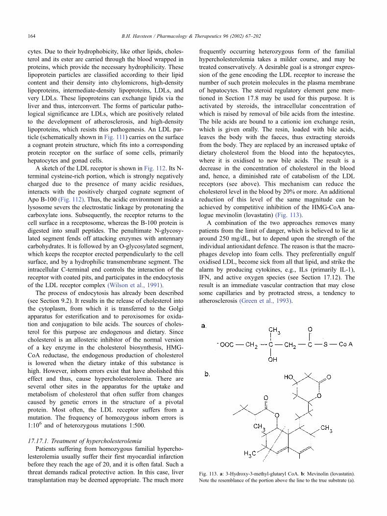
cytes. Due to their hydrophobicity, like other lipids, choles-
terol and its ester are carried through the blood wrapped in
proteins, which provide the necessary hydrophilicity. These
lipoprotein particles are classified according to their lipid
content and their density into chylomicrons, high-density
lipoproteins, intermediate-density lipoproteins, LDLs, and
very LDLs. These lipoproteins can exchange lipids via the
liver and thus, interconvert. The forms of particular patho-
logical significance are LDLs, which are positively related
to the development of atherosclerosis, and high-density
lipoproteins, which resists this pathogenesis. An LDL par-
ticle (schematically shown in Fig. 111) carries on the surface
a cognant protein structure, which fits into a corresponding
protein receptor on the surface of some cells, primarily
hepatocytes and gonad cells.
A sketch of the LDL receptor is shown in Fig. 112. Its N-
terminal cysteine-rich portion, which is strongly negatively
charged due to the presence of many acidic residues,
interacts with the positively charged cognate segment of
Apo B-100 (Fig. 112). Thus, the acidic environment inside a
lysosome severs the electrostatic linkage by protonating the
carboxylate ions. Subsequently, the receptor returns to the
cell surface in a receptosome, whereas the B-100 protein is
digested into small peptides. The penultimate N-glycosy-
lated segment fends off attacking enzymes with antennary
carbohydrates. It is followed by an O-glycosylated segment,
which keeps the receptor erected perpendicularly to the cell
surface, and by a hydrophilic transmembrane segment. The
intracellular C-terminal end controls the interaction of the
receptor with coated pits, and participates in the endocytosis
of the LDL receptor complex (Wilson et al., 1991).
The process of endocytosis has already been described
(see Section 9.2). It results in the release of cholesterol into
the cytoplasm, from which it is transferred to the Golgi
apparatus for esterification and to peroxisomes for oxida-
tion and conjugation to bile acids. The sources of choles-
terol for this purpose are endogenous and dietary. Since
cholesterol is an allosteric inhibitor of the normal version
of a key enzyme in the cholesterol biosynthesis, HMG-
CoA reductase, the endogenous production of cholesterol
is lowered when the dietary intake of this substance is
high. However, inborn errors exist that have abolished this
effect and thus, cause hypercholesterolemia. There are
several other sites in the apparatus for the uptake and
metabolism of cholesterol that often suffer from changes
caused by genetic errors in the structure of a pivotal
protein. Most often, the LDL receptor suffers from a
mutation. The frequency of homozygous inborn errors is
1:106 and of heterozygous mutations 1:500.
17.17.1. Treatment of hypercholesterolemia
Patients suffering from homozygous familial hypercho-
lesterolemia usually suffer their first myocardial infarction
before they reach the age of 20, and it is often fatal. Such a
threat demands radical protective action. In this case, liver
transplantation may be deemed appropriate. The much more
frequently occurring heterozygous form of the familial
hypercholesterolemia takes a milder course, and may be
treated conservatively. A desirable goal is a stronger expres-
sion of the gene encoding the LDL receptor to increase the
number of such protein molecules in the plasma membrane
of hepatocytes. The steroid regulatory element gene men-
tioned in Section 17.8 may be used for this purpose. It is
activated by steroids, the intracellular concentration of
which is raised by removal of bile acids from the intestine.
The bile acids are bound to a cationic ion exchange resin,
which is given orally. The resin, loaded with bile acids,
leaves the body with the faeces, thus extracting steroids
from the body. They are replaced by an increased uptake of
dietary cholesterol from the blood into the hepatocytes,
where it is oxidised to new bile acids. The result is a
decrease in the concentration of cholesterol in the blood
and, hence, a diminished rate of catabolism of the LDL
receptors (see above). This mechanism can reduce the
cholesterol level in the blood by 20% or more. An additional
reduction of this level of the same magnitude can be
achieved by competitive inhibition of the HMG-CoA ana-
logue mevinolin (lovastatin) (Fig. 113).
A combination of the two approaches removes many
patients from the limit of danger, which is believed to lie at
around 250 mg/dL, but to depend upon the strength of the
individual antioxidant defence. The reason is that the macro-
phages develop into foam cells. They preferentially engulf
oxidised LDL, become sick from all that lipid, and strike the
alarm by producing cytokines, e.g., ILs (primarily IL-1),
IFN, and active oxygen species (see Section 17.12). The
result is an immediate vascular contraction that may close
some capillaries and by protracted stress, a tendency to
atherosclerosis (Green et al., 1993).
Fig. 113. a: 3-Hydroxy-3-methyl-glutaryl CoA. b: Mevinolin (lovastatin).
Note the resemblance of the portion above the line to the true substrate (a).
B.H. Havsteen / Pharmacology & Therapeutics 96 (2002) 67–202164
17.17.2. Sites of flavonoid action in cholesterol metabolism
The activity of the key enzyme of cholesterol biosyn-
thesis, HMG-CoA reductase, is regulated by phosphoryla-
tion/dephosphorylation reactions. It is inhibited by the
action of a cAMP-dependent protein phosphokinase, which
phosphorylates serine or threonine residues using ATP as
substrate (Stryer, 1995). However, since the coenzyme
cAMP is cleaved by the enzyme cAMP PDE, which is
inhibited by flavonoids (Ferrell et al., 1980; Stein et al.,
1999), the consequence is that the cAMP concentration
increases and that phosphorylation of the HMG-CoA reduc-
tase is enhanced, but endogenous cholesterol production is
diminished. In addition, the flavonoids can interact with the
enzyme protein phosphatase, which liberates the aliphatic
phosphoesters from HMG-CoA reductase, thus restoring the
activity of this enzyme. The reason is that all known
phosphatases are metalloenzymes, which carry a divalent
heavy metal ion cofactor (Zn2 + ) in their active site, where it
is indispensable for catalytic action (Yamanaka et al., 1997).
However, heavy metal ions form complexes with suitable
ligands, and happen to have a strong affinity for flavonoids.
The result is that the metal ion changes its position in its
site. In this case, the rearrangement has a negative effect on
catalytic activity (Hiermann & Kartnig, 1978). Thus, flavo-
noids inhibit HMG-CoA reductase by a dual mechanism.
However, flavonoids probably also exert their influence on
steroid metabolism at other pivotal points. The hydropho-
bicity of their aglycone suggests that they are carried into
hepatocytes by lipoproteins, released from the lysosomes
into the cytosol, and bound to the cytoplasmic steroid
receptor, which carries them into the cell nucleus. There,
the receptor part of the complex is likely to interact with the
steroid regulatory element or the flavonoid may intercalate
itself between the bases of this DNA segment. Any of these
mechanisms may explain the known oestrogenic effect of
some flavonoids, e.g., silymarin (Sonnenbichler et al., 1980;
Mikziek, 1993; Mitcher et al., 1982; Sani et al., 1993;
Cavallini et al., 1978; Plump et al., 1992), and the frequently
observed lowering of the blood cholesterol level after the
regular intake of the flavonoids (Igarashi & Ohmuma, 1995;
Fremont et al., 1999).
The pregnancy-preventative activity of some flavonoids
(Santti et al., 1998; Sani et al., 1993) found by mice opens
the interesting possibility of an application in humans. Since
the toxicity of most flavonoids is much lower than that of,
e.g., morning-after pills, the prospect of developing a safe
drug for women who have second thoughts the day after
coitus seems promising (Grotz & Gunther, 1971).
17.18. Stimulation of the immune system by flavonoids
The immunodeficiency diseases dramatically illustrate
the immense importance of the immune system for the
protection of our health, especially against infection. The
inborn error, which inactivates the enzyme adenine deam-
inase, causes a generalised inefficiency of the cellular
immune system in a way that is yet poorly understood.
Infants of such heritage are very susceptible to infections,
and experience a ceaseless series of influenza, pneumonia,
etc., until they wear out and die within a few years, unless
they are confined to a sterile tent or receive a complete
adenine deaminase gene by transplantation. In fact, this
operation was the first example of a successful therapeutic
gene transplantation. Since the bureaucratic and technical
effort required to carry out such an operation is still high,
even in the United States, it is useful to bear in mind that
flavonoids kill many infectants or bring about their destruc-
tion by activating endogenous defence systems that may still
be available (see Section 17.2). In the case of HIV infec-
tions, we have to deal with a resourceful virus, which so far
has resisted most attempts on its life that could be tolerated
by the patient. Theoretically, flavonoids should have an
excellent chance of preventing the proliferation of HIV
since they inhibit reverse transcriptase, induce IFNs, and
inactivate the enzyme that prepares the precursor of the
protein building blocks of the viral capsid (Brinkworth et
al., 1992). However, to the knowledge of the author, so far
no one has attempted to eliminate HIV in this way, neither in
vitro nor in vivo.
A clear reciprocal relationship exists between the prolif-
eration of HIV and the decline of T4-lymphocytes [see
Section 17.13, as well as Nowak & McMichael (1995)].
Apparently, the HIV is preferentially engulfed by follicular
dendritic cells, which are members of the macrophage
family (Heath et al., 1995) residing in the spleen and the
lymph nodes. These cells provoke, by mechanisms such as
those outlined in Section 9, a hyperactivity of the immune
system, which renders it more susceptible to the virus. Over
a period of � 10 years of incessant battle, the HIV patient is
vulnerable to all kinds of opportunistic infections, which,
within a relatively short time, leads to death. Some of these
infectants are likely to be sensitive to flavonoids (Ching &
Baguley, 1987; Hornung et al., 1988; Wiltrout et al., 1988;
Dimov et al., 1991; Wang et al., 1998). It seems that
flavonoids, which are much less harmful to patients than
the antiviral drugs in current use, could improve the quality
of life of those suffering from AIDS. They may even
prolong the lives of such patients, but so far, no one has
examined the effect of flavonoids on AIDS. In the case of a
disease of similar gravity, cancer, flavone acetic acid has
been applied with significant result, at least in vitro (Parkins
et al., 1993; Ching et al., 1994; Deschner et al., 1993;
Evelhoch et al., 1988). As mentioned in Section 17.8, the
biological effect of the synthetic compound flavone acetic
acid may differ from that of natural flavonoids, e.g.,
quercetin.
The stimulating effect of flavonoids can be rationalised
as follows: PGs suppress T-lymphocytes by inhibiting TH-
cells or activating TS-cells. (Goodwin & Webb, 1980; Webb
& Jamieson, 1976; Barasoain et al., 1980). The flavonoids
inhibit the PG COX, thus eliminating the suppression of the
immune response.
B.H. Havsteen / Pharmacology & Therapeutics 96 (2002) 67–202 165
17.19. The potential of flavonoids in the acquired
immunodeficiency syndrome prophylaxis and therapy
17.19.1. Introduction
AIDS is one of the recent epidemic diseases that has been
feared the most, because it is insidious, lethal, and incurable.
Since the first case of this disease was not recognised until
the 1960s, the principal users of flavonoids, the lay medical
practitioners, had no experience with AIDS. They also
lacked the insight into the nature of this disease that could
justify an experimental treatment of AIDS with flavonoids.
Hence, the indications of the possible usefulness of flavo-
noids as preventive, or even therapeutic, agents came from
basic medical research, especially on viral infection and
proliferation. In the 1970s, Montainer and Gallo isolated
and identified a virus, which at first was named human T-
lymphocyte-associated virus (HTLV)-III and later renamed
HIV (Gallo & Montagnier, 1988). It belongs to the family of
lente virus, which also includes herpes and hepatitis viruses.
The members of this virus family propagate slowly. There-
fore, the infection is difficult to recognize in its early stage.
In the case of the prevailing strain that causes AIDS, HIV-I,
the virus gains time to hide in a class of T-lymphocytes,
which primarily are located in lymph nodes and the spleen.
The target cells of HIV-I particles are TH-lymphocytes. The
latter are characterised by the presence in the plasma
membrane of a K + -ion channel named CD4, through which
the virus particles enter the cell. This entry is facilitated by a
neighbouring lymphokine receptor.
Since HIV particles reach their target cells within sec-
onds after entry into the blood, immune surveillance hardly
has a chance to take notice and to respond to the threat until
much later when the host cells die and liberate scores of new
virus particles. Hence, the disease usually has progressed far
before anti-HIV antibodies can be detected in the blood. If
the infection is left untreated, the reservoir of TH-lympho-
cytes in the body will become depleted over a period that
depends on the efficiency of the individual immune defence,
but which usually lasts � 10 years. Then, the disease enters
the active AIDS phase, in which opportunistic infections of
many kinds, including bacteria, viruses, and carcinogens,
spread unopposed by the destroyed immune system. Even-
tually, the patients die in misery from a coagulopathy
induced by one of the secondary infectants.
AIDS has been one of the greatest challenges to modern
medicine, and an unprecedented investment has beenmade in
research into this disease in the past 30 years. Yet, despite all
the efforts, AIDS remains incurable, although considerable
advances have been made in the suppression of the viral load
and, hence, in the quality of life of the patients. Besides, much
new knowledge has been gained on the structure of viruses,
on viral pathogenesis, and on prophylaxis.
Since HIV is a retrovirus, most attention initially was
given to the development of specific inhibitors of the ret-
rotranscriptase that converts the viral RNA genome to the
corresponding DNA form, which can be incorporated into
human chromosomes. Unfortunately, this process resembles
vital cellular processes to such an extent that the effective
inhibitors of this enzyme also were very toxic. Hence, they
could only be given in low doses, and uninfected cells were
simultaneously severely damaged. Interestingly, some of the
flavonoids also specifically inhibited the retrotranscriptase
in vitro, and they did so without undesirable side effects in
cell culture. Since experimental animal AIDS models are
rare and expensive, no intact HIV-infected animals have
been treated with flavonoids so far.
Another promising point of attack on the life cycle of
HIV is the preparation of the precursor of the virus capsid
protein. This precursor protein must be freed of a terminal
peptide by proteolytic cleavage before it can fold into a
conformation that aggregates to a sphere enclosing the viral
genome. If this process fails, the viral genes are exposed to
endonucleases in the cytoplasm, which soon will destroy
them. Specific inhibitors of this viral protease have been
developed and used with some success, especially when
they were used together with retrotranscriptase inhibitors.
Flavonoids capable of inhibiting the viral protease without
deleterious effects have been found, but they have not been
tested on intact HIV-infected organisms for the same reason
as the one mentioned above.
Since flavonoids have been applied successfully in the
treatment of a variety of diseases, including such which are
components of AIDS, the question has been raised several
times whether patients suffering from AIDS could also
benefit from these compounds, the toxicity of which is
much less than that of common antiviral drugs. Previously,
the answer to that question has been hesitant for a number of
reasons. First, AIDS patients are often emotionally very
sensitive due to the gravity of their illness, the very poor
prognosis, and the low quality of life during the unpleasant
and expensive, yet ineffective treatment. Second, this dis-
ease is often contracted by persons of the lower social strata
and by persons suffering from psychiatric diseases that
tempt them to seek any pleasure, which is still available,
including narcotics, alcohol, tobacco, and sex. Third, the
public and, not the least, the AIDS patients themselves are
skeptical toward claims of the benefits of simple, inexpen-
sive, and mild drugs after the failure of the intensive search
extending over more than 10 years for antibodies capable of
eliminating the HIV that is causing AIDS. Despite the lack
of therapeutic methods capable of eradicating the disease,
significant advances were gained with combinations of
drugs, which reached different targets on the virus and the
enzymes it encodes simultaneously. Thus, the viral titer
could be suppressed for a period of � 10 years and the
quality of life improved considerably during this therapy.
Since flavonoids might contribute to such a development,
e.g., by elimination of some of the opportunistic infections,
which often become fatal, when the immune defence finally
is destroyed by the virus, a brief discussion of the potential
benefits of flavonoids to AIDS patients seems to be appro-
priate.
B.H. Havsteen / Pharmacology & Therapeutics 96 (2002) 67–202166
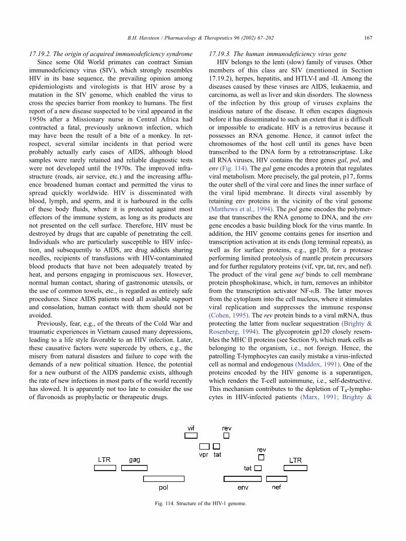
17.19.2. The origin of acquired immunodeficiency syndrome
Since some Old World primates can contract Simian
immunodeficiency virus (SIV), which strongly resembles
HIV in its base sequence, the prevailing opinion among
epidemiologists and virologists is that HIV arose by a
mutation in the SIV genome, which enabled the virus to
cross the species barrier from monkey to humans. The first
report of a new disease suspected to be viral appeared in the
1950s after a Missionary nurse in Central Africa had
contracted a fatal, previously unknown infection, which
may have been the result of a bite of a monkey. In ret-
rospect, several similar incidents in that period were
probably actually early cases of AIDS, although blood
samples were rarely retained and reliable diagnostic tests
were not developed until the 1970s. The improved infra-
structure (roads, air service, etc.) and the increasing afflu-
ence broadened human contact and permitted the virus to
spread quickly worldwide. HIV is disseminated with
blood, lymph, and sperm, and it is harboured in the cells
of these body fluids, where it is protected against most
effectors of the immune system, as long as its products are
not presented on the cell surface. Therefore, HIV must be
destroyed by drugs that are capable of penetrating the cell.
Individuals who are particularly susceptible to HIV infec-
tion, and subsequently to AIDS, are drug addicts sharing
needles, recipients of transfusions with HIV-contaminated
blood products that have not been adequately treated by
heat, and persons engaging in promiscuous sex. However,
normal human contact, sharing of gastronomic utensils, or
the use of common towels, etc., is regarded as entirely safe
procedures. Since AIDS patients need all available support
and consolation, human contact with them should not be
avoided.
Previously, fear, e.g., of the threats of the Cold War and
traumatic experiences in Vietnam caused many depressions,
leading to a life style favorable to an HIV infection. Later,
these causative factors were supercede by others, e.g., the
misery from natural disasters and failure to cope with the
demands of a new political situation. Hence, the potential
for a new outburst of the AIDS pandemic exists, although
the rate of new infections in most parts of the world recently
has slowed. It is apparently not too late to consider the use
of flavonoids as prophylactic or therapeutic drugs.
17.19.3. The human immunodeficiency virus gene
HIV belongs to the lenti (slow) family of viruses. Other
members of this class are SIV (mentioned in Section
17.19.2), herpes, hepatitis, and HTLV-I and -II. Among the
diseases caused by these viruses are AIDS, leukaemia, and
carcinoma, as well as liver and skin disorders. The slowness
of the infection by this group of viruses explains the
insidious nature of the disease. It often escapes diagnosis
before it has disseminated to such an extent that it is difficult
or impossible to eradicate. HIV is a retrovirus because it
possesses an RNA genome. Hence, it cannot infect the
chromosomes of the host cell until its genes have been
transcribed to the DNA form by a retrotranscriptase. Like
all RNA viruses, HIV contains the three genes gal, pol, and
env (Fig. 114). The gal gene encodes a protein that regulates
viral metabolism. More precisely, the gal protein, p17, forms
the outer shell of the viral core and lines the inner surface of
the viral lipid membrane. It directs viral assembly by
retaining env proteins in the vicinity of the viral genome
(Matthews et al., 1994). The pol gene encodes the polymer-
ase that transcribes the RNA genome to DNA, and the env
gene encodes a basic building block for the virus mantle. In
addition, the HIV genome contains genes for insertion and
transcription activation at its ends (long terminal repeats), as
well as for surface proteins, e.g., gp120, for a protease
performing limited proteolysis of mantle protein precursors
and for further regulatory proteins (vif, vpr, tat, rev, and nef).
The product of the viral gene nef binds to cell membrane
protein phosphokinase, which, in turn, removes an inhibitor
from the transcription activator NF-kB. The latter moves
from the cytoplasm into the cell nucleus, where it stimulates
viral replication and suppresses the immune response
(Cohen, 1995). The rev protein binds to a viral mRNA, thus
protecting the latter from nuclear sequestration (Brighty &
Rosenberg, 1994). The glycoprotein gp120 closely resem-
bles the MHC II proteins (see Section 9), which mark cells as
belonging to the organism, i.e., not foreign. Hence, the
patrolling T-lymphocytes can easily mistake a virus-infected
cell as normal and endogenous (Maddox, 1991). One of the
proteins encoded by the HIV genome is a superantigen,
which renders the T-cell autoimmune, i.e., self-destructive.
This mechanism contributes to the depletion of T4-lympho-
cytes in HIV-infected patients (Marx, 1991; Brighty &
Fig. 114. Structure of the HIV-1 genome.
B.H. Havsteen / Pharmacology & Therapeutics 96 (2002) 67–202 167
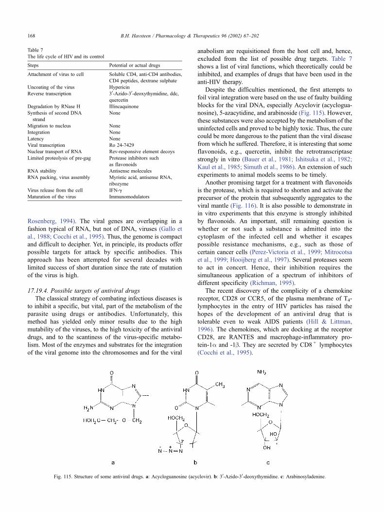
Rosenberg, 1994). The viral genes are overlapping in a
fashion typical of RNA, but not of DNA, viruses (Gallo et
al., 1988; Cocchi et al., 1995). Thus, the genome is compact
and difficult to decipher. Yet, in principle, its products offer
possible targets for attack by specific antibodies. This
approach has been attempted for several decades with
limited success of short duration since the rate of mutation
of the virus is high.
17.19.4. Possible targets of antiviral drugs
The classical strategy of combating infectious diseases is
to inhibit a specific, but vital, part of the metabolism of the
parasite using drugs or antibodies. Unfortunately, this
method has yielded only minor results due to the high
mutability of the viruses, to the high toxicity of the antiviral
drugs, and to the scantiness of the virus-specific metabo-
lism. Most of the enzymes and substrates for the integration
of the viral genome into the chromosomes and for the viral
anabolism are requisitioned from the host cell and, hence,
excluded from the list of possible drug targets. Table 7
shows a list of viral functions, which theoretically could be
inhibited, and examples of drugs that have been used in the
anti-HIV therapy.
Despite the difficulties mentioned, the first attempts to
foil viral integration were based on the use of faulty building
blocks for the viral DNA, especially Acyclovir (acyclogua-
nosine), 5-azacytidine, and arabinoside (Fig. 115). However,
these substances were also accepted by the metabolism of the
uninfected cells and proved to be highly toxic. Thus, the cure
could be more dangerous to the patient than the viral disease
from which he suffered. Therefore, it is interesting that some
flavonoids, e.g., quercetin, inhibit the retrotranscriptase
strongly in vitro (Bauer et al., 1981; Ishitsuka et al., 1982;
Kaul et al., 1985; Simuth et al., 1986). An extension of such
experiments to animal models seems to be timely.
Another promising target for a treatment with flavonoids
is the protease, which is required to shorten and activate the
precursor of the protein that subsequently aggregates to the
viral mantle (Fig. 116). It is also possible to demonstrate in
in vitro experiments that this enzyme is strongly inhibited
by flavonoids. An important, still remaining question is
whether or not such a substance is admitted into the
cytoplasm of the infected cell and whether it escapes
possible resistance mechanisms, e.g., such as those of
certain cancer cells (Perez-Victoria et al., 1999; Mitrocotsa
et al., 1999; Hooijberg et al., 1997). Several proteases seem
to act in concert. Hence, their inhibition requires the
simultaneous application of a spectrum of inhibitors of
different specificity (Richman, 1995).
The recent discovery of the complicity of a chemokine
receptor, CD28 or CCR5, of the plasma membrane of T4-
lymphocytes in the entry of HIV particles has raised the
hopes of the development of an antiviral drug that is
tolerable even to weak AIDS patients (Hill & Littman,
1996). The chemokines, which are docking at the receptor
CD28, are RANTES and macrophage-inflammatory pro-
tein-1a and -1b. They are secreted by CD8 + lymphocytes
(Cocchi et al., 1995).
Fig. 115. Structure of some antiviral drugs. a: Acycloguanosine (acyclovir). b: 30-Azido-30-deoxythymidine. c: Arabinosyladenine.
Table 7
The life cycle of HIV and its control
Steps Potential or actual drugs
Attachment of virus to cell Soluble CD4, anti-CD4 antibodies,
CD4 peptides, dextrane sulphate
Uncoating of the virus Hypericin
Reverse transcription 30-Azido-30-deoxythymidine, ddc,
quercetin
Degradation by RNase H Illincaquinone
Synthesis of second DNA
strand
None
Migration to nucleus None
Integration None
Latency None
Viral transcription Ro 24-7429
Nuclear transport of RNA Rev-responsive element decoys
Limited proteolysis of pre-gag Protease inhibitors such
as flavonoids
RNA stability Antisense molecules
RNA packing, virus assembly Myristic acid, antisense RNA,
ribozyme
Virus release from the cell IFN-g
Maturation of the virus Immunomodulators
B.H. Havsteen / Pharmacology & Therapeutics 96 (2002) 67–202168
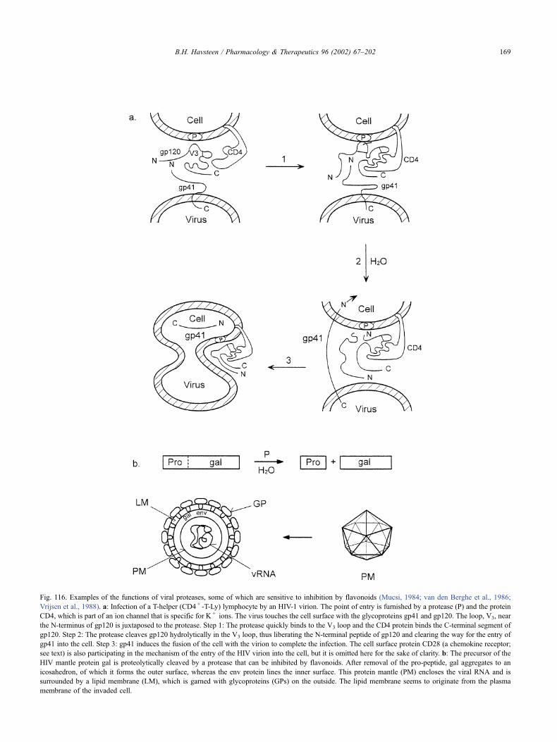
Fig. 116. Examples of the functions of viral proteases, some of which are sensitive to inhibition by flavonoids (Mucsi, 1984; van den Berghe et al., 1986;
Vrijsen et al., 1988). a: Infection of a T-helper (CD4 + -T-Ly) lymphocyte by an HIV-1 virion. The point of entry is furnished by a protease (P) and the protein
CD4, which is part of an ion channel that is specific for K + ions. The virus touches the cell surface with the glycoproteins gp41 and gp120. The loop, V3, near
the N-terminus of gp120 is juxtaposed to the protease. Step 1: The protease quickly binds to the V3 loop and the CD4 protein binds the C-terminal segment of
gp120. Step 2: The protease cleaves gp120 hydrolytically in the V3 loop, thus liberating the N-terminal peptide of gp120 and clearing the way for the entry of
gp41 into the cell. Step 3: gp41 induces the fusion of the cell with the virion to complete the infection. The cell surface protein CD28 (a chemokine receptor;
see text) is also participating in the mechanism of the entry of the HIV virion into the cell, but it is omitted here for the sake of clarity. b: The precursor of the
HIV mantle protein gal is proteolytically cleaved by a protease that can be inhibited by flavonoids. After removal of the pro-peptide, gal aggregates to an
icosahedron, of which it forms the outer surface, whereas the env protein lines the inner surface. This protein mantle (PM) encloses the viral RNA and is
surrounded by a lipid membrane (LM), which is garned with glycoproteins (GPs) on the outside. The lipid membrane seems to originate from the plasma
membrane of the invaded cell.
B.H. Havsteen / Pharmacology & Therapeutics 96 (2002) 67–202 169
17.20. The use of flavonoids in birth control (fertility control)
The world is threatened by a population explosion that
can only be controlled medically, since restrictions imposed
by the authorities, e.g., severe taxes on children, are insuf-
ficient to calm one of the basic human urges, the sexual urge.
Therefore, the medical profession is called upon to develop
safe methods of reversible, transient sterilisation. Although
surgical techniques for this purpose exist, most of the
individuals involved prefer the use of drugs. Effective
contraceptive drugs and inhibitors of the implantation of
fertilised eggs in the uterus have been used for several
decades with a low rate of thrombotic accident, about
1:100,000. However, the alarming press reports of these
few incidents have scared millions of women, with the result
that these measures to counteract the baby boom are resisted.
However, there are other means of contraception. They
have existed for centuries, but most of them are long
forgotten. These methods were based on herbal concoctions
prepared by lay practitioners of dubious, mostly empirical
training, but some of these procedures worked quite well.
The authorities, which needed more babies, especially male
ones that would grow up to become soldiers, banned these
witch brews and burned many of their protagonists at the
stake. Interestingly, a recent article (Weniger et al., 1982)
reports that the flavonoids in a common plant are inhibitors
of the implantation of the fertilised mouse egg in the uterus.
Such flavonoids may very well have been present in
medieval preparations, which were used for population
control in the villages. Since flavonoids neither give rise
to coagulopathies nor cause neurological side effects such as
headaches, except for a few transient allergic responses, the
possible use of these compounds for birth control should be
investigated. Some flavonoids also inhibit the hyaluronidase
in sperm, which facilitates the entry of the spermatocyte into
the oocyte (Li et al., 1997).
18. Interaction of flavonoids with other drugs
Drug interactions create increasing problems to the
design and control of modern medical treatments, as new
insights into pathogenesis lead to the introduction of ever
more complex drugs. Thus, the mutual interaction between
two drugs or between a drug and a food component, e.g.,
alcohol, can drastically displace the therapeutic window of
one or both of the drugs and, thus, render them useless or
dangerous. Although the flavonoids are considered as non-
toxic components of common foods, they can influence the
pharmacological potency of certain drugs significantly. One
of the first examples of this complication was observed in a
kidney dialysis ward, in which patients with a stable kidney
transplant received drugs that improved blood circulation.
Some of these patients consumed grapefruit juice with their
meals, but soon after suffered the deleterious effects of the
increased potency of their medication. This phenomenon
was chosen as the topic of a medical dissertation at the
University of Kiel in 1998, and the effect was confirmed in
a controlled, prospective project.
This finding has been corroborated recently by Benet and
colleagues (Soldner et al., 1999; Wacher et al., 1998; Kim,
H. K. et al., 1999; Chan et al., 1998; Ameer & Weintraub,
1997). They found that a component of the juice, probably
the flavonoid naringenin, which is present in significant
concentration, activates phosphoglycoprotein in the epithe-
lial cells of the intestine and suppresses the expression of the
cytochrome P450 3A4 gene. The former protein enhances
the uptake from the intestine of many drugs, including
vinblastine, cyclosporine, digoxin, fexofenadine, and losar-
tan, and the latter initiates the oxidative decomposition of
the drugs. Since the drugs mentioned are used frequently in
the therapy of cancer, hypertension, HIV, immune disorders,
and other serious conditions, any interference with their
bioavailability has to be taken into consideration by clini-
cians (Wiseman, 1999; Jeong et al., 1999; Di Pietro et al.,
1999; Fukuda et al., 1997).
The mechanism of the enhancement of the activity of the
drug for improving blood circulation was identified as the
inhibition of a cytochrome P450-dependent monooxyge-
nase, which initiates the destruction of the vascular agent
by multiple hydroxylations of its aromatic nucleus. A
subsequent oxidative step then cleaves the aromatic ring,
which renders the product sufficiently water soluble for its
excretion through the kidneys with the urine. The site of
interaction of the flavonoid with the cytochrome P450 has
not been determined yet, but that clarification should be a
simple matter since this enzyme possesses several structural
features, which are likely points of attack for flavonoids
(Ramos & Sultatos, 1998):
(1) It contains several heavy metal ions, e.g., Fe2 + and
Cu2 + , which can bind flavonoids if the metal ion is
located in an accessible position near the surface of the
protein (see Section 16).
(2) The enzyme needs co-substrates, e.g., tetrahydrofolic
acid and NADPH, which bind to the enzyme at sites at
which flavonoids by similar enzymes can compete (see
Section 12).
(3) The catalytic mechanism of cytochrome P450 contains
steps in which there is a transient formation of a free
radical. Such radicals are efficiently scavenged by
flavonoids (see Sections 3.6 and 10).
Although grapefruit juice contains several flavonoids,
naringenin is qualitatively and quantitatively considered as
the principal component. Hence, the influence of this
compound in its pure form on blood pressure has been
investigated. Other studies on different flavonoids, primarily
quercetin, have confirmed the ability of some such sub-
stances to influence the efficiency of various kinds of
medication. Since flavonoids are ubiquitous to food prod-
ucts from plants, the lesson to be learned from these
B.H. Havsteen / Pharmacology & Therapeutics 96 (2002) 67–202170
observations is that nutrition and medication must be
considered together in future therapy plans.
19. Prospects of further applications of flavonoids
The use of flavonoids for the prevention and cure of
diseases is already widespread. The conscious use of these
substances is more common in the developing and in the
emerging countries than in the industrialised part of the
world. In the latter, the demands by the medical authorities
on the proven efficiency and safety of new candidate drugs,
for which authorisation is applied, are high and rigid. Hardly
anybody opposes such strict rules, but the consequence is
that only very few flavonoids are ready to pass the grade
yet, because the required tests have not been completed.
Besides, the wealth of information on flavonoids in the
scientific literature, more than 1000 articles have appeared
in reputable journals, has only rarely been reviewed from a
medical point of view. Hence, the subject is absent in the
curricula of all but a few medical and pharmaceutical
schools, and most young researchers in medicine or bio-
chemistry would not come upon the idea or want to risk
their career on pioneering work in a virgin field, even if it is
promising, when there are enough safe developmental
projects from which to choose.
In this situation, most developed countries allow only
the sale of products based on mixtures of flavonoids, e.g.,
labelled as propolis or its derivatives, as food supple-
ments, instead of as drugs. These restrictions are not
likely to be eased before much more work on the
properties of individual flavonoids has been documented.
This review supplies some of the necessary information
and encourages work in the field of flavonoids. As stated
in Section 4, a great variety of different flavonoids exists,
and each of these compounds possesses its own physical,
chemical, and physiological properties. Although the latter
are overlapping to a certain extend for closely related
species, each compound intended for medical purposes
must be evaluated by a specified procedure that is time-
consuming.
Another problem is logistic. The procurement of flavo-
noids requires their extraction from plants or bee products,
e.g., propolis, or chemical synthesis. Since the physiological
concentration of flavonoids in plants, as it is often the case
for signal substances, is low and the extraction, therefore, is
laborious, an attractive alternative is to use propolis, which
contains a much higher flavonoid concentration—an aver-
age of 4% by weight. In other words, the bees have done a
considerable part of the selection and collection work. A
typical propolis sample contains about 25 different flavo-
noids in significant concentration. Therefore, the extract
from propolis contains most of the biochemical properties
commonly associated with flavonoids, e.g., the antioxidant
effect and radical scavenging. More special physiological
effects, such as oestrogenic or contraceptive actions, must
be sought in extracts from selected plants. Neunaber (1995)
has examined the flavonoid content of various European,
Asian, and American varieties of propolis by HPLC. Neu-
naber also tested the antimicrobiological properties of some
of the isolated flavonoids.
The production of drugs by organic synthesis is often
preferred when it is feasible because the raw materials are
simple and inexpensive; the manufacture can be controlled
and automated; and the costs of cultivation, harvesting,
refining, and transportation are small or eliminated. About
20 synthetic flavonoids of high purity are already available
on the market. More probably could be added since the
organic synthesis of some classes of flavonoids is quite
simple (Yang, 1980). However, the current price of raw
propolis on the market, where it is offered on the scale of
several tons, is so low that the synthetic flavonoids can only
compete if a pure substance is required, e.g., for experi-
ments or for the treatment of certain diseases.
The possibility of extracting the flavonoids in propolis
by a supercritical method has been tried using CO2. The
initial results were not encouraging because the product
was an unattractive powder, which did not seem to be
usable for further processing. However, it is possible that
the problems can be circumvented by a change in the
process variables or in the solvent. Supercritical water,
which is astonishingly hydrophobic, is a possible alter-
native to CO2. These methods are new and require expen-
sive installations. Besides, the problem of separating the
flavonoids remains. In the laboratory, it is easily achieved
by HPLC or capillary chromatography (Stobiecki et al.,
1988; Pietta et al., 1983; Ackermann, 1991), but the
experience on the performance of such procedures on the
industrial scale is scant.
In the near future, most applications of flavonoids
probably would have to rely on relatively few synthetic
substances and products based on propolis. If the expec-
tations of successful treatment of a wide variety of diseases
with flavonoids are fulfilled, then the market on these
substances could soon be empty and the prices of flavonoids
products would rise to a level comparable with that of other
drugs. The bee keepers, among others, would welcome such
a development since their economy suffers from rising cost
of feed (sugar) and veterinary service (e.g., to treat Varoa
infections) and falling prices of their traditional products,
e.g., honey. Bees are indispensable for pollination of grain
and other plants that provide food products for human
consumption. An increased use of flavonoids (e.g., from
propolis) for health purposes would indirectly support plant
breeding and, hence, the society as a whole by facilitating
the expansion of apiculture.
Acknowledgements
Thanks are due to Mrs. R. Thun for her assistance with
the experimental work and to Miss S. Schiemenz for her
B.H. Havsteen / Pharmacology & Therapeutics 96 (2002) 67–202 171
help with the manuscript, as well as with the extensive
literature search.
References
Abate, C., Patel, L., Rauscher, F. J., III, & Curran, T. (1990). Redox reg-
ulation of Fox and Jun DNA-binding activity in vitro. Science 249,
1157–1161.
Ackermann, T. (1991). Fast chromatographic study of propolis crudes.
Food Chem 42, 135–138.
Adlercreutz, H. (1984). Does fiber-rich food containing animal lignan pre-
cursors protect against both colon and breast cancer? An extension of
the ‘fiber hypothesis’. Gastroenterology 86, 761–764.
Aeschbacher, H. -U., Meier, H., & Ruch, E. (1982). Nonmutagenicity in
vivo of the food flavonol quercetin. Nutr Cancer 4, 90–98.
Afanas’ev, E. B., Dorozhko, A. I., Brodskii, A. V., Kostyuk, V. A., &
Potapovitch, A. I. (1989). Chelating and free radical scavenging mech-
anisms of inhibitory action of rutin and quercetin in lipid peroxidation.
Biochem Pharmacol 38, 1763–1769.
Agarwal, O. P., & Nagaratnam, A. (1981). Radioprotective property of
flavonoids in man. Toxicon 19, 201–204.
Agullo, G., Gamet-Payrastre, L., Fernandez, Y., Anciaux, N., Demigne, C.,
& Remesy, C. (1996). Comparative effects of flavonoids on the growth,
viability and metabolism of a colonic adenocarcinoma cell line (HT29
cells). Cancer Lett 105, 61–70.
Agullo, G., Gamet-Payrastre, L., Manenti, S., Viala, C., Remesy, C., Chap,
H., & Payrastre, B. (1997). Relationship between flavonoid structure
and inhibition of phosphatidylinositol 3-kinase: a comparison with ty-
rosine kinase and protein kinase C inhibition. Biochem Pharmacol 53,
1649–1657.
Ahmadiani, A., Fereidoni, M., Semnanian, S., Kamalinejad, M., & Saremi,
S. (1998). Antinociceptive and anti-inflammatory effects of Sambucus
ebulus rhizome extract in rats. J Ethnopharmacol 61, 229–235.
Ait-Si-Ali, S., Ramirez, S., Barre, F. -X., Dkhissi, F., Magnaghi-Jaulin, L.,
Girault, J. A., Robin, P., Knibiehler, M., Pritchard, L. L., Ducommun,
B., Trouche, D., & Harel-Ballau, A. (1998). Histone acetyl transferase
activity of CBP is controlled by cycle-dependent kinases and oncopro-
tein E1A. Nature 396, 184–186.
Akiyama, T., Ishida, J., Nakagawa, S., Ogawara, H., Watanabe, S., Itoh, N.,
Shibuya, M., & Fukami, Y. (1987). Genistein, a specific inhibitor of
tyrosine specific protein kinases. J Biol Chem 262, 5592–5595.
Albrecht, H., Yoder, J. I., & Phillips, D. A. (1999). Flavonoids promote
haustoria formation in the root parasite triphysaria versicolor. Plant
Physiol 119, 585–592.
Alexandrakis, M., Singh, L., Boucher, W., Letourneau, R., Theofilopoulos,
P., & Theoharides, T. C. (1999). Differential effect of flavonoids on
inhibition of secretion and accumulation of secretory granules in rat
basophilic leukemia cells. Int J Immunopharmacol 21, 379–390.
al Makdessi, S., Sweidan, H., Dietz, K., & Jacob, R. (1999). Protective
effect of Crataegus oxyacantha against reperfusion arrhythmias after
global no-flow ischemia in the rat heart. Basic Res Cardiol 94, 71–77.
Ambrose, A. M., & de Eds, F. (1949). Further observations on the effect of
rutin and related compounds on cutaneous capillaries. J Pharmacol Exp
Ther 97, 115–119.
Ambrose, A. M., & de Eds, F. (1974). Effect of rutin on permeability of
cutaneous capillary. J Pharmacol Exp Ther 190, 359–363.
Ambrose, A. M., Robbies, D. J., & de Eds, F. (1951). Comparative tox-
icities of quercetin and quercitrin. J Am Pharm Assoc 41, 119–122.
Ambrose, A. M., Robbies, D. J., & de Eds, F. (1952). Comparative toxicities
of quercetin and quercitrin. J Am Pharm Assoc Sci Ed 16, 119–122.
Ameer, B., & Weintraub, R. A. (1997). Drug interactions with grapefruit
juice. Clin Pharmacokinet 33, 103–121.
Ames, B. N., McCann, J., & Yamazaki, E. (1975). Methods for detecting
carcinogens and mutagens with salmonella-mammalian microsome mu-
tagenicity test. Mutat Res 31, 347–363.
Amoras, M., Sauvager, F., Girre, L., & Cormier, M. (1992a). In vitro anti-
viral activity of propolis. Apiologie 23, 231–240.
Amoras, M., Simoes, C. M., Girre, L., Sauvager, F., & Cormier, M.
(1992b). Synergistic effect of flavones and flavonols against herpes
simplex virus type 1 in cell culture. Comparison with the antiviral
activity of propolis. J Nat Prod 55, 1732–1740.
Amstad, P., Peskin, A., Shah, G., Mirault, M. -E., Morat, R., Zbinden, I., &
Cerutti, P. (1991). The balance between Cu, Zn-superoxide dismutase
and catalase affects the sensitivity of mouse epidermal cells to oxidative
stress. Biochemistry 30, 9305–9313.
Anderson, D., Basaran, N., Dobrzynska, M. M., Basaran, A. A., & Yu,
T. W. (1997). Modulating effects of flavonoids on food mutagens in
human blood and sperm samples in the comet assay. Teratog Carcinog
Mutagen 17, 45–58.
Anderson, D., Dobrzynska, M. M., Basaran, N., Basaran, A., & Yu, T. W.
(1998). Flavonoids modulate comet assay responses to food mutagens
in human lymphocytes and sperm. Mutat Res 402, 269–277.
Angel, J., Berenbaum, F., LeDenmat, C., Nevalainen, T., Masliah, J., &
Fournier, C. (1994). Interleukin 1-induced prostaglandin E2 biosynthe-
sis in human synovial cells involves the activation of cytosolic phos-
pholipase A2 and cyclooxygenase 2. Eur J Biochem 226, 125–131.
Applegate, L. A., Luscher, P., & Tyrrell, R. M. (1991). Induction of heme
oxygenase: a general response to oxidative stress in cultured mamma-
lian cells. Cancer Res 51, 974–978.
Apps, D. K., & Glover, L. A. (1978). Isolation and characterization of
magnesium triphosphatase from the chromaffine granule membrane.
FEBS Lett 85, 254–258.
Ares, J. J., & Outt, P. E. (1998). Gastroprotective agents for the prevention
of NSAID-induced gastropathy. Curr Pharm Design 4, 17–36.
Arora, A., Nair, M. G., & Strasburg, G. M. (1998). Structure-activity rela-
tionships for antioxidant activities of a series of flavonoids in a li-
posomal system. Free Radic Biol Med 24, 1355–1363.
Arts, I. C. W., Hollman, P. C. H., & Kromhout, D. (1999). Chocolate as a
source of tea flavonoids. Lancet 354, 488.
Arturson, G. (1972). Effect of O-(b-hydroxyethyl)-rutosides (HR) on the
increased microvascular permeability in experimental skin burns. Acta
Chir Scand 138, 111–117.
Aruoma, O. I. (1999). Antioxidant actions of plant foods: use of oxidative
DNA damage as a tool for studying antioxidant efficacy. Free Radic
Res 30, 419–427.
Asen, S., Norris, K. H., & Stewart, R. N. (1971). Effect of pH and con-
centration of the anthocyanin flavonol copigment complex on the color
of better-times roses-D. J Am Soc Hortic Sci 96, 770–773.
Asen, S., Norris, K. H., & Stewart, R. N. (1972). Copigmentation of an-
thocyanins in plant tissues and its effect on color. Phytochemistry 11,
1139–1144.
Asen, S., Stewart, R. N., & Norris, K. H. (1977). Anthocyanin and pH
involved in the color of ‘heavenly blue’ morning glory. Phytochemistry
16, 1118–1119.
Asgary, S., Naderi, G., Sarrafzadegan, N., Ghassemi, N., Boshtam, M.,
Rafie, M., & Arefian, A. (1999). Anti-oxidant effect of flavonoids on
hemoglobin glycosylation. Pharm Acta Helv 73, 223–226.
Astassi, G., Griet, P., Berthelon, J. -J., & Collonges, F. (1985). Synthesis
and antitumor activity of some 8-substituted -4-oxo-4H-1-benzopyrans.
Eur J Med Chim Ther 20, 393–402.
Attaway, J. A., & Buslig, B. S. (1998). Antithrombogenic and antiathero-
genic effects of citrus flavonoids. Contributions of Ralph C. Robbins.
Adv Exp Med Biol 439, 165–173.
Austin, C. A., Patel, S., Ono, K., Nakane, H., & Fisher, L. M. (1992). Site-
specific DNA cleavage by mammalian topoisomerase II induced by
novel flavone and catechin derivatives. Biochem J 282, 883–889.
Avila, M. A., Velasco, J. A., Causado, J., & Notario, V. (1994). Quercetin
mediates the down-regulation of mutant p53 in the human breast cancer
cell line MDA-MB468. Cancer Res 54, 2424–2428.
Aviram, M., & Fuhrman, B. (1998). Polyphenolic flavonoids inhibit macro-
phage-mediated oxidation of LDL and attenuate atherogenesis. Athero-
sclerosis 137(suppl.), S45–S50.
B.H. Havsteen / Pharmacology & Therapeutics 96 (2002) 67–202172
Bae, E. A., Han, M. J., & Kim, D. H. (1999). In vitro anti-Helicobacter
pylori activity of some flavonoids and their metabolites. Planta Med
65, 442–443.
Bagaev, IuN. (1978). Flavonoids in the complex treatment of patients with
pulmonary tuberculosis. (Russian). Probl Tuberk 12, 74–75.
Bagavandoss, P., & Midgley Jr., A. R. (1988). Biphasic action of retinoids
on gonadotropin-receptor induction in rat granulosa cells in vitro. Life
Sci 43, 1607–1614.
Bai, F., Matsui, T., Ohtani-Fujita, N., Matsukawa, Y., Ding, Y., & Sakai, T.
(1998). Promoter activation and following induction of the p21/WAF1
gene by flavone is involved in G1 phase arrest in A549 lung adeno-
carcinoma cells. FEBS Lett 437, 61–64.
Baird, I. M., Hughes, R. E., Wilson, H. K., Davies, J. E., & Howard, A. N.
(1979). The effects of ascorbic acid and flavonoids on the occurrence of
symptoms normally associated with the common cold. Am J Clin Nutr
32, 1686–1690.
Baker, M. E. (1998). Flavonoids as hormones. A perspective from an
analysis of molecular fossils. Adv Exp Med Biol 439, 249–267.
Baker, M. E., Medlock, K. L., & Sheehan, D. M. (1998). Flavonoids inhibit
estrogen binding to rat alpha-fetoprotein. Proc Soc Exp Biol Med 217,
317–321.
Baker, W., & Robinson, R. (1928). Synthetic experiments in the iso-flavone
group. III. A synthesis of genistein. J Chem Soc, 3445–3448.
Bakir, F., Damluji, S. F., Amin-Zaki, L., Murtadho, M., Khalidi, A., Al-
Rawi, N. Y., Tikriti, S., Dhalur, N. I., Clarkson, T. W., Smith, J. C., &
Doherty, R. A. (1973). Methylmercury poisoning in Iraq. Science 181,
230–241.
Bakkeskov, S., Aanstoot, H. J., Christgau, S., Reetz, A., Solimena, M.,
Cascalho, M., Folli, F., Richter-Olsen, H., & DeCamilli, P. (1990).
Identification of the 64k autoantigen in insulin-dependent diabetes as
the GABA-synthesizing enzyme glutamic acid decarboxylase. Nature
347, 151–156. Erratum: Nature 347, 782 (1990).
Balentine, D. A., Wiseman, S. A., & Bouwens, L. C. (1997). The chemistry
of tea flavonoids. Crit Rev Food Sci Nutr 37, 693–704.
Bankova, V. S., Popov, S. S., & Marekov, N. L. (1982). High performance
liquid chromatographic analysis of flavonoids from propolis. J Chro-
matogr 242, 135–143.
Bankova, V. S., Popov, S. S., & Marekov, N. L. (1983). A study on fla-
vonoids of popolis. J Nat Prod 46, 471–474.
Bankova, V. S., Dyulgerov, A., Popov, S. S., & Marekov, N. L. (1987). A
GC/MS study of the propolis phenolic constituents. Z Naturforsch 42e,
147–151.
Bankova, V. S., Popov, S. S., Marekov, N. L., Manolova, N., Maximova, V.,
Gregova, G., Serkedjieva, J., & Uzenov, S. (1988). On the chemical
composition of some propolis fractions with antiviral action. Acta Mi-
crobiol Bulg 23, 52–57.
Barasoain, I., Rojo, J. M., & Portoles, A. (1980). ‘‘In vivo’’ effects of
acetylsalicylic acid and two ether-derived compounds on primary im-
mune response and lymphoblastic transformation. Immunopharmacol-
ogy 2, 293–300.
Barbour, B., Szatkowski, M., Ingledew, N., & Attwell, D. (1989). Arach-
idonic acid induces a prolonged inhibition of glutamate uptake into glial
cells. Nature 342, 918–920.
Barger, S. W., & Mattson, M. P. (1995). The secreted form of the Alzheim-
er’s b-amyloid precursor protein stimulates a membrane-associated gua-
nylate cyclase. Biochem J 311, 45–47.
Barinaga, M. (1993). Carbon monoxide: killer to brain messenger in one
step. Science 259, 309.
Barinaga, M. (1996). Forging a path to cell death. Science 273, 735–737.
Bartholomew, R. M., & Ryan, D. S. (1980). Lack of mutagenicity of some
phytoestrogens in the salmonella/mammalian microsome assay. Mutat
Res 78, 317–321.
Batterham, T. J., & Highet, R. J. (1964). Nuclear magnetic resonance
spectra of flavonoids. Aust J Chem 17, 428–439.
Bauer, U. J., Selway, J. W. T., Batchelor, J. F., Tisdale, M., Caldwell, E. C.,
& Young, D. A. B. (1981). 40,60-Dichloro-flavone (bw6836). A new
antirhinovirus compound. Nature 292, 369–370.
Baumann, J., Wurm, G., & von Bruchhausen, F. (1980). Hemmung der
Prostaglandinsynthese durch Flavonoide und Phenolderivate im Ver-
gleich mit deren. O-Radikalfangereigenschaften. Arch Pharm (Wein-
heim) 313, 330–337.
Baumann, J., von Bruchhausen, F., & Wurm, G. (1981). Flavonoids and
arachidonic acid metabolism. Stud Org Chem 11, 411–419.
Baumann, K., Mandelkow, E. -M., Biernat, J., Piwnica-Worms, H., &
Mandelkow, E. (1993). Abnormal Alzheimer-like phosphorylation of
tau protein by cyclin-dependent kinases cdk2 and cdk5. FEBS Lett
336, 417–424.
Becker, V., Stevely, W., Hamburger, V., Tabor, E., Asher, V., & Hadar, J.
(1981). Effect of the flavonoid (+)cyanidinol-2 on procollagen biosyn-
thesis and transport in normal and ataxia telangiectasis cultured skin
fibroblasts. Connect Tissue Res 8, 77–84.
Beecher, G. R., Warden, B. A., & Merken, H. (1999). Analysis of tea
polyphenols. Proc Soc Exp Biol Med 220, 267–270.
Beladi, I., Pusztai, R., Mucsi, I., Balkay, M., & Gabor, M. (1977). Activity
of some flavonoids against viruses. Ann N Y Acad Sci 284, 358–364.
Beladi, I., Pusztai, R., Balkay, M., Mucsi, I., & Tarodi, B. (1979). Interferon
induction by adenovirus. Postgrad Med J 55, 128–134.
Bennett, J. P., Gomperts, B. D., & Wollenweber, E. (1981). Inhibitory
effects of natural flavonoids on secretion from mast cells and neutro-
phils. Arzneimittelforschung 31, 433–437.
Bentsath, A., Rusznyak, S., & Szent-Gyorgyi, A. (1936). Vitamine nature
of flavones. Nature 138, 798.
Beretz, A., Anton, R., & Stocklet, J. C. (1978). Flavonoid compounds are
potent inhibitors of cyclic AMP phosphodiesterase. Experientia 34,
1054–1058.
Beretz, A., Stierl, A., Sutter-Bay, A., Launay, J., Casenavo, J. P., & Anton,
R. (1981). Flavonoids as potent inhibitors of cyclic AMP phosphodies-
terase. In M. G. L. Farkas, F. Kellog, & H. Wagner (Eds.), Flavonoids
and Bioflavonoids (p. 421). Amsterdam: Elsevier.
Beretz, A., Cazenave, J. -P., & Anton, R. (1982). Inhibition of aggregation
and secretion of human platelets by quercetin and other flavonoids.
Structure-activity relationships. Agents Actions 12, 382–387.
Beretz, A., Briancon-Scheid, F., Stierle, A., Corre, G., Anton, R., & Caze-
nave, J. P. (1986). Inhibition of human platelet cyclic AMP phospho-
diesterase by a hemisynthetic flavonoid, amentoflavone hexaacetate.
Biochem Pharmacol 35, 257–262.
Berkarda, B., Bouffard-Eyuboglu, H., & Derman, U. (1983). The effect of
coumarin derivatives on the immunological system of man. Agents
Actions 13, 50–52.
Bernard, F. X., Sable, S., Cameron, B., Provost, J., Desnottes, J. F., Crouzet,
J., & Blanche, F. (1997). Glycosylated flavones as selective inhibitors
of topoisomerase IV. Antimicrob Agents Chemother 41, 992–998.
Bernheim, R. A., Brown, T. H., Gustowsky, H. S., & Woessner, D. E.
(1959). Temperature dependence of proton relaxation times in aqueous
solutions of paramagnetic ions. J Chem Phys 30, 950–956.
Berton, G., Schneider, C., & Romeo, D. (1980). Inhibition by quercetin of
activation of polymorphonuclear leucocyte functions. Stimulus-specific
effects. Biochim Biophys Acta 595, 47–55.
Bertorello, A. M., Aperia, A., Walaas, S. I., Nairn, A. C., & Greengard, P.
(1991). Phosphorylation of the catalytic subunit of Na+/K+-ATPase
inhibits the activity of the enzyme. Proc Natl Acad Sci USA 88,
11359–11362.
Bettini, V., Fraccaro, A., & Legrenzi, E. (1978a). Effect of a flavonoid (4-
methylesculetol) on the response of isolated calf hepatic arteries to
angiotensin II. Boll Soc Ital Biol. Sper 53, 938–941.
Bettini, V., Legrenzi, E., Dalla Valle, C., & Petrella, C. (1978b). Effect of a
flavonoid (4-methylesculetin) on the response of isolated segments of
coronary arteries to adrenalin. Boll Soc Ital Biol Sper 53, 2027–2030.
Bettini, V., Legrenzi, E., & Monanni, L. (1978c). Effect of a flavonoid (4-
methylesculetin) on the response of isolated calf arteries to histamine.
Boll Soc Ital Biol Sper 53, 2015–2019.
Bettini, V., Legrenzi, E., & Petrella, C. (1978d). Influence of a flavonoid (4-
methylesculetol) on the response of isolated calf coronary vessels to
noradrenalin. Boll Soc Ital Biol Sper 53, 1096–1100.
B.H. Havsteen / Pharmacology & Therapeutics 96 (2002) 67–202 173
Bilyk, A., & Sapers, G. M. (1985). Distribution of quercetin and kaempfer-
ol in lettuce, kale, chive, garlic chive, leek, horseradish, red radish and
red cabbage tissues. J Agric Food Chem 33, 226–228.
Bissery, M.-C., Valeriote, F. A., Chabot, G. S., Crissman, J. S., Yost, C., &
Corbett, T. H. (1988). Flavone acetic acid (NSC 347512)-induced
DNA damage in Glasgow osteogenic sarcoma in vivo. Cancer Res
48, 1279–1285.
Bjeldanes, L. F., & Chang, G. W. (1977). Mutagenic activity of quercetin
and related compounds. Science 197, 577–578.
Bjerrum, J. (1958). Stability Constants. London: Chemical Society.
Bjorkman, P. J., & Parham, P. (1990). Structure, function and diversity of
class I major histocompatibility complex molecules. Annu Rev Biochem
59, 253–288.
Blardi, P., De Lalla, A., Volpi, L., & Di Perri, T. (1999). Stimulation of
endogenous adenosine release by oral administration of quercetin and
resveratrol in man. Drugs Exp Clin Res 25, 105–110.
Bloch, K. (1983). Sterole structure and membrane function. Crit Rev Bio-
chem 14, 47–92.
Bloembergen, N., Purcell, E. M., & Pound, R. V. (1948). Relaxation effects
in nuclear magnetic resonance absorption. Physiol Rev 73, 679–712.
Blostein-Fujii, A., DiSilvestro, R. A., Frid, D., & Katz, C. (1999). Short term
citrus flavonoid supplementation of type II diabetic women: no effect on
lipoprotein oxidation tendencies. Free Radic Res 30, 315–320.
Blumenkrantz, N., & Asboe-Hansen, G. (1980). Studies on the effect of
(+)-catechin on connective tissue in the guinea pig and in organ culture.
Scand J Reumatol 9, 1–5.
Bohm, H., Boeing, H., Hempel, J., Raab, B., & Kroke, A. (1998). Flavo-
nols, flavone and anthocyanins as natural antioxidants of food and their
possible role in the prevention of chronic diseases. (German). Z Ernah-
rungswiss 37, 147–163.
Bohm, K., & Lamprecht, W. (1959). Flavonoide und Herzmuskelstoffwech-
sel. Arztl Forsch 11, 543–548.
Bohr, D. F., McIvor, B. C., & Rinehart, J. F. (1949). The effect of various
flavone glycosides on the rate of passage of Evans blue through the
damaged capillary wall. J Pharmacol Exp Ther 97, 243–249.
Bok, S. H., Lee, S. H., Park, Y. B., Bae, K. H., Son, K. H., Jeong, T. S., &
Choi, M. S. (1999). Plasma and hepatic cholesterol and hepatic activ-
ities of 3-hydroxy-3-methyl-glutaryl-CoA reductase and acyl CoA: cho-
lesterol transferase are lower in rats fed citrus peel extract or a mixture
of citrus bioflavonoids. J Nutr 129, 1182–1185.
Bolton, T., & Casley-Smith, J. R. (1975). An in vitro demonstration of
proteolysis by macrophages and its increase with coumarin. Experientia
31, 271–273.
Bonvehi, J. S., Coll, F. V., & Jorda, R. E. (1994). The composition, active
components and bacteriostatic activity of propolis in dietetics. J Am Oil
Chem Soc 71, 529–532.
Booth, A. N., Murray, C. W., Jones, F. T., & de Eds, F. (1956). The
metabolic fate of rutin and quercetin in the animal body. J Biol Chem
233, 251–257.
Borradaile, N. M., Carroll, K. K., & Kurowska, E. M. (1999). Regulation of
HepG2 cell apolipoprotein B metabolism by the citrus flavanones hes-
peretin and naringenin. Lipids 34, 591–598.
Bors, W., Michel, C., & Stettmaier, K. (1997). Antioxidant effects of fla-
vonoids. Biofactors 6, 399–402.
Boulton, D. W., Walle, U. K., & Walle, T. (1999). Fate of the flavonoid
quercetin in human cell lines: chemical instability and metabolism.
J Pharm Pharmacol 51, 353–359.
Bourdillat, B., Delautier, D., Labat, C., Benveniste, J., Potier, P., & Brink,
C. (1988). Hispidulin, a natural flavone, inhibits human platelet aggre-
gation by increasing cAMP levels. Eur J Pharmacol 147, 1–6.
Bourne, H. R. (1994). The importance of being GTP. Nature 369, 611–612.
Bourne, L. C., & Rice-Evans, C. A. (1999). Detecting and measuring
bioavailability of phenolics and flavonoids in humans: pharmacoki-
netics of urinary excretion of dietary ferulic acid. Methods Enzymol
299, 91–106.
Bovey, F. A. (1969). Nuclear Magnetic Resonance Spectroscopy. New
York: Academic Press.
Bracke, M. E., Depypere, H. T., Boterberg, T., Van Marck, V. L., Vennek-
ens, K. M., Vanluchene, E., Nuytinck, M., Serreyn, R., & Mareel,
M. M. (1999). Influence of tangeretin on tamoxifen’s therapeutic
benefit in mammary cancer. J Natl Cancer Inst 91, 354–359.
Brade, L., Holst, O., & Brade, H. (1993). An artificial glycoconjugate
containing the bisphosphorylated glucosamine disaccharide backbone
of lipid A binds lipid A monoclonal antibodies. Infect Immun 61,
4514–4517.
Bradley, D. V., & Cazort, R. J. (1970). Relief of bladder spasms by flavox-
ate. A comparative study. J Clin Pharmacol 10, 65–68.
Brandao, M. G., Krettli, A. U., Soares, L. S., Nery, C. G., Marinuzzi,
H. C. (1997). Antimalarial activity of extracts and fractions from Bi-
dens pilosa and other Bidens species (Asteraceae) correlated with the
presence of acetylene and flavonoid compounds. J Ethnopharmacol 57,
131–138.
Bresnick, K., & Birt, D. (1988). Inhibitory effects of the flavonoid apigenin
on epidermal ornithine decarboxylase and mouse skin tumorigenesis.
Proc Am Assoc Cancer Res 29, 136.
Briggs, L. H., & Colebrook, L. D. (1962). Infrared spectra of flavanones
and flavones. Carbonyl and hydroxyl stretching and CH out-of-plane
bending absorption. Spectrochim Acta 18, 939–957.
Brighty, D. W., & Rosenberg, M. (1994). A cis-acting repressive sequence
that overlaps the Rev-responsive element of human immunodeficiency
virus type 1 regulates nuclear retention of env mRNA independently of
known splicing signals. Proc Natl Acad Sci USA 91, 8314–8318.
Brinkworth, R. I., Stoermer, M. J., & Fairlie, D. P. (1992). Flavones are
inhibitors of HIV 1 protease. Biochem Biophys Res Commun 188,
631–637.
Bronsted, J. N., & Pedersen, K. (1923). Die ketalytische Zersetzung des
Nitramides und ihne physikalisch-chemische Bedentung. Z Physik
Chem 108, 185–235.
Brostoff, J., Scadding, G. K., Male, D., & Roitt, I. M. (1991). Clinical
Immunology. London: Gower Medical Publishers.
Broughton, W. J., & Perret, X. (1999). Genealogy of legume-Rhizobium
symbioses. Curr Opin Plant Biol 2, 305–311.
Brouillard, R., & Cheminat, A. (1988). Flavonoids and plant color. Prog
Clin Biol Res 280, 93–106.
Brouillard, R., George, F., & Fougerousse, A. (1997). Polyphenols pro-
duced during red wine ageing. Biofactors 6, 403–410.
Brown, J. E., & Rice-Evans, C. A. (1998). Luteolin-rich artichoke extract
protects low density lipoprotein from oxidation in vitro. Free Radic Res
29, 247–255.
Brown, J. E., Khodr, H., Hider, R. C., & Rice-Evans, C. A. (1998). Struc-
tural dependence of flavonoid interactions with Cu2+ ions: implications
for their antioxidant properties. Biochem J 330, 1173–1178.
Brown, J. P. (1980). A review of the genetic effects of naturally occurring
flavonoids, anthraquinones and related compounds. Mutat Res 78,
243–277.
Brown, J. P., Dietrich, P. S., & Brown, R. J. (1977). Frameshift mutage-
nicity of certain naturally occurring phenolic compounds in the ‘‘sal-
monella/microsome’’ test: activation of anthraquinones and flavonoid
glycoside by gut bacterial enzymes. Mutat Res 66, 223–240.
Brown, J. P., Crosby, G. A., Dubois, G. B., Enderlin, F. E., & Hale, R. L.
(1979). Flavonoid sweeteners. Synthesis and intestinal absorption of
selected sulfalkylated hesperitin-3-14C-derivatives in the rat. J Agric
Food Chem 26, 1418–1422.
Brown, M. S., & Goldstein, J. L. (1984). How LDL receptors influence
cholesterol and atherosclerosis. Sci Am 251, 58–66.
Brown, S., Wiebel, F. J., Gelboin, H. V., & Minna, J. D. (1976). Assign-
ment of a locus required for flavoprotein-linked monooxygenase ex-
pression to human chromosome 2. Proc Natl Acad Sci USA 73,
4628–4632.
Bruillard, R. (1981). Origin of the exceptional colour stability of the zebrina
anthocyanin. Phytochemistry 20, 143–145.
Bruillard, R. (1982). Flavonoids. In P. Markakis (Ed.), Anthocyanins as
Food Colors (pp. 1–40). New York: Academic Press.
Bruillard, R., & Delaponte, B. (1977). Chemistry of anthocyanin pigments.
B.H. Havsteen / Pharmacology & Therapeutics 96 (2002) 67–202174
2. Kinetic and thermodynamic study of proton transfer, hydration and
tautomeric reactions of malvidin-3-glucoside. J Am Chem Soc 99,
8461–8468.
Bruillard, R., Iacobucci, G. A., & Sweeny, J. G. (1982). Chemistry of
anthocyanin pigments. 9. UV-visible spectrophotometric determination
of the acidity constants of apigenidin and three related 3-deoxyflavy-
lium salts. J Am Chem Soc 104, 7585–7590.
Brunger, A. T., Clore, G. M., Gronenborn, A. M., Soffrich, R., & Nilges,
M. (1993). Assessing the quality of solution NMR structures by com-
plete cross-validation. Science 261, 328–331.
Buddecke, E. (1994). Grundriß der Biochemie. Berlin: de Gruyter.
Buening, M. K., Chang, R. L., Huang, M. T., Fortner, J. G., & Wood, A. K.
(1981). Activation and inhibition of benzo(a)pyrene and aflatoxin B1
metabolism in human liver microsomes by naturally occurring flavo-
noids. Cancer Res 41, 67–72.
Cai, Z., Li, X., & Katsumura, Y. (1999). Interaction of hydrated electron
with dietary flavonoids and phenolic acids: rate constants and transient
spectra studied by pulse radiolysis. Free Radic Biol Med 27, 822–829.
Calias, P., Galanopoulus, T., Maxwell, M., Khayat, A., Graves, D., Anto-
niades, H. N., & Dalarcao, M. (1996). Synthesis of inositol-2-phos-
phate-cuercetin conjugates. Carbohydr Res 292, 83–90.
Calman, J. (1972). A new method for the assessment of tissue fluid con-
centrations of flavonoids. Angiologica 9, 181–192.
Caltagirone, S., Ranelletti, F. O., Rinelli, A., Maggiano, N., Colasante, A.,
Musiani, P., Aiello, F. B., & Piantelli, M. (1997). Interaction with type
II estrogen binding sites and antiproliferative activity of tamoxifen and
quercetin in human non-small-cell lung cancer. Am J Respir Cell Mol
Biol 17, 51–59.
Calzada, F., Meckes, M., & Cedillo-Rivera, R. (1999). Antiamoebic and
antigiardial activity of plant flavonoids. Planta Med 65, 78–80.
Cancalon, P. F. (1999). Analytical monitoring of citrus juices by using
capillary electrophoresis. J AOAC Int 82, 95–106.
Canivenc-Lavier, M. C., Vernevaut, M. F., Totis, M., Siess, M. H., Magda-
lou, J., & Suschetet, M. (1996). Comparative effects of flavonoids and
model inducers on drug-metabolizing enzymes in rat liver. Toxicology
114, 19–27.
Cantley, L. C., & Hammes, G. G. (1976). Investigation of quercetin binding
sites on chloroplast coupling factor 1. Biochemistry 15, 1–8.
Cao, G., Sofic, E., & Prior, R. L. (1997). Antioxidant and prooxidant
behavior of flavonoids: structure-activity relationships. Free Radic Biol
Med 22, 749–760.
Cao, Y., & Cao, R. (1999). Angiogenesis inhibited by drinking tea. Nature
398, 381.
Carpenedo, F., Bortignon, C., Bruni, A., & Santi, R. (1969). Effect of
quercetin on membrane-linked activities. Biochem Pharmacol 18,
1495–1500.
Carr, H. Y., & Purcell, E. M. (1954). Effects of diffusion on free pre-
cession in nuclear magnetic resonance experiments. Physiol Rev 94,
630–638.
Carver, J. H., Carrano, A. V., & MacGregoe, J. T. (1983). Genetic effects of
the flavonols quercetin, kaempferol and galangin on Chinese hamster
ovary cells in vitro. Mutat Res 113, 45–60.
Casalini, C., Lodovici, M., Briani, C., Paganelli, G., Remy, S., Cheynier, V.,
& Dolara, P. (1999). Effect of complex polyphenols and tannins from
red wine (WCPT) on chemically induced oxidative DNA damage in the
rat. Eur J Nutr 38, 190–195.
Casley-Smith, R. J. (1976). The actions of benzo-pyrones on the blood-
tissue-lymph system. Folia Angiol 24, 7–22.
Casley-Smith, J. R., & Bolton, T. (1973). Electron microscopy of the effect
of histamine and thermal injury on the blood and lymphatic epithelium
and the mesothelium of the mouse’s diaphragm together with the influ-
ence of coumarine and rutin. Experientia 29, 1386–1388.
Casley-Smith, J. R., & Casley-Smith, J. R. (1986). High-Protein Oedemas
and the Benzo-gamma-pyrones. Sydney: Lippincott.
Casley-Smith, J. R., & Piller, N. B. (1974). The pathogenesis of oedemas
and the therapeutic action of coumarin and related compounds. Folia
Angiol 3, 33–59.
Casley-Smith, J. R., Foldi-Borcsok, E., & Foldi, M. (1974). Fine-structural
aspects of lymphoedema in various tissues and the effects of treatment
with coumarin and troxerutin. Br J Exp Pathol 55, 88–93.
Cavallini, L., Bindoli, A., & Siliprandi, N. (1978). Comparative evaluation
of an anti-peroxidative action of silymarin and other flavonoids. Phar-
macol Res Commun 10, 133–136.
Cazenave, J. P., Beretz, A., Stierle, A., & Anton, R. (1981). Role of cyclic
AMP in the inhibition of human platelet functions by quercetin, a
flavonoid that potentiates prostaglandin I2 effect. Thromb Haemost
46, 173.
Cea, G. F. A., Etcheberry, K. F. C., & Dulout, F. N. (1983). Induction of
micronuclei in mouse bone marrow cells by the flavonoid 5,30,40-
trihydroxy-3,6,7,8-tetramethoxyflavone (THTMF). Mutat Res 119,
339–342.
Cecchini, M. G., Fleisch, H., & Muhibauer, R. C. (1997). Ipriflavone in-
hibits bone resorption in intact and ovariectomized rats. Calcif Tissue
Int 61(suppl. 1), S9–S11.
Ceriani, M. F., Darlington, T. K., Staknis, D., Mas, P., Petti, A. A., Weitz,
C. J., & Kay, S. A. (1999). Light-dependent sequestration of timeless
by chryptochrome. Science 285, 553–556.
Cetta, G., Pallavicini, G., Tenni, R., & Bisi, C. (1978). Influence of flavo-
noid-copper complexes on cross-linking in elastin. Ital J Biochem 26,
317–327.
Chai, J. S., Yokozawa, S., & Matsubaro, H. (1981). Antihyperlidemic effect
of flavonoids from prunus davidiana. J Nat Prod 54, 218–224.
Chan, W. K., Nguyen, L. T., Miller, V. P., & Harris, R. Z. (1998). Mech-
anism-based inactivation of human cytochrome P450 3A4 by grapefruit
juice and red wine. Life Sci 62, L135–L142.
Chang, R. L., Huang, M. T., Wood, A. W., Woug, C. Q., Newmark, H.,
Yagi, H., Sayer, J. M., Jerina, D. M., & Conney, A. H. (1985). Effect of
ellagic acid and hydroxylated flavonoids on the tumorigenicity of ben-
zo(a)pyrene and (+-)-7b,8alpha-dihydroxy-9a-,1a-epoxy-7,8,9,10-tetra-hydro-benzo(a)pyrene on mouse skin and in the newborn mouse.
Carcinogenesis (Lond) 6, 1127–1133.
Chang, W. S., Lee, Y. L., Lu, F. J., & Chiang, H. C. (1994). Inhibitory
effects of flavonoids on xanthine oxidase. Anticancer Res 13,
2165–2170.
Chaumontet, C., Droumaguet, C., Bex, V., Heberden, C., Gaillard-Sanchez,
I., & Martel, P. (1997). Flavonoids (apigenin, tangeretin) counteract
tumor promoter-induced inhibition of intercellular communication of
rat liver epithelial cells. Cancer Lett 114, 207–210.
Cheminat, A., & Brouillard, R. (1986). PMR investigation of 3-O-(b-d-glucosyl)malvidin structural transformations in aqueous solutions. Tet-
rahedron Lett 27, 4457–4460.
Chen, C., Wei, T., Gao, Z., Zhao, B., Hou, J., Xu, H., Xin, W., & Packer, L.
(1999). Different effects of the constituents of EGb761 on apoptosis in
rat cerebellar granule cells induced by hydroxyl radicals. Biochem Mol
Biol Int 47, 397–405.
Chen, L. J., & Hrazdina, G. (1981). Structural aspects of anthocyanin-
flavonoid complex formation and its role in plant color. Phytochemistry
20, 297–303.
Chen, R. F., Edelhoch, H., & Steiner, R. F. (1969). Fluorescence of proteins.
In S. J. Leach (Ed.), Physical Principles and Techniques of Protein
Chemistry (pp. 171–244). New York: Academic Press.
Chen, Y. T., Zheng, R. L., Jia, Z. J., & Ju, Y. (1990). Flavonoids as
superoxide scavengers and antioxidants. Free Radic Biol Med 9,
19–22.
Cheng, K. -J., Krishnamurty, H. G., Jones, G. A., & Simson, F. J. (1969).
Isolation and identification of rumen bacteria capable of anaerobic rutin
degradation. Can J Microbiol 16, 1365–1371.
Cheong, H., Ryu, S. Y., Oak, M. H., Cheon, S. H., Yoo, G. S., & Kim,
K. M. (1998). Studies of structure activity relationship of flavonoids
for the anti-allergic actions. Arch Pharmacol Res 21, 478–481.
Chi, H., Hsieh, A. K., Ny, C. L., Lee, H. K., & Li, S. F. Y. (1994).
Determination of components in propolis by capillary electrophoresis
and photodiode array detection. J Chromatogr 680, 593–598.
Chin, G. J. (1999). Chryptochrome, a yellow protein in bacteria and
B.H. Havsteen / Pharmacology & Therapeutics 96 (2002) 67–202 175
plants, which activates genes for chalcone biosynthesis. Science 285,
295.
Ching, L. -M., & Baguley, B. C. (1987). Induction of natural killer cell
activity by the antitumor compound flavone acetic acid (NSC 347512).
Eur J Cancer Clin Oncol 23, 1047–1050.
Ching, L. M., Joseph, W. R., Crosier, K. E., & Baguley, B. C. (1994).
Induction of TNF alpha-mRNA in human and murine cells by the
flavone acetic acid analogue, 5,6-dimethyl-exanthenone-4 acetic acid
(NSC 640428). Cancer Res 54, 870–872.
Chipault, J. R., Mizuno, G. R., Hawkins, J. M., & Lundberg, W. O. (1952).
The antioxidant properties of natural spices. Food Res 17, 46–55.
Choi, J. S., Park, K. Y., Moon, S. H., Rhee, S. H., & Young, H. S. (1994).
Antimutagenic effect of plant flavonoids in the Salmonella assay sys-
tem. Arch Pharmacol Res 17, 71–75.
Choi, S. U., Kim, K. H., Choi, E. J., Park, S. H., Lee, C. O., Jung, N. P.,
Yoon, S. K., & Ryu, S. Y. (1999). P-glycoprotein (Pgp) does not affect
the cytotoxicity of flavonoids from Sophora flavescens, which also have
no effects on Pgp action. Anticancer Res 19, 2035–2040.
Choudhury, R., Srai, S. K., Debnam, E., & Rice-Evans, C. A. (1999).
Urinary excretion of hydroxycinnamates and flavonoids after oral and
intravenous administration. Free Radic Biol Med 27, 278–286.
Chylack, L. T., Jr., & Cheng, H. M. (1979). Sugar metabolism in the
crystalline lens. Surv Ophthalmol 23, 26–37.
Cishek, M. B., Galloway, M. T., Karim, M., German, J. B., & Kappagoda,
C. T. (1997). Effect of red wine on endothelium-dependent relaxation in
rabbits. Clin Sci (Colch) 93, 507–511.
Cizmarik, J., & Matel, I. (1970). Examination of the chemical composition
of propolis. I. Isolation and identification of the 3,4-dihydroxycinnamic
acid (caffeic acid) from propolis. Experientia 26, 713.
Cizmarik, J., & Matel, I. (1973). Examination of the chemical composition
of propolis. II. Isolation and identification of 4-hydroxyl-3-methoxy-
cinnamic acid from propolis. J Apic Res 12, 63–65.
Cizmarik, J., & Trupl, J. (1976). Wirkung von Propolis auf Bakterien.
Pharmazie 31, 63–65.
Clark-Lewis, J. W., Jackman, L. M., & Spotswood, T. M. (1964). Nuclear
magnetic resonance spectra, stereochemistry and conformation of flavan
derivatives. Aust J Chem 17, 632–648.
Clemetson, C. A. B., & Andersen, I. (1966). Plant polyphenols as antiox-
idants for ascorbic acid. Ann N Y Acad Sci 136, 341–376.
Cocchi, F., DeVico, A. L., Garzino-Demo, A., Arya, S. K., Gallo, R. C., &
Luzzo, P. (1995). Identification of RANTES, MIP-1 alpha, and MIP-1
beta as the major HIV-suppressive factors produced by CD8+ T cells.
Science 270, 1811–1815.
Cochet, C., Feige, J. J., Pirollet, F., Keramidas, M., & Chambaz, E. M.
(1982). Selective inhibition of a cyclic nucleotide-independent protein
kinase (G-type casein kinases) by quercetin and related polyphenols.
Biochem Pharmacol 31, 1357–1361.
Cody, V., Middleton Jr., E. , & Harborne, J. B. (1986a). Plant Flavonoids in
Biology and Medicine. Biochemical, Pharmacological and Structure-
activity Relationships. New York: Liss.
Cody, V., Koehrle, J., Auf’mkolk, M., & Hesch, R.-D. (1986b). Structure-
activity relationships of flavonoid deiodinase inhibitors and enzyme
active-site models. In V. Cody, E. Middleton Jr., & J. B. Harborne
(Eds.), Plant Flavonoids in Biology and Medicine. Biochemical, Phar-
macological and Structure-activity Relationships (pp. 373–382). New
York: Liss.
Cohen, J. (1995). AIDS mood upbeat—for a change. Science 267,
959–960.
Cohn, M. (1970). Magnetic resonance studies of enzyme-substrate com-
plexes with paramagnetic probes as illustrated by creatine kinase. Quart
Rev Biophys 3, 61–89.
Cohn, M., Danchin, A., & Grunberg-Manago, M. (1969). Proton magnetic
relaxation studies of marganous complexes of transfer RNA and related
compounds. J Mol Biol 39, 199–217.
Conney, A. H., Buening, M. K., Pantuck, E. J., Pantuck, C. B., Fortner, J. G.,
Anderson, K. E., & Kappas, A. (1981). Regulation of human drug me-
tabolism by dietary factors. Ciba Found Symp 76, 147–167.
Conseil, G., Baubichon-Cortay, H., Dayan, G., Jault, J. M., Barron, D., &
Di Pietro, A. (1998). Flavonoids: a class of modulators with bifunc-
tional interactions at vicinal ATP- and steroid-binding sites on mouse P-
glycoprotein. Proc Natl Acad Sci USA 95, 9831–9836.
Conti, C., Mastromarino, P., Sgro, R., & Desideri, N. (1998). Anti-picor-
navirus activity of synthetic flavone-3-yl esters. Antiviral Chem Chemo-
ther 9, 511–515.
Conti, M., & Setnikar, I. (1975). Flavoxate, a potent phosphodiesterase
inhibitor. Arch Int Pharmacodyn Ther 213, 186–189.
Corbett, T. H., Bissery, M. C., Wozniak, A., Plowman, J., Polin, L., Tapa-
zoglou, E., Dieckman, J., & Valeriote, F. (1986). Activity of flavone
acetic acid (NSC 347512) against solid tumors in mice. Invest New
Drugs 4, 207–220.
Cotelle, N., Bernier, J. L., Henichort, J. P., Catteau, J. P., Gaydon, E., &
Wallet, J. C. (1992). Scavenger and antioxidant properties of the syn-
thetic flavones. Free Radic Biol Med 13, 211–220.
Courbat, P., Favre, J., Guerne, R., & Uhlmann, G. (1966). Contribution a
l’etude d’un produit de b-hydroxyethylation du rutoside. Partie 2. Iden-
tification des constituents majeurs par spectrophotometrie ultraviolete.
Helv Chim Acta 49, 1420–1424.
Crawford, D., Zbinden, I., Amstad, P., & Cerutti, P. (1988). Oxidant stress
induces the proto-oncogenes c-fos and c-myc in mouse epidermal cells.
Oncogene 3, 27–32.
Crespy, V., Morand, C., Manach, C., Besson, C., Demigne, C., & Remesy,
C. (1999). Part of quercetin absorbed in the small intestine is conju-
gated and further secreted in the intestinal lumen. Am J Physiol 277,
G120–G126.
Crismon, J. M., Beretz, R. R., Madden, J. D., & Fuhrman, F. A. (1951).
Rutin and other flavonoids as potentiators of terminal vascular re-
sponses to epinephrine and as antagonist of vasodepressor materials.
Am J Physiol 164, 391–399.
Croft, K. D. (1998). The chemistry and biological effects of flavonoids and
phenolic acids. Ann N Y Acad Sci 854, 435–442.
Crone, C. (1965). Facilitated transfer of glucose from blood into brain
tissue. J Physiol (Lond) 181, 103–113.
Crone, C. (1986). A blood-brain barrier as a tight epithelium: where is
information lacking? Ann N Y Acad Sci 481, 174–185.
Cushman, D. W., & Ondetti, M. A. (1991). History of the design of capto-
pril and related inhibitors of angiotensin converting enzyme. Hyper-
tension 17, 589–592.
Cutting, W. C., Dreisbach, R. H., & Matsushima, F. (1953). Antiviral
effects of flavonoids. Stanford Med Bull 11, 227–229.
Czihay, G., Langer, H., & Ziegler, H. (1976). Flavonoid Biosynthesis.
Berlin, Heidelberg, New York: Springer Verlag.
Dai, R., Jacobson, K. A., Robinson, R. C., & Friedman, F. K. (1997).
Differential effects of flavonoids on testosterone-metabolizing cyto-
chrome P450s. Life Sci 61, L75–L80.
da Luz, P. L., Serrano Junior, C. V., Chacra, A. P., Monteiro, H. P., Yoshida,
V. M., Furtado, M., Ferreira, S., Gutierrez, P., & Pileggi, F. (1999). The
effect of red wine on experimental atherosclerosis: lipid-independent
protection. Exp Mol Pathol 65, 150–159.
Danchin, A. (1969). Binding of metal ions to macromolecules through an
NMR spectrometric method of investigation. J Theor Biol 25, 317–330.
Dano, P., Moller, E. H., & Jarnum, S. (1979). Effects of the natural product
propolis on colitis ulcerosa and morbus Crohn. (Danish). Ugeskr Lae
141, 1888–1890.
Danon, D., & Elazar, E. (1968). Glucose utilization in stagnated blood in
vitro and in plastic bags in vivo and the effect of O-(b-hydroxyethyl)-rutoside on its rate. Med Exp Int J Exp Med 18, 247–254.
Das, M., Bickers, D. R., & Mukhtar, H. (1984). Plant phenols as in vitro
inhibitors of glutathion-S-transferase(s). Biochem Biophys Res Commun
120, 427–433.
Das, M., Khan, W. A., Asokan, O., Bichers, D. R., & Mucktar, H. (1987a).
Inhibition of polycyclic aromatic hydrocarbon-DNA adduct formation
in epidermis and lungs of SENCAR mice by naturally occurring plant
phenols. Cancer Res 47, 767–773.
Das, M., Mucktar, H., Bik, D. P., Bichers, D. R., & Mucktar, H. (1987b).
B.H. Havsteen / Pharmacology & Therapeutics 96 (2002) 67–202176
Inhibition of epidermal xenobiotic metabolism in SENCAR mice by
naturally occurring plant phenols. Cancer Res 47, 760–766.
Das, N. P. (1989). Flavonoids in Biology and Medicine III. Singapore:
Singapore University Press.
Das, N. P., & Ramanathan, L. (1992). Studies on flavonoids and related
compounds as antioxidants in food. In A. S. H. Ong, & L. Packer
(Eds.), Lipid-soluble Antioxidants: Biochemistry and Clinical Applica-
tions (pp. 295–306). Basel: Birkhauser Verlag.
Day, A. J., Du Pont, M. S., Ridley, S., Rhodes, M., Rhodes, M. I. C.,
Morgan, M. R. A., & Williamson, G. (1998). Deglycosylation of fla-
vonoid and isoflavonoid glucosides by human small intestine and liver
b-glycosidase activity. FEBS Lett 436, 71–75.
Debiaggi, M., Tateo, F., Pagau, L., Luini, M., & Romero, E. (1990). Effects
of propolis flavonoids on virus infectivity and replication. Microbiolog-
ica 13, 207–213.
de Eds, F. (1968). Flavonoid metabolism. In M. Florkin, & E. H. Stotz
(Eds.), Comprehensive Biochemistry, Vol. 20, Ch. 3 (pp. 127–171).
Amsterdam: Elsevier.
de la Lostra, C. A., Martin, M. J., & Motilva, V. (1994). Antiulcer and
gastroprotective effects of quercetin: a gross and histological study.
Pharmacology 48, 56–62.
de la Puerta, R., Forder, R. A., & Hoult, J. R. (1999). Inhibition of leuko-
cyte eicosanoid generation and radical scavenging activity by gnapha-
lin, a lipophilic flavonol isolated from Helichrysum picardii. Planta
Med 65, 507–511.
Denis, L., Morton, M. S., & Griffiths, K. (1999). Diet and its preventive
role in prostatic disease. Eur Urol 35, 377–387.
Deranlean, D. A. (1969). Theory of the measurement of weak nuclear
complexes. I. General considerations. J Am Chem Soc 91, 4044–4050.
de Rijk, M. C., Breteler, M. M., den Breeijen, J. H., Launer, L. J.,
Grobbee, D. E., van der Meche, F. G., & Hofman, A. (1997). Dietary
antioxidants and Parkinson disease. The Rotterdam Study. Arch Neurol
54, 762–765.
Deschner, E. E., Ruperto, J. F., Wong, G. Y., & Newmark, H. L. (1993).
Effect of dietary quercetin and rutin on AOM-induced acute colonic
epithelial abnormalities in mice fed a high-fat diet. Nutr Cancer 20,
199–204.
De Stefani, E., Ronco, A., Mendilaharsu, M., & Deneo-Pellegrini, H.
(1999). Diet and risk of cancer of the upper aerodigestive tract-II.
Nutrients. Oral Oncol 35, 22–26.
de Vries, J. H., Janssen, P. L., Hollman, P. C., van Staveren, W. A., &
Katan, M. B. (1997). Consumption of quercetin and kaempferol in free-
living subjects eating a variety of diets. Cancer Lett 114, 141–144.
Di Carlo, G., Mascolo, N., Izza, A. A., Capasso, F., & Autore, G. (1994).
Effects of quercetin on the gastrointestinal tracht of rats and mice.
Phytother Res 8, 42–45.
Di Carlo, G., Mascolo, N., Izzo, A. A., & Capasso, F. (1999). Flavonoids:
old and new aspects of a class of natural therapeutic drugs. Life Sci 65,
337–353.
Dieckmann, J. (1973). Klinische Untersuchungen uber den Einfluß einer
Coumarin-Rutin-Kombination bei der Odemprophylaxe und der Che-
motherapie in der Mund-, Kiefer- und Gesichtschirurgie. Ringelh Biol
Umsch 28, 1–112.
Dimov, V., Ivanovska, N., Manolova, N., Bankova, Y., Nikolov, N., &
Popov, S. (1991). Immunomodulatory action of propolis, influence of
anti-infectious protection and macrophage function. Apiologie 22,
155–162.
Di Pietro, A., Dayan, G., Conseil, G., Steinfels, E., Krell, T., Trompier, D.,
Baubichon, C. H., & Jault, J. (1999). P-glycoprotein-mediated resist-
ance to chemotherapy in cancer cells: using recombinant cytosolic do-
mains to establish structure-function relationships. Braz J Med Biol Res
32, 925–939.
Disseling, T. (1996). Molecular signals and cell responses in symbiosis.
Biol Chem Hoppe Seyler 377, 915.
Dixon, R. A., Howles, P. A., Lamb, C., He, X. Z., & Reddy, J. T. (1998).
Prospects of the metabolic engineering of bioactive flavonoids and
related phenylpropanoid compounds. Adv Exp Med Biol 439, 55–66.
Djerassi, C. (1960). Optical Rotatory Dispersion. New York: McGraw-Hill.
Dock, L., Undeman, O., Graslund, A., & Jernstrom, B. (1978). Floures-
cence study of DNY-complexes formed after metabolic activation
of benz-(a)-pyrene derivatives. Biochem Biophys Res Commun 85,
1275–1282.
Donovan, J. L., Bell, J. R., Kasim-Karakas, S., German, J. B., Walzem, R. L.,
Hansen, R. J., & Waterhouse, A. L. (1999). Catechin is present as
metabolites in human plasma after consumption of red wine. J Nutr
129, 1662–1668.
Donovan, J. W. (1969). Ultraviolet absorption. In S. J. Leach (Ed.), Phys-
ical Principles and Techniques of Protein Chemistry (pp. 102–170).
New York-London: Academic Press.
Dons, R. F., & Doughty, C. C. (1976). Isolation and characterization of
aldose reductase from calf brain. Biochim Biophys Acta 452, 1–12.
Dowd, O., Hershline, R., Harn, S. W., & Naganathan, S. (1995). Vitamin K
and energy transduction: a base strength amplification mechanism.
Science 269, 1684–1691.
Dragic, T., Litwin, V., Allaway, G. P., Martin, S. R., Huang, Y., Nagashima,
K. A., Cayanan, C., Maddon, P. J., Koup, R. A., Moore, J. P., & Paxton,
W. A. (1996). HIV1 entry into CD4+ cells is mediated by the chemo-
kine receptor CC-CKR-5. Nature 381, 667–673.
Duarte, J., Vizcaino, F. P., Utrilla, P., Jiminez, J., Tamarga, J., & Zarzuerlo,
A. (1993). Vasodilatory effects of flavonoids in rat aortic smooth
muscle. Structure-activity relationship. Gen Pharmacol 24, 857–862.
DuBois, J. L., & Sneden, A. T. (1995). Dehydrolicoisoflavone, a new
isoflavone from Swartzia polyphylla. J Nat Prod 58, 629–632.
Dunne, A. T. M., Crowan, J. E., Keane, J., O’Kelly, B. M., O’Sullivan, D.,
Roche, M. M., Ryan, P. M., & Wheeler, T. S. (1950). Thermal cyclisa-
tion of O-aroyloxyacetaurones. A new synthesis of flavonoids. J Chem
Soc, 1252–1259.
Duthie, S. J., & Dobson, V. L. (1999). Dietary flavonoids protect human
colonocyte DNA from oxidative attack in vitro. Eur J Nutr 38, 28–34.
Easty, D. L. (1977). Dinatriumchromoglycat-haltige Augentropfen bei al-
lergischer conjunctivitis. Clin Allergy 7, 62–67.
Edington, B. V., Whelan, S. A., & Hightower, L. E. (1989). Inhibition of
heat shock (stress) protein induction by deuterium oxide and glycerol:
additional support for the abnormal protein hypothesis of induction. J
Cell Physiol 139, 219–228.
Edwards, J. M., Raffauf, R. F., & LeQuesne, P. W. (1979). Anti-neoplastic
activity and cytotoxicity of flavones, isoflavones and flavanones. J Nat
Prod 42, 85–91.
Edwards, M. J., Gamel, J. W., & Feuer, E. J. (1998). Improvement in
the prognosis of breast cancer from 1965 to 1984. J Clin Oncol 16,
1030–1035.
Efrat, S., Tal, M., & Loelish, H. F. (1994). The pancreatic b-cell glucosesensor. Trends Biochem Sci 19, 535–538.
Eigen, M. (1964). Proton transfer, acid-base catalysis and enzymatic hy-
drolysis. Angew Chem Int Ed 3, 1–72.
el-Gammal, A. A., & Mansour, R. M. (1986). Antimicrobiological activities
of some flavonoid compounds. Zentralbl Mikrobiol 141, 561–565.
El’-kommos, M. I., & Maksiutina, N. P. (1978). Spectrophotometric deter-
mination of flavonoids with a solution of 4-amino-antipyrine. (Ukra-
nian). Farm Zh 3, 60–65.
Ellinger, C. A., Henika, P. R., & MacGregor, J. T. (1984). Mutagenicity of
flavones, chromones and acetophenones in salmonella typhimurium.
New structure-activity relationships. Mutat Res 135, 77–86.
Elliott, A. J., Schreiber, S. A., Thomas, G., & Pardini, R. S. (1992). In-
hibition of glutathion reductase by flavonoids. A structure-activity
study. Biochem Pharmacol 44, 1603–1608.
End, D. W., Look, R. A., Shaffer, N. L., Balles, E. A., & Persico, F. J.
(1987). Non-selective inhibition of mammalian protein kinases by fla-
vonoids in vitro. Res Commum Chem Pathol Pharmacol 56, 75–86.
Erturk, E., Hatcher, J. F., Nunoya, T., Pamukco, A. M., & Bryan, G. T.
(1984). Hepatic tumors in Sprague-Dawley (SD) and Fischer 344 (F)
female rats chronically exposed to quercetin (Q) and its glycoside rutin
(R). Prog Am Assoc Cancer Res 25, 25–95.
Evelhoch, J. L., Bissery, M. -C., Chabot, G. G., Simpson, N. E., McCoy,
B.H. Havsteen / Pharmacology & Therapeutics 96 (2002) 67–202 177
C. L., Heilbrun, L. K., & Corbett, T. H. (1988). Flavone acetic acid
(NSC 347512) induced modulation of murine tumor physiology moni-
tored by in vivo nuclear magnetic resonance spectroscopy. Cancer
Res 48, 4747–4755.
Eversted, R. P., van Bergen, P. F., Peakman, T. M., Leigh-Fisbank, E. C.,
Horton, M. C., Edwards, D., Biddle, M., Kjolbye-Biddle, B., & Row-
ley-Conwy, P. A. (1997). Archaeological frankincense. Nature (Lond)
390, 667–668.
Eyring, H., & Polanyi, Y. (1931). Uber einberche Gasreaktionen. Z Physik
Chem 12B, 279–311.
Fang, N., & Casida, J. E. (1998). Anticancer action of cube insecticide:
correlation for rotenoid constituents between inhibition of NADH:ubi-
quinone oxidoreductase and induced ornithine decarboxylase activities.
Proc Natl Acad Sci USA 95, 3380–3384.
Farkas, L., Geratz, J. D., Shaver, S. R., & Tidwell, R. R. (1986). Flavonoids
and Bioflavonoids. Amsterdam: Elsevier.
Fekermann, S., Schroder, G., Schmidt, J., Strack, D., Edrad, R. A., Helar-
iutta, Y., Elomaa, P., Kotilainan, M., Kilpelainen, I., Proksch, P., Teeri,
T. H., & Schroder, J. (1998). Secondary resistance. New pathways to
polyketides in plants. Nature (Lond) 396, 387–390.
Felix, W. (1972). Zum Mechanismus der antiodematosen Wirkung von O-
(b-Hydroxyethyl)-Rutosiden. Dtsch Med J 23, 403–405.
Ferguson, R. C., & Phillips, W. D. (1967). High-resolution nuclear mag-
netic resonance spectroscopy. Advances in instrumentation in this field
are leading to new applications in chemistry and biology. Science 157,
257–267.
Ferrandina, G., Scambia, G., Benedetti Panici, P., Bonnano, G., De Vin-
cenzo, R., Rumi, C., Bussa, S., Genuardi, M., Spica Romano, V., &
Mancuso, S. (1992). Effects of dexamethasone on the growth and epi-
dermal-growth-factor-receptor expression of the OVCA 433 ovarian
cancer cells. Mol Cell Endocrinol 83, 183–193.
Ferrell, J. E., Chang Sing, P. G. D., Loew, G., King, R., Mansour, J. M., &
Mansour, T. E. (1977). Structure-activity studies of flavonoids as inhib-
itors of cyclic AMP phosphodiesterase and relationship to quantum
chemical indices. Mol Pharmacol 16, 556–568.
Ferrell, J. E., Jr., Chang Sing, P. D., Loew, G., King, R., Mansour, J. M., &
Mansour, T. E. (1980). Structure-activity studies of flavonoids as inhib-
itors of cyclic AMP-phosphodiesterase and relationship to quantum
chemical indices. Mol Pharmacol 16, 556–568.
Ferriola, P. C., Cody, V., & Middleton, E., Jr. (1989). Protein kinase C
inhibition by plant flavonoids. Kinetic mechanisms and structure-activ-
ity relationships. Biochem Pharmacol 38, 1617–1624.
Fewtrell, C. M., & Gomperts, B. G. (1977a). Effect of flavone inhibitors of
transport ATPases on histamine secretion from rat mast cells. Nature
265, 635–636.
Fewtrell, C. M., & Gomperts, B. D. (1977b). Quercetin: a novel inhibitor of
Ca2+ influx and exocytosis in rat peritoneal mast cells. Biochim Bio-
phys Acta 469, 52–60.
Ficking, M. M., Fischer, A., Mann, B. R., Packer, J., & Vaughan, J. (1959).
Hammett substituent constants for electron-withdrawing substituents:
dissociation of phenols, anilinium ions and dimethylanilinium ions.
J Am Chem Soc 81, 4226–4230.
Filipovic, I., von Figura, K., & Buddecke, E. (1972). Studies on the
(+)-catechin action on the metabolism of bovine arterial tissue.
Angiologica 9, 204–212.
Fine, A., Matsui, R., Zhan, X., Poliks, C. F., Smith, B. D., & Goldstein,
R. H. (1992). Discordant regulation of human type-1 collagen genes
by prostaglandin E2. Biochim Biophys Acta 1135, 67–72.
Fisher, D. G., Tal, N., Novick, D., Barak, S., & Rubinstein, M. (1993).
An antiviral soluble form of the LDL-receptor induced by interferon.
Science 262, 250–253.
Flad, H. -D., & Gemsa, D. (1991). Zytokine. In D. Gemsa (Ed.), Immuno-
logie ( pp. 45–62). Stuttgart: Thieme.
Foerster, T. (1951). Fluoreszenz organischer Verbindungen. Gottingen: Van
den Hoeck and Ruppecht.
Foldi, M., & Zoltan, O. T. (1970). Das Lernvermogen bei der experi-
mentellen lymphogenen Encephalopathie unter dem Einfluß von Cou-
marin aus Melitus officinalis. Arzneimittelforschung (Drug Res) 20,
1614–1616.
Foldi, M., Zoltan, O. T., & Pinkovich, I. (1970). Die Wirkung von Rutin
und Coumarin auf den Verlauf einer experimentellen Thrombophlebitis.
Arzneimittelforschung (Drug Res) 20, 1629–1630.
Foldi-Borksok, E., & Foldi, M. (1975). Treatment of experimental
vasogenic cerebral edema with benzopyrones. Blood Vessels 12,
98–102.
Folkers, K., Langsjoen, P., & Langsjoen, P. H. (1992). Therapy with coen-
zyme Q10 of patents in heart failure who are eligible or ineligible for
transplant. Biochem Biophys Res Commun 182, 247–253.
Fotsis, T., Pepper, M. S., Aktas, E., Breit, S., Rasku, S., Adlercreutz, H.,
Wahala, K., Montesano, R., & Schweigerer, L. (1997). Flavonoids,
dietary-derived inhibitors of cell proliferation and in vitro angiogenesis.
Cancer Res 57, 2916–2921.
Fragner, P. (1964). Vitamine, Chemie und Biochemie. Jena: VEB Fischer.
Franke, A. A., Cooney, R. V., Custer, L. J., Mordan, L. J., & Tanaka, Y.
(1998). Inhibition of neoplastic transformation and bioavailability of
dietary flavonoid agents. Adv Exp Med Biol 439, 237–248.
Fremont, L., Gozzelino, M. T., Franchi, M. P., & Linard, A. (1998). Dietary
flavonoids reduce lipid peroxidation in rats fed polyunsaturated or
monounsaturated fat diets. J Nutr 128, 1495–1502.
Fremont, L., Belguendouz, L., & Delpal, S. (1999). Antioxidant activity of
resveratrol and alcohol-free wine polyphenols related to LDL oxidation
and polyunsaturated fatty acids. Life Sci 64, 2511–2521.
Freudenreich, C. H., & Kreuzer, N. (1994). Localization of an aminoacri-
dine antitumor agent in a type II topoisomerase-DNA complex. Proc
Natl Acad Sci USA 91, 11007–11011.
Fritz-Niggli, H. (1968). Inhibited oxidative phosphorylation in rat liver
mitochondria of congenitally jaundized Gunn rats and the protective
action of hydroxyethylrutosides against bilirubin-induced damage.
Med Exp 18, 239–246.
Fritz-Niggli, H., & Frohlich, E. (1980). Reduction of radiation-induced
early skin damage (mouse foot) by O-(hydroxyethyl)-rutoside. (Ger-
man). ROFO Fortschr Geb Rontgenstr Nuklearmed 133, 316–321.
Fritz-Niggli, H., & Rao, K. (1978). Rutoside and radiation induced regres-
sion of experimental tumors. (German). Fortschr Med 96, 1198–1202.
Frohlich, D. R., Burris, T. E., & Brindley, W. A. (1989). Characterization of
glutathion-S-transferase in a solitary bee, megachile rotunda (fab.) (hy-
menoptero: megachilidea) and inhibition by chalcones, flavones, quer-
cetin and triphanediol. Comp Biochem Physiol 94, 661–665.
Frostad, A. B. (1977). The treatment of seasonal allergic rhinitis with a 2%
aqueous solution of sodium cromoglycate delivered by a metered dose
nasal spray. Clin Allergy 7, 347–353.
Fruton, J. S., & Simmonds, S. (1959). General Biochemistry. New York:
Wiley.
Fujita, Y., Uehara, I., Morimoto, Y., Nakashima, M., Hatano, T., & Okuda,
T. (1988). Studies on inhibition mechanism of autoxidation by tannins
and flavonoids. II. Inhibition mechanism of caffeetannins isolated from
leaves of Artemisia species on lipoxygenase dependent lipid peroxi-
dation. J Pharm Soc Jpn 108, 129–135. Erratum: J Pharm Soc Jpn
108, 591 (1988).
Fukuda, K., Ohta, T., & Yamazoe, Y. (1997). Grapefruit component in-
teracting with rat and human P450 CYP3A: possible involvement of
non-flavonoid components in drug interaction. Biol Pharm Bull 20,
560–564.
Funa, N., Ohnishi, Y., Fujii, I., Shibuya, M., Ebizuka, Y., & Horinouchi, S.
(1999). A new pathway for polyketide synthesis in microorganisms.
Nature 400, 897–899.
Furchgott, R. F., & Zawadzki, J. V. (1980). The obligatory role of endo-
thelial cells in the relaxation of arterial smooth muscle by acetylcholine.
Nature 288, 373–376.
Fushiya, S., Kishi, Y., Hattori, K., Batkhuu, J., Takano, F., Singab, A. N., &
Okuyama, T. (1999). Flavonoids from Cleome droserifolia suppress NO
production in activated macrophages in vitro. Planta Med 65, 404–407.
Gabor, M. (1972a). The Anti-inflammatory Action of Flavonoids. Bukarest:
Akademia Kiado.
B.H. Havsteen / Pharmacology & Therapeutics 96 (2002) 67–202178
Gabor, M. (1972b). Pharmacological effects of flavonoids on blood vessels.
Angiologica 9, 98–102.
Gabor, M. (1981). The effect of O-(b-hydroxyethyl)-rutosides (HR) on the
skin capillary resistance of rats. Arzneimittelforschung 31, 442–445.
Gabor, M. (1988). Szent-Gyorgyi and the bioflavonoids: new results and
perspectives of pharmacological research into benzo-pyrone derivatives.
In V. Cody, J. B. Harborne, & A. Beretz (Eds.), Plant Flavonoids in
Biology and Medicine. II. Biochemical, Cellular and Medical Proper-
ties, Progress in Clinical and Biological Research (pp. 1–15). New
York: A.R. Liss.
Gaffield, W. (1970). Circular dichroism, optical rotatory dispersion and
absolute configuration of flavanones, 4-hydroxyflavanones and their
glycosides. Tetrahedron 26, 4093–4108.
Galati, G., Chan, T., Wu, B., & O’Brien, P. J. (1999). Glutathione-depend-
ent generation of reactive oxygen species by the peroxidase-catalyzed
redox cycling of flavonoids. Chem Res Toxicol 12, 521–525.
Galensa, R., & Herrmann, K. (1979). Analysis of flavonoid glycosides by
high pressure liquid chromatography. (German). Z Lebensm-Unters-
Forsch 166, 355–356.
Gallo, R. C., & Montagnier, L. (1988). AIDS in 1988. Sci Am 259, 41–48.
Gallo, R., Wong-Staal, F., Montagnier, L., Haseltine, W. A., & Yoshida, M.
(1988). HIV/HTLV gene nomenclature. Nature 333, 504.
Games, D., Adams, D., Alessandrini, R., Barbour, R., Berthelette, P., Black-
well, C., Carr, T., Clemens, J., Donaldson, T., Gillespie, F., Guido, T.,
Hagaplan, S., Johnson-Wood, K., Khan, K., Lee, M., Leibowitz, P.,
Lieberburg, I.,, Little, S., Masliah, E., McConlogue, L., Mosstaga-
Zavato, M., Mucke, K., Paganini, L., Penniman, E., Power, M.,
Schenk, D., Seubert, P., Snyder, B., Boriano, F., Tan, H., Vitale, J.,
Wadsworth, S., Wolozin, B., & Zhao, J. (1995). Alzheimer-type neuro-
pathology in transgenic mice overexpressing V717F b-amyloid precur-
sor protein. Nature 373, 523–527.
Gamet-Payrastre, L., Manenti, S., Gratacap, M. P., Tulliez, J., Chap, H., &
Payrastre, B. (1999). Flavonoids and the inhibition of PKC and PI 3-
kinase. Gen Pharmacol 32, 279–286.
Gansauge, S., Gansauge, F., Gause, H., Poch, B., Schoenberg, M. H., &
Berger, H. G. (1997). Induction of apoptosis in proliferating, human
fibroblasts by oxygen radicals is associated with a p53 and p21waf
[CIC] induction. FEBS Lett 404, 6–10.
Garcia-Closas, R., Agudo, A., Gonzalez, C. A., & Riboli, E. (1998). Intake
of specific carotenoids and flavonoids and the risk of lung cancer in
women in Barcelona, Spain. Nutr Cancer 32, 154–158.
Garcia-Closas, R., Gonzalez, C. A., Agudo, A., & Riboli, E. (1999). Intake
of specific carotenoids and flavonoids and the risk of gastric cancer in
Spain. Cancer Causes Control 10, 71–75.
Garcia-Viguera, C., Ferreres, F., & Tomas-Barberau, F. A. (1993). Study of
Canadian propolis by GC-MS and HPLC. Z Naturforsch 48, 731–735.
Gardiner, S. E., Schroeder, J., Matern, U., Hammer, D., & Hahlbrock, K.
(1980). mRNA-dependent regulation of UDP-apiose synthase activity
in irradiated plant cells. J Biol Chem 255, 10752–10757.
Gauthier, A., Gulick, P. J., & Ibrahim, R. K. (1998). Characterization of two
cDNA clones which encode O-methyltransferases for the methylation of
both flavonoid and phenylpropanoid compounds. Arch Biochem Bio-
phys 351, 243–249.
Gawron, O., Duggan, M., & Grelecki, C. J. (1952). Manometric determi-
nation of dissociation constants of phenols. Anal Chem 24, 969–970.
Geissman, T. A. (1962). The pharmacological and other activities of fla-
vonoid compounds in food. In T. W. Goodwin (Ed.), Chemistry and
Biochemistry of Plant Pigments (p. 546). London: Academic Press.
Geissman, T. A. (1963). Phenolic plant constituents. In M. Florkin, &
E. H. Stotz (Eds.), Comprehensive Biochemistry, Vol. 9, Part C
(pp. 213–250). Amsterdam: Elsevier.
Geissman, T. A., & Harborne, J. B. (1955). The chemistry of flower pig-
mentation in antirrhisum majus. IV. The albinus (-m m - n n) form. Arch
Biochem Biophys 55, 447–454.
Gemsa, D., & Resch, K. (1991). Entzundung. In D. Gemsa (Ed.), Immu-
nologie (pp. 124–143). Stuttgart: Thieme.
Gerdin, B., & Svensjo, E. (1983). Inhibitory effect of the flavonoid O-
(b-hydroxyethyl)-rutoside on increased microvascular permeability
induced by various agents in rat skin. Int J Microcirc Clin Exp
2, 39–46.
Gerritsen, M. E. (1998). Flavonoids: inhibitors of cytokine induced gene
expression. Adv Exp Med Biol 439, 183–190.
Ghisalberti, E. L. (1979). Propolis. Bee World 60, 59–84.
Ghosal, S., & Jaiswal, D. K. (1980). Chemical constituents of gentianacaae.
28: flavonoids of Enicostemma hyssopifolium (Willd.) Verd. J Pharm
Sci 69, 53–56.
Ginanneschi, M., Acciai, M. C., Sertoli, A., & Bracci, S. (1989). Propolis
allergy: synthesis and patch testing of gamma, gamma-dimethylallyl
caffeic acid ester and its O-methyl derivatives. Contact Dermatitis 21,
267–279.
Girard, P., Sercombe, C., LeLem, G., Seylaz, J., & Patier, P. (1995). A new
synthetic flavonoid protects EDGF-induced relaxation in rabbit arteries
in vitro: evidence for superoxide scavenging. Biochem Pharmacol 49,
1533–1540.
Glencross, R. G., Felsenstein, G. N., & King, H. G. G. (1972). Separation
and determination of isoflavones in the protein concentrate from red
clover leaves. J Sci Food Agric 23, 371–376.
Goel, R. K., Pandey, V. B., Dwivedi, S. P. D., & Rao, Y. U. (1988).
Antiinflammatory and antiulcer effects of kaempferol, a flavone isolated
from Rhamus procumbens. Indian J Exp Biol 26, 121–124.
Goker, H., Tuncbilek, M., Leoncini, G., Buzzi, E., Mazzei, M., Rolland, Y.,
& Ertan, R. (1995). Synthesis and inhibitory activities on platelet ag-
gregation of some flavonoid analogues. Arzneimittelforschchung (Drug
Res) 45, 150–154.
Goldstein, J. L., & Brown, M. S. (1990). Regulation of the mevalonate
pathway. Nature 343, 425–430.
Goodwin, J. S., & Webb, D. R. (1980). Regulation of the immune response
by prostaglandins. Clin Immunol Immunopath 15, 106–122.
Goto, T., Tammry, H., Kawai, T., Hoshiono, T., Harada, N., & Konto,
T. (1986). Chemistry of metal anthocyanins. Ann N Y Acad Sci 471,
155–173.
Gough, C., Galera, C., Vasse, J., Webster, G., Cocking, E. C., & Denarie, J.
(1997). Specific flavonoids promote intercellular root colonization of
Arabidopsis thaliana by Azorhizobium caulinodans ORS571. Mol Plant
Microbe Interact 10, 560–570.
Graefe, E. U., Derendorf, H., & Veit, M. (1999). Pharmacokinetics and
bioavailability of the flavonol quercetin in humans. Int J Clin Pharma-
col Ther 37, 219–233.
Graham, S., Dayal, H., & Swanson, M. (1978). Diet in the epidemiology of
cancer of the colon and rectum. J Natl Cancer Inst 51, 709–714.
Grainger, D. W., Reichert, A., Ringsdorf, H., & Salesse, C. (1989). An
enzyme caught in action: direct imaging of hydrolytic function and
domain formation of phospholipase A2 in phosphatidyl choline mono-
layers. FEBS Lett 252, 73–82.
Graziani, Y., & Chayoth, R. (1977). Elevation of cyclic AMP level in
Ehrlich ascites tumor cells by quercetin. Biochem Pharmacol 26,
1259–1261.
Graziani, Y., & Chayoth, R. (1979). Regulation of cyclic AMP level and
synthesis of DNA, RNA and protein by quercetin in Ehrlich ascites
cells. Biochem Pharmacol 28, 397–403.
Graziani, Y., Winikoff, J., & Chayoth, R. (1977). Regulation of cyclic AMP
level and lactic acid production in Ehrlich ascites cells. Biochim Bio-
phys Acta 497, 499–506.
Graziani, Y., Chayoth, R., Karny, N., Feldman, B., & Levy, J. (1981).
Regulation of protein kinase activity by quercetin in Ehrlich ascites
tumor cells. Biochem Pharmacol 714, 415–421.
Graziani, Y., Erikson, E., & Erikson, R. L. (1983). The effect of
quercetin on the phosphorylation activity of the Rous sarcoma virus
transforming gene product in vitro and in vivo. Eur J Biochem 135,
583–589.
Graziani, Y., Erikson, E., & Erikson, R. L. (1987). The effect of quercetin on
the phosphorylation activity of the Rous-sarcoma-virus-transforming
gene product in vitro and in vivo. Eur J Biochem 135, 583–589.
Green, E. S., Cooper, C. E., Davies, M. J., & Rice-Evans, C. (1993).
B.H. Havsteen / Pharmacology & Therapeutics 96 (2002) 67–202 179
Antioxidant drug and the inhibition of low density lipoprotein oxida-
tion. Biochem Soc Trans 21, 362–366.
Greenaway, W., Scaysbrook, T., & Whatley, F. R. (1987). The analysis of
bud exudate of populus x euramerica and propolis by gas chromatog-
raphy-mass spectrometry. Proc R Soc Lond 232, 249–272.
Greenaway, W., Scaysbrook, T., & Whatley, F. A. (1991). The composition
and plant origins of propolis. A report of work at Oxford. Bee World 1,
107–117.
Griebel, G., Perrault, G., Tan, S., Schoemaker, H., & Sanger, D. J. (1999).
Pharmacological studies on synthetic flavonoids: comparison with dia-
zepam. Neuropharmacology 38, 965–977.
Griffiths, K., Denis, L., Turkes, A., & Morton, M. S. (1998). Phytoestro-
gens and diseases of the prostate gland. Baillieres Clin Endocrinol
Metab 12, 625–647.
Griffiths, K., Morton, M. S., & Denis, L. (1999). Certain aspects of mo-
lecular endocrinology that relate to the influence of dietary factors on
the pathogenesis of prostate cancer. Eur Urol 35, 443–455.
Griffiths, L. A. (1975). The role of the intestinal microflora in flavonoid
metabolism. In L. Farkas, M. Babor, & F. Kalley (Eds.), Topics in
Flavonoid Chemistry and Biochemistry (pp. 201–213). Amsterdam:
Elsevier.
Griffiths, L. A., & Barrow, A. (1972). The fate of orally and parenterally
administered flavonoids in the mammal. The significance of biliary
excretion. Angiologica 9, 162–174.
Griffiths, L. A., & Smith, G. E. (1972a). Metabolism of quercetin and
related compounds in the rat. Metabolite formation in vivo and by the
intestinal microflora in vitro. Biochem J 130, 141–151.
Griffiths, L. A., & Smith, G. E. (1972b). Metabolism of apigenin and
related compounds in the rat. Biochem J 128, 901–911.
Grigg, G. W. (1978). Genetic effects of coumarins.Mutat Res 47, 161–181.
Groteweld, E., Drummond, B. J., Bowen, B., & Peterson, T. (1994). The
myb-homologous P gene controls phlobophene pigmentation in maize
floral organs by directly activating a flavonoid biosynthetic gene subset.
Cell 76, 543–554.
Grotz, W., & Gunther, R. (1971). Prufung einer Coumarin-Rutin-Kombi-
nation auf Teratigenitat durch fetale Skelettuntersuchungen. Arzneimit-
telforschung (Drug Res) 21, 2016–2022.
Grunberger, D., Banerjee, R., Eisunger, K., Oltz, E. M., Efros, L., Caldwell,
M., Estevez, V., & Nakanishi, K. (1988). Preferential cytotoxicity on
tumor cells by caffeic acid phenylethyl ester isolated from propolis.
Experientia 44, 230–232.
Gschwendt, M., Horn, F., Kittstein, W., & Marks, F. (1983). Inhibition of
the calcium- and phospholipid-dependent protein kinase activity from
mouse brain cytosol by quercetin. Biochem Biophys Res Commun 117,
444–447.
Guendjev, Z. (1978). Experimental myocardial infarction of the rat and
stimulation of the revascularisation by the flavonoid drug grataemon.
Arzneimittelforschung (Drug Res) 27, 1576–1579.
Gugler, R., Loschik, M., & Dengler, M. J. (1975). Disposition of quercetin
in man after single oral and intravenous doses. Eur J Clin Pharmacol 9,
229–235.
Gulati, S., Singh, A. K., Irazu, C., Orak, J., Rajagopalan, P. R., Fitts,
C. T., & Singh, I. (1992). Ischemia-reperfusion injury: biochemical
alterations in peroxisomes of rat kidney. Arch Biochem Biophys 295,
90–100.
Gupta, R. S. (1990). Mitochondria, molecular chaperone proteins and the in
vivo assembly of microtubules. Trends Biochem Sci 15, 415–418.
Guthrie, N., & Carroll, K. K. (1998). Inhibition of mammary cancer by
citrus flavonoids. Adv Exp Med Biol 439, 227–236.
Guttner, J., Veckenstedt, A., Heinecke, H., & Pusztai, R. (1982). Effect
of quercetin on the course of mengo virus infection in immuno-
deficient and normal mice. A histological study. Acta Virol 26,
148–155.
Gyorgy, I., Antus, S., Blazovics, A., & Foldiak, G. (1992). Substituent
effects in the free radical reaction of silybin. Radiation-induced
oxidation of the flavonoid at neutral pH. Int J Radiat Biol 61,
603–610.
Habs, M., Habs, H., Berger, M. R., & Schmahl, D. (1984). Negative dose-
response study for carcinogenicity of orally administered rutin sulphate
in Sprague-Dawley rats. Cancer Lett 23, 103–108.
Habtemariam, S. (1997). Flavonoids as inhibitors or enhancers of the cy-
totoxicity of tumor necrosis factor-alpha in L-929 tumor cells. J Nat
Prod 60, 775–778.
Haenen, G. R., & Bast, A. (1999). Nitric oxide radical scavenging of
flavonoids. Methods Enzymol 301, 490–503.
Haenen, G. R., Paquay, J. B., Korthouwer, R. E., & Bast, A. (1997).
Peroxynitrite scavenging by flavonoids. Biochem Biophys Res Commun
236, 591–593.
Hagen, S. E., Prasad, J. V., Boyer, F. E., Domagala, J. M., Ellsworth, E. L.,
Gajda, C., Hamilton, H. W., Markoski, L. J., Steinbaugh, B. A., Tait,
B. D., Lunney, E. A., Tummino, P. J., Ferguson, D., Hupe, D., Nou-
han, C., Gracheck, S. J., Saunders, J. M., & VanderRoest, S. (1997).
Synthesis of 5,6-dihydro-4-hydroxy-2-pyrones as HIV-1 protease in-
hibitors: the profound effect of polarity on antiviral activity. J Med
Chem 40, 3707–3711.
Hagiwara, M., Inoue, S., Tanaka, T., Nunoki, K., Ito, M., & Hidaka, H.
(1988). Differential effects of flavonoids as inhibitors of tyrosine pro-
tein kinases and serine/threonine protein kinases. Biochem Pharmacol
37, 2987–2992.
Halpern, J. (1961). Mechanisms of electron transfer and related processes in
solution. Q Rev Chem 15, 207–236.
Hammersen, F. (1972). The fine structure of different types of experimental
edemas for testing the effect of vasoactive drugs demonstrated with a
flavonoid. Angiologica 9, 194–222.
Hammett, L. P. (1935). Some relations between reaction rates and equili-
brium constants. Chem Rev 17, 125–136.
Hammons, G. J., Fletcher, J. V., Stepps, K. R., Smith, E. A., Balentine,
D. A., Harbowy, M. E., & Kadlubar, F. F. (1999). Effects of chemo-
protective agents on the metabolic activation of the carcinogenic
arylamines PhIP and 4-aminobiphenyl in human and rat liver micro-
somes. Nutr Cancer 33, 46–52.
Harborne, J. B. (1964). Plant polyphenols XI. The structure of acylated
anthocyanins. Phytochemistry 3, 151–160.
Harborne, J. B. (1967). Comparative Biochemistry of the Flavonoids. New
York: Academic Press.
Harborne, J. B. (1986). Nature, distribution and function of plant flavo-
noids. Prog Clin Biol Res 213, 15–24.
Harborne, J. B. (1988a). Flavonoids in the environment: structure-activity
relationships. Prog Clin Biol Res 280, 17–27.
Harborne, J. B. (1988b). The Flavonoids. Advances in Research Since
1980. New York: Chapman and Hall.
Harborne, J. B. (1992). The Flavonoids. Advances in Research Since 1986.
New York: Chapman and Hall.
Harborne, J. B., Inghaus, J. L., King, I., & Payne, M. (1976). The isopen-
tenyl isoflavone luteone as a proinfectional antifungal agent in the
genus lupines. Phytochemistry 15, 1485.
Harbury, H. A. (1953). Oxidative-reduction potentials of horseradish per-
oxidase. J Am Chem Soc 75, 4625.
Harbury, H. A. (1957). Oxidation-reduction potentials of horseradish per-
oxidase. J Biol Chem 225, 1009–1024.
Hardigree, A. A., & Epler, J. L. (1978). Comparative mutagenesis of plant
flavonoids in microbial systems. Mutat Res 58, 231–239.
Harnaj, V. (1975). Die Propolis. Bucharest: Apimondia.
Harper, J. W., Adami, G. R., Wei, N., Keyomarsi, K., & Elledge, S. J.
(1993). The p21 cdk-interacting protein cip1 is a potent inhibitor of
G1 cyclin-dependent kinases. Cell 75, 805–816.
Harper, K. H. (1966). Influence of parenterally administered trihydroxye-
thylrutoside upon capillary resistance. Arzneimittelforschung (Drug
Res) 16, 1556–1559.
Harrington, C. R., & Colaco, C. A. (1994). Alzheimer’s disease. A glyca-
tion connection. Nature 370, 247–248.
Hasato, T., Naganawa, H., Kumagai, M., Aoyagi, T., & Umezawa, H.
(1979). b-Galactosidase-inhibiting new isoflavonoids produced by acti-
nomycetes. J Antibiot 32, 217–222.
B.H. Havsteen / Pharmacology & Therapeutics 96 (2002) 67–202180
Hashimoto, T., Tori, M., Asakawa, Y., & Wollenweber, E. (1988). The
synthesis of two allergenic constituents of propolis and poplar bud
excretion. Z Naturforsch 43, 470–472.
Hasrat, J. A., De Bruyne, T., De Backer, J. P., Vauquelin, G., & Vlietinck,
A. J. (1997). Cirsimarin and cirsimaritin, flavonoids of Microtea debilis
(Phytolaccaceae) with adenosine antagonistic properties in rats: leads
for new therapeutics in acute renal failure. J Pharm Pharmacol 49,
1150–1156.
Hassig, A., Liang, W. X., Schwabl, H., & Stampfli, K. (1999). Flavonoids
and tannins: plant-based antioxidants with vitamin character. Med Hy-
potheses 52, 479–481.
Hatcher, J. F., & Bryan, G. T. (1985). Factors affecting the mutagenic
activity of quercetin for Salmonella typhimurium TA98: metal ions,
antioxidants and pH. Mutat Res 148, 13–23.
Hausen, B. M., & Wollenweber, E. (1988). Propolis allergy (III). Sensi-
tization studies with minor constituents. Contact Dermatitis 19,
296–303.
Hausen, B. M., Wollenweber, E., Seuff, H., & Post, B. (1987a). Propolis
allergy (I). Origin, properties, usage and literature review. Contact Der-
matitis 17, 163–170.
Hausen, B. M., Wollenweber, E., Seuff, H., & Post, B. (1987b). Propolis
allergy (II). The sensitizing properties of 1,1-dimethylallyl caffeic aid
and ester. Contact Dermatitis 17, 171–177.
Hausen, B. M., Evers, P., Steuve, H. T., Konig, W. A., & Wollenweber, E.
(1992). Propolis allergy (IV). Studies of further sensitizers from prop-
olis and constituents common to propolis, poplar buds and balsam of
Peru. Contact Dermatitis 26, 34–44.
Havsteen, B. (1979). Biochemical effects of flavonoids. Lebensmettilfor-
schung 289, 1–6.
Havsteen, B. (1983). Flavonoids, a class of natural products of high phar-
macological potency. Biochem Pharmacol 30, 1141–1148.
Heath, S. L., Tew, J. G., Tew, J. G., Szakal, A. K., & Burton, G. F. (1995).
Follicular dendritic cells and human immunodeficiency virus infectivity.
Nature 377, 740–744.
Hegui, E., Suchy, Y., & Nagy, M. (1990). Propolis allergy. Hausarzt 41,
675–679.
Heilmann, J., & Merfort, I. (1998). Current understanding of the metabo-
lism of flavonoids. II. Adsorption and metabolism of flavones, flavo-
nones, flavanes, proanthocyanidens and isoflavonoids. (German).
Pharm Unserer Zeit 27, 173–183.
Heller, W., & Forkmann, G. (1988). Biosynthesis. In J. B. Harborne (Ed.),
The Flavonoids. Advances in Research Since 1980 (pp. 400–426). New
York: Chapman and Hall.
Hempel, J., Pforte, H., Raab, B., Engst, W., Bohm, H., & Jacobasch, G.
(1999). Flavonols and flavones of parsley cell suspension culture
change the antioxidative capacity of plasma in rats. Nahrung 43,
201–204.
Hendrickson, H. P., Sahafayen, M., Bell, M. A., Kaufman, A. D., Hadwiger,
M. E., & Lunte, C. E. (1994). Relationship of flavonoid oxidation
potential and effect on rat hepatic microsomal metabolism of benzene
and phenol. J Pharmaceut Biomed Anal 12, 335–341.
Henning, A. (1974). Propolis in der sowjetischen Medizin. Medizin Osteur-
opa 6, 3–8.
Hensley, K., Carney, J. M., Mattson, M. P., Aksenova, M., Harris, M., Wu,
J. F., Floyd, R. A., & Butterfield, D. A. (1994). A model for b-amyloid
aggregation and neurotoxicity based on free radical generation by the
peptide. Relevance to Alzheimer’s disease. Proc Natl Acad Sci USA 91,
3270–3274.
Hentschel, E., Bradstatter, G., Dragozics, B., Hirschl, A. M., Nemee, H.,
Schutze, K., Taufer, M., & Wurzer, H. (1993). The effect of ranitidine
and amoxicillin plus metronidazole on the eradication of helicobacter
pylori and the recurrence of duodenal ulcer. N Engl J Med 328,
308–312.
Herbst, W. P. (1970). Double-blind comparison of flavoxate and propanthe-
line as urologic anti-spasmadics. J Biol Chem 240, 877.
Hercules, D. M. (1966). Fluorescence and Phosphorescence Analysis. New
York: Wiley-Interscience.
Herrmann, K. (1976). Flavonols and flavones in food plants. A review. J
Food Technol 11, 433–448.
Hers, H. G. (1960). L’aldose reductase. Biochim Biophys Acta 37, 120–126.
Hertog, M. G., Feskens, E. J., Hollman, P. C., Katan, M. B., & Kromhout,
D. (1993). Dietary antioxidant flavonoids and risk of coronary heart
disease: the Zutphen Elderly Study. Lancet 342, 1007–1011.
Heyman, S., & Kinoshito, J. H. (1976). Isolation and properties of lens
aldose reductase. J Biol Chem 240, 877.
Hiermann, A., & Kartnig, T. (1978). Isolation of flavonoids using oxides
and salts of bivalent cations. II. Sorption of flavonoids to CaO, MgO
and ZnO. (German). Arch Pharm (Weinheim) 311, 609–615.
Hill, C. M., & Littman, D. R. (1996). Natural resistance to HIV? Nature
382, 668–669.
Hine, J. (1956). Physical Organic Chemistry. New York: McGraw-Hill.
Hirono, I., Ueno, I., Hosaka, S., Takahashi, H., Matsushima, T., & Natori,
S. (1981). Carcinogenicity examination of quercetin and rutin in ACI
rats. Cancer Lett 13, 15–21.
Hladon, B., Bylka, W., Ellnain-Wojtaszek, L., Skrzypczak, L., Szafarek, P.,
Chodera, A., & Kowalewski, Z. (1980). In vitro studies of the
cytostatic activity of propolis extracts. Arzneimittelforschung 30,
1847–1848.
Hladovec, J. (1977). Antithrombotic effects of some flavonoids alone and
combined with acetylsalicylic acid. Arzneimittelforschung (Drug Res)
27, 1989–1992.
Hladovec, J. (1979a). Antithrombotic drugs and experimental thrombosis.
Cor Vasa 20, 135–141.
Hladovec, J. (1979b). Endothelial injury by nicotine and its prevention.
Experientia 34, 1585–1586.
Hockenbery, D. M., Oltvai, Z. N., Yin, X. -M., Milliman, C. L., & Kors-
meyer, S. J. (1993). Bcl 2 functions in an antioxidant pathway to pre-
vent apoptosis. Cell 75, 241–251.
Hodgson, J. M., Puddey, I. B., Burke, V., Beilin, L. J., & Jordan, N. (1999).
Effects on blood pressure of drinking green and black tea. J Hypertens
17, 457–463.
Hodnick, W. F., Kung, F. S., Roettgen, W. J., Bohmont, C. W., & Pardini,
R. S. (1986). Inhibition of mitochondrial respiration and production of
toxic oxygen radicals by flavonoids. A structure-activity study. Biochem
Pharmacol 35, 2345–2357.
Hodnick, W. F., Ahmad, S., & Pardini, R. S. (1998). Induction of oxidative
stress by redox active flavonoids. Adv Exp Med Biol 439, 131–150.
Hoffman, P. L., Wermitlo, B., & von Wartburg, J. P. (1981). Multiple
aldehyde reductases of human brain. Adv Exp Med Biol 132, 749–759.
Hoffman, R., Graham, L., & Newlands, E. S. (1989). Enhanced anti-pro-
liferative effect of bisulphan by quercetin on the human leukaemia cell
line K562. Br J Cancer 59, 347–348.
Hofmann, J., Doppler, W., Jakob, A., Maly, K., Posch, L., Uberall, F., &
Grunucke, M. (1988). Enhancement of the antiproliferative effect of cis-
diaminedichloroplatinum (II) and nitrogen mustard by inhibition of
protein kinase C. Int J Cancer 42, 382–388.
Hofmann, J., Fiebig, H. H., Winterhalter, B. R., Berger, D. P., & Grunicke,
H. (1990). Enhancement of the antiproliferative effect of cis-diaminedi-
chloroplatinum by quercetin. Int J Cancer 45, 536–539.
Holden, C. (1999). Cheers for chocolate. Science 284, 39.
Hollman, P. C. (1997). Bioavailability of flavonoids. Eur J Clin Nutr
51(suppl. 1), S66–S69.
Hollman, P. C., & Katan, M. B. (1997). Absorption, metabolism and
health effects of dietary flavonoids in man. Biomed Pharmacother
51, 305–310.
Hollman, P. C., & Katan, M. B. (1998). Bioavailability and health effects of
dietary flavonols in man. Arch Toxicol Suppl 20, 237–248.
Hollman, P. C., & Katan, M. B. (1999). Health effects and bioavailability of
dietary flavonols. Free Radic Res 31(suppl.), S75–S80.
Hollman, P. C., Hertog, M. G., & Katan, M. B. (1996). Role of dietary
flavonoids in protection against cancer and coronary heart disease. Bio-
chem Soc Trans 24, 785–789.
Hollman, P. C., Tijburg, L. B., & Yang, C. S. (1997). Bioavailability of
flavonoids from tea. Crit Rev Food Sci Nutr 37, 719–738.
B.H. Havsteen / Pharmacology & Therapeutics 96 (2002) 67–202 181
Hollman, P. C., Bijsman, M. N., van Gameren, Y., Cnossen, E. P., de Vries,
J. H., & Katan, M. B. (1999a). The sugar moiety is a major determinant
of the absorption of dietary flavonoid glycosides in man. Free Radic
Res 31, 569–573.
Hollman, P. C., Feskens, E. J., & Katan, M. B. (1999b). Tea flavonols in
cardiovascular disease and cancer epidemiology. Proc Soc Exp Biol
Med 220, 198–202.
Honcha, W., Platte, H. -D., Oesch, F., & Friedberg, T. (1995). Rela-
tionship between the microsomal epoxide hydrolase and the hep-
atocellular transport of bile acids and xenobiotics. Biochem J 311,
975–979.
Honohan, T., Hale, R. L., Brown, J. P., & Wingard, R. E. (1976). Syn-
thesis and metabolic fate of hesperitin-3-14C. J Agric Food Chem 24,
906–911.
Hooijberg, J. H., Broxterman, H. J., Heijn, M., Fles, D. L., Lankelma, J.,
& Pinedo, H. M. (1997). Modulation by (iso)flavonoids of the AT-
Pase activity of the multidrug resistance protein. FEBS Lett 413,
344–348.
Hope, W. C., Fenton, A. F., Nagy, C. F., Batula-Bernardo, C., & Coffey,
J. W. (1983). In vitro inhibition of the biosynthesis of slow reacting
substance of anaphylaxis (CSRS-A) and lipoxygenase activity by
quercetin. Biochem Pharmacol 32, 367–371.
Horisberger, J. -D., Jannin, P., Reuben, M. A., Lasater, L. S., Chow, D. C.,
Forte, J. G., Sachs, G., Rossier, B. C., & Geering, K. (1991). The H+-
ATPase b-subunit can act as a surrogate for the b-subunit of Na+/K+-pumps. J Biol Chem 266, 19131–19134.
Horn, R., von der Muhll, M., Comte, M., & Grandroques, C. (1970). Action
de quelque catechines sur l’activite d’un enzyme (la cytochrome-oxy-
dase) de la chaine respiratoire. Experientia 26, 1081–1082.
Hornung, R. L., Young, H. A., Urba, V. J., & Wiltrout, R. H. (1988).
Immunomodulation of natural killer cell activity by flavone acetic acid:
occurrence via induction of interferon alpha/beta. J Natl Cancer Inst
80, 1226–1231.
Hosaka, S., & Hirono, I. (1981). Carcinogenicity test of quercetin by pul-
monary-adenoma bioassay in strain A mice. Gann 72, 327–328.
Hosokawa, N., Hosokawa, Y., Sakai, T., Yoshida, M., Marui, N., Nishino,
H., Kawai, K., & Aoike, A. (1990). Inhibitory effect of quercetin on the
synthesis of a possibly cell-cycle-related 17-kDa protein in human co-
lon cancer cells. Int J Cancer 45, 1119–1124.
Hosokawa, N., Hirayoshi, K., Kudo, H., Takechi, H., Aoike, A., Kawai, K.,
& Nagata, K. (1992). Inhibition of the activation of heat shock factor in
vivo and in vitro by flavonoids. Mol Cell Biol 12, 3490–3498.
Hostettmann, K., & Hostettmann, M. (1982). Analysis of flavonoids.
In K. R. Markham (Ed.), Techniques of Flavonoid Identification
(pp. 1–15). London: Academic Press.
Howard, M. T., Neece, S. H., Matson, S. W., & Kreuzer, K. N. (1994).
Disruption of a topoisomerase-DNA cleavage complex by a DNA heli-
case. Proc Natl Acad Sci USA 91, 12031–12035.
Huang, B., Eberstadt, M., Olejniczak, E. T., Meadows, R. P., & Fesik, S. W.
(1996). NMR structure and mutagenesis of the Fas (APO1/CD95) death
domain. Nature 384, 638–641.
Huang, M. T., Chang, R. L., Fortuner, J. G., & Conney, A. H. (1981).
Studies on the mechanism of activation of microsomal benzo(a)pyrene
hydroxylation by flavonoids. J Biol Chem 256, 6829–6836.
Huang, M. T., Johnson, E. F., Muller-Eberhard, U., Koop, D. R., & Coon,
M. J. (1982). Specificity in benzy(a)pyrene hydroxylation by cyto-
chrome P450 isoenzymes from rabbit liver microsomes. J Biol Chem
256, 10897–10901.
Huang, M.-T., Wood, A. W., Newmark, H. L., Sayer, J., Yagi, H., Jerina,
D. M., & Conney, A. H. (1983). Inhibition of the mutagenicity of bay-
region diolepoxides of polycyclic aromatic hydrocarbons by phenolic
plant flavonoids. Carcinogenesis (Lond) 4, 1631–1637.
Huang, Z., Fasco, M. J., & Kaminsky, L. S. (1997). Inhibition of estrone
sulfatase in human liver microsomes by quercetin and other flavonoids.
J Steroid Biochem Mol Biol 63, 9–15.
Hufford, C. D., & Lasswell, W. I. (1978). Antimicrobial activities of con-
stituents of uvaria chamae. Lloydia 41, 156–160.
Hughes, D. A. (1999). Effects of dietary antioxidants on the immune func-
tion of middle-aged adults. Proc Nutr Soc 58, 79–84.
Hughes, R. E., & Wilson, H. K. (1977). Flavonoids: some physiological
and nutritional considerations. Prog Med Chem 14, 285–301.
Hugnet, A. I., Ma’nez, S., & Alcaraz, M. J. (1990). Superoxide scavenging
properties of flavonoids in a non-enzymic system. Z Naturforsch C45,
19–24.
Hume, D. A., Weidemann, M. J., & Ferber, E. (1979). Preferential inhib-
ition by quercetin of mitogen-stimulated thymocyte glucose transport. J
Natl Cancer Inst 62, 1243–1246.
Igarashi, K., & Ohmuma, M. (1995). Effects of isorhamnetin, rhamnetin
and quercetin on the concentration of cholesterol and lipoperoxide in
the serum and liver and on the blood antioxidative enzyme activities of
rats. Biosci Biotechnol Biochem 59, 595–601.
Ignarro, L. J., Buga, G. M., Wood, K. S., Byrns, R. E., & Chaudhuri, G.
(1987). Endothelium-derived relaxation factor produced and released
from artery and vein is nitric oxide. Proc Natl Acad Sci USA 84,
9265–9269.
Iio, M., Ushijima, K., Fujito, M., Matsura, M., & Miyatake, S. (1980).
Effect of flavonoids on alkaline phosphatase. Nippon Nogeikagaku
Kaishi 54, 171–175.
Iio, M., Himeno, S., & Miyauchi, K. (1983). Effect of flavonoids on glyox-
alase I activity. Nippon Nogeikagaku Kaishi 57, 765–770.
Iio, M., Moriyama, A., Matsumoto, Y., Takaki, N., & Fukumoto, M. (1985).
Inhibition of xanthine oxidase by flavonoids. Agric Biol Chem 49,
2173–2176.
Iio, M., Kawagushi, H., Sakota, Y., Otonari, J., & Nitahara, H. (1993).
Effects of polyphenols incl. flavonoids on GSH-transferase and GSH-
redeuctase. Biosci Biotechnol Biochem 57, 1678.
Ilek, B., & Fischer, H. (1998). Flavonoids stimulate chloride ion conduc-
tance of human airway epithelium in vitro and in vivo. Am J Physiol
275, L902–L910.
Imahori, K., & Nicola, N. A. (1973). Optical rotatory dispersion and the
main chain conformation of proteins. In S. J. Leach (Ed.), Physical
Principles and Techniques of Protein Chemistry (pp. 358–440). New
York: Academic Press.
Isai, S. H., Liang, Y. C., Lin-Shiau, S. Y., & Lin, J. K. (1999). Suppression
of TNFalpha-mediated NFkappaB activity by myrecetin and other fla-
vonoids through down-regulating the activity of IKK in ECU304 cells.
J Cell Biochem 74, 606–615.
Ishii, K., Furuta, T., & Kasuya, Y. (1996). Determination of naringin and
naringenin in human plasma by high-performance liquid chromatogra-
phy. J Chromatogr B Biomed Appl 683, 225–229.
Ishitsuka, H., Ohsana, C., Chiua, T., Umeda, I., & Suhara, Y. (1982).
Antipicornavirus flavone Ro 09-0179. Antimicrobiol Agents Chemother
22, 611–616.
Issaq, H. J. (1997). Capillary electrophoresis of natural products. Electro-
phoresis 18, 2438–2452.
Ito, A., Nose, T., Takahashi, S., & Mori, Y. (1995). COX inhibitors augment
the production of pro-matrix metalloproteinase 9 (pregelatinase B) in
rabbit articular chondrocytes. FEBS Lett 360, 75–79.
Itoigawa, M., Takeya, K., Ito, C., & Furukawa, H. (1999). Structure-activity
relationship of cardiotonic flavonoids in guinea-pig papillary muscle. J
Ethnopharmacol 65, 267–272.
Jablonski, P. P., & Anderson, J. W. (1984). Role of a flavonoid in the
peroxide-dependent oxidation of glutathione catalyzed by pea extracts.
Phytochemistry 23, 1865–1869.
Jacobs, M., & Rubery, P. H. (1988). Naturally occurring auxin transport
regulators. Science 241, 346–349.
Jadetzky, O. (1964). The study of specific molecular interactions by nuclear
magnetic relaxation measurements. Adv Chem Phys 7, 499–531.
Jager, W., Zembsch, B., Wolschann, P., Pittenauer, E., Senderowicz, A. M.,
Sausville, E. A., Sedlacek, H. H., Graf, J., & Thalhammer, T. (1998).
Metabolism of the anticancer drug flavopiridol, a new inhibitor of cy-
clin dependent kinases, in rat liver. Life Sci 62, 1861–1873.
Jain, S., Sareen, V., & Narula, A. (1993). Oestrogenic and pregnancy
interceptory efficacy of a flavonoid mixture from grangea-maderaspa-
B.H. Havsteen / Pharmacology & Therapeutics 96 (2002) 67–202182
tana poir (Artemisia maderaspatana) in the mouse. Phytother Res 7,
381–383.
Janes, K., & Bumba, V. (1974). Composition of bee glue. (German). Phar-
mazie 29, 544–545.
Janoutova, J., Buckiova, D., & Jelinek, R. (1996). Interaction between
quercetin and heat shock. A preliminary study on the chick embryo.
Folia Biol Prague 42, 231–234.
Jay, M., Gonnet, J. -F., Wollenweber, E., & Voirin, B. (1975). Sur l’analyse
qualitative des aglycones flavoniques dans une optique chimiotaxino-
mique. Phytochemistry 14, 1605–1612.
Jencks, W. P., & Carriuolo, J. (1960). Reactivity of nucleophilic reagents
toward esters. J Am Chem Soc 82, 1778–1786.
Jeong, H. J., Shin, Y. G., Kim, I. H., & Pezzuto, J. M. (1999). Inhibition of
aromatase activity by flavonoids. Arch Pharm Res 22, 309–312.
Jiang, Z., Swem, L. R., Rushing, B. G., Devanathan, S., Tollin, G.,
& Bauer, C. E. (1999). Bacterial photoreceptor with similarity to
photoactive yellow protein and plant phytochromes. Science 285,
406–409.
Jinsart, W., Ternai, B., & Polya, G. M. (1992). Inhibition of rat liver cyclic
AMP-dependent protein kinase by flavonoids. Biol Chem Hoppe Seyler
373, 205–211.
Johnson, H. M., Bazer, F. W., Szente, B. E., & Jarpe, M. A. (1994). How
interferons fight disease. Sci Am 270, 40–47.
Johnston, R. N., & Kucey, B. L. (1988). Competitive inhibition of hsp70
gene expression causes thermosensitivity. Science 242, 1551–1554.
Jones, E., & Hughes, R. E. (1982). Quercetin, flavonoids and the life-span
of mice. Exp Gerontol 17, 213–217.
Joo, F., Temesva’ri, P., & Dux, E. (1983). Regulation of the macromolec-
ular transport in the brain microvessels: the role of cyclic GMP. Brain
Res 278, 165–174.
Jorgensen, E. G., & Geissman, T. A. (1955). The chemistry of flower
pigmentation in antirrhisum majus color genotypes. III. Relative an-
thocyanin and aurone concentrations. Arch Biochem Biophys 55,
389–402.
Jorgensen, L. V., Cornett, C., Justesen, U., Skibsted, L. H., & Dragsted,
L. C. (1998). Two-electron electrochemical oxidation of quercetin
and kaempferol changes only the flavonoid C-ring. Free Radic
Res 29, 339–350.
Kader, P. F., & Sharpless, H. E. (1978). Structure-activity studies of aldose
reductase inhibitors containing the 4-oxo-4H-chroman ring system. Bi-
ophys Chem 8, 81–85.
Kalkbrenner, F., Wurm, G., & von Brochhausen, F. (1992). In vitro inhi-
bition and stimulation of purified prostaglandin endoperoxidase syn-
thase by flavonoids. Structure-activity relationship. Pharmacology 44,
1–12.
Kanai, N., Lu, R., Satriaw, J. A., Bao, X., Walkoff, A. W., & Schuster, V. L.
(1995). Identification and characterization of a prostaglandin transport-
er. Science 268, 866–869.
Kandaswami, C., Perkins, E., Solonink, D. S., Drzewiecki, G., & Middle-
ton, E., Jr. (1991). Antiproliferative effects of citrus flavonoids on a
human squamous cell carcinoma in vitro. Cancer Lett 56, 147–152.
Kane, D. J., Sarafian, T. A., Anton, R., Hahn, H., Gralla, E. B., Valentine,
J. S., Ord, T., & Bredesen, D. E. (1993). Bcl-2 inhibition of neural
death: decreased generation of reactive oxygen species. Science 262,
1274–1277.
Kaneko, T., & Baba, N. (1999). Protective effect of flavonoids on endo-
thelial cells against linoleic acid hydroperoxide-induced toxicity. Biosci
Biotechnol Biochem 63, 323–328.
Karlson, P., Doenecke, D., & Koalman, J. (1993). Kurzes Lehrbuch der
Biochemie fur Mediziner und Naturwissenschaftler. Stuttgart: Thieme.
Karre, K. (1995). Express yourself or die: peptides, MHC molecules and
NK cells. Science 267, 978–979.
Karting, T., & Hiermann, A. (1978). Studies on the isolation of flavonoids
using oxides and salts of divalent cations. 1. Studies on pure substances
of chelating flavonoids. Arch Pharm (Weinheim) 310, 737–743.
Kato, R., Nakadate, T., Yamamoto, S., & Sugimura, T. (1985). Inhibition of
12-O-tetradecanoyl-phorbol-13-acetate induced tumor promotion and
ornithine decarboxylase activity by quercetin: possible involvement of
lipoxygenase inhibition. Carcinogenesis (Lond) 4, 1301–1305.
Kaul, T. N., Middleton, E., Jr., & Ogra, P. L. (1985). Antiviral effect of
flavonoids on human viruses. J Med Virol 15, 71–79.
Kavutcu, M., & Melzig, M. F. (1999). In vitro effects of selected flavonoids
on the 50-nucleotidase activity. Pharmazie 54, 457–459.
Kawaguchi, K., Mizuno, T., Aida, K., & Uchino, K. (1997). Hesperidin as
an inhibitor of lipases from porcine pancreas and Pseudomonas. Biosci
Biotechnol Biochem 61, 102–104.
Kawaii, S., Tomono, Y., Katase, E., Ogawa, K., & Yano, M. (1999a).
Antiproliferative activity of flavonoids on several cancer cell lines.
Biosci Biotechnol Biochem 63, 896–899.
Kawaii, S., Tomono, Y., Katase, E., Ogawa, K., & Yano, M. (1999b). Effect
of citrus flavonoids on HL-60 cell differentiation. Anticancer Res 19,
1261–1269.
Kazakov, A. L. (1980). Effect of the total flavonoid preparations from
clover species on the development of experimental hyperlipidemia.
Farm Zh 2, 60–63.
Keller, G., & Leuenberger, P. M. (1980). Aldose reductase inhibitors
and cataract formation. (French). Klin Monatsbl Augenheilkd 176,
514–515.
Khayyal, M. T., El-Ghazaly, M. A., & El-Khatib, A. S. (1993). Mechanisms
involved in the antiinflammatory effect of propolis extract. Drug Exp
Clin Res 19, 197–203.
Khokha, R., Waterhouse, P., Yagel, S., Lala, P. K., Overall, C. M., Norton,
G., & Denhardt, D. T. (1989). Antisense RNA-induced reduction in
murine TIMP levels confers oncogenicity in Swiss 3T3 cells. Science
243, 947–950.
Khushbaktova, Z. A., Syrev, V. N., & Batirov, E. K. (1991). Effects of
flavonoids on the course of hyperlipidemia and atherosclerosis in an
experiment. Khim-Farm Z H USSR 25, 53–57.
Kim, D., & Clapham, P. E. (1989). K+-channels in cardiac cells activated
by arachidonic acid and phospholipids. Science 244, 1174–1176.
Kim, H. K., Cheon, B. S., Kim, Y. H., Kim, S. Y., & Kim, H. P. (1999).
Effects of naturally occurring flavonoids on nitric oxide production in
the macrophage cell line RAW 264.7 and their structure-activity rela-
tionships. Biochem Pharmacol 58, 759–765.
Kim, J. H., Kim, S. H., Alfieri, A. A., & Young, C. W. (1984). Quercetin,
an inhibitor of lactate transport and a hyperthermic sensitizer of HeLa
cells. Cancer Res 44, 102–106.
Kim, R. B., Wandel, C., Leake, B., Cvetkovic, M., Frennes, M. F., Demp-
sey, P. J., Roden, M. M., Belase, F., Chaudhary, A. K., Roden, D. M.,
Wood, A. J., & Wilkinson, G. R. (1999). Interrelationship between
substrates and inhibitors of human CYP3A and P-glycoprotein. Pharm
Res 16, 408–414.
Kimmich, G. A., & Randles, J. (1978). Phloretin-like action of bioflavo-
noids on sugar accumulation capability of isolated intestinal cells.
Membr Biochem 1, 221–237.
Kimura, Y., Okuda, T., & Okuda, H. (1993). Effects of flavonoids iso-
lated from licorice roots (glycyrrhia-inflata bat.) on arachidonic acid
metabolism and aggregation in human platelets. Phytother Res 7,
341–347.
Kimura, Y., Yokoi, K., Matsushita, N., & Okuda, H. (1997). Effects of fla-
vonoids isolated from scutellariae radix on the production of tissue-type
plasminogen activator and plasminogen activator inhibitor-1 induced by
thrombin and thrombin receptor agonist peptide in cultured human um-
bilical vein endothelial cells. J Pharm Pharmacol 49, 816–822.
Kirby, L. T., & Styles, E. D. (1970). Flavonoids associated with specific
gene action in maize aleurone and the role of light in substituting for the
action of a gene. Can J Genet Cytol 12, 934–940.
Kitaoka, M., Kadokawa, H., Sugano, M., Ichikawa, K., Taki, M., Takaishi,
S., Iijima, Y., Tsutsumi, S., Boriboon, M., & Akiyama, T. (1998). Pre-
nylflavonoids: a new class of non-steroidal phytoestrogen (Part 1). Iso-
lation of 8-isopentenylnaringenin and an initial study on its structure-
activity relationship. Planta Med 64, 511–515.
Klemm, K. J. (1966). Alterations of the vessel wall in the capillary bed after
irradiation with fast electrons. Experimental study of the protective
B.H. Havsteen / Pharmacology & Therapeutics 96 (2002) 67–202 183
substance trihydroxyethylrutoside (THR) in the rabbit’s ear chamber. In
H. Harders, & S. Switzerland (Eds.), The 4th European Conference on
Microcirculation. Cambridge, UK, Basel: Karger Medical and Scientific
Publishers.
Klemm, K. J. (1967). Alteration of the vessel wall in the capillary bed after
irradiation with fast electrons. Experimental study of the protective
substances trihydroxy-ethyl-rutosides (THR) by means of the rabbit’s
ear chamber. Bibl Anat 1, 207–213.
Knekt, P., Jarvinen, R., Seppanen, R., Hellovaara, M., Teppo, L., Pukkala,
E., & Aromaa, A. (1997). Dietary flavonoids and the risk of lung cancer
and other malignant neoplasms. Am J Epidemiol 146, 223–230.
Kobuchi, H., Virgili, F., & Packer, L. (1999). Assay of inducible form of
nitric oxide synthase activity: effect of flavonoids and plant extracts.
Methods Enzymol 301, 504–513.
Koenig, R. J., Blobstein, S. H., & Cerami, A. (1977). Structure of carbo-
hydrate of hemoglobin A1c. J Biol Chem 252, 2992–2997.
Koh, M. S., & Willoughby, D. A. (1979). A comparison of coumarin and
levamisole on parameters of the inflammatory system. Agents Actions 9,
284–288.
Koishi, M., Hosakawa, N., Sato, M., Nakai, A., Hirayoshi, K., Hiraoka, M.,
Abe, M., & Nagata, K. (1992). Quercetin, an inhibitor of HSP synthesis,
inhibits acquisition of thermotolerance in a human colon carcinoma cell
line. Jpn J Cancer Res 83, 1216–1222.
Kolaczkowski, M., Kolaczowska, A., Luczynski, J., Witek, S., & Goffeau,
A. (1998). In vivo characterization of the drug resistance profile of the
major ABC transporters and other components of the yeast pleiotropic
drug resistance network. Microb Drug Resist 4, 143–158.
Konig, A., Schwartz, G. K., Mohammad, R. M., Al-Katib, A., & Gabrilove,
J. L. (1997). The novel cyclin-dependent kinase inhibitor flavopiridol
downregulates Bcl-2 and induces growth arrest and apoptosis in chronic
B-cell leukemia lines. Blood 90, 4307–4312.
Korkina, L. G., & Afanas’ev, I. B. (1997). Antioxidant and chelating prop-
erties of flavonoids. Adv Pharmacol 38, 151–163.
Kostyuk, V. A., Potapovitch, A. I., Tereshchenko, S. M., & Afanas’ev, I. B.
(1988). Antioxidant activity of flavonoids in various lipid peroxidation
systems. Biochem USSR 53, 1190–1194.
Kowalsky, A., & Cohn, M. (1964). Application of nuclear magnetic reso-
nance in biochemistry. Annu Rev Biochem 33, 481–518.
Krol, W., Czuba, Z., Scheller, S., Gabrys, J., Grabiec, S., & Shani, J.
(1990). Antioxidant property of ethanolic extract of propolis (EEP) as
evaluated by inhibiting the chemiluminescence oxidation of luminol.
Biochem Int 21, 593–597.
Krol, W., Czuba, Z., Scheller, S., Paradowski, Z., & Shani, Z. (1994).
Structure-activity relationship in the ability of flavonoids to inhibit
chemoluminescence. J Ethnopharmacol 41, 121–126.
Kubota, T., Yoshikawa, S., & Matsubara, H. (1992). Kinetic mechanism
of beef heart ubiquinol:cytochrome c oxidoreductase. J Biochem 111,
91–98.
Kudo, M., Naito, Z., Yokoyama, M., & Asano, G. (1999). Effects of quer-
cetin and sunphenon on responses of cancer cells to heat shock damage.
Exp Mol Pathol 66, 66–75.
Kuehl, F. A., Jr., & Egan, R. W. (1980). Prostaglandins, arachidonic acid
and inflammation. Science 210, 978–984.
Kuhnau, J. (1976). The flavonoids. A class of semi-essential food compo-
nents: their role in human nutrition. World Rev Nutr Diet 24, 117–191.
Kujumgiev, A., Tsvetkova, I., Serkedjieva, Y., Bankova, V., Christov, R., &
Popov, S. (1999). Antibacterial, antifungal and antiviral activity of prop-
olis of different geographic origin. J Ethnopharmacol 64, 235–240.
Kukreja, R. C., Okabe, E., Schreier, G. M., & Hess, M. L. (1988). Oxygen
radical-mediated lipid peroxidation and inhibition of Ca2+-ATPase ac-
tivity of cardiac sarcoplasmic reticulum. Arch Biochem Biophys 261,
447–457.
Kulmacz, R. J., Pendleton, R. B., & Lands, W. E. M. (1994). Interaction
between peroxidase and cyclooxygenase activities in prostaglandin-en-
doperoxide synthase. Interpretation of reaction kinetics. J Biol Chem
269, 5527–5536.
Kuntz, S., Wenzel, U., & Daniel, H. (1999). Comparative analysis of the
effects of flavonoids on proliferation, cytotoxicity, and apoptosis in
human colon cancer cell lines. Eur J Nutr 38, 133–142.
Kuo, S. M. (1996). Antiproliferative potency of structurally distinct dietary
flavonoids on human colon cancer cells. Cancer Lett 110, 41–48.
Kuo, S. M., & Leavitt, P. S. (1999). Genistein increases metallothionein
expression in human intestinal cells, Caco-2. Biochem Cell Biol 77,
79–88.
Kupchan, S. M., & Bauerschmidt, E. (1971). Cytotoxic flavonols from
Baccharis sorothroides. Phytochemistry 10, 664–666.
Kupchan, S. M., Knox, J. R., & Udayamurtly, M. S. (1978). Tumor inhib-
itors. VIII. Eupatorin, a new cytotoxic flavone from eupatirium semi-
serratum. J Pharm Sci 54, 929–930.
Kurachi, Y., Ito, H., Sugimoto, T., Shimizu, T., Miki, I., & Ui, M. (1989).
Arachidonic acid metabolites as intracellular modulators of the G-pro-
tein-gated cardiac K+-channel. Nature 337, 555–557.
Kuriki, Y., & Racker, E. (1976). Inhibition of (Na+, K+) adenosine
triphosphatase and its partial reactions with quercetin. Biochem J
15, 4951–4956.
Kuttan, R., Donnelly, P. V., & Di Ferrante, N. (1981). Collagen treated with
(+)-catechin becomes resistant to the action of mammalian collagenase.
Experientia 37, 221–223.
Kyo, R., Nakahata, N., Sakakibara, I., Kuba, M., & Ohizumi, Y. (1998).
Baicalin and baicalein, constituents of an important medicinal plant,
inhibit intracellular Ca2+-elevation by reducing phospholipase C activ-
ity in C6 rat glioma cells. J Pharm Pharmacol 50, 1179–1182.
Kyogoku, K., Hatayama, K., Yokomori, S., Saziki, R., Nakane, S., Sasaji-
ma, M., Sawada, J., Ohzeki, M., & Tanaka, I. (1979). Anti-ulcer effect
of isoprenyl flavonoids. II. Synthesis and anti-ulcer activity of new
chalcones related to sophoradin. Chem Pharm Bull (Tokyo) 27,
2943–2953.
Kyriakidis, S. M., Sotiroudis, T. G., & Evangelopoulos, A. E. (1986).
Interaction of flavonoids with rabbit muscle phosphorylase kinase. Bio-
chim Biophys Acta 871, 121–129.
Lahann, H., & Purucker, H. (1975). Bioflavonoide, ‘‘Vitamin P’’. In E.
Muller, J. Houben, & T. Weyl (Eds.), Methoden der Organischen
Chemie (pp. 962–976). Stuttgart: Thieme Verlag.
Lairon, D., & Amiot, M. J. (1999). Flavonoids in food and natural antiox-
idants in wine. Curr Opin Lipidol 10, 23–28.
Lake, B. G., & Parke, D. V. (1972). Induction of aryl hydrocarbon hydrox-
ylase in various tissues of the rat by methylenedioxyphenyl compounds.
Biochem J 136, 86.
Lama, G., Angelucci, C., Bruzzese, N., Iacopino, F., Nori, S. L., D’Atri, S.,
Turriziani, M., Bonmassar, E., & Sica, G. (1998). Sensitivity of human
melanoma cells to oestrogens, tamoxifen and quercetin: is there any
relationship with type I and II oestrogen binding site expression? Mel-
anoma Res 8, 313–322.
Lamb, D. J., Mitchinson, M. S., & Leake, D. S. (1995). Transition metal
ions within human atherosclerotic lesions can catalyze the oxidation of
LDL by macrophages. FEBS Lett 374, 12–16.
Landry, J., Bernier, D., Chre’tien, P., Nicole, L. M., Tanguay, R. M., &
Marceau, N. (1982). Synthesis and degradation of heat shock proteins
during development and decay of thermotolerance. Cancer Res 42,
2457–2461.
Lang, D. R., & Racker, E. (1974). Effects of quercetin and F1-inhibitor on
mitochondrial ATPase and energy-linked reactions in submitochondrial
particles. Biochim Biophys Acta 333, 180–186.
Larocca, L. M., Piantelli, M., Leone, G., Sica, S., Teofili, L., Benedetti
Panici, P., Scambia, G., Mancuso, S., Capelli, A., & Ranelletti, F. O.
(1990). Type II oestrogen binding sites in acute lymphoid and myeloid
leukaemias: growth inhibitory effect of oestrogen and flavonoids. Br J
Haematol 75, 489–495.
Larson, J. S., Schuetz, T. J., & Kingston, R. E. (1988). Activation in vitro of
sequence-specific DNA-binding by a human regulatory factor. Nature
(Lond) 335, 372–375.
Lasker, J. M., Huang, M. T., & Conney, A. H. (1984). In vitro and in vivo
activation of oxidative drug metabolism by flavonoids. J Pharmacol
Exp Ther 229, 162–170.
B.H. Havsteen / Pharmacology & Therapeutics 96 (2002) 67–202184
Laughton, M. J., Halliwell, B., Evans, P. J., & Hoult, J. R. (1989). Anti-
oxidant and pro-oxidant actions of the plant phenolics quercetin, gos-
sypol and myrecetin. Effects on lipid peroxidation, hydroxyl radical
generation and bleomycin-dependent damage to DNA. Biochem Phar-
macol 38, 2859–2865.
Laurence, W. J. C., & Price, J. R. (1940). The genetics and chemistry of
flower colour variation. Biol Rev 15, 35–58.
Lavie, P. (1978). A Remarkable Hive Product: Propolis. Bukarest: Api-
mondia.
Law, A., Wu, J., Zeng, L. H., & Wu, T. W. (1999). Aortic endothelial cells
damaged by a nitric oxide donor and protected by flavonoids. Life Sci
64, L199–L204.
Lean, M. E., Noroozi, M., Kelly, I., Burns, J., Talwar, D., Sattar, N., &
Crozier, A. (1999). Dietary flavonols protect diabetic human lympho-
cytes against oxidative damage to DNA. Diabetes 48, 176–181.
Le Bail, J. C., Laroche, T., Marre-Fournier, F., & Habrioux, G. (1998).
Aromatase and 17beta-hydroxysteroid dehydrogenase inhibition by fla-
vonoids. Cancer Lett 133, 101–106.
Lee, H., Wang, H. W., Su, H. Y., & Hao, N. J. (1994). The structure-
function relationships of flavonoids as inhibitors of cytochrome P450-
enzymes in rat liver microsomes and the mutagenicity of 2-amino-3-
methyl-imidaza-4,5f-quinoline. Mutagenesis 9, 101–106.
Lee, H. S., Ko, H. R., Ryu, S. Y., Oh, W. K., Kim, B. Y., Ahn, S. C.,
Mheen, T. I., & Ahn, J. S. (1997). Inhibition of phospholipase C gamma
1 by the prenylated flavonoids from Sophora flavescens. Planta Med
63, 266–268.
Lee, H., Yeom, H., Kim, Y. G., Yoon, C. N., Jin, C., Choi, J. S., Kim, B. R.,
& Kim, D. H. (1998). Structure-related inhibition of human hepatic
caffeine N3-demethylation by naturally occurring flavonoids. Biochem
Pharmacol 55, 1369–1375.
Lee, M. S., Hook, D. S., Kems, E. H., Volk, K. J., & Rosenberg, I. E.
(1993). Rapid screening of fermentation broths for flavones using tan-
dem spectrometry. Biol Mass Spectrom 22, 84–88.
Lee, M. W. (1994). Determination of the structure for polysubstituted fla-
vonoid and 6-C-glucosyl flavonoids using 13C-1H long range cou-
plings. Arch Pharmacol Res 17, 487–489.
Lee, T. P., Matteliano, M. L., & Middleton Jr., E. (1982). Effect of quercetin
on human polymorphonuclear leucocyte lysosomal enzyme release and
phospholipid metabolism. Life Sci 31, 2765–2774.
Lejeune, B., Vennat, B., Renegat, F., Gardelle, D., Foucher, D., & Pourrat,
A. (1984). Propolis, extraits et utilisation dans des shampooings et
lotions. Perfumes Cosmetiques Aromes 56, 65–69.
Lenne-Gouverneur, A. F., Lobstein, A., Haan-Archipoff, G., Duportail, G.,
Anton, R., & Kuhry, J. G. (1999). Interactions of the monomeric
and dimeric flavones apigenin and amentoflavone with the plasma
membrane of L929 cells; a fluorescence study. Mol Membr Biol 16,
157–165.
Leonteva, T. P., Kazakov, A. L., & Ryzhenkov, V. E. (1979). Effect of the
total flavonoids from red clover and chick pea on the lipid content in the
blood and liver of rats. Vopr Med Khim 25, 444–447.
Leteurtre, F., Fujimori, A., Tanizawa, A., Chabra, A., Mazunder, A., Kohl-
hagen, G., Nakano, H., & Pommier, Y. (1994). Saintopin, a dual inhib-
itor of topoisomerases I and II, as a probe for drug-enzyme interaction. J
Biol Chem 269, 28702–28707.
Leuenberger, P. M. (1978). Diabetic cataract and flavonoids. Klin Monatsbl
Augenheilkd 172, 460–462.
Levy, J., Teuerstein, I., Marbach, M., Radian, S., & Sharoni, Y. (1984). Ty-
rosine kinase activity in the DMBA-induced rat mammary tumor: inhi-
bition by quercetin. Biochem Biophys Res Commun 123, 1227–1233.
Levy-Lahad, E., Wijsman, E. M., Nemens, E., Anderson, L., Goddard, K.
A. B., Weber, J. L., Bird, T. D., & Schellenberg, G. D. (1995). A
familial Alzheimer’s disease locus on chromosome 1. Science 269,
970–977.
Li, G. C., & Werb, Z. (1982). Correlation between synthesis of heat shock
proteins and development of thermotolerance in Chinese hamster fibro-
blasts. Proc Natl Acad Sci USA 79, 3218–3222.
Li, M. W., Yudin, A. I., VandeVoort, C. A., Sabeur, K., Primakoff, P., &
Overstreet, J. W. (1997). Inhibition of monkey sperm hyaluronidase
activity and heterologous cumulus penetration by flavonoids. Biol Re-
prod 56, 1383–1389.
Liang, Y. C., Huang, Y. T., Tsai, S. H., Lin-Shiau, S. Y., Chen, C. F., & Lin,
J. K. (1999). Suppression of inducible cyclooxygenase and inducible
nitric oxide synthase by apigenin and related flavonoids in mouse mac-
rophages. Carcinogenesis 20, 1945–1952.
Lien, E. J., Ren, S., Bui, H. H., & Wang, R. (1999). Quantitative structure-
activity relationship analysis of phenolic antioxidants. Free Radic Biol
Med 26, 285–294.
Limasset, B., Ojasoo, T., le Doucen, C., & Dore, J. C. (1999). Inhibition of
chemiluminescence in human PMNs by monocyclic phenolic acids and
flavonoids. Planta Med 65, 23–29.
Lin, C. N., Shieh, W. L., Ko, F. N., & Teng, C. M. (1993). Antiplatelet
activity of some prenylflavonoids. Biochem Pharmacol 45, 509–512.
Lin, C. N., Lu, C. M., Lin, H. C., Fang, S. C., Shieh, B. J., Hsu, M. F.,
Wang, J. P., Ko, F. N., & Teng, C. M. (1996). Novel antiplatelet con-
stituents from Formosan Moraceous plants. J Nat Prod 59, 834–838.
Lin, Y. M., Chen, F. C., & Lee, K. H. (1989). Antitumor agents 102.
Hinokiflavone, a cytotoxic principle from Rhus succeduea and the cy-
totoxicity of the related bioflavonoids. Planta Med 55, 166–168.
Link, K. P., Overman, R. S., Sullivan, W. R., Huebner, C. F., & Scheel, L.
D. (1943). Haemorrhagic sweet clover disease. XI. Hypo-prothrombi-
nemia in the rat induced by salicylic acid. J Biol Chem 147, 463–474.
Lockett, M. F., & Jarman, D. A. (1958). Assay of capillary action of
flavonoid compounds. Br J Pharmacol 13, 11–16.
Lonchampt, M., Guardiola, B., Sicot, N., Bertraud, M., Perdrix, L., &
Duhault, J. (1989). Protective effect of a purified flavonoid fraction
against reactive oxygen radicals. In vivo and in vitro study. Arzneimit-
telforschung (Drug Res) 39, 882–885.
Long, G. D., Dechatelet, L. R., O’Flaherty, J. T., McCall, C. E., Bass, D. A.,
Shirley, P. S., & Parce, J. W. (1981). Effects of quercetin on magnesi-
um-dependent adenosine triphosphatase and the metabolism of poly-
morphonuclear leukocytes. Blood 57, 561–566.
Lopes, N. P., Chicaro, P., Kato, M. J., Albuquerque, S., & Yoshida, M.
(1998). Flavonoids and lignans from Virola surinamensis twigs and
their in vitro activity against Trypanosoma cruzi. Planta Med 64,
667–668.
Lorenzo, P. S., Rubio, M. C., Medina, J. H., & Adlergraschinsky, E. (1996).
Involvement of monamine oxidase and noradrenalin uptake in the pos-
itive chronotropic effects of apigenin in rat atria. Eur J Pharmacol 312,
203–207.
Luddens, H., & Havsteen, B. (1986). Characterization of the porcine ACTH
receptor with the aid of a monoclonal antibody. Biol Chem Hoppe
Seyler 367, 539–547.
Luz, Z., & Meiboom, S. (1964). Proton relaxation in dilute solutions of
cobalt (II) and nickel (II) ions in methanol and the rate of methanol
exchange of the solvation sphere. J Chem Phys 40, 2686–2692.
Mabry, T. J., & Ulubelen, A. (1980). Chemistry and utilization of phenyl-
propanoids including flavonoids, coumarins and lignans. J Agric Food
Chem 28, 188–195.
Mabry, T. J., Markham, K. R., & Thomas, M. G. (1970). The Systematic
Identification of Flavonoids. Berlin, Heidelberg, New York: Springer-
Verlag.
Mabry, T. J., Liu, Y. L., Pearecc, J., Dellamonica, G., Chopin, J., Markham,
K. R., Paton, N. H., & Smith, P. (1984). New flavonoids from sugar
cane (saccharum). J Nat Prod 47, 127–130.
MacGregor, J. T. (1979). Mutagenicity studies in vivo and in vitro. Toxicol
Appl Pharmacol 48, A47.
MacGregor, J. T. (1986a). Genetic and carcinogenic effects of plant flavo-
noids: an overview. Nutritional and toxicological aspects of food safety.
Adv Exp Med Biol 177, 497–526.
MacGregor, J. T. (1986b). Genetic toxicity of dietary flavonoids. Prog Clin
Biol Res 213, 33–43.
MacGregor, J. T., & Jurd, L. (1978). Mutagenicity of plant flavonoids:
structural requirements for mutagenic activity in Salmonella typhimu-
rium. Mutat Res 54, 297–309.
B.H. Havsteen / Pharmacology & Therapeutics 96 (2002) 67–202 185
Maddox, J. (1991). AIDS research turned upside down. Nature 353, 297.
Makela, S., Poutanen, M., Kostian, M. L., Lehtimaki, N., Strauss, L., Santti,
R., & Vihko, R. (1998). Inhibition of 17beta-hydroxysteroid oxidore-
ductase by flavonoids in breast and prostate cancer cells. Proc Soc Exp
Biol Med 217, 310–316.
Makino, T., Ono, T., Muso, E., & Honda, G. (1998). Inhibitory effect of
Perilla frutescens and its phenolic constituents on cultured murine me-
sangial cell proliferation. Planta Med 64, 541–545.
Makita, H., Tanaka, T., Fujitsuka, H., Tatematsu, N., Satoh, K., Hara, A., &
Mori, H. (1996). Chemoprevention of 4-nitroquinoline 1-oxide-induced
rat oral carcinogenesis by the dietary flavonoids chalcone, 2-hydroxy-
chalcone, and quercetin. Cancer Res 56, 4904–4909.
Malaveille, C., Hautefeuille, A., Pignatelli, B., Talaska, G., Vineis, P., &
Bartsch, H. (1996). Dietary phenolics as anti-mutagens and inhibitors of
tobacco-related DNA adduction in the urothelium of smokers. Carcino-
genesis 17, 2193–2200.
Malaveille, C., Hautefeuille, A., Pignatelli, B., Talaska, G., Vineis, P., &
Bartsch, H. (1998). Antimutagenic dietary phenolics as antigenotoxic
substances in urothelium of smokers. Mutat Res 402, 219–224.
Manach, C., Morand, C., Crespy, V., Demigne, C., Texier, O., Regerat, F.,
& Remesy, C. (1998). Quercetin is recovered in human plasma as
conjugated derivatives which retain antioxidant properties. FEBS Lett
426, 331–336.
Mandelkow, E.-M., & Mandelkow, E. (1993). Tau as a marker for Alzheim-
er’s disease. Trends Biochem Sci 18, 480–483.
Mandelkow, E.-M., & Mandelkow, E. (1998). Tau in Alzheimer’s disease.
Trends Cell Biol 8, 425–427.
Manthey, J. A., Grohmann, K., Montanari, A., Ash, K., & Manthey, C. L.
(1999). Polymethoxylated flavones derived from citrus suppress tumor
necrosis factor-alpha expression by human monocytes. J Nat Prod 62,
441–444.
Mantle, D., Falkous, G., & Perry, E. K. (1999). Effect of flavonoids on
protease activities in human skeletal muscle tissue in vitro. Clin Chim
Acta 285, 13–20.
Marcus, R. A. (1963). On the theory of oxidation-reduction reactions
involving electron transfer. V. Comparison and properties of electro-
chemical and chemical rate constants. Rev Phys Chem 67, 2853–2889.
Marinov, B. S., & Evtodienko, J. V. (1994). Estimation of redox properties
of chemical compounds by their reactions with free radicals. Anal Bio-
chem 220, 154–159.
Markaverich, B. M., & Clark, J. H. (1979). Two binding sites for estradiol
in rat uterine nuclei: relationship to uterotropic response. Endocrinology
105, 1458–1462.
Markaverich, B. M., Upchurch, S., & Clark, J. H. (1981). Progesterone and
dexamethasone antagonism of uterine growth: a role for a second nu-
clear binding site for estradiol in estrogen action. J Steroid Biochem 14,
125–132.
Markaverich, B. M., Roberts, R. R., Finney, R. W., & Clark, J. H. (1983).
Preliminary characterization of an endogenous inhibitor of [3H]-estra-
diol binding in rat uterine nuclei. J Biol Chem 258, 11663–11671.
Markaverich, B. M., Roberts, R. R., Alejandro, M. A., Johnson, G. A.,
Middleditch, B. S., & Clark, J. H. (1988). Bioflavonoid interaction with
rat uterine type II binding sites and cell growth inhibition. J Steroid
Biochem 30, 71–78.
Markaverich, B. M., Gregory, R. R., Alejandro, M. A., Varma, R. S.,
Johnson, G. A., & Middleditch, B. S. (1989). Estrogen regulation of
methyl-p-hydroxyphenyllactate hydrolysis: correlation with estrogen
stimulation of rat uterine growth. J Steroid Biochem 33, 867–876.
Markaverich, B. M., Gregory, R. R., Alejandro, M. A., Kittrell, F. S.,
Medina, D., Clark, J. H., Varma, M., & Varma, R. S. (1990). Methyl
p-hydroxyphenyllactate and nuclear type II binding sites in malignant
cells: metabolic fate and mammary tumor growth. Cancer Res 50,
1470–1478.
Markaverich, B. M., Schauweker, T. H., Gregory, R. R., Varma, M., Kit-
trell, F. S., Medina, D., & Varma, R. S. (1992). Nuclear type II sites and
malignant cell proliferation: inhibition by 2,6-bis-benzylidenecyclohex-
anones. Cancer Res 52, 2482–2488.
Markham, K. R. (1982). Techniques of Flavonoid Identification. London:
Academic Press.
Martin, M. J., La -Casa, C., Alarcon-de-la-Lastra, C., Cabeza, J., Villegas,
I., & Motilva, V. (1998). Anti-oxidant mechanisms involved in gastro-
protective effects of quercetin. Z Naturforsch [C] 53, 82–88.
Martin, P. M., Horwitz, K. B., Ryan, D. S., & McGuire, W. L. (1978).
Phytoestrogen interaction with estrogen receptors in human breast can-
cer cells. Endocrinology 103, 1860–1867.
Maruta, A., Enaka, K., & Umeda, M. (1979). Mutagenicity of quercetin and
kaempferol on cultured mammalian cells. GANN 70, 273–276.
Marx, J. (1991). Clue found to T cell loss in AIDS. Science 253, 798–800.
Masferrer, J. L., Zweifel, B. S., Manning, P. T., Hauser, S. D., Leahy, K. M.,
Smith, W. G., Isakson, P. C., & Seibert, K. (1994). Selective inhibition
of inducible cyclooxygenase 2 in vivo is non-inflammatory and non-
ulcerogenic. Proc Natl Acad Sci USA 91, 3228–3232.
Masquelier, J., Michaud, J., Laparra, J., & Dumon, M. C. (1979). Flavo-
noids and pycnogenols. Int J Vitam Nutr Res 49, 307–311.
Matern, U., Potts, J. R., & Hahlbrock, K. (1981). Two flavonoid-specific
malonyltransferases from cell suspension cultures of petrosilium hor-
ense. Partial purification and some properties of malonyl-CoA: fla-
vone/flavonol-7-O-glycoside malonyltransferase and malonyl-CoA:
flavonol-3-O-glucoside malonyltransferase. Arch Biochem Biophys
208, 233–241.
Matthews, S., Barlow, P., Boyd, J., Barton, G., Russell, R., Mills, H.,
Cunningham, M., Meyers, N., Burns, N., Clark, N., Kingsman, S.,
Kingsman, A., & Campbell, I. (1994). Structural similarity between
the p17 matrix protein of HIV-1 and interferon-gamma. Nature 370,
666–668.
Mauri, P. L., Iemoli, L., Gardana, C., Riso, P., Simonetti, P., Porrini, M.,
& Pietta, P. G. (1999). Liquid chromatography/electrospray ionization
mass spectrometric characterization of flavonol glycosides in tomato
extracts and human plasma. Rapid Commun Mass Spectrom 13,
924–931.
Maxwell, S., & Thorpe, G. (1996). Tea flavonoids have little short term
impact on serum antioxidant activity. Br Med J 313, 229.
McClure, J. W. (1975). Physiology and functions of flavonoids. In J. B.
Harborne, T. J. Mabry, & H. Mabry (Eds.), The Flavonoids, Ch. 18
(pp. 970–1055). New York: Academic Press.
McConnell, H. M., McLean, A. D., & Reilly, C. A. (1955). Analysis of
spin-spin multiplets in NMR spectra. J Chem Phys 23, 1152–1159.
McEwan, A. J., & McArdle, C. S. (1971). Effect of hydroxyethylrutosides
on blood oxygen levels and venous insufficiency symptoms in varicose
veins. Br Med J 2, 138–141.
McMurrough, I., Henningan, G. P., & Laughrey, M. J. (1985). Quantitative
analysis of hop flavonols using high-performance liquid chromatogra-
phy. J Agric Food Chem 30, 1102–1106.
Medina, J. H., Viola, H., Wolfman, C., Marder, M., Wasowski, C.,
Calvo, D., & Paladini, A. C. (1997). Overview—flavonoids: a
new family of benzodiazepine receptor ligands. Neurochem Res
22, 419–425.
Medina, L., Perez Ramos, J., Ramirez, R., Selman, M., & Pardo, A. (1994).
Leucotriene C4 upregulates collagenase expression and synthesis in
human lung fibroblasts. Biochim Biophys Acta 1224, 168–174.
Meehan, T., & Straub, K. (1979). Double-stranded DNA stereoselectively
binds benzo(a)pyrene diol epoxides. Nature 277, 410–412.
Merati, G., Pasquali, P., Vergani, C., & Landi, L. (1992). Antioxidant
activity of ubiquinone-3 in human low density lipoprotein. Free Radic
Res Commun 15, 11–17.
Metcalfe, J. C. (1970). Nuclear magnetic resonance spectroscopy. In S. J.
Leach (Ed.), Physical Principles and Techniques of Protein Chemistry
(pp. 275–364). New York: Academic Press.
Metzner, J., & Schneidewind, E. M. (1978). Untersuchungen zur Frage
nach potenzierenden Eigenschaften der Propolis-Inhaltstoffe. Pharma-
zie 33, 465.
Metzner, J., Bekemeier, H., Paintz, M., & Schneidewind, E. (1979). On the
anti-microbiological activity of propolis and propolis constituents. (Ger-
man). Pharmazie 34, 97–102.
B.H. Havsteen / Pharmacology & Therapeutics 96 (2002) 67–202186
Metzner, J., Bekemeier, H., & Weber, F. G. (1982). Influence of flavonoids
on capillary permeability, carrageenan edema and histamine and pros-
taglandin E2 spasms. Agents Actions 10, 129–134.
Middleton, E., Jr. (1998). Effect of plant flavonoids on immune and in-
flammatory cell function. Adv Exp Med Biol 439, 175–182.
Middleton E., Jr., & Drzewiecki, G. (1982). Effects of flavonoids and
transitional metal cations on antigen-induced histamine release from
human basophiles. Biochem Pharmacol 31, 1449–1453.
Middleton, E. M., & Teramura, A. H. (1993). The role of flavonol glyco-
sides and carotinoids in protecting soybean from ultraviolet-B damage.
Plant Physiol 103, 741–752.
Middleton, E., Jr., Krishnarao, D. G., Atkins, D., & Drzewiecki, G. (1980).
The flavonoids: a brief review and study of effects on antigen-induced
histamine release from human basophils. Trans Am Clin Climatol Assoc
92, 234–235.
Middleton, E., Jr., Drzewiecki, G., & Krishnarao, D. (1989). Quercetin: an
inhibitor of antigen-induced human basophilic histamine release. J Im-
munol 127, 546–550.
Miki, T., Yu, L., & Yu, C. A. (1992). Characterization of ubisemiquinone
radicals in succinate-ubiquinone reductase. Arch Biochem Biophys 293,
61–102.
Mikziek, R. J. (1993). Commonly occurring plant flavonoids have estro-
genic activity. Mol Pharmacol 44, 37–43.
Mildvan, A. S., & Cohen, M. (1965). Kinetic and magnetic resonance
studies of the pyruvate kinase reaction. I. Divalent metal complexes
of pyruvate kinase. J Biol Chem 240, 238–246.
Mildvan, A. S., & Cohen, M. (1970). Aspects of enzyme mechanisms
studied by nuclear spin relaxation induced by paramagnetic probes.
Adv Enzymol 33, 1–70.
Miller, D. K., & Lenard, J. (1981). Antihistaminics, local anaesthetics
and other amines as antiviral agents. Proc Natl Acad Sci USA 78,
3605–3609.
Miller, M., Lukowski, J., Pao, J. K. M., Donishefsky, A. T., Omidinsky,
J. G., Sakagudic, K., Sakamoto, H., Appella, E., Grenenber, A. M.,
& Clore, G. M. (1996). The oligomerization domain of p53: crystal
structure of the trigonal form. FEBS Lett 399, 166–170.
Miranda, C. L., Stevens, J. F., Helmrich, A., Henderson, M. C., Rodriguez,
R. J., Yang, Y. H., Deinzer, M. L., Barnes, D. W., & Buhler, D. R.
(1999). Antiproliferative and cytotoxic effects of prenylated flavonoids
from hops (Humulus lupulus) in human cancer cell lines. Food Chem
Toxicol 37, 271–285.
Mitcher, L. A., Tekkepalli, H., Waner, P. B. B., Kuo, S., Shanhel, D. M., &
Stewart, G. (1982). Naringenin, apigenin and genistein are weak estro-
gens, which bind to the estrogen receptor and elicit estrogen-induced
responses. Antimutagenic and anticarcinogenic effects. Mutat Res 267,
229–241.
Mitrocotsa, D., Bosch, S., Mitaku, S., Dimas, C., Skaltsounis, A. L., Har-
vala, C., Briand, G., & Roussakis, C. (1999). Cytotoxicity against
human leukemic cell lines, and the activity on the expression of resist-
ance genes of flavonoids from Platanus orientalis. Anticancer Res 19,
2085–2088.
Miura, K., & Nakatoni, N. (1983). Antioxidative activity of flavonoids from
tlupne (tlupnus vulgaris l.). Agric Biol Chem 53, 3043–3045.
Miura, S., Watanabe, J., Iomito, T., Sano, M., & Iomito, I. (1994). The
inhibitory effects of tea polyphenols (flavone-3-al-derivatives) on
Cu2+-mediated oxidative modification of LDL. Biol Pharm Bull 17,
1567–1572.
Miyagi, Y., Miwa, K., & Inoue, H. (1997). Inhibition of human low-density
lipoprotein oxidation by flavonoids in red wine and grape juice. Am J
Cardiol 80, 1627–1631.
Miyahara, M., Ohtaka, H., Katayoma, H., Tatsuma, Y., Miyaichi, Y., &
Tomisnorri, T. (1993). Structure-activity relationship of flavonoids in
suppressing rat liver lipid peroxidation. Yakugaku Zasshi J Pharm Soc
JPn 113, 133–154.
Moller, J. V. (1969). Excretion of organic acids by the rat kidney. Doctoral
Thesis, University of Aarhus.
Molnar, J., Beladi, I., Domonkos, K., Foldeak, S., Boda, K., & Veckenstedt,
A. (1981). Antitumor activity of flavonoids on NK/Ly ascites tumor
cells. Neoplasma 28, 11–18.
Montanari, A., Chen, J., & Widmer, W. (1998). Citrus flavonoids: a review
of past biological activity against disease. Discovery of new flavonoids
from Dancy tangerine cold pressed peel oil solids and leaves. Adv Exp
Med Biol 439, 103–116.
Morrow, J. D., Hill, K. E., Burk, R. F., Nammour, T. M., Badr, K. F., &
Roberts, L. J. (1990). A series of prostaglandin F2-like compounds are
produced in vivo in humans by a non-cyclooxygenase, free radical-
catalyzed mechanism. Proc Natl Acad Sci USA 87, 9383–9387.
Mortenson, L. E., & Thorneley, R. N. (1979). Structure and function of
nitrogenase. Annu Rev Biochem 48, 387–418.
Mosser, D. D., Theodorakis, N. G., & Morimoto, R. I. (1988). Coordinate
changes in heat shock element-binding activity and HSP70 gene tran-
scription rates in human cells. Mol Cell Biol 8, 4736–4744.
Mosser, D. D., Kotzbauer, P. T., Sarge, K. D., & Morimoto, R. I. (1990). In
vitro activation of heat shock transcription factor DNA-binding by cal-
cium and biochemical conditions that affect protein conformation. Proc
Natl Acad Sci USA 87, 3748–3752.
Moyano, E., Martinez-Garcia, J. F., & Martin, C. (1996). Apparent redun-
dancy in Myp gen function provides gearing for the control of flavonoid
biosynthesis in Abtirrhinum flowers. Plant Cell 8, 1519–1532.
Mozingo, R., & Atkins, H. (1938). Hydrogenation of pyrones. J Am Chem
Soc 60, 669–675.
Mukohata, Y., Nakabayashi, S., & Higashida, M. (1978). Quercetin, an
energy transfer inhibitor in photophosphorylation. FEBS Lett 85,
215–218.
Mullink, H., & von Blomberg, M. (1980). Influence of anti-inflammatory
drugs on the interaction of lymphocytes and macrophages. Agents Ac-
tions 10, 512–515.
Murakami, S., Muramatsu, M., & Tomisawa, K. (1999). Inhibition of gas-
tric H + , K( + )-ATPase by flavonoids: a structure-activity study. J En-
zym Inhib 14, 151–166.
Murray, C. W., Booth, A. N., de Eds, F., & Jones, F. T. (1954). Absorption
and metabolism of rutin and quercetin in the rabbit. J Am Pharm Assoc
43, 361–364.
Mucsi, I. (1984). Combined antiviral effects of flavonoids and 5-ethyl-20-
deoxyuridine on the multiplication of herpesviruses. Acta Virol 28,
395–400.
Mucsi, I., & Pragai, B. M. (1985). Inhibition of virus multiplication and
alteration of cyclic AMP level in cell cultures by flavonoids. Experi-
entia 41, 930–931.
Mucsi, I., Gyulai, Z., & Beladi, I. (1992). Combined effects of flavonoids
and acyclovir against herpesviruses in cell cultures. Acta Microbiol
Hung 39, 137–147.
Nagai, T., Moriguchi, R., Suzuki, Y., Tomimori, T., & Yamada, H. (1995a).
Mode of action of the anti-influenza virus activity of plant flavonoid,
5,7,40-trihydroxy-8-methoxyflavone, from the roots of Scutellaria bai-
calensis. Antiviral Res 26, 11–25.
Nagai, T., Suzuki, Y., Tomimori, T., & Yamada, H. (1995b). Antiviral
activity of plant flavonoid, 5,7,40-trihydroxy-8-methoxyflavone, from
the roots of Scutellaria baicalensis against influenza A (H3N2) and B
viruses. Biol Pharm Bull 18, 295–299.
Nagao, M., Morita, N., Yahagi, T., Shimizu, M., Kuroyanagi, M., Fukuoka,
M., Yoshihira, K., Natori, S., Fujino, T., & Sugimura, T. (1981). Muta-
genicities of 61 flavonoids and 11 related compounds. Environ Mutag 3,
401–419.
Nakadate, T., Yamamoto, S., Aizu, E., & Kato, R. (1984). Effects of fla-
vonoids and antioxidants on 12-O-tetradecanoyl-phorbol-13-acetate-
caused epidermal ornithine decarboxylase induction and tumor promo-
tion in relation to lipoxygenase inhibition by these compounds. Gann
75, 214–222.
Negishi, M., Odani, N., Koizumi, T., Takahashi, S., & Ichikawa, A. (1995).
Involvement of protein kinase in delta 12-prostaglandin J2-induced ex-
pression of rat heme oxygenase-1 gene. FEBS Lett 372, 279–282.
Nesnow, S., & Bergman, H. (1981). Metabolism of alpha-naphthoflavone
by rat liver microsomes. Cancer Res 41, 2621–2626.
B.H. Havsteen / Pharmacology & Therapeutics 96 (2002) 67–202 187
Neunaber, E. (1995). Phytochemische und mikrobiologische Untersuchun-
gen von Propolis aus verschiedener Provienzen als Beitrag zur Kenntnis
der Wirkprinzipien in Propolis. Doctoral Dissertation, Free University
of Berlin.
Niebes, P. (1972). Influence of flavonoids on mucopolysaccharide metab-
olism in the venous wall. (French). Angiologica 9, 226–234.
Niisato, N., Ito, Y., & Marunaka, Y. (1999). Activation of Cl � channel and
Na + /K + /2Cl� cotransporter in renal epithelial A6 cells by flavonoids:
genistein, daidzein, and apigenin. Biochem Biophys Res Commun 254,
368–371.
Nikolov, S., Giogieva, E., Kaleva, M., Vasileo, V., Todarov, S., & Drianska,
S. (1976). Thin-layer chromatography of propolis extracts. Proceedings
of the International Congress on Agriculture, Grenoble (p. 235). Bu-
karest: Apimondia.
Nose, K. (1984). Inhibition by flavonoids of RNA synthesis in permeable
WI-38 cells and of transcription by RNA polymerase II. Biochem Phar-
macol 33, 3823–3827.
Nowak, M. A., & McMichael, A. J. (1995). How HIV defeats the immune
system. Sci Am 273, 58–65.
Oganesyan, E. T., Vasilenko, Y. K., Khochatryan, M. M., & Pyshcheo, A. I.
(1989). Synthesis of flavone derivatives having hypolipidemic effect.
Klin-Farm Z H SSSR 23, 1353.
Ohshima, H., Yoshie, Y., Auriol, S., & Gilibert, I. (1998). Antioxidant and
pro-oxidant actions of flavonoids: effects on DNA damage induced by
nitric oxide, peroxynitrite and nitroxyl anion. Free Radic Biol Med 25,
1057–1065.
Okonenko, L. B. (1986). Propolis as an inhibitor of free radical lipid oxi-
dation in salmonellosis. Vopr Med Khim 32, 45–48.
Oldreive, C., Zhao, K., Paganga, G., Halliwell, B., & Rice-Evans, C.
(1998). Inhibition of nitrous acid-dependent tyrosine nitration and
DNA base deamination by flavonoids and other phenolic compounds.
Chem Res Toxicol 11, 1574–1579.
Olifson, L. E., Osadchaia, N. D., Chakovskaia, E. V., & Semchenko, Iu. P.
(1978). New food dye from sorghum grain hull and its toxicological
characteristics. (Russian). Vopr Pitan 1, 76–80.
Ohnishi, E., & Bannai, H. (1993). Quercetin potentiates TNF-induced anti-
viral activity. Antiviral Res 22, 327–331.
Ono, K., Nakane, H., Fukushima, M., Chermann, J. C., & Barre-Sinoussi,
F. (1990). Differential inhibitory effects of various flavonoids on the
activities of reverse transcriptase and cellular DNA and RNA poly-
merases. Eur J Biochem 190, 469–476. Erratum: Eur J Biochem 199,
769 (1990).
Osada, H., Magae, J., Watanabe, C., & Isono, K. (1988). Rapid screening
method for inhibitors of protein kinase C. J Antibiot 41, 925–931.
O’Sullivan, W. J., & Cohn, M. (1966). Magnetic resonance investigations
of the metal complexes formed in the manganese-activated creatine
kinase reaction. J Biol Chem 241, 3104–3115.
O’Sullivan, W. J., Marsden, K. H., & Leigh, J. S., Jr. (1973). Pulsed nuclear
magnetic resonance. In S. J. Leach (Ed.), Physical Principles and Tech-
niques of Protein Chemistry, Vol. C (pp. 246–300). New York: Aca-
demic Press.
Oyama, Y., Fuchs, P. A., Katayama, N., & Noda, K. (1994). Myricetin and
quercetin, the flavonoid constituents of Ginkgo biloba extract, greatly
reduce oxidative metabolism in both resting and Ca2 + -loaded brain
neurons. Brain Res 635, 125–129.
Pace-Asciak, C. R., Hahn, S., Diamandis, E. P., Soleas, G., & Goldberg,
D. M. (1995). The red wine phenolics trans-resveratrol and quercetin
block human platelet aggregation and eicosanoid synthesis: implica-
tions for protection against coronary heart disease. Clin Chim Acta
235, 207–219.
Packer, L., Rimbach, G., & Virgili, F. (1999). Antioxidant activity and
biologic properties of a procyanidin-rich extract from pine (Pinus mar-
itima) bark, pycnogenol. Free Radic Biol Med 27, 704–724.
Paintz, M., & Metzner, J. (1979). On the local anaesthetic action of propolis
and some of its constituents. Pharmazie 34, 839–841.
Paladini, A. C., Marder, M., Viola, H., Wolfman, C., Wasowski, C., &
Medina, J. H. (1999). Flavonoids and the central nervous system: from
forgotten factors to potent anxiolytic compounds. J Pharm Pharmacol
51, 519–526.
Pamukcu, A. M., Yalciner, S., Hatcher, J. F., & Bryan, G. T. (1980). Quer-
cetin, a rat intestinal and bladder carcinogen present in bracken fern
(Pteridium aquilinum). Cancer Res 40, 3468–3472.
Pandya, S., Iyer, P., Gaitonde, V., Parekh, T., & Desai, A. (1999). Chemo-
taxis of Rhizobium sp.S2 towards Cajanus cajan root exudate and its
major components. Curr Microbiol 38, 205–209.
Pangarova, T., Pavlovaa, A., & Dimov, N. (1980). On the analysis of
flavonoids. Pharmazie 35, 501–512.
Pangarova, T., Pavlovaa, A., & Dimov, N. (1986). On the analysis of
flavonoids. Pharmazie 8, 501–502.
Panthong, A., Tassaneeyakul, W., Kanjanapothi, D., Tantiwachwuttikul, P.,
& Reutrakul, V. (1989). Anti-inflammatory activity of 5,7-dimethoxy-
flavone. Planta Med 55, 133–136.
Parellada, J., Suarez, G., & Guinea, M. (1998). Inhibition of zinc metal-
lopeptidases by flavonoids and related phenolic compounds: structure-
activity relationships. J Enzym Inhib 13, 347–359.
Paris, R., & Moury, J. (1964). Action sur la permeabilitie capillarie de
divers types des flavonoides. Ann Pharm Franc 22, 489–493.
Paris, R., & Vairel, C. (1949). Action comparee du autoside et de quelques
derives flavoniques sur la resistance et la permeabilite des capillaries.
Ann Pharm Franc 7, 510.
Park, H. J., Lee, Y. W., Park, H. H., Lee, Y. S., Kwon, I. B., & Yu, J. H.
(1998). Induction of quinone reductase by a methanol extract of Scutel-
laria baicalensis and its flavonoids in murine Hepa 1c1c7 cells. Eur J
Cancer Prev 7, 465–471.
Park, K. Y., Lee, S. H., Min, B. K., Lee, K. S., Choi, J. S., Chung, S. R.,
Min, K. R., & Kim, Y. (1999). Inhibitory effect of luteolin 40-O-gluco-
side from Kummerowia striata and other flavonoids on interleukin-5
bioactivity. Planta Med 65, 457–459.
Parkins, C. S., Denekamp, J., & Chaplin, D. J. (1993). Enhancement of
mitomycin-C cytotoxicity by combination with flavone acetic acid in a
murine tumour. Anticancer Res 13, 1437–1442.
Parmar, N. S., & Ghosh, M. N. (1979). Effect of gossypin, a flavonoid, on
the formation of galactose-induced cataracts in rats. Exp Eye Res 29,
229–232.
Partridge, E., & Keen, N. T. (1976). Association of the phyto alexin kievi-
tone with single gene resistance of cowpeas to phytophthora-vignea.
Phytopathology 66, 426–429.
Paya, M., Halliwell, B., & Hoult, J. R. (1992). Peroxyl radical scavenging
by a series of coumarins. Free Radic Res Commun 17, 293–298.
Peach, K. (1955). Biosynthesis of plant pigments. Annu Rev Plant Physiol
6, 273.
Pearson, J. D., Carleton, J. S., Beesley, J. E., Hutchings, A., & Gordon, J. L.
(1979). Granulocyte adhesion to endothelium in culture. J Cell Sci 38,
225–235.
Pedersen, E. (1977). Studies on the effect and mode of action of flavoxate
in human urinary bladder and sphincter. Urol Inter 32, 202–208.
Peinado, J., & Florinda, J. (1988). Kinetic fluorometric determination of
flavonoids at the nanomolecular level. Analyst 113, 555–558.
Pellegrini, S., & Schindler, C. (1993). Early events in signalling by inter-
ferons. Trends Biochem Sci 18, 338–342. Erratum: Trends Biochem Sci
18, 428 (1993).
Pene, J., Rousset, F., Briere, F., Chretien, I., Bonnefoy, J. Y., Spits, H.,
Yokota, T., Arai, N., Arai, K., & Banchereau, J. (1988). IgE production
by normal human lymphocytes is induced by interleukin 4 and sup-
pressed by interferons gamma and alpha and prostaglandin E2. Proc
Natl Acad Sci USA 85, 6880–6884.
Pepeljnjak, S., Jalsenjak, I., & Maysinger, D. (1985). Flavonoid content in
propolis extracts and growth inhibition of Bacillus subtilis. Pharmazie
40, 122–123.
Perez-Victoria, J. M., Chiquero, M. J., Conseil, G., Dayan, G., Di Pietro,
A., Barron, D., Castanys, S., & Gamarro, F. (1999). Correlation between
the affinity of flavonoids binding to the cytosolic site of Leishmania
tropica multidrug transporter and their efficiency to revert parasite re-
sistance to daunomycin. Biochemistry 38, 1736–1743.
B.H. Havsteen / Pharmacology & Therapeutics 96 (2002) 67–202188
Perret, X., Freiberg, C., Rosenthal, A., Broughton, W. J., & Fellay, R.
(1999). High-resolution transcriptional analysis of the symbiotic plas-
mid of Rhizobium sp. NGR234. Mol Microbiol 32, 415–425.
Perrissoud, D., & Weibel, I. (1980). Protective effect of (+)cyanidanol-3 in
acute liver injury induced by galactosamine or carbon tetrachloride in
the rat. Naunyn Schmiedebergs Arch Pharmacol 312, 285–291.
Peters, N. K., Frost, J. W., & Long, S. R. (1986). A plant flavone, luteolin,
induces expression of Rhizobium meliloti nodulation genes. Science
233, 977–980.
Petersen, H. O. (1977). Allergy to propolis in patients with eczema. (Dan-
ish). Ugeskr Laeger 139, 2331–2333.
Petersen, H. O., & Afzelius, H. (1977). Maximization test with propolis on
albino guinea pigs. (Danish). Ugeskr Laeger 139, 2329–2331.
Petkov, E., Nikolov, N., & Uzunov, P. (1981). Inhibitory effect of some
flavonoids and falvonoid mixtures on cyclic AMP phosphodiesterase
activity of rat heart. Planta Med 43, 183–186.
Petrakis, P. L., Kallianos, A. G., Wender, S. H., & Slatler, M. R. (1959).
Metabolic studies of quercetin labeled with 14C. Arch Biochem Biophys
85, 264–271.
Pfister, J. R., Wymann, W. E., Schuler, M. E., & Roszkowski, A. P. (1980).
Inhibition of histamine-induced gastric secretion by flavone-6-carbox-
ylic acids. J Med Chem 23, 335–338.
Pforte, H., Hempel, J., & Jacobasch, G. (1999). Distribution pattern of a
flavonoid extract in the gastrointestinal lumen and wall of rats. Nahrung
43, 205–208.
Picot, D., Loll, P. J., & Garavito, R. M. (1994). The X-ray crystal structure
of the membrane protein prostaglandin H2 synthase-1. Nature 367,
243–249.
Picq, M., Dubois, M., Munari-Silem, Y., Prigent, A. F., & Pacheco, H.
(1989). Flavonoid modulation of protein kinase C activation. Life Sci
44, 1563–1571.
Pietta, P. G., Calatroni, A., & Zu, C. (1983). High performance liquid
chromatography analysis of flavonoids from Ononis spinosa. J. Chro-
matogr 280, 172–175.
Piller, N. B. (1977). Tissue levels of (3-14C) coumarin in the rat: distribution
and excretion. Br J Exp Pathol 58, 28–34.
Piller, N. B. (1978). A morphological assessment of the stimulatory effect
of coumarin on macrophages. Br J Exp Pathol 59, 93–96.
Piller, N. B. (1979). The induction of controlled proteolysis in high protein
oedemas by coumarin. Z Lymphol 3, 110–113.
Pincemail, J., Deby, C., Thirion, A., de Bruyn-Dister, M., & Goutier, R.
(1988). Human myeloperoxidase activity is inhibited in vitro by quer-
cetin. Comparison with three related compounds. Experientia 44,
450–453.
Plowman, J., Narayanan, V. L., Dykes, D., Szarvasi, E., Briet, P., Yoder,
O. C., & Paull, K. D. (1986). Flavone acetic acid: a novel agent with
preclinical antitumor activity against colon adenocarcinoma 38 in
mice. Cancer Treat Rep 70, 631–635.
Plump, A. S., Smith, J. D., Hayek, T., Aalto-Setala, K., Walsh, A., Ver-
stuyft, J. G., Rubin, E. M., & Breslow, J. L. (1992). Severe hyper-
cholesterolemia and atherosclerosis in apolipoprotein E-deficient mice
created by homologous recombination in ES cells. Cell 71, 343–353.
Podhajcer, O. L., Friedlander, M., & Graziani, Y. (1980). Effect of lip-
osome-encapsulated quercetin on DNA synthesis, lactate production,
and cyclic adenosine 30:50-monophosphate level in Ehrlich ascites tumor
cells. Cancer Res 40, 1344–1350.
Pollock, J., & Heath, H. (1975). Studies on the effects of bb0-iminodipro-
pionitrile and O-(b-hydroxyethyl)-rutoside on ADP-activated aggrega-
tion of rat platelets in relation to the development of diabetic
microangiopathy. Biochem Pharmacol 24, 397–400.
Porcellati, S., Costantini, V., Prosdocimi, M., Stasi, M., Pistolesi, R., Nenci,
G. G., & Goracci, G. (1990). The coumarin derivative AD6 inhibits the
release of arachidonic acid by interfering with phospholipase A2 activ-
ity in human platelets stimulated with thrombin. Agents Actions 29,
364–373.
Pratt, D. E., & Watts, B. M. (1964). The antioxidant activity of vegetable
extracts. I. Flavone aglycones. J Food Sci 29, 27.
Price, B. D., & Calderwood, S. K. (1991). Ca2+ is essential for multistep
activation of the heat shock factor in permeabilized cells. Mol Cell Biol
11, 3365–3368.
Prince, R. C., & Gunson, D. E. (1993). Rising interest in nitric oxide
synthase. Trends Biochem Sci 18, 35–36.
Prome, J. C. (1999). Nod factors, chemical signal exchange between bac-
teria and leguminous plants in nitrogen fixing symbiosis. Ann Pharm Fr
57, 240–245.
Radouco-Thomas, S., Grumsbach, P., Nosao, G., & Tadouco-Thomas, C.
(1964). The anti-inflammatory effect of trihydroxyethylrutoside as used
in the granuloma pouch. Life Sci 3, 459–469.
Ramaswamy, A. S., Jayaraman, S., Sirsi, M., & Rao, K. H. (1972). Anti-
bacterial action of some naturally occurring citrus bioflavonoids. Indian
J Exp Biol 10, 72–73.
Ramos, S., & Sultatos, L. (1998). Flavonoid-induced alterations in cyto-
chrome P450-dependent biotransformation of the organophosphorus
insecticide parathion in the mouse. Toxicology 131, 155–167.
Ranelletti, F. O., Piantelli, M., Carbone, A., Rinelli, A., Scambia, G.,
Panici, P. B., & Mancuso, S. (1988). Type II estrogen-binding sites
and 17 beta-hydroxysteroid dehydrogenase activity in human periph-
eral blood mononuclear cells. J Clin Endocrinol Metab 67, 888–892.
Ranelletti, F. O., Ricci, R., Larocca, L. M., Maggiano, N., Capelli, A.,
Scambia, G., Benedetti-Panici, P., Mancuso, S., Rumi, C., & Piantelli,
M. (1992). Growth-inhibitory effect of quercetin and presence of type-II
estrogen-binding sites in human colon-cancer cell lines and primary
colorectal tumors. Int J Cancer 50, 486–492.
Ratty, A. K., & Das, N. P. (1988). Effects of flavonoids on nonenzymatic
lipid peroxidation: structure-activity relationship. Biochem Med Metab
Biol 39, 69–79.
Re, R., Pellegrini, N., Proteggente, A., Pannala, A., Yang, M., & Rice-Evans,
C. (1999). Antioxidant activity applying an improved ABTS radical
cation decolorization assay. Free Radic Biol Med 26, 1231–1237.
Reed, G. H., Cohn, M., & O’Sullivan, W. J. (1970). Analysis of equilibrium
data from proton magnetic relaxation rates of water for manganese-
nucleotide-kinase ternary complexes. J Biol Chem 245, 6547–6552.
Reiners, J. J., Jr., Clift, R., & Mathieu, P. (1999). Suppression of cell cycle
progression by flavonoids: dependence on the aryl hydrocarbon recep-
tor. Carcinogenesis 20, 1561–1566.
Rennie, J. (1992). Trends in parasitology. Living together. Sci Am 266,
122–133. Erratum: Sci Am 266, 10 (1992).
Reuben, J., & Cohn, M. (1970). Magnetic resonance studies of manganese
(II) binding sites of pyruvate kinase. Temperature effects and frequency
dependence of proton relaxation rates of water. J Biol Chem 245,
6539–6546.
Rice-Evans, C. A., & Miller, N. J. (1996). Antioxidant activities of fla-
vonoids as bioactive components of food. Biochem Soc Trans 24,
790–795.
Richman, D. D. (1995). Human immunodeficiency virus. Protease uninhi-
bited. Nature 374, 494.
Richter, M., Ebermann, R., & Marian, B. (1999). Quercetin-induced apop-
tosis in colorectal tumor cells: possible role of EGF receptor signaling.
Nutr Cancer 34, 88–99.
Ring, K. (1976). Modulation of membrane permeability by different flavo-
noids. Biol Chem Hoppe Seyler 1 (Abstract #05-5-321).
Rish, L., & Rodriguez, J. C. (1972). Effect of O-(b-hydroxyethyl)-rutosideson oedema in chronic venous insufficiency of the lower limb. A double
blind trial. Angiologica 9, 62–66.
Ritov, V. B., Goldman, R., Stoyanovsky, D. A., Menshikova, E. V., &
Kagan, V. E. (1995). Antioxidant paradoxes of phenolic compounds:
peroxyl radical scavenger and lipid antioxidant, etoposide (VP-16), in-
hibits sarcoplasmic reticulum Ca2 + -ATPase via thiol oxidation by its
phenoxyl radical. Arch Biochem Biophys 321, 140–152.
Robak, J., & Gryglewski, R. J. (1988). Flavonoids are scavengers of super-
oxide anions. Biochem Pharmacol 37, 837–841.
Robbins, R. C. (1977). Stabilization of flow properties of blood with phe-
nylbenzo-gama-pyrone derivatives (flavonoids). Int J Vitam Nutr Res
47, 373–382.
B.H. Havsteen / Pharmacology & Therapeutics 96 (2002) 67–202 189
Robinson, G. M., & Robinson, R. (1931). CLXXXII. A survey of antho-
cyanins. I. Biochem J 25, 1687–1705.
Rodgers, E. H., & Grant, M. H. (1998). The effect of the flavonoids,
quercetin, myricetin and epicatechin on the growth and enzyme activ-
ities of MCF7 human breast cancer cells. Chem Biol Interact 116,
213–228.
Rohwedder, D. (1987). Propolis. Der Staff aus dem Gesundheit est Ta-
schenbachberlag. Berlin: BTV Taschenbuchverlags GmbH.
Romanova, D., & Vachalkova, A. (1999). UV spectrometric and DC po-
larographic studies on apigenin and luteolin. Archiv Pharm Res 22,
173–178.
Ronzere, M. C., Herbage, D., Garrone, R., & Frey, J. (1981). Influence of
some flavonoids on reticulation of collagen fibrils in vitro. Biochem
Pharmacol 30, 1771–1776.
Rose, S. S. (1970). A report on the use of an hydroxyethylrutoside in
symptoms due to venous back pressure and allied conditions in the
lower limbs. Br J Clin Pract 24, 161–164.
Roth, G. J., & Siok, C. J. (1978). Acetylation of the NH2-terminal
serine of prostaglandin synthetase by aspirin. J Biol Chem 253,
3782–3784.
Roush, W. (1995). New ways to avoid organ rejection buoy hopes. Science
270, 234–235.
Ruckstuhl, M., & Landry, Y. (1981). Inhibition of lung cyclic AMP- and
cyclic GMP-phosphodiesterases by flavonoids and other chromone-like
compounds. Biochem Pharmacol 30, 697–702.
Ruckstuhl, M., Beretz, A., Anton, R., & Landry, Y. (1979). Flavonoids are
selective cyclic GMP phosphodiesterase inhibitors. Biochem Pharmacol
28, 535–538.
Ryan, J. J., Ketcham, A. S., & Wexler, H. (1968). Warfarin treatment of
mice bearing autochthonous tumors: effect on spontaneous metastases.
Science 162, 1493–1494.
Rylski, M., Duriasz-Rowinska, H., & Rewerski, W. (1979). The analgesic
action of some flavonoids in the hot plate test. Acta Physiol Pol 30,
385–388.
Sahu, R. K., Basu, R., & Sharma, A. (1981). Genetic toxicological of some
plant flavonoids by the micronucleus test. Mutat Res 89, 69–74.
Saito, D., Shirai, A., Matsushima, T., Sugimura, T., & Hirono, I. (1980).
Test of carcinogenicity of quercetin, a widely distributed mutagen in
food. Teratog Carcinog Mutagen 1, 213–221.
Saito, N., Osawa, Y., & Hauashi, K. (1971). Platyconin, a new acylated
anthrocyanin in Chinese bellflower. Platycodon grandiflorum. Phyto-
chemistry 10, 445–447.
Sakagami, H., Oi, T., & Satoh, K. (1999). Prevention of oral diseases by
polyphenols. In Vivo 13, 155–171.
Saliou, C., Rihn, B., Cillard, J., Okamoto, T., & Packer, L. (1998). Selective
inhibition of NF-kB activation by the flavonoid hepatoprotector sily-
marin in HepG2. Evidence for different activating pathways. FEBS Lett
440, 8–12.
Salter, D. W., Custead-Jones, S., & Cook, J. S. (1978). Quercetin inhibits
hexose transport in a human diploid fibroblast. J Membr Biol 40, 67–76.
Salvayre, R., Braguet, P., Perruchat, T., & Douste-Blazy, L. (1981). Com-
parison of the scavenger effect of bilberry anthocyanines with various
flavonoids. In L. Fargas, M. Gabor, F. Kallag, & Wagner H. (Eds.),
Flavonoids and Bioflavonoids, Vol. 11 (p. 437). Amsterdam: Elsevier.
Sani, S., Sareen, V., & Narule, A. (1993). Oestrogenic and pregnancy
interceptory efficacy of a flavonoid mixture from grangee-incherespa-
tanapoir (Artemisia madaraspatana) in the mouse. Phytother Res 7,
381–383.
Santti, R., Makela, S., Strauss, L., Korkman, J., & Kostian, M. L. (1998).
Phytoestrogens: potential endocrine disruptors in males. Toxicol Ind
Health 14, 223–237.
Saponara, R., & Bosisio, E. (1998). Inhibition of cAMP-phosphodiesterase
by biflavones of Ginkgo biloba in rat adipose tissue. J Nat Prod 61,
1386–1387.
Sasajima, M., Nakane, S., Saziki, R., Saotome, H., Hatayama, K., Kyogo-
ku, K., & Tanaka, I. (1978). Studies on the anti-ulcer effects of iso-
prenyl flavonoids (1). The anti-ulcer effects of isoprenyl chalcone
extracted from Sophora subprostrata. Nippon Yakurigaku Zasshi 74,
897–905.
Scambia, G., Ranelletti, F. O., Benedetti Panici, P., Bonanno, G., De Vin-
cenzo, R., Piantelli, M., & Mancuso, S. (1990a). Synergistic antiproli-
ferative activity of quercetin and cisplatin on ovarian cancer cell
growth. Anticancer Drugs 1, 45–48.
Scambia, G., Ranelletti, F. O., Panici, P. B., Piantelli, M., Bonanno, G., De
Vincenzo, R., Ferrandina, G., Rumi, C., Larocca, L. M., & Mancuso, S.
(1990b). Inhibitory effect of quercetin on OVCA 433 cells and presence
of type II oestrogen binding sites in primary ovarian tumours and cul-
tured cells. Br J Cancer 62, 942–946.
Scambia, G., Ranelletti, F. O., Benedetti Panici, P., Piantelli, M., Bonanno,
G., De Vincenzo, R., Ferrandina, G., Pierelli, L., Capelli, A., & Man-
cuso, S. (1991). Quercetin inhibits the growth of a multidrug-resistant
estrogen-receptor-negative MCF-7 human breast-cancer cell line ex-
pressing type II estrogen-binding sites. Cancer Chemother Pharmacol
28, 255–258.
Scambia, G., Ranelletti, F. O., Benedetti Panici, P., Piantelli, M., Bonanno,
G., De Vincenzo, R., Ferrandina, G., Maggiano, N., Capelli, A., &
Mancuso, S. (1992). Inhibitory effect of quercetin on primary ovarian
and endometrial cancers and synergistic activity with cis-diamminedi-
chloroplatinum (II). Gynecol Oncol 45, 13–19.
Scambia, G., Ranelletti, F. O., Panici, P. B., Piantelli, M., De Vincenzo, R.,
Ferrandina, G., Bonanno, G., Capelli, A., & Mancuso, S. (1993). Quer-
cetin induces type-II estrogen-binding sites in estrogen-receptor-nega-
tive (MDA-MB231) and estrogen-receptor-positive (MCF-7) human
breast-cancer cell lines. Int J Cancer 54, 462–466.
Scheinman, R. I., Cogswell, P. C., Lofquist, A. K., & Baldwin, A. S., Jr.
(1995). Role of transcriptional activation of IkBa in mediation of im-
munosuppression by glucocorticoids. Science 270, 283–286.
Scheller, S., Wilczok, T., Imielski, S., Krol, W., Gabrys, J., & Shani, J.
(1990). Free radical scavenging by ethanol extract of propolis. Int J
Radiat Biol 57, 461–465.
Schiller, A. A. (1951). Mechanism of action of vitamin P flavonoid (rutin)
on the cutaneous circulation. Am J Physiol 165, 293–305.
Schmidt, H., Hampel, C. M., Schmidt, G., Riess, E., & Rodel, C. (1980).
Double-blind trial of the effect of a propolis-containing mouthwash on
inflamed and healthy gingiva. (German). Stomat DDR 30, 491–497.
Schreck, R., Rieber, P., & Baeuerle, P. A. (1991). Reactive oxygen
intermediates as apparently widely used messengers in the activation
of the NF-kappa B transcription factor and HIV-1. EMBO J 10,
2247–2258.
Schubert, S. Y., Lansky, E. P., & Neeman, I. (1999). Antioxidant and
eicosanoid enzyme inhibition properties of pomegranate seed oil and
fermented juice flavonoids. J Ethnopharmacol 66, 11–17.
Schuler, T. M., & Frosch, P. J. (1988). Propolis-induced contact allergy.
(German). Hautarzt 39, 139–142.
Schwartz, A., & Middleton, E., Jr. (1984). Comparison of the effects of
quercetin with those of other flavonoids on the generation and ef-
fector function of cytotoxic T lymphocytes. Immunopharmacology 7,
115–126.
Schwartz, A., Sutton, S. L., & Middleton, E., Jr. (1982). Quercetin inhi-
bition of the induction and function of cytotoxic T lymphocytes. Im-
munopharmacology 4, 125–138.
Schwartz, A. G., & Rate, W. R. (1979). Inhibition of aflatoxin B1-induced
cytotoxicity and binding to DNA in cultured rat liver cells by naturally
occurring flavones. J Environ Pathol Toxicol 2, 1021–1028.
Sears, D. W., & Beychok, S. (1973). Circular dichroism. In S. J. Leach
(Ed.), Physical Principles and Techniques of Protein Chemistry, Vol. C
(pp. 446–585). New York: Academic Press.
Seibert, K., Zhang, Y., Leahy, K., Hauser, S., Masferrer, J., Perkins, W.,
Lee, L., & Isakson, P. (1994). Pharmacological and biochemical dem-
onstration of the role of cyclooxygenase 2 in inflammation and pain.
Proc Natl Acad Sci USA 91, 12013–12017.
Seidel, G., & Endell, W. (1978). Prolonged action of drugs in rats with
flavonoid-deficiency. Experientia 34, 1614–1615.
Seino, Y., Naga, M., Yahago, T., Sugimura, T., Yasuda, T., & Nashimura, T.
B.H. Havsteen / Pharmacology & Therapeutics 96 (2002) 67–202190

(1979). Mutagens in Japanese pickle identified as flavonoids.Mutat Res
68, 117–123.
Sempinska, E., Kostka, B., Krolikowska, M., & Kalisiak, E. (1977). Effect
of flavonoids on the platelet adhesiveness in repeatedly bred rats. Pol J
Pharmacol Pharm 29, 7–10.
Sen, G. C., & Lengyel, P. (1992). The interferon system. A bird’s eye view
of its biochemistry. J Biol Chem 267, 5017–5020.
Seshadri, T. R. (1951). Biochemistry of natural pigments. Annu Rev Bio-
chem 20, 487–509.
Setnikar, I., Bjarnason, E. V., & Dake, P. (1960). Pharmacological proper-
ties of piperidano-ethyl-3-methul-flavone-9-carboxylate hydrochloride,
a smooth muscle relaxant. J Pharmacol Exp Ther 130, 356–363.
Sharma, C. P., Kaushal, G. P., Sareen, V. K., Singh, S., & Bhatia, I. S.
(1981). The in vitro metabolism of flavonoids by whole rumen contents
and its fractions. Zentralbl Veterinarmed [A] 28, 27–34.
Sharma, R. D. (1979). Effect of various isoflavones on lipid levels in triton-
treated rats. Atherosclerosis 33, 371–375.
Sharma, R., Gupta, S. K., & Arora, R. B. (1971). Structure-activity relation-
ship in some isoflavonoids with reference to their estrogenic activity. J
Sci Indian Res 30, 190–192.
Shibato, K., Hayashi, K., & Isaka, R. (1949). Measurement of pH-values in
extracts of flower, fruit and leaf tissues. Acta Phytochem 15, 17.
Shibata, S., Inoue, H., Iwata, S., Ma, R. D., Yu, L. J., Ueyama, H., Takaya-
su, J., Hasegawa, T., Tokuda, H., & Nishino, A. (1991). Inhibitory
effects of licochalcone A isolated from Glycyrrhiza inflata root on
inflammatory ear edema and tumour promotion in mice. Planta Med
57, 221–224.
Shimokawa, T., & Smith, W. L. (1992). Prostaglandin endoperoxide syn-
thase. The aspirin acetylation region. J Biol Chem 267, 12387–12392.
Shoshan, V., & MacLennan, D. H. (1981). Quercetin interaction with the
(Ca2 + +Mg2 + )-ATPase of sarcoplasmic reticulum. J Biol Chem 256,
887–892.
Shoshan, V., Campbell, K. P., MacLennan, D. H., Frodis, W., & Britt, B. A.
(1980). Quercetin inhibits Ca2+ uptake but not Ca2+ release by sarco-
plasmic reticulum in skinned muscle fibers. Proc Natl Acad Sci USA 77,
4435–4438.
Showell, H. J., Naccache, P. H., Walenga, R. W., Dalecki, M., Feinstein,
M. B., Shaafi, R. I., & Becker, E. L. (1981). The effects of quercetin,
1-tosylamido-2-phenylethyl chloromethyl ketone, cytochalasin A and
nordihydroguaiaretic acid on lysosomal enzyme secretion, arachidonic
acid metabolism and Ca2 + fluxes in rabbit neutrophils. J Reticuloen-
dothel Soc 30, 167–181.
Shub, T. A., Kagramonova, K. A., Kivman, G. Y., Tikhenev, A. I., &
Gratoenko, V. I. (1978). Antimicrobial activity of propolis extracts.
Pharm Chem, 1242–1244.
Simmen, U., Burkard, W., Berger, K., Schaffner, W., & Lundstrom, K.
(1989). Extracts and constituents of Hypercum perforatum inhibit the
binding of various ligands to recombinant receptors expressed with the
Semliki Forest virus system. J Recept Signal Transduct Res 19, 59–74.
Simmen, U., Schweitzer, C., Burkard, W., Schaffner, W., & Lundstrom, K.
(1998). Hypericum perforatum inhibits the binding of mu- and kappa-
opioid receptor expressed with the Semliki Forest virus system. Pharm
Acta Helv 73, 53–56.
Simpson, F. J., Jones, G. A., & Wolin, E. A. (1969). Anaerobic degradation
of some bioflavonoids by microflora of the rumen. Can J Microbiol 15,
972–974.
Simuth, J., Trnovsky, J., & Jelokova, J. (1986). Inhibition of bacterial
DNA-dependent RNA polymerases and restriction endonuclease by
UV-absorbing components from propolis. Pharmazie 41, 131–132.
Singh, P., Jain, S., Bhala, M., Goyal, R. B., Jayaprokash, D., Lohiya,
N. K. (1990). Wagonin, 5,7-dihydroxy-8-methoxy-flavone as oestro-
genic and anti-implantational agent in the rat. Phytother Res 4, 86–89.
Singleton, V. L. (1981). Naturally occurring food toxicants: phenolic sub-
stances of plant origin common in foods. Adv Food Res 27, 149–242.
Smith, G. P., Calveley, S. B., Smith, M. J., & Baguley, B. C. (1987). Flavone
acetic acid (NSC 347512) induces haemorrhagic necrosis of mouse colon
26 and 38 tumours. Eur J Cancer Clin Oncol 23, 1209–1211.
Simuth, J., Trnovsky, J., & Jelokova, J. (1986). Inhibition of bacterial
DNA-dependent RNA polymerases and restriction endonuclease by
UV-absorbing components from propolis. Pharmazie 41, 131–132.
Sims, P. (1959). The stability constants of some metal chelates of ortho-
amino-phenols. J Chem Soc, 3648–3650.
Snow, S. (1996). The Essence of Essiac. Box 396, Port Carling, Ontario,
P0B 1J0, Canada: Sheila Snow.
Sokolova, V. E., & Liubartseva, L. A. (1979). Effect of flavonoids on
aspects of nitrogen metabolism in experimental uremia. (Russian). Vopr
Med Khim 25, 379–382.
Sokolova, V. E., Vasil’chenko, E. A., & Izmalova, I. K. (1978). Anabolic
action of flavonoids. (Russian). Farmakol Toksikol 41, 323–327.
Soldner, A., Christians, U., Susanto, M., Wacher, V. J., Silverman, J. A., &
Benet, L. Z. (1999). Grapefruit juice activates P-glycoprotein-mediated
drug transport. Pharm Res 16, 478–485.
Sonnenbichler, J., & Pohl, A. (1980). Mechanism of silybin action, IV.
Structure-action relationship. (German). Hoppe-Seyler’s Z Physiol
Chem 361, 1757–1761.
Sonnenbichler, J., Mattersberger, J., & Hanser, G. (1980). Mechanism of
silybin action, III. Resorption of the flavonolignane derivative silybin
into rat liver cells. (German). Hoppe-Seyler’s Z Physiol Chem 361,
1751–1756.
Sorensen, A. H., & Hansen, H. (1970). Chronic venous insufficiency trea-
ted with hydroxyethylrutosides (HR). Angiologica 7, 187–192.
Sorger, P. K., & Pelham, H. R. (1987). Purification and characterization
of a heat-shock element binding protein from yeast. EMBO J 6,
3035–3041.
Sorger, P. K., & Pelham, H. R. (1988). Yeast heat shock factor is an
essential DNA-binding protein that exhibits temperature-dependent
phosphorylation. Cell 54, 855–864.
Souza, M. F., Tome, A. R., & Rao, V. S. (1999). Inhibition by the bio-
flavonoid ternatin of aflatoxin B1-induced lipid peroxidation in rat liver.
J Pharm Pharmacol 51, 125–129.
Spector, M., O’Neal, S., & Racker, E. (1980a). Reconstitution of the
Na +K + pump of Ehrlich ascites tumor and enhancement of efficiency
by quercetin. J Biol Chem 255, 5504–5507.
Spector, M., O’Neal, S., & Racker, E. (1980b). Phosphorylation of the beta
subunit of Na +K + -ATPase in Ehrlich ascites tumor by a membrane-
bound protein kinase. J Biol Chem 255, 8370–8373.
Spencer, J. P., Chowrimootoo, G., Choudhury, R., Debnam, E. S., Srai,
S. K., & Rice-Evans, C. (1999). The small intestine can both absorb
and glucuronidate luminal flavonoids. FEBS Lett 458, 224–230.
Spilkova, J., & Dusek, J. (1996). Natural substances with antioxidant ac-
tivity. (Czech). Ceska Slov Farm 45, 296–301.
Spilkova, J., & Hubik, J. (1986). The biological effects of flavonoids.
(Czech). Cesk Farm 35, 278–286.
Spilkova, J., & Hubik, J. (1988). Biological actions of flavonoids. (Ger-
man). Pharm Unserer Zeit 17, 1–9.
Spilkova, J., & Hubik, J. (1992). The biological actions of flavonoids. II.
(German). Pharm Unserer Zeit 21, 174–182.
Squadrito, F., Altavilla, D., Squadrito, G., Saitta, A., Cucinotta, D., Minu-
toli, L., Deodato, B., Ferlito, M., Campo, G. M., Bova, A., & Caputi,
A. P. (2000). Genistein supplementation and estrogen replacement
therapy improve endothelial dysfunction induced by ovariectomy in
rats. Cardiovasc Res 45, 454–462.
Srivastava, S. P., Kumar, K. U., & Kaufman, R. J. (1998). Phosphorylation
of eukaryotic translation initiation factor 2 mediates apoptosis in re-
sponse to activation of the double-stranded RNA-dependent protein
kinase. J Biol Chem 273, 2416–2423.
Srivastava, P., Sharma, P. K., & Dogra, R. C. (1999). Inducers of nod genes
of Rhizobium ciceri. Microbiol Res 154, 49–55.
Steerenberg, P. A., Garssen, J., Dortant, P., Hollman, P. C., Alink, G. M.,
Dekker, M., Bueno-de-Mesquita, H. B., & Van Loveren, H. (1998).
Protection of UV-induced suppression of skin contact hypersensitivity:
a common feature of flavonoids after oral administration? Photochem
Photobiol 67, 456–461.
Stein, J. H., Keevil, J. G., Wiebe, D. A., Aeschlimann, S., & Folts, J. D.
B.H. Havsteen / Pharmacology & Therapeutics 96 (2002) 67–202 191
(1999). Purple grape juice improves endothelial function and reduces
the susceptibility of LDL cholesterol to oxidation in patients with cor-
onary artery disease. Circulation 100, 1050–1055.
Stenlid, G. (1976). Effects of flavonoids on the polar transport of auxins.
Plant Physiol 38, 262–266.
Stewart, R. N., Norris, K. N., & Asen, S. (1975). Microspectrophotometric
measurement of pH and pH-effect on color of petal epidermis cells.
Phytochemistry 14, 937–942.
Stobiecki, M., Olechnowicz-Stepien, W., Rzadkowska-Bodalska, H., Ci-
sowski, W., & Budko, E. (1988). Identification of flavonoid glycosides
isolated from plants by fast action bombardement mass spectrometry
and gas chromatography/mass spectrometry. Biomed Mass Spectrom
5, 589.
Strohecker, R., & Henning, P. (1963). Vitamin-Bestimungen. Weinheim/
Bergstrasse: Verlag Chemie.
Struckmann, J. R. (1999). Clinical efficacy of micronized purified flavonoid
fraction: an overview. J Vasc Res 36(suppl. 1), 37–41.
Stryer, L. (1988). Biochemistry (3rd edn.). New York: Freeman.
Stryer, L. (1995). Biochemistry (4th edn.). New York: Freeman.
Stubbe, J. (1994). Protein structure. Controlling radical reactions. Nature
370, 502.
Sugimura, T., & Nagao, M. (1979). Mutagenic factors in cooked foods.
CRC Crit Rev Toxicol 6, 189–209.
Sugimura, T., Nagao, M., Matsushimua, T., Yahagi, T., Seino, Y., Shira, S.,
Sawamura, M., Natori, S., Yoshihara, K., Fukuoka, M., & Kuroyanagi,
M. (1977). Mutagenicity of flavone derivatives. Proc Jpn Acad 53,
58–61.
Suolinna, E. M., Lang, D. R., & Racker, E. (1974). Quercetin, an artificial
regulator of the high aerobic glycolysis of tumor cells. J Natl Cancer
Inst 53, 1515–1519.
Suolinna, E. M., Buchsbaum, R. N., & Racker, E. (1975). The effect of
flavonoids on aerobic glycolysis and growth of tumor cells. Cancer Res
35, 1865–1872.
Suzuki, H. (1967). Electronic Absorption Spectra, and Geometry of Organ-
ic Molecules; An Application of Molecular Orbital Theory. New York:
Academic Press.
Suzuki, K., Kinoshita, N., Matsuda, Y., Higashiyama, S., Kuzuya, T., Min-
amino, T., Tada, M., & Taniguchi, N. (1992). Elevation of immuno-
reactive serum Mn-superoxide dismutase in patients with acute myo-
cardial infarction. Free Radic Res Commun 15, 325–334.
Svensjo, E., Arfons, A.-E., Arturson, G., Bergquist, D., & Rutili, G.
(1977). Effects of 0-(b-hydroxytehol)-nutoside (HR) on macromolec-
ular permeability in the hamster cheek pouch microvascular. In A.
Kappert (Ed.), New Trends in Venous Disease (pp. 296–301). Bern:
Huber Publishers.
Tainer, J. A., Getzoff, E. D., Richardson, J. S., & Richardson, D. C. (1983).
Structure and mechanism of copper, zinc superoxide dismutase. Nature
306, 284–287.
Tait, B. D., Hagen, S., Domagala, J., Ellsworth, E. L., Gajda, C., Ham-
ilton, H. W., Prasad, J. V., Ferguson, D., Graham, N., Hupe, D.,
Nouhan, C., Tummino, P. J., Humblet, C., Lunney, E. A., Pavlovsky,
A., Rubin, J., Gracheck, S. J., Baldwin, E. T., Bhat, T. N., Erickson,
J. W., Gulnik, S. V., & Liu, B. (1997). 4-Hydroxy-5,6-dihydropyrones.
2. Potent non-peptide inhibitors of HIV protease. J Med Chem 40,
3781–3792.
Takahashi, H., Aiso, S., Hirono, I., Matsushima, T., & Sugimura, T. (1983).
Carcinogenicity test of quercetin and kaempferol in rats by oral admin-
istration. J Food Safety 5, 55–60.
Takahashi, Y., Nagao, M., Fujino, T., Yamaizumi, Z., & Sugimura, T.
(1979). Mutagens in Japanese pickle identified as flavonoids. Mutat
Res 68, 117–123.
Takei, M., Matumoto, T., Itoh, T., Endo, K., & Muramatu, M. (1988). Role
of cyclic AMP during histamine release. Histamine release is not di-
rectly related to increase in cyclic AMP levels in rat mast cells activated
by concanavalin A, anti-IgE, antigen, prostaglandin D2 and isoproter-
enol. Biol Chem Hoppe-Seyler 369, 765–773.
Tarayre, J. P., & Lauressergues, E. (1980). The anti-oedematous effect of an
association of proteolytic enzymes, flavonoids, sterolic heterosides of
vuscus aculeatus and ascorbic acid. Ann Pharm Fr 37, 191–198.
Taskov, M. (1977). On the coronary and cardiotonic action of crataemon.
Acta Physiol Pharmacol Bulg 3, 53–57.
Tatsuda, H. (1947). The conversion of 20-hydroxycholcones into flavones. J
Chem Soc Japan 63, 935–942.
Ten Cate, J. W., van Haeringen, N. J., Gerritsen, J., & Glasius, E. (1973).
Biological activity of a semisynthetic flavonoid, O-(b-hydroxyethyl)ru-toside: light-scattering and metabolic studies of human red cells and
platelets. Clin Chem 19, 31–35.
Terao, J., & Piskula, M. K. (1999). Flavonoids and membrane lipid perox-
idation inhibition. Nutrition 15, 790–791.
Tesi, D., & Forssmann, W. G. (1971). Permeability studies with the inter-
stitial tissue of the rat mesentery. II. Effect of a flavonoid on the perme-
ability of interstitial tissue. Pflugers Archiv Eur J Physiol 322, 188–191.
Thaisrivongs, S., Romero, D. L., Tommasi, R. A., Janakiraman, M. N.,
Strohbach, J. W., Turner, S. R., Biles, C., Morge, R. R., Johnson,
P. D., Aristoff, P. A., Tomich, P. K., Lynn, J. C., Horng, M. M.,
Chong, K. T., Hinshaw, R. R., Howe, W. J., Finzel, B. C., & Waten-
paugh, K. D. (1996). Structure-based design of HIV protease inhibitors:
5,6-dihydro-4-hydroxy-2-pyrones as effective, nonpeptidic inhibitors. J
Med Chem 39, 4630–4642.
Thompson, M., Williams, C. R., & Elliot, G. E. (1976). Stability of flavo-
noid complexes of copper(II) and flavonoid antioxidant activity. Anal
Chim Acta 85, 375–381.
Till, V., & Thielmann, K. (1989). Pathobiochemie (2nd edn.). Weinheim:
VCH Verlag.
Tinoco, I., Jr. (1970). Application of optical rotatory dispersion and circu-
lar dichrosim to the study of biopolymers. Methods Biochem Anal 18,
81–203.
Tomova, I., & Taskov, M. (1978). On the vascular action of crataemon.
Acta Physiol Pharmacol Bulg 4, 42–48.
Trnovsky, J., Letourneau, R., Haggag, E., Boucher, W., & Theoharides,
T. C. (1993). Quercetin-induced expression of rat mast cell protease
II and accumulation of secretory granules in rat basophilic leukemia
cells. Biochem Pharmacol 46, 2315–23126. Erratum: Biochem Phar-
macol 47, 929 (1994).
Tsai, S. H., Liang, Y. C., Lin-Shiau, S. Y., & Lin, J. K. (1999). Suppression
of TNFa-mediated NFkB activity by myricetin and other flavonoids
through downregulating the activity of IKK in ECV304 cells. J Cell
Biochem 74, 606–615.
Tschopp, M., Pometta, D., & Babel, J. (1970). Diabetic retinopathy: study
of the action of O-betahydroxyethyl-rutosides (HR) by retinal fluores-
ceinography. Diabetologia 6, 475–481.
Tsuda, T., Shiga, K., Ohshima, K., Kawakishi, S., & Osawa, T. (1996).
Inhibition of lipid peroxidation and the active oxygen radical scaveng-
ing effect of anthocyanin pigments isolated from Phaseolus vulgaris
L. Biochem Pharmacol 52, 1033–1039.
Tubaro, A., Del Negro, P., Bianchi, P., Romussi, G., & Della Loggia, R.
(1989). Topical anti-inflammatory activity of a new acylated flavonoid.
Agents Actions 26, 229–230.
Turner, A. J., & Hryszko, J. (1980). Isolation and characterization of rat
liver aldehyde reductase. Biochim Biophys Acta 613, 256–265.
Ubeda, A., Esteve, M. L., Alcaraz, M. J., Cheeseman, K. H., & Slater, T. F.
(1995). Effects of flavonoids on cytochrome P450 from rat liver micro-
somes: inhibition of enzyme activities and protection against peroxida-
tive damage. Phytother Res 9, 416–420.
Uda, Y., Price, K. R., Williamson, G., & Rhodes, M. J. (1997). Induction of
the anticarcinogenic marker enzyme, quinone reductase, in murine hep-
atoma cells in vitro by flavonoids. Cancer Lett 120, 213–216.
Uddin, S. (1994). Repair of quercetin-induced single-strand breaks by a cell
free system. Biochem Mol Biol Int 32, 341–347.
Ueno, I., Nakano, N., & Hirono, I. (1983). Metabolic fate of 14C quercetin
in the ACI rat. Jpn J Exp Med 53, 41–50.
Uma Devi, P., Ganasoundari, A., Rao, B. S., & Srinivasan, K. K. (1999). In
vivo radioprotection by ocimum flavonoids: survival of mice. Radiat
Res 151, 74–78.
B.H. Havsteen / Pharmacology & Therapeutics 96 (2002) 67–202192
Umarova, F. T., Khushbactova, Z. A., Batirov, E. H., & Mekler, V. M.
(1998). Inhibition of Na + ,K( + )-ATPase by flavonoids and their ino-
tropic effect. Investigation of the structure-activity relationship. Membr
Cell Biol 12, 27–40.
Umezawa, K., Matsushima, T., Sugimura, T., Hiakawa, T., Tanaka, M.,
Katoh, Y., & Takajana, S. (1977). In vitro transformation of hamster
embryo cells by quercetin. Toxicol Lett, 175–178.
Urba, W. J., Longo, D. L., Lombardo, F. A., & Weiss, R. B. (1988).
Enhancement of natural killer activity in human peripheral blood by
flavone acetic acid. J Natl Cancer Inst 80, 521–525.
Ursini, F., Maiorino, M., Morazzoni, P., Roveri, A., & Pifferi, G. (1994). A
novel antioxidant flavonoid (IdB 1031) affecting molecular mechanisms
of cellular activation. Free Radic Biol Med 16, 547–553.
van Acker, S. A., Tromp, M. N., Haenen, G. R., van der Vijgh, W. J., &
Bast, A. (1995). Flavonoids as scavengers of nitric oxide radical. Bio-
chem Biophys Res Commun 214, 755–759.
van Acker, S. A., de Groot, M. J., van den Berg, D. J., Tromp, M. N.,
Donne-Op den Kelder, G., van der Vijgh, W. J., & Bast, A. (1996). A
quantum chemical explanation of the antioxidant activity of flavonoids.
Chem Res Toxicol 9, 1305–1312.
van Acker, S. A., Boven, E., Kuiper, K., van den Berg, D. J., Grimbergen,
J. A., Kramer, K., Bast, A., & van der Vijgh, W. J. (1997). Mono-
hydroxyethylrutoside, a dose-dependent cardioprotective agent, does
not affect the antitumor activity of doxorubicin. Clin Cancer Res 3,
1747–1754.
van Canegham, P. (1972). Influence of some hydrosoluble substances with
vitamin P activity on the fragility of lysosomes invitro. Biochem Phar-
macol 21, 1543–1548.
van Cauwenberge, H., & Franchimont, P. (1967). Action of Z 6000 (trihy-
droxyethylrutoside) on various experimental inflammation tests carried
out in rats. (French). Arch Int Pharmacodyn Ther 170, 74–80.
van Cauwenberge, H., & Franchimont, P. (1968). Study of the antiphlogis-
tic properties of trihydroxyethylrutoside. (German). Zentralbl Phleb 7,
110–121.
van den Berghe, D. A., Vlietrik, A. J., & van Hoff, L. (1986). Plant prod-
ucts as potential antiviral agents. Bull Inst Pasteur 81, 101–147.
van der Hoeven, J. C., Bruggeman, I. M., & Debets, F. M. (1984).
Genotoxicity of quercetin in cultured mammalian cells. Mutat Res
136, 9–21.
Vane, J. R. (1971). Inhibition of prostaglandin synthesis as a mechanism of
action for aspirin-like drugs. Nat New Biol 231, 232–235.
Vane, J. R., Mitchell, J. A., Appleton, I., Tomlinson, A., Bishop-Bailey,
D., Croxtall, J., & Willoughby, D. A. (1994). Inducible isoforms of
cyclooxygenase and nitric-oxide synthase in inflammation. Proc Natl
Acad Sci USA 91, 2046–2050.
van Heyningen, R. (1959). Formation of polyols by the lens of the rat with
sugar cataracts. Nature 184, 194–195.
van het Hof, K. H., Wiseman, S. A., Yang, C. S., & Tijburg, L. B. (1999).
Plasma and lipoprotein levels of tea catechins following repeated tea
consumption. Proc Soc Exp Biol Med 220, 203–209.
van Rijn, J., & van den Berg, J. (1997). Flavonoids as enhancers of X-ray-
induced cell damage in hepatoma cells. Clin Cancer Res 3, 1775–1779.
Varma, S. D., Mikuni, I., & Kinoshita, J. H. (1962). Flavonoids as inhibitors
of lens aldose reductase. Exp Eye Res 1, 405.
Varma, S. D., Mikuni, I., & Kinoshita, J. H. (1975). Flavonoids as inhibitors
of lens aldose reductase. Science 188, 1215–1216.
Varma, S. D., Mizuno, A., & Kinoshita, J. H. (1977). Diabetic cataracts and
flavonoids. Science 195, 205–206.
Varma, S. D., El-Aguizy, H. K., & Richards, R. D. (1980). Refractive
change in alloxan diabetic rabbits. Control by flavonoids I. Acta Oph-
thalmol 58, 748–759.
Veckenstedt, A., & Pusztai, R. (1981). Mechanism of antiviral action of
quercetin against cardiovirus infection in mice. Antiviral Res 1,
249–261.
Veckenstedt, A., Beladi, I., & Mucsi, I. (1978). Effect of treatment with
certain flavonoids on Mengo virus-induced encephalitis in mice. Arch
Virol 57, 255–260.
Veckenstedt, A., Guttner, J., & Beladi, I. (1987). Synergistic action of
quercetin and murine alpha/beta interferon in the treatment of Mengo
virus infection in mice. Antiviral Res 7, 169–178.
Verma, A., Hirsch, D. J., Glatt, C. E., Ronnett, G. V., & Snyder, S. H.
(1993). Carbon monoxide: a putative neural messenger. Science 259,
381–384. Erratum: Science 263, 15 (1994).
Verma, A. K., Johnson, J. A., Gould, M. N., & Tanner, M. A. (1988).
Inhibition of 7,12-dimethylbenz(a)anthracene- and N-nitrosomethylur-
ea-induced rat mammary cancer by dietary flavonol quercetin. Cancer
Res 48, 5754–5758. Erratum: Cancer Res 49, 1073 (1989).
Verma, V. S. (1973). Study on the effect of flavonoids on the infectivity of
potato virus X. Zentralbl Bakteriol Parasitenkd Infektionskr Hyg 128,
467–472.
Vertounnen, J., van den Enden, M., Simoeus, L., & de Leeuw, I. (1994).
Flavonoid treatment reduces glycation and lipid peroxidation in exper-
imental diabetic rats. Phytother Res 8, 430–432.
Vinson, J. A. (1998). Flavonoids in foods as in vitro and in vivo antiox-
idants. Adv Exp Med Biol 439, 151–164.
Voelter, W., & Doughty, C. C. (1978). 0-(Beta-Hydroxyethyl)-Rutoside.
Experimentelle und Klinische Ergebmisse. Berlin: Springer.
Velcovsky, H. G., & Federlin, K. (1977). Disodium cromoglycat (intal) in
the treatment of allergic states of the respiratory system. (German).
Immun Infek 5, 179–183.
Vrijsen, R., Everaert, L., & Boeye, A. (1988). Antiviral activity of flavones
and potentiation by ascorbate. J Gen Virol 69, 1749–1751.
Wacher, V. J., Silverman, J. A., Zhang, Y., & Benet, L. Z. (1998). Role of P-
glycoprotein and cytochrome P450 3A in limiting oral absorption of
peptides and peptidomimetics. J Pharm Sci 87, 1322–1330.
Wacker, A., & Eilmes, H. G. (1978). Antiviral activity of plant components.
1st communication: Flavonoids. (German). Arzneimittelforschung 28,
347–350.
Wada, S., He, P., Watanabe, N., Sakata, K., & Sugiyama, K. (1999). Sup-
pression of D-galactosamine-induced rat liver injury by glycosidic fla-
vonoids-rich fraction from green tea. Biosci Biotechnol Biochem 63,
570–572.
Wahl, L. M., Corcoran, M. E., Mergenhagen, S. E., & Finbloom, D. S.
(1990). Inhibition of phospholipase activity in human monocytes by
IFN-g blocks endogenous prostaglandin E2-dependent collagenase pro-
duction. J Immunol 144, 3518–3522.
Wakabayashi, I. (1999). Inhibitory effects of baicalein and wogonin on
lipopolysaccharide-induced nitric oxide production in macrophages.
Pharmacol Toxicol 84, 288–291.
Wang, H., & Joseph, J. A. (1999). Structure-activity relationships of quer-
cetin in antagonizing hydrogen peroxide-induced calcium dysregulation
in PC12 cells. Free Radic Biol Med 27, 683–694.
Wang, H. K., Xia, Y., Yang, Z. Y., Natschke, S. L., & Lee, K. H. (1998).
Recent advances in the discovery and development of flavonoids and
their analogues as antitumor and anti-HIV agents. Adv Exp Med Biol
439, 191–225.
Wang, J. N., Hou, C. Y., Liu, Y. L., Lin, L. Z., Gil, R. R., & Cordell, G. A.
(1994). Swertifrancheside, an HIV-reverse transcriptase inhibitor and
the first flavone-xanthone dimer, from Swertia franchetiana. J Nat Prod
57, 211–217.
Wang, P. F., & Zheng, R. L. (1992). Inhibitions of the autoxidation of
linoleic acid by flavonoids in micelles. Chem Phys Lipids 63, 37–40.
Wargovich, M. J., Eng, V. W., & Newmark, H. L. (1985). Inhibition by
plant phenols of benzo[a]pyrene-induced nuclear aberrations in mam-
malian intestinal cells: a rapid in vivo assessment method. Food Chem
Toxicol 23, 47–49.
Watson, D. G., & Pitt, A. R. (1998). Analysis of flavonoids in tablets
and urine by gas chromatography/mass spectrometry and liquid chro-
matography/mass spectrometry. Rapid Commun Mass Spectrom 12,
153–156.
Watson, D. G., & Oliveira, E. J. (1999). Solid-phase extraction and gas
chromatography-mass spectrometry determination of kaempferol and
quercetin in human urine after consumption of Ginkgo biloba tablets.
J Chromatogr B Biomed Sci Appl 723, 203–210.
B.H. Havsteen / Pharmacology & Therapeutics 96 (2002) 67–202 193
Wattenberg, L. W., Page, M. A., & Leong, J. L. (1968). Induction of
increased benzpyrene hydroxylase activity by flavones and related com-
pounds. Cancer Res 28, 934–937.
Webb, D. R., Jr., & Jamieson, T. (1976). Control of mitogen-induced trans-
formation: characterization of a splenic suppressor cell and its mode of
action. Cell Immunol 24, 45–57.
Wei, Y. Q., Zhao, X., Kariya, Y., Fukata, H., Teshigawara, K., & Uchida, A.
(1994). Induction of apoptosis by quercetin: involvement of heat shock
protein. Cancer Res 54, 4952–4957.
Weniger, B., Haag-Berrurier, M., & Anton, R. (1982). Plants of Haiti used
as antifertility agents. J Ethnopharmacol 6, 67–84.
Whelan, G. W. (1960). Advanced Organic Chemistry. New York: Wiley.
Williams, G. M. (1986). Food-borne carcinogens. Prog Clin Biol Res 206,
73–81.
Wilson, B. S., Deanin, G. G., & Oliver, J. M. (1991). Regulation of IgE
receptor-mediated secretion from RBL-2H3 mast cells by GTP binding-
proteins and calcium. Biochem Biophys Res Commun 174, 1064–1069.
Wiltrout, R. H., Boyd, M. R., Back, T. C., Salup, R. R., Arthur, J. A., &
Hornung, R. L. (1988). Flavone-8-acetic acid augments systemic natu-
ral killer cell activity and synergizes with IL-2 for treatment of murine
renal cancer. J Immunol 140, 3261–3265.
Winter, J., Moore, L. H., Dowell, V. R., Jr., & Bokkenheuser, V. D. (1989).
C-ring cleavage of flavonoids by human intestinal bacteria. Appl Envi-
ron Microbiol 55, 1203–1208.
Wiseman, H. (1999). The bioavailability of non-nutrient plant factors: diet-
ary flavonoids and phyto-oestrogens. Proc Nutr Soc 58, 139–146.
Wismer, R. (1963). The action of tri-hydroxyethyl-nutoside on the perme-
ability of the capillaries in man. Praxis 52, 1412–1414.
Wleklik, M., Zahorska, R., & Luczak, M. (1987). Interferon-inducing ac-
tivity of flavonoids. Acta Microbiol Pol 36, 151–154.
Wleklik, M., Luczak, M., Panasiak, W., Kobus, M., & Lammer-Zarawska,
E. (1988). Structural basis for antiviral activity of flavonoids-naturally
occurring compounds. Acta Virol 32, 522–525.
Wozniak, K. D., & Braun, W. (1972). Erste Ergebnisse der Behandling mit
Propolislosing (Mylyt R) bei Mykosen und Ekzemen. Medicamentum
13, 114–116.
Wrzesinski, J., Michalowski, D., Ciesiolka, J., & Krzyzosiak, W. J. (1995).
Specific RNA cleavages induced by manganese ions. FEBS Lett 374,
62–68.
Wuthrich, A., & Schatzmann, H. J. (1980). Inhibition of the red cell cal-
cium pump by quercetin. Cell Calcium 1, 21–35.
Wuthrich, K. (1976). NMR in Biological Research: Peptides and Proteins.
Amsterdam: North-Holland Publishing Company.
Xiao, D., Gu, Z. L., Bai, J. P., & Wang, Z. (1995). Effects of quercetin on
aggregation and intracellular free calcium of platelets. Acta Pharmacol
Sin 16, 223–226.
Xu, L. X., & Liu, A. R. (1981). Coulometric titration of flavonoids. (Chi-
nese). Yao Hsueh Hsueh Pao 16, 132–138.
Yamanaka, N., Oda, O., & Nagao, S. (1997). Green tea catechins such as
(� )-epicatechin and (� )-epigallocatechin accelerate Cu2 + -induced
low density lipoprotein oxidation in propagation phase. FEBS Lett
401, 230–234.
Yamashita, N., Tanemura, H., & Kawanishi, S. (1999). Mechanism of
oxidative DNA damage induced by quercetin in the presence of Cu(II).
Mutat Res 425, 107–115.
Yamauchi, R., Kato, K., Oida, S., Kanaeda, J., & Ueno, Y. (1992). Bensyl
caffeate, an antioxidative component isolated from propolis. Biosci Bio-
technol Biochem 56, 1321–1322.
Yang, J. M. (1980). Synthesis of some flavonoid compounds with 30- or 40-
substituted methyl group. (Chinese). Acta Pharmacol Sin 15, 684–687.
Yap, W. H., Teo, T. S., & Tan, Y. H. (1986). An early event in the interferon-
induced transmembrane signaling process. Science 234, 355–358.
Yazaki, Y. (1976). Co-pigmentation and the colour change with age in
petals of Fuchsia hybrida. Bot Mag Tokyo 89, 45.
Yin, F., Giuliano, A. E., & Van Herle, A. J. (1999). Growth inhibitory
effects of flavonoids in human thyroid cancer cell lines. Thyroid 9,
369–376.
Yochum, L., Kushi, L. H., Meyer, K., & Folsom, A. R. (1999). Dietary
flavonoid intake and risk of cardiovascular disease in postmenopausal
women. Am J Epidemiol 149, 943–949. Erratum: Am J Epidemiol 150,
432 (1999).
Yokozawa, T., Dong, E., Kawai, Y., Gemba, M., & Shimizu, M. (1999).
Protective effects of some flavonoids on the renal cellular membrane.
Exp Toxicol Pathol 51, 9–14.
Yoshida, M., Sakai, T., Hosokawa, N., Marui, N., Matsumoto, K., Fujioka,
A., Nishino, H., & Aoike, A. (1990). The effect of quercetin on cell
cycle progression and growth of human gastric cancer cells. FEBS Lett
260, 10–13.
Yoshimoto, T., Furukawa, M., Yamamoto, S., Horie, T., & Watanabe-Koh-
no, S. (1983). Flavonoids: potent inhibitors of arachidonate 5-lipoxy-
genase. Biochem Biophys Res Commun 116, 612–618.
You, K. M., Jong, H. G., & Kim, H. P. (1999). Inhibition of cyclooxyge-
nase/lipoxygenase from human platelets by polyhydroxylated/methoxy-
lated flavonoids isolated from medicinal plants. Arch Pharm Res 22,
18–24.
Younes, M., & Siegers, C. P. (1981). Inhibitory action of some flavonoids
on enhanced spontaneous lipid peroxidation following glutathione de-
pletion. Planta Med 43, 240–244.
Zaharko, D. S., Grieshaber, C. K., Plowman, J., & Cradock, J. C. (1986).
Therapeutic and pharmacokinetic relationships of flavone acetic acid:
an agent with activity against solid tumors. Cancer Treat Rep 70,
1415–1421.
Zahringer, U., Ebel, J., & Grisebach, H. (1978). Induction of phytoalexin
synthesis in soybean. Elicitor-induced increase in enzyme activities of
flavonoid biosynthesis and incorporation of mevalonate into glyceollin.
Archiv Biochem Biophys 188, 450–455.
Zhou, Y. C., & Zheng, R. L. (1991). Phenolic compounds and an analog as
superoxide anion scavengers and antioxidants. Biochem Pharmacol 42,
1177–1179.
Zhu, B. T., & Liehr, J. G. (1994). Quercetin increases the severity of
estradiol-induced tumorigenesis in hamster kidney. Toxicol Appl Phar-
macol 125, 149–158.
Zimarino, V., & Wu, C. (1987). Induction of sequence-specific binding of
Drosophila heat shock activator protein without protein synthesis. Na-
ture 327, 727–730.
Zloch, Z. (1977). Significance of flavonoids and related polyphenol com-
pounds in nutrition. (Czech). Cesk Gastroenterol Vyz 31, 207–210.
Zloch, Z., & Ginter, E. (1979). Effect of flavonoids on vitamin C activity of
D-isoascorbic acid. Physiol Bohemoslov 28, 519–524.
Zloch, Z., & Sidlova, A. (1977). Relationship between the biological action
of flavonoids and the function of vitamin C in the organism. (Czech).
Cesk Gastroenterol Vyz 31, 340–343.
Zwillenberg-Fridman, C., & Zwillenberg, L. O. (1972). In vitro induction of
ultrastructural connective tissue changes characteristic of varicosis; their
inhibition by catechin derivatives. Angiologica 9, 301–310.
Supplementary references
Adlercreutz, H., Wang, G. J., Lapcik, O., Hampl, R., Wahala, K., Makela,
T., Lusa, K., Talme, M., & Mikola, H. (1998). Time-resolved fluoroim-
munoassay for plasma enterolactone. Anal Biochem 265, 208–215.
Adlova, G. P., Denisova, S. V., Ilidzhev, A. K., Smirnova, G. A., Ratnikova,
T. N., Arep’eva, A. A., & Mel’nikova, V. A. (1998). The development
of bacterial growth stimulants from plants. (Russian). Zh Mikrobiol
Epidemiol, 13–17.
Amos, S., Okwuasaba, F. K., Gamaniel, K., Akah, P., & Wambebe, C.
(1998). Inhibitory effects of the aqueous extract of Pavetta crassipes
leaves on gastrointestinal and uterine smooth muscle preparations iso-
lated from rabbits, guinea pigs and rats. J Ethnopharmacol 61, 209–213.
Antonio, M. A., & Souza Brito, A. R. (1998). Oral anti-inflammatory and
anti-ulcerogenic activities of a hydroalcoholic extract and partitioned
fractions of Turnera ulmifolia (Turneraceae). J Ethnopharmacol 61,
215–228.
B.H. Havsteen / Pharmacology & Therapeutics 96 (2002) 67–202194
Aviram, M., Rosenblat, M., Billecke, S., Erogul, J., Sorenson, R., Bisgaier,
C. L., Newton, R. S., & La Du, B. (1999). Human serum paraoxonase
(PON 1) is inactivated by oxidized low density lipoprotein and pre-
served by antioxidants. Free Radic Biol Med 26, 892–904.
Beecher, G. R. (1998). Nutrient content of tomatoes and tomato products.
Proc Soc Exp Biol Med 218, 98–100.
Belguendouz, L., Fremont, L., & Linard, A. (1997). Resveratrol inhibits
metal ion-dependent and independent peroxidation of porcine low-den-
sity lipoproteins. Biochem Pharmacol 53, 1347–1355.
Belinky, P. A., Aviram, M., Fuhrman, B., Rosenblat, M., & Vaya, J. (1998).
The antioxidative effects of the isoflavan glabridin on endogenous con-
stituents of LDL during its oxidation. Atherosclerosis 137, 49–61.
Bellato, C. M., Balatti, P. A., Pueppke, S. G., & Krishnan, H. B. (1996).
Proteins from cells of Rhizobium fredii bind to DNA sequences prece-
dingnolX, a flavonoid-inducible nod gene that is not associated with a
nod box. Mol Plant-Microbe Interact 9, 457–463.
Berrens, L., & de la Cuadra Lopez, B. (1997). Haemolytic complement
consumption by Parietaria pollen extracts in relation to peptide-bound
flavonoids. Cell Mol Life Sci 53, 275–278.
Berrens, L., de la Cuadra, B., & Gallego, M. T. (1997). Complement in-
activation by allergenic plant pollen extracts. Life Sci 60, 1497–1503.
Bilto, Y. Y., & Abdalla, S. S. (1998). Effects of selected flavonoids on
deformability, osmotic fragility and aggregation of human erythrocytes.
Clin Hemorheol Microcirc 18, 165–173.
Bladergroen, M. R., & Spaink, H. P. (1998). Genes and signal molecules
involved in the rhizobia-leguminoseae symbiosis. Curr Opin Plant Biol
1, 353–359.
Botting, K. J., Young, M. M., Pearson, A. E., Harris, P. J., & Ferguson,
L. R. (1999). Antimutagens in food plants eaten by Polynesians:
micronutrients, phytochemicals and protection against bacterial muta-
genicity of the heterocyclic amine 2-amino-3-methylimidazo[4,5-
f]quinoline. Food Chem Toxicol 37, 95–103.
Bouic, P. J., & Lamprecht, J. H. (1999). Plant sterols and sterolins: a review
of their immune-modulating properties. Altern Med Rev 4, 170–177.
Bourne, L. C., & Rice-Evans, C. A. (1998). Urinary detection of hydrox-
ycinnamates and flavonoids in humans after high dietary intake of fruit.
Free Radic Res 28, 429–438.
Brandao, M. G. L., Freire, N., & Vianna-Soares, C. D. (1998). Surveillance
of phytotherapeutic drugs in the state of Minas Gerais. Quality assess-
ment of commercial samples of chamomile. (Portuguese). Cad Saude
Publica 14, 613–616.
Bravo, L. (1998). Polyphenols: chemistry, dietary sources, metabolism, and
nutritional significance. Nutr Rev 56, 317–333.
Breinholt, V., & Larsen, J. C. (1998). Detection of weak estrogenic flavo-
noids using a recombinant yeast strain and a modified MCF7 cell pro-
liferation assay. Chem Res Toxicol 11, 622–629.
Briukhanov, V. M., Zverev, IaF., Sanarov, E. M., & Penzina, T. N. (1998).
The chemico-pharmacological patterns in the action of plants from the
family of Pyrolaceae. (Russian). Eksp Klin Farmakol 61, 58–61.
Broer, S., Broer, A., Schneider, H. P., Stegen, C., Halestrap, A. P., &
Deitmer, J. W. (1999). Characterization of the high-affinity monocar-
boxylate transporter MCT2 in Xenopus laevis oocytes. Biochem J 341,
529–535.
Butterweck, V., Wall, A., Lieflander-Wulf, U., Winterhoff, H., & Nahrstedt,
A. (1997). Effects of the total extract and fractions of Hypericum per-
foratum in animal assays for antidepressant activity. Pharmacopsychia-
try 30(suppl. 2), 117–124.
Bylka, W., & Matlawska, I. (1997a). Flavonoids from Aquilegia vulgaris L.
Part I. Isocytisoside and its derivatives. Acta Pol Pharm 54, 331–333.
Bylka, W., & Matlawska, I. (1997b). Flavonoids from Aquilegia vulgaris
L. Part II. Derivatives of apigenin and luteolin. Acta Pol Pharm 54,
335–337.
Calapai, G., Crupi, A., Firenzuoli, F., Costantino, G., Inferrera, G., Campo,
G. M., & Caputi, A. P. (1999). Effects of Hypericum perforatum on
levels of 5-hydroxytryptamine, noradrenaline and dopamine in the
cortex, diencephalon and brainstem of the rat. J Pharm Pharmacol
51, 723–728.
Candlish, J. K., & Das, N. P. (1996). Antioxidants in food and chronic
degenerative diseases. Biomed Environ Sci 9, 117–123.
Carnat, A., Carnat, A. P., Fraisse, D., & Lamaison, J. L. (1999). Stan-
dardization of the sour orange flower and leaf. (French). Ann Pharm
Fr 57, 410–414.
Carnat, A., Fraisse, D., Carnat, A. P., Groubert, A., & Lamaison, J. L.
(1998). Normalization of hedge mustard, Sisymbrium officinale L.
(French). Ann Pharm Fr 56, 36–39.
Carotenuto, A., Fattorusso, E., Lanzotti, V., Magno, S., De Feo, V., &
Cicala, C. (1997). The flavonoids of Allium neapolitanum. Phytochem-
istry 44, 949–957.
Catapano, A. L. (1997). Antioxidant effect of flavonoids. Angiology 48,
39–44.
Cechinel Filho, V., Santos, A. R., De Campos, R. O., Miguel, O. G., Yunes,
R. A., Ferrari, F., Messana, I., & Calixto, J. B. (1996). Chemical and
pharmacological studies of Phyllanthus caroliniensis in mice. J Pharm
Pharmacol 48, 1231–1236.
Challem, J. J. (1999). Toward a new definition of essential nutrients: is
it now time for a third ‘vitamin’ paradigm? Med Hypotheses 52,
417–422.
Chan, S. C., Chang, Y. S., Wang, J. P., Chen, S. C., & Kuo, S. C. (1998).
Three new flavonoids and antiallergic, anti-inflammatory constituents
from the heartwood of Dalbergia odorifera. Planta Med 64, 153–158.
Chan, T., Galati, G., & O’Brien, P. J. (1999). Oxygen activation during
peroxidase catalysed metabolism of flavones or flavanones. Chem Biol
Interact 122, 15–25.
Checa, A., Ortiz, A. R., de Pascual-Teresa, B., & Gago, F. (1997). Assess-
ment of solvation effects on calculated binding affinity differences:
trypsin inhibition by flavonoids as a model system for congeneric ser-
ies. J Med Chem 40, 4136–4145.
Chen, C., Wei, T., Gao, Z., Zhao, B., Hou, J., Xu, H., Xin, W., & Packer,
L. (1999). Different effects of the constituents of EGb761 on apoptosis
in rat cerebellar granule cells induced by hydroxyl radicals. Biochem
Mol Biol Int 47, 397–405.
Cheng, Z., Radominska-Pandya, A., & Tephly, T. R. (1999). Studies on the
substrate specificity of human intestinal UDP-glucuronosyltransferases
1A8 and 1A10. Drug Metab Dispos 27, 1165–1170.
Cherubini, A., Beal, M. F., & Frei, B. (1999). Black tea increases the
resistance of human plasma to lipid peroxidation in vitro, but not ex
vivo. Free Radic Biol Med 27, 381–387.
Chida, M., Suzuki, K., Nakanishi-Ueda, T., Ueda, T., Yasuhara, H.,
Koide, R., & Armstrong, D. (1999). In vitro testing of antioxidants
and biochemical end-points in bovine retinal tissue. Ophthalmic Res
31, 407–415.
Chrungoo, V. J., Reen, R. K., Singh, K., & Singh, J. (1997). Effects of
silymarin on UDP-glucuronic acid and glucuronidation activity in the
rat isolated hepatocytes and liver in relation to D-galactosamine toxicity.
Indian J Exp Biol 35, 256–263.
Closa, D., Torres, M., Hotter, G., Bioque, G., Leon, O. S., Gelpi, E., &
Rosello-Catafau, J. (1997). Prostanoids and free radicals in Cl4C-in-
duced hepatotoxicity in rats: effect of astilbin. Prostaglandins Leukot
Essent Fatty Acids 56, 331–334.
Constant, J. (1997a). Alcohol, ischemic heart disease, and the French para-
dox. Clin Cardiol 20, 420–424.
Constant, J. (1997b). Alcohol, ischemic heart disease, and the French para-
dox. Coron Artery Dis 8, 645–649.
Cos, P., Ying, L., Calomme, M., Hu, J. P., Cimanga, K., Van Poel, B.,
Pieters, L., Vlietinck, A. J., & Vanden Berghe, D. (1998). Structure-
activity relationship and classification of flavonoids as inhibitors of
xanthine oxidase and superoxide scavengers. J Nat Prod 61, 71–76.
Cossins, E., Lee, R., & Packer, L. (1998). ESR studies of vitamin C re-
generation, order of reactivity of natural source phytochemical prepa-
rations. Biochem Mol Biol Int 45, 583–597.
Cowan, M. M. (1999). Plant products as antimicrobial agents. Clin Micro-
biol Rev 12, 564–582.
Craig, W. J. (1999). Health-promoting properties of common herbs. Am J
Clin Nutr 70, 491S–499S.
B.H. Havsteen / Pharmacology & Therapeutics 96 (2002) 67–202 195
Critchfield, J. W., Coligan, J. E., Folks, T. M., & Butera, S. T. (1997).
Casein kinase II is a selective target of HIV-1 transcriptional inhibitors.
Proc Natl Acad Sci USA 94, 6110–6115.
Curtis-Prior, P., Vere, D., & Fray, P. (1999). Therapeutic value of Ginkgo
biloba in reducing symptoms of decline in mental function. J Pharm
Pharmacol 51, 535–541. Erratum: J Pharm Pharmacol 51, 1466 (1999).
Daniel, O., Meier, M. S., Schlatter, J., & Frischknecht, P. (1999). Selected
phenolic compounds in cultivated plants: ecologic functions, health
implications, and modulation by pesticides. Environ Health Perspect
107(suppl. 1), 109–114.
de Groot, H., & Rauen, U. (1998). Tissue injury by reactive oxygen species
and the protective effects of flavonoids. Fundam Clin Pharmacol 12,
249–255.
de Sa Ferreira, I. C., & Ferrao Vargas, V. M. (1999). Mutagenicity of
medicinal plant extracts in Salmonella/microsome assay. Phytother
Res 13, 397–400.
Devi, P. U., Bisht, K. S., & Vinitha, M. (1998). A comparative study of
radioprotection by Ocimum flavonoids and synthetic aminothiol protec-
tors in the mouse. Br J Radiol 71, 782–784.
Dexiu, Z., Yi, W., & Jingfang, Z. (1998). Effects of some factors on callus
growth and flavonoids formation in callus cultures of Saussurea medu-
sa. Chin J Biotechnol 14, 141–147.
Dickancaite, E., Nemeikaite, A., Kalvelyte, A., & Cenas, N. (1998). Proox-
idant character of flavonoid cytotoxicity: structure-activity relation-
ships. Biochem Mol Biol Int 45, 923–930.
Dimpfel, W., Schober, F., & Mannel, M. (1998). Effects of a methanolic
extract and a hyperforin-enriched CO2 extract of St. John’s Wort (Hy-
pericum perforatum) on intracerebral field potentials in the freely mov-
ing rat (Tele-Stereo-EEG). Pharmacopsychiatry 31(suppl. 1), 30–35.
Diplock, A. T., Charleux, J. L., Crozier-Willi, G., Kok, F. J., Rice-Evans,
C., Roberfroid, M., Stahl, W., & Vina-Ribes, J. (1998). Functional food
science and defence against reactive oxidative species. Br J Nutr
80(suppl. 1), S77–S112.
Di Stefano, R. (1999). Advances in the study of secondary metabolites
occurring in grapes and wines. Drugs Exp Clin Res 25, 53–56.
Duthie, S. J., Collins, A. R., Duthie, G. G., & Dobson, V. L. (1997).
Quercetin and myricetin protect against hydrogen peroxide-induced
DNA damage (strand breaks and oxidised pyrimidines) in human lym-
phocytes. Mutat Res 393, 223–231.
Duthie, S. J., Johnson, W., & Dobson, V. L. (1997). The effect of dietary
flavonoids on DNA damage (strand breaks and oxidised pyrimdines)
and growth in human cells. Mutat Res 390, 141–151.
Edenharder, R., & Tang, X. (1997). Inhibition of the mutagenicity of
2-nitrofluorene, 3-nitrofluoranthene and 1-nitropyrene by flavonoids,
coumarins, quinones and other phenolic compounds. Food Chem Tox-
icol 35, 357–372.
Edenharder, R., Rauscher, R., & Platt, K. L. (1997). The inhibition by
flavonoids of 2-amino-3-methylimidazo[4,5-f]quinoline metabolic acti-
vation to a mutagen: a structure-activity relationship study. Mutat Res
379, 21–32.
Ehlting, J., Buttner, D., Wang, Q., Douglas, C. J., Somssich, I. E., &
Kombrink, E. (1999). Three 4-coumarate:coenzyme A ligases in Ara-
bidopsis thaliana represent two evolutionarily divergent classes in an-
giosperms. Plant J 19, 9–20.
El Euch, C., Jay-Allemand, C., Pastuglia, M., Doumas, P., Charpentier, J. P.,
Capelli, P., & Jouanin, L. (1998). Expression of antisense chalcone
synthase RNA in transgenic hybrid walnut microcuttings. Effect on
flavonoid content and rooting ability. Plant Mol Biol 38, 467–479.
el-Mousallamy, A. M. (1998). Leaf flavonoids of Albizia lebbeck. Phyto-
chemistry 48, 759–761.
el-Sohly, H. N., el-Feraly, F. S., Joshi, A. S., & Walker, L. A. (1997).
Antiviral flavonoids from Alkanna orientalis. Planta Med 63, 384.
el-Sohly, H. N., Joshi, A., Li, X. C., & Ross, S. A. (1999). Flavonoids from
Maclura tinctoria. Phytochemistry 52, 141–145.
El Tahir, A., Satti, G. M., & Khalid, S. A. (1999). Antiplasmodial activity
of selected Sudanese medicinal plants with emphasis on Maytenus sen-
egalensis (Lam.) Exell. J Ethnopharmacol 64, 227–233.
Fang, N., & Casida, J. E. (1999). New bioactive flavonoids and stilbenes in
cube resin insecticide. J Nat Prod 62, 205–210.
Fauconneau, B., Waffo-Teguo, P., Huguet, F., Barrier, L., Decendit, A., &
Merillon, J. M. (1997). Comparative study of radical scavenger and
antioxidant properties of phenolic compounds from Vitis vinifera cell
cultures using in vitro tests. Life Sci 61, 2103–2110.
Fejes, S., Kery, A., Blazovics, A., Lugasi, A., Lemberkovics, E., Petri, G.,
& Szoke, E. (1998). Investigation of the in vitro antioxidant effect of
Petroselinum crispum (Mill.) Nym. ex A. W. Hill. (Hugarian). Acta
Pharm Hung 68, 150–156.
Ferguson, L. R., & Harris, P. J. (1999). Protection against cancer by wheat
bran: role of dietary fibre and phytochemicals. Eur J Cancer Prev 8,
17–25.
Fishkin, R. J., & Winslow, J. T. (1997). Endotoxin-induced reduction of
social investigation by mice: interaction with amphetamine and anti-
inflammatory drugs. Psychopharmacology (Berl) 132, 335–341.
Folch-Mallol, J. L., Manyani, H., Marroqui, S., Sousa, C., Vargas, C., Nava,
N., Colmenero-Flores, J. M., Quinto, C., & Megias, M. (1998). Sulfa-
tion of nod factors via nodHPQ is nodD independent in Rhizobium
tropici CIAT899. Mol Plant Microbe Interact 11, 979–987.
Fotsis, T., Pepper, M. S., Montesano, R., Aktas, E., Breit, S., Schweigerer,
L., Rasku, S., Wahala, K., & Adlercreutz, H. (1998). Phytoestrogens
and inhibition of angiogenesis. Baillieres Clin Endocrinol Metab 12,
649–666.
Fraisse, D., Carnat, A., Carnat, A. P., & Lamaison, J. L. (1999a). Stan-
dardization of the aerial parts of Alchemilla. (French). Ann Pharm Fr
57, 401–405.
Fraisse, D., Carnat, A., Carnat, A. P., & Lamaison, J. L. (1999b). Stan-
dardization of hazel leaf. (French). Ann Pharm Fr 57, 406–409.
Fuhrman, B., Buch, S., Vaya, J., Belinky, P. A., Coleman, R., Hayek, T., &
Aviram, M. (1997). Licorice extract and its major polyphenol glabridin
protect low-density lipoprotein against lipid peroxidation: in vitro and
ex vivo studies in humans and in atherosclerotic apolipoprotein E-de-
ficient mice. Am J Clin Nutr 66, 267–275.
Gabrielska, J., Oszmianski, J., & Lamer-Zarawska, E. (1997). Protective
effect of plant flavonoids on the oxidation of lecithin liposomes. Phar-
mazie 52, 170–171.
Gao, B., Yu, J., & Xiao, P. (1969). Chemical constituents from the aerial
part of Epimedium brevicornum Maxim. (Chinese). Chung Kuo Chung
Yao Tsa Chih 21, 290–292, 319.
Gayathri Devi, A., Henderson, S. A., & Drewnowski, A. (1997). Sensory
acceptance of Japanese green tea and soy products is linked to genetic
sensitivity to 6-n-propylthiouracil. Nutr Cancer 29, 146–151.
Ginter, E. (1998). Cardiovascular disease prevention in eastern Europe.
Nutrition 14, 452–457.
Girard, C., Muyard, F., Bevalot, F., Tillequin, F., Vaquette, J., Sevenet, T.,
& Litaudon, M. (1999). Polyoxygenated flavones from the leaves of
comptonella microcarpa. J Nat Prod 62, 1188–1189.
Gonzalez De Peredo, A., Leonce, S., Monneret, C., & Dauzonne, D.
(1998). Synthesis and biological evaluation of flavanones and flavones
related to podophyllotoxin. Chem Pharm Bull (Tokyo) 46, 79–83.
Gonzalez Romano, M. L., Gallego, M. T., & Berrens, L. (1996). Extraordi-
nary stability of IgE-binding Parietaria pollen allergens in relation to
chemically bound flavonoids. Mol Immunol 33, 1287–1293.
Grayer, R. J., & Veitch, N. C. (1998). An 8-hydroxylated external flavone
and its 8-O-glucoside from Becium grandiflorum. Phytochemistry 47,
779–782.
Grotewold, E., Chamberlin, M., Snook, M., Siame, B., Butler, L., Swenson,
J., Maddock, S., Clair, G. S., & Bowen, B. (1998). Engineering secon-
dary metabolism in maize cells by ectopic expression of transcription
factors. Plant Cell 10, 721–740.
Guo, B., & Xiao, P. (1996). Determination of flavonoids in different parts of
five epimedium plants. (Chinese). Chung Kuo Chung Yao Tsa Chih 21,
523–574.
Halliwell, B. (1996). Oxidative stress, nutrition and health. Experimental
strategies for optimization of nutritional antioxidant intake in humans.
Free Radic Res 25, 57–74.
B.H. Havsteen / Pharmacology & Therapeutics 96 (2002) 67–202196
Halliwell, B. (1999). Oxygen and nitrogen are pro-carcinogens. Damage to
DNA by reactive oxygen, chlorine and nitrogen species: measurement,
mechanism and the effects of nutrition. Mutat Res 443, 37–52.
Hammad, H. M., & Abdalla, S. S. (1997). Pharmacological effects of
selected flavonoids on rat isolated ileum: structure-activity relationship.
Gen Pharmacol 28, 767–771.
Haraguchi, H., Ishikawa, H., Sanchez, Y., Ogura, T., Kubo, Y., & Kubo, I.
(1997). Antioxidative constituents in Heterotheca inuloides. Bioorg
Med Chem 5, 865–871.
Hasrat, J. A., Pieters, L., Claeys, M., Vlietinck, A., De Backer, J. P., &
Vauquelin, G. (1997). Adenosine-1 active ligands: cirsimarin, a flavone
glycoside from Microtea debilis. J Nat Prod 60, 638–641.
Hayek, T., Fuhrman, B., Vaya, J., Rosenblat, M., Belinky, P., Coleman, R.,
Elis, A., & Aviram, M. (1997). Reduced progression of atherosclerosis
in apolipoprotein E-deficient mice following consumption of red wine,
or its polyphenols quercetin or catechin, is associated with reduced
susceptibility of LDL to oxidation and aggregation. Arterioscler
Thromb Vasc Biol 17, 2744–2752.
Heider, J., & Fuchs, G. (1997). Anaerobic metabolism of aromatic com-
pounds. Eur J Biochem 243, 577–596.
Heinrich, M., Robles, M., West, J. E., Ortiz de Montellano, B. R., &
Rodriguez, E. (1998). Ethnopharmacology of Mexican asteraceae
(Compositae). Annu Rev Pharmacol Toxicol 38, 539–565.
Helsby, N. A., Williams, J., Kerr, D., Gescher, A., & Chipman, J. K.
(1997). The isoflavones equol and genistein do not induce xenobiot-
ic-metabolizing enzymes in mouse and in human cells. Xenobiotica 27,
587–596.
Henry, F., Rakic, L., Van Cromphaut, I., Pierard-Franchimont, C., &
Pierard, G. E. (1998). Skin microcirculation, adherence molecules
and inflammatory dermatoses. (French). Rev Med Liege 53, 479–482.
Hidalgo Baez, D., de los Rios, C., Crescente, O., & Caserta, A. (1998).
Antibacterial and chemical evaluation of Chromolaena moritziana. J
Ethnopharmacol 59, 203–206.
Hirobe, C., Qiao, Z. S., Takeya, K., & Itokawa, H. (1997). Cytotoxic
flavonoids from Vitex agnus-castus. Phytochemistry 46, 521–524.
Hitzenberger, G. (1997). Therapeutic effectiveness of flavonoids illustrated
by daflon 500 mg. (German). Wien Med Wochenschr 147, 409–412.
Hodgson, J. M., Puddey, I. B., Beilin, L. J., Mori, T. A., & Croft, K. D.
(1998). Supplementation with isoflavonoid phytoestrogens does not
alter serum lipid concentrations: a randomized controlled trial in hu-
mans. J Nutr 128, 728–732.
Hooijberg, J. H., Broxterman, H. J., Scheffer, G. L., Vrasdonk, C., Heijn,
M., de Jong, M. C., Scheper, R. J., Lankelma, J., & Pinedo, H. M.
(1999). Potent interaction of flavopiridol with MRP1. Br J Cancer
81, 269–276.
Hu, W. J., Kawaoka, A., Tsai, C. J., Lung, J., Osakabe, K., Ebinuma, H., &
Chiang, V. L. (1998). Compartmentalized expression of two structurally
and functionally distinct 4-coumarate:CoA ligase genes in aspen (Pop-
ulus tremuloides). Proc Natl Acad Sci USA 95, 5407–5412.
Huber, W. W., McDaniel, L. P., Kaderlik, K. R., Teitel, C. H., Lang, N. P.,
& Kadlubar, F. F. (1997). Chemoprotection against the formation of
colon DNA adducts from the food-borne carcinogen 2-amino-1-meth-
yl-6-phenylimidazo[4,5-b]pyridine (PhIP) in the rat. Mutat Res 376,
115–122.
Imamura, Y., Migita, T., Otagiri, M., Choshi, T., & Hibino, S. (1999).
Purification and catalytic properties of a tetrameric carbonyl reductase
from rabbit heart. J Biochem (Tokyo) 125, 41–47.
Inoue, T., & Jackson, E. K. (1999). Strong antiproliferative effects of
baicalein in cultured rat hepatic stellate cells. Eur J Pharmacol 378,
129–135.
Ishikawa, T., Suzukawa, M., Ito, T., Yoshida, H., Ayaori, M., Nishiwaki,
M., Yonemura, A., Hara, Y., & Nakamura, H. (1997). Effect of tea
flavonoid supplementation on the susceptibility of low-density lipo-
protein to oxidative modification. Am J Clin Nutr 66, 261–266.
Janssen, K., Mensink, R. P., Cox, F. J., Harryvan, J. L., Hovenier, R.,
Hollman, P. C., & Katan, M. B. (1998). Effects of the flavonoids
quercetin and apigenin on hemostasis in healthy volunteers: results
from an in vitro and a dietary supplement study. Am J Clin Nutr 67,
255–262.
Jordan, B. R., James, P. E., & A-H-Mackerness, S. (1998). Factors affecting
UV-B-induced changes in Arabidopsis thaliana L. gene expression: the
role of development, protective pigments and the chloroplast signal.
Plant Cell Physiol 39, 769–778.
Jorgensen, L. V., & Skibsted, L. H. (1998). Flavonoid deactivation of
ferrylmyoglobin in relation to ease of oxidation as determined by cyclic
voltammetry. Free Radic Res 28, 335–351.
Jung, H. A., Park, J. C., Chung, H. Y., Kim, J., & Choi, J. S. (1999).
Antioxidant flavonoids and chlorogenic acid from the leaves of Eriobo-
trya japonica. Arch Pharmacol Res 22, 213–218.
Justesen, U., Knuthsen, P., & Leth, T. (1997). Determination of plant poly-
phenols in Danish foodstuffs by HPLC-UVand LC-MS detection. Can-
cer Lett 114, 165–167.
Juurlink, B. H., & Paterson, P. G. (1998). Review of oxidative stress in
brain and spinal cord injury: suggestions for pharmacological and nutri-
tional management strategies. J Spinal Cord Med 21, 309–334.
Kaack, K., & Austed, T. (1998). Interaction of vitamin C and flavonoids in
elderberry (Sambucus nigra L.) during juice processing. Plant Foods
Hum Nutr 52, 187–198.
Kakiuchi, N., Nishikawa, S., Hattori, M., & Shimotohno, K. (1999). A high
throughput assay of the hepatitis C virus nonstructural protein 3 serine
proteinase. J Virol Methods 80, 77–84.
Kalmijn, S., Feskens, E. J., Launer, L. J., & Kromhout, D. (1997). Poly-
unsaturated fatty acids, antioxidants, and cognitive function in very old
men. Am J Epidemiol 145, 33–41.
Kaltenbach, M., Schroder, G., Schmelzer, E., Lutz, V., & Schroder, J.
(1999). Flavonoid hydroxylase from Catharanthus roseus: cDNA, het-
erologous expression, enzyme properties and cell-type specific expres-
sion in plants. Plant J 19, 183–193.
Kanazawa, K., Yamashita, T., Ashida, H., & Danno, G. (1998). Antimuta-
genicity of flavones and flavonols to heterocyclic amines by specific
and strong inhibition of the cytochrome P450 1A family. Biosci Bio-
technol Biochem 62, 970–977.
Kassie, F., Parzefall, W., Musk, S., Johnson, I., Lamprecht, G., Sontag, G.,
& Knasmuller, S. (1996). Genotoxic effects of crude juices from Bras-
sica vegetables and juices and extracts from phytopharmaceutical prep-
arations and spices of cruciferous plants origin in bacterial and
mammalian cells. Chem Biol Interact 102, 1–16.
Katan, M. B. (1997). Flavonoids and heart disease. Am J Clin Nutr 65,
1542–1543.
Kiec-Kononowicz, K., & Robak, J. (1996). Evaluation of mercaptoalkyl
derivatives of imidazolidine-4-one as potential antioxidants and free
radical scavengers. Farmaco 51, 819–824.
Kikkawa, F. (1996). Regulation of matrix-degrading enzymes in gyneco-
logic cancer tissues and cells. (Japanese). Nippon Sanka Fujinka Gakkai
Zasshi 48, 618–622.
Kim, D. H., Jung, E. A., Sohng, I. S., Han, J. A., Kim, T. H., & Han, M. J.
(1998). Intestinal bacterial metabolism of flavonoids and its relation to
some biological activities. Arch Pharmacol Res 21, 17–23.
Kim, H. P., Mani, I., Iversen, L., & Ziboh, V. A. (1998). Effects of natu-
rally-occurring flavonoids and biflavonoids on epidermal cyclooxyge-
nase and lipoxygenase from guinea-pigs. Prostaglandins Leukot Essent
Fatty Acids 58, 17–24.
Kim, S. J., Lim, M. H., Chun, I. K., & Won, Y. H. (1997). Effects of
flavonoids of Ginkgo biloba on proliferation of human skin fibroblast.
Skin Pharmacol 10, 200–205.
Kim, S. U., Hwang, E. I., Nam, J. Y., Son, K. H., Bok, S. H., Kim, H. E., &
Kwon, B. M. (1999). Inhibition of chitin synthase II by catechins from
stem bark of Taxus cuspidata. Planta Med 65, 97–98.
Kim, S. Y., Gao, J. J., Lee, W. C., Ryu, K. S., Lee, K. R., & Kim, Y. C.
(1999). Antioxidative flavonoids from the leaves of Morus alba. Arch
Pharmacol Res 22, 81–85.
Kimira, M., Arai, Y., Shimoi, K., & Watanabe, S. (1998). Japanese intake of
flavonoids and isoflavonoids from foods. J Epidemiol 8, 168–175.
Kimura, Y., Matsushita, N., & Okuda, H. (1997a). Effects of baicalein
B.H. Havsteen / Pharmacology & Therapeutics 96 (2002) 67–202 197
isolated from Scutellaria baicalensis on interleukin 1 beta- and tumor
necrosis factor alpha-induced adhesion molecule expression in cul-
tured human umbilical vein endothelial cells. J Ethnopharmacol 57,
63–67.
Kimura, Y., Okuda, H., & Ogita, Z. (1997b). Effects of flavonoids isolated
from scutellariae radix on fibrinolytic system induced by trypsin in
human umbilical vein endothelial cells. J Nat Prod 60, 598–601.
King, A., & Young, G. (1999). Characteristics and occurrence of phenolic
phytochemicals. J Am Diet Assoc 99, 213–218.
King, C. D., Green, M. D., Rios, G. R., Coffman, B. L., Owens, I. S.,
Bishop, W. P., & Tephly, T. R. (1996). The glucuronidation of exoge-
nous and endogenous compounds by stably expressed rat and human
UDP-glucuronosyltransferase 1.1. Arch Biochem Biophys 332, 92–100.
Kitagawa, I., Chen, W. Z., Taniyama, T., Harada, E., Hori, K., Kobayashi,
M., & Ren, J. (1998). Quantitative determination of constituents in
various licorice roots by means of high performance liquid chromatog-
raphy. Yakugaku Zasshi 118, 519–528.
Klein, M., Martinoia, E., & Weissenbock, G. (1997). Transport of lucifer
yellow CH into plant vacuoles—evidence for direct energization of a
sulphonated substance and implications for the design of new molecular
probes. FEBS Lett 420, 86–92.
Kleszczynska, H., Oswiecimska, M., Sarapuk, J., Witek, S., & Przestalski, S.
(1999). Protective effect of quaternary piperidinium salts on lipid oxi-
dation in the erythrocyte membrane. Z Naturforsch [C] 54, 424–428.
Koley, A. P., Buters, J. T. M., Robinson, R. C., Markowitz, A., & Friedman,
F. K. (1997). Differential mechanisms of cytochrome P450 inhibition
and activation by alpha-naphthoflavone. J Biol Chem 272, 3149–3152.
Kostyuk, V. A., & Potapovich, A. I. (1998). Antiradical and chelating
effects in flavonoid protection against silica-induced cell injury. Arch
Biochem Biophys 355, 43–48.
Krishnaswamy, K., & Raghuramulu, N. (1998). Bioactive phytochemicals
with emphasis on dietary practices. Indian J Med Res 108, 167–181.
Kritz, H., & Sinzinger, H. (1997). Tea consumption, lipid metabolism, and
atherosclerosis. Wien Klin Wochenschr 109, 944–948.
Krizkova, L., Nagy, M., Polonyi, J., & Ebringer, L. (1998). The effect of
flavonoids on ofloxacin-induced mutagenicity in Euglena gracilis. Mu-
tat Res 416, 85–92.
Kromhout, D. (1999a). Fatty acids, antioxidants, and coronary heart disease
from an epidemiological perspective. Lipids 34(suppl.), S27–S31.
Kromhout, D. (1999b). Serum cholesterol in cross-cultural perspective. The
Seven Countries Study. Acta Cardiol 54, 155–158.
Kromhout, D., Bloemberg, B. P., Feskens, E. J., Hertog, M. G., Menotti, A.,
& Blackburn, H. (1996). Alcohol, fish, fibre and antioxidant vitamins
intake do not explain population differences in coronary heart disease
mortality. Int J Epidemiol 25, 753–759.
Kuo, S. M. (1997). Dietary flavonoid and cancer prevention: evidence and
potential mechanism. Crit Rev Oncog 8, 47–69.
Kuo, S. M. (1998). Transepithelial transport and accumulation of flavone in
human intestinal Caco-2 cells. Life Sci 63, 2323–2331.
Kuo, S. M., Morehouse, H. F., Jr., & Lin, C. P. (1997). Effect of antipro-
liferative flavonoids on ascorbic acid accumulation in human colon
adenocarcinoma cells. Cancer Lett 116, 131–137.
Lake, B. G., Beamand, J. A., Tredger, J. M., Barton, P. T., Renwick, A. B.,
& Price, R. J. (1999). Inhibition of xenobiotic-induced genotoxicity
in cultured precision-cut human and rat liver slices. Mutat Res 440,
91–100.
Laplaud, P. M., Lelubre, A., & Chapman, M. J. (1997). Antioxidant action
of Vaccinium myrtillus extract on human low density lipoproteins in
vitro: initial observations. Fundam Clin Pharmacol 11, 35–40.
Laplaze, L., Gherbi, H., Frutz, T., Pawlowski, K., Franche, C., Macheix,
J. J., Auguy, F., Bogusz, D., & Duhoux, E. (1999). Flavan-containing
cells delimit frankia-infected compartments in casuarina glauca nod-
ules. Plant Physiol 121, 113–122.
Latha, R. M., Geetha, T., & Varalakshmi, P. (1998). Effect of Vernonia
cinerea less flower extract in adjuvant-induced arthritis. Gen Pharmacol
31, 601–606.
Lawson, K. D., Middleton, S. J., & Hassall, C. D. (1997). Olestra, a non-
absorbed, noncaloric replacement for dietary fat: a review. Drug Metab
Rev 29, 651–703.
Le Bail, J. C., Varnat, F., Nicolas, J. C., & Habrioux, G. (1998). Estrogenic
and antiproliferative activities on MCF-7 human breast cancer cells by
flavonoids. Cancer Lett 130, 209–216.
Levesque, E., Beaulieu, M., Green, M. D., Tephly, T. R., Belanger, A., &
Hum, D. W. (1997). Isolation and characterization of UGT2B15(Y85):
a UDP-glucuronosyltransferase encoded by a polymorphic gene. Phar-
macogenetics 7, 317–325.
Li, C., Homma, M., & Oka, K. (1997). Chromatographic identification of
phenolic compounds in human urine following oral administration of
the herbal medicines Daisaiko-to and Shosaiko-to. J Chromatogr B
Biomed Sci Appl 693, 191–198.
Li, C., Homma, M., & Oka, K. (1998). Characteristics of delayed excretion
of flavonoids in human urine after administration of Shosaiko-to, a
herbal medicine. Biol Pharm Bull 21, 1251–1257.
Li, J., Li, H., Kadota, S., Namba, T., Miyahara, T., & Khan, U. G. (1996).
Effects on cultured neonatal mouse calvaria of the flavonoids isolated
from Boerhaavia repens. J Nat Prod 59, 1015–1018.
Li, J. S., Zhao, Y. Y., Wang, B., Li, X. L., & Ma, L. B. (1996). Separation
and identification of the flavonoids from Buddleia officinalis Maxim.
(Chinese). Yao Hsueh Hsueh Pao 31, 849–854.
Li, W., Guo, B., Xiao, P., Pan, J., Lu, M., & Zhang, R. (1996a). Chemical
constituents of Epimedium wanshanense S. Z. He et Guo. (Chinese).
Chung Kuo Chung Yao Tsa Chih 21, 614–640.
Li, W. K., Zhang, R. Y., & Xiao, P. G. (1996b). Flavonoids from Epime-
dium wanshanense. Phytochemistry 43, 527–530.
Li, W., Asada, Y., & Yoshikawa, T. (1998). Antimicrobial flavonoids from
Glycyrrhiza glabra hairy root cultures. Planta Med 64, 746–747.
Li, X. C., Cai, L., & Wu, C. D. (1997). Antimicrobial compounds from
Ceanothus americanus against oral pathogens. Phytochemistry 46,
97–102.
Liang, H. R., Siren, H., Jyske, P., & Reikkola, M. L. (1997). Character-
ization of flavonoids in extracts from four species of Epimedium by
micellar electrokinetic capillary chromatography with diode-array de-
tection. J Chromatogr Sci 35, 117–125.
Lin, C. N., Kuo, S. H., Chung, M. I., Ko, F. N., & Teng, C. M. (1997). A
new flavone C-glycoside and antiplatelet and vasorelaxing flavones
from Gentiana arisanensis. J Nat Prod 60, 851–853.
Lin, J. K., Chen, Y. C., Huang, Y. T., & Lin-Shiau, S. Y. (1997). Suppres-
sion of protein kinase C and nuclear oncogene expression as possible
molecular mechanisms of cancer chemoprevention by apigenin and
curcumin. J Cell Biochem Suppl 28–29, 39–48.
Lindahl, M., & Tagesson, C. (1997). Flavonoids as phospholipase A2 in-
hibitors: importance of their structure for selective inhibition of group II
phospholipase A2. Inflammation 21, 347–356.
Linseisen, J., Radtke, J., & Wolfram, G. (1997). Flavonoid intake of adults
in a Bavarian subgroup of the national food consumption survey. (Ger-
man). Z Ernahrungswiss 36, 403–412.
Lissi, E. A., Modak, B., Torres, R., Escobar, J., & Urzua, A. (1999). Total
antioxidant potential of resinous exudates from Heliotropium species,
and a comparison of the ABTS and DPPH methods. Free Radic Res 30,
471–477.
Lo, Y. L., & Huang, J. D. (1999). Comparison of effects of natural or
artificial rodent diet on etoposide absorption in rats. In Vivo 13, 51–55.
Lorkiewicz, Z. (1997). Nodulation genes in the Rhizobium—plant signal
exchange. Acta Biochim Pol 44, 1–12.
Lugasi, A., Blazovics, A., Dworschk, E., & Feher, J. (1997). Cardio-pro-
tective effect of red wine as reflected in the literature. (Hungarian). Orv
Hetil 138, 673–678.
Luiz da Silva, E., Tsushida, T., & Terao, J. (1998). Inhibition of mammalian
15-lipoxygenase-dependent lipid peroxidation in low-density lipopro-
tein by quercetin and quercetin monoglucosides. Arch Biochem Biophys
349, 313–320.
Lukacin, R., & Britsch, L. (1997). Identification of strictly conserved his-
tidine and arginine residues as part of the active site in Petunia hybrida
flavanone 3beta-hydroxylase. Eur J Biochem 249, 748–757.
B.H. Havsteen / Pharmacology & Therapeutics 96 (2002) 67–202198
Ma, J. Y., Wang, Z. T., Xu, L. S., & Xu, G. J. (1999). A sesquiterpene
lactone glucoside from Ixeris denticulata f. pinnatipartita. Phytochem-
istry 50, 113–115.
Manez, S., Recio, M. C., Gil, I., Gomez, C., Giner, R. M., Waterman, P. G.,
& Rios, J. L. (1999). A glycosyl analogue of diacylglycerol and other
antiinflammatory constituents from Inula viscosa. J Nat Prod 62,
601–604.
Manthey, J. A., & Grohmann, K. (1998). Flavonoids of the orange sub-
family Aurantioideae. Adv Exp Med Biol 439, 85–101.
Mariscal, A., Gomez-Aracena, J., Varo, M. C., & Fernandez-Crehuet, J.
(1998). Interference by carbohydrate substrates, flavonoids, and mono-
saccharide derivatives on bacterial beta-D-glucuronidase assays. Arch
Environ Contam Toxicol 35, 588–953.
Martinez, J., Silvan, A. M., Abad, M. J., Bermejo, P., Villar, A., &
Sollhuber, M. (1997). Isolation of two flavonoids from Tanacetum
microphyllum as PMA-induced ear edema inhibitors. J Nat Prod
60, 142–144.
Mata, R., Rojas, A., Acevedo, L., Estrada, S., Calzada, F., Rojas, I., Bye,
R., & Linares, E. (1997). Smooth muscle relaxing flavonoids and ter-
penoids from Conyza filaginoides. Planta Med 63, 31–35.
Matthee, G., Wright, A. D., & Konig, G. M. (1999). HIV reverse tran-
scriptase inhibitors of natural origin. Planta Med 65, 493–506.
Mazur, W. M., Wahala, K., Rasku, S., Salakka, A., Hase, T., & Adlercreutz,
H. (1998). Lignan and isoflavonoid concentrations in tea and coffee. Br
J Nutr 79, 37–45.
McAnlis, G. T., McEneny, J., Pearce, J., & Young, I. S. (1997). The effect
of various dietary flavonoids on the susceptibility of low density li-
poproteins to oxidation in vitro using both metallic and non-metallic
oxidising agents. Biochem Soc Trans 25, 142S.
McAnlis, G. T., McEneny, J., Pearce, J., & Young, I. S. (1998). Black tea
consumption does not protect low density lipoprotein from oxidative
modification. Eur J Clin Nutr 52, 202–206.
McPartland, J. M., & Pruitt, P. L. (1999). Side effects of pharmaceuticals
not elicited by comparable herbal medicines: the case of tetrahydrocan-
nabinol and marijuana. Altern Ther Health Med 5, 57–62.
Meckes, M., Calzada, F., Tapia-Contreras, A., & Cedillo-Rivera, R. (1999).
Antiprotozoal properties of Helianthemum glomeratum. Phytother Res
13, 102–195.
Melzig, M. F., Loose, R., & Schonherr, G. (1997). Effect of flavonoids on
daunomycin-induced toxicity in cultivated endothelial cells. Pharmazie
52, 793–796.
Mendoza, L., Wilkens, M., & Urzua, A. (1997). Antimicrobial study of the
resinous exudates and of diterpenoids and flavonoids isolated from
some Chilean Pseudognaphalium (Asteraceae). J Ethnopharmacol 58,
85–88.
Menotti, A. (1999). Diet, cholesterol and coronary heart disease. A per-
spective. Acta Cardiol 54, 169–172.
Miura, Y. H., Tomita, I., Watanabe, T., Hirayama, T., & Fukui, S. (1998).
Active oxygens generation by flavonoids. Biol Pharm Bull 21, 93–96.
Miyake, Y., Yamamoto, K., Tsujihara, N., & Osawa, T. (1998). Protective
effects of lemon flavonoids on oxidative stress in diabetic rats. Lipids
33, 689–695.
Miyazawa, T., & Nakagawa, K. (1998). Structure-related emission spectro-
metric analysis of the chemiluminescence of catechins, theaflavins and
anthocyanins. Biosci Biotechnol Biochem 62, 829–832.
Moon, J. Y., Lee, D. W., & Park, K. H. (1998). Inhibition of 7-ethoxy-
coumarin O-deethylase activity in rat liver microsomes by naturally
occurring flavonoids: structure-activity relationships. Xenobiotica 28,
117–126.
Moran, J. F., Klucas, R. V., Grayer, R. J., Abian, J., & Becana, M. (1997).
Complexes of iron with phenolic compounds from soybean nodules and
other legume tissues: prooxidant and antioxidant properties. Free Radic
Biol Med 22, 861–870.
Morino, M., Tsuzuki, T., Ishikawa, Y., Shirakami, T., Yoshimura, M., Kiyo-
suke, Y., Matsunaga, K., Yoshikumi, C., & Saijo, N. (1997). Specific
regulation of HSPs in human tumor cell lines by flavonoids. In Vivo 11,
265–270.
Moro, S., Li, A. H., & Jacobson, K. A. (1998a). Molecular modeling
studies of human A3 adenosine antagonists: structural homology and
receptor docking. J Chem Inf Comput Sci 38, 1239–1248.
Moro, S., van Rhee, A. M., Sanders, L. H., & Jacobson, K. A. (1998b).
Flavonoid derivatives as adenosine receptor antagonists: a comparison
of the hypothetical receptor binding site based on a comparative mo-
lecular field analysis model. J Med Chem 41, 46–52.
Mortimer, P. S. (1997). Therapy approaches for lymphedema. Angiology
48, 87–91.
Nair, S., & Gupta, R. (1996). Dietary antioxidant flavonoids and coronary
heart disease. J Assoc Physicians India 44, 699–702.
Narisawa, T. (1998). An overview on chemoprevention of colorectal cancer.
(Japanese). Nippon Geka Gakkai Zasshi 99, 362–367.
Ng, T. B., Ling, J. M., Wang, Z. T., Cai, J. N., & Xu, G. J. (1996).
Examination of coumarins, flavonoids and polysaccharopeptide for
antibacterial activity. Gen Pharmacol 27, 1237–1240.
Ng, T. B., Huang, B., Fong, W. P., & Yeung, H. W. (1997). Anti-human
immunodeficiency virus (anti-HIV) natural products with special em-
phasis on HIV reverse transcriptase inhibitors. Life Sci 61, 933–949.
Nielsen, S. E., Kall, M., Justesen, U., Schou, A., & Dragsted, L. O. (1997).
Human absorption and excretion of flavonoids after broccoli consump-
tion. Cancer Lett 114, 173–174.
Nielsen, S. E., Breinholt, V., Justesen, U., Cornett, C., & Dragsted, L. O.
(1998). In vitro biotransformation of flavonoids by rat liver micro-
somes. Xenobiotica 28, 389–401.
Niwa, Y. (1999). Oxidative injury and its defense system in vivo. (Japa-
nese). Rinsho Byori 47, 189–209.
Noreen, Y., Ringbom, T., Perera, P., Danielson, H., & Bohlin, L. (1998).
Development of a radiochemical cyclooxygenase-1 and -2 in vitro assay
for identification of natural products as inhibitors of prostaglandin bio-
synthesis. J Nat Prod 61, 2–7.
Novotny, L., Rauko, P., Abdel-Hamid, M., & Vachalkova, A. (1999). Kojic
acid—a new leading molecule for a preparation of compounds with an
anti-neoplastic potential. Neoplasma 46, 89–92.
Oliveira, N. G., Rodrigues, A. S., Chaveca, T., & Rueff, J. (1997). Induc-
tion of an adaptive response to quercetin, mitomycin C and hydrogen
peroxide by low doses of quercetin in V79 Chinese hamster cells.
Mutagenesis 12, 457–462.
Opletal, L., Sovova, M., Dittrich, M., Solich, P., Dvorak, J., Kratky, F.,
Cerovsky, J., & Hofbauer, J. (1997). Phytotherapeutic aspects of dis-
eases of the circulatory system. 6. Leuzea carthamoides (WILLD.) DC:
the status of research and possible use of the taxon. (Czech). Ceska Slov
Farm 46, 247–255.
Orallo, F., Lamela, M., Camina, M., Uriarte, E., & Calleja, J. M. (1998).
Preliminary study of the potential vasodilator effects on rat aorta of
centaurein and centaureidin, two flavonoids from Centaurea corcubio-
nensis. Planta Med 64, 116–119.
Osman, H. E., Maalej, N., Shanmuganayagam, D., & Folts, J. D. (1998).
Grape juice but not orange or grapefruit juice inhibits platelet activity in
dogs and monkeys. J Nutr 128, 2307–2312.
Otero, P., Viana, M., Herrera, E., & Bonet, B. (1997). Antioxidant and
prooxidant effects of ascorbic acid, dehydroascorbic acid and flavo-
noids on LDL submitted to different degrees of oxidation. Free Radic
Res 27, 619–626.
Paganga, G., & Rice-Evans, C. A. (1997). The identification of flavonoids
as glycosides in human plasma. FEBS Lett 401, 78–82.
Panes, J., Gerritsen, M. E., Anderson, D. C., Miyasaka, M., & Granger,
D. N. (1996). Apigenin inhibits tumor necrosis factor-induced inter-
cellular adhesion molecule-1 upregulation in vivo. Microcirculation 3,
279–286.
Paper, D. H. (1998). Natural products as angiogenesis inhibitors. Planta
Med 64, 686–695.
Parenti, P., Pizzigoni, A., Hanozet, G., Hakim, E. H., Makmur, L., Ach-
mad, S. A., & Giordana, B. (1998). A new prenylated flavone from
Artocarpus champeden inhibits the K(+)-dependent amino acid trans-
port in Bombyx mori midgut. Biochem Biophys Res Commun 244,
445–448.
B.H. Havsteen / Pharmacology & Therapeutics 96 (2002) 67–202 199

Parmar, N. S., & Parmar, S. (1998). Anti-ulcer potential of flavonoids.
Indian J Physiol Pharmacol 42, 343–351.
Parmar, V. S., Jha, A., Bisht, K. S., Taneja, P., Singh, S. K., Kumar, A.,
Poonam, R., Jain, R., & Olsen, C. E. (1999). Constituents of the yew
trees. Phytochemistry 50, 1267–1304.
Percy, J. D., Philip, R., & Vodkin, L. O. (1999). A defective seed coat
pattern (Net) is correlated with the post-transcriptional abundance of
soluble proline-rich cell wall proteins. Plant Mol Biol 40, 603–613.
Pereda-Miranda, R., Bernard, C. B., Durst, T., Arnason, J. T., Sanchez-
Vindas, P., Poveda, L., & San Roman, L. (1997). Methyl 4-hydroxy-
3-(30-methyl-20-butenyl)benzoate, major insecticidal principle from
Piper guanacastensis. J Nat Prod 60, 282–284.
Pieroni, A., Heimler, D., Pieters, L., van Poel, B., & Vlietinck, A. J. (1996).
In vitro anti-complementary activity of flavonoids from olive (Olea
europaea L.) leaves. Pharmazie 51, 765–768.
Pietta, P. G., Gardana, C., & Mauri, P. L. (1997). Identification of Gingko
biloba flavonol metabolites after oral administration to humans. J Chro-
matogr B Biomed Sci Appl 693, 249–255.
Pietta, P., Simonetti, P., Gardana, C., Brusamolino, A., Morazzoni, P., &
Bombardelli, E. (1998). Relationship between rate and extent of cat-
echin absorption and plasma antioxidant status. Biochem Mol Biol Int
46, 895–903.
Pillow, P. C., Duphorne, C. M., Chang, S., Contois, J. H., Strom, S. S.,
Spitz, M. R., & Hursting, S. D. (1999). Development of a database for
assessing dietary phytoestrogen intake. Nutr Cancer 33, 3–19.
Pistelli, L., Bertoli, A., Giachi, I., & Manunta, A. (1998). Flavonoids from
genista ephedroides. J Nat Prod 61, 1404–1406.
Plaumann, B., Fritsche, M., Rimpler, H., Brandner, G., & Hess, R. D.
(1996). Flavonoids activate wild-type p53. Oncogene 13, 1605–1614.
Plewa, M. J., Wagner, E. D., Berhow, M. A., Conway, A., Rayburn, A. L.,
& Anderson, D. (1999). Antimutagenic activity of chemical fractions
isolated from a commercial soybean processing by-product. Teratog
Carcinog Mutagen 19, 121–135.
Plumb, G. W., Dupont, M. S., & Williamson, G. (1997). Modulation of
AAPH-induced oxidative stress in cell culture by flavonoids. Biochem
Soc Trans 25, S560.
Prasain, J. K., Tezuka, Y., Hase, K., Basnet, P., Dong, H., Namba, T., &
Kadota, S. (1998). Inhibitory effect of diarylheptanoids on nitric oxide
production in activated murine macrophages. Biol Pharm Bull 21,
371–374.
Princen, H. M., van Duyvenvoorde, W., Buytenhek, R., Blonk, C., Tijburg,
L. B., Langius, J. A., Meinders, A. E., & Pijl, H. (1998). No effect of
consumption of green and black tea on plasma lipid and antioxidant
levels and on LDL oxidation in smokers. Arterioscler Thromb Vasc Biol
18, 833–841.
Pueppke, S. G., Bolanos-Vasquez, M. C., Werner, D., Bec-Ferte, M. P.,
Prome, J. C., & Krishnan, H. B. (1998). Release of flavonoids by the
soybean cultivars McCall and peking and their perception as signals by
the nitrogen-fixing symbiont sinorhizobium fredii. Plant Physiol 117,
599–606.
Quinlan, P., Lane, J., & Aspinall, L. (1997). Effects of hot tea, coffee
and water ingestion on physiological responses and mood: the role of
caffeine, water and beverage type. Psychopharmacology (Berl) 134,
164–173.
Rakotoarison, D. A., Gressier, B., Trotin, F., Brunet, C., Dine, T., Luyckx,
M., Vasseur, J., Cazin, M., Cazin, J. C., & Pinkas, M. (1997). Anti-
oxidant activities of polyphenolic extracts from flowers, in vitro callus
and cell suspension cultures of Crataegus monogyna. Pharmazie 52,
60–64.
Ramdas, L., & Budde, R. J. (1998). The instability of polyhydroxylated
aromatic protein tyrosine kinase inhibitors in the presence of manga-
nese. Cancer Biochem Biophys 16, 375–385.
Retey, J. (1996). Enzymatic catalysis by Friedel-Crafts-type reactions. Na-
turwissenschaften 83, 439–447.
Revuelta, M. P., Cantabrana, B., & Hidalgo, A. (1997). Depolarization-
dependent effect of flavonoids in rat uterine smooth muscle contraction
elicited by CaCl2. Gen Pharmacol 29, 847–857.
Reyes, M., Martin, C., Alarcon de la Lastra, C., Trujillo, J., Toro, M. V., &
Ayuso, M. J. (1996). Antiulcerogenicity of the flavonoid fraction from
Erica andevalensis Cabezudo-Rivera. Z Naturforsch [C] 51, 563–569.
Rezaeipoor, R., Saeidnia, S., & Kamalinejad, M. (1999). Immunosuppres-
sive activity of Achillea talagonica on humoral immune responses in
experimental animals. J Ethnopharmacol 65, 273–276.
Ribeiro, A., Pilo-Veloso, D., Romanha, A. J., & Zani, C. L. (1997). Try-
panocidal flavonoids from Trixis vauthieri. J Nat Prod 60, 836–841.
Ridgway, T., O’Reilly, J., West, G., Tucker, G., & Wiseman, H. (1996).
Potent antioxidant properties of novel apple-derived flavonoids with
commercial potential as food additives. Biochem Soc Trans 24, 391S.
Ridgway, T., Tucker, G., & Wiseman, H. (1997). Novel bioconversions for
the production of designer antioxidant and colourant flavonoids using
polyphenol oxidases. Biotechnol Genet Eng Rev 14, 165–190.
Rimm, E. B., Katan, M. B., Ascherio, A., Stampfer, M. J., & Willett, W. C.
(1996). Relation between intake of flavonoids and risk for coronary heart
disease in male health professionals. Ann Intern Med 125, 384–389.
Robbins, M. P., Bavage, A. D., Strudwicke, C., & Morris, P. (1998). Ge-
netic manipulation of condensed tannins in higher plants. II. Analysis of
birdsfoot trefoil plants harboring antisense dihydroflavonol reductase
constructs. Plant Physiol 116, 1133–1144.
Robinson, E. E., Maxwell, S. R., & Thorpe, G. H. (1997). An investigation
of the antioxidant activity of black tea using enhanced chemilumines-
cence. Free Radic Res 26, 291–302.
Rohnert, U., Schneider, W., & Elstner, E. F. (1998). Superoxide-dependent
and -independent nitrite formation from hydroxylamine: inhibition by
plant extracts. Z Naturforsch [C] 53, 241–249.
Rowland, I. (1999). Optimal nutrition: fibre and phytochemicals. Proc Nutr
Soc 58, 415–419.
Sadzuka, Y., Sugiyama, T., Shimoi, K., Kinae, N., & Hirota, S. (1997).
Protective effect of flavonoids on doxorubicin-induced cardiotoxicity.
Toxicol Lett 92, 1–7.
Sahu, S. C., & Gray, G. C. (1996). Pro-oxidant activity of flavonoids:
effects on glutathione and glutathione S-transferase in isolated rat liver
nuclei. Cancer Lett 104, 193–196.
Sahu, S. C., & Gray, G. C. (1997). Lipid peroxidation and DNA damage
induced by morin and naringenin in isolated rat liver nuclei. Food Chem
Toxicol 35, 443–447.
Salgueiro, J. B., Ardenghi, P., Dias, M., Ferreira, M. B., Izquierdo, I., &
Medina, J. H. (1997). Anxiolytic natural and synthetic flavonoid ligands
of the central benzodiazepine receptor have no effect on memory tasks
in rats. Pharmacol Biochem Behav 58, 887–891.
Santos, A. C., Uyemura, S. A., Lopes, J. L., Bazon, J. N., Mingatto, F. E., &
Curti, C. (1998). Effect of naturally occurring flavonoids on lipid per-
oxidation and membrane permeability transition in mitochondria. Free
Radic Biol Med 24, 1455–1461.
Schalk, M., Batard, Y., Seyer, A., Nedelkina, S., Durst, F., & Werck-Reich-
hart, D. (1997). Design of fluorescent substrates and potent inhibitors of
CYP73As, P450s that catalyze 4-hydroxylation of cinnamic acid in
higher plants. Biochemistry 36, 15253–15261.
Schneider, J., Kaffarnik, H., & Steinmetz, A. (1996). Alcohol, lipid metab-
olism and coronary heart disease (German). Herz 21, 217–226.
Schramm, D. D., Collins, H. E., Hawley, R. S., & German, J. B. (1998).
Unaltered meiotic chromosome segregation in Drosophila melanogaster
raised on a 5% quercetin diet. Food Chem Toxicol 36, 585–589.
Schreiber, M., & Trojan, S. (1998). Protective effect of flavonoids and
tocopherol in high altitude hypoxia in the rat: comparison with ascorbic
acid. Cesk Fysiol 47, 51–52.
Schroder-van der Elst, J. P., van der Heide, D., Rokos, H., Kohrle, J., &
Morreale de Escobar, G. (1997). Different tissue distribution, elimina-
tion, and kinetics of thyroxine and its conformational analog, the syn-
thetic flavonoid EMD 49209 in the rat. Endocrinology 138, 79–84.
Schroder-van der Elst, J. P., van der Heide, D., Rokos, H., Morreale de
Escobar, G., & Kohrle, J. (1998). Synthetic flavonoids cross the placenta
in the rat and are found in fetal brain. Am J Physiol 274, E253–E256.
Schultze, M., & Kondorosi, A. (1998). Regulation of symbiotic root nodule
development. Annu Rev Genet 32, 33–57.
B.H. Havsteen / Pharmacology & Therapeutics 96 (2002) 67–202200
Serkedjieva, J., & Ivancheva, S. (1999). Antiherpes virus activity of ex-
tracts from the medicinal plant Geranium sanguineum L. J Ethnophar-
macol 64, 59–68.
Sestili, P., Guidarelli, A., Dacha, M., & Cantoni, O. (1998). Quercetin
prevents DNA single strand breakage and cytotoxicity caused by tert-
butylhydroperoxide: free radical scavenging versus iron chelating
mechanism. Free Radic Biol Med 25, 196–200.
Shapiro, A. B., & Ling, V. (1997). Effect of quercetin on Hoechst 33342
transport by purified and reconstituted P-glycoprotein. Biochem Phar-
macol 53, 587–596.
Sharaf, M., el-Ansari, M. A., Matlin, S. A., & Saleh, N. A. (1997). Four
flavonoid glycosides from Peganum harmala. Phytochemistry 44,
533–536.
Shi, H., & Niki, E. (1998). Stoichiometric and kinetic studies on Ginkgo
biloba extract and related antioxidants. Lipids 33, 365–370.
Shi, Q., Chen, K., Morris-Natschke, S. L., & Lee, K. H. (1998). Recent
progress in the development of tubulin inhibitors as antimitotic antitu-
mor agents. Curr Pharm Design 4, 219–248.
Siess, M. H., Mas, J. P., Canivenc-Lavier, M. C., & Suschetet, M. (1996).
Time course of induction of rat hepatic drug-metabolizing enzyme ac-
tivities following dietary administration of flavonoids. J Toxicol Envi-
ron Health 49, 481–496.
Silvan, A. M., Abad, M. J., Bermejo, P., & Villar, A. (1998). Effects of
compounds extracted from Tanacetum microphyllum on arachidonic
acid metabolism in cellular systems. Planta Med 64, 200–203.
Singh, R. K., & Nath, G. (1999). Antimicrobial activity of Elaeocarpus
sphaericus. Phytother Res 13, 448–450.
Singh, R. K., Nath, G., Goel, R. K., & Bhattacharya, S. K. (1998). Phar-
macological actions of Abies pindrow Royle leaf. Indian J Exp Biol 36,
187–191.
Skaper, S. D., Fabris, M., Ferrari, V., Dalle Carbonare, M., & Leon, A.
(1997). Quercetin protects cutaneous tissue-associated cell types includ-
ing sensory neurons from oxidative stress induced by glutathione de-
pletion: cooperative effects of ascorbic acid. Free Radic Biol Med 22,
669–678.
So, F. V., Guthrie, N., Chambers, A. F., & Carroll, K. K. (1997). Inhi-
bition of proliferation of estrogen receptor-positive MCF-7 human
breast cancer cells by flavonoids in the presence and absence of excess
estrogen. Cancer Lett 112, 127–133. Erratum: Cancer Lett 120, 245
(1997).
Soleas, G. J., Diamandis, E. P., & Goldberg, D. M. (1997). Wine as a
biological fluid: history, production, and role in disease prevention. J
Clin Lab Anal 11, 287–313.
Soliman, K. F., & Mazzio, E. A. (1998). In vitro attenuation of nitric oxide
production in C6 astrocyte cell culture by various dietary compounds.
Proc Soc Exp Biol Med 218, 390–397.
Soulimani, R., Younos, C., Jarmouni, S., Bousta, D., Misslin, R., &
Mortier, F. (1997). Behavioural effects of Passiflora incarnata L. and
its indole alkaloid and flavonoid derivatives and maltol in the mouse. J
Ethnopharmacol 57, 11–20.
Spedding, G. (1996). A non-radioactive assay system for screening for
inhibitors of RNA-dependent reverse transcriptase activity; an analysis
using aurintricarboxylic acid and plant flavonoids. J Mol Recognit 9,
499–502.
Stajner, D., Milic, N., & Canadanovic-Brunet, J. (1999). An investigation
into the antioxidant activity of Allium nutans L. Phytother Res 13,
333–336.
Stefani, E. D., Boffetta, P., Deneo-Pellegrini, H., Mendilaharsu, M., Carzo-
glio, J. C., Ronco, A., & Olivera, L. (1999). Dietary antioxidants and
lung cancer risk: a case-control study in Uruguay. Nutr Cancer 34,
100–110.
Steinmetz, K. A., & Potter, J. D. (1996). Vegetables, fruit, and cancer
prevention: a review. J Am Diet Assoc 96, 1027–1039.
Strom, S. S., Yamamura, Y., Duphorne, C. M., Spitz, M. R., Babaian, R. J.,
Pillow, P. C., & Hursting, S. D. (1999). Phytoestrogen intake and pros-
tate cancer: a case-control study using a new database. Nutr Cancer 33,
20–25.
Sudheesh, S., Presannakumar, G., Vijayakumar, S., & Vijayalakshmi, N. R.
(1997). Hypolipidemic effect of flavonoids from Solanum melongena.
Plant Foods Hum Nutr 51, 321–330.
Sudheesh, S., Sandhya, C., Sarah Koshy, A., & Vijayalakshmi, N. R.
(1999). Antioxidant activity of flavonoids from Solanum melongena.
Phytother Res 13, 393–396.
Sun, B., & Fukuhara, M. (1997). Effects of co-administration of butylated
hydroxytoluene, butylated hydroxyanisole and flavonoids on the acti-
vation of mutagens and drug-metabolizing enzymes in mice. Toxicology
122, 61–72.
Szklarz, G. D., & Halpert, J. R. (1998). Molecular basis of P450 inhibition
and activation: implications for drug development and drug therapy.
Drug Metab Dispos 26, 1179–1184.
Tanaka, T., Makita, H., Kawabata, K., Mori, H., Kakumoto, M., Satoh, K.,
Hara, A., Sumida, T., Fukutani, K., & Ogawa, H. (1997). Modulation of
N-methyl-N-amylnitrosamine-induced rat oesophageal tumourigenesis
by dietary feeding of diosmin and hesperidin, both alone and in combi-
nation. Carcinogenesis 18, 761–769.
Tanaka, T., Makita, H., Kawabata, K., Mori, H., Kakumoto, M., Satoh,
K., Hara, A., Sumida, T., & Ogawa, H. (1997). Chemoprevention of
azoxymethane-induced rat colon carcinogenesis by the naturally oc-
curring flavonoids, diosmin and hesperidin. Carcinogenesis 18,
957–965.
Tanaka, T., Makita, H., Ohnishi, M., Mori, H., Satoh, K., Hara, A., Sumida,
T., Fukutani, K., & Ogawa, H. (1997). Chemoprevention of 4-nitro-
quinoline 1-oxide-induced oral carcinogenesis in rats by flavonoids
diosmin and hesperidin, each alone and in combination. Cancer Res
57, 246–252.
Tereschuk, M. L., Riera, M. V., Castro, G. R., & Abdala, L. R. (1997).
Antimicrobial activity of flavonoids from leaves of Tagetes minuta. J
Ethnopharmacol 56, 227–232.
Tijburg, L. B., Mattern, T., Folts, J. D., Weisgerber, U. M., & Katan, M. B.
(1997). Tea flavonoids and cardiovascular disease: a review. Crit Rev
Food Sci Nutr 37, 771–785.
Torronen, R., Hakkinen, S., Karenlampi, S., & Mykkanen, H. (1997).
Flavonoids and phenolic acids in selected berries. Cancer Lett 114,
191–192.
Tsai, W. J., Hsin, W. C., & Chen, C. C. (1996). Antiplatelet flavonoids from
seeds of Psoralea corylifolia. J Nat Prod 59, 671–672.
Ueng, Y. F., Kuwabara, T., Chun, Y. J., & Guengerich, F. P. (1997). Coop-
erativity in oxidations catalyzed by cytochrome P450 3A4. Biochemis-
try 36, 370–381.
Urzua, A., Mendoza, L., Tojo, E., & Rial, M. E. (1999). Acylated flavo-
noids from pseudognaphalium species. J Nat Prod 62, 381–382.
van Acker, S. A., van Balen, G. P., van den Berg, D. J., Bast, A., & van der
Vijgh, W. J. (1998). Influence of iron chelation on the antioxidant
activity of flavonoids. Biochem Pharmacol 56, 935–943.
van de Sande, K., & Bisseling, T. (1997). Signalling in symbiotic root
nodule formation. Essays Biochem 32, 127–142.
van der Sluis, A. A., Dekker, M., & Jongen, W. M. (1997). Flavonoids as
bioactive components in apple products. Cancer Lett 114, 107–108.
van het Hof, K. H., de Boer, H. S., Wiseman, S. A., Lien, N., Westrate,
J. A., & Tijburg, L. B. (1997). Consumption of green or black tea does
not increase resistance of low-density lipoprotein to oxidation in hu-
mans. Am J Clin Nutr 66, 1125–1132.
Veit, M., & Pauli, G. F. (1999). Major flavonoids from arabidopsis thaliana
leaves. J Nat Prod 62, 1301–1303.
Vela, S. M., Souccar, C., Lima-Landman, M. T., & Lapa, A. J. (1997).
Inhibition of gastric acid secretion by the aqueous extract and purified
extracts of Stachytarpheta cayennensis. Planta Med 63, 36–39.
Vennat, B., Arvouet-Grand, A., & Pourrat, A. (1998). Skin healing prepa-
rations: compared in vitro diffusion of the active ingredients. Drug Dev
Ind Pharm 24, 253–260.
Versantvoort, C. H., Rhodes, T., & Twentyman, P. R. (1996). Acceleration
of MRP-associated efflux of rhodamine 123 by genistein and related
compounds. Br J Cancer 74, 1949–1954.
Vettorello, G., Cerreta, G., Derwish, A., Cataldi, A., Schettino, A. Occhio-
B.H. Havsteen / Pharmacology & Therapeutics 96 (2002) 67–202 201
norelli S., & Donini, I. (1996). Contribution of a combination of alpha
and beta benzopyrones, flavonoids and natural terpenes in the treatment
of lymphedema of the lower limbs at the 2d stage of the surgical classi-
fication. (Italian). Minerva Cardioangiol 44, 447–455.
Vierheilig, H., Bago, B., Albrecht, C., Poulin, M. J., & Piche, Y. (1998).
Flavonoids and arbuscular-mycorrhizal fungi. Adv Exp Med Biol 439,
9–33.
Vimala, R., Nagarajan, S., Alam, M., Susan, T., & Joy, S. (1997). Antiin-
flammatory and antipyretic activity of Michelia champaca Linn., (white
variety), Ixora brachiata Roxb. and Rhynchosia cana (Willd.) D.C.
flower extract. Indian J Exp Biol 35, 1310–1314.
Visioli, F., Bellosta, S., & Galli, C. (1998). Oleuropein, the bitter principle
of olives, enhances nitric oxide production by mouse macrophages. Life
Sci 62, 541–546.
Viturro, C., Molina, A., & Schmeda-Hirschmann, G. (1999). Free radical
scavengers from Mutisia friesiana (Asteraceae) and Sanicula graveolens
(Apiaceae). Phytother Res 13, 422–424.
Vladimirov Iu, A. (1998). Free radicals and antioxidants. (Russian). Vestn
Ross Akad Med, 43–51.
Vlietinck, A. J., De Bruyne, T., Apers, S., & Pieters, L. A. (1998). Plant-
derived leading compounds for chemotherapy of human immunodefi-
ciency virus (HIV) infection. Planta Med 64, 97–109.
Wahdan, H. A. (1998). Causes of the antimicrobial activity of honey. In-
fection 26, 26–31.
Wang, C., & Kurzer, M. S. (1997). Phytoestrogen concentration determines
effects on DNA synthesis in human breast cancer cells. Nutr Cancer 28,
236–247.
Waugh, J. S., & Fessenden, R. W. (1957). Nuclear magnetic resonance
spectra of hydrocarbons: the free electron model. J Am Chem Soc 79,
846–849.
Wawer, I., & Zielinska, A. (1997). 13C-CP-MAS-NMR studies of flavo-
noids. I. Solid-state conformation of quercetin, quercetin 50-sulphonic
acid and some simple polyphenols. Solid State Nucl Magn Reson 10,
33–38.
Weber, G., Shen, F., Prajda, N., Yang, H., Li, W., Yeh, A., Csokay, B., Olah,
E., & Look, K. Y. (1997). Regulation of the signal transduction program
by drugs. Adv Enzyme Regul 37, 35–55.
Wegener, T., & Fintelmann, V. (1999). Pharmacological properties and
therapeutic profile of artichoke (Cynara scolymus L.). (German). Wien
Med Wochenschr 149, 241–247.
Wickramasinghe, S. N., Hasan, R., & Khalpey, Z. (1996). Differences in the
serum levels of acetaldehyde and cytotoxic acetaldehyde-albumin com-
plexes after the consumption of red and white wine: in vitro effects of
flavonoids, vitamin E, and other dietary antioxidants on cytotoxic com-
plexes. Alcohol Clin Exp Res 20, 799–803.
Williams, C. A., Harborne, J. B., Newman, M., Greenham, J., & Eagles, J.
(1997). Chrysin and other leaf exudate flavonoids in the genus Pelar-
gonium. Phytochemistry 46, 1349–1353.
Williams, C. A., Harborne, J. B., Geiger, H., & Hoult, J. R. (1999). The
flavonoids of Tanacetum parthenium and T. vulgare and their anti-in-
flammatory properties. Phytochemistry 51, 417–423.
Williamson, G., Faulkner, K., & Plumb, G. W. (1998). Glucosinolates
and phenolics as antioxidants from plant foods. Eur J Cancer Prev 7,
17–21.
Windmill, K. F., McKinnon, R. A., Zhu, X., Gaedigk, A., Grant, D. M., &
McManus, M. E. (1997). The role of xenobiotic metabolizing enzymes
in arylamine toxicity and carcinogenesis: functional and localization
studies. Mutat Res 376, 153–160.
Wolle, J., Hill, R. R., Ferguson, E., Devall, L. J., Trivedi, B. K., Newton,
R. S., & Saxena, U. (1996). Selective inhibition of tumor necrosis
factor-induced vascular cell adhesion molecule-1 gene expression by
a novel flavonoid. Lack of effect on transcription factor NF-kappa B.
Arterioscler Thromb Vasc Biol 16, 1501–1508.
Woo, E. R., Kwak, J. H., Kim, H. J., & Park, H. (1998). A new preny-
lated flavonol from the roots of Sophora flavescens. J Nat Prod 61,
1552–1554.
Yamada, K., Shoji, K., Mori, M., Ueyama, T., Matsuo, N., Oka, S., Nishi-
yama, K., & Sugano, M. (1999). Structure-activity relationship of poly-
phenols on inhibition of chemical mediator release from rat peritoneal
exudate cells. In Vitro Cell Dev Biol Anim 35, 169–174.
Yang, M., Tanaka, T., Hirose, Y., Deguchi, T., Mori, H., & Kawada, Y.
(1997). Chemopreventive effects of diosmin and hesperidin on N-butyl-
N-(4-hydroxybutyl)nitrosamine-induced urinary-bladder carcinogenesis
in male ICR mice. Int J Cancer 73, 719–724.
Yannai, S., Day, A. J., Williamson, G., & Rhodes, M. J. (1998). Character-
ization of flavonoids as monofunctional or bifunctional inducers of
quinone reductase in murine hepatoma cell lines. Food Chem Toxicol
36, 623–630.
Yanoshita, R., Chang, H. W., Son, K. H., Kudo, I., & Samejima, Y. (1996).
Inhibition of lysoPAF acetyltransferase activity by flavonoids. Inflamm
Res 45, 546–549.
Yasuda, T., & Ohsawa, K. (1998). Urinary metabolites of daidzin orally
administered in rats. Biol Pharm Bull 21, 953–957.
Ying, M., Li, X., Sun, C., Wu, K., Cong, J., & Wang, D. (1996). Effect of
total flavonoids of radix Astragali on free radical production by myo-
cardial ischemia-reperfusion in rats. (Chinese). Chung Kuo Chung Yao
Tsa Chih 21, 304–320.
Yokozawa, T., Chen, C. P., Dong, E., Tanaka, T., Nonaka, G. I., &
Nishioka, I. (1998). Study on the inhibitory effect of tannins and
flavonoids against the 1,1-diphenyl-2 picrylhydrazyl radical. Biochem
Pharmacol 56, 213–222.
Yoshikawa, M., Murakami, T., Ueda, T., Yoshizumi, S., Ninomiya, K.,
Murakami, N., Matsuda, H., Saito, M., Fujii, W., Tanaka, T., & Yama-
hara, J. (1997). Bioactive constituents of Chinese natural medicines. III.
Absolute stereostructures of new dihydroflavonols, hovenitins I, II, and
III, isolated from hoveniae semen seu fructus, the seed and fruit of
Hovenia dulcis THUNB. (Rhamnaceae): inhibitory effect on alcohol-
induced muscular relaxation and hepatoprotective activity. Yakugaku
Zasshi 117, 108–118.
Yoshino, M., & Murakami, K. (1998). Interaction of iron with polyphenolic
compounds: application to antioxidant characterization. Anal Biochem
257, 40–44.
Young, J. F., Nielsen, S. E., Haraldsdottir, J., Daneshvar, B., Lauridsen, S. T.,
Knuthsen, P., Crozier, A., Sandstrom, B., Dragsted, L. O. (1999). Effect
of fruit juice intake on urinary quercetin excretion and biomarkers of
antioxidative status. Am J Clin Nutr 69, 87–94.
Zahir, A., Jossang, A., Bodo, B., Provost, J., Cosson, J. P., & Sevenet, T.
(1999). Five new flavone 5-O-glycosides from Lethedon tannaensis:
lethedosides and lethediosides. J Nat Prod 62, 241–243.
Zava, D. T., & Duwe, G. (1997). Estrogenic and antiproliferative properties
of genistein and other flavonoids in human breast cancer cells in vitro.
Nutr Cancer 27, 31–40.
Zeng, L. H., Wu, J., Fung, B., Tong, J. H., Mickle, D., & Wu, T. W. (1997).
Comparative protection against oxyradicals by three flavonoids on cul-
tured endothelial cells. Biochem Cell Biol 75, 717–720.
Zhai, S., Dai, R., Friedman, F. K., & Vestal, R. E. (1998). Comparative
inhibition of human cytochromes P450 1A1 and 1A2 by flavonoids.
Drug Metab Dispos 26, 989–992.
Zhang, K., Yang, E. B., Tang, W. Y., Wong, K. P., & Mack, P. (1997).
Inhibition of glutathione reductase by plant polyphenols. Biochem
Pharmacol 54, 1047–1053.
Zhang, Y. Y., Don, H. Y., Guo, Y. Z., Ageta, H., Harigaya, Y., Onda, M.,
Hashimoto, K., Ikeya, Y., Okada, M., & Maruno, M. (1998). Compa-
rative study of Scutellaria planipes and Scutellaria baicalensis. Biomed
Chromatogr 12, 31–33.
Zhu, B. T., Taneja, N., Loder, D. P., Balentine, D. A., & Conney, A. H.
(1998). Effects of tea polyphenols and flavonoids on liver microsomal
glucuronidation of estradiol and estrone. J Steroid Biochem Mol Biol
64, 207–215.
Zhu, M., Phillipson, J. D., Greengrass, P. M., & Bowery, N. G. (1996).
Chemical and biological investigation of the root bark of Clerodendrum
mandarinorum. Planta Med 62, 393–396.
B.H. Havsteen / Pharmacology & Therapeutics 96 (2002) 67–202202
Recommended