1
Stasis Dermatitis and Leg Ulcers
Basic Dermatology Curriculum
Last updated January 23, 2016
Module Instructions
The following module contains a number of blue, underlined terms which are hyperlinked to the dermatology glossary, an illustrated interactive guide to clinical dermatology and dermatopathology.
We encourage the learner to read all the hyperlinked information.
2
Goals and Objectives The purpose of this module is to help medical students
develop a systematic approach to evaluation and management of patients presenting with stasis dermatitis and leg ulcers.
By completing this module, the learner will be able to: • Recognize the clinical presentation of stasis dermatitis • List treatment and preventative measures for stasis
dermatitis • List the most frequent causes of leg ulcers and describe
their presentations • Describe proper wound care and treatment for leg ulcers • Discuss when to refer a patient with leg ulcers to a
specialist
3

Case One
Mrs. Lillian Paulsen
4
Case One: History HPI: Mrs. Paulsen is a 74-year-old woman who presents to
the dermatology clinic with leg discoloration for the past six months. The “rash” does not hurt, but occasionally itches. She has not tried any treatment.
PMH: diabetes (last hemoglobin A1c was 6.7), hypertension, obesity. No history of atopic dermatitis.
Medications: ACE-inhibitor, thiazide diuretic, sulfonylurea Allergies: none Family history: noncontributory Social history: lives with her husband in a nearby town Health-related behaviors: no tobacco, drug use, or alcohol ROS: no leg pain when walking or at rest
5
6
Case One, Question 1
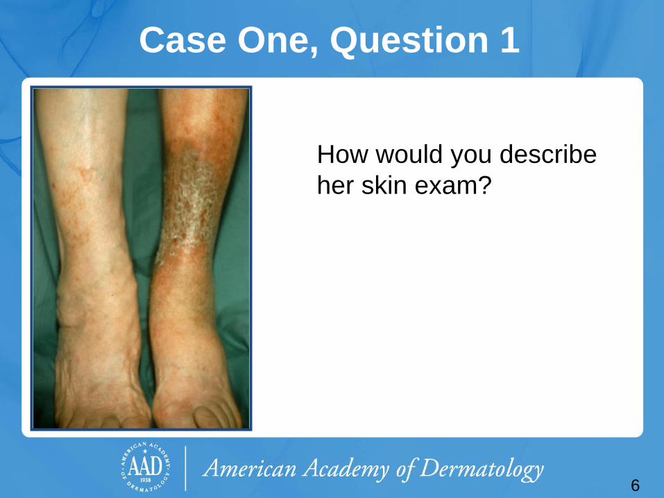
How would you describe her skin exam?
7
Case One, Question 1
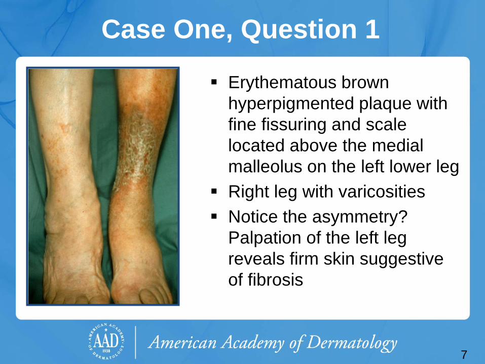
Erythematous brown hyperpigmented plaque with fine fissuring and scale located above the medial malleolus on the left lower leg
Right leg with varicosities Notice the asymmetry?
Palpation of the left leg reveals firm skin suggestive of fibrosis
Case One, Question 2
What is the most likely diagnosis? a. Atopic dermatitis b. Cellulitis c. Erysipelas d. Stasis dermatitis e. Tinea corporis
8
Case One, Question 2 Answer: d What is the most likely diagnosis?
a. Atopic dermatitis (adults with AD have a history of childhood AD and a different distribution of skin involvement)
b. Cellulitis (cellulitis occurs more acutely, presents with fever and pain, more erythema, well-demarcated and without pruritus or scale)
c. Erysipelas (a form of cellulitis caused by acute beta-hemolytic group A streptococcal infection of the skin)
d. Stasis dermatitis e. Tinea corporis (would expect sharply marginated,
erythematous annular patches with central clearing)
9
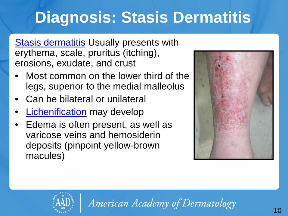
Diagnosis: Stasis Dermatitis Stasis dermatitis Usually presents with erythema, scale, pruritus (itching), erosions, exudate, and crust • Most common on the lower third of the
legs, superior to the medial malleolus • Can be bilateral or unilateral • Lichenification may develop • Edema is often present, as well as
varicose veins and hemosiderin deposits (pinpoint yellow-brown macules)
10
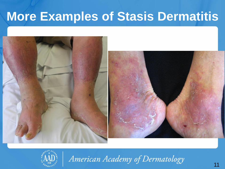
More Examples of Stasis Dermatitis
11
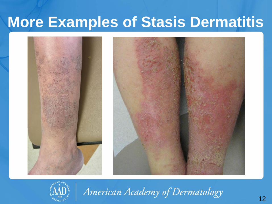
More Examples of Stasis Dermatitis
12
Venous Insufficiency Stasis dermatitis is a cutaneous sign of
venous insufficiency. Normally, venous blood returns from the
superficial venous system via perforating veins into the deep venous system.
Venous stasis occurs when the valves in the deep or perforating veins become incompetent, causing reflux into the superficial system (venous hypertension).
13
Venous Insufficiency
Risk factors for venous insufficiency: • Heredity • Age (older) • Female • Pregnancy
Chronic venous disease is extremely common and is associated with a reduced quality of life secondary to pain, decreased physical function, and mobility
14
• Obesity • Prolonged standing • Greater height
Venous Insufficiency Early signs of venous insufficiency:
• Tenderness • Edema • Hyperpigmentation
Late signs: • Lipodermatosclerosis (subcutaneous fat is
replaced by fibrosis that eventually impedes venous and lymphatic flow leading to edema above the fibrosis)
• Venous ulcers • Scars that appear porcelain white and atrophic
• Telangiectasias • Varicose veins
15
16
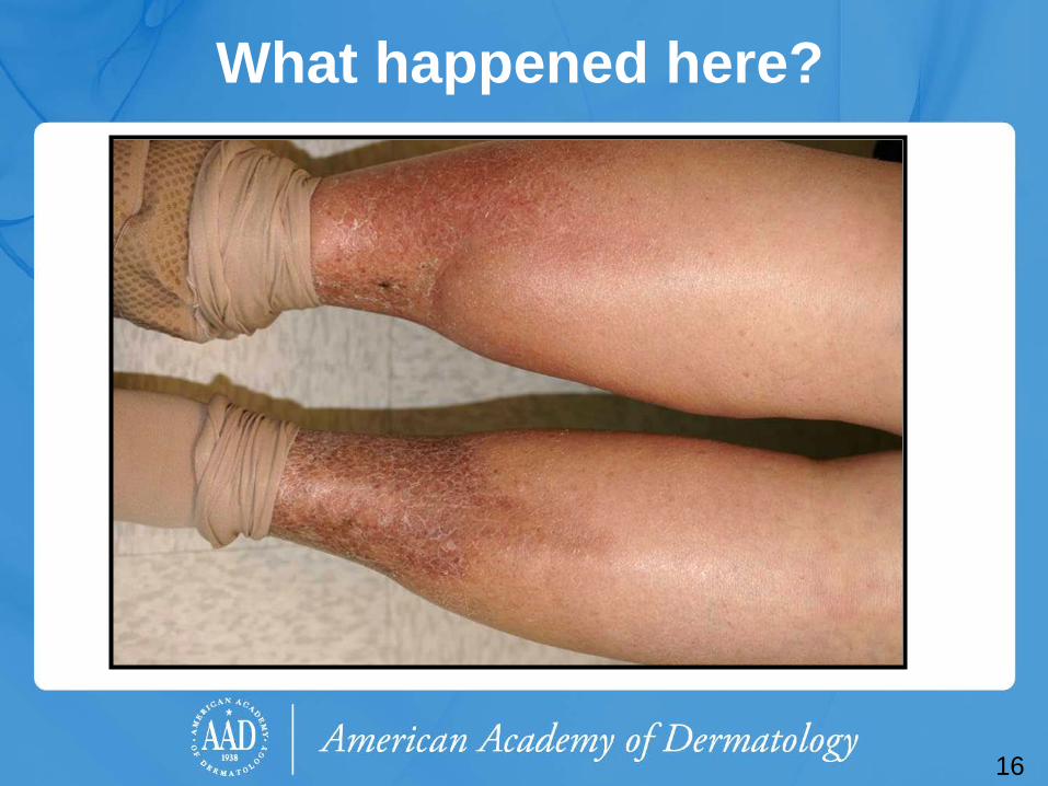
What happened here?
17
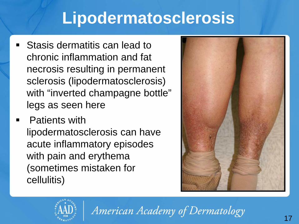
Lipodermatosclerosis Stasis dermatitis can lead to
chronic inflammation and fat necrosis resulting in permanent sclerosis (lipodermatosclerosis) with “inverted champagne bottle” legs as seen here
Patients with lipodermatosclerosis can have acute inflammatory episodes with pain and erythema (sometimes mistaken for cellulitis)
18
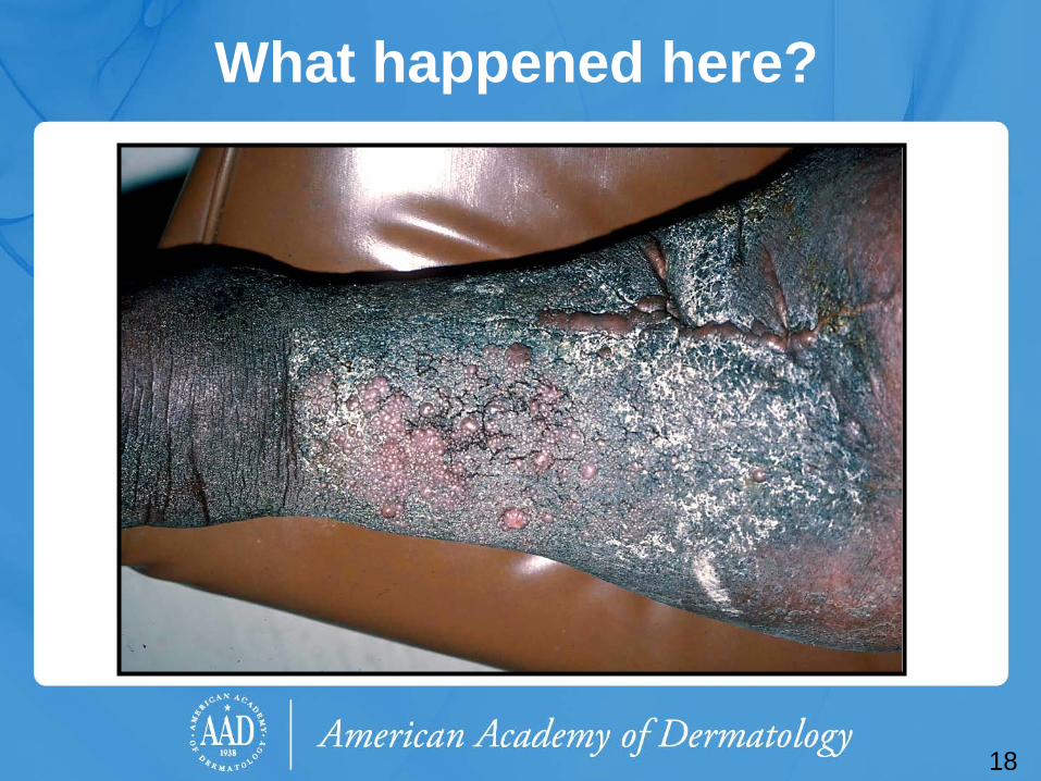
What happened here?
19
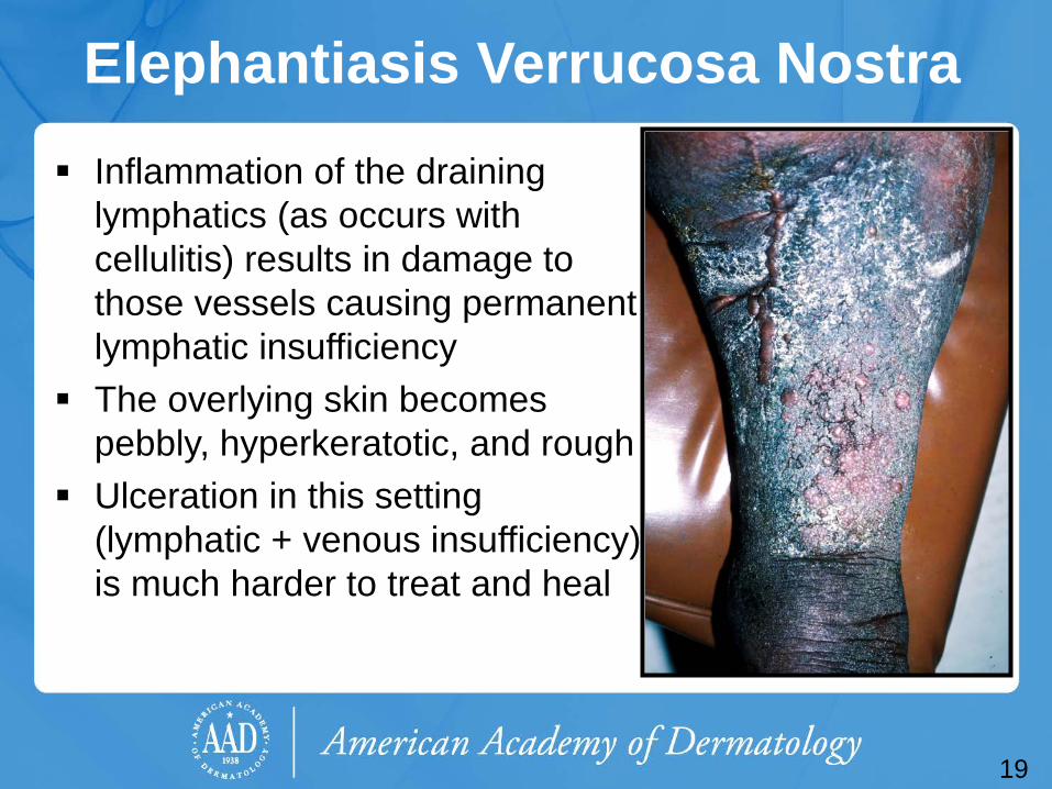
Elephantiasis Verrucosa Nostra Inflammation of the draining
lymphatics (as occurs with cellulitis) results in damage to those vessels causing permanent lymphatic insufficiency
The overlying skin becomes pebbly, hyperkeratotic, and rough
Ulceration in this setting (lymphatic + venous insufficiency) is much harder to treat and heal
Case One, Question 2
Which of the following are complications of venous insufficiency?
a. Cellulitis b. Contact dermatitis c. Recurrent ulceration d. Venous thrombosis e. All of the above
20
Case One, Question 2 Answer: e Which of the following are complications of venous insufficiency?
a. Cellulitis b. Contact dermatitis c. Recurrent ulceration d. Venous thrombosis e. All of the above
21
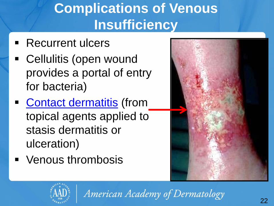
Complications of Venous Insufficiency
Recurrent ulcers Cellulitis (open wound
provides a portal of entry for bacteria)
Contact dermatitis (from topical agents applied to stasis dermatitis or ulceration)
Venous thrombosis
22
Leg Ulcers and Contact Dermatitis
Leg ulcers are at risk for sensitization to products used to treat wound healing, causing contact dermatitis.
This is due to the intrinsic allergenic properties of many ointments and wound products, the duration of use, and the disrupted skin barrier.
The resultant dermatitis exacerbates poor wound healing and/or recurrence of leg ulcers.
23
Stasis Dermatitis: Treatment
One must treat both the dermatitis and the underlying venous insufficiency • Application of super-high and high
potency steroids to area of dermatitis • Elevation (to reduce edema) • Compression therapy with leg wraps • Change wraps weekly, or more often if the
lesion is very weepy
24
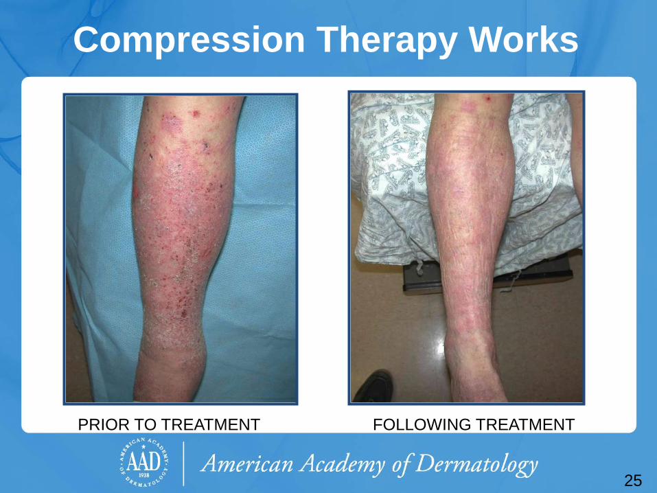
Compression Therapy Works
PRIOR TO TREATMENT
25
FOLLOWING TREATMENT

Case Two
Mr. Patrick Baily
26
Case Two: History HPI: Mr. Baily is a 50-year-old man presenting to his primary care
provider with pain in his left leg. He developed a “weeping spot” a few weeks ago, which he tried treating with an over-the-counter antibiotic ointment.
PMH: history of a DVT 5 years ago after a transatlantic flight, no longer on anticoagulation, hypertension, type 2 diabetes
Medications: thiazide diuretic, ACE-inhibitor, glyburide, metformin Allergies: none Family history: father with type 2 diabetes and hypertension Social history: lives with wife in an apartment, works in
construction Health-related behaviors: smokes 1 cigarette/day ROS: as above
27
28
Case Two, Question 1
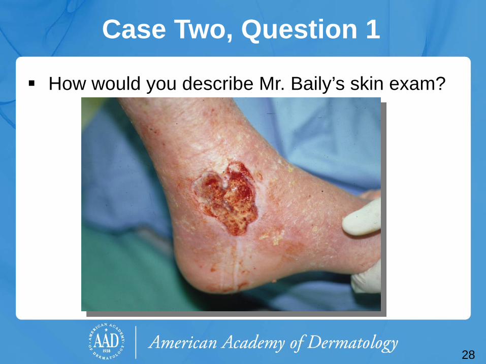
How would you describe Mr. Baily’s skin exam?
29
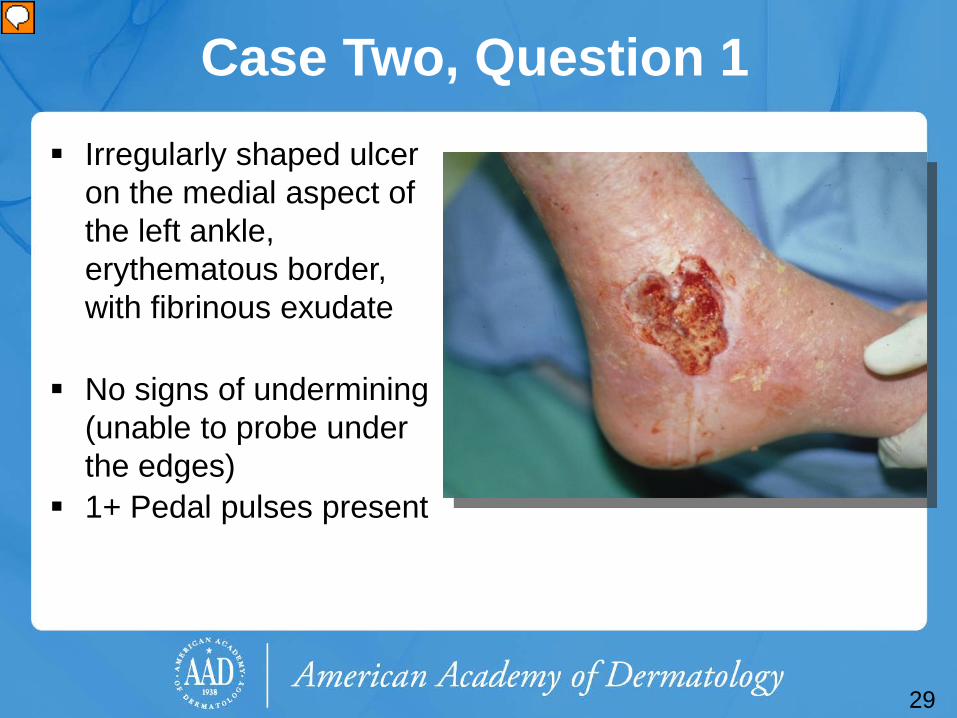
Case Two, Question 1 Irregularly shaped ulcer
on the medial aspect of the left ankle, erythematous border, with fibrinous exudate
No signs of undermining (unable to probe under the edges)
1+ Pedal pulses present
Case Two, Question 2
Given the history and exam, what type of ulcer is on Mr. Baily’s left leg?
a. Arterial b. Diabetic c. Pressure d. Venous e. Auto-inflammatory
30
Case Two, Question 2
Answer: d Given the history and exam, what type of ulcer is on Mr. Baily’s left leg?
a. Arterial b. Diabetic c. Pressure d. Venous e. Auto-inflammatory
31
32
Venous Insufficiency Ulcers Active or healed venous leg ulcers occur in ~ 1% of
the general population Typical appearance is a tender, shallow, irregular
ulcer with a fibrinous base, always occur below the knee • Usually located on the medial ankle or along the line
of the long or short saphenous veins • Accompanied with leg edema, hemosiderin
pigmentation, +/- dermatitis of the leg Patients may experience symptoms of aching or
pain. Discomfort may be relieved by elevation.
Leg Ulcers Causes of chronic leg ulcers include:
• Venous insufficiency 45-60% • Arterial insufficiency 10-20% • Combination of venous and arterial 10-15% • Diabetic 15-25% • Malignancy, vasculitis, collagen-vascular diseases,
and dermal manifestations of systemic disease may present as ulcers on the lower extremity
Smoking and obesity increase the risk for ulcer development and persistence (independent of the underlying cause)
33
Case Two, Question 3
What is the most appropriate next step in evaluating Mr. Baily?
a. Measure the blood pressure in the left arm and left ankle
b. Obtain a skin biopsy c. Treat the ulcer with topical antibiotics d. Use electrocautery to stop the weeping e. Topical steroids to reduce inflammation
34
Case Two, Question 3 Answer: a Which of the following is the most appropriate next step in evaluating Mr. Baily?
a. Measure the blood pressure in the left arm and left ankle (arterial pulse exam and ankle-brachial index is recommended in all patients with clinical diagnosis of venous ulcer)
b. Obtain a skin biopsy (recommended only if atypical features are present and or the ulcer has no improvement after 6 weeks of standard wound care and compression therapy)
c. Treat the ulcer with topical antibiotics (no, in fact topical antibiotic ointments may lead to a contact dermatitis)
d. Use electrocautery to stop the weeping (trauma may worsen the wound instead of improve it)
e. Topical steroids to reduce inflammation (recommended only in the presence of severe dermatitis)
35
Ankle/Brachial Index (ABI) Measure the ABI to exclude arterial occlusive
disease • Compression therapy (used to treat venous
insufficiency) is contraindicated in patients with ABI <0.5 or absolute ankle pressure <60mmHg
The ABI is the ratio of systolic blood pressure in the ankle to the systolic blood pressure in the brachial artery
• Normal: ≥ 0.8 • < 0.8 = indication of peripheral arterial disease
36
Ankle/Brachial Index (ABI) The ABI is reliable except in diabetes (may
be falsely high) The Society for Vascular Surgery/American
Venous Forum Joint Clinical Practice Guidelines recommend: pulse exam, Venous Duplex Ultrasound and ABI in all patients presenting with clinical diagnosis of venous ulcer.
37
Venous Ulcers: Evaluation Exam notes should document the ulcer size and location. Physical exam should include the evaluation of peripheral
pulses, capillary refill time, peripheral neuropathy, and deep tendon reflexes
Duplex ultrasound documents the presence and etiology of venous insufficiency. Venous plethysmography is only recommended if Duplex ultrasound does not provide diagnostic information. • Findings may warrant surgical intervention with endoscopic
venous laser ablation, which may prevent further complication, helpful for patients with pathologic perforating veins
38
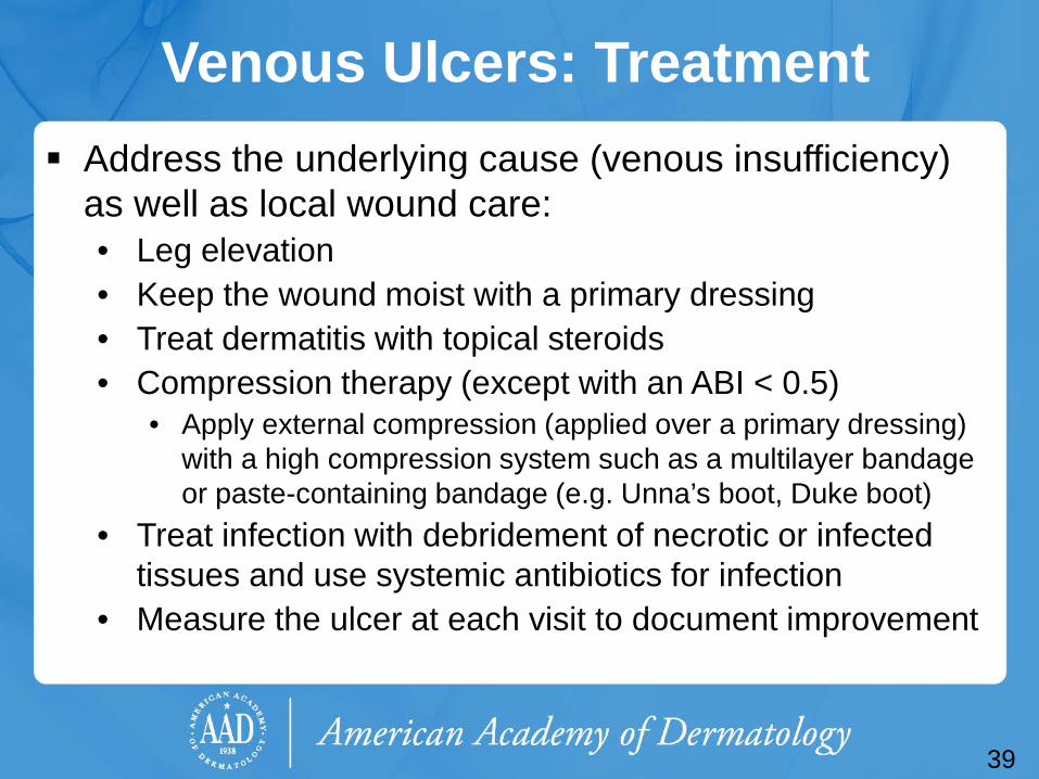
Venous Ulcers: Treatment Address the underlying cause (venous insufficiency)
as well as local wound care: • Leg elevation • Keep the wound moist with a primary dressing • Treat dermatitis with topical steroids • Compression therapy (except with an ABI < 0.5)
• Apply external compression (applied over a primary dressing) with a high compression system such as a multilayer bandage or paste-containing bandage (e.g. Unna’s boot, Duke boot)
• Treat infection with debridement of necrotic or infected tissues and use systemic antibiotics for infection
• Measure the ulcer at each visit to document improvement
39
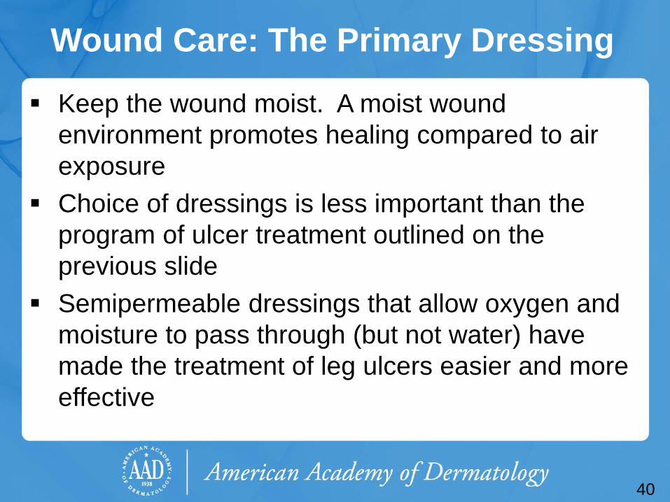
Wound Care: The Primary Dressing
Keep the wound moist. A moist wound environment promotes healing compared to air exposure
Choice of dressings is less important than the program of ulcer treatment outlined on the previous slide
Semipermeable dressings that allow oxygen and moisture to pass through (but not water) have made the treatment of leg ulcers easier and more effective
40
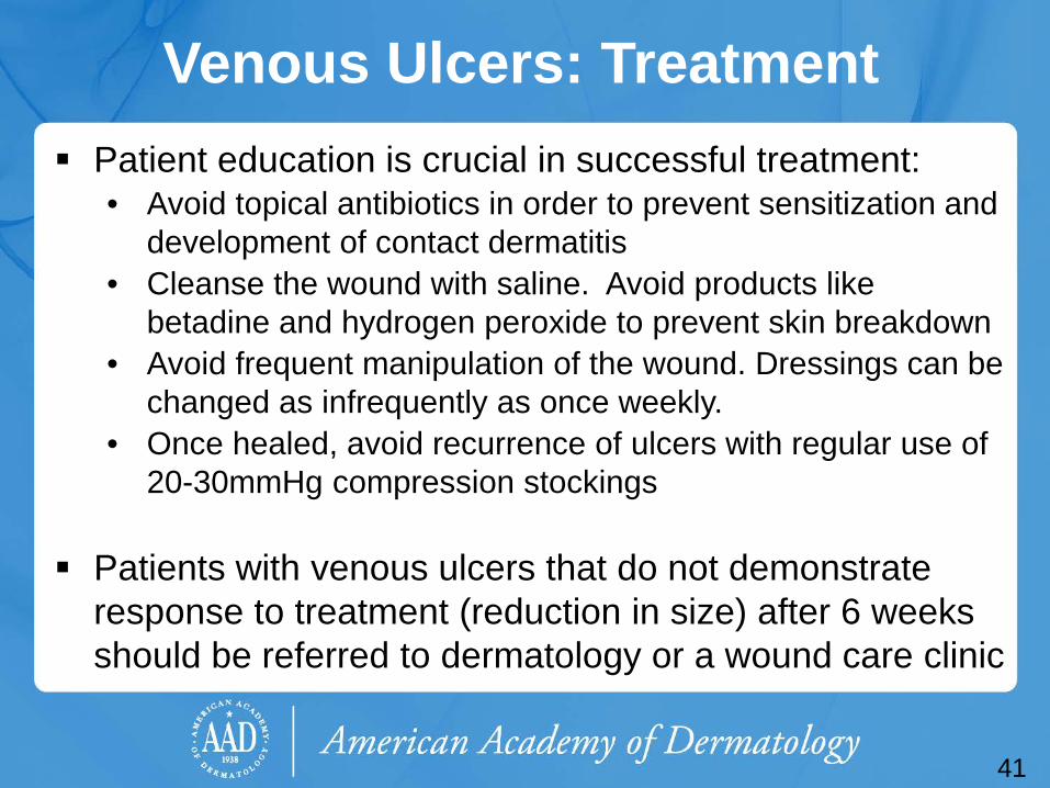
Venous Ulcers: Treatment Patient education is crucial in successful treatment:
• Avoid topical antibiotics in order to prevent sensitization and development of contact dermatitis
• Cleanse the wound with saline. Avoid products like betadine and hydrogen peroxide to prevent skin breakdown
• Avoid frequent manipulation of the wound. Dressings can be changed as infrequently as once weekly.
• Once healed, avoid recurrence of ulcers with regular use of 20-30mmHg compression stockings
Patients with venous ulcers that do not demonstrate response to treatment (reduction in size) after 6 weeks should be referred to dermatology or a wound care clinic
41
Case Three
Mr. Robert Lund
42
Case Three: History HPI: Mr. Lund is a 60-year-old man presenting to his primary care
provider with a painful “sore” on his right lateral leg. He reports “cramping pain” in his calves when walking, but the current pain is more localized to the skin.
PMH: hyperlipidemia, hypertension, angina (stable) Medications: statin, thiazide diuretic, sublingual nitroglycerin when
needed, aspirin Allergies: NKDA Family history: father with an MI at age 65, mother with diabetes Social history: lives with his wife, works in sales, 2 grown children Health-related behavior: smokes ½ pack of cigarettes/day, one
glass of wine nightly, no drug use ROS: no shortness of breath or recent chest pain
43
44
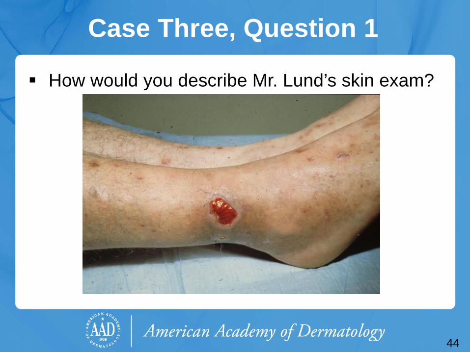
Case Three, Question 1
How would you describe Mr. Lund’s skin exam?
45
Case Three, Question 1
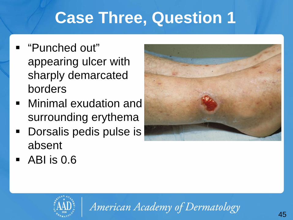
“Punched out” appearing ulcer with sharply demarcated borders
Minimal exudation and surrounding erythema
Dorsalis pedis pulse is absent
ABI is 0.6
Arterial Ulcers
Arterial ulcers are caused by peripheral arterial disease
Occur on the lower leg, usually over sites of pressure and trauma: pretibial, supramalleolar, and at distant points, such as toes and heels
Appear “punched out,” with well-demarcated edges and a pale base
Exudation is minimal Associated findings of ischemia include loss of
hair on feet and lower legs, shiny atrophic skin
46
47
Arterial Ulcers
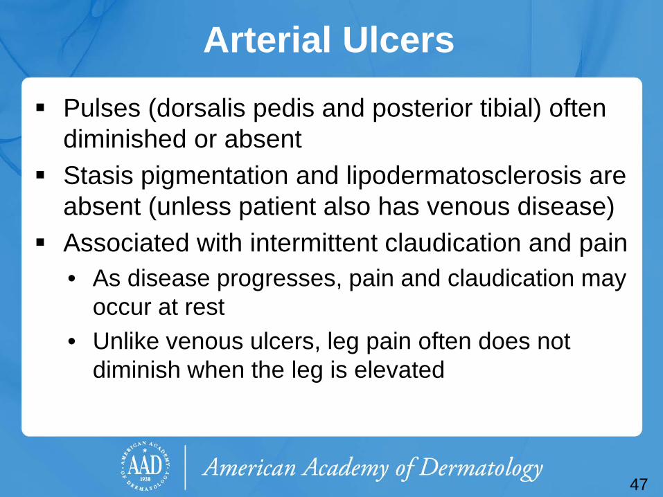
Pulses (dorsalis pedis and posterior tibial) often diminished or absent
Stasis pigmentation and lipodermatosclerosis are absent (unless patient also has venous disease)
Associated with intermittent claudication and pain • As disease progresses, pain and claudication may
occur at rest • Unlike venous ulcers, leg pain often does not
diminish when the leg is elevated

Case Three, Question 2
Which of the following recommendations should take priority?
a. Encourage him to ambulate b. Encourage him to stop smoking c. Make sure his blood pressure and
hyperlipidemia are under good control d. Refer to a vascular surgeon
48
Case Three, Question 2 Answer: d Which of the following recommendations should take priority?
a. Encourage him to ambulate b. Encourage him to stop smoking c. Make sure his blood pressure and hyperlipidemia
are under good control d. Refer to a vascular surgeon (although all the
answer choices are correct, the main goal of therapy is the re-establishment of adequate arterial supply)
49
Arterial Ulcers: Treatment
Refer to a vascular surgeon for restoration of arterial blood flow with percutaneous or surgical arterial reconstruction
Patients should stop smoking, optimize control of diabetes, hypertension, and hyperlipidemia
Weight loss and exercise are also helpful All types of ulcers require proper wound care as
outlined above in venous ulcer treatment
50

Case Four
Mr. Ryan Stricklin
51
Case Four: History HPI: Mr. Stricklin is a 46-year-old man who presents to his
primary care provider with lesions on the bottom of his foot. He noticed these lesions a few months ago when he was changing his socks at the gym. He reports keeping them clean with hydrogen peroxide.
PMH: type 1 diabetes x 25 years, hernia repair 20 years ago Medications: insulin (glargine and regular) Allergies: none Family history: noncontributory Social history: lives alone, works as a realtor Health-related behaviors: no tobacco, alcohol, or dug use ROS: no fevers, sweats or chills
52
53
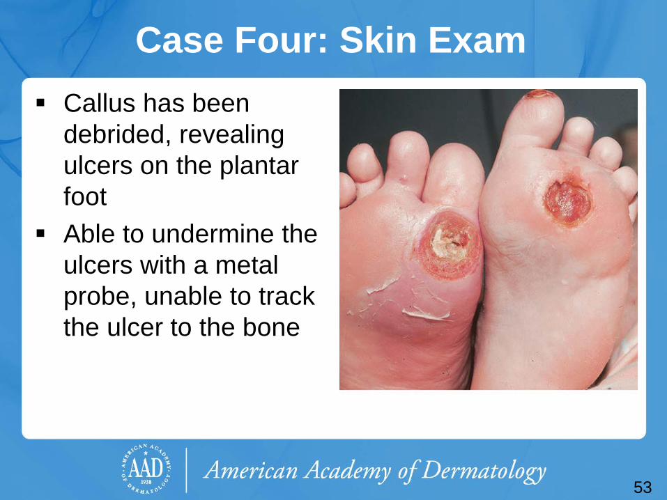
Case Four: Skin Exam Callus has been
debrided, revealing ulcers on the plantar foot
Able to undermine the ulcers with a metal probe, unable to track the ulcer to the bone
54
Diabetic (Neuropathic) Foot Ulcers
Peripheral neuropathy, pressure, insufficient microvasculature and trauma play prominent roles in the development of diabetic ulcers
Usually located on the plantar surface under the metatarsal heads or on the toes
Repetitive mechanical forces lead to callus, which is the most important preulcerative lesion in the neuropathic foot
55
Diabetic (Neuropathic) Foot Ulcers
Lifetime risk of a person with diabetes developing a foot ulcer is as high as 25%
Risk factors for foot ulcers include: • Cigarette smoking • Past foot ulcer history • Peripheral vascular dz • Previous amputation
• Poor glycemic control • Peripheral neuropathy • Diabetic nephropathy • Visual impairment
56
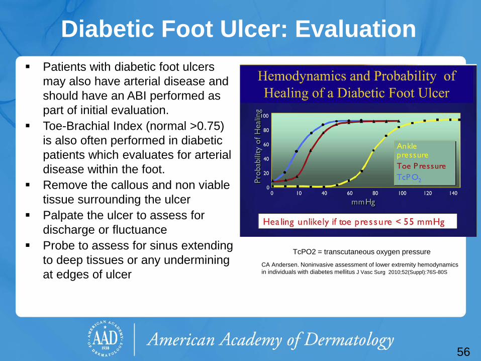
Diabetic Foot Ulcer: Evaluation Patients with diabetic foot ulcers
may also have arterial disease and should have an ABI performed as part of initial evaluation.
Toe-Brachial Index (normal >0.75) is also often performed in diabetic patients which evaluates for arterial disease within the foot.
Remove the callous and non viable tissue surrounding the ulcer
Palpate the ulcer to assess for discharge or fluctuance
Probe to assess for sinus extending to deep tissues or any undermining at edges of ulcer
CA Andersen. Noninvasive assessment of lower extremity hemodynamics in individuals with diabetes mellitus J Vasc Surg 2010;52(Suppl):76S-80S
TcPO2 = transcutaneous oxygen pressure
Diabetic Foot Ulcer: Treatment Diabetic patients with foot ulcers are best managed in a
multidisciplinary setting (vascular surgery if indicated, wound care specialists, endocrinologists, physical therapy, dietician)
Order an imaging study if concerned about osteomyelitis Plain radiographs 33% sensitive initially90% sensitive at 4 weeks
of symptoms MRI sensitivity is 92-100% but can’t be used in patients with
implanted devices
Patients with findings of osteomyelitis should be admitted to the hospital for further evaluation and treatment
57
Diabetic Foot Ulcer: Evaluation and Treatment
Most importantly, protect the ulcer from excessive pressure • Offloading devices with total contact casts or orthotic
inserts (a podiatrist with expertise in the management of the diabetic foot is extremely helpful)
• Restrict weight bearing of the involved extremity Dressings should maintain a moist environment Application of platelet-derived growth factor gel has
been shown to improve wound healing in diabetic foot ulcers
Very little evidence to support expensive wound-vac or negative pressure wound care devices.
58
Case Four, Question 1 Which of the following statements about Mr. Stricklin is likely to be true?
a. He has diabetic neuropathy b. He should continue to use hydrogen
peroxide to keep his lesions clean c. He should wear open-toed shoes d. None of the above
59
Case Four, Question 1 Answer: a Which of the following statements about Mr. Stricklin is likely to be true?
a. He has diabetic neuropathy (diabetic neuropathy can cause a loss of protective pain sensation as well as motor dysfunction)
b. He should continue to use hydrogen peroxide to keep his lesions clean (not true. Hydrogen peroxide interferes with wound healing)
c. He should wear open-toed shoes (diabetic patients should avoid open-toed and pointed shoes)
d. None of the above
60
Diabetic Foot Ulcers: Prevention Education about ulcer prevention should be provided for all diabetic patients
• Glycemic control is essential in preventing diabetes associated complications, including peripheral neuropathy
• Patients should receive annual foot examinations, with a clinical assessment for peripheral vascular disease and monofilament test for peripheral neuropathy
• Patients should examine their own feet regularly • If present, treat tinea pedis (to prevent the associated skin barrier
disruption) • Encourage smoking cessation (risk factor for vascular disease and
neuropathy) • Optimize treatment of hypertension, hyperlipidemia, and obesity
61

Case Five
Mrs. Melinda Dellinger
62
Case Five: History HPI: Mrs. Dellinger is a 50-year-old woman presenting to her
primary medical provider with a 4-day history of new, very painful lesions on her hand and thigh. She initially thought these lesions were bug bites, but they now appear to be expanding and look more like ulcers.
PMH: inflammatory bowel disease (well-controlled) Medications: sulfasalazine daily, multivitamin, fish oil Allergies: no known drug allergies Family history: brother with ulcerative colitis Social history: lives with husband and 20-year-old daughter, works
full-time as a high school teacher Health-related behaviors: reports no alcohol, tobacco, or drug use ROS: no fevers, joint pains, abdominal pain or diarrhea
63
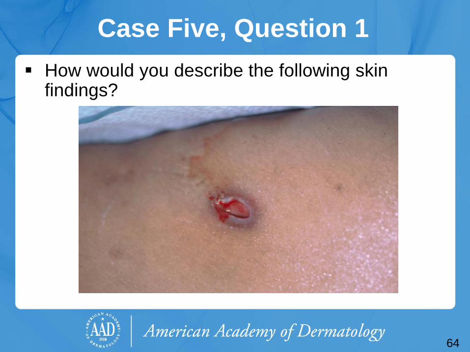
Case Five, Question 1 How would you describe the following skin
findings?
64
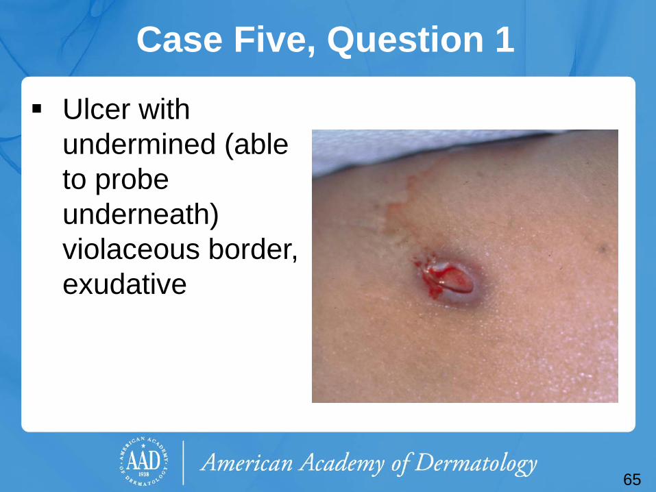
Case Five, Question 1
Ulcer with undermined (able to probe underneath) violaceous border, exudative
65
Case Five, Question 2 Given the history and exam findings, Mrs. Dellinger’s primary care provider is concerned about pyoderma gangrenosum (PG) and made an urgent referral to the dermatology clinic. Which of the following is true about PG?
a. A biopsy of PG is diagnostic b. Debridement of the ulcer will help the healing
process c. PG is a slow process d. PG is often mistaken as a spider bite e. PG is painless
66
Case Five, Question 2 Answer: d Which of the following is true about PG?
a. A biopsy of PG is diagnostic (Not true. Histology can look similar to an infection)
b. Debridement of the ulcer ill help the healing process (No! In fact, PG is triggered and made worse by trauma – a process called pathergy)
c. PG is a slow process (Not true. PG rapidly progresses) d. PG is often mistaken as a spider bite (True! In fact, if
you think the patient has a spider bite, we recommend you consider PG or MRSA in the differential)
e. PG is painless (Not true. PG is often very painful)
67
Pyoderma Gangrenosum (PG) PG is an auto-inflammatory ulcerative process mediated
by an influx of neutrophils into the dermis Begins as a small sterile pustule which breaks down and
rapidly expands forming an ulcer with an undermined violaceous border
Satellite ulcerations may merge with the central larger ulcer
Rapid progression (days to weeks) Can occur anywhere on the body (most frequently
occurs on the lower extremities) Can be very painful
68
Pyoderma Gangrenosum PG is triggered by trauma (pathergy), including insect
bites, surgical debridement, attempts to graft • PG is often misdiagnosed as a brown recluse spider bite or
an infection and is debrided, which worsens the condition The majority of patients with PG do not have an underlying
condition, PG is often associated with a wide range of other pathologies that the patient should be evaluated for • Inflammatory bowel disease (1.5%-5% of patients get PG),
rheumatoid arthritis, hematologic gammopathies, aerodigestive malignancies
1/3 of PG patients have arthritis: seronegative, asymmetric, monoarticular, large joint
69
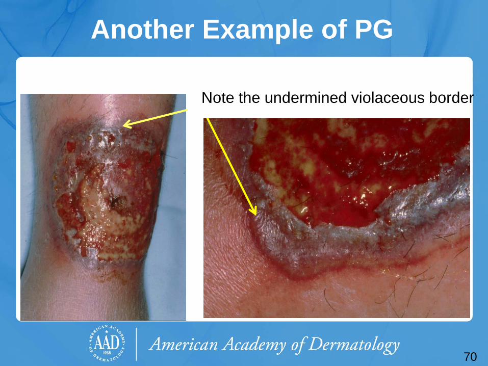
Another Example of PG
Note the undermined violaceous border
70
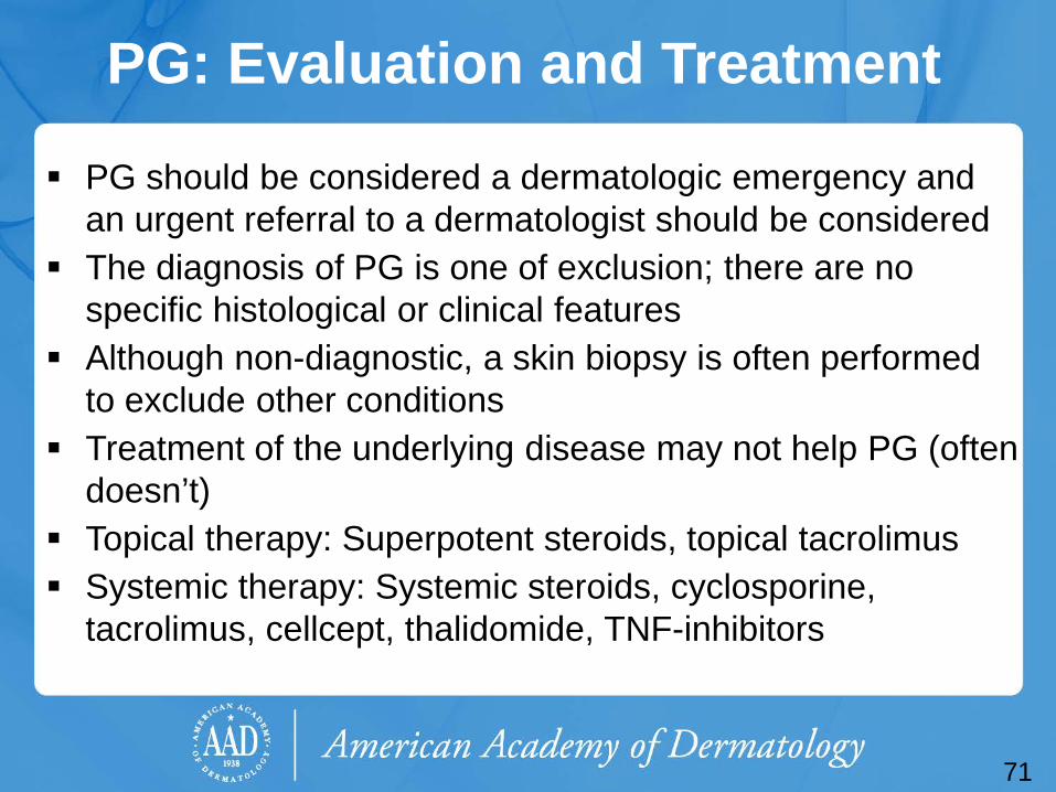
PG: Evaluation and Treatment
PG should be considered a dermatologic emergency and an urgent referral to a dermatologist should be considered
The diagnosis of PG is one of exclusion; there are no specific histological or clinical features
Although non-diagnostic, a skin biopsy is often performed to exclude other conditions
Treatment of the underlying disease may not help PG (often doesn’t)
Topical therapy: Superpotent steroids, topical tacrolimus Systemic therapy: Systemic steroids, cyclosporine,
tacrolimus, cellcept, thalidomide, TNF-inhibitors
71
72
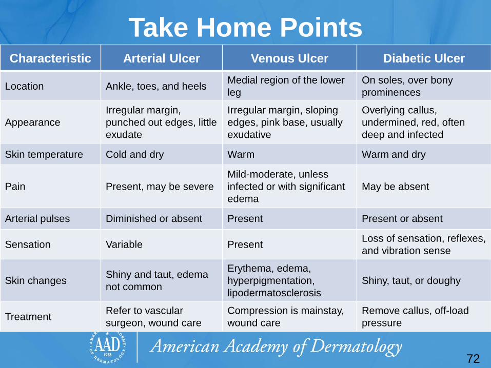
Take Home Points Characteristic Arterial Ulcer Venous Ulcer Diabetic Ulcer
Location Ankle, toes, and heels Medial region of the lower leg
On soles, over bony prominences
Appearance Irregular margin, punched out edges, little exudate
Irregular margin, sloping edges, pink base, usually exudative
Overlying callus, undermined, red, often deep and infected
Skin temperature Cold and dry Warm Warm and dry
Pain Present, may be severe Mild-moderate, unless infected or with significant edema
May be absent
Arterial pulses Diminished or absent Present Present or absent
Sensation Variable Present Loss of sensation, reflexes, and vibration sense
Skin changes Shiny and taut, edema not common
Erythema, edema, hyperpigmentation, lipodermatosclerosis
Shiny, taut, or doughy
Treatment Refer to vascular surgeon, wound care
Compression is mainstay, wound care
Remove callus, off-load pressure
Take Home Points Stasis dermatitis is a cutaneous marker for venous
insufficiency The most common types of leg ulcers include venous,
arterial, combined (venous and arterial), and diabetic Diagnosis of leg ulcers may be made clinically, but
evaluation with non-invasive vascular imaging and the ABI will guide treatment
Treatment of venous leg ulcers includes leg elevation, compression, and wound care
Patients with abnormal ABI or Doppler findings should be referred to a vascular surgeon for evaluation.
73
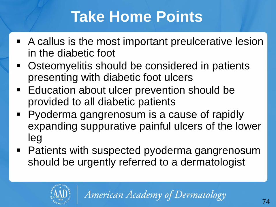
Take Home Points A callus is the most important preulcerative lesion
in the diabetic foot Osteomyelitis should be considered in patients
presenting with diabetic foot ulcers Education about ulcer prevention should be
provided to all diabetic patients Pyoderma gangrenosum is a cause of rapidly
expanding suppurative painful ulcers of the lower leg
Patients with suspected pyoderma gangrenosum should be urgently referred to a dermatologist
74
Acknowledgements This module was developed by the American
Academy of Dermatology Medical Student Core Curriculum Workgroup from 2008-2012.
Primary authors: Sarah D. Cipriano, MD, MPH; Timothy G. Berger, MD, FAAD.
Peer reviewers: Theodora Moro, MD; Patrick McCleskey, MD, FAAD; Peter A. Lio, MD, FAAD.
Revisions and editing: Heather W. Wickless, MD, MPH, FAAD.
Last revised January 2016.
75
End of the Module O’Donnell TF, Passman MA, et.al.: Management of venous leg ulcers: Clinical
practice guidelines of the Society for Vascular Surgery and the American Venous Forum. SVS/AVF Joint Clinical Practice Guidelines Committee—Venous Leg Ulcer. J.Vasc Surg 2014; 60: 3S-59s
Brownrigg JB, Apelqvist J, et.al.: Evidence-based management of PAD and the diabetic foot. Eur.J.Vasc and Endovasc Surg; 45: June 2013 673-681.
Berger T, Hong J, Saeed S, Colaco S, Tsang M, Kasper R. The Web-Based Illustrated Clinical Dermatology Glossary. MedEdPORTAL; 2007. Available from: www.mededportal.org/publication/462.
Boulton AJM, et al. Comprehensive Foot Examination and Risk Assessment. A report of the Task Force of the Foot Care Interest Group of the American Diabetes Association, with endorsement by the American Association of Clinical Endocrinologists. Diabetes Care. 2008;31:1679-1685.
Burton Claude S, Burkhart Craig, Goldsmith Lowell A, "Chapter 175. Cutaneous Changes in Venous and Lymphatic Insufficiency" (Chapter). Wolff K, Goldsmith LA, Katz SI, Gilchrest B, Paller AS, Leffell DJ: Fitzpatrick's Dermatology in General Medicine, 7e.
76
End of the Module James WD, Berger TG, Elston DM, “Chapter 35. Cutaneous Vascular Diseases” (chapter).
Andrews’ Diseases of the Skin Clinical Dermatology. 10th ed. Philadelphia, Pa: Saunders Elsevier; 2006: 845-851.
Kalish J, Hamdan A. Management of diabetic foot problems. J Vasc Surg 2010;51:476-486. Miller O. Fred. Leg Ulcer Therapy. Presentation at Geisinger Medical Clinic. 5/2010. Philips T. “Chapter 17. Ulcers” in Bolognia JL, Jorizzo JL, Rapini RP: Dermatology. 2nd ed.
Elsevier;2008: 261-276. Powell Frank C, Hackett Bridget C, "Chapter 32. Pyoderma Gangrenosum" (Chapter). Wolff
K, Goldsmith LA, Katz SI, Gilchrest B, Paller AS, Leffell DJ: Fitzpatrick's Dermatology in General Medicine, 7e: http://www.accessmedicine.com/content.aspx?aID=2952781.
Robson MC, et al. Guidelines for the treatment of venous ulcers. Wound Rep Reg. 2006;14:649-662.
Machet L, Couhe C, et.al. A high prevalence of sensitization still persists in leg ulcer patients: a retrospective series of 106 patients tested between 2001 and 2002 and a meta-analysis of 1975-2003 data. Br.J. Dermatol. 2004; 150: 929-935.
Wolff K, Johnson RA, "Section 16. Skin Signs of Vascular Insufficiency" (Chapter). Wolff K, Johnson RA: Fitzpatrick's Color Atlas & Synopsis of Clinical Dermatology, 6e: http://www.accessmedicine.com/content.aspx?aID=5189520.
77

To take the quiz, click on the following link: https://www.aad.org/quiz/stasis-dermatitis-leg-ulcers-learners
Recommended