Six degree-of-freedom knee joint kinematics in obese individuals with knee pain during gait
CitationLi, Jing-Sheng, Tsung-Yuan Tsai, David T. Felson, Guoan Li, and Cara L. Lewis. 2017. “Six degree-of-freedom knee joint kinematics in obese individuals with knee pain during gait.” PLoS ONE 12 (3): e0174663. doi:10.1371/journal.pone.0174663. http://dx.doi.org/10.1371/journal.pone.0174663.
Published Versiondoi:10.1371/journal.pone.0174663
Permanent linkhttp://nrs.harvard.edu/urn-3:HUL.InstRepos:32630588
Terms of UseThis article was downloaded from Harvard University’s DASH repository, and is made available under the terms and conditions applicable to Other Posted Material, as set forth at http://nrs.harvard.edu/urn-3:HUL.InstRepos:dash.current.terms-of-use#LAA
Share Your StoryThe Harvard community has made this article openly available.Please share how this access benefits you. Submit a story .
Accessibility
RESEARCH ARTICLE
Six degree-of-freedom knee joint kinematics
in obese individuals with knee pain during gait
Jing-Sheng Li1,2, Tsung-Yuan Tsai1, David T. Felson3,4, Guoan Li1, Cara L. Lewis2,3*
1 Bioengineering Laboratory, Department of Orthopaedic Surgery, Massachusetts General Hospital /
Harvard Medical School, Boston, Massachusetts, United States of America, 2 College of Health and
Rehabilitation Science: Sargent College, Boston University, Boston, Massachusetts, United States of
America, 3 Clinical Epidemiology Research and Training Unit, Boston University School of Medicine, Boston,
Massachusetts, United States of America, 4 NIHR Manchester Musculoskeletal Biomedical Research Unit,
Manchester Academic Health Science Centre, Manchester, United Kingdom
Abstract
Knee joint pain is a common symptom in obese individuals and walking is often prescribed
as part of management programs. Past studies in obese individuals have focused on stand-
ing alignment and kinematics in the sagittal and coronal planes. Investigation of 6 degree-
of-freedom (6DOF) knee joint kinematics during standing and gait is important to thoroughly
understand knee function in obese individuals with knee pain. This study aimed to investi-
gate the 6DOF knee joint kinematics in standing and during gait in obese patients using a
validated fluoroscopic imaging system. Ten individuals with obesity and knee pain were
recruited. While standing, the knee was in 7.4±6.3˚of hyperextension, 2.8±3.3˚ of abduction
and 5.6±7.3˚ of external rotation. The femoral center was located 0.7±3.1mm anterior and
5.1±1.5mm medial to the tibial center. During treadmill gait, the sagittal plane motion, i.e.,
flexion/extension and anterior-posterior translation, showed a clear pattern. Specifically,
obese individuals with knee pain maintained the knee in more flexion and more anterior tibial
translation during most of the stance phase of the gait cycle and had a reduced total range
of knee flexion when compared to a healthy non-obese group. In conclusion, obese individu-
als with knee pain used hyperextension knee posture while standing, but maintained the
knee in more flexion during gait with reduced overall range of motion in the 6DOF analysis.
Introduction
The prevalence of obesity is increasing in the United States and throughout the world [1, 2]. In
2011–2012, 34.9% of adults in the United States were obese [3]. Musculoskeletal disorders are
commonly seen in obese individuals and one of the most common and disabling of these is
knee osteoarthritis (OA) [4].
Individuals who are obese and have knee pain may adopt different gait patterns to compen-
sate both for the extra weight and joint pain. While no prior studies have focused specifically
on obese persons with knee pain, studies of obese adults showed that they walked with
decreased velocity [5, 6]. As body mass index (BMI) increases, gait speed decreases [7]. Past
PLOS ONE | https://doi.org/10.1371/journal.pone.0174663 March 24, 2017 1 / 11
a1111111111
a1111111111
a1111111111
a1111111111
a1111111111
OPENACCESS
Citation: Li J-S, Tsai T-Y, Felson DT, Li G, Lewis CL
(2017) Six degree-of-freedom knee joint
kinematics in obese individuals with knee pain
during gait. PLoS ONE 12(3): e0174663. https://
doi.org/10.1371/journal.pone.0174663
Editor: John Leicester Williams, University of
Memphis, UNITED STATES
Received: August 22, 2016
Accepted: March 13, 2017
Published: March 24, 2017
Copyright: © 2017 Li et al. This is an open access
article distributed under the terms of the Creative
Commons Attribution License, which permits
unrestricted use, distribution, and reproduction in
any medium, provided the original author and
source are credited.
Data Availability Statement: All relevant data are
within the paper and its Supporting Information
files.
Funding: This work was supported by the National
Institutes of Health, P60AR47785, to David T
Felson and the National Institutes of Health,
AR063235, to Cara L Lewis. The funders had no
role in study design, data collection and analysis,
decision to publish, or preparation of the
manuscript.
Competing interests: The authors have declared
that no competing interests exist.
studies of knee joint kinematics mainly focused on sagittal and coronal plane motions, i.e.,
knee joint flexion/extension [5, 8–10] and varus/valgus rotation [9, 10]. Even so, there is no
clear consensus on knee joint kinematics in obese individuals during walking. For example,
Haight et al.[8] reported that obese individuals walked with a less flexed knee during the stance
phase compared to non-obese individuals. Vismara et al. [11] concluded that the range of knee
flexion excursion during gait was not significantly different than a healthy group. The incon-
sistency and variation in the literature may be due to differences in measurement methods or
the presence of different lower extremity joint pathology such as pain which is extremely com-
mon in obese adults and may cause gait modifications. For instance, knee pain is a major
symptom in individuals with knee OA and reduced range of knee flexion during gait has been
frequently reported [12–14].
Most previous investigations of obese gait used skin marker motion analysis systems [5, 6,
8, 15]. The kinematic data derived from a skin marker-based motion capture system are vul-
nerable to soft tissue artifacts [16, 17]. According to Peters et al.[16] the magnitudes of soft tis-
sue artifacts were greater than 30 mm on the femur and up to 15 mm on the tibia. Cappozo
et al. [18] quantified rotation errors of 6–20˚ on the femur and 4–10˚ on the tibia [18].
Although some biomechanical researchers have tried to reduce soft tissue artifact by using an
obesity-specific marker set to investigate gait patterns [15], accurate detailed kinematics in 6
degree-of-freedom (6DOF) among obese individuals have not been elucidated. As past studies
have shown that standing posture and gait pattern were affected by body weight [5, 8–10, 19],
a better understanding of the standing posture and gait pattern in 6DOF is critical. Such infor-
mation would provide a foundation on which to build better walking programs, which are
commonly suggested as a means to increase the energy expenditure of obese individuals [20,
21] and especially to design a treatment program for obese persons with knee pain.
Recent advancement of imaging technology, such as the dual fluoroscopy imaging system
(DFIS), makes it possible to track in-vivo bone motion without soft tissue artifacts [22–24].
This methodology has been validated[25] with submillimeter and subdegree accuracy in trans-
lation and rotation. In this study, we evaluated knee joint kinematics in standing and during
gait in obese individuals with knee pain using DFIS[25] and compared them to a healthy non-
obese control group without knee pain. We hypothesized that there would be distinct motion
patterns in obese individuals with knee pain which would differ from typical patterns in a
healthy population.
Methods
Participants
Ten obese individuals with knee pain on most of the last 30 days (8 females, 2 males; age (mean
±SD): 42.8±10.1 (range: 30.2–56.5) years; BMI: 39.6±2.8 (range: 35.3–44.3) kg/m2, body mass:
109.6±13.0 (range: 86.0–130.9) kg, body height: 166.1±8.5 (range: 152–184) cm) were recruited
to participate in this study and the study protocol was approved by Boston University School of
Medicine and Massachusetts General Hospital Institutional Review Boards. Written informed
consent approved by both Institutional Review Boards was obtained for each subject. All partic-
ipants reported being able to walk without assistance. For each participant, the more painful
knee was selected for evaluation except that knees that had undergone surgery were excluded.
If knee pain increased sharply in a short period of walking, the participant was also excluded.
Experiment procedures
A posteroanterior standing knee joint plain radiograph in a slight flexion position was taken
for each participant in the obesity group and graded by an experienced rheumatologist (DTF)
Obese knee gait pattern
PLOS ONE | https://doi.org/10.1371/journal.pone.0174663 March 24, 2017 2 / 11
to determine the severity of osteoarthritis using Kellgren-Lawrence grading scale[26]. The
selected knee was then scanned by a 3-Tesla MR machine (Philips, Achieva, Eindhoven,
The Netherlands) with a sagittal Proton Density-Weighted (PDW), Spectral Attenuated Inver-
sion Recovery (SPAIR) sequence (FOV: 160mm x 160mm, TR = 1800ms, TE = 30ms, flip
angle = 90˚, thickness = 1mm, in-plane resolution = 512 x 512). All MR images were reviewed
and used for segmentation to construct a 3-dimensional (3D) bony surface model of the knee,
including the femur and tibia. To better understand the knee joint pain status, we adopted two
relevant questions, one from the Western Ontario and McMaster Universities Osteoarthritis
Index (WOMAC) questionnaire (pain on 4-point Likert scale while walking on a flat surface)
and one on overall pain level using a visual analog scale. The Likert scale was scored 0 as no
pain to 4 as extreme pain and visual analog scale was rated on a 0–100 scale.
In the fluoroscopic experiment, one pair of dual fluoroscopic images of the knee in static
standing was obtained to evaluate comfortable standing posture in the participants with
obesity. The participant was then asked to walk on a treadmill at 1.5mph (0.67m/s) with a thy-
roid collar over their throat and lead apron over their chest to upper thigh. After a warm-up
period on the treadmill, the knee was imaged by the DFIS (Philips, BV Pulsera, Eindhoven,
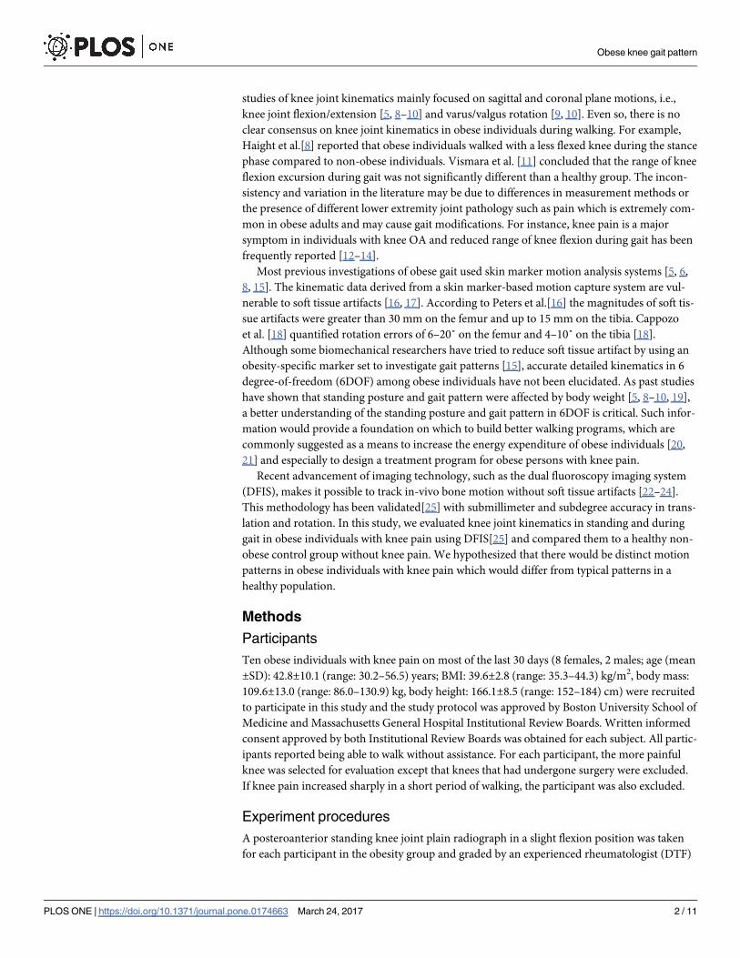
The Netherlands) at 30 frames per second with an 8ms pulse-width (Fig 1A) [22, 25]. This sys-
tem captured knee motion along two oblique views (medioposterior-lateroanterior and latero-
posterior-medioanterior) (Fig 1B). All the output images were corrected for distortion using a
calibration grid and customized algorithm [27, 28] developed on MATLAB software (Math-
work, Natick, MA, USA). The fluoroscopic images and the MR-based 3D knee bony models
were then imported to a virtual fluoroscopic environment for 2D-3D registration procedure
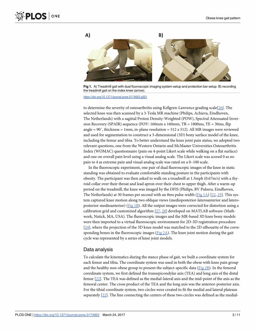
[24], where the projection of the 3D knee model was matched to the 2D silhouette of the corre-
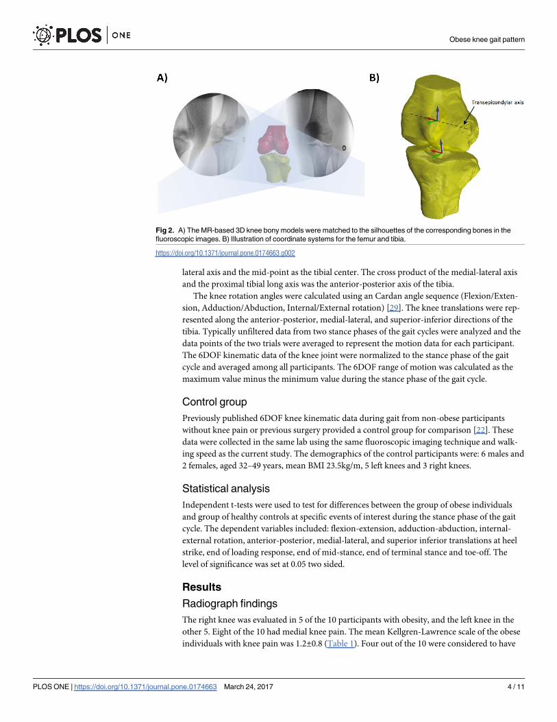
sponding bones in the fluoroscopic images (Fig 2A). The knee joint motion during the gait
cycle was represented by a series of knee joint models.
Data analysis
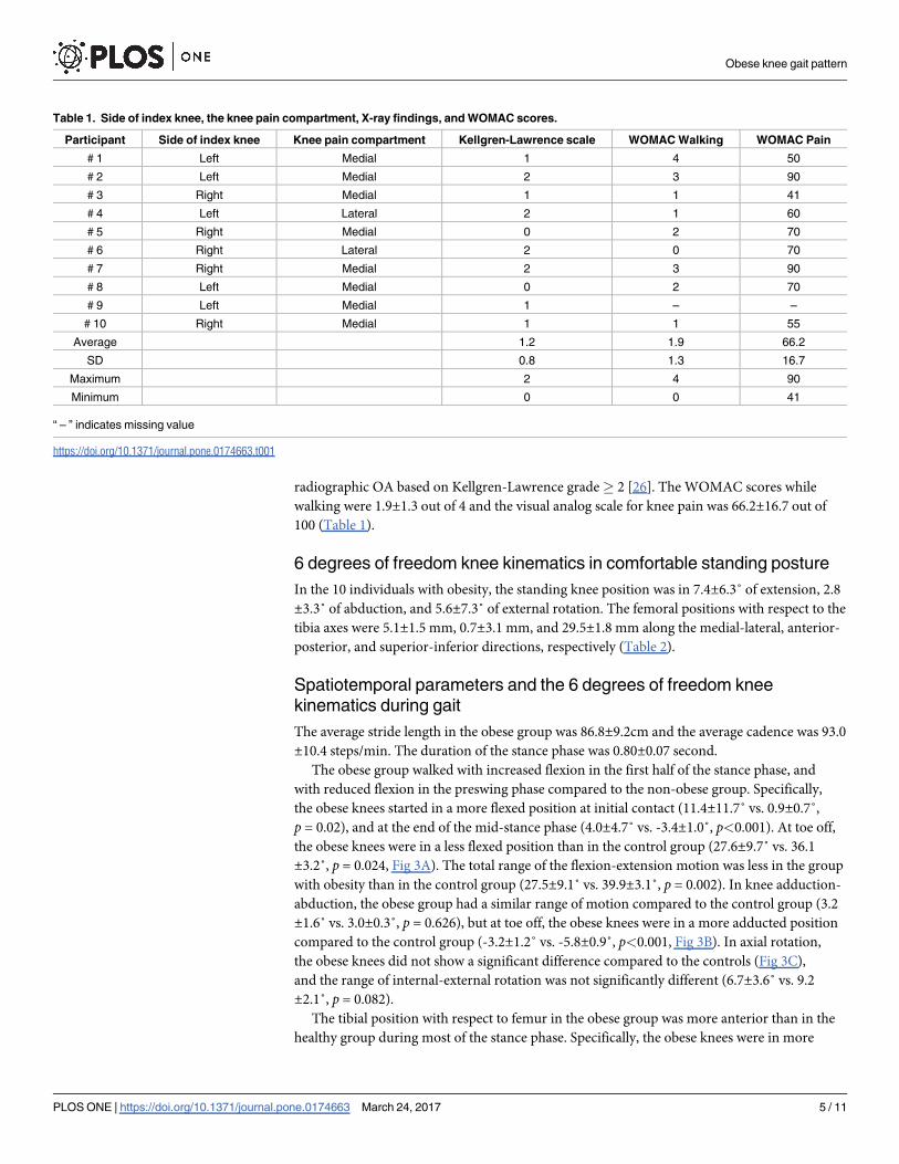
To calculate the kinematics during the stance phase of gait, we built a coordinate system for
each femur and tibia. The coordinate system was used in both the obese with knee pain group
and the healthy non-obese group to present the subject-specific data (Fig 2B). In the femoral
coordinate system, we first defined the transepicondylar axis (TEA) and long axis of the distal
femur [22]. The TEA was defined as the medial-lateral axis and the mid-point of the axis as the
femoral center. The cross product of the TEA and the long axis was the anterior-posterior axis.
For the tibial coordinate system, two circles were created to fit the medial and lateral plateaus
separately [22]. The line connecting the centers of these two circles was defined as the medial-
Fig 1. A) Treadmill gait with dual fluoroscopic imaging system setup and protection bar setup. B) recording
the treadmill gait on the index knee (arrow).
https://doi.org/10.1371/journal.pone.0174663.g001
Obese knee gait pattern
PLOS ONE | https://doi.org/10.1371/journal.pone.0174663 March 24, 2017 3 / 11
lateral axis and the mid-point as the tibial center. The cross product of the medial-lateral axis
and the proximal tibial long axis was the anterior-posterior axis of the tibia.
The knee rotation angles were calculated using an Cardan angle sequence (Flexion/Exten-
sion, Adduction/Abduction, Internal/External rotation) [29]. The knee translations were rep-
resented along the anterior-posterior, medial-lateral, and superior-inferior directions of the
tibia. Typically unfiltered data from two stance phases of the gait cycles were analyzed and the
data points of the two trials were averaged to represent the motion data for each participant.
The 6DOF kinematic data of the knee joint were normalized to the stance phase of the gait
cycle and averaged among all participants. The 6DOF range of motion was calculated as the
maximum value minus the minimum value during the stance phase of the gait cycle.
Control group
Previously published 6DOF knee kinematic data during gait from non-obese participants
without knee pain or previous surgery provided a control group for comparison [22]. These
data were collected in the same lab using the same fluoroscopic imaging technique and walk-
ing speed as the current study. The demographics of the control participants were: 6 males and
2 females, aged 32–49 years, mean BMI 23.5kg/m, 5 left knees and 3 right knees.
Statistical analysis
Independent t-tests were used to test for differences between the group of obese individuals
and group of healthy controls at specific events of interest during the stance phase of the gait
cycle. The dependent variables included: flexion-extension, adduction-abduction, internal-
external rotation, anterior-posterior, medial-lateral, and superior inferior translations at heel
strike, end of loading response, end of mid-stance, end of terminal stance and toe-off. The
level of significance was set at 0.05 two sided.
Results
Radiograph findings
The right knee was evaluated in 5 of the 10 participants with obesity, and the left knee in the
other 5. Eight of the 10 had medial knee pain. The mean Kellgren-Lawrence scale of the obese
individuals with knee pain was 1.2±0.8 (Table 1). Four out of the 10 were considered to have
Fig 2. A) The MR-based 3D knee bony models were matched to the silhouettes of the corresponding bones in the
fluoroscopic images. B) Illustration of coordinate systems for the femur and tibia.
https://doi.org/10.1371/journal.pone.0174663.g002
Obese knee gait pattern
PLOS ONE | https://doi.org/10.1371/journal.pone.0174663 March 24, 2017 4 / 11
radiographic OA based on Kellgren-Lawrence grade� 2 [26]. The WOMAC scores while
walking were 1.9±1.3 out of 4 and the visual analog scale for knee pain was 66.2±16.7 out of
100 (Table 1).
6 degrees of freedom knee kinematics in comfortable standing posture
In the 10 individuals with obesity, the standing knee position was in 7.4±6.3˚ of extension, 2.8
±3.3˚ of abduction, and 5.6±7.3˚ of external rotation. The femoral positions with respect to the
tibia axes were 5.1±1.5 mm, 0.7±3.1 mm, and 29.5±1.8 mm along the medial-lateral, anterior-
posterior, and superior-inferior directions, respectively (Table 2).
Spatiotemporal parameters and the 6 degrees of freedom knee
kinematics during gait
The average stride length in the obese group was 86.8±9.2cm and the average cadence was 93.0
±10.4 steps/min. The duration of the stance phase was 0.80±0.07 second.
The obese group walked with increased flexion in the first half of the stance phase, and
with reduced flexion in the preswing phase compared to the non-obese group. Specifically,
the obese knees started in a more flexed position at initial contact (11.4±11.7˚ vs. 0.9±0.7˚,
p = 0.02), and at the end of the mid-stance phase (4.0±4.7˚ vs. -3.4±1.0˚, p<0.001). At toe off,
the obese knees were in a less flexed position than in the control group (27.6±9.7˚ vs. 36.1
±3.2˚, p = 0.024, Fig 3A). The total range of the flexion-extension motion was less in the group
with obesity than in the control group (27.5±9.1˚ vs. 39.9±3.1˚, p = 0.002). In knee adduction-
abduction, the obese group had a similar range of motion compared to the control group (3.2
±1.6˚ vs. 3.0±0.3˚, p = 0.626), but at toe off, the obese knees were in a more adducted position
compared to the control group (-3.2±1.2˚ vs. -5.8±0.9˚, p<0.001, Fig 3B). In axial rotation,
the obese knees did not show a significant difference compared to the controls (Fig 3C),
and the range of internal-external rotation was not significantly different (6.7±3.6˚ vs. 9.2
±2.1˚, p = 0.082).
The tibial position with respect to femur in the obese group was more anterior than in the
healthy group during most of the stance phase. Specifically, the obese knees were in more
Table 1. Side of index knee, the knee pain compartment, X-ray findings, and WOMAC scores.
Participant Side of index knee Knee pain compartment Kellgren-Lawrence scale WOMAC Walking WOMAC Pain
# 1 Left Medial 1 4 50
# 2 Left Medial 2 3 90
# 3 Right Medial 1 1 41
# 4 Left Lateral 2 1 60
# 5 Right Medial 0 2 70
# 6 Right Lateral 2 0 70
# 7 Right Medial 2 3 90
# 8 Left Medial 0 2 70
# 9 Left Medial 1 – –
# 10 Right Medial 1 1 55
Average 1.2 1.9 66.2
SD 0.8 1.3 16.7
Maximum 2 4 90
Minimum 0 0 41
“ – ” indicates missing value
https://doi.org/10.1371/journal.pone.0174663.t001
Obese knee gait pattern
PLOS ONE | https://doi.org/10.1371/journal.pone.0174663 March 24, 2017 5 / 11
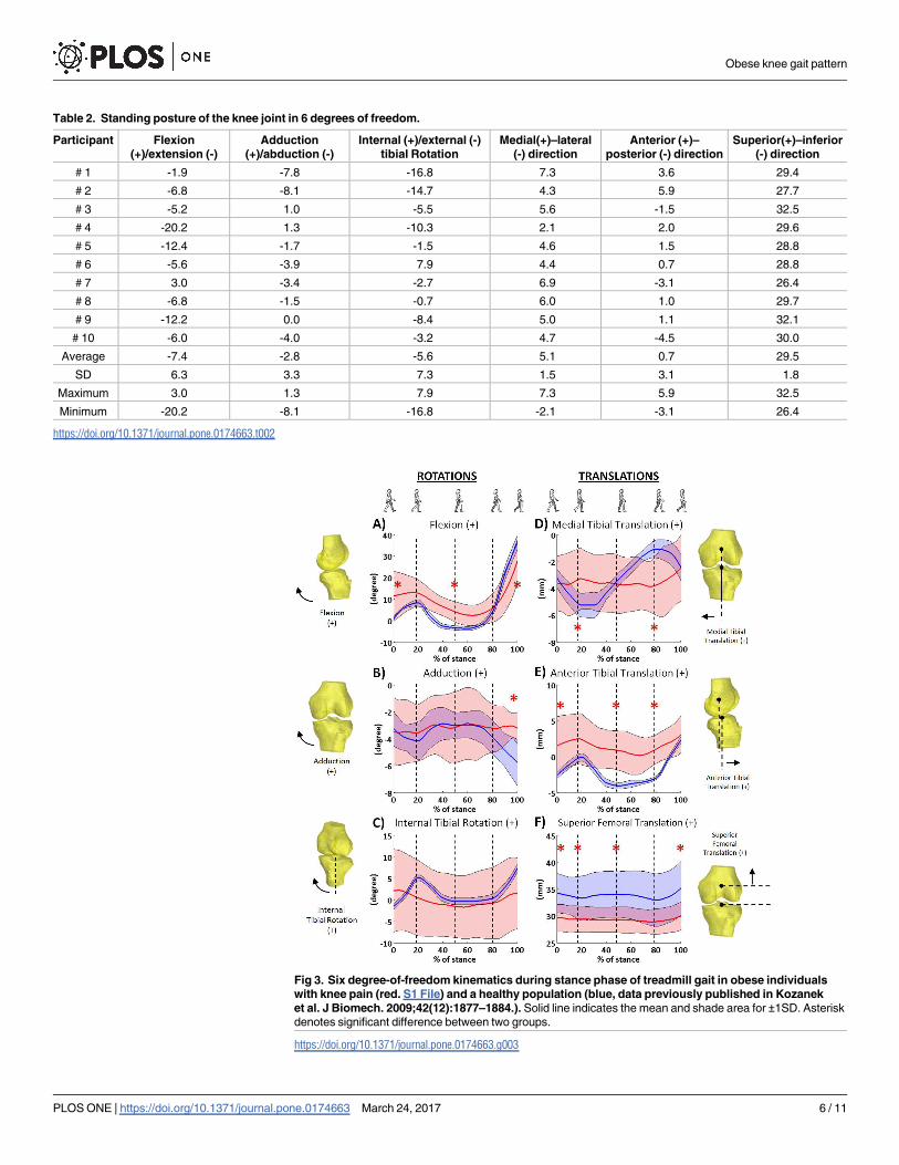
Table 2. Standing posture of the knee joint in 6 degrees of freedom.
Participant Flexion
(+)/extension (-)
Adduction
(+)/abduction (-)
Internal (+)/external (-)
tibial Rotation
Medial(+)–lateral
(-) direction
Anterior (+)–
posterior (-) direction
Superior(+)–inferior
(-) direction
# 1 -1.9 -7.8 -16.8 7.3 3.6 29.4
# 2 -6.8 -8.1 -14.7 4.3 5.9 27.7
# 3 -5.2 1.0 -5.5 5.6 -1.5 32.5
# 4 -20.2 1.3 -10.3 2.1 2.0 29.6
# 5 -12.4 -1.7 -1.5 4.6 1.5 28.8
# 6 -5.6 -3.9 7.9 4.4 0.7 28.8
# 7 3.0 -3.4 -2.7 6.9 -3.1 26.4
# 8 -6.8 -1.5 -0.7 6.0 1.0 29.7
# 9 -12.2 0.0 -8.4 5.0 1.1 32.1
# 10 -6.0 -4.0 -3.2 4.7 -4.5 30.0
Average -7.4 -2.8 -5.6 5.1 0.7 29.5
SD 6.3 3.3 7.3 1.5 3.1 1.8
Maximum 3.0 1.3 7.9 7.3 5.9 32.5
Minimum -20.2 -8.1 -16.8 -2.1 -3.1 26.4
https://doi.org/10.1371/journal.pone.0174663.t002
Fig 3. Six degree-of-freedom kinematics during stance phase of treadmill gait in obese individuals
with knee pain (red. S1 File) and a healthy population (blue, data previously published in Kozanek
et al. J Biomech. 2009;42(12):1877–1884.). Solid line indicates the mean and shade area for ±1SD. Asterisk
denotes significant difference between two groups.
https://doi.org/10.1371/journal.pone.0174663.g003
Obese knee gait pattern
PLOS ONE | https://doi.org/10.1371/journal.pone.0174663 March 24, 2017 6 / 11
anterior translation than the control group at initial contact (1.5±4.1mm vs. -2.6±0.7mm,
p = 0.011, Fig 3E), at the end of the mid-stance phase (0.8±2.7mm vs. -4.0±0.5mm, p<0.001),
and at the end of terminal stance (1.4±2.0 mm vs. -2.9±0.7mm, p<0.001), but the translation
at the end of the stance phase was not significantly different (3.1±2.6mm vs. 2.3±1.0mm,
p = 0.395). Also, the range of anterior-posterior translation was not significantly different in
the groups (2.5±1.0 mm vs. 6.4±1.2 mm, p = 0.151) (Fig 3E). The obese group had less range of
medial-lateral translation than the control group (2.7±1.1mm vs. 4.3±0.6mm, p = 0.001), and
were significantly different at the end of loading response and terminal stance phases (Fig 3D).
The obese knees did not have a significantly different range of superior-inferior translation
(1.8±0.6mm vs. 2.8±1.1mm, p = 0.054), but had significantly lower values than the control
group during most of the stance phase of the gait cycle (Fig 3F).
Discussion
This study investigated individuals with obesity and knee pain in standing and during gait
under DFIS surveillance. While standing, the knee was in about 7.4˚ hyperextension, slight
abduction (valgus), and about 5.6˚ of external rotation. During treadmill gait, the largest rota-
tional excursion was in flexion-extension and the largest translational excursion was in the
anterior-posterior direction, while motions in the other planes were smaller.
Few studies have evaluated standing posture in individuals with obesity. One study using a
radiographic hip-knee-ankle measurement, found that obese individuals stood in a slightly
knee flexed position [19]. However our obese individuals with knee pain tended to use a
hyperextension strategy in standing. The adaptation in standing posture may be due to the
high BMI in our sample (� 35) and knee pain. This posture is thought to reduce the demand
on the quadriceps and could potentially prevent fatigue [30]. Our study also provided informa-
tion in the other degrees of freedom. The analysis of the relative position between the femur
and tibia indicated that the center of the femur was near the center in anterior-posterior direc-
tion, but located at the medial portion of the tibial plateau. These data provide additional data
for more complete understanding of standing posture correction.
During walking, spatiotemporal parameters may be affected by gait speed. The speed we
tested was controlled at 0.67m/s (1.5 mph), which is slightly slower than the reported self-
selected walking speed of 0.73 to 1.08 m/s for an obese population [7, 31]. At this gait speed,
we found that obese individuals with knee pain had slightly increased cadence and decreased
stride length compared to individuals walking at a similar gait speed [7, 32], and had increased
stride duration and stance phase compared to individuals walking at a faster gait speed [9, 31].
The speed preference and participant characteristics, such as knee pain and presence of osteo-
arthritis, may contribute to differences between studies.
Several studies have reported knee kinematics during gait using skin marker motion analy-
sis in obese individuals [5, 6, 8]. In these studies, obese individuals were found to walk with a
similar pattern or with more knee extension and reduced range of motion in the sagittal plane
compared to non-obese individuals, and this strategy was assumed to decrease the exertion of
the knee extensors and prevent fatigue [10, 33]. Our findings were not in full agreement with
past studies [5, 8]. Similar to past studies, our obese individuals walked with a smaller range of
flexion-extension motion during the stance phase compared to a healthy population (Fig 3A)
[22]. However, our obese individuals walked in a more flexed knee position. While it is com-
monly thought that a more knee extended position will reduce demand on muscle, our partici-
pants did not display this adaptation. The combined smaller range of flexion-extension
motion with a more knee flexed position during walking may increase the demands on the
quadriceps and other extensors, and this gait pattern may result in early muscle fatigue. Our
Obese knee gait pattern
PLOS ONE | https://doi.org/10.1371/journal.pone.0174663 March 24, 2017 7 / 11

findings also showed a larger variation in knee flexion angles at initial contact, indicating dif-
ferent gait adaptation strategies could be used among individuals. The initial contact flexion
angles were weakly correlated with their WOMAC pain score during walking (r = -0.34), sug-
gesting that pain status may be a major contributor to the gait pattern.
Knee pain is a commonly reported symptom by patients with knee OA, and kinematic
changes in the sagittal plane during gait were frequently reported [12–14]. Individuals with
knee OA walked in a more extended position and reduced range of flexion-extension motion.
This gait pattern had been further replicated by studies of experimental knee pain [34]. Obese
individuals have also been found to walk with a similar gait pattern. Our study including indi-
viduals with combined obesity and knee pain walked with a smaller range of flexion-extension
motion similar to patients with knee OA, but in a more knee flexed position during most
stance phase when compared to a healthy non-obese group. This finding suggests that our
obese individuals with knee pain may adopt a unique gait pattern, which is not typically found
in patients with either obesity or knee OA.
A previous study found no difference in axial rotation between obese and non-obese groups
[6], and our findings were in agreement with this; however, the pattern of axial rotation in our
participants seemed to differ from that of healthy knees [22]. Healthy knees internally rotate
after initial contact to a peak rotation at the end of loading response (Fig 3C) [22]. Instead,
the tibia in our obese individuals was externally rotating to a peak of about 3.9˚ around the
end of mid-stance and reversed after that point, implying the pivoting mechanism observed in
healthy participants [22] may be changed in obese individuals. The large variation in the inter-
nal-external rotation angles in our obese individuals suggests that obese individuals may adopt
diverse strategies to avoid pain during walking.
In the coronal plane, past studies found similar or more adduction in obese individuals
compared to non-obese group during gait [5, 6]. In our study, we found adduction-abduction
in coronal plane motion in our obese group was not significantly different compared to our
control group (Fig 3B) [22]. We note that the majority of our subjects did not have medial
osteoarthritis which might have been present in previous studies.
The smaller range of motion of the obese patients indicated that these individuals use a stiff-
ening knee strategy despite maintaining the knee in more flexion during functional activities
[35]. This strategy for reduced range of motion is also found in knee OA patients [12–14, 36]
and obese individuals without knee pain [8], meaning both extra body weight and pain con-
tribute to the gait pattern change. As walking has been routinely suggested for obese patients
as a safe activity to increase energy expenditure, increasing the knee joint range of motion
while walking should be addressed, and this could potentially better distribute contact stress to
prevent the local stress concentration.
Several limitations need to be mentioned in interpreting the results. First, our participants
were severely obese (BMI� 35.0) and with knee pain; therefore, the results may not generalize
to the less obese population (BMI: 30.0–34.9) with or without knee pain. Our study sample
was 80% female, so the results may not generalize to all obese individuals with knee pain,
although, like our sample, most obese individuals with knee pain are women. The control
group was predominantly male, and this may also have contributed to the differences in kine-
matics found. Treadmill walking was used in this study to assess the kinematics, so the results
may not generalize to walking overground. The definition of the coordinate system in this
study was based on local bone geometry; therefore, the values found in this study may be
slightly different from studies using other coordinate system definitions. Lastly the loading
from the lead protection gowns (9.1kg, 7.0–10.6% of body mass of the obese participants)
could have contributed to the gait pattern changes; however, it is unlikely that this contributed
significantly to the differences found.
Obese knee gait pattern
PLOS ONE | https://doi.org/10.1371/journal.pone.0174663 March 24, 2017 8 / 11
In conclusion, obese individuals with knee pain used hyper-extension knee posture while
standing, but maintained the knee in more flexion during gait with reduced overall range of
motion in the 6DOF analysis compared to a healthy group. In addition to the facilitation of
greater total range of sagittal plane motion with a knee extension strategy during gait, increas-
ing the knee joint range of motion in other directions should be addressed in weight manage-
ment exercise programs.
Supporting information
S1 File. Gait data file. Six degree-of-freedom kinematics during stance phase of treadmill gait
in obese individuals with knee pain.
(CSV)
Author Contributions
Conceptualization: JL TT GL DTF.
Data curation: JL TT DTF GL CLL.
Formal analysis: JL TT DTF GL CLL.
Funding acquisition: DTF.
Investigation: JL TT GL.
Methodology: JL TT GL.
Project administration: DTF GL CLL.
Resources: JL TT DTF GL.
Software: JL TT.
Supervision: DTF GL CLL.
Validation: JL TT GL.
Visualization: JL TT GL.
Writing – original draft: JL.
Writing – review & editing: JL TT DTF GL CLL.
References1. Fryar CD, Carroll MD, Ogden CL. Prevalence of Overweight, Obesity, and Extreme Obesity Among
Adults: United States, 1960–1962 Through 2011–2012. NCHS Health E-Stat [Web Page]. 2014 [cited
2016 July 28]. http://www.cdc.gov/nchs/data/hestat/obesity_adult_11_12/obesity_adult_11_12.htm.
2. World Health Organization. World Health Statistics Report 2012 [Web Page]. [cited 2016 July 28].
http://apps.who.int/iris/bitstream/10665/44844/1/9789241564441_eng.pdf.
3. Ogden CL, Carroll MD, Kit BK, Flegal KM. Prevalence of childhood and adult obesity in the United
States, 2011–2012. JAMA. 2014; 311(8):806–14. Epub 2014/02/27. https://doi.org/10.1001/jama.2014.
732 PMID: 24570244
4. Sturmer T, Gunther KP, Brenner H. Obesity, overweight and patterns of osteoarthritis: the Ulm Osteoar-
thritis Study. J Clin Epidemiol. 2000; 53(3):307–13. Epub 2000/04/13. PMID: 10760642
5. Ko S, Stenholm S, Ferrucci L. Characteristic gait patterns in older adults with obesity—results from the
Baltimore Longitudinal Study of Aging. Journal of biomechanics. 2010; 43(6):1104–10. Epub 2010/01/
19. https://doi.org/10.1016/j.jbiomech.2009.12.004 PMID: 20080238
Obese knee gait pattern
PLOS ONE | https://doi.org/10.1371/journal.pone.0174663 March 24, 2017 9 / 11
6. Lai PP, Leung AK, Li AN, Zhang M. Three-dimensional gait analysis of obese adults. Clinical biome-
chanics (Bristol, Avon). 2008; 23 Suppl 1:S2–6. Epub 2008/04/01.
7. de Souza SA, Faintuch J, Valezi AC, Sant’ Anna AF, Gama-Rodrigues JJ, de Batista Fonseca IC, et al.
Gait cinematic analysis in morbidly obese patients. Obes Surg. 2005; 15(9):1238–42. Epub 2005/11/02.
https://doi.org/10.1381/096089205774512627 PMID: 16259878
8. Haight DJ, Lerner ZF, Board WJ, Browning RC. A comparison of slow, uphill and fast, level walking on
lower extremity biomechanics and tibiofemoral joint loading in obese and nonobese adults. Journal of
orthopaedic research: official publication of the Orthopaedic Research Society. 2014; 32(2):324–30.
Epub 2013/10/16.
9. Sheehan KJ, Gormley J. The influence of excess body mass on adult gait. Clinical biomechanics (Bris-
tol, Avon). 2013; 28(3):337–43. Epub 2013/02/06.
10. McMillan AG, Pulver AM, Collier DN, Williams DS. Sagittal and frontal plane joint mechanics throughout
the stance phase of walking in adolescents who are obese. Gait & posture. 2010; 32(2):263–8. Epub
2010/06/25.
11. Vismara L, Romei M, Galli M, Montesano A, Baccalaro G, Crivellini M, et al. Clinical implications of gait
analysis in the rehabilitation of adult patients with "Prader-Willi" Syndrome: a cross-sectional compara-
tive study ("Prader-Willi" Syndrome vs matched obese patients and healthy subjects). Journal of neu-
roengineering and rehabilitation. 2007; 4:14. https://doi.org/10.1186/1743-0003-4-14 PMID: 17493259
12. Al-Zahrani KS, Bakheit AM. A study of the gait characteristics of patients with chronic osteoarthritis of
the knee. Disability and rehabilitation. 2002; 24(5):275–80. PMID: 12004973
13. Baliunas AJ, Hurwitz DE, Ryals AB, Karrar A, Case JP, Block JA, et al. Increased knee joint loads dur-
ing walking are present in subjects with knee osteoarthritis. Osteoarthritis and cartilage / OARS, Osteo-
arthritis Research Society. 2002; 10(7):573–9.
14. Kaufman KR, Hughes C, Morrey BF, Morrey M, An KN. Gait characteristics of patients with knee osteo-
arthritis. Journal of biomechanics. 2001; 34(7):907–15. PMID: 11410174
15. Lerner ZF, Board WJ, Browning RC. Effects of an obesity-specific marker set on estimated muscle and
joint forces in walking. Medicine and science in sports and exercise. 2014; 46(6):1261–7. Epub 2014/
02/13. https://doi.org/10.1249/MSS.0000000000000218 PMID: 24518193
16. Peters A, Galna B, Sangeux M, Morris M, Baker R. Quantification of soft tissue artifact in lower limb
human motion analysis: a systematic review. Gait & posture. 2010; 31(1):1–8. Epub 2009/10/27.
17. Tsai TY, Lu TW, Kuo MY, Lin CC. Effects of soft tissue artifacts on the calculated kinematics and kinet-
ics of the knee during stair-ascent. Journal of biomechanics. 2011; 44(6):1182–8. Epub 2011/02/08.
https://doi.org/10.1016/j.jbiomech.2011.01.009 PMID: 21296352
18. Cappozzo A, Catani F, Leardini A, Benedetti MG, Croce UD. Position and orientation in space of bones
during movement: experimental artefacts. Clinical biomechanics (Bristol, Avon). 1996; 11(2):90–100.
Epub 1996/03/01.
19. Jalai CM, Diebo BG, Cruz DL, Poorman GW, Vira S, Buckland AJ, et al. The impact of obesity on com-
pensatory mechanisms in response to progressive sagittal malalignment. The spine journal: official jour-
nal of the North American Spine Society. 2016.
20. Messier SP. Diet and exercise for obese adults with knee osteoarthritis. Clin Geriatr Med. 2010; 26
(3):461–77. Epub 2010/08/12. https://doi.org/10.1016/j.cger.2010.05.001 PMID: 20699166
21. Poirier P, Despres JP. Exercise in weight management of obesity. Cardiol Clin. 2001; 19(3):459–70.
Epub 2001/09/26. PMID: 11570117
22. Kozanek M, Hosseini A, Liu F, Van de Velde SK, Gill TJ, Rubash HE, et al. Tibiofemoral kinematics and
condylar motion during the stance phase of gait. Journal of biomechanics. 2009; 42(12):1877–84. Epub
2009/06/06. https://doi.org/10.1016/j.jbiomech.2009.05.003 PMID: 19497573
23. Li JS, Hosseini A, Cancre L, Ryan N, Rubash HE, Li G. Kinematic characteristics of the tibiofemoral
joint during a step-up activity. Gait & posture. 2013; 38(4):712–6. Epub 2013/04/02.
24. Tsai TY, Li JS, Wang S, Lin H, Malchau H, Li G, et al. A novel dual fluoroscopic imaging method for
determination of THA kinematics: in-vitro and in-vivo study. J Biomech. 2013; 46(7):1300–4. Epub
2013/03/19. https://doi.org/10.1016/j.jbiomech.2013.02.010 PMID: 23497800
25. Li G, Van de Velde SK, Bingham JT. Validation of a non-invasive fluoroscopic imaging technique for the
measurement of dynamic knee joint motion. J Biomech. 2008; 41(7):1616–22. Epub 2008/04/09.
https://doi.org/10.1016/j.jbiomech.2008.01.034 PMID: 18394629
26. Kellgren JH, Lawrence JS. Radiological assessment of osteo-arthrosis. Annals of the rheumatic dis-
eases. 1957; 16(4):494–502. Epub 1957/12/01. PMID: 13498604
27. Gronenschild E. Correction for geometric image distortion in the x-ray imaging chain: local technique
versus global technique. Medical physics. 1999; 26(12):2602–16. https://doi.org/10.1118/1.598800
PMID: 10619246
Obese knee gait pattern
PLOS ONE | https://doi.org/10.1371/journal.pone.0174663 March 24, 2017 10 / 11
28. Gronenschild E. The accuracy and reproducibility of a global method to correct for geometric image dis-
tortion in the x-ray imaging chain. Medical physics. 1997; 24(12):1875–88. https://doi.org/10.1118/1.
598101 PMID: 9434970
29. Grood ES, Suntay WJ. A joint coordinate system for the clinical description of three-dimensional
motions: application to the knee. J Biomech Eng. 1983; 105(2):136–44. Epub 1983/05/01. PMID:
6865355
30. Loudon JK, Goist HL, Loudon KL. Genu recurvatum syndrome. The Journal of orthopaedic and sports
physical therapy. 1998; 27(5):361–7. https://doi.org/10.2519/jospt.1998.27.5.361 PMID: 9580896
31. Blaszczyk JW, Plewa M, Cieslinska-Swider J, Bacik B, Zahorska-Markiewicz B, Markiewicz A. Impact
of excess body weight on walking at the preferred speed. Acta Neurobiol Exp (Wars). 2011; 71(4):528–
40. Epub 2012/01/13.
32. Browning RC, Kram R. Effects of obesity on the biomechanics of walking at different speeds. Medicine
and science in sports and exercise. 2007; 39(9):1632–41. Epub 2007/09/07. https://doi.org/10.1249/
mss.0b013e318076b54b PMID: 17805097
33. Maffiuletti NA, Jubeau M, Munzinger U, Bizzini M, Agosti F, De Col A, et al. Differences in quadriceps
muscle strength and fatigue between lean and obese subjects. Eur J Appl Physiol. 2007; 101(1):51–9.
Epub 2007/05/04. https://doi.org/10.1007/s00421-007-0471-2 PMID: 17476522
34. Henriksen M, Graven-Nielsen T, Aaboe J, Andriacchi TP, Bliddal H. Gait changes in patients with knee
osteoarthritis are replicated by experimental knee pain. Arthritis Care Res (Hoboken). 2010; 62(4):501–
9. Epub 2010/04/15.
35. Bytyqi D, Shabani B, Lustig S, Cheze L, Karahoda Gjurgjeala N, Neyret P. Gait knee kinematic alter-
ations in medial osteoarthritis: three dimensional assessment. Int Orthop. 2014; 38(6):1191–8. Epub
2014/03/13. https://doi.org/10.1007/s00264-014-2312-3 PMID: 24619388
36. Duffell LD, Southgate DF, Gulati V, McGregor AH. Balance and gait adaptations in patients with early
knee osteoarthritis. Gait & posture. 2014; 39(4):1057–61.
Obese knee gait pattern
PLOS ONE | https://doi.org/10.1371/journal.pone.0174663 March 24, 2017 11 / 11
Recommended


![An Ankle-Foot Prosthesis Emulator with Control of ...biomechatronics.cit.cmu.edu/publications/Collins_2015_ICRA.pdf · ankle and knee kinematics [1], reduce metabolic rate [2], and](https://img.pdfslide.us/doc/110x75/5f6b17783154671dfb1d5945/an-ankle-foot-prosthesis-emulator-with-control-of-ankle-and-knee-kinematics.jpg)


![The Kinematic Alignment 16 Technique for Total Knee ... · 177 non-physiological knee ligament laxities and residual instability [11, 15] and abnormal 10, knee kinematics [1316, ,](https://img.pdfslide.us/doc/110x75/60bbb243c19342776239ee29/the-kinematic-alignment-16-technique-for-total-knee-177-non-physiological-knee.jpg)