SIMULATION ON TRANSDERMAL PATCH FOR BREAST CANCER THERAPY
BY USING COMSOL
TAN MING SIEN
Report submitted in partial fulfilment of the requirements
for the award of the degree of
Bachelor of Chemical Engineering (Biotechnology)
Faculty of Chemical Engineering and Natural Resources
UNIVERSITI MALAYSIA PAHANG
NOVEMBER 2010
ii
ABSTRACT
Breast cancer is the most common form of cancer affecting women in Malaysia.
Conventional drug treatment for breast cancer, chemotherapy would destroy the cancer
cells because of the medicine targets on rapidly dividing cells. However, healthy cells
and tissues in blood, mouth, intestinal tract, nose, nails, vagina and hair also divide
rapidly, they could be damaged. A more promising technology called transdermal patch
has been introduced due to side effects are common expected results from conventional
treatment. Therefore, the aim of this study was to treat breast cancer by delivering drug
from transdermal patch precisely and safely to targeted cancer cell so that reducing the
side effects and dosage of drug used. The objectives of this study were to determine the
drug concentration at breast tumor, to investigate the relationship between drug
diffusivity and drug delivery efficiency, and to evaluate the efficiency of drug delivery
under other parameters (i.e. deepness of tumor, temporal and spatial placement of
transdermal patch). Available software, COMSOL was used in this study. Drug
concentrations that able to diffuse and reach tumor in breast were studied. The
simulation results showed that there was optimal drug diffusivity for maximum
concentration of drug reached tumor in breast. However, below and higher than this
drug diffusivity optimal value, the delivery of drug concentration was poorer when the
lesser. Production of microchannels in the skin by microneedle can increase the drug
diffusivity and ensure delivery of pharmacologically effective concentration of drug to
the targeted site, breast cancer cell. Deeper the tumor grown within breast, lesser drug‟s
concentration could be diffused to it. However, this could be solved by changing the
place of transdermal patch application. The nearer the spatial placement of transdermal
patch to tumor growth in the breast on the breast skin increased the effectiveness of
drug delivery to tumor. The longer the temporal placement resulted in higher drug
concentration could be delivered to breast tumor. However, this constant concentration
gradient only achieved for less than one month. After this, the concentration gradient
would become zero. As a conclusion, the drug diffusivity, deepness of breast tumor,
spatial and temporal placement of transdermal patch must be taken into account when
engineering, constructing and applying the transdermal patch in order to achieve the
maximum breast cancer treatment with reducing the undesired side effects.

iii
ABSTRAK
Kanser payudara merupakan jenis kanser yang paling utama di kalangan wanita
di Malysia. Ubat rawatan konvensional untuk kanser payudara, kemoterapi akan
menghancurkan sel-sel kanser kerana ubat target pada sel-sel yang membahagi dengan
cepat. Namun, sel-sel dan rangkaian yang sihat dalam darah, mulut, saluran, usus
hidung, kuku, vagina dan rambut juga membelah dengan cepat, mereka boleh
dirosakkan. Satu teknologi yang lebih menjanjikan disebut patch transdermal telah
diperkenalkan kerana kesan sampingan merupakan umum hasil daripada rawatan
konvensional. Oleh itu, tujuan dari penelitian ini adalah untuk mengubati kanser
payudara dengan menghantarkan ubat dari patch transdermal dengan tepat dan selamat
pada sel kanser yang disasarkan supaya mengurangkan kesan sampingan dan dosis ubat
yang digunakan. Objektif-objektif dari penelitian ini adalah untuk menentukan
konsentrasi ubat pada tumor di payudara, mengkaji perhubungan antara keresapan ubat
dengan keberkesanan penghantaran ubat, dan menilai keberkesanan penghantaran ubat
dalam parameter yang berbeza seperti kedalaman tumor dalam payudara, penempatan
spasial dan temporal patch transdermal pada kulit. Tersedia software, COMSOL
digunakan dalam kajian ini. Ubat konsentrasi yang dapat menyebar dan mencapai tumor
dalam payudara dipelajari. Keputusan simulasi menunjukkan bahawa ada ubat
keresapan optimum untuk konsentrasi ubat maksimum capai pada tumor dalam
payudara. Namun, di bawah atau di atas nilai keresapan ubat yang optimum ini,
penghantaran konsentrasi ubat menjadi lemah ketika semakin berkurangan keresapan
ubat. Produksi microchannels di kulit dengan microneedle dapat meningkatkan
keresapan ubat dan memastikan penghantaran konsentrasi farmakologi ubat yang
berkesan ke tempat yang disasarkan, sel kanser payudara. Semakin dalam pertumbuhan
tumor dalam payudara, semakin berkurangan ubat dapat meresap ke tumor. Namun
begitu, scenario ini dapat diatasi dengan menukarkan tempat menempatkan patch.
Semakin dekat penempatan spasial patch transdermal pada pertumbuhan tumor dalam
payudara pada kulit payudara meningkatkan keberkesanan penghantaran ubat untuk
tumor. Semakin lama penempatan temporal menghasilkan kepekatan ubat yang boleh
disampaikan untuk tumor payudara lebih tinggi. Namun, kecerunan konsentrasi ini
hanya dapat dicapai kurang daripada satu bulan. Selepas ini, kecerunan konsentrasi ubat
akan menjadi kosong. Sebagai kesimpulan, keresapan ubat, kedalaman pertumbuhan
tumor dalam payudara, penempatan spasial dan temporal transdermal patch harus
diperhitungkan sewaktu kejuruteraan, membina dan melaksanakan patch transdermal
untuk mencapai rawatan kanser payudara maksimum dengan mengurangkan kesan
sampingan yang tidak diingini.

iv
TABLE OF CONTENTS
Page
ACKNOWLEDGEMENTS i
ABSTRACT ii
ABSTRAK iii
TABLE OF CONTENTS iv
LIST OF TABLES vii
LIST OF FIGURES viii
LIST OF SYMBOLS x
LIST OF ABBREAVIATIONS xi
CHAPTER 1 INTRODUCTION
1.1 Background of Study 1
1.2 Problem Statement 2
1.3 Objectives 2
1.4
1.5
Research Scope
Rationales and Significances
3
3
1.6
Organization of Thesis 3
CHAPTER 2 LITERATURE REVIEW
2.1 Overview of Breast Cancer Statistic 5
2.2
Conventional Breast Cancer Drug Therapy
2.2.1 Docetaxel
2.2.2 Doxorubicin (Adriamycin)
2.2.3 Herceptin (Trastuzumab)
2.2.4 Paclitaxel (Taxol)
2.2.5 Methorexate (Maxtrex)
7
8
8
9
10
10
2.3 Transdermal Application 11
2.4 Patch Design 12
v
CHAPTER 3 METHODOLOGY
3.1
3.2
3.3
3.4
3.5
3.6
Material and Instrumentation
Methodology Flow Chart
Collection of Drug Diffusivity Data
Collection of Breast Volume Data
Designation of Mathematical Model
3.5.1 Governing Equation
3.5.2 Boundary Conditions
Implementation in COMSOL
3.6.1 Steps for Solving Specified Problem in COMSOL
22
22
24
24
25
25
26
26
26
CHAPTER 4 RESULT AND DISCUSSION
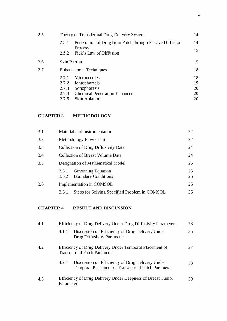
4.1
4.2
4.3
Efficiency of Drug Delivery Under Drug Diffusivity Parameter
4.1.1 Discussion on Efficiency of Drug Delivery Under
Drug Diffusivity Parameter
Efficiency of Drug Delivery Under Temporal Placement of
Transdermal Patch Parameter
4.2.1 Discussion on Efficiency of Drug Delivery Under
Temporal Placement of Transdermal Patch Parameter
Efficiency of Drug Delivery Under Deepness of Breast Tumor
Parameter
28
35
37
38
39
2.5 Theory of Transdermal Drug Delivery System
2.5.1 Penetration of Drug from Patch through Passive Diffusion
Process
2.5.2 Fick‟s Law of Diffusion
14
14
15
2.6 Skin Barrier 15
2.7
Enhancement Techniques
2.7.1 Microneedles
2.7.2 Iontophoresis
2.7.3 Sonophoresis
2.7.4 Chemical Penetration Enhancers
2.7.5 Skin Ablation
18
18
19
20
20
20

vi
4.4
4.5
4.3.1 Discussion on Efficiency of Drug Delivery Under
Deepness of Breast Tumor Parameter
Efficiency of Drug Delivery Under Spatial Placement of
Transdermal Patch Parameter
4.4.1 Discussion on Efficiency of Drug Delivery Under
Spatial Placement of Transdermal Patch Parameter
Summary
42
42
43
44
CHAPTER 5 CONCLUSION AND RECOMMENDATIONS
5.1
5.2
Conclusion
Recommendations
43
44
REFERENCES 45
APPENDICES
A Degree Final Year Project Gantt Chart 48
B1
B2
Breast Images for Simulation Run of 1-13
Graphs for Simulation Run of 1-13
53
67

vii
LIST OF TABLES
Table No. Title Page
2.1
2.2
3.1
4.1
4.2
4.3
4.4
Deaths from cancers in Malaysia women for the years of 1994,
1995 and 1998.
Differences between transdermal application and chemotherapy.
Properties of Doxorubicin.
Concentration of drug at breast tumor with different drug
diffusivity, from 10-9
to 10-1
.
Concentration of drug at breast tumor, after one week, two
weeks, one month, two months, three months, four months, five
months and six months.
Concentration of drug at tumor that grown at the top and bottom
of breast.
Concentration of drug at tumor when patch aplied at different
locations.
6
12
24
29
37
39
42
viii
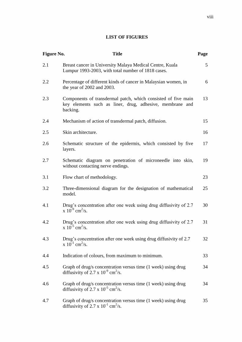
LIST OF FIGURES
Figure No. Title Page
2.1
2.2
2.3
2.4
2.5
2.6
2.7
3.1
3.2
4.1
4.2
4.3
4.4
4.5
4.6
4.7
Breast cancer in University Malaya Medical Centre, Kuala
Lumpur 1993-2003, with total number of 1818 cases.
Percentage of different kinds of cancer in Malaysian women, in
the year of 2002 and 2003.
Components of transdermal patch, which consisted of five main
key elements such as liner, drug, adhesive, membrane and
backing.
Mechanism of action of transdermal patch, diffusion.
Skin architecture.
Schematic structure of the epidermis, which consisted by five
layers.
Schematic diagram on penetration of microneedle into skin,
without contacting nerve endings.
Flow chart of methodology.
Three-dimensional diagram for the designation of mathematical
model.
Drug‟s concentration after one week using drug diffusivity of 2.7
x 10-9
cm2/s.
Drug‟s concentration after one week using drug diffusivity of 2.7
x 10-5
cm2/s.
Drug‟s concentration after one week using drug diffusivity of 2.7
x 10-1
cm2/s.
Indication of colours, from maximum to minimum.
Graph of drug/s concentration versus time (1 week) using drug
diffusivity of 2.7 x 10⁻⁹
cm2/s.
Graph of drug/s concentration versus time (1 week) using drug
diffusivity of 2.7 x 10⁻⁵
cm2/s.
Graph of drug/s concentration versus time (1 week) using drug
diffusivity of 2.7 x 10⁻¹
cm2/s.
5
6
13
15
16
17
19
23
25
30
31
32
33
34
34
35
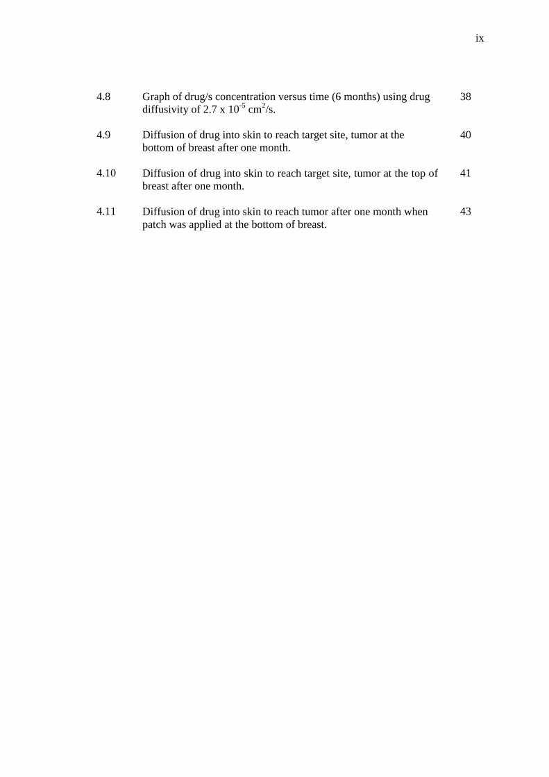
ix
4.8
4.9
4.10
4.11
Graph of drug/s concentration versus time (6 months) using drug
diffusivity of 2.7 x 10⁻⁵
cm2/s.
Diffusion of drug into skin to reach target site, tumor at the
bottom of breast after one month.
Diffusion of drug into skin to reach target site, tumor at the top of
breast after one month.
Diffusion of drug into skin to reach tumor after one month when
patch was applied at the bottom of breast.
38
40
41
43
x
LIST OF SYMBOLS
C
c
D
Da
ɗ
g
h
mm
mol
r
s
Concentration
Centimeter
Diffusion Constant
Dalton
Delta
Gram
Height
Millimeter
Mole
Radius
Second
t
µm
z
Π
%
>
Time
Micrometer
z-axis
Pi
Percentage
More than

xi
LIST OF ABBREVIATIONS
AC
e.g.
FEC
i.e.
MMM
SI
Doxorubicin and Cyclophosphamide
Example given
Epirubicin, Cyclophosphamide and Fluoruocil
That is
Methotrexate, Mitozantrone and Mitomycin
International System of Units

CHAPTER 1
INTRODUCTION
This chapter presents the overview of the background for disease of breast
cancer and breast cancer drug therapies. The problem statement of current
technology is also stated in this chapter. The objectives of the study also presented in
this chapter. The scope and importance of work are defined. Lastly, this chapter also
outlines the organization of the thesis.
1.1 BACKGROUND OF STUDY
Breast cancer is the most common form of cancer among women other than
other skin cancer, and the number of cases for breast cancer is increased annually.
About one in nineteen women in Malaysia are at risk. Breast cancer is also the
leading cause of cancer-related death for women. In Peninsula Malaysia, the
mortality rate per 100,000 showed an increase from 3.7 per 100,000 in 1982 to 5.8
per 100,000 in 1990. However, since only one-third of deaths in Peninsula Malaysia
were medically certified, the mortality rate from breast cancer was actually higher.
(Yip, C. H. and Ng, E. H., 1996).
Generally, chemotherapy is the common drug therapy for breast cancer.
Many anticancer drugs are designed to simply kill cancer cells. They are often in a
semi-specific fashion because the anticancer drugs target on rapidly dividing cells.
Similar to cancer cells, the normal cells in blood, mouth, intestinal tract, nose, nails,
vagina and hair also divide rapidly. Consequently, the distribution of anticancer
drugs in healthy organs or tissues is especially undesirable due to the potential for

2
severe side effects. This phenomenon greatly limits the maximal allowable dose of
the drugs. However, low local drugs‟ concentration will cause less effect on
destroying the cancer cells. Therefore, there is still a need for high or frequent drugs
dosing.
To overcome such limitations, a new and more promising technology,
transdermal technology has been introduced for breast cancer drug therapy. This
application provides an alternative route for delivering drugs to cancer cells, which is
through the largest organ of humans‟ bodies, skin. In this method, a transdermal
patch is purposely engineered and constructed to allow the delivering of drugs
precisely to the targeted cancer cells with fewer side effects. Site-specific drugs
delivery is a concept that has the potential to increase local drugs‟ concentrations,
and thereby produce more effective medicines with reducing the dosage of drugs
used.
1.2 PROBLEM STATEMENT
The new technology, transdermal patch allows the delivering of anticancer
drugs to targeted cancer cell as effectively as possible without side effects and
reducing the dosage of drugs used. However, transdermal application has limitation
due to the remarkable barrier properties of the outermost layer of skin, stratum
corneum. This layer is mainly consisted of lipids, has no blood flow, and thus plays a
key role in limiting the diffusion of drugs to the bloodstream. For transdermal drugs
delivery system to be effective, the drugs must obviously be able to penetrate this
skin barrier and reach the targeted cancer cells. To achieve this, suitable modification
has been made on the transdermal delivery system. It is hard to deny that drug
diffusivity is a very important factor to be considered.
1.3 OBJECTIVES
The aim of this study was to treat breast cancer by delivering drug from
transdermal patch precisely to targeted cancer cells so that reducing the side effects
and dosage of drugs used. This can be achieved by the following specific objectives:
1. To determine the drug concentration at breast tumor,
3
2. To investigate the relationship between drug diffusivity and drug delivery
efficiency, and
3. To evaluate the efficiency of drug delivery under other parameters (i.e.
deepness of tumor, temporal and spatial placement of transdermal patch).
1.4 RESEARCH SCOPE
This study mainly focused on the efficiency of drug delivery from
transdermal patch, which was evaluated by response parameter, drug concentration
successfully reaching at the breast tumor. The study was carried out by using
COMSOL software. Before implementing the COMSOL software, data collection for
drug diffusivity and breast volume were needed. After data collection, simulation
was run by using COMSOL software. The simulation was conducted with identified
independent variables (i.e. drug diffusivity, deepness of tumor, temporal and spatial
placement of transdermal patch). Simulation produced with different parameters was
saved for further analyses. Graphs that relate the drug concentration at breast tumor
were also developed. From the developed graphs, the association between the
efficiency of drug delivery and drug concentration could be made.
1.5 RATIONALES AND SIGNIFICANCES
This study had the significance to do a new strategy on developing a more
promising technology to treat breast cancer. This study also important for eliminate
the side effects or toxicities caused by conventional drug therapies. This was
replaced by the application of new technology, transdermal patch. The other
significance was including reducing the drug dosage used for breast cancer treatment.
Reduce side effects and drug dosage used, thereby decreasing the cost of breast
cancer treatment.
1.6 ORGANIZATION OF THESIS
This thesis is mainly delegated into five different chapters. In first chapter,
the background of study, problem statement, objectives, scope and significances of
project are reviewed in order to list out the tasks and act as a guideline for this study.

4
The statistic of breast cancer, skin structure, theoretical background, previous studies
and some basic information related to the project title are detailed in the second
chapter. Chapter 3 presents the methodology used from the starting until the end of
the study, which including the material or instrument used for completing the study,
designing the simulation and how to run the simulation. An overview of overall
methodology that designated in a flow chart as guideline for task sequences also
included in this chapter.
Chapter 4 shows the results obtained from the simulation. Several graphs
made to preview the relationship between the drug concentration and time, which
were resulting from different parameters also presented. At the end of this chapter,
results, some of findings and the sources of errors that affect the simulation outcomes
are discussed in details. The last chapter, chapter 5 states the conclusions of the
study. Recommendations for improvement of the study in the future also are outlined
in this chapter.

CHAPTER 2
LITERATURE REVIEW
This chapter includes the statistic of breast cancer, drug therapies of breast
cancer, structure of skin, theories of transdermal application and some basic
information related to the study. Studies on some recent patents related to the field of
transdermal patch are also presented.
2.1 OVERWIEW OF BREAST CANCER STATISTIC
The problem of cancer in Malaysia is in a growing trend, and breast cancer is
the most common cancer among women. Over the years from 1993 to 2003, there
were a total of 1818 breast cancer patients in the University Hospital. The number of
breast cancer patients increased annually, with the highest recorded in 2003. This
was 6 times the number of breast cancer patients in 1993 (Figure 2.1).
Figure 2.1: Breast cancer in University Malaya Medical Centre, Kuala Lumpur
1993-2003, with total number of 1818 cases.
Source: College Of Radiology Breast Health Information Centre (2008)
6
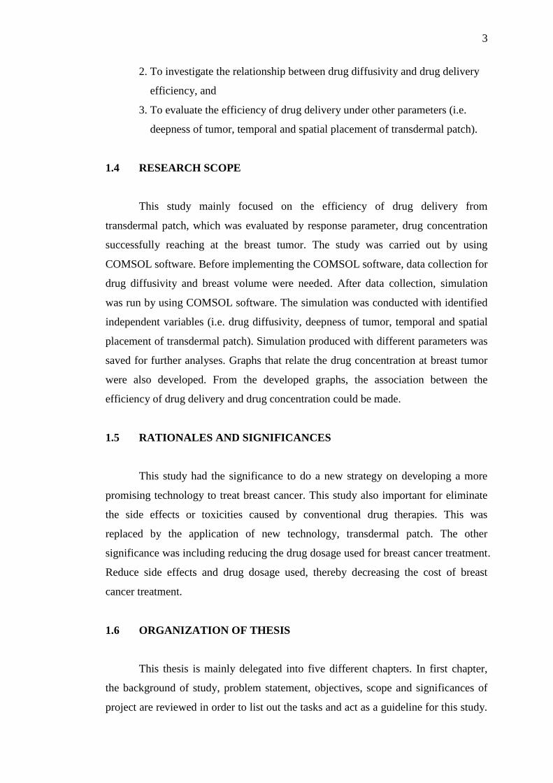
Breast cancer formed 30% and 31.1% of newly diagnosed cancer cases in
women in 2002 and 2003. This was followed by cancer of the cervix, which only
formed 12% and 12.9% of total female cancers in 2002 and 2003 respectively
(Figure 2.2).
Figure 2.2: Percentage of different kinds of cancer in Malaysian women, in the year
of 2002 and 2003.
Source: College Of Radiology Breast Health Information Centre (2008)
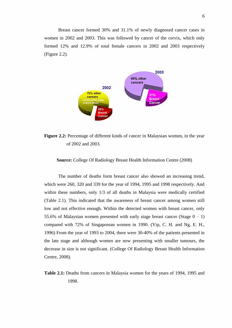
The number of deaths form breast cancer also showed an increasing trend,
which were 260, 320 and 339 for the year of 1994, 1995 and 1998 respectively. And
within these numbers, only 1/3 of all deaths in Malaysia were medically certified
(Table 2.1). This indicated that the awareness of breast cancer among women still
low and not effective enough. Within the detected women with breast cancer, only
55.6% of Malaysian women presented with early stage breast cancer (Stage 0 – 1)
compared with 72% of Singaporean women in 1990. (Yip, C. H. and Ng, E. H.,
1996) From the year of 1993 to 2004, there were 30-40% of the patients presented in
the late stage and although women are now presenting with smaller tumours, the
decrease in size is not significant. (College Of Radiology Breast Health Information
Centre, 2008).
Table 2.1: Deaths from cancers in Malaysia women for the years of 1994, 1995 and
1998.
7
Source: Vital Statistic Malaysia
2.2 CONVENTIONAL BREAST CANCER DRUG THERAPY
The most common breast cancer drug therapy, also known as chemotherapy,
in simplest sense, is a treatment with the aid of drugs that killing microorganisms or
cancerous cells. The chemotherapy drugs can be swallowed by someone as tablets or
capsules, or injected intravenously through the veins. When the drugs reach cancer
cells, the drugs work by disrupting the growth of cancer cells.
However, when the drugs circulate in the blood, they not only reach cancer
cells, but wherever they are in body. Many anticancer drugs are designed simply kill
cancer cells. They have no the capability to distinguish between the cancer cells and
normal cells. In contrast, they work by killing the cells that are actively growing and
dividing into new cells. Cancer cells do grow and divide much more often than
normal cells because most normal cells grow and divide in a precise, orderly way.
Thus, cancer cells are more likely to be killed by the chemotherapy drugs. However,
there are still some normal cells do divide and grow rapidly such as cells in hair
follicles, nails, mouth, intestinal tract and bone marrow. Chemotherapy drugs can
unintentionally harm these other types of rapidly dividing cells. As a result, the
distribution of anticancer drugs in healthy and normal cells is especially undesirable
due to the potential for severe side effects.
The side effects caused are significantly depending on the types of drugs,
drugs dosage used, how long the drugs are taken and how though of the own body of

8
the one who consuming these chemotherapy drugs. The possibilities of common side
effects caused by some different kinds of anticancer drugs are discussed with details
in the next sub-topic.
2.2.1 DOCETAXEL
Docetaxel is one of the taxane drugs that were originally developed from the
yew tree. Docetaxel is a man-made drug that was first made from the needles of the
yew tree. It is known by its brand name, Taxotere. It works by stopping the cancer
cells from separating into two new cells, so it blocks the growth of the cancer.
Common side effects caused by this drug are as following (Cancer Health UK,
2009):-
Have temporary drop in the number of blood cells made by the bone marrow.
For example, a drop in white blood cells result a person with increased risk of
getting an infection. When there is a dropping of red blood cells, a person
will easy feel tired and breathless. Besides, bruising easily is another common
side effect due to a drop in platelets.
Fatigue is the most disruptive side effect where tiredness often carries on
after treatment has just ended.
Patients may experience fluid retention such as swelling of the hands and feet
and resulting in weight gain and breathless.
Some people develop soreness, redness and peeling on the palms of the hands
and soles of the feet.
Patients may have rash, hair loss, sore mouth, diarrhoe, numbness and
tingling in hands and feets, allergic reaction during the infusion and
fingernails become discoloured.
Docetaxel may have a harmful effect on a baby developing in the womb.
2.2.2 DOXORUBICIN (ADRIAMYCIN)
Doxorubicin works by binding to the cancer cells‟ DNA and blocking an
important enzyme called topo-isomerase II. This makes the DNA get tangled up and
9
the cancer cells cannot divide and grow. The common side effects associated with
doxorubicin are listed below (Cancer Health UK, 2009):-
Have temporary drop in the number of blood cells made by the bone marrow.
For example, a drop in white blood cells result a person with increased risk of
getting an infection. When there is a dropping of red blood cells, a person
will easy feel tired and breathless. Besides, bruising easily is another common
side effect due to a drop in platelets.
Fatigue during and after treatment.
Feeling or being sick may be severe.
Urine may become a pink or red colour for one or two days after treatment.
Patients may have hair loss, sore mouth, sensitivity to sunlight, black or
brown lines may appear in the creases of skin, watery eyes and conjunctivitis
but very rare to happen.
A woman may stop or temporarily stop having periods, and loss of fertility.
Doxorubicin may have a harmful effect on a developing baby.
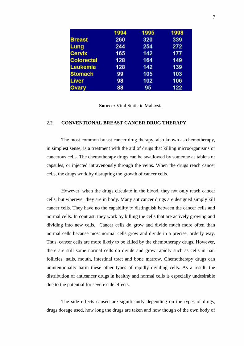
2.2.3 HERCEPTIN (TRASTUZUMAB)
Herceptin is a monoclonal antibody. It targets a protein called HER2. This
protein is found in roughly 25 to 30 percent of breast cancers. By interfering with
this protein, this medicine can stop cancer cell growth. With this drug, the common
side effects are:-
Breathing difficulties.
Chest pain or palpitations.
Cough.
Dizziness or fainting.
Fever or chills, sore throat.
Skin rash, itching or hives.
Swelling of the legs or ankles.
Usually weak or tired.
Loss of appetite.
Headache.
10
Muscle aches.
Nausea.
2.2.4 PACLITAXEL (TAXOL)
Paclitaxel is in a class of drugs known as taxanes. It slows or stops the growth
of cancer cells in body. Side effects from paclitaxel are common, and include:-
Nausea and vomiting.
Loss of appetite.
Change in taste.
Thinned or brittle hair.
Pain in the joints of the arms or legs lasting 2-3 days.
Changes in the colour of the nails.
Tingling in the hands or toes.
2.2.5 METHOREXATE (MAXTREX)
Methotrexate is similar to a normal body molecule called folinic acid, but has
a slightly different in structure. So it stops some cells working properly. Cancer cells
need to make and repair DNA to grow and multiply. Anti-metabolites often stop cells
making and repairing DNA. Methotrexate also stops some normal cells working
properly, causing side effects as following (Cancer Health UK, 2009):-
Have temporary drop in the number of blood cells made by the bone
marrow. For example, a drop in white blood cells result a person
with increased risk of getting an infection. When there is a dropping
of red blood cells, a person will easy feel tired and breathless.
Besides, bruising easily is another common side effect due to a drop
in platelets.
Fatigue during and after treatment.
A patient may experience taste changes, mouth sores, diarrhoe, gritty
eyes and hair loss or hair thinning.
This drug may harm a baby developing in the womb.

11
Besides of individual chemotherapy drugs, there are some chemotherapy
combinations of drugs used for killing cancer cells such as doxorubicin and
cyclophosphamide (AC), methotrexate, mitozantrone and mitomycin (MMM),
epirubicin, cyclophosphamide and fluoruocil (FEC) and the list goes on. These
combinations are kwon as combination regimens and different combinations of drugs
have different side effects.
2.3 TRANSDERMAL APPLICATION
Transdermal patch is providing an effective alternative route for delivering
drugs to cancer cells, which is through the largest organ of human bodies, skin.
Transdermal delivery of medications was foreshadowed in earlier eras by the use of
certain plasters and ointments. (Stanley, S., 2004)
Since the drugs are delivered directly from human‟s skin to bloodstream, it
has provided a wide variety of advantages compared to chemotherapy. First of all,
side effects associated with traditional delivery method could be eliminated due to
site-specific delivery of drugs to the targeted site, cancer cells without circulating
through the whole body like chemotherapy. The second benefit resulted from this
shortened metabolic pathway of transdermal route is allowing for reduced
pharmacological dosing. (Girish, C., 2006) Studies have shown that when
formulations are delivered topically, as little as 5% of the drug can be make it to the
cells where we need it when we taking a drug orally. This is because a large
proportion of drug is destroyed and neutralized in stomach, intestine and liver before
reaching bloodstream. On the other hands, transdermal technology ensures as much
as 95% of a supplement reaches the cells it is needed. (Department of Pharmacology,
University of Dublin)
Some other benefits of using transdermal application are including the
transdermal patch provides the controlled release of drugs directly into the
bloodstream through intact skin. By delivering a steady flow of drugs into the
bloodstream for an extended period of time, transdermal system can avoid peak- and
– effect of oral or injectable therapy and can enable more controlled effective
12
treatment. Furthermore, transdermal patch application is convenient to use because it
offers multi-day dosing and is flexible to terminate the drug administration by simply
removing the patch from skin when there is toxicity observed. Last but not least,
patients who have difficulty with swallowing pills or receiving injections, patch
offers an effective alternative for them. The pros of transdermal appliction over
chemotherapy are summarized in the table below (Table 2.2).
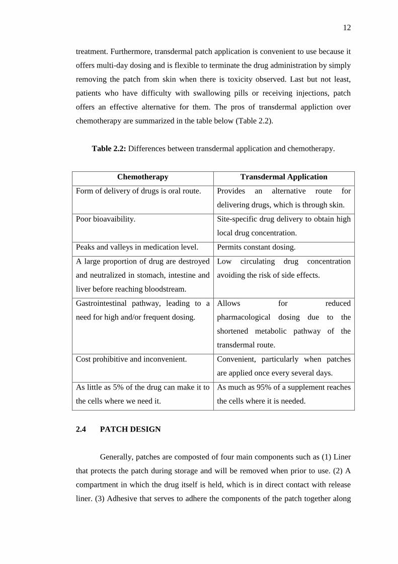
Table 2.2: Differences between transdermal application and chemotherapy.
Chemotherapy Transdermal Application
Form of delivery of drugs is oral route. Provides an alternative route for
delivering drugs, which is through skin.
Poor bioavaibility. Site-specific drug delivery to obtain high
local drug concentration.
Peaks and valleys in medication level. Permits constant dosing.
A large proportion of drug are destroyed
and neutralized in stomach, intestine and
liver before reaching bloodstream.
Low circulating drug concentration
avoiding the risk of side effects.
Gastrointestinal pathway, leading to a
need for high and/or frequent dosing.
Allows for reduced
pharmacological dosing due to the
shortened metabolic pathway of the
transdermal route.
Cost prohibitive and inconvenient. Convenient, particularly when patches
are applied once every several days.
As little as 5% of the drug can make it to
the cells where we need it.
As much as 95% of a supplement reaches
the cells where it is needed.
2.4 PATCH DESIGN
Generally, patches are composted of four main components such as (1) Liner
that protects the patch during storage and will be removed when prior to use. (2) A
compartment in which the drug itself is held, which is in direct contact with release
liner. (3) Adhesive that serves to adhere the components of the patch together along
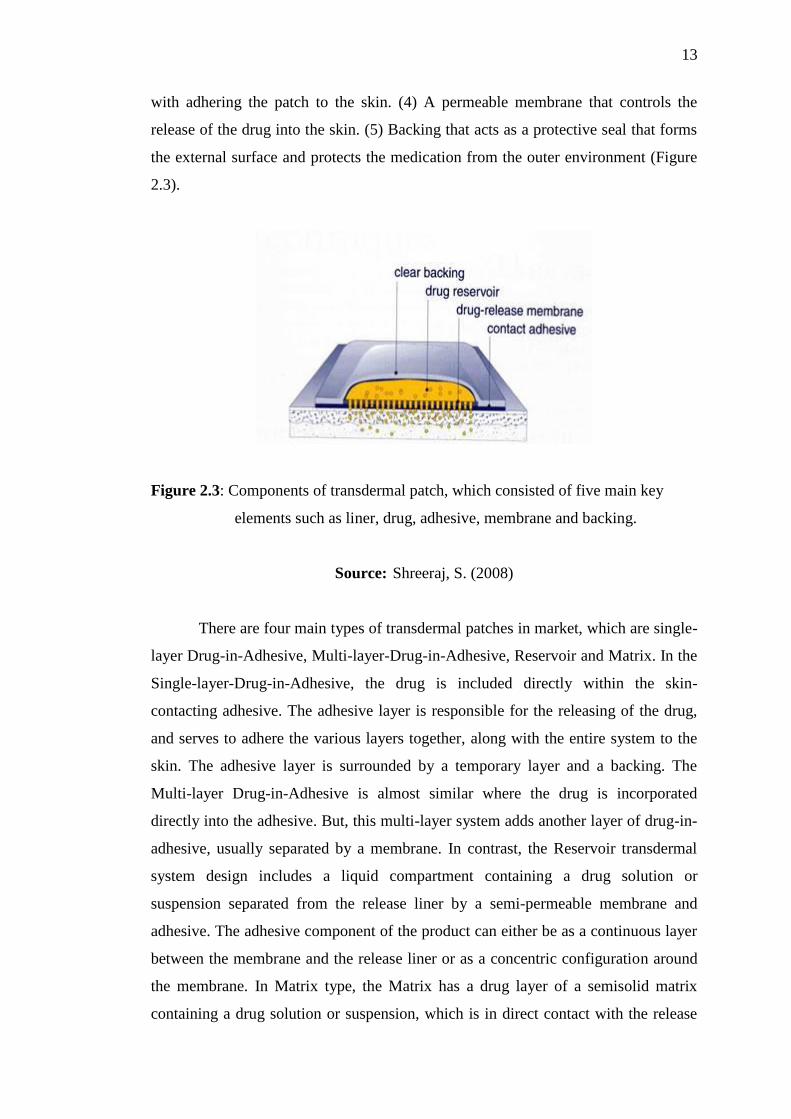
13
with adhering the patch to the skin. (4) A permeable membrane that controls the
release of the drug into the skin. (5) Backing that acts as a protective seal that forms
the external surface and protects the medication from the outer environment (Figure
2.3).
Figure 2.3: Components of transdermal patch, which consisted of five main key
elements such as liner, drug, adhesive, membrane and backing.
Source: Shreeraj, S. (2008)
There are four main types of transdermal patches in market, which are single-
layer Drug-in-Adhesive, Multi-layer-Drug-in-Adhesive, Reservoir and Matrix. In the
Single-layer-Drug-in-Adhesive, the drug is included directly within the skin-
contacting adhesive. The adhesive layer is responsible for the releasing of the drug,
and serves to adhere the various layers together, along with the entire system to the
skin. The adhesive layer is surrounded by a temporary layer and a backing. The
Multi-layer Drug-in-Adhesive is almost similar where the drug is incorporated
directly into the adhesive. But, this multi-layer system adds another layer of drug-in-
adhesive, usually separated by a membrane. In contrast, the Reservoir transdermal
system design includes a liquid compartment containing a drug solution or
suspension separated from the release liner by a semi-permeable membrane and
adhesive. The adhesive component of the product can either be as a continuous layer
between the membrane and the release liner or as a concentric configuration around
the membrane. In Matrix type, the Matrix has a drug layer of a semisolid matrix
containing a drug solution or suspension, which is in direct contact with the release
14
liner. The adhesive layer in this patch surrounds the drug layer partially overlaying it.
(Yury, B., 2006)
2.5 THEORY OF TRANSDERMAL DRUG DELIVERY SYSTEM
A transdermal drug delivery device, transdermal patch works in a very simply
way. When the patient applies the patch to the skin, the medication is releasing form
the vehicles and begins permeating through the skin into bloodstream at a rate
regulated by the membrane. Once succeed to penetrate through the skin barrier, the
drug will be uptake by the capillary network and finally reaches the targeted cancer
cells. Lastly, activation of the pharmacological response to the cancer cells is
happening. The whole process can be listed as below (Girish, C., 2006):-
1. Release of the medicament from the vehicle.
2. Penetration through the skin barrier.
3. Uptake of the drug by the capillary network in the dermal capillary layer.
4. Activation of the pharmacological response. (Banker, G.S. and Rhodes,
C.T., 1990)
In this study, the second step, penetration through the skin barrier was the
main key element to be emphasized. In theory, this step is carried out through a
passive diffusion process and it will be discussed in the coming sub-topic later.
2.5.1 PENETRATION OF DRUG FROM PATCH THROUGH PASSIVE
DIFFUSION PROCESS
As introduced in the sub-topic above, the transportation of drug from patch to
bloodstream is done through a passive diffusion process (Figure 2.4). In other words,
a drug is applied in a relatively high dosage to the inside of a patch, which is worn on
the skin for an extended period of time. Then, the drug is diffused into the body
directly from transdermal patch across the skin barrier due to there is high
concentration of drug on the patch and low concentration of drug in the bloodstream.
The drug will keep diffusing into the blood for a long period of time, maintaining the
constant concentration of drug in the blood flow.
Recommended


![GAris panduan kaunseling ubat-ubatan edisi ke-3...penglihatan, masalah pendengaran ,masalah administrati ubat [contoh: Ryles’ tube] dan lain-lainnya) iv) Pesakit yang mempunyai masalah](https://img.pdfslide.us/doc/110x75/5e244014188f464217395fe5/garis-panduan-kaunseling-ubat-ubatan-edisi-ke-3-penglihatan-masalah-pendengaran.jpg)