Sensory System
• Transmits sensory information collected by receptors to the CNS
Outline
1- General principles of sensory physiology
2- The somatosensory System
3- Olfaction
4- Taste
5- Hearing and Equilibrium
6- Vision
Outline
1- General principles of sensory physiology
2- The somatosensory System
3- Olfaction
4- Taste
5- Hearing and Equilibrium
6- Vision

1- General Principles of Sensory Physiology
• Receptor physiology
• Sensory pathways
• Sensory coding
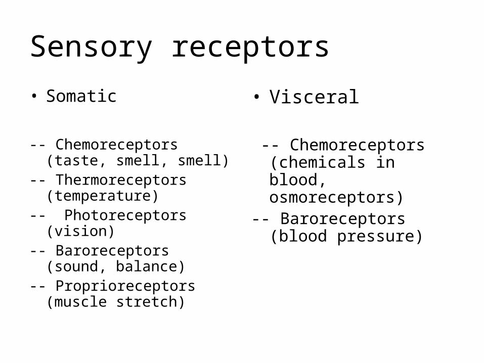
Sensory receptors
• Somatic
-- Chemoreceptors (taste, smell, smell)
-- Thermoreceptors (temperature)
-- Photoreceptors (vision)-- Baroreceptors (sound,
balance)-- Proprioreceptors (muscle
stretch)
• Visceral
-- Chemoreceptors (chemicals in blood, osmoreceptors)
-- Baroreceptors (blood pressure)
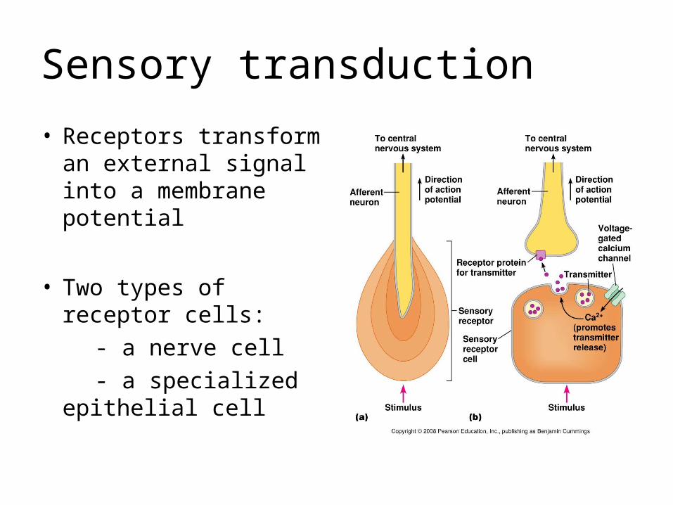
Sensory transduction
• Receptors transform an external signal into a membrane potential
• Two types of receptor cells:
- a nerve cell
- a specialized epithelial cell
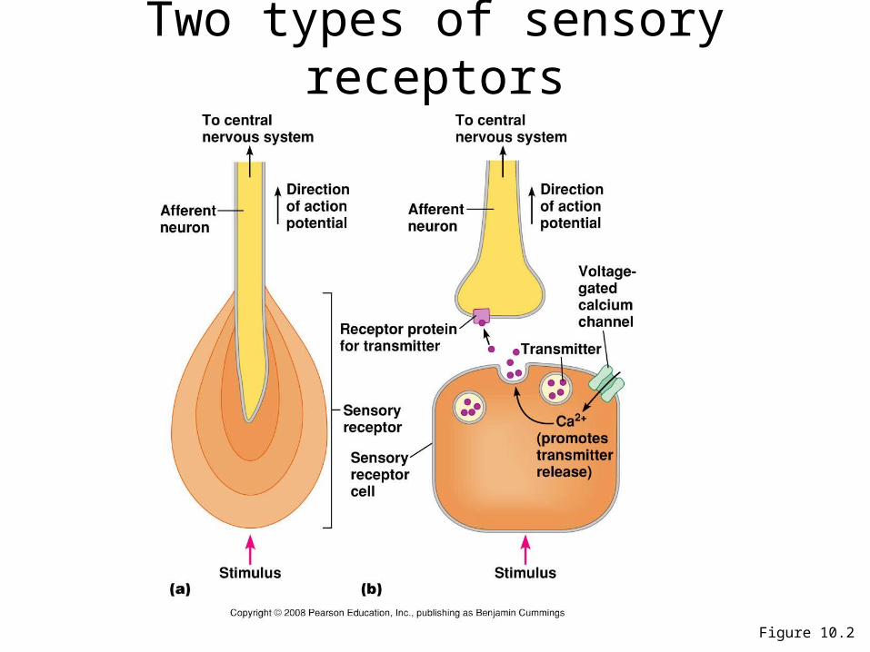
Figure 10.2
Two types of sensory receptors

Receptor adaptation
• Tonic receptors -- slow acting, -- no adaptation:
continue to for impulses as long as the stimulus is there
(ex: proprioreceptors)
• Phasic receptors -- quick acting, adapt: stop firing
when stimuli are constant (ex: smell)
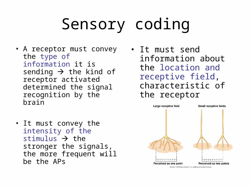
Sensory coding
• A receptor must convey the type of information it is sending the kind of receptor activated determined the signal recognition by the brain
• It must convey the intensity of the stimulus the stronger the signals, the more frequent will be the APs
• It must send information about the location and receptive field, characteristic of the receptor
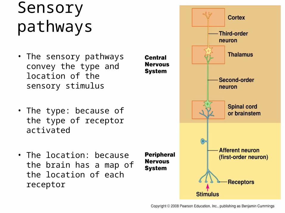
Sensory pathways
• The sensory pathways convey the type and location of the sensory stimulus
• The type: because of the type of receptor activated
• The location: because the brain has a map of the location of each receptor
Outline
1- General principles of sensory physiology
2- The somatosensory System
3- Olfaction
4- Taste
5- Hearing and Equilibrium
6- Vision
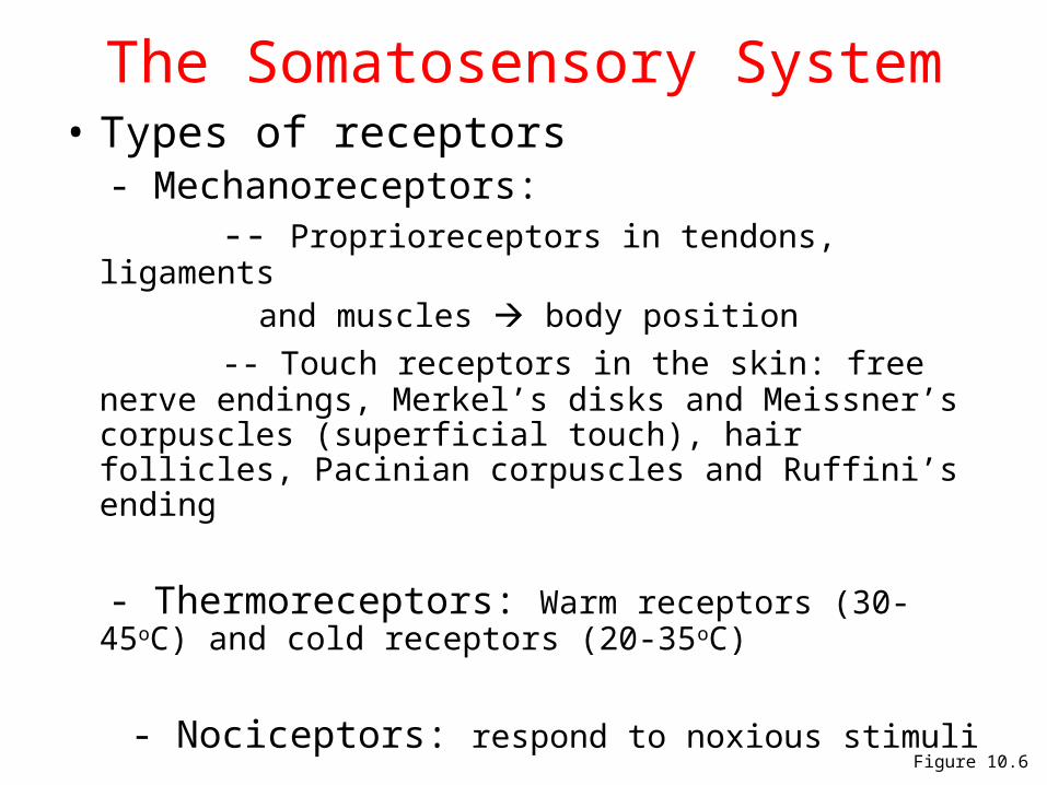
Figure 10.6
The Somatosensory System• Types of receptors - Mechanoreceptors: -- Proprioreceptors in tendons, ligaments and muscles body position
-- Touch receptors in the skin: free nerve endings, Merkel’s disks and Meissner’s corpuscles (superficial touch), hair follicles, Pacinian corpuscles and Ruffini’s ending
- Thermoreceptors: Warm receptors (30-45oC) and
cold receptors (20-35oC)
- Nociceptors: respond to noxious stimuli
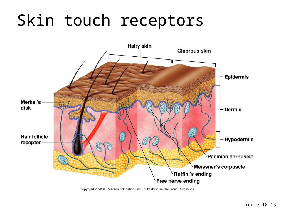
Figure 10.13
Skin touch receptors
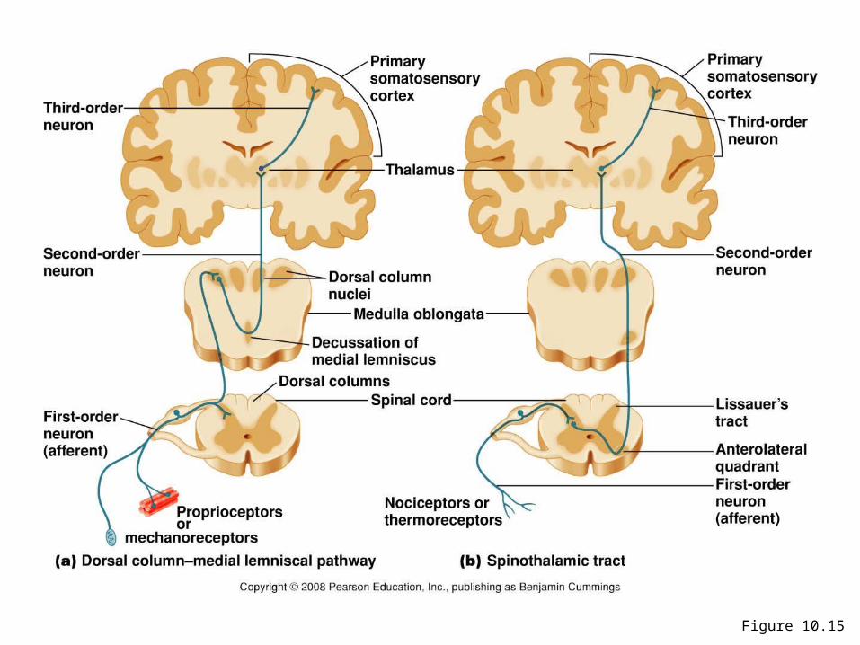
Figure 10.15
Pain perception
• Fast pain: sharp and well localized, transmitted by myelinated axons
• Slow pain: dull aching sensation, not well localized, transmitted by unmyelinated axons
• Visceral pain: not as well localized as pain originating from the skin pain impulses travel on secondary axons dedicated to the somatic afferents referred pain
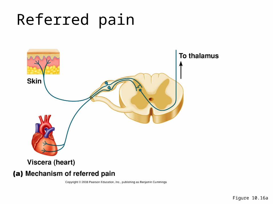
Figure 10.16a
Referred pain
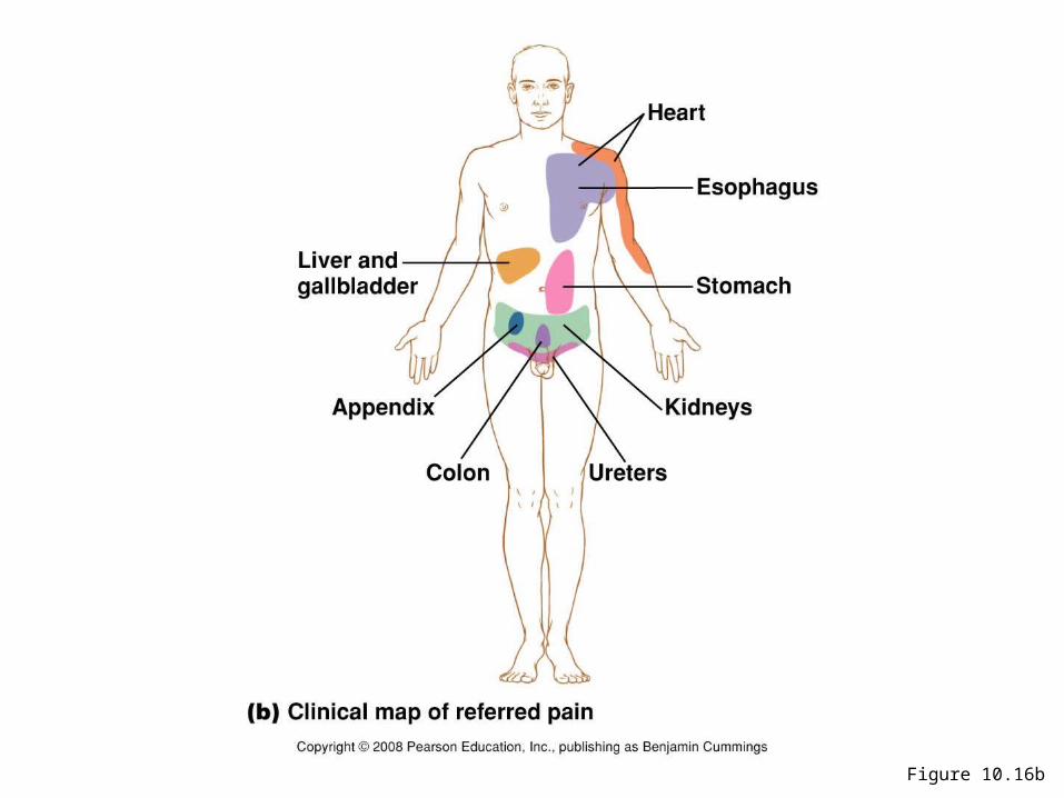
Figure 10.16b
• What is Phantom pain?
Outline
1- General principles of sensory physiology
2- The somatosensory System
3- Olfaction
4- Taste
5- Hearing and Equilibrium
6- Vision
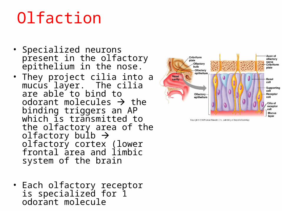
Olfaction
• Specialized neurons present in the olfactory epithelium in the nose.
• They project cilia into a mucus layer. The cilia are able to bind to odorant molecules the binding triggers an AP which is transmitted to the olfactory area of the olfactory bulb olfactory cortex (lower frontal area and limbic system of the brain
• Each olfactory receptor is specialized for 1 odorant molecule
Outline
1- General principles of sensory physiology
2- The somatosensory System
3- Olfaction
4- Taste
5- Hearing and Equilibrium
6- Vision
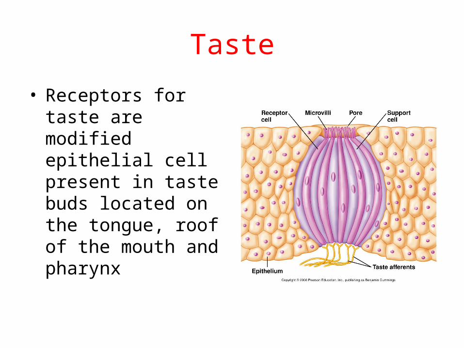
Taste
• Receptors for taste are modified epithelial cell present in taste buds located on the tongue, roof of the mouth and pharynx
• Four primary types of taste receptors : sour, salt, sweet and bitter (and a new one: umami)
• The binding of the receptor to a taste molecule triggers the entry of calcium in the cell release of neurotransmitter in a synapse with a neuron
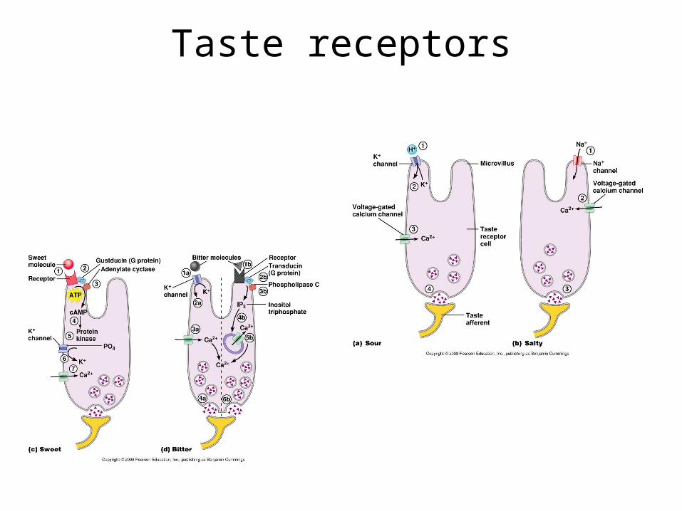
Taste receptors
Neural pathway
• Taste impulses travel through nerves VII, IX and X to a gustatory nucleus in the medulla oblongata (cross over) thalamus gustatory cortex located in the parietal lobe in the mouth area.
• What is the flavor of food?
Outline
1- General principles of sensory physiology
2- The somatosensory System
3- Olfaction
4- Taste
5- Hearing and Equilibrium
6- Vision
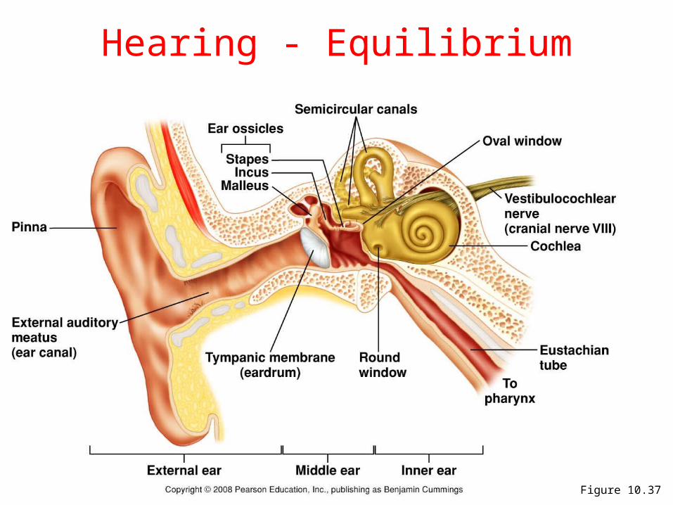
Figure 10.37
Hearing - Equilibrium
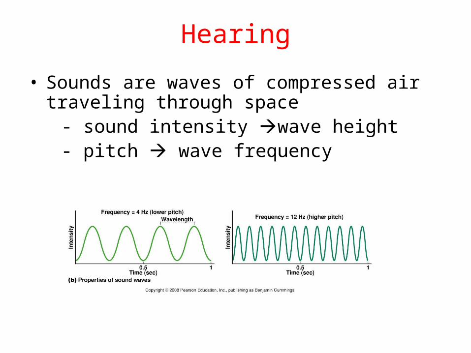
Hearing
• Sounds are waves of compressed air traveling through space
- sound intensity wave height - pitch wave frequency
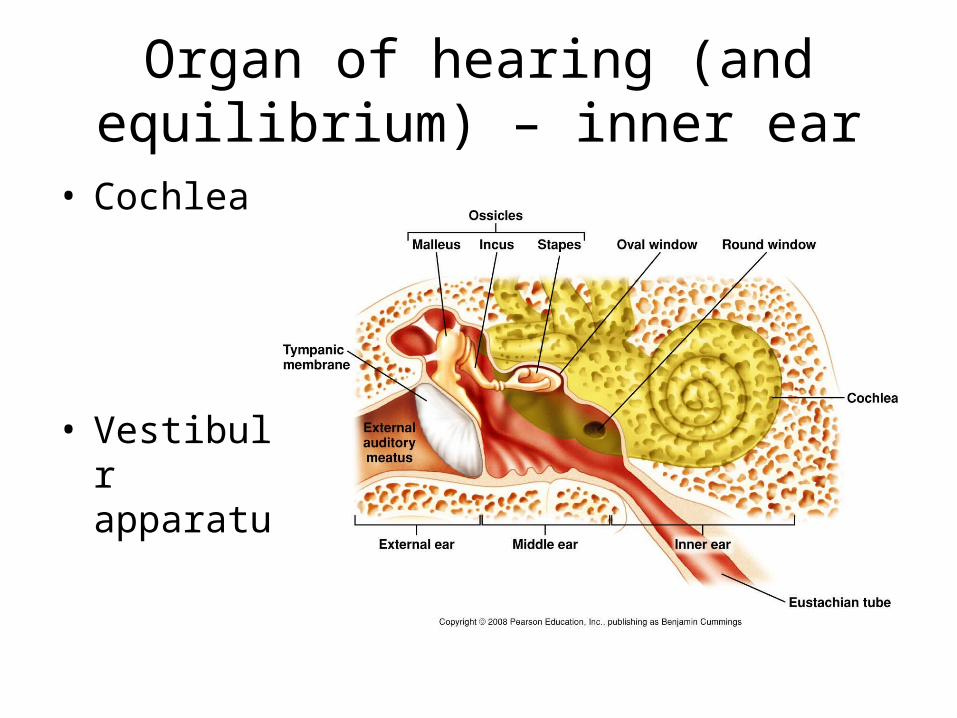
Organ of hearing (and equilibrium) – inner ear
• Cochlea
• Vestibular apparatus
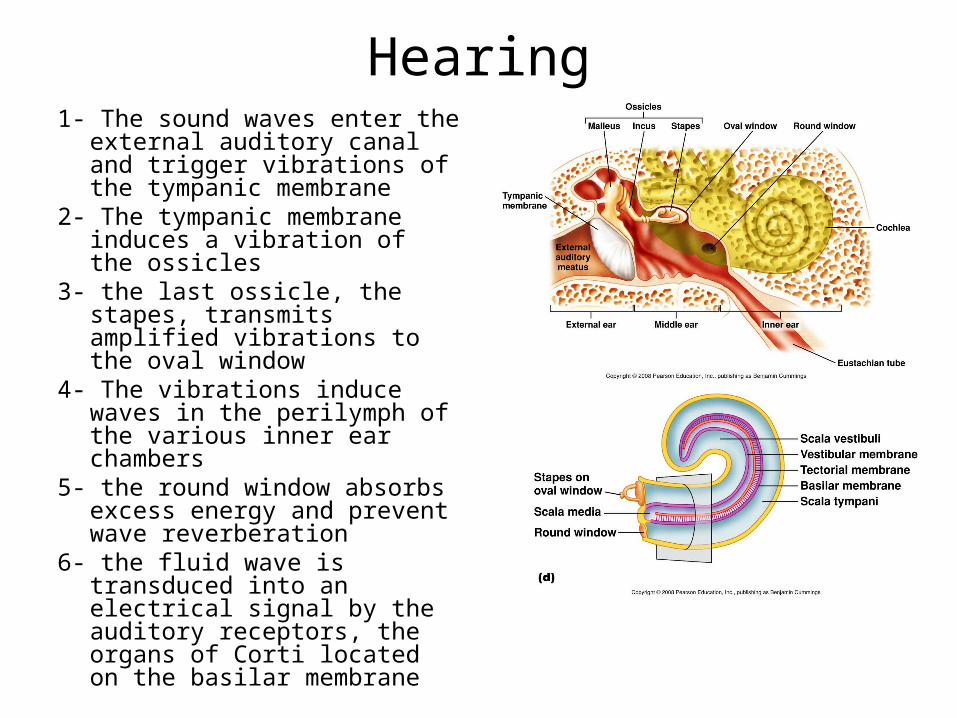
Hearing1- The sound waves enter the
external auditory canal and trigger vibrations of the tympanic membrane
2- The tympanic membrane induces a vibration of the ossicles
3- the last ossicle, the stapes, transmits amplified vibrations to the oval window
4- The vibrations induce waves in the perilymph of the various inner ear chambers
5- the round window absorbs excess energy and prevent wave reverberation
6- the fluid wave is transduced into an electrical signal by the auditory receptors, the organs of Corti located on the basilar membrane
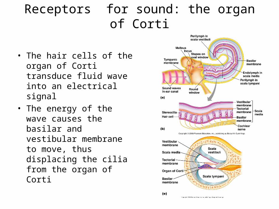
Receptors for sound: the organ of Corti
• The hair cells of the organ of Corti transduce fluid wave into an electrical signal
• The energy of the wave causes the basilar and vestibular membrane to move, thus displacing the cilia from the organ of Corti
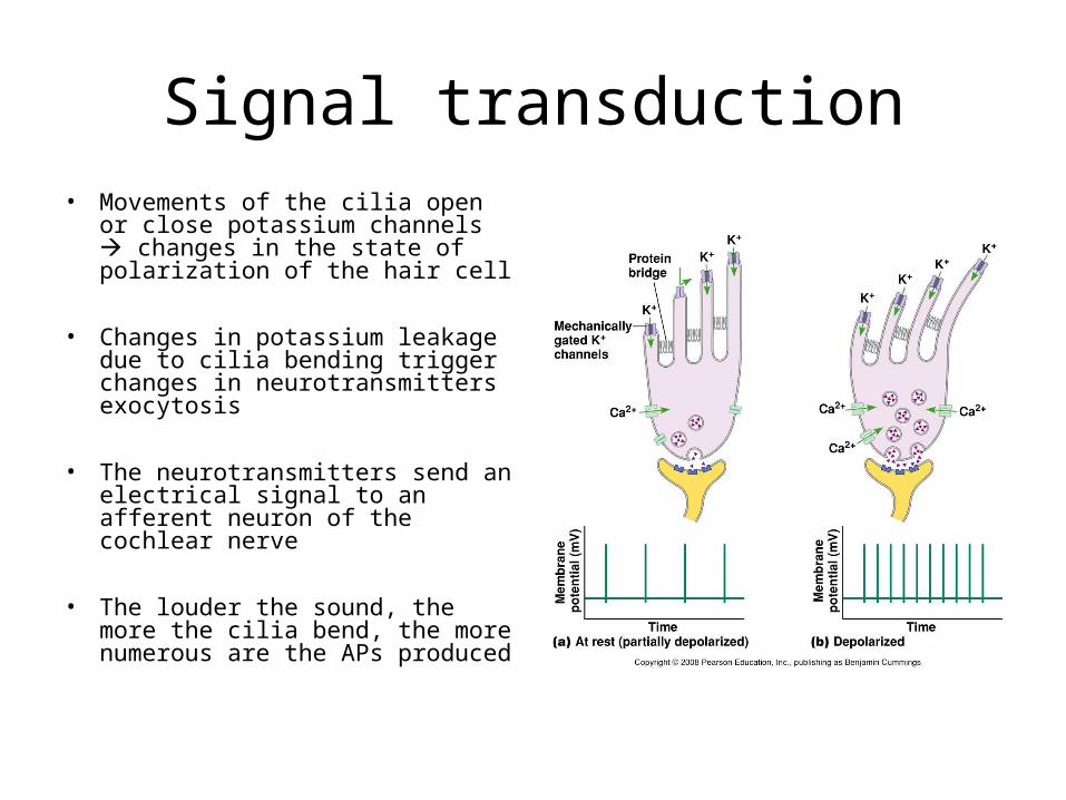
Signal transduction• Movements of the cilia open or
close potassium channels changes in the state of polarization of the hair cell
• Changes in potassium leakage due to cilia bending trigger changes in neurotransmitters exocytosis
• The neurotransmitters send an electrical signal to an afferent neuron of the cochlear nerve
• The louder the sound, the more the cilia bend, the more numerous are the APs produced
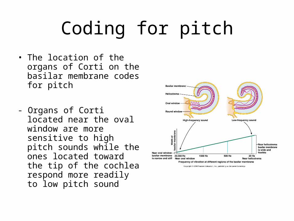
Coding for pitch
• The location of the organs of Corti on the basilar membrane codes for pitch
- Organs of Corti located near the oval window are more sensitive to high pitch sounds while the ones located toward the tip of the cochlea respond more readily to low pitch sound
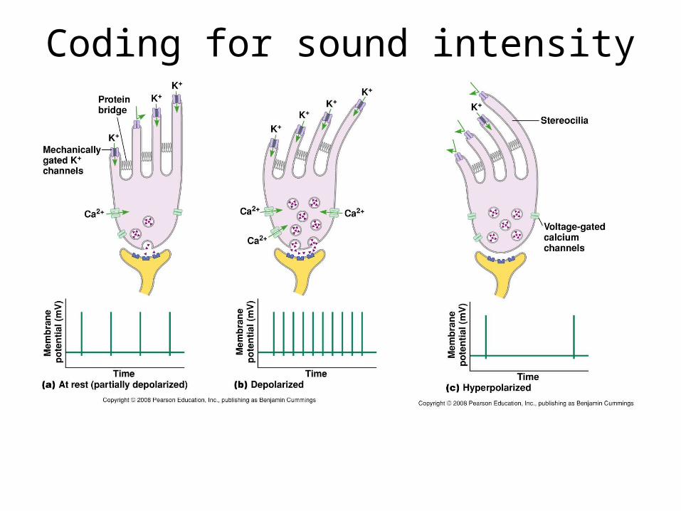
Coding for sound intensity
Neural pathway for sounds
• Cochlear nerve nucleus in medulla oblongata thalamus auditory cortex in the temporal lobe
• So, how do we perceive the direction from which a sound is coming from?
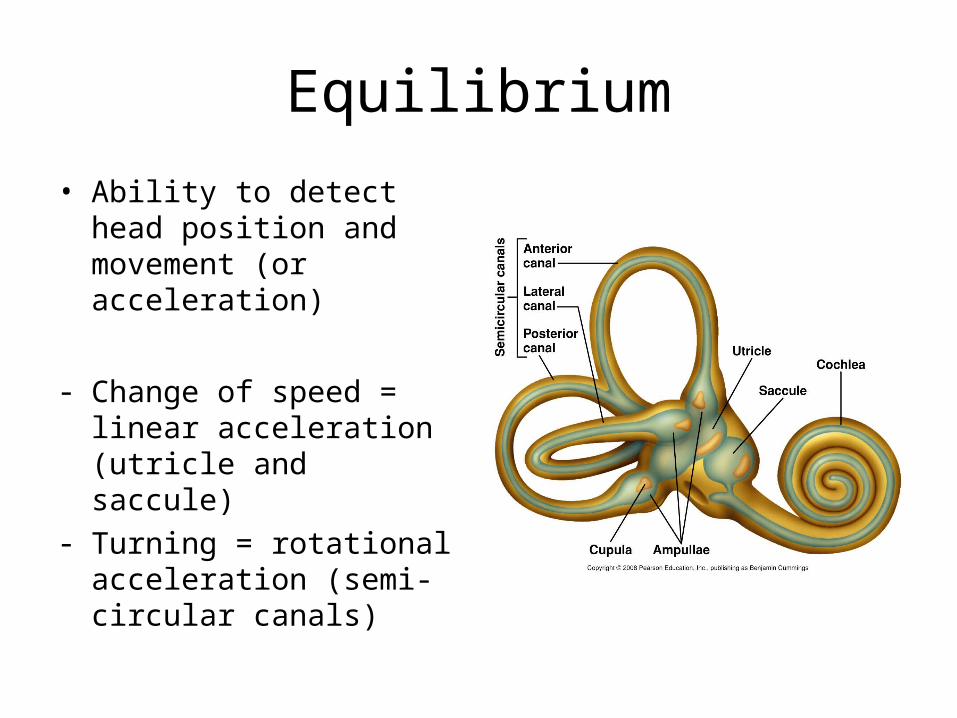
Equilibrium
• Ability to detect head position and movement (or acceleration)
- Change of speed = linear acceleration (utricle and saccule)
- Turning = rotational acceleration (semi-circular canals)
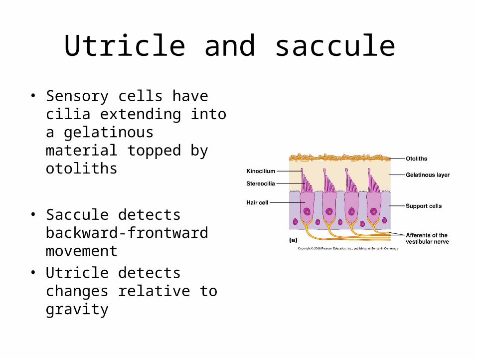
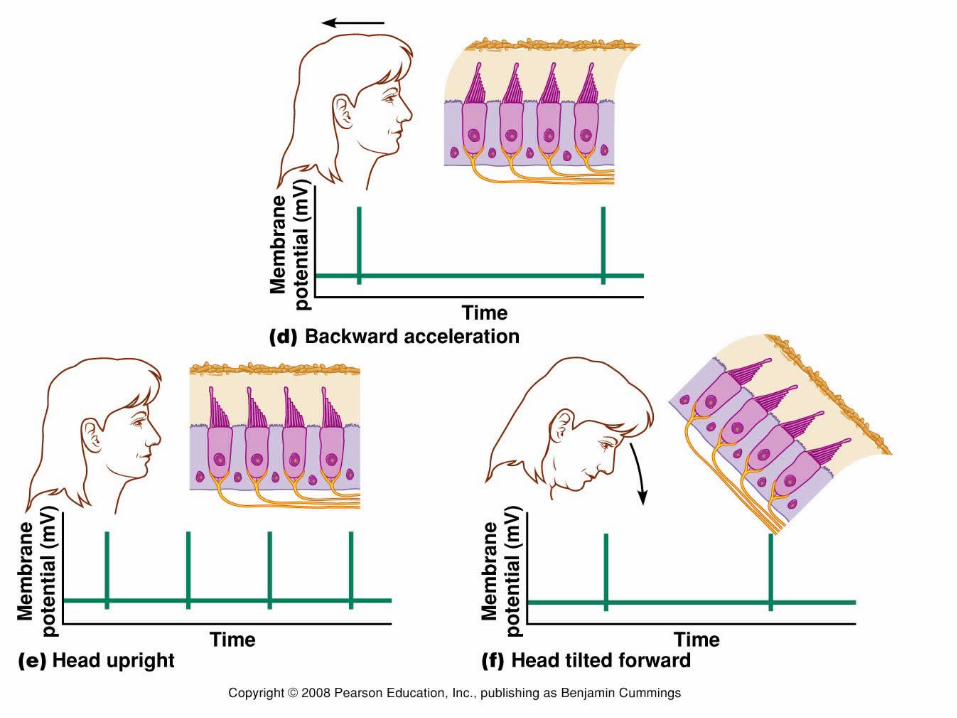
Utricle and saccule
• Sensory cells have cilia extending into a gelatinous material topped by otoliths
• Saccule detects backward-frontward movement
• Utricle detects changes relative to gravity
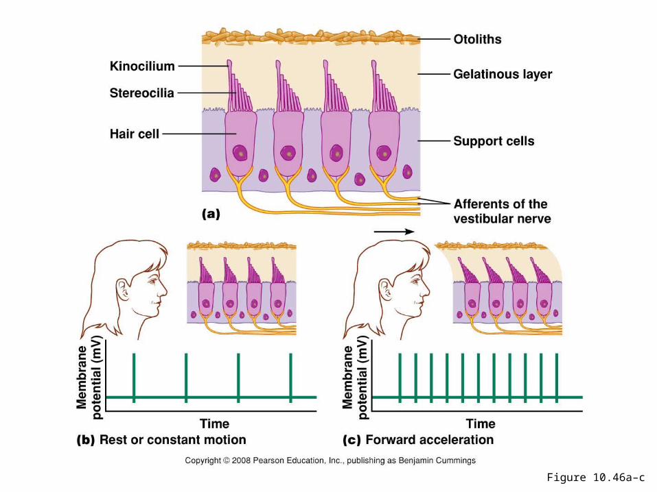
Figure 10.46a–c
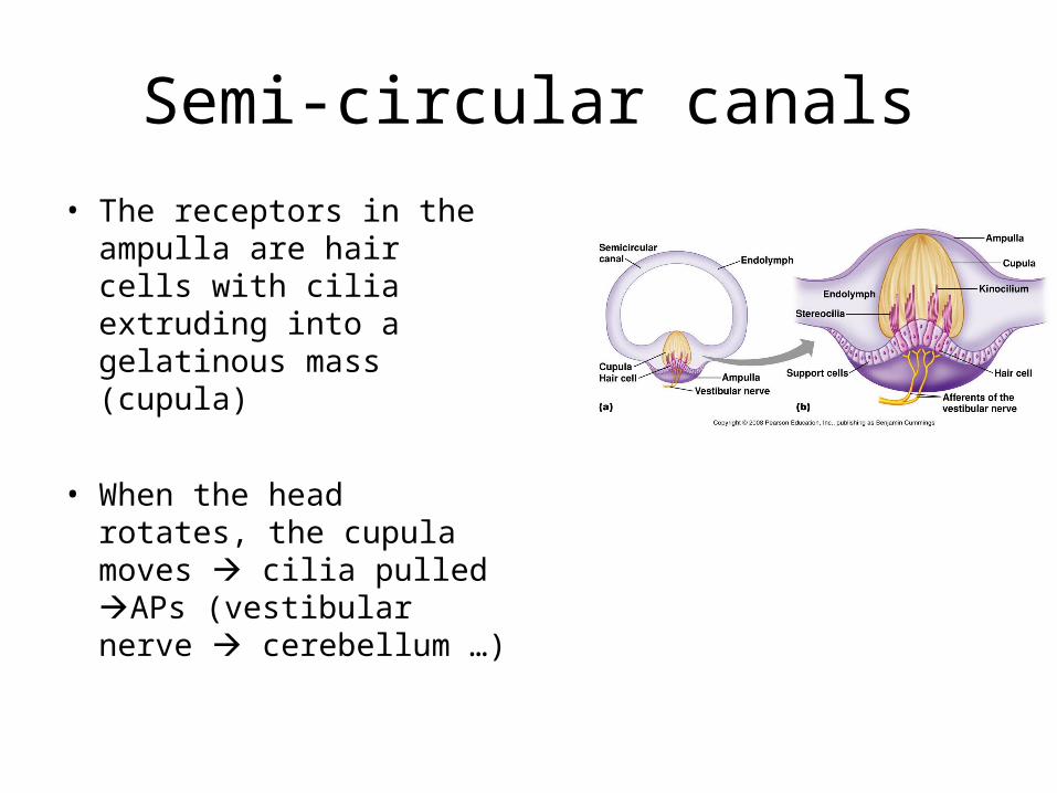
Semi-circular canals
• The receptors in the ampulla are hair cells with cilia extruding into a gelatinous mass (cupula)
• When the head rotates, the cupula moves cilia pulled APs (vestibular nerve cerebellum …)
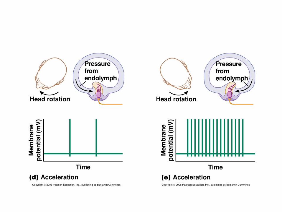
• So why does a person become dizzy after he/she stops spinning?
Outline
1- General principles of sensory physiology
2- The somatosensory System
3- Olfaction
4- Taste
5- Hearing and Equilibrium
6- Vision
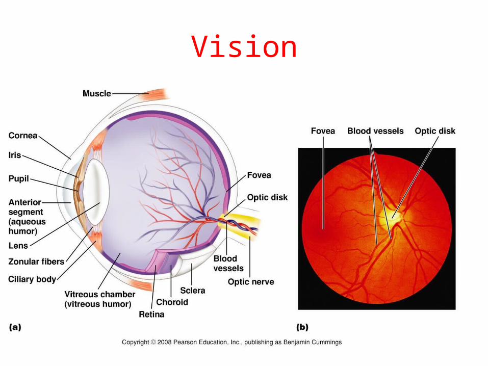
Vision
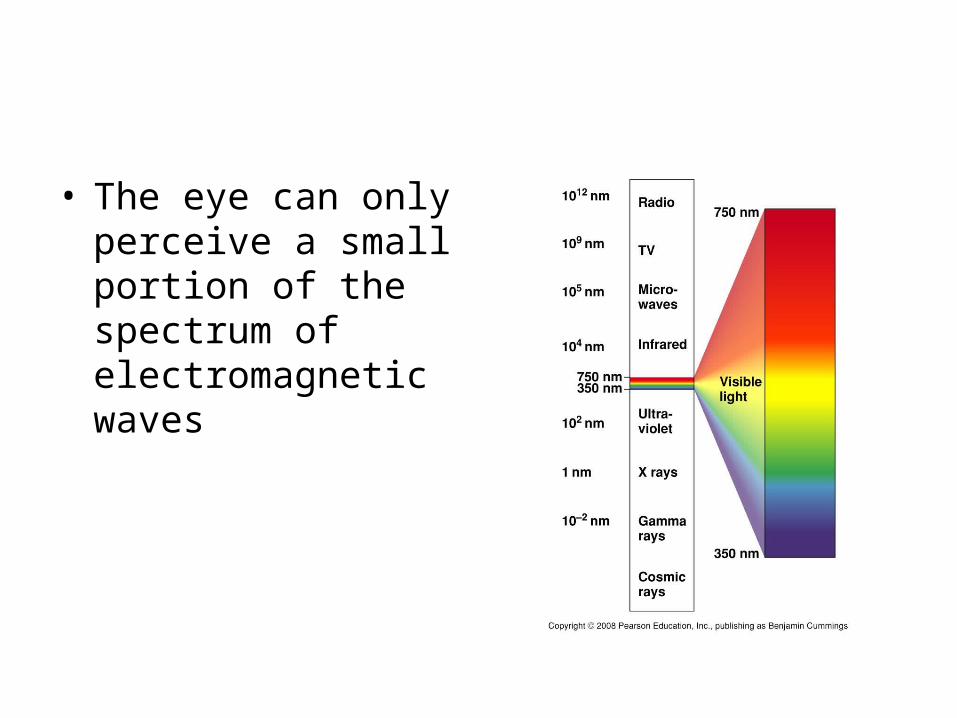
• The eye can only perceive a small portion of the spectrum of electromagnetic waves
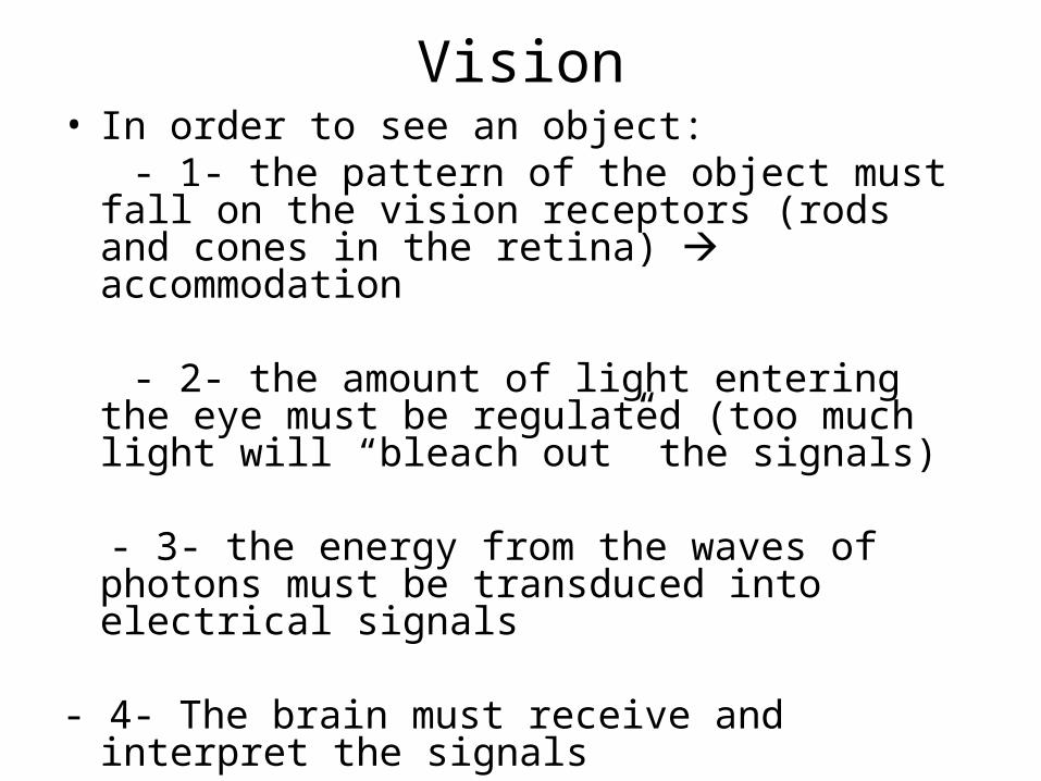
Vision• In order to see an object: - 1- the pattern of the object must fall on the
vision receptors (rods and cones in the retina) accommodation
- 2- the amount of light entering the eye must be regulated (too much light will “bleach out” the signals)
- 3- the energy from the waves of photons must be transduced into electrical signals
- 4- The brain must receive and interpret the signals
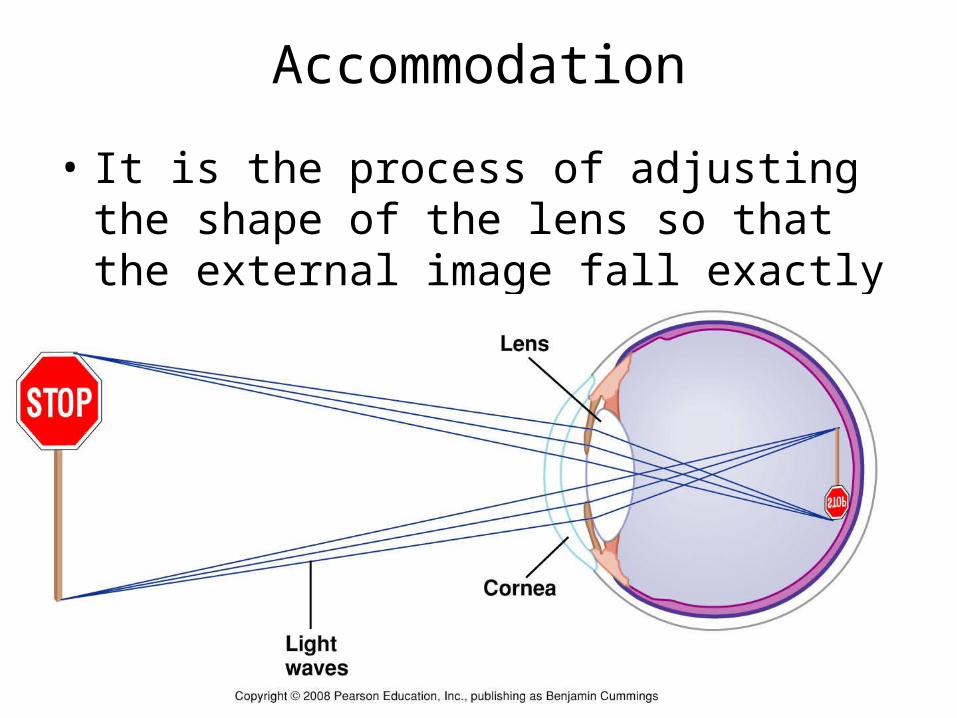
Accommodation
• It is the process of adjusting the shape of the lens so that the external image fall exactly on the retina
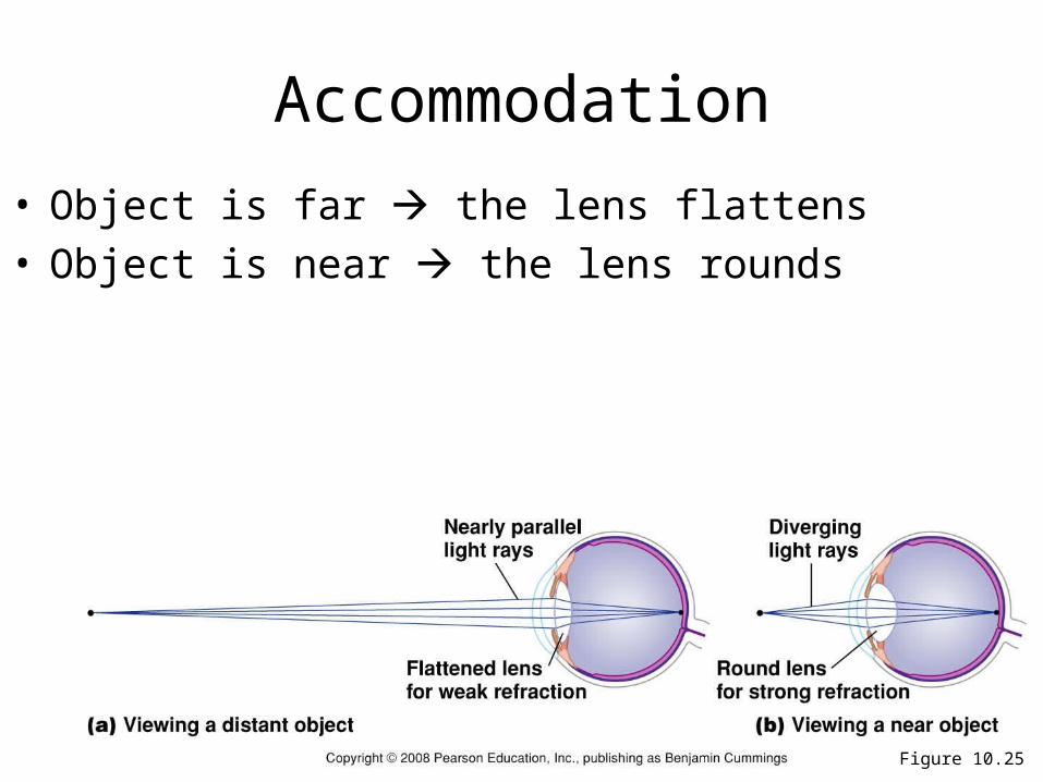
Figure 10.25
Accommodation
• Object is far the lens flattens• Object is near the lens rounds
Accommodation abnormalities
• Myopia
• Hyperopia
• Astigmatism: the cornea is irregular irregular pattern of vision
• Presbyopia: stiffening of the lens occurring with aging increased difficulty with near vision
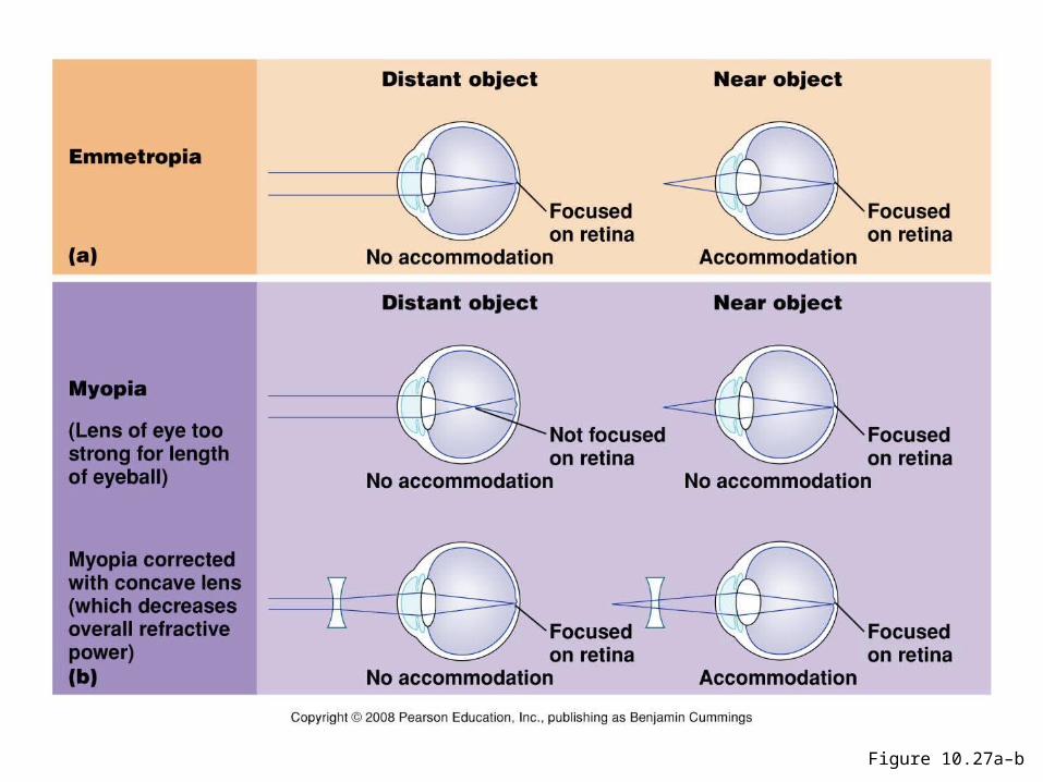
Figure 10.27a–b
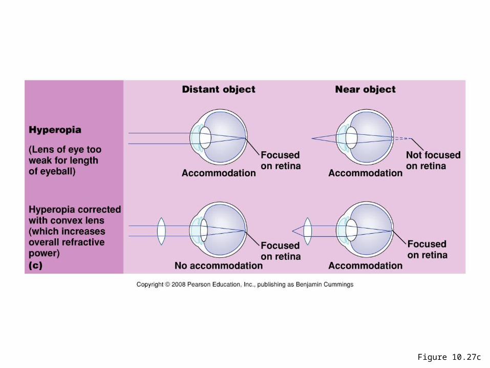
Figure 10.27c
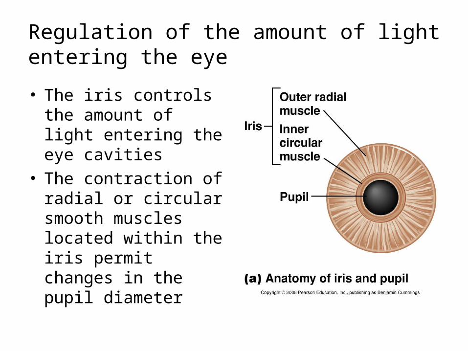
Regulation of the amount of light entering the eye
• The iris controls the amount of light entering the eye cavities
• The contraction of radial or circular smooth muscles located within the iris permit changes in the pupil diameter
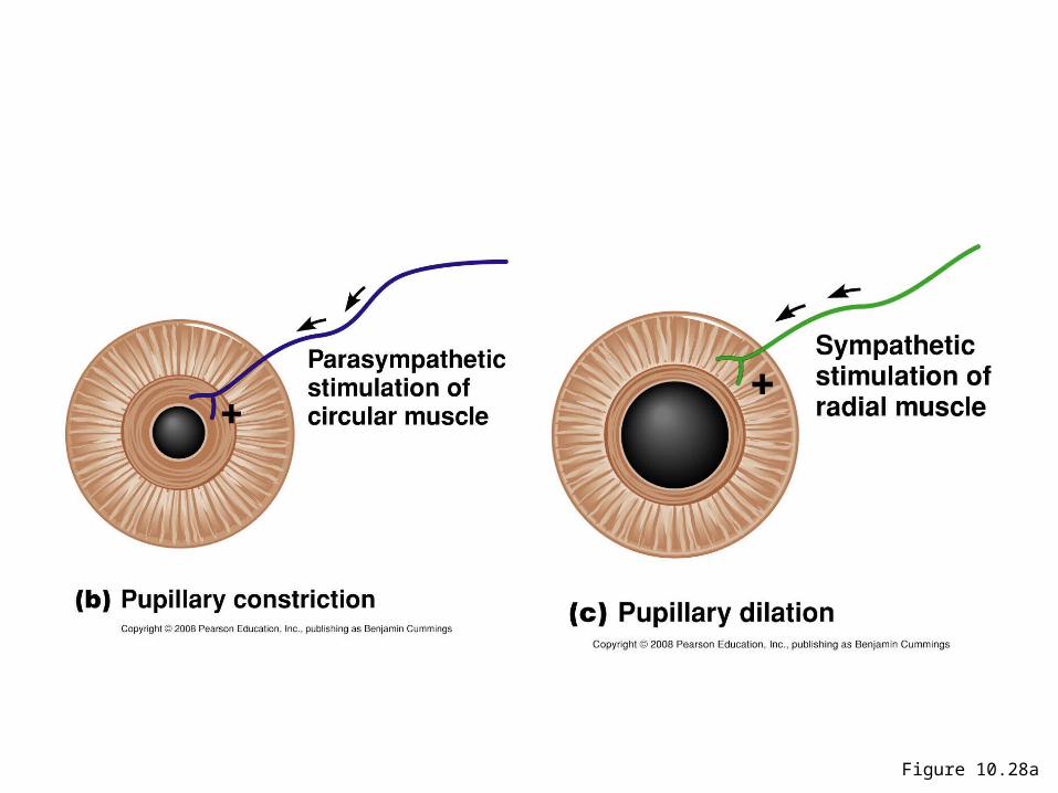
Figure 10.28a
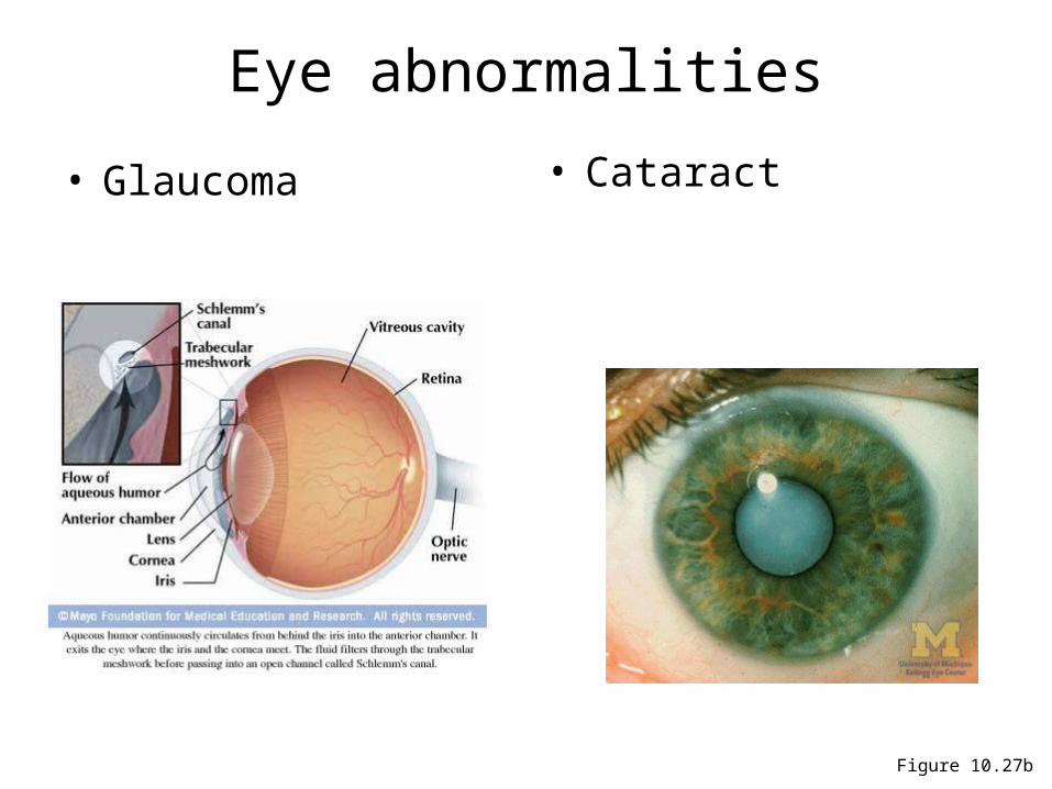
Figure 10.27b
Eye abnormalities
• Glaucoma • Cataract
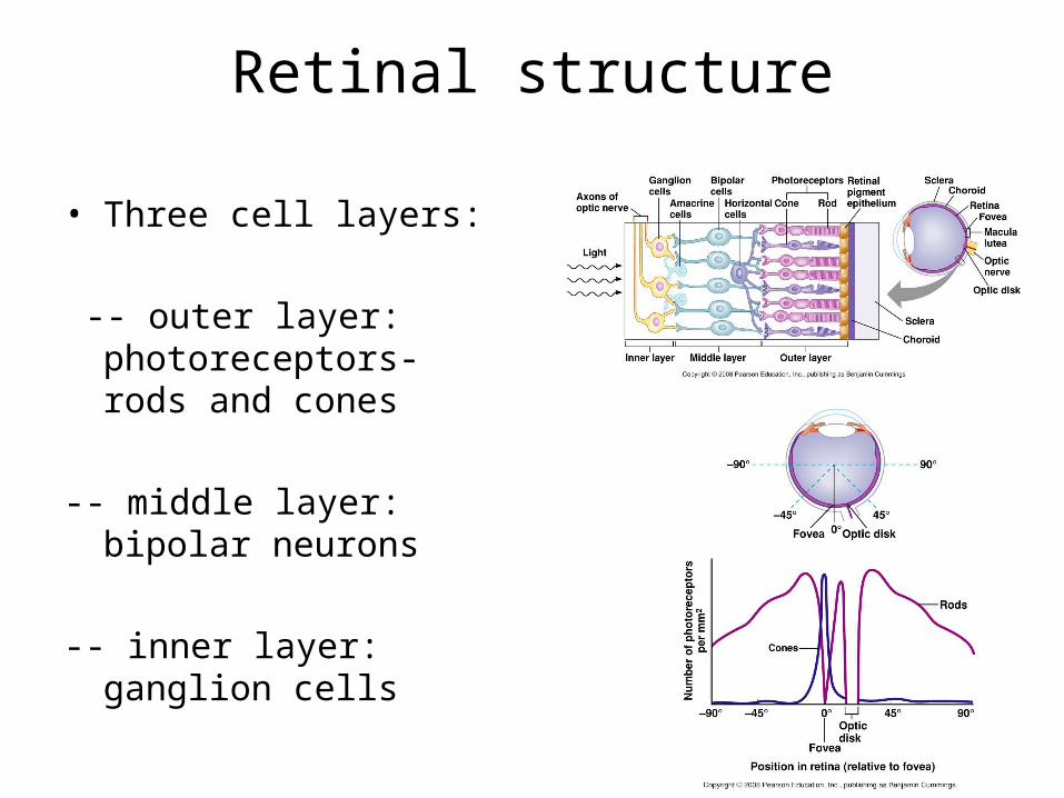
Retinal structure
• Three cell layers:
-- outer layer: photoreceptors- rods and cones
-- middle layer: bipolar neurons
-- inner layer: ganglion cells
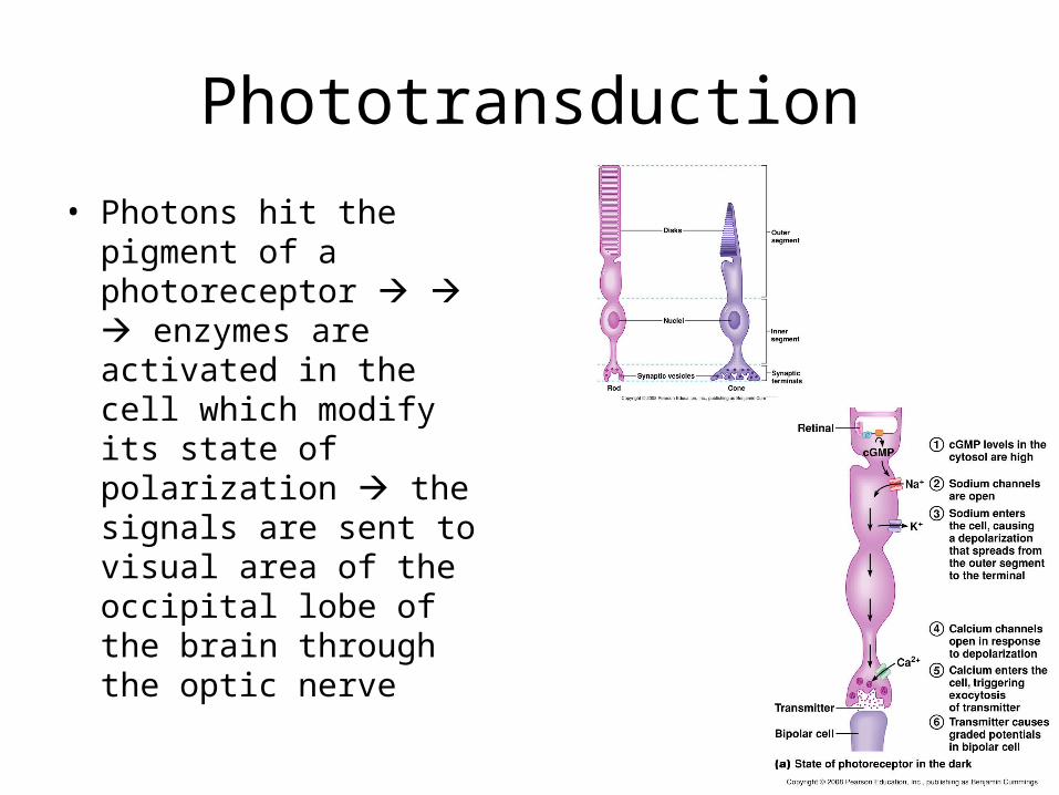
Phototransduction
• Photons hit the pigment of a photoreceptor enzymes are activated in the cell which modify its state of polarization the signals are sent to visual area of the occipital lobe of the brain through the optic nerve
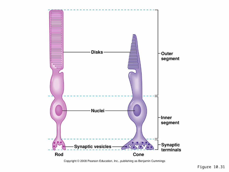
Figure 10.31
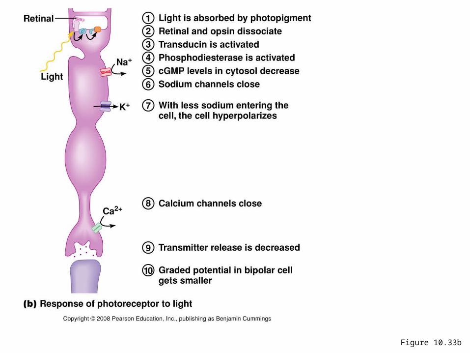
Figure 10.33b
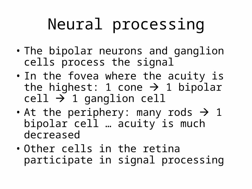
Neural processing
• The bipolar neurons and ganglion cells process the signal
• In the fovea where the acuity is the highest: 1 cone 1 bipolar cell 1 ganglion cell
• At the periphery: many rods 1 bipolar cell … acuity is much decreased
• Other cells in the retina participate in signal processing
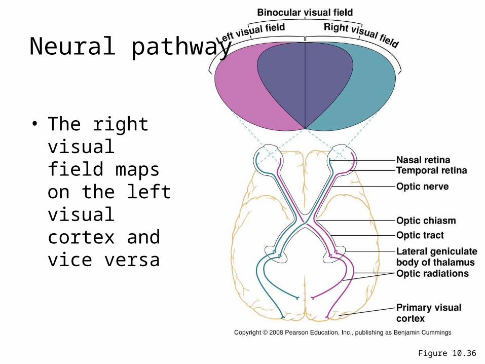
Figure 10.36
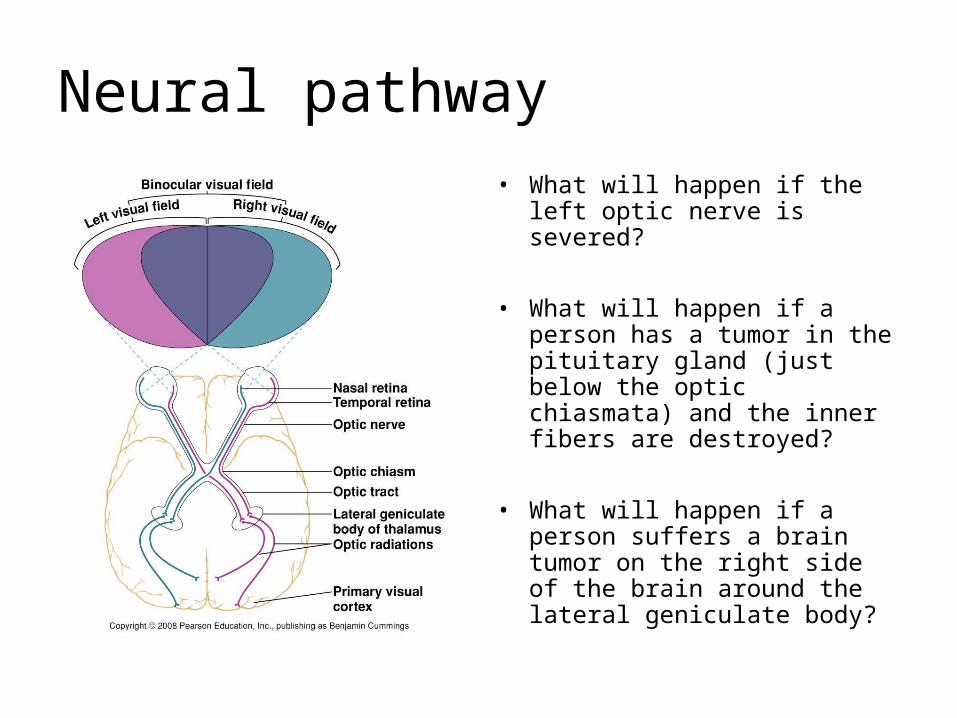
Neural pathway
• The right visual field maps on the left visual cortex and vice versa
Neural pathway
• What will happen if the left optic nerve is severed?
• What will happen if a person has a tumor in the pituitary gland (just below the optic chiasmata) and the inner fibers are destroyed?
• What will happen if a person suffers a brain tumor on the right side of the brain around the lateral geniculate body?
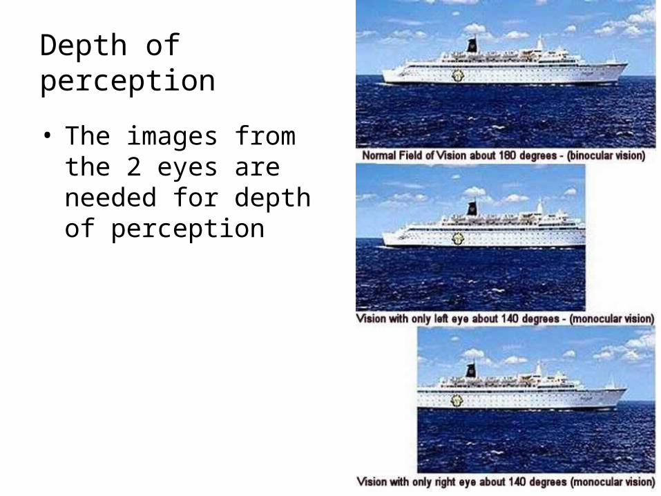
Depth of perception
• The images from the 2 eyes are needed for depth of perception
Readings:
• Chp. 10, p. 253-301
• Not expected:
- Pain gate-control theory, p. 268.
- Phototransduction, p. 277-280
Recommended