A rough guide to A rough guide to AcylcarnitinesAcylcarnitines
Roy Talbot & Nigel [email protected]
Dept. of Clinical Chemistry,Sheffield Children’s Hospital
MenuMenuAcylcarnitines GA-II/MADD
Basic Tandem MS CPT-IItheorySCADD ß-Ketothiolase
MCADD MMA/PA
LCHADD IVA
VLCADD Plasma vs. DBS
GA-I Derivatisation
What are acylcarnitines?What are acylcarnitines?• Fatty acyl ester of L-carnitine• Facilitate entry of long-chain fatty acids
(LC-FA) into the mitochondrion via the Carnitine Shuttle
– LC-FA’s act as important fuels for many tissues (e.g. skeletal & cardiac muscle) via ß-oxidation
• In fatty-acid oxidation defects, acylcarnitine species accumulate and are released into the circulation
– pattern of acylcarnitine species can be diagnostic for a number of ß-oxidation defects
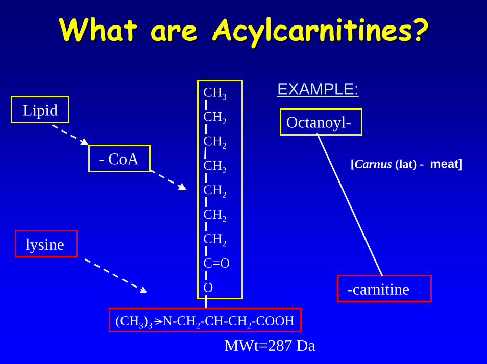
What are Acylcarnitines?What are Acylcarnitines?
EXAMPLE:Lipid
[Carnus (lat) - meat]
Octanoyl-
-carnitine
- CoA
lysine
CH3
CH2
CH2
CH2
CH2
CH2
CH2
C=O
O
(CH3)3 N-CH2-CH-CH2-COOH
MWt=287 Da
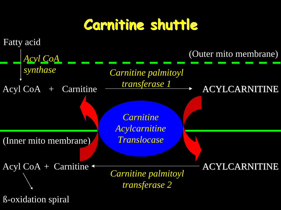
Carnitine shuttleCarnitine shuttleFatty acid
Acyl CoA synthase Carnitine palmitoyl
transferase 1
ACYLCARNITINEACYLCARNITINECarnitine palmitoyl
transferase 2
Acyl CoA ACYLCARNITINEACYLCARNITINE
Carnitine Acylcarnitine Translocase
(Outer mito membrane)
Carnitine+
(Inner mito membrane)
CarnitineAcyl CoA +
ß-oxidation spiral
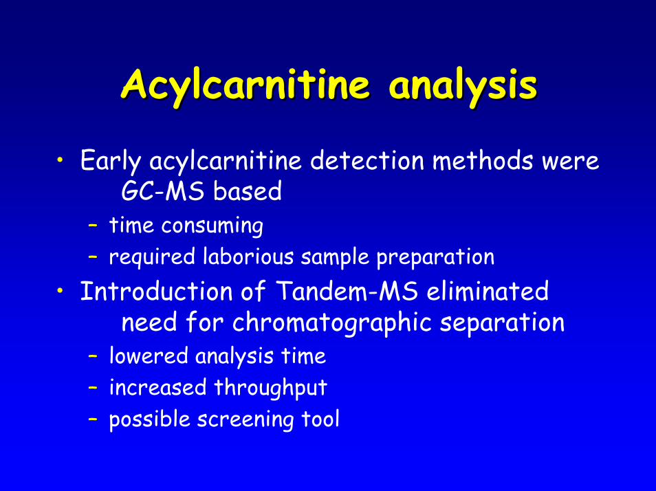
Acylcarnitine analysisAcylcarnitine analysis
• Early acylcarnitine detection methods were GC-MS based
– time consuming– required laborious sample preparation
• Introduction of Tandem-MS eliminated need for chromatographic separation
– lowered analysis time– increased throughput– possible screening tool
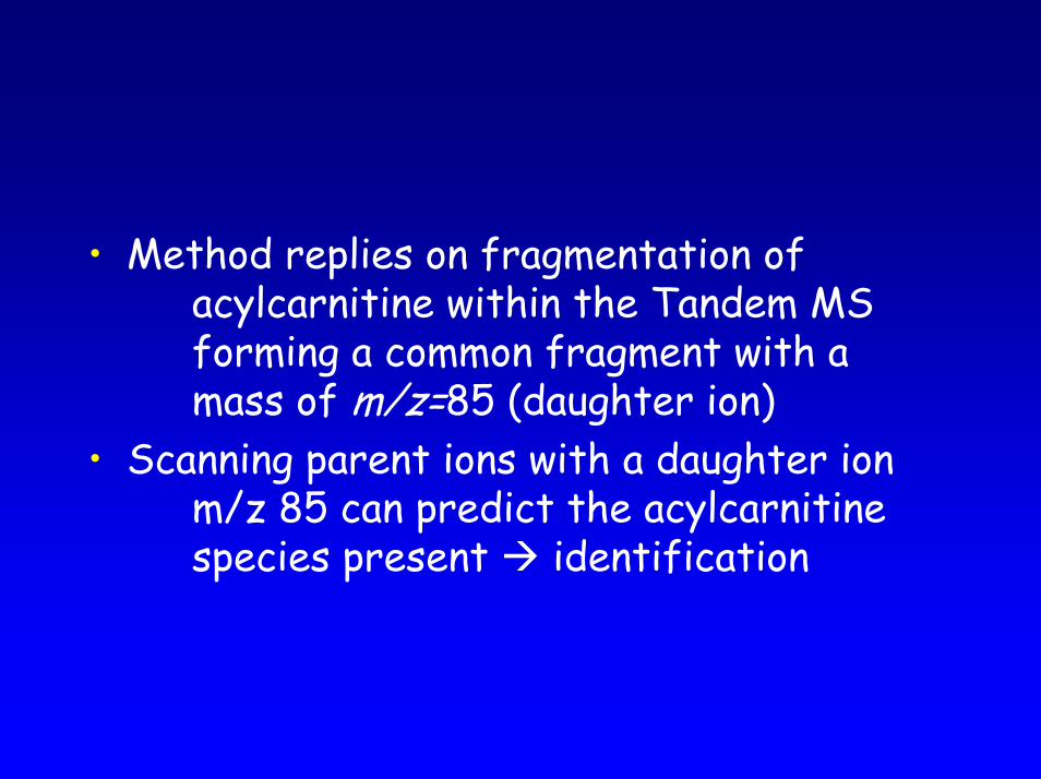
• Method replies on fragmentation of acylcarnitine within the Tandem MS forming a common fragment with a mass of m/z=85 (daughter ion)
• Scanning parent ions with a daughter ion m/z 85 can predict the acylcarnitine species present identification
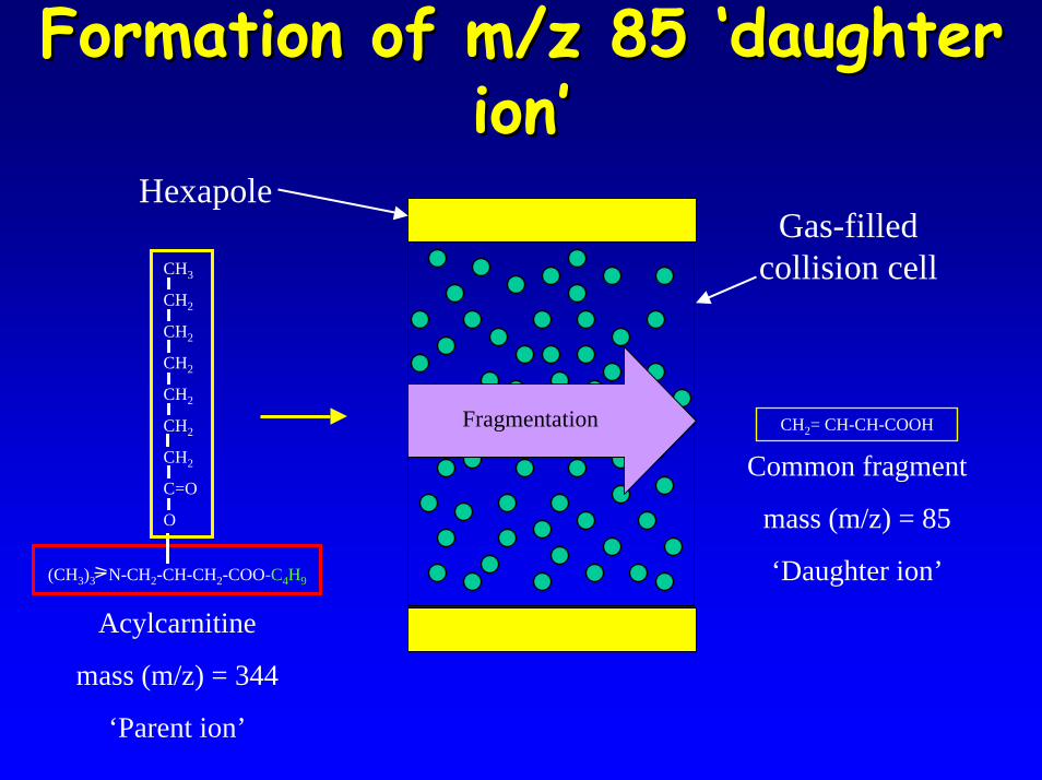
Formation of m/z 85 ‘daughter Formation of m/z 85 ‘daughter ion’ion’
CH2= CH-CH-COOH
(CH3)3 N-CH2-CH-CH2-COO-C4H9
CH3
CH2
CH2
CH2
CH2
CH2
CH2
C=O
O
Fragmentation
Gas-filled collision cell
Hexapole
Common fragment
mass (m/z) = 85
‘Daughter ion’
Acylcarnitine
mass (m/z) = 344
‘Parent ion’
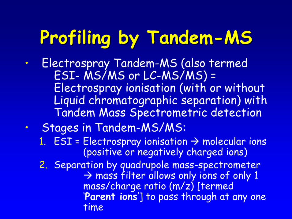
Profiling by TandemProfiling by Tandem--MSMS• Electrospray Tandem-MS (also termed
ESI- MS/MS or LC-MS/MS) = Electrospray ionisation (with or without Liquid chromatographic separation) with Tandem Mass Spectrometric detection
• Stages in Tandem-MS/MS:1. ESI = Electrospray ionisation molecular ions
(positive or negatively charged ions)2. Separation by quadrupole mass-spectrometer
mass filter allows only ions of only 1 mass/charge ratio (m/z) [termed ‘Parent ions’] to pass through at any one time

Profiling by TandemProfiling by Tandem--MSMS3. Fragmentation of Parent ion within an inert
gas (e.g. argon) containing collision cell situated between the 2 quadrupole mass filters
4. Separation by second quadrupole mass filter (allows only ions of only 1 m/z [termed ‘Daughter ions’])
5. Electron- or photo-multiplier detection identification and/or quantitation by stable isotope dilution
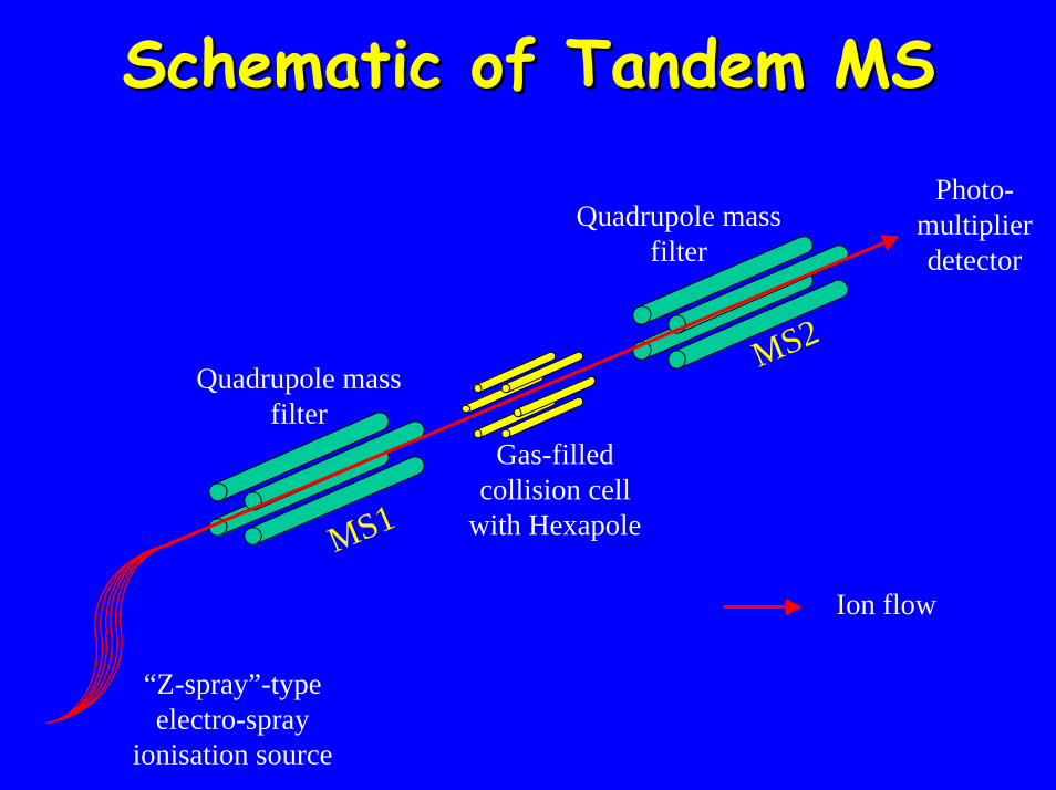
Schematic of Tandem MS Schematic of Tandem MS Photo-
multiplier detector
Quadrupole mass filter
Quadrupole mass filter
Gas-filled collision cell
with Hexapole
Ion flow
MS1
MS2
“Z-spray”-type electro-spray
ionisation source
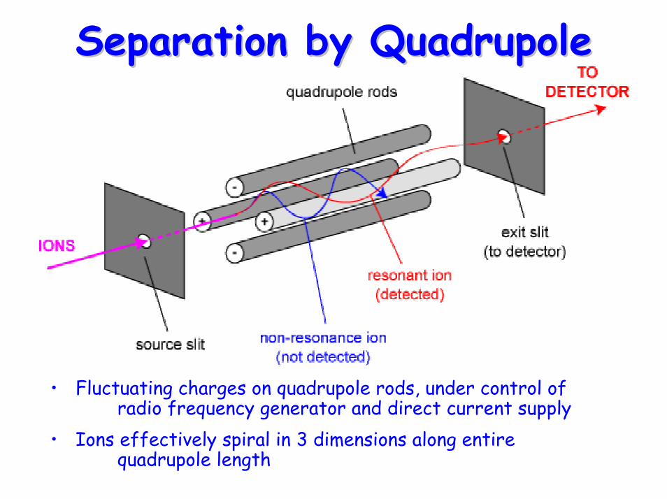
Separation by QuadrupoleSeparation by Quadrupole
• Fluctuating charges on quadrupole rods, under control of radio frequency generator and direct current supply
• Ions effectively spiral in 3 dimensions along entire quadrupole length
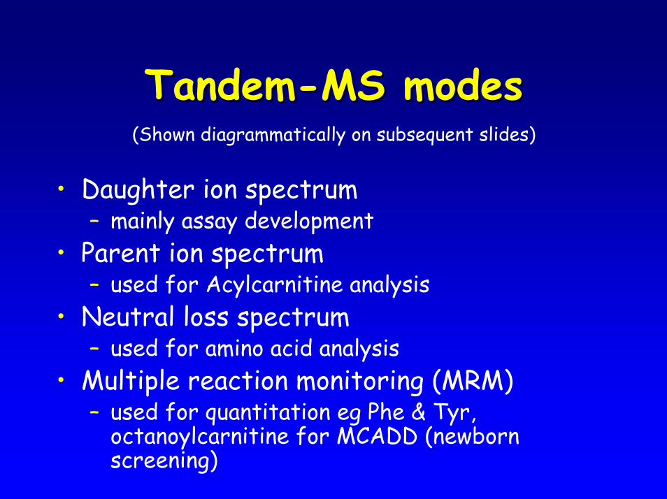
TandemTandem--MS modes MS modes (Shown diagrammatically on subsequent slides)
• Daughter ion spectrum – mainly assay development
• Parent ion spectrum– used for Acylcarnitine analysis
• Neutral loss spectrum– used for amino acid analysis
• Multiple reaction monitoring (MRM)– used for quantitation eg Phe & Tyr,
octanoylcarnitine for MCADD (newborn screening)
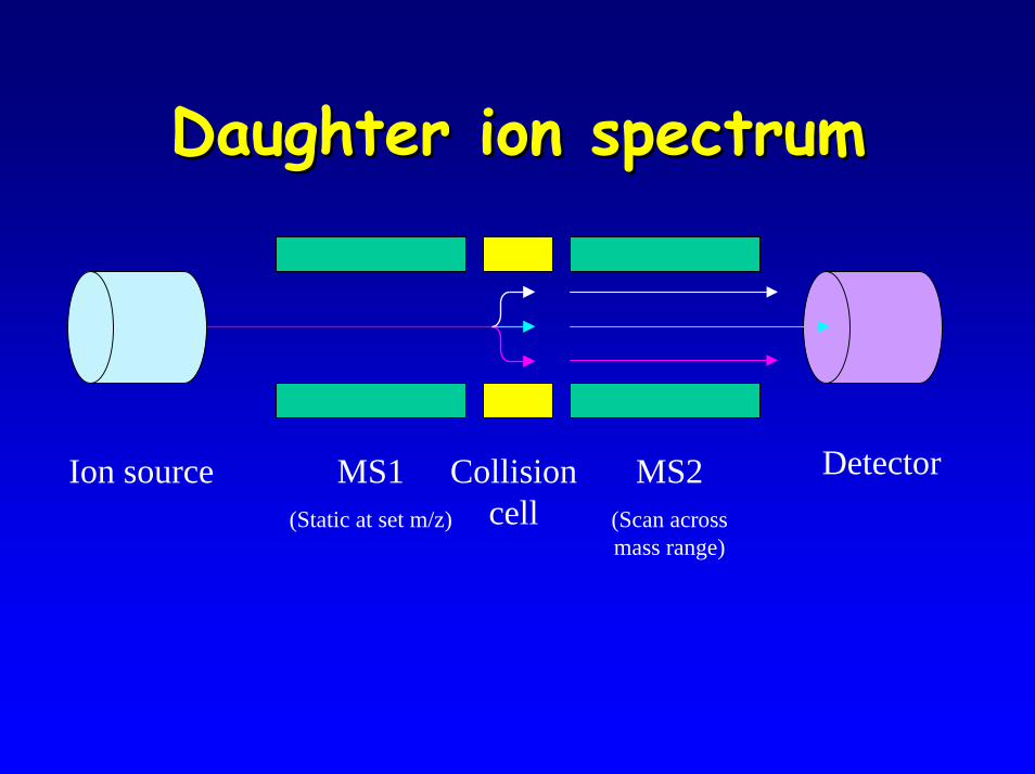
Daughter ion spectrumDaughter ion spectrum
DetectorMS1(Static at set m/z)
MS2(Scan across mass range)
Ion source Collision cell
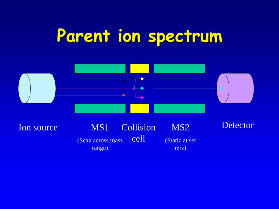
Parent ion spectrumParent ion spectrum
DetectorMS1(Scan across mass
range)
MS2(Static at set
m/z)
Ion source Collision cell
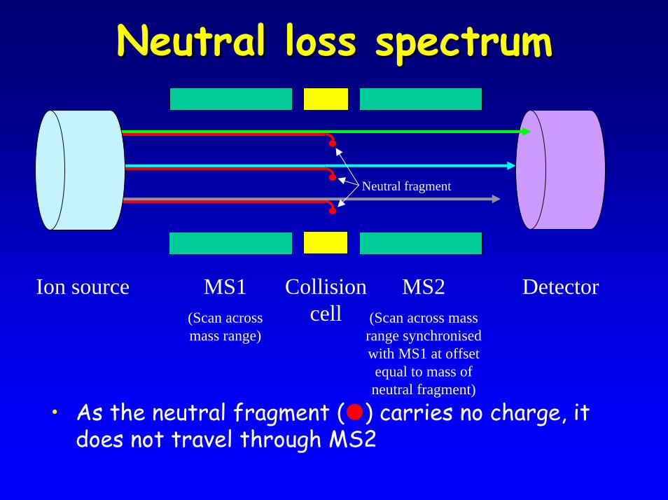
Neutral loss spectrumNeutral loss spectrum
• As the neutral fragment ( ) carries no charge, it does not travel through MS2
DetectorMS1(Scan across mass range)
MS2(Scan across mass range synchronised with MS1 at offset equal to mass of neutral fragment)
Ion source Collision cell
Neutral fragment
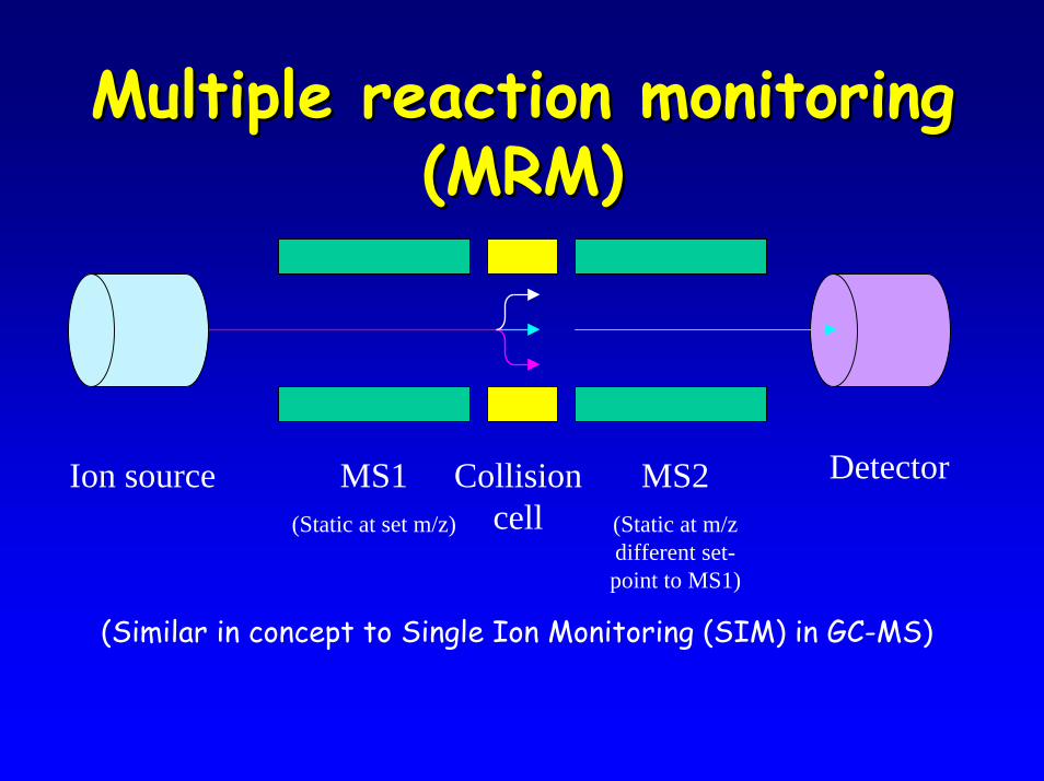
Multiple reaction monitoring Multiple reaction monitoring (MRM)(MRM)
DetectorMS1(Static at set m/z)
MS2(Static at m/z different set-point to MS1)
Ion source Collision cell
(Similar in concept to Single Ion Monitoring (SIM) in GC-MS)
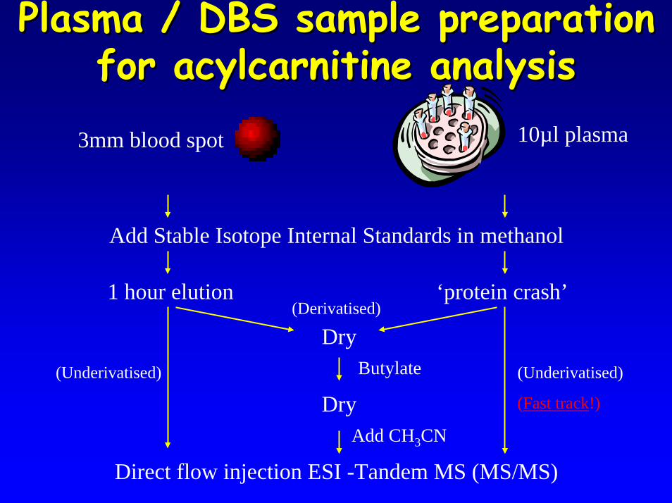
Plasma / DBS sample preparation Plasma / DBS sample preparation for acylcarnitine analysisfor acylcarnitine analysis
3mm blood spot 10µl plasma
Add Stable Isotope Internal Standards in methanol
1 hour elution ‘protein crash’
DryButylate
DryAdd CH3CN
(Derivatised)
(Underivatised) (Underivatised)
(Fast track!)
Direct flow injection ESI -Tandem MS (MS/MS)
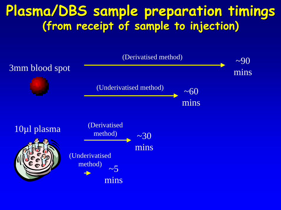
Plasma/DBS sample preparation timingsPlasma/DBS sample preparation timings(from receipt of sample to injection)(from receipt of sample to injection)
(Derivatised method) ~90 mins3mm blood spot
(Underivatised method) ~60 mins
10µl plasma (Derivatised method)
(Underivatised method) ~5
mins
~30 mins
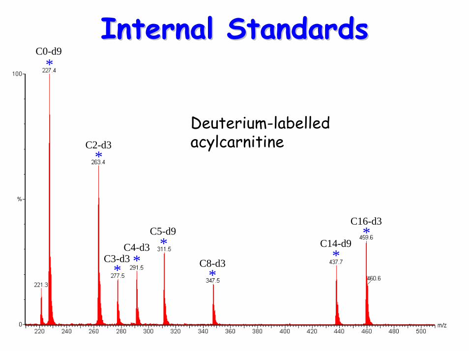
Internal StandardsInternal Standards
C16-d3
*
* **
**
*
C2-d3
C3-d3C4-d3
C5-d9
C8-d3
C14-d9
*C0-d9
Deuterium-labelled acylcarnitine
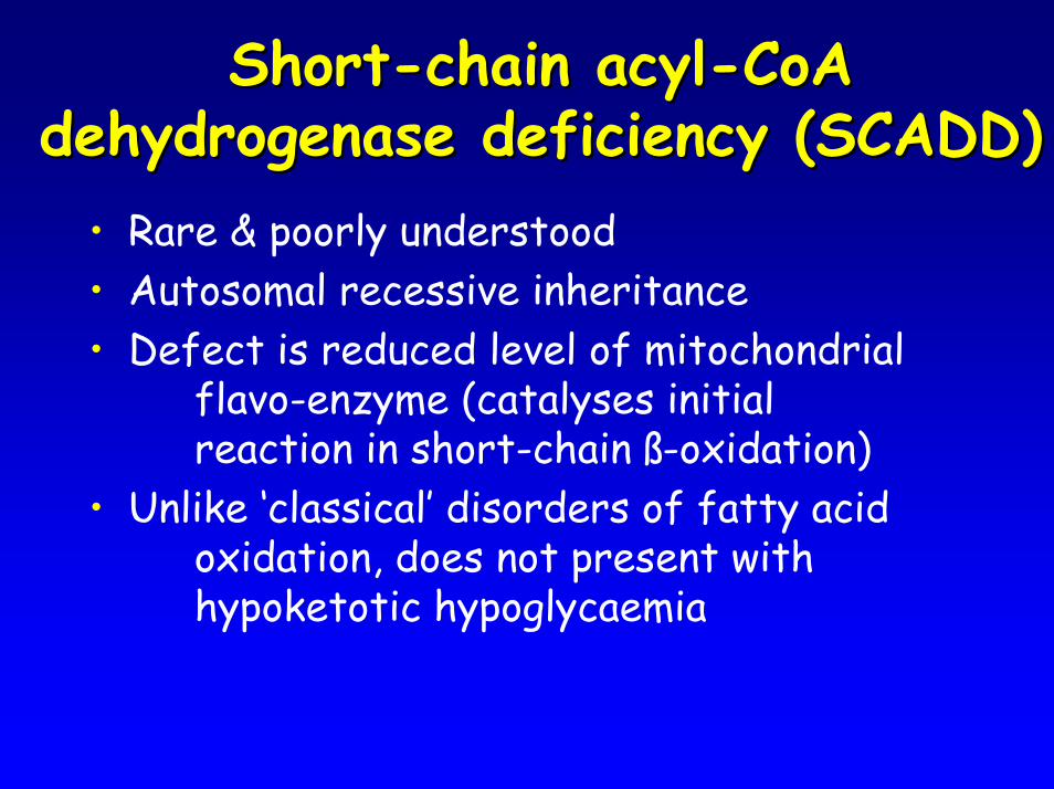
ShortShort--chain acylchain acyl--CoA CoA dehydrogenase deficiency (SCADD)dehydrogenase deficiency (SCADD)
• Rare & poorly understood• Autosomal recessive inheritance• Defect is reduced level of mitochondrial
flavo-enzyme (catalyses initial reaction in short-chain ß-oxidation)
• Unlike ‘classical’ disorders of fatty acid oxidation, does not present with hypoketotic hypoglycaemia
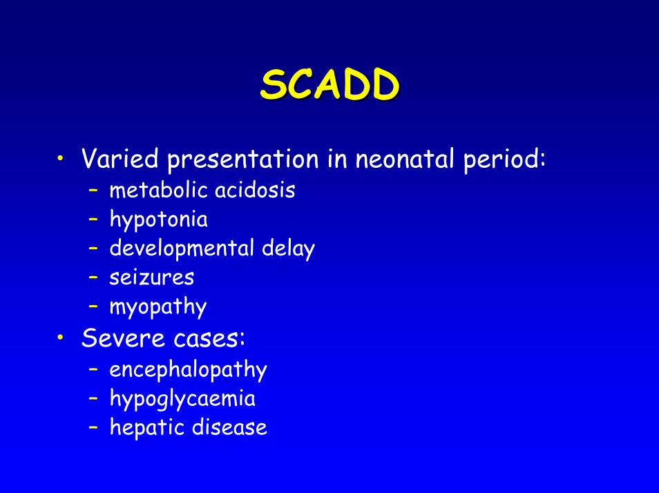
SCADDSCADD• Varied presentation in neonatal period:
– metabolic acidosis– hypotonia– developmental delay– seizures– myopathy
• Severe cases:– encephalopathy– hypoglycaemia– hepatic disease
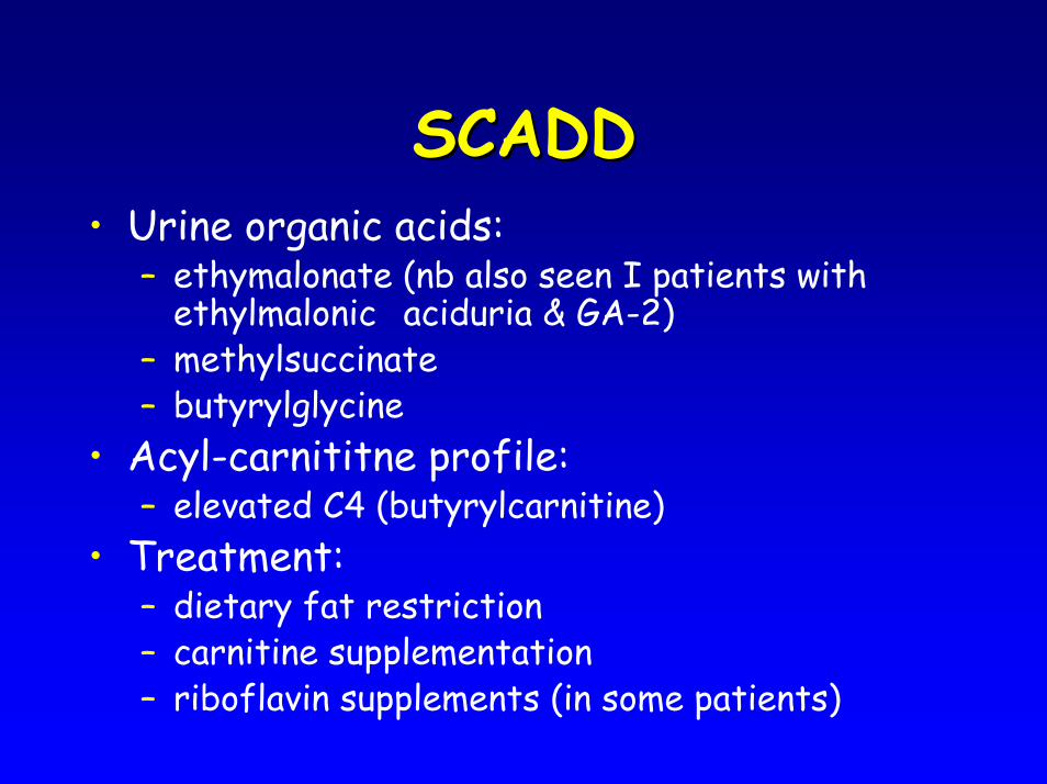
SCADDSCADD• Urine organic acids:
– ethymalonate (nb also seen I patients with ethylmalonic aciduria & GA-2)
– methylsuccinate– butyrylglycine
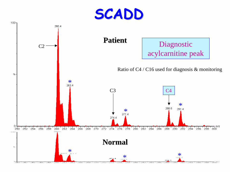
• Acyl-carnititne profile: – elevated C4 (butyrylcarnitine)
• Treatment:– dietary fat restriction– carnitine supplementation– riboflavin supplements (in some patients)
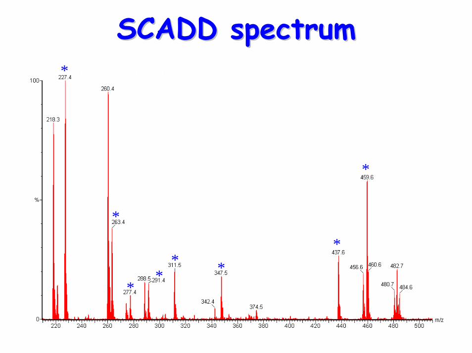
SCADD spectrumSCADD spectrum*
*
**
* **
*
*
*
* * * **
*
*
*
* * * **
*
PatientPatient
NormalNormal
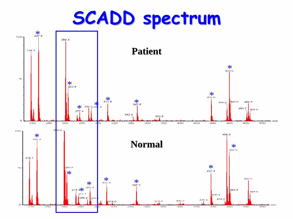
SCADD spectrumSCADD spectrum
*
* *
PatientPatient
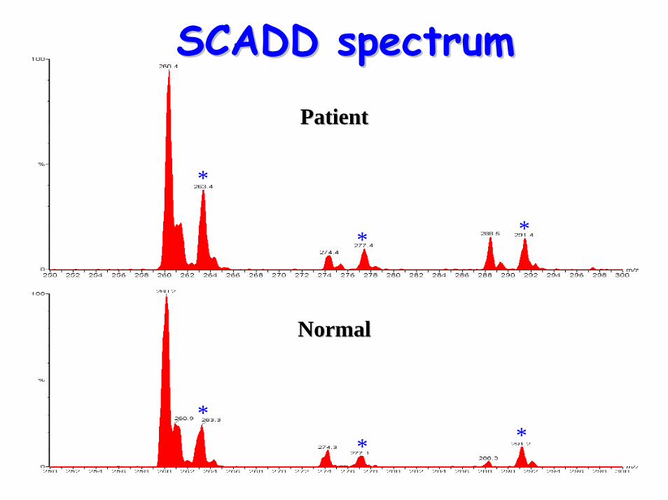
SCADD spectrumSCADD spectrum
** *
NormalNormal
*
**
** *
PatientPatient
NormalNormal
C2
C3 C4
Diagnostic acylcarnitine peak
Ratio of C4 / C16 used for diagnosis & monitoring
SCADDSCADD
MediumMedium--chain acylchain acyl--CoA CoA dehydrogenase deficiency (MCADD)dehydrogenase deficiency (MCADD)
• Commonest fatty-acid oxidation defect• Autosomal recessive inheritance• Incidence 1 in 10,000-20,000 births
(depending on population)• First crisis is fatal in 20-25% of cases• Mean age of presentation is 12 months• ~85% of cases are due to the mutation
K304E• Presentation often follows periods of
intercurrent illness or vomiting
MCADDMCADD
• Presentation (episodic):– hypoketotic hypoglycaemia– myopathy or cardiomyopathy– hyperammonaemia– hypotonia– lethargy– encephalopathy– hepatomegaly
• Urine organic acids:– increased medium-chain dicarboxylic acids– hexanoyl-, suberyl- and phenylpropionyl-
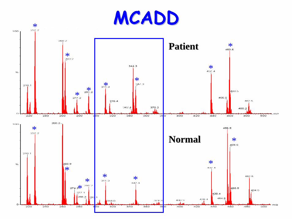
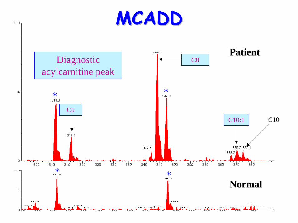
glycines• Acylcarnitine profile:
– elevated C6, C10:1 & C8 (octanoylglycine)• Treatment:
– avoid prolonged fasting,– carnitine supplementation (during crisis)– cornstarch [slow release carbohydrate]
supplementation (during crisis)
MCADDMCADD
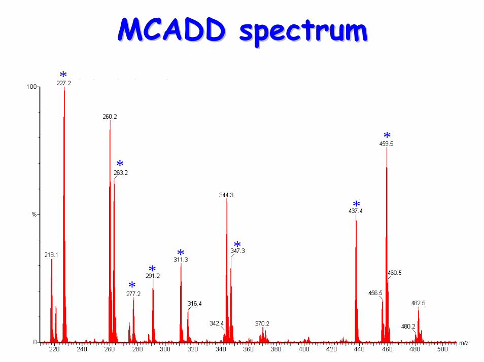
MCADD spectrumMCADD spectrum*
*
**
* *
*
*
MCADDMCADD*
*
* * * **
*
*
*
** * *
*
*
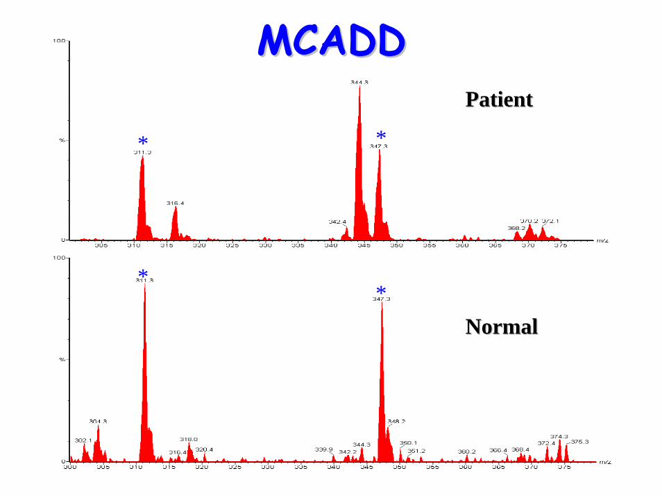
PatientPatient
NormalNormal
**
* *
PatientPatient
MCADDMCADD
NormalNormal
**
* *
C10C6
C8
C10:1
PatientPatient
NormalNormal
Diagnostic acylcarnitine peak
MCADD MCADD
• Multi-enzyme protein complex containing enzyme activities:
– L-3-hydroxyacyl-CoA DHG– 2-enoyl-CoA hydratase– 3-oxoacylCoA thiolase
• 2 disorders described:– Long-chain 3-hydroxyacyl-CoA dehydrogenase
deficiency (LCHADD)– deficiency in all 3 enzymes of the tri-functional
protein complex (MTP)
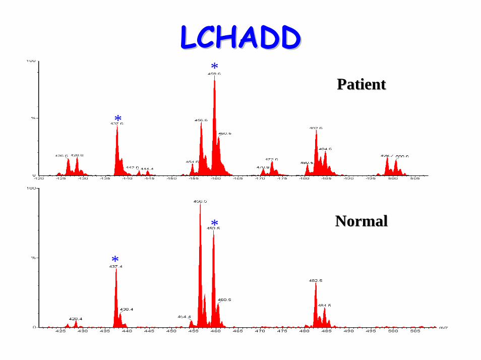
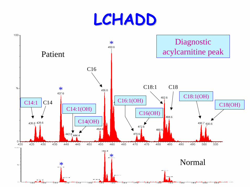
LongLong--chain 3chain 3--hydroxyacylhydroxyacyl--CoA CoA dehydrogenase deficiency dehydrogenase deficiency
(LCHADD)(LCHADD)
LCHAD/MTP DeficiencyLCHAD/MTP Deficiency
• LCHADD is more common than MTP deficiency
• Association of LCHADD with maternal HELLP syndrome (haemolysis, elevated liver enzymes, low platelets)
• Defect is metabolism of long chain fatty acids (C-12 to C-16 in length)
LCHAD/MTP DeficiencyLCHAD/MTP Deficiency• Marked clinical heterogeneity associated
with LCHADD, but presentation may include:
– acute hypoketotic hypoglycaemic encephalopathy
– hypotonia– cardiomyopathy– hepatomegaly leading to:
• cirrhosis• fulminant liver failure
LCHAD/MTP DeficiencyLCHAD/MTP Deficiency
• Late onset presentation:– exercise-induced myopathy &
rhabdomyolysis– cardiomyopathy
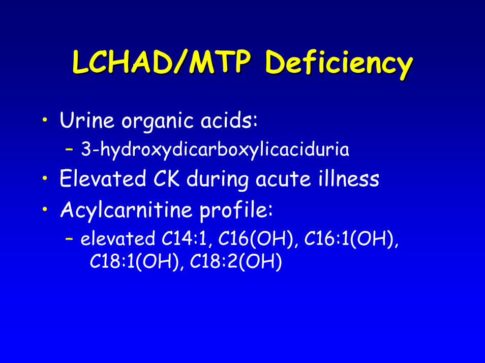
LCHAD/MTP DeficiencyLCHAD/MTP Deficiency
• Urine organic acids:– 3-hydroxydicarboxylicaciduria
• Elevated CK during acute illness• Acylcarnitine profile:
– elevated C14:1, C16(OH), C16:1(OH), C18:1(OH), C18:2(OH)
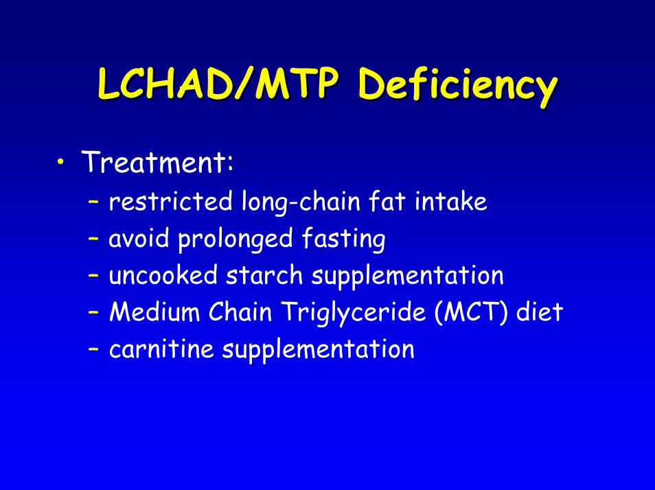
LCHAD/MTP DeficiencyLCHAD/MTP Deficiency
• Treatment:– restricted long-chain fat intake– avoid prolonged fasting– uncooked starch supplementation – Medium Chain Triglyceride (MCT) diet– carnitine supplementation
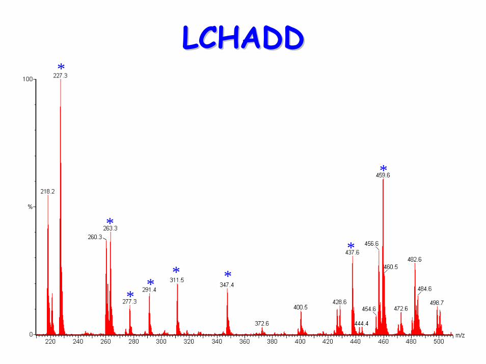
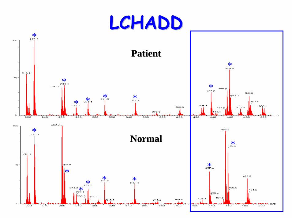
LCHADDLCHADD
*
**
* *
*
*
*
LCHADDLCHADD
*
*
* * **
*
*
*
*
** * *
*
*
PatientPatient
NormalNormal
LCHADDLCHADDPatientPatient
*
*
*
*
NormalNormal
LCHADDLCHADD
Normal**
Patient
C14
C18
C16
C18:1(OH)
C16(OH)
C14:1C14:1(OH)
C14(OH)
C18(OH)C16:1(OH)
Diagnostic acylcarnitine peak
*
* C18:1
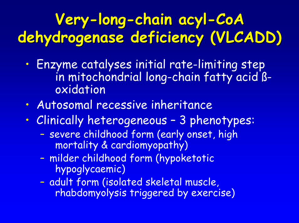
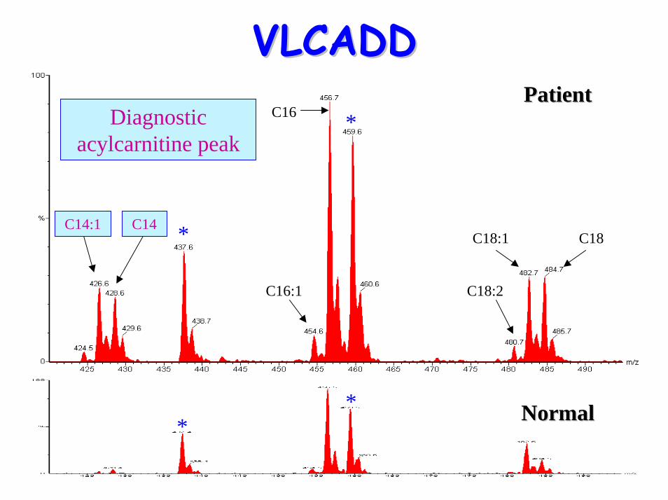
VeryVery--longlong--chain acylchain acyl--CoA CoA dehydrogenase deficiency (VLCADD)dehydrogenase deficiency (VLCADD)• Enzyme catalyses initial rate-limiting step
in mitochondrial long-chain fatty acid ß-oxidation
• Autosomal recessive inheritance• Clinically heterogeneous – 3 phenotypes:
– severe childhood form (early onset, high mortality & cardiomyopathy)
– milder childhood form (hypoketotic hypoglycaemic)
– adult form (isolated skeletal muscle, rhabdomyolysis triggered by exercise)
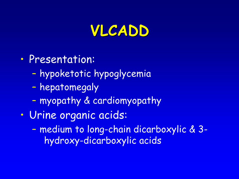
VLCADDVLCADD
• Presentation:– hypoketotic hypoglycemia– hepatomegaly– myopathy & cardiomyopathy
• Urine organic acids:– medium to long-chain dicarboxylic & 3-
hydroxy-dicarboxylic acids
VLCADDVLCADD
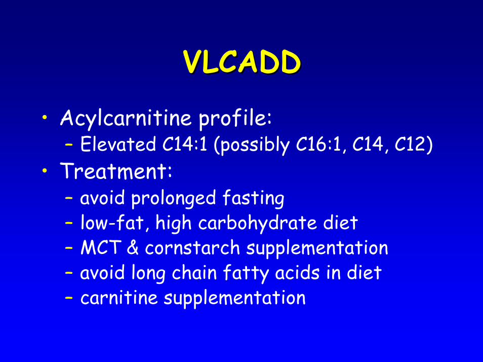
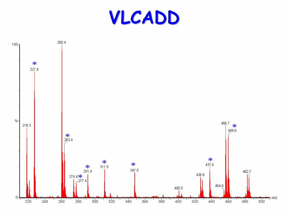
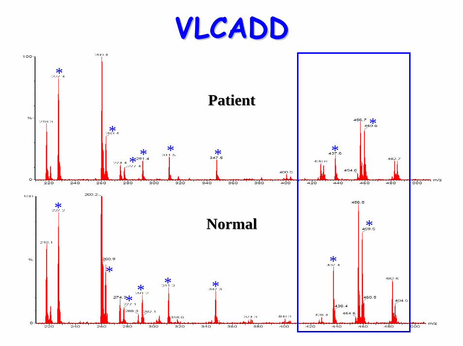
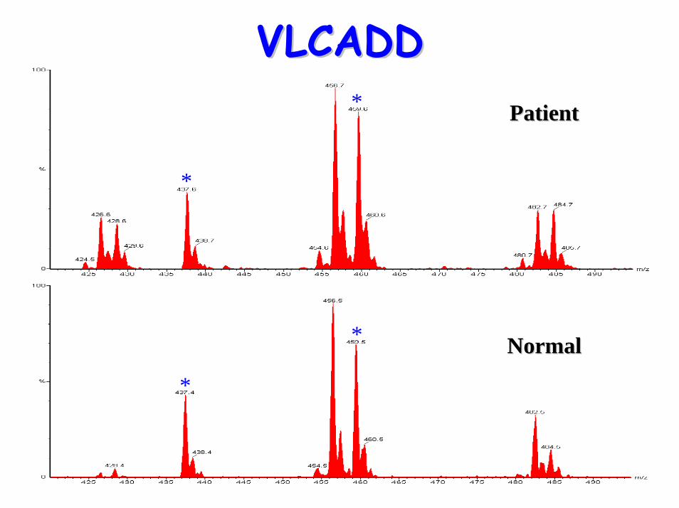
• Acylcarnitine profile: – Elevated C14:1 (possibly C16:1, C14, C12)
• Treatment:– avoid prolonged fasting– low-fat, high carbohydrate diet– MCT & cornstarch supplementation– avoid long chain fatty acids in diet– carnitine supplementation
VLCADDVLCADD
*
*
** * * *
*
VLCADDVLCADD*
*
* * * * *
*
*
*
** * *
*
*
PatientPatient
NormalNormal
VLCADDVLCADD
*
* PatientPatient
*
* NormalNormal
*
*PatientPatient
VLCADDVLCADD
C16:1
C16
C18:1
C18:2
C18
Diagnostic acylcarnitine peak
C14C14:1
** NormalNormal
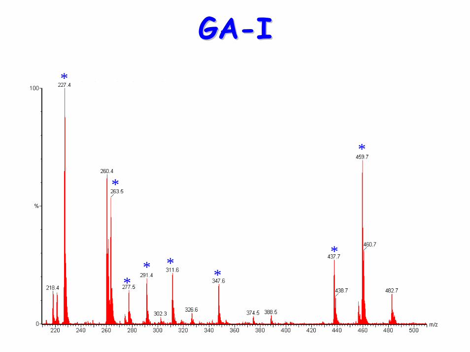
Glutaric aciduria type 1 (GAGlutaric aciduria type 1 (GA--I)I)• Defect: Glutaryl-CoA dehydrogenase
deficiency• Pathways affected: lysine, hydroxylysine
and tryptophan• Presentation:
– macrocephaly– neurodegeneration– dystonia– ataxia and dyskinesia– seizures– frontotemporal atrophy on MRI & CT– hypotonia– death due to Reye-like syndrome
GAGA--II
• Urine organic acids:– increased glutarate– 3-hydroxyglutarate– glutaconate
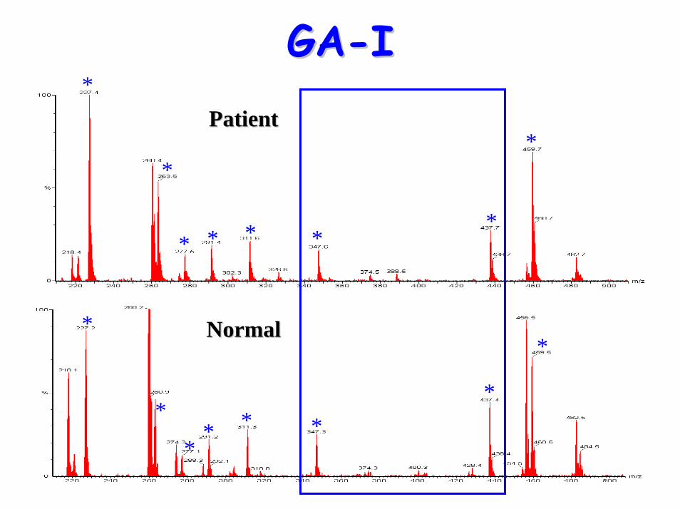
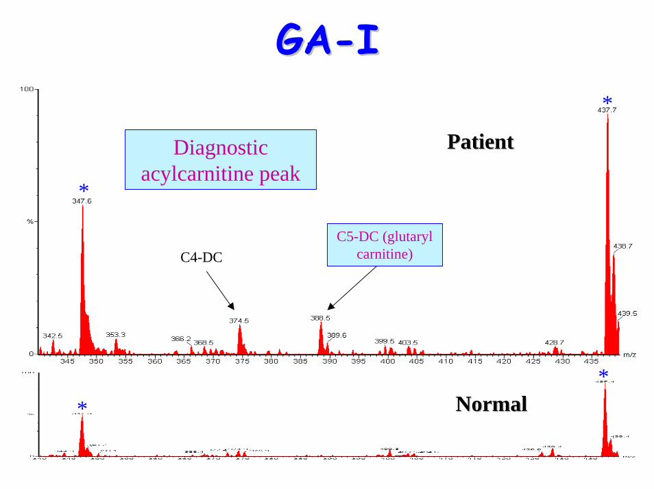
• Acylcarnitine profile:– elevated C5-DC (glutaryl carnitine)
• NB Metabolites not always reliably increased
GAGA--II
• Treatment:– lysine and tryptophan restricted
diet– riboflavin supplementation– carnitine supplementation– i.v. glucose during acute illness
GAGA--II*
*
** *
*
*
*
GAGA--II*
*
* * * *
*
*
*
*
** * *
*
*
PatientPatient
NormalNormal
GAGA--II
*
*PatientPatient
*
*NormalNormal
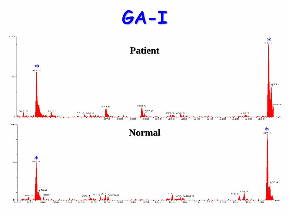
GAGA--II
*
*
**
PatientPatient
NormalNormal
C4-DC
Diagnostic acylcarnitine peak
C5-DC (glutaryl carnitine)
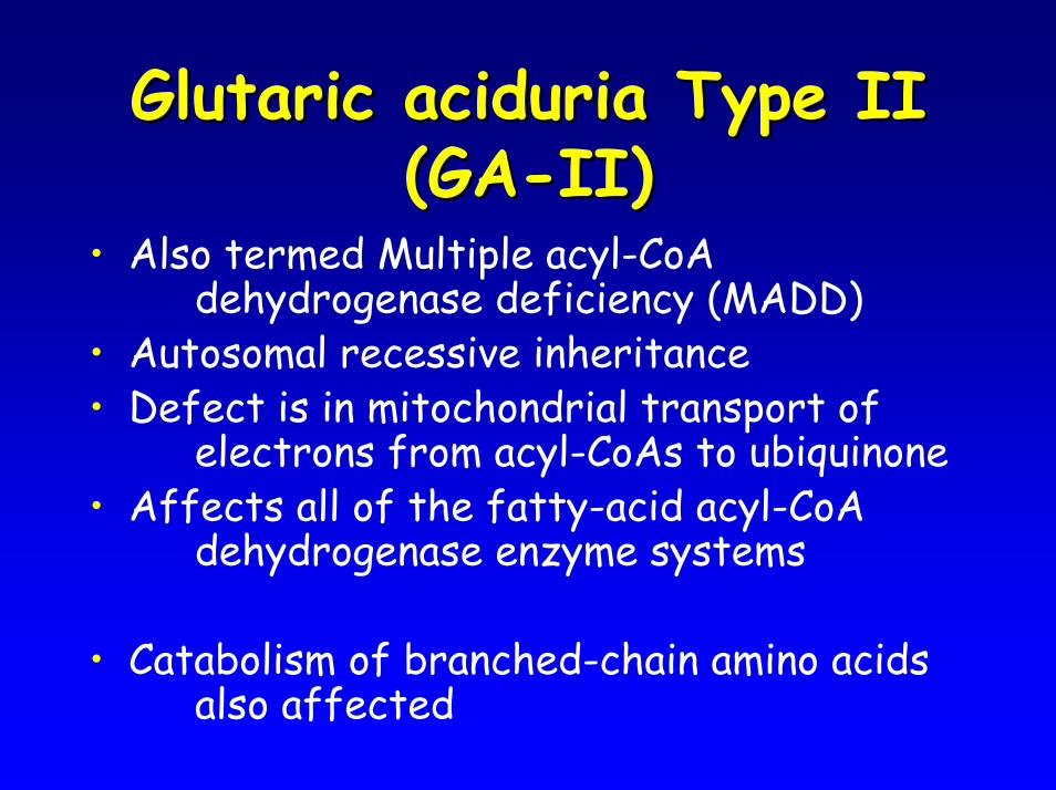
Glutaric aciduria Type II Glutaric aciduria Type II (GA(GA--II)II)
• Also termed Multiple acyl-CoA dehydrogenase deficiency (MADD)
• Autosomal recessive inheritance• Defect is in mitochondrial transport of
electrons from acyl-CoAs to ubiquinone• Affects all of the fatty-acid acyl-CoA
dehydrogenase enzyme systems
• Catabolism of branched-chain amino acids also affected
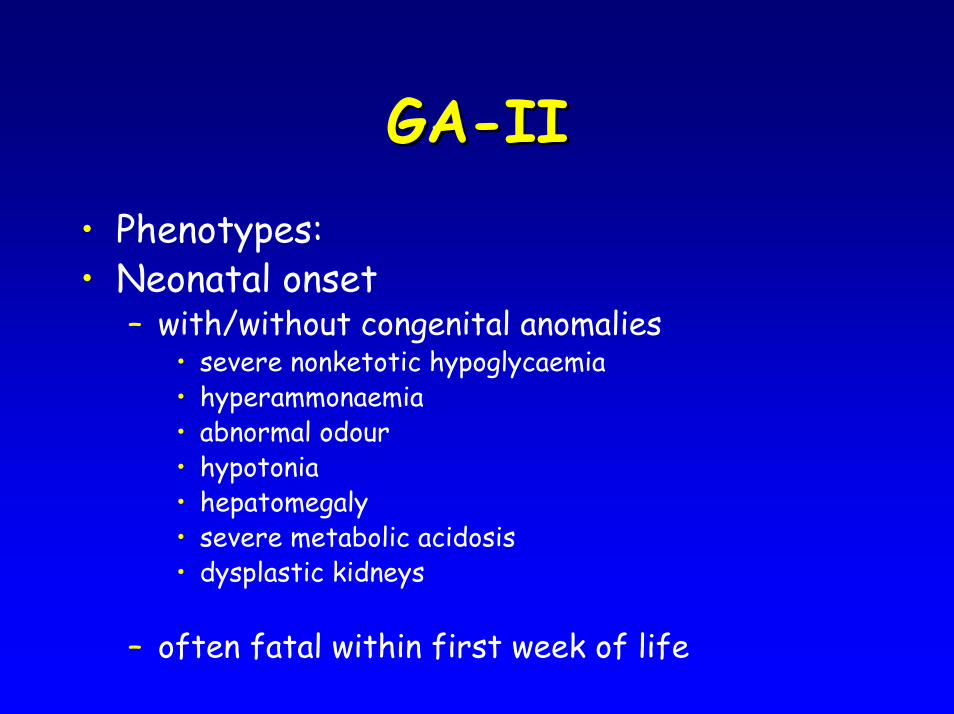
GAGA--IIII• Phenotypes:• Neonatal onset
– with/without congenital anomalies• severe nonketotic hypoglycaemia• hyperammonaemia• abnormal odour• hypotonia• hepatomegaly• severe metabolic acidosis• dysplastic kidneys
– often fatal within first week of life
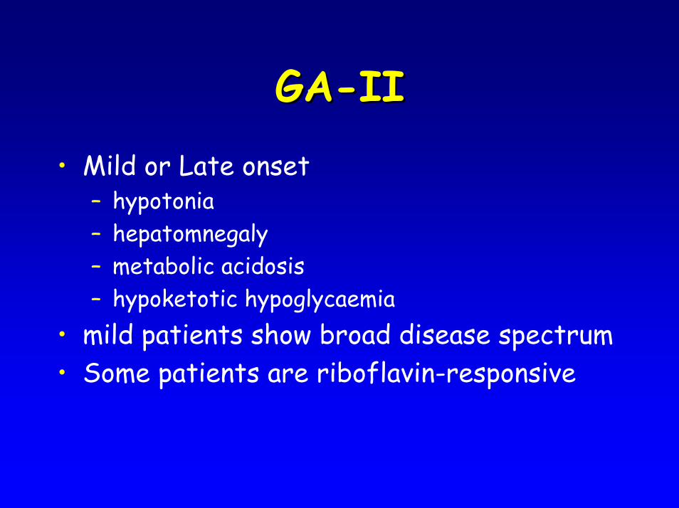
GAGA--IIII
• Mild or Late onset – hypotonia– hepatomnegaly– metabolic acidosis– hypoketotic hypoglycaemia
• mild patients show broad disease spectrum• Some patients are riboflavin-responsive
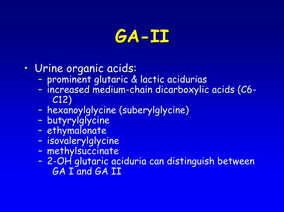
GAGA--IIII• Urine organic acids:
– prominent glutaric & lactic acidurias – increased medium-chain dicarboxylic acids (C6-
C12)– hexanoylglycine (suberylglycine)– butyrylglycine – ethymalonate– isovalerylglycine – methylsuccinate– 2-OH glutaric aciduria can distinguish between
GA I and GA II
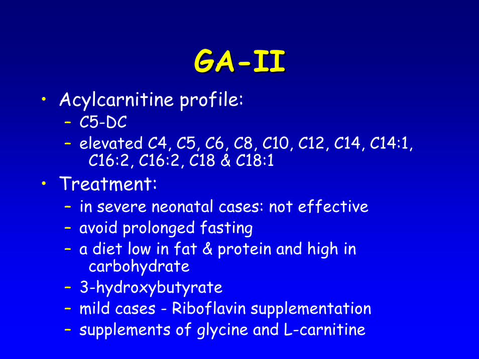
GAGA--IIII• Acylcarnitine profile:
– C5-DC– elevated C4, C5, C6, C8, C10, C12, C14, C14:1,
C16:2, C16:2, C18 & C18:1• Treatment:
– in severe neonatal cases: not effective– avoid prolonged fasting– a diet low in fat & protein and high in
carbohydrate– 3-hydroxybutyrate– mild cases - Riboflavin supplementation– supplements of glycine and L-carnitine
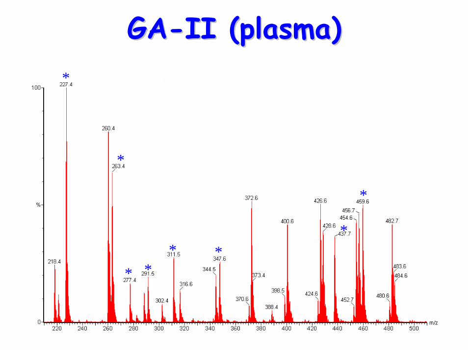
GAGA--II (plasma)II (plasma)*
*
* ** *
*
*
*
*
** * *
*
*
*
*
* ** *
**
PatientPatient
NormalNormal
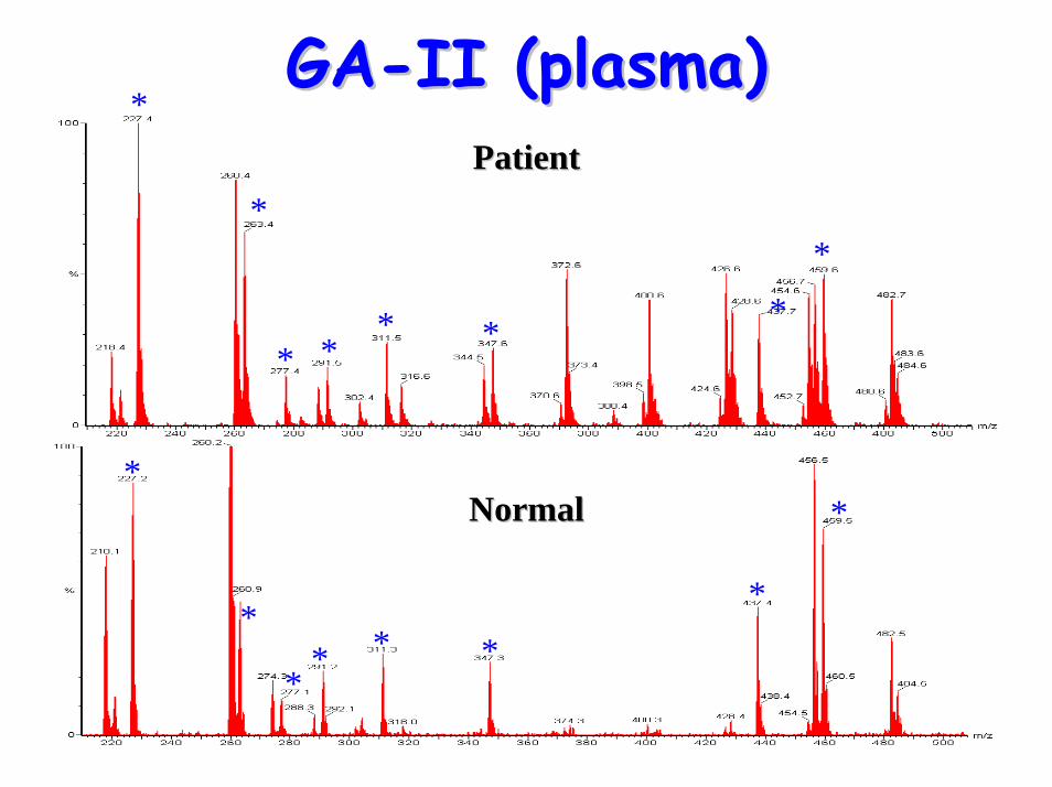
GAGA--II (plasma)II (plasma)
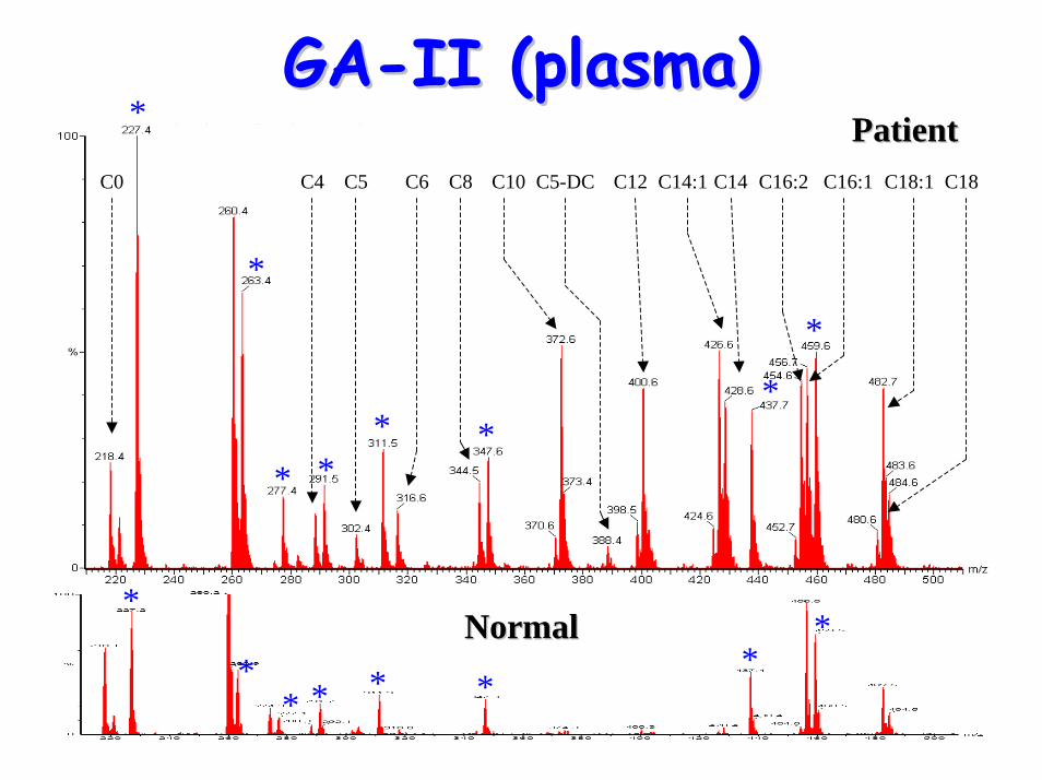
GAGA--II (plasma)II (plasma)
*
** * * *
**
*
*
* ** *
*
*
PatientPatient
NormalNormal
C0 C4 C5 C6 C8 C10 C5-DC C12 C14:1 C14 C16:2 C16:1 C18:1 C18
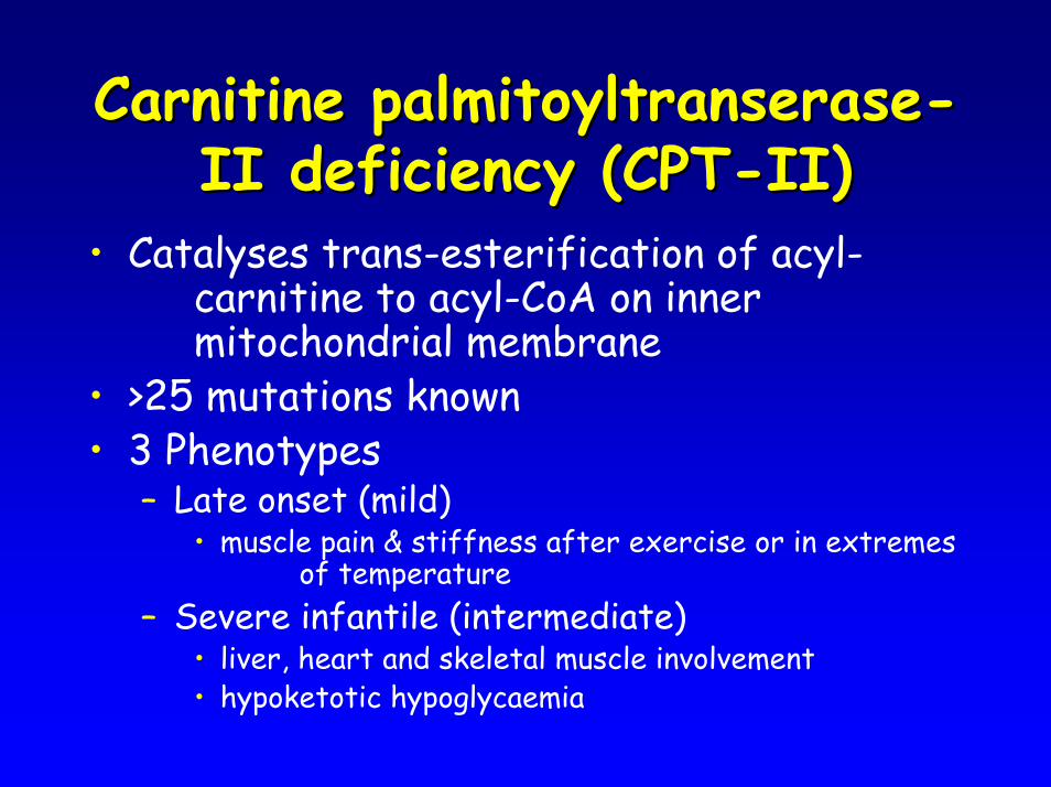
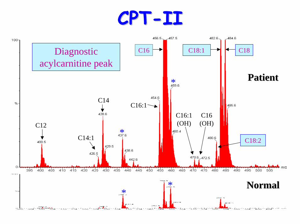
Carnitine Carnitine palmitoyltranserasepalmitoyltranserase--II deficiency (CPTII deficiency (CPT--II)II)
• Catalyses trans-esterification of acyl-carnitine to acyl-CoA on inner mitochondrial membrane
• >25 mutations known• 3 Phenotypes
– Late onset (mild)• muscle pain & stiffness after exercise or in extremes
of temperature– Severe infantile (intermediate)
• liver, heart and skeletal muscle involvement• hypoketotic hypoglycaemia
CPTCPT--IIII
– Lethal neonatal form• hypoketotic hypoglycaemia• liver disease• hypotonia• cardiomyopathy• congenital abnormalities
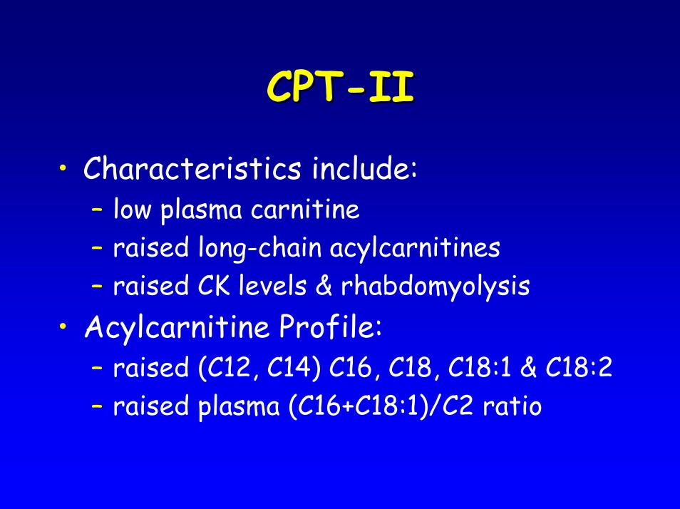
CPTCPT--IIII
• Characteristics include:– low plasma carnitine– raised long-chain acylcarnitines– raised CK levels & rhabdomyolysis
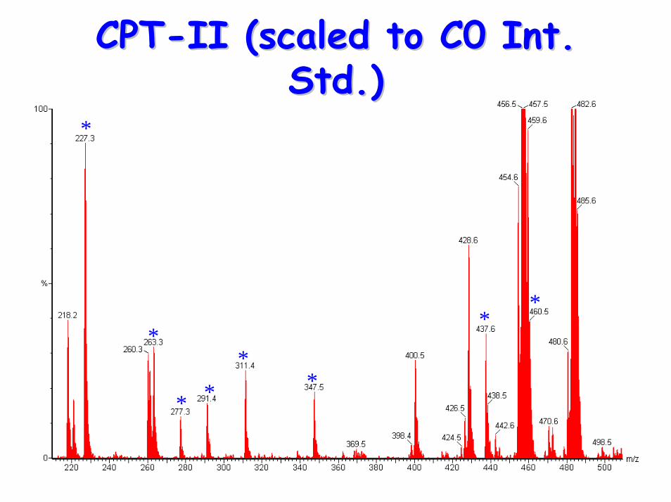
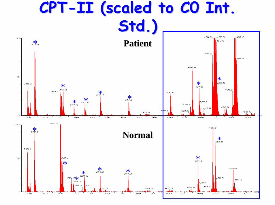
• Acylcarnitine Profile:– raised (C12, C14) C16, C18, C18:1 & C18:2– raised plasma (C16+C18:1)/C2 ratio
CPTCPT--IIII
• Treatment:– avoid prolonged fasting– low-fat, high carbohydrate diet– MCT & cornstarch supplementation– carnitine supplementation – i.v. glucose during acute episodes
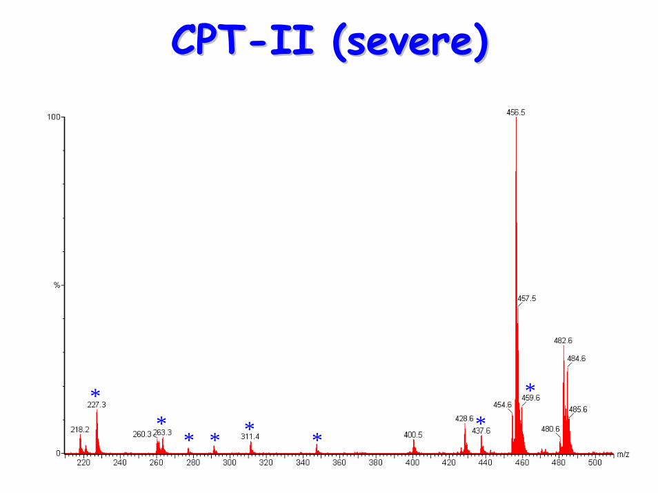
CPTCPT--II (severe)II (severe)
**
* * * **
*
*
*
* **
*
**
CPTCPT--II (scaled to C0 Int. II (scaled to C0 Int. Std.)Std.)
*
*
* ** *
* *
*
*
* * * **
*
PatientPatient
NormalNormal
CPTCPT--II (scaled to C0 Int. II (scaled to C0 Int. Std.)Std.)
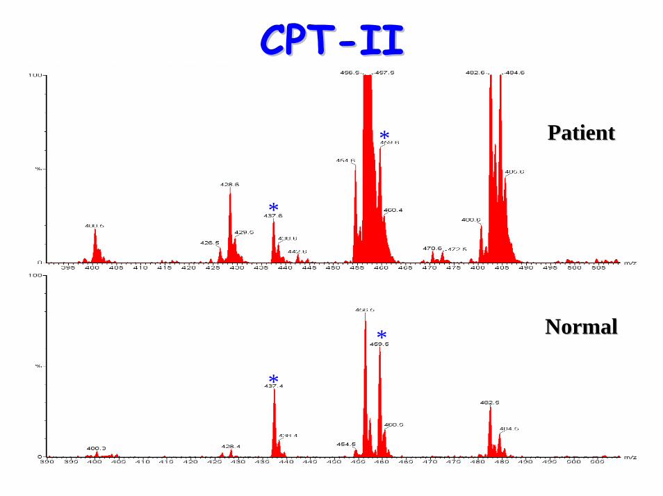
CPTCPT--IIII
*
*
*
* PatientPatient
NormalNormal
CPTCPT--IIII
*
* PatientPatient
C12
C14:1
C16:1(OH)
C14
C16 (OH)
Diagnostic acylcarnitine peak
C16:1
C18:1 C18
C18:2
C16
** NormalNormal
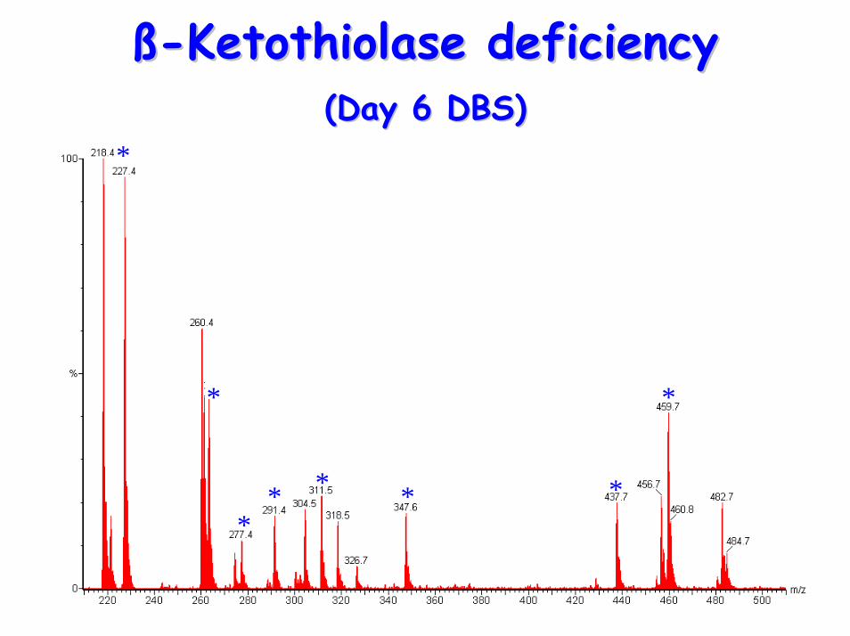
ßß--Ketothiolase deficiencyKetothiolase deficiency• Defect: deficiency in enzyme that converts
2-methylacetoacetyl-CoA to propionyl-CoA and acetyl-CoA
– ß-Ketothiolase – sixth step of isoleucine pathway
• Autosomal recessive inheritance• Neonatal presentation is rare• Clinical heterogeneity in presentation:
– recurrent, severe metabolic acidosis with ketosis
– vomiting and diarrhoea– lethargy
ßß--Ketothiolase deficiencyKetothiolase deficiency• Urine organic acids:
– raised 2-methyl-3-hydroxybutyrate– 2-methylacetoacetate– tiglylglycine– ketone bodies
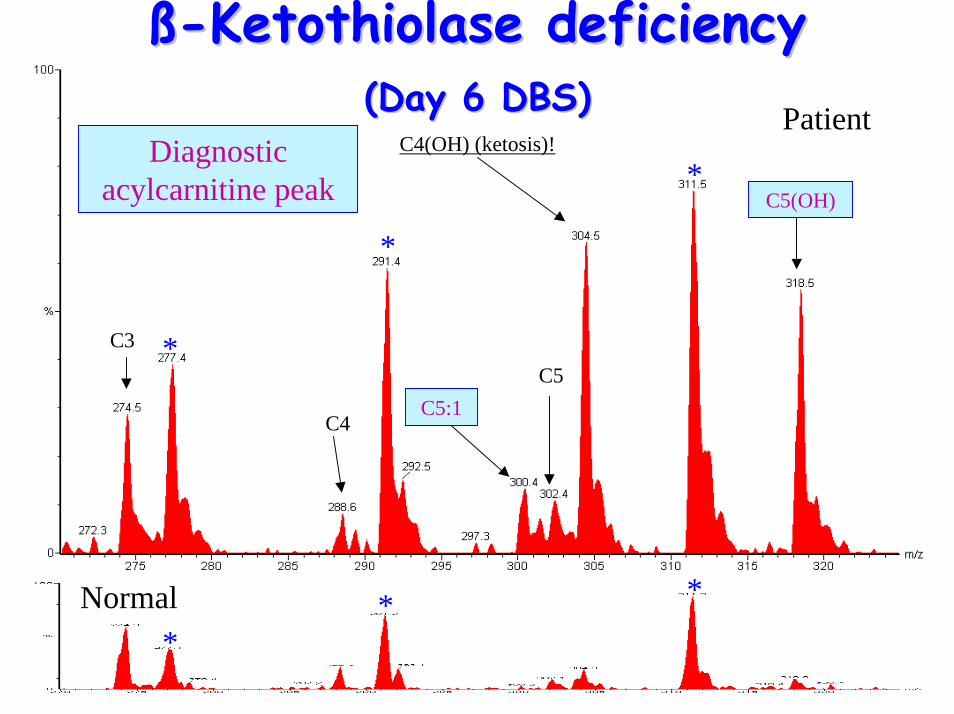
• Acylcarnitine Profile– raised C5(OH) (2-Methyl-3-hydroxy-
butyrylcarnitine), C5:1 (tiglylcarnitine)
ßß--Ketothiolase deficiencyKetothiolase deficiency
• Treatment:– avoid prolonged fasting– restricted isoleucine intake– bicarbonate therapy and i.v. glucose
during acute crises– carnitine supplementation.
*
** * * *
**
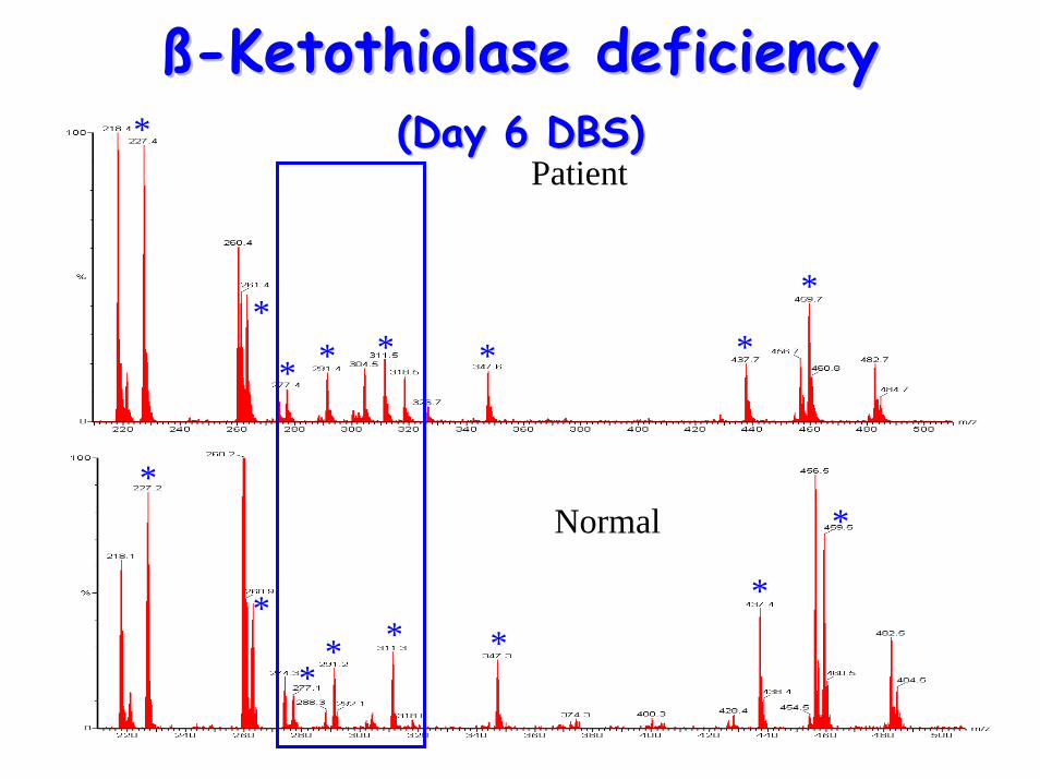
ßß--Ketothiolase deficiencyKetothiolase deficiency(Day 6 DBS)(Day 6 DBS)
*
*
* * * *
**
*
*
** * *
*
*
Patient
Normal
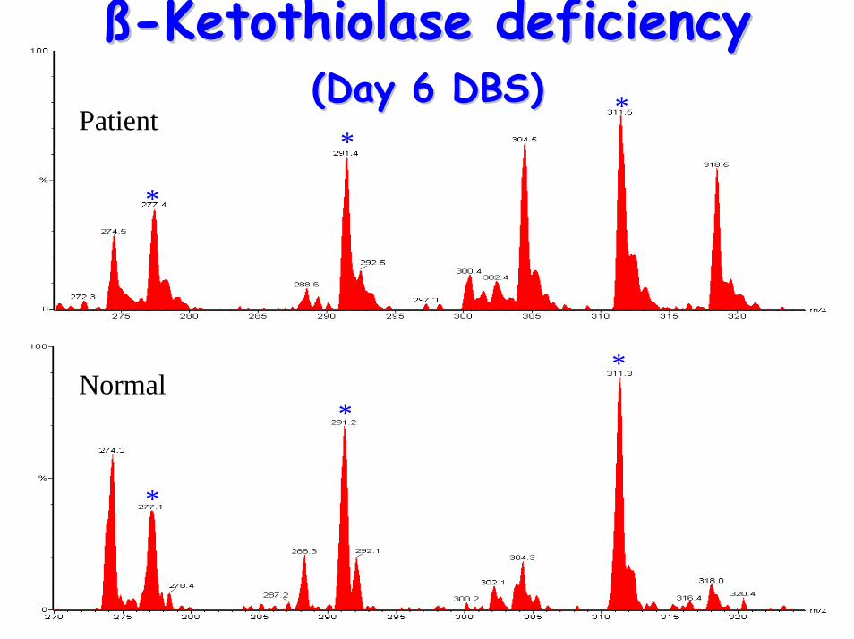
ßß--Ketothiolase deficiencyKetothiolase deficiency(Day 6 DBS)(Day 6 DBS)
Normal*
*
*
ßß--Ketothiolase deficiencyKetothiolase deficiency(Day 6 DBS)(Day 6 DBS)
*
**Patient
* *
*
*
*
*
Patient
ßß--Ketothiolase deficiencyKetothiolase deficiency(Day 6 DBS)(Day 6 DBS)
Normal
C4(OH) (ketosis)!
C5
C3
C4
C5(OH)
C5:1
Diagnostic acylcarnitine peak
Methylmalonic aciduria Methylmalonic aciduria (MMA)(MMA)
• Enzyme: methylmalonyl CoA mutase– catalyses formation of succinyl CoA from
methylmalonyl CoA in branched chain amino-acid catabolism pathway
– enzyme requires Vitamin B12 as a co-factor• Autosomal recessive inheritance• Various forms including Vit B12 responsive
& non-responsive
MMAMMA• Wide clinical spectrum• Presentation:
– gross ketosis– metabolic acidosis – recurrent vomiting dehydration– Failure to thrive– hyperammonaemia mental retardation– characteristic facial features (eg low set ears,
high forehead broad nasal bridge etc)– hypotonia– death if not treated
MMAMMA
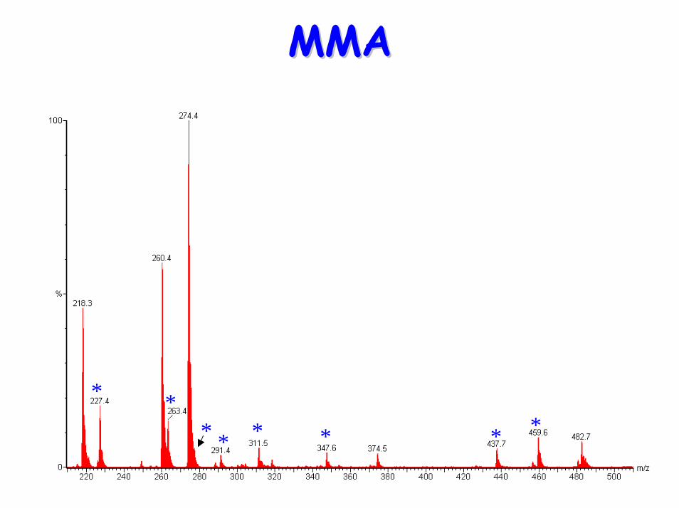
• Urine organic acids: Raised– Methylmalonate– Methylcitrate – 3-OH-propionate
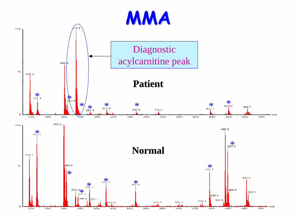
• Acylcarnitine profile:– Raised C3 propionyl carnitine
MMAMMA
• Treatment:– protein-restricted diet (nb isoleucine,
threonine etc are essential amino acids for normal growth & development)
– Vitamin B12 injections– carnitine supplementation (replace
intracellular stores)– oral antibiotic therapy (decrease gut
propionate production)
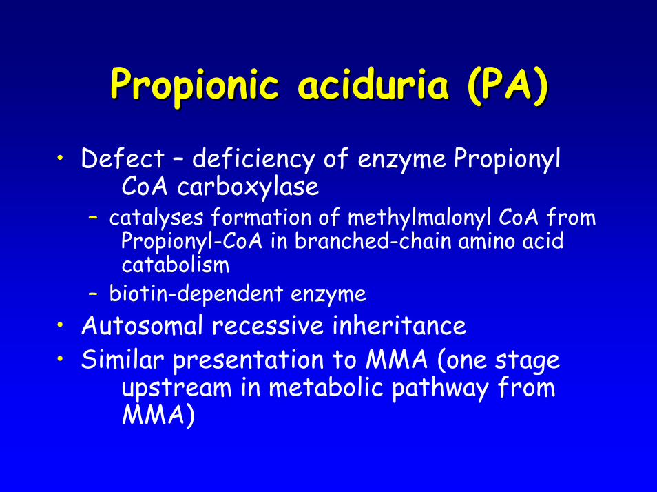
Propionic aciduria (PA)Propionic aciduria (PA)• Defect – deficiency of enzyme Propionyl
CoA carboxylase – catalyses formation of methylmalonyl CoA from
Propionyl-CoA in branched-chain amino acid catabolism
– biotin-dependent enzyme• Autosomal recessive inheritance• Similar presentation to MMA (one stage
upstream in metabolic pathway from MMA)
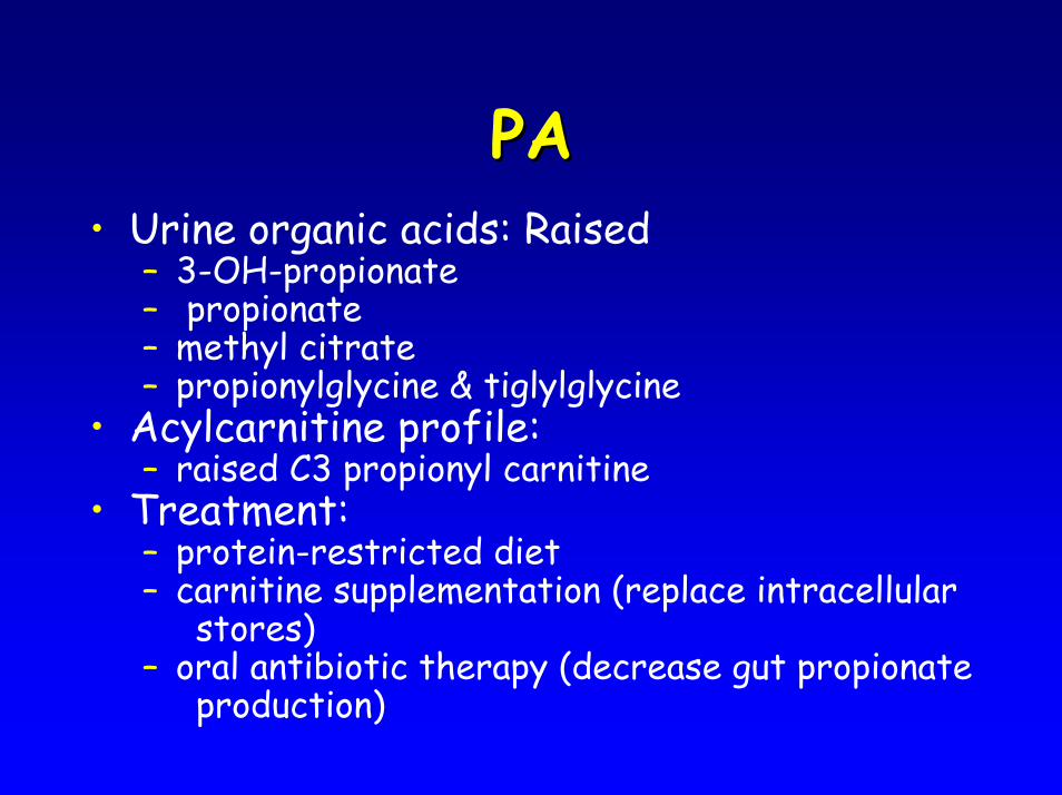
PAPA• Urine organic acids: Raised
– 3-OH-propionate– propionate– methyl citrate– propionylglycine & tiglylglycine
• Acylcarnitine profile:– raised C3 propionyl carnitine
• Treatment:– protein-restricted diet– carnitine supplementation (replace intracellular
stores)– oral antibiotic therapy (decrease gut propionate
production)
MMAMMA
* *
* * * * **
* ** * * * * *
*
*
** * *
*
*
Diagnostic acylcarnitine peak
PatientPatient
NormalNormal
MMAMMA
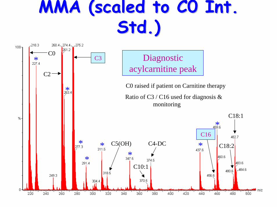
MMA (scaled to C0 Int. MMA (scaled to C0 Int. Std.)Std.)
*
*
**
** *
*
C0 raised if patient on Carnitine therapy
Ratio of C3 / C16 used for diagnosis & monitoring
C4-DC
C10:1
C5(OH)
C3
C2
C0
C18:1
C18:2
C16
Diagnostic acylcarnitine peak
Isovaleric aciduria (IVA)Isovaleric aciduria (IVA)• Defect: Isovaleryl-CoA
dehydrogenase deficiency– catalyses formation of 3-methyl-
crotonyl-CoA from Isovaleryl-CoA during leucine catabolism
• Autosomal recessive inheritance
IVAIVA
• Presentation includes:– vomiting– metabolic acidosis & ketosis– characteristic odour ‘sweaty feet’– failure to thrive– hypotonia– encephalopathy
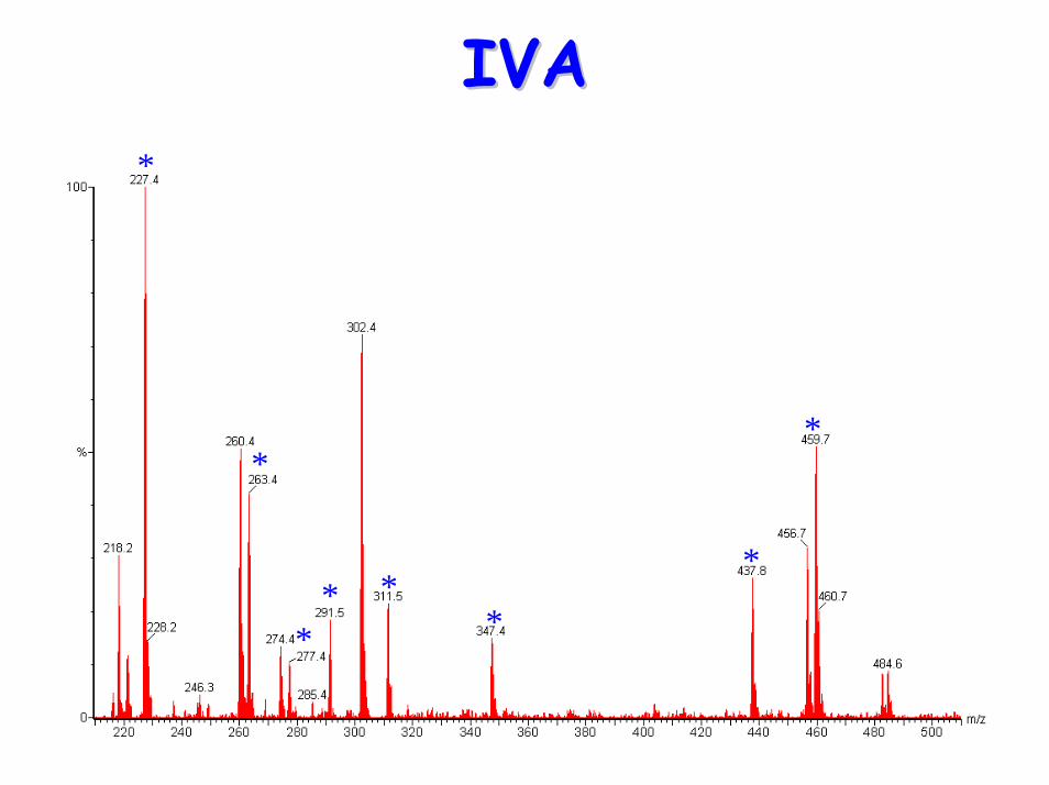
IVAIVA• Urine organic acids: Raised
– 4-hydroxyisovaleric acid– isovaleryl glycine– 3-hydroxyisovalerate – Methylsuccinate– isovalerylglucuronide
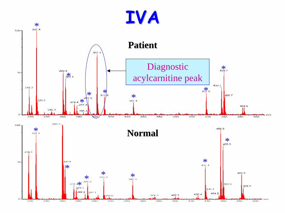
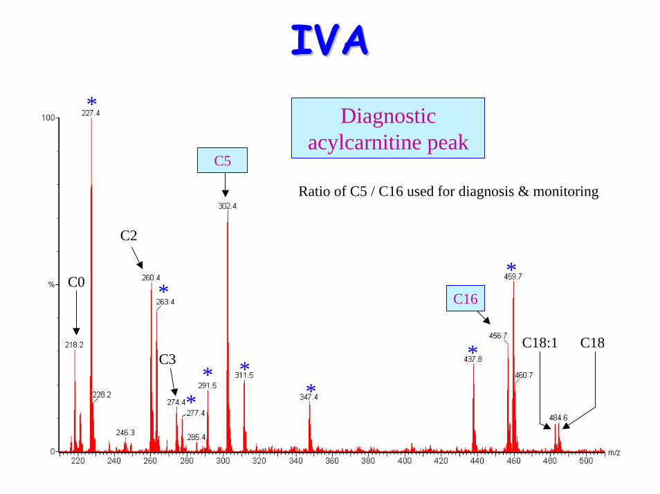
• Acylcarnititne profile:– Raised C5 (isovaleryl carnitine)– NB Pivoxilsulbactam antibiotics form m/z 302
peak (pivaloylcarnitine butyl ester)
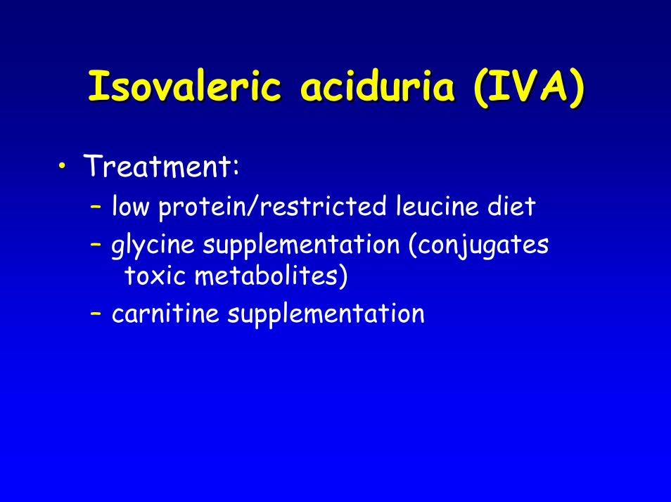
Isovaleric aciduria (IVA)Isovaleric aciduria (IVA)
• Treatment:– low protein/restricted leucine diet– glycine supplementation (conjugates
toxic metabolites)– carnitine supplementation
IVAIVA*
*
** *
*
*
*
IVAIVA*
*
** * *
*
*
*
*
** * *
*
*
PatientPatient
NormalNormal
Diagnostic acylcarnitine peak
IVAIVA*
*
** *
*
*
*
C2
C0
C18C18:1C3
Diagnostic acylcarnitine peak
Ratio of C5 / C16 used for diagnosis & monitoring
C5
C16
Sample type Sample type –– plasma or plasma or DBS?DBS?
• Traditional isotope-dilution methods require liquid samples for quantitation
• Advantages of DBS– easy to transport (ie post to lab)– easy to store– in UK all babies have DBS taken at 6 days a
useful retrospective sample bank
Sample type Sample type –– plasma or DBS?plasma or DBS?• Disadvantages of DBS
– requires elution from DBS ( slower than plasma)
– ?recovery during elution • ?use of ratios instead of absolute values
– ?volume of blood per DBS - ?depends on haematocrit
• Differences between DBS & plasma– Altered profile – long-chain acylcarnitines
reside within red cell non-polar lipid-bilayer • Reference ranges not directly comparable
– Plasma maybe more representative of disease state
*
*
* * * * *
*
*
*
* * * **
*
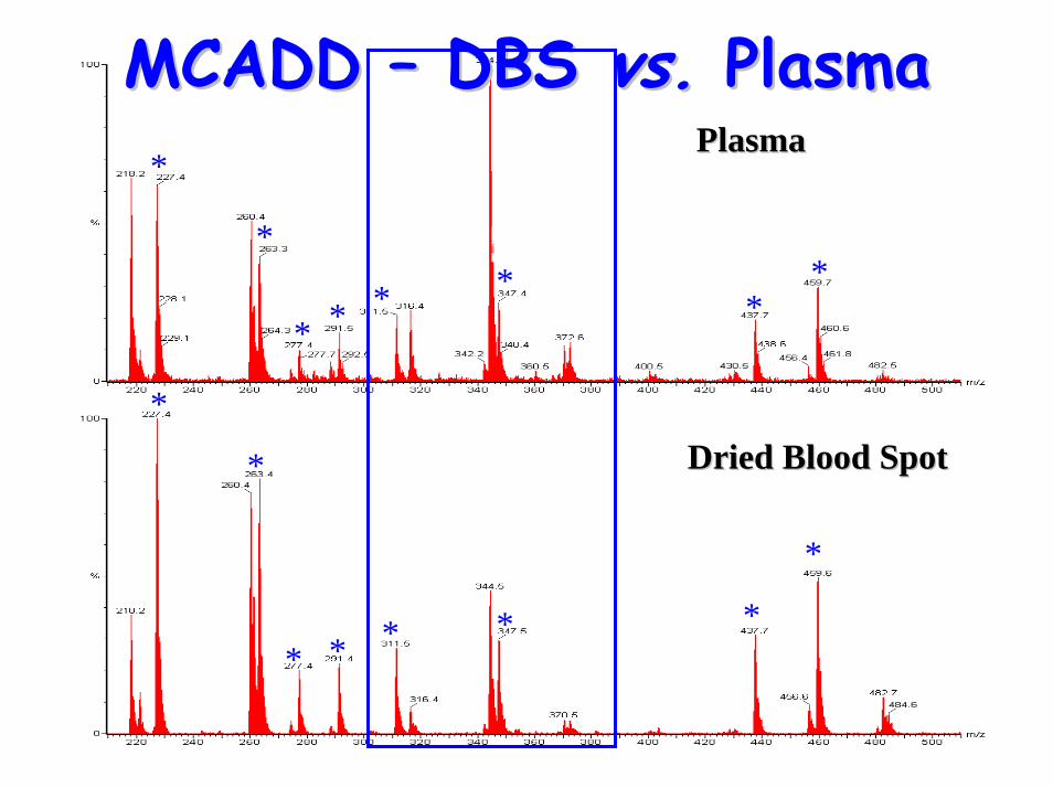
PlasmaPlasma
Dried Blood SpotDried Blood Spot
MCADD MCADD –– DBS DBS vs.vs. PlasmaPlasma
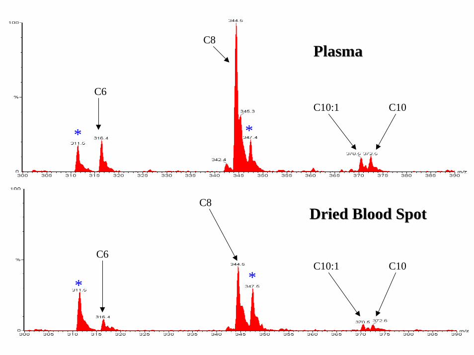
PlasmaPlasma
Dried Blood SpotDried Blood Spot
**
**
C6
C6
C8
C8
C10:1 C10
C10:1 C10
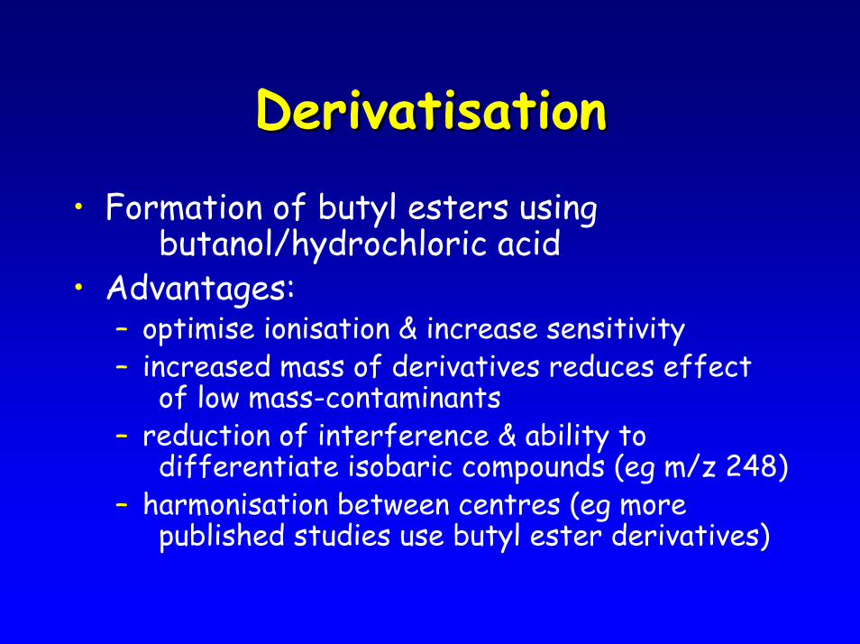
DerivatisationDerivatisation• Formation of butyl esters using
butanol/hydrochloric acid• Advantages:
– optimise ionisation & increase sensitivity– increased mass of derivatives reduces effect
of low mass-contaminants– reduction of interference & ability to
differentiate isobaric compounds (eg m/z 248)– harmonisation between centres (eg more
published studies use butyl ester derivatives)
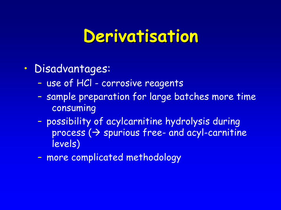
DerivatisationDerivatisation
• Disadvantages:– use of HCl - corrosive reagents – sample preparation for large batches more time
consuming– possibility of acylcarnitine hydrolysis during
process ( spurious free- and acyl-carnitine levels)
– more complicated methodology
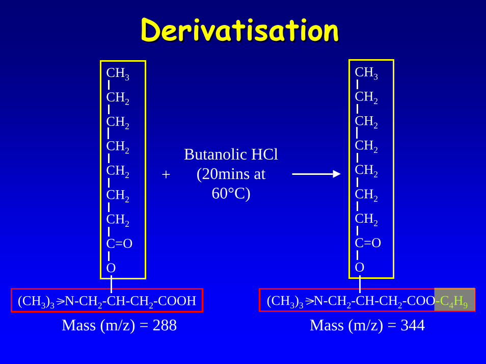
DerivatisationDerivatisationCH3
CH2
CH2
CH2
CH2
CH2
CH2
C=O
O
(CH3)3 N-CH2-CH-CH2-COOH
CH3
CH2
CH2
CH2
CH2
CH2
CH2
C=O
O
(CH3)3 N-CH2-CH-CH2-COO-C4H9
+Butanolic HCl
(20mins at 60°C)
Mass (m/z) = 288 Mass (m/z) = 344
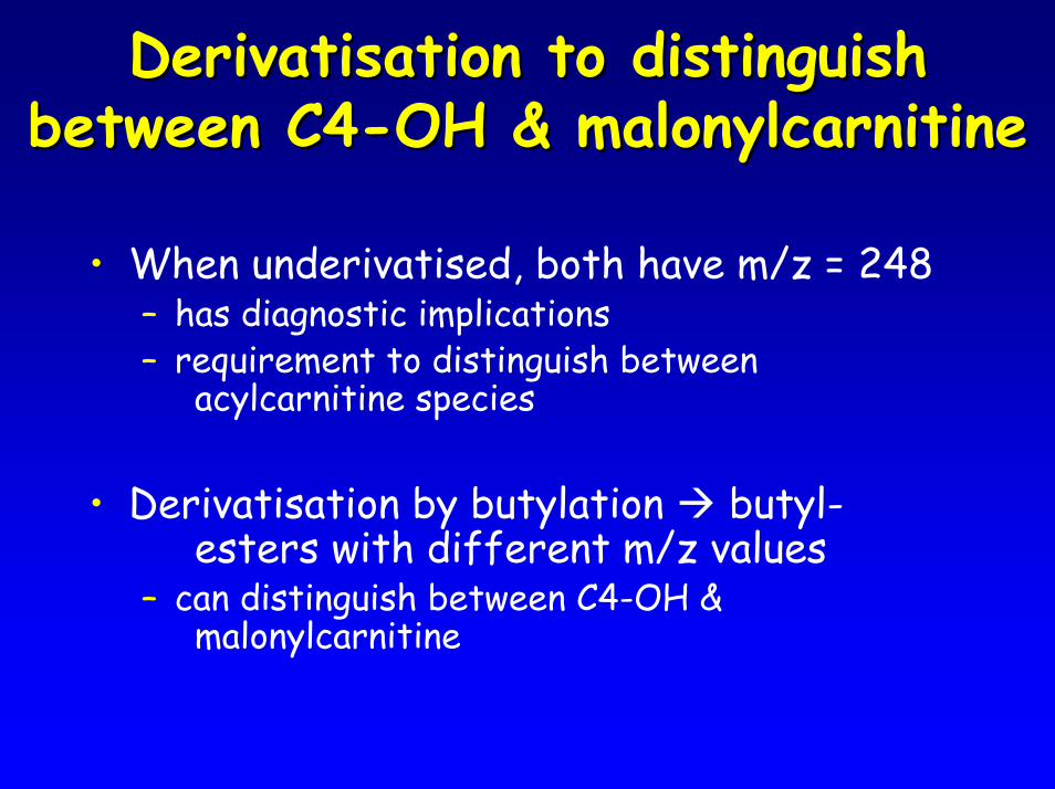
DerivatisationDerivatisation to distinguish to distinguish between C4between C4--OH & OH & malonylcarnitinemalonylcarnitine
• When underivatised, both have m/z = 248– has diagnostic implications– requirement to distinguish between
acylcarnitine species
• Derivatisation by butylation butyl-esters with different m/z values
– can distinguish between C4-OH & malonylcarnitine
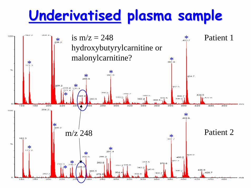
UnderivatisedUnderivatised plasma sampleplasma sample
*
*
* ** *
*
*
*
*
**
* **
*
m/z 248 Patient 2
Patient 1is m/z = 248 hydroxybutyrylcarnitine or malonylcarnitine?
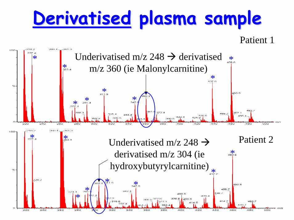
DerivatisedDerivatised plasma sampleplasma sample
Underivatised m/z 248 derivatised m/z 360 (ie Malonylcarnitine)
**
* **
*
*
*
* *
**
* **
*Underivatised m/z 248
derivatised m/z 304 (ie hydroxybutyrylcarnitine)
Patient 2
Patient 1
Current approach in SCHCurrent approach in SCH
• Underivatised– newborn screening– urgent plasma analysis– confirmation for routine investigation
(pseudo-glutarylcarnitinaemia)• Derivatised
– re-run for confirmation– routine investigation

Recommended