R E V I EW AR T I C L E
Fungal developmentoftheplant pathogenUstilagomaydisEvelyn Vollmeister1, Kerstin Schipper1, Sebastian Baumann1,2, Carl Haag1, Thomas Pohlmann1,2,Janpeter Stock1 & Michael Feldbrugge1,2
1Institute for Microbiology, Heinrich Heine University Dusseldorf, Dusseldorf, Germany; and 2Max Planck Institute for Terrestrial Microbiology,
Marburg, Germany
Correspondence: Michael Feldbrugge,
Institute for Microbiology, Heinrich Heine
University Dusseldorf, Universitatsstr. 1, Bldg.
26.12, 40225 Dusseldorf, Germany. Tel.: 149
211 81 15475; fax: 149 211 81 15370;
e-mail: [email protected]
Received 10 March 2011; revised 20 June 2011;
accepted 22 June 2011.
DOI:10.1111/j.1574-6976.2011.00296.x
Editor: Gerhard Braus
Keywords
effector; endosome; homeodomain
transcription factor; microtubule; mRNA
transport; post-transcriptional regulation.
Abstract
The maize pathogen Ustilago maydis has to undergo various morphological
transitions for the completion of its sexual life cycle. For example, haploid cells
respond to pheromone by forming conjugation tubes that fuse at their tips. The
resulting dikaryon grows filamentously, expanding rapidly at the apex and
inserting retraction septa at the basal pole. In this review, we present progress on
the underlying mechanisms regulating such defined developmental programmes.
The key findings of the postgenomic era are as follows: (1) endosomes function not
only during receptor recycling, but also as multifunctional transport platforms; (2)
a new transcriptional master regulator for pathogenicity is part of an intricate
transcriptional network; (3) determinants for uniparental mitochondrial inheri-
tance are encoded at the a2 mating-type locus; (4) microtubule-dependent mRNA
transport is important in determining the axis of polarity; and (5) a battery of
fungal effectors encoded in gene clusters is crucial for plant infection. Importantly,
most processes are tightly controlled at the transcriptional, post-transcriptional
and post-translational levels, resulting in a complex regulatory network. This
intricate system is crucial for the timing of the correct order of developmental
phases. Thus, new insights from all layers of regulation have substantially advanced
our understanding of fungal development.
Introduction
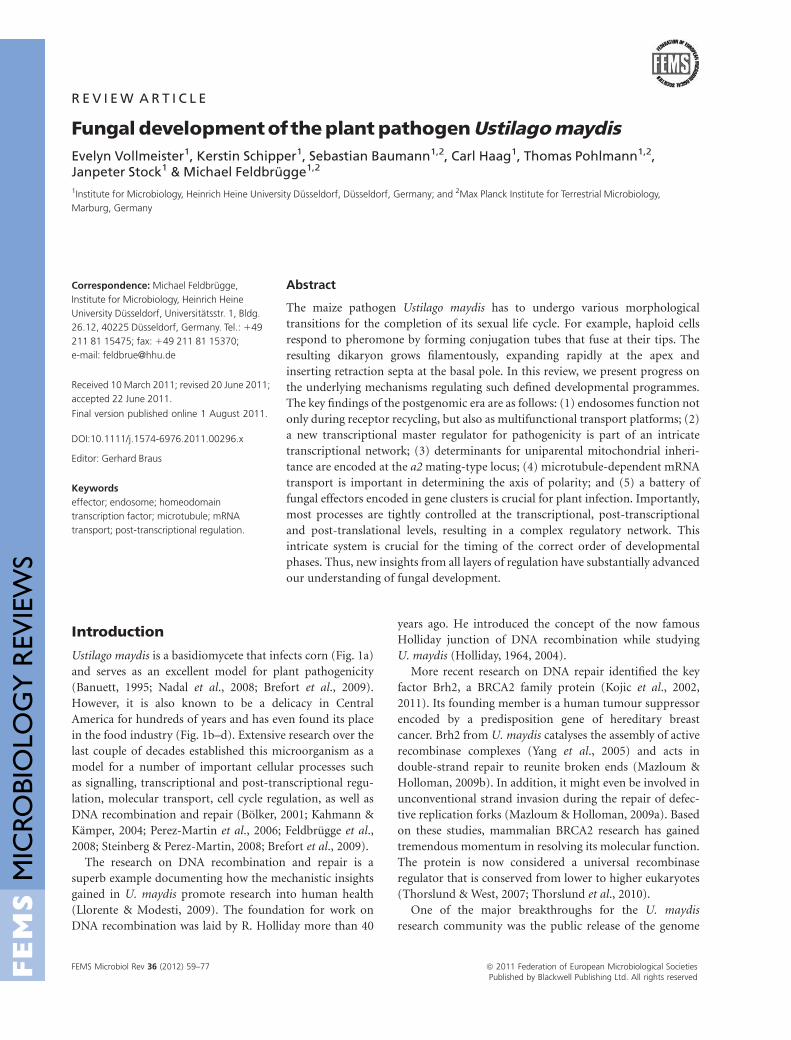
Ustilago maydis is a basidiomycete that infects corn (Fig. 1a)
and serves as an excellent model for plant pathogenicity
(Banuett, 1995; Nadal et al., 2008; Brefort et al., 2009).
However, it is also known to be a delicacy in Central
America for hundreds of years and has even found its place
in the food industry (Fig. 1b–d). Extensive research over the
last couple of decades established this microorganism as a
model for a number of important cellular processes such
as signalling, transcriptional and post-transcriptional regu-
lation, molecular transport, cell cycle regulation, as well as
DNA recombination and repair (Bolker, 2001; Kahmann &
Kamper, 2004; Perez-Martin et al., 2006; Feldbrugge et al.,
2008; Steinberg & Perez-Martin, 2008; Brefort et al., 2009).
The research on DNA recombination and repair is a
superb example documenting how the mechanistic insights
gained in U. maydis promote research into human health
(Llorente & Modesti, 2009). The foundation for work on
DNA recombination was laid by R. Holliday more than 40
years ago. He introduced the concept of the now famous
Holliday junction of DNA recombination while studying
U. maydis (Holliday, 1964, 2004).
More recent research on DNA repair identified the key
factor Brh2, a BRCA2 family protein (Kojic et al., 2002,
2011). Its founding member is a human tumour suppressor
encoded by a predisposition gene of hereditary breast
cancer. Brh2 from U. maydis catalyses the assembly of active
recombinase complexes (Yang et al., 2005) and acts in
double-strand repair to reunite broken ends (Mazloum &
Holloman, 2009b). In addition, it might even be involved in
unconventional strand invasion during the repair of defec-
tive replication forks (Mazloum & Holloman, 2009a). Based
on these studies, mammalian BRCA2 research has gained
tremendous momentum in resolving its molecular function.
The protein is now considered a universal recombinase
regulator that is conserved from lower to higher eukaryotes
(Thorslund & West, 2007; Thorslund et al., 2010).
One of the major breakthroughs for the U. maydis
research community was the public release of the genome
Final version published online 1 August 2011.
FEMS Microbiol Rev 36 (2012) 59–77 ª 2011 Federation of European Microbiological SocietiesPublished by Blackwell Publishing Ltd. All rights reserved
MIC
ROBI
OLO
GY
REV
IEW
S
sequence combined with a profound manual annotation
that resulted in high-quality data currently curated in the
database MUMDB at MIPS (Kamper et al., 2006; MIPS
U. maydis database; and the Munich information centre for
protein sequences, respectively; http://mips.helmholtz-
muenchen.de/genre/proj/ustilago/). This spurred the appli-
cation of new approaches such as transcriptome-wide DNA
microarrays (Scherer et al., 2006; Zarnack et al., 2008;
Heimel et al., 2010b), proteome-wide approaches (Bohmer
et al., 2007), gene replacement strategies for efficient knock-
outs or more sophisticated promoter and gene fusions
(Brachmann et al., 2004; Kamper, 2004; Garcia-Pedrajas
et al., 2008). Establishing fluorescence proteins such as eGfp,
mRfp, mCherry, Yfp, Cfp, photoactivatable Gfp and split-
Yfp facilitated comprehensive in vivo localization, colocali-
zation and interaction studies (Steinberg & Perez-Martin,
2008; Heimel et al., 2010a; Schuster et al., 2011b).
In this review, we will use the sexual life cycle of U. maydis
as a blue print to describe recent findings focusing on the
postgenomic era. Complementary information can be
found in earlier reviews (Kahmann & Kamper, 2004;
Feldbrugge et al., 2006) and in the special issue of Fungal
Genetics & Biology dedicated to U. maydis (Kronstad, 2008).
For the sake of clarity, the life cycle is divided into four
distinct phases: proliferation of haploid cells, mating, fila-
mentation and plant infection. However, the results de-
scribed here indicate that key players of the underlying
cellular events are often important at several additional
stages during the life cycle.
Synopsis of the plant-dependent life cycle
The saprophytic phase can be considered as the default state
and starts with meiosis during the germination of diploid
teliospores. The resulting haploid cells proliferate by bud-
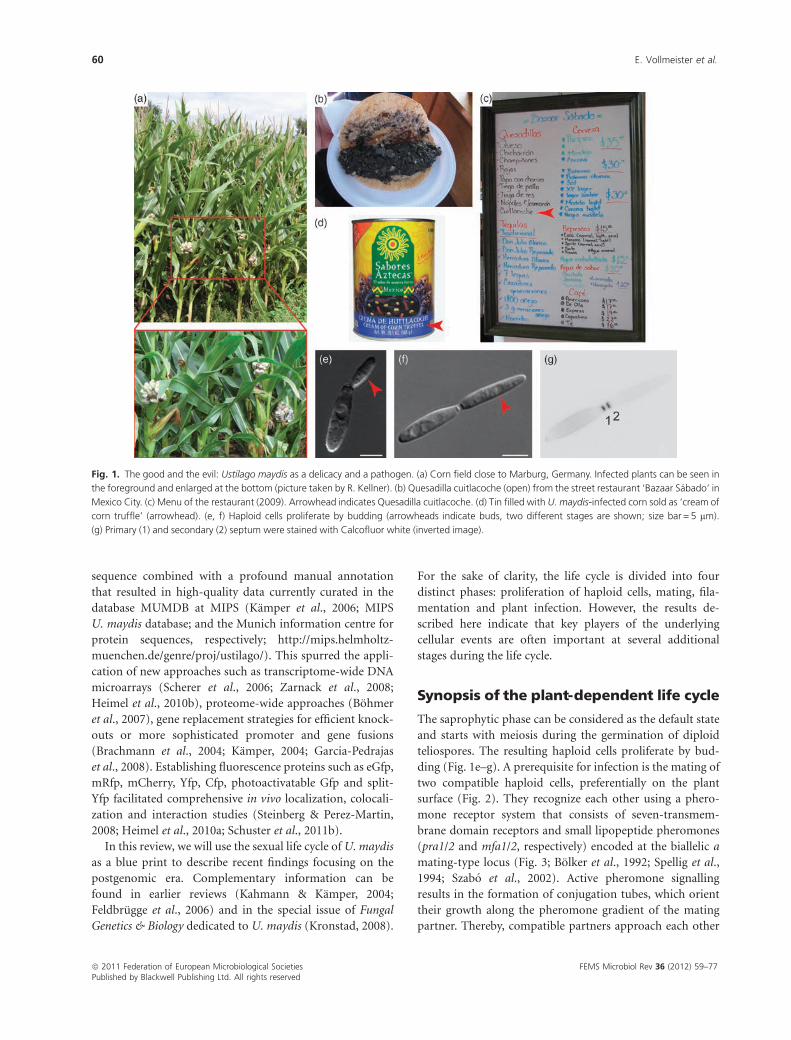
ding (Fig. 1e–g). A prerequisite for infection is the mating of
two compatible haploid cells, preferentially on the plant
surface (Fig. 2). They recognize each other using a phero-
mone receptor system that consists of seven-transmem-
brane domain receptors and small lipopeptide pheromones
(pra1/2 and mfa1/2, respectively) encoded at the biallelic a
mating-type locus (Fig. 3; Bolker et al., 1992; Spellig et al.,
1994; Szabo et al., 2002). Active pheromone signalling
results in the formation of conjugation tubes, which orient
their growth along the pheromone gradient of the mating
partner. Thereby, compatible partners approach each other
Fig. 1. The good and the evil: Ustilago maydis as a delicacy and a pathogen. (a) Corn field close to Marburg, Germany. Infected plants can be seen in
the foreground and enlarged at the bottom (picture taken by R. Kellner). (b) Quesadilla cuitlacoche (open) from the street restaurant ‘Bazaar Sabado’ in
Mexico City. (c) Menu of the restaurant (2009). Arrowhead indicates Quesadilla cuitlacoche. (d) Tin filled with U. maydis-infected corn sold as ‘cream of
corn truffle’ (arrowhead). (e, f) Haploid cells proliferate by budding (arrowheads indicate buds, two different stages are shown; size bar = 5 mm).
(g) Primary (1) and secondary (2) septum were stained with Calcofluor white (inverted image).
ª 2011 Federation of European Microbiological Societies FEMS Microbiol Rev 36 (2012) 59–77Published by Blackwell Publishing Ltd. All rights reserved
60 E. Vollmeister et al.
and fuse at their tips, initiating plasmogamy (Fig. 2;
Snetselaar et al., 1996). Characteristic for basidiomycetes,
plasmogamy and karyogamy are separated in time (Kruzel &
Hull, 2010). A stable dikaryon is formed that grows fila-
mentously with a defined axis of polarity. Hyphae expand
at the apical growth cone (Fig. 2) and insert retraction septa
at regular intervals at the basal pole. These septa separate the
viable compartment from hyphal segments devoid of a
cytoplasm, resulting in the formation of regularly spaced
empty sections (Lehmler et al., 1997; Steinberg et al., 1998).
The nuclei travel to the centre of the cell and are positioned
in a defined distance of about 10 mm from each other as well
as about 50 mm from the tip (Steinberg et al., 1998; Fuchs
et al., 2005).
The developmental switch resulting in hyphal growth is
genetically controlled by the action of homeodomain tran-
scription factors encoded as two separate subunits, bWest
(bW) and bEast (bE), at the multiallelic b mating-type locus
(Kronstad & Leong, 1990; Gillissen et al., 1992). The activity
of the transcription factor is elegantly coupled to plasmo-
gamy because it can only function as a heterodimer with
subunits derived from different mating partners (e.g. bW1/
bE19, bW5/bE8, etc.; Kamper et al., 1995). At present, 19
different b alleles that promote outbreeding are known
(J. Kamper, pers. commun.; Barnes et al., 2004).
The dikaryotic filament is the infectious form of the fungus.
It grows in close contact with the plant and is able to sense
surface signals that trigger the formation of appressoria
(Mendoza-Mendoza et al., 2009a; Lanver et al., 2010). These
are specialized infection structures that enable the pathogen to
enter the plant. Initially, hyphae grow intracellularly by
invagination of the plant plasma membrane, establishing a
tight interaction zone with colonized host cells (Snetselaar &
Mims, 1993; Doehlemann et al., 2008b). At later stages,
proliferation also occurs intercellularly and the dikaryotic
mycelium grows towards bundle sheets (Doehlemann et al.,
2008b). Subsequently, massive proliferation and hyphal frag-
mentation occurs. This is accompanied by an irregular divi-
sion of host cells, resulting in the formation of tumours at all
aerial parts of the plants. After karyogamy, black diploid
teliospores develop within the tumours. Ripe tumours rupture
and mature spores are spread by the wind. Under favourable
conditions, teliospores germinate and release haploid cells.
The life cycle begins anew.
Proliferation of haploid cells andcytokinesis
Saprophytic cells exhibit a defined cylindrical cell shape
comparable to Schizosaccharomyces pombe. However, they
do not divide by insertion of a central septum, but prolifer-
ate by budding like Saccharomyces cerevisiae (Fig. 1e–g).
During proliferation, the bud expands by polar tip growth,
presumably by actin-mediated secretion of remodelling
enzymes and building blocks of the cell wall (Banuett &
Herskowitz, 2002; Weber et al., 2006). This notion is
supported by the observation that an actin-dependent class
V myosin localizes to regions of polar growth and is
important for cell morphology (Weber et al., 2003). After
the daughter cell reaches a certain size, the nucleus migrates
from the centre of the mother into the bud where division
occurs (Holliday, 1974; O’Donnell & McLaughlin, 1984). At
the mother/bud neck region, the nuclear envelope is striped
off, nuclear pore complexes are disassembled and chromo-
somes are released, enabling an open mitosis that takes place
within the daughter cell (Steinberg et al., 2001; Straube et al.,
2005; Theisen et al., 2008). This process is most probably
regulated by a conserved signalling pathway containing
small GTPase Ras3 and dual-function germinal centre
kinase Don3 (Straube et al., 2005; Sandrock et al., 2006).
Fig. 2. Mating on the plant surface. Two haploid cells (coloured yellow and red in the middle panel) landed in close proximity on the plant surface.
Conjugation tubes (coloured yellow and red in the right panel) grew towards each other and fused at their tips. The resulting filament (coloured orange
in the right panel) with two nuclei (coloured yellow and red in the right panel; note that their position is an approximation) grows on the plant surface
[the uncoloured figure on the left is reprinted from Kamper et al. (2006) with permission from the Nature Publishing Group].
FEMS Microbiol Rev 36 (2012) 59–77 ª 2011 Federation of European Microbiological SocietiesPublished by Blackwell Publishing Ltd. All rights reserved
Development of the pathogen U. maydis 61
Chromosome segregation as well as nuclear movement
depend on astral microtubules and involve the molecular
motor dynein (Fink et al., 2006), a minus-end-directed
motor that is split untypically into two subunits Dyn1 and
Dyn2 in U. maydis (Straube et al., 2001).
Nuclear and cytoplasmic microtubule organizing centres
(MTOCs, minus-ends of microtubules) nucleate dynamic
microtubules that are organized in antiparallel bundles
traversing the length of unbudded cells (Straube et al.,
2003). During cell division, MTOCs are transported by
dynein towards the neck region, where they polarize the
microtubule cytoskeleton (Fink & Steinberg, 2006). Grow-
ing plus-ends reach into the distal pole of the mother as well
as towards the growing pole of the daughter cell (Straube
et al., 2003).
One transport function of microtubules is the movement
of endosomes containing the t-SNARE-like protein Yup1
(Wedlich-Soldner et al., 2000). These endosomes shuttle
bidirectionally along microtubules. Their active transport is
mediated by the concerted action of dynein and the plus-
end-directed molecular motor Kin3, a Kinesin-3 type motor
related to UNC104 and KIF1A from higher eukaryotes
(Wedlich-Soldner et al., 2002). Strains carrying a tempera-
ture-sensitive mutation in yup1 are altered in cell shape,
indicating that yup1-positive endosomes are important for
morphology (Wedlich-Soldner et al., 2000). However, in-
hibition of microtubule function does not cause such
morphological alterations (Fuchs et al., 2005), indicating
that endosomes might exhibit a microtubule-independent
function in regulating morphology during cell growth. Two
lines of evidence suggest that endosomal transport is neces-
sary for septation. Firstly, endosomes accumulate at the site
of septum formation (Wedlich-Soldner et al., 2000; Schink
& Bolker, 2009) and secondly kin3D strains are defective in
cytokinesis due to aberrant septum formation (Wedlich-
Soldner et al., 2002).
To complete the cell division, mother and daughter cells
need to form two septa consecutively (Fig. 1e–g). Between
these septa, a vacuolar fragmentation zone is formed, defining
the location of the breakdown of the connecting cell wall and
the physical separation (O’Donnell & McLaughlin, 1984;
Banuett & Herskowitz, 2002; Weinzierl et al., 2002).
Mutants in this process were identified by their aberrant
donut-shaped colonies on plates, hence their name don1 and
don3 (Weinzierl et al., 2002; Feldbrugge et al., 2006). In a
liquid medium, don mutants exhibit a distinct cell separation
defect. They form tree-like cell clusters due to their failure to
form a secondary septum (Weinzierl et al., 2002). Don1 is a
guanine nucleotide exchange factor (GEF) that specifically
activates the small G-protein Cdc42 (Weinzierl et al., 2002;
Mahlert et al., 2006; Hlubek et al., 2008). Don3 is a dual-
function germinal centre kinase that acts parallel to the Cdc42
signalling pathway (Weinzierl et al., 2002; Sandrock et al.,
2006). Chemical genetics revealed that Don3 kinase triggers
the dynamic rearrangement of septins from hourglass-shaped
collars into ring-like structures, depending on a contractile
actomyosin ring (Bohmer et al., 2008, 2009). Thus, a signalling
network containing small G-protein Cdc42 and kinase Don3
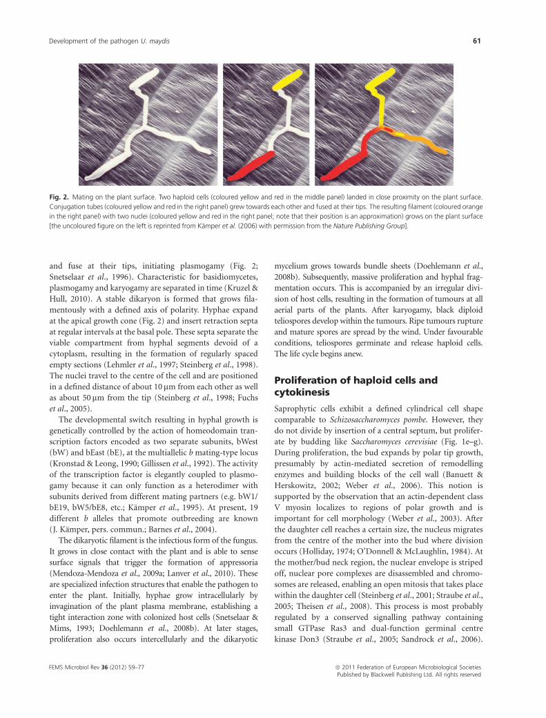
Fig. 3. Networks of signalling and transcription factors regulate the
pheromone response. (a) Schematic representation of the pheromone
signalling network. Lipopeptide pheromone (a1 or a2, encoded by mfa1
or mfa2, respectively) activates its cognate seven-transmembrane domain
receptor (Pra2 or Pra1, respectively). The activated receptor triggers a
signalling pathway containing the scaffold protein Ubc2 and a pheromone-
responsive MAPK module (MAPKKK Kpp4, MAPKK Fuz7 and MAPK
Kpp2). The MAPK regulates Prf1 via phosphorylation and at the transcrip-
tional level. In addition, the MAPKmodule regulates two Prf1-independent
events: conjugation tube formation and cell cycle arrest in G2. MAPK
activity is counteracted by phosphatase Rok1, which is activated at the
transcriptional level by the MAPK module, resulting in negative feedback
regulation. Crosstalk occurs at the level of cAMP signalling. cAMP-
dependent PKA phosphorylates Prf1. This is apparently a prerequisite for
signalling and a connection to environmental signals (see text). (b) At least
four different transcription factors, Rop1, Crk X (yet unknown Crk1-
dependent transcription factor) Hap2 and Prf1 (shown as ovals), recognize
defined cis-active elements in the prf1 promoter (RRS1–3, Rop1-responsive
sequence; CCAAT, CAT box; PREs, pheromone response elements; UAS,
upstream activating sequence; bent arrow, transcriptional start site). There-
by, various environmental and nutritional signals could be funnelled into
the pheromone response.
ª 2011 Federation of European Microbiological Societies FEMS Microbiol Rev 36 (2012) 59–77Published by Blackwell Publishing Ltd. All rights reserved
62 E. Vollmeister et al.
is crucial in determining septin and actin-ring organization
during secondary septum formation.
Substantial progress has recently been made by con-
necting components of this signalling network to micro-
tubule-dependent transport (Schink & Bolker, 2009). The
authors demonstrate that Don1 is targeted to Yup1-positive
endosomes via the specific interaction of the lipid-binding
FYVE domain of Don1 with phosphatidylinositol 3-phos-
phate, a lipid present on these endosomes. Strains expres-
sing a don1 variant with mutations in the FYVE domain are
defective in secondary septum formation like kin3D strains.
The data suggest that the exchange factor Don1 is targeted
towards sites of secondary septum formation by microtu-
bule-dependent transport of endosomes. Thereby, local
activation of its cognate G-protein Cdc42 results in a
signalling cascade orchestrating secondary septum forma-
tion and separation (Schink & Bolker, 2009). Apart from the
recycling of pheromone receptors (Steinberg, 2007b), this
is a new function for these endosomes in U. maydis and fits
the general view of endosomes operating as multipurpose
platforms (Gould & Lippincott-Schwartz, 2009).
Mating and pheromone signalling
Pheromone signalling determining fungal mating belongs to
the best-characterized biological signal transduction phe-
nomena studied in eukaryotes (Elion, 2000; Saito, 2010).
The main framework was elucidated in S. cerevisiae and, in
comparison with various other fungi such as S. pombe and
Cryptococcus neoformans, pheromone signalling proved to
be an evolutionarily conserved mechanism (Davey, 1998;
Bahn et al., 2007). Key components are G-protein-coupled
pheromone receptors that recognize cognate peptide pher-
omones, resulting in the activation of an evolutionarily
conserved MAPK module consisting of three kinases (mito-
gen-activated protein kinase module; MAPKKK, MAPKK
and MAPK). The terminal MAPK phosphorylates, among
others, transcription factors that determine pheromone-
regulated gene expression. A common feature is that the
central MAPK pathways crosstalk with other signalling
routes to discern appropriate mating conditions such as the
nutritional status (Davey, 1998; Saito, 2010).
In U. maydis mating of two haploid cells is a prerequisite
for infection (Fig. 2), a process that is regulated by the a and
b mating-type loci. The lipopeptide pheromone of one
mating partner is perceived by the cognate receptor of the
compatible partner (Bolker et al., 1992; Spellig et al., 1994;
Szabo et al., 2002). This elicits an intracellular signal
transduction pathway leading to the activation of a defined
MAPK module (Fig. 3a; MAPKKK Kpp4, MAPKK Fuz7 and
MAPK Kpp2; summarized in Feldbrugge et al., 2006). Direct
phosphorylation of the central HMG box transcription
factor Prf1 (high mobility group; Hartmann et al., 1996;
Muller et al., 1999; Kaffarnik et al., 2003) triggers the
expression of a defined set of genes containing pheromone
response elements in their regulatory regions (Urban et al.,
1996b; Zarnack et al., 2008). Among these are the a mating-
type genes, mfa1/2 and pra1/2, enhancing the pheromone
response. This feed-forward regulation is further supported
by autoactivation of the prf1 gene at the transcriptional level
(Fig. 3b; Hartmann et al., 1999).
Genome-wide transcriptional profiling revealed that
MAPK regulation of Prf1 is necessary for the expression of
a defined subset of pheromone-responsive genes. Analysis of
strains expressing Prf1 variants with mutated MAPK sites
showed that in the majority of cases, pheromone respon-
siveness of target genes was completely lost while in the
remaining cases regulation was alleviated. Among the latter
are genes encoding the key transcription factors for filamen-
tous growth bW/bE as well as Rbf1 (Scherer et al., 2006;
Heimel et al., 2010b). Thus, target genes respond differen-
tially to MAPK phosphorylation of Prf1 (Zarnack et al.,
2008) and MAPK signalling via Prf1 prepares the main
transcriptional cascade that regulates filamentous growth
and further pathogenic development (Heimel et al., 2010b;
Wahl et al., 2010b).
In addition, the pheromone-responsive MAPK module is
involved in the regulation of the pheromone-induced cell
cycle arrest in the G2 phase, which is a Prf1-independent
process (Fig. 3a; Garcia-Muse et al., 2003). This ensures
synchronization of the cell cycle of both nuclei after cell
fusion. Pheromone stimulation as well as transcriptional
activation of a constitutively active MAPKK triggers reduced
expression of several genes encoding cell cycle regulators
such as B-type cyclin Clb1 or orthologues of polo kinases
(Perez-Martin et al., 2006; Zarnack et al., 2008). It is
noteworthy that MAPK signalling also promotes a negative
feedback loop by inducing the expression of Rok1, a dual-
specificity phosphatase that inactivates MAPK Kpp2
(Fig. 3a; Di Stasio et al., 2009).
Moreover, the MAPK module is responsible for eliciting
the formation of conjugation tubes, another Prf1-indepen-
dent event (Fig. 3a; Muller et al., 2003). During the polar
growth of these filaments, the pheromone receptor localizes
to their tips. This specific localization as well as receptor
turnover by endocytosis might be important for the orienta-
tion within the pheromone gradient. Thereby, conjugation
tubes of compatible mating partners grow towards each
other and fuse at their tips (Snetselaar et al., 1996; Fuchs
et al., 2006). After plasmogamy, pheromone signalling is
switched off by an active bW/bE heterodimer (Laity et al.,
1995; Urban et al., 1996b; Heimel et al., 2010a).
Mating on the surface of the plant is crucial for infection.
Thus, cell recognition and fusion are influenced by environ-
mental factors such as nutrient availability, hydroxy fatty
acids or hydrophobic surfaces (Hartmann et al., 1999;
FEMS Microbiol Rev 36 (2012) 59–77 ª 2011 Federation of European Microbiological SocietiesPublished by Blackwell Publishing Ltd. All rights reserved
Development of the pathogen U. maydis 63
Mendoza-Mendoza et al., 2009a). Two molecular mechan-
isms for the underlying signalling integration event have
been identified. Firstly, an evolutionarily conserved cAMP
signalling pathway that consists of a receptor-coupled
heterotrimeric G-protein, adenylate cyclase and protein
kinase A (PKA) influences mating (Feldbrugge et al., 2006;
Brefort et al., 2009). An important molecular mechanism is
the direct phosphorylation of Prf1 by cAMP-activated
PKA, a regulatory event that is apparently a prerequisite
for pheromone response (Fig. 3a; Kaffarnik et al., 2003).
PKA-mediated activation of Prf1 is also sufficient to increase
the expression of mating-type genes, indicating that upre-
gulation of the intracellular cAMP level favours mating
(Kaffarnik et al., 2003; Zarnack et al., 2008). Crosstalk of
the PKA and MAPK signalling is also important at later
stages of the pathogenic development (Feldbrugge et al.,
2006; Brefort et al., 2009).
Secondly, prf1 expression at the transcriptional level is
tightly controlled by various regulatory elements in the
promoter region (Fig. 3b). For example, the upstream
activating sequence (UAS) element integrates different car-
bon sources (Hartmann et al., 1999) and the unusual MAPK
Crk1 signals via this element (Garrido et al., 2004). In
addition, the HMG box transcription factor Rop1 as well as
the CCAAT-box-binding protein Hap2 are crucial for the
activation of prf1 expression via binding sites in the prf1
promoter (Brefort et al., 2005; Mendoza-Mendoza et al.,
2009b). Thus, at least four different transcriptional activa-
tors (Prf1, yet unknown UAS-binding protein X, Rop1,
Hap2) that integrate potentially different signals converge
at the prf1 promoter to accurately control the expression of
this key transcription factor (Fig. 3b). This is an excellent
example of how a central signalling node can integrate
various external and internal cues to determine optimal
mating conditions.
Filamentous growth and transcriptionalnetworking
Homeodomain transcription factors constitute crucial
players in developmental programmes not only in fungi,
but also in plants and animals (Pearson et al., 2005; Hay &
Tsiantis, 2010). They often function as master regulators
eliciting transcriptional cascades that include additional
transcription factors. In U. maydis, the homeodomain
transcription factor bW/bE is necessary and sufficient to
regulate filamentous growth. This Prf1-activated key regula-
tor connects sexual and pathogenic development. Right after
its discovery at the b mating-type locus, it was proposed
that, like homeodomain transcription factors from higher
eukaryotes, it triggers a defined transcriptional programme
essential for development (Kronstad & Leong, 1990; Schulz
et al., 1990; Gillissen et al., 1992). However, it turned out to
be more difficult than expected to identify the responsible
target genes. Only very recently a major breakthrough was
achieved with the advent of genome-wide transcriptional
profiling (Kahmann & Kamper, 2004; Heimel et al., 2010b;
Wahl et al., 2010b).
Initially, it was discovered that the activity of the transcrip-
tion factor was regulated by heterodimerization of subunits
derived from different mating partners (Kamper et al., 1995).
This information was used to demonstrate that an active bW/
bE heterodimer is sufficient to elicit pathogenic development.
Haploid strains were generated that express active versions
under the control of their own promoters. These strains are
called solopathogenic because they infect plants independent
of mating (Bolker et al., 1995).
For the identification of functionally important compo-
nents of filamentous growth, various comparative methods
were applied investigating haploid cells and b-induced
filaments. Several genes were identified that exhibited signifi-
cantly increased expression during filamentous growth,
but unfortunately, these were all dispensable for infection
(Schauwecker et al., 1995; Wosten et al., 1996; Romeis et al.,
2000).
A significant improvement was the generation of haploid
strains expressing active bW2/bE1 variants under the con-
trol of carbon- or nitrogen-source-regulated promoters. For
the first time, this allowed a defined time-resolved monitor-
ing of the underlying regulatory cascade (Brachmann et al.,
2001). Using RNA fingerprinting, 10 additional targets
could be identified (Brachmann et al., 2001). For example,
the involvement of MAPK Kpp6 in the early steps of
infection such as plant penetration could be demonstrated
(Brachmann et al., 2003).
The same strains were the foundation for applying high-
density microarrays covering around 90% of all predicted
genes (Heimel et al., 2010b). Performing extensive time-
course experiments with carbon- and nitrogen-source-
mediated activation of the bW/bE heterodimer resulted in
the detection of 345 bW/bE-responsive genes (206 induced
and 139 repressed; Heimel et al., 2010b). Global analysis of
the respective gene functions revealed that 20 genes involved
in cell wall remodelling, such as chitin synthases, exochiti-
nases, chitin deacetylases as well as glucanases, are induced,
indicating active remodelling of the fungal cell wall during
filamentous growth (Heimel et al., 2010b). A number of
clustered genes encoding potential effector proteins for
plant interaction were also activated (Kamper et al., 2006;
Heimel et al., 2010b). Among the downregulated genes are
several encoding cell cycle regulators such as cyclins Cln1,
Clb1 and Clb2. For Clb1, it has been shown that repression
leads to G2 cell cycle arrest (Garcia-Muse et al., 2004).
Apparently, the bW/bE heterodimer is responsible for
maintaining the G2 cell cycle arrest until the filament
ª 2011 Federation of European Microbiological Societies FEMS Microbiol Rev 36 (2012) 59–77Published by Blackwell Publishing Ltd. All rights reserved
64 E. Vollmeister et al.
resumes the cell cycle after plant penetration (Perez-Martin
et al., 2006).
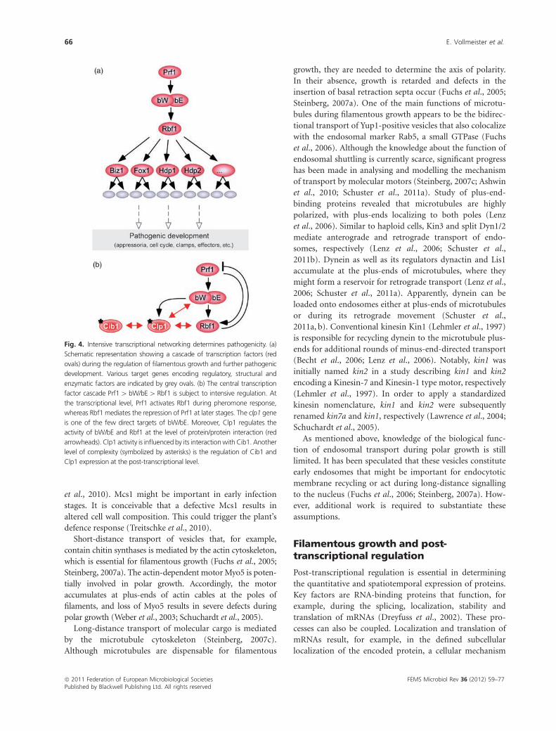
One of the most rewarding results of this analysis was the
identification of the downstream master regulator Rbf1.
This zinc finger transcription factor, which is an immediate
early target of the bW/bE heterodimer at the transcriptional
level, is responsible for the vast majority of the gene
expression programme (Fig. 4a; Heimel et al., 2010b).
Accordingly, overexpression of Rbf1 is sufficient to trigger
filamentous growth. Because it is difficult to envision that
hundreds of different bW/bE heterodimers trigger the same
transcriptional programme, the presence of such a master
regulator was no surprise. Interestingly, rbf1 is also a direct
Prf1 target and its expression is pheromone induced via
MAPK signalling (Fig. 4b; Zarnack et al., 2008). Thus,
during the initial phase of mating, Rbf1 activity must be
inhibited by a currently unknown mechanism.
Rbf1 amplifies the transcriptional cascade by the activa-
tion of various transcriptional regulators such as Biz1 and
Fox1 (Fig. 4a; Heimel et al., 2010b). Biz1 is a zinc finger-
containing factor involved in cell cycle regulation and is
important for appressoria formation and plant penetration
(Flor-Parra et al., 2006). Fox1 regulates the expression of
numerous effector proteins that appear to be involved in the
suppression of plant defence (Zahiri et al., 2010).
Using a temperature-sensitive mutant, it was demon-
strated that the active bW/bE transcription factor is not only
needed to establish the transcriptional cascade of pathogenic
development, but is also crucial at later stages. Interfering
with the activity of bW/bE in planta results in an unusual
activation of pheromone signalling, defects in cell cycle
synchronization and reduced expression of effector proteins.
This aberrant regulation correlates with impaired infection
(Wahl et al., 2010b).
Another important direct target of the bW/bE transcrip-
tion factor is clp1 (Fig. 4b). It encodes a regulator that is
required for clamp formation, a special structure needed for
nuclear sorting during mitotic division of the dikaryon in
planta (Scherer et al., 2006). Recently, the level of complexity
was added to by demonstrating that Clp1 interacts with bW
and counteracts bW/bE-dependent functions such as G2 cell
cycle arrest and filamentous growth (Fig. 4b). Clp1 also
interacts with Rbf1 during the repression of pheromone
signalling (Heimel et al., 2010a). Furthermore, Clp1 binds
Cib1, a bZIP transcription factor required for pathogenic
development, whose activity is not regulated at the tran-
scriptional level, but at the level of alternative splicing (Fig.
5b; Heimel et al., 2010a). Thus, besides the simple linear
transcription cascade Prf14 bW/bE4Rbf1 (Fig. 4a), there
is an underlying complicated network of transcription
factors and protein interactions that mediate intensive feed-
back and feedforward regulation to orchestrate pathogenic
development (Fig. 4b).
Filamentous growth and cytoskeletalfunctions
In general, fungal hyphae exhibit a characteristic growth
mode. At their apical pole, a growth cone is responsible for
the rapid expansion of membrane and cell wall. In subapical
regions, filaments are partitioned by septa forming molecu-
lar barriers (Harris, 2006; Steinberg, 2007a; Fischer et al.,
2008; Zarnack & Feldbrugge, 2010). Polar growth is
mediated by intensive membrane dynamics, i.e. local exo-
and endocytosis that is supported by vesicle trafficking. This
might be promoted by distinct membrane microdomains at
the apical pole that are rich in specific lipids such as
sphingolipids and ergosterol (Harris et al., 2005; Steinberg,
2007b). Two juxtaposed macromolecular units are involved
in growth cone function: the Spitzenkorper and the polari-
some. The latter is a multiprotein complex consisting of
landmark proteins, signalling molecules such as small
GTPases and formins that nucleate actin cables for short-
distance transport (Harris et al., 2005; Sudbery & Court,
2007). The Spitzenkorper is thought to function as a supply
centre for exocytotic vesicles (Gierz & Bartnicki-Garcia,
2001; Bartnicki-Garcia, 2002), for example, specialized
vesicles, so-called chitosomes, transport and export chitin
synthases as well as building blocks for cell wall synthesis
(Bartnicki-Garcia, 2006; Riquelme et al., 2007). Active
transport along the actin and microtubule cytoskeleton is
mediated by molecular motors for short- and long-distance
delivery of vesicles, respectively. This process is streamlined for
efficiency and any disturbances lead to reduced growth rates
or a complete blockage (Harris, 2006; Steinberg, 2007a).
In U. maydis, a number of recent observations confirm
that dikaryotic filaments grow like typical hyphae (Stein-
berg, 2007a). For example, the landmark protein Spa2
localizes to the tip (Carbo & Perez-Martin, 2008) and
sphingolipid biosynthesis is important for polar growth.
These lipids are most likely part of membrane micro-
domains at the hyphal tip (Canovas & Perez-Martin, 2009)
and could function to enrich signalling components with
membrane anchors such as the small G protein Rac1 (Mahlert
et al., 2006). Septins might serve as diffusion barriers organiz-
ing these membrane subdomains. Strains carrying corre-
sponding loss-of-function mutations are affected in
establishing the axis of polarity, whereas septins are dispen-
sable during the maintenance of filamentous growth (Alvarez-
Tabares & Perez-Martin, 2010).
Analysis of eight different chitin synthases revealed that
Chs7 was particularly important for polar growth and
localizes to the hyphal tip like its related family members
Chs5 and Mcs1 (Weber et al., 2006). The chitin synthase
Mcs1 contains a unique domain with a similarity to myosin
motors (Weber et al., 2006). This domain is not required for
motility, but apparently for different functions (Treitschke
FEMS Microbiol Rev 36 (2012) 59–77 ª 2011 Federation of European Microbiological SocietiesPublished by Blackwell Publishing Ltd. All rights reserved
Development of the pathogen U. maydis 65
et al., 2010). Mcs1 might be important in early infection
stages. It is conceivable that a defective Mcs1 results in
altered cell wall composition. This could trigger the plant’s
defence response (Treitschke et al., 2010).
Short-distance transport of vesicles that, for example,
contain chitin synthases is mediated by the actin cytoskeleton,
which is essential for filamentous growth (Fuchs et al., 2005;
Steinberg, 2007a). The actin-dependent motor Myo5 is poten-
tially involved in polar growth. Accordingly, the motor
accumulates at plus-ends of actin cables at the poles of
filaments, and loss of Myo5 results in severe defects during
polar growth (Weber et al., 2003; Schuchardt et al., 2005).
Long-distance transport of molecular cargo is mediated
by the microtubule cytoskeleton (Steinberg, 2007c).
Although microtubules are dispensable for filamentous
growth, they are needed to determine the axis of polarity.
In their absence, growth is retarded and defects in the
insertion of basal retraction septa occur (Fuchs et al., 2005;
Steinberg, 2007a). One of the main functions of microtu-
bules during filamentous growth appears to be the bidirec-
tional transport of Yup1-positive vesicles that also colocalize
with the endosomal marker Rab5, a small GTPase (Fuchs
et al., 2006). Although the knowledge about the function of
endosomal shuttling is currently scarce, significant progress
has been made in analysing and modelling the mechanism
of transport by molecular motors (Steinberg, 2007c; Ashwin
et al., 2010; Schuster et al., 2011a). Study of plus-end-
binding proteins revealed that microtubules are highly
polarized, with plus-ends localizing to both poles (Lenz
et al., 2006). Similar to haploid cells, Kin3 and split Dyn1/2
mediate anterograde and retrograde transport of endo-
somes, respectively (Lenz et al., 2006; Schuster et al.,
2011b). Dynein as well as its regulators dynactin and Lis1
accumulate at the plus-ends of microtubules, where they
might form a reservoir for retrograde transport (Lenz et al.,
2006; Schuster et al., 2011a). Apparently, dynein can be
loaded onto endosomes either at plus-ends of microtubules
or during its retrograde movement (Schuster et al.,
2011a, b). Conventional kinesin Kin1 (Lehmler et al., 1997)
is responsible for recycling dynein to the microtubule plus-
ends for additional rounds of minus-end-directed transport
(Becht et al., 2006; Lenz et al., 2006). Notably, kin1 was
initially named kin2 in a study describing kin1 and kin2
encoding a Kinesin-7 and Kinesin-1 type motor, respectively
(Lehmler et al., 1997). In order to apply a standardized
kinesin nomenclature, kin1 and kin2 were subsequently
renamed kin7a and kin1, respectively (Lawrence et al., 2004;
Schuchardt et al., 2005).
As mentioned above, knowledge of the biological func-
tion of endosomal transport during polar growth is still
limited. It has been speculated that these vesicles constitute
early endosomes that might be important for endocytotic
membrane recycling or act during long-distance signalling
to the nucleus (Fuchs et al., 2006; Steinberg, 2007a). How-
ever, additional work is required to substantiate these
assumptions.
Filamentous growth and post-transcriptional regulation
Post-transcriptional regulation is essential in determining
the quantitative and spatiotemporal expression of proteins.
Key factors are RNA-binding proteins that function, for
example, during the splicing, localization, stability and
translation of mRNAs (Dreyfuss et al., 2002). These pro-
cesses can also be coupled. Localization and translation of
mRNAs result, for example, in the defined subcellular
localization of the encoded protein, a cellular mechanism
Fig. 4. Intensive transcriptional networking determines pathogenicity. (a)
Schematic representation showing a cascade of transcription factors (red
ovals) during the regulation of filamentous growth and further pathogenic
development. Various target genes encoding regulatory, structural and
enzymatic factors are indicated by grey ovals. (b) The central transcription
factor cascade Prf14 bW/bE4 Rbf1 is subject to intensive regulation. At
the transcriptional level, Prf1 activates Rbf1 during pheromone response,
whereas Rbf1 mediates the repression of Prf1 at later stages. The clp1 gene
is one of the few direct targets of bW/bE. Moreover, Clp1 regulates the
activity of bW/bE and Rbf1 at the level of protein/protein interaction (red
arrowheads). Clp1 activity is influenced by its interactionwith Cib1. Another
level of complexity (symbolized by asterisks) is the regulation of Cib1 and
Clp1 expression at the post-transcriptional level.
ª 2011 Federation of European Microbiological Societies FEMS Microbiol Rev 36 (2012) 59–77Published by Blackwell Publishing Ltd. All rights reserved
66 E. Vollmeister et al.
that is conserved from bacteria to humans (St Johnston,
2005; Du et al., 2007; Nevo-Dinur et al., 2011). mRNA
localization is mainly achieved by active transport along the
actin or the microtubule cytoskeleton. In S. cerevisiae, ASH1
mRNA is actively transported from the mother cell towards
the distal pole of the daughter cell. Local translation at the
pole ensures that the encoded transcription factor specifi-
cally localizes to the nucleus of the daughter cell in order to
ascertain a distinct transcriptional programme (Jansen,
2001; Zarnack & Feldbrugge, 2007). ASH1 mRNA transport
is mediated by two RNA-binding proteins, She2p and
She3p, that cooperatively bind RNA elements, so-called
zipcodes, in the target mRNA. This is essential for actin-
dependent transport mediated by myosin (Bohl et al., 2000;
Muller et al., 2011). Recently, it was reported that She3p-
mediated ASH1 mRNA transport is also operational in
filaments of Candida albicans, most likely to regulate hyphal
morphology and invasive growth (Elson et al., 2009).
Microtubule-dependent mRNA transport is mainly stu-
died in higher eukaryotes. Important processes regulated at
this level are oocyte and embryo development in Drosophila
melanogaster as well as neuronal processes in mammals (St
Johnston, 2005; Dahm et al., 2007). Although the RNA-
binding proteins and the transported target mRNAs are
evolutionarily not conserved, the general concept holds true
for all investigated transport processes to date: specific RNA-
binding proteins recognize zipcodes forming large mRNPs
(mRNA-containing ribonucleoprotein complexes) that are
transported along microtubules by kinesin(s) and dynein
(Holt & Bullock, 2009).
The importance of such control at the post-transcrip-
tional level is an emerging theme for U. maydis. This
additional layer of regulation functions in concert with the
extensive regulation at the level of transcription described
above (Feldbrugge et al., 2008; Vollmeister & Feldbrugge,
2010). For example, although clp1 is a direct and immediate
early target gene of bW/bE, the protein accumulates only at
later stages during plant penetration, suggesting transla-
tional control (Scherer et al., 2006). Moreover, precise
expression of the Clp1 interaction partner Cib1 appears to
be regulated by alternative splicing (Heimel et al., 2010a).
Investigating various RNA-binding proteins in U. maydis
revealed that two of them, Khd4 and Rrm4, are important
for filamentous growth (Becht et al., 2005). The loss of Khd4
causes pleiotropic effects such as altered cell and filament
morphology as well as defects in pheromone response
and plant infection (Becht et al., 2005). The protein contains
at least five potential RNA-binding domains of the KH
type (hnRNP K homology domain, KH1-5). It constitutes
the founding member of a multi-KH domain family of
fungal proteins with representatives in other pathogens such
as C. neoformans and C. albicans. Khd4 recognizes
the sequence AUACCC via the two central tandem-KH
domains (KH3 and KH4; Vollmeister et al., 2009). Interest-
ingly, this motif is enriched in the 30 untranslated region of
mRNAs and a number of transcripts, which exhibit altered
expression in khd4D strains, contain this binding site.
Because most mRNAs show an increased abundance in
deletion strains, Khd4 might promote mRNA instability
(Vollmeister et al., 2009; Vollmeister & Feldbrugge, 2010).
Direct target mRNAs, whose expression is regulated by
Khd4 and whose function can be related to the mutant
phenotypes, are yet to be identified. To this end, we are
currently conducting in vivo UV crosslinking experiments
(Konig et al., 2010).
The deletion of rrm4 causes specific defects during
filamentous growth, namely bipolar growth and failure to
insert retraction septa (Fig. 5a–c; Becht et al., 2005, 2006).
Interestingly, this mutant phenotype is reminiscent of
growth defects of strains affected in microtubule functions
(Lehmler et al., 1997; Steinberg et al., 1998; Fuchs et al.,
2005; Schuchardt et al., 2005; Schuster et al., 2011a). Rrm4
contains three N-terminal RNA recognition motifs and a C-
terminal MLLE domain that mediate RNA binding (Becht
et al., 2006; Konig et al., 2007) and protein/protein interac-
tions, respectively (Kozlov et al., 2010). Subcellular localiza-
tion revealed that Rrm4 shuttles in mRNPs along
microtubules (Fig. 5d–e; Supporting Information, Movies
S1 and S2). RNA binding as well as the formation of motile
mRNPs are essential for protein function (Becht et al., 2006;
Konig et al., 2007). The deletion of kin1 results in the
accumulation of Rrm4 at the poles. This is consistent with
the hypothesis that split dynein mediates the retrograde
transport of mRNPs, because conventional kinesin mediates
anterograde transport of split dynein to the plus-ends of
microtubules (Becht et al., 2006; S. Baumann, T. Pohlmann
& M. Feldbrugge, unpublished data). In vivo UV cross-
linking revealed that Rrm4 binds a distinct set of mRNAs
that contain a potential CA-rich binding motif (Konig et al.,
2009). These mRNAs encode proteins of cytotopically
related groups such as proteins involved in translation,
mitochondrial proteins or polarity factors. Important ex-
amples are ubi1 and rho3 encoding a natural fusion of
ubiquitin with ribosomal protein Rpl40 and a small GTPase,
respectively (Konig et al., 2009). RNA live imaging revealed
that these mRNAs are molecular cargos of the Rrm4 trans-
port unit. Remarkably, the CA-rich 30 UTR of ubi1 functions
as a cis-active region in increasing the amount and proces-
sivity of trafficking (Konig et al., 2009). This is a character-
istic feature of so-called mRNA zipcodes. Rho3 accumulates
at retraction septa, suggesting a regulatory role during this
process. These data led to the current model that Rrm4
functions in long-distance transport of mRNAs, a process
that is conserved throughout evolution (Fig. 5f–g; Zarnack
& Feldbrugge, 2007, 2010). The local translation of Rrm4
target mRNAs appears to be important for the subcellular
FEMS Microbiol Rev 36 (2012) 59–77 ª 2011 Federation of European Microbiological SocietiesPublished by Blackwell Publishing Ltd. All rights reserved
Development of the pathogen U. maydis 67
ª 2011 Federation of European Microbiological Societies FEMS Microbiol Rev 36 (2012) 59–77Published by Blackwell Publishing Ltd. All rights reserved
68 E. Vollmeister et al.
localization of encoded proteins (Fig. 5f–g). Intriguingly, we
could recently show that Rrm4-containing mRNPs are
cotransported with Yup1-positive endosomes, suggesting
yet another function of microtubule-dependent transport
of endosomes (discussed above; S. Baumann, T. Pohlmann
& M. Feldbrugge, unpublished data).
Filamentous growth and mitochondrialinheritance
Sexual reproduction in eukaryotes goes along with the
combination of nuclear and organelle genomes, such as
mitochondrial genomes, in the zygote. This allows recombi-
nation events to accelerate evolution. However, a common
process in eukaryotic sex is uniparental mitochondrial
inheritance, a process that results in the asexual inheritance
of the organelle genome. A possible explanation is the
avoidance of evolutionary conflicts caused by a heteroplas-
mic situation. For example, mitochondria with increased
replication rates, but decreased functional performance
might dominate the population, resulting in disadvantages
for the zygote (Partridge & Hurst, 1998; Xu, 2005). Possible
mechanisms for uniparental mitochondrial inheritance in-
clude unequal size and mitochondrial numbers of gametes
(Xu, 2005). An extreme example is human oocytes carrying
about 10 000 more mitochondrial genomes than sperm cells,
providing for a confident head start of maternal mitochon-
dria in the population. Other mechanisms operating post-
fusion of the gametes include the interplay with nuclear-
encoded genes (Basse, 2010). For example, the mating-type
specific homeodomain genes control uniparental inheri-
tance in C. neoformans, suggesting that respective target
genes encode proteins that determine uniparental inheri-
tance (Xu, 2005; Yan et al., 2007).
In U. maydis, the inheritance of mitochondria is deter-
mined at late stages of filamentous growth when the fungus
penetrates the plant. This coincides with the start of cell
division of the dikaryon, and mitochondria are passed on to
the next cell. Importantly, uniparental mitochondrial in-
heritance exists in U. maydis and the a2 mitotype is
predominantly inherited (Fedler et al., 2009). How is this
achieved at the molecular level? Initially, it was observed that
the a2 mating-type locus, but not its a1 counterpart
contains two genes, lga2 and rga2 (left and right genes of
the a2 locus), which are only found in U. maydis and
close relatives such as the head smut fungus Sporisorium
reilianum (Urban et al., 1996a; Schirawski et al., 2005b).
Because Lga2 contains a mitochondrial import signal, it was
proposed that it might be involved in uniparental mito-
chondrial inheritance (Urban et al., 1996a). Work accom-
plished in the last few years has supported this hypothesis
and has shed some light on the underlying mechanism
(Basse, 2010).
During mating conjugation tubes fuse, plasmogamy
occurs and cellular contents including the mitochondria are
mixed (Fig. 2). How is it possible to separate the two
mitochondrial populations to ensure uniparental inheri-
tance? The key player is Lga2, a protein that is attached to
the outside of the mitochondria (Bortfeld et al., 2004;
Mahlert et al., 2009). This protein serves two main func-
tions. Firstly, it inhibits the fusion of the mitochondria
(Fig. 6) to prevent the mobilization of homing introns and
secondly, Lga2 triggers mitochondrial fragmentation and
mtDNA degradation (Bortfeld et al., 2004; Mahlert et al.,
2009). Rga2 also localizes to the mitochondria even though
a clear targeting signal is missing (Bortfeld et al., 2004). It
appears to counteract Lga2 by protecting the mitochondria
of the a2 mitotype (Fedler et al., 2009; Basse, 2010).
Based on the expression of these genes, the following
model can be proposed (Basse, 2010). Like in all genes of the
a locus, lga2 and rga2 expression is activated upon pher-
omone signalling (Urban et al., 1996b). Thereby, a2 mito-
type mitochondria are preloaded with Lga2 and Rga2 before
cell fusion. In contrast to mfa1/2 and pra1/2, which are
repressed by an active bW/bE heterodimer, lga2 is one of the
few directly activated targets of the bW/bE regulator, and
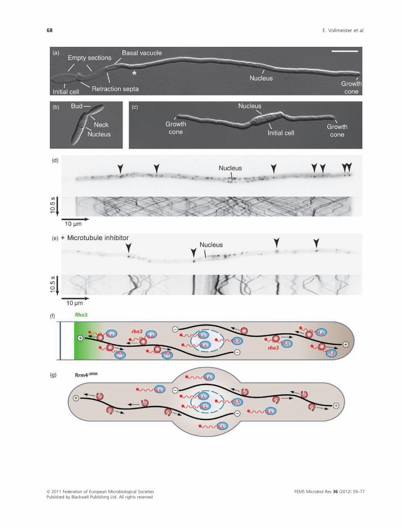
Fig. 5. Microtubule-dependent shuttling of Rrm4-containing mRNPs is important for filamentous growth. (a) Filament of a monokaryotic strain
expressing active bW2/bE1 variants under the control of a nitrogen-source-regulated promoter. Filaments are grown for 6 h under inducing conditions.
Asterisk marks the predicted position of the next retraction septum (size bar = 10 mm). (b) rrm4D cells show no aberrant growth phenotype. (c) rrm4Dfilament exhibits defects in the formation of a single axis of polarity. The initial cell forms two growth cones and fails to insert a retraction septum (Becht
et al., 2006). (d) Filament expressing Rrm4 fused to Gfp. The fusion protein accumulates in defined cytoplasmic particles (arrowheads in the inverted
image detecting Gfp fluorescence) that shuttle bidirectionally along microtubules (kymograph in the lower part). In the kymograph, time is plotted vs.
distance. Thus, motion of Rrm4 particles is visible as defined tracks (note the reversal of shuttling at the poles). Picture and kymograph correspond to
Movie S1. (e) Filament expressing Rrm4 fused to Gfp was treated with the microtubule inhibitor benomyl (Fuchs et al., 2005). Presented as described in
(d). Picture and kymograph correspond to Movie S2. (f) Model depicting microtubule-dependent transport of mRNAs. Rrm4-containing particles (dark
red circles) transport mRNAs (red wavy lines) carrying the poly(A)-binding protein (blue ovals) bidirectionally along microtubules (black lines). The
transport of rho3 mRNA might promote the accumulation of Rho3 at the retraction septum. (g) Deletion of RRM1 to RRM3 in Rrm4 causes a loss-of-
function phenotype. Filaments growmostly bipolar and target mRNAs are no longer transported along microtubules. The mutant Rrm4 is still part of the
shuttling units, indicating that Rrm4 is an integral component of the transport machinery and does not just hitchhike like the poly(A)-binding protein [(f),
(g) are reprinted from Zarnack & Feldbrugge (2010) with permission from the American Society of Microbiology].
FEMS Microbiol Rev 36 (2012) 59–77 ª 2011 Federation of European Microbiological SocietiesPublished by Blackwell Publishing Ltd. All rights reserved
Development of the pathogen U. maydis 69
rga2 expression is maintained constant (Romeis et al., 2000;
Brachmann et al., 2001). Consistently, early during plasmo-
gamy, Lga2 prevents the fusion of mitochondria (Fig. 6) and
over time Lga2 eliminates those mitochondria that are not
sufficiently protected by Rga2, namely mitochondria of the
a1 mitotype (Basse, 2010).
Possible downstream effectors for the two functions of
Lga2 are dynamin-related GTPase Dnm1 and the mitochon-
drial protein Mrb1. Dnm1 is crucial in preventing mitochon-
drial fusion in an Lga2-dependent manner (Mahlert et al.,
2009). Mrb1, a regulator of the p32 family, interacts with
Rga2 and appears to counteract the function of Lga2 (Bort-
feld et al., 2004; Basse, 2010). This is based on the following
observation. While a2mrb1D strains fail to infect plants,
virulence can be restored by the deletion of lga2. Importantly,
dikaryons of a1/a2mrb1D strains are impaired in virulence
because they are arrested at the early phase of infection when
uniparental inheritance should occur (Bortfeld et al., 2004).
However, its precise mode of action is currently unclear.
This is now the first glimpse of this process, but as so
often, the story is more complex. For example, the dom-
inance of a2 mitotypes is not 100%, indicating additional
mechanisms independent of the Lga2/Rga2 system (Bortfeld
et al., 2004; Fedler et al., 2009). Moreover, in S. reilianum,
there are three mating types: a1, a2 and a3. Lga2 and rga2 are
present on a2, but successful mating and potential unipar-
ental mitochondrial inheritance is possible between a1 and
a3 (Schirawski et al., 2005b).
Plant infection and fungal effectorproteins
For successful infection, mating should take place on the
plant surface, such as on the leaves, stems or part of the
flower. Therefore, it is not surprising that plant-derived
signals stimulate the formation of filaments as well as
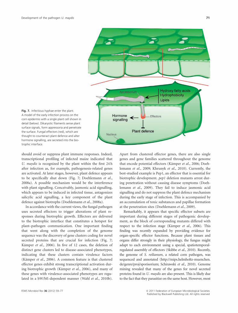
appressoria (Fig. 7). For example, corn lipids trigger fila-
mentous growth in liquid culture (Klose et al., 2004).
Moreover, hydrophobicity acts as an inducer of filamenta-
tion and appressorium differentiation on artificial surfaces
and the latter is further enhanced by the addition of hydroxy
fatty acids. Because both signals are present on the plant
surface, they are most likely also operational under natural
conditions (Fig. 7; Mendoza-Mendoza et al., 2009a).
Two signalling components, the tetraspan membrane
protein Sho1 and its interaction partner the single trans-
membrane mucin Msb2, appear to be involved in sensing
the hydrophobic signal. Accordingly, the deletion of the
corresponding genes strongly reduces appressoria forma-
tion. Sho1 interacts specifically with MAPK Kpp6, suggest-
ing that Sho1/Msb2 operate upstream of a Kpp6-containing
MAPK module during early infection (Lanver et al., 2010).
The perception of host signals is not the only crucial event
that is a prerequisite for the development of infection
structures on the plant surface. Additional processes involve
actin-mediated functions (Berndt et al., 2010) as well as
components of the N- and O-glycosylation pathways (Schir-
awski et al., 2005a; Fernandez-Alvarez et al., 2010). Unfortu-
nately, insights into the underlying mechanisms that
influence early infection are currently missing.
Once biotrophic fungi enter the plant, they depend on
living material for growth and nutrition, so that many
biotrophs form specialized feeding structures termed haus-
toria (Mendgen & Hahn, 2002). During plant invasion of U.
maydis fungal hyphae invaginate the plasma membrane of
infected host cells, leading to the formation of a
biotrophic interface without extensive feeding structures.
Instead, hyphae orient their growth towards vascular bun-
dles, where they might feed on transported sugars (Doehle-
mann et al., 2008a; Brefort et al., 2009). In line with this, a
novel sucrose transporter, Srt1, was recently described as a
major component ensuring fungal nutrition (Fig. 7; Wahl
et al., 2010a). Interestingly, this transporter has a higher
substrate affinity than a comparable plant transporter,
suggesting a redirection of sugar flow towards the parasite
(Wahl et al., 2010a).
The invasion and proliferation of pathogens within a
plant are often accompanied by host recognition, usually
triggering defence. Thus, for survival, a successful biotroph
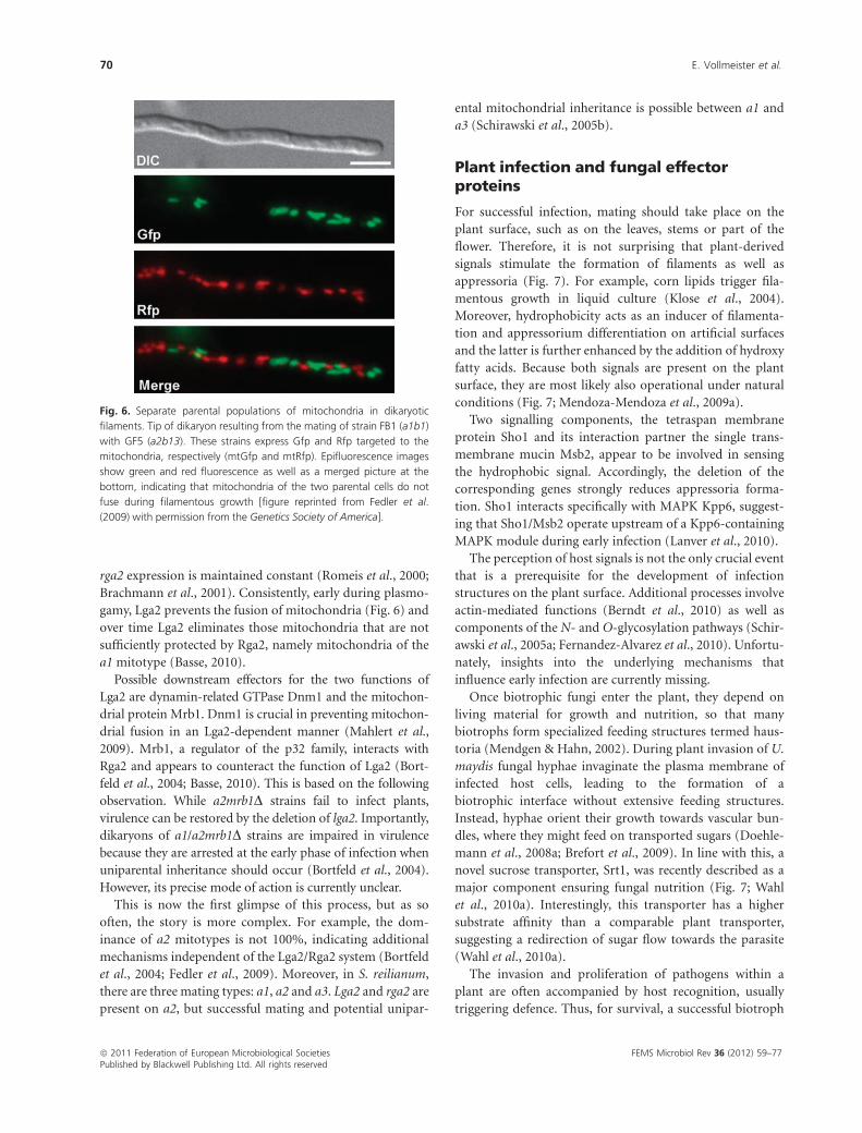
Fig. 6. Separate parental populations of mitochondria in dikaryotic
filaments. Tip of dikaryon resulting from the mating of strain FB1 (a1b1)
with GF5 (a2b13). These strains express Gfp and Rfp targeted to the
mitochondria, respectively (mtGfp and mtRfp). Epifluorescence images
show green and red fluorescence as well as a merged picture at the
bottom, indicating that mitochondria of the two parental cells do not
fuse during filamentous growth [figure reprinted from Fedler et al.
(2009) with permission from the Genetics Society of America].
ª 2011 Federation of European Microbiological Societies FEMS Microbiol Rev 36 (2012) 59–77Published by Blackwell Publishing Ltd. All rights reserved
70 E. Vollmeister et al.
should avoid or suppress plant immune responses. Indeed,
transcriptional profiling of infected maize indicated that
U. maydis is recognized by the plant within the first 24 h
after infection as, for example, pathogenesis-related genes
are activated. At later stages, however, plant defence appears
to be specifically shut down (Fig. 7; Doehlemann et al.,
2008a). A possible mechanism would be the interference
with plant signalling. Conceivably, jasmonic acid signalling,
which appears to be induced in infected tissue, antagonizes
salicylic acid signalling, a key component of the plant
defence against biotrophs (Doehlemann et al., 2008a).
In accordance with the current views, the fungal pathogen
uses secreted effectors to trigger alterations of plant re-
sponses during biotrophic growth. Effectors are delivered
to the biotrophic interface that constitutes a hotspot for
plant–pathogen communication. One important finding
that went along with the completion of the genome
sequence was the discovery of gene clusters coding for novel
secreted proteins that are crucial for infection (Fig. 7;
Kamper et al., 2006). In five of 12 cases, the deletion of
distinct gene clusters led to disease-associated phenotypes,
indicating that these clusters contain virulence factors
(Kamper et al., 2006). A common feature is that clustered
effector genes exhibit strong transcriptional activation dur-
ing biotrophic growth (Kamper et al., 2006), and many of
these genes with virulence-associated phenotypes are regu-
lated in a bW/bE-dependent manner (Wahl et al., 2010b).
Apart from clustered effector genes, there are also single
genes and gene families scattered throughout the genome
that encode potential effectors (Kamper et al., 2006; Doeh-
lemann et al., 2009; Khrunyk et al., 2010). Currently, the
best-studied example is Pep1, an effector that is essential for
biotrophic development. pep1 deletion mutants arrest dur-
ing penetration without causing disease symptoms (Doeh-
lemann et al., 2009). They fail to induce jasmonic acid
signalling and do not suppress the plant defence mechanism
during the early stage of infection. This is accompanied by
an accumulation of toxic substances and papillae formation
at the penetration sites (Doehlemann et al., 2009).
Remarkably, it appears that specific effector subsets are
important during different stages of pathogenic develop-
ment, as the block of corresponding mutants differed with
respect to the infection stage (Kamper et al., 2006). This
finding was recently expanded by providing evidence for
organ-specific effector functions. Because plant tissues and
organs differ strongly in their physiology, the fungus might
adapt to each environment using a special, spatiotemporal-
regulated assembly of effectors (Skibbe et al., 2010). Recently,
the genome of S. reilianum, a related corn pathogen, was
sequenced and annotated (http://mips.helmholtz-muenchen.
de/genre/proj/sporisorium; Schirawski et al., 2010). Genome
mining revealed that many of the genes for novel secreted
proteins found in U. maydis are also present. This is likely due
to the fact that they parasitize on the same host. However, most
Fig. 7. Infectious hyphae enter the plant.
A model of the early infection process on the
corn epidermis with a single plant cell shown in
detail (below). Dikaryotic filaments sense plant
surface signals, form appressoria and penetrate
the surface. Fungal effectors (red), which are
thought to counteract plant defence and alter
hormone signalling, are secreted into the bio-
trophic interface.
FEMS Microbiol Rev 36 (2012) 59–77 ª 2011 Federation of European Microbiological SocietiesPublished by Blackwell Publishing Ltd. All rights reserved
Development of the pathogen U. maydis 71
shared effectors are highly divergent, which may be attributed
to the differing infection strategies of U. maydis and S.
reilianum (Schirawski et al., 2010). In essence, during develop-
ment in planta U. maydis relies on numerous, thoroughly
regulated effectors to induce the host to foster its development.
Conclusions
Our tour following the fungal development of U. maydis
revealed quite a number of remarkable recent findings.
Highlights are for example (1) the endosomal transport of
signalling components during cytokinesis (Schink & Bolker,
2009); (2) the discovery of Rbf1 and the underlying exten-
sive regulation at the transcriptional and post-transcrip-
tional level (Heimel et al., 2010a, b); (3) uniparental
mitochondrial inheritance (Fedler et al., 2009); (4) micro-
tubule-dependent mRNA transport (Konig et al., 2009); and
(5) fungal effectors during infection (Kamper et al., 2006;
Brefort et al., 2009).
In the future, resolving the underlying molecular me-
chanisms will be essential in answering the following key
questions: how do endosomes function during membrane
recycling, signalling and microtubule-dependent transport?
How is the expression of transcription factors regulated at
the post-transcriptional level? What are the precise molecu-
lar functions of effectors?
The results obtained will further advance U. maydis as a
simple eukaryotic model for various aspects of basic cellular
functions. Within the last few years, a few scientists have
spearheaded distinct research areas covering several aspects
of cell biology and plant pathogenicity. Now appears to be
the right time for expansion.
Acknowledgements
We acknowledge lab members and Dr V. Gohre for critically
reading the manuscript. Our deepest gratitude is due to R.
Kellner, Dr H. Leal-Lara, Dr K.M. Sneetselar and Dr C. Basse
for images shown in Figs 1a, d, 2 and 6, respectively. Special
thanks are due to Drs G. Guerra-Sanchez and J. Pablo
Pardo-Vazquez for introducing M.F. to Quesadilla cuitla-
coche. We are grateful to Dr R. Kahmann and the Max
Planck Institute for Terrestrial Microbiology in Marburg for
their generous support. S.B. and T.P. are members of the
International Max-Planck Research School for Environmental,
Cellular and Molecular Microbiology, C.H. is a member of the
HHUD graduate school Molecules of Infection and J.S.
participates in the graduate school of the CLIB Graduate
Cluster Industrial Biotechnology. Our research was in part
financed by grants from the German Science Foundation
(DFG Fe448/3 and DFG FOR1334).
References
Alvarez-Tabares I & Perez-Martin J (2010) Septins from the
phytopathogenic fungus Ustilago maydis are required for
proper morphogenesis but dispensable for virulence. PLoS
One 5: e12933.
Ashwin P, Lin C & Steinberg G (2010) Queueing induced by
bidirectional motor motion near the end of a microtubule.
Phys Rev E Stat Nonlin Soft Matter Phys 82: 051907.
Bahn YS, Xue C, Idnurm A, Rutherford JC, Heitman J &
Cardenas ME (2007) Sensing the environment: lessons from
fungi. Nat Rev Microbiol 5: 57–69.
Banuett F (1995) Genetics of Ustilago maydis, a fungal pathogen
that induces tumors in maize. Annu Rev Genet 29: 179–208.
Banuett F & Herskowitz I (2002) Bud morphogenesis and the
actin and microtubule cytoskeletons during budding in the
corn smut fungus, Ustilago maydis. Fungal Genet Biol 37:
149–170.
Barnes CW, Szabo LJ, May G & Groth JV (2004) Inbreeding levels
of two Ustilago maydis populations. Mycologia 96: 1236–1244.
Bartnicki-Garcia S (2002) Hyphal tip growth: outstanding
questions. Molecular Biology of Fungal Development (Osiewacz
HD, ed), pp. 29–58. Dekker (Marcel), New York.
Bartnicki-Garcia S (2006) Chitosomes: past, present and future.
FEMS Yeast Res 6: 957–965.
Basse CW (2010) Mitochondrial inheritance in fungi. Curr Opin
Microbiol 13: 712–719.
Becht P, Vollmeister E & Feldbrugge M (2005) Role for RNA-
binding proteins implicated in pathogenic development of
Ustilago maydis. Eukaryot Cell 4: 121–133.
Becht P, Konig J & Feldbrugge M (2006) The RNA-binding
protein Rrm4 is essential for polarity in Ustilago maydis and
shuttles along microtubules. J Cell Sci 119: 4964–4973.
Berndt P, Lanver D & Kahmann R (2010) The AGC Ser/Thr
kinase Aga1 is essential for appressorium formation and
maintenance of the actin cytoskeleton in the smut fungus
Ustilago maydis. Mol Microbiol 78: 1484–1499.
Bohl F, Kruse C, Frank A, Ferring D & Jansen RP (2000) She2p, a
novel RNA-binding protein tethers ASH1 mRNA to the
Myo4p myosin motor via She3p. EMBO J 19: 5514–5524.
Bohmer C, Bohmer M, Bolker M & Sandrock B (2008) Cdc42 and
the Ste20-like kinase Don3 act independently in triggering
cytokinesis in Ustilago maydis. J Cell Sci 121: 143–148.
Bohmer C, Ripp C & Bolker M (2009) The germinal centre kinase
Don3 triggers the dynamic rearrangement of higher-order
septin structures during cytokinesis in Ustilago maydis. Mol
Microbiol 74: 1484–1496.
Bohmer M, Colby T, Bohmer C, Brautigam A, Schmidt J & Bolker
M (2007) Proteomic analysis of dimorphic transition in the
phytopathogenic fungus Ustilago maydis. Proteomics 7:
675–685.
Bolker M (2001) Ustilago maydis – a valuable model system for
the study of fungal dimorphism and virulence. Microbiology
147: 1395–1401.
ª 2011 Federation of European Microbiological Societies FEMS Microbiol Rev 36 (2012) 59–77Published by Blackwell Publishing Ltd. All rights reserved
72 E. Vollmeister et al.
Bolker M, Urban M & Kahmann R (1992) The a mating type
locus of U. maydis specifies cell signaling components. Cell 68:
441–450.
Bolker M, Genin S, Lehmler C & Kahmann R (1995) Genetic
regulation of mating, and dimorphism in Ustilago maydis. Can
J Bot 73: 320–325.
Bortfeld M, Auffarth K, Kahmann R & Basse CW (2004) The
Ustilago maydis a2 mating-type locus genes lga2 and rga2
compromise pathogenicity in the absence of the
mitochondrial p32 family protein Mrb1. Plant Cell 16:
2233–2248.
Brachmann A, Weinzierl G, Kamper J & Kahmann R (2001)
Identification of genes in the bW/bE regulatory cascade in
Ustilago maydis. Mol Microbiol 42: 1047–1063.
Brachmann A, Schirawski J, Muller P & Kahmann R (2003) An
unusual MAP kinase is required for efficient penetration of the
plant surface by Ustilago maydis. EMBO J 22: 2199–2210.
Brachmann A, Konig J, Julius C & Feldbrugge M (2004) A reverse
genetic approach for generating gene replacement mutants in
Ustilago maydis. Mol Genet Genomics 272: 216–226.
Brefort T, Muller P & Kahmann R (2005) The high-mobility-
group domain transcription factor Rop1 is a direct regulator of
prf1 in Ustilago maydis. Eukaryot Cell 4: 379–391.
Brefort T, Doehlemann G, Mendoza-Mendoza A, Reissmann S,
Djamei A & Kahmann R (2009) Ustilago maydis as a pathogen.
Annu Rev Phytopathol 47: 423–445.
Canovas D & Perez-Martin J (2009) Sphingolipid biosynthesis is
required for polar growth in the dimorphic phytopathogen
Ustilago maydis. Fungal Genet Biol 46: 190–200.
Carbo N & Perez-Martin J (2008) Spa2 is required for
morphogenesis but it is dispensable for pathogenicity in the
phytopathogenic fungus Ustilago maydis. Fungal Genet Biol 45:
1315–1327.
Dahm R, Kiebler M & Macchi P (2007) RNA localisation in the
nervous system. Semin Cell Dev Biol 18: 216–223.
Davey J (1998) Fusion of a fission yeast. Yeast 14: 1529–1566.
Di Stasio M, Brefort T, Mendoza-Mendoza A, Munch K &
Kahmann R (2009) The dual specificity phosphatase Rok1
negatively regulates mating and pathogenicity in Ustilago
maydis. Mol Microbiol 73: 73–88.
Doehlemann G, Wahl R, Horst RJ et al. (2008a) Reprogramming
a maize plant: transcriptional and metabolic changes induced
by the fungal biotroph Ustilago maydis. Plant J 56: 181–195.
Doehlemann G, Wahl R, Vranes M, de Vries RP, Kamper J &
Kahmann R (2008b) Establishment of compatibility in the
Ustilago maydis/maize pathosystem. J Plant Physiol 165: 29–40.
Doehlemann G, van der Linde K, Assmann D, Schwammbach D,
Hof A, Mohanty A, Jackson D & Kahmann R (2009) Pep1, a
secreted effector protein of Ustilago maydis, is required for
successful invasion of plant cells. PLoS Pathog 5: e1000290.
Dreyfuss G, Kim VN & Kataoka N (2002) Messenger-RNA-
binding proteins and the messages they carry. Nat Rev Mol Cell
Bio 3: 195–205.
Du TG, Schmid M & Jansen RP (2007) Why cells move messages:
the biological functions of mRNA localization. Semin Cell Dev
Biol 18: 171–177.
Elion EA (2000) Pheromone response, mating and cell biology.
Curr Opin Microbiol 3: 573–581.
Elson SL, Noble SM, Solis NV, Filler SG & Johnson AD (2009) An
RNA transport system in Candida albicans regulates hyphal
morphology and invasive growth. PLoS Genet 5: e1000664.
Fedler M, Luh KS, Stelter K, Nieto-Jacobo F & Basse CW (2009)
The a2 mating-type locus genes lga2 and rga2 direct
uniparental mitochondrial DNA (mtDNA) inheritance and
constrain mtDNA recombination during sexual development
of Ustilago maydis. Genetics 181: 847–860.
Feldbrugge M, Bolker M, Steinberg G, Kamper J & Kahmann R
(2006) Regulatory and structural netwoks orchestrating
mating, dimorphism, cell shape, and pathogenesis in Ustilago
maydis. The Mycota I (Kues U & Fischer R, eds), pp. 375–391.
Springer-Verlag, Berlin.
Feldbrugge M, Zarnack K, Vollmeister E, Baumann S, Koepke J,
Konig J, Munsterkotter M & Mannhaupt G (2008) The
posttranscriptional machinery of Ustilago maydis. Fungal
Genet Biol 45: S40–S46.
Fernandez-Alvarez A, Elias-Villalobos A & Ibeas JI (2010) The
requirement for protein O-mannosylation for Ustilago maydis
virulence seems to be linked to intrinsic aspects of the
infection process rather than an altered plant response. Plant
Signal Behav 5: 412–414.
Fink G & Steinberg G (2006) Dynein-dependent motility of
microtubules and nucleation sites supports polarization of the
tubulin array in the fungus Ustilago maydis. Mol Biol Cell 17:
3242–3253.
Fink G, Schuchardt I, Colombelli J, Stelzer E & Steinberg G
(2006) Dynein-mediated pulling forces drive rapid mitotic
spindle elongation in Ustilago maydis. EMBO J 25: 4897–4908.
Fischer R, Zekert N & Takeshita N (2008) Polarized growth in
fungi – interplay between the cytoskeleton, positional markers
and membrane domains. Mol Microbiol 68: 813–826.
Flor-Parra I, Vranes M, Kamper J & Perez-Martin J (2006) Biz1, a
zinc finger protein required for plant invasion by Ustilago
maydis, regulates the levels of a mitotic cyclin. Plant Cell 18:
2369–2387.
Fuchs U, Manns I & Steinberg G (2005) Microtubules are
dispensable for the initial pathogenic development but
required for long-distance hyphal growth in the corn smut
fungus Ustilago maydis. Mol Biol Cell 16: 2746–2758.
Fuchs U, Hause G, Schuchardt I & Steinberg G (2006)
Endocytosis is essential for pathogenic development in the
corn smut fungus Ustilago maydis. Plant Cell 18: 2066–2081.
Garcia-Muse T, Steinberg G & Perez-Martın J (2003)
Pheromone-induced G2 arrest in the phytopathogenic fungus
Ustilago maydis. Eukaryot Cell 2: 494–500.
Garcia-Muse T, Steinberg G & Perez-Martın J (2004)
Characterization of B-type cyclins in the smut fungus Ustilago
maydis: roles in morphogenesis and pathogenicity. J Cell Sci
117: 487–506.
FEMS Microbiol Rev 36 (2012) 59–77 ª 2011 Federation of European Microbiological SocietiesPublished by Blackwell Publishing Ltd. All rights reserved
Development of the pathogen U. maydis 73
Garcia-Pedrajas MD, Nadal M, Kapa LB, Perlin MH, Andrews DL
& Gold SE (2008) DelsGate, a robust and rapid gene deletion
construction method. Fungal Genet Biol 45: 379–388.
Garrido E, Voß U, Muller P, Kahmann R & Perez-Martın J (2004)
The induction of sexual development and virulence in the
smut fungus Ustilago maydis on Crk1, a novel MAPK protein.
Gene Dev 18: 3117–3130.
Gierz G & Bartnicki-Garcia S (2001) A three-dimensional model
of fungal morphogenesis based on the vesicle supply center
concept. J Theor Biol 208: 151–164.
Gillissen B, Bergemann J, Sandmann C, Schroeer B, Bolker M &
Kahmann R (1992) A two-component regulatory system for
self/non-self recognition in Ustilago maydis. Cell 68: 647–657.
Gould GW & Lippincott-Schwartz J (2009) New roles for
endosomes: from vesicular carriers to multi-purpose
platforms. Nat Rev Mol Cell Bio 10: 287–292.
Harris SD (2006) Cell polarity in filamentous fungi: shaping the
mold. Int Rev Cytol 251: 41–77.
Harris SD, Read ND, Roberson RW, Shaw B, Seiler S, Plamann M
& Momany M (2005) Polarisome meets Spitzenkorper:
microscopy, genetics, and genomics converge. Eukaryot Cell 4:
225–229.
Hartmann HA, Kahmann R & Bolker M (1996) The pheromone
response factor coordinates filamentous growth and
pathogenicity in Ustilago maydis. EMBO J 15: 1632–1641.
Hartmann HA, Kruger J, Lottspeich F & Kahmann R (1999)
Environmental signals controlling sexual development of the
corn smut fungus Ustilago maydis through the transcriptional
regulator Prf1. Plant Cell 11: 1293–1306.
Hay A & Tsiantis M (2010) KNOX genes: versatile regulators of
plant development and diversity. Development 137:
3153–3165.
Heimel K, Scherer M, Schuler D & Kamper J (2010a) The Ustilago
maydis Clp1 protein orchestrates pheromone and b-dependent
signaling pathways to coordinate the cell cycle and pathogenic
development. Plant Cell 22: 2908–2922.
Heimel K, Scherer M, Vranes M, Wahl R, Pothiratana C, Schuler
D, Vincon V, Finkernagel F, Flor-Parra I & Kamper J (2010b)
The transcription factor Rbf1 is the master regulator for b-
mating type controlled pathogenic development in Ustilago
maydis. PLoS Pathog 6: e1001035.
Hlubek A, Schink KO, Mahlert M, Sandrock B & Bolker M (2008)
Selective activation by the guanine nucleotide exchange factor
Don1 is a main determinant of Cdc42 signalling specificity in
Ustilago maydis. Mol Microbiol 68: 615–623.
Holliday R (1964) A mechanism for gene conversion in fungi.
Genet Res 5: 282–304.
Holliday R (1974) Ustilago maydis. Handbook of Genetics (King
RC, ed), pp. 575–595. Plenum Press, New York.
Holliday R (2004) Early studies on recombination and DNA
repair in Ustilago maydis. DNA Repair 3: 671–682.
Holt CE & Bullock SL (2009) Subcellular mRNA localization in
animal cells and why it matters. Science 326: 1212–1216.
Jansen RP (2001) mRNA localization: message on the move. Nat
Rev Mol Cell Bio 2: 247–256.
Kaffarnik F, Muller P, Leibundgut M, Kahmann R & Feldbrugge
M (2003) PKA and MAPK phosphorylation of Prf1 allows
promoter discrimination in Ustilago maydis. EMBO J 22:
5817–5826.
Kahmann R & Kamper J (2004) Ustilago maydis: how its biology
relates to pathogenic development. New Phytol 164: 31–42.
Kamper J (2004) A PCR-based system for highly efficient
generation of gene replacement mutants in Ustilago maydis.
Mol Genet Genomics 271: 103–110.
Kamper J, Reichmann M, Romeis T, Bolker M & Kahmann R
(1995) Multiallelic recognition: nonself-dependent
dimerization of the bE and bW homeodomain proteins in
Ustilago maydis. Cell 81: 73–83.
Kamper J, Kahmann R, Bolker M et al. (2006) Insights from the
genome of the biotrophic fungal plant pathogen Ustilago
maydis. Nature 444: 97–101.
Khrunyk Y, Munch K, Schipper K, Lupas AN & Kahmann R
(2010) The use of FLP-mediated recombination for the
functional analysis of an effector gene family in the biotrophic
smut fungus Ustilago maydis. New Phytol 187: 957–968.
Klose J, de Sa MM & Kronstad JW (2004) Lipid-induced
filamentous growth in Ustilago maydis. Mol Microbiol 52:
823–835.
Kojic M, Kostrub CF, Buchman AR & Holloman WK (2002)
BRCA2 homolog required for proficiency in DNA repair,
recombination, and genome stability in Ustilago maydis. Mol
Cell 10: 683–691.
Kojic M, Zhou Q, Fan J & Holloman WK (2011) Mutational
analysis of Brh2 reveals requirements for compensating
mediator functions. Mol Microbiol 79: 180–191.
Konig J, Julius C, Baumann S, Homann M, Goringer HU &
Feldbrugge M (2007) Combining SELEX and yeast three-
hybrid system for in vivo selection and classification of RNA
aptamers. RNA 13: 614–622.
Konig J, Baumann S, Koepke J, Pohlmann T, Zarnack K &
Feldbrugge M (2009) The fungal RNA-binding protein Rrm4
mediates long-distance transport of ubi1 and rho3 mRNAs.
EMBO J 28: 1855–1866.
Konig J, Zarnack K, Rot G, Curk T, Kayikci M, Zupan B, Turner
DJ, Luscombe NM & Ule J (2010) iCLIP reveals the function of
hnRNP particles in splicing at individual nucleotide
resolution. Nat Struct Mol Biol 17: 909–915.
Kozlov G, Menade M, Rosenauer A, Nguyen L & Gehring K
(2010) Molecular determinants of PAM2 recognition by the
MLLE domain of poly(A)-binding protein. J Mol Biol 397:
397–407.
Kronstad JW (2008) Mining the genome of the biotrophic fungal
pathogen Ustilago maydis. Fungal Genet Biol 45 (suppl 1):
S1–S2.
Kronstad JW & Leong SA (1990) The b mating-type locus of
Ustilago maydis contains variable and constant regions. Gene
Dev 4: 1384–1395.
Kruzel EK & Hull CM (2010) Establishing an unusual cell type:
how to make a dikaryon. Curr Opin Microbiol 13: 706–711.
ª 2011 Federation of European Microbiological Societies FEMS Microbiol Rev 36 (2012) 59–77Published by Blackwell Publishing Ltd. All rights reserved
74 E. Vollmeister et al.
Laity C, Giasson L, Campbell R & Kronstad J (1995)
Heterozygosity at the b mating-type locus attenuates fusion in
Ustilago maydis. Curr Genet 27: 451–459.
Lanver D, Mendoza-Mendoza A, Brachmann A & Kahmann R
(2010) Sho1 and Msb2-related proteins regulate appressorium
development in the smut fungus Ustilago maydis. Plant Cell 22:
2085–2101.
Lawrence CJ, Dawe RK, Christie KR et al. (2004) A standardized
kinesin nomenclature. J Cell Biol 167: 19–22.
Lehmler C, Steinberg G, Snetselaar KM, Schliwa M, Kahmann R
& Bolker M (1997) Identification of a motor protein required
for filamentous growth in Ustilago maydis. EMBO J 16:
3464–3473.
Lenz JH, Schuchardt I, Straube A & Steinberg G (2006) A dynein
loading zone for retrograde endosome motility at microtubule
plus-ends. EMBO J 25: 2275–2286.
Llorente B & Modesti M (2009) Fungal BRCA2 ortholog Brh2
brings 50 end strand invasion back on stage. Mol Cell 36:
539–540.
Mahlert M, Leveleki L, Hlubek A, Sandrock B & Bolker M (2006)
Rac1 and Cdc42 regulate hyphal growth and cytokinesis in the
dimorphic fungus Ustilago maydis. Mol Microbiol 59: 567–578.
Mahlert M, Vogler C, Stelter K, Hause G & Basse CW (2009) The
a2 mating-type-locus gene lga2 of Ustilago maydis interferes
with mitochondrial dynamics and fusion, partially in
dependence on a Dnm1-like fission component. J Cell Sci 122:
2402–2412.
Mazloum N & Holloman WK (2009a) Brh2 promotes a template-
switching reaction enabling recombinational bypass of lesions
during DNA synthesis. Mol Cell 36: 620–630.
Mazloum N & Holloman WK (2009b) Second-end capture in
DNA double-strand break repair promoted by Brh2 protein of
Ustilago maydis. Mol Cell 33: 160–170.
Mendgen K & Hahn M (2002) Plant infection and the
establishment of fungal biotrophy. Trends Plant Sci 7: 352–356.
Mendoza-Mendoza A, Berndt P, Djamei A, Weise C, Linne U,
Marahiel M, Vranes M, Kamper J & Kahmann R (2009a)
Physical–chemical plant-derived signals induce differentiation
in Ustilago maydis. Mol Microbiol 71: 895–911.
Mendoza-Mendoza A, Eskova A, Weise C, Czajkowski R &
Kahmann R (2009b) Hap2 regulates the pheromone response
transcription factor Prf1 in Ustilago maydis. Mol Microbiol 72:
683–698.
Muller M, Heym RG, Mayer A, Kramer K, Schmid M, Cramer P,
Urlaub H, Jansen RP & Niessing D (2011) A cytoplasmic
complex mediates specific mRNA recognition and localization
in yeast. PLoS Biol 9: e1000611.
Muller P, Aichinger C, Feldbrugge M & Kahmann R (1999) The
MAP kinase Kpp2 regulates mating and pathogenic
development in Ustilago maydis. Mol Microbiol 34:
1007–1017.
Muller P, Weinzierl G, Brachmann A, Feldbrugge M & Kahmann
R (2003) Mating and pathogenic development of the smut
fungus Ustilago maydis are regulated by one mitogen-activated
protein kinase cascade. Eukaryot Cell 2: 1187–1199.
Nadal M, Garcia-Pedrajas MD & Gold SE (2008) Dimorphism in
fungal plant pathogens. FEMS Microbiol Lett 284: 127–134.
Nevo-Dinur K, Nussbaum-Shochat A, Ben-Yehuda S & Amster-
Choder O (2011) Translation-independent localization of
mRNA in E. coli. Science 331: 1081–1084.
O’Donnell KL & McLaughlin DJ (1984) Postmeiotic mitosis,
basidiospore development, and septation in Ustilago maydis.
Mycologia 76: 486–502.
Partridge L & Hurst LD (1998) Sex and conflict. Science 281:
2003–2008.
Pearson JC, Lemons D & McGinnis W (2005) Modulating Hox
gene functions during animal body patterning. Nat Rev Genet
6: 893–904.
Perez-Martin J, Castillo-Lluva S, Sgarlata C, Flor-Parra I,
Mielnichuk N, Torreblanca J & Carbo N (2006) Pathocycles:
Ustilago maydis as a model to study the relationships between
cell cycle and virulence in pathogenic fungi. Mol Genet
Genomics 276: 211–229.
Riquelme M, Bartnicki-Garcıa S, Gonzalez-Prieto JM, Sanchez-
Leon E, Verdın-Ramos JA, Beltran-Aguilar A & Freitag M
(2007) Spitzenkorper localization and intracellular traffic of
green fluorescent protein-labeled CHS-3 and CHS-6 chitin
synthases in living hyphae of Neurospora crassa. Eukaryot Cell
6: 1853–1864.
Romeis T, Brachmann A, Kahmann R & Kamper J (2000)
Identification of a target gene for the bE–bW homeodomain
protein complex in Ustilago maydis. Mol Microbiol 37: 54–66.
Saito H (2010) Regulation of cross-talk in yeast MAPK signaling
pathways. Curr Opin Microbiol 13: 677–683.
Sandrock B, Bohmer C & Bolker M (2006) Dual function of the
germinal centre kinase Don3 during mitosis and cytokinesis in
Ustilago maydis. Mol Microbiol 62: 655–666.
Schauwecker F, Wanner G & Kahmann R (1995) Filament-
specific expression of a cellulase gene in the dimorphic fungus
Ustilago maydis. Biol Chem H-S 376: 617–625.
Scherer M, Heimel K, Starke V & Kamper J (2006) The Clp1
protein is required for clamp formation and pathogenic
development of Ustilago maydis. Plant Cell 18: 2388–2401.
Schink KO & Bolker M (2009) Coordination of cytokinesis and
cell separation by endosomal targeting of a Cdc42-specific
guanine nucleotide exchange factor in Ustilago maydis. Mol
Biol Cell 20: 1081–1088.
Schirawski J, Bohnert HU, Steinberg G, Snetselaar K, Adamikowa
L & Kahmann R (2005a) Endoplasmic reticulum glucosidase II
is required for pathogenicity of Ustilago maydis. Plant Cell 17:
3532–3543.
Schirawski J, Heinze B, Wagenknecht M & Kahmann R (2005b)
Mating type loci of Sporisorium reilianum: novel pattern
with three a and multiple b specificities. Eukaryot Cell 4:
1317–1327.
Schirawski J, Mannhaupt G, Munch K et al. (2010) Pathogenicity
determinants in smut fungi revealed by genome comparison.
Science 330: 1546–1548.
Schuchardt I, Assmann D, Thines E, Schuberth C & Steinberg G
(2005) Myosin-V, Kinesin-1, and Kinesin-3 cooperate in
FEMS Microbiol Rev 36 (2012) 59–77 ª 2011 Federation of European Microbiological SocietiesPublished by Blackwell Publishing Ltd. All rights reserved
Development of the pathogen U. maydis 75
hyphal growth of the fungus Ustilago maydis. Mol Biol Cell 16:
5191–5201.
Schulz B, Banuett F, Dahl M, Schlesinger R, Schafer W, Martin T,
Herskowitz I & Kahmann R (1990) The b alleles of U. maydis,
whose combinations program pathogenic development, code
for polypeptides containing a homeodomain-related motif.
Cell 60: 295–306.
Schuster M, Kilaru S, Ashwin P, Lin C, Severs NJ & Steinberg G
(2011a) Controlled and stochastic retention concentrates
dynein at microtubule ends to keep endosomes on track.
EMBO J 30: 652–664.
Schuster M, Lipowsky R, Assmann MA, Lenz P & Steinberg G
(2011b) Transient binding of dynein controls bidirectional
long-range motility of early endosomes. P Natl Acad Sci USA
109: 3618–3623.
Skibbe DS, Doehlemann G, Fernandes J & Walbot V (2010) Maize
tumors caused by Ustilago maydis require organ-specific genes
in host and pathogen. Science 328: 89–92.
Snetselaar KM & Mims CW (1993) Infection of maize stigmas by
Ustilago maydis: light and electron microscopy. Phytopathology
83: 843–850.
Snetselaar KM, Bolker M & Kahmann R (1996) Ustilago maydis
mating hyphae orient their growth toward pheromone
sources. Fungal Genet Biol 20: 299–312.
Spellig T, Bolker M, Lottspeich F, Frank RW & Kahmann R (1994)
Pheromones trigger filamentous growth in Ustilago maydis.
EMBO J 13: 1620–1627.
Steinberg G (2007a) Hyphal growth: a tale of motors, lipids, and
the Spitzenkorper. Eukaryot Cell 6: 351–360.
Steinberg G (2007b) On the move: endosomes in fungal growth
and pathogenicity. Nat Rev Microbiol 5: 309–316.
Steinberg G (2007c) Tracks for traffic: microtubules in the plant
pathogen Ustilago maydis. New Phytol 174: 721–733.
Steinberg G & Perez-Martin J (2008) Ustilago maydis, a new
fungal model system for cell biology. Trends Cell Biol 18:
61–67.
Steinberg G, Schliwa M, Lehmler C, Bolker M, Kahmann R &
McIntosh JR (1998) Kinesin from the plant pathogenic fungus
Ustilago maydis is involved in vacuole formation and
cytoplasmic migration. J Cell Sci 111: 2235–2246.
Steinberg G, Wedlich-Soldner R, Brill M & Schulz I (2001)
Microtubules in the fungal pathogen Ustilago maydis are
highly dynamic and determine cell polarity. J Cell Sci 114:
609–622.
St Johnston D (2005) Moving messages: the intracellular
localization of mRNAs. Nat Rev Mol Cell Bio 6: 363–375.
Straube A, Enard W, Berner A, Wedlich-Soldner R, Kahmann R &
Steinberg G (2001) A split motor domain in a cytoplasmic
dynein. EMBO J 20: 5091–5100.
Straube A, Brill M, Oakley BR, Horio T & Steinberg G (2003)
Microtubule organization requires cell cycle-dependent
nucleation at dispersed cytoplasmic sites: polar and
perinuclear microtubule organizing centers in the plant
pathogen Ustilago maydis. Mol Biol Cell 14: 642–657.
Straube A, Weber I & Steinberg G (2005) A novel mechanism of
nuclear envelope break-down in a fungus: nuclear migration
strips off the envelope. EMBO J 24: 1674–1685.
Sudbery P & Court H (2007) Polarised growth in fungi. The
Mycota VIII (Howard RJ & Gow NAR, eds), pp. 137–166.
Springer-Verlag, Berlin.
Szabo Z, Tonnis M, Kessler H & Feldbrugge M (2002) Structure–
function analysis of lipopeptide pheromones from the plant
pathogen Ustilago maydis. Mol Genet Genomics 268: 362–370.
Theisen U, Straube A & Steinberg G (2008) Dynamic
rearrangement of nucleoporins during fungal ‘open’ mitosis.
Mol Biol Cell 19: 1230–1240.
Thorslund T & West SC (2007) BRCA2: a universal recombinase
regulator. Oncogene 26: 7720–7730.
Thorslund T, McIlwraith MJ, Compton SA, Lekomtsev S,
Petronczki M, Griffith JD & West SC (2010) The breast cancer
tumor suppressor BRCA2 promotes the specific targeting of
RAD51 to single-stranded DNA. Nat Struct Mol Biol 17:
1263–1265.
Treitschke S, Doehlemann G, Schuster M & Steinberg G (2010)
The myosin motor domain of fungal chitin synthase V is
dispensable for vesicle motility but required for virulence
of the maize pathogen Ustilago maydis. Plant Cell 22:
2476–2494.
Urban M, Kahmann R & Bolker M (1996a) The biallelic a mating
type locus of Ustilago maydis: remnants of an additional
pheromone gene indicate evolution from a multiallelic
ancestor. Mol Gen Genet 250: 414–420.
Urban M, Kahmann R & Bolker M (1996b) Identification of the
pheromone response element in Ustilago maydis. Mol Gen
Genet 251: 31–37.
Vollmeister E & Feldbrugge M (2010) Posttranscriptional control
of growth and development in Ustilago maydis. Curr Opin
Microbiol 13: 693–699.
Vollmeister E, Haag C, Zarnack K, Baumann S, Konig J,
Mannhaupt G & Feldbrugge M (2009) Tandem KH domains
of Khd4 recognize AUACCC and are essential for regulation of
morphology as well as pathogenicity in Ustilago maydis. RNA
15: 2206–2218.
Wahl R, Wippel K, Goos S, Kamper J & Sauer N (2010a)
A novel high-affinity sucrose transporter is required for
virulence of the plant pathogen Ustilago maydis. PLoS Biol 8:
e1000303.
Wahl R, Zahiri A & Kamper J (2010b) The Ustilago maydis b
mating type locus controls hyphal proliferation and expression
of secreted virulence factors in planta. Mol Microbiol 75:
208–220.
Weber I, Gruber C & Steinberg G (2003) A class-V myosin
required for mating, hyphal growth, and pathogenicity in the
dimorphic plant pathogen Ustilago maydis. Plant Cell 15:
2826–2842.
Weber I, Assmann D, Thines E & Steinberg G (2006) Polar
localizing class V myosin chitin synthases are essential during
early plant infection in the plant pathogenic fungus Ustilago
maydis. Plant Cell 18: 225–242.
ª 2011 Federation of European Microbiological Societies FEMS Microbiol Rev 36 (2012) 59–77Published by Blackwell Publishing Ltd. All rights reserved
76 E. Vollmeister et al.
Wedlich-Soldner R, Bolker M, Kahmann R & Steinberg G (2000)
A putative endosomal t-SNARE links exo- and endocytosis in
the phytopathogenic fungus Ustilago maydis. EMBO J 19:
1974–1986.
Wedlich-Soldner R, Straube A, Friedrich MW & Steinberg G
(2002) A balance of KIF1A-like kinesin and dynein organizes
early endosomes in the fungus Ustilago maydis. EMBO J 21:
2946–2957.
Weinzierl G, Leveleki L, Hassel A, Kost G, Wanner G & Bolker M
(2002) Regulation of cell separation in the dimorphic fungus
Ustilago maydis. Mol Microbiol 45: 219–231.
Wosten HA, Bohlmann R, Eckerskorn C, Lottspeich F,
Bolker M & Kahmann R (1996) A novel class of small
amphipathic peptides affect aerial hyphal growth and
surface hydrophobicity in Ustilago maydis. EMBO J 15:
4274–4281.
Xu J (2005) The inheritance of organelle genes and genomes:
patterns and mechanisms. Genome 48: 951–958.
Yan Z, Sun S, Shahid M & Xu J (2007) Environment factors can
influence mitochondrial inheritance in the fungus
Cryptococcus neoformans. Fungal Genet Biol 44: 315–322.
Yang H, Li Q, Fan J, Holloman WK & Pavletich NP (2005) The
BRCA2 homologue Brh2 nucleates RAD51 filament formation
at a dsDNA–ssDNA junction. Nature 433: 653–657.
Zahiri A, Heimel K, Wahl R, Rath M & Kamper J (2010) The
Ustilago maydis forkhead transcription factor Fox1 is involved
in the regulation of genes required for the attenuation of plant
defenses during pathogenic development. Mol Plant Microbe
In 23: 1118–1129.
Zarnack K & Feldbrugge M (2007) mRNA trafficking in fungi.
Mol Genet Genomics 278: 347–359.
Zarnack K & Feldbrugge M (2010) Microtubule-dependent
mRNA transport in fungi. Eukaryot Cell 9: 982–990.
Zarnack K, Eichhorn H, Kahmann R & Feldbrugge M (2008)
Pheromone-regulated target genes respond differentially to
MAPK phosphorylation of transcription factor Prf1. Mol
Microbiol 69: 1041–1053.
Supporting Information
Additional Supporting Information may be found in the
online version of this article:
Movie S1. Rrm4-Gfp fusion protein moves bidirectionally
along cytoskeletal tracks in filaments of Ustilago maydis
strain AB33 (150-ms exposure time, 70 frames, five frames/
second display rate; quick time format, 9565 kb).
Movie S2. Disturbed movement of Rrm4-Gfp fusion pro-
tein after treatment with microtubule inhibitor benomyl in
filaments of Ustilago maydis strain AB33 (150-ms exposure
time, 70 frames, five frames/second display rate; quick time
format, 102 kb).
Please note: Wiley-Blackwell is not responsible for the
content or functionality of any supporting materials sup-
plied by the authors. Any queries (other than missing
material) should be directed to the corresponding author
for the article.
FEMS Microbiol Rev 36 (2012) 59–77 ª 2011 Federation of European Microbiological SocietiesPublished by Blackwell Publishing Ltd. All rights reserved
Development of the pathogen U. maydis 77
Recommended