Diabetic Foot Management
Yunita Puspa SariSurgery DivisionRSUD Cileungsi

Epidemiology
• Cellulitis occurs 9 times more frequently in diabetics than non-diabetics
• Osteomyelitis of the foot 12 times more frequently in diabetics than non-diabetics
• Foot ulcerations and infections are the most common reason for a diabetic to be admitted to the hospital

Epidemiology
• 25 % of diabetics will develop a foot ulcer• 40-80% of these ulcers will become infected• 25 % of these will become deep• 50 % of patients with cellulitis will have
another episode within 2 years

Epidemiology(of amputation)
• 25-50 % of diabetic foot infections lead to minor amputations
• 10-40 % require major amputations• 10-30 % of patients with a diabetic foot ulcer
will go on to amputation

Pathophysiology
• Metabolic derangement• Faulty wound healing• Neuropathy• Angiopathy• Mechanical stress• Patient and provider neglect


Poor Wound Healing
• Poor granuloma formation• Prolonged persistence of abscess • Higher rate of carriage of Staph Aureus in the
nares • Bullae, necrobiosis• Nail fungi (Tenia)

Poor Immune Function
• Poor PMN functions– Migration, phagocytosis, intracellular killing,
chemotaxis• Ketosis impairs leukocyte function• Monocyte mediated immune function diminished• Hyperglycemia impairs complement fixation

Sensory Neuropathy
• Unaware of a foreign body– Pressure in shoes– Abrasions in shoes– Tears or brakes in the skin

Motor Neuropathy
• Architectural deformities– Hammer or claw toe– High plantar arch– Subluxation of metatarsals

Autonomic Neuropathy
• Anhidrosis – Dry, cracked skin
• Arterial to venous shunting• Temperature regulation disorders

Angiopathy
• Can play a primary role– Microangiopathy +/-
• Certainly plays a primary role in healing– Pulsatile flow will augment healing
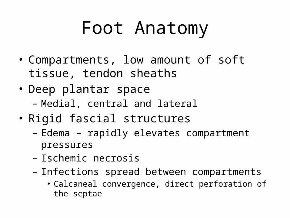
Foot Anatomy
• Compartments, low amount of soft tissue, tendon sheaths
• Deep plantar space– Medial, central and lateral
• Rigid fascial structures– Edema – rapidly elevates compartment pressures– Ischemic necrosis– Infections spread between compartments
• Calcaneal convergence, direct perforation of the septae

Diagnosis
• Clinical presentation– Presence of purulence– Pain, swelling, ulceration, sinus tract formation, crepitation– Systemic infection (fever, rigors, vomitting, tachycardia,
change in mental status, malaise)• Surprisingly uncommon
– Metabolic disorder (hyperglycemia, ketosis, azotemia)– Should be considered even when local signs are less
severe

Evaluation
• Describe lesion and signs of inflammation• Measure wound (? Photograph ?)– Define whether infection is present and cause– Examine soft tissue for crepitus, sinus tract, abscess
• Determine inflow• Neurologic status? Sensation, motor, autonomic • Plain radiographs osteomyelitis (cortical
erosions, periosteal reaction)

Surgical Intervention
• Surgical– “Salvage the foot but not at the expense of the leg
or the patient”– Early surgical debridement decreases LOS,
improves foot salvage and decreases morbidity and mortality
• Debridement• Remove all necrotic tissue and pus including eschar• Remove all callus• Debride bone

Treatment
• Plantar abscess– Foot edema– Central plantar infections – worse outcomes– Wide incision and drainage necessary

Treatment
• Empiric antibiotic therapy– Staph– Strep– GNR– Enterococcus– Anaerobes– *Tailor to clinical progress

Antibiotic thoughts
• Mild (po) – Augmentin/Levofloxacin (+Clinda)– Bactrim/Flagyl
• Moderate (IV until stable then po)– Unasyn or other Gorilla-cillin– Clinda & Levofloxacin
• Severe (IV only)– Imipenem– Amp/Tobra/Clinda– Vanco/Aztreonam/Flagyl

Antibiotic thoughts
• Duration of therapy– No good studies– Once active infection resolved plus 2 days– Osteomyelitis• 6 weeks• Can use Flouroquinolones and clindamycin

Prevention


THANK YOU
Recommended