Albert Leung, HMS IIIGillian Lieberman, M.D.
BIDMC Radiology ClerkshipFebruary 22, 2010

OverviewIndex PatientPeriosteal ReactionsDifferential DiagnosisPrinciples of Osteoid OsteomasBone AnatomyMenu of TestsRadiologic ImagesTreatments
http://peakrunningperformance.com/webpages/images/stories/skeleton.gif

Learning ObjectivesRecognize the clinical presentation of osteoid osteomaUnderstand the differential diagnosis for this clinical presentationReview the principles of osteoid osteoma and its classification within the context of bone anatomyLearn the menu of appropriate radiologic imaging studies and the indicationsStudy the radiologic presentations and recognize classic findingsKnow how to medically and surgically manage osteoidosteomas

Index Patient: Presenting HistoryChief Complaint
Right wrist painHistory
18 yr old R hand dominant male with persistent R wrist pain for 4 monthsPain is worse at night, waking patient from his sleepPain relief with ibuprofenNo history of trauma or repetitive motions with hand
Review of SystemsDenies numbness/tingling, fevers, chills, night sweats
Past, Family, Social HistoriesNon‐significant

Index Patient: Physical ExamPalpable nodule (3.5 x 3 cm) over the radial aspect of the R distal radius with focal tenderness and swellingNo erythema, discoloring, ecchymosis, or drainageFull range of motion of R shoulder, elbow, wrist, and fingers; no strength deficitsNormal sensation in all distributions+2 pulses bilaterallyNo palpable axillary lymph nodes

Our Patient: Radiograph of cortical osteoid osteoma
in R distal radius
Fusiform
cortical thickening
Ovoid sclerotic lesion with
central radiolucency1.1 x 1.3 cm
AP Radiograph
Image from BIDMC PACS

Our Patient: R distal radius osteoid osteoma
on CT Imaging
Focal areas of radiolucency
surrounded by sclerotic
regions that occupy the cortex and invade the medulla
Axial C‐
CT
Image from BIDMC PACS

Our Patient: Periosteal reaction on CT Imaging
Periosteal reaction: elevated cortex
from bony expansion
Image from BIDMC PACS
Coronal C- CT of R distal radius

Periosteal Reaction: Non‐aggressive Types
Rana RS, Wu JS, and Eisenberg RL. “Periosteal Reaction.” American Journal of Roentgenology. Oct 2009. 193(4): W259-272.

Periosteal Reaction: Aggressive Types
Rana RS, Wu JS, and Eisenberg RL. “Periosteal Reaction.” American Journal of Roentgenology. Oct 2009. 193(4): W259-272.

Partial Differential Diagnosis for Wrist Pain/Bone Mass
MassesBenign Neoplasms
Osteoid OsteomaOsteoblastomaOsteomaEnostosis (bone island)
Ganglion cystInfection
Brodie abscess (subacuteosteomyelitis)Osteomyelitis
InflammationTenosynovitisRheumatoid arthritis
Degenerative ConditionsOsteoarthritisStress fracture
NeurologicalCarpal tunnel syndrome

Our Patient’s Differential Diagnosis
Unlikely given patient’s history and presentation

Distinguishing Characteristics on Radiologic Imaging
MassesBenign Neoplasms
Osteoid Osteoma ‐ < 2 cm; radiolucent nidus with surrounding sclerosis; may spontaneously regressOsteoblastoma – large (> 2 cm); no regression in size over timeOsteoma – cold bone scan; absent periosteal reaction & radiolucent nidusEnostosis – cold bone scan; thorny radiations; low signal on T2w MRI
InfectionBrodie abscess – cortical destruction with a linear, serpentine tract extending away from abscess
Degenerative ConditionsStress fracture – linear radiolucency perpendicular, rather than parallel, to cortex

Background: Osteoid Osteoma
DemographicsMajority of patients are young (< 35 yr old)Males > females (2‐3:1)
Benign skeletal neoplasmConsists of a spherical nidus of osteoid tissue & bony trabeculae superimposed on highly vascularized connective tissueMay initially appear on radiograph as a small sclerotic bone island within a circular lucencyNO malignant potential
SizeRanges from 0.5‐2 cm (avg 1.5 cm)No growth progressionMay regress spontaneously over years

Background: Osteoid
Osteoma (continued)
MechanismUnknown etiologyNidus consists of highly vascular osteoblastic proliferation, surrounded by a secondary zone of sclerosisElevated prostaglandin E2 levels in nidus responsible for bone pain & vasodilationTumor infarction may be involved during cases of spontaneous regression
LocationsTypically affects the appendicular skeleton
Metaphysis/diaphysis of long bones: 70%Femur/tibia: 55%Phalanges of hands/feet: 20%Spine: 10% ‐ Causes painful scoliosis with concavity towards the lesionPosterior elements: 90%Extremely rare in skull & facial bones

Osteoid
osteoma
vs Osteoblastoma
Osteoid
osteoma OsteoblastomaUsually < 2 cm diameterPresents with intense pain, often sharply localized and worse at nightPain characteristically relieved by aspirin/NSAIDsNon‐aggressive behaviorVariable locations: femur, tibia, fibula, humerus, hands/feet, vertebraeNeural staining reveals axons throughout the tumor (may explain bone pain)
Usually > 2cm diameterLack of intense painAggressive behaviorTypically in the vertebrae or major bones of the lower extremityOften affects the spongiosaof the boneAbsence of neural axons upon staining

Review of Bone Anatomy
tsagalis.net/bones/anatomy.jpg
tsagalis.net/bones/anatomy.jpg
Classification of Osteoid
OsteomasCortical
Most common location (80%)Radiolucent nidus seen within the bone cortex and surrounded by fusiform cortical thickening/laminated periosteal formation
CancellousIntramedullary lesion; mild reactive sclerosis & difficult to identify, significantly delaying the diagnosisCommon sites: femoral neck, posterior spine, hands & feet
IntraarticularJoint effusion or synovitis
SubperiostealRound mass adjacent to cortexAbsent periosteal reactionVery rare

Gitelis
S, Wilkins R, and Conrad EU, III. “Benign Bone Tumors.”
Journal of Bone & Joint Surgery (Am). 1995; 77: 1756‐1782.
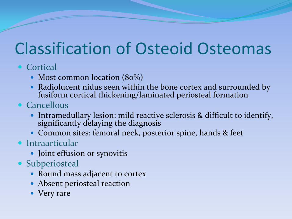
Osteoid Osteomas
Classic Clinical PresentationFocal bone pain that worsens at night & increases with activityPain is relieved by aspirin within 30 minutes (75% of cases)Local swelling and point tendernessExacerbation of pain with alcohol
http://library.thinkquest.org/08aug/01036/Imagini/Sleep.jpg
http://paddyk.files.wordpress.com/2009/11/5-aspirin.jpg

Menu of Radiologic TestsPlain Radiographs*Computed Tomography*MRIUltrasonographyNuclear Imaging*Angiography
* Usually used for evaluating osteoid
osteomas

Plain RadiographsMain imaging techniqueDiagnostic in 75% of casesClassic Appearance
Well‐defined radiolucent nidus with surrounding zone of sclerosisCentral nidus is typically < 1.5 cm in diameter
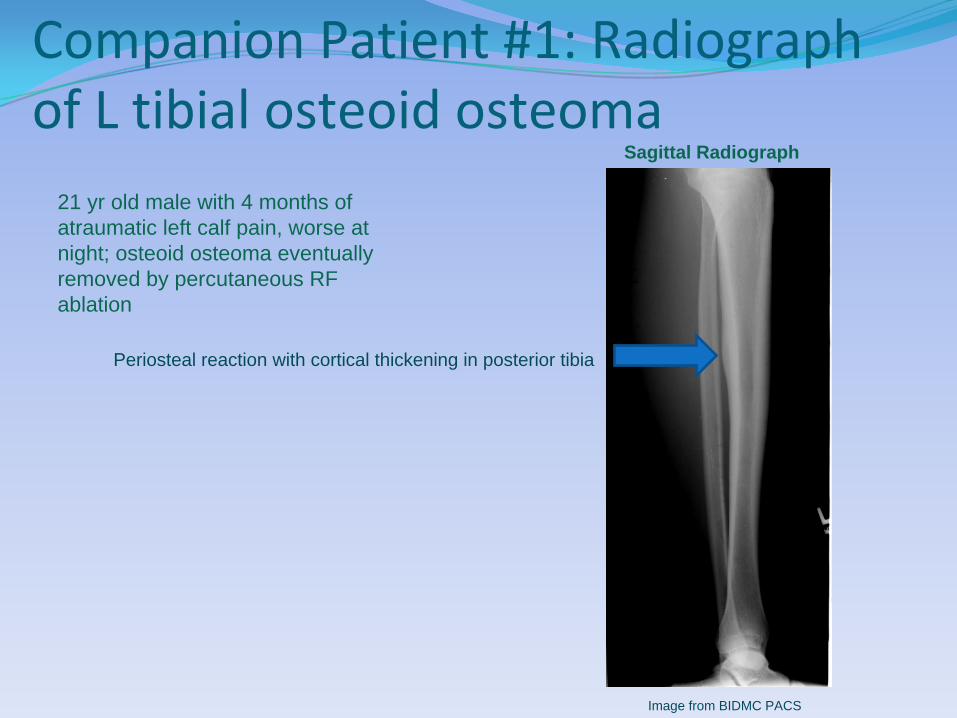
Companion Patient #1: Radiograph of L tibial
osteoid
osteoma
21 yr old male with 4 months of atraumatic left calf pain, worse at night; osteoid osteoma eventually removed by percutaneous RF ablation
Periosteal reaction with cortical thickening in posterior tibia
Sagittal Radiograph
Image from BIDMC PACS

CT ImagingBest imaging tool for osteoid osteomaStudy of choice for localizing the nidusGood at evaluating complex anatomy (e.g. – spinal pedicles, laminae, femoral neck, hands, feet)

Companion Patient #1: L tibial osteoid
osteoma
on CT Imaging
Sagittal C- CTAxial C- CT
Mature periosteal reaction and thickening with central radiolucency
Image from BIDMC PACS Image from BIDMC PACS

Back To Our Index Patient: R distal radius osteoid
osteoma
on CT
Central radiolucent nidus
within a sclerotic zone
Osteoid
osteoma
eventually removed by
open surgical curettage
Image from BIDMC PACS
Sagittal C- CT

MRIAdvantages
Easily detects edema in the soft tissues & bone marrowBetter at diagnosing cancellous/intramedullary osteoidosteomasGood for evaluating joint effusion/synovitis for intraarticular lesions
AppearanceT1w: nidus is isointense to muscleT2w: radiolucent areas of nidus with intermediate to high signal intensity

Companion Patient #2: Radiograph of L tibial
osteoid
osteoma
18 yr old male with nocturnal focal pain in L proximal tibia for 2 years; relief with NSAIDs; osteoid osteoma eventually removed by percutaneous RF ablation
Non-aggressive thick periosteal reaction of the L medial tibial metaphysis
AP Radiograph
Image from BIDMC PACS

Companion Patient #2: L tibial osteoid
osteoma
on Axial CT
5 mm radiolucent nidus
Thick periosteal reaction (10 mm)
Axial C- CT
Image from BIDMC PACS

Companion Patient #2: L tibial osteoid
osteoma
on Coronal CT
Oval-shaped radiolucent lesion within the medial tibial cortexDimensions: 4 x 4 x 18 mm
Coronal C- CT
Image from BIDMC PACS

Companion Patient #2: L tibial osteoid
osteoma
on MRI STIR
Edema appears as high signal intensity within the bone marrow
Coronal C- MRI STIR
Image from BIDMC PACS

Companion Patient #2: L tibial osteoid
osteoma
on MRI T1w
Axial C- MRI T1wCoronal C- MRI T1w
Low signal intensity edema in soft tissue structures surrounding the cortical lesion
Image from BIDMC PACS Image from BIDMC PACS

Nuclear Imaging: Bone ScanUses technetium‐99m phosphonatesTumor site demonstrates focal area of intense uptakeDouble density sign: small focus of increased activity (nidus) surrounded by a larger area of less intense activity (reactive sclerosis)Tracer is excreted through kidneys and urinary bladder
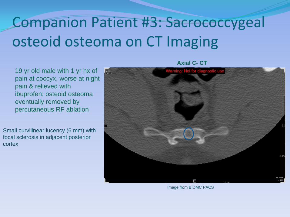
Companion Patient #3: Sacrococcygeal osteoid
osteoma
on CT Imaging
19 yr old male with 1 yr hx of pain at coccyx, worse at night pain & relieved with ibuprofen; osteoid osteoma eventually removed by percutaneous RF ablation
Small curvilinear lucency (6 mm) with focal sclerosis in adjacent posterior cortex
Image from BIDMC PACS
Axial C- CT

Companion Patient #3: CT guided biopsy of a sacrococcygeal
osteoid
osteoma
CT guided needle core biopsy of the radiolucent nidus
Image from BIDMC PACS
Axial C- CT

Companion Patient #3: Coronal bone scan of a sacrococcygeal
osteoid
osteoma
Small intense focus of tracer uptake in the coccyx with double density sign
Coronal Bone Scan
Image from BIDMC PACS

Companion Patient #3: Sagittal
bone scan of a sacrococcygeal
osteoid
osteoma
Abnormally increased focal area of intense tracer uptake
Image from BIDMC PACS
Sagittal Bone Scan

UltrasonographyMay be used for guidance of percutaneous biopsiesDoppler U/S detects the highly vascular nidusUseful for detecting intra‐articular osteoid osteomas

AngiographyCentral nidus is highly vascularIntense circumscribed blush that develops during the early arterial phase and persists into venous phase is diagnosticAngiography is useful for distinguishing osteoid osteomafrom a Brodie abscess

TreatmentsMedical Management
NSAIDSSurgical Management
Open surgical curettageCT guided
Percutaneous radio‐frequency (RF) ablation, laser, ethanol, or thermocoagulation therapy
AnesthesiaGeneral or spinal

SummaryOsteoid osteoma (OO) is a latent benign bone tumor that classically presents as focal pain that is worse at night and relieved by NSAIDsCortical osteoid osteomas are often associated with non‐aggressive periosteal reactionsOOs and osteoblastomas are histologically similar but differ in size, pain intensity, location, aggressiveness, and neural staining patternsOOs are classified by their relative position to the bone: cortical, cancellous, intraarticular, and subperiosteal

Summary ContinuedOOs have a classic appearance on plain radiographs and CT imaging: central radiolucent nidus with surrounding zone of sclerosisCT imaging is the best imaging tool for OOs and is the study of choice for localizing the nidusMRI is useful for detecting soft tissue/bone marrow edema and intramedullary OOsNuclear bone scans may localize the tumor and demonstrate focal areas of intense tracer uptake; double density sign is occasionally seenDoppler U/S can detect the highly vascular nidusAngiography may distinguish OOs from a Brodie abscess: intense blush that persists into venous phase is diagnostic of OO

ReferencesAssoun
J, Richardi
G, Railhac
JJ, et al. “Osteoid
osteoma: MR imaging versus CT.”
Radiology 1994; 191 (1): 217‐23.
Bilchik
T, Heyman
S, Siegel A, and Alavi
A. “Osteoid
osteoma: the role of radionuclide bone imaging, conventional
radiography and computed tomography in its management.”
The Journal of Nuclear Medicine. Feb 1992; 33(2): 269‐
271.
Davies AM and Wellings
RM. “Imaging of bone tumors.”
Current Opinion in Radiology. 1992; 4(6): 32‐38.
Dorfman
HD and Czerniak
B. Bone Tumors (1st
ed). St. Louis, MO, Mosby, 1998.
Gil S, Marco SF, Arenas J, et al. “Doppler duplex color localization of osteoid
osteomas.”
Skeletal Radiology. Feb 1999;
28(2): 107‐110.
Gitelis
S, Wilkins R, and Conrad EU, III. “Benign Bone Tumors.”
Journal of Bone & Joint Surgery (Am ed). 1995;77:1756‐
1782.
Greco F, Tamburrelli
F, Ciabattoni
G. “Prostaglandins in osteoid
osteoma.”
International Orthopedics. 1991; 15(1): 35‐37.
Greenspan A and Remagen
W. Differential Diagnosis of Tumors and Tumor‐like Lesions of Bones and Joints (1st
ed).
Philadelphia, PA, Lippencott
Williams & Wilkins, 1998.
Perkins AC and Hardy JG. “Intra‐operative nuclear medicine in surgical practice.”
Nuclear Medicine Communications.
Dec 1996; 17(12): 1006‐1015.
Rana
RS, Wu JS, and Eisenberg RL. “Periosteal Reaction.”
American Journal of Roentgenology. Oct 2009. 193(4):W259‐
272.

AcknowledgementsDr. Gillian Lieberman
Dr. Jim Wu
Dr. Erica Gupta
Dr. Jay Catena
Special Thanks To:Dr. Corrie Yablon
Dr. Aarti Sekhar
Maria Levantakis
Patients
Recommended