46 INFOTECH OULU Annual Report 2006
OPTOELECTRONICS AND MEASUREMENT UNIT (OPME)
Professor Risto Myllylä and Professor Markku Moilanen, Optoelectronics and Measurement Techniques
Laboratory, Department of Electrical and Information Engineering, University of Oulu
Dr. Jouni Tornberg and Research Professor Harri Kopola, VTT, Oulu
Juha Kalliokoski, Research Director of the Measurement and Sensor Laboratory (Kajaani),
University of Oulu
[email protected],[email protected],
[email protected], [email protected], [email protected]
http://www.infotech.oulu.fi/opme
Background and Mission
The Optoelectronics and Measurement Unit (OPME) com-
prises an intensive collaboration network of researchers at
the Optoelectronics and Measurement Techniques Labora-
tory at the University of Oulu, the Measurement and Sen-
sor Laboratory (MILA) in Kajaani and VTT. OPME
employs 37 postgraduate students and 41 other persons in
the research work. The group carries on high-level research
in optoelectronics and measurements techniques, wireless
instrumentation with particular emphasis on the practical
applications of these techniques, material techniques,
photonics integration and testing needed in advanced sen-
sors. A new technology field, printed intelligence, is inte-
grated to the Optoelectronics and Measurements Unit to
bring together professionals from all the above mentioned
fields into close collaboration to improve the know-how
and education possibilities in this area.
The practical research activities developed in the OPME
unit include, but are not limited to, measuring and model-
ing the propagation of light in turbulent and scattering me-
dia such as atmosphere, tissues, pulp and paper; electronics
testing techniques such as embedded testing and RF test-
ing; wireless sensors and sensor network for healthcare
applications; optical and micro- sensors produced with
MEMS technology, camera and machine vision in optical
measurement; advanced optical, ultrasonic, and microwave
techniques specially applying in wood processing; and print-
able manufacture techniques which integrate traditionally
optoelectronic devices to polymer platforms.
As a participant in the Infotech Oulu graduate school, the
OPME unit also arranges lecture series and graduate school
courses for graduate students. During 2006, the group or-
ganized the traditional 6th Infotech Oulu Workshop on Op-
toelectronic Devices and Instrumentation, concentrating this
time on ‘Printed electronics and optoelectronics’, In addi-
tion, six international lectures and courses were held in the
group. They were ‘The principles and applications of lin-
ear and nonlinear spectroscopy’ ‘Surface plasmon reso-
nance’, ‘Wireless sensor network technology for ubiquitous
healthcare applications’, ‘OCT system analysis and Monte
Carlo simulation’, ‘Laser diagnostics of highly scattering
media: evolution of optical properties’ and ‘Femtosecond
pulse propagation in scattering media’.
In 2006, there were 21 new projects opened in the OPME
unit, taking the total project number up to 42. The group
published 31 journal articles, 35 international conference
papers and 7 other scientific papers. Research collabora-
tion has been active with 16 domestic partners located in
Finnish universities and VTT, and 20 international partners
coming from universities and institutes in the USA, Canada,
Japan, Korea, China, Russia, the UK, Ireland, Poland, Ger-
many, and Sweden. The international visits for research
activities involved 15 foreign visits to the group and 11
research visits from the group aboard.
The Academy of Finland granted the OPME unit (together
with EMPART group) FiDiPro (Finland Distinguished Pro-
fessor Program) funding to hire professor Ghassan Jabbour
from Arizona State University, USA, to build up “The cen-
ter for printing of nano-to-mega photonics, electronics, and
bioinformatics”.
Scientific Progress
The following research examples illustrate the scientific
progress of the OPME unit. These research projects have
been carried out in active collaboration between both do-
mestic and international research institutions and industrial
partners.
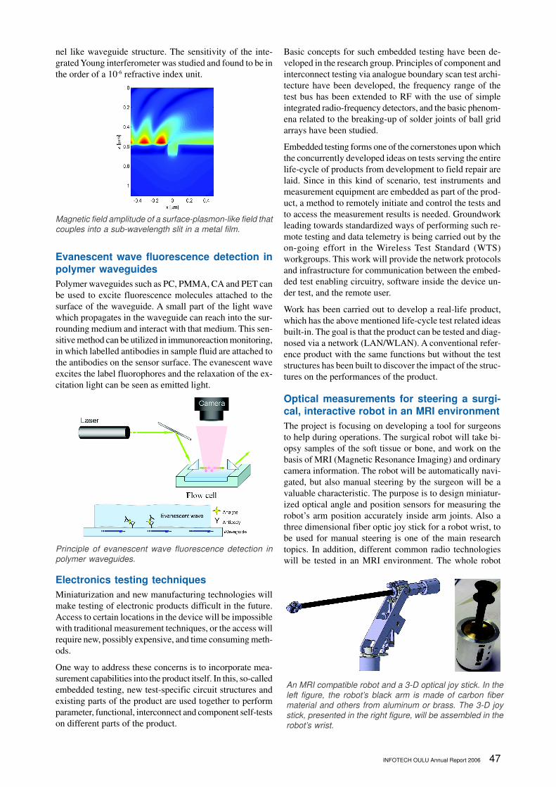
Modeling of light transmission through sub-
wavelength apertures
Modern optical technologies, such as near-field scanning
optical microscopy, optical lithography, heat-assisted mag-
netic recording and optical data storage, demand efficient
near-field light sources that provide light confinement well
beyond the fundamental diffraction limit. In theory, the fun-
damental diffraction limit can be exceeded - in a brute force
fashion - by spatially limiting the extent of an incident
wavefront by imposing a very small transmitting aperture
in the path of an incident light beam. This approach, how-
ever, suffers from the extremely small light transmission
efficiency through the aperture. The main objective of this
project is to develop new ways to enhance light transmis-
sion through sub-wavelength apertures. During 2006, we
have studied, via finite difference time domain (FDTD)
modeling, fundamental characteristics of a single circular
hole in a thin metal film, light transmission through a sub-
wavelength aperture filled with a high index dielectric, light
coupling into surface plasmons by circular grooves, and
coupling of surface plasmons into apertures. The waveguide
chip in this project was fabricated from siloxane material,
which can be processed by UV-lithography to have a chan-
INFOTECH OULU Annual Report 2006 47
nel like waveguide structure. The sensitivity of the inte-
grated Young interferometer was studied and found to be in
the order of a 10-6 refractive index unit.
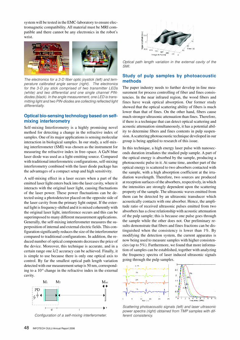
Evanescent wave fluorescence detection in
polymer waveguides
Polymer waveguides such as PC, PMMA, CA and PET can
be used to excite fluorescence molecules attached to the
surface of the waveguide. A small part of the light wave
which propagates in the waveguide can reach into the sur-
rounding medium and interact with that medium. This sen-
sitive method can be utilized in immunoreaction monitoring,
in which labelled antibodies in sample fluid are attached to
the antibodies on the sensor surface. The evanescent wave
excites the label fluorophores and the relaxation of the ex-
citation light can be seen as emitted light.
Electronics testing techniques
Miniaturization and new manufacturing technologies will
make testing of electronic products difficult in the future.
Access to certain locations in the device will be impossible
with traditional measurement techniques, or the access will
require new, possibly expensive, and time consuming meth-
ods.
One way to address these concerns is to incorporate mea-
surement capabilities into the product itself. In this, so-called
embedded testing, new test-specific circuit structures and
existing parts of the product are used together to perform
parameter, functional, interconnect and component self-tests
on different parts of the product.
Basic concepts for such embedded testing have been de-
veloped in the research group. Principles of component and
interconnect testing via analogue boundary scan test archi-
tecture have been developed, the frequency range of the
test bus has been extended to RF with the use of simple
integrated radio-frequency detectors, and the basic phenom-
ena related to the breaking-up of solder joints of ball grid
arrays have been studied.
Embedded testing forms one of the cornerstones upon which
the concurrently developed ideas on tests serving the entire
life-cycle of products from development to field repair are
laid. Since in this kind of scenario, test instruments and
measurement equipment are embedded as part of the prod-
uct, a method to remotely initiate and control the tests and
to access the measurement results is needed. Groundwork
leading towards standardized ways of performing such re-
mote testing and data telemetry is being carried out by the
on-going effort in the Wireless Test Standard (WTS)
workgroups. This work will provide the network protocols
and infrastructure for communication between the embed-
ded test enabling circuitry, software inside the device un-
der test, and the remote user.
Work has been carried out to develop a real-life product,
which has the above mentioned life-cycle test related ideas
built-in. The goal is that the product can be tested and diag-
nosed via a network (LAN/WLAN). A conventional refer-
ence product with the same functions but without the test
structures has been built to discover the impact of the struc-
tures on the performances of the product.
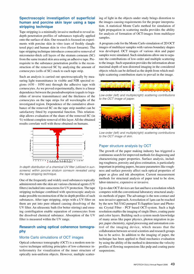
Optical measurements for steering a surgi-
cal, interactive robot in an MRI environment
The project is focusing on developing a tool for surgeons
to help during operations. The surgical robot will take bi-
opsy samples of the soft tissue or bone, and work on the
basis of MRI (Magnetic Resonance Imaging) and ordinary
camera information. The robot will be automatically navi-
gated, but also manual steering by the surgeon will be a
valuable characteristic. The purpose is to design miniatur-
ized optical angle and position sensors for measuring the
robot’s arm position accurately inside arm joints. Also a
three dimensional fiber optic joy stick for a robot wrist, to
be used for manual steering is one of the main research
topics. In addition, different common radio technologies
will be tested in an MRI environment. The whole robot
Magnetic field amplitude of a surface-plasmon-like field that
couples into a sub-wavelength slit in a metal film.
Principle of evanescent wave fluorescence detection in
polymer waveguides.
An MRI compatible robot and a 3-D optical joy stick. In the
left figure, the robot’s black arm is made of carbon fiber
material and others from aluminum or brass. The 3-D joy
stick, presented in the right figure, will be assembled in the
robot’s wrist.
48 INFOTECH OULU Annual Report 2006
system will be tested in the EMC-laboratory to ensure elec-
tromagnetic compatibility. All material must be MRI com-
patible and there cannot be any electronics in the robot’s
wrist.
Optical bio-sensing technology based on self-
mixing interferometry
Self-mixing Interferometry is a highly promising novel
method for detecting a change in the refractive index of
samples. One of its major applications is sensing molecular
interaction in biological samples. In our study, a self-mix-
ing interferometer (SMI) was chosen as the instrument for
measuring the refractive index in free–space. A GaN blue
laser diode was used as a light-emitting source. Compared
with traditional interferometric configurations, self-mixing
interferometry combined with the laser diode package has
the advantages of a compact setup and high sensitivity.
A self-mixing effect in a laser occurs when a part of the
emitted laser light enters back into the laser cavity, where it
interacts with the original laser light, causing fluctuations
of the laser power. These power fluctuations can be de-
tected using a photodetector placed on the opposite side of
the laser cavity from the primary light output. If the exter-
nal light is frequency-shifted and it is mixed coherently with
the original laser light, interference occurs and this can be
superimposed to many different measurement applications.
Generally, the self-mixing interferometer measures the su-
perposition of internal and external electric fields. This con-
figuration significantly reduces the size of the interferometer
compared to traditional configurations. In addition, the re-
duced number of optical components decreases the price of
the device. Moreover, this technique is accurate, and in a
certain range one λ/2 accuracy can be achieved. Finally, it
is simple to use because there is only one optical axis to
control. By far the smallest optical path length variation
detected with our measurement setup is 50 nm, correspond-
ing to a 10-6 change in the refractive index in the external
cavity.
Study of pulp samples by photoacoustic
methods
The paper industry needs to further develop in-line mea-
surement for process controlling of fiber and fines consis-
tencies. In the near infrared region, the wood fibers and
fines have weak optical absorption. Our former study
showed that the optical scattering ability of fibers is much
lower than that of fines. On the other hand, fibers cause
much stronger ultrasonic attenuation than fines. Therefore,
if there is a technique that can detect optical scattering and
acoustic attenuation simultaneously, it has a potential abil-
ity to determine fibers and fines contents in pulp suspen-
sion. A scattering photoacoustic technique developed in our
group is being applied to research of this issue.
In this technique, a high energy laser pulse with nanosec-
onds duration irradiates the studied pulp sample. A part of
the optical energy is absorbed by the sample, producing a
photoacoustic pulse in it. At same time, another part of the
optical energy is scattered to two absorbers contacted with
the sample, with a high absorption coefficient at the irra-
diation wavelength. Therefore, two sources are produced
at reception surfaces of the absorbers, respectively, in which
the intensities are strongly dependent upon the scattering
property of the sample. The ultrasonic waves emitted from
them can be detected by an ultrasonic transducer which
acoustically contacts with one absorber. Hence, the ampli-
tude ratio of received ultrasonic pulses emitted from two
absorbers has a close relationship with acoustic attenuation
of the pulp sample; this is because one pulse goes through
the sample while the other does not. Our preliminary re-
sults demonstrate that fibers and fines fractions can be dis-
tinguished when the consistency is lower than 1%. By
modifying the detection system, the current apparatus is
now being used to measure samples with higher consisten-
cies (up to 5%). Furthermore, we found that more informa-
tion of samples can be established, together with analyzing
the frequency spectra of laser induced ultrasonic signals
going through the pulp samples.
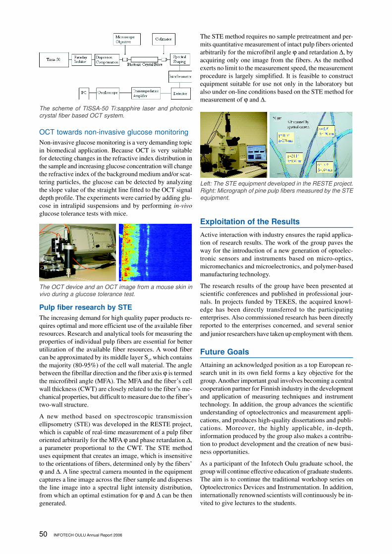
The electronics for a 3-D fiber optic joystick (left) and tem-
perature calibrated angle sensor (right). The electronics
for the 3-D joy stick comprised of two transmitter LEDs
(white) and two differential and one single channel PIN-
diodes (black). In the angle measurement, one LED is trans-
mitting light and two PIN-diodes are collecting reflected light
differentially.
Configuration of a self-mixing interferometer.
Optical path length variation in the external cavity of the
SMI.
Scattering photoacoustic signals (left) and laser ultrasonic
power spectra (right) obtained from TMP samples with dif-
ferent consistency.
INFOTECH OULU Annual Report 2006 49
Spectroscopic investigation of superficial
human and porcine skin layer using a tape
stripping technique
Tape stripping is a minimally invasive method to reveal in-
depth penetration profiles of substances topically applied
onto the surface of skin. Our research is focused on experi-
ments with porcine skin in vitro (ears of freshly slaugh-
tered pigs) and human skin in vivo (flexor forearm). The
tape stripping technique introduces consecutive removal of
micrometer-thick cell layers of the stratum corneum (SC)
from the same treated skin area using an adhesive tape. Pre-
requisite to the substance penetration profile is the recon-
struction of the removed SC by analyzing the amount of
corneocytes (cells of SC) stuck to each tape strip.
Such an analysis is carried out spectroscopically by mea-
suring light transmittance in visible and NIR spectral re-
gions (430 - 1050 nm) through the adhesive tape with
corneocytes. As we proved experimentally, there is a linear
dependence between the pseudoabsorption (equals to loga-
rithm of inverse transmittance) and the thickness of the
corneocytes on the tape strips for all wavelengths of the
investigated region. Dependence of the cumulative absor-
bance of the removed SC on the tape strip number can be
satisfactory fitted by exponential function. This relation-
ship allows evaluation of the share of the removed SC (in
%) without complete removal of this layer. All the obtained
results correlate well with those obtained on humans.
One of the frequently and widely used substances topically
administered onto the skin are various chemical agents (UV
filters) included into sunscreens for UV protection. The tape
stripping technique combined with spectroscopic analysis
make possible reconstruction of the in-depth profiles of such
substances. After tape stripping, strips with a UV filter on
them are put into pure ethanol causing dissolving of the
UV filter. An ultrasonic bath (for better stirring) and ensu-
ing centrifugation enable separation of corneocytes from
the dissolved chemical substance. Absorption of the UV
filter is measured within the UV range.
Research using optical coherence tomogra-
phy
Monte Carlo simulations of OCT images
Optical coherence tomography (OCT) is a modern non-in-
vasive technique utilizing principles of low-coherence in-
terferometry for visualization of the inner structure of
optically non-uniform objects. However, multiple scatter-
ing of light in the objects under study brings distortion to
the images causing requirements for the proper interpreta-
tion. A statistical Monte Carlo method for simulation of
light propagation in scattering media provides the ability
for analysis of formation of OCT-images from multilayer
samples.
A program code for the Monte Carlo simulation of 2D OCT
images of multilayer samples with various boundary shapes
was developed. OCT images of various skin and paper
samples were simulated. Such simulations allow one to sepa-
rate the contributions of low-order and multiple scattering
to the image. Such separation provides the information about
maximal depth of non-distorted imaging in the considered
objects which can be defined as the depth from which mul-
tiple scattering contribution starts to prevail in the image.
Paper structure analysis by OCT
The growth of the paper making industry has triggered a
continuous search for improved methods for diagnosing and
characterizing paper properties. Surface analysis, includ-
ing roughness, porosity and gloss estimation, is particularly
important in printing papers, because parameters like rough-
ness and surface porosity affect such optical properties of
paper as gloss and ink absorption. Current measurement
methods for structural analysis of paper tend to be slow,
labor-intensive, expensive or invasive.
Up-to-date OCT devices are fast and have a resolution which
competes with the conventional laboratory structural analy-
sis methods of paper. The advantage is the non-contact and
non-invasive approach. A resolution of 1µm can be reached
by the new Nd:YAG pumped Ti:Sapphire laser and Photo-
nic Crystal Fiber - PCF based OCT-system. Such a high
resolution enables the imaging of single fibers, coating paste
and color layers. Building such a system needs knowledge
of many areas like paper physics, photon migration in pa-
per, paper chemistry, signal processing and automation con-
trol of the imaging device, which means that the
collaboration between several scientists and research groups
has to be active. In addition to the imaging of paper, the
technique has been applied to fluid mechanics evaluation
by using the ability of the method to determine the velocity
profiles of flowing suspensions like pulp and coating paste
suspensions.
In-depth distribution of a chemical UV filter (utilized in sun-
screens) within porcine stratum corneum revealed using
the tape stripping technique.
Low-order (left) and multiple(right) scattering contributions
to the OCT image of paper.
Low-order (left) and multiple(right) scattering contributions
to the OCT image of skin.
50 INFOTECH OULU Annual Report 2006
OCT towards non-invasive glucose monitoring
Non-invasive glucose monitoring is a very demanding topic
in biomedical application. Because OCT is very suitable
for detecting changes in the refractive index distribution in
the sample and increasing glucose concentration will change
the refractive index of the background medium and/or scat-
tering particles, the glucose can be detected by analyzing
the slope value of the straight line fitted to the OCT signal
depth profile. The experiments were carried by adding glu-
cose in intralipid suspensions and by performing in-vivo
glucose tolerance tests with mice.
Pulp fiber research by STE
The increasing demand for high quality paper products re-
quires optimal and more efficient use of the available fiber
resources. Research and analytical tools for measuring the
properties of individual pulp fibers are essential for better
utilization of the available fiber resources. A wood fiber
can be approximated by its middle layer S2, which contains
the majority (80-95%) of the cell wall material. The angle
between the fibrillar direction and the fiber axis ϕ is termed
the microfibril angle (MFA). The MFA and the fiber’s cell
wall thickness (CWT) are closely related to the fiber’s me-
chanical properties, but difficult to measure due to the fiber’s
two-wall structure.
A new method based on spectroscopic transmission
ellipsometry (STE) was developed in the RESTE project,
which is capable of real-time measurement of a pulp fiber
oriented arbitrarily for the MFA ϕ and phase retardation ∆,
a parameter proportional to the CWT. The STE method
uses equipment that creates an image, which is insensitive
to the orientations of fibers, determined only by the fibers’
ϕ and ∆. A line spectral camera mounted in the equipment
captures a line image across the fiber sample and disperses
the line image into a spectral light intensity distribution,
from which an optimal estimation for ϕ and ∆ can be then
generated.
The STE method requires no sample pretreatment and per-
mits quantitative measurement of intact pulp fibers oriented
arbitrarily for the microfibril angle ϕ and retardation ∆, by
acquiring only one image from the fibers. As the method
exerts no limit to the measurement speed, the measurement
procedure is largely simplified. It is feasible to construct
equipment suitable for use not only in the laboratory but
also under on-line conditions based on the STE method for
measurement of ϕ and ∆.
Exploitation of the Results
Active interaction with industry ensures the rapid applica-
tion of research results. The work of the group paves the
way for the introduction of a new generation of optoelec-
tronic sensors and instruments based on micro-optics,
micromechanics and microelectronics, and polymer-based
manufacturing technology.
The research results of the group have been presented at
scientific conferences and published in professional jour-
nals. In projects funded by TEKES, the acquired knowl-
edge has been directly transferred to the participating
enterprises. Also commissioned research has been directly
reported to the enterprises concerned, and several senior
and junior researchers have taken up employment with them.
Future Goals
Attaining an acknowledged position as a top European re-
search unit in its own field forms a key objective for the
group. Another important goal involves becoming a central
cooperation partner for Finnish industry in the development
and application of measuring techniques and instrument
technology. In addition, the group advances the scientific
understanding of optoelectronics and measurement appli-
cations, and produces high-quality dissertations and publi-
cations. Moreover, the highly applicable, in-depth,
information produced by the group also makes a contribu-
tion to product development and the creation of new busi-
ness opportunities.
As a participant of the Infotech Oulu graduate school, the
group will continue effective education of graduate students.
The aim is to continue the traditional workshop series on
Optoelectronics Devices and Instrumentation. In addition,
internationally renowned scientists will continuously be in-
vited to give lectures to the students.
The scheme of TISSA-50 Ti:sapphire laser and photonic
crystal fiber based OCT system.
The OCT device and an OCT image from a mouse skin in
vivo during a glucose tolerance test.
Left: The STE equipment developed in the RESTE project.
Right: Micrograph of pine pulp fibers measured by the STE
equipment.
INFOTECH OULU Annual Report 2006 51
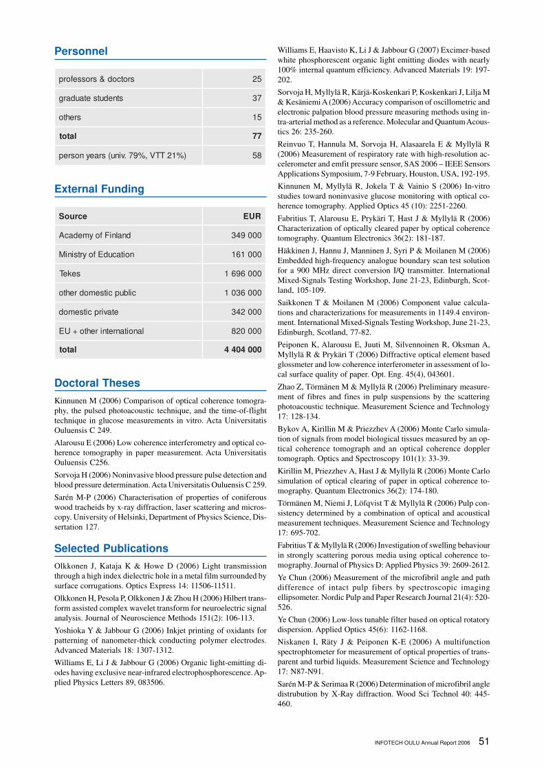
Personnel
srotcod&srosseforp 52
stnedutsetaudarg 73
srehto 51
latot 77
)%12TTV,%97.vinu(sraeynosrep 85
External Funding
ecruoS RUE
dnalniFfoymedacA 000943
noitacudEfoyrtsiniM 000161
sekeT 0006961
cilbupcitsemodrehto 0006301
etavirpcitsemod 000243
lanoitanretnirehto+UE 000028
latot 0004044
Doctoral Theses
Kinnunen M (2006) Comparison of optical coherence tomogra-
phy, the pulsed photoacoustic technique, and the time-of-flight
technique in glucose measurements in vitro. Acta Universitatis
Ouluensis C 249.
Alarousu E (2006) Low coherence interferometry and optical co-
herence tomography in paper measurement. Acta Universitatis
Ouluensis C256.
Sorvoja H (2006) Noninvasive blood pressure pulse detection and
blood pressure determination. Acta Universitatis Ouluensis C 259.
Sarén M-P (2006) Characterisation of properties of coniferous
wood tracheids by x-ray diffraction, laser scattering and micros-
copy. University of Helsinki, Department of Physics Science, Dis-
sertation 127.
Selected Publications
Olkkonen J, Kataja K & Howe D (2006) Light transmission
through a high index dielectric hole in a metal film surrounded by
surface corrugations. Optics Express 14: 11506-11511.
Olkkonen H, Pesola P, Olkkonen J & Zhou H (2006) Hilbert trans-
form assisted complex wavelet transform for neuroelectric signal
analysis. Journal of Neuroscience Methods 151(2): 106-113.
Yoshioka Y & Jabbour G (2006) Inkjet printing of oxidants for
patterning of nanometer-thick conducting polymer electrodes.
Advanced Materials 18: 1307-1312.
Williams E, Li J & Jabbour G (2006) Organic light-emitting di-
odes having exclusive near-infrared electrophosphorescence. Ap-
plied Physics Letters 89, 083506.
Williams E, Haavisto K, Li J & Jabbour G (2007) Excimer-based
white phosphorescent organic light emitting diodes with nearly
100% internal quantum efficiency. Advanced Materials 19: 197-
202.
Sorvoja H, Myllylä R, Kärjä-Koskenkari P, Koskenkari J, Lilja M
& Kesäniemi A (2006) Accuracy comparison of oscillometric and
electronic palpation blood pressure measuring methods using in-
tra-arterial method as a reference. Molecular and Quantum Acous-
tics 26: 235-260.
Reinvuo T, Hannula M, Sorvoja H, Alasaarela E & Myllylä R
(2006) Measurement of respiratory rate with high-resolution ac-
celerometer and emfit pressure sensor, SAS 2006 – IEEE Sensors
Applications Symposium, 7-9 February, Houston, USA, 192-195.
Kinnunen M, Myllylä R, Jokela T & Vainio S (2006) In-vitro
studies toward noninvasive glucose monitoring with optical co-
herence tomography. Applied Optics 45 (10): 2251-2260.
Fabritius T, Alarousu E, Prykäri T, Hast J & Myllylä R (2006)
Characterization of optically cleared paper by optical coherence
tomography. Quantum Electronics 36(2): 181-187.
Häkkinen J, Hannu J, Manninen J, Syri P & Moilanen M (2006)
Embedded high-frequency analogue boundary scan test solution
for a 900 MHz direct conversion I/Q transmitter. International
Mixed-Signals Testing Workshop, June 21-23, Edinburgh, Scot-
land, 105-109.
Saikkonen T & Moilanen M (2006) Component value calcula-
tions and characterizations for measurements in 1149.4 environ-
ment. International Mixed-Signals Testing Workshop, June 21-23,
Edinburgh, Scotland, 77-82.
Peiponen K, Alarousu E, Juuti M, Silvennoinen R, Oksman A,
Myllylä R & Prykäri T (2006) Diffractive optical element based
glossmeter and low coherence interferometer in assessment of lo-
cal surface quality of paper. Opt. Eng. 45(4), 043601.
Zhao Z, Törmänen M & Myllylä R (2006) Preliminary measure-
ment of fibres and fines in pulp suspensions by the scattering
photoacoustic technique. Measurement Science and Technology
17: 128-134.
Bykov A, Kirillin M & Priezzhev A (2006) Monte Carlo simula-
tion of signals from model biological tissues measured by an op-
tical coherence tomograph and an optical coherence doppler
tomograph. Optics and Spectroscopy 101(1): 33-39.
Kirillin M, Priezzhev A, Hast J & Myllylä R (2006) Monte Carlo
simulation of optical clearing of paper in optical coherence to-
mography. Quantum Electronics 36(2): 174-180.
Törmänen M, Niemi J, Löfqvist T & Myllylä R (2006) Pulp con-
sistency determined by a combination of optical and acoustical
measurement techniques. Measurement Science and Technology
17: 695-702.
Fabritius T & Myllylä R (2006) Investigation of swelling behaviour
in strongly scattering porous media using optical coherence to-
mography. Journal of Physics D: Applied Physics 39: 2609-2612.
Ye Chun (2006) Measurement of the microfibril angle and path
difference of intact pulp fibers by spectroscopic imaging
ellipsometer. Nordic Pulp and Paper Research Journal 21(4): 520-
526.
Ye Chun (2006) Low-loss tunable filter based on optical rotatory
dispersion. Applied Optics 45(6): 1162-1168.
Niskanen I, Räty J & Peiponen K-E (2006) A multifunction
spectrophtometer for measurement of optical properties of trans-
parent and turbid liquids. Measurement Science and Technology
17: N87-N91.
Sarén M-P & Serimaa R (2006) Determination of microfibril angle
distrubution by X-Ray diffraction. Wood Sci Technol 40: 445-
460.
Recommended