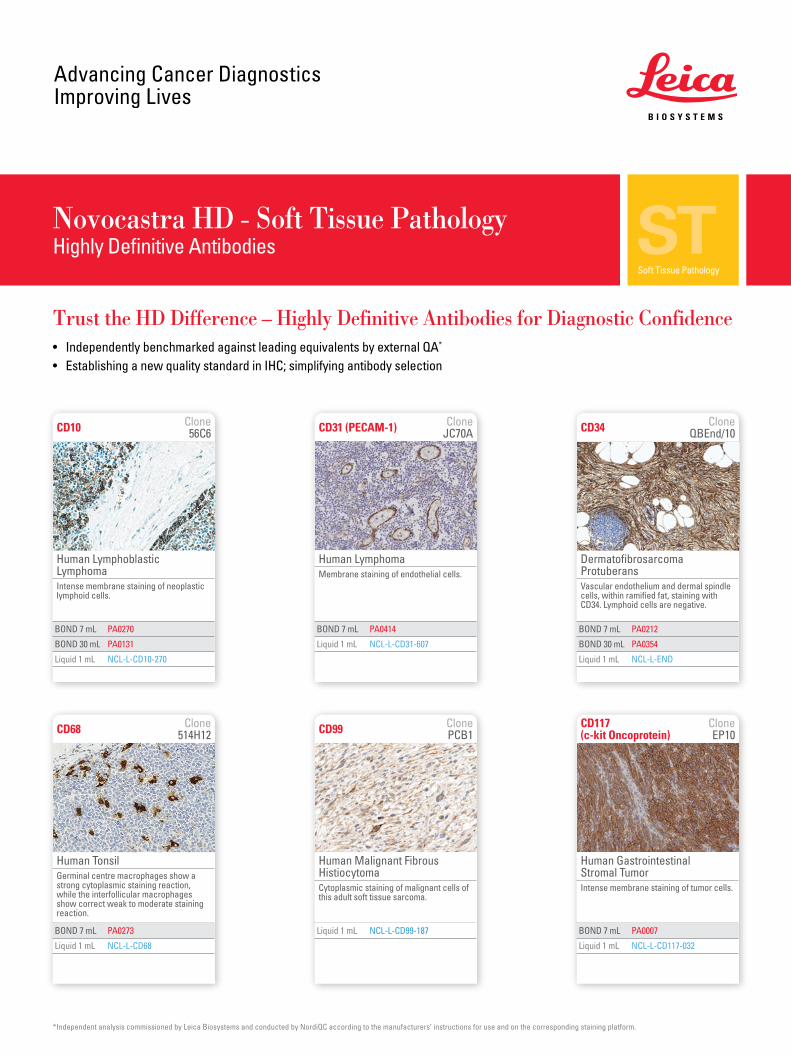
Novocastra HD - Soft Tissue PathologyHighly Definitive Antibodies
Trust the HD Difference – Highly Definitive Antibodies for Diagnostic Confidence• Independently benchmarked against leading equivalents by external QA*
• Establishing a new quality standard in IHC; simplifying antibody selection
*Independent analysis commissioned by Leica Biosystems and conducted by NordiQC according to the manufacturers’ instructions for use and on the corresponding staining platform.
BOND 7 mL PA0270
BOND 30 mL PA0131
Liquid 1 mL NCL-L-CD10-270
CD10 Clone 56C6 CD34 Clone
QBEnd/10
BOND 7 mL PA0212
BOND 30 mL PA0354
Liquid 1 mL NCL-L-END
CD68 Clone 514H12
BOND 7 mL PA0273
Liquid 1 mL NCL-L-CD68
Human LymphomaMembrane staining of endothelial cells.
CD31 (PECAM-1) Clone JC70A
BOND 7 mL PA0414
Liquid 1 mL NCL-L-CD31-607
Human Lymphoblastic LymphomaIntense membrane staining of neoplastic lymphoid cells.
Dermatofibrosarcoma ProtuberansVascular endothelium and dermal spindle cells, within ramified fat, staining with CD34. Lymphoid cells are negative.
Human TonsilGerminal centre macrophages show a strong cytoplasmic staining reaction, while the interfollicular macrophages show correct weak to moderate staining reaction.
BOND 7 mL PA0007
Liquid 1 mL NCL-L-CD117-032
Human Gastrointestinal Stromal TumorIntense membrane staining of tumor cells.
CD117 (c-kit Oncoprotein)
Clone EP10
Liquid 1 mL NCL-L-CD99-187
Human Malignant Fibrous Histiocytoma Cytoplasmic staining of malignant cells of this adult soft tissue sarcoma.
CD99 Clone PCB1
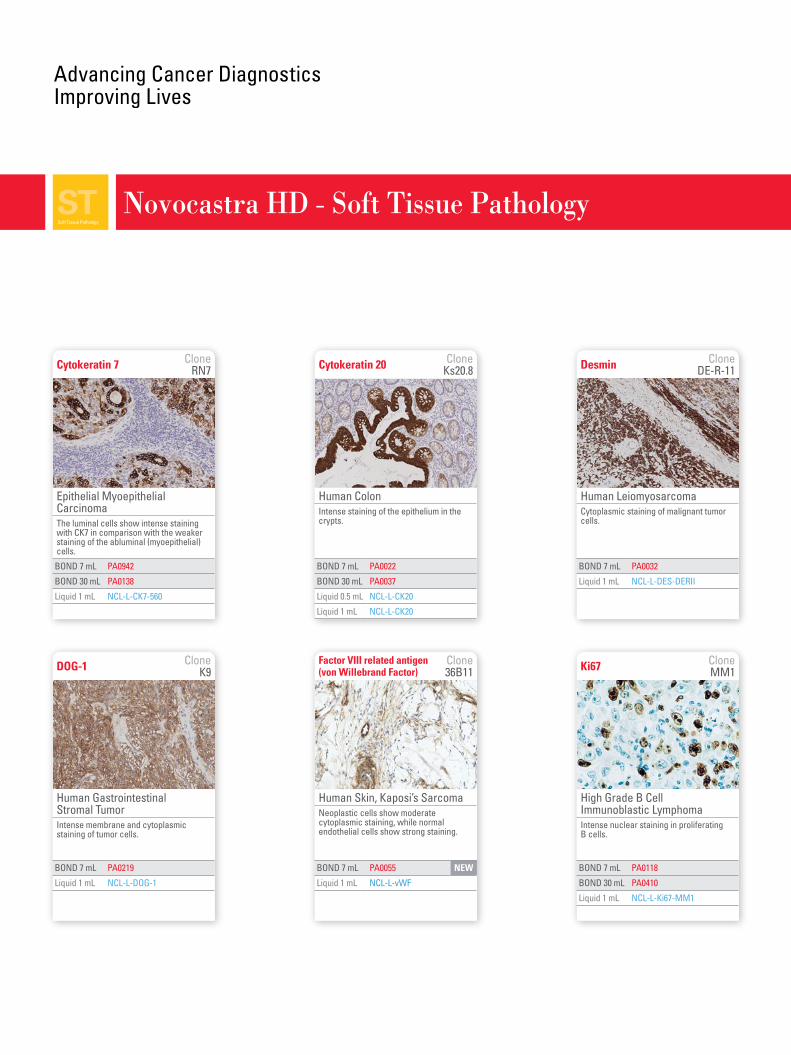
Cytokeratin 20 Clone Ks20.8
BOND 7 mL PA0022
BOND 30 mL PA0037
Liquid 0.5 mL NCL-L-CK20
Liquid 1 mL NCL-L-CK20
Human LeiomyosarcomaCytoplasmic staining of malignant tumor cells.
Human Gastrointestinal Stromal TumorIntense membrane and cytoplasmic staining of tumor cells.
DOG-1 Clone K9
Desmin Clone DE-R-11
BOND 7 mL PA0219
Liquid 1 mL NCL-L-DOG-1
BOND 7 mL PA0032
Liquid 1 mL NCL-L-DES-DERII
Ki67 Clone MM1
BOND 7 mL PA0118
BOND 30 mL PA0410
Liquid 1 mL NCL-L-Ki67-MM1
Novocastra HD - Soft Tissue Pathology
Human ColonIntense staining of the epithelium in the crypts.
High Grade B Cell Immunoblastic LymphomaIntense nuclear staining in proliferating B cells.
Cytokeratin 7 Clone RN7
BOND 7 mL PA0942
BOND 30 mL PA0138
Liquid 1 mL NCL-L-CK7-560
Epithelial Myoepithelial CarcinomaThe luminal cells show intense staining with CK7 in comparison with the weaker staining of the abluminal (myoepithelial) cells.
Human Skin, Kaposi’s SarcomaNeoplastic cells show moderate cytoplasmic staining, while normal endothelial cells show strong staining.
Factor VIII related antigen (von Willebrand Factor)
Clone 36B11
BOND 7 mL PA0055 NEW
Liquid 1 mL NCL-L-vWF
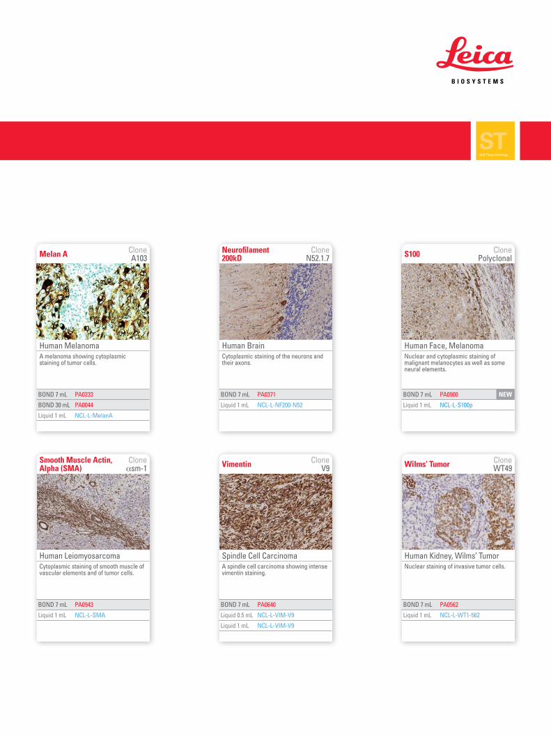
Melan A Clone A103
BOND 7 mL PA0233
BOND 30 mL PA0044
Liquid 1 mL NCL-L-MelanA
BOND 7 mL PA0371
Liquid 1 mL NCL-L-NF200-N52
Neurofilament 200kD
Clone N52.1.7
BOND 7 mL PA0640
Liquid 0.5 mL NCL-L-VIM-V9
Liquid 1 mL NCL-L-VIM-V9
BOND 7 mL PA0562
Liquid 1 mL NCL-L-WT1-562
Vimentin Clone V9 Wilms’ Tumor Clone
WT49
Human Melanoma A melanoma showing cytoplasmic staining of tumor cells.
Spindle Cell CarcinomaA spindle cell carcinoma showing intense vimentin staining.
Human Kidney, Wilms’ TumorNuclear staining of invasive tumor cells.
BOND 7 mL PA0943
Liquid 1 mL NCL-L-SMA
Human LeiomyosarcomaCytoplasmic staining of smooth muscle of vascular elements and of tumor cells.
Smooth Muscle Actin, Alpha (SMA)
Clone αsm-1
Human BrainCytoplasmic staining of the neurons and their axons.
Human Face, MelanomaNuclear and cytoplasmic staining of malignant melanocytes as well as some neural elements.
BOND 7 mL PA0900 NEW
Liquid 1 mL NCL-L-S100p
S100 Clone Polyclonal
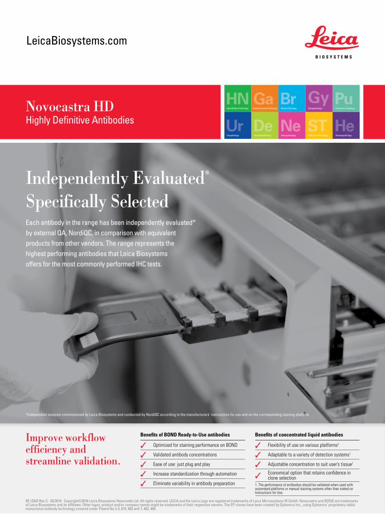
LeicaBiosystems.com
Novocastra HDHighly Definitive Antibodies
Benefits of BOND Ready-to-Use antibodies
✓ Optimized for staining performance on BOND
✓ Validated antibody concentrations
✓ Ease of use: just plug and play
✓ Increase standardization through automation
✓ Eliminate variability in antibody preparation
Benefits of concentrated liquid antibodies
✓ Flexibility of use on various platforms1
✓ Adaptable to a variety of detection systems1
✓ Adjustable concentration to suit user’s tissue1
✓ Economical option that retains confidence in clone selection
1. The performance of antibodies should be validated when used with automated platforms or manual staining systems other than stated on Instructions for Use.
Improve workflow efficiency and streamline validation.
95.13342 Rev C · 03/2016 · Copyright©2016 Leica Biosystems Newcastle Ltd. All rights reserved. LEICA and the Leica Logo are registered trademarks of Leica Microsystems IR GmbH. Novocastra and BOND are trademarks of Leica Biosystems and its affiliates. Other logos, product and/or company names might be trademarks of their respective owners. The EP clones have been created by Epitomics Inc., using Epitomics’ proprietary rabbit monoclonal antibody technology covered under Patent No.’s 5, 675, 063 and 7, 402, 409.
Independently Evaluated* Specifically SelectedEach antibody in the range has been independently evaluated* by external QA, NordiQC, in comparison with equivalent products from other vendors. The range represents the highest performing antibodies that Leica Biosystems offers for the most commonly performed IHC tests.
*Independent analysis commissioned by Leica Biosystems and conducted by NordiQC according to the manufacturers’ instructions for use and on the corresponding staining platform.
Recommended