New insight into biophysics of lipid membranes with high resolution IXS
Mikhail Zhernenkov
06/27/2019

2
Fast and slow dynamics in a cell membrane
XPCSSlow dynamics on mesoscale: seconds to µs
Slow dynamics:Lipid domain (rafts)
formation and diffusion, anomalous subdiffusion
IXSFast dynamics on molecular
scale: picoseconds
Ultrafast dynamics: Protein and lipid diffusion, lateral and transmembrane transport
Fig. from Science 327, 46 (2009)
Dynamic processes in a cell membrane is virtually impossible to study due to its complexity
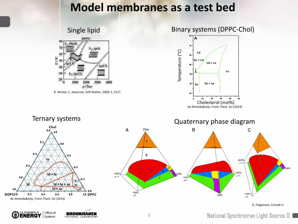
Model membranes as a test bed
Ternary systems Quaternary phase diagram
G. Feigenson, Cornell U
de Almeida&Joly, Front. Plant. Sci (2014)
Binary systems (DPPC-Chol)
R. Winter, C. Jeworrek, Soft Matter, 2009, 5, 3157
Single lipid
de Almeida&Joly, Front. Plant. Sci (2014)
3

4
Model membranes as a test bed
Elson et al. Annu. Rev. Biophys. 2010. 39:207–26Predicted and observed phenomena in lipid layers span broad length andtimescales Experimental techniques cover most
of “large” and relatively “slow” lipid behavior.
Still do not adequately determine “small” and “fast”
5
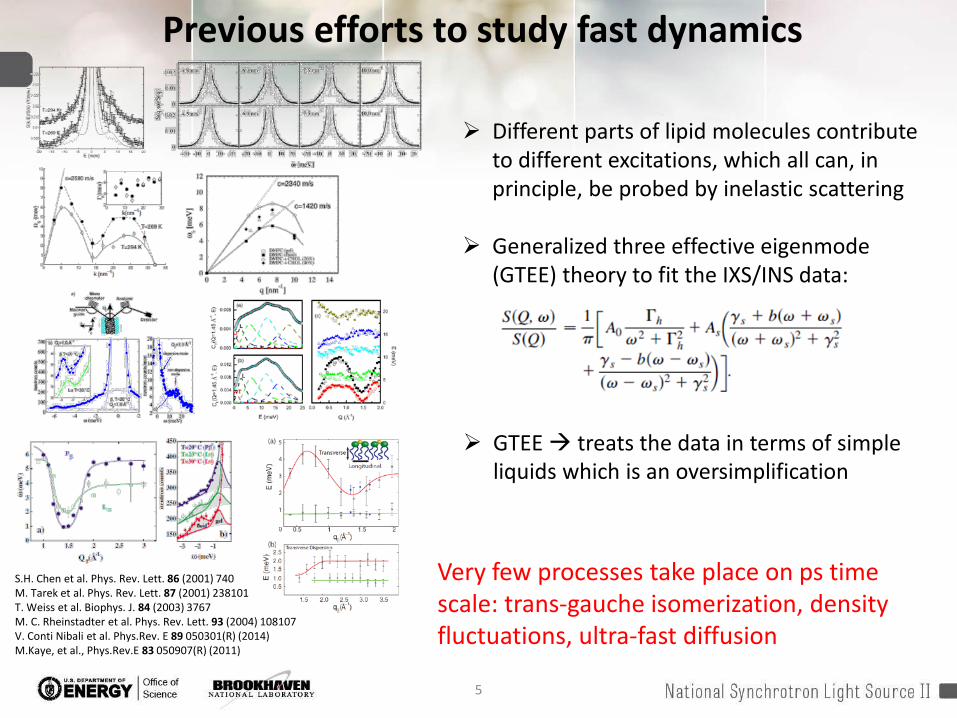
Previous efforts to study fast dynamics
GTEE treats the data in terms of simple liquids which is an oversimplification
S.H. Chen et al. Phys. Rev. Lett. 86 (2001) 740M. Tarek et al. Phys. Rev. Lett. 87 (2001) 238101T. Weiss et al. Biophys. J. 84 (2003) 3767M. C. Rheinstadter et al. Phys. Rev. Lett. 93 (2004) 108107V. Conti Nibali et al. Phys.Rev. E 89 050301(R) (2014)M.Kaye, et al., Phys.Rev.E 83 050907(R) (2011)
Very few processes take place on ps time scale: trans-gauche isomerization, density fluctuations, ultra-fast diffusion
Different parts of lipid molecules contribute to different excitations, which all can, in principle, be probed by inelastic scattering
Generalized three effective eigenmode(GTEE) theory to fit the IXS/INS data:
6
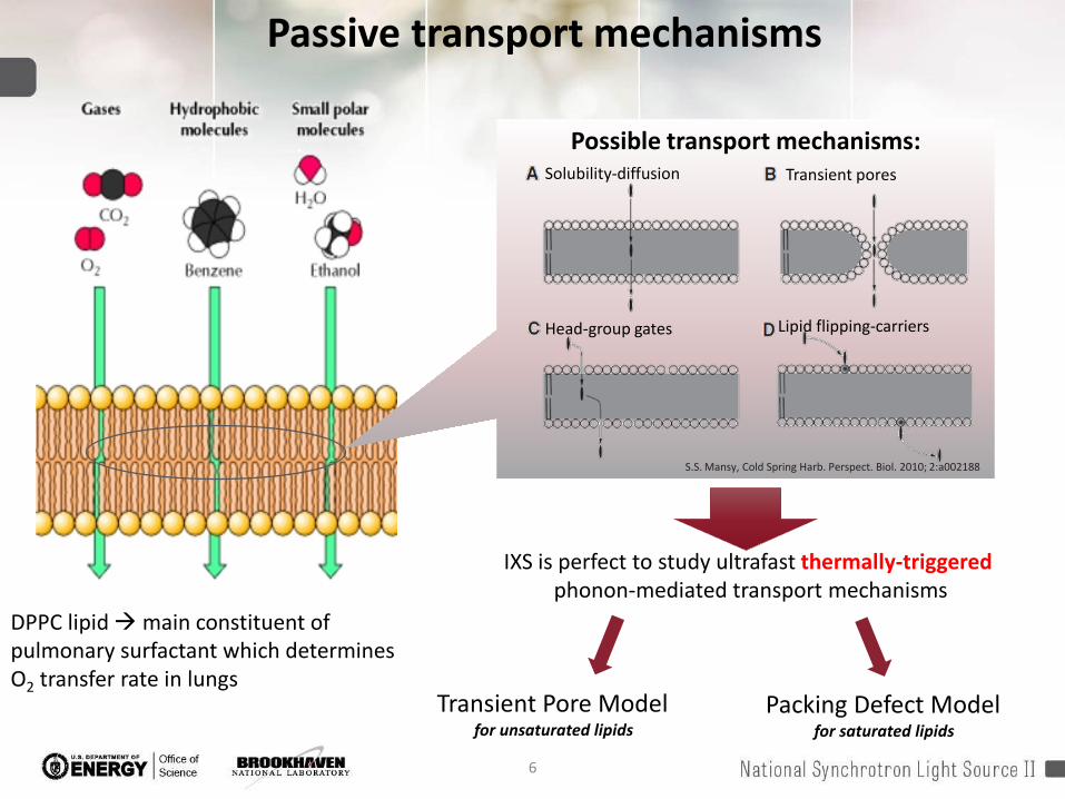
Possible transport mechanisms:Solubility-diffusion Transient pores
Head-group gates Lipid flipping-carriers
S.S. Mansy, Cold Spring Harb. Perspect. Biol. 2010; 2:a002188
IXS is perfect to study ultrafast thermally-triggered phonon-mediated transport mechanisms
Transient Pore Modelfor unsaturated lipids
Packing Defect Modelfor saturated lipids
Passive transport mechanisms
DPPC lipid main constituent of pulmonary surfactant which determines O2 transfer rate in lungs
7
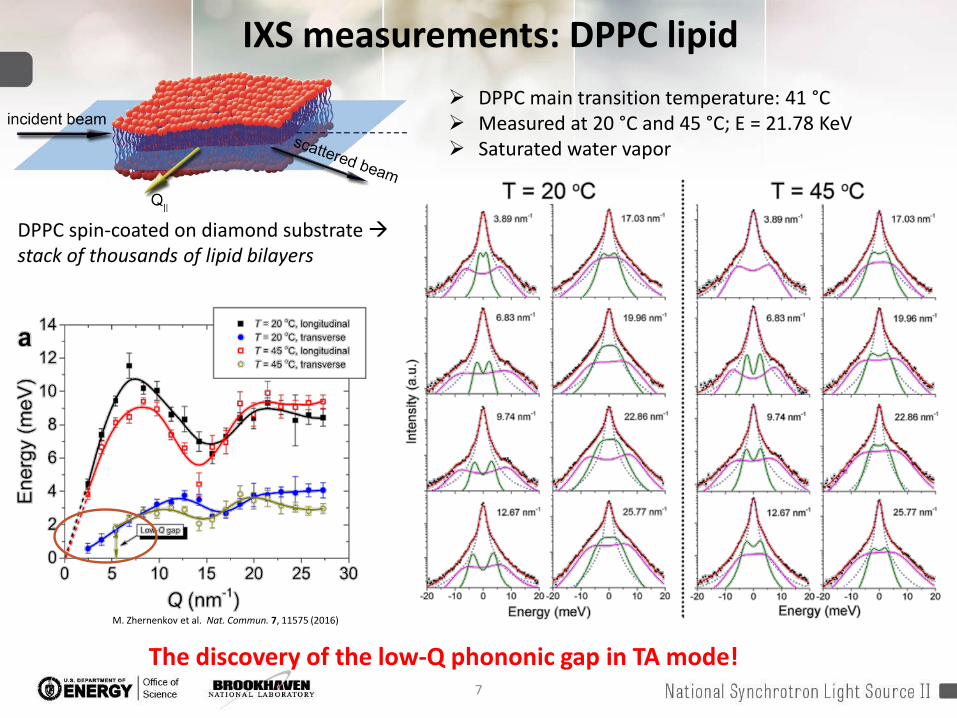
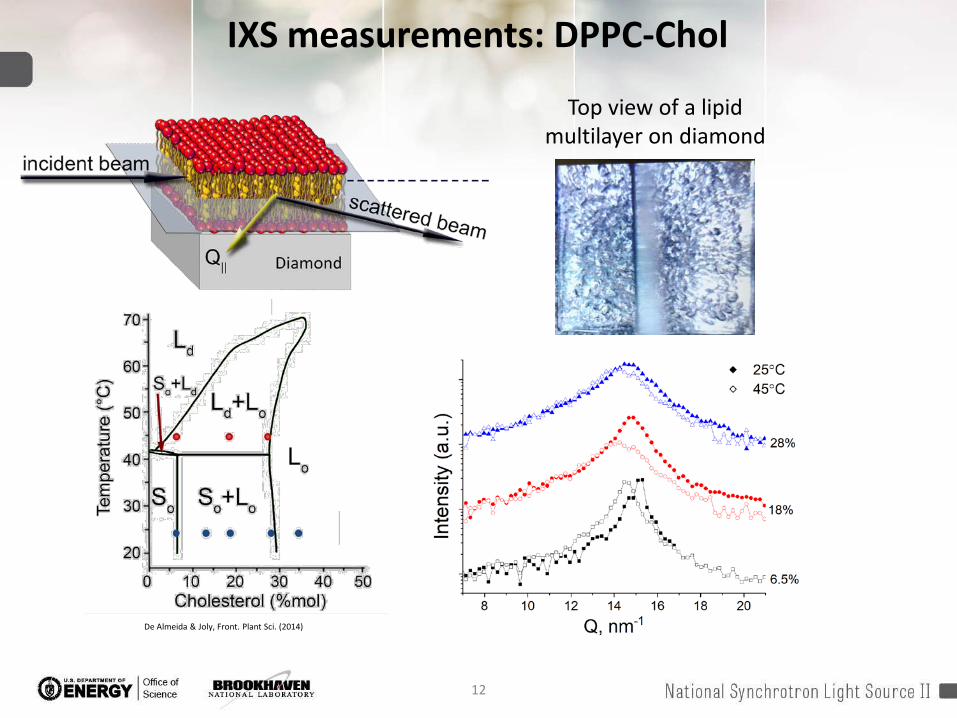
IXS measurements: DPPC lipid DPPC main transition temperature: 41 °C Measured at 20 °C and 45 °C; E = 21.78 KeV Saturated water vapor
DPPC spin-coated on diamond substrate stack of thousands of lipid bilayers
M. Zhernenkov et al. Nat. Commun. 7, 11575 (2016)
The discovery of the low-Q phononic gap in TA mode!

8
Low-Q phononic gap in TA modeLow-Q phononic gap
No phonon propagation at Q < Qgap
The cut-off value Qgap(at which gap occurs) Characteristic length-scale
dgap = 2π/Qgap of a local “order”(no phonons at smaller Q more disorder on long scale! )
Qgap
D. Bolmatov, et al. Ann. Phys. 363, 221-242 (2015)
D. Bolmatov, et al. J. Phys. Chem. Lett. 6, 3048-3053 (2015)
9
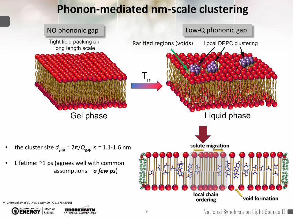
• the cluster size dgap = 2π/Qgap is ~ 1.1-1.6 nm
• Lifetime: ~1 ps (agrees well with common assumptions – a few ps)
Phonon-mediated nm-scale clustering
Low-Q phononic gap NO phononic gap
M. Zhernenkov et al. Nat. Commun. 7, 11575 (2016)
Rarified regions (voids)
10
Theory of solute diffusion through a membrane:
ultra-fast “hopping”, or “rattling” between thermally-triggered voids
partition coefficient strongly depends on the local chain ordering solute exclusion within the region
Potential formation of water fingers inside voids proton translocation through membrane Adv. Drug Deliv. Rev. 58, 1357–1378 (2006)
J. Am. Chem. Soc. 117, 4118–4129 (1995)Cold Spring Harb. Perspect. Biol. (2010), 2, a002188
We observe:
nm-scaled short-lived molecular clusters local chain ordering, or density fluctuations
Increased disorder beyond the cluster size indication of the transient voids formation
Size and the life time of the clusters agrees well with the theory prediction
Phonon-mediated nm-scale clustering
11
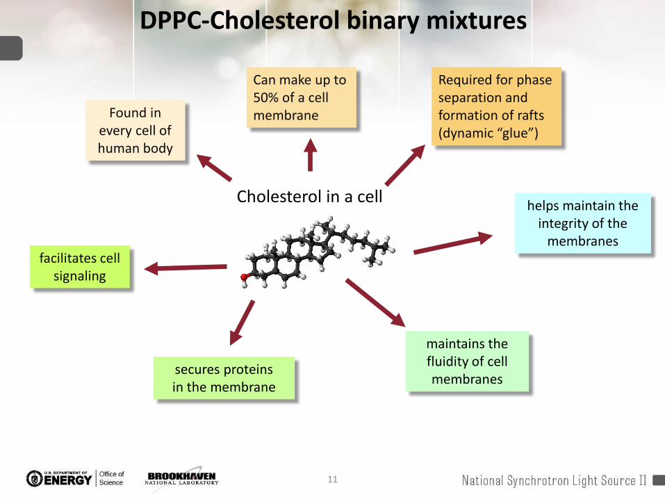
DPPC-Cholesterol binary mixtures
Cholesterol in a cell
maintains the fluidity of cell membranes
secures proteins in the membrane
facilitates cell signaling
helps maintain the integrity of the
membranes
Found in every cell of human body
Can make up to 50% of a cell membrane
Required for phase separation and formation of rafts (dynamic “glue”)
12
De Almeida & Joly, Front. Plant Sci. (2014)
IXS measurements: DPPC-Chol
Top view of a lipid multilayer on diamond
13
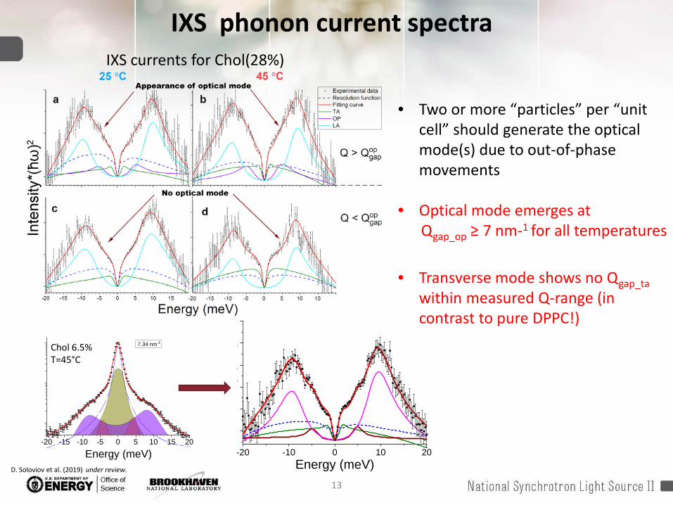
IXS phonon current spectra
-20 -15 -10 -5 0 5 10 15 20
7.34 nm-1
Energy (meV)
• Two or more “particles” per “unit cell” should generate the optical mode(s) due to out-of-phase movements
• Optical mode emerges at Qgap_op ≥ 7 nm-1 for all temperatures
• Transverse mode shows no Qgap_tawithin measured Q-range (in contrast to pure DPPC!)
-20 -10 0 10 20
y (
)
Energy (meV)
IXS currents for Chol(28%)
Chol 6.5%T=45°C
D. Soloviov et al. (2019) under review.
14
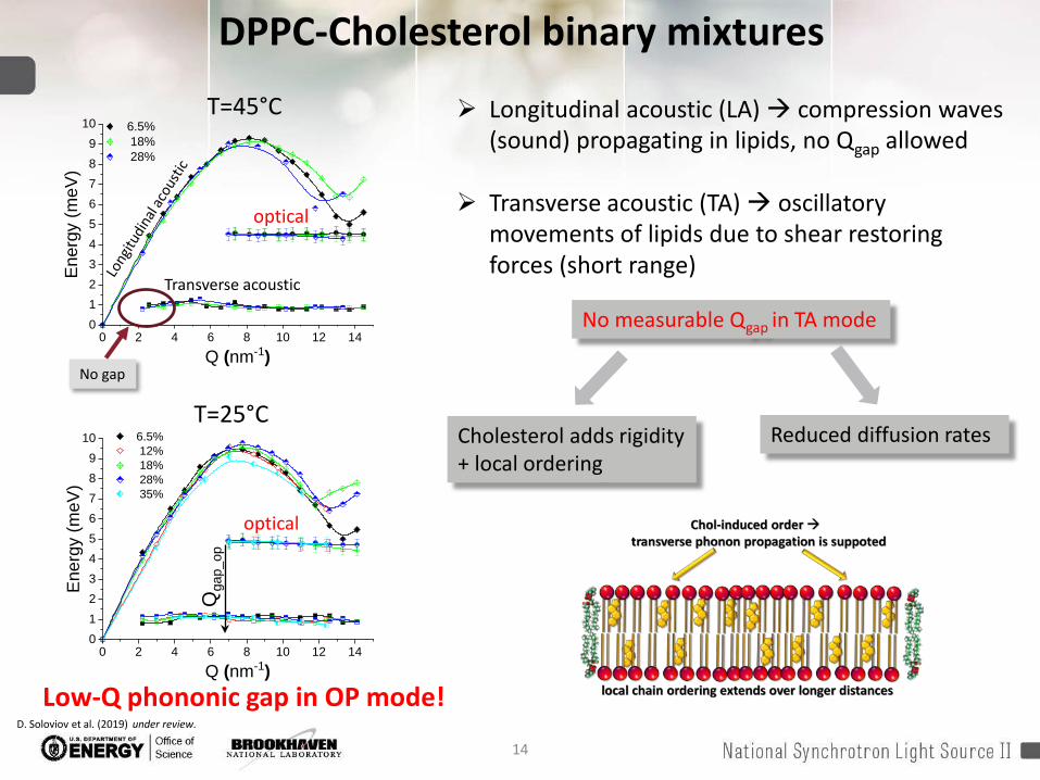
DPPC-Cholesterol binary mixtures
0 2 4 6 8 10 12 140123456789
10
Ener
gy (m
eV)
Q (nm-1)
6.5% 12% 18% 28% 35%
0 2 4 6 8 10 12 140123456789
10
Ener
gy (m
eV)
Q (nm-1)
6.5% 18% 28%
T=45°C
T=25°C
Transverse acoustic
optical
optical
Longitudinal acoustic (LA) compression waves (sound) propagating in lipids, no Qgap allowed
Transverse acoustic (TA) oscillatory movements of lipids due to shear restoring forces (short range)
No gap
No measurable Qgap in TA mode
Cholesterol adds rigidity + local ordering
Reduced diffusion rates
Qga
p_op
Low-Q phononic gap in OP mode!D. Soloviov et al. (2019) under review.

15
Origin of phononic gap in optical mode
Image credit: Wikipedia
Optical and acoustic vibrations in linear diatomic chain
Optical mode frequency:
L
Under a continuous medium approximation (Q0)the optical mode cannot exhibit the phononic gap Qgap!
at Q0
α - interparticle force constant
16
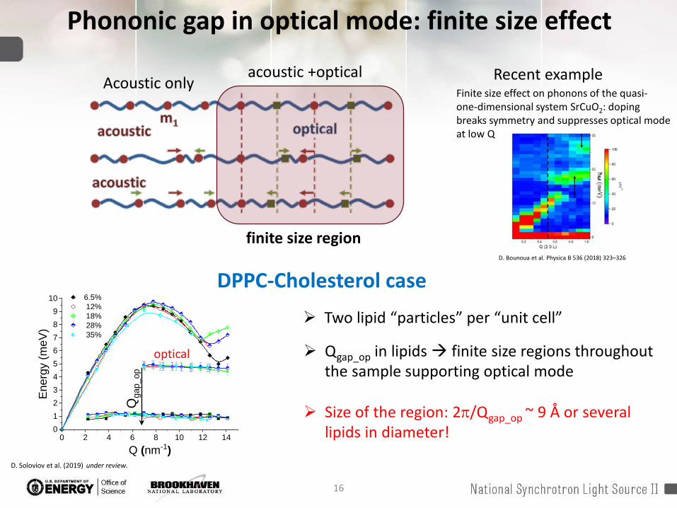
Phononic gap in optical mode: finite size effect
acoustic +opticalAcoustic only
finite size region D. Bounoua et al. Physica B 536 (2018) 323–326
Finite size effect on phonons of the quasi-one-dimensional system SrCuO2: doping breaks symmetry and suppresses optical mode at low Q
Recent example
0 2 4 6 8 10 12 140123456789
10
Ener
gy (m
eV)
Q (nm-1)
6.5% 12% 18% 28% 35%
optical
Qga
p_op
Qgap_op in lipids finite size regions throughout the sample supporting optical mode
Size of the region: 2π/Qgap_op ~ 9 Å or several lipids in diameter!
DPPC-Cholesterol case Two lipid “particles” per “unit cell”
D. Soloviov et al. (2019) under review.
17
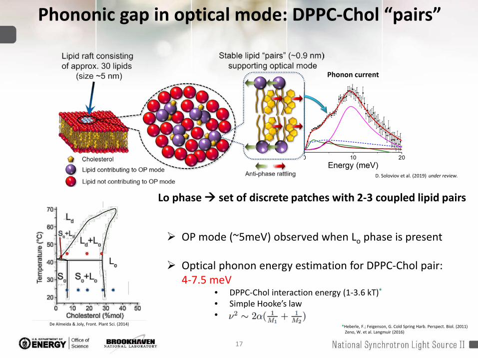
Phononic gap in optical mode: DPPC-Chol “pairs”
De Almeida & Joly, Front. Plant Sci. (2014)
OP mode (~5meV) observed when Lo phase is present
Optical phonon energy estimation for DPPC-Chol pair: 4-7.5 meV
• DPPC-Chol interaction energy (1-3.6 kT)*
• Simple Hooke’s law•
Lo phase set of discrete patches with 2-3 coupled lipid pairs
D. Soloviov et al. (2019) under review.
*Heberle, F.; Feigenson, G. Cold Spring Harb. Perspect. Biol. (2011)Zeno, W. et al. Langmuir (2016)
18
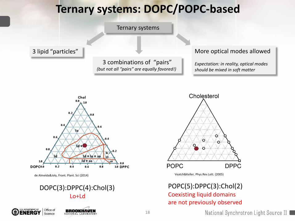
Ternary systems: DOPC/POPC-based
de Almeida&Joly, Front. Plant. Sci (2014) Veatch&Keller, Phys.Rev.Lett. (2005)
DOPC(3):DPPC(4):Chol(3)Lo+Ld
POPC(5):DPPC(3):Chol(2)Coexisting liquid domainsare not previously observed
Ternary systems
3 combinations of “pairs”(but not all “pairs” are equally favored!)
3 lipid “particles” More optical modes allowed
Expectation: in reality, optical modes should be mixed in soft matter

19
IXS: ternary systems at 37°C
• Optical mode emerges at Qgap_op ≥ 8.3 nm-1
• OP energy: 3.5-3.9meV
• Size of the region: 2π/Qgap_op~ 7.5 Å or several lipids in diameter
• OP energy estimation for DOPC/POPC-DPPC pairs: 1.6 meV (unfavorable pairs)
• Unsaturated lipids reduce the size of discrete patches!
• No patches in Ld phase with excess of unsaturated lipidsD. Soloviov et al. (2019) under review.
20
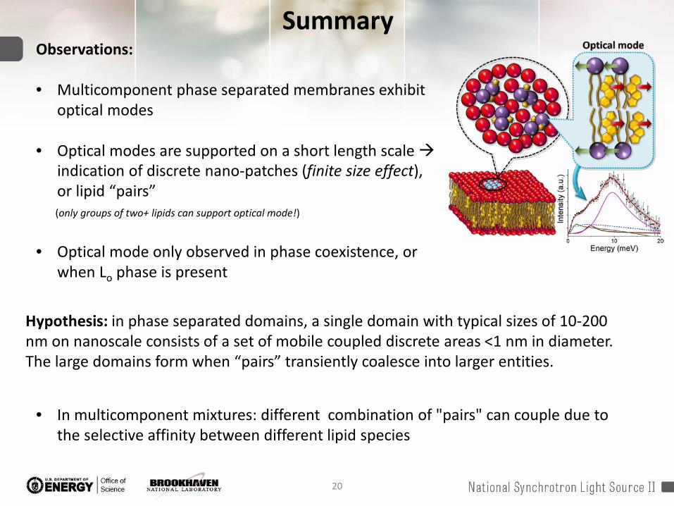
SummaryObservations:
• Multicomponent phase separated membranes exhibit optical modes
• Optical modes are supported on a short length scale indication of discrete nano-patches (finite size effect), or lipid “pairs” (only groups of two+ lipids can support optical mode!)
• Optical mode only observed in phase coexistence, or when Lo phase is present
Hypothesis: in phase separated domains, a single domain with typical sizes of 10-200 nm on nanoscale consists of a set of mobile coupled discrete areas <1 nm in diameter. The large domains form when “pairs” transiently coalesce into larger entities.
• In multicomponent mixtures: different combination of "pairs" can couple due to the selective affinity between different lipid species
21
THANK YOU!
Acknowledgements
A. Bosak K. Zhernenkov A. CunsoloY. Cai
D. Soloviov B. Toperverg
D. Bolmatov H. Uchiyama E. Lyman I. LeventalF. Heberle
Recommended