Embed Size (px)
Citation preview

9HSTFMG*affbhg+
ISBN 978-952-60-5517-6 ISBN 978-952-60-5518-3 (pdf) ISSN-L 1799-4934 ISSN 1799-4934 ISSN 1799-4942 (pdf) Aalto University School of Science Department of Biomedical Engineering and Computational Science
BUSINESS + ECONOMY ART + DESIGN + ARCHITECTURE SCIENCE + TECHNOLOGY CROSSOVER DOCTORAL DISSERTATIONS
Aalto-D
D 2
20/2
013
AJA
Y K
UM
AR
MA
HA
LK
A
Control of P
rotein Oligom
erization and De-oligom
erization on Lipid M
embranes
Aalto
Unive
rsity
Department of Biomedical Engineering and Computational Science
Control of Protein Oligomerization and De-oligomerization on Lipid Membranes
AJAY KUMAR MAHALKA
DOCTORAL DISSERTATIONS


Aalto University publication series DOCTORAL DISSERTATIONS 220/2013
Control of Protein Oligomerization and De-oligomerization on Lipid Membranes
AJAY KUMAR MAHALKA
A doctoral dissertation completed for the degree of Doctor of Science in Technology (Doctor of Philosophy) to be presented with due permission of the Aalto University School of Science for public examination and debate in Auditorium at a public examination held at the lecture hall E, Otakaari 1 of the Aaalto University on the 24th January 2014 at 12 noon.
Aalto University School of Science Dep. of Biomedical Engineering and Computational Science Helsinki Biophysics and Biomembrane Group (HBBG)

Supervising professor Professor Paavo K. J. Kinnunen Helsinki Biophysics and Biomembrane Group Department of Biomedical Engineering and Computational Science Aalto University School of Science Finland Thesis advisor Professor Paavo K. J. Kinnunen Preliminary examiners Docent Hongxia Zhao Institute of Biotechnology University of Helsinki, Finland Docent Tuomas Haltia Department of Biosciences University of Helsinki, Finland Opponent Assoc. Professor Günther H.J. Peters Department of Chemistry Technical University of Denmark Denmark
Aalto University publication series DOCTORAL DISSERTATIONS 220/2013 © Ajay Kumar Mahalka ISBN 978-952-60-5517-6 ISBN 978-952-60-5518-3 (pdf) ISSN-L 1799-4934 ISSN 1799-4934 (printed) ISSN 1799-4942 (pdf) http://urn.fi/URN:ISBN:978-952-60-5518-3 Unigrafia Oy Helsinki 2013 Finland

Abstract Aalto University, P.O. Box 11000, FI-00076 Aalto www.aalto.fi
Author Ajay Mahalka Name of the doctoral dissertation Control of Protein Oligomerization and De-oligomerization on Lipid Membranes Publisher School of Science Unit Department of Biomedical Engineering and Computational Science
Series Aalto University publication series DOCTORAL DISSERTATIONS 220/2013
Field of research Biomedical Engineering and Biophysics
Manuscript submitted 4 October 2013 Date of the defence 24 January 2014
Permission to publish granted (date) 26 November 2013 Language English
Monograph Article dissertation (summary + original articles)
Abstract Oligomerization of protein into amyloid fibrils is central to the pathogenesis of several
neurodegenerative disorders. Amyloid fibrillation and the cytotoxic actions of amyloids are membrane-associated processes. The interactions of amyloid-forming proteins with lipids at the membrane surface accelerate fibrillation and induce membrane permeabilization. Oligomerization also plays a functional role in antimicrobial defense and controls the catalytic activity of phospholipase A2 (PLA2). The protein oligomerization and amyloid formation can be modulated by heat shock protein 70 (Hsp70). Thus, the aim of the present work was to study membrane-associated protein oligomerization and its modulation by Hsp70 on the phospholipid model membrane system. Sequence analyses revealed that antimicrobial peptides (AMPs) contained sequence motifs that showed propensities for self-assembly, aggregation, and oligomerization into amyloid fibrils. The presence of such oligomerization-mediating sequences was characteristic of amyloidogenic cytotoxic proteins, including gelsolin involved in familial Finnish type amyloidosis (FAF). 1-Palmitoyl-2-(9'-oxo-nonanoyl)-sn-glycero-3-phosphocholine (PoxnoPC), an oxidized phospholipid, accelerated fibrillation of the core amyloidogenic segment of gelsolin.The PoxnoPC-mediated fibrillation of gelsolin was dependent on both the concentration and the aggregation state of PoxnoPC. Fibril growth followed simple nucleation-dependent kinetics with the formation of transient prefibrillar oligomers in the lag phase. Subsequently, in order to understand the functional role of membrane-associated Hsp70, we studied lipid-Hsp70 interactions. The association of Hsp70 with phospholipid membranes was highly dependent on their lipid compositions. Hsp70 associated with phosphatidylcholine bilayers and penetrated into the hydrocarbon region. In contrast to the above data, in the presence of negatively charged phospholipids, Hsp70 bound peripherally to membrane surfacesby extended phospholipid anchorage. A specific pH-dependent association of Hsp70 with bis(monoacylglycero)phosphate, an acidic phospholipid enriched in the inner lysosomal membrane, activated lysosomal acid sphingomyelinase and promoted cell survival. We also showed that the Hsp70 sustained the hydrolytic activity of PLA2 by modulating the oligomerization and transformation of PLA2 into amyloid fibers. Hsp70 attenuated the lysophosphatidylcholine-induced inhibition and amyloid formation of PLA2 in an ATP-dependent manner. Finally, an oligomerization-mediating sequence in PLA2 was identified. Synthetic peptides corresponding to amyloidogenic, aggregation-promoting regions inhibited the hydrolytic activity of PLA2.
Keywords amyloid, antimicrobial peptides, fluorescence, gelsolin, heat shock protein 70, membranes, oligomers, phospholipid, phospholipase A2, and tryptophan
ISBN (printed) 978-952-60-5517-6 ISBN (pdf) 978-952-60-5518-3
ISSN-L 1799-4934 ISSN (printed) 1799-4934 ISSN (pdf) 1799-4942
Location of publisher Helsinki Location of printing Espoo Year 2013
Pages 176 urn http://urn.fi/URN:ISBN:978-952-60-5518-3


To my family


7
Control of Protein Oligomerization and De-oligomerization on Lipid Membranes
CONTENTS
LIST OF ORIGINAL PUBLICATIONS ............................................................. 9
AUTHOR’S CONTRIBUTION .......................................................................... 11
LIST OF ABBREVIATIONS AND SYMBOLS ................................................ 12
1. REVIEW OF THE LITERATURE ................................................................ 15
1.1. Overview of the cell membrane and phospholipids .................................... 15
1.2. Membrane-associated amyloid formation ................................................... 17 1.2.1. Amyloid formation by antimicrobial peptides (AMPs) ........................ 17 1.2.2. Amyloid formation by gelsolin in Finnish type familial amyloidosis (FAF) .............................................................................................................. 20 1.2.3. Amyloid formation in the control of enzyme activity .......................... 21
1.3. Modulation of amyloid formation by heat shock protein 70 (Hsp70) ......... 22 1.3.1. Structure and function of Hsp70 ........................................................... 22 1.3.2. Protective role of Hsp70 in protein misfolding disorders ..................... 23 1.3.3. Membrane association of Hsp70 .......................................................... 25
3. MATERIALS AND METHODS ..................................................................... 27
3.1. Materials ...................................................................................................... 27
3.2. Methods ....................................................................................................... 28 3.2.1. Sequence analysis using bioinformatics tools ...................................... 28 3.2.2. Preparation of large unilamellar vesicles (LUV) and lipid dispersion . 28 3.2.3. Fluorescence spectroscopy ................................................................... 29
3.2.3.1. Thioflavin T (ThT) kinetic assay ................................................... 29 3.2.3.2. Steady-state Trp fluorescence measurements ................................ 29 3.2.3.3. Quenching of Trp emission by acrylamide (AcrA) ....................... 29 3.2.3.4. Quenching of Trp emission by brominated phospholipids ............ 30
3.2.4. Penetration of Hsp70 into lipid monolayers ......................................... 30 3.2.5. 90-degree light scattering ..................................................................... 30 3.2.6. Transmission electron microscopy ....................................................... 30 3.2.7. Assay for phospholipase A2 (PLA2) .................................................... 30
4. RESULTS .......................................................................................................... 32
4.1. Prediction of conformational ambiguity and amyloidogenic regions .......... 32 4.1.1. α-Helical AMPs (I) ............................................................................... 32 4.1.2. PLA2 (VI) ............................................................................................. 32
4.2. Influence of oxidized phospholipids in gelsolin fibrillation (II) ................. 33 4.2.1. Kinetics of gelsolin fibrillation by ThT fluorescence assay ................. 33

Table of contents
8
4.2.2. Trp fluorescence spectroscopy of the gelsolin peptide ......................... 34
4.3. Interaction of Hsp70 with the phospholipid membrane (III and IV) ........... 36 4.3.1. Light scattering measurements ............................................................. 36 4.3.2. Trp fluorescence emission of Hsp70 .................................................... 37 4.3.3. Quenching of Trp by AcrA ................................................................... 39 4.3.4. Quenching of Trp by brominated phospholipids .................................. 41
4.3.4.1. Quenching of Trp by brominated phosphatidylcholines (Br2PCs) 41 4.3.4.2. Quenching of Trp by brominated cardiolipin (Br8CL) .................. 43 4.3.4.3. Quenching of Trp by brominated bismonoacylglycerol phosphate (Br4BMP) .................................................................................................... 43 4.3.4.4. Quenching of Trp by brominated phosphatidylserine (Br2PS) ...... 44
4.3.5. Penetration of Hsp70 into lipid monolayers ......................................... 44
4.4. Modulation of the hydrolytic activity of PLA2 by Hsp70 (V) .................... 45
4.5. Effects of oligomerization-mediating sequences of PLA2 on its hydrolytic activity (VI) ........................................................................................................ 47
5. DISCUSSION .................................................................................................... 49
5.1. Amyloid-prone sequences are an inherent features of amphipathic α-helical AMPs (I) ............................................................................................................. 49
5.2. Mechanism of gelsolin fibrillation in the presence of oxidized phospholipids (II) ....................................................................................................................... 50
5.3. Hsp70 binds peripherally to acidic phospholipid membrane surfaces (III) . 51
5.4. The Hsp70-BMP interaction is essential for lysosomal membrane stabilization (IV) ................................................................................................. 52
5.5. Hsp70 controls PLA2 oligomerization (V) ................................................. 52
5.6. A peptide derived from putative oligomerization-mediating sequences inhibits PLA2 (VI) .............................................................................................. 53
6. ACKNOWLEDGEMENTS ............................................................................. 55
7. REFRENCES .................................................................................................... 57
APPENDIX-ORIGINAL PUBLICATIONS

List of original publications
9
LIST OF ORIGINAL PUBLICATIONS This thesis is based on following original publications, referred to in the text by their Roman numerals I–VI.
I. Mahalka, A. K., and Kinnunen, P. K. J., Binding of amphipathic alpha-
helical antimicrobial peptides to lipid membranes, lessons from temporins B and L, BBA Biomembranes 1788 (2009) 1600-1609
II. Mahalka, A. K., Maury, C. P. J., and Kinnunen, P. K. J., 1-palmitoyl-2-
(9'-oxononanoyl)-sn-glycero-3-phosphocholine, an oxidized phospholipid accelerates Finnish type familial gelsolin amyloidosis in vitro, Biochemistry 50 (2011) 4877-4889
III. Mahalka, A. K., Kirkegaard, T., Jukola, L. T. I., Jäättelä, M., and
Kinnunen, P. K. J., Human heat shock protein 70 (Hsp70) as a peripheral membrane protein (in revision for BBA Biomembranes)
IV. Kirkegaard, T., Roth, A. G., Petersen, N. H. T., Mahalka, A. K., Olsen, O.
D., Moilanen, I., Zylicz, A., Knudsen, J., Sandhoff, K., Arenz, C., Kinnunen, P. K. J., Nylandsted, J., and Jäättelä, M., Hsp70 stabilizes lysosomes and reverts Niemann-Pick disease-associated lysosomal pathology, Nature 463 (2010) 549-553
V. Mahalka, A. K.,* Code, C.,* Rezaijahromi, B., Kirkegaard, T., Jäättelä,
M., and Kinnunen, P. K. J., Activation of phospholipase A2 by Hsp70 in vitro, BBA Biomembranes 1808 (2011) 2569-2572
VI. Mahalka, A. K., and Kinnunen, P. K. J., Class specific peptide inhibitors
for secretory phospholipases A2, Biochem Biophys Res Commun 436 (2013) 349–353
*=equal contribution
Publication V has previously been used as a part of PhD Christian Code´s dissertation “PLA2 Interfacial Activation on Lipid Interfaces Promoting Fibril Formation”, 2013, Aalto university, Finland. The publications have been reproduced here with permission from their copyright holders.

List of other publications
10
LIST OF OTHER PUBLICATIONS VII. Parry, M. J., Alakoskela, J. M., Khandelia, H., Kumar, S.A., Jäättelä, M.,
Mahalka, A. K., and Kinnunen, P. K. J., High-affinity small molecule-phospholipid complex formation: binding of siramesine to phosphatidic acid, J Am Chem Soc 130 (2008) 12953-12960.
VIII. Code, C., Mahalka, A. K., Bry, K., and Kinnunen, P. K. J., Activation of
phospholipase A2 by 1-palmitoyl-2-(9´-oxononanoyl)-sn-glycero-3-phosphocholine in vitro, BBA Biomembranes 1798 (2010) 1593-6000
IX. Kinnunen, P. K. J., Kaarniranta, K., and Mahalka, A. K., Protein-oxidized
phospholipid interaction in cellular signalling: From biophysics to clinical correlations, BBA Biomembranes 1818 (2012) 2446-2455
X. Olrichs, N. K., Mahalka, A. K., Kaloyanova, D., Kinnunen, P. K. J., and
Helms, B., GAPR-1 forms amyloid fibrils by interaction with acidic phospholipids and inhibits Aβ aggregation. Implications for the CAP superfamily, (In revision for Amyloid)

Authors’ contribution
11
AUTHOR’S CONTRIBUTION The candidate’s contributions to the publications included in this thesis are as follows:
I. The candidate conducted the sequence analysis, contributed to the first draft of the manuscript, and was responsible for most of the referencing. The candidate wrote the first draft of the comments for reviewers.
II. The candidate contributed to the design and execution of the experiments,
analyzed the data, prepared the figures, wrote the first draft of the manuscript, and was responsible for most of the referencing. The candidate wrote the first draft of the comments for reviewers.
III. The candidate conducted the experiments, prepared the figures, wrote the first draft of the manuscript, and was responsible for most of the referencing.
IV. The candidate conducted the experiments for and prepared Figures 2a and c.
V. The candidate conducted the experiments, prepared the figures, and wrote the first draft of the comments for reviewers.
VI. The candidate contributed to execution of the experiments, analyzed the
data, prepared the figures, wrote the first draft of the manuscript, and was responsible for most of the referencing.

Abbreviations and symbols
12
LIST OF ABBREVIATIONS AND SYMBOLS Aβ amyloid β peptide AcrA acrylamide AD Alzheimer’s diseases AMD age-related macular degeneration AMP antimicrobial peptide aSMase acid sphingomyelinase a.u. arbitrary unit BMP bis-monoacylglycerophosphate Br2PC brominated phosphatidylcholine 6,7Br2-PC 1-palmitoyl-2-(6,7-dibromo)stearoyl-sn-glycero-3-
phosphocholine 9,10Br2-PC 1-palmitoyl-2-(9,10-dibromo)stearoyl-sn-glycero-3-
phosphocholine 11,12Br2-PC 1-palmitoyl-2-(11,12-dibromo)stearoyl-sn-glycero-3-
phosphocholine Br4BMP bis[mono(9,10)-dibromostearoyl]glycerophosphate 6,7Br2-PC 1-palmitoyl-2-(6,7-dibromo)stearoyl-sn-glycero-3-
phosphocholine 9,10Br2-PS 1-palmitoyl-2-(9,10-dibromo)stearoyl-sn-glycero-3-
phospho-L-serine Br8CL tetra(9,10-dibromo stearoyl)cardiolipin Br2PS brominated phosphatidylserine bv bee venom CL cardiolipin Chol cholesterol CMC critical micelle concentration C28-O-PHPM 1-octosanyl-2-(pyren-1-yl)hexanoyl-sn-glycero-3-
phosphatidylmonomethylester CSSP continuum secondary structure predictor DnaK E. coli heat shock protein 70 EDTA ethylenediaminetetraacetic acid EM electron microscopy ER endoplasmic reticulum F fluorescence intensity F0 initial fluorescence intensity FAF familial Finnish type amyloidosis FtG179-194 Finnish mutant type D187N gelsolin179-194 (H2N-
SWESFNNGNCFILDLG-CONH2) Grp75 mitochondrial heat shock protein 70

Abbreviations and symbols
13
Grp78 endoplasmic reticulum heat shock protein 70
Hepes N-2-hydroxyethylpiperazine-N’-2-ethanesulfonic acid Hsc70 constitutively expressed heat shock protein 70 Hsp70 heat shock protein of ≈70 kDa Hsp70-∆NBD recombinant Hsp70 lacking the nucleotide binding domain Hsp70-∆SBD recombinant Hsp70 lacking the substrate binding domain Hsp70-W90F recombinant Hsp70 with substitution W90F Hsp70-W580F recombinant Hsp70 with substitution W580F IAPP islet amyloid polypeptide Kapp apparent rate constant Lf lacrimal fluid LfPLA2 lacrimal fluid phospholipase A2 L/P lipid/protein molar ratio LUV large unilamellar vesicles lysoPC 1-hexadecanoly-2-lyso-sn-glycerol-3-phosphocholine NBD nucleotide binding domain NCBI national center for biotechnology information NPD Niemann-Pick diseases P critical packing parameter PA phosphatidic acid PASTA prediction of amyloid structure aggregation PazePC 1-palmitoyl-2-azelaoyl-sn-glycero-3-phosphocholine PC phosphatidylcholine PD Parkinson’s diseases PE phosphatidylethanolamine PG phosphotidylglycerol PI phosphatidylinositol PKC protein kinase C PLA2 phospholipase A2 POPC 1-palmitoyl-2-oleoyl-sn-glycero-3-phosphocholine POPG 1-palmitoyl-2-oleoyl-sn-glycero-3-phospho-rac-glycerol POPS 1-palmitoyl-2-oleoyl-sn-glycero-3-phospho-L-serine PoxnoPC 1-palmitoyl-2-(9'-oxo-nonanoyl)-sn-glycero-3-
phosphocholine PS phosphatidylserine ROS reactive oxygen species RSI relative changes in liposome 90° light scattering sPLA2 secretory phospholipase A2 SBD substrate binding domain SDS sodium dodecyl sulfate

Abbreviations and symbols
14
SM sphingomyelin sn streochemical SUV small unilamellar vesicles syn synuclein t1/2 time to 50% of maximal fluorescence temB temporin B ThT thioflavin T Tm main phase transition temperature toCL 1,1',2,2'-tetraoleoyl cardiolipin UV ultraviolet wt wild type wtG179-194 wild type gelsolin179-194 (H2N-SWESFNNGDCFILDLG-
CONH2) X mole fraction ε molar extinction coefficient π surface pressure π0 initial surface pressure ∆π increment in surface pressure πc critical packing pressure λ wavelength ∆λ spectral center of mass

Overview of the cell membrane and phospholipids
15
1. REVIEW OF THE LITERATURE
1.1. Overview of the cell membrane and phospholipids
Biological membranes are essential components of cells, comprising thousands of distinct lipids (glycerophospholipids, sphingolipids, and sterols), sugars, and proteins (Kinnunen 1991, Sud et al. 2007). Membranes not only function as selective permeable barriers, but also divide cells into distinct structural and functional compartments, such as mitochondria, lysosomes, the Golgi apparatus, and the endoplasmic reticulum (ER, Mouritsen 2005). Cell membranes play important roles in a large number of vital physiological functions, such as cell-cell communication, cell division, cell signaling, cellular fusion, intra- and extracellular transport, and cell motility (Mouritsen 2005). Lipid-protein interactions in the membrane are key to understanding a large number of cellular processes including signal transduction, enzyme catalysis, and antimicrobial defense (Escriba et al. 2008). Lipid-protein interactions are controlled by membrane-associated physicochemical properties, such as phase, curvature strain, lateral pressure, surface charge, and structure and composition of membrane lipids (Kinnunen et al. 1994).
The most widely accepted structure model for the cell membrane was proposed by Singer and Nicholson in 1972. They postulated that the membrane acts as a pseudo two-dimensional fluid, allowing both proteins and lipids to move freely (Singer et al. 1972). Since then, the development of new experimental techniques has contributed significantly to the advancement of our understanding of membrane structure and function. The lipid compositions of the plasma membrane and cell organelle membranes differ widely, and vary according to cell type. Eukaryotic cell membranes are heterogeneous and asymmetrical with respect to the composition, distribution, and physical state of lipids (Stier et al. 1973, Karnovsky et al. 1982, Kinnunen 1991). The exofacial leaflets of the plasma membrane are highly enriched in phosphatidylcholines (PCs) and sphingomyelins (SMs), whereas charged phosphatidylethanolamine (PE), phosphatidylserine (PS), phosphatidylinositol (PI), phosphotidylglycerol (PG), and phosphatidic acid (PA) are mainly found in the cytoplasmic leaflet (van Meer et al. 2008). Loss of lipid asymmetry has been observed in the plasma membrane of cancer cells. Moreover, anionic phospholipids, such as PS and PE, translocate to the outer surface of apoptotic cells (Zwaal et al. 2005). Sphingolipids and sterols are abundant in the plasma membrane, but only found in low amounts in the membranes of intracellular organelles (Zambrano et al. 1975, Spector et al. 1985). Bis-

Review of litrature
16
monoacylglycerophosphate (BMP) is a major constituent of late endosomes and lysosomes, whereas cardiolipin (CL) is largely confined to mitochondria (Comte et al. 1976, Kobayashi et al. 2002).
In an aqueous environment, phospholipids spontaneously self-assemble into different shapes, such as micelles, vesicles, or bilayers (Israelachvili et al. 1980, Chan et al. 2007). The shapes of membrane structures depend on the effective molecular shape of the lipid described by packing parameter P = V/al (Israelachvili et al. 1980), where V represents the effective volume of the hydrophobic part of the molecule, a stands for the area of the hydrophilic head group, and l is the length of the chain(s) in their fully extended configuration. The single-chain phospholipids lyso-SM and -PC have p-values between 0.33 and 0.5 and therefore form micelles, whereas PC, PS, PI, PG, and PA have p-values close to 1 and therefore form stable lamellar bilayers. The effective molecular shape of the lipid is also influenced by hydrophobicity, hydrogen bonding, hydration, and electrostatic and van der Waals interactions.
Lamellar lipid bilayers are the biologically most relevant lipid structure and may have different phase transitions depending upon the temperature and its lipid composition. The lipid bilayer shows a crystalline-like state (Lβ) at lower temperatures and a disordered, fluid-like state (Lα) at higher temperatures, particularly after transition temperatures (Tm, Kinnunen 1991, Alakoskela et al. 2004). A phase transition is driven by the entropy gain arising from trans-gauche bond rotation of the acyl chains, which greatly increases the rate and extent of molecular motions (Heimburg 1998). A high degree of conformational flexibility exists in the Lα state, which leads to a decrease in bilayer thickness and van der Waals attractions between the hydrocarbon chains (Heimburg 1998). Cholesterol has been found to induce the liquid ordered phase to a phospholipid bilayer by attenuating the rotational and lateral diffusion of the phospholipid in bilayers and is very efficient in attenuating membrane partitioning and intercalation of amphipathic peptides (Sood et al. 2008a, Sood et al. 2008b).
The membrane surface charge and potential have been shown to regulate binding of peripheral proteins to membrane surface (Rytomaa et al. 1992, Kinnunen et al. 1994). The presence of anionic lipids, such as PS, CL, BMP, and PA, in the membrane attracts positively charged ions at the membrane surface (Cevc 1990). The anionic membrane surfaces also attract protons, creating a low pH environment with the bulk remaining unaltered in a buffer medium. Membranes containing 20% CL cause a high local negative surface charge, which attracts protons, thereby generating a local low pH environment on the membrane surface (Gorbenko et al. 2006b).

Membrane-associated amyloid formation
17
1.2. Membrane-associated amyloid formation
Protein oligomerization into amyloid fibrils has been implicated in the pathogenesis of several debilitating neurodegenerative diseases (Dobson 1999) and has been shown to lead to progressive accumulation of protein in amyloid plaques or inclusion bodies (Dobson 2003, Chiti et al. 2006). Many studies using animal models and biochemical and biophysical experiments support that aggregation and amyloid formation by beta amyloid peptide (Aβ), gelsolin, α-synuclein (α-syn), and islet amyloid polypeptide (IAPP), respectively, lead to development of Alzheimer’s diseases (AD), Finnish type familial amyloidosis (FAF), Parkinson’s diseases (PD), prion disease, and type 2 diabetes (Dobson 2003, Kinnunen 2009, Kinnunen et al. 2012). In each of the above diseases, specific proteins self-assemble into a variety of small, soluble oligomers that undergo further assembly into protofibrils before finally becoming mature fibrils (Dobson 2003). The amyloid fibers consist of highly ordered cross β-sheet structures that align perpendicularly to the fibril long axis (Sunde et al. 1997). Transient prefibrillar oligomers or protofibrils are cytotoxic species, whereas mature fibrils have been shown to be stable and nontoxic to the cell (Bucciantini et al. 2002, Stefani et al. 2003). The permeabilization of the cell membrane by intermediate protofibrils renders biological membranes leaky, which causes cell death (Kayed et al. 2004, Stefani 2007, Hebda et al. 2009). Increasing evidence indicates that interactions between amyloid fibrils and cell membranes may essentially contribute to amyloid cytotoxicity.
Phospholipid membranes are important in controlling amyloid formation (Gorbenko et al. 2006a, Kinnunen 2009). Membrane surfaces promote protein accumulation and provide an environment that can induce conformational changes to destabilize the native structure of a protein. Membrane also serves as a template for protein assembly. It also alleviates electrostatic repulsion between charged monomers and eventually drives an ordered polymerization of cytotoxic proteins/peptides (Kinnunen 2009). Several experimental data have shown that the presence of acidic phospholipids in the lipid membrane accelerates amyloid formation by amyloid forming peptides (Zhao et al. 2004).
1.2.1. Amyloid formation by antimicrobial peptides (AMPs)
AMPs are ubiquitously found in many species and are one type of evolutionary effector molecule involved in the mechanisms of host defense (Lehrer et al. 1999,

Review of litrature
18
Hancock et al. 2000). AMPs play essential roles in eukaryotic innate immunity and provide the first line of defense against microbial invasion (Tossi et al. 2000, Zasloff 2002a, Zasloff 2002b). AMPs exhibit a broad spectrum of activity against gram-negative and gram-positive bacteria, fungi, and yeast, and some AMPs have anticancer and antiviral activities (Tiozzo et al. 1998). Interestingly, many AMPs are effective against drug-resistant pathogens (Koczulla et al. 2003). Therefore, understanding of the mechanisms of action of AMPs is required in order to develop a new class of peptide-based antibiotics for therapeutic purposes (Hancock 1997, Hoskin et al. 2008).
The structural diversity of AMPs is vast. Based on their secondary structures, AMPs are classified into four major classes: α-helical, β-sheet, looped, and extended peptides (Hancock et al. 1998, Epand et al. 1999). The mechanism through which AMPs execute their cytotoxic functions depends on a number of physiochemical properties of their amino acid sequences, i.e., net positive charge, amphipathicity, hydrophobicity, and aggregation in a membrane environment (Shai 1999). The negative charge of the outer surface of the bacterial membrane promotes the binding of cationic AMPs (Matsuzaki 1999). A large number of AMPs exert their antimicrobial and cytotoxic effects by disrupting the organization of their target cell membrane, causing membrane permeabilization (Kourie et al. 2000). Membrane permeabilization has been explained in terms of different models, such as the carpet, barrel-stave, toroidal-pore, and detergent-type membrane lytic mechanisms (Shai 1999). Table 1
In the presence of acidic phospholipid bilayers, several AMPs (Table 1) form
Congo red- and thioflavin T (ThT)-stained fibrils, a characteristic feature of amyloid-type fibers (Zhao et al. 2004, Zhao et al. 2005). Amyloid-forming
Membrane-induced amyloid formation by host defense peptides AMP Ref. LL37 Sood et al. 2008a
Magainin 2 Zhao et al. 2004
Melittin Zhao et al. 2004 Plantracin A Zhao et al. 2006
Temporin (tem) B and L Sood et al. 2007
Sakacin P Zhao et al. 2004
Eosinophil cationic peptide Torrent et al. 2010 Dermaseptins Auvynet et al. 2008, Gossler-Schofberger et al.
2009
Protegrin-1 Jang et al. 2011

Membrane-associated amyloid formation
19
peptides/proteins constitute another class of cytotoxic biomolecules. They exert their action by permeabilization of cellular membranes in a manner similar to that of AMPs (Kourie et al. 2000). The cytotoxic action of amyloid-forming proteins seems to be generic and occurs via transient prefibrillar oligomers in the folding/aggregation free-energy landscape preceding the formation of nontoxic mature amyloid (Dobson 2003, Kayed et al. 2004). AMP-lipid interactions at the membrane surface increase the rate of peptide aggregation and fibrillogenesis, especially when the membrane contains anionic phospholipids (Gorbenko et al. 2006a).
The “leaky slit” model was proposed based on amyloid formation by AMPs on the membrane surface (Fig. 1, Zhao et al. 2006). According to the leaky slit model, membrane-bound AMPs are arranged in a linear, amphipathic array to form oligomers or protofibirils, which drive the membrane to become leaky (Zhao et al. 2006). The hydrophobic surface of the above oligomers interacts with the hydrophobic core and hydrophilic surface of the membrane, which line up in a channel to form the so-called “leaky slit” (Zhao et al. 2006). The amphipathic and fibrillar nature of oligomers is essential for permeabilization and cytotoxicity in this model. For several AMPs, α-helix formation is augmented in the presence of phospholipids (Turner et al. 1998, Zhao et al. 2002, Sood et al. 2007). The amphipathic characteristics of AMPs are enhanced in response to this α-helix transition, with hydrophilic and hydrophobic side chains becoming accommodated on different faces of the helix (Epand et al. 1999, Shai 1999). The hydrophobic face of the helix inserts into the membrane and contacts the acyl chain region of the bilayer. Simultaneously, the cationic hydrophilic face of the amphipathic α-helix interacts electrostatically with the head groups of negatively charged phospholipids. This electrostatic interaction between AMPs and anionic head groups neutralizes the excess charge of membrane-associated AMPs and reduces the repulsion between them (Abraham et al. 2007). Membranes thus provide an environment where AMPs can adopt conformations and orientations that promote their aggregation.
Figure 1: The “leaky slit” membrane defect caused by transient amphipathic α-helical oligomers. The hydrophobic surface of the oligomers is in contact with the hydrocarbon chain of the bilayer, while the hydrophilic surface interacts with hydrophilic head groups of the lipids. (Reprinted with permission from, Zhao et al. 2006).

Review of litrature
20
The described characteristic amyloid-type fiber formation by AMPs indicates that sequences of AMPs should contain motifs responsible for conformational changes and oligomerization on the membrane surface. These features are expected to be represented by relatively short stretches and could be identified by sequence analyses.
1.2.2. Amyloid formation by gelsolin in Finnish type familial amyloidosis (FAF)
FAF is a globally distributed neurodegenerative disease, originally reported in southeastern Finland (de la Chapelle et al. 1992). FAF is typically an age-related disorder that manifests in the third or fourth decade of life. An extensive deposition of D187N mutant gelsolin fragments have been observed as an amyloid in various tissues of patients suffering from FAF (Maury et al. 1990, Maury 1991, Maury et al. 1992, Kiuru et al. 1999, Kiuru-Enari et al. 2005, Tanskanen et al. 2007, Shokouhi et al. 2008). Gelsolin, a multifunctional hexameric protein made up of homologous domains, functions to regulate actin assembly (Janmey et al. 1987, Yin 1987). Initially, mutation of Asp-187 to Asn was thought to render gelsolin susceptible to aberrant proteolysis, generating an amyloidogenic precursor that deposit as amyloid plaque in FAF (Chen et al. 2001, Huff et al. 2003, De Strooper 2010). Mechanisms of amyloid formation by gelsolin have been studied by several laboratories over the past few decades (Kelly 1996, Ratnaswamy et al. 1999, Sekijima et al. 2005, Suk et al. 2006). However, the molecular mechanism through which gelsolin aggregation leads to neurodegeneration in FAF remains unknown. Interestingly, the two forms of gelsolin (intracellular 81 kDa and secreted 83 kDa) are generated by alternative splicing. While both splice variants may contain the D187N/Y mutation, only the secreted 83-kDa gelsolin is associated with amyloidosis in FAF (Kangas et al. 2002). The accumulation of amyloid-like aggregates in the extracellular matrix strongly suggests that changes in the local chemical environment of the membrane with age play an important role in FAF (Kinnunen 2009, Kinnunen et al. 2012).
Oxidative stress has been implicated in the pathogenesis of a number of age-related diseases, including FAF (Montine et al. 2002, Tanskanen et al. 2006, Li et al. 2010, Singh et al. 2010). Depletion of intracellular antioxidant pools together with the inability to overcome oxidative damage by enzymes such as superoxide dismutases, catalases, and glutathione peroxidase drastically increases reactive oxygen species (ROS) levels in the cell (Halliwell 1989, Galli et al. 2005, Bieschke et al. 2006). ROS introduce a plethora of chemical modifications in biomolecules,

Membrane-associated amyloid formation
21
especially to membrane phospholipids (Fruhwirth et al. 2007, Stemmer et al. 2012). Recent studies have demonstrated that oxidatively modified lipids can accelerate the formation of amyloid fibrils by Aβ and IAPP (Koppaka et al. 2000, Komatsu et al. 2007, Kinnunen et al. 2010). Oxidatively modified phospholipids also represent molecular targets for amyloidogenic peptides, such as LL-37 (Mattila et al. 2008). Accordingly, investigation of gelsolin fibrillation in the presence of the oxidized phospholipids will serve as a foundation for understanding the aggregation of gelsolin in age related FAF.
1.2.3. Amyloid formation in the control of enzyme activity
Amyloid formation is not only associated with host defense mechanisms and neurodegenerative disorders, but also plays an important role in controlling the activity of lipid-associated enzymes, such as phospholipase A2 (PLA2). This enzyme constitutes one of the largest families of lipolytic enzymes, which catalyze the hydrolysis of the ester bond at the sn-2 position of glycerolphospholipids, releasing free fatty acids and lysophospholipids (Balsinde et al. 1999, Balsinde et al. 2002). PLA2 plays an important functional role in metabolism, digestion, antimicrobial activity, and cellular signaling (Six et al. 2000). The PLA2 superfamily currently consists of 15 groups of proteins, which differ in their primary sequences, structures, and catalytic mechanisms (Scott et al. 1990, Dennis 1997, Six et al. 2000). The catalytic mechanisms and structures are conserved with a high degree of sequence homology among secretory PLA2 (sPLA2) from different species and groups (Dennis 1994, Six et al. 2000). PLA2 has been implicated in diverse inflammatory diseases, such as cancer, ischemia, atherosclerosis, and schizophrenia (Gattaz et al. 1987, Farooqui et al. 2006). Moreover, PLA2 is major protein component of venom.
The activity of PLA2 is influenced by the composition and phase state of the phospholipid interface (Pieterson et al. 1974, Wells 1974, Op den Kamp et al. 1975). Oligomerization of PLA2 into catalytically active prefibrillar oligomers is involved in the control of PLA2 hydrolytic activity (Code et al. 2008). The expression of hydrolytic activity of PLA2, accompanied by enhancement of ThT fluorescence has been observed, and followed by the formation of inactive Congo red-stained amyloid-like fibrils (Code et al. 2008). Moreover, incubation of PLA2 with lysoPC in vitro induces fibrillation and results in PLA2 inhibition (Cunningham et al. 2008, Code et al. 2010). PLA2 also forms fibrils on supported lipid bilayers, as demonstrated by atomic force and fluorescence microscopy

Review of litrature
22
(Chibowski et al. 2008, Chiu et al. 2009). Amyloidogenic peptides, such as Aβ, as well as several host defense peptides, such as indolicidin, tem B, tem L, and magainin, activate PLA2 (Lehtonen et al. 1995, Zhao et al. 2003). Hetero-oligomeric interactions and fibril formation between PLA2 and tem B have also been observed (Code et al. 2009).
1.3. Modulation of amyloid formation by heat shock protein 70 (Hsp70)
1.3.1. Structure and function of Hsp70
Hsp70 constitutes a highly conserved family of protein chaperones that regulate protein homeostasis and promote cell survival under physiological conditions (Hartl et al. 1995). The E. coli Hsp70 protein DnaK shares approximately 50% amino acid identity with eukaryotic Hsp70 proteins (Suppini et al. 2004). Interestingly, all eukaryotes have more than one gene encoding Hsp70 proteins. The human Hsp70 family consists of at least 13 members, which have been characterized according to their different expression levels, amino acid sequences, subcellular localizations, and functions (Daugaard et al. 2007, Brocchieri et al. 2008, Kampinga et al. 2009). Some Hsp70 proteins are constitutively expressed, whereas others are strictly stress-inducible (Daugaard et al. 2007). The four major members of the human Hsp70 family are stress-inducible Hsp70 (Hsp70-1a and Hsp70-1b, 72 kDa), constitutively expressed Hsc70 (Hsp70-8, 73 kDa), glucose-regulated protein 78 (Hsp70-5 or Grp78, 78 kDa; localized in the endoplasmic reticulum), and mtHsp70 (Hsp70-9, mortalin, or Grp75, 75 kDa; localized in the mitochondria) (Munro et al. 1986, Dworniczak et al. 1987, Bhattacharyya et al. 1995).
The structure of Hsp70 proteins is highly conserved across species and consists of two domains, the N-terminal nucleotide-binding domain (NBD, 44 kDa, residues 1–385) and the C-terminal substrate-binding domain (SBD, 28 kDa, residues 386–640), as shown in Fig. 2 (Bukau et al. 1998). Several residues of the NBD are involved in ATP binding, but K71 is essential for the hydrolysis of ATP (Flaherty et al. 1994, Wilbanks et al. 1994, O'Brien et al. 1996). Three distinct conformations of the NBD have been demonstrated: nucleotide-free, ADP-bound, and ATP-bound NBD (Zhang et al. 2004). SBD is subdivided into the substrate-binding β-sheet subdomain (SBDβ, 25 kDa, residues 393–502), composed of eight antiparallel β-strands, and an α-helical subdomain (SBDα, 10 kDa), having five α

Structure and function of Hsp70
23
helices that function as a lid (Zhu et al. 1996). The C-terminal EEVD motif of SBD is conserved in all eukaryotic Hsp70 proteins and facilitates the binding of co-chaperones (Schlecht et al. 2011).
Figure 2: Tentative 3D ribbon structure of Hsp70 based on the crystal structures of bovine Hsc70 and the SBD of Hsp70 (pdb codes: 1yuwA and 2p32), homology modeled by the Discovery studio. Hsp70 has two functional domains NBD and SBD, which are shown in blue and green, respectively. W90 and W580 are shown as ball and stick models (red).
NBD and SBD are connected via a flexible and highly conserved 14-residue linker (Mosser et al. 2000). The conformations of NBD and SBD are coupled for Hsp70 (Fung et al. 1996), DnaK (Buchberger et al. 1995), and Grp78 (Kassenbrock et al. 1989). NBD appears to regulate the conformation of SBD in an ATP-dependent manner, enabling SBD to recognize and interact with extended hydrophobic regions in partially unfolded proteins (Rudiger et al. 1997a, Rudiger et al. 1997b). Interdomain communication upon ATP binding is relayed by the conformational switching of the proline from cis to trans (i.e., the proline switch) in NBD, thereby constituting a molecular switch to control opening and closing of the SBD (Bertelsen et al. 2009, Chiappori et al. 2012). The presence of ATP in NBD accelerates the binding and release of polypeptides in SBD. However, it is still unclear whether the binding or hydrolysis of ATP causes the release of peptides (Hu et al. 2006).
Hsp70 facilitates a range of processes, including protein folding, transport of proteins across membranes, and regulation of protein degradation and aggregation (Lindquist, et al. 1988). In addition to counteracting protein aggregation, Hsp70 also promotes cell survival by inhibiting the permeabilization of lysosomal membranes (Nylandsted, et al. 2004).
1.3.2. Protective role of Hsp70 in protein misfolding disorders Cells have evolved complex cellular machinery, including molecular chaperones, which promote efficient folding and prevent aggregation (Tyedmers et al. 2010,
W90 W580

Review of litrature
24
Hartl et al. 2011). Several studies have shown that Hsp70 plays a crucial role in modulating protein aggregation and neurodegeneration (Muchowski et al. 2005). Hsp70, along with many chaperones, provides the first line of defense against aberrant misfolded and aggregated proteins in the crowded cellular environment (Ellis 2001, Muchowski et al. 2005). Interestingly, overexpression of Hsp70 inhibits the formation of toxic oligomers and prevents the formation of amyloid aggregates in different diseases models (Sittler et al. 2001, Auluck et al. 2002, Lotz et al. 2010). The key function of Hsp70 is to help proteins fold correctly into their functional conformation and to prevent nonspecific aggregation of misfolded proteins (Lindquist et al. 1988).
Hsp70 accumulation has been observed in amyloid plaques of patient suffering from Alzheimer’s disease (AD) (Hamos et al. 1991, Perez et al. 1991). Pathologically, AD is characterized by an extracellular accumulation of Aβ in the senile plaque and the intracellular accumulation of abnormally phosphorylated tau protein as neurofibrillary tangles (Hardy et al. 2002). Hsp70 transiently interacts with amyloid precursor protein in the ER and attenuates the secretion of Aβ peptides (Yang et al. 1998). Further studies in a Caenorhabditis elegans disease model of AD have shown that Hsp70 interacts with intracellular Aβ in the cytosol (Fonte et al. 2002). In vitro studies have indicated that Hsp70 inhibits early stages of aggregation, but does not prevent fibrillation (Evans et al. 2006). Hsp70 also activates microglia for phagocytic digestion of extracellular Aβ plaques (Kakimura et al. 2002). Overall, Hsp70 overexpression plays a neuroprotective role and rescues neurons from the toxic effects of Aβ (Kakimura et al. 2002, Magrane et al. 2004).
A similar neuroprotective role of Hsp70 has also been observed in Parkinson’s disease (PD), a movement disorder caused by progressive and selective loss of dopaminergic neurons (Lotharius et al. 2002, Witt 2010). In PD α-syn aggregates and accumulated in inclusion bodies. Hsp70 associates with prefibrillar species to prevent fibril assembly of α-syn and attenuate the cytotoxicity induced by soluble oligomers (Hohfeld et al. 1995, Dedmon et al. 2005, Huang et al. 2006, Pemberton et al. 2011). The SBD of Hsp70 interacts with α-syn via its central hydrophobic region (residues 61–95), a hydrophobic β-sheet core involved in initiating the conversion of α-syn into amyloid fibrils (Giasson et al. 2001, Luk et al. 2008). This finding is consistent with in vivo observations that Hsp70 overexpression reduces α-syn accumulation and toxicity in both mouse and Drosophila models of PD (Auluck et al. 2002, Klucken et al. 2004).
Moreover, recent results have indicated that surface of phospholipid membranes accelerate protein aggregation and oligomerization leading to formation of cytotoxic intermediates. Accordingly, elucidation of the molecular mechanisms of

Structure and function of Hsp70
25
Hsp70 membrane attachment is required in order to understand role of Hsp70 in membrane-induced protein oligomerization.
1.3.3. Membrane association of Hsp70
Homologous members of the Hsp70 family are present in all cellular compartments, including the cytosol, nucleus, mitochondria (Bhattacharyya et al. 1995), lysosomes (Nylandsted et al. 2004), endosomes (Kang et al. 2007), ER (Lammert et al. 1997), and plasma membranes (Multhoff et al. 1995, Hantschel et al. 2000, Farkas et al. 2003). In primary tumors of different origins, a fraction of stress-induced Hsp70 translocates to the outer surface of the plasma membrane and the membranes of the endolysosomal compartment (Hantschel et al. 2000, Nylandsted et al. 2004). Hsp70 is expressed at elevated levels in various human tumors, and the level of Hsp70 expression often correlates with tumor grade and poor prognosis in human breast cancer (Ciocca et al. 2005). Hsp70 also blocks apoptotic pathways in the mitochondria (Powers et al. 2009). Recently, the ER-localized Hsp70 was found to promote the proliferation of tumor cells (Lee et al. 2008). Hsp70 also inhibits lysosomal membrane permeabilization, which effectively inhibits tumor cell death by the release of lysosomal proteases into the cytosol (Nylandsted et al. 2004). Multiple reports have demonstrated the association of Hsp70 family members with lipid membranes in normal cells, tumor cells, and tumor-derived cell lines (Alder et al. 1990, Multhoff et al. 1995, Mamelak et al. 1997, Arispe et al. 2000, Gross et al. 2003). However, the mechanisms mediating Hsp70 association with the lipid membrane remain unknown. Accordingly, elucidation of the interactions of Hsp70 with organelle-specific phospholipids is critical to improving our understanding of the functions of membrane-associated Hsp70.

Aims of the study
26
2. AIMS OF THE STUDY The purpose of the present work was to study membrane-associated amyloid formation and its modulation by Hsp70.
The major objectives of the study were
1. To identify oligomerization-mediating sequences in α-helical AMPs 2. To study the effects of oxidized phospholipids in gelsolin fibrillation 3. To investigate phospholipid-Hsp70 interactions 4. To investigate the influence of Hsp70 on PLA2 oligomerization 5. To assess the hydrolytic activity of PLA2 in presence of its
oligomerization-mediating sequences

Materials and methods
27
3. MATERIALS AND METHODS Detailed descriptions of the materials and methods used in this study can be found in the published articles associated with this work (I–VI). Short descriptions are given here for easy reference (Table 2).
3.1. Materials
1-Palmitoyl-2-(9'-oxo-nonanoyl)-sn-glycero-3-phosphocholine (PoxnoPC), 1-palmitoyl-2-azelaoyl-sn-glycero-3-phosphocholine (PazePC), 1-palmitoyl-2-oleoyl-sn-glycero-3-phosphocholine (POPC), 1-palmitoyl-2-oleoyl-sn-glycero-3-phospho-rac-glycerol (POPG), 1-palmitoyl-2-oleoyl-sn-glycero-3-phospho-L-serine (POPS), 1,1',2,2'-tetraoleoyl cardiolipin (toCL), BMP, and lysoPC (1-hexadecanoly-2-lyso-sn-glycerol-3-phosphocholine) were from Avanti Polar Lipids (Alabaster, AL, USA). 1-Octosanyl-2-(pyren-1-yl) hexanoyl-sn-glycero-3-phosphatidylmonomethyl ester (C28-O-PHPM) was from Invitrogen (Eugene, OR, USA). Acrylamide (AcrA), cholesterol, EDTA, Hepes, NaCl, ThT, and bee venom (bv) PLA2 were from Sigma (St. Louis, MO, USA), and sphingosine was from Matreya (Pleasant Gap, PA, USA). KMYFNLI, YNFLIMK, AALSYGFYG, wild type gelsolin179–194 (wtG179–194, SWESFNNGDCFILDLG), and the Finnish mutant
type D187N gelsolin179–194 (FtG179–194, SWESFNNGNCFILDLG) were from Genscript Corporation (Piscataway, NJ, USA). Tears were collected from healthy volunteers after a brief exposure to the vapors from freshly minced onions. The collected lacrimal fluid (Lf) was stored at -20°C until use. wtHsp70, Hsp70-W90F, Hsp70-W580F, Hsp70-∆SBD, and Hsp70-∆NBD constructs were supplied by Prof. Marja Jäättelä (Copenhagen, Denmark). BMP, POPS, and toCL were brominated as described by East and Lee (East et al. 1982) to yield bis[mono(9,10)-dibromostearoyl]glycerol-phosphate (Br4BMP), 1-palmitoyl-2-(9,10-dibromo)-stearoyl-sn-glycero-3-phospho-L-serine (Br2PS), and tetra(9,10-dibromostearoyl)cardiolipin (Br8CL). The purities of the lipids were verified by thin-layer chromatography on silicic acid-coated plates (Merck, Darmstadt, Germany) developed with a chloroform/methanol/water mixture (65:25:4, v/v/v). Examination of the plates after iodine staining revealed no impurities. Lipid stock solutions were prepared in chloroform and stored at -20°C. Concentrations of the lipid stock solutions were determined gravimetrically using a high-precision electrobalance (Cahn, Cerritos, CA, or SuperG, Kibron Inc., Espoo, Finland) as described previously (Tejera-Garcia et al. 2012), while pyrene-labeled lipids were

Materials and methods
28
determined spectrophotometrically using the molar extinction coefficient ε = 42,000 cm−1 at 342 nm.
3.2. Methods
Experiments were carried out in accordance with the methods discussed in the original publications. Detailed experimental procedures used in this thesis are given in the respective publications (I–VI).
3.2.1. Sequence analysis using bioinformatics tools The sequences of 76 temporins from different species and 112 AMPs belonging to diverse families were retrieved from the literature and the Antimicrobial Peptide Database, respectively (Wang et al. 2004). Sequences of bv and human Lf PLA2 were retrieved from NCBI. Sequences were analyzed by the CSSP (Boden et al. 2006), SecStr (Hamodrakas 1988) online server for the identification of regions with conformational flexibility as well as for susceptibility to oligomerization, aggregation, and amyloid formation as predicted by AGGRESCAN (Conchillo-Sole et al. 2007), TANGO (Fernandez-Escamilla et al. 2004), and PASTA (Trovato et al. 2007).
3.2.2. Preparation of large unilamellar vesicles (LUV) and lipid dispersion Lipids were dissolved and mixed in chloroform to obtain the indicated compositions before removing the solvent under a stream of nitrogen. Lipid residues were subsequently maintained under reduced pressure for at least two hours and then hydrated for 60 min at a temperature above the Tm of the lipid (above the main phase transition temperature of the lipid) with the indicated buffers to yield the desired lipid concentration. In order to obtain LUV, the hydrated lipid mixtures were extruded through polycarbonate membranes with a 100-nm pore size (Nucleapore Inc., Pleasanton, CA, USA) using a LiposoFast small-volume homogenizer (Avestin, Ottawa, Canada). Liposomes around 100 nm size have prepared and utilized for various medical applications and drug delivery (Zou et al. 2010, Tejera-Garcia et al. 2011, Buckiova et al. 2012, Ranjan et al. 2012). To

Materials and methods
29
obtain aqueous dispersions of neat PoxnoPC and lysoPC, solutions were placed in a bath sonicator for 20 min instead of using an extruder, and samples were vortexed several times. Small unilamellar vesicles (SUVs) for PLA2 kinetic studies were prepared by rapidly injecting ethanolic lipid stock solution into the buffer with a Hamilton microsyringe. Details concerning the preparation of vesicles used in this study are given in the respective original publications (II–VI). 3.2.3. Fluorescence spectroscopy 3.2.3.1. Thioflavin T (ThT) kinetic assay The kinetics of fibril formation were monitored by fluorescence spectroscopy for 50 μM ThT in a total volume of 200 μL of 20 mM Hepes, 0.1 mM EDTA, and 150 mM NaCl (pH 7.0) using a fluorescence microplate reader (SPECTRAFluor Plus, Tecan GmbH, Salzburg, Austria), equipped with 430/35 excitation and 485/10 emission filters. ThT fluorescence was monitored over time at 27°C, and the data from wells were corrected for baseline, normalized, and plotted as fluorescence intensity (a.u.) vs. time. Kinetic parameters (lag time, t1/2, and Kapp) were derived for each of these curves by fitting the data as described previously (Nielsen et al. 2001). 3.2.3.2. Steady-state Trp fluorescence measurements All fluorescence measurements were conducted with a Perkin-Elmer LS50B spectrofluorometer using quartz cuvettes with a 1-cm path length and both emission and excitation bandpasses set at 5 nm. Excitation was set at 290 or 295 nm, and emission recorded from 308 to 450 nm, averaging three or five scans. Spectra were corrected for the contribution of light scattering in the presence of vesicles, normalized, and corrected for volume changes. From these data, the emission peak positions, peak intensities, and spectral centers of mass were determined. 3.2.3.3. Quenching of Trp emission by acrylamide (AcrA) In order to determine the exposure of Trp to the aqueous phase, AcrA, a water-soluble collisional quencher (Tallmadge et al. 1989), was added in six subsequent micromolar aliquots. Spectra were recorded as described above in the absence or presence of the indicated lipid compositions. All data were adjusted by subtracting

Materials and methods
30
background measurements and light scattering due to liposomes, normalized, and corrected for volume changes and the inner filter effect. 3.2.3.4. Quenching of Trp emission by brominated phospholipids Liposomes containing brominated phospholipids (6,7-, 9,10-, and 11,12-Br2PC, Br2PS, Br4BMP, or Br8CL) were used to monitor possible contacts of the Trp residues of Hsp70 with the lipid acyl chains (McIntosh et al. 1987). Differences in the quenching of Trp fluorescence by (6,7)-, (9,10)-, and (11,12)-Br2-PC were used to estimate the apparent depth of penetration of Trp into the lipid bilayers by the parallax method (Chattopadhyay et al. 1987).
3.2.4. Penetration of Hsp70 into lipid monolayers Penetration of Hsp70 into lipid monolayers (Langmuir-films) residing on a gas/water interface was studied as described in Publication III. 3.2.5. 90-degree light scattering Intensity of 90° light scattering was used to assay Hsp70 binding to LUV, as described by Nelsestuen and Lim (Nelsestuen et al. 1977).
3.2.6. Transmission electron microscopy FtG179–194 fibrils in the presence and absence of PoxnoPC were assessed by FEI Tecnai F12 (Philips Electron Optics, Eindhoven, Netherland) electron microscope operated at 80 kV.
3.2.7. Assay for phospholipase A2 (PLA2) PLA2 activity was determined by a kinetic assay as previously described (Thuren et al. 1985, Thuren et al. 1987, Thuren et al. 1988, Mustonen et al. 1998).

Materials and methods
31
Table 2
List of methods used in articles I–VI
Experiment II III IV V VI ThT fluorescence assay X
Steady-state Trp fluorescence measurements X X X
Quenching of Trp emission by AcrA X X
Quenching of Trp emission by brominated phospholipids
X
Penetration of the Hsp70 into lipid monolayers X
90° light scattering X Transmission electron microscopy X
Assay for PLA2 X X

Results
32
4. RESULTS
4.1. Prediction of conformational ambiguity and amyloidogenic regions
4.1.1. α-Helical AMPs (I) In order identify the structural motifs required for aggregation and amyloid formation, we analyzed sequences of a diverse group of α-helical AMPs (112 peptides) and temporins (76 peptides), for a total of 188 peptides, by bioinformatics tools. CSSP (Boden et al. 2006) and SecStr (Hamodrakas 1988) identified 161 and 152 AMPs, respectively, that contained motifs of varying length having intrinsic preferences for both α-helix and β-sheet structures and were potentially capable of undergoing conformational changes (Table 3).
Aggregating and amyloidogenic regions in α-helical AMPs were searched by AGGRESCAN (Conchillo-Sole et al. 2007), PASTA (Trovato et al. 2007), and TANGO (Fernandez-Escamilla et al. 2004). Out of 188 AMPs, 175, 120, and 155 were found to have regions containing amyloidogenic motifs potentially forming aggregates and amyloids, respectively, using these three servers (Table 3). Importantly, conformational switches predicted in the sequences of these AMPs (Young et al. 1999) coincided with the above high-packing density aggregating and amyloidogenic regions. In conclusion, these short amino acid stretches in the AMPs showed propensities for random coil, α-helix, and β-sheet structures, as well as for self-assembly, aggregation, and oligomerization into amyloid fibrils. Table 3
Number of positive predictions by respective algorithms
Method AMPs (122) Temporins (76) CSSP 103 58 SecStr 82 70 AGGRESCAN 107 68 PASTA 82 38 TANGO 95 60
4.1.2. PLA2 (VI) The aggregation and fibrillation of PLA2 at the membrane interface suggests that PLA2 sequences possess structural features promoting oligomerization and aggregation. Accordingly, PLA2 sequences were analyzed by AGGRESCAN

Gelsolin-PoxnoPC
33
(Conchillo-Sole et al. 2007), TANGO (Fernandez-Escamilla et al. 2004), and PASTA (Trovato et al. 2007) to identify amyloid-forming regions that were potentially capable of undergoing oligomerization. 85KMYFNLI91 and 17AALSYGFYG25 were identified in the sequences of bv and human LfPLA2, respectively, by the above algorithms as regions potentially forming amyloid-type aggregates. 4.2. Influence of oxidized phospholipids in gelsolin fibrillation (II)
4.2.1. Kinetics of gelsolin fibrillation by ThT fluorescence assay The kinetics of fibril formation by the 16-residue peptide FtG179–194 in the presence of phospholipid membranes were monitored by ThT fluorescence assays. The segment comprising residues 179–194 of gelsolin contains the mutation D187N, which was identified previously as the amyloidogenic core of gelsolin in FAF (Maury et al. 1994). ThT binds to amyloid protofibrils and fibrils, resulting in a significant red shift in the maximum emission wavelength, together with an increased quantum yield (Naiki et al. 1989). We studied the fibrillation kinetics of wtG179–194 and FtG179–194 in the presence of varying concentrations of different lipid membranes POPG (X = 0.2), PazePC (X = 0.2), PoxnoPC (X = 0.2), sphingosine (X = 0.2), or cholesterol (X = 0.2) in POPC liposomes. Among the above lipid compositions, only POPC/PoxnoPC (XPoxnoPC = 0.2, Fig.3, Panel A) LUV and neat PoxnoPC (Fig.3, Panel B) were found to accelerate the fibrillation of FtG179–194. Interestingly, the enhancement of ThT intensities was directly proportional to the total lipid concentration.
Figure 3: Kinetics of fibril formation by 15 μM FtG179–194 in the presence of 0 (○), 6.25 (∆), 12.5 (□), 25 (◊), 50 (▲), 75 (■), and 100 μM (▼) PoxnoPC/POPC (XPoxnoPC = 0.2, panel A) LUV and neat PoxnoPC (panel B) in a total volume of 200 μL of 50 μM ThT in 20 mM Hepes, 0.1 mM EDTA, and 150 mM NaCl (pH 7.0), measured at 27°C without agitation/stirring. Concentrations in mixed lipid vesicles refer to total phospholipids.

Results
34
The ThT fluorescence kinetics for FtG179–194 over a period of up to 240 h in the presence of varying concentrations of PoxnoPC were nucleation-dependent following sigmoidal increases in emission with time. Next, various kinetic parameters (lag time, t1/2, and Kapp) were derived for each of these curves by fitting the data as described previously (Nielsen et al. 2001). The amplitude of ThT emission increased with the concentration of PoxnoPC, reaching a plateau at around 25–30 μM lipid and then decreasing at higher lipid concentrations (Fig. 4, panel A). At close to micellar concentrations (approximately 22.5 μM), PoxnoPC induced a maximal increase in ThT fluorescence as compared to either below or above the critical micelle concentration (CMC). Moreover, the lag time was proportional to the PoxnoPC concentration (Fig. 4, panel B). The t1/2, i.e., the half-life of the reaction, increased with the concentration of PoxnoPC both below and above the CMC and was longer than that recorded for the peptide alone in the buffer (Fig. 4, panel C). The rate Kapp of FtG179–194 amyloid formation increased with the concentration of PoxnoPC and approached a maximum of around 25 μM lipid before decreasing at above 30 μM PoxnoPC (Fig. 4, panel D). Above the CMC (> 25–30 μM), increasing the concentration of PoxnoPC gradually decreased the value of Kapp for fibril formation because of surface dilution of the peptide.
Figure 4: The amplitude of ThT emission (panel A), lag time (panel B), t1/2 (panel C), and Kapp (apparent growth rate constants, panel D) for 5 (▼), 10 (■), 15 (▲), and 20 μM (●) FtG179–194 in the presence of varying concentrations of PoxnoPC.
4.2.2. Trp fluorescence spectroscopy of the gelsolin peptide
In order to gain insight into PoxnoPC-FtG179–194 interactions, steady-state Trp fluorescence spectroscopy was used. Trp fluorescence emission depends on solvent polarity and reflects the exposure of Trp residues to water (Lakowicz 1983). We

Gelsolin-PoxnoPC
35
observed a reduction in the quantum yield of FtG179–194 in the presence of POPC/PoxnoPC (X = 0.2) liposome, suggesting that W180 was in contact with the membrane hydrocarbon-water interface (Fig. 5, panel A). The progressive enhancement in Trp emission upon the addition of neat PoxnoPC indicated contact of the W180 residue with a hydrophobic region of PoxnoPC. In the presence of PoxnoPC, saturation in Trp fluorescence emission was close to a 1:1 FtG179–
194/PoxnoPC molar ratio, suggesting that the reaction was stoichiometric, with the formation of a 1:1 complex (Fig. 5, panel A). Furthermore, the efficiency of quenching of Trp emission was assessed using the water-soluble collisional quencher AcrA (Fig. 5, panel B). The efficient quenching of W180 by AcrA below the CMC of PoxnoPC revealed that the W180 residue was in contact with the aqueous phase. Considerably reduced quenching was observed at concentrations above the CMC, suggesting that W180 was accommodated in the hydrocarbon region of the micelle.
Figure 5: Panel A: Relative fluorescence intensity of Trp in the presence of 10 (■) or 20 μM (●) FtG179–194 with the data points measured 1 min after each consecutive addition of PoxnoPC and depicted as a function of the L/P molar ratio. Also shown is the decrease in fluorescence in the presence of POPC/PoxnoPC (XPoxnoPC = 0.2, ▲) liposomes. Panel B: Stern-Volmer plots for the quenching by AcrA of W180 fluorescence in the presence of 20 μM FtG179–194 in a total volume of 1.8 ml of 20 mM Hepes, 0.1 mM EDTA, and 150 mM NaCl at pH 7.0 (□) or in the presence of 12.5 (■), 25 (▲), or 50 (▼) μM PoxnoPC. The temperature was maintained at 25°C.
The long-term effects of PoxnoPC on Trp fluorescence of the FtG179–194 peptide
shows a pronounced and progressive enhancement in W180 emission, suggesting that W180 in FtG179–194 was transferred into the hydrophobic milieu (Fig. 6, panel A and B).

Results
36
Figure 6: Panel A: Relative fluorescence intensity (left Y axis, ●) and decrease in λmax (blue shift, right Y axis, ■) for W180. Panel B: Time dependence of the quenching of W180 fluorescence by AcrA in the presence of 20 μM FtG179–194 as a function of time in a total volume of 1.8 ml of 20 mM Hepes, 0.1 mM EDTA, and 150 mM NaCl at pH 7.0 and in the presence of 50 μM PoxnoPC. F0 and F represent fluorescence intensities recorded with continuous stirring in the absence and presence of 250 μM AcrA, respectively. The temperature was maintained at 25°C.
In conclusion, our data from Trp fluorescence measurements suggested the
occurrence of several consecutive processes. First, there was an instant (within minutes) interaction between PoxnoPC and FtG179–194, resulting in an increase in the quantum yield of W180 emission. Simultaneously, the attenuated quenching of W180 by AcrA suggested that the peptide could enter into the membrane or reside on the level of the PC head group. The next phase (after about 6 h) was revealed by the progressive decrease in Trp emission, possibly because of a slow structural transition and assembly of FtG179–194 into amyloid fibrils, as suggested by electron microscopy (Fig. 7).
Figure 7: Transmission electron micrographs of fibrils formed by 10 μM FtG179–194 in the presence of 10 μM PoxnoPC after 66 (panel A) or 160 h (panel B). Scale bars represent 200 nm.
4.3. Interaction of Hsp70 with the phospholipid membrane (III and IV)
4.3.1. Light scattering measurements
The assessment of Hsp70 binding to POPC, as well as CL, BMP, and POPS containing in PC LUV, was studied by relative changes in liposome 90° light scattering (RSI), as described by Nelsestuen and Lim (Nelsestuen et al. 1977). The small change in RSI upon Hsp70 addition to LUV indicated the weak binding of Hsp70 to the bilayer at pH 7.4 (Fig. 8, panel A). However, decreasing the pH to 4.5 greatly enhanced the binding of Hsp70 to membranes containing acidic

Hsp70-lipid interactions
37
phospholipids, especially BMP (Fig. 8, panel B). Our data revealed that the interaction of Hsp70 with the phospholipid membranes depended on both the lipid composition and pH. The drop in light scattering upon binding of Hsp70 to PS at pH 4.5 was probably due to aggregation of the protein with the LUV.
0 20 40 600 20 40 60
1.0
1.2
1.4
1.6
1.8
2.0
Hsp70 (nM)
B
Rel
ativ
e ch
ange
in li
poso
me
900 li
ght s
catte
r
Hsp70 (nM)
A
Figure 8: Relative changes in liposome 90° light scattering upon addition of Hsp70 (in 0.02-nmol aliquots) to liposomes composed of POPC (■), POPS/POPC (XPOPS = 0.2, ●), CL/POPC (XCL = 0.2, ▲), or BMP/POPC (XBMP = 0.2, ▼). The initial total lipid concentration was 40 μM. The data are depicted as the ratio (F/F0) of the liposome scattering intensity in the presence of the indicated Hsp70 (F) to that in 0.1 mM EDTA, 20 mM Hepes, or MES (F0) at pH 7.4 (panel A), or pH 4.5 (panels B). 4.3.2. Trp fluorescence emission of Hsp70
Subsequently, Trp fluorescence was used to observe the binding of Hsp70 to LUV mimicking organelle-specific phospholipid membranes. In buffer, Hsp70 had a fluorescence emission maximum at around 342 nm (Fig. 9, panels A and B). Upon the addition of POPC LUV, a large increase in the relative fluorescence intensity (RFI), together with a decrease (blue shift) in the wavelength of the maximal emission (λmax), indicated that Hsp70 Trp was accommodated in the more hydrophobic environment resulting from the presence of POPC LUV (Fig. 9). The RFI in presence of POPC equilibrated slowly, requiring up to 15–20 min. The curves revealed no signs of saturation, thus indicating a low affinity interaction, most likely arising from weak, nonspecific hydrophobic partitioning of Hsp70 to the POPC bilayer.
Moreover, LUV containing the acidic phospholipids PS and CL also caused an increase in RFI, yet with a smaller reduction in λmax. When negatively charged lipids were present in the LUV, changes in fluorescence induced by the negatively charged liposomes were more rapid, and apparent equilibria were reached faster. The lower RFI in the presence of PS- and CL-containing LUV compared to POPC-containing LUV may reflect the vicinity of the Trp residues to the surface charges of acidic phospholipid membranes. No significant differences in the λmax were seen for spectra recorded between pH 7.4 and 6.0 with the lipid compositions used (Fig. 9, panels C and D). At pH 6.0, the affinity of Hsp70 for CL/PC LUV seemed to be high, and the binding saturated at an L/P of 75 (Fig. 9, panel F).

Results
38
-5
-4
-3
-2
-1
0
1
0 50 100 150 2001
2
3
0 50 100 150 200
315 330 345 360 375 3900
1
2
3
4
315 330 345 360 375 390
C
(cen
ter o
f mas
s), n
m
D
E
F/F 0
lipid/protein, molar ratio
F
lipid/protein, molar ratio
A
, nm
B
, nm
Figure 9: Tryptophan fluorescence spectra for wtHsp70 (o) in 20 mM Hepes, 0.1 mM EDTA, or in the presence of 95 μM (total lipid) LUV at pH 7.4 (panel A) or 6.0 (panel B). The effects of lipid binding on the decrement in the spectral center of mass (∆λ) for Hsp70 Trp fluorescence at pH 7.4 (panel C) and 6.0 (panel D) and the relative fluorescence intensities (F/F0) at pH 7.4 (panel E) and 6.0 (panel F) are shown. LUV were composed of POPC (●), CL/POPC (XCL = 0.2, ▲), or POPS/POPC (XPOPS = 0.2, ■). The initial protein concentration was 0.43 μM, and the total concentration of lipids was increased in 10 μM increments up to 100 μM.
In order to elucidate the contributions of W90 and W580 to the above Trp
fluorescence signals and the involvement of NBD and SBD in lipid interactions, Trp fluorescence emissions were recorded using the Hsp70 mutants Hsp70-W90F and Hsp70-W580F. Similar to wtHsp70, a significant enhancement in Trp fluorescence was seen for both Hsp70-W580F and Hsp70-W90F in the presence of PC, CL/PC (XCL = 0.2), or PS/PC (XPS = 0.2) LUV (Fig. 10). Accordingly, both NBD and SBD seemed to contribute to the membrane attachment of Hsp70. Compared to wtHsp70, the affinities of the W90F and W580F mutants for CL seemed to differ, saturating at approximately L/P ≈ 50 and 75, respectively (Fig.10, panel D).
Additionally, considering the acidic environment of the endolysosomal compartment upon maturation to lysosomes, the binding of BMP with Hsp70, Hsp70-W90F, and Hsp70-W580F, as well as the NBD and SBD constructs Hsp70-∆SBD and Hsp70-∆NBD, were compared at pH 4.5. The reduction in relative peak fluorescence intensities for the Hsp70 mutants Hsp70-W90F and Hsp70-ΔNBD, but not for those of Hsp70-W580F and Hsp70-ΔSBD, revealed that the NBD contained a high-affinity BMP binding site in Hsp70 (Fig. 11).

Hsp70-lipid interactions
39
0 50 100 150 200
1
2
3
4
1
2
3
4
0 50 100 150 200
1
2
3
4
lipid/protein, molar ratio
EF/
F 0F/
F 0
A B
lipid/protein, molar ratio
D
F
C
F/F 0
Figure 10: Relative fluorescence intensities for Hsp70 (○), Hsp70-W90F (W580, ■), and Hsp70-W580F (W90, ▲) in the presence of POPC (panels A and B), CL/POPC (XCL = 0.2, panels C and D), and POPC/POPS (XPOPS = 0.2, panels E and F) LUV. The initial protein concentration was 0.43 μM, and the concentration of lipid was increased in 10-μM increments. The data are depicted as the ratio (F/F0) of the emission measured in the presence of the indicated LUV (F) to the emission intensity in 20 mM Hepes, 0.1 mM EDTA (F0) at pH 7.4 (left panels) or pH 6.0 (right panels).
0 50 100 150 200 250 3000.8
1.2
1.6
2.0
2.4
2.8
F/F
0
lipid/protein, molar ratio Figure 11: Relative fluorescence intensities for Hsp70 (○), Hsp70-W90F ( ), Hsp70W580F (▼), Hsp70-∆NBD (SBD, ▲), and Hsp70-∆SBD (NBD, ●) in the presence of BMP/POPC (XCL = 0.2) LUV at pH 4.5. The initial protein concentration was 30 nM, and the concentrations of lipids were increased in 1-μM increments. The data are depicted as the ratio (F/F0) of the emission in the presence of the indicated LUV (F) to the emission intensity in 0.1 mM EDTA, 20 mM MES (F0) at 37°C.
4.3.3. Quenching of Trp by AcrA
In order to verify the intercalation of Hsp70 into the hydrophobic region of the bilayers, as indicated by the above changes in Trp emission spectra, we assessed

Results
40
the efficiency of quenching of Trp emission by the water-soluble collisional quencher AcrA (Lakowicz 1983). Consistent with the penetration of Hsp70 Trp residues into the POPC surface, quenching by AcrA was reduced in the presence of POPC LUV (Fig. 12). Surprisingly, the opposite was observed for negatively charged CL- and PS-containing LUV at both pH 7.4 and 6.0, and binding to lipids resulting in an enhancement of quenching by AcrA (Fig. 12). This indicated that upon binding to the acidic phospholipid membrane, a conformational change occurred, rendering at least one of the Trp residues in Hsp70 more accessible for AcrA quenching.
0 10 20 30 40 50 60
1.0
1.1
1.2
1.3
0 10 20 30 40 50 60
1.0
1.1
1.2
1.3A
F 0/F
[AcrA], mM
B
F 0/F
[acrylamide], mM Figure 12: Quenching of Trp fluorescence of membrane-bound Hsp70 by AcrA. The concentrations of lipids and Hsp70 were 95 and 0.4 μM, respectively, corresponding to an L/P of approximately 234. The data are represented as the ratio of initial fluorescence intensity (F0) to the intensity measured in the presence of AcrA (F), at pH 7.4 (panel A) and 6.0 (panel B). The liposomes were composed of POPC (●), CL/POPC (XCL = 0.2, ▲), or POPS/POPC (XPOPS = 0.2, ■). Also shown are data for Hsp70 in buffer (○).
1.0
1.1
1.2
1.3
0 10 20 30 40 50 60
1.0
1.1
1.2
1.3
1.0
1.1
1.2
1.3
0 10 20 30 40 50 60
F 0/F
A
[AcrA], mM
C
F 0/F
E
F 0/F
B
[AcrA], mM
D
F
Figure 13: Acrylamide quenching for Hsp70 (○), Hsp70-W90F (W580, ■), and Hsp70-W580F (W90, ▲) in the presence of POPC (panels A and B), CL/POPC (XCL = 0.2, panels C and D), or

Hsp70-lipid interactions
41
POPS/POPC (XPOPS = 0.2, panels E and F) LUV. The concentrations of lipids and proteins were 95 and 0.4 μM, respectively, corresponding to an L/P of approximately 234. The data are represented as the ratio of initial fluorescence intensity (F0) to the intensity measured in the presence of increasing concentrations of AcrA (F) at pH 7.4 (left panels) or pH 6.0 (right panels).
The shielding of W90 in Hsp70-W580F from AcrA in the presence of PC LUV
indicated that W90 in NBD contacted the POPC bilayer hydrocarbon region, while W580 in SBD still remained accessible to the bulk aqueous phase (Fig. 13, panel B). Interestingly, compared to Hsp70, attenuated quenching of Hsp70-W90F and Hsp70-W580F Trp residue fluorescence by AcrA was observed in the presence of CL (Fig. 13, panel D), suggesting that both domains were required for the opening of the Hsp70 structure when bound to CL. The reduced quenching of W90F and W580F by AcrA in the presence of PS/PC LUV revealed that the Trp residues were shielded from access to AcrA in the bulk aqueous phase (Fig. 13, panels E and F). 4.3.4. Quenching of Trp by brominated phospholipids 4.3.4.1. Quenching of Trp by brominated phosphatidylcholines (Br2PCs)
0.9
1.0
1.1
1.2
1.3
1.4
1.5
0.9
1.0
1.1
1.2
1.3
1.4
1.5
0 50 100 150 2000.9
1.0
1.1
1.2
1.3
1.4
1.5
0 50 100 150 200
A
F 0/F
B
C
F 0/F
D
E
F 0/F
lipid/protein, molar ratio
F
lipid/protein, molar ratio Figure 14: Quenching of Hsp70 Trp fluorescence by 6,7- (■), 9,10- (●), or 11,12Br2-PC (▲) containing (X = 0.3) LUV. The latter was composed of POPC (panels A and B), CL/POPC (XCL = 0.2, panels C and D), or POPS/POPC (XPOPS = 0.2, panels E and F) LUV. The quenching efficiencies are depicted as the ratio of relative fluorescence intensities with LUV (F0) to LUV with the indicated Br2PCs (F), measured at pH 7.4 (left panels) or pH 6.0 (right panels).

Results
42
The efficient quenching of Hsp70 Trp by 6,7-, 9,10-, and 11,12-Br2PC in POPC LUV at pH 7.4 indicated that Trp residues seemed to be found in the vicinity of carbon atoms 6 and 10 of the fatty acid chain (Fig. 14, panel A). At pH 6.0, there penetration of the Trp residues into POPC bilayers was less apparent (Fig. 14, panel B). Binding of Hsp70 to CL-containing liposomes with brominated PCs (Fig. 14, panel D) at pH 6.0 revealed efficient quenching with similar dependence on L/P as seen for the Trp emission of Hsp70 in the presence of CL/PC LUV, with saturation observed at an L/P of approximately 50 (Fig. 9, panel F). Accordingly, at pH 6.0, the affinity of Hsp70 for CL/POPC LUV seemed to be high (Fig. 14, panels E and F). Judging from the lack of quenching by the brominated PCs, Hsp70 did not seem to insert into PS-containing membranes, suggesting that this acidic phospholipid occupied the lipid-binding site in Hsp70, which was needed for the quenching by brominated PCs, and displaced the latter from acidic phospholipid-binding sites.
1.0
1.2
1.4
1.6
1.0
1.2
1.4
1.6
0 50 100 150 2000 50 100 150 200
1.0
1.2
1.4
1.6
F 0/F
A
E
F 0/F
B
lipid/protein, molar ratio
D
F
lipid/protein, molar ratio
C
F 0/F
Figure 15: Quenching of Hsp70 (○), Hsp70-W90F (W580, ■), and Hsp70-W580F (W90, ▲) Trp fluorescence by 6,7Br2-PC (X = 0.3) in POPC (panels A and B), CL/POPC (XCL = 0.2, panels C and D), or POPS/POPC (XPOPS = 0.2, panels E and F) LUV. The quenching efficiencies are depicted as the ratio of relative fluorescence intensities with liposomes (F0) to LUV with 6,7Br2PC (F), measured at pH 7.4 (left panels) or pH 6.0 (right panels).
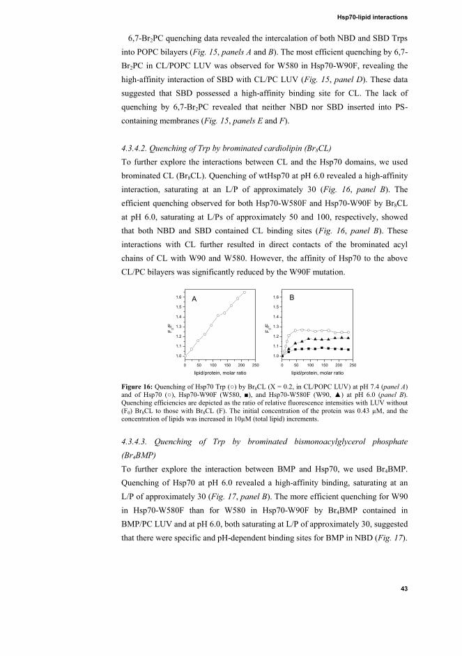
Hsp70-lipid interactions
43
6,7-Br2PC quenching data revealed the intercalation of both NBD and SBD Trps into POPC bilayers (Fig. 15, panels A and B). The most efficient quenching by 6,7-Br2PC in CL/POPC LUV was observed for W580 in Hsp70-W90F, revealing the high-affinity interaction of SBD with CL/PC LUV (Fig. 15, panel D). These data suggested that SBD possessed a high-affinity binding site for CL. The lack of quenching by 6,7-Br2PC revealed that neither NBD nor SBD inserted into PS-containing membranes (Fig. 15, panels E and F). 4.3.4.2. Quenching of Trp by brominated cardiolipin (Br8CL) To further explore the interactions between CL and the Hsp70 domains, we used brominated CL (Br8CL). Quenching of wtHsp70 at pH 6.0 revealed a high-affinity interaction, saturating at an L/P of approximately 30 (Fig. 16, panel B). The efficient quenching observed for both Hsp70-W580F and Hsp70-W90F by Br8CL at pH 6.0, saturating at L/Ps of approximately 50 and 100, respectively, showed that both NBD and SBD contained CL binding sites (Fig. 16, panel B). These interactions with CL further resulted in direct contacts of the brominated acyl chains of CL with W90 and W580. However, the affinity of Hsp70 to the above CL/PC bilayers was significantly reduced by the W90F mutation.
0 50 100 150 200 250
1.0
1.1
1.2
1.3
1.4
1.5
1.6
0 50 100 150 200 250
1.0
1.1
1.2
1.3
1.4
1.5
1.6
F 0/F
lipid/protein, molar ratio
B
F 0/F
lipid/protein, molar ratio
A
Figure 16: Quenching of Hsp70 Trp (○) by Br8CL (X = 0.2, in CL/POPC LUV) at pH 7.4 (panel A) and of Hsp70 (○), Hsp70-W90F (W580, ■), and Hsp70-W580F (W90, ▲) at pH 6.0 (panel B). Quenching efficiencies are depicted as the ratio of relative fluorescence intensities with LUV without (F0) Br8CL to those with Br8CL (F). The initial concentration of the protein was 0.43 μM, and the concentration of lipids was increased in 10μM (total lipid) increments. 4.3.4.3. Quenching of Trp by brominated bismonoacylglycerol phosphate (Br4BMP) To further explore the interaction between BMP and Hsp70, we used Br4BMP. Quenching of Hsp70 at pH 6.0 revealed a high-affinity binding, saturating at an L/P of approximately 30 (Fig. 17, panel B). The more efficient quenching for W90 in Hsp70-W580F than for W580 in Hsp70-W90F by Br4BMP contained in BMP/PC LUV and at pH 6.0, both saturating at L/P of approximately 30, suggested that there were specific and pH-dependent binding sites for BMP in NBD (Fig. 17).

Results
44
0 50 100 150 200
1.0
1.2
1.4
1.6
0 50 100 150 200
1.0
1.2
1.4
1.6
F 0/F
lipid/protein, molar ratio
A
F 0/F
lipid/protein, molar ratio
B
Figure 17: Quenching of Hsp70 Trp fluorescence by 6,7-(■), 9,10-(●), or 11,12Br2-PC (▲, X = 0.3) contained in BMP/POPC (XBMP = 0.2) LUV at pH 6.0 (panel A). Quenching of Hsp70 (○), Hsp70-W90F (W580, ■), and Hsp70-W580F (W90, ▲) Trp fluorescence by Br4BMP (X = 0.2, in POPC LUV) at pH 6.0 (panel B). The quenching efficiencies are depicted as the ratio of relative fluorescence intensities with LUV without (F0) and with the brominated lipid (F), as indicated. 4.3.4.4. Quenching of Trp by brominated phosphatidylserine (Br2PS) In keeping with binding of PS to Hsp70 an efficient quenching of Trp emission by Br2PS was observed (Fig. 18, panels A and B). Furthermore, the quenching of both Hsp70-W90F and Hsp70-W580F Trp residues by Br2PS at pH 6.0, both saturating at L/P of approximately 100 and 50, respectively, suggested that both NBD and SBD contained binding sites for PS (Fig. 18, panel B).
0 50 100 150 200 250
1.0
1.1
1.2
1.3
1.4
1.5
1.6
0 50 100 150 200 250
1.0
1.1
1.2
1.3
1.4
1.5
1.6
F 0/F
lipid/protein, molar ratio
B
F 0/F
lipid/protein, molar ratio
A
Figure 18: Quenching of Hsp70 Trp (○) by Br2PS (X = 0.2, in PS/POPC LUV) at pH 7.4 (panel A) and pH 6.0 for Hsp70 (○), Hsp70-W90F (W580, ■), and Hsp70-W580F (W90, ▲, panel B). Quenching efficiencies are depicted as the ratio of relative fluorescence intensities with LUV with PS (F0) to those with Br2PS (F). The initial concentration of the protein was 0.43 μM, while the concentration of lipids was increased in 10-μM increments. 4.3.5. Penetration of Hsp70 into lipid monolayers
In order to study the extent of the insertion of Hsp70 into membranes, the penetration of Hsp70 into lipid monolayers (Langmuir-films) residing on a gas/water interface was measured using a Langmuir balance. More specifically, the insertion of Hsp70 into monolayers at a range of initial lateral pressures π0 was observed by recording the increase in the surface pressure Δπ following the injection of Hsp70 into the aqueous subphase. The equilibrium lateral pressures estimated for biomembranes are approximately 33–35 mN/m (Demel et al. 1975). POPC Langmuir films gave exclusion pressures πc of 47 and 38 mN/m for Hsp70 at pH 7.4 and 6.0, respectively, indicating that Hsp70 could efficiently insert into PC films (Fig. 19). However, the impact of acidic phospholipids on Hsp70
Hsp70-lipid interactions
45
penetration was distinctly different from that with POPC, with low exclusion pressures πc measured for Hsp70 monolayers composed of CL/PC (XCL = 0.20), BMP/PC (XBMP = 0.20), and PS/PC (XPS = 0.20, Table 4). Low exclusion pressures in the presence of acidic phospholipids suggested a lack of significant intercalation of Hsp70 into lipid films.
10 20 30 40 500
5
10
15
20
25
30
0 10 20 30 400
5
10
15
20
25
30
, mN
/m
o, mN/m
A
, mN
/m
o, mN/m
B
Figure 19: Penetration of Hsp70 into lipid monolayers using 20 mM Hepes, 0.1 mM EDTA as the buffer at pH 7.4 (panel A) or 6.0 (panel B), illustrated as a function of the initial surface pressure π0 and the increase in surface pressure (Δπ) following the injection of Hsp70 into the subphase (0.1 μM final concentration). Lipid monolayers were POPC (●), CL/POPC (XCL = 0.2, ▲), POPS/POPC (XPOPS = 0.2, ■), POPS/Chol/Spm (XPOPS = 0.10, XChol = XSpm = 0.45, □), and BMP/POPC (XBMP = 0.2, ∆). Table 4
Critical packing pressures (πc) for Hsp70 and corresponding lipid composition
Lipid composition πc (mN/m)
pH 7.4 pH 6.0 POPC 47 38
CL/PC (XCL = 0.20) 27.5 31
BPM/PC (XBMP = 0.20) 27 PS/PC (XPOPS = 0.20) 33 35
POPS/Chol/Spm (XPOPS = 0.10, XChol = XSpm = 0.45) 31 34
4.4. Modulation of the hydrolytic activity of PLA2 by Hsp70 (V)
Hsp70 has been shown to attenuate the oligomerization of proteins into amyloid fibers, as previously observed with amyloid-forming proteins (Muchowski et al. 2005). Accordingly, the influence of Hsp70 on the oligomerization and hydrolytic activity of PLA2 was studied in order to understand the functional role of Hsp70 in protein oligomerization at the membrane interface. A small increase in the extent of hydrolysis of PLA2 was observed in the presence of Hsp70 without ATP,

Results
46
suggesting that Hsp70 promoted the activity of PLA2 (Fig. 20). Interestingly, a 30% increase in the hydrolytic activity of bvPLA2 was observed in the presence of Hsp70 plus ATP, indicating that Hsp70 modulated the activity of PLA2 in an ATP-dependent manner (Fig. 20).
Figure 20: Hydrolysis of 1.2 μM C28-O-PHPM by 2 nM PLA2 with 2 μMATP-Mg2+ (Δ), or in the absence (○) and presence of 2 nM Hsp70 (□) and with 2 μM ATP-Mg2+ (■) in 2.0 ml of 5 mM Hepes, 0.1 mM EDTA, and 1 mM CaCl2, at pH 7.4 and 37°C, with stirring. Each point represents the average of three independent experiments, and the error bars indicate standard deviations. The symbols represent only one data point out of 300, and the continuous lines connect each data point.
Next, we studied the effects of Hsp70 on lysoPC-induced inhibition of PLA2.
LysoPC is known to induce amyloid oligomerization of PLA2. Accordingly, we accelerated amyloid oligomerization of PLA2 by pre-incubation with lysoPC (Fig. 21). The pre-incubation of PLA2 with lysoPC attenuated the extent of hydrolysis (Fig. 21). However, when Hsp70 was present together with ATP, the amount of product was significantly increased compared to those for PLA2 alone and for PLA2 pre-incubated with lysoPC (Fig. 21).
Figure 21: Hydrolytic activity as a function of time for 1.25 μM C28-O-PHPM with 2 nM PLA2 (○), for 2 nM PLA2 pre-incubated with 20 μM lysoPC prior to adding to the substrate (●), and for 2 nM PLA2 pre-incubated with 20 μM lysoPC prior to adding the substrate, in which 2 nM Hsp70 and 2 μM ATP-Mg2+ (■) were added.

PLA2-Hsp70 interactions
47
4.5. Effects of oligomerization-mediating sequences of PLA2 on its hydrolytic activity (VI)
The oligomerization-mediating sequences KMYFNLI and AALSYGFYG in bv and human LfPLA2, respectively, can mediate PLA2-PLA2 interactions and likely influence the activity of PLA2. Accordingly, the hydrolytic activities of PLA2 in presence of its oligomerization-mediating sequences were assessed by observing the hydrolysis of the pyrene-labeled phospholipid C28-O-PHPM analog.
Figure 22: Inhibition of bvPLA2 by 85KMYFNLI91. Hydrolysis of 1.25 μM C28-0-PHPM by 2 nM bvPLA2 (○) was measured as a function of time in the presence of 2 (□) or 4 nM (∆) peptide added to the substrate (without pre-incubation). Alternatively, 4 pmol of the peptide was pre-incubated with 4 pmol of bvPLA2 for 5 (●), 10 (■), or 20 (▲) min prior to addition to the substrate.
The oligomerization-mediating sequences of sPLA2 were found to attenuate the hydrolytic activity of sPLA2. More specifically, hydrolysis of the negatively charged phospholipid analog C28-O-PHPM by bvPLA2 in presence of 2 nM KMYFNLI was decreased to 77%–85% (Fig. 22). This attenuation became more pronounced in presence of 4 nM of KMYFNLI (Fig. 22). Furthermore, pre-incubation of bvPLA2 with KMYFNLI prior to the assay caused complete inhibition of the hydrolytic activity of PLA2 (Fig. 22).
In control experiments, a scrambled peptide (YNFLIMK) was used in which the
amino acid composition was identical to that of KMYFNLI, but with a randomized sequence. Importantly, the scrambled peptide did not influence the hydrolytic activity of bvPLA2 (Fig. 23).

Results
48
Figure 23: Hydrolysis of 1.25 μM C28-0-PHPM by 2 nM bvPLA2 (○) was measured as a function of time in the presence of 80 nM of YNFLIMK (□) or 17AALSYGFYG25 (∆) pre-incubated with the enzyme for 10 min prior to addition to the substrate solution.
We then studied the influence of AALSYGFYG, an oligomerization-mediating
sequence of LfPLA2, on the hydrolytic activity of LfPLA2 (Table 1). The activity of LfPLA2 decreased by 28% in presence of 40 nM AALSYGFYG. However, in presence 80 nM AALSYGFYG, more pronounced attenuation was observed. The hydrolysis of C28-O-PHPM by LfPLA2 also decreased to approximately 36% (Fig. 24). Furthermore, to assess the cross reactivity of this inhibition in different sPLA2 proteins, the effects of AALSYGFYG on the activity of bvPLA2 were evaluated. Interestingly, AASLSYGFYG did not influence bvPLA2 activity (Fig. 23).
Figure 24: Hydrolysis of 1.25 μM C28-0-PHPM by 1 μL (○) LfPLA2 as a function of time in the presence of 40 (□) or 80 (∆) nM 17AALSYGFYG25 pre-incubated with the enzyme for 5 min prior to addition to the substrate solution. Reactions were monitored in 2.0 ml of 5 mM Hepes, 0.1 mM EDTA, and 1 mM CaCl2 at pH 7.4 and 37°C.

Discussion
49
5. DISCUSSION
5.1. Amyloid-prone sequences are an inherent features of amphipathic α-helical AMPs (I)
AMPs sequences can adopt different conformations depending on their environments (Rozek et al. 2000, Jenssen et al. 2006). Lipid membranes also play an important role in modulating the conformations of AMPs (Latal et al. 1997, Wang et al. 1998). Changes in the secondary structure of AMPs enable them to interact with and destabilize lipid bilayers of their target cells, exposing the hydrophobic regions needed for membrane incorporation and subsequent aggregation (Giangaspero et al. 2001). AMPs are unstructured in an aqueous solution, but have well-defined secondary structures upon interaction with acidic phospholipid bilayers (Hirsh et al. 1996). In the absence of lipid, no peptide oligomerization was observed, suggesting that the hydrophobic regions were not exposed in solution. The membrane environment thus promotes conformational switching to expose hydrophobic residues, which then drive self-association by hydrophobic interactions between the non-polar side chains. Hydrophobicity, the propensity to form α-helical or β-sheet secondary structures, and the net charge of the polypeptide chain modulate aggregation of proteins in a partially or completely unfolded state (Calamai et al. 2003). Here, we showed that the information about the secondary structure of α-helical AMPs in lipid bilayers, which was critical for their activity, was coded in their sequences.
Amyloidogenic proteins/peptides contain hydrophobic sequences that, once exposed, are highly prone to oligomerization (Lopez de la Paz et al. 2004). Recent results have demonstrated that AMPs, such as tem L, tem B, LL-37, magainin 2, and plantaricin A, form amyloid-like Congo red-staining fibers in membranes containing negatively charged phospholipids (Zhao et al. 2005, Zhao et al. 2006, Sood et al. 2007). Sequence analysis indicated that amyloid-prone sequence were common features of α-helical AMPs, thus supporting the notion that the cytotoxic actions of α-helical AMPs and amyloid-forming peptides/proteins were the same and that these peptides possessed specific sequence motifs encoding the amyloid core structure. In conclusion, conformational switching, aggregation, and amyloid formation are inherent to the sequences of α-helical AMPs. The structural transformation of fibril-forming proteins is a prerequisite to form amyloid aggregates (Dobson 2003). The recognition of ambivalent sequences that can be easily converted between α-helical and β-sheet structures and amyloid fibrils in a

Discussion
50
proper membrane environment further reveals an important new aspect of the molecular mechanisms of amphipathic α-helical AMPs.
5.2. Mechanism of gelsolin fibrillation in the presence of oxidized phospholipids (II)
FAF is a neurodegenerative disease that involves the deposition of D187N/Y mutant gelsolin fragments as amyloid in various tissues, accompanied by dermatologic, neurologic, and ophthalmologic disorders (Maury et al. 1990, Maury et al. 2003). The formation and cytotoxic activity of amyloid proteins are membrane-associated processes (Gorbenko et al. 2006a, Kinnunen 2009) wherein lipid-protein interactions accelerate fibrillation and induce membrane permeabilization (Zhao et al. 2004). Similar to other amyloid diseases, FAF is associated with oxidative stress. The latter results in an extensive chemical modification of biomolecules, such as the formation of a myriad of phospholipids with oxidatively modified acyl chains containing various functional groups. Here, we demonstrated that PoxnoPC accelerated the fibrillation of FtG179–194. PoxnoPC, formed upon oxidative scission of PCs bearing a cis-9 double bond containing a fatty acid (e.g., palmitoleic, linoleic, or linolenic acid) in the sn-2 position (Fruhwirth et al. 2007), is predominantly found in the extracellular compartment (Palinski et al. 1989). Therefore, the interactions of the extracellular D187N mutant gelsolin with oxidized phospholipids may be involved in the development of FAF. Moreover, human skin experiences the most UV light exposure, and this can also result in lipid oxidation (Fruhwirth et al. 2007). Therefore, this may explain the deposition of aggregated gelsolin in the cutaneous tissue in FAF patients (Tanskanen et al. 2006).
The kinetic parameters revealed in this study indicated that the acceleration of fibrillation was directly dependent on both the concentration and the physical (aggregation) state of PoxnoPC. The maximum increases in ThT fluorescence and Kapp for fibril formation were observed at close to the CMC (approximately 22.5 μM) of PoxnoPC. The rates of fibril elongation and nucleation were proportional to the concentration of PoxnoPC, while the rates of nucleation were different below and above the CMC. Our observation was consistent with enhanced amyloid fibril formation in the presence of lipids and surfactants at submicellar concentrations by apolipoprotein C-II (Hatters et al. 2001), Aβ (Sabate et al. 2005) and α- and β-syn (Rivers et al. 2008). Oxidative modifications of phospholipids profoundly alter the biophysical properties of membranes, including their polarity profiles, and are therefore expected to have pronounced effects on membrane-associated amyloid

Discussion
51
formation (Kinnunen et al. 2012). More specifically, oxidatively modified chains no longer remain in the membrane hydrocarbon region, but can adopt the so-called extended conformation protruding into the aqueous phase (Sabatini et al. 2006, Mattila et al. 2008).
Fibril formation by FtG179–194 in the presence of PoxnoPC followed simple nucleation-dependent kinetics with the formation of a metastable intermediate in the lag phase (Fig. 25). The appearance of a concentration-dependent lag phase indicated that PoxnoPC may stabilize transient protofibrils/prefibrillar oligomers in the folding/aggregation free energy landscape preceding the formation of nontoxic mature amyloids by FtG179–194 (Kinnunen 2009). More specifically, Trp fluorescence data suggested that a 1:1 complex formation by PoxnoPC and FtG179–
194 (most likely via a Schiff base) occurred at the membrane hydrocarbon-water interface in close proximity to the PC head group. The nucleation from transient lipid-peptide intermediates was slow and rate-limiting to the overall fibril formation process. The slow structural transition of FtG179–194 in peptide-lipid comicelle prefibrillar oligomers led to the formation of protofibrils. Finally, fibril elongation would proceed via the addition of monomers to the existing protofibrils.
Figure 25: A schematic model for fibril formation by FtG179–194 in the presence of a PoxnoPC micelle, obeying a concentration-dependent nucleation mechanism. 5.3. Hsp70 binds peripherally to acidic phospholipid membrane surfaces (III)
We demonstrated that Hsp70 bound to the organelle-specific lipids CL, BMP, and PS found in mitochondria, lysosomes, and the membrane surface of cancer cells, respectively. Additionally, Hsp70 associated with the above acidic phospholipid-containing membranes surfaces peripherally, without insertion into the lipid bilayers. An electrostatic attraction, hydrogen bonding, and partial penetration have been purposed to be the underlying mechanisms for peripheral membrane attachment of proteins. Here, we demonstrated a new mechanism of Hsp70 binding
Elongation Mature amyloid fibre Oxidized phospholipid
Gelsolin +
Discussion
52
to membrane surfaces involving hydrophobic interactions without the penetration into lipid bilayers.
Our results of Trp fluorescence emission and quenching by AcrA along with Langmuir-balance data indicated that Hsp70 did not penetrate into bilayers in the presence of CL/PC LUV. Hsp70 bound peripherally to the CL/PC bilayer surface. However, the above result contradicted Trp quenching by brominated PCs and CL in keeping with the insertion of Hsp70 into the bilayer. The above contradiction, where the brominated acyl chain was in contact with the Trp residue of Hsp70 without intercalation, allowed us to propose a mechanism in which brominated phospholipid acyl chains of PC and CL adopted the extended conformation, as previously described for cytochrome c (Kinnunen 1992, Kinnunen 1996). Similarly, Hsp70 also bound to BMP and PS peripherally via an extended phospholipid anchorage.
5.4. The Hsp70-BMP interaction is essential for lysosomal membrane stabilization (IV)
The association and localization of Hsp70 to the lysosomal membrane leads to a cytoprotective effect and interferes with lysosomal cell death pathways (Jäättelä 1999, Nylandsted et al. 2000, Nylandsted et al. 2004). Here, we showed that Hsp70 selectively bound to membranes containing BMP in an acidic environment, which suggested the specificity of Hsp70 towards lysosomal membranes in stress conditions. The Hsp70-BMP interaction plays a crucial role in the activation of the acid sphingomyelinase (aSMase) and stabilization of lysosomes. The enzymatic conversion of SM to ceramide enhances membrane acyl chain order and increases the lateral packing of lipid bilayers in vitro (Holopainen et al. 1998). The direct interaction between Hsp70 and BMP was required for this activation, and the downstream cytoprotective properties of Hsp70 could be blocked by an antibody against a BMP. Moreover, W90 of NBD was required for this interaction, and its mutation resulted in an Hsp70 protein with compromised BMP-binding ability, rendering it inactive and unable to prevent lysosomal membrane permeabilization.
5.5. Hsp70 controls PLA2 oligomerization (V)
In an ATP-dependent process, Hsp70 binds to unfolded and misfolded substrates and guides them to their native conformations (Lindquist et al. 1988). Here, we showed that Hsp70 could rescue the activity of PLA2 and reverted lysoPC-induced inactivation of PLA2 in an ATP-dependent manner. Most likely Hsp70 attenuating

Discussion
53
the conversion of active prefibrillar oligomers of PLA2 into inactive amyloid fibers (Fig. 26) which resulted in more extensive hydrolysis of pyrene-labeled phospholipid C28-O-PHPM analog. Our results are in line with earlier observations that the chaperone action of Hsp70 was mediated by its binding to prefibrillar species of Aβ (Evans et al. 2006), α-syn (Dedmon et al. 2005), and polyglutamine (Wacker et al. 2004) rather than to mature fibrils. Moreover, Hsp70 has also been shown to increase the activity protein kinase C (PKC), serving as a stabilizing factor for PKC (Bazzi et al. 1992, Gao et al. 2002). Our results on the activation of aSMase and PLA2 suggested that Hsp70 promoted the catalytic activity of lipolytic enzyme by prolonging the lifetime of the active state of enzymes (Fig. 26).
The binding of ATP to NBD induces a conformational change in SBD, which allows substrate recognition. The ATP-dependent activation of PLA2 by Hsp70 indicated the association of SBD with PLA2. The structural features for substrate interaction/recognition involve a stretch of seven hydrophobic residues, with charged residues vicinal to both ends, found within every 36 amino acid stretch in the proteome (Rudiger et al. 1997b). Hsp70 recognizes hydrophobic residues in the substrates, i.e., structural features typically exposed by non-native proteins, but normally buried in native protein. However, it is still unclear which sequence of PLA2 interacts with the Hsp70.
Figure 26: A schematic model for the activation of PLA2 by Hsp70 in the presence of ATP-Mg2+
by attenuation of the conversion of catalytically active protofibrils into inactive amyloid fibers.
5.6. A peptide derived from putative oligomerization-mediating sequences inhibits PLA2 (VI)
PLA2 has been implicated in inflammation and is thus connected to diverse diseases, such as cancer, ischemia, atherosclerosis, and schizophrenia (Dennis et al. 2011). Therefore, the activity of PLA2 needs to be critically controlled. Along these lines, the catalytic rate of PLA2 can be modulated by amyloidogenic peptides, e.g., amyloid Aβ-peptide (Lehtonen et al. 1996), several AMPs, e.g., tem B, tem L, indolicidin, magainin 2 (Zhao et al. 2003), LL-37 (Pochet et al. 2006),
Activity
+ Hsp70
amyloid-like fibrils Oligomers `protofibrils´ ++++ (High) ---- (Low)

Discussion
54
bombolitin III (Signor et al. 1994), and melittin (Koumanov et al. 2003), which all enhance the activity of PLA2.
The interfacial activation and oligomerization of PLA2 are interconnected processes. Along these lines, we showed that peptides derived from putative oligomerization-mediating sequences could inhibit PLA2. Moreover, tem B has been proposed to influence the conformational equilibrium of PLA2 by forming higher-order co-oligomers (Code et al. 2009), thus activating PLA2 (Zhao et al. 2003). The amyloidogenic sequence in bvPLA2 is found within to the interfacial recognition site, which binds to the substrate (Scott et al. 1990) and is implicated in the neurotoxicity of bvPLA2 (Kini et al. 1986, Nicolas et al. 1997). Based on the findings mentioned above, we can conclude that a shift in oligomerization can be induced by peptides binding to the enzyme via both nonspecific and specific interactions. Identification of oligomerization-mediating sequences in different lipid-activated enzymes could be used to obtain specific and potent peptide inhibitors.

Acknowledgements
55
6. ACKNOWLEDGEMENTS The research presented in this thesis was carried out at the Helsinki Biophysics Biomembrane Group (HBBG), Institute of Biomedicine, University of Helsinki during the years 2007-2010 and in the Department of Biomedical Engineering and Computational Science (BECS), Aalto University School of Science during the years 2010-2013. This thesis would not have been possible without many valuable contributions. First of all, I would like to express my deepest gratitude to my supervisor Professor Paavo Kinnunen for providing me the opportunity to pursue a Ph.D. in the field of membranes biophysics. I feel privileged to have you as an excellent mentor whose research skills and knowledge have been indisputable in the research field. I would like to extend my sincerest gratitude for your advices, understanding, inspiring guidance and consistent support throughout my research stay. You are really gem of a person whose principles, ideologies, profound scientific knowledge and enthusiasm towards new discoveries have been invaluable sources of inspiration. I am also very grateful to Professor Marja Jäättelä and Dr. Thomas Kirkegaard of Danish cancer society research center, Copenhagen, Denmark for being a wonderful research collaborator. I sincerely thank the reviewers of this thesis, Docent Hongxia Zhao from institute of biotechnology and Docent Tuomas Haltia from department of biosciences, University of Helsinki for accepting and ranking my thesis work among top 10% of the dissertations in the field of research. Assoc. Prof. Günther Peters (Technical University of Denmark) is deeply thanked for accepting the role of an opponent. It is my honor to have him as an opponent. I thank the past and present members of HBBG for vibrant atmosphere and valued friendship. I thank Chris, Laura and Behnam for collaboration in the original publications of this thesis work. I thank Sanjeev, Rohit and Roman for helpful comments on dissertation manuscript. I also extend my thanks to Roberto, Juha-Matti, Vladimir, Aqeel, Juha-Pekka, Karen, Yegor, Mikko, Pavol, Petteri, Sakari,Tuuli, Samuli, Olga and Kateryna as well as visiting scientists for making the last six years an enriching experience. I will certainly miss our endless discussions on the drink table. I thank laboratory technicians Kristiina Söderholm for all her time and effort when I was at university of Helsinki.

Acknowledgements
56
I am indebted to Sanjeev and Shishir for great care, support and help showered upon me. It was a great pleasure to discuss science and life with you. I owe million thanks to both of you. I thank Shishir and Sanjeev for proof reading the dissertation manuscript. I thank Amit and Pankaj for inspiring me to study chemistry and biotechnology. I also wish to express my gratitude to Dr. R K. Trikha, University of Delhi, my mentor during my undergraduate studies, and Prof. R. Barthwal, Indian Institute of Technology, Roorkee, my master’s thesis supervisor and mentor. I sincerely thank many friends and colleagues for their help and friendship. It gives me pleasure to thank my friends: Tommi, Akhilesh, Sriram, Bhupendra, Kiran, Aniket, Anmol, Leena, and Ashwini for their constructive discussions and wonderful time we shared in Helsinki. I also thank my friends Malviya, Abhishek, Vikrant, Swati and Indra. I thank Navneet, Kavita, Rahul, Prajakta, Sachin and Sheeba for their warm hospitality. Above all my stay could not have been possible without the unconditional support of my love Khusboo and little princess Aarya. I thank you for keeping my stay full of fun and balanced with work life. I have no words for my parents (Premlata & Ashok), uncles (Binod, Nirmal, Pawan and Anil), and aunts (Pramila, Prity, Sangeeta and Usha) for being constant source of inspiration. I especially want to show gratitude to my uncle Anil, my brother Anup and my sister Ruby for their unparalleled support and motivation. I thank BECS, HBBG, FEBS and EuroMembrane for travel grants which enable me to present my work at different international conferences. I also thank the SITRA, Finland for SITRA fellowship for incoming Indian researcher for the 7 months as well as the travel grants. I also thank BECS for their financial support for 7 month to finish my PhD thesis. I also thank Facebook, tweeter, Skype, Internet and Nokia for connecting me to my friends and family. Ajay Mahalka Dec 12, 2013 Espoo/Helsinki

References
57
7. REFRENCES
Abraham T, Marwaha S, Kobewka DM, Lewis RN, Prenner EJ, Hodges RS, McElhaney RN (2007) The relationship between the binding to and permeabilization of phospholipid bilayer membranes by GS14dK4, a designed analog of the antimicrobial peptide gramicidin S. Biochim Biophys Acta 1768:2089-2098.
Alakoskela J-I, Kinnunen PKJ (2004) Phospholipid main phase transition assessed by fluorescence spectroscopy. Rev Fluoresc 1:257-297.
Alder GM, Austen BM, Bashford CL, Mehlert A, Pasternak CA (1990) Heat shock proteins induce pores in membranes. Biosci Rep 10:509-518.
Arispe N, De Maio A (2000) ATP and ADP modulate a cation channel formed by Hsc70 in acidic phospholipid membranes. J Biol Chem 275:30839-30843.
Auluck PK, Chan HY, Trojanowski JQ, Lee VM, Bonini NM (2002) Chaperone suppression of alpha-synuclein toxicity in a Drosophila model for Parkinson's disease. Science 295:865-868.
Auvynet C, El Amri C, Lacombe C, Bruston F, Bourdais J, Nicolas P, Rosenstein Y (2008) Structural requirements for antimicrobial versus chemoattractant activities for dermaseptin S9. FEBS J 275:4134-4151.
Balsinde J, Winstead MV, Dennis EA (2002) Phospholipase A(2) regulation of arachidonic acid mobilization. FEBS Lett 531:2-6.
Balsinde J, Balboa MA, Insel PA, Dennis EA (1999) Regulation and inhibition of phospholipase A2. Annu Rev Pharmacol Toxicol 39:175-189.
Bazzi MD, Nelsestuen GL (1992) Autophosphorylation of protein kinase C may require a high order of protein-phospholipid aggregates. J Biol Chem 267:22891-22896.
Bertelsen EB, Chang L, Gestwicki JE, Zuiderweg ER (2009) Solution conformation of wild-type E. coli Hsp70 (DnaK) chaperone complexed with ADP and substrate. Proc Natl Acad Sci U S A 106:8471-8476.
Bhattacharyya T, Karnezis AN, Murphy SP, Hoang T, Freeman BC, Phillips B, Morimoto RI (1995) Cloning and subcellular localization of human mitochondrial hsp70. J Biol Chem 270:1705-1710.
Bieschke J, Zhang Q, Bosco DA, Lerner RA, Powers ET, Wentworth P,Jr, Kelly JW (2006) Small molecule oxidation products trigger disease-associated protein misfolding. Acc Chem Res 39:611-619.
Boden M, Yuan Z, Bailey TL (2006) Prediction of protein continuum secondary structure with probabilistic models based on NMR solved structures. BMC Bioinformatics 7:68.
Brocchieri L, Conway de Macario E, Macario AJ (2008) hsp70 genes in the human genome: Conservation and differentiation patterns predict a wide array of overlapping and specialized functions. BMC Evol Biol 8:19-2148-8-19.
Bucciantini M, Giannoni E, Chiti F, Baroni F, Formigli L, Zurdo J, Taddei N, Ramponi G, Dobson CM, Stefani M (2002) Inherent toxicity of aggregates implies a common mechanism for protein misfolding diseases. Nature 416:507-511.
Buchberger A, Theyssen H, Schroder H, McCarty JS, Virgallita G, Milkereit P, Reinstein J, Bukau B (1995) Nucleotide-induced conformational changes in the ATPase and substrate binding

References
58
domains of the DnaK chaperone provide evidence for interdomain communication. J Biol Chem 270:16903-16910.
Buckiova D, Ranjan S, Newman TA, Johnston AH, Sood R, Kinnunen PK, Popelar J, Chumak T, Syka J (2012) Minimally invasive drug delivery to the cochlea through application of nanoparticles to the round window membrane. Nanomedicine (Lond) 7:1339-1354.
Bukau B, Horwich AL (1998) The Hsp70 and Hsp60 chaperone machines. Cell 92:351-366.
Calamai M, Taddei N, Stefani M, Ramponi G, Chiti F (2003) Relative influence of hydrophobicity and net charge in the aggregation of two homologous proteins. Biochemistry 42:15078-15083.
Cevc G (1990) Membrane electrostatics. Biochim Biophys Acta 1031:311-382.
Chan YH, Boxer SG (2007) Model membrane systems and their applications. Curr Opin Chem Biol 11:581-587.
Chattopadhyay A, London E (1987) Parallax method for direct measurement of membrane penetration depth utilizing fluorescence quenching by spin-labeled phospholipids. Biochemistry 26:39-45.
Chen CD, Huff ME, Matteson J, Page L, Phillips R, Kelly JW, Balch WE (2001) Furin initiates gelsolin familial amyloidosis in the Golgi through a defect in Ca(2+) stabilization. EMBO J 20:6277-6287.
Chiappori F, Merelli I, Colombo G, Milanesi L, Morra G (2012) Molecular mechanism of allosteric communication in Hsp70 revealed by molecular dynamics simulations. PLoS Comput Biol 8:e1002844.
Chibowski E, Holysz L, Jurak M (2008) Effect of a lipolytic enzyme on wettability and topography of phospholipid layers deposited on solid support. Colloid Surf A: Physiocochem Eng Aspects 321:131-136.
Chiti F, Dobson CM (2006) Protein misfolding, functional amyloid, and human disease. Annu Rev Biochem 75:333-366.
Chiu CR, Huang WN, Wu WG, Yang TS (2009) Fluorescence single-molecule study of cobra phospholipase A2 action on a supported gel-phase lipid bilayer. Chemphyschem 10:549-558.
Ciocca DR, Calderwood SK (2005) Heat shock proteins in cancer: diagnostic, prognostic, predictive, and treatment implications. Cell Stress Chaperones 10:86-103.
Code C, Mahalka AK, Bry K, Kinnunen PKJ (2010) Activation of phospholipase A2 by 1-palmitoyl-2-(9'-oxo-nonanoyl)-sn-glycero-3-phosphocholine in vitro. Biochim Biophys Acta 1798:1593-1600.
Code C, Domanov YA, Killian JA, Kinnunen PKJ (2009) Activation of phospholipase A2 by temporin B: formation of antimicrobial peptide-enzyme amyloid-type cofibrils. Biochim Biophys Acta 1788:1064-1072.
Code C, Domanov Y, Jutila A, Kinnunen PKJ (2008) Amyloid-type fiber formation in control of enzyme action: interfacial activation of phospholipase A2. Biophys J 95:215-224.
Comte J, Maisterrena B, Gautheron DC (1976) Lipid composition and protein profiles of outer and inner membranes from pig heart mitochondria. Comparison with microsomes. Biochim Biophys Acta 419:271-284.
Conchillo-Sole O, de Groot NS, Aviles FX, Vendrell J, Daura X, Ventura S (2007) AGGRESCAN: a server for the prediction and evaluation of "hot spots" of aggregation in polypeptides. BMC Bioinformatics 8:65.

References
59
Cunningham TJ, Yao L, Lucena A (2008) Product inhibition of secreted phospholipase A2 may explain lysophosphatidylcholines' unexpected therapeutic properties. J Inflamm (Lond) 5:17.
Daugaard M, Rohde M, Jäättelä M (2007) The heat shock protein 70 family: Highly homologous proteins with overlapping and distinct functions. FEBS Lett 581:3702-3710.
de la Chapelle A, Kere J, Sack GH,Jr, Tolvanen R, Maury CP (1992) Familial amyloidosis, Finnish type: G654----a mutation of the gelsolin gene in Finnish families and an unrelated American family. Genomics 13:898-901.
De Strooper B (2010) Proteases and proteolysis in Alzheimer disease: a multifactorial view on the disease process. Physiol Rev 90:465-494.
Dedmon MM, Christodoulou J, Wilson MR, Dobson CM (2005) Heat shock protein 70 inhibits alpha-synuclein fibril formation via preferential binding to prefibrillar species. J Biol Chem 280:14733-14740.
Demel RA, Geurts van Kessel WS, Zwaal RF, Roelofsen B, van Deenen LL (1975) Relation between various phospholipase actions on human red cell membranes and the interfacial phospholipid pressure in monolayers. Biochim Biophys Acta 406:97-107.
Dennis EA, Cao J, Hsu YH, Magrioti V, Kokotos G (2011) Phospholipase A2 enzymes: physical structure, biological function, disease implication, chemical inhibition, and therapeutic intervention. Chem Rev 111:6130-6185.
Dennis EA (1997) The growing phospholipase A2 superfamily of signal transduction enzymes. Trends Biochem Sci 22:1-2.
Dennis EA (1994) Diversity of group types, regulation, and function of phospholipase A2. J Biol Chem 269:13057-13060.
Dobson CM (2003) Protein folding and misfolding. Nature 426:884-890.
Dobson CM (1999) Protein misfolding, evolution and disease. Trends Biochem Sci 24:329-332.
Dworniczak B, Mirault ME (1987) Structure and expression of a human gene coding for a 71 kd heat shock 'cognate' protein. Nucleic Acids Res 15:5181-5197.
East JM, Lee AG (1982) Lipid selectivity of the calcium and magnesium ion dependent adenosinetriphosphatase, studied with fluorescence quenching by a brominated phospholipid. Biochemistry 21:4144-4151.
Ellis RJ (2001) Macromolecular crowding: an important but neglected aspect of the intracellular environment. Curr Opin Struct Biol 11:114-119.
Epand RM, Vogel HJ (1999) Diversity of antimicrobial peptides and their mechanisms of action. Biochim Biophys Acta 1462:11-28.
Escriba PV, Gonzalez-Ros JM, Goni FM, Kinnunen PKJ, Vigh L, Sanchez-Magraner L, Fernandez AM, Busquets X, Horvath I, Barcelo-Coblijn G (2008) Membranes: a meeting point for lipids, proteins and therapies. J Cell Mol Med 12:829-875.
Evans CG, Wisen S, Gestwicki JE (2006) Heat shock proteins 70 and 90 inhibit early stages of amyloid beta-(1-42) aggregation in vitro. J Biol Chem 281:33182-33191.
Farkas B, Hantschel M, Magyarlaki M, Becker B, Scherer K, Landthaler M, Pfister K, Gehrmann M, Gross C, Mackensen A, et al (2003) Heat shock protein 70 membrane expression and melanoma-associated marker phenotype in primary and metastatic melanoma. Melanoma Res 13:147-152.

References
60
Farooqui AA, Ong WY, Horrocks LA (2006) Inhibitors of brain phospholipase A2 activity: their neuropharmacological effects and therapeutic importance for the treatment of neurologic disorders. Pharmacol Rev 58:591-620.
Fernandez-Escamilla AM, Rousseau F, Schymkowitz J, Serrano L (2004) Prediction of sequence-dependent and mutational effects on the aggregation of peptides and proteins. Nat Biotechnol 22:1302-1306.
Flaherty KM, Wilbanks SM, DeLuca-Flaherty C, McKay DB (1994) Structural basis of the 70-kilodalton heat shock cognate protein ATP hydrolytic activity. II. Structure of the active site with ADP or ATP bound to wild type and mutant ATPase fragment. J Biol Chem 269:12899-12907.
Fonte V, Kapulkin V, Taft A, Fluet A, Friedman D, Link CD (2002) Interaction of intracellular beta amyloid peptide with chaperone proteins. Proc Natl Acad Sci U S A 99:9439-9444.
Fruhwirth GO, Loidl A, Hermetter A (2007) Oxidized phospholipids: from molecular properties to disease. Biochim Biophys Acta 1772:718-736.
Fung KL, Hilgenberg L, Wang NM, Chirico WJ (1996) Conformations of the nucleotide and polypeptide binding domains of a cytosolic Hsp70 molecular chaperone are coupled. J Biol Chem 271:21559-21565.
Galli F, Piroddi M, Annetti C, Aisa C, Floridi E, Floridi A (2005) Oxidative stress and reactive oxygen species. Contrib Nephrol 149:240-260.
Gao T, Newton AC (2002) The turn motif is a phosphorylation switch that regulates the binding of Hsp70 to protein kinase C. J Biol Chem 277:31585-31592.
Gattaz WF, Kollisch M, Thuren T, Virtanen JA, Kinnunen PKJ (1987) Increased plasma phospholipase-A2 activity in schizophrenic patients: reduction after neuroleptic therapy. Biol Psychiatry 22:421-426.
Giangaspero A, Sandri L, Tossi A (2001) Amphipathic alpha helical antimicrobial peptides. Eur J Biochem 268:5589-5600.
Giasson BI, Murray IV, Trojanowski JQ, Lee VM (2001) A hydrophobic stretch of 12 amino acid residues in the middle of alpha-synuclein is essential for filament assembly. J Biol Chem 276:2380-2386.
Gorbenko GP, Kinnunen PKJ (2006a) The role of lipid-protein interactions in amyloid-type protein fibril formation. Chem Phys Lipids 141:72-82.
Gorbenko GP, Molotkovsky JG, Kinnunen PKJ (2006b) Cytochrome C interaction with cardiolipin/phosphatidylcholine model membranes: effect of cardiolipin protonation. Biophys J 90:4093-4103.
Gossler-Schofberger R, Hesser G, Muik M, Wechselberger C, Jilek A (2009) An orphan dermaseptin from frog skin reversibly assembles to amyloid-like aggregates in a pH-dependent fashion. FEBS J 276:5849-5859.
Gross C, Koelch W, DeMaio A, Arispe N, Multhoff G (2003) Cell surface-bound heat shock protein 70 (Hsp70) mediates perforin-independent apoptosis by specific binding and uptake of granzyme B. J Biol Chem 278:41173-41181.
Halliwell B (1989) Oxidants and the central nervous system: some fundamental questions. Is oxidant damage relevant to Parkinson's disease, Alzheimer's disease, traumatic injury or stroke? Acta Neurol Scand Suppl 126:23-33.
Hamodrakas SJ (1988) A protein secondary structure prediction scheme for the IBM PC and compatibles. Comput Appl Biosci 4:473-477.

References
61
Hamos JE, Oblas B, Pulaski-Salo D, Welch WJ, Bole DG, Drachman DA (1991) Expression of heat shock proteins in Alzheimer's disease. Neurology 41:345-350.
Hancock RE, Scott MG (2000) The role of antimicrobial peptides in animal defenses. Proc Natl Acad Sci U S A 97:8856-8861.
Hancock RE, Lehrer R (1998) Cationic peptides: a new source of antibiotics. Trends Biotechnol 16:82-88.
Hancock RE (1997) Peptide antibiotics. Lancet 349:418-422.
Hantschel M, Pfister K, Jordan A, Scholz R, Andreesen R, Schmitz G, Schmetzer H, Hiddemann W, Multhoff G (2000) Hsp70 plasma membrane expression on primary tumor biopsy material and bone marrow of leukemic patients. Cell Stress Chaperones 5:438-442.
Hardy J, Selkoe DJ (2002) The amyloid hypothesis of Alzheimer's disease: progress and problems on the road to therapeutics. Science 297:353-356.
Hartl FU, Bracher A, Hayer-Hartl M (2011) Molecular chaperones in protein folding and proteostasis. Nature 475:324-332.
Hartl FU, Martin J (1995) Molecular chaperones in cellular protein folding. Curr Opin Struct Biol 5:92-102.
Hatters DM, Lawrence LJ, Howlett GJ (2001) Sub-micellar phospholipid accelerates amyloid formation by apolipoprotein C-II. FEBS Lett 494:220-224.
Hebda JA, Miranker AD (2009) The interplay of catalysis and toxicity by amyloid intermediates on lipid bilayers: insights from type II diabetes. Annu Rev Biophys 38:125-152.
Heimburg T (1998) Mechanical aspects of membrane thermodynamics. Estimation of the mechanical properties of lipid membranes close to the chain melting transition from calorimetry. Biochim Biophys Acta 1415:147-162.
Hirsh DJ, Hammer J, Maloy WL, Blazyk J, Schaefer J (1996) Secondary structure and location of a magainin analogue in synthetic phospholipid bilayers. Biochemistry 35:12733-12741.
Hohfeld J, Minami Y, Hartl FU (1995) Hip, a novel cochaperone involved in the eukaryotic Hsc70/Hsp40 reaction cycle. Cell 83:589-598.
Holopainen JM, Subramanian M, Kinnunen PKJ (1998) Sphingomyelinase induces lipid microdomain formation in a fluid phosphatidylcholine/sphingomyelin membrane. Biochemistry 37:17562-17570.
Hoskin DW, Ramamoorthy A (2008) Studies on anticancer activities of antimicrobial peptides. Biochim Biophys Acta 1778:357-375.
Hu B, Mayer MP, Tomita M (2006) Modeling Hsp70-mediated protein folding. Biophys J 91:496-507.
Huang C, Cheng H, Hao S, Zhou H, Zhang X, Gao J, Sun QH, Hu H, Wang CC (2006) Heat shock protein 70 inhibits alpha-synuclein fibril formation via interactions with diverse intermediates. J Mol Biol 364:323-336.
Huff ME, Page LJ, Balch WE, Kelly JW (2003) Gelsolin domain 2 Ca2+ affinity determines susceptibility to furin proteolysis and familial amyloidosis of finnish type. J Mol Biol 334:119-127.
Israelachvili JN, Marcelja S, Horn RG (1980) Physical principles of membrane organization. Q Rev Biophys 13:121-200.

References
62
Jäättelä M (1999) Heat shock proteins as cellular lifeguards. Ann Med 31:261-271.
Jang H, Arce FT, Mustata M, Ramachandran S, Capone R, Nussinov R, Lal R (2011) Antimicrobial protegrin-1 forms amyloid-like fibrils with rapid kinetics suggesting a functional link. Biophys J 100:1775-1783.
Janmey PA, Stossel TP (1987) Modulation of gelsolin function by phosphatidylinositol 4,5-bisphosphate. Nature 325:362-364.
Jenssen H, Hamill P, Hancock RE (2006) Peptide antimicrobial agents. Clin Microbiol Rev 19:491-511.
Kakimura J, Kitamura Y, Takata K, Umeki M, Suzuki S, Shibagaki K, Taniguchi T, Nomura Y, Gebicke-Haerter PJ, Smith MA, et al (2002) Microglial activation and amyloid-beta clearance induced by exogenous heat-shock proteins. FASEB J 16:601-603.
Kampinga HH, Hageman J, Vos MJ, Kubota H, Tanguay RM, Bruford EA, Cheetham ME, Chen B, Hightower LE (2009) Guidelines for the nomenclature of the human heat shock proteins. Cell Stress Chaperones 14:105-111.
Kang BS, Ahn JY, Kim MK, Kim HJ, Kang L, Lim HC, Park KS, Lee JS, Seo JS, Cha CI, et al (2007) Heat shock protein 70 alters the endosome-lysosomal localization of huntingtin. Exp Mol Med 39:38-46.
Kangas H, Seidah NG, Paunio T (2002) Role of proprotein convertases in the pathogenic processing of the amyloidosis-associated form of secretory gelsolin. Amyloid 9:83-87.
Karnovsky MJ, Kleinfeld AM, Hoover RL, Klausner RD (1982) The concept of lipid domains in membranes. J Cell Biol 94:1-6.
Kassenbrock CK, Kelly RB (1989) Interaction of heavy chain binding protein (BiP/GRP78) with adenine nucleotides. EMBO J 8:1461-1467.
Kayed R, Sokolov Y, Edmonds B, McIntire TM, Milton SC, Hall JE, Glabe CG (2004) Permeabilization of lipid bilayers is a common conformation-dependent activity of soluble amyloid oligomers in protein misfolding diseases. J Biol Chem 279:46363-46366.
Kelly JW (1996) Alternative conformations of amyloidogenic proteins govern their behavior. Curr Opin Struct Biol 6:11-17.
Kini RM, Iwanaga S (1986) Structure-function relationships of phospholipases. I: Prediction of presynaptic neurotoxicity. Toxicon 24:527-541.
Kinnunen PKJ, Kaarniranta K, Mahalka AK (2012) Protein-oxidized phospholipid interactions in cellular signaling for cell death: From biophysics to clinical correlations. Biochim Biophys Acta 1818:2446-2455.
Kinnunen PKJ (2009) Amyloid formation on lipid membrane surfaces. The Open Biology Journal 2:163-175.
Kinnunen PKJ On the Mechanisms of the Lamellar- Hexagonal HII Phase Transition and the Biological Significance of the HII Propensity In: Lasic DD, Barenholz Y, Eds. (1996) Nonmedical Application of Liposomes, CRC Press, Florida, pp 153-171.
Kinnunen PKJ, Koiv A, Lehtonen JY, Rytomaa M, Mustonen P (1994) Lipid dynamics and peripheral interactions of proteins with membrane surfaces. Chem Phys Lipids 73:181-207.
Kinnunen PKJ (1992) Fusion of lipid bilayers: a model involving mechanistic connection to HII phase forming lipids. Chem Phys Lipids 63:251-258.
Kinnunen PKJ (1991) On the principles of functional ordering in biological membranes. Chem Phys Lipids 57:375-399.

References
63
Kinnunen PKJ, Domanov YA, Mattila JPaV, T. (2010) Formation of lipid/peptide tubules by IAPP and temporin B on supported lipid membranes. Soft Matter. DOI: 10.1039/C2FD20089A.
Kinnunen PKJ (1996) On the molecular-level mechanisms of peripheral protein-membrane interactions induced by lipids forming inverted non-lamellar phases. Chemistry and Physics of Lipids 81:151-166.
Kiuru S, Nieminen T, Partinen M (1999) Obstructive sleep apnoea syndrome in hereditary gelsolin-related amyloidosis. J Sleep Res 8:143-149.
Kiuru-Enari S, Keski-Oja J, Haltia M (2005) Cutis laxa in hereditary gelsolin amyloidosis. Br J Dermatol 152:250-257.
Klucken J, Shin Y, Masliah E, Hyman BT, McLean PJ (2004) Hsp70 Reduces alpha-Synuclein Aggregation and Toxicity. J Biol Chem 279:25497-25502.
Kobayashi T, Beuchat MH, Chevallier J, Makino A, Mayran N, Escola JM, Lebrand C, Cosson P, Kobayashi T, Gruenberg J (2002) Separation and characterization of late endosomal membrane domains. J Biol Chem 277:32157-32164.
Koczulla AR, Bals R (2003) Antimicrobial peptides: current status and therapeutic potential. Drugs 63:389-406.
Komatsu H, Liu L, Murray IV, Axelsen PH (2007) A mechanistic link between oxidative stress and membrane mediated amyloidogenesis revealed by infrared spectroscopy. Biochim Biophys Acta 1768:1913-1922.
Koppaka V, Axelsen PH (2000) Accelerated accumulation of amyloid beta proteins on oxidatively damaged lipid membranes. Biochemistry 39:10011-10016.
Koumanov K, Momchilova A, Wolf C (2003) Bimodal regulatory effect of melittin and phospholipase A2-activating protein on human type II secretory phospholipase A2. Cell Biol Int 27:871-877.
Kourie JI, Shorthouse AA (2000) Properties of cytotoxic peptide-formed ion channels. Am J Physiol Cell Physiol 278:C1063-87.
Lakowicz JR (1983) Principles of Fluorescence Spectroscopy., New York: Plenum.
Lammert E, Arnold D, Nijenhuis M, Momburg F, Hammerling GJ, Brunner J, Stevanovic S, Rammensee HG, Schild H (1997) The endoplasmic reticulum-resident stress protein gp96 binds peptides translocated by TAP. Eur J Immunol 27:923-927.
Latal A, Degovics G, Epand RF, Epand RM, Lohner K (1997) Structural aspects of the interaction of peptidyl-glycylleucine-carboxyamide, a highly potent antimicrobial peptide from frog skin, with lipids. Eur J Biochem 248:938-946.
Lee HK, Xiang C, Cazacu S, Finniss S, Kazimirsky G, Lemke N, Lehman NL, Rempel SA, Mikkelsen T, Brodie C (2008) GRP78 is overexpressed in glioblastomas and regulates glioma cell growth and apoptosis. Neuro Oncol 10:236-243.
Lehrer RI, Ganz T (1999) Antimicrobial peptides in mammalian and insect host defence. Curr Opin Immunol 11:23-27.
Lehtonen JY, Holopainen JM, Kinnunen PKJ (1996) Activation of phospholipase A2 by amyloid beta-peptides in vitro. Biochemistry 35:9407-9414.
Lehtonen JY, Kinnunen PKJ (1995) Phospholipase A2 as a mechanosensor. Biophys J 68:1888-1894.
Li GH, Arora PD, Chen Y, McCulloch CA, Liu P (2010) Multifunctional roles of gelsolin in health and diseases. Med Res Rev.

References
64
Lindquist S, Craig EA (1988) The heat-shock proteins. Annu Rev Genet 22:631-677.
Lopez de la Paz M, Serrano L (2004) Sequence determinants of amyloid fibril formation. Proc Natl Acad Sci U S A 101:87-92.
Lotharius J, Brundin P (2002) Pathogenesis of Parkinson's disease: dopamine, vesicles and alpha-synuclein. Nat Rev Neurosci 3:932-942.
Lotz GP, Legleiter J, Aron R, Mitchell EJ, Huang SY, Ng C, Glabe C, Thompson LM, Muchowski PJ (2010) Hsp70 and Hsp40 functionally interact with soluble mutant huntingtin oligomers in a classic ATP-dependent reaction cycle. J Biol Chem 285:38183-38193.
Luk KC, Mills IP, Trojanowski JQ, Lee VM (2008) Interactions between Hsp70 and the hydrophobic core of alpha-synuclein inhibit fibril assembly. Biochemistry 47:12614-12625.
Magrane J, Smith RC, Walsh K, Querfurth HW (2004) Heat shock protein 70 participates in the neuroprotective response to intracellularly expressed beta-amyloid in neurons. J Neurosci 24:1700-1706.
Mamelak D, Lingwood C (1997) Expression and sulfogalactolipid binding specificity of the recombinant testis-specific cognate heat shock protein 70. Glycoconj J 14:715-722.
Matsuzaki K (1999) Why and how are peptide-lipid interactions utilized for self-defense? Magainins and tachyplesins as archetypes. Biochim Biophys Acta 1462:1-10.
Mattila JP, Sabatini K, Kinnunen PKJ (2008) Oxidized phospholipids as potential molecular targets for antimicrobial peptides. Biochim Biophys Acta 1778:2041-2050.
Maury CP, Nurmiaho-Lassila EL, Boysen G, Liljestrom M (2003) Fibrillogenesis in gelsolin-related familial amyloidosis. Amyloid 10 Suppl 1:21-25.
Maury CP, Nurmiaho-Lassila EL, Rossi H (1994) Amyloid fibril formation in gelsolin-derived amyloidosis. Definition of the amyloidogenic region and evidence of accelerated amyloid formation of mutant Asn-187 and Tyr-187 gelsolin peptides. Lab Invest 70:558-564.
Maury CP, Kere J, Tolvanen R, de la Chapelle A (1992) Homozygosity for the Asn187 gelsolin mutation in Finnish-type familial amyloidosis is associated with severe renal disease. Genomics 13:902-903.
Maury CP (1991) Gelsolin-related amyloidosis. Identification of the amyloid protein in Finnish hereditary amyloidosis as a fragment of variant gelsolin. J Clin Invest 87:1195-1199.
Maury CP, Kere J, Tolvanen R, de la Chapelle A (1990) Finnish hereditary amyloidosis is caused by a single nucleotide substitution in the gelsolin gene. FEBS Lett 276:75-77.
McIntosh TJ, Holloway PW (1987) Determination of the depth of bromine atoms in bilayers formed from bromolipid probes. Biochemistry 26:1783-1788.
Montine TJ, Neely MD, Quinn JF, Beal MF, Markesbery WR, Roberts LJ, Morrow JD (2002) Lipid peroxidation in aging brain and Alzheimer's disease. Free Radic Biol Med 33:620-626.
Mosser DD, Caron AW, Bourget L, Meriin AB, Sherman MY, Morimoto RI, Massie B (2000) The chaperone function of hsp70 is required for protection against stress-induced apoptosis. Mol Cell Biol 20:7146-7159.
Mouritsen OG (2005) Life as a Matter of Fat: The Emerging Science of Lipidomics, Springer-Verlag, Berlin and Heidelberg.
Muchowski PJ, Wacker JL (2005) Modulation of neurodegeneration by molecular chaperones. Nat Rev Neurosci 6:11-22.

References
65
Multhoff G, Botzler C, Wiesnet M, Muller E, Meier T, Wilmanns W, Issels RD (1995) A stress-inducible 72-kDa heat-shock protein (HSP72) is expressed on the surface of human tumor cells, but not on normal cells. Int J Cancer 61:272-279.
Munro S, Pelham HR (1986) An Hsp70-like protein in the ER: identity with the 78 kd glucose-regulated protein and immunoglobulin heavy chain binding protein. Cell 46:291-300.
Mustonen P, Lehtonen JY, Kinnunen PKJ (1998) Binding of quinacrine to acidic phospholipids and pancreatic phospholipase A2. Effects on the catalytic activity of the enzyme. Biochemistry 37:12051-12057.
Naiki H, Higuchi K, Hosokawa M, Takeda T (1989) Fluorometric determination of amyloid fibrils in vitro using the fluorescent dye, thioflavin T1. Anal Biochem 177:244-249.
Nelsestuen GL, Lim TK (1977) Equilibria involved in prothrombin- and blood-clotting factor X-membrane binding. Biochemistry 16:4164-4171.
Nicolas JP, Lin Y, Lambeau G, Ghomashchi F, Lazdunski M, Gelb MH (1997) Localization of structural elements of bee venom phospholipase A2 involved in N-type receptor binding and neurotoxicity. J Biol Chem 272:7173-7181.
Nielsen L, Khurana R, Coats A, Frokjaer S, Brange J, Vyas S, Uversky VN, Fink AL (2001) Effect of environmental factors on the kinetics of insulin fibril formation: elucidation of the molecular mechanism. Biochemistry 40:6036-6046.
Nylandsted J, Gyrd-Hansen M, Danielewicz A, Fehrenbacher N, Lademann U, Hoyer-Hansen M, Weber E, Multhoff G, Rohde M, Jäättelä M (2004) Heat shock protein 70 promotes cell survival by inhibiting lysosomal membrane permeabilization. J Exp Med 200:425-435.
Nylandsted J, Brand K, Jäättelä M (2000) Heat shock protein 70 is required for the survival of cancer cells. Ann N Y Acad Sci 926:122-125.
O'Brien MC, Flaherty KM, McKay DB (1996) Lysine 71 of the chaperone protein Hsc70 Is essential for ATP hydrolysis. J Biol Chem 271:15874-15878.
Op den Kamp JA, Kauerz MT, van Deenen LL (1975) Action of pancreatic phospholipase A2 on phosphatidylcholine bilayers in different physical states. Biochim Biophys Acta 406:169-177.
Palinski W, Rosenfeld ME, Yla-Herttuala S, Gurtner GC, Socher SS, Butler SW, Parthasarathy S, Carew TE, Steinberg D, Witztum JL (1989) Low density lipoprotein undergoes oxidative modification in vivo. Proc Natl Acad Sci U S A 86:1372-1376.
Pemberton S, Madiona K, Pieri L, Kabani M, Bousset L, Melki R (2011) Hsc70 protein interaction with soluble and fibrillar alpha-synuclein. J Biol Chem 286:34690-34699.
Perez N, Sugar J, Charya S, Johnson G, Merril C, Bierer L, Perl D, Haroutunian V, Wallace W (1991) Increased synthesis and accumulation of heat shock 70 proteins in Alzheimer's disease. Brain Res Mol Brain Res 11:249-254.
Pieterson WA, Vidal JC, Volwerk JJ, de Haas GH (1974) Zymogen-catalyzed hydrolysis of monomeric substrates and the presence of a recognition site for lipid-water interfaces in phospholipase A2. Biochemistry 13:1455-1460.
Pochet S, Tandel S, Querriere S, Tre-Hardy M, Garcia-Marcos M, De Lorenzi M, Vandenbranden M, Marino A, Devleeschouwer M, Dehaye JP (2006) Modulation by LL-37 of the responses of salivary glands to purinergic agonists. Mol Pharmacol 69:2037-2046.
Powers MV, Clarke PA, Workman P (2009) Death by chaperone: HSP90, HSP70 or both? Cell Cycle 8:518-526.

References
66
Ranjan S, Sood R, Dudas J, Glueckert R, Schrott-Fischer A, Roy S, Pyykko I, Kinnunen PK (2012) Peptide-mediated targeting of liposomes to TrkB receptor-expressing cells. Int J Nanomedicine 7:3475-3485.
Ratnaswamy G, Koepf E, Bekele H, Yin H, Kelly JW (1999) The amyloidogenicity of gelsolin is controlled by proteolysis and pH. Chem Biol 6:293-304.
Rivers RC, Kumita JR, Tartaglia GG, Dedmon MM, Pawar A, Vendruscolo M, Dobson CM, Christodoulou J (2008) Molecular determinants of the aggregation behavior of alpha- and beta-synuclein. Protein Sci 17:887-898.
Rozek A, Friedrich CL, Hancock RE (2000) Structure of the bovine antimicrobial peptide indolicidin bound to dodecylphosphocholine and sodium dodecyl sulfate micelles. Biochemistry 39:15765-15774.
Rudiger S, Buchberger A, Bukau B (1997a) Interaction of Hsp70 chaperones with substrates. Nat Struct Biol 4:342-349.
Rudiger S, Germeroth L, Schneider-Mergener J, Bukau B (1997b) Substrate specificity of the DnaK chaperone determined by screening cellulose-bound peptide libraries. EMBO J 16:1501-1507.
Rytomaa M, Mustonen P, Kinnunen PKJ (1992) Reversible, nonionic, and pH-dependent association of cytochrome c with cardiolipin-phosphatidylcholine liposomes. J Biol Chem 267:22243-22248.
Sabate R, Estelrich J (2005) Stimulatory and inhibitory effects of alkyl bromide surfactants on beta-amyloid fibrillogenesis. Langmuir 21:6944-6949.
Sabatini K, Mattila JP, Megli FM, Kinnunen PKJ (2006) Characterization of two oxidatively modified phospholipids in mixed monolayers with DPPC. Biophys J 90:4488-4499.
Schlecht R, Erbse AH, Bukau B, Mayer MP (2011) Mechanics of Hsp70 chaperones enables differential interaction with client proteins. Nat Struct Mol Biol 18:345-351.
Scott DL, White SP, Otwinowski Z, Yuan W, Gelb MH, Sigler PB (1990) Interfacial catalysis: the mechanism of phospholipase A2. Science 250:1541-1546.
Sekijima Y, Wiseman RL, Matteson J, Hammarstrom P, Miller SR, Sawkar AR, Balch WE, Kelly JW (2005) The biological and chemical basis for tissue-selective amyloid disease. Cell 121:73-85.
Shai Y (1999) Mechanism of the binding, insertion and destabilization of phospholipid bilayer membranes by alpha-helical antimicrobial and cell non-selective membrane-lytic peptides. Biochim Biophys Acta 1462:55-70.
Shokouhi G, Khosroshahi HT (2008) Ardalan-Shoja-Kiuru syndrome--hereditary gelsolin amyloidosis plus retinitis pigmentosa. Nephrol Dial Transplant 23:1071; author reply 1071-2.
Signor G, Mammi S, Peggion E, Ringsdorf H, Wagenknecht A (1994) Interaction of bombolitin III with phospholipid monolayers and liposomes and effect on the activity of phospholipase A2. Biochemistry 33:6659-6670.
Singer SJ, Nicolson GL (1972) The fluid mosaic model of the structure of cell membranes. Science The fluid mosaic model of the structure of cell membranes:720-31.
Singh M, Dang TN, Arseneault M, Ramassamy C (2010) Role of by-products of lipid oxidation in Alzheimer's disease brain: a focus on acrolein. J Alzheimers Dis 21:741-756.
Sittler A, Lurz R, Lueder G, Priller J, Lehrach H, Hayer-Hartl MK, Hartl FU, Wanker EE (2001) Geldanamycin activates a heat shock response and inhibits huntingtin aggregation in a cell culture model of Huntington's disease. Hum Mol Genet 10:1307-1315.

References
67
Six DA, Dennis EA (2000) The expanding superfamily of phospholipase A(2) enzymes: classification and characterization. Biochim Biophys Acta 1488:1-19.
Sood R, Domanov Y, Pietiainen M, Kontinen VP, Kinnunen PKJ (2008a) Binding of LL-37 to model biomembranes: insight into target vs host cell recognition. Biochim Biophys Acta 1778:983-996.
Sood R, Kinnunen PKJ (2008b) Cholesterol, lanosterol, and ergosterol attenuate the membrane association of LL-37(W27F) and temporin L. Biochim Biophys Acta 1778:1460-1466.
Sood R, Domanov Y, Kinnunen PKJ (2007) Fluorescent temporin B derivative and its binding to liposomes. J Fluoresc 17:223-234.
Spector AA, Yorek MA (1985) Membrane lipid composition and cellular function. J Lipid Res 26:1015-1035.
Stefani M (2007) Generic cell dysfunction in neurodegenerative disorders: role of surfaces in early protein misfolding, aggregation, and aggregate cytotoxicity. Neuroscientist 13:519-531.
Stefani M, Dobson CM (2003) Protein aggregation and aggregate toxicity: new insights into protein folding, misfolding diseases and biological evolution. J Mol Med (Berl) 81:678-699.
Stemmer U, Hermetter A (2012) Protein modification by aldehydophospholipids and its functional consequences. Biochim Biophys Acta 1818:2436-2445.
Stier A, Sackmann E (1973) Spin labels as enzyme substrates. Heterogeneous lipid distribution in liver microsomal membranes. Biochim Biophys Acta 311:400-408.
Sud M, Fahy E, Cotter D, Brown A, Dennis EA, Glass CK, Merrill AH,Jr, Murphy RC, Raetz CR, Russell DW, et al (2007) LMSD: LIPID MAPS structure database. Nucleic Acids Res 35:D527-32.
Suk JY, Zhang F, Balch WE, Linhardt RJ, Kelly JW (2006) Heparin accelerates gelsolin amyloidogenesis. Biochemistry 45:2234-2242.
Sunde M, Serpell LC, Bartlam M, Fraser PE, Pepys MB, Blake CC (1997) Common core structure of amyloid fibrils by synchrotron X-ray diffraction. J Mol Biol 273:729-739.
Suppini JP, Amor M, Alix JH, Ladjimi MM (2004) Complementation of an Escherichia coli DnaK defect by Hsc70-DnaK chimeric proteins. J Bacteriol 186:6248-6253.
Tallmadge DH, Huebner JS, Borkman RF (1989) Acrylamide quenching of tryptophan photochemistry and photophysics. Photochem Photobiol 49:381-386.
Tanskanen M, Paetau A, Salonen O, Salmi T, Lamminen A, Lindsberg P, Somer H, Kiuru-Enari S (2007) Severe ataxia with neuropathy in hereditary gelsolin amyloidosis: a case report. Amyloid 14:89-95.
Tanskanen M, Kiuru-Enari S, Tienari P, Polvikoski T, Verkkoniemi A, Rastas S, Sulkava R, Paetau A (2006) Senile systemic amyloidosis, cerebral amyloid angiopathy, and dementia in a very old Finnish population. Amyloid 13:164-169.
Tejera-Garcia R, Ranjan S, Zamotin V, Sood R, Kinnunen PK (2011) Making unilamellar liposomes using focused ultrasound. Langmuir 27:10088-10097.
Tejera-Garcia R, Connell L, Shaw WA, Kinnunen PKJ (2012) Gravimetric determination of phospholipid concentration. Chem Phys Lipids 165:689-695.
Thuren T, Virtanen JA, Somerharju PJ, Kinnunen PKJ (1988) Phospholipase A2 assay using an intramolecularly quenched pyrene-labeled phospholipid analog as a substrate. Anal Biochem 170:248-255.

References
68
Thuren T, Virtanen JA, Verger R, Kinnunen PKJ (1987) Hydrolysis of 1-palmitoyl-2-[6-(pyren-1-yl)]hexanoyl-sn-glycero- 3-phospholipids by phospholipase A2: effect of the polar head-group. Biochim Biophys Acta 917:411-417.
Thuren T, Virtanen JA, Lalla M, Kinnunen PKJ (1985) Fluorometric assay for phospholipase A2 in serum. Clin Chem 31:714-717.
Tiozzo E, Rocco G, Tossi A, Romeo D (1998) Wide-spectrum antibiotic activity of synthetic, amphipathic peptides. Biochem Biophys Res Commun 249:202-206.
Torrent M, Odorizzi F, Nogues MV, Boix E (2010) Eosinophil cationic protein aggregation: identification of an N-terminus amyloid prone region. Biomacromolecules 11:1983-1990.
Tossi A, Sandri L, Giangaspero A (2000) Amphipathic, alpha-helical antimicrobial peptides. Biopolymers 55:4-30.
Trovato A, Seno F, Tosatto SC (2007) The PASTA server for protein aggregation prediction. Protein Eng Des Sel 20:521-523.
Turner J, Cho Y, Dinh NN, Waring AJ, Lehrer RI (1998) Activities of LL-37, a cathelin-associated antimicrobial peptide of human neutrophils. Antimicrob Agents Chemother 42:2206-2214.
Tyedmers J, Mogk A, Bukau B (2010) Cellular strategies for controlling protein aggregation. Nat Rev Mol Cell Biol 11:777-788.
van Meer G, Voelker DR, Feigenson GW (2008) Membrane lipids: where they are and how they behave. Nat Rev Mol Cell Biol 9:112-124.
Wacker JL, Zareie MH, Fong H, Sarikaya M, Muchowski PJ (2004) Hsp70 and Hsp40 attenuate formation of spherical and annular polyglutamine oligomers by partitioning monomer. Nat Struct Mol Biol 11:1215-1222.
Wang W, Smith DK, Moulding K, Chen HM (1998) The dependence of membrane permeability by the antibacterial peptide cecropin B and its analogs, CB-1 and CB-3, on liposomes of different composition. J Biol Chem 273:27438-27448.
Wang Z, Wang G (2004) APD: the Antimicrobial Peptide Database. Nucleic Acids Res 32:D590-2.
Wells MA (1974) The mechanism of interfacial activation of phospholipase A2. Biochemistry 13:2248-2257.
Wilbanks SM, DeLuca-Flaherty C, McKay DB (1994) Structural basis of the 70-kilodalton heat shock cognate protein ATP hydrolytic activity. I. Kinetic analyses of active site mutants. J Biol Chem 269:12893-12898.
Witt SN (2010) Hsp70 molecular chaperones and Parkinson's disease. Biopolymers 93:218-228.
Yang Y, Turner RS, Gaut JR (1998) The chaperone BiP/GRP78 binds to amyloid precursor protein and decreases Abeta40 and Abeta42 secretion. J Biol Chem 273:25552-25555.
Yin HL (1987) Gelsolin: calcium- and polyphosphoinositide-regulated actin-modulating protein. Bioessays 7:176-179.
Young M, Kirshenbaum K, Dill KA, Highsmith S (1999) Predicting conformational switches in proteins. Protein Sci 8:1752-1764.
Zambrano F, Fleischer S, Fleischer B (1975) Lipid composition of the Golgi apparatus of rat kidney and liver in comparison with other subcellular organelles. Biochim Biophys Acta 380:357-369.
Zasloff M (2002a) Antimicrobial peptides in health and disease. N Engl J Med 347:1199-1200.

References
69
Zasloff M (2002b) Antimicrobial peptides of multicellular organisms. Nature 415:389-395.
Zhang Y, Zuiderweg ER (2004) The 70-kDa heat shock protein chaperone nucleotide-binding domain in solution unveiled as a molecular machine that can reorient its functional subdomains. Proc Natl Acad Sci U S A 101:10272-10277.
Zhao H, Sood R, Jutila A, Bose S, Fimland G, Nissen-Meyer J, Kinnunen PKJ (2006) Interaction of the antimicrobial peptide pheromone Plantaricin A with model membranes: implications for a novel mechanism of action. Biochim Biophys Acta 1758:1461-1474.
Zhao H, Jutila A, Nurminen T, Wickstrom SA, Keski-Oja J, Kinnunen PKJ (2005) Binding of endostatin to phosphatidylserine-containing membranes and formation of amyloid-like fibers. Biochemistry 44:2857-2863.
Zhao H, Tuominen EK, Kinnunen PKJ (2004) Formation of amyloid fibers triggered by phosphatidylserine-containing membranes. Biochemistry 43:10302-10307.
Zhao H, Kinnunen PKJ (2003) Modulation of the activity of secretory phospholipase A2 by antimicrobial peptides. Antimicrob Agents Chemother 47:965-971.
Zhao H, Rinaldi AC, Di Giulio A, Simmaco M, Kinnunen PKJ (2002) Interactions of the antimicrobial peptides temporins with model biomembranes. Comparison of temporins B and L. Biochemistry 41:4425-4436.
Zhu X, Zhao X, Burkholder WF, Gragerov A, Ogata CM, Gottesman ME, Hendrickson WA (1996) Structural analysis of substrate binding by the molecular chaperone DnaK. Science 272:1606-1614.
Zou J, Sood R, Ranjan S, Poe D, Ramadan UA, Kinnunen PK, Pyykko I (2010) Manufacturing and in vivo inner ear visualization of MRI traceable liposome nanoparticles encapsulating gadolinium. J Nanobiotechnology 8:32-3155-8-32.
Zwaal RF, Comfurius P, Bevers EM (2005) Surface exposure of phosphatidylserine in pathological cells. Cell Mol Life Sci 62:971-988.

9HSTFMG*affbhg+
ISBN 978-952-60-5517-6 ISBN 978-952-60-5518-3 (pdf) ISSN-L 1799-4934 ISSN 1799-4934 ISSN 1799-4942 (pdf) Aalto University School of Science Department of Biomedical Engineering and Computational Science
BUSINESS + ECONOMY ART + DESIGN + ARCHITECTURE SCIENCE + TECHNOLOGY CROSSOVER DOCTORAL DISSERTATIONS
Aalto-D
D 2
20/2
013
AJA
Y K
UM
AR
MA
HA
LK
A
Control of P
rotein Oligom
erization and De-oligom
erization on Lipid M
embranes
Aalto
Unive
rsity
Department of Biomedical Engineering and Computational Science
Control of Protein Oligomerization and De-oligomerization on Lipid Membranes
AJAY KUMAR MAHALKA
DOCTORAL DISSERTATIONS