1
Neural Tube Defects
Nicholas D.E. Greene, Andrew J. Copp
Neural Development Unit, Newlife Birth Defects Research Centre, Institute of Child Health,
University College London, WC1N 1EH, UK
Phone: +44 2079052217
2
Table of contents
1. INTRODUCTION
2. UNDERSTANDING THE EMBRYONIC BASIS OF NTDS - NEURAL TUBE CLOSURE
2.1 Primary neurulation; sub-types of NTDs relate to stages of closure
2.2 Primary neurulation in humans
2.3 Secondary neurulation
3. MECHANISMS UNDERLYING NEURAL TUBE CLOSURE
3.1 Shaping of the neural plate – convergent extension is required for initiation of closure
3.2 Bending of the neural folds – regulation by Shh and BMP signalling
3.3 Cranial neurulation – additional complexity and sensitivity to disruption
3.4 Adhesion and fusion of the neural folds
3.5 Regulation of cell proliferation and cell death
4. CLINICAL FEATURES OF NEURAL TUBE DEFECTS
3.5 Open NTDs and associated conditions
3.6 Diagnosis, treatment and maternal-fetal surgery
3.7 Disorders of the closed neural tube
5. CAUSES OF NTDs
5.1 Environment factors
5.2 Genetics of NTDs
5.3 Gene-regulatory mechanisms and NTDs
6. PRIMARY PREVENTION OF NTDs
6.1 Folic acid supplementation and fortification
6.2 Folate-resistant NTDs
7. FUTURE PERSPECTIVES
3
Keywords
anencephaly, spina bifida, folic acid, genetics
ABSTRACT
Neural tube defects (NTDs), including spina bifida and anencephaly are severe birth defects of the
central nervous system that originate during embryonic development if the neural fails to
completely close. Human NTDs are multifactorial, with contribution of both genetic and
environmental factors. The genetic basis is not yet well understood but several non-genetic risk
factors have been identified as well as the possibility for prevention by maternal folic acid
supplementation. Mechanisms underlying neural tube closure and NTDs may be inferred from
experimental models, which have revealed numerous genes whose loss of function causes NTDs, as
well as details of critical cellular and morphological events whose regulation is essential for closure.
Such models also provide an opportunity to investigate potential risk factors and to develop novel
preventive therapies.
4
1. INTRODUCTION
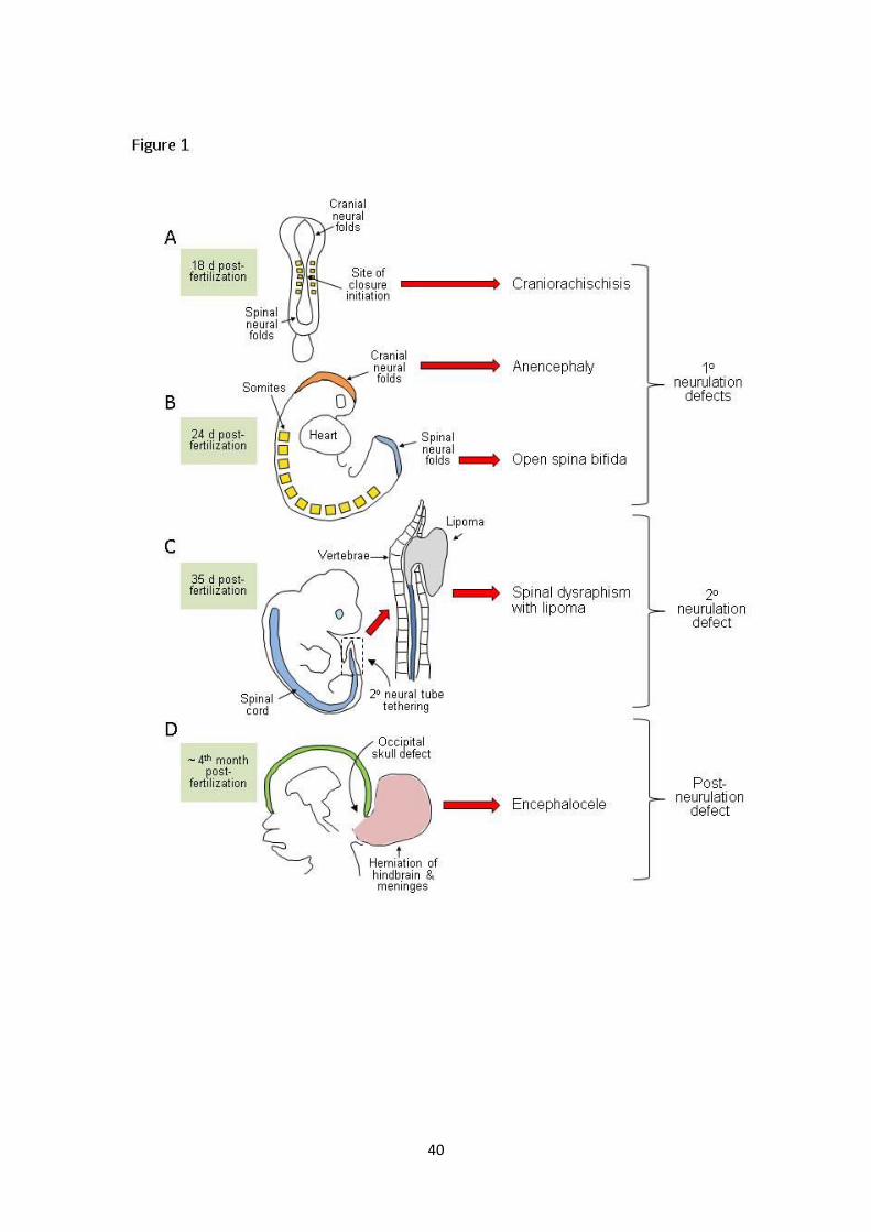
Neural tube defects (NTDs) are severe birth defects of the central nervous system that originate
during embryogenesis and result from failure of the morphogenetic process of neural tube closure.
In higher vertebrates the neural tube is generated by shaping, bending and fusion of the neural plate
and fusion in the dorsal midline progressively seals the neural tube as it forms. If closure is not
completed, neuroepithelium remains exposed to the environment and consequently subject to
degeneration and neuronal deficit. The type and severity of these ‘open’ NTDs varies with the level
of the body axis which is affected. Thus, failure of closure in the prospective brain or spinal cord
result in anencephaly and open spina bifida (myelomeningocele), respectively.
While the unifying feature of open NTDs is the failure of completion of neural tube closure, there are
many different possible causes, both genetic and environmental. In humans, it appears that most
NTDs are multifactorial in causation, resulting from an additive contribution of several risk factors
which are each individually insufficient to disrupt neural tube closure (the multifactorial threshold
model)(Harris & Juriloff 2007). The challenge of identifying the primary cause of NTDs in individual
patients is highlighted by the numerous candidate genes and environmental factors indicated by
epidemiological studies and experimental models. Moreover, the potential for gene-gene and gene-
environment interactions introduces further potential complexity.
2. UNDERSTANDING THE EMBRYONIC BASIS OF NTDS - NEURAL TUBE CLOSURE
Determination of the specific causes of NTDs is best achieved in the context of an understanding of
the mechanisms underlying neural tube closure (reviewed by (Copp & Greene 2013, Greene & Copp
2009). Given the inaccessibility of the neurulation-stage human embryo, our knowledge of the key
principles of neural tube closure comes mainly from analysis of experimental models, particularly
other mammals, amphibians and birds, in which primary neural tube closure is achieved through
folding and fusion of the neuroepithelium.
2.1 Primary neurulation; sub-types of NTDs relate to stages of closure
In the prospective brain and most of the spinal cord, neural tube formation essentially involves the
bending of the neuroepithelium in the midline to generate neural folds that elevate, meet and fuse
in the dorsal midline (primary neurulation). Rather than simultaneously rolling up along the extent of
the rostro-caudal axis, neural tube closure is discontinuous with distinct sites of initiation located at
characteristic axial levels. Moreover, the morphological and molecular requirements for closure vary
along the body axis, such that an individual NTD usually only affects a portion of the neural tube.
5
NTDs can thus be attributed to failure of particular initiation events or disruption of the progression
of closure between these sites.
In mice, closure is first achieved on embryonic day 8.5 at the level of the hindbrain/cervical
boundary (Closure 1) and failure of this event leads to craniorachischisis (Copp et al. 2003). Closure
initiates at a second site on embryonic day 9, Closure 2, in the caudal forebrain or
forebrain/midbrain boundary. Once initial contact and fusion have been established between the
tips of the neural folds, closure spreads bi-directionally from the sites of Closure 1 and 2 and in a
caudal direction from the rostral end of the neural tube (Closure 3). The open regions of neural
folds, termed neuropores, gradually shorten leading to complete closure of the anterior neuropore
(between Closures 2 and 3) on embryonic day 9, and the hindbrain neuropore (between Closures 1
and 2) a few hours later. Cranial NTDs (anencephaly) result from failure of Closure 2, or incomplete
‘zippering’ between Closures 1 and 2, which closes the midbrain and hindbrain. If fusion does not
progress from the anterior end of the neural plate (Closure 3), the resultant phenotype is a ‘split
face’ usually accompanied by forebrain anencephaly.
Unlike the cranial region where closure proceeds bidirectionally, spinal neurulation is entirely
caudally directed as the embryo continues to grow. Primary neurulation completes with final closure
of the posterior neuropore on embryonic day 10. Impaired progression of closure, and consequently
the presence of a persistently open posterior neuropore, results in spina bifida and the size of the
ensuing lesion relates directly to the axial level at which closure stops.
2.2 Primary neurulation in humans
Examination of human embryos suggests that initiation of closure is discontinuous, as in the mouse
(Nakatsu et al. 2000, O'Rahilly & Müller 2002). Bending of the neural plate begins at around 17-18
days after fertilisation, with an equivalent event to Closure 1 at around 22 days and completion of
closure at the posterior neuropore by 26-28 days post-fertilisation. It appears that closure of the
forebrain and midbrain in human embryos may be achieved by progression between the site of
Closure 1 and the rostral end of the neural plate without an intervening initiation site analogous to
Closure 2 (O'Rahilly & Müller 2002, Sulik et al. 1998).
2.3 Secondary neurulation
In mice and humans, the neural tube caudal to the mid-sacral region is continuous with the caudal
end of the primary neural tube but forms by a distinct process, termed secondary neurulation
6
(Schoenwolf 1984, Copp & Brook 1989). This process involves condensation of a population of tail
bud-derived cells bud to form an epithelial rod that undergoes canalisation to form the lumen of the
tube in the lower sacral and coccygeal regions. Malformations resulting from disturbance of
secondary neurulation are ‘closed’ (skin covered) and often involve tethering of the spinal cord, with
associated ectopic lipomatous material (Lew & Kothbauer 2007).
3. MECHANISMS UNDERLYING NEURAL TUBE CLOSURE
Studies of neurulation-stage embryos, both normal and developing NTDs, provide insights into key
molecular and cellular pathways underlying the morphological tissue movements of neural tube
closure (Copp & Greene 2010). In addition to ubiquitous requirements, the occurrence of isolated
NTDs at cranial or caudal levels in humans and different mouse models suggests the likely
involvement of region-specific mechanisms, dependent on different gene products.
3.1 Shaping of the neural plate – convergent extension is required for initiation of closure
Concomitant with the onset of neural tube closure, the neural plate undergoes narrowing in the
medio-lateral axis (convergence) and elongation in the rostro-caudal axis (extension), owing to
intercalation of cells at the midline (Keller 2002). Convergent extension depends on activity of a non-
canonical Wnt signalling pathway, homologous to the planar cell polarity (PCP) pathway first
described in Drosophila as regulating cell polarity in the plane of epithelia (Goodrich & Strutt 2011).
Signalling occurs via a Frizzled (Fzd) membrane receptor and cytoplasmic Dishevelled (Dvl), but
without stabilisation of beta-catenin.
Functional disruption of PCP mediators prevents convergent extension and the neural plate remains
broad in Xenopus (Wallingford & Harland 2001, Wallingford & Harland 2002) and mouse embryos
(Greene et al. 1998, Ybot-Gonzalez et al. 2007). Hence, closure 1 fails, leading to craniorachischisis,
in mice homozygous for mutations in ‘core PCP’ genes including Vangl2, Celsr1, or double mutants
for Dvl-1 and -2, or Fzd-3 and -6 (Juriloff & Harris 2012). Craniorachischisis also results from mutation
of the PCP-related genes Scrb1 (Murdoch et al. 2001) and Ptk7 (Lu et al. 2004) or genes encoding
accessory proteins, such as Sec24b which affects Vangl2 transport (Merte et al. 2010). Ultimately,
failure of closure initiation in PCP-mutant embryos is thought to result from insufficient proximity of
the neural folds owing to the broadened midline.
Failure of closure 1 in the majority of ‘core’ PCP mutant embryos precludes analysis of a requirement
for convergent extension at later stages of neurulation. However, spina bifida occurs in some loop-
7
tail heterozygotes (Vangl2Lp/+)(Copp et al. 1994) and in compound heterogotes of Vangl2Lp/+ with
mutations of Ptk7, Sec24b or Sdc4 (Lu et al. 2004, Merte et al. 2010, Escobedo et al. 2013). Moreover,
non-canonical Wnt signalling is compromised in Lrp6 null embryos that develop spina bifida (Gray et
al. 2013). These observations suggest a likely continued requirement for PCP signalling as spinal
neurulation proceeds.
Despite the entirely open spinal neural tube in Vangl2Lp/Lp embryos with craniorachischisis, closure
does occur in the forebrain and much of the midbrain implying that PCP-dependent convergent
extension is not required throughout the cranial region. Nonetheless, exencephaly is observed in
digenic combinations of Vangl2Lp/+ with some Wnt pathway genes (e.g. Dvl3+/-, Fzd1+/- and Fzd2+/-
(Etheridge et al. 2008, Yu et al. 2010). Exencephaly also develops in mutants for the PCP ‘effector’
genes Fuz or Intu but the role of these genes in cilium-dependent hedgehog signalling seems more
likely to explain their loss-of-function effect on cranial neural tube closure than a role in regulating
convergent extension (Gray et al. 2009, Zeng et al. 2010, Heydeck & Liu 2011) (see Section 3.2). Thus,
components of PCP signalling potentially impact on neural tube closure via multiple cellular
mechanisms.
3.2 Bending of the neural folds – regulation by Shh and BMP signalling
In order to achieve closure, the neuroepithelium must bend to bring the tips of the neural folds into
apposition. Bending occurs in a stereotypical manner at ‘hinge points’; a median hinge point (MHP)
in the midline and paired dorsolateral hinge points (DLHPs) that arise laterally (Shum & Copp 1996).
The morphology varies along the body axis with differing modes in the upper (MHP only), mid- (MHP
and DLHPs) and caudal (DLHPs only) regions of the primary neural tube.
The mechanisms underlying neuroepithelial bending are not fully understood, but one notable
feature of the MHP is the predominance of wedge-shaped cells (wider basally than apically)
compared to non-bending regions (Schoenwolf & Smith 1990). At neural plate stages the
neuroepithelium is a pseudostratified epithelium in which nuclei move to the basal pole during S-
phase owing to inter-kinetic nuclear migration. Prolongation of S-phase at the MHP provides a
possible means by which regulation of the cell cycle may contribute to cell wedging and hence MHP-
formation (Schoenwolf & Smith 1990).
Bending is regulated by signals emanating from non-neural tissues dorsal and ventral to the neural
folds (reviewed by (Greene & Copp 2009). The MHP is induced by signals from the notochord,
located immediately ventral to the midline of the neuroepithelium (Smith & Schoenwolf 1989, Ybot-
8
Gonzalez et al. 2002). At the molecular level, notochord-derived Shh induces the floor plate of the
neural tube at the site of the MHP (Placzek & Briscoe 2005, Chiang et al. 1996). However, this is not
essential for spinal neural tube closure which completes in the absence of a floor plate in mouse
embryos lacking Shh or Fox A2 (Chiang et al. 1996, Ang & Rossant 1994). Thus, the MHP may be
functionally important in floor plate development but is not essential for neural tube closure.
In contrast to the MHP, DLHPs appear essential for closure of the neural tube in the low spinal
region. For example, Zic2 mutant embryos, in which DLHPs are absent, develop severe spina bifida
(Ybot-Gonzalez et al. 2007). The formation of DLHPs is actively regulated, with interplay of inhibitory
and inductive signals determining their appearance at different axial levels (Copp & Greene 2013).
These include inhibitory effects of Shh signalling from the notochord and BMP signalling from the
surface ectoderm at the dorsal tips of the neural folds. These signals are opposed by the BMP
antagonist noggin whose expression in the dorsal neural folds is sufficient to induce DLHP (Ybot-
Gonzalez et al. 2002, Ybot-Gonzalez et al. 2007).
In contrast to absence of Shh signalling, NTDs do result from mutations which enhance Shh
signalling, for example through deficient function of inhibitory or cilia-related genes such as Gli3,
Rab23, Fkbp8, Tulp3 and Ift40 (Murdoch & Copp 2010, Miller et al. 2013). Mutants involving
increased Shh signalling display NTDs at cranial and/or spinal levels. While spina bifida appears to be
associated with suppression of dorsolateral bending of the neural folds (Murdoch & Copp 2010), the
mechanism underlying cranial NTDs is not clear.
3.3 Cranial neurulation – additional complexity and sensitivity to disruption
The neural folds in the cranial region bend in the midline and dorsolaterally as in the spinal region
but the closure process appears morphologically more complex. The folds are initially biconvex, with
the tips facing away from the midline, and then switch to a biconcave shape allowing the tips to
approach in the midline. The additional complexity of cranial compared with spinal neurulation
appears to be reflected in a more extensive genetic underpinning and a greater sensitivity to
disruption, at least in rodents. Exencephaly occurs in approximately three times as many knockout
mouse models as spina bifida and is the NTD type most commonly induced by teratogens (Copp et
al. 1990, Harris & Juriloff 2010).
Cranial neurulation may rely on specific contributory factors that are not involved in the spinal
region such as expansion of the mesenchyme underlying the neural folds (Greene & Copp 2009,

9
Zohn & Sarkar 2012). Moreover, disruption of the actin cytoskeleton prevents closure in the cranial
but not the spinal region (Morriss-Kay & Tuckett 1985, Ybot-Gonzalez & Copp 1999). Similarly,
exencephaly is observed but spinal neurulation completes successfully in null mutants for several
cytoskeletal components (e.g. n-cofilin, vinculin) (Gurniak et al. 2005, Xu et al. 1998). Nevertheless,
apically-located actin microfilaments are present throughout the neuroepithelium (Sadler et al.
1982), while functional disruption of the cytoskeleton–associated proteins MARCKS-related protein
or Shroom3 cause both spinal and cranial NTDs (Hildebrand & Soriano 1999, Xu et al. 1998),
suggesting that regulation of the acto-myosin cytoskeleton plays a role in closure in both regions.
Shroom proteins appear to play a key role: expression of Shroom in Xenopus is sufficient to induce
apical constriction of epithelial cells while functional disruption inhibits neural fold bending and
suppresses closure (Haigo et al. 2003).
3.4 Adhesion and fusion of the neural folds
Once the neural folds meet at the dorsal midline, a process of adhesion, fusion and remodelling
gives rise to two discrete epithelial layers, with the nascent neural tube overlain by an intact surface
ectoderm (Pai et al. 2012). At the closure site the neural fold tips are composed of neuroepithelium
continuous with the non-neural surface ectoderm. The cell type that adheres first may differ at
varying axial levels (Geelen & Langman 1979, Ray & Niswander 2012). Nevertheless at all levels,
initial contact appears to involve sub-cellular protrusions, resembling lamellipodia and filopodia,
observed by electron microscopy (Geelen & Langman 1979) and in live embryos (Pyrgaki et al. 2010).
The molecular basis of adhesion is not well characterised, perhaps due to functional redundancy
among the proteins involved. However, a role for interaction of cell surface ephrin receptors with
Eph ligands is suggested by the occurrence of cranial NTDs in mice lacking ephrin-A5 or EphA7
(Holmberg et al. 2000), and delayed spinal closure in embryos exposed to peptides that block
ephrinA/EphA interactions (Abdul-Aziz et al. 2009).
Knockout of protease-activated receptors (PAR1 and PAR2) in the surface ectoderm also causes
cranial NTDs, implicating a role for signalling via these G-protein coupled receptors in closure
(Camerer et al. 2010). Further evidence for the function of the non-neural ectoderm is provided by
Grhl2 null mutants which fail in closure throughout the cranial region and exhibit spina bifida (Rifat
et al. 2010, Werth et al. 2010, Brouns et al. 2011). Grhl2 is expressed in the surface ectoderm
overlying the neural folds and regulates expression of several components of the apical adhesion
junction complex, including E-cadherin (Werth et al. 2010, Pyrgaki et al. 2011).
10
3.5 Regulation of cell proliferation and cell death
During neurulation the embryo grows rapidly. Cell cycle exit and neuronal differentiation begin in
the neuroepithelium shortly after closure and maintenance of adequate proliferation in the
neuroepithelium appears crucial for closure, particularly in the cranial region. Thus, in mice NTDs can
be caused by exposure to anti-mitotic agents (Copp et al. 1990) or mutation of genes encoding
proteins associated with cell-cycle progression (e.g. neurofibromin 1, nucleoporin) or prevention of
neuronal differentiation (e.g. Notch pathway genes Hes1, Hes3, RBP-J )(Harris & Juriloff 2010,
Harris & Juriloff 2007). Conversely, excessive cell proliferation is also associated with NTDs in several
mouse models, such as Phactr4 mutants (Kim et al. 2007).
Characteristic patterns of apoptotic cell death occur in the neural folds and the midline of the closed
neural tube (Geelen & Langman 1979, Massa et al. 2009, Yamaguchi et al. 2011). Increased cell
death could hypothetically inhibit closure through compromising the functional and/or mechanical
integrity of the neuroepithelium. It is associated with NTDs in a number of teratogen-induced and
genetic models, although only rarely has a direct causal link been definitively established (Copp &
Greene 2013, Fukuda et al. 2011). The occurrence of exencephaly in mice lacking apoptosis-related
genes such as caspase3 or Apaf1 suggests a requirement for apoptosis in closure (Harris & Juriloff
2010). However, forebrain and spinal closure occurs normally in these models and pharmacological
suppression of apoptosis does not cause NTDs, suggesting that it is dispensable for completion of
closure (Massa et al. 2009).
4. CLINICAL FEATURES OF NEURAL TUBE DEFECTS
4.1 Open NTDs and associated conditions
Open NTDs can result from failure of closure at a de novo initiation site or incomplete progression of
closure following successful initiation. Where embryos are available for examination, as in
experimental models, NTDs can be recognised during or immediately after neurulation stages owing
to the persistently open neural folds. However, at later embryonic and fetal stages the
morphological appearance varies considerably owing to secondary changes and degeneration.
In cranial NTDs, the open neural folds undergo growth and differentiation and typically appear to
bulge from the developing brain, termed exencephaly. Inability to form the skull vault over the open
region leads to degeneration of the exposed neural tissue and the characteristic appearance of
11
anencephaly, observed later in human or rodent pregnancy (Wood & Smith 1984, Seller 1995). Both
anencephaly and craniorachischisis (~10% of NTDs) are lethal conditions at or shortly after birth.
Open neural folds in the spinal region prevent the sclerotome-derived vertebral arches from
covering the neuroepithelium, the consequent opening in the vertebral column giving rise to the
term spina bifida (Copp et al. 2013). The neural tissues may be contained within a meninges covered
sac that protrudes through the open vertebrae (myelomeningocele; spina bifida cystica), or exposed
directly to the amniotic fluid (myelocele). Babies born with open spina bifida usually survive with
appropriate medical care, but suffer neurological impairment whose severity depends on the level of
the lesion. Associated conditions include hydrocephalus, Chiari type II malformation and vertebral
abnormalities as well as genitourinary and gastrointestinal disorders.
4.2 Diagnosis, treatment and maternal-fetal surgery
NTDs can be diagnosed prenatally by ultrasound (Cameron & Moran 2009). However, where
prenatal diagnosis is not routinely available and/or therapeutic abortion not an option, many babies
with NTDs are born. Post-natal medical care for babies born with open spina bifida usually involves
surgery to close and cover the lesion. Multiple subsequent surgeries are commonly required to
alleviate tethering of the spinal cord, treat hydrocephalus and/or address orthopaedic and urological
problems.
As open NTDs arise early during pregnancy, there is a prolonged period during which secondary
neurological damage may occur owing to exposure of nervous tissue to the amniotic fluid
environment. These considerations provided impetus for development of in utero fetal surgery for
spina bifida which may improve neurological outcome compared with post-natal repair, although
with fetal and maternal risks (Adzick et al. 1998, Adzick et al. 2011). Experimental models of spina
bifida are being used to investigate the possible combination of surgical intervention with additional
therapy, intended to remediate neural damage. Examples include the implantation of biodegradable
scaffolds to promote neural regeneration and/or neural stem cells to populate the damaged spinal
cord (Saadai et al. 2011, Saadai et al. 2013).
4.3 Disorders of the closed neural tube
This review focuses on open NTDs, characterised by failure of neural tube closure. Various other
conditions are also associated with abnormalities of the closed spinal cord and are often categorised
as NTDs under a broader definition. There is also a less well-defined group of closed spinal NTDs in
12
which the vertebral arches are malformed but covered by skin. These conditions, including spina
bifida occulta and ‘spinal dysraphisms’, vary widely in clinical presentation. The more severe sub-
types are associated with various abnormalities of the spinal cord, lipoma and/or anorectal
abnormalities. The embryonic origin of closed spina bifida is not well defined but is hypothesised to
involve abnormalities of secondary neurulation (Copp et al. 2013).
Abnormal development of the vertebrae or cranium may also allow herniation of the closed neural
tube through the affected region in the rare form of spina bifida, meningocele (spina bifida cystica)
or encephalocele, respectively.
5. CAUSES OF NTDs
NTDs are among the most common birth defects worldwide with a prevalence that varies from 0.5
to more than 10 per 1,000 pregnancies. This likely reflects differing contributions from risk factors
such as nutritional status, prevalence of obesity and diabetes, usage of folic acid supplementation
and/or fortification, the presence of environmental toxicants and differing genetic predisposition
between ethnic groups. In most populations there is also a striking gender bias with a higher
prevalence of anencephaly among females than males. Many NTD mouse strains also show a female
preponderance among cranial NTDs, apparently reflecting a fundamental higher sensitivity of cranial
neural tube closure to disturbance in female embryos (Juriloff & Harris 2012). Overall, although a
number of risk factors have been identified these may account for less than half of NTDs, suggesting
that additional genetic and non-genetic factors remain to be identified (Agopian et al. 2013).
5.1 Environment factors
Various teratogenic agents induce NTDs in rodent models (Copp et al. 1990, Copp & Greene 2010).
In humans, teratogens that have been associated with NTDs include the anti-convulsant drug
valproic acid (Wlodarczyk et al. 2012), and the fungal product fumonisin (Missmer et al. 2006). Other
non-genetic risk factors include maternal fever and excessive use of hot tubs (Moretti et al. 2005),
consistent with the induction of NTDs by hypothermia in rodent models.
Maternal obesity or diabetes are well-recognised risk factors for NTDs (Correa et al. 2003).
Determination of the cause of diabetes-related NTDs is hampered by the complexity of the diabetic
milieu, although hyperglycemia alone is sufficient to cause NTDs in cultured rodent embryos. It has
been proposed that NTDs may result from increased oxidative stress, altered expression of genes
such as Pax3, and neuroepithelial cell apoptosis (Fine et al. 1999, Reece 2012). Recent findings
13
suggest that activation of apoptosis signal-regulating kinase 1 (ASK1) in hyperglycaemic conditions,
leads to activation of the apoptosis mediator caspase 8 via stimulation of the FoxO3a transcription
factor (Yang et al. 2013).
Nutritional factors and folate
The historical link between lower socioeconomic status and higher risk of birth defects led to
examination of the possible involvement of nutritional factors in NTDs. Lower levels of the B-vitamin
folate were observed in mothers of NTD fetuses (Smithells et al. 1976), prompting an intervention
trial of a folic acid-containing multivitamin supplement for prevention of NTD recurrence (Smithells
et al. 1981, Schorah 2008). A multi-centre randomised controlled trial confirmed that maternal folic
acid supplementation (at 4 mg/day) significantly reduces the recurrence risk (Wald et al. 1991).
Additional clinical trials provided evidence for reduction of occurrence risk (Czeizel & Dudás 1992,
Berry et al. 1999, Czeizel et al. 2011).
Questions remain over the mechanism by which folic acid prevents NTDs (Blom et al. 2006, Copp et
al. 2013). Although maternal folate status is a risk factor, in most cases, maternal folate levels are
within the ‘normal’ range and rarely clinically deficient. Nonetheless, there is an inverse relationship
between blood folate concentration and risk of an affected pregnancy (Daly et al. 1995). It has been
suggested that sub-optimal folate levels may contribute to development of NTDs in individuals who
are genetically susceptible. Such a gene-environment interaction has been demonstrated in mice,
where folate deficiency does not cause NTDs, unless present in combination with mutation of a
predisposing gene, such as Pax3 (Burren et al. 2008).
Folate one-carbon metabolism comprises a complex network of inter-linked reactions that mediate
transfer of one-carbon groups for a number of biosynthetic processes (Stover 2009). Among these,
attention has particularly focussed on the requirement for nucleotide biosynthesis and methylation
reactions in neural tube closure. Abnormal thymidylate and purine biosynthesis have been identified
in mouse NTD models (Fleming & Copp 1998, Beaudin et al. 2011) and in a proportion of NTD cases
(Dunlevy et al. 2007), while deficient methylation may also be implicated in NTDs (Section 5.3).
5.2 Genetics of NTDs
Most NTDs occur sporadically, with a relative scarcity of multi-generational families. Nevertheless,
there is strong evidence for a genetic component in the etiology of NTDs and the pattern of
inheritance favours a multifactorial polygenic or oligogenic model, as opposed to an effect of single
14
genes with partial penetrance (Harris & Juriloff 2007). Most studies of NTD genetics have focussed
on one or more candidate genes (reviewed by (Boyles et al. 2005, Greene et al. 2009, Harris &
Juriloff 2010). In general these have been: (i) human orthologues of genes whose mutation causes
NTDs in mice, of which there are more than 200 examples; or (ii) genes related to environmental risk
factors, particularly folate metabolism.
Case-control association studies have implicated several genes while mutation screening by
sequencing has identified putative pathogenic mutations. However, the definitive assignment of a
gene variant as causative is complicated by the apparent multigenic nature of NTDs, and the large
number of possible candidate genes, modifier genes, epigenetic factors and environmental
influences. Moreover, where putative mutations have been identified in specific genes, each has
only been involved in a small proportion of NTD patients, suggesting that there is considerable
heterogeneity underlying the genetic basis of NTDs. Thus, although the morphological and cellular
basis of neural tube closure has become increasingly well understood, the genetic basis of NTDs in
individual cases remains largely unclear.
Gene-gene interactions and effect of modifier genes
Mouse studies suggest three broad mechanisms by which genetic interactions may result in NTDs.
(1) In some instances functional redundancy makes it necessary for mutation of two orthologous
genes, (e.g. Dvl1-Dvl2 (Hamblet et al. 2002), Cdx1-Cdx2 double knockouts (Savory et al. 2011)), in
order to reveal a requirement in neural tube closure. (2) Additive effects of heterozygous mutations
may result in NTDs that resemble those of individual homozygotes (e.g. Dvl3 with Vangl2Lp
(Etheridge et al. 2008). (3) Variation in the penetrance and expressivity of NTD phenotypes between
inbred strains of mice is widely reported and thought to reflect variants in modifier genes. For
example, the rate of exencephaly resulting from Cecr1 mutation is strongly affected by FVB/N strain
background (Davidson et al. 2007). While the identity of modifier genes for NTDs has rarely been
determined, a variant in Lmnb1 is present in some mouse strains and significantly increases the
frequency of NTDs in curly tail (Grhl3ct) embryos (de Castro et al. 2012).
Genes implicated through experimental models
In mice, mutation of genes encoding components of the PCP pathway causes NTDs (Section 3.1).
Sequencing of PCP genes in humans has identified putative mutations in CELSR1, VANGL1, VANGL2,
FZD6, SCRIB1 and DVL2 in a proportion of patients with craniorachischisis, spina bifida, anencephaly
and closed forms of spina bifida (Kibar et al. 2007, Lei et al. 2010, Robinson et al. 2012, De Marco et
15
al. 2013, Chandler et al. 2012, Lei et al. 2013) and reviewed by (Juriloff & Harris 2012). As in mice,
heterozygous human PCP mutations may hypothetically interact with other genetic NTD risk factors
in a digenic or polygenic fashion, to cause a range of NTD types. This could potentially involve
summation of multiple variants in PCP genes. For example, a putative mutation in DVL2 was
identified in a spina bifida patient in combination with a second, previously identified missense
variant in VANGL2 (De Marco et al. 2013).
Among other genes implicated in NTDs from mouse models, association studies have not provided
evidence for a major contribution to risk and few positive results have emerged from sequencing-
based mutation screens. As data begins to emerge from large-scale exome sequencing studies of
NTD patients, it will become possible to evaluate the contribution of multiple genes in the same
patient cohorts and the mutational load associated with individual risk.
Analysis of genes related to environmental risk factors
The identification of environmental factors such as maternal diabetes and folate status as risk
factors for NTDs provides impetus for analysis of related genes in affected families. Risk could
potentially be associated with maternal genotype, if genetic variation alters maternal metabolism
and secondarily affects the developing embryo. The inheritance of maternal alleles by the embryo
complicates interpretation of such effects. Alternatively, a genetically determined abnormality in the
embryo itself could influence risk of NTDs; potentially through interaction with a predisposing
environmental factor. For example, it may be informative to analyse genetic data on folate-related
genes in the context of maternal folate status (Etheredge et al. 2012).
Association with risk of spina bifida has been reported for several genes implicated in diabetes,
obesity, glucose metabolism and oxidative stress, including GLUT1, SOD1 and SOD2 (Davidson et al.
2008, Kase et al. 2012). Maternal variants in the obesity-related genes FTO, LEP and TCF7L2 are also
associated with NTDs, consistent with maternal obesity being a risk factor (Lupo et al. 2012).
Genes related to folate one-carbon metabolism have been perhaps the most intensively group of
candidates for NTDs (reviewed by (Blom et al. 2006, Greene et al. 2009, Shaw et al. 2009)). The
C677T polymorphism of MTHFR, which encodes an alanine to valine substitution, has been
associated with NTDs. The TT genotype is found at higher frequency among cases than controls in
some populations (e.g. Irish) but not others (e.g. Hispanics) (Botto & Yang 2000). Several studies
16
indicate positive associations with other folate-related genes, including MTRR, although these have
generally not been not observed in all study populations.
In mice, mutations in folate-metabolising enzymes (e.g. Mthfd1) are sometimes lethal before the
stage of neural tube closure (e.g. (MacFarlane et al. 2009, Christensen et al. 2013) while others do
not disrupt closures (eg. (Chen et al. 2001, Di Pietro et al. 2002). Null embryos for the folate
receptor, Folr1 die pre-neurulation but develop NTDs when supplemented with sufficient folic acid
to prevent early lethality (Piedrahita et al. 1999). NTDs are also observed in Shmt1 knockouts, under
folate-deficient conditions (Beaudin et al. 2011). In contrast, NTDs occur ‘spontaneously’ in mice
carrying loss-of-function alleles of Amt (Narisawa et al. 2012) or Mthfd1L (Momb et al. 2013), both of
which encode enzymes of mitochondrial folate metabolism (Tibbetts & Appling 2010). Interestingly,
the homologous genes in humans has also been linked to NTDs. Missense mutations have been
identified in NTD patients in AMT, as well as GLDC which encodes its partner enzyme in the glycine
cleavage system (Narisawa et al. 2012). Genetic associations with NTDs have been reported for
MTHFD1L (Parle-McDermott et al. 2009) and SLC23A32 (MFTC), encoding a mitochondrial folate
transporter (Pangilinan et al. 2012). Altogether, these findings suggest that NTD risk is influenced by
function of mitochondrial folate metabolism, a major source of one-carbon units to the cytoplasm.
5.3 Gene-regulatory mechanisms and NTDs
In addition to the potential multigenic nature of NTDs, identification of causative genes may be
complicated by the potential involvement of aberrant gene expression, perhaps resulting from
mutations in regulatory elements. For example, mutations resulting in insufficient expression of
Grhl3 or excess expression of Grhl2 cause NTDs in mice in the absence of coding mutations
(Gustavsson et al. 2007, Brouns et al. 2011). Further complexity may be added by the potential for
regulation by epigenetic modifications such as DNA methylation, histone modification or chromatin
remodelling, each of which has been associated with NTDs in mice and in some cases in humans
(reviewed by (Harris & Juriloff 2010, Greene et al. 2011)). For example, methylation of LINE-1
genomic elements was lower than normal in DNA of anencephalic but not spina bifida fetuses (Wang
et al. 2010).
A simple model predicts a positive correlation between folate status and methylation. However,
data from human pregnancy suggests the relationship is not straightforward (Crider et al. 2012). A
recent study found an inverse correlation of LINE-1 methylation with maternal and cord blood
folate, while different imprinted genes showed positive or negative associations (Haggarty et al.
17
2013). Somewhat counter-intuitively use of folic acid supplements was associated with reduced
LINE-1 methylation.
A requirement for DNA methylation in mouse neural tube closure is suggested by the occurrence of
NTDs in knockouts of Dnmt3b, encoding a DNA methyltransferase, and in embryos cultured with 5-
azacytidine (Okano et al. 1999, Matsuda & Yasutomi 1992). Similarly, inhibition of the methylation
cycle reduces DNA methylation and causes NTDs in cultured mouse embryos (Dunlevy et al. 2006,
Burren et al. 2008). However, Mthfr null embryos do not develop NTDs despite a significant
reduction in global DNA methylation (Chen et al. 2001), nor is there an exacerbating effect of Mthfr
loss-of-function on Pax3 or curly tail mutants, although both show increased rates of NTDs under
folate-deficient conditions (Pickell et al. 2009, Burren et al. 2008, de Castro et al. 2010). Thus,
questions remain over the relationship between folate status, DNA methylation and risk of NTDs.
Other epigenetic mechanisms include various modifications of histone proteins, which potentially
miss-regulate genes that influence neurulation. NTDs occur in mice carrying mutations in the histone
demethylases Jarid2 (Takeuchi et al. 1999) and Fbxl10 (Fukuda et al. 2011). Similarly, histone
acetylases and deacetylases, which regulate the equilibrium of histone acetylation, are implicated in
NTDs. An acetylase-specific knock-in mutation of Gcn5 causes cranial NTDs (Bu et al. 2007), as does
loss of function of another histone acetylase, p300 (Yao et al. 1998). Increased acetylation is also
associated with NTDs. For example, cranial NTDs occur in mice carrying mutations in histone
deacetylases Sirt1 or Hdac4 (Cheng et al. 2003, Vega et al. 2004). The teratogenic effects of valproic
acid and trichostin A may also be mediated through their inhibition of histone deacetylases (Finnell
et al. 2002).
6. PRIMARY PREVENTION OF NTDs
6.1 Folic acid supplementation and fortification
The reduction in risk of NTDs following maternal folic acid supplementation led to public health
recommendations that women who may become pregnant should consume 0.4 mg of folic acid daily
or 4 mg daily following a previous affected pregnancy (Czeizel et al. 2011). To ensure that additional
folate was received, food fortification programmes were introduced in many countries. This
approach has raised blood folate levels and been associated with lower frequency of NTDs (Crider et
al. 2011). The magnitude of effect varies, with greatest reduction where pre-existing rates were
higher (Blencowe et al. 2010, Rosenthal et al. 2013). Some countries have delayed decision on
fortification owing to safety concerns (e.g. possible enhancement of bowel cancer) but a recent
18
meta-analysis found no evidence for increased cancer rates following folic acid supplementation
(Vollset et al. 2013).
6.2 Folate-resistant NTDs
Folic acid supplementation in clinical trials has not approached 100% NTD prevention and an
estimated one-third of NTDs may be folic acid-resisant (Blencowe et al. 2010). A study in the USA,
where folate fortification of food is mandatory, found no apparent protective effect of folic acid
supplements (Mosley et al. 2009), suggesting that increased dosage would not necessarily provide
additional preventive effects.
Given the multifactorial causation of NTDs it seems reasonable to suppose that optimal prevention
will require a combination of multiple interventions. Possible approaches may relate to folate one-
carbon metabolism. For example, like folate there is a graded relationship between lower levels of
circulating vitamin B12 and increasing risk of an NTD-affected pregnancy (Molloy et al. 2009). Perhaps
use of B12 supplements would further reduce the frequency of NTDs, although this remains to be
tested.
Another possibility is that folic acid cannot ameliorate some defects that result from abnormal folate
metabolism, owing to defects in the intervening enzymes required to transfer one carbon units to
key downstream metabolites. In this case supplementation with alternative folates, such as 5-methyl
THF (Czeizel et al. 2011), or key downstream molecules could potentially be advantageous. For
example, supplementation with formate prevented NTDs in Mthfd1L null mice (Momb et al. 2013),
while combinations of thymidine and purine precursors prevented NTDs in curly tail mice, in which
folic acid is not protective (Leung et al. 2013).
In addition to folate and vitamin B12, lower maternal levels of other vitamins, including vitamin C,
have been reported in NTDs (Smithells et al. 1976). Conversely, intake of several vitamins and
maternal diet are associated with lower risk of NTDs suggesting that nutrients other than folic acid
may be beneficial (Chandler et al. 2012, Sotres-Alvarez et al. 2013). Experimental analysis of
individual vitamins found myo-inositol deficiency to cause NTDs in cultured rodent embryos
(Cockroft 1988). Inositol supplementation significantly reduced the frequency of NTDs in curly tail
mice (Greene & Copp 1997) and in rodent models of diabetes (Reece et al. 1997).
7. FUTURE PERSPECTIVES
19
Experimental models provide systems for analysis of the developmental events of neural tube
closure and fundamental cellular and morphological processes continue to be defined in more detail.
In principal NTDs may result from insufficiency of one or more of the key driving forces (eg, cellular
properties and/or morphological movements) that are necessary to achieve closure, for example,
through mutation of a PCP gene. Alternatively, a genetic lesion or environmental insult may disrupt
the closure process even where the underlying machinery is intact, for example through induction of
aberrant cellular behaviour such as excess apoptosis. Experimental models require careful analysis
to disentangle these possibilities. A key challenge will be to understand how the molecular and
cellular determinants of neurulation relate to the biomechanical forces required to fold the
neuroepithelium to achieve closure.
Advances in exome and whole genome sequencing offer the potential to begin to understand the
genetic basis of NTDs in humans. The multifactorial complexity of NTDs means that analysis of data
from such studies will present a major challenge. Moreover, there will be a need to integrate genetic
data with information on epigenetic and environmental factors to obtain a more complete
understanding of the cause of individual NTDs.
Folic acid supplementation provides a means to reduce NTD risk and represents a major public
health advance. Nevertheless, the heterogeneity of NTDs suggests that primary prevention may be
best achieved by multiple interventions and use of additional micronutrients alongside folic acid may
provide an opportunity to further reduce risk.
ACKNOWLEDGMENTS
Work in the authors laboratory is funded by the Medical Research Council (J003794), Newlife
Foundation (11-1206) and the Wellcome Trust (087525).
20
Literature Cited
1. Abdul-Aziz NM, Turmaine M, Greene ND, Copp AJ. 2009. EphrinA-EphA receptor interactions
in mouse spinal neurulation: implications for neural fold fusion. Int. J Dev. Biol.
53:559-68
2. Adzick NS, Sutton LN, Crombleholme TM, Flake AW. 1998. Successful fetal surgery for spina
bifida. Lancet 352:1675-6
3. Adzick NS, Thom EA, Spong CY, Brock JW, III, Burrows PK et al. 2011. A randomized trial of
prenatal versus postnatal repair of myelomeningocele. N. Engl. J. Med. 364:993-
1004
4. Agopian AJ, Tinker SC, Lupo PJ, Canfield MA, Mitchell LE. 2013. Proportion of neural tube
defects attributable to known risk factors. Birth Defects Res. A Clin. Mol. Teratol.
97(1):42-6
5. Ang S-L, Rossant J. 1994. HNF-3 is essential for node and notochord formation in mouse
development. Cell 78:561-74
6. Beaudin AE, Abarinov EV, Noden DM, Perry CA, Chu S et al. 2011. Shmt1 and de novo
thymidylate biosynthesis underlie folate-responsive neural tube defects in mice. Am.
J. Clin. Nutr. 93:789-98
7. Berry RJ, Li Z, Erickson JD, Li S, Moore CA et al. 1999. Prevention of neural-tube defects with
folic acid in China. N. Engl. J. Med. 341(20):1485-90
21
8. Blencowe H, Cousens S, Modell B, Lawn J. 2010. Folic acid to reduce neonatal mortality from
neural tube disorders. Int. J. Epidemiol. 39 Suppl 1:i110-i121
9. Blom HJ, Shaw GM, Den Heijer M, Finnell RH. 2006. Neural tube defects and folate: case far
from closed. Nat. Rev. Neurosci. 7(9):724-31
10. Botto LD, Yang Q. 2000. 5,10-Methylenetetrahydrofolate reductase gene variants and
congenital anomalies: a HuGE review. Am. J. Epidemiol. 151(9):862-77
11. Boyles AL, Hammock P, Speer MC. 2005. Candidate gene analysis in human neural tube
defects. Am. J. Med. Genet. C. Semin. Med. Genet. 135(1):9-23
12. Brouns MR, de Castro SC, Terwindt-Rouwenhorst EA, Massa V, Hekking JW et al. 2011. Over-
expression of Grhl2 causes spina bifida in the Axial defects mutant mouse. Hum.
Mol. Genet. 20:1536-46
13. Bu P, Evrard YA, Lozano G, Dent SY. 2007. Loss of Gcn5 acetyltransferase activity leads to
neural tube closure defects and exencephaly in mouse embryos. Mol. Cell Biol.
27:3405-16
14. Burren KA, Savery D, Massa V, Kok RM, Scott JM et al. 2008. Gene-environment interactions
in the causation of neural tube defects: folate deficiency increases susceptibility
conferred by loss of Pax3 function. Hum. Mol. Genet. 17:3675-85
15. Camerer E, Barker A, Duong DN, Ganesan R, Kataoka H et al. 2010. Local protease signalling
contributes to neural tube closure in the mouse embryo. Dev. Cell 18:25-38
22
16. Cameron M, Moran P. 2009. Prenatal screening and diagnosis of neural tube defects.
Prenatal Diag. 29:402-11
17. Chandler AL, Hobbs CA, Mosley BS, Berry RJ, Canfield MA et al. 2012. Neural tube defects
and maternal intake of micronutrients related to one-carbon metabolism or
antioxidant activity. Birth Defects Res. A Clin. Mol. Teratol. 94(11):864-74
18. Chen Z, Karaplis AC, Ackerman SL, Pogribny IP, Melnyk S et al. 2001. Mice deficient in
methylenetetrahydrofolate reductase exhibit hyperhomocysteinemia and decreased
methylation capacity, with neuropathology and aortic lipid deposition. Hum. Mol.
Genet. 10(5):433-43
19. Cheng HL, Mostoslavsky R, Saito S, Manis JP, Gu Y et al. 2003. Developmental defects and
p53 hyperacetylation in Sir2 homolog (SIRT1)-deficient mice. Proc. Natl. Acad. Sci. U.
S. A 100(19):10794-9
20. Chiang C, Litingtung Y, Lee E, Young KE, Corden JL et al. 1996. Cyclopia and defective axial
patterning in mice lacking Sonic hedgehog gene function. Nature 383:407-13
21. Christensen KE, Deng L, Leung KY, Arning E, Bottiglieri T et al. 2013. A novel mouse model for
genetic variation in 10-formyltetrahydrofolate synthetase exhibits disturbed purine
synthesis with impacts on pregnancy and embryonic development. Hum. Mol.
Genet. 22(18):3705-19
22. Cockroft DL. 1988. Changes with gestational age in the nutritional requirements of
postimplantation rat embryos in culture. Teratology. 38:281-90
23
23. Copp AJ, Brook FA. 1989. Does lumbosacral spina bifida arise by failure of neural folding or
by defective canalisation? J. Med. Genet. 26:160-6
24. Copp AJ, Brook FA, Estibeiro JP, Shum ASW, Cockroft DL. 1990. The embryonic development
of mammalian neural tube defects. Prog. Neurobiol. 35:363-403
25. Copp AJ, Checiu I, Henson JN. 1994. Developmental basis of severe neural tube defects in
the loop-tail (Lp) mutant mouse: Use of microsatellite DNA markers to identify
embryonic genotype. Dev. Biol. 165:20-9
26. Copp AJ, Greene ND. 2013. Neural tube defects-disorders of neurulation and related
embryonic processes. Wiley. Interdiscip. Rev. Dev. Biol. 2(2):213-27
27. Copp AJ, Greene NDE. 2010. Genetics and development of neural tube defects. J. Pathol.
220:217-30
28. Copp AJ, Greene NDE, Murdoch JN. 2003. The genetic basis of mammalian neurulation. Nat.
Rev. Genet. 4:784-93
29. Copp AJ, Stanier P, Greene ND. 2013. Neural tube defects: recent advances, unsolved
questions, and controversies. Lancet Neurol. 12(8):799-810
30. Correa A, Botto L, Liu YC, Mulinare J, Erickson JD. 2003. Do multivitamin supplements
attenuate the risk for diabetes-associated birth defects? Pediatrics 111:1146-51
31. Crider KS, Bailey LB, Berry RJ. 2011. Folic acid food fortification-its history, effect, concerns,
and future directions. Nutrients. 3(3):370-84
24
32. Crider KS, Yang TP, Berry RJ, Bailey LB. 2012. Folate and DNA methylation: a review of
molecular mechanisms and the evidence for folate's role. Adv. Nutr. 3(1):21-38
33. Czeizel AE, Dudás I. 1992. Prevention of the first occurrence of neural-tube defects by
periconceptional vitamin supplementation. N. Engl. J. Med. 327:1832-5
34. Czeizel AE, Dudas I, Paput L, Banhidy F. 2011. Prevention of neural-tube defects with
periconceptional folic acid, methylfolate, or multivitamins? Ann. Nutr. Metab
58:263-71
35. Daly LE, Kirke PN, Molloy A, Weir DG, Scott JM. 1995. Folate levels and neural tube defects -
Implications for prevention. JAMA 274(21):1698-702
36. Davidson CE, Li Q, Churchill GA, Osborne LR, McDermid HE. 2007. Modifier locus for
exencephaly in Cecr2 mutant mice is syntenic to the 10q25.3 region associated with
neural tube defects in humans. Physiol Genomics 31:244-51
37. Davidson CM, Northrup H, King TM, Fletcher JM, Townsend I et al. 2008. Genes in glucose
metabolism and association with spina bifida. Reprod. Sci. 15(1):51-8
38. de Castro SC, Leung KY, Savery D, Burren K, Rozen R et al. 2010. Neural tube defects induced
by folate deficiency in mutant curly tail (Grhl3) embryos are associated with
alteration in folate one-carbon metabolism but are unlikely to result from
diminished methylation. Birth Defects Res. A Clin Mol. Teratol. 88:612-8
25
39. de Castro SC, Malhas A, Leung KY, Gustavsson P, Vaux DJ et al. 2012. Lamin b1
polymorphism influences morphology of the nuclear envelope, cell cycle
progression, and risk of neural tube defects in mice. PLoS Genet. 8(11):e1003059
40. De Marco P, Merello E, Consales A, Piatelli G, Cama A et al. 2013. Genetic analysis of
disheveled 2 and disheveled 3 in human neural tube defects. J. Mol. Neurosci.
49(3):582-8
41. Di Pietro E, Sirois J, Tremblay ML, Mackenzie RE. 2002. Mitochondrial NAD-dependent
methylenetetrahydrofolate dehydrogenase-methenyltetrahydrofolate
cyclohydrolase is essential for embryonic development. Mol. Cell Biol. 22(12):4158-
66
42. Dunlevy LPE, Burren KA, Mills K, Chitty LS, Copp AJ, Greene NDE. 2006. Integrity of the
methylation cycle is essential for mammalian neural tube closure. Birth Defects
Research (Part A) 76:544-52
43. Dunlevy LPE, Chitty LS, Doudney K, Burren KA, Stojilkovic-Mikic T et al. 2007. Abnormal
folate metabolism in foetuses affected by neural tube defects. Brain 130:1043-9
44. Escobedo N, Contreras O, Munoz R, Farias M, Carrasco H et al. 2013. Syndecan 4 interacts
genetically with Vangl2 to regulate neural tube closure and planar cell polarity.
Development 140(14):3008-17
45. Etheredge AJ, Finnell RH, Carmichael SL, Lammer EJ, Zhu H et al. 2012. Maternal and infant
gene-folate interactions and the risk of neural tube defects. Am. J. Med. Genet. A
158A(10):2439-46
26
46. Etheridge SL, Ray S, Li S, Hamblet NS, Lijam N et al. 2008. Murine dishevelled 3 functions in
redundant pathways with dishevelled 1 and 2 in normal cardiac outflow tract,
cochlea, and neural tube development. PLoS. Genet 4:e1000259
47. Fine EL, Horal M, Chang TI, Fortin G, Loeken MR. 1999. Evidence that elevated glucose
causes altered gene expression, apoptosis, and neural tube defects in a mouse
model of diabetic pregnancy. Diabetes 48(12):2454-62
48. Finnell RH, Waes JGV, Eudy JD, Rosenquist TH. 2002. Molecular basis of environmentally
induced birth defects. Annu. Rev. Pharmacol. Toxicol. 42:181-208
49. Fleming A, Copp AJ. 1998. Embryonic folate metabolism and mouse neural tube defects.
Science 280:2107-9
50. Fukuda T, Tokunaga A, Sakamoto R, Yoshida N. 2011. Fbxl10/Kdm2b deficiency accelerates
neural progenitor cell death and leads to exencephaly. Mol. Cell Neurosci. 46:614-24
51. Geelen JAG, Langman J. 1979. Ultrastructural observations on closure of the neural tube in
the mouse. Anat. Embryol. 156:73-88
52. Goodrich LV, Strutt D. 2011. Principles of planar polarity in animal development.
Development 138(10):1877-92
53. Gray JD, Kholmanskikh S, Castaldo BS, Hansler A, Chung H et al. 2013. LRP6 exerts non-
canonical effects on Wnt signaling during neural tube closure. Hum. Mol. Genet.
22(21):4267-81
27
54. Gray RS, Abitua PB, Wlodarczyk BJ, Szabo-Rogers HL, Blanchard O et al. 2009. The planar cell
polarity effector Fuz is essential for targeted membrane trafficking, ciliogenesis and
mouse embryonic development. Nat. Cell Biol. 11:1225-32
55. Greene ND, Copp AJ. 2009. Development of the vertebrate central nervous system:
formation of the neural tube. Prenatal Diag. 29:303-11
56. Greene ND, Stanier P, Moore GE. 2011. The emerging role of epigenetic mechanisms in the
aetiology of neural tube defects. Epigenetics. 6:875-83
57. Greene NDE, Copp AJ. 1997. Inositol prevents folate-resistant neural tube defects in the
mouse. Nature Med. 3:60-6
58. Greene NDE, Gerrelli D, Van Straaten HWM, Copp AJ. 1998. Abnormalities of floor plate,
notochord and somite differentiation in the loop-tail (Lp) mouse: a model of severe
neural tube defects. Mech. Dev. 73:59-72
59. Greene NDE, Stanier P, Copp AJ. 2009. Genetics of human neural tube defects. Hum. Mol.
Genet. 18:R113-R129
60. Gurniak CB, Perlas E, Witke W. 2005. The actin depolymerizing factor n-cofilin is essential for
neural tube morphogenesis and neural crest cell migration. Dev. Biol. 278(1):231-41
61. Gustavsson P, Greene ND, Lad D, Pauws E, de Castro SC et al. 2007. Increased expression of
Grainyhead-like-3 rescues spina bifida in a folate-resistant mouse model. Hum. Mol.
Genet. 16(21):2640-6
28
62. Haggarty P, Hoad G, Campbell DM, Horgan GW, Piyathilake C, McNeill G. 2013. Folate in
pregnancy and imprinted gene and repeat element methylation in the offspring. Am.
J. Clin. Nutr. 97(1):94-9
63. Haigo SL, Hildebrand JD, Harland RM, Wallingford JB. 2003. Shroom induces apical
constriction and is required for hingepoint formation during neural tube closure.
Curr. Biol 13(24):2125-37
64. Hamblet NS, Lijam N, Ruiz-Lozano P, Wang J, Yang Y et al. 2002. Dishevelled 2 is essential for
cardiac outflow tract development, somite segmentation and neural tube closure.
Development 129:5827-38
65. Harris MJ, Juriloff DM. 2007. Mouse mutants with neural tube closure defects and their role
in understanding human neural tube defects. Birth Defects Res. A Clin Mol. Teratol.
79(3):187-210
66. Harris MJ, Juriloff DM. 2010. An update to the list of mouse mutants with neural tube
closure defects and advances toward a complete genetic perspective of neural tube
closure. Birth Defects Res. A Clin Mol. Teratol. 88:653-69
67. Heydeck W, Liu A. 2011. PCP effector proteins inturned and fuzzy play nonredundant roles in
the patterning but not convergent extension of mammalian neural tube. Dev. Dyn.
240:1938-48
68. Hildebrand JD, Soriano P. 1999. Shroom, a PDZ domain-containing actin-binding protein, is
required for neural tube morphogenesis in mice. Cell 99(5):485-97
29
69. Holmberg J, Clarke DL, Frisén J. 2000. Regulation of repulsion versus adhesion by different
splice forms of an Eph receptor. Nature 408:203-6
70. Juriloff DM, Harris MJ. 2012. A consideration of the evidence that genetic defects in planar
cell polarity contribute to the etiology of human neural tube defects. Birth Defects
Res. A Clin. Mol. Teratol. 94(10):824-40
71. Juriloff DM, Harris MJ. 2012. Hypothesis: the female excess in cranial neural tube defects
reflects an epigenetic drag of the inactivating X chromosome on the molecular
mechanisms of neural fold elevation. Birth Defects Res. A Clin. Mol. Teratol.
94(10):849-55
72. Kase BA, Northrup H, Morrison AC, Davidson CM, Goiffon AM et al. 2012. Association of
copper-zinc superoxide dismutase (SOD1) and manganese superoxide dismutase
(SOD2) genes with nonsyndromic myelomeningocele. Birth Defects Res. A Clin. Mol.
Teratol. 94(10):762-9
73. Keller R. 2002. Shaping the vertebrate body plan by polarized embryonic cell movements.
Science 298:1950-4
74. Kibar Z, Torban E, McDearmid JR, Reynolds A, Berghout J et al. 2007. Mutations in VANGL1
associated with neural-tube defects. N. Engl. J Med. 356(14):1432-7
75. Kim TH, Goodman J, Anderson KV, Niswander L. 2007. Phactr4 regulates neural tube and
optic fissure closure by controlling PP1-, Rb-, and E2F1-regulated cell-cycle
progression. Dev. Cell 13(1):87-102
30
76. Lei Y, Zhu H, Duhon C, Yang W, Ross ME et al. 2013. Mutations in Planar Cell Polarity Gene
SCRIB Are Associated with Spina Bifida. PLoS. One. 8(7):e69262
77. Lei YP, Zhang T, Li H, Wu BL, Jin L, Wang HY. 2010. VANGL2 mutations in human cranial
neural-tube defects. N. Engl. J. Med. 362:2232-5
78. Leung KY, De Castro SC, Savery D, Copp AJ, Greene ND. 2013. Nucleotide precursors prevent
folic acid-resistant neural tube defects in the mouse. Brain 136(Pt 9):2836-41
79. Lew SM, Kothbauer KF. 2007. Tethered cord syndrome: an updated review. Pediatr.
Neurosurg. 43(3):236-48
80. Lu X, Borchers AG, Jolicoeur C, Rayburn H, Baker JC, Tessier-Lavigne M. 2004. PTK7/CCK-4 is
a novel regulator of planar cell polarity in vertebrates. Nature 430(6995):93-8
81. Lupo PJ, Canfield MA, Chapa C, Lu W, Agopian AJ et al. 2012. Diabetes and obesity-related
genes and the risk of neural tube defects in the national birth defects prevention
study. Am. J. Epidemiol. 176:1101-9
82. MacFarlane AJ, Perry CA, Girnary HH, Gao D, Allen RH et al. 2009. Mthfd1 is an essential
gene in mice and alters biomarkers of impaired one-carbon metabolism. J. Biol.
Chem. 284(3):1533-9
83. Massa V, Savery D, Ybot-Gonzalez P, Ferraro E, Rongvaux A et al. 2009. Apoptosis is not
required for mammalian neural tube closure. Proc. Natl. Acad. Sci. U. S. A 106:8233-
8
31
84. Matsuda M, Yasutomi M. 1992. Inhibition of cephalic neural tube closure by 5-azacytidine in
neurulating rat embryos in vitro. Anat. Embryol. 185:217-23
85. Merte J, Jensen D, Wright K, Sarsfield S, Wang Y et al. 2010. Sec24b selectively sorts Vangl2
to regulate planar cell polarity during neural tube closure. Nat. Cell Biol 12:41-6
86. Miller KA, Ah-Cann CJ, Welfare MF, Tan TY, Pope K et al. 2013. Cauli: a mouse strain with an
ift140 mutation that results in a skeletal ciliopathy modelling jeune syndrome. PLoS.
Genet. 9(8):e1003746
87. Missmer SA, Suarez L, Felkner M, Wang E, Merrill AH, Jr. et al. 2006. Exposure to fumonisins
and the occurrence of neural tube defects along the Texas-Mexico border. Environ.
Health Perspect. 114:237-41
88. Molloy AM, Kirke PN, Troendle JF, Burke H, Sutton M et al. 2009. Maternal vitamin B12
status and risk of neural tube defects in a population with high neural tube defect
prevalence and no folic acid fortification. Pediatrics 123:917-23
89. Momb J, Lewandowski JP, Bryant JD, Fitch R, Surman DR et al. 2013. Deletion of Mthfd1l
causes embryonic lethality and neural tube and craniofacial defects in mice. Proc.
Natl. Acad. Sci. U. S. A 110:549-54
90. Moretti ME, Bar-Oz B, Fried S, Koren G. 2005. Maternal hyperthermia and the risk for neural
tube defects in offspring: systematic review and meta-analysis. Epidemiology
16:216-9
32
91. Morriss-Kay GM, Tuckett F. 1985. The role of microfilaments in cranial neurulation in rat
embryos: effects of short-term exposure to cytochalasin D. J. Embryol. Exp. Morphol.
88:333-48
92. Mosley BS, Cleves MA, Siega-Riz AM, Shaw GM, Canfield MA et al. 2009. Neural tube defects
and maternal folate intake among pregnancies conceived after folic acid fortification
in the United States. Am. J Epidemiol. 169:9-17
93. Murdoch JN, Copp AJ. 2010. The relationship between Hedgehog signalling, cilia and neural
tube defects. Birth Defects Res. A Clin Mol. Teratol. 88:633-52
94. Murdoch JN, Rachel RA, Shah S, Beermann F, Stanier P et al. 2001. Circletail, a new mouse
mutant with severe neural tube defects: Chromosomal localisation and interaction
with the loop-tail mutation. Genomics 78:55-63
95. Nakatsu T, Uwabe C, Shiota K. 2000. Neural tube closure in humans initiates at multiple
sites: evidence from human embryos and implications for the pathogenesis of neural
tube defects. Anat. Embryol. 201(6):455-66
96. Narisawa A, Komatsuzaki S, Kikuchi A, Niihori T, Aoki Y et al. 2012. Mutations in genes
encoding the glycine cleavage system predispose to neural tube defects in mice and
humans. Hum. Mol. Genet. 21:1496-503
97. O'Rahilly R, Müller F. 2002. The two sites of fusion of the neural folds and the two
neuropores in the human embryo. Teratology. 65:162-70
33
98. Okano M, Bell DW, Haber DA, Li E. 1999. DNA methyltransferases Dnmt3a and Dnmt3b are
essential for de novo methylation and mammalian development. Cell 99(3):247-57
99. Pai YJ, Abdullah NL, Mohd-Zin SW, Mohammed RS, Rolo A et al. 2012. Epithelial fusion
during neural tube morphogenesis. Birth Defects Res. A Clin. Mol. Teratol. 94:817-23
100. Pangilinan F, Molloy AM, Mills JL, Troendle JF, Parle-McDermott A et al. 2012. Evaluation of
common genetic variants in 82 candidate genes as risk factors for neural tube
defects. BMC Med. Genet. 13:62
101. Parle-McDermott A, Pangilinan F, O'Brien KK, Mills JL, Magee AM et al. 2009. A common
variant in MTHFD1L is associated with neural tube defects and mRNA splicing
efficiency. Hum. Mutat. 30:1650-6
102. Pickell L, Li D, Brown K, Mikael LG, Wang XL et al. 2009. Methylenetetrahydrofolate
reductase deficiency and low dietary folate increase embryonic delay and placental
abnormalities in mice. Birth Defects Res. A Clin. Mol. Teratol. 85(6):531-541f
103. Piedrahita JA, Oetama B, Bennett GD, Van Waes J, Kamen BA et al. 1999. Mice lacking the
folic acid-binding protein Folbp1 are defective in early embryonic development.
Nature Genet. 23(2):228-32
104. Placzek M, Briscoe J. 2005. The floor plate: Multiple cells, multiple signals. Nat. Rev.
Neurosci. 6:230-40
105. Pyrgaki C, Liu A, Niswander L. 2011. Grainyhead-like 2 regulates neural tube closure and
adhesion molecule expression during neural fold fusion. Dev. Biol. 353:38-49
34
106. Pyrgaki C, Trainor P, Hadjantonakis AK, Niswander L. 2010. Dynamic imaging of mammalian
neural tube closure. Dev. Biol 344:941-7
107. Ray HJ, Niswander L. 2012. Mechanisms of tissue fusion during development. Development
139(10):1701-11
108. Reece EA. 2012. Diabetes-induced birth defects: what do we know? What can we do? Curr.
Diab. Rep. 12(1):24-32
109. Reece EA, Khandelwal M, Wu YK, Borenstein M. 1997. Dietary intake of myo-inositol and
neural tube defects in offspring of diabetic rats. Am. J. Obstet. Gynecol. 176:536-9
110. Rifat Y, Parekh V, Wilanowski T, Hislop NR, Auden A et al. 2010. Regional neural tube closure
defined by the Grainy head-like transcription factors. Dev. Biol 345:237-45
111. Robinson A, Escuin S, Doudney K, Vekemans M, Stevenson RE et al. 2012. Mutations in the
planar cell polarity genes CELSR1 and SCRIB are associated with the severe neural
tube defect craniorachischisis. Hum. Mutat. 33:440-7
112. Rosenthal J, Casas J, Taren D, Alverson CJ, Flores A, Frias J. 2013. Neural tube defects in Latin
America and the impact of fortification: a literature review. Public Health Nutr.:1-14
113. Saadai P, Nout YS, Encinas J, Wang A, Downing TL et al. 2011. Prenatal repair of
myelomeningocele with aligned nanofibrous scaffolds-a pilot study in sheep. J.
Pediatr. Surg. 46(12):2279-83
35
114. Saadai P, Wang A, Nout YS, Downing TL, Lofberg K et al. 2013. Human induced pluripotent
stem cell-derived neural crest stem cells integrate into the injured spinal cord in the
fetal lamb model of myelomeningocele. J. Pediatr. Surg. 48(1):158-63
115. Sadler TW, Greenberg D, Coughlin P, Lessard JL. 1982. Actin distribution patterns in the
mouse neural tube during neurulation. Science 215:172-4
116. Savory JG, Mansfield M, Rijli FM, Lohnes D. 2011. Cdx mediates neural tube closure through
transcriptional regulation of the planar cell polarity gene Ptk7. Development
138:1361-70
117. Schoenwolf GC. 1984. Histological and ultrastructural studies of secondary neurulation of
mouse embryos. Am. J. Anat. 169:361-74
118. Schoenwolf GC, Smith JL. 1990. Epithelial cell wedging: a fundamental cell behavior
contributing to hinge point formation during epithelial morphogenesis. Semin. Dev.
Biol. 1:325-34
119. Schorah C. 2008. Dick Smithells, folic acid, and the prevention of neural tube defects. Birth
Defects Res. A Clin Mol. Teratol. 85:254-9
120. Seller MJ. 1995. Sex, neural tube defects, and multisite closure of the human neural tube.
Am. J. Med. Genet. 58:332-6
121. Shaw GM, Lu W, Zhu H, Yang W, Briggs FB et al. 2009. 118 SNPs of folate-related genes and
risks of spina bifida and conotruncal heart defects. BMC. Med. Genet. 10(1):49
36
122. Shum ASW, Copp AJ. 1996. Regional differences in morphogenesis of the neuroepithelium
suggest multiple mechanisms of spinal neurulation in the mouse. Anat. Embryol.
194:65-73
123. Smith JL, Schoenwolf GC. 1989. Notochordal induction of cell wedging in the chick neural
plate and its role in neural tube formation. J. Exp. Zool. 250:49-62
124. Smithells RW, Sheppard S, Schorah CJ. 1976. Vitamin deficiencies and neural tube defects.
Arch. Dis. Child. 51:944-50
125. Smithells RW, Sheppard S, Schorah CJ, Seller MJ, Nevin NC et al. 1981. Apparent prevention
of neural tube defects by periconceptional vitamin supplementation. Arch. Dis.
Child. 56:911-8
126. Sotres-Alvarez D, Siega-Riz AM, Herring AH, Carmichael SL, Feldkamp ML et al. 2013.
Maternal dietary patterns are associated with risk of neural tube and congenital
heart defects. Am. J. Epidemiol. 177(11):1279-88
127. Stover PJ. 2009. One-carbon metabolism-genome interactions in folate-associated
pathologies. J. Nutr. 139:2402-5
128. Sulik KK, Zuker RM, Dehart DB, Cessot F, Delezoide AL et al. 1998. Normal patterns of neural
tube closure differ in the human and the mouse. Proceedings of the Greenwood
Genetic Center 18:129-30
37
129. Takeuchi T, Kojima M, Nakajima K, Kondo S. 1999. jumonji gene is essential for the
neurulation and cardiac development of mouse embryos with a C3H/He
background. Mech. Dev. 86(1-2):29-38
130. Tibbetts AS, Appling DR. 2010. Compartmentalization of Mammalian folate-mediated one-
carbon metabolism. Annu. Rev. Nutr. 30:57-81
131. Vega RB, Matsuda K, Oh J, Barbosa AC, Yang X et al. 2004. Histone deacetylase 4 controls
chondrocyte hypertrophy during skeletogenesis. Cell 119(4):555-66
132. Vollset SE, Clarke R, Lewington S, Ebbing M, Halsey J et al. 2013. Effects of folic acid
supplementation on overall and site-specific cancer incidence during the
randomised trials: meta-analyses of data on 50 000 individuals. Lancet
133. Wald N, Sneddon J, Densem J, Frost C, Stone R, MRC Vitamin Study Res Group. 1991.
Prevention of neural tube defects: Results of the Medical Research Council Vitamin
Study. Lancet 338:131-7
134. Wallingford JB, Harland RM. 2001. Xenopus Dishevelled signaling regulates both neural and
mesodermal convergent extension: parallel forces elongating the body axis.
Development 128(13):2581-92
135. Wallingford JB, Harland RM. 2002. Neural tube closure requires Dishevelled-dependent
convergent extension of the midline. Development 129(24):5815-25
38
136. Wang L, Wang F, Guan J, Le J, Wu L et al. 2010. Relation between hypomethylation of long
interspersed nucleotide elements and risk of neural tube defects. Am. J. Clin Nutr.
91:1359-67
137. Werth M, Walentin K, Aue A, Schonheit J, Wuebken A et al. 2010. The transcription factor
grainyhead-like 2 regulates the molecular composition of the epithelial apical
junctional complex. Development 137(22):3835-45
138. Wlodarczyk BJ, Palacios AM, George TM, Finnell RH. 2012. Antiepileptic drugs and pregnancy
outcomes. Am. J. Med. Genet. A 158A(8):2071-90
139. Wood LR, Smith MT. 1984. Generation of anencephaly: 1. Aberrant neurulation and 2.
Conversion of exencephaly to anencephaly. J. Neuropath. exp. Neurol. 43:620-33
140. Xu WM, Baribault H, Adamson ED. 1998. Vinculin knockout results in heart and brain defects
during embryonic development. Development 125:327-37
141. Yamaguchi Y, Shinotsuka N, Nonomura K, Takemoto K, Kuida K et al. 2011. Live imaging of
apoptosis in a novel transgenic mouse highlights its role in neural tube closure. J.
Cell Biol 195:1047-60
142. Yang P, Li X, Xu C, Eckert RL, Reece EA et al. 2013. Maternal Hyperglycemia Activates an
ASK1-FoxO3a-Caspase 8 Pathway That Leads to Embryonic Neural Tube Defects. Sci.
Signal. 6(290):ra74
39
143. Yao TP, Oh SP, Fuchs M, Zhou ND, Ch'ng LE et al. 1998. Gene dosage-dependent embryonic
development and proliferation defects in mice lacking the transcriptional integrator
p300. Cell 93:361-72
144. Ybot-Gonzalez P, Cogram P, Gerrelli D, Copp AJ. 2002. Sonic hedgehog and the molecular
regulation of neural tube closure. Development 129:2507-17
145. Ybot-Gonzalez P, Copp AJ. 1999. Bending of the neural plate during mouse spinal
neurulation is independent of actin microfilaments. Dev. Dyn. 215:273-83
146. Ybot-Gonzalez P, Gaston-Massuet C, Girdler G, Klingensmith J, Arkell R et al. 2007. Neural
plate morphogenesis during mouse neurulation is regulated by antagonism of BMP
signalling. Development 134:3203-11
147. Ybot-Gonzalez P, Savery D, Gerrelli D, Signore M, Mitchell CE et al. 2007. Convergent
extension, planar-cell-polarity signalling and initiation of mouse neural tube closure.
Development 134:789-99
148. Yu H, Smallwood PM, Wang Y, Vidaltamayo R, Reed R, Nathans J. 2010. Frizzled 1 and frizzled
2 genes function in palate, ventricular septum and neural tube closure: general
implications for tissue fusion processes. Development 137:3707-17
149. Zeng H, Hoover AN, Liu A. 2010. PCP effector gene Inturned is an important regulator of cilia
formation and embryonic development in mammals. Dev. Biol
150. Zohn IE, Sarkar AA. 2012. Does the cranial mesenchyme contribute to neural fold elevation
during neurulation? Birth Defects Res. A Clin. Mol. Teratol. 94(10):841-8
40
Recommended