Embed Size (px)
Citation preview
M Y E L O M E N I N G O C E L E , T H E RESULT OF RUPTURE OF T H E E M B R Y O N I C N E U R A L T U B E
W. JAMES GARDNER, M.D. Department of Neurological Surgery
The IV stumbling blocks to truth: I. The influence of fragile or unworthy authority.
II. Custom. III. The imperfection of the undisciplined senses. IV. Concealment of ignorance by ostentation of seeming wisdom.
—Br. Roger Bacon, O.F.M. (1214-1294 A.D.)
SINCE the days of Von Recklinghausen1 it has been recognized that myeloschisis
1 (an open portion of the neural tube) is the embryonic forerunner and basic lesion in the common form of myelomeningocele.2 The closed portion of the neural tube, particularly above the myelomeningocele, is dilated (hydromyelia). In many infants, this dilatation of the central canal increases as it passes toward the lesion, and there is progressive attenuation first of the roof plate, then of the floor plate, until the cord bifurcates into two imperfect cords3,4 (diastematomyelia) each with a dilated central canal. Each cord is rotated 90 degrees so that the anterior fissures face each other in the mid-line.5 The dilated central canal of each cord as it enters the myelomeningocele opens onto a flat mass of neural tissue representing "an exposed unclosed neural plate divided in half down the midline."6
Caudal to the myelomeningocele these two plates come together again to form a spinal cord.
Solely on the basis of appearance, it has been assumed that the open, everted neural tube of the myeloschisis represents a failure of the tube to close.1,2
Morgagni,7 however, believed that "these watery tumors of the vertebrae" repre-sented a disruption resulting from the pressure of fluid "descending in the tube of the spine" from the hydrocephalic head. Von Recklinghausen1 discredited Morgagni's hydromyelic theory. He refused to believe that hydrostatic pressure could accomplish such disruption, and because the neural tube was open, he asserted that it had failed to close.
Recently Patten,8 in a study of three human embryos with myeloschisis, demonstrated local overgrowth of neural tissue at the site of the defect, and there-fore concluded that this overgrowth had prevented the tube from closing. He maintained this view despite Fowler's' demonstration that similar overgrowth in the chick embryo occurs after slitting open the roof plate of a closed neural tube.
Therefore, to this day, because of custom and the influence of the great Von Recklinghausen's authority, the araphic theory has gone unchallenged, even though
8 8 Cleveland Clinic Quarterly
only. All other uses require permission. on March 24, 2022. For personal usewww.ccjm.orgDownloaded from
MYELOMENINGOCELE
embryologic, pathologic, clinical, and experimental evidence favors Morgagni's less fragile hypothesis. The purpose of this paper is to assemble this evidence and show that the open neural tube in myeloschisis is the result of rupture and not failure to close.
Embryologic Basis
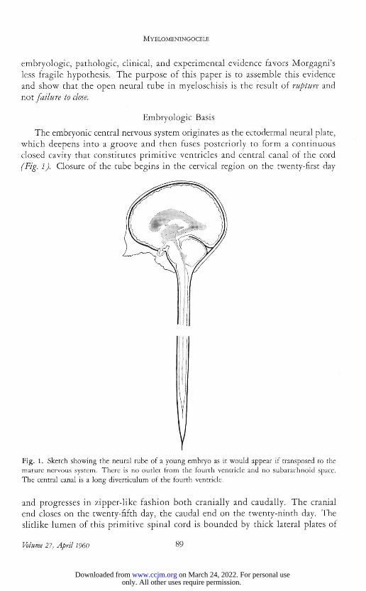
The embryonic central nervous system originates as the ectodermal neural plate, which deepens into a groove and then fuses posteriorly to form a continuous closed cavity that constitutes primitive ventricles and central canal of the cord (Fig. 1). Closure of the tube begins in the cervical region on the twenty-first day
r Fig. 1. Sketch showing the neural tube of a young embryo as it would appear if transposed to the mature nervous system. There is no outlet from the fourth ventricle and no subarachnoid space. The central canal is a long diverticulum of the fourth ventricle.
and progresses in zipper-like fashion both cranially and caudally. The cranial end closes on the twenty-fifth day, the caudal end on the twenty-ninth day. The slitlike lumen of this primitive spinal cord is bounded by thick lateral plates of
Volume 21, April I960 8 9
only. All other uses require permission. on March 24, 2022. For personal usewww.ccjm.orgDownloaded from
GARDNER
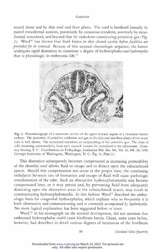
neural tissue and by thin roof and floor plates. The cord is bordered laterally by paired mesodermal somites, posteriorly by cutaneous ectoderm, anteriorly by meso-dermal notochord, and beyond that by endoderm constituting primitive gut (Fig. 2). Weed10 has shown that fluid forms in this closed cavity before facilities are provided for its removal. Because of this unusual chronologic sequence, the lumen undergoes rapid distention to constitute a degree of hydrocephalus and hydromyelia that is physiologic in embryonic life.11
Fig. 2. Photomicrograph of a transverse section of the upper thoracic region of a 16-somite human embryo. The proximity of primitive epidermis and gut to the thin roof and floor plates of the neural tube is well shown. The notochord resembles an outpouching of the primitive gut. The mass of cells streaming anteromedially from each myocele toward the notochord is the sclerotome. (Cour-tesy Sensing, E. C.: Contributions to Embryology, Institution Pub. No. 583, Vol. 33, Feb. 28, 1949; Carnegie Institution of Washington, Washington, D. C.; Fig. 11, Plate 2.)
This distention subsequently becomes compensated as increasing permeability of the rhombic roof allows fluid to escape and to dissect open the subarachnoid spaces. Should this compensation not occur at the proper time, the continuing imbalance between rate of formation and escape of fluid will cause pathologic overdistention of the tube. Such an obstructive hydrocephalomyelia may become compensated later, or it may persist and, by preventing fluid from adequately dissecting open the absorptive areas in the subarachnoid spaces, may result in communicating hydrocephalomyelia. In this fashion Weed10 described the embry-ologic basis for congenital hydrocephalus, which explains why so frequently it is both obstructive and communicating and so commonly accompanied by hydromyelia. No more logical explanation has been suggested before or since.
Weed,10 in his monograph on the normal development, did not mention that embryonal hydrocephalus could cause hindbrain hernia. Chiari, some years before, however, had described in detail various degrees of herniation of the hindbrain
9 0 Cleveland Clinic Quarterly
only. All other uses require permission. on March 24, 2022. For personal usewww.ccjm.orgDownloaded from
MYELOMENINGOCELE
caused by prenatal hydrocephalus. His type-2 herniation subsequently was entitled the Arnold-Chizti malformation. Though Chiari's type-1 herniation (pressure coning) now is recognized to be the result of hydrocephalus, by a curious reversal of its discoverer's logic, his type 2 is believed by some to cause it!
Pathologic Anatomy
Previous reports11,12 have pointed out that the imperforate, distended, neural tube, i.e., obstructive hydrocephalomyelia, which is normal in the embryo, is present also in the infant with myelomeningocele and in the adult with syringo-myelia; that these pathologic states therefore represent the postnatal persistence of conditions that are physiologic in the embryo; that this obstructive hydro-cephalomyelia, uncompensated in myelomeningocele, is compensated in syringo-myelia; that each of these states is accompanied by a Chiari or by the Dandy-Walker malformation; and that the infant with myelomeningocele, like the adult with syringomyelia, may have a diverticulum of his hydromyelic central canal constituting a "true syrinx."
In 1876, Leyden13 came to the conclusion that hydromyelia and syringomyelia are identical, that syringomyelia in the adult is a "rest" of a congenital hydro-myelia that "cuts itself o f f " from the central canal posteriorly. A similar view expressed recently by Greenfield14 is as follows: "In some cases, especially when hydromyelia is associated with the Arnold-Chiari malformation in the adult, only the ventral wall of the cavity may be covered by ependyma and the remainder by a thick firm layer of neuroglial fibres. Or a syringomyelic cavity may have formed at one side of a hydromyelia. Such cases form a link between hydromyelia and syringomyelia, and are apt to be assigned to one or the other category according to whether or not the cavitation has produced the classical symptoms of syringo-myelia during life." From surgical experience with more than 50 cases, I can firmly state that hydromyelia and syringomyelia are one and the same disease.
The adult with syringomyelia therefore has hydromyelia. The adolescent with diastematomyelia has hydromyelia that progresses caudally into the split spinal cord. This is demonstrated in the case of Herren and Edwards5 and in case 3 of Walker.15 The infant with myelomeningocele has hydromyelia that progresses caudally into myeloschisis (Fig. 3), or into diastematomyelia that in turn pro-gresses into myeloschisis;3,6 the latter sequence is well illustrated by Benda's16
case 1. Herren and Edwards5 point out that there is no normal embryonic stage in which the primitive cord is double, and therefore diastematomyelia must represent the splitting of a single neural tube. The downward anatomic sequence in these infants indicates that hydromyelia is the forerunner of both diastemato-myelia and myeloschisis: that increasing distention of the central canal (hydro-myelia) progresses to internal rupture of the thin roof and floor plates (diastema-tomyelia) , and then to external rupture through the primitive skin into the amniotic cavity (myeloschisis).
Volume 27, April I960 9 1
only. All other uses require permission. on March 24, 2022. For personal usewww.ccjm.orgDownloaded from

GARDNER
Fig. 3- Photograph of the spinal cord of week-old infant with myelomeningocele and Chiari type-2 malformation. The cervical cord in cross section is bulky and encroaches on the central canal be-cause it has been telescoped from above downward by herniation of the hindbrain. As successive sections approach the myelomeningocele (not shown) the hydromyelia enlarges.
The ontogenetic sequence described above suggests that embryonal hydro-cephalomyelia, compensated at the normal time, results in a normal individual; compensated a bit too late it results in symptoms of syringomyelia in adult life; compensated still later, in symptoms of diastematomyelia in adolescence; and uncompensated it results in myelomeningocele in the newborn infant. Thus there is both an ontogenetic as well as an anatomic sequence indicating that myeloschisis represents the rupture of an overdistended neural tube—not a failure to close.
Hypothesis
Any theory that attempts to explain the origin of myelomeningocele, of neces-sity, must explain the hydrocephalomyelia and the deformity of the hindbrain that accompany it. In a previous paper'1 it was pointed out that, in some instances, myelomeningocele has been accompanied by the Dandy-Walker, or by the Chiari type-1 (pressure coning) instead of the usual Chiari type-2 (Arnold-Chiari) mal-formation. It was further shown that: each of these malformations of the hind-brain with its accompanying hydrocephalomyelia is the result of embryonal atresia of the fourth ventricle; the large posterior fossa of the Dandy-Walker malforma-tion develops if the coverings of the hindbrain yield more readily to the increased pressure and thus displace the lateral sinuses and attached tentorium in the cephalad direction (i.e., prevent their caudad migration); the small posterior fossa of the Chiari type-2 malformation develops if the coverings of the forebrain expand disproportionately and thus displace the lateral sinuses and tentorium (i.e., cause them to migrate too far) in a caudad direction (Fig. 4).
The sequence of events as they have occurred in the infant born with myelo-meningocele and Chiari type-2 malformation I believe to be as follows. The neural
9 2 Cleveland Clinic Quarterly
only. All other uses require permission. on March 24, 2022. For personal usewww.ccjm.orgDownloaded from
MYELOMENINGOCELE
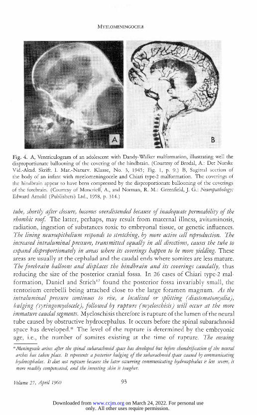
Fig. 4. A, Ventriculogram of an adolescent with Dandy-Walker malformation, illustrating well the disproportionate ballooning of the covering of the hindbrain. (Courtesy of Brodai, A.: Det Norske Vid.-Akad. Skrift. I. Mat.-Naturv. Klasse, No. 3, 1945; Fig. 1, p. 9.) B, Sagittal section of the body of an infant with myelomeningocele and Chiari type-2 malformation. The coverings of the hindbrain appear to have been compressed by the disproportionate ballooning of the coverings of the forebrain. (Courtesy of Moncrieff, A., and Norman, R. M.: Greenfield, J. G.: Neuropathology; Edward Arnold (Publishers) Ltd., 1958, p. 314.)
tube, shortly after closure, becomes overdistended because of inadequate permeability of the rhombic roof. The latter, perhaps, may result from maternal illness, avitaminosis, radiation, ingestion of substances toxic to embryonal tissue, or genetic influences. The lining neuroepithelium responds to stretching, by more active cell reproduction. The increased intraluminal pressure, transmitted equally in all directions, causes the tube to expand disproportionately in areas where its coverings happen to be more yielding. These areas are usually at the cephalad and the caudal ends where somites are less mature. The forebrain balloons and displaces the hindbrain and its coverings caudally, thus reducing the size of the posterior cranial fossa. In 26 cases of Chiari type-2 mal-formation, Daniel and Strich17 found the posterior fossa invariably small, the tentorium cerebelli being attached close to the large foramen magnum. As the intraluminal pressure continues to rise, a localized or splitting (diastematomyelia), bulging (syringomyelocele), followed by rupture (myeloschisis) will occur at the more immature caudal segments. Myeloschisis therefore is rupture of the lumen of the neural tube caused by obstructive hydrocephalus. It occurs before the spinal subarachnoid space has developed.* The level of the rupture is determined by the embryonic age, i.e., the number of somites existing at the time of rupture. The ensuing
* Meningocele arises after the spinal subarachnoid space has developed but before chondrification of the neural arches has taken place. It represents a posterior bulging of the subarachnoid space caused by communicating hydrocephalus. It ekes not rupture because the later occurring communicating hydrocephalus is less severe, is more readily compensated, and the investing skin is tougher.
Volume 27, April I960 9 3
only. All other uses require permission. on March 24, 2022. For personal usewww.ccjm.orgDownloaded from
GARDNER
collapse of the head causes its redundant lining of neural tissue to wrinkle. This explains the plication and overgrowth found by Patten18 in his human embryo with myeloschisis. Relief of hydrocephalus by rupture of the tube explains why War-kany, Wilson, and Geiger" found that in their rats with experimental myeloschisis, hydrocephalus was absent and, conversely, that in litter mates without myeloschisis it was present. The discharge of ventricular fluid into the amniotic sac removes the hydrostatic pressure essential to the normal dissection of the subarachnoid spaces.10
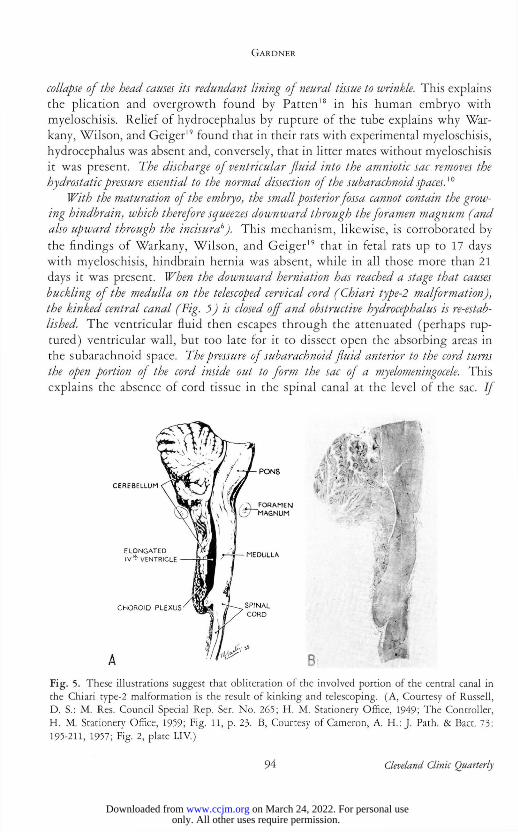
With the maturation of the embryo, the small posterior fossa cannot contain the grow-ing hindhrain, which therefore squeezes downward through the foramen magnum (and also upward through the incisurab). This mechanism, likewise, is corroborated by the findings of Warkany, Wilson, and Geiger" that in fetal rats up to 17 days with myeloschisis, hindbrain hernia was absent, while in all those more than 21 days it was present. When the downiuard herniation has reached a stage that causes buckling of the medulla on the telescoped cervical cord (Chiari type-2 malformation), the kinked central canal (Fig. 5 J is closed off and obstructive hydrocephalus is re-estab-lished. The ventricular fluid then escapes through the attenuated (perhaps rup-tured) ventricular wall, but too late for it to dissect open the absorbing areas in the subarachnoid space. The pressure of subarachnoid fluid anterior to the cord turns the open portion of the cord inside out to form the sac of a myelomeningocele. This explains the absence of cord tissue in the spinal canal at the level of the sac. If
Fig. 5. These illustrations suggest that obliteration of the involved portion of the central canal in the Chiari type-2 malformation is the result of kinking and telescoping. (A, Courtesy of Russell, D. S.: M. Res. Council Special Rep. Ser. No. 265; H. M. Stationery Office, 1949; The Controller, H. M. Stationery Office, 1959; Fig. 11, p. 23. B, Courtesy of Cameron, A. H.: J. Path. & Bact. 73: 195-211, 1957; Fig. 2, plate LIV.)
)
9 4 Cleveland Clinic Quarterly
only. All other uses require permission. on March 24, 2022. For personal usewww.ccjm.orgDownloaded from
MYELOMENINGOCELE
rupture of the attenuated sac occurs, subarachnoid fluid escapes and hydrocephalus once more may be relieved. This second rupture explains some cases of myelomeningocele with Chiari type-2 malformation but without hydrocephalus.6,20
The blood vessels nourishing the exposed neural tissue, distend because of lack of over-lying tissue support. Thus originates the area medullovasculosa described by Von Recklinghausen.1
Other Associated Anomalies
Morgagni's7 hydromyelic theory will explain every anomaly associated with myelomeningocele. The mechanism of formation of the Chiari type-1, Chiari type-2, and Dandy-Walker malformations have been described. Secondary distor-tion of surrounding tissues caused by the overdistention of the neural tube renders unnecessary the premise21 that there must be associated primary disturbances in ectoderm, mesoderm, or endoderm to explain the accompanying dysplasias of skin, skeleton, muscle, genitourinary or gastrointestinal tracts.
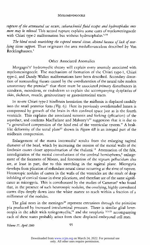
In severe Chiari type-2 hindbrain herniation the midbrain is displaced caudally into the small posterior fossa (Fig. 6). Here its previously overdistended lumen is compressed by growth of the brain in this confined space just as is the fourth ventricle. This explains the associated stenosis and forking (plication) of the aqueduct, and confirms MacFarlane and Maloney's22 suggestion that it is due to "a generalized compression of the hind end of the ventricular system." A beak-like deformity of the tectal plate23 shown in Figure 4B is an integral part of the midbrain compression.
Enlargement of the massa intermedia4 results from the enlarging sagittal diameter of the head, which by increasing the tension of the mesial walls of the forebrain causes closer approximation of the thalami.12 Attenuation of the falx, interdigitation of the mesial convolutions of the cerebral hemispheres,6 enlarge-ment of the foramens of Monro, and fenestration of the septum pellucidum also are, at least in part, due to this stretching in the sagittal plane. Microgyria develops from plication of redundant neural tissue occurring at the time of rupture. Heterotopic nodules of cortex in the walls of the ventricles are the result of deep infolding of cortical tissue in these plications, and therefore are of the same signifi-cance as microgyria. This is corroborated by the studies of Cameron6 who found that, in the presence of such heterotopic nodules, the overlying, highly convoluted cortex dips deeply down into the white matter to reach within a fraction of a millimeter of the nodules.
The glial nests in the meninges24 represent extrusions through the primitive pia produced by increased intraluminal pressure. There is similar glial heter-otopia in the adult with syringomyelia,24 and the neoplasia 3,5,25 accompanying each of these states probably arises from these displaced embryonal cell rests.
Volume 21, April I960 9 5
only. All other uses require permission. on March 24, 2022. For personal usewww.ccjm.orgDownloaded from
GARDNER
Fig. 6. Necropsy photograph. Child aged four years and five months had Chiari type-2 malforma-tion, congenital hydrocephalus, and myelomeningocele. The hydrocephalic forebrain has been re-moved. The entire attachment of the tentorium is dislocated caudally including the superior petrosal sinuses which are depressed well below the petrous ridges. Note that the cerebral peduncles are displaced downward behind the clivus into the posterior fossa while the superior surface of the vermis bulges upward through the incisura. The aqueduct, compressed in the midbrain hernia, cannot be visualized. This necropsy study illustrates the advantages of fixation in situ and removal of the hydrocephalic cerebral hemispheres before disturbing the relationship of the structures of the posterior fossa. (Gardner, W. J. : Cleveland Clin. Quart. 26: 206-222; Oct. 1959, Fig. 4, p. 210.)
Diastematomyelia results when the increased intraluminal pressure causes separation of the roof plate, permitting the lateral plates to open like a book that is subsequently torn along its binding; i.e., the floor plate. (The layer of tissue reinforcing the roof plate is thinner than that of the floor plate; therefore, the roof plate separates more readily. This mechanism also will explain the pronounced tendency for syringomyelic cavities to involve the posterior columns.) Each lateral plate thus rotated through 90 degrees will form an imperfect hydromyelic cord above the level at which external rupture has resulted in myeloschisis.
The finding at necropsy of myelodysplasia without hydromyelia does not prove that it was not the result of pre-existing hydromyelia. Since the physiologic
9 6 Cleveland Clinic Quarterly
only. All other uses require permission. on March 24, 2022. For personal usewww.ccjm.orgDownloaded from
MYELOMENINGOCELE
degree of hydromyelia present in every normal embryo becomes compensated, there is no reason that a pathologic degree likewise should not become compen-sated at least in mild cases. Such compensation occurring early enough will permit the growing spinal cord to encroach on its distended lumen.
Likewise, the occasional finding of ventricles of normal size in cases of microgyria or Chiari malformation does not prove that there was not pre-existing hydrocephalus subsequently reduced by compensation or by rupture of the neural tube. Similarly, the overgrowth and excessive plication of the neural tubes of Patten's18 embryos that did not have myeloschisis, could have resulted from hydro-cephalomyelia either compensated during the life of the embryo, or reduced after death by cerebrospinal fluid absorption occasioned by its low colloid osmotic tension.
In some instances of myelodysplasia, as in those reported by Walker,15 a dilated vertebral canal may constitute the only evidence of a pre-existing dilatation of the neural tube. Holtzer26 has shown experimentally that the diameter of the developing spinal canal is determined by the diameter of the neural tube that it encloses. He states that: "Migrating pre-cartilage cells respond in a discrimina-tory and stereotyped fashion to the presence of any neural tissue. By maintaining a characteristic distance from the neural tissue the pre-cartilage cells are deployed in such a fashion that a lumen will eventually be formed in the cartilage whose size is a function of the enclosed nerve bundle." This work of Holtzer also ex-plains the mid-line bony spur separating the cords in diastematomyelia.
Craniolacunia represents interference with chondrification, and subsequent ossification caused by compression and stretching of the mesodermal anlage of the skull. Separation and distortion of the paired sclerotomes by the overdistended neural tube will explain spina bifida posterior, spina bifida anterior (hemiverte-brae), dilated vertebral canal, scoliosis, fused vertebrae, and deformities of ribs and sternum; distortion of the neighboring limb buds—the club feet; distortion of the overlying ectoderm—cutaneous dysplasia; distortion of the intermediate mesoderm—deformities of the genitourinary tract; distortion and adhesion to the wall of the primitive gut—alimentary diverticuli and cysts described by Saunders,27
and by McLetchie, Purves, and Saunders.28
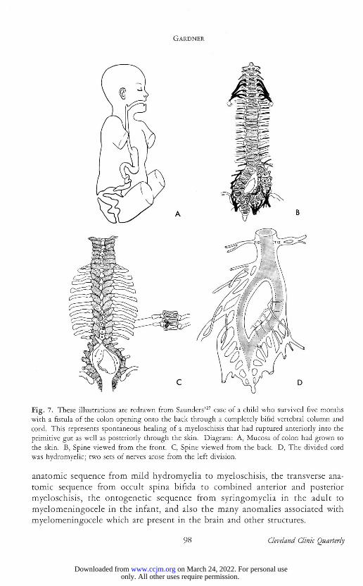
Finally, a simultaneous rupture of the roof plate into the amniotic sac, and of the floor plate into the primitive gut will explain the rare, usually stillborn infants in whom a fistula of the gut opens onto the floor of a myeloschisis (Fig. 1). This makes it unnecessary to invoke Bremer's29 purely hypothetic "accessory neurenteric canal" or any of the other earlier theories that Saunders27
has summarized in his review and to which the reader is referred.
Conclusions
Therefore we find that Morgagni's hydromyelic theory is less fragile than Von Recklinghausen's araphic theory; that Morgagni's theory will explain the vertical
Volume 21, April 1960 97
only. All other uses require permission. on March 24, 2022. For personal usewww.ccjm.orgDownloaded from
GARDNER
Fig. 7. These illustrations are redrawn from Saunders'27 case of a child who survived five months with a fistula of the colon opening onto the back through a completely bifid vertebral column and cord. This represents spontaneous healing of a myeloschisis that had ruptured anteriorly into the primitive gut as well as posteriorly through the skin. Diagram: A, Mucosa of colon had grown to the skin. B, Spine viewed from the front. C, Spine viewed from the back. D, The divided cord was hydromyelic; two sets of nerves arose from the left division.
anatomic sequence from mild hydromyelia to myeloschisis, the transverse ana-tomic sequence from occult spina bifida to combined anterior and posterior myeloschisis, the ontogenetic sequence from syringomyelia in the adult to myelomeningocele in the infant, and also the many anomalies associated with myelomeningocele which are present in the brain and other structures.
9 8 Cleveland Clinic Quarterly
only. All other uses require permission. on March 24, 2022. For personal usewww.ccjm.orgDownloaded from
MYELOMENINGOCELE
Acknowledgment
The author, as a clinician, wishes to acknowledge his indebtedness to those in other fields, particularly to the fundamental contribution of Weed on the embryonal development of the cerebrospinal fluid spaces, to Chiari, to Russell, to Saunders, and to Cameron for their contributions on the pathology, and to Holtzer, to Fowler, and to Warkany, Wilson, and Geiger for their experimental contributions.
References
1. von Recklinghausen, F.: Untersuchungen iiber die Spina bifida. Arch. path. Anat. 105: 243; 373, 1886.
2. Cameron, A. H.: Spinal cord lesion in spina bifida cystica. Lancet 2: 171-174, 1956.
3. Weil, A., and Matthews, W. B.: Duplication of spinal cord, with spina bifida and syringo-myelia. Arch. Path. 20: 882-890, 1935.
4. Kapsenberg, J. G., and van Lookeren Campagne, J. A.: Case of spina bifida combined with diastomatomyely, anomaly of Chiari and hydrocephaly. Acta nat. 7: 366-388, 1949.
5. Herren, R. Y., and Edwards, J. E.: Diplomyelia (duplication of spinal cord). Arch. Path. 30: 1203-1214, 1940.
6. Cameron, A. H.: Arnold-Chiari and other neuro-anatomical malformations associated with spina bifida. J. Path. & Bact. 73: 195-211, 1957.
7. Morgagni, G. B.: De Sedibus et Causis Morborum 1761. Trans, by B. Alexander. London: A. Millar and T. Cadell, 1769.
8. Patten, B. M. : Embryological stages in establishing of myeloschisis with spina bifida. Am. J. Anat. 93: 365-395, 1953.
9. Fowler, I.: Responses of chick neural tube in mechanically produced spina bifida. J. Exper. Zool. 123: 115-151, 1953.
10. Weed, L. H.: The Development of the Cerebro-Spinal Spaces in Pig and in Man. Washington, D. C : Carnegie Inst., 1917, 116 pp., 4°.
11. Gardner, W, J . : Anatomic features common to Arnold-Chiari and Dandy-Walker malformations suggest common origin. Cleveland Clin. Quart. 26: 206-222, 1959.
12. Gardner, W. J . : Anatomic anomalies common to myelomeningocele of infancy and syringo-myelia of adulthood suggest common origin. Cleveland Clin. Quart. 26: 118-133, 1959.
13. Leyden, E.: Ueber Hydromyelus und Syringomyelie. (About hydromyelia and syringomyelia.) Arch. path. Anat. 68: 1-26, 1876.
14. Greenfield, J . G., and others: Neuropathology. London: Edward Arnold (Publishers) Ltd., 1958, 640 pp.
15. Walker, A. E.: Dilatation of vertebral canal associated with congenital anomalies of spinal cord. Am. J . Roentgenol. 52: 571-582, 1944.
16. Benda, C. E.: Dysraphic states. J. Neuropath. & Exper. Neurol. 18: 56-74, 1959.
Volume 27, April 1960 9 9
only. All other uses require permission. on March 24, 2022. For personal usewww.ccjm.orgDownloaded from

GARDNER
17. Daniel, P. M., and Strieh, S. J. : Some observations on congenital deformity of central nervous system known as Arnold-Chiari malformation. J. Neuropath. & Exper. Neurol. 17: 255-266, 1958.
18. Patten, B. M.: Overgrowth of neural tube in young human embryos. Anat. Rec. 113: 381-393, 1952.
19. Warkany, J . ; Wilson, J. G., and Geiger, J. F.: Myeloschisis and myelomeningocele produced experimentally in rat. J. Comp. Neurol. 109: 35-64, 1958.
20. Russell, D. S., and Donald, C.: Mechanism of internal hydrocephalus in spina bifida. Brain 58: 203-215, 1935.
21. Lichtenstein, B.: "Spinal dysraphism"; spina bifida and myelodysplasia. Arch. Neurol. & Psychiat. 44: 792-810, 1940.
22. MacFarlane, A., and Maloney, A. F. J. : Appearance of aqueduct and its relationship to hydro-cephalus in Arnold-Chiari malformation. Brain 80: 479-491, 1957.
23. Feigin, I.: Arnold-Chiari malformation with associated analogous malformation of midbrain. Neurology 6: 22-31, 1956.
24. Cooper, I. S., and Kernohan, J. W.: Heterotopic glial nests in subarachnoid space: histopatho-logic characteristics, mode of origin and relation to meningeal gliomas. J. Neuropath. & Exper. Neurol. 10: 16-29, 1951.
25. Poser, C. M.: The Relationship Between Syringomyelia and Neoplasm. Pub. No. 262, Am. Lect. Ser., monograph in Am. Lect. in Neurol., edited by C. D. Aring. Springfield, III.: Charles C Thomas, Pub., 1956, 98 pp.
26. Holtzer, H.: Experimental analysis of development of spinal column. Part I. Response to pre-cartilage cells to size variations of spinal cord. J. Exper. Zool. 121: 121-147, 1952.
27. Saunders, R. L. deC. H.: Combined anterior and posterior spina bifida in living neonatal human female. Anat. Rec. 87: 255-278, 1943.
28. McLetchie, N. G. B.; Purves, J. K., and Saunders, R. L. deC. H.: Genesis of gastric and certain intestinal diverticula and enterogenous cysts. Surg. Gynec. & Obst. 99: 135-141, 1954.
29. Bremer, J. L.: Dorsal intestinal fistula; accessory neurenteric canal; diastematomyelia. A. M. A. Arch. Path. 54: 132-138, 1952.
1 0 0 Cleveland Clinic Quarterly
only. All other uses require permission. on March 24, 2022. For personal usewww.ccjm.orgDownloaded from