Mesurer et modifier l'élasticité des tissus avec les ultrasons:
de l'élastographie à la chirurgie non invasive
Jean-Francois Aubry
1. Director of research CNRS, Institut Langevin, Equipe Physique des Ondes pour la
Médecine, CNRS, INSERM, ESPCI Paris Tech, Paris, France
2. Visiting Associate Prof, Department of Radiation Oncology, Univ. of Virginia, USA
Mesurer l'élasticité des tissus avec les ultrasons
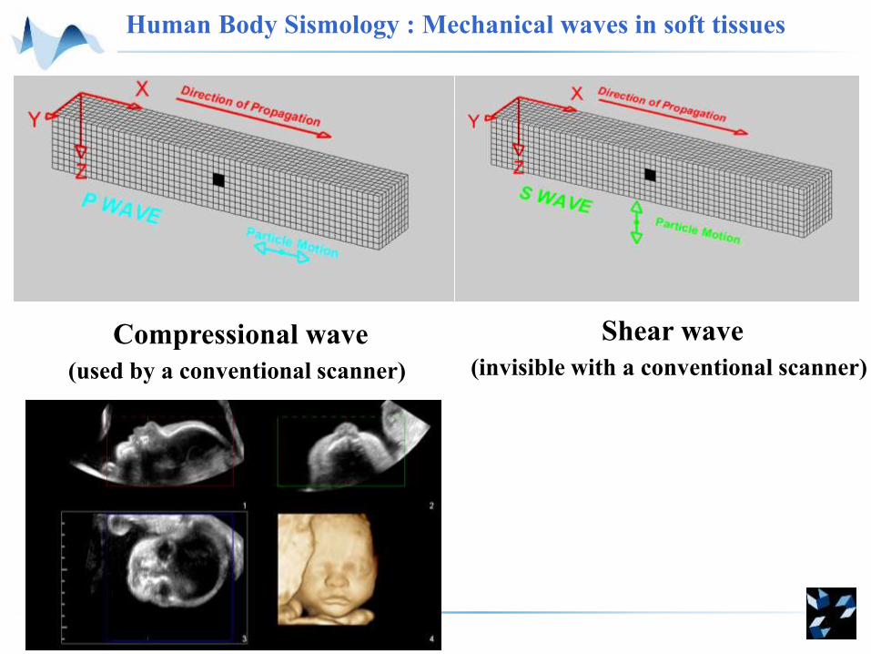
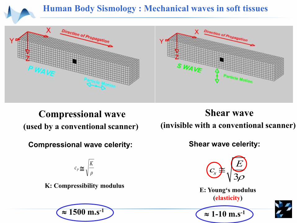
Human Body Sismology : Mechanical waves in soft tissues
Compressional wave
(used by a conventional scanner)
Shear wave
(invisible with a conventional scanner)
Human Body Sismology : Mechanical waves in soft tissues
Compressional wave
(used by a conventional scanner)
Shear wave
(invisible with a conventional scanner)
1500 m.s-1 1-10 m.s-1
3s
Ec
ρ
KcP
E: Young‘s modulus
(elasticity)
K: Compressibility modulus
Compressional wave celerity: Shear wave celerity:
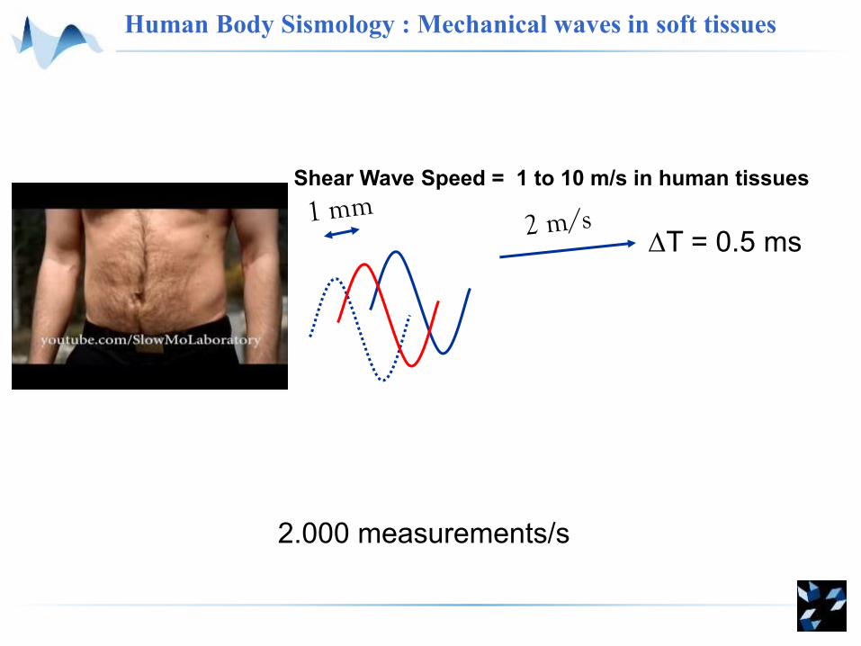
T = 0.5 ms
Shear Wave Speed = 1 to 10 m/s in human tissues
2.000 measurements/s
Human Body Sismology : Mechanical waves in soft tissues
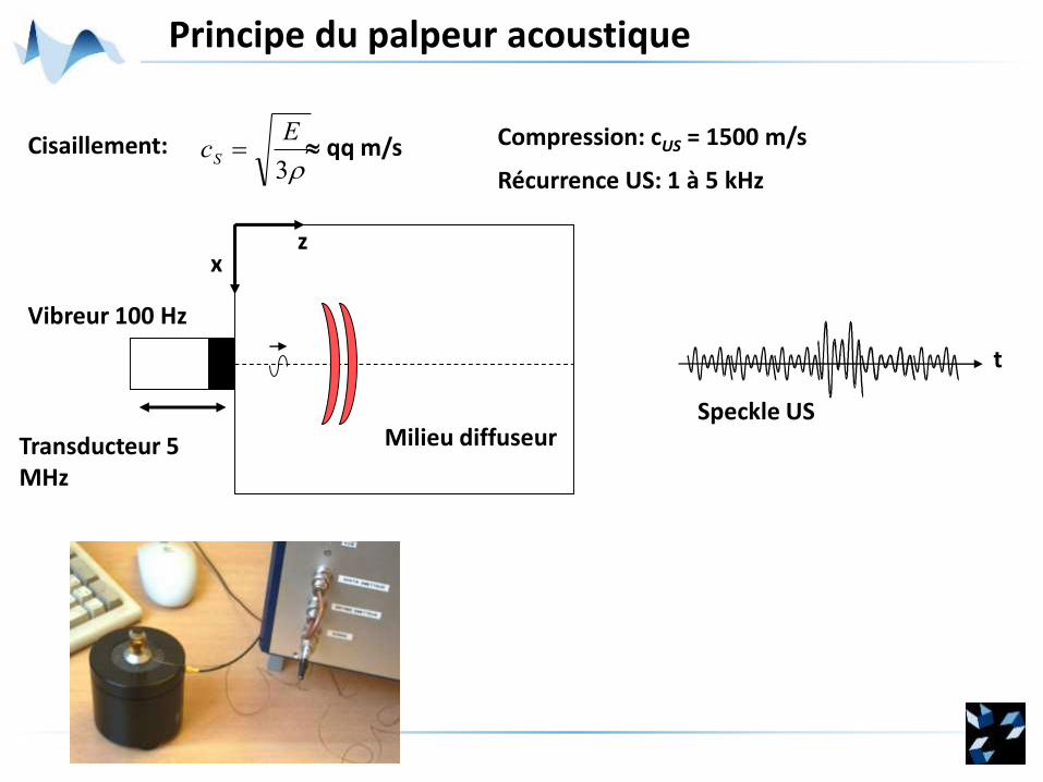
Principe du palpeur acoustique
Milieu diffuseur
xz
Vibreur 100 Hz
Transducteur 5 MHz
Speckle US
t
3
EcS Cisaillement: Compression: cUS = 1500 m/s
Récurrence US: 1 à 5 kHz
qq m/s

Mesure du déplacement axial
Profondeur
layer
A scan at shot n
Recurence time ~ 200 ms1 µm < d < 100 µm
R
d
max
Profondeur
Prof.
Prof.A scan
at shot n+1
Profondeur
Cross-correlation in moving window
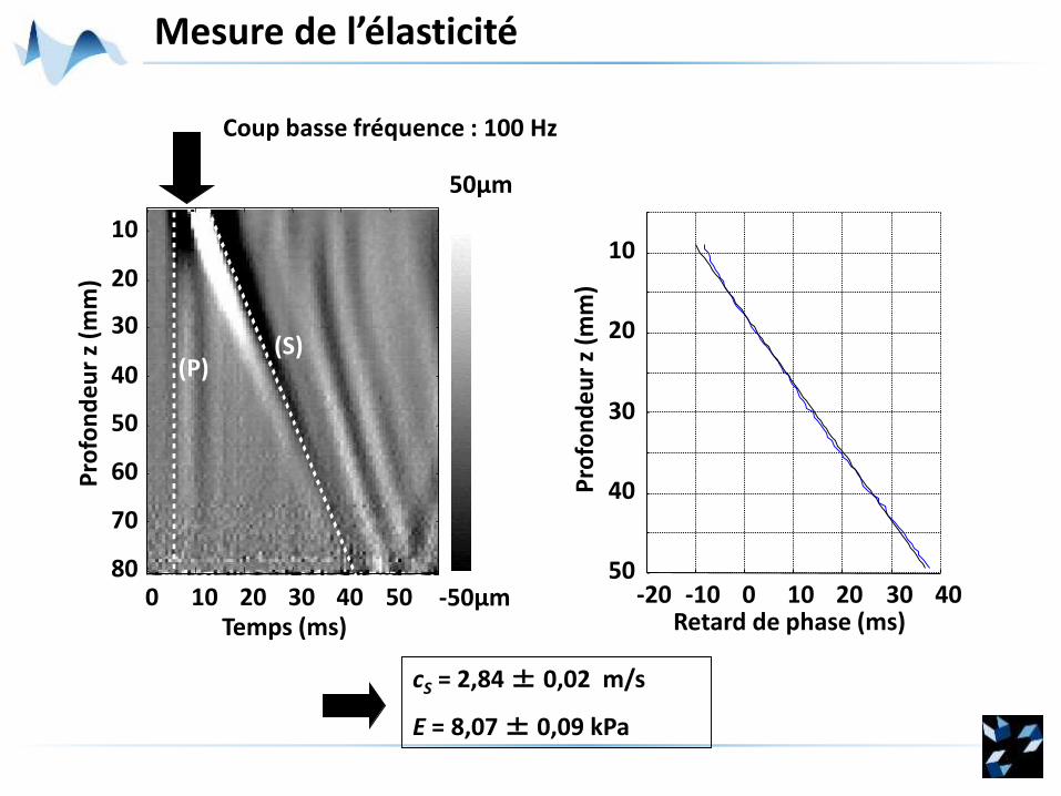
Coup basse fréquence : 100 Hz
Mesure de l’élasticité
-20 -10 0 10 20 30 40Retard de phase (ms)
50
40
30
20
10
Pro
fon
de
ur
z (m
m)
cS = 2,84 ± 0,02 m/s
E = 8,07 ± 0,09 kPa
Pro
fon
de
ur
z (m
m)
0 10 20 30 50
10
20
30
40
50
60
70
80
(P)
-50µm
50µm
(S)
Temps (ms)40
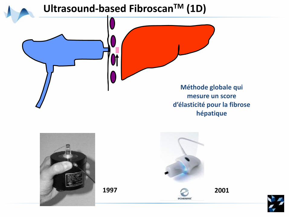
Ultrasound-based FibroscanTM (1D)
Méthode globale qui mesure un score
d’élasticité pour la fibrosehépatique
1997 2001
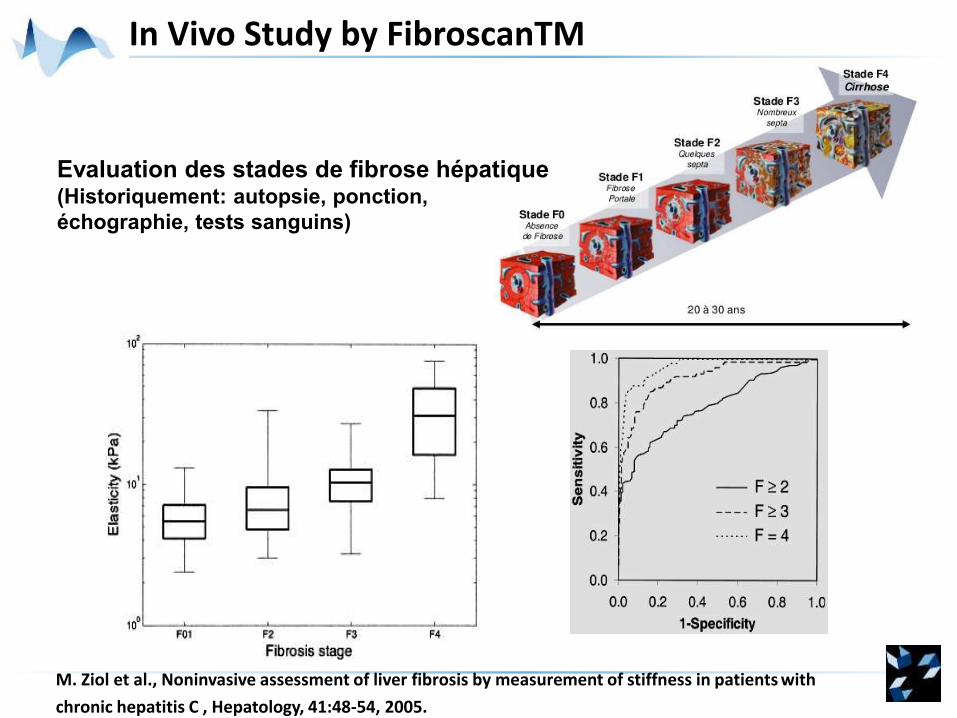
In Vivo Study by FibroscanTM
M. Ziol et al., Noninvasive assessment of liver fibrosis by measurement of stiffness in patients with
chronic hepatitis C , Hepatology, 41:48-54, 2005.
Evaluation des stades de fibrose hépatique(Historiquement: autopsie, ponction,
échographie, tests sanguins)
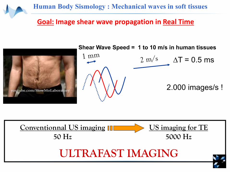
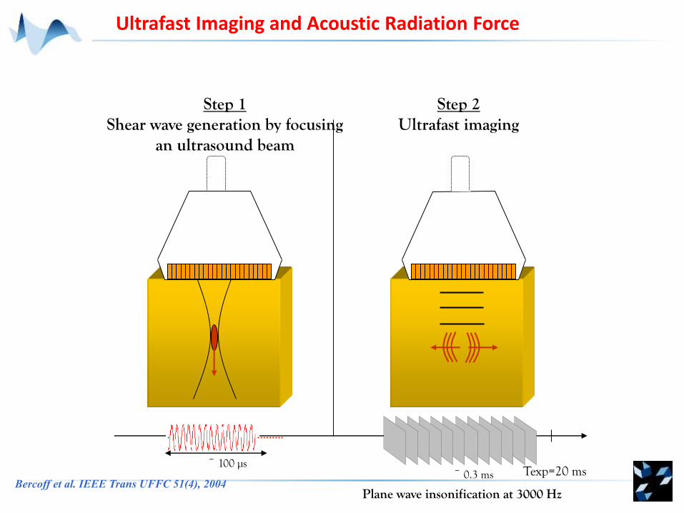
Goal: Image shear wave propagation in Real Time
T = 0.5 ms
2.000 images/s !
Shear Wave Speed = 1 to 10 m/s in human tissues
Conventionnal US imaging50 Hz
US imaging for TE5000 Hz
ULTRAFAST IMAGING
Human Body Sismology : Mechanical waves in soft tissues
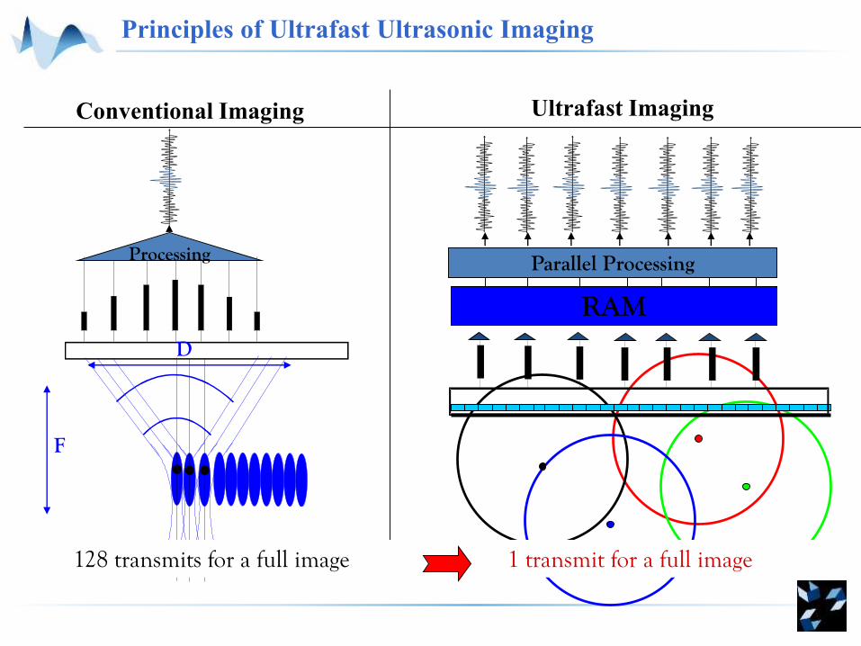
D
F
Conventional Imaging Ultrafast Imaging
RAM
Parallel ProcessingProcessing
128 transmits for a full image 1 transmit for a full image
Principles of Ultrafast Ultrasonic Imaging
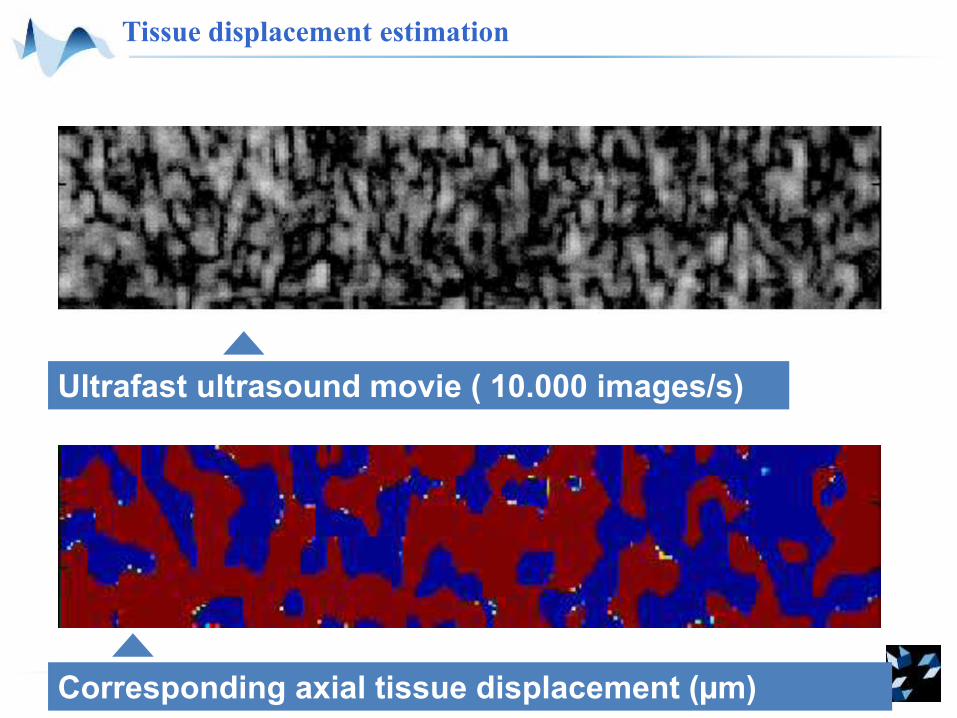
Tissue displacement estimation
Corresponding axial tissue displacement (µm)
Ultrafast ultrasound movie ( 10.000 images/s)
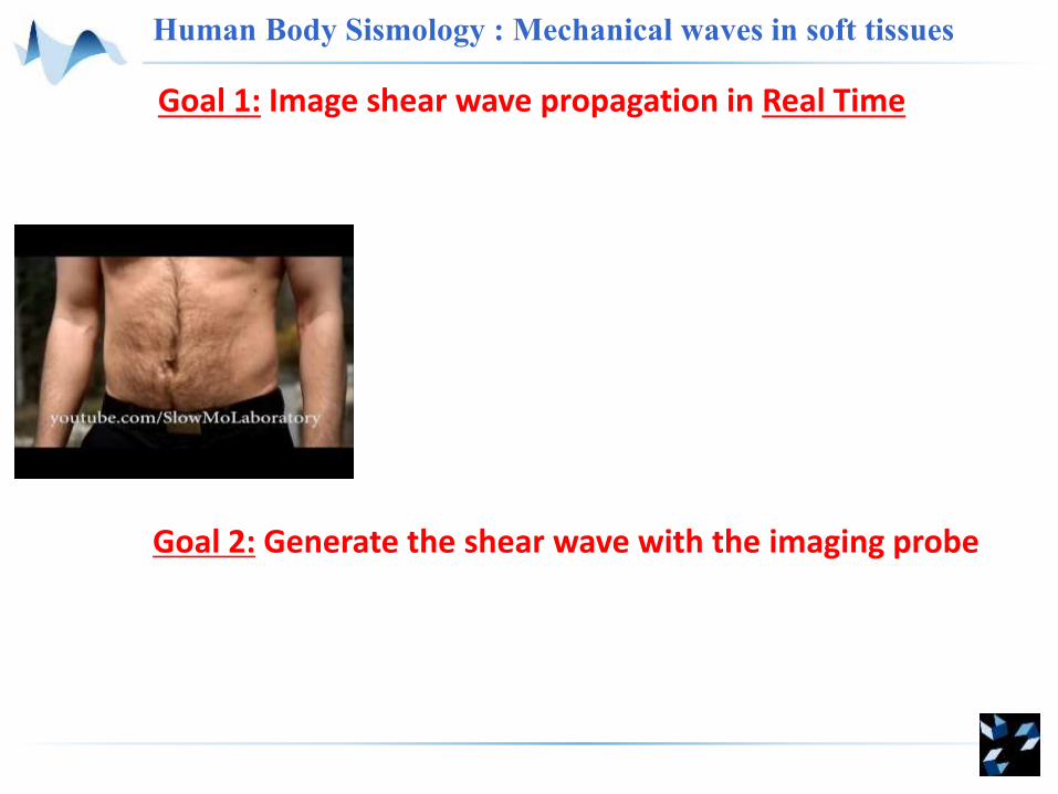
Goal 1: Image shear wave propagation in Real Time
Human Body Sismology : Mechanical waves in soft tissues
Goal 2: Generate the shear wave with the imaging probe
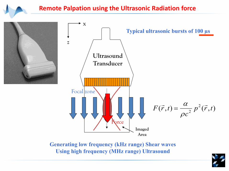
UltrasoundTransducer
Imaged Area
x
z
Focal zone
Force
),(),(2
2trp
ctrF
Remote Palpation using the Ultrasonic Radiation force
Typical ultrasonic bursts of 100 µs
Generating low frequency (kHz range) Shear waves
Using high frequency (MHz range) Ultrasound
~ 100 µs
Step 1Shear wave generation by focusing
an ultrasound beam
Plane wave insonification at 3000 Hz
Texp=20 ms~ 0.3 ms
Step 2Ultrafast imaging
Ultrafast Imaging and Acoustic Radiation Force
Bercoff et al. IEEE Trans UFFC 51(4), 2004

Mapping Visco-Elasticity: Inverse problem of SW Propagation
kPa
• Freehand / does not change anything to the echographic exam
• Quantitative
• Operator independent = reproducible
• Ultrafast / Insensitive to motion artefacts and boundary conditions.
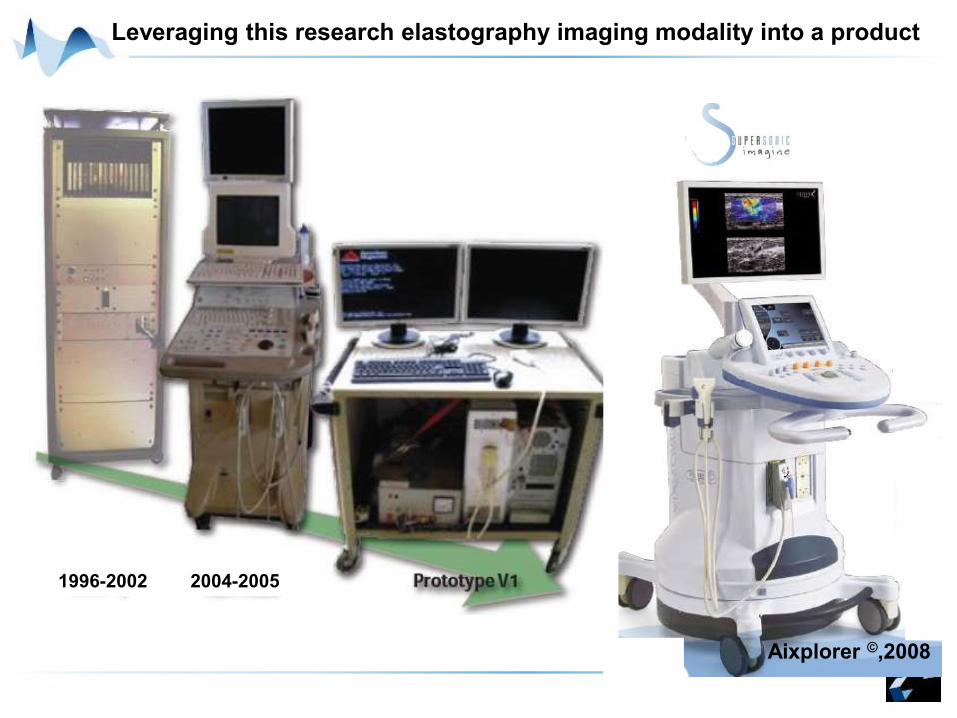
Leveraging this research elastography imaging modality into a product
Aixplorer ©,2008
1996-2002 2004-2005
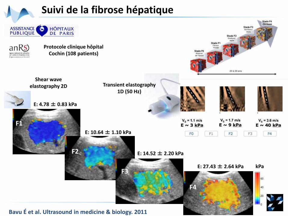
Protocole clinique hôpital Cochin (108 patients)
Transient elastography 1D (50 Hz)
F1
F2
F4
F3
Shear waveelastography 2D
E: 4.78 ± 0.83 kPa
E: 10.64 ± 1.10 kPa
E: 27.43 ± 2.64 kPa kPa
E: 14.52 ± 2.20 kPa
Suivi de la fibrose hépatique
Bavu É et al. Ultrasound in medicine & biology. 2011
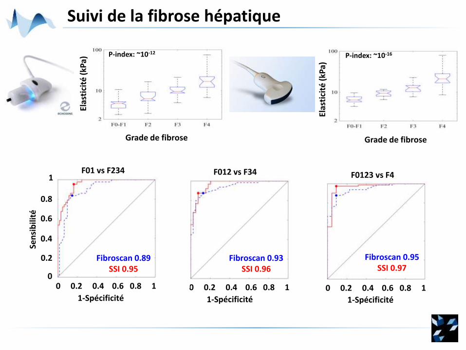
Suivi de la fibrose hépatique
Grade de fibrose
Elas
tici
té (
kPa)
0 0.2 0.4 0.6 0.8 1
1-Spécificité
1
0.8
0.6
0.4
0.2
0
Sen
sib
ilité
F01 vs F234
Fibroscan 0.89SSI 0.95
Fibroscan 0.95SSI 0.97
0 0.2 0.4 0.6 0.8 1
1-Spécificité
F0123 vs F4
Fibroscan 0.93SSI 0.96
0 0.2 0.4 0.6 0.8 1
1-Spécificité
F012 vs F34
Grade de fibrose
Elas
tici
té (
kPa)
P-index: ~10-12 P-index: ~10-16
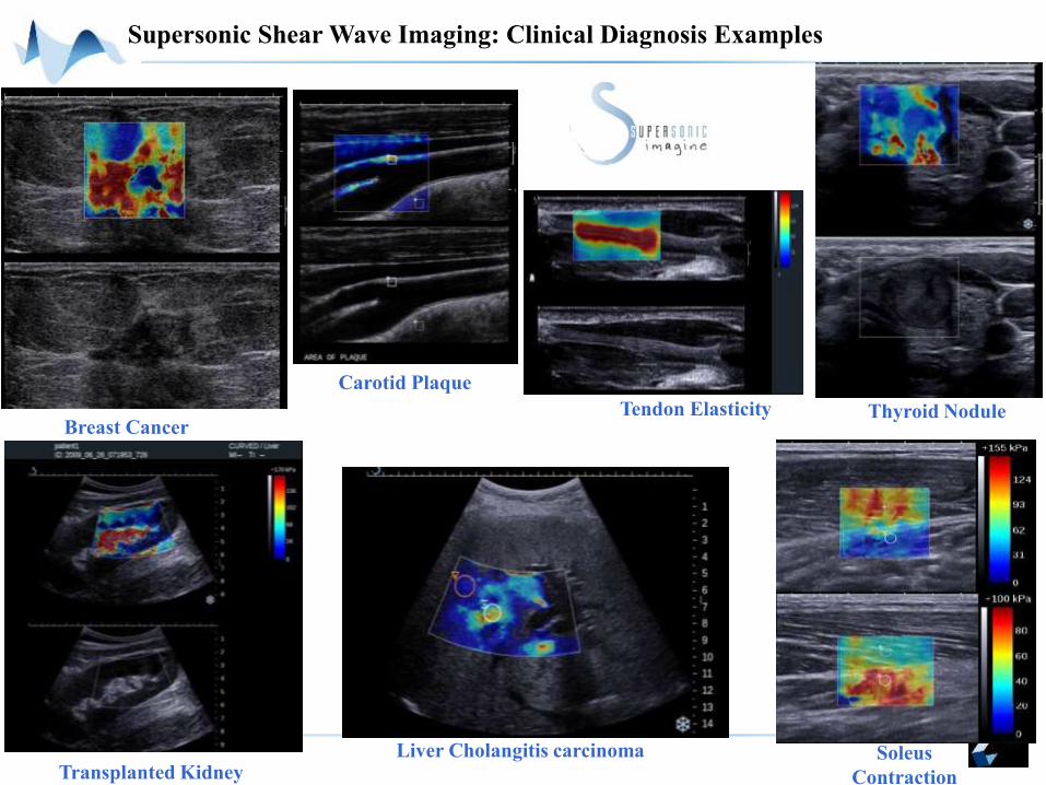
Liver Cholangitis carcinomaTransplanted Kidney
Tendon Elasticity
Carotid Plaque
Thyroid Nodule
Soleus
Contraction
Supersonic Shear Wave Imaging: Clinical Diagnosis Examples
Breast Cancer
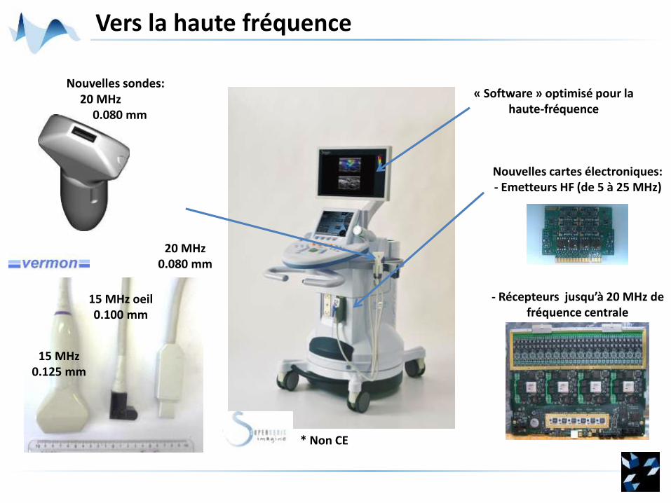
Vers la haute fréquence
Nouvelles sondes:« Software » optimisé pour la
haute-fréquence
Nouvelles cartes électroniques: - Emetteurs HF (de 5 à 25 MHz)
- Récepteurs jusqu’à 20 MHz de fréquence centrale
* Non CE
20 MHz 0.080 mm
15 MHz0.125 mm
15 MHz oeil 0.100 mm
20 MHz0.080 mm
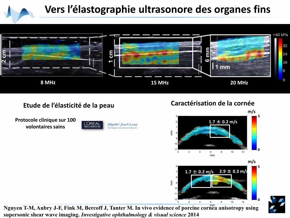
Vers l’élastographie ultrasonore des organes fins
1 mm
2 c
m
1 c
m
6 m
m
Etude de l’élasticité de la peau
Protocole clinique sur 100 volontaires sains
Caractérisation de la cornée
mm
mm
Velocity map [0 - 5 m/s]
0 2 4 6 8 10 12
6
7
8
9
10
11
12 0
0.2
0.4
0.6
0.8
1
mm
mm
Velocity map [0 - 5 m/s]
0 2 4 6 8 10 12
5
6
7
8
9
10
11 0
0.2
0.4
0.6
0.8
1
1.7 ± 0.2 m/s
2.9 ± 0.3 m/s1.7 ± 0.2 m/s
5
5
0
0
m/s
m/s
8 MHz 15 MHz 20 MHz
Nguyen T-M, Aubry J-F, Fink M, Bercoff J, Tanter M. In vivo evidence of porcine cornea anisotropy using
supersonic shear wave imaging. Investigative ophthalmology & visual science 2014
Modifier l'élasticité des tissus avec les ultrasons
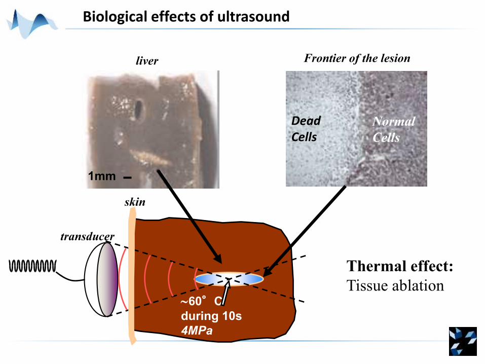
60°C
during 10s
4MPa
skin
Normal
Cells
Dead Cells
Frontier of the lesion
transducer
Thermal effect:
Tissue ablation
liver
1mm
Biological effects of ultrasound
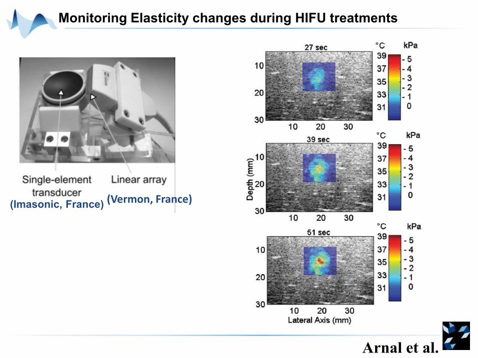
Monitoring Elasticity changes during HIFU treatments
(Vermon, France)
Arnal et al.
(Imasonic, France)
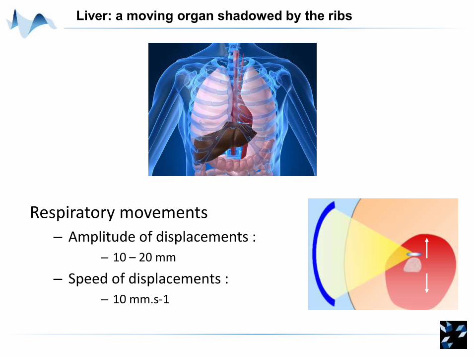
Liver: a moving organ shadowed by the ribs
Respiratory movements
– Amplitude of displacements : – 10 – 20 mm
– Speed of displacements : – 10 mm.s-1
time
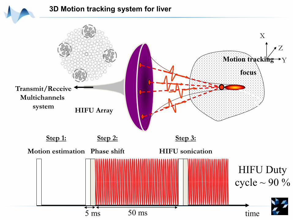
Motion tracking
focus
Step 2:
Phase shift
Step 1:
Motion estimation
Step 3:
HIFU sonication
HIFU Duty
cycle ~ 90 %
Transmit/Receive
Multichannels
systemHIFU Array
X
Y
Z
3D Motion tracking system for liver
5 ms 50 ms
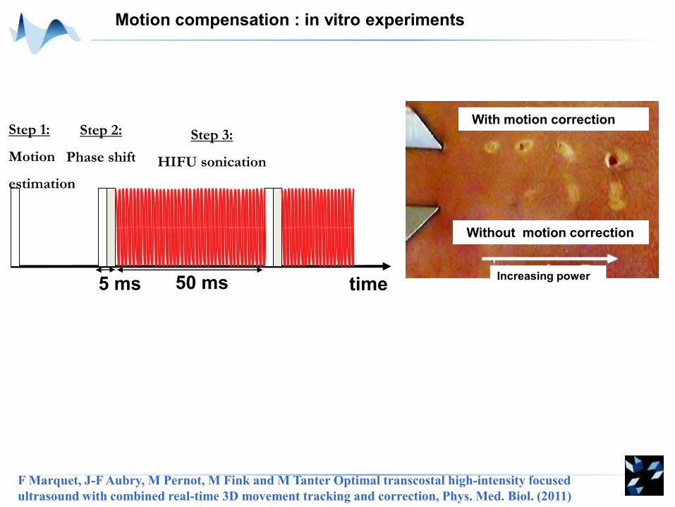
Motion compensation : in vitro experiments
With motion correction
Without motion correction
Increasing powertime
Step 2:
Phase shift
Step 1:
Motion
estimation
Step 3:
HIFU sonication
5 ms 50 ms
F Marquet, J-F Aubry, M Pernot, M Fink and M Tanter Optimal transcostal high-intensity focused
ultrasound with combined real-time 3D movement tracking and correction, Phys. Med. Biol. (2011)
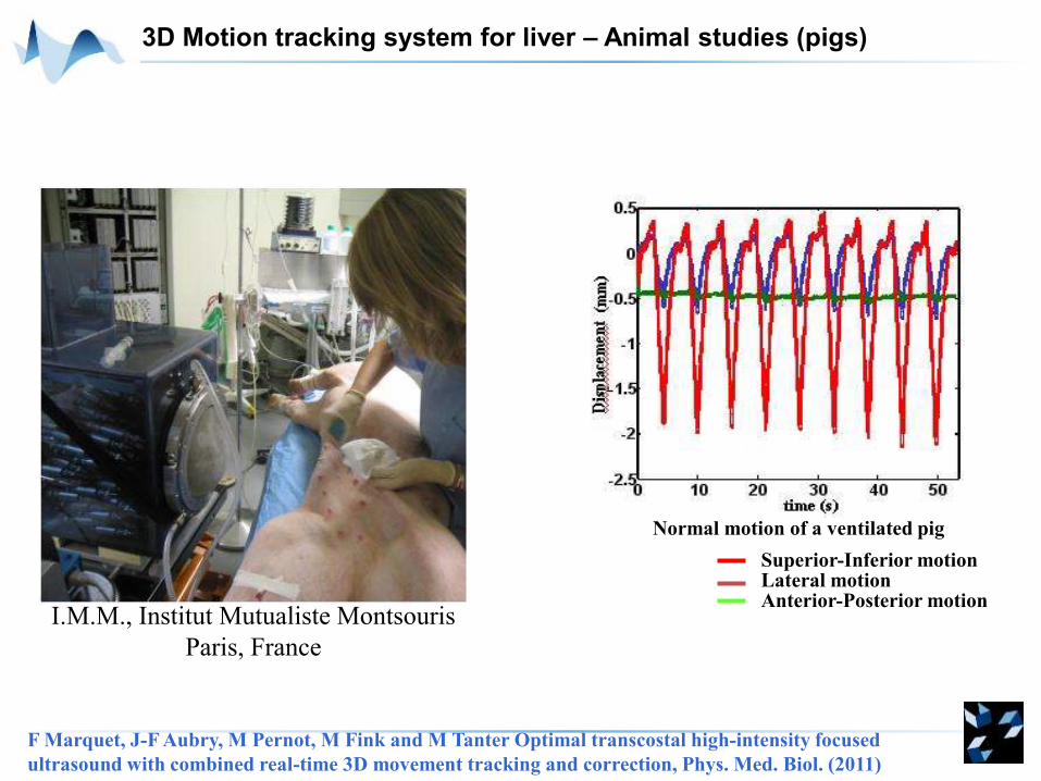
3D Motion tracking system for liver – Animal studies (pigs)
I.M.M., Institut Mutualiste Montsouris
Paris, France
Normal motion of a ventilated pig
Superior-Inferior motionLateral motionAnterior-Posterior motion
F Marquet, J-F Aubry, M Pernot, M Fink and M Tanter Optimal transcostal high-intensity focused
ultrasound with combined real-time 3D movement tracking and correction, Phys. Med. Biol. (2011)
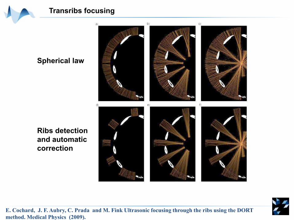
Transribs focusing
Spherical law
Ribs detection
and automatic
correction
E. Cochard, J. F. Aubry, C. Prada and M. Fink Ultrasonic focusing through the ribs using the DORT
method. Medical Physics (2009).
0 10 20 30 40 50 60 -2
0
2
4
6
8
10
Time (s)
Te
mp
era
ture
at
rib
s s
urc
fac
e (
°C)
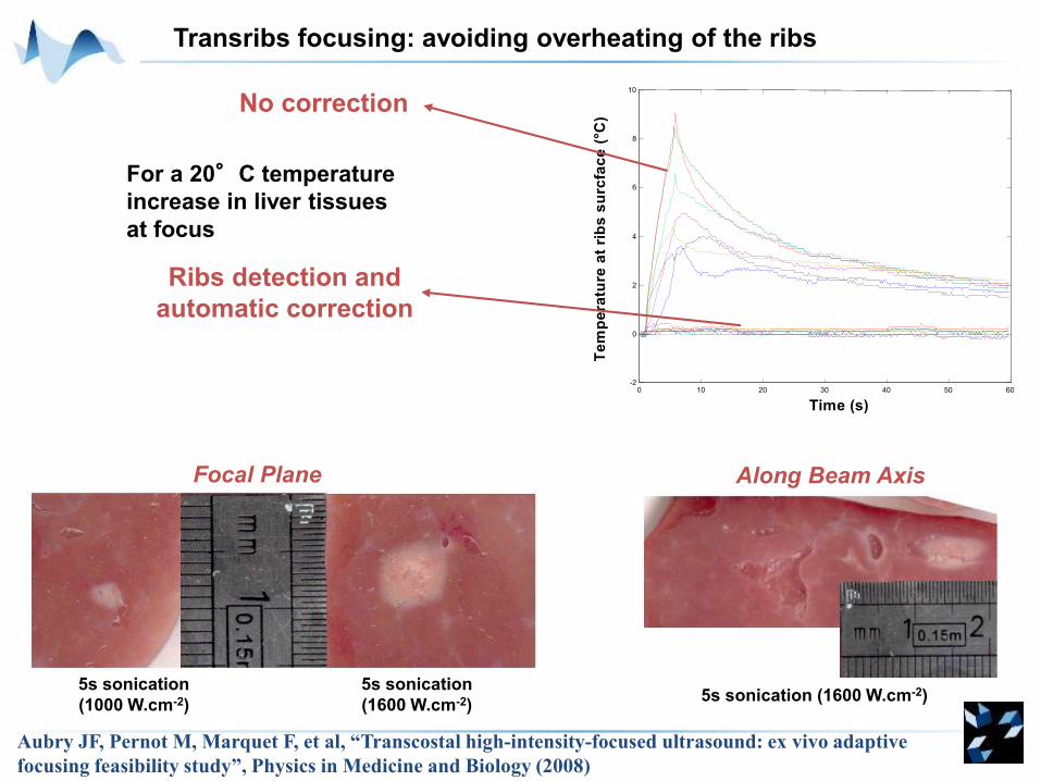
For a 20°C temperature
increase in liver tissues
at focus
No correction
Ribs detection and
automatic correction
5s sonication
(1000 W.cm-2)
5s sonication
(1600 W.cm-2)5s sonication (1600 W.cm-2)
Focal Plane Along Beam Axis
Aubry JF, Pernot M, Marquet F, et al, “Transcostal high-intensity-focused ultrasound: ex vivo adaptive
focusing feasibility study”, Physics in Medicine and Biology (2008)
Transribs focusing: avoiding overheating of the ribs
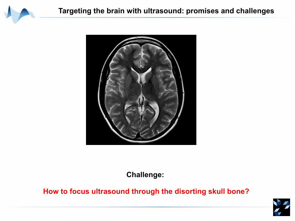
Targeting the brain with ultrasound: promises and challenges
Challenge:
How to focus ultrasound through the disorting skull bone?
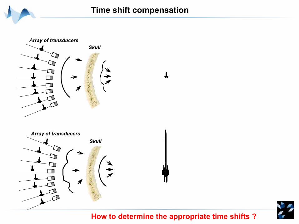
Time shift compensation
Skull
Array of transducers
Skull
Array of transducers
How to determine the appropriate time shifts ?
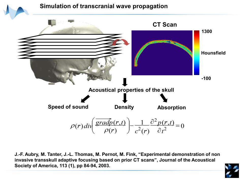
Simulation of transcranial wave propagation
CT Scan
Acoustical properties of the skull
1300
-100
Speed of sound Density Absorption
Hounsfield
0),(
)(
1
)(
),()(
2
2
2
-
t
trp
rcr
trpgraddivr
J.-F. Aubry, M. Tanter, J.-L. Thomas, M. Pernot, M. Fink, “Experimental demonstration of non
invasive transskull adaptive focusing based on prior CT scans”, Journal of the Acoustical
Society of America, 113 (1), pp 84-94, 2003.
Intensity around focus without correction
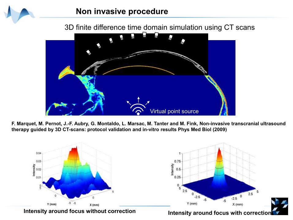
3D finite difference time domain simulation using CT scans
Non invasive procedure
Virtual point source
F. Marquet, M. Pernot, J.-F. Aubry, G. Montaldo, L. Marsac, M. Tanter and M. Fink, Non-invasive transcranial ultrasound
therapy guided by 3D CT-scans: protocol validation and in-vitro results Phys Med Biol (2009)
Intensity around focus with correction
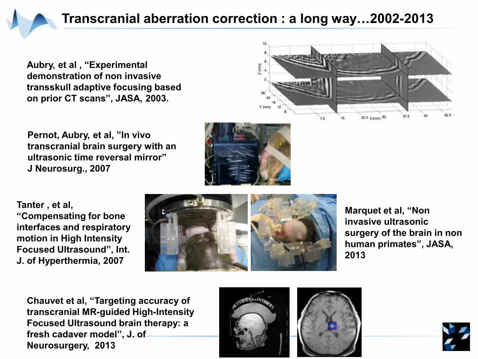
Transcranial aberration correction : a long way…2002-2013
Pernot, Aubry, et al, ”In vivo
transcranial brain surgery with an
ultrasonic time reversal mirror”
J Neurosurg., 2007
Aubry, et al , “Experimental
demonstration of non invasive
transskull adaptive focusing based
on prior CT scans”, JASA, 2003.
Tanter , et al,
“Compensating for bone
interfaces and respiratory
motion in High Intensity
Focused Ultrasound”, Int.
J. of Hyperthermia, 2007
Marquet et al, “Non
invasive ultrasonic
surgery of the brain in non
human primates”, JASA,
2013
Chauvet et al, “Targeting accuracy of
transcranial MR-guided High-Intensity
Focused Ultrasound brain therapy: a
fresh cadaver model”, J. of
Neurosurgery, 2013
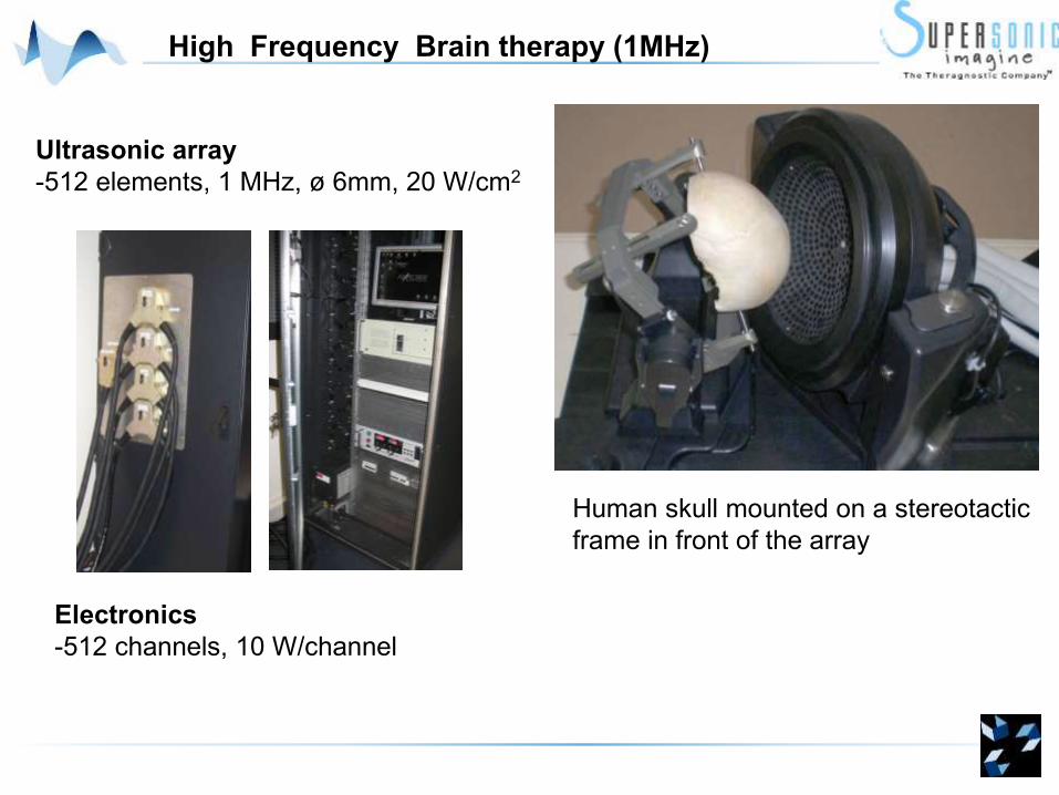
High Frequency Brain therapy (1MHz)
Ultrasonic array
-512 elements, 1 MHz, ø 6mm, 20 W/cm2
Human skull mounted on a stereotactic
frame in front of the array
Electronics
-512 channels, 10 W/channel
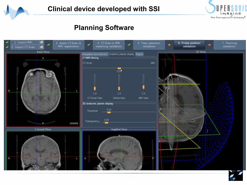
Planning Software
Clinical device developed with SSI
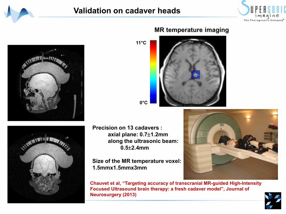
MR temperature imaging
Precision on 13 cadavers :
axial plane: 0.71.2mm
along the ultrasonic beam:
0.52.4mm
Size of the MR temperature voxel:
1.5mmx1.5mmx3mm
11°C
0°C
Validation on cadaver heads
Chauvet et al, “Targeting accuracy of transcranial MR-guided High-Intensity
Focused Ultrasound brain therapy: a fresh cadaver model”, Journal of
Neurosurgery (2013)
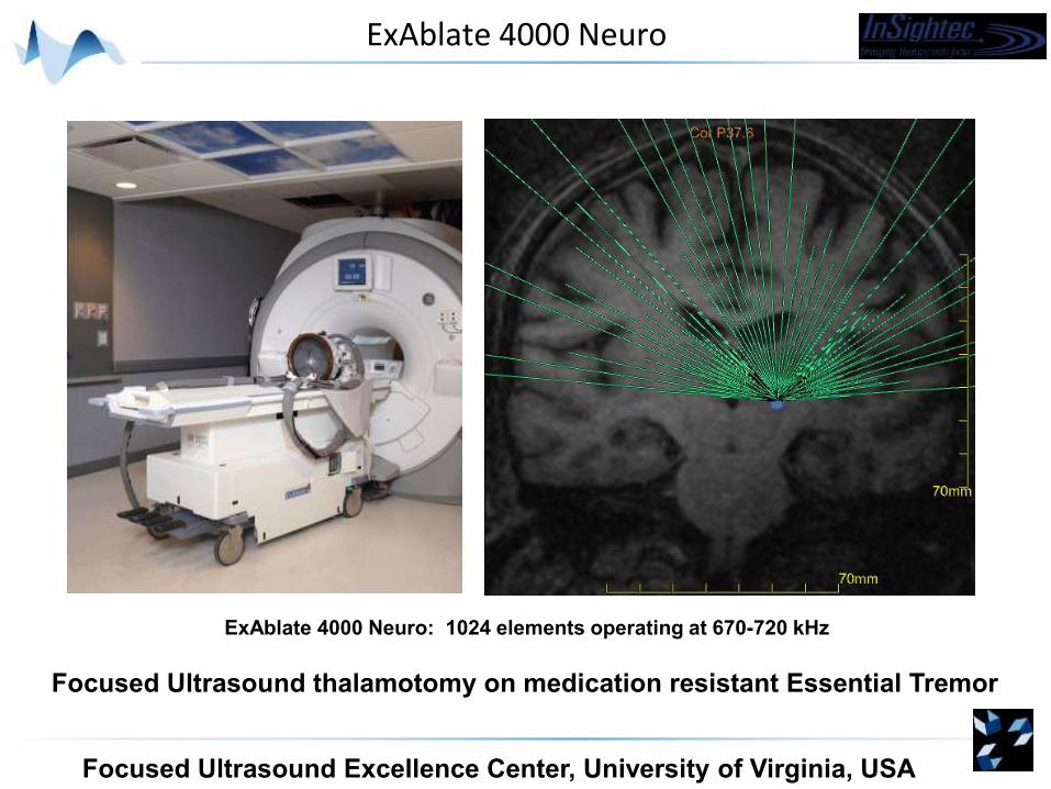
ExAblate 4000 Neuro
ExAblate 4000 Neuro: 1024 elements operating at 670-720 kHz
Focused Ultrasound Excellence Center, University of Virginia, USA
Focused Ultrasound thalamotomy on medication resistant Essential Tremor
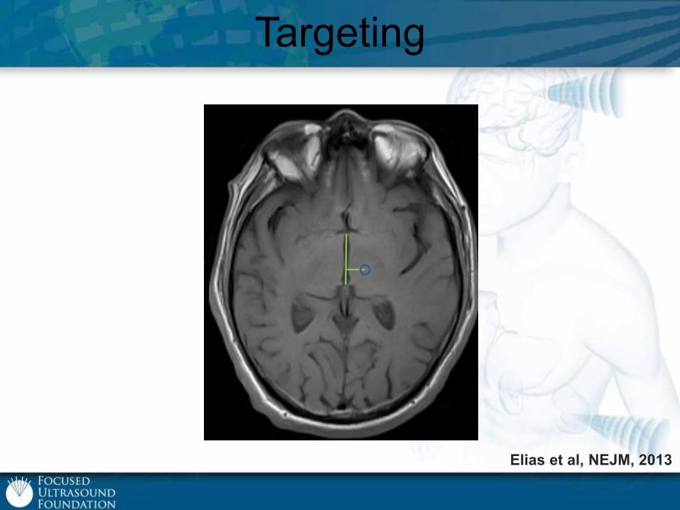
Targeting
Elias et al, NEJM, 2013
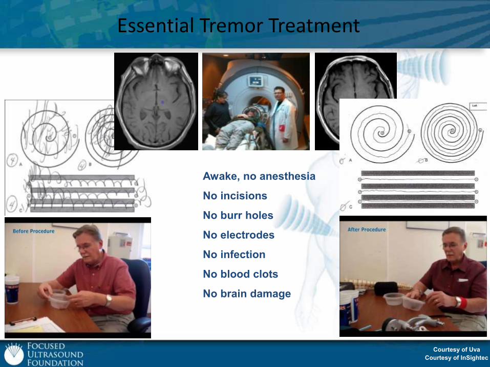
Essential Tremor Treatment
Awake, no anesthesia
No incisions
No burr holes
No electrodes
No infection
No blood clots
No brain damage
Courtesy of Uva
Courtesy of InSightec
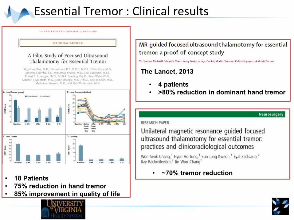
Essential Tremor : Clinical results
• 18 Patients
• 75% reduction in hand tremor
• 85% improvement in quality of life
• 4 patients
• >80% reduction in dominant hand tremor
The Lancet, 2013
• ~70% tremor reduction

Can we induce more subtle effectsin the brain?
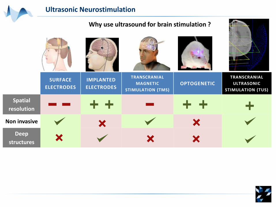
SURFACE
ELECTRODES
IMPLANTED
ELECTRODES
TRANSCRANIAL
MAGNETIC
STIMULATION (TMS)
OPTOGENETICTRANSCRANIAL
ULTRASONIC
STIMULATION (TUS)
Spatial
resolution
Non invasive
Deep
structures
Why use ultrasound for brain stimulation ?
Ultrasonic Neurostimulation
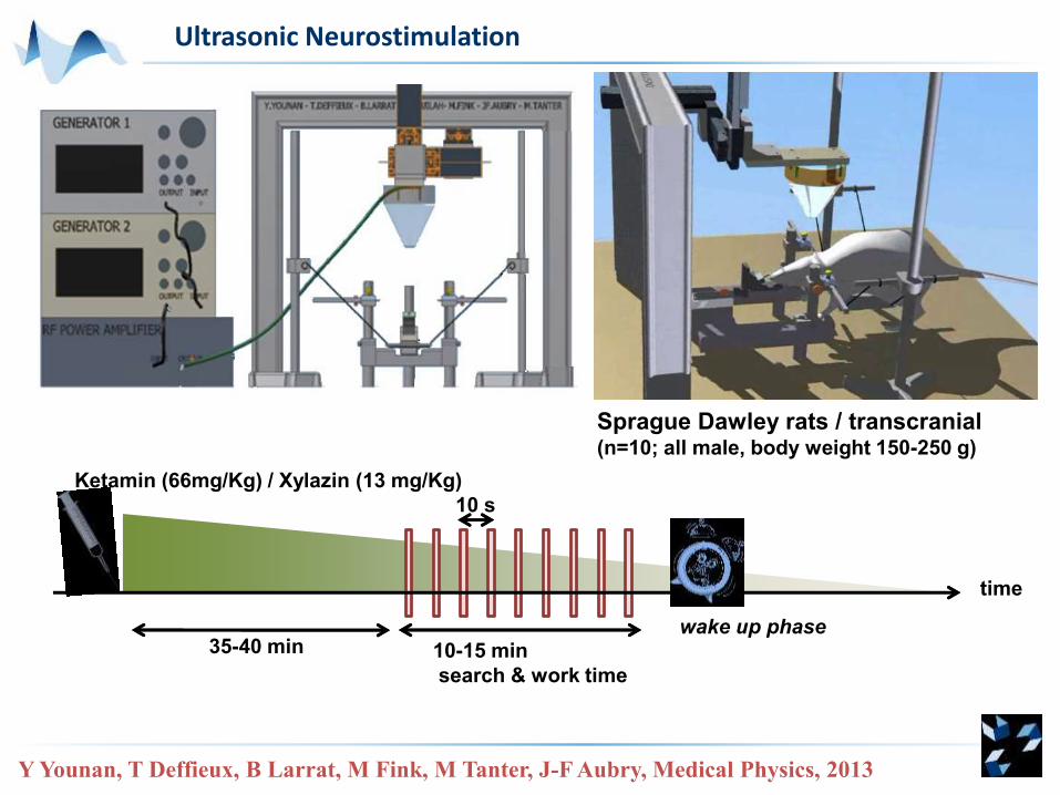
35-40 min
Ketamin (66mg/Kg) / Xylazin (13 mg/Kg)
10 s
10-15 min
search & work time
time
wake up phase
Sprague Dawley rats / transcranial(n=10; all male, body weight 150-250 g)
Y Younan, T Deffieux, B Larrat, M Fink, M Tanter, J-F Aubry, Medical Physics, 2013
Ultrasonic Neurostimulation
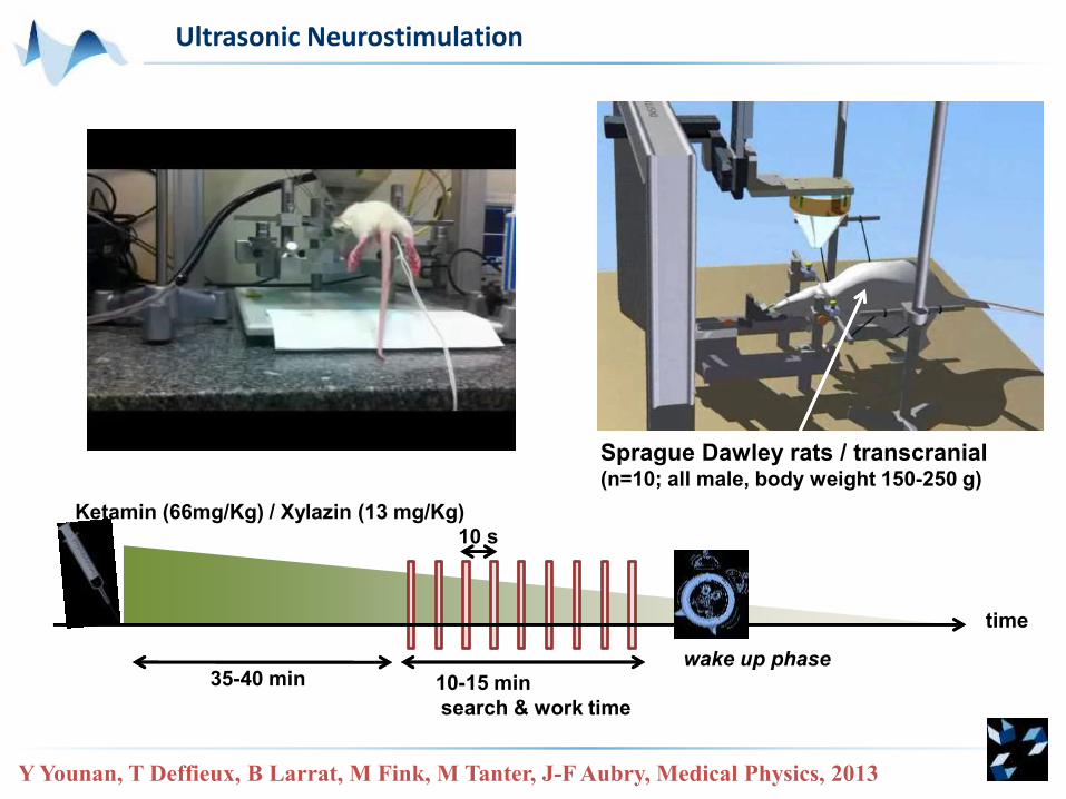
Sprague Dawley rats / transcranial(n=10; all male, body weight 150-250 g)
35-40 min
Ketamin (66mg/Kg) / Xylazin (13 mg/Kg)
10 s
10-15 min
search & work time
time
wake up phase
Y Younan, T Deffieux, B Larrat, M Fink, M Tanter, J-F Aubry, Medical Physics, 2013
Ultrasonic Neurostimulation
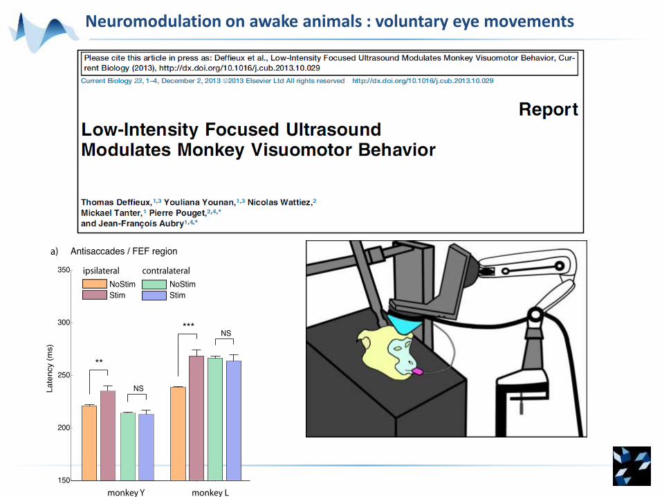
Neuromodulation on awake animals : voluntary eye movements
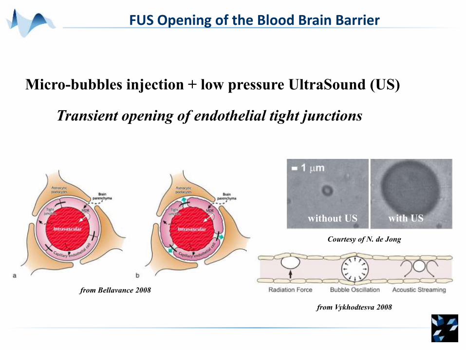
FUS Opening of the Blood Brain Barrier
Micro-bubbles injection + low pressure UltraSound (US)
Transient opening of endothelial tight junctions
from Bellavance 2008
from Vykhodtesva 2008
without US with US
Courtesy of N. de Jong
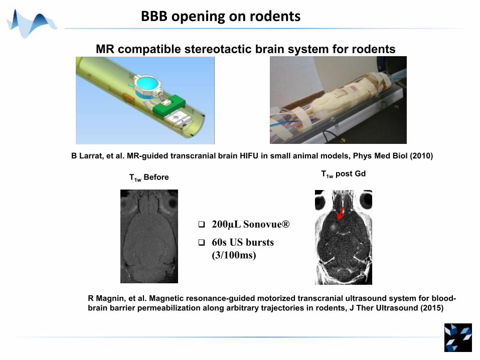
BBB opening on rodents
B Larrat, et al. MR-guided transcranial brain HIFU in small animal models, Phys Med Biol (2010)
MR compatible stereotactic brain system for rodents
T1w post Gd
200µL Sonovue®
60s US bursts
(3/100ms)
T1w Before
R Magnin, et al. Magnetic resonance-guided motorized transcranial ultrasound system for blood-
brain barrier permeabilization along arbitrary trajectories in rodents, J Ther Ultrasound (2015)
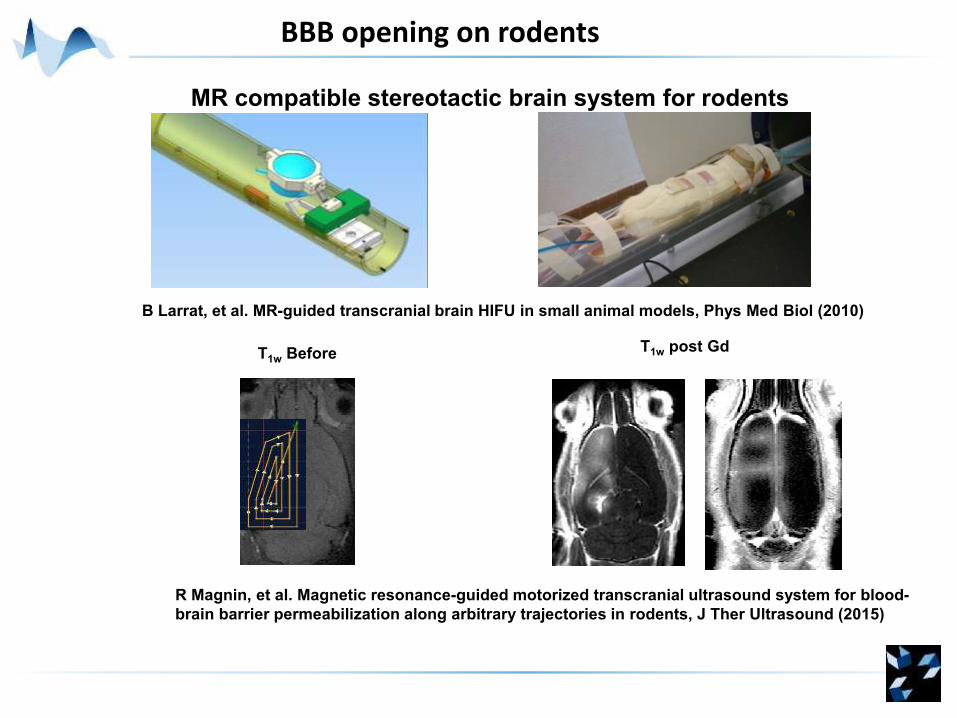
BBB opening on rodents
B Larrat, et al. MR-guided transcranial brain HIFU in small animal models, Phys Med Biol (2010)
MR compatible stereotactic brain system for rodents
T1w post GdT1w Before
R Magnin, et al. Magnetic resonance-guided motorized transcranial ultrasound system for blood-
brain barrier permeabilization along arbitrary trajectories in rodents, J Ther Ultrasound (2015)
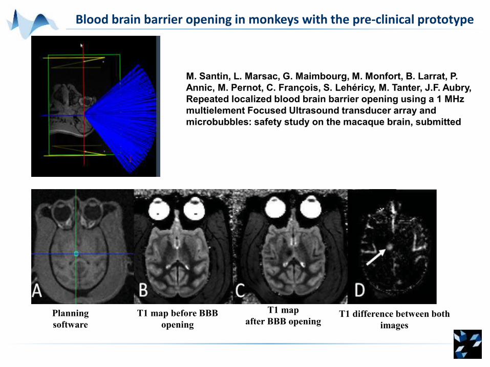
Blood brain barrier opening in monkeys with the pre-clinical prototype
M. Santin, L. Marsac, G. Maimbourg, M. Monfort, B. Larrat, P.
Annic, M. Pernot, C. François, S. Lehéricy, M. Tanter, J.F. Aubry,
Repeated localized blood brain barrier opening using a 1 MHz
multielement Focused Ultrasound transducer array and
microbubbles: safety study on the macaque brain, submitted
Planning
software
T1 map before BBB
opening
T1 map
after BBB openingT1 difference between both
images
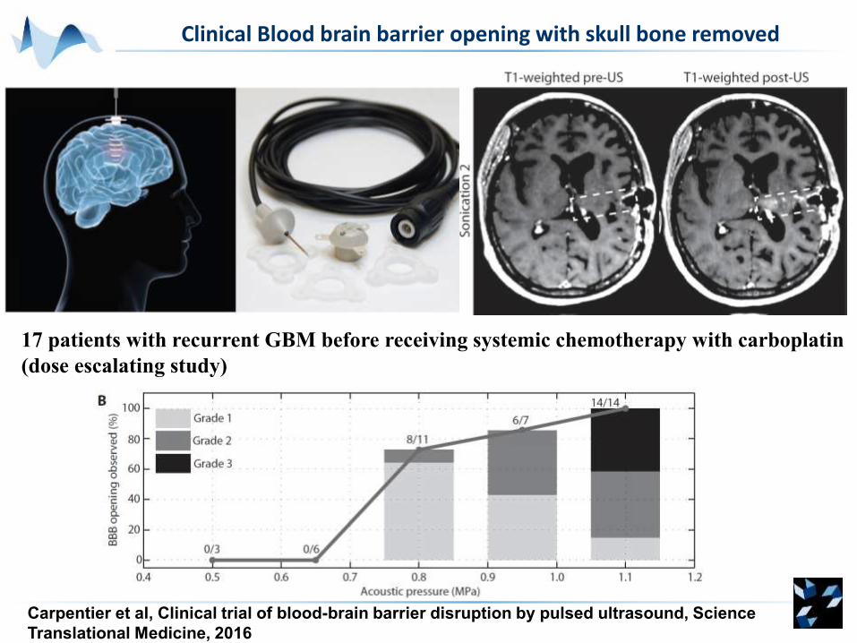
Clinical Blood brain barrier opening with skull bone removed
Carpentier et al, Clinical trial of blood-brain barrier disruption by pulsed ultrasound, Science
Translational Medicine, 2016
17 patients with recurrent GBM before receiving systemic chemotherapy with carboplatin
(dose escalating study)
Ultrafast Functional
Imaging
Ultrasonic
NeurostimulationThermal Therapy
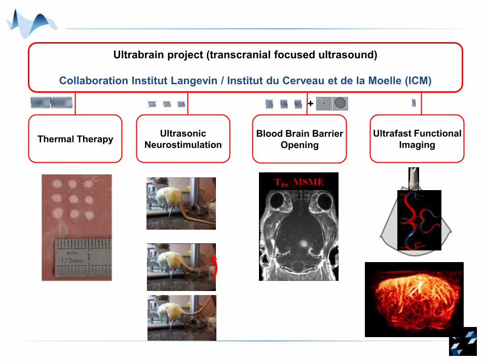
Ultrabrain project (transcranial focused ultrasound)
Collaboration Institut Langevin / Institut du Cerveau et de la Moelle (ICM)
Blood Brain Barrier
Opening
+
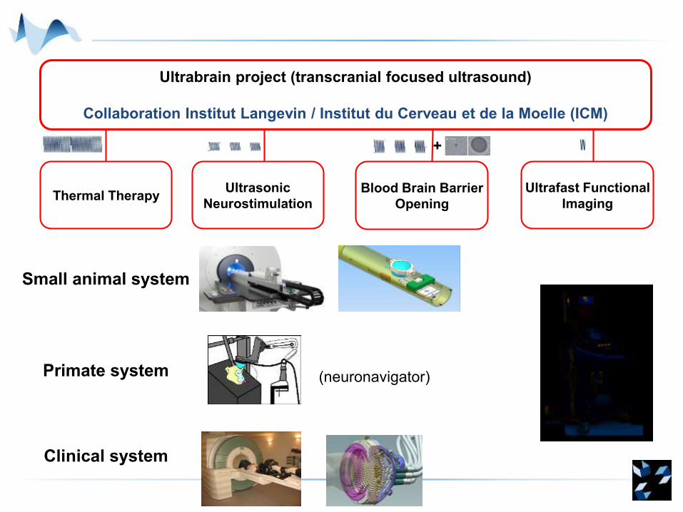
Ultrafast Functional
Imaging
Ultrasonic
NeurostimulationThermal Therapy
Ultrabrain project (transcranial focused ultrasound)
Collaboration Institut Langevin / Institut du Cerveau et de la Moelle (ICM)
Blood Brain Barrier
Opening
+
Small animal system
Primate system
Clinical system
(neuronavigator)
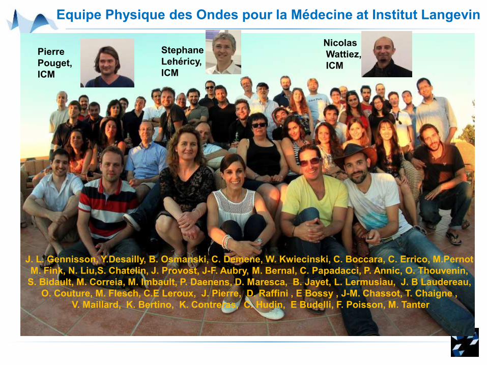
Equipe Physique des Ondes pour la Médecine at Institut Langevin
J. L. Gennisson, Y.Desailly, B. Osmanski, C. Demene, W. Kwiecinski, C. Boccara, C. Errico, M.Pernot
M. Fink, N. Liu,S. Chatelin, J. Provost, J-F. Aubry, M. Bernal, C. Papadacci, P. Annic, O. Thouvenin,
S. Bidault, M. Correia, M. Imbault, P. Daenens, D. Maresca, B. Jayet, L. Lermusiau, J. B Laudereau,
O. Couture, M. Flesch, C.E Leroux, J. Pierre, D. Raffini , E Bossy , J-M. Chassot, T. Chaigne ,
V. Maillard, K. Bertino, K. Contreras, C. Hudin, E Budelli, F. Poisson, M. Tanter
Pierre
Pouget,
ICM
Nicolas
Wattiez,
ICM
Stephane
Lehéricy,
ICM
Acknowledgements
Credits for some of the pictures:
FUS foundation
Jeff Elias & Robert Dallapiazza, UVA
This work was supported by :
Agence Nationale de la Recherche : Equipex Ultrabrain, ref
ANR-10-EQPX-15
Bettencourt – Schueller foundation:
Save the date:
Winter School on
THERAPEUTIC ULTRASOUND
Les HOUCHES, FranceDirectors:
G. ter Haar (UK), V. Khokhlova (Russia)
& J.-F. Aubry (France)
March 26-31, 2017
Save the date
17th ISTU meeting, Nanjing, May 30-June 2 2017
Jinling Hotel, Nanjing
www.istu.org
Recommended