A. AKKOÇ, G. SÖNMEZ, K. YANIK, A. ALASONYALILAR DEMİRER, N. Y. GÜL, M. C. FERRERAS ESTRADA
369
Turk. J. Vet. Anim. Sci.
2011; 35(5): 369-373
© TÜBİTAK
doi:10.3906/vet-1011-573
Malignant giant-cell tumor of bone with lymph node
involvement in a cat
Ahmet AKKOÇ1,
*, Gürsel SÖNMEZ1, Kemal YANIK
2, Aylin ALASONYALILAR DEMİRER
1, Nihal Y. GÜL
2,
M. Carmen FERRERAS ESTRADA3
1Department of Pathology, Faculty of Veterinary Medicine, Uludağ University, 16059 Görükle, Bursa - TURKEY2Department of Surgery, Faculty of Veterinary Medicine, Uludağ University, 16059 Görükle, Bursa - TURKEY
3Department of Animal Health, Faculty of Veterinary Medicine, University of León - SPAIN
Received: 10.11.2010
Abstract: Th e present study describes giant-cell tumor of bone (GCToB) with lymph node involvement in a 5-year-old
crossbred cat. Th e animal was referred to the surgery clinic with progressive subcutaneous swelling in the left proximal
femoral region, severe lameness, constipation, and dysuria. A moderately fi rm, subcutaneous, palpable mass, 9 cm in
diameter, was observed, and biopsy samples were taken. Histopathologically, the mass was constituted by ovoid-shaped
mononuclear cells intermixed with many multinucleated giant cells (MGC). Immunohistochemically, the giant cells
were positively stained with antivimentin, and the same cells were negative for antidesmin and anti-S100 staining.
Tartrate-resistant acid phosphatase (TRAP) activity in tumor cells was evaluated and the tumor was diagnosed as
malignant GCToB; the cat was euthanized. Macroscopically, while the regional lymph nodes were intact, giant cells
were found in the left popliteal lymph node during microscopy. Although a few cases of GCToB have been reported in
cats, the case herein displays, for the fi rst time, evidence of lymph node involvement during the process of metastasis.
Key words: Cat, giant-cell tumor of bone, femur, lymph node involvement, immunohistochemistry
Bir kedide lenf yumrusu metastazlı kemiğin dev hücreli tümörü
Özet: Sunulan bu çalışmada, 5 yaşlı bir kedide rastlanan ve lenf yumrusu metastazına sahip kemiğin dev hücreli
tümörü rapor edilmiştir. Hayvan cerrahi kliniğine sol femurun proksimal kısmında deri altı şişkinlik, topallama,
kabızlık ve ağrılı idrar şikayeti ile getirilmiştir. Dokuz santimetre çapa ulaşan ve orta sertlikteki kitleden tanı amaçlı
biyopsi alınarak patoloji laboratuarında değerlendirilmiştir. Histopatolojik olarak kitlenin çok sayıdaki dev hücreleri
ve ovoid sekili mononukleer hücrelerden oluştuğu gözlenmiştir. Yapılan immunohistokimyasal boyamalarda bahsi
geçen hücrelerin anti-vimentin antikorları ile pozitif, anti-desmin ve S-100 antikorları ile negatif olarak boyandıkları
görülmüştür. Hücrelerdeki tartarat dirençli asit fosfataz aktivitesi değerlendirilerek kitle kemiğin dev hücreli tümörü
olarak teşhis edilmiştir. Ötenazi sonrası makroskobik muayenede popliteal lenf yumrularında herhangi bir lezyon
gözlenmezken, mikroskobik incelemede sol popliteal lenf yumrusunda metastaza rastlanmıştır. Kedilerde kemiğin dev
hücreli tümörüne ait raporlar bulunsa da, sunulan olguda lenf yumrusu metastazına rastlanılması, bu tipteki tümörlerin
lenfojen metastaz ile yayılabildiklerini gösterir ilk kanıtları içermektedir.
Anahtar sözcükler: Kedi, kemigin dev hücreli tümörü, femur, popliteal lenf yumrusu, immunohistokimya
Case Report
* E-mail: [email protected]
Malignant giant-cell tumor of bone with lymph node involvement in a cat
370
Introduction
Giant-cell tumor of bone (GCToB) is an
uncommon primary bone neoplasm that usually
occurs in the long bones of both domestic animals
and humans (1,2). Th e most common sites include
the distal femur, proximal tibia, and distal radius.
GCToB is an enigmatic tumor that is histologically
benign but clinically shows local invasion and
metastatic potential (1). In some cases, dissemination
to the lungs and extraskeletal sites was reported (3,4).
Radiologically, GCToB shows typical destructive
osteolytic lesions in aff ected bones (3,5,6). Th e
tumoral mass appears either gray to white or gray to
brownish in color up to its vascular stroma (1,4-7).
Histopathologically, the tumor is mainly composed
of multinucleated giant cells in a moderately
vascularized network of proliferating round, oval,
or spindle-shaped stromal cells (1,4-9). Although
the exact origin of the tumor remains obscure, it has
been suggested that the tumor originates from the
stromal cells of the bone marrow (1,4,9-11). Th ere are
few reported cases of metastatic GCToB in cats, and
the way in which metastasis of tumor cells relates to
distant organs is not clear. To the best of the authors’
knowledge, the case described in the present study is
the fi rst observation of the involvement of the lymph
nodes during the metastasis of such a tumor in cats.
Material and methods
Case history
A 5-year-old crossbred cat was referred to the
surgery clinic with progressive swelling in the
left proximal femoral region, severe lameness,
constipation, and dysuria. Th e cat was unable to bear
weight on the left hind leg. In the anamnesis, it was
discovered that the cat had been operated on for a
left femoral fracture 4 years earlier. Treatment had
consisted of an open reduction and internal fi xation
with an intramedullary pin.
Examination
In the clinical examination, a moderately fi rm,
subcutaneous palpable mass was seen in the left
femoral region and biopsy samples were taken.
Radiological examination revealed prominent
osteolytic changes in the proximal epiphyseal part
of the left femur. In light of the histopathologic
examination of the biopsy samples and the results of
the radiological examination, the cat was diagnosed
with GCToB. Euthanasia followed due to the poor
prognosis, including the suspicion of local invasion of
the neighboring tissues. At necropsy, a tumoral mass
was found in the proximal part of the left femur; it
was subcutaneous, grayish to white, and moderately
fi rm, 8 × 9 × 6 cm in size, and it was observed to be
compressing adjacent muscles (Figure 1).
Th e dorsal part of the pelvic cavity was invaded
by nodular masses of various sizes compressing the
adjacent tissues and organs. Prominent adhesions
were noticed between tumoral masses and the serosal
surface of the rectum. Th e large intestines were fi lled
with dry feces, and the urinary bladder was full of
dark yellowish urine. Similar tumoral masses also
occupied the dorsal left part of the hip.
Macroscopically, no distant lesions or
macrometastases were observed in the organs located
in the abdominal or thoracic cavity. When the
longitudinal section of the left femur was examined,
tumoral tissue proliferation was observed from the
medullar region to the epiphyseal part of the femur.
F igure 1. Subcutaneous mass located in the proximal part of the
left femur. LF: left femur.

A. AKKOÇ, G. SÖNMEZ, K. YANIK, A. ALASONYALILAR DEMİRER, N. Y. GÜL, M. C. FERRERAS ESTRADA
371
Analysis
Tissue samples taken from the femur, various parts of the subcutaneous mass, rectum, regional skeletal muscles, local lymph nodes, lung, skin, and other major organs were fi xed in buff ered formalin and processed routinely. Tissues sections of 5 μm were cut from the paraffi n-embedded tissue blocks and all slides were then stained with hematoxylin-eosin (H&E). Selected slides were also stained with a standard streptavidin-biotin-peroxidase complex method using 1:500 diluted mouse antihuman antibodies against desmin, vimentin, and S100 (Lab Vision, Fremont, CA, USA), as previously described (12). Tartrate-resistant acid phosphatase (TRAP) activity in tumor cells was evaluated with a commercially available kit (Acid Phosphatase, Leucocyte (TRAP) Kit 387, Sigma-Aldrich Co., St. Louis, MO, USA) (4,13).
Results
Radiological fi ndings
Irregularly marginated osteolytic changes were found, including the destruction of the medullary cavity and the adjacent cortex of the left femur (Figure 2). Th e center was mostly radiolucent with increasing
density toward the periphery. No periosteal response
or fracture was found. In the examination of the
lungs, metastatic involvement was not observed.
Microscopic fi ndings
Light microscopic examination of the H&E-
stained slides showed the presence of ovoid-shaped
mononuclear cells intermixed with multinucleated
giant cells (up to 105 nucleuses) with irregular round-
to-ovoid nuclei and small, centrally located nucleoli
in a vascularized stroma (Figure 3). Multinucleated
giant cells (MGCs) were uniformly scattered
throughout the sections. In some areas, pinkish
collagen fi bers, disseminated small hemorrhagic foci,
siderocytes, and coagulation necrosis were noticed.
Mitotic fi gures were not common and no metastasis
was observed in the lungs or other major organs. In
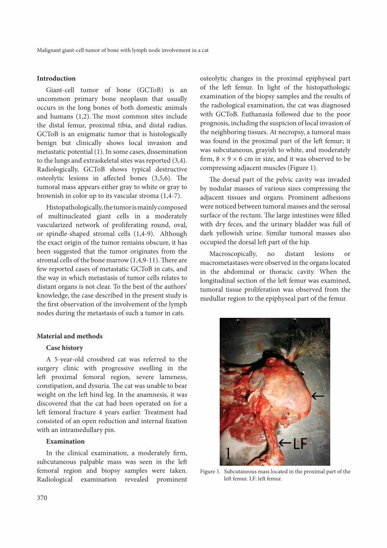
the left popliteal lymph node, however, MGCs with
a similar morphology to that of the primary tumoral
site were found in the cortical sinuses (Figure 4). Th e
invasion of regional muscles, the serosal wall of the
rectum, and the dermal tissue of the skin by tumor
cells revealed that the tumor was locally aggressive
and destructive. Osteoid tissue production was noted
as a pinkish, homogenous material located in the
peripheral parts of the tumoral mass.
Almost all of the tumoral cells gave a strong
positive reaction to the vimentin antibody, and
no staining was observed with the desmin or S100
antibodies. While the regional muscle tissues were
Figure 2. Lateral-medial radiographic view of the mass (arrow),
including lytic changes (empty arrow) in the proximal
part of left femur.
Figure 3. Local invasion of regional muscles by tumoral cells and
giant cells (arrows) via hematoxylin & eosin staining;
bar = 120 μm.
Malignant giant-cell tumor of bone with lymph node involvement in a cat
372
positively stained with desmin antibody, the tumoral
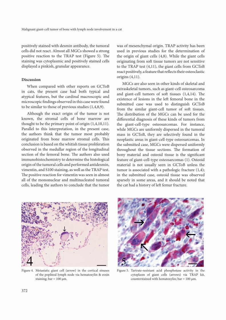
cells did not react. Almost all MGCs showed a strong
positive reaction to the TRAP test (Figure 5). Th e
staining was cytoplasmic and positively stained cells
displayed a pinkish, granular appearance.
Discussion
When compared with other reports on GCToB
in cats, the present case had both typical and
atypical features, but the cardinal macroscopic and
microscopic fi ndings observed in this case were found
to be similar to those of previous studies (1,4,8,9).
Although the exact origin of the tumor is not
known, the stromal cells of bone marrow are
thought to be the primary point of origin (1,4,10,11).
Parallel to this interpretation, in the present case,
the authors think that the tumor most probably
originated from bone marrow stromal cells. Th is
conclusion is based on the whitish tissue proliferation
observed in the medullar region of the longitudinal
section of the femoral bone. Th e authors also used
immunohistochemistry to determine the histological
origin of the tumoral cells and performed antidesmin,
vimentin, and S100 staining, as well as the TRAP test.
Th e positive reaction for vimentin was seen in almost
all of the mononuclear and multinucleated tumoral
cells, leading the authors to conclude that the tumor
was of mesenchymal origin. TRAP activity has been
used in previous studies for the determination of
the origin of giant cells (4,8). While the giant cells
originating from soft tissue tumors are not sensitive
to the TRAP test (4,11), the giant cells from GCToB
react positively, a feature that refl ects their osteoclastic
origins (4,11).
MGCs are also seen in other kinds of skeletal and
extraskeletal tumors, such as giant-cell osteosarcoma
and giant-cell tumors of soft tissues (1,4,14). Th e
existence of lesions in the left femoral bone in the
submitted case was used to distinguish GCToB
from the similar giant-cell tumor of soft tissues.
Th e distribution of the MGCs can be used for the
diff erential diagnosis of these kinds of tumors from
the giant-cell-type osteosarcomas. For instance,
while MGCs are uniformly dispersed in the tumoral
mass in GCToB, they are selectively found in the
neoplastic areas in giant-cell-type osteosarcomas. In
the submitted case, MGCs were dispersed uniformly
throughout the tissue sections. Th e formation of
bony material and osteoid tissue is the signifi cant
feature of giant-cell-type osteosarcomas (1). Osteoid
material is not usually seen in GCToB unless the
tumor is associated with a pathologic fracture (1,4);
in the submitted case, osteoid tissue was observed
sparsely in some areas, and it should be noted that
the cat had a history of left femur fracture.
Figure 4. Metastatic giant cell (arrow) in the cortical sinuses
of the popliteal lymph node via hematoxylin & eosin
staining; bar = 100 μm.
Figure 5. Tartrate-resistant acid phosphotase activity in the
cytoplasm of giant cells (arrows) via TRAP kit,
counterstained with hematoxylin; bar = 100 μm.

A. AKKOÇ, G. SÖNMEZ, K. YANIK, A. ALASONYALILAR DEMİRER, N. Y. GÜL, M. C. FERRERAS ESTRADA
373
Classically, GCToB is known to have a benign nature, even though the existence of metastatic cases in cats has been observed (3,4,11). Distant metastases to the lung or other organs are rare (<10%) in humans (2,3), but the rate is not clear in cats, as the number of the cases is quite limited. In the present case study, the tumor prominently displayed locally aggressive and invasive features. Even though macroscopic metastasis was not seen in the organs, MGCs were found microscopically in the cortical sinuses in the left popliteal lymph node.
In humans, aft er surgical removal of the tumoral masses, the estimated recurrence rate is about 40%-60% because of the deep dermal infi ltration (2,3,14). Such infi ltration may explain why this tumor oft en demonstrates local recurrence even aft er surgical removal. In the present case, tumoral cells also infi ltrated the dermis. Information about the recurrence rate in cats aft er surgical removals is scarce due to the limited number of studies.
In the present case, the subject experienced constipation and dysuria. Th e authors connected these 2 complaints with the local mechanical and
invasive eff ects of tumoral masses, which oft en cause diffi culties in the passages of both the intestine and the urethra. Veterinary practitioners should be aware of this condition.
In humans, the prognosis associated with the treatment of malignant giant-cell tumors is dependent upon the degree of cellular diff erentiation, the number of mitotic fi gures, the location of the tumor, the degree of involvement of surrounding tissues, the involvement of local lymph nodes, and the success of surgical excision. Due to the fact that there have been very few reports on GCToB in cats, the present study is the fi rst to provide valuable information on the route of metastasis in cats affl icted with GCToB. Th e authors hope that this report will be helpful for cat practitioners during the examination and sampling of suspected cases of giant-cell tumor of bone in cats.
Information on the incidence, age, sex, site, and breed predisposition for GCToB tumors in cats is inadequate. Th is causes diffi culties in predicting the behavior of these kinds of tumors aft er surgical excision and therapy. For this reason, surgeons oft en choose euthanasia.
References
1. Th ompson, K.G., Pool, R.R.: Tumors of bones. In: Meuten, D.J.,
Ed. Tumors in Domestic Animals. 4th ed. Iowa State Press,
Ames, IA, USA. 2002; 245-317.
2. Sung, H.W., Kuo, D.P., Shu, W.P., Chai, Y.B., Liu, C.C., Li, S.M.:
Giant-cell tumor of bone: analysis of two hundred and eight
cases in Chinese patients. J. Bone Joint. Surg. Am., 1982; 64:
755-761.
3. Tubbs, W.S., Brown, L.R., Beabout, J.W., Rock, M.G., Unni,
K.K.: Benign giant-cell tumor of bone with pulmonary
metastases: clinical fi ndings and radiologic appearance of
metastases in 13 cases. Am. J. Roentgenol., 1992; 158: 331-334.
4. Ferreras, M.C., Fuertes, M., Perez, V., Benavides, J., Garcia-
Pariente, C., Reyes, L.E., Garcia-Marin, J.F.: Giant cell tumour
of bone in a cat with extraskeletal metastases: Pathological and
immunohistochemical study. J. Vet. Med. A., 2005; 52: 225-229.
5. Rosen, M.P.: Giant cell tumour of the temporal bone. Am. J.
Roentgenol., 1991; 156: 1290-1292.
6. McManus, M.P., Wood, A.K.W., Christiansen, J.S., Craig, L.E.:
Lytic lesions in the distal humerus of a dog. Vet. Clin. Pathol.,
2001; 30: 121-123.
7. Howard, E.B., Kenyon, A.J.: Malignant osteoclastoma (giant
cell tumor) in the cat with mast-cell response. Cornell Vet.,
1967; 57: 399-409.
8. Walsh, B.A., Rhodes, W.H.: Giant cell tumour of bone in a cat.
J. Small Anim. Pract., 1995; 36: 325-329.
9. Th ornburg, P.: Giant cell tumour of bone in a cat. Vet. Pathol.,
1979; 16: 255-257.
10. Jösten, M., Rudolph, A.: Methods for the diff erentiation of
giant cells in canine and feline neoplasias in paraffi n sections.
Zentralbl. Veterinarmed. A., 1997; 44: 159-166.
11. Zheng, M.H., Robbins, P., Xu, J., Huang, L., Wood, D.J.,
Papadimitriou, J.M.: Th e histogenesis of giant cell tumour
of bone: a model of interaction between neoplastic cells and
osteoclasts. Histol Histopathol., 2001; 16: 297-307.
12. Akkoc, A., Ozyigit, M.O., Cangul, I.T.: Valvular cardiac
myxoma in a dog. J. Vet. Med. A., 2005; 54: 356-358.
13. Toyosawa, S., Ogawa, Y., Chang, C.K., Hong, S.S., Yagi, T.,
Kuwahara, H., Wakasa, K., Sakurai, M.: Histochemistry of
tartrate-resistant acid phosphatise and carbonic anhydrase
isoenzyme II on osteoclast-like giant cells bone tumours.
Virchows Arch. Pathol. Anat. Histopathol., 1991; 418: 255-261.
14. Garma-Avina, A.: Malignant fi brous histiocytoma of the giant
cell type in a cat. J. Comp. Pathol., 1987; 97: 551-557.
Recommended