Embed Size (px)
Citation preview
Treatment of Lymphedema
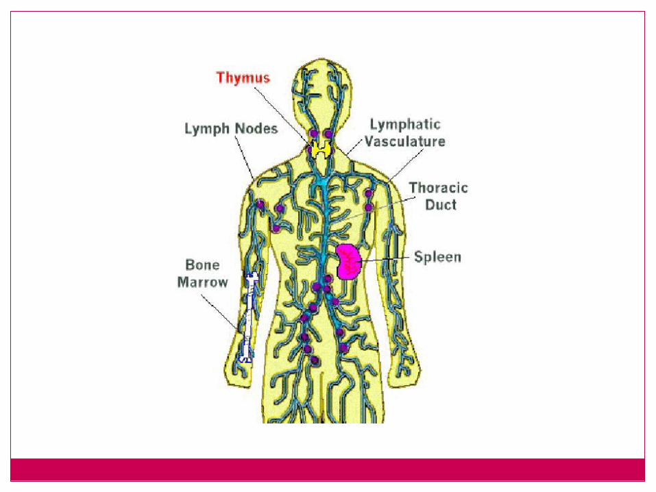
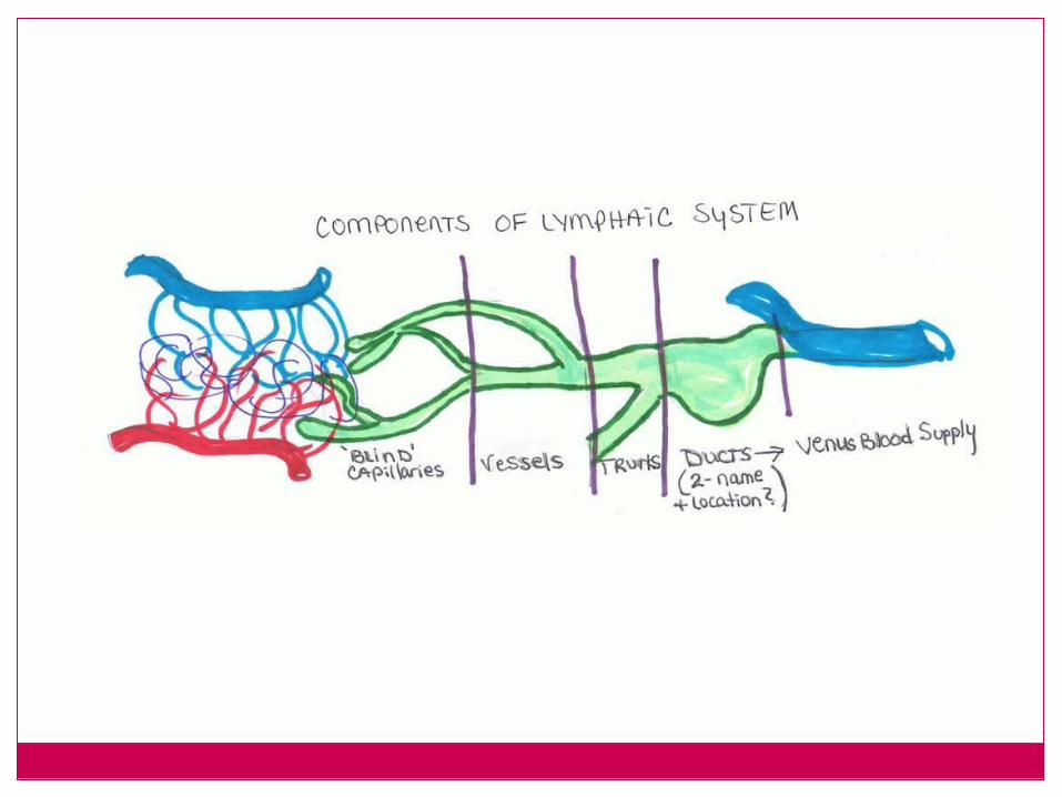
Lymph Anatomy
Lymph nodesLymph vesselsThymus glandSpleenTonsilsPeyer’s patches
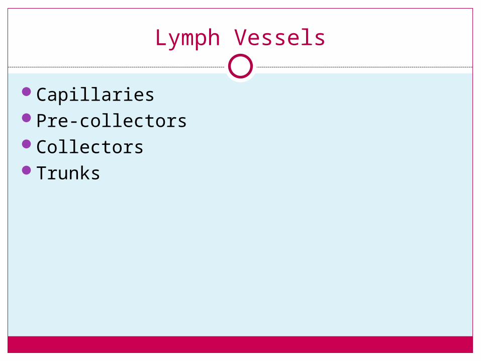
Lymph Vessels
CapillariesPre-collectorsCollectorsTrunks
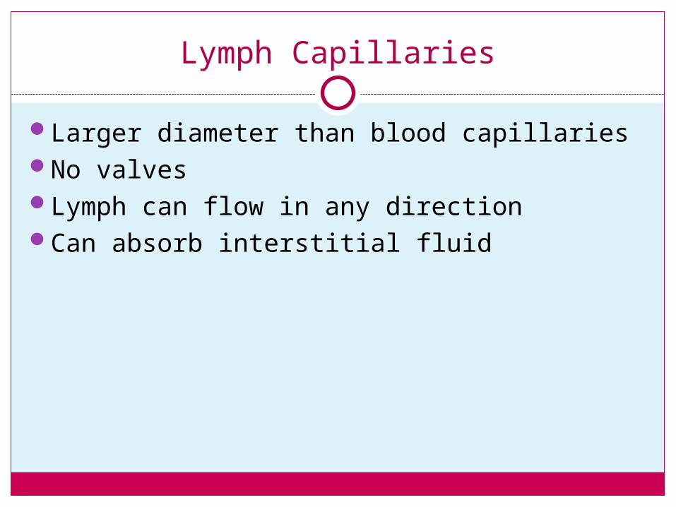
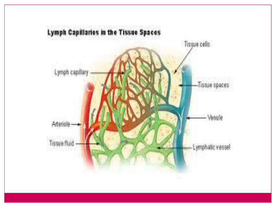
Lymph Capillaries
Larger diameter than blood capillariesNo valvesLymph can flow in any directionCan absorb interstitial fluid
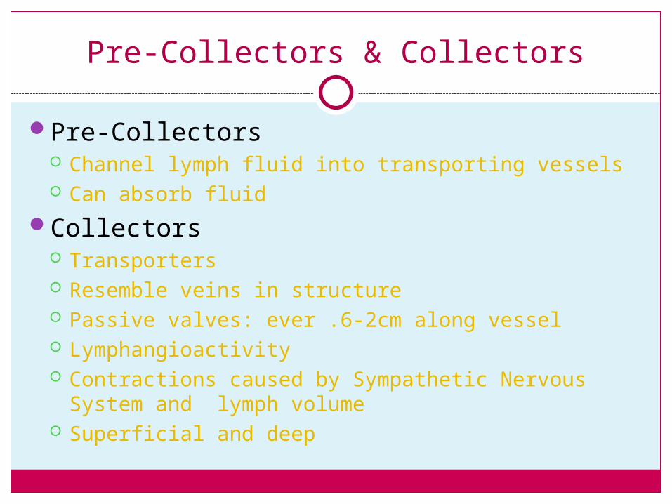
Pre-Collectors & Collectors
Pre-Collectors Channel lymph fluid into transporting vessels Can absorb fluid
Collectors Transporters Resemble veins in structure Passive valves: ever .6-2cm along vessel Lymphangioactivity Contractions caused by Sympathetic Nervous System
and lymph volume Superficial and deep
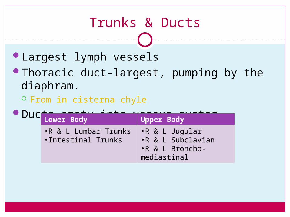
Trunks & Ducts
Largest lymph vesselsThoracic duct-largest, pumping by the
diaphram. From in cisterna chyle
Ducts empty into venous systemLower Body Upper Body
•R & L Lumbar Trunks•Intestinal Trunks
•R & L Jugular•R & L Subclavian•R & L Broncho-mediastinal
Lymph Fluid/Lymphatic Load
Consists of: Proteins (1/2 of bodies protien) Water Cells (RBC, WBC, Lymphocytes) Waste Products Fat (intestinal lymph, chyle)
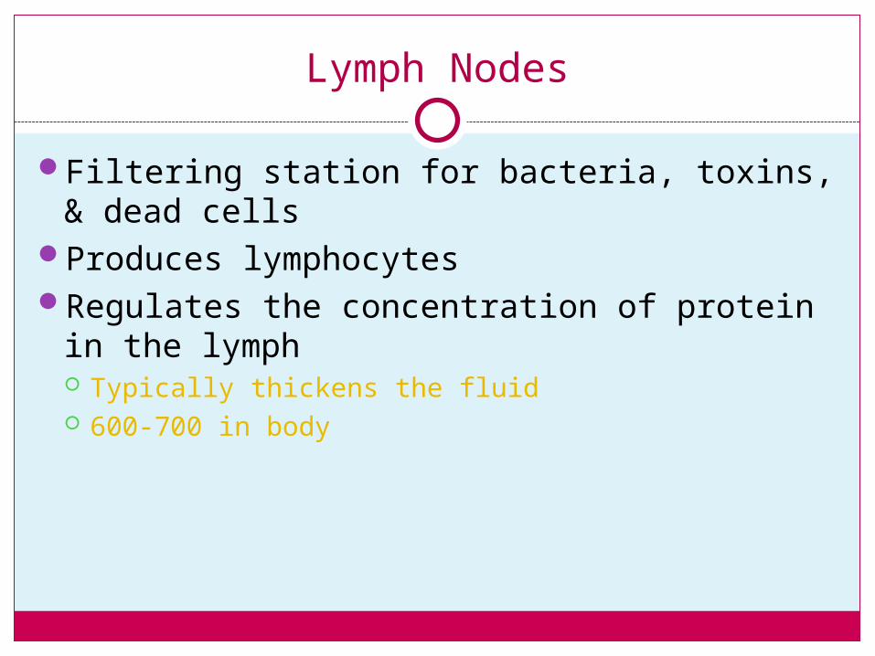
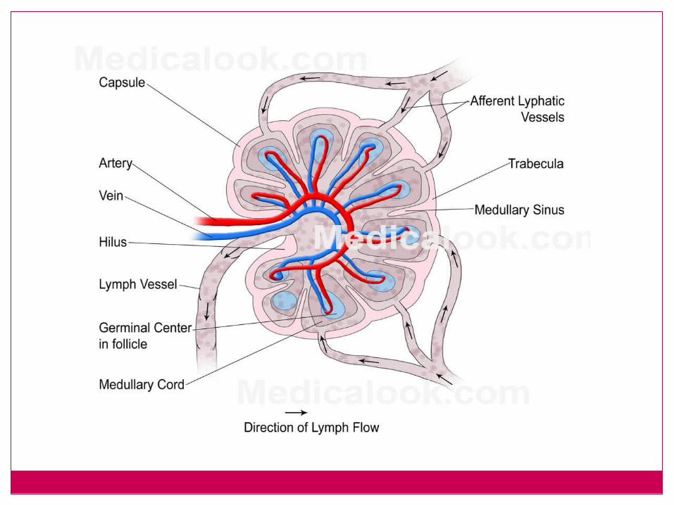
Lymph Nodes
Filtering station for bacteria, toxins, & dead cells
Produces lymphocytesRegulates the concentration of protein in the
lymph Typically thickens the fluid 600-700 in body
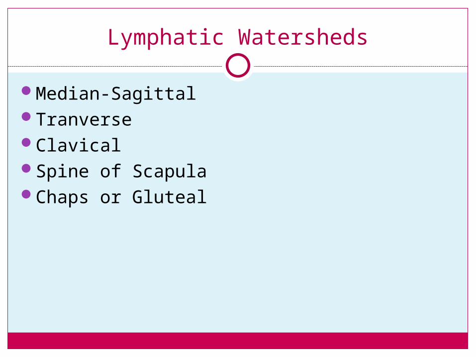
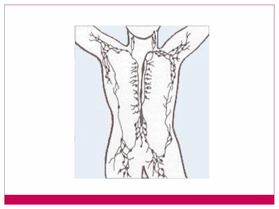
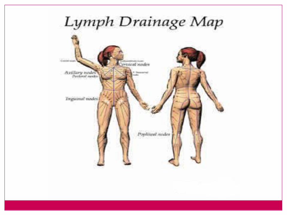
Lymphatic Watersheds
Median-SagittalTranverseClavicalSpine of ScapulaChaps or Gluteal
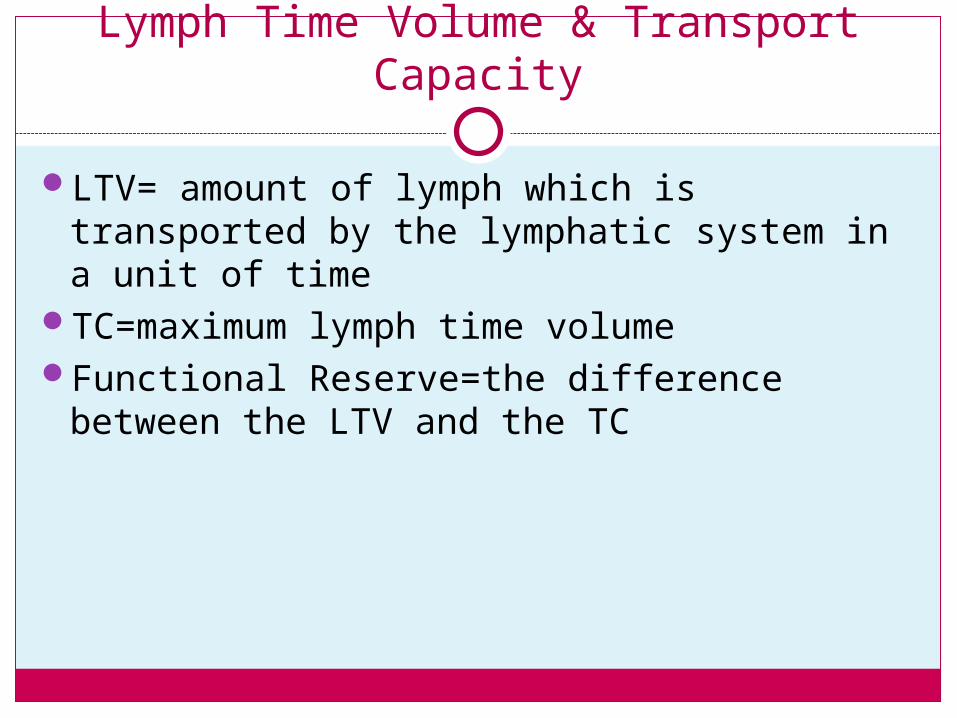
Lymph Time Volume & Transport Capacity
LTV= amount of lymph which is transported by the lymphatic system in a unit of time
TC=maximum lymph time volumeFunctional Reserve=the difference between
the LTV and the TC
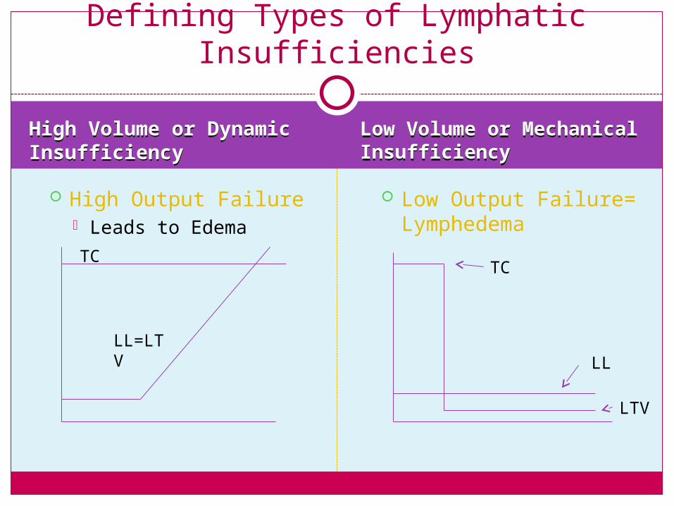
High Volume or Dynamic InsufficiencyHigh Volume or Dynamic Insufficiency
Low Volume or Mechanical InsufficiencyLow Volume or Mechanical Insufficiency
High Output Failure Leads to Edema
Low Output Failure= Lymphedema
Defining Types of Lymphatic Insufficiencies
TC
LL=LTV
TC
LTV
LL
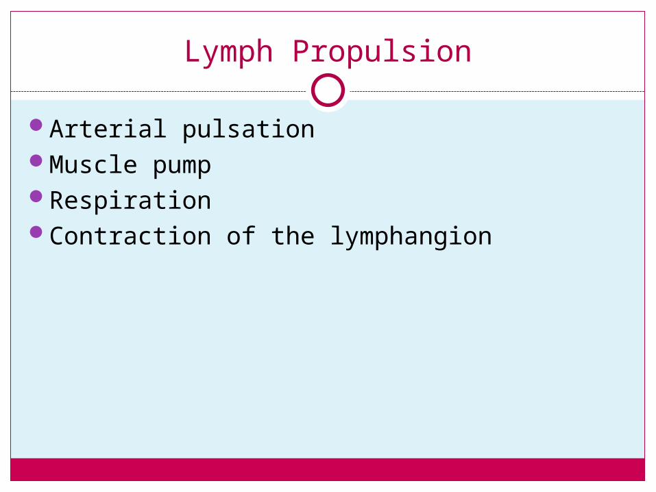
Lymph Propulsion
Arterial pulsationMuscle pumpRespirationContraction of the lymphangion
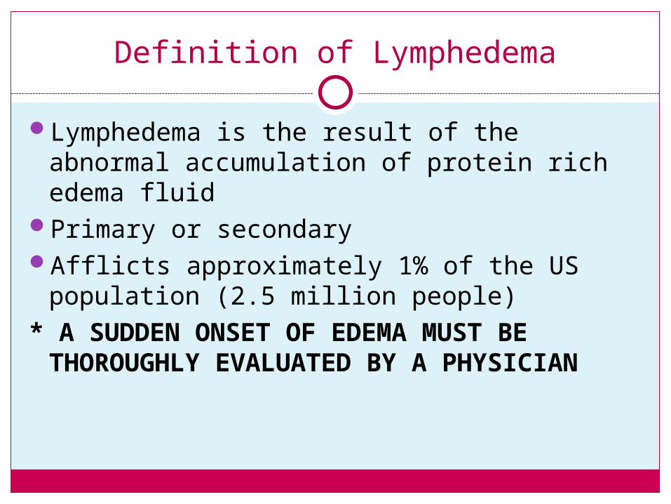
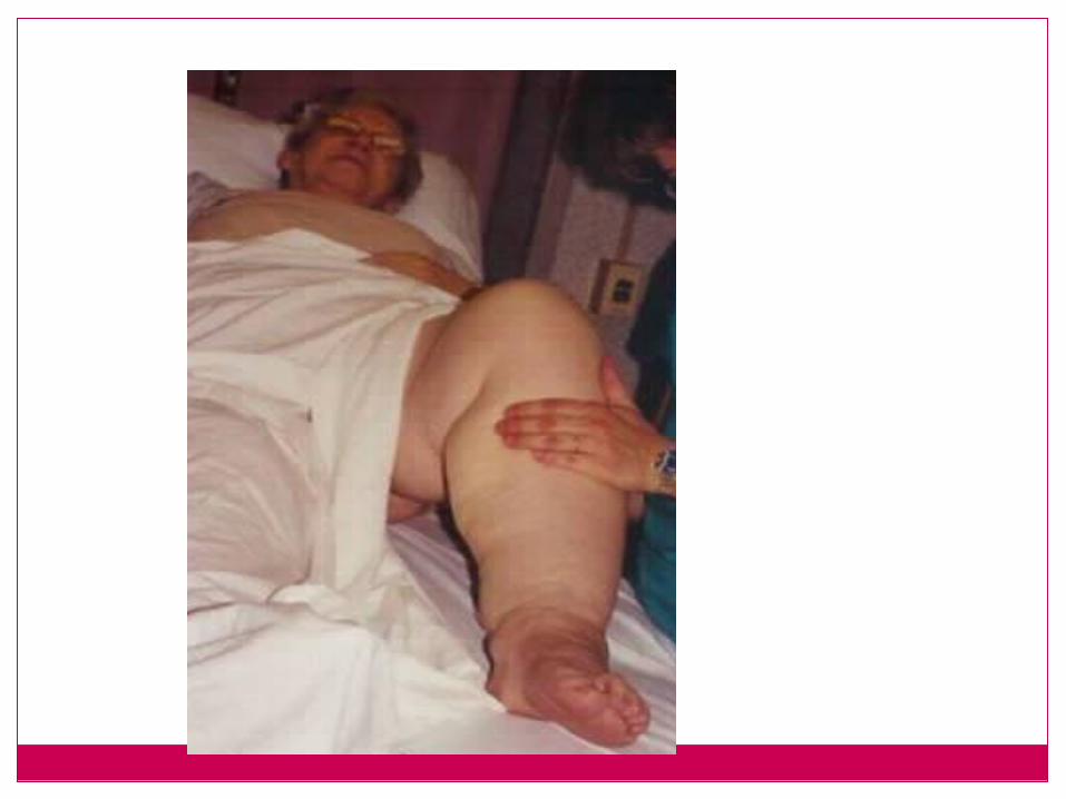
Definition of Lymphedema
Lymphedema is the result of the abnormal accumulation of protein rich edema fluid
Primary or secondaryAfflicts approximately 1% of the US
population (2.5 million people)* A SUDDEN ONSET OF EDEMA MUST BE
THOROUGHLY EVALUATED BY A PHYSICIAN
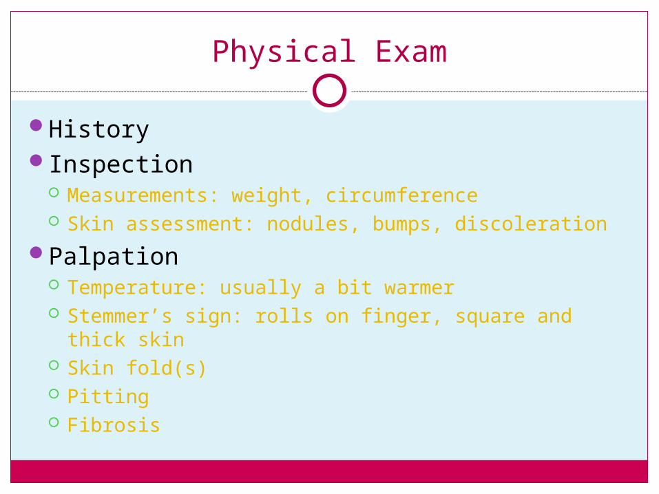
Physical Exam
HistoryInspection
Measurements: weight, circumference Skin assessment: nodules, bumps, discoleration
Palpation Temperature: usually a bit warmer Stemmer’s sign: rolls on finger, square and thick skin Skin fold(s) Pitting Fibrosis
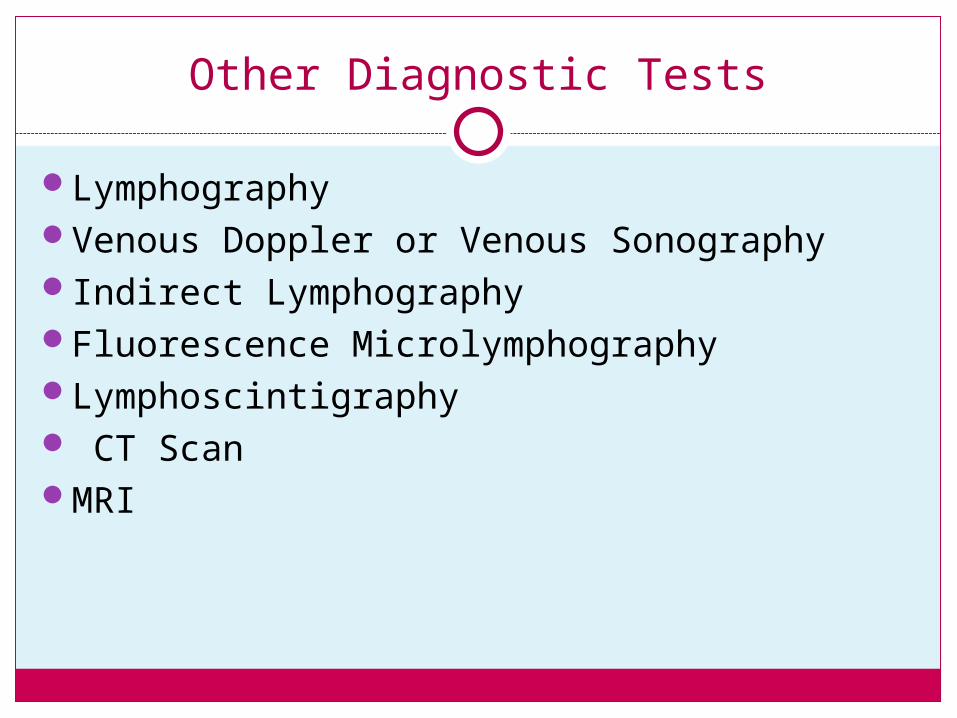
Other Diagnostic Tests
LymphographyVenous Doppler or Venous SonographyIndirect LymphographyFluorescence MicrolymphographyLymphoscintigraphy CT ScanMRI
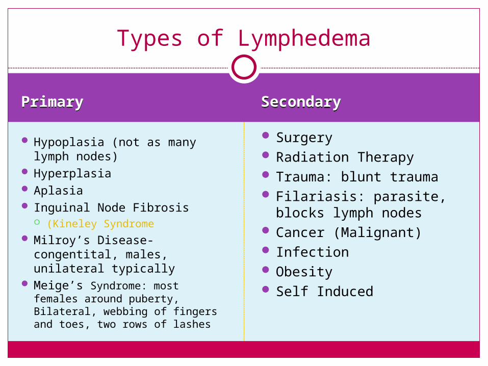
PrimaryPrimary SecondarySecondary
Hypoplasia (not as many lymph nodes)
Hyperplasia Aplasia Inguinal Node Fibrosis
(Kineley Syndrome Milroy’s Disease-congentital,
males, unilateral typically Meige’s Syndrome: most
females around puberty, Bilateral, webbing of fingers and toes, two rows of lashes
Surgery Radiation Therapy Trauma: blunt trauma Filariasis: parasite, blocks
lymph nodes Cancer (Malignant) Infection Obesity Self Induced
Types of Lymphedema
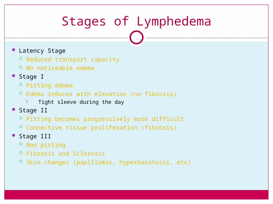
Stages of Lymphedema
Latency Stage Reduced transport capacity No noticeable edema
Stage I Pitting edema Edema reduces with elevation (no fibrosis)
Tight sleeve during the day Stage II
Pitting becomes progressively more difficult Connective tissue proliferation (fibrosis)
Stage III Non pitting Fibrosis and Sclerosis Skin changes (papillomas, hyperkeratosis, etc)
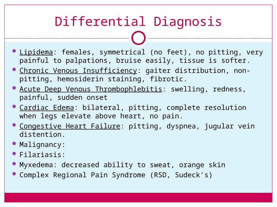
Differential Diagnosis
Lipidema: females, symmetrical (no feet), no pitting, very painful to palpations, bruise easily, tissue is softer.
Chronic Venous Insufficiency: gaiter distribution, non-pitting, hemosiderin staining, fibrotic.
Acute Deep Venous Thrombophlebitis: swelling, redness, painful, sudden onset
Cardiac Edema: bilateral, pitting, complete resolution when legs elevate above heart, no pain.
Congestive Heart Failure: pitting, dyspnea, jugular vein distention.
Malignancy: Filariasis: Myxedema: decreased ability to sweat, orange skin Complex Regional Pain Syndrome (RSD, Sudeck’s)
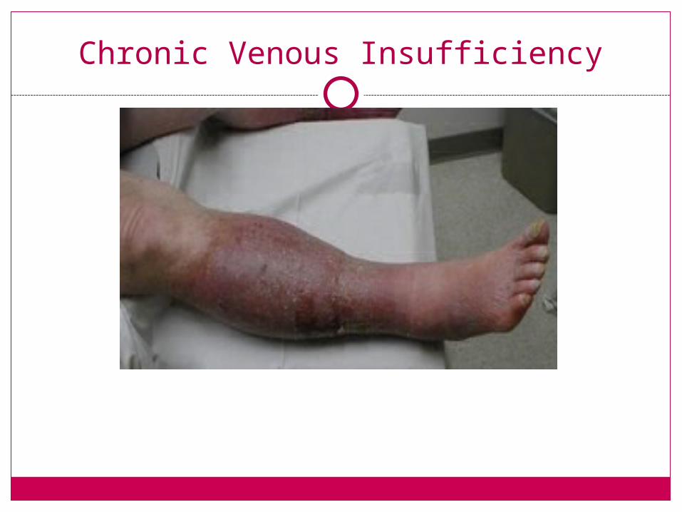
Chronic Venous Insufficiency
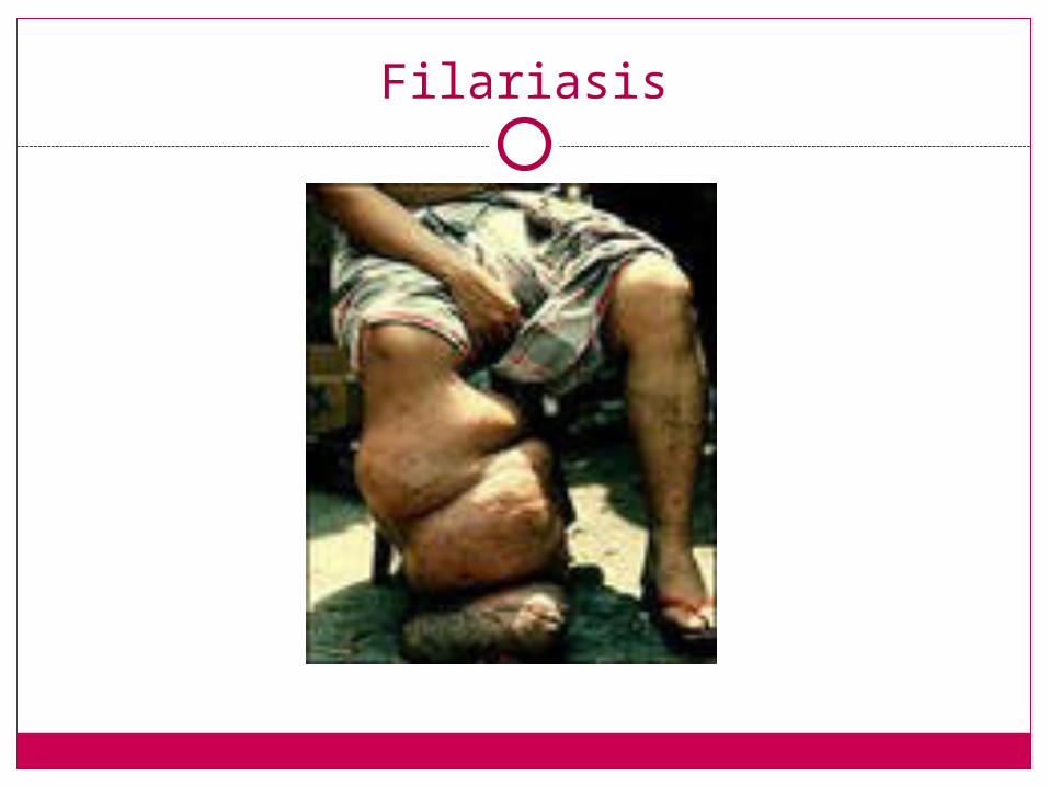
Filariasis
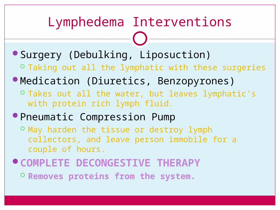
Lymphedema Interventions
Surgery (Debulking, Liposuction) Taking out all the lymphatic with these surgeries
Medication (Diuretics, Benzopyrones) Takes out all the water, but leaves lymphatic's with
protein rich lymph fluid.
Pneumatic Compression Pump May harden the tissue or destroy lymph collectors,
and leave person immobile for a couple of hours.
COMPLETE DECONGESTIVE THERAPY Removes proteins from the system.
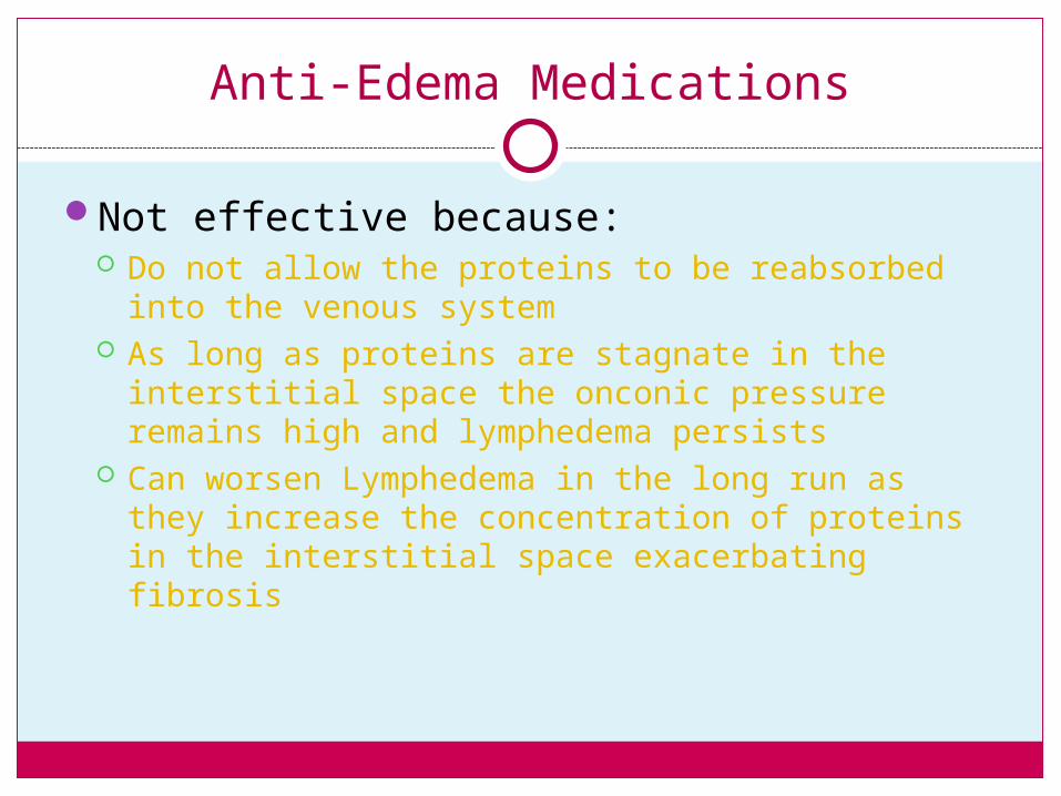
Anti-Edema Medications
Not effective because: Do not allow the proteins to be reabsorbed into the
venous system As long as proteins are stagnate in the interstitial
space the onconic pressure remains high and lymphedema persists
Can worsen Lymphedema in the long run as they increase the concentration of proteins in the interstitial space exacerbating fibrosis
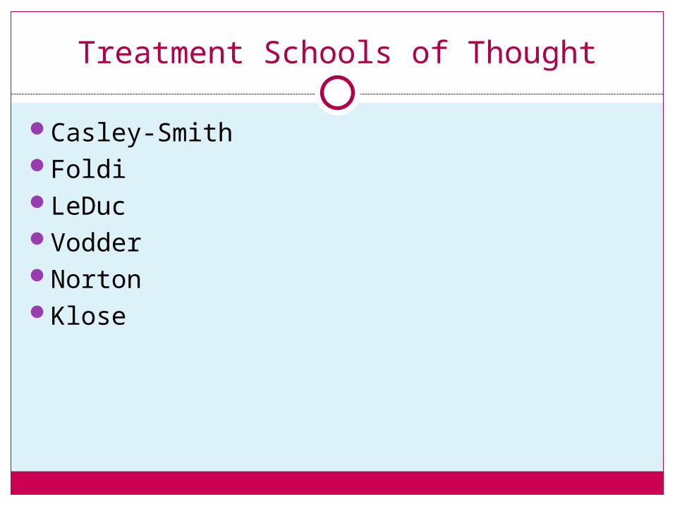
Treatment Schools of Thought
Casley-SmithFoldiLeDucVodderNortonKlose
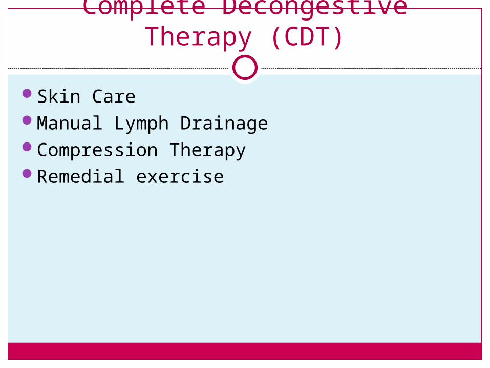
Complete Decongestive Therapy (CDT)
Skin CareManual Lymph DrainageCompression TherapyRemedial exercise
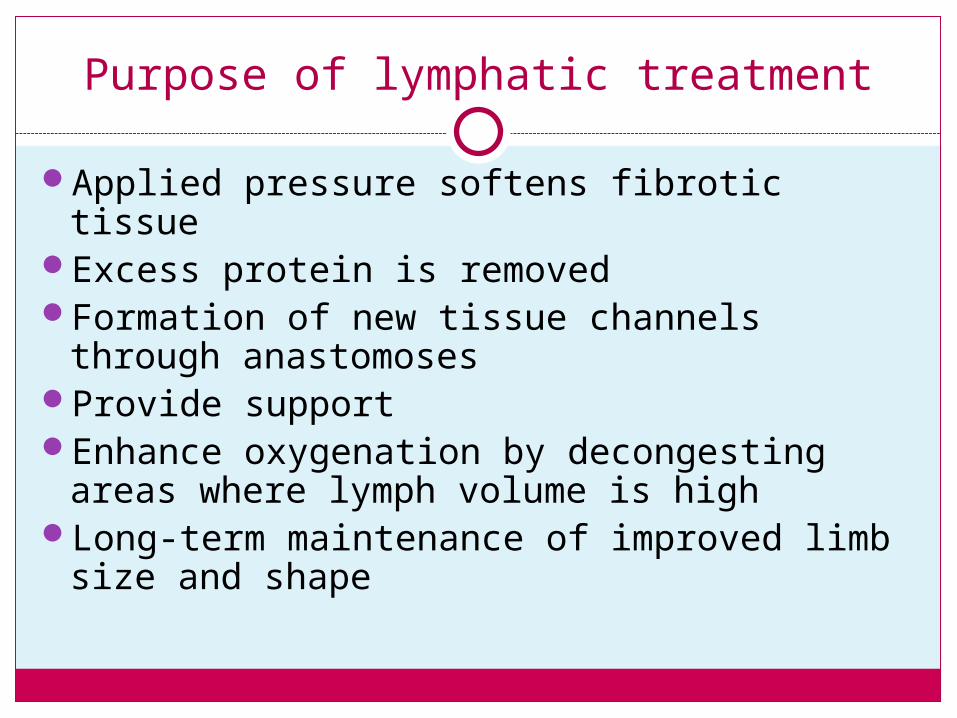
Purpose of lymphatic treatment
Applied pressure softens fibrotic tissueExcess protein is removedFormation of new tissue channels through
anastomoses Provide supportEnhance oxygenation by decongesting areas
where lymph volume is highLong-term maintenance of improved limb size
and shape
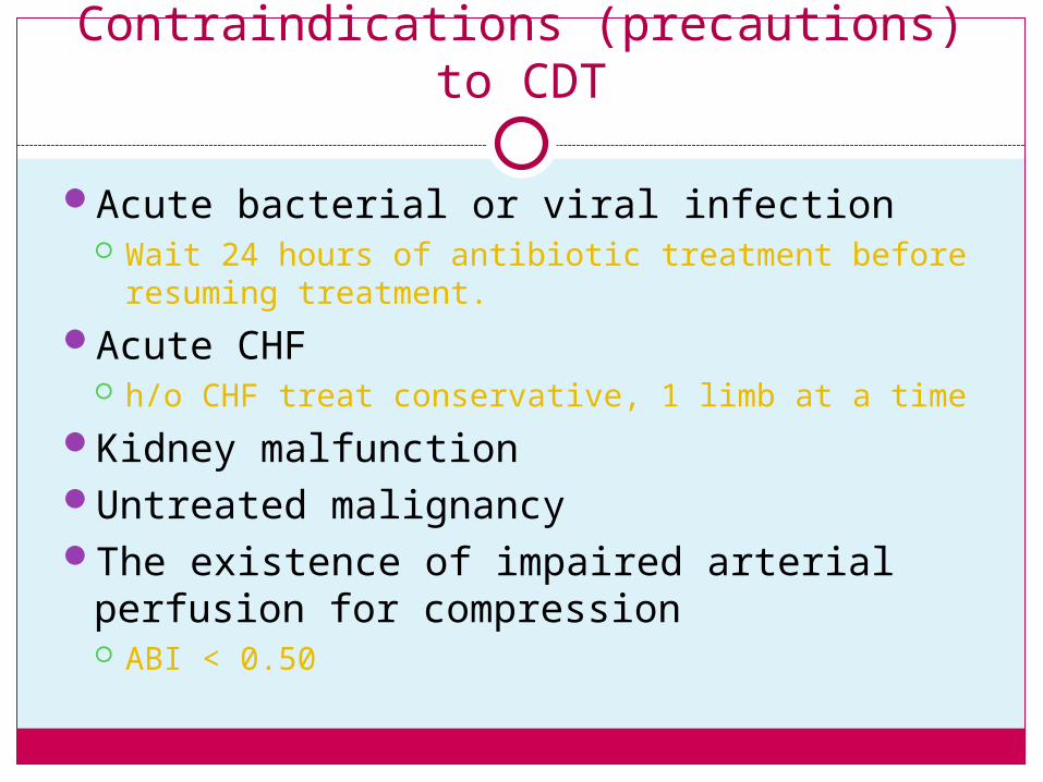
Contraindications (precautions) to CDT
Acute bacterial or viral infection Wait 24 hours of antibiotic treatment before
resuming treatment.
Acute CHF h/o CHF treat conservative, 1 limb at a time
Kidney malfunctionUntreated malignancyThe existence of impaired arterial perfusion
for compression ABI < 0.50
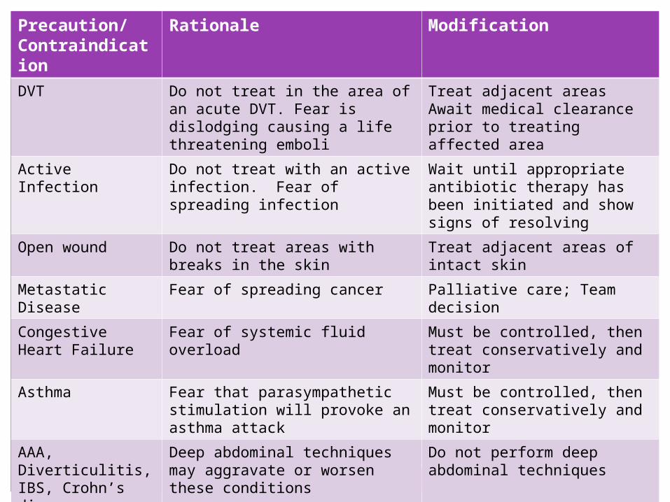
Precaution/Contraindication
Rationale Modification
DVT Do not treat in the area of an acute DVT. Fear is dislodging causing a life threatening emboli
Treat adjacent areasAwait medical clearance prior to treating affected area
Active Infection Do not treat with an active infection. Fear of spreading infection
Wait until appropriate antibiotic therapy has been initiated and show signs of resolving
Open wound Do not treat areas with breaks in the skin
Treat adjacent areas of intact skin
Metastatic Disease
Fear of spreading cancer Palliative care; Team decision
Congestive Heart Failure
Fear of systemic fluid overload Must be controlled, then treat conservatively and monitor
Asthma Fear that parasympathetic stimulation will provoke an asthma attack
Must be controlled, then treat conservatively and monitor
AAA, Diverticulitis, IBS, Crohn’s disease
Deep abdominal techniques may aggravate or worsen these conditions
Do not perform deep abdominal techniques
Pregnancy Fear deep abdominal techniques may harm the fetus or uterus
Do not perform deep abdominal techniques
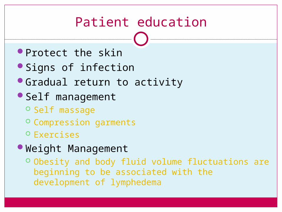
Patient education
Protect the skinSigns of infectionGradual return to activitySelf management
Self massage Compression garments Exercises
Weight Management Obesity and body fluid volume fluctuations are
beginning to be associated with the development of lymphedema
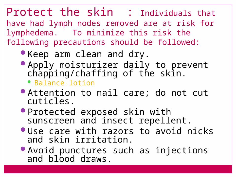
Protect the skin : Individuals that have had lymph nodes removed are at risk for lymphedema. To minimize this risk the following precautions should be followed:
Keep arm clean and dry.Apply moisturizer daily to prevent
chapping/chaffing of the skin. Balance lotion
Attention to nail care; do not cut cuticles.Protected exposed skin with sunscreen
and insect repellent.Use care with razors to avoid nicks and
skin irritation.Avoid punctures such as injections and
blood draws.
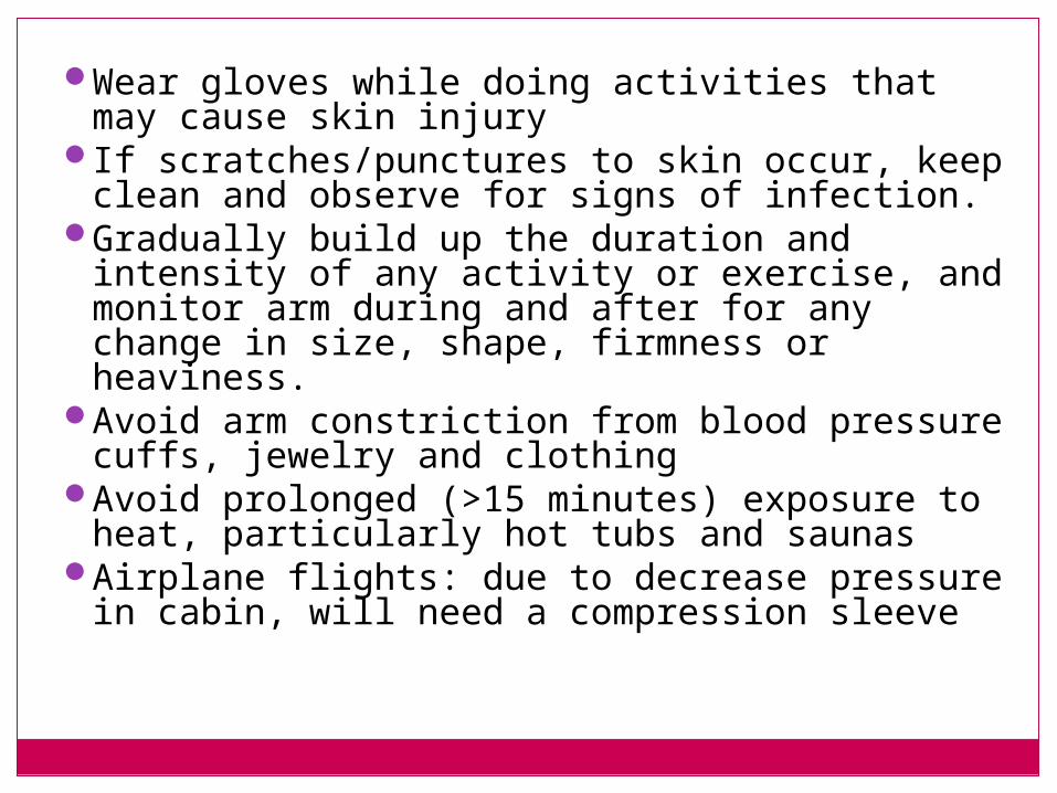
Wear gloves while doing activities that may cause skin injury
If scratches/punctures to skin occur, keep clean and observe for signs of infection.
Gradually build up the duration and intensity of any activity or exercise, and monitor arm during and after for any change in size, shape, firmness or heaviness.
Avoid arm constriction from blood pressure cuffs, jewelry and clothing
Avoid prolonged (>15 minutes) exposure to heat, particularly hot tubs and saunas
Airplane flights: due to decrease pressure in cabin, will need a compression sleeve
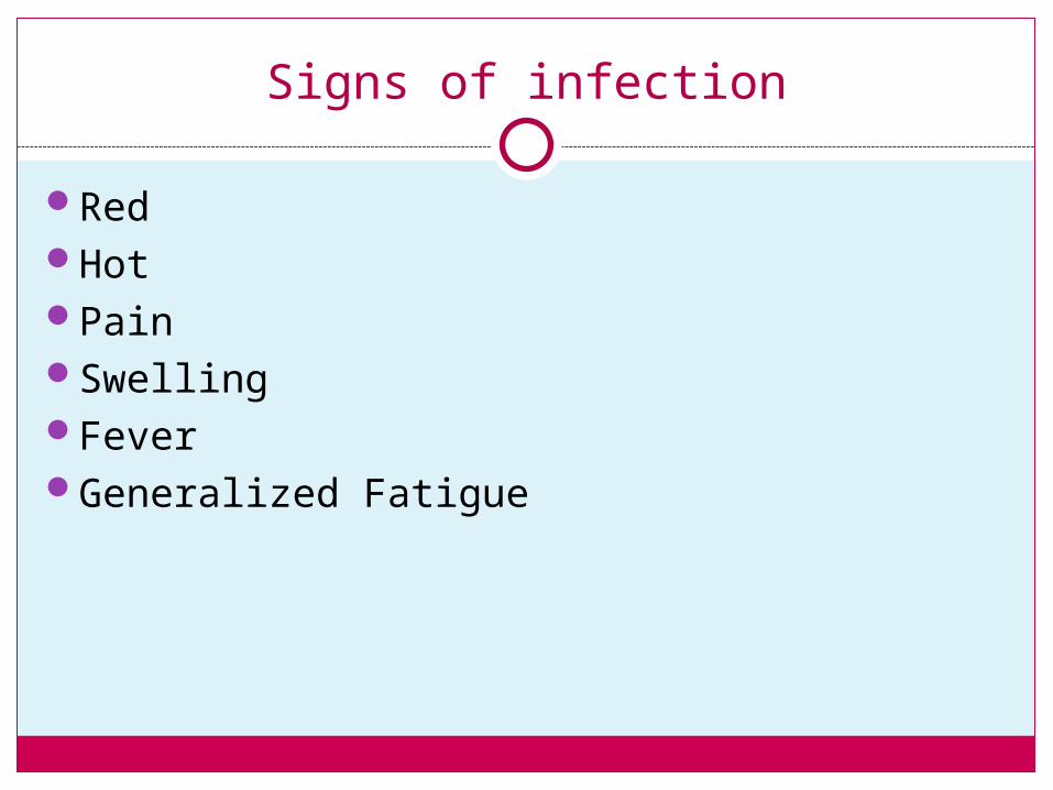
Signs of infection
RedHotPainSwellingFeverGeneralized Fatigue
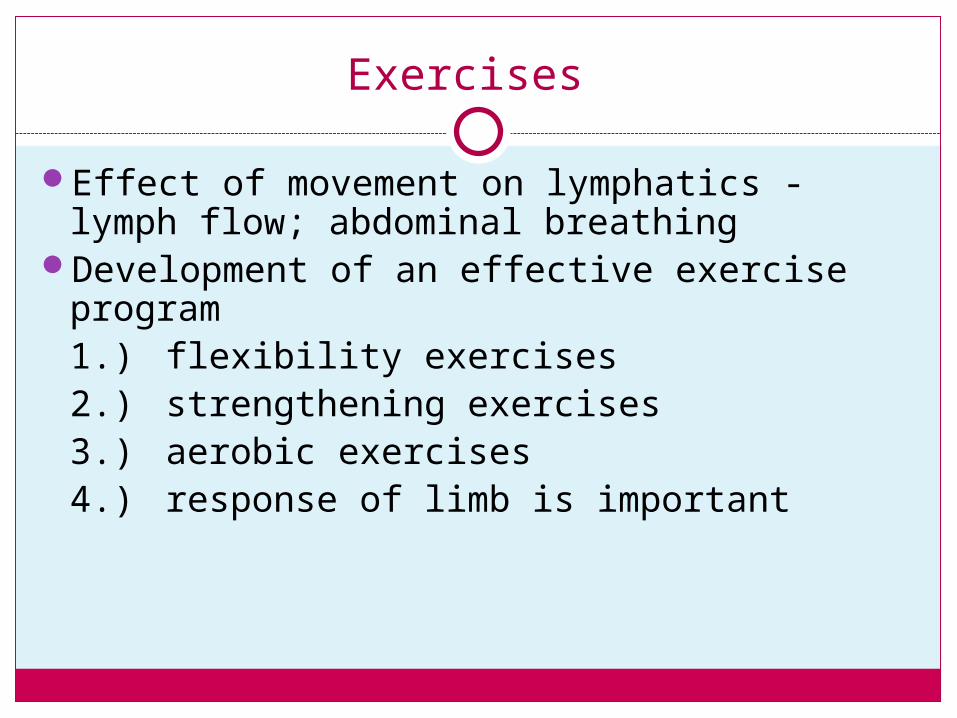
Exercises
Effect of movement on lymphatics - lymph flow; abdominal breathing
Development of an effective exercise program1.) flexibility exercises2.) strengthening exercises3.) aerobic exercises4.) response of limb is important
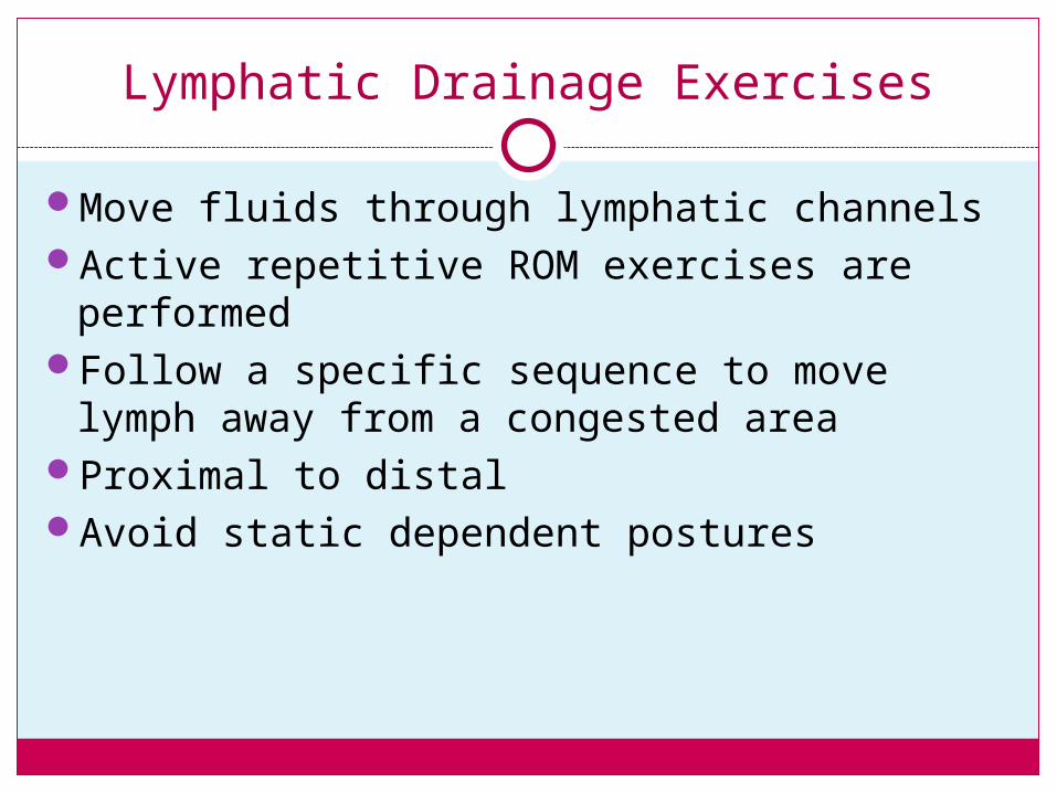
Lymphatic Drainage Exercises
Move fluids through lymphatic channelsActive repetitive ROM exercises are
performedFollow a specific sequence to move lymph
away from a congested areaProximal to distalAvoid static dependent postures
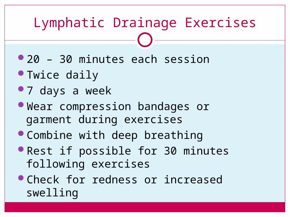
Lymphatic Drainage Exercises
20 – 30 minutes each sessionTwice daily7 days a weekWear compression bandages or
garment during exercisesCombine with deep breathingRest if possible for 30 minutes
following exercisesCheck for redness or increased
swelling
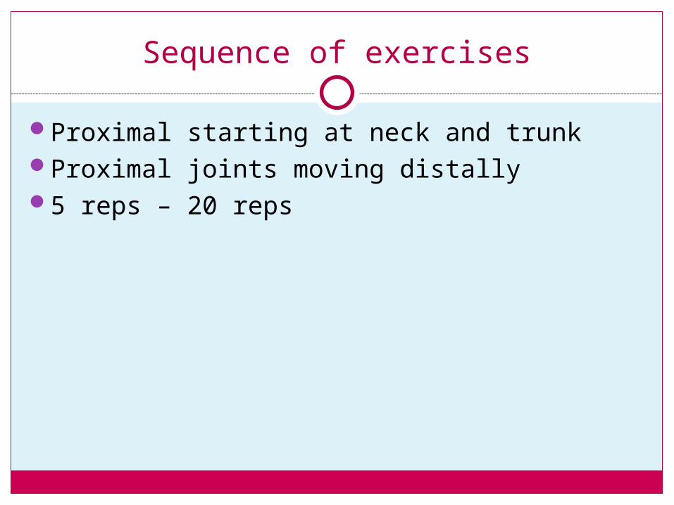
Sequence of exercises
Proximal starting at neck and trunkProximal joints moving distally5 reps – 20 reps
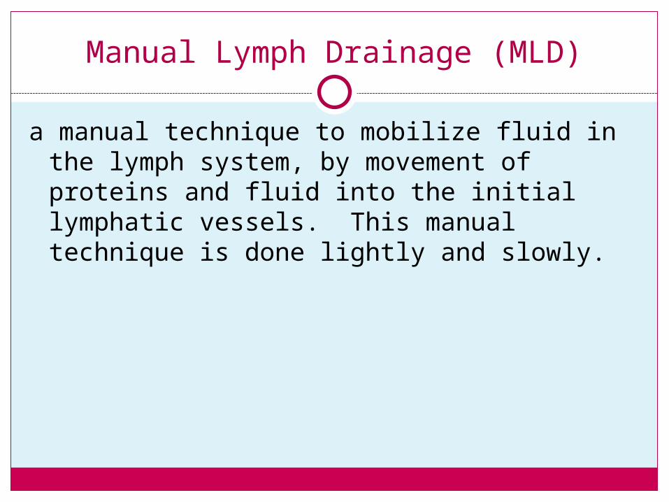
Manual Lymph Drainage (MLD)
a manual technique to mobilize fluid in the lymph system, by movement of proteins and fluid into the initial lymphatic vessels. This manual technique is done lightly and slowly.
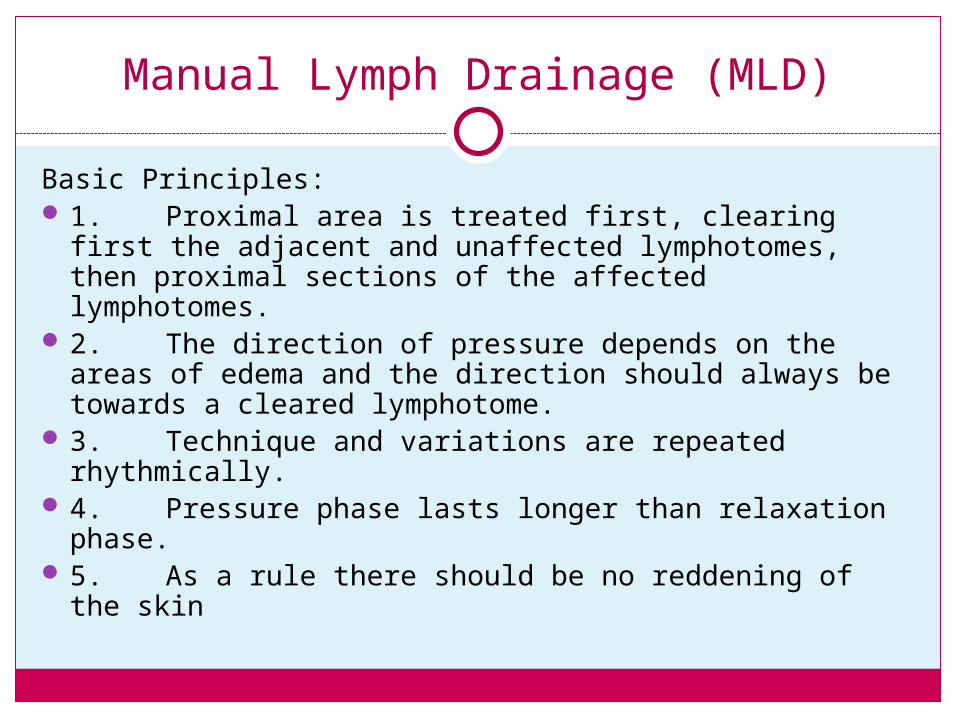
Manual Lymph Drainage (MLD)
Basic Principles: 1. Proximal area is treated first, clearing first the
adjacent and unaffected lymphotomes, then proximal sections of the affected lymphotomes.
2. The direction of pressure depends on the areas of edema and the direction should always be towards a cleared lymphotome.
3. Technique and variations are repeated rhythmically. 4. Pressure phase lasts longer than relaxation phase. 5. As a rule there should be no reddening of the skin
Manual Lymph Drainage (MLD)
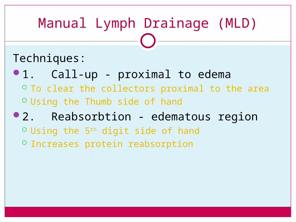
Techniques:1. Call-up - proximal to edema
To clear the collectors proximal to the area Using the Thumb side of hand
2. Reabsorbtion - edematous region Using the 5th digit side of hand Increases protein reabsorption
Manual Lymph Drainage (MLD)
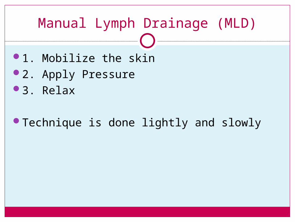
1. Mobilize the skin2. Apply Pressure3. Relax
Technique is done lightly and slowly
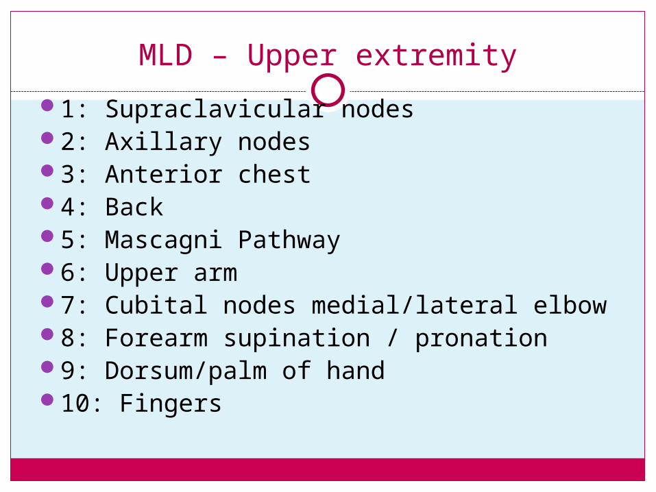
MLD – Upper extremity
1: Supraclavicular nodes2: Axillary nodes3: Inguinal nodes4: Thigh5: Popliteal fossa6: Calf7: Malleolli8: Dorsum of foot9: Toes
Upper Extremity mld
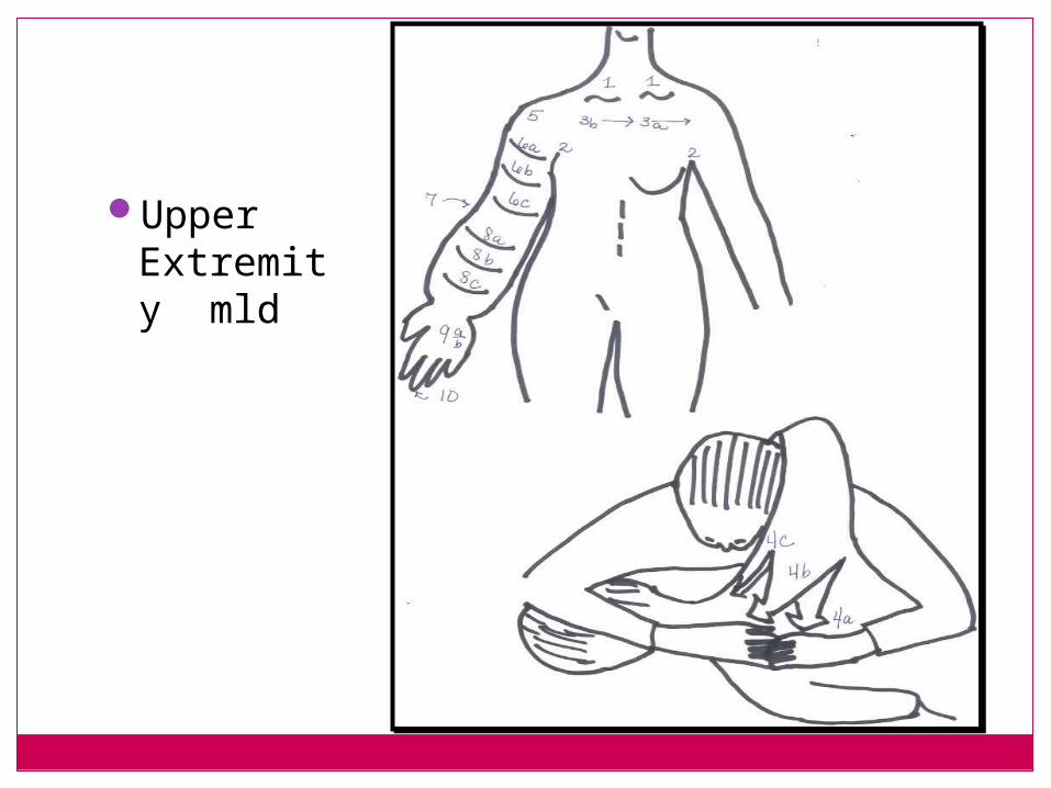
MLD – Upper extremity
1: Supraclavicular nodes2: Axillary nodes3: Anterior chest4: Back5: Mascagni Pathway6: Upper arm7: Cubital nodes medial/lateral elbow8: Forearm supination / pronation9: Dorsum/palm of hand10: Fingers
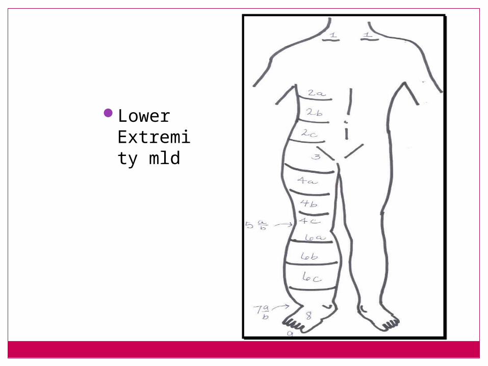
Lower Extremity mld
Protocol
Duration 2 weeks UE 3 – 4 Weeks LE
Frequency 5 days a week
Arm 30 - 45 minutes
Leg 45 - 60 minutes
Wear Bandages During all awake hours
Week 1 Emphasis on Bandages and reduction of Swelling
Week 2-3 Facilitate Physician order for Garment Self Management of Edema
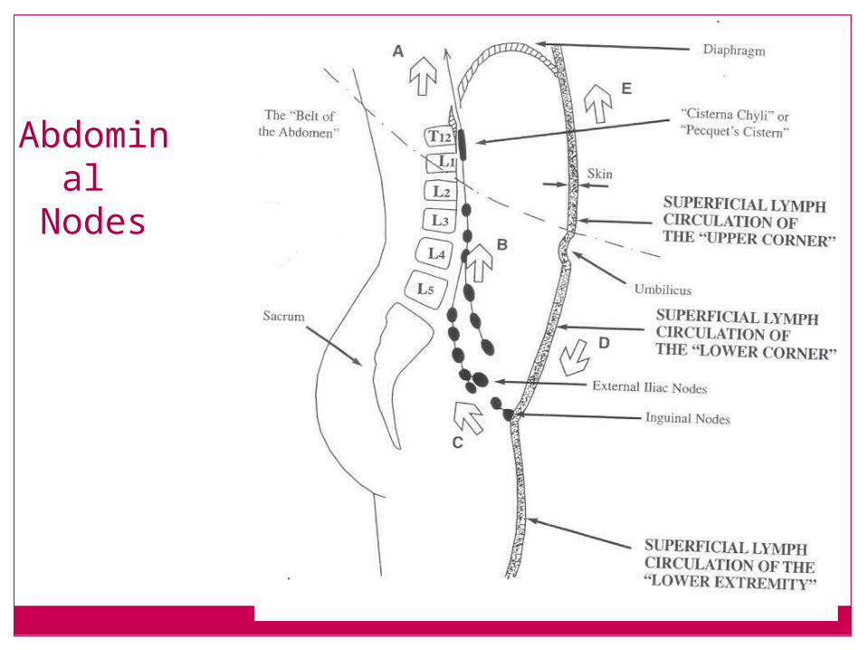
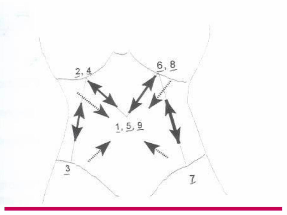
Abdominal
Nodes
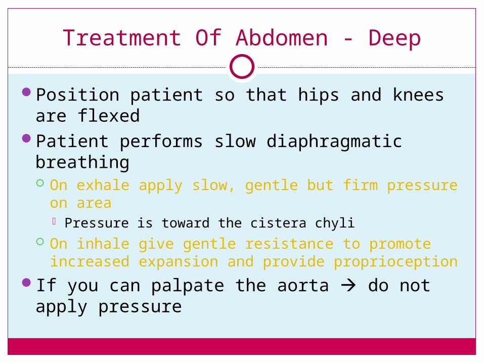
Treatment Of Abdomen - Deep
Position patient so that hips and knees are flexed
Patient performs slow diaphragmatic breathing On exhale apply slow, gentle but firm pressure on area
Pressure is toward the cistera chyli On inhale give gentle resistance to promote increased
expansion and provide proprioceptionIf you can palpate the aorta do not apply
pressure
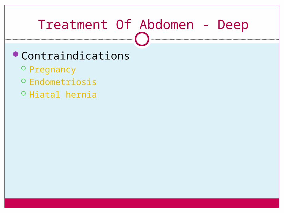
Treatment Of Abdomen - Deep
Contraindications Pregnancy Endometriosis Hiatal hernia
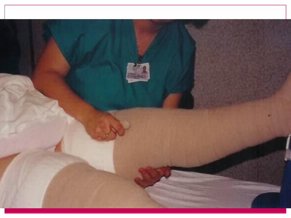
Compression bandages
Compression bandages
Compression bandages have been shown to produce a micromassage effect that improves lymph transport.
Increase temperature of up to 5 degrees enhances the lymphangion mobility
Bandages
Resting pressure - Pressure from the outside in the resting position of the muscle. Pressure applied from fascia, bandages
Working pressure - Pressure from the inside when the muscles are active. Pressure generated by the muscles
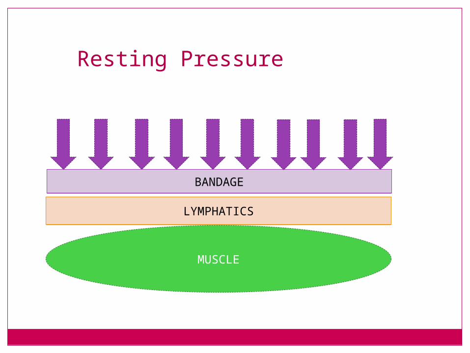
Resting Pressure
BANDAGEBANDAGE
LYMPHATICSLYMPHATICS
MUSCLE
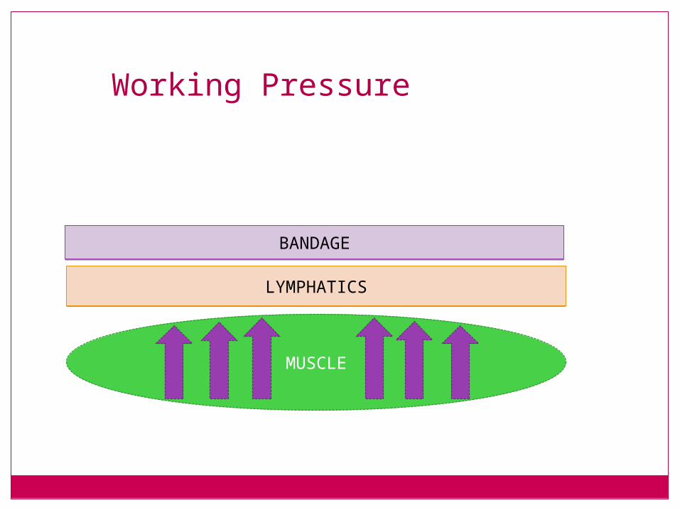
Working Pressure
BANDAGEBANDAGE
LYMPHATICSLYMPHATICS
MUSCLE
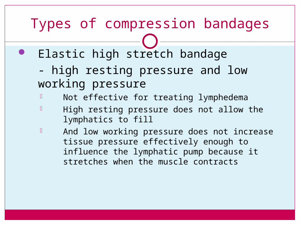
Types of compression bandages
Elastic high stretch bandage- high resting pressure and low working pressure Not effective for treating lymphedema High resting pressure does not allow the
lymphatics to fill And low working pressure does not increase tissue
pressure effectively enough to influence the lymphatic pump because it stretches when the muscle contracts
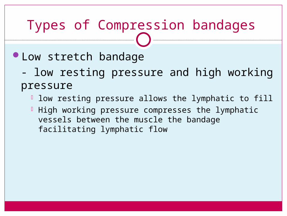
Types of Compression bandages
Low stretch bandage- low resting pressure and high working pressure
low resting pressure allows the lymphatic to fill High working pressure compresses the lymphatic vessels
between the muscle the bandage facilitating lymphatic flow
Low Stretch Compression Bandages
Form a semi rigid support which causes an increase in interstitial pressure when the muscle contracts
When a patient wears low stretch compression bandages while sleeping or resting the increased interstitial pressure will reduce the amount of fluid and protein leaving the arteriole (ultra filtration) and less edema is formed
When a patient wears low stretch compression bandages during activity the increased interstitial pressure not only reduces ultra filtration but increases reabsorbtion into the lymphatic system which decreases lymphedema and well as venous edema
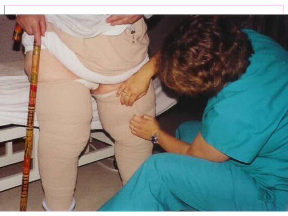
Principles of Bandaging
Must use Low stretchAlways start distally and proceed
proximallyMaintain moderate tensionAvoid creases and foldsUse tape to secure…not clips or
pinsApplied with greater pressure
distally than proximallyDo not extend bandage to maximal
length
Principles of Bandaging – con’t
Check pressure gradientPlace more layers for increase
compression rather than applying them more tightly
Fill indentations with padding or foam pieces
Cover as much of the limb as possible
Compression to be worn until next visit
Exercise with bandages on to take advantage of muscle pump effect
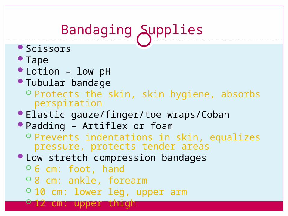
Bandaging SuppliesScissorsTape Lotion – low pHTubular bandage
Protects the skin, skin hygiene, absorbs perspiration
Elastic gauze/finger/toe wraps/CobanPadding – Artiflex or foam
Prevents indentations in skin, equalizes pressure, protects tender areas
Low stretch compression bandages 6 cm: foot, hand 8 cm: ankle, forearm 10 cm: lower leg, upper arm 12 cm: upper thigh
When to instruct the patient to remove the bandages
If the patient gets short of breath or has heart palpations
If the fingers/toes are numb, blue or tingling
If the wraps fall offIf the patient is experiencing too much pain
Compression Therapy
Compression therapy is the application of external pressure on body tissue to support the elasticity of the skin and its underlying vessels
Phase I with Compression BandagesPhase II with medical compression Garments
Rationale for using compression therapy:
Compression therapy directly effects the underlying lymphatic vessels, veins and tissue.
Improves the efficacy of the muscle pump by creating a semi-rigid support for the muscle to work against
Causes a mild increase in total tissue pressure
Improves and maintains the shape of the limb
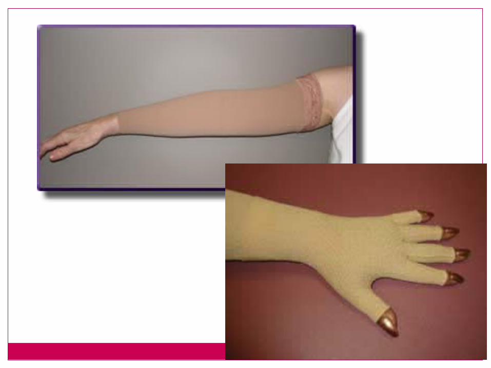
Compression Garments
Not designed to decrease edema- only to maintain the edema reduced by the treatments
Increases reabsorbtionIncreases tissue pressureready made vs. customill fitting garment is worse than not wearing
one at all
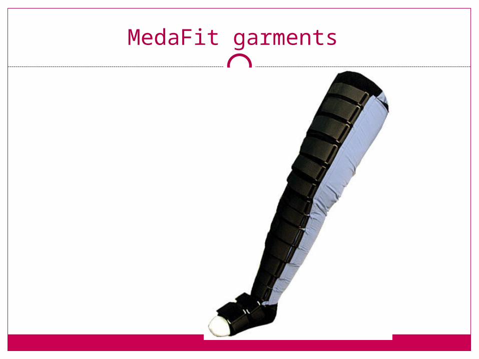
MedaFit garments
Donning Compression Garment
For LE : put on in bedUse gloves to don and doffApply on an “empty” limb
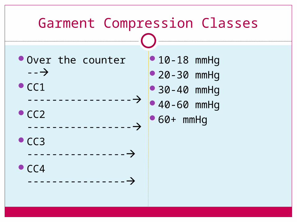
Garment Compression Classes
Over the counter --CC1 -----------------CC2 -----------------CC3 ----------------CC4 ----------------
10-18 mmHg20-30 mmHg30-40 mmHg40-60 mmHg60+ mmHg
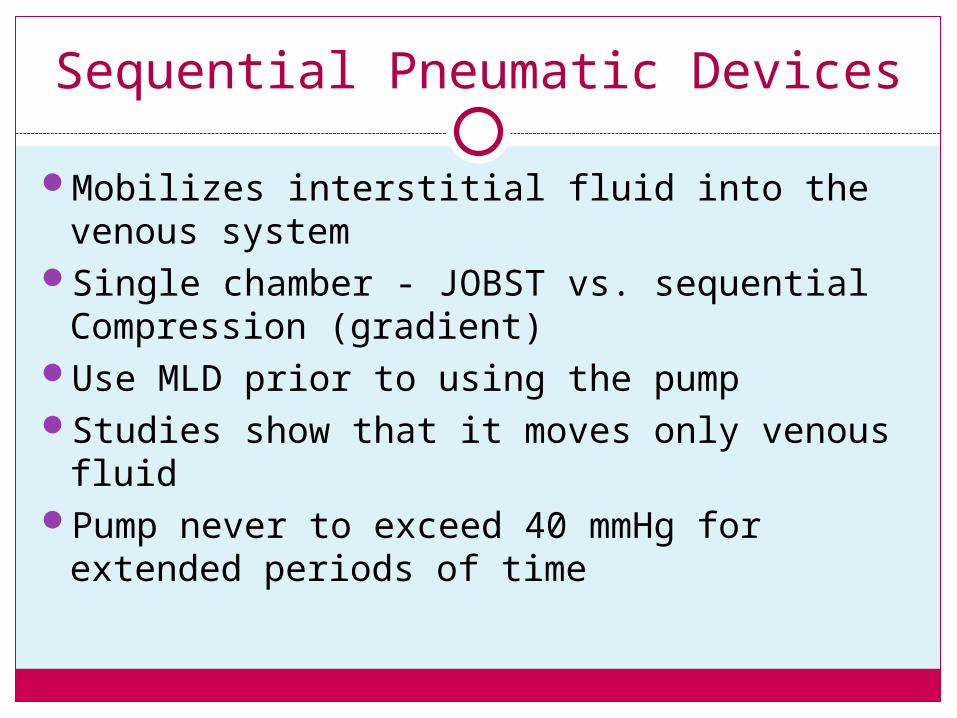
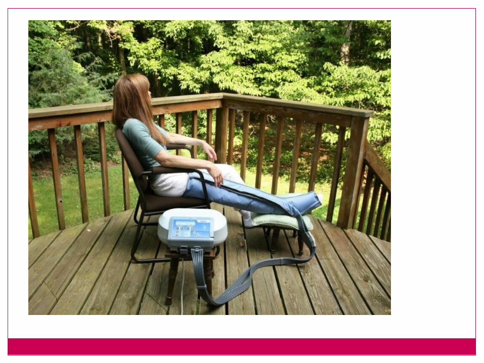
Sequential Pneumatic Devices
Mobilizes interstitial fluid into the venous system
Single chamber - JOBST vs. sequential Compression (gradient)
Use MLD prior to using the pumpStudies show that it moves only venous fluidPump never to exceed 40 mmHg for extended
periods of time
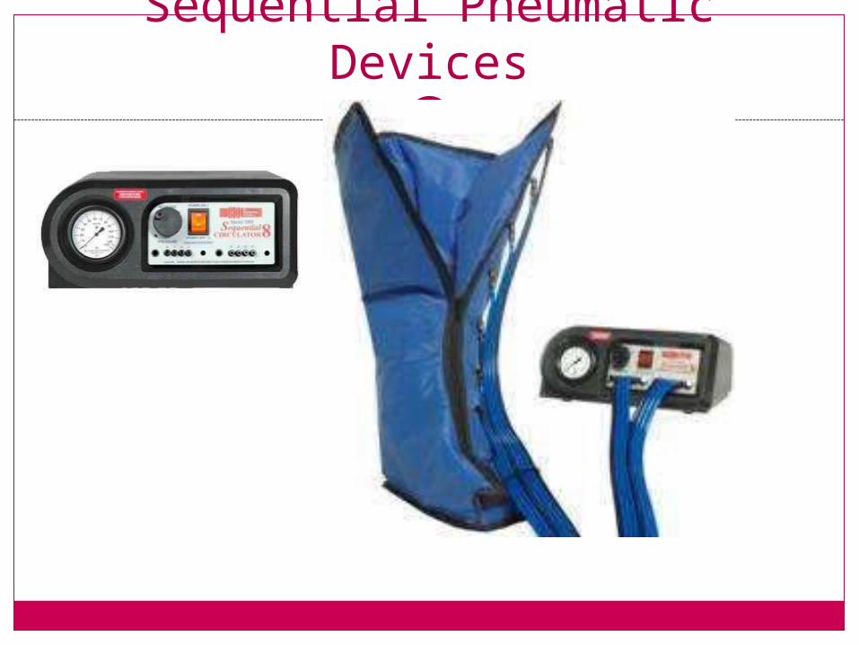
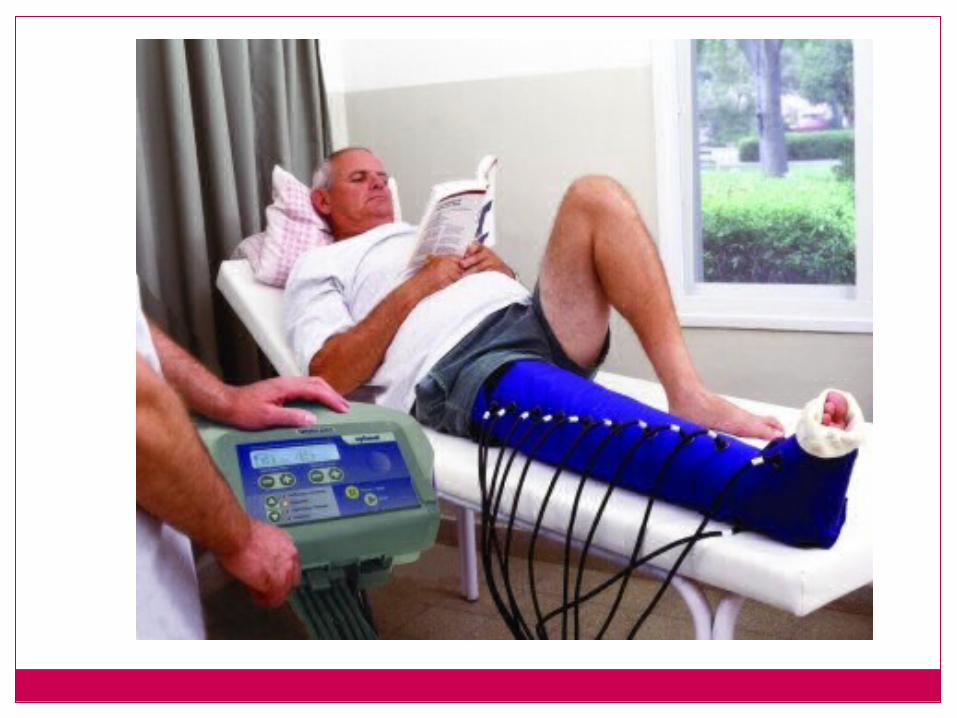
Sequential Pneumatic Devices
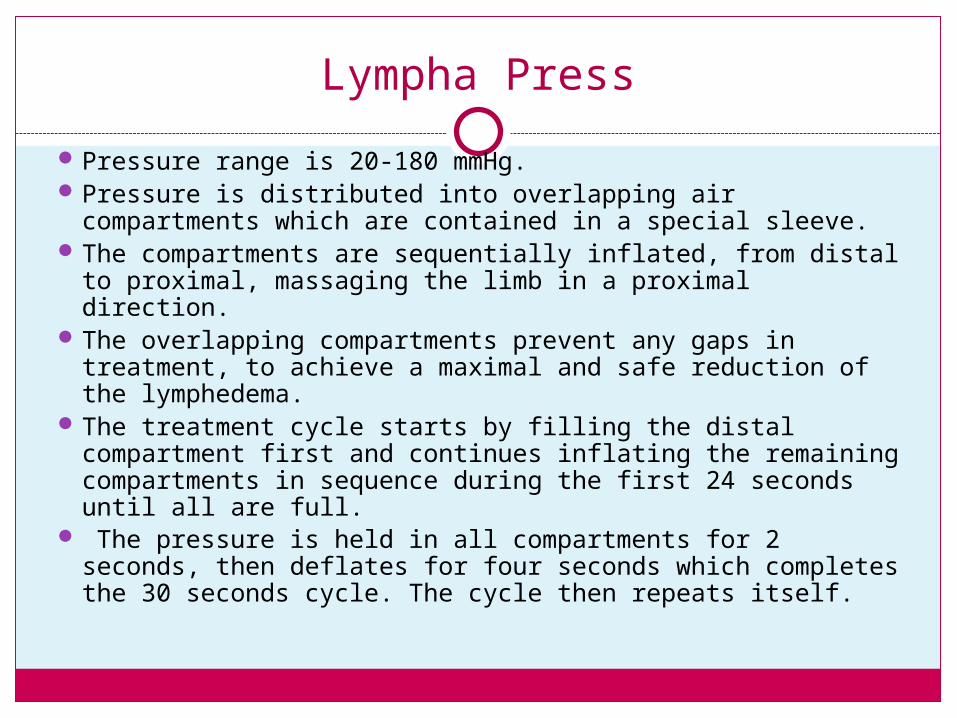
Lympha Press
Pressure range is 20-180 mmHg. Pressure is distributed into overlapping air
compartments which are contained in a special sleeve. The compartments are sequentially inflated, from distal
to proximal, massaging the limb in a proximal direction. The overlapping compartments prevent any gaps in
treatment, to achieve a maximal and safe reduction of the lymphedema.
The treatment cycle starts by filling the distal compartment first and continues inflating the remaining compartments in sequence during the first 24 seconds until all are full.
The pressure is held in all compartments for 2 seconds, then deflates for four seconds which completes the 30 seconds cycle. The cycle then repeats itself.
LASER
Another new frontier in the treatment of lymphedema involves using the laser.
From various trials lasers appear to help lymph flow, shown to be effective improvement of wound healing, and it has been used effectively in treating edema from DVT’s.
The FDA has approved a laser device to be used in the treatment of post-mastectomy arm lymphedema. Clinical trials are currently underway for leg lymphedema.
Lymphedema and its complications can causing "scarring" of the lymphatic system. The laser is useful in removing the scar tissue, thereby helping lymph flow.
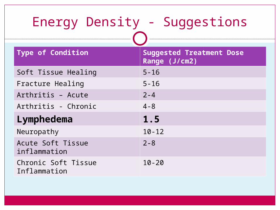
Energy Density - Suggestions
Type of Condition Suggested Treatment Dose Range (J/cm2)
Soft Tissue Healing 5-16
Fracture Healing 5-16
Arthritis – Acute 2-4
Arthritis - Chronic 4-8
Lymphedema 1.5Neuropathy 10-12
Acute Soft Tissue inflammation 2-8
Chronic Soft Tissue Inflammation 10-20
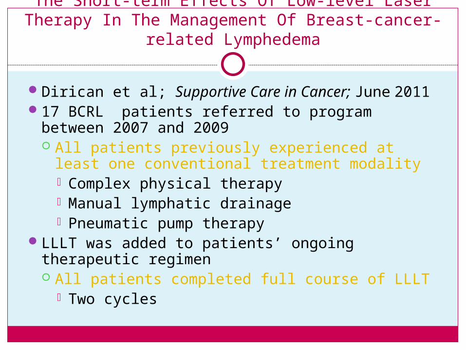
The Short-term Effects Of Low-level Laser Therapy In The Management Of Breast-cancer-related
Lymphedema
Dirican et al; Supportive Care in Cancer; June 201117 BCRL patients referred to program between
2007 and 2009 All patients previously experienced at least one
conventional treatment modality Complex physical therapy Manual lymphatic drainage Pneumatic pump therapy
LLLT was added to patients’ ongoing therapeutic regimen All patients completed full course of LLLT
Two cycles

Results Difference between sums of the circumferences of
both affected and unaffected arms Decreased 54% after first cycle Decreased 73% after second cycle
Pain score 14 out of 17 experienced decreased pain with
motion by an average of 40% after first cycle and 62.7% after second cycle
Scar mobility Increased in 13 patients
Range of motion Improved in 14 patients