TECHNOLOGY FEATURE OPEN
Large-scale computational drug repositioning to findtreatments for rare diseasesRajiv Gandhi Govindaraj1, Misagh Naderi 1, Manali Singha1, Jeffrey Lemoine1,2 and Michal Brylinski 1,3
Rare, or orphan, diseases are conditions afflicting a small subset of people in a population. Although these disorders collectivelypose significant health care problems, drug companies require government incentives to develop drugs for rare diseases due toextremely limited individual markets. Computer-aided drug repositioning, i.e., finding new indications for existing drugs, is acheaper and faster alternative to traditional drug discovery offering a promising venue for orphan drug research. Structure-basedmatching of drug-binding pockets is among the most promising computational techniques to inform drug repositioning. In orderto find new targets for known drugs ultimately leading to drug repositioning, we recently developed eMatchSite, a new computerprogram to compare drug-binding sites. In this study, eMatchSite is combined with virtual screening to systematically exploreopportunities to reposition known drugs to proteins associated with rare diseases. The effectiveness of this integrated approach isdemonstrated for a kinase inhibitor, which is a confirmed candidate for repositioning to synapsin Ia. The resulting datasetcomprises 31,142 putative drug-target complexes linked to 980 orphan diseases. The modeling accuracy is evaluated against thestructural data recently released for tyrosine-protein kinase HCK. To illustrate how potential therapeutics for rare diseases can beidentified, we discuss a possibility to repurpose a steroidal aromatase inhibitor to treat Niemann-Pick disease type C. Overall, theexhaustive exploration of the drug repositioning space exposes new opportunities to combat orphan diseases with existing drugs.DrugBank/Orphanet repositioning data are freely available to research community at https://osf.io/qdjup/.
npj Systems Biology and Applications (2018) 4:13 ; doi:10.1038/s41540-018-0050-7
INTRODUCTIONRepositioning drugs to treat conditions for which they were notoriginally intended is an emerging strategy offering a faster andcheaper route to develop new treatments compared to traditionaldrug discovery.1 Since repurposed molecules not only haveoptimized pharmacokinetics, pharmacodynamics, and toxicityprofiles, but are also already approved by the U.S. Food and DrugAdministration (FDA), this approach speeds up the evaluation ofdrug candidates in clinical trials at the reduced risk of failure. Drugrepositioning is expected to play a major role in the developmentof treatments for rare, or orphan, diseases defined as thosedisorders afflicting <200,000 patients in the United States. Eventhough rare diseases collectively affect more than 350 millionpeople worldwide (https://globalgenes.org/rare-diseases-facts-statistics/), developing new therapeutics for their small individualmarkets is not profitable enough to warrant commercial interest.2
On that account, many countries passed orphan drug legislation,such as the Orphan Drug Act of 1983 in the U.S., in order toprovide financial inducements in terms of the market exclusivityand reduced development costs. Legislators work on the OrphanProduct Extensions Now Accelerating Cures and Treatments(OPEN) Act to extend the market exclusivity for repurposingalready approved drugs to treat rare diseases,3 signifying theimportance of drug repositioning to orphan disease research.It is noteworthy that most repurposed drugs are the result of
serendipitous observations made either in the lab or duringclinical tests. Sildenafil is perhaps the most recognized example of
a repositioned compound. Originally developed to treat hyperten-sion and angina pectoris in the 1980s, it was later repurposed toerectile dysfunction and pulmonary arterial hypertension.4 Otherexamples are amantadine and memantine. The former wasintroduced in the 1960s as a prophylactic agent in respiratoryinfections.5 A few years later, a patient with Parkinson’s diseaseexperienced a dramatic improvement in her symptoms during thedaily administration of amantadine for influenza prophylaxis.6 Thisanecdotal observation stimulated research on using amantadineand other members of the aminoadamantane class of moleculesto treat neurological diseases. Indeed, amantadine is presentlyapproved by the FDA as both an antiviral and an antiparkinsoniandrug. Structurally similar to amantadine, memantine was alsosynthesized in the 1960s as a putative hypoglycemic agent,though it was found to be devoid of such activity. It was laterdiscovered that memantine is an uncompetitive antagonist ofglutamatergic N-methyl-D-aspartate (NMDA) receptors7 and cur-rently, memantine is used to treat moderate to severe Alzheimer-type dementia.8
A clear necessity for rational approaches to find alternativeindications for existing therapeutics has stimulated the develop-ment of computational methods for drug repositioning.9 Manycurrently available algorithms exploit the fact that proteins withsimilar pockets tend to have similar functions and recognizesimilar molecules.10 For instance, the sequence-order independentprofile-profile alignment (SOIPPA) program employs Delaunaytessellation of Cα atoms and geometric potentials to compare
Received: 2 November 2017 Revised: 22 January 2018 Accepted: 3 February 2018
1Department of Biological Sciences, Louisiana State University, Baton Rouge, LA 70803, USA; 2Division of Computer Science and Engineering, Louisiana State University, BatonRouge, LA 70803, USA and 3Center for Computation and Technology, Louisiana State University, Baton Rouge, LA 70803, USACorrespondence: Michal Brylinski ([email protected])
www.nature.com/npjsba
Published in partnership with the Systems Biology Institute
binding pockets.11 Further, SiteAlign measures distances betweendruggable pockets with cavity fingerprints constructed byprojecting eight topological and physicochemical properties ontoa multidimensional, discretized space.12 Both SOIPPA andSiteAlign have been used in drug repurposing, for example,SOIPPA helped reveal new targets for entacapone and tolca-pone,13 whereas SiteAlign detected the cross-reaction of proteinkinase inhibitors with a protein regulating neurotransmitterrelease in the synapse.14
Notwithstanding the success of existing methods to recognizesimilar pockets, many of these algorithms perform well onlyagainst the experimental structures of proteins complexed withsmall molecules. Utilizing datasets of target structures withpredicted binding sites poses a formidable challenge for pocketmatching programs because of inevitable inaccuracies in theannotation of binding residues. To alleviate this issue, we recentlydeveloped eMatchSite, which offers a high tolerance to residuemisannotations and, to some extent, structure imperfections inligand-binding regions.15,16 In this communication, we combineeMatchSite and structure-based virtual screening (VS) withAutoDock Vina17 in order to enhance the accuracy of bindingsite matching. Subsequently, we demonstrate the effectiveness ofeMatchSite/VS for a kinase inhibitor, which is a confirmedcandidate for repositioning to synapsin Ia. Next, this methodologyis employed to explore new opportunities to combat orphanconditions through a large-scale repositioning of existing drugs toproteins linked to rare diseases.18 The results are discussed withrespect to the structural data recently released in the Protein DataBank (PDB)19 for tyrosine-protein kinase HCK, as well as apossibility to repurpose a steroidal aromatase inhibitor to treatNiemann-Pick disease type C. Overall, the protocol combiningprotein structure modeling, binding site prediction and matching,and structure-based virtual screening holds a significant promiseto systematically explore the drug repositioning space at thesystems level.
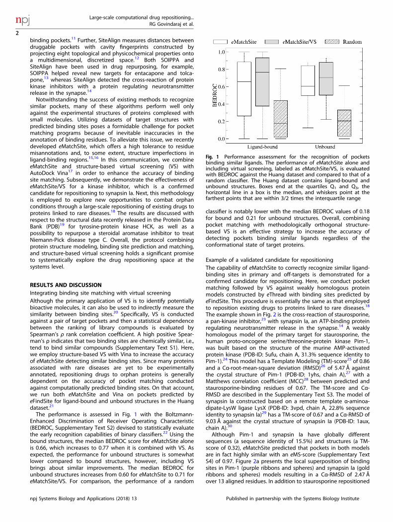
RESULTS AND DISCUSSIONIntegrating binding site matching with virtual screeningAlthough the primary application of VS is to identify potentiallybioactive molecules, it can also be used to indirectly measure thesimilarity between binding sites.20 Specifically, VS is conductedagainst a pair of target pockets and then a statistical dependencebetween the ranking of library compounds is evaluated bySpearman’s ρ rank correlation coefficient. A high positive Spear-man’s ρ indicates that two binding sites are chemically similar, i.e.,tend to bind similar compounds (Supplementary Text S1). Here,we employ structure-based VS with Vina to increase the accuracyof eMatchSite detecting similar binding sites. Since many proteinsassociated with rare diseases are yet to be experimentallyannotated, repositioning drugs to orphan proteins is generallydependent on the accuracy of pocket matching conductedagainst computationally predicted binding sites. On that account,we run both eMatchSite and Vina on pockets predicted byeFindSite for ligand-bound and unbound structures in the Huangdataset.21
The performance is assessed in Fig. 1 with the Boltzmann-Enhanced Discrimination of Receiver Operating Characteristic(BEDROC, Supplementary Text S2) devised to statistically evaluatethe early recognition capabilities of binary classifiers.22 Using thebound structures, the median BEDROC score for eMatchSite aloneis 0.66, which increases to 0.77 when it is combined with VS. Asexpected, the performance for unbound structures is somewhatlower compared to bound structures, however, including VSbrings about similar improvements. The median BEDROC forunbound structures increases from 0.60 for eMatchSite to 0.71 foreMatchSite/VS. For comparison, the performance of a random
classifier is notably lower with the median BEDROC values of 0.18for bound and 0.21 for unbound structures. Overall, combiningpocket matching with methodologically orthogonal structure-based VS is an effective strategy to increase the accuracy ofdetecting pockets binding similar ligands regardless of theconformational state of target proteins.
Example of a validated candidate for repositioningThe capability of eMatchSite to correctly recognize similar ligand-binding sites in primary and off-targets is demonstrated for aconfirmed candidate for repositioning. Here, we conduct pocketmatching followed by VS against weakly homologous proteinmodels constructed by eThread with binding sites predicted byeFindSite. This procedure is essentially the same as that employedto reposition existing drugs to proteins linked to rare diseases.18
The example shown in Fig. 2 is the cross-reaction of staurosporine,a pan-kinase inhibitor,23 with synapsin Ia, an ATP-binding proteinregulating neurotransmitter release in the synapse.14 A weaklyhomologous model of the primary target for staurosporine, thehuman proto-oncogene serine/threonine-protein kinase Pim-1,was built based on the structure of the murine AMP-activatedprotein kinase (PDB-ID: 5ufu, chain A, 31.3% sequence identity toPim-1).24 This model has a Template Modeling (TM)-score25 of 0.86and a Cα-root-mean-square deviation (RMSD)26 of 5.47 Å againstthe crystal structure of Pim-1 (PDB-ID: 1yhs, chain A),27 with aMatthews correlation coefficient (MCC)28 between predicted andstaurosporine-binding residues of 0.67. The TM-score and Cα-RMSD are described in the Supplementary Text S3. The model ofsynapsin Ia constructed based on a remote template α-aminoa-dipate-LysW ligase LysX (PDB-ID: 3vpd, chain A, 22.8% sequenceidentity to synapsin Ia)29 has a TM-score of 0.67 and a Cα-RMSD of9.03 Å against the crystal structure of synapsin Ia (PDB-ID: 1aux,chain A).30
Although Pim-1 and synapsin Ia have globally differentsequences (a sequence identity of 15.5%) and structures (a TM-score of 0.32), eMatchSite predicted that pockets in both modelsare in fact highly similar with an eMS-score (Supplementary TextS4) of 0.97. Figure 2a presents the local superposition of bindingsites in Pim-1 (purple ribbons and spheres) and synapsin Ia (goldribbons and spheres) models resulting in a Cα-RMSD of 2.47 Åover 13 aligned residues. In addition to staurosporine repositioned
Fig. 1 Performance assessment for the recognition of pocketsbinding similar ligands. The performance of eMatchSite alone andincluding virtual screening, labeled as eMatchSite/VS, is evaluatedwith BEDROC against the Huang dataset and compared to that of arandom classifier. The Huang dataset contains ligand-bound andunbound structures. Boxes end at the quartiles Q1 and Q3, thehorizontal line in a box is the median, and whiskers point at thefarthest points that are within 3/2 times the interquartile range
Large-scale computational drug repositioning...RG Govindaraj et al.
2
npj Systems Biology and Applications (2018) 13 Published in partnership with the Systems Biology Institute
1234567890():,;
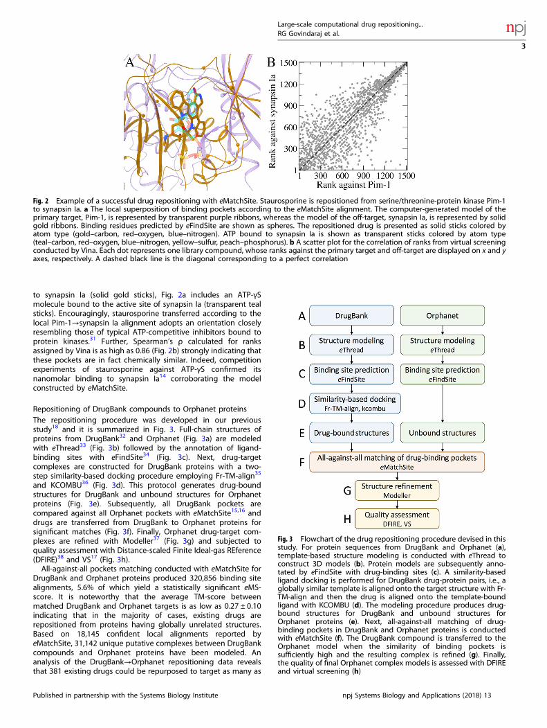
to synapsin Ia (solid gold sticks), Fig. 2a includes an ATP-γSmolecule bound to the active site of synapsin Ia (transparent tealsticks). Encouragingly, staurosporine transferred according to thelocal Pim-1→synapsin Ia alignment adopts an orientation closelyresembling those of typical ATP-competitive inhibitors bound toprotein kinases.31 Further, Spearman’s ρ calculated for ranksassigned by Vina is as high as 0.86 (Fig. 2b) strongly indicating thatthese pockets are in fact chemically similar. Indeed, competitionexperiments of staurosporine against ATP-γS confirmed itsnanomolar binding to synapsin Ia14 corroborating the modelconstructed by eMatchSite.
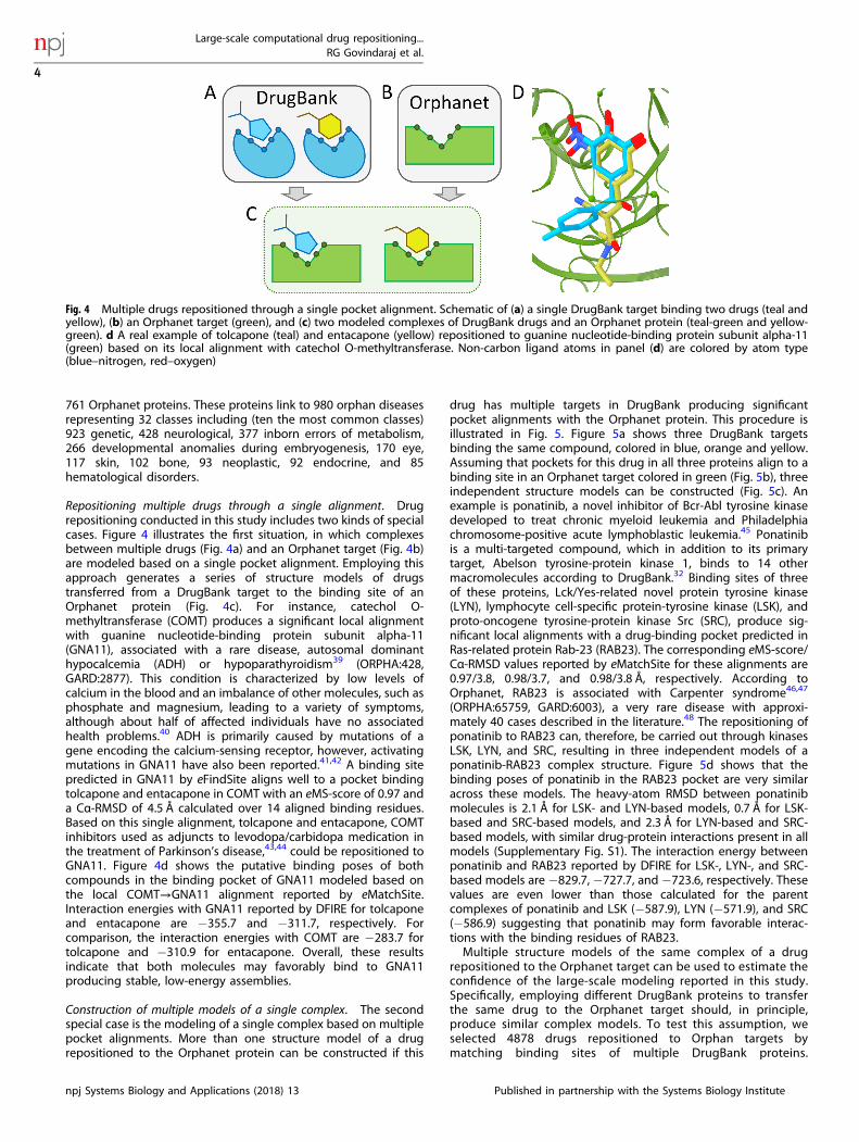
Repositioning of DrugBank compounds to Orphanet proteinsThe repositioning procedure was developed in our previousstudy18 and it is summarized in Fig. 3. Full-chain structures ofproteins from DrugBank32 and Orphanet (Fig. 3a) are modeledwith eThread33 (Fig. 3b) followed by the annotation of ligand-binding sites with eFindSite34 (Fig. 3c). Next, drug-targetcomplexes are constructed for DrugBank proteins with a two-step similarity-based docking procedure employing Fr-TM-align35
and KCOMBU36 (Fig. 3d). This protocol generates drug-boundstructures for DrugBank and unbound structures for Orphanetproteins (Fig. 3e). Subsequently, all DrugBank pockets arecompared against all Orphanet pockets with eMatchSite15,16 anddrugs are transferred from DrugBank to Orphanet proteins forsignificant matches (Fig. 3f). Finally, Orphanet drug-target com-plexes are refined with Modeller37 (Fig. 3g) and subjected toquality assessment with Distance-scaled Finite Ideal-gas REference(DFIRE)38 and VS17 (Fig. 3h).All-against-all pockets matching conducted with eMatchSite for
DrugBank and Orphanet proteins produced 320,856 binding sitealignments, 5.6% of which yield a statistically significant eMS-score. It is noteworthy that the average TM-score betweenmatched DrugBank and Orphanet targets is as low as 0.27 ± 0.10indicating that in the majority of cases, existing drugs arerepositioned from proteins having globally unrelated structures.Based on 18,145 confident local alignments reported byeMatchSite, 31,142 unique putative complexes between DrugBankcompounds and Orphanet proteins have been modeled. Ananalysis of the DrugBank→Orphanet repositioning data revealsthat 381 existing drugs could be repurposed to target as many as
Fig. 2 Example of a successful drug repositioning with eMatchSite. Staurosporine is repositioned from serine/threonine-protein kinase Pim-1to synapsin Ia. a The local superposition of binding pockets according to the eMatchSite alignment. The computer-generated model of theprimary target, Pim-1, is represented by transparent purple ribbons, whereas the model of the off-target, synapsin Ia, is represented by solidgold ribbons. Binding residues predicted by eFindSite are shown as spheres. The repositioned drug is presented as solid sticks colored byatom type (gold–carbon, red–oxygen, blue–nitrogen). ATP bound to synapsin Ia is shown as transparent sticks colored by atom type(teal–carbon, red–oxygen, blue–nitrogen, yellow–sulfur, peach–phosphorus). b A scatter plot for the correlation of ranks from virtual screeningconducted by Vina. Each dot represents one library compound, whose ranks against the primary target and off-target are displayed on x and yaxes, respectively. A dashed black line is the diagonal corresponding to a perfect correlation
Fig. 3 Flowchart of the drug repositioning procedure devised in thisstudy. For protein sequences from DrugBank and Orphanet (a),template-based structure modeling is conducted with eThread toconstruct 3D models (b). Protein models are subsequently anno-tated by eFindSite with drug-binding sites (c). A similarity-basedligand docking is performed for DrugBank drug-protein pairs, i.e., aglobally similar template is aligned onto the target structure with Fr-TM-align and then the drug is aligned onto the template-boundligand with KCOMBU (d). The modeling procedure produces drug-bound structures for DrugBank and unbound structures forOrphanet proteins (e). Next, all-against-all matching of drug-binding pockets in DrugBank and Orphanet proteins is conductedwith eMatchSite (f). The DrugBank compound is transferred to theOrphanet model when the similarity of binding pockets issufficiently high and the resulting complex is refined (g). Finally,the quality of final Orphanet complex models is assessed with DFIREand virtual screening (h)
Large-scale computational drug repositioning...RG Govindaraj et al.
3
Published in partnership with the Systems Biology Institute npj Systems Biology and Applications (2018) 13
761 Orphanet proteins. These proteins link to 980 orphan diseasesrepresenting 32 classes including (ten the most common classes)923 genetic, 428 neurological, 377 inborn errors of metabolism,266 developmental anomalies during embryogenesis, 170 eye,117 skin, 102 bone, 93 neoplastic, 92 endocrine, and 85hematological disorders.
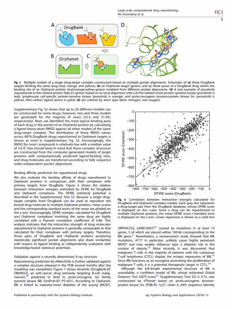
Repositioning multiple drugs through a single alignment. Drugrepositioning conducted in this study includes two kinds of specialcases. Figure 4 illustrates the first situation, in which complexesbetween multiple drugs (Fig. 4a) and an Orphanet target (Fig. 4b)are modeled based on a single pocket alignment. Employing thisapproach generates a series of structure models of drugstransferred from a DrugBank target to the binding site of anOrphanet protein (Fig. 4c). For instance, catechol O-methyltransferase (COMT) produces a significant local alignmentwith guanine nucleotide-binding protein subunit alpha-11(GNA11), associated with a rare disease, autosomal dominanthypocalcemia (ADH) or hypoparathyroidism39 (ORPHA:428,GARD:2877). This condition is characterized by low levels ofcalcium in the blood and an imbalance of other molecules, such asphosphate and magnesium, leading to a variety of symptoms,although about half of affected individuals have no associatedhealth problems.40 ADH is primarily caused by mutations of agene encoding the calcium-sensing receptor, however, activatingmutations in GNA11 have also been reported.41,42 A binding sitepredicted in GNA11 by eFindSite aligns well to a pocket bindingtolcapone and entacapone in COMT with an eMS-score of 0.97 anda Cα-RMSD of 4.5 Å calculated over 14 aligned binding residues.Based on this single alignment, tolcapone and entacapone, COMTinhibitors used as adjuncts to levodopa/carbidopa medication inthe treatment of Parkinson’s disease,43,44 could be repositioned toGNA11. Figure 4d shows the putative binding poses of bothcompounds in the binding pocket of GNA11 modeled based onthe local COMT→GNA11 alignment reported by eMatchSite.Interaction energies with GNA11 reported by DFIRE for tolcaponeand entacapone are −355.7 and −311.7, respectively. Forcomparison, the interaction energies with COMT are −283.7 fortolcapone and −310.9 for entacapone. Overall, these resultsindicate that both molecules may favorably bind to GNA11producing stable, low-energy assemblies.
Construction of multiple models of a single complex. The secondspecial case is the modeling of a single complex based on multiplepocket alignments. More than one structure model of a drugrepositioned to the Orphanet protein can be constructed if this
drug has multiple targets in DrugBank producing significantpocket alignments with the Orphanet protein. This procedure isillustrated in Fig. 5. Figure 5a shows three DrugBank targetsbinding the same compound, colored in blue, orange and yellow.Assuming that pockets for this drug in all three proteins align to abinding site in an Orphanet target colored in green (Fig. 5b), threeindependent structure models can be constructed (Fig. 5c). Anexample is ponatinib, a novel inhibitor of Bcr-Abl tyrosine kinasedeveloped to treat chronic myeloid leukemia and Philadelphiachromosome-positive acute lymphoblastic leukemia.45 Ponatinibis a multi-targeted compound, which in addition to its primarytarget, Abelson tyrosine-protein kinase 1, binds to 14 othermacromolecules according to DrugBank.32 Binding sites of threeof these proteins, Lck/Yes-related novel protein tyrosine kinase(LYN), lymphocyte cell-specific protein-tyrosine kinase (LSK), andproto-oncogene tyrosine-protein kinase Src (SRC), produce sig-nificant local alignments with a drug-binding pocket predicted inRas-related protein Rab-23 (RAB23). The corresponding eMS-score/Cα-RMSD values reported by eMatchSite for these alignments are0.97/3.8, 0.98/3.7, and 0.98/3.8 Å, respectively. According toOrphanet, RAB23 is associated with Carpenter syndrome46,47
(ORPHA:65759, GARD:6003), a very rare disease with approxi-mately 40 cases described in the literature.48 The repositioning ofponatinib to RAB23 can, therefore, be carried out through kinasesLSK, LYN, and SRC, resulting in three independent models of aponatinib-RAB23 complex structure. Figure 5d shows that thebinding poses of ponatinib in the RAB23 pocket are very similaracross these models. The heavy-atom RMSD between ponatinibmolecules is 2.1 Å for LSK- and LYN-based models, 0.7 Å for LSK-based and SRC-based models, and 2.3 Å for LYN-based and SRC-based models, with similar drug-protein interactions present in allmodels (Supplementary Fig. S1). The interaction energy betweenponatinib and RAB23 reported by DFIRE for LSK-, LYN-, and SRC-based models are −829.7, −727.7, and −723.6, respectively. Thesevalues are even lower than those calculated for the parentcomplexes of ponatinib and LSK (−587.9), LYN (−571.9), and SRC(−586.9) suggesting that ponatinib may form favorable interac-tions with the binding residues of RAB23.Multiple structure models of the same complex of a drug
repositioned to the Orphanet target can be used to estimate theconfidence of the large-scale modeling reported in this study.Specifically, employing different DrugBank proteins to transferthe same drug to the Orphanet target should, in principle,produce similar complex models. To test this assumption, weselected 4878 drugs repositioned to Orphan targets bymatching binding sites of multiple DrugBank proteins.
Fig. 4 Multiple drugs repositioned through a single pocket alignment. Schematic of (a) a single DrugBank target binding two drugs (teal andyellow), (b) an Orphanet target (green), and (c) two modeled complexes of DrugBank drugs and an Orphanet protein (teal-green and yellow-green). d A real example of tolcapone (teal) and entacapone (yellow) repositioned to guanine nucleotide-binding protein subunit alpha-11(green) based on its local alignment with catechol O-methyltransferase. Non-carbon ligand atoms in panel (d) are colored by atom type(blue–nitrogen, red–oxygen)
Large-scale computational drug repositioning...RG Govindaraj et al.
4
npj Systems Biology and Applications (2018) 13 Published in partnership with the Systems Biology Institute
Supplementary Fig. S2 shows that up to 20 different models canbe constructed for some drugs, however, two and three modelsare generated for the majority of cases (52.4 and 21.9%,respectively). Next, we identified the most typical binding poseof each drug in the pocket of an Orphanet protein by calculatinga ligand heavy-atom RMSD against all other models of the samedrug-target complex. The distribution of these RMSD valuesacross 4878 DrugBank drugs repositioned to Orphanet targets isshown as inset in Supplementary Fig. S2. Encouragingly, theRMSD for most compounds is relatively low with a median valueof 3.6 Å. One should keep in mind that these complex structuresare constructed from the computer-generated models of targetproteins with computationally predicted ligand-binding sites,and drug molecules are transferred according to fully sequenceorder-independent pocket alignments.
Binding affinity prediction for repositioned drugsWe also evaluate the binding affinity of drugs repositioned toOrphanet proteins in comparison with their complexes withprimary targets from DrugBank. Figure 6 shows the relationbetween interaction energies estimated by DFIRE for DrugBankand Orphanet complexes. The DFIRE statistical potential isdescribed in the Supplementary Text S5. Because a single drug-target complex from DrugBank can be used to reposition thebound drug molecule to multiple Orphanet proteins, mean scoresand the corresponding standard errors of the mean are plotted onthe y-axis. Encouragingly, DFIRE energies calculated for DrugBankand Orphanet complexes involving the same drug are highlycorrelated with a Pearson correlation coefficient of 0.86. Thisanalysis indicates that the interaction strength of drug moleculesrepositioned to Orphanet proteins is generally comparable to thatcalculated for their complexes with primary targets. Therefore,those pairs of DrugBank and Orphanet proteins producingstatistically significant pocket alignments also share similaritieswith respect to ligand binding as independently evaluated withknowledge-based statistical potentials.
Validation against a recently determined X-ray structureRepositioning prediction by eMatchSite is further validated againsta complex structure released in the PDB several months after themodeling was completed. Figure 7 shows ibrutinib (DrugBank-ID:DB09053), an anti-cancer drug primarily targeting B-cell malig-nancies,49 predicted to bind to proto-oncogene, Src familytyrosine kinase Blk (UniProt-ID: P51451). According to Orphanet,Blk is linked to maturity-onset diabetes of the young (MODY,
ORPHA:552, GARD:3697)50 caused by mutations in at least 13genes, 5 of which are placed within 100 kb corresponding to theBlk gene.51 Nonetheless, a reassessment study showed that Blkmutations, A71T in particular, unlikely cause highly penetrantMODY and may weakly influence type 2 diabetes risk in thecontext of obesity.52 More recently, it was discovered thatmalignant T cells in the majority of patients with the cutaneousT-cell lymphoma (CTCL) display the ectopic expression of Blk.53
Since Blk functions as an oncogene promoting the proliferation ofmalignant T cells, it is a potential therapeutic target in CTCL.54
Although the full-length experimental structure of Blk isunavailable, a confident model of Blk, whose estimated GlobalDistance Test (GDT)-score55 (Supplementary Text S2) is 0.72, wasconstructed by eThread based on proto-oncogene tyrosine-protein kinase Src (PDB-ID: 1y57, chain A, 64% sequence identity
Fig. 6 Correlation between interaction energies calculated forDrugBank and Orphanet complex models. Each gray dot representsa drug-target pair from the DrugBank database, whose DFIRE scoreis displayed on the x-axis. Since a drug can be repositioned tomultiple Orphanet proteins, the mean DFIRE score ± standard erroris displayed on the y-axis. Linear regression is shown as a solid line
Fig. 5 Multiple models of a single drug-target complex constructed based on multiple pocket alignments. Schematic of (a) three DrugBanktargets binding the same drug (teal, orange, and yellow), (b) an Orphanet target (green), and (c) three poses of a DrugBank drug within thebinding site of an Orphanet protein (teal/orange/yellow-green) modeled from different pocket alignments. (d) A real example of ponatinibrepositioned to Ras-related protein Rab-23 (green) based on its local alignment with Lck/Yes-related novel protein tyrosine kinase (ponatinib isteal), lymphocyte cell-specific protein-tyrosine kinase (ponatinib is orange), and proto-oncogene tyrosine-protein kinase Src (ponatinib isyellow). Non-carbon ligand atoms in panel (d) are colored by atom type (blue–nitrogen, red–oxygen)
Large-scale computational drug repositioning...RG Govindaraj et al.
5
Published in partnership with the Systems Biology Institute npj Systems Biology and Applications (2018) 13
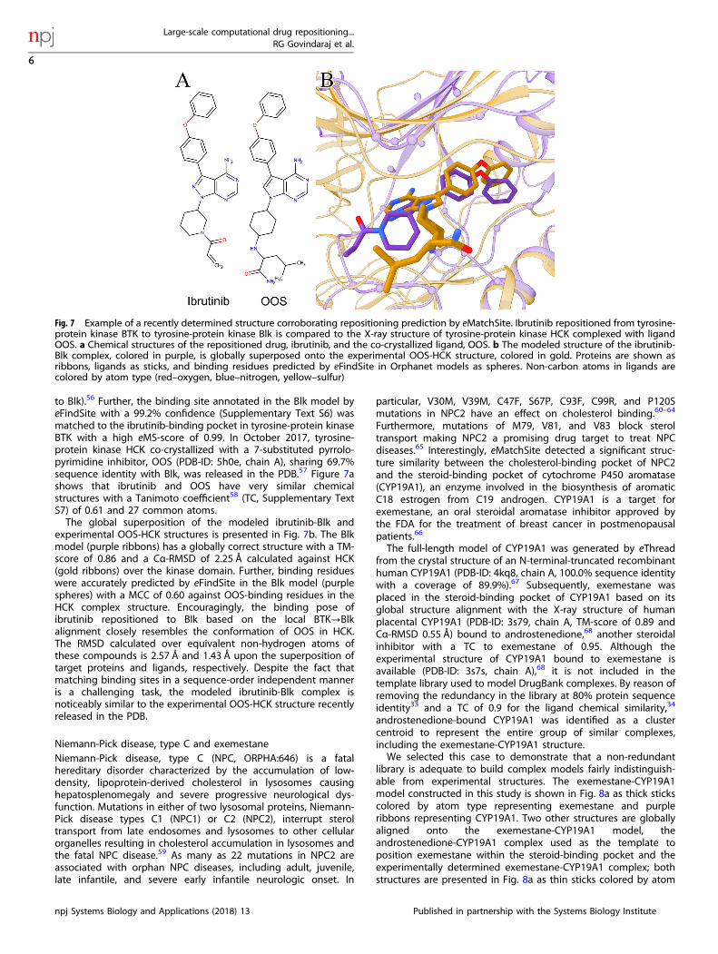
to Blk).56 Further, the binding site annotated in the Blk model byeFindSite with a 99.2% confidence (Supplementary Text S6) wasmatched to the ibrutinib-binding pocket in tyrosine-protein kinaseBTK with a high eMS-score of 0.99. In October 2017, tyrosine-protein kinase HCK co-crystallized with a 7-substituted pyrrolo-pyrimidine inhibitor, OOS (PDB-ID: 5h0e, chain A), sharing 69.7%sequence identity with Blk, was released in the PDB.57 Figure 7ashows that ibrutinib and OOS have very similar chemicalstructures with a Tanimoto coefficient58 (TC, Supplementary TextS7) of 0.61 and 27 common atoms.The global superposition of the modeled ibrutinib-Blk and
experimental OOS-HCK structures is presented in Fig. 7b. The Blkmodel (purple ribbons) has a globally correct structure with a TM-score of 0.86 and a Cα-RMSD of 2.25 Å calculated against HCK(gold ribbons) over the kinase domain. Further, binding residueswere accurately predicted by eFindSite in the Blk model (purplespheres) with a MCC of 0.60 against OOS-binding residues in theHCK complex structure. Encouragingly, the binding pose ofibrutinib repositioned to Blk based on the local BTK→Blkalignment closely resembles the conformation of OOS in HCK.The RMSD calculated over equivalent non-hydrogen atoms ofthese compounds is 2.57 Å and 1.43 Å upon the superposition oftarget proteins and ligands, respectively. Despite the fact thatmatching binding sites in a sequence-order independent manneris a challenging task, the modeled ibrutinib-Blk complex isnoticeably similar to the experimental OOS-HCK structure recentlyreleased in the PDB.
Niemann-Pick disease, type C and exemestaneNiemann-Pick disease, type C (NPC, ORPHA:646) is a fatalhereditary disorder characterized by the accumulation of low-density, lipoprotein-derived cholesterol in lysosomes causinghepatosplenomegaly and severe progressive neurological dys-function. Mutations in either of two lysosomal proteins, Niemann-Pick disease types C1 (NPC1) or C2 (NPC2), interrupt steroltransport from late endosomes and lysosomes to other cellularorganelles resulting in cholesterol accumulation in lysosomes andthe fatal NPC disease.59 As many as 22 mutations in NPC2 areassociated with orphan NPC diseases, including adult, juvenile,late infantile, and severe early infantile neurologic onset. In
particular, V30M, V39M, C47F, S67P, C93F, C99R, and P120Smutations in NPC2 have an effect on cholesterol binding.60–64
Furthermore, mutations of M79, V81, and V83 block steroltransport making NPC2 a promising drug target to treat NPCdiseases.65 Interestingly, eMatchSite detected a significant struc-ture similarity between the cholesterol-binding pocket of NPC2and the steroid-binding pocket of cytochrome P450 aromatase(CYP19A1), an enzyme involved in the biosynthesis of aromaticC18 estrogen from C19 androgen. CYP19A1 is a target forexemestane, an oral steroidal aromatase inhibitor approved bythe FDA for the treatment of breast cancer in postmenopausalpatients.66
The full-length model of CYP19A1 was generated by eThreadfrom the crystal structure of an N-terminal-truncated recombinanthuman CYP19A1 (PDB-ID: 4kq8, chain A, 100.0% sequence identitywith a coverage of 89.9%).67 Subsequently, exemestane wasplaced in the steroid-binding pocket of CYP19A1 based on itsglobal structure alignment with the X-ray structure of humanplacental CYP19A1 (PDB-ID: 3s79, chain A, TM-score of 0.89 andCα-RMSD 0.55 Å) bound to androstenedione,68 another steroidalinhibitor with a TC to exemestane of 0.95. Although theexperimental structure of CYP19A1 bound to exemestane isavailable (PDB-ID: 3s7s, chain A),68 it is not included in thetemplate library used to model DrugBank complexes. By reason ofremoving the redundancy in the library at 80% protein sequenceidentity33 and a TC of 0.9 for the ligand chemical similarity,34
androstenedione-bound CYP19A1 was identified as a clustercentroid to represent the entire group of similar complexes,including the exemestane-CYP19A1 structure.We selected this case to demonstrate that a non-redundant
library is adequate to build complex models fairly indistinguish-able from experimental structures. The exemestane-CYP19A1model constructed in this study is shown in Fig. 8a as thick stickscolored by atom type representing exemestane and purpleribbons representing CYP19A1. Two other structures are globallyaligned onto the exemestane-CYP19A1 model, theandrostenedione-CYP19A1 complex used as the template toposition exemestane within the steroid-binding pocket and theexperimentally determined exemestane-CYP19A1 complex; bothstructures are presented in Fig. 8a as thin sticks colored by atom
Fig. 7 Example of a recently determined structure corroborating repositioning prediction by eMatchSite. Ibrutinib repositioned from tyrosine-protein kinase BTK to tyrosine-protein kinase Blk is compared to the X-ray structure of tyrosine-protein kinase HCK complexed with ligandOOS. a Chemical structures of the repositioned drug, ibrutinib, and the co-crystallized ligand, OOS. b The modeled structure of the ibrutinib-Blk complex, colored in purple, is globally superposed onto the experimental OOS-HCK structure, colored in gold. Proteins are shown asribbons, ligands as sticks, and binding residues predicted by eFindSite in Orphanet models as spheres. Non-carbon atoms in ligands arecolored by atom type (red–oxygen, blue–nitrogen, yellow–sulfur)
Large-scale computational drug repositioning...RG Govindaraj et al.
6
npj Systems Biology and Applications (2018) 13 Published in partnership with the Systems Biology Institute
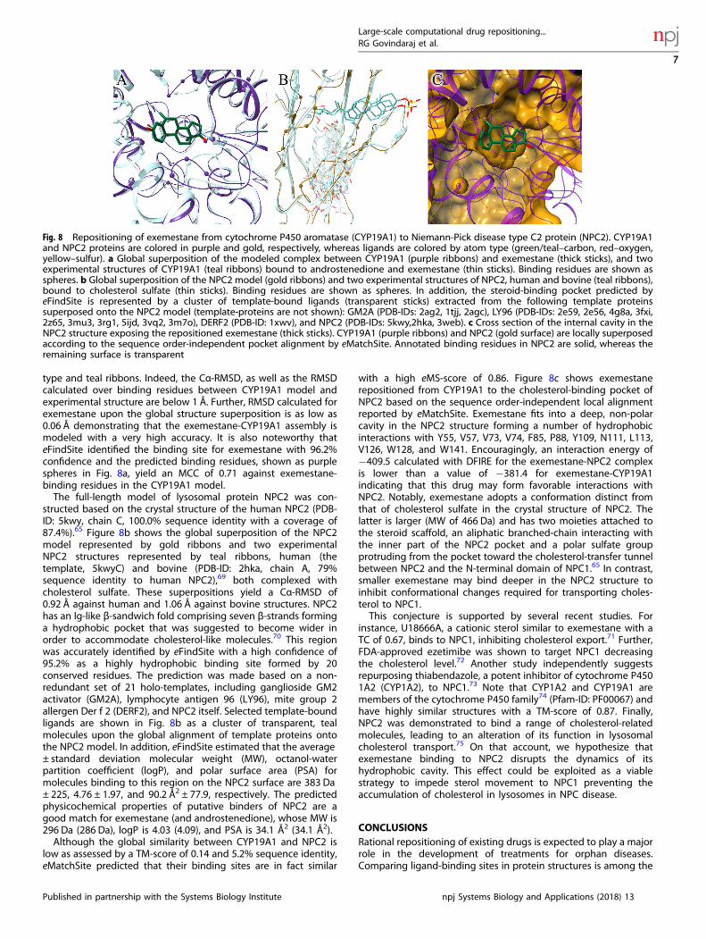
type and teal ribbons. Indeed, the Cα-RMSD, as well as the RMSDcalculated over binding residues between CYP19A1 model andexperimental structure are below 1 Å. Further, RMSD calculated forexemestane upon the global structure superposition is as low as0.06 Å demonstrating that the exemestane-CYP19A1 assembly ismodeled with a very high accuracy. It is also noteworthy thateFindSite identified the binding site for exemestane with 96.2%confidence and the predicted binding residues, shown as purplespheres in Fig. 8a, yield an MCC of 0.71 against exemestane-binding residues in the CYP19A1 model.The full-length model of lysosomal protein NPC2 was con-
structed based on the crystal structure of the human NPC2 (PDB-ID: 5kwy, chain C, 100.0% sequence identity with a coverage of87.4%).65 Figure 8b shows the global superposition of the NPC2model represented by gold ribbons and two experimentalNPC2 structures represented by teal ribbons, human (thetemplate, 5kwyC) and bovine (PDB-ID: 2hka, chain A, 79%sequence identity to human NPC2),69 both complexed withcholesterol sulfate. These superpositions yield a Cα-RMSD of0.92 Å against human and 1.06 Å against bovine structures. NPC2has an Ig-like β-sandwich fold comprising seven β-strands forminga hydrophobic pocket that was suggested to become wider inorder to accommodate cholesterol-like molecules.70 This regionwas accurately identified by eFindSite with a high confidence of95.2% as a highly hydrophobic binding site formed by 20conserved residues. The prediction was made based on a non-redundant set of 21 holo-templates, including ganglioside GM2activator (GM2A), lymphocyte antigen 96 (LY96), mite group 2allergen Der f 2 (DERF2), and NPC2 itself. Selected template-boundligands are shown in Fig. 8b as a cluster of transparent, tealmolecules upon the global alignment of template proteins ontothe NPC2 model. In addition, eFindSite estimated that the average± standard deviation molecular weight (MW), octanol-waterpartition coefficient (logP), and polar surface area (PSA) formolecules binding to this region on the NPC2 surface are 383 Da± 225, 4.76 ± 1.97, and 90.2 Å2 ± 77.9, respectively. The predictedphysicochemical properties of putative binders of NPC2 are agood match for exemestane (and androstenedione), whose MW is296 Da (286 Da), logP is 4.03 (4.09), and PSA is 34.1 Å2 (34.1 Å2).Although the global similarity between CYP19A1 and NPC2 is
low as assessed by a TM-score of 0.14 and 5.2% sequence identity,eMatchSite predicted that their binding sites are in fact similar
with a high eMS-score of 0.86. Figure 8c shows exemestanerepositioned from CYP19A1 to the cholesterol-binding pocket ofNPC2 based on the sequence order-independent local alignmentreported by eMatchSite. Exemestane fits into a deep, non-polarcavity in the NPC2 structure forming a number of hydrophobicinteractions with Y55, V57, V73, V74, F85, P88, Y109, N111, L113,V126, W128, and W141. Encouragingly, an interaction energy of−409.5 calculated with DFIRE for the exemestane-NPC2 complexis lower than a value of −381.4 for exemestane-CYP19A1indicating that this drug may form favorable interactions withNPC2. Notably, exemestane adopts a conformation distinct fromthat of cholesterol sulfate in the crystal structure of NPC2. Thelatter is larger (MW of 466 Da) and has two moieties attached tothe steroid scaffold, an aliphatic branched-chain interacting withthe inner part of the NPC2 pocket and a polar sulfate groupprotruding from the pocket toward the cholesterol-transfer tunnelbetween NPC2 and the N-terminal domain of NPC1.65 In contrast,smaller exemestane may bind deeper in the NPC2 structure toinhibit conformational changes required for transporting choles-terol to NPC1.This conjecture is supported by several recent studies. For
instance, U18666A, a cationic sterol similar to exemestane with aTC of 0.67, binds to NPC1, inhibiting cholesterol export.71 Further,FDA-approved ezetimibe was shown to target NPC1 decreasingthe cholesterol level.72 Another study independently suggestsrepurposing thiabendazole, a potent inhibitor of cytochrome P4501A2 (CYP1A2), to NPC1.73 Note that CYP1A2 and CYP19A1 aremembers of the cytochrome P450 family74 (Pfam-ID: PF00067) andhave highly similar structures with a TM-score of 0.87. Finally,NPC2 was demonstrated to bind a range of cholesterol-relatedmolecules, leading to an alteration of its function in lysosomalcholesterol transport.75 On that account, we hypothesize thatexemestane binding to NPC2 disrupts the dynamics of itshydrophobic cavity. This effect could be exploited as a viablestrategy to impede sterol movement to NPC1 preventing theaccumulation of cholesterol in lysosomes in NPC disease.
CONCLUSIONSRational repositioning of existing drugs is expected to play a majorrole in the development of treatments for orphan diseases.Comparing ligand-binding sites in protein structures is among the
Fig. 8 Repositioning of exemestane from cytochrome P450 aromatase (CYP19A1) to Niemann-Pick disease type C2 protein (NPC2). CYP19A1and NPC2 proteins are colored in purple and gold, respectively, whereas ligands are colored by atom type (green/teal–carbon, red–oxygen,yellow–sulfur). a Global superposition of the modeled complex between CYP19A1 (purple ribbons) and exemestane (thick sticks), and twoexperimental structures of CYP19A1 (teal ribbons) bound to androstenedione and exemestane (thin sticks). Binding residues are shown asspheres. b Global superposition of the NPC2 model (gold ribbons) and two experimental structures of NPC2, human and bovine (teal ribbons),bound to cholesterol sulfate (thin sticks). Binding residues are shown as spheres. In addition, the steroid-binding pocket predicted byeFindSite is represented by a cluster of template-bound ligands (transparent sticks) extracted from the following template proteinssuperposed onto the NPC2 model (template-proteins are not shown): GM2A (PDB-IDs: 2ag2, 1tjj, 2agc), LY96 (PDB-IDs: 2e59, 2e56, 4g8a, 3fxi,2z65, 3mu3, 3rg1, 5ijd, 3vq2, 3m7o), DERF2 (PDB-ID: 1xwv), and NPC2 (PDB-IDs: 5kwy,2hka, 3web). c Cross section of the internal cavity in theNPC2 structure exposing the repositioned exemestane (thick sticks). CYP19A1 (purple ribbons) and NPC2 (gold surface) are locally superposedaccording to the sequence order-independent pocket alignment by eMatchSite. Annotated binding residues in NPC2 are solid, whereas theremaining surface is transparent
Large-scale computational drug repositioning...RG Govindaraj et al.
7
Published in partnership with the Systems Biology Institute npj Systems Biology and Applications (2018) 13
most promising computational techniques to inform drugrepurposing efforts. In this study, we demonstrate that combiningeMatchSite with structure-based virtual screening enhances theaccuracy of the detection of similar binding pockets. Thispromising methodology was employed to match drug-bindingpockets from DrugBank with those from Orphanet exposing anumber of opportunities to combat orphan diseases with existingdrugs.
MATERIALS AND METHODSDrugBank and Orphanet datasetsThe DrugBank dataset includes proteins binding FDA-approved drugs witha molecular weight of 150–550 Da selected from DrugBank,32 whereas theOrphanet dataset contains proteins associated with rare disordersobtained from Orphanet (http://www.orpha.net). Target structures com-posed of 50–999 amino acids in both datasets were modeled with eThread,a template-based structure prediction algorithm.33 In the next step, drug-binding pockets were predicted by eFindSite34 in confidently modeledtarget DrugBank and Orphanet proteins whose estimated GDT-score is≥0.4. Drug repositioning utilizes only those binding sites assigned a highand moderate confidence. Further, we devised a two-step alignmentprotocol to position drug compounds within the predicted bindingpockets in the DrugBank proteins. First, holo-templates selected byeFindSite were structurally aligned onto the target protein with Fr-TM-align35 and then the drug molecule was superposed onto the most similartemplate-bound ligand according to the chemical alignment constructedby KCOMBU.36 The Orphanet dataset comprises 922 proteins, whereas theDrugBank dataset contains 2012 drug-protein complexes formed by 715drugs and 348 proteins.
Matching DrugBank and Orphanet pocketsAll-against-all matching of drug-binding pockets in DrugBank andOrphanet proteins was conducted with eMatchSite.15,16 This algorithmconstructs sequence order-independent alignments of pocket residues bysolving the assignment problem with machine learning and the Kuhn-Munkres algorithm.76,77 Local alignments are then assigned a similarityscore, called the eMS-score, which measures the overlap of variousphysicochemical features and evolutionary profiles. For significant matchesidentified with eMatchSite, drugs bound to the DrugBank target weretransferred to a binding site in the Orphanet protein upon thesuperposition of the two pockets according to the local alignment.Subsequently, the constructed complexes of drugs repositioned toOrphanet proteins were rebuilt with Modeller37 in order to refine drug-target interactions eliminating steric clashes. The quality of final complexmodels is assessed by a knowledge-based statistical energy function forprotein-ligand complexes with DFIRE38 and VS with Vina.17
Huang datasetThe Huang dataset was originally compiled to evaluate the performance ofgeometry-based methods to predict binding pockets21 and then it wasadopted to assess the accuracy of pocket comparison algorithms.78 Fromthis dataset, we selected 107 proteins for which eFindSite correctlyannotated binding sites within a distance of 8 Å from the geometric centerof the bound ligand in the experimental complex structure. These targetproteins bind the following ligands, adenosine, biotin, fructose-6-phosphate, α-L-fucose, β-D-galactose, guanine, α-D-mannose, O1-methyl-mannose, 4-phenyl-1H-imidazole, palmitic acid, retinol, and 2’-deoxyur-idine 5’-monophosphate. The comprehensive information on the Huangdataset is given in Supplementary Table S1.
Virtual screeningA target binding site is subjected to VS with AutoDock Vina17 against anon-redundant library of 1515 FDA-approved drugs compiled previously.20
MGL tools79 and Open Babel80 were used to add polar hydrogens andpartial charges, as well as to convert target proteins and librarycompounds to the PDBQT format. For each docking ligand, the optimalsearch space centered on the binding site annotated with eFindSite wascalculated from its radius of gyration.81 Molecular docking was carried outwith AutoDock Vina 1.1.2 and the default set of parameters.
Data availabilityData generated for the repositioning of DrugBank drugs to Orphanetproteins are available from the Open Science Framework at https://osf.io/qdjup/. The source codes of programs used in this study are available fromGitHub, eThread: https://github.com/michal-brylinski/ethread, eFindSite:https://github.com/michal-brylinski/efindsite, and eMatchSite: https://github.com/michal-brylinski/ematchsite.
ACKNOWLEDGEMENTSThis work was supported by the National Institute of General Medical Sciences of theNational Institutes of Health [R35GM119524].
ADDITIONAL INFORMATIONSupplementary Information accompanies the paper on the npj Systems Biology andApplications website (https://doi.org/10.1038/s41540-018-0050-7).
Competing interests: The authors declare no competing financial interests.
Publisher's note: Springer Nature remains neutral with regard to jurisdictional claimsin published maps and institutional affiliations.
REFERENCES1. Ashburn, T. T. & Thor, K. B. Drug repositioning: identifying and developing new
uses for existing drugs. Nat. Rev. Drug. Discov. 3, 673–683 (2004).2. Provost, G. “Homeless” or “orphan” drugs. Am. J. Hosp. Pharm. 25, 609 (1968).3. Kwok, A. K. & Koenigbauer, F. M. Incentives to repurpose existing drugs for
orphan indications. ACS Med Chem. Lett. 6, 828–830 (2015).4. Boolell, M. et al. Sildenafil: an orally active type 5 cyclic GMP-specific phospho-
diesterase inhibitor for the treatment of penile erectile dysfunction. Int. J. Impot.Res. 8, 47–52 (1996).
5. Callmander, E. & Hellgren, L. Amantadine hydrochloride as a prophylactic inrespiratory infections. A double-blind investigation of its clinical use and serol-ogy. J. Clin. Pharmacol. J. New. Drugs 8, 186–189 (1968).
6. Schwab, R. S., Poskanzer, D. C., England, A. C. Jr & Young, R. R. Amantadine inParkinson’s disease. Review of more than two years’ experience. JAMA 222,792–795 (1972).
7. Bormann, J. Memantine is a potent blocker of N-methyl-D-aspartate (NMDA)receptor channels. Eur. J. Pharmacol. 166, 591–592 (1989).
8. Olivares, D. et al. N-methyl D-aspartate (NMDA) receptor antagonists and mem-antine treatment for Alzheimer’s disease, vascular dementia and Parkinson’sdisease. Curr. Alzheimer Res. 9, 746–758 (2012).
9. Li, J. et al. A survey of current trends in computational drug repositioning. Brief.Bioinform. 17, 2–12 (2016).
10. Ehrt, C., Brinkjost, T. & Koch, O. Impact of binding site comparisons on medicinalchemistry and rational molecular design. J. Med. Chem. 59, 4121–4151 (2016).
11. Xie, L. & Bourne, P. E. Detecting evolutionary relationships across existing foldspace, using sequence order-independent profile-profile alignments. Proc. NatlAcad. Sci. USA 105, 5441–5446 (2008).
12. Schalon, C., Surgand, J. S., Kellenberger, E. & Rognan, D. A simple and fuzzymethod to align and compare druggable ligand-binding sites. Proteins 71,1755–1778 (2008).
13. Kinnings, S. L. et al. Drug discovery using chemical systems biology: repositioningthe safe medicine Comtan to treat multi-drug and extensively drug resistanttuberculosis. PLoS Comput. Biol. 5, e1000423 (2009).
14. Defranchi, E. et al. Binding of protein kinase inhibitors to synapsin I inferred frompair-wise binding site similarity measurements. PLoS One 5, e12214 (2010).
15. Brylinski, M. eMatchSite: sequence order-independent structure alignments ofligand binding pockets in protein models. PLoS Comput. Biol. 10, e1003829(2014).
16. Brylinski, M. Local alignment of ligand binding sites in proteins for poly-pharmacology and drug repositioning. Methods Mol. Biol. 1611, 109–122 (2017).
17. Trott, O. & Olson, A. J. AutoDock Vina: improving the speed and accuracy ofdocking with a new scoring function, efficient optimization, and multithreading.J. Comput. Chem. 31, 455–461 (2010).
18. Brylinski, M., Naderi, M., Govindaraj, R. G. & Lemoine, J. eRepo-ORP: Exploring theopportunity space to combat orphan diseases with existing drugs. J. Mol. Biol.https://doi.org/10.1016/j.jmb.2017.12.001 (2018).
19. Berman, H. M. et al. The Protein Data Bank. Acta Crystallogr. D. Biol. Crystallogr. 58,899–907 (2002).
Large-scale computational drug repositioning...RG Govindaraj et al.
8
npj Systems Biology and Applications (2018) 13 Published in partnership with the Systems Biology Institute
20. Govindaraj, R. G. & Brylinski, M. Comparative assessment of strategies to identifysimilar ligand-binding pockets in proteins. bioRxiv https://doi.org/10.1101/268565 (2018).
21. Huang, B. & Schroeder, M. LIGSITEcsc: predicting ligand binding sites using theConnolly surface and degree of conservation. BMC Struct. Biol. 6, 19 (2006).
22. Truchon, J. F. & Bayly, C. I. Evaluating virtual screening methods: good and badmetrics for the “early recognition” problem. J. Chem. Inf. Model. 47, 488–508(2007).
23. Karaman, M. W. et al. A quantitative analysis of kinase inhibitor selectivity. Nat.Biotechnol. 26, 127–132 (2008).
24. Cokorinos, E. C. et al. Activation of skeletal muscle AMPK promotes glucosedisposal and glucose lowering in non-human primates and mice. Cell. Metab. 25,1147–1159 (2017). e1110.
25. Zhang, Y. & Skolnick, J. Scoring function for automated assessment of proteinstructure template quality. Proteins 57, 702–710 (2004).
26. Kabsch, W. A solution for the best rotation to relate two sets of vectors. ActaCrystallogr. A. 32, 922–923 (1976).
27. Jacobs, M. D. et al. Pim-1 ligand-bound structures reveal the mechanism ofserine/threonine kinase inhibition by LY294002. J. Biol. Chem. 280, 13728–13734(2005).
28. Matthews, B. W. Comparison of the predicted and observed secondary structureof T4 phage lysozyme. Biochim. Biophys. Acta 405, 442–451 (1975).
29. Ouchi, T. et al. Lysine and arginine biosyntheses mediated by a common carrierprotein in Sulfolobus. Nat. Chem. Biol. 9, 277–283 (2013).
30. Esser, L. et al. Synapsin I is structurally similar to ATP-utilizing enzymes. EMBO. J.17, 977–984 (1998).
31. Walker, E. H. et al. Structural determinants of phosphoinositide 3-kinase inhibitionby wortmannin, LY294002, quercetin, myricetin, and staurosporine. Mol. Cell. 6,909–919 (2000).
32. Wishart, D. S. et al. DrugBank: a comprehensive resource for in silico drug dis-covery and exploration. Nucleic Acids Res. 34, D668–672 (2006).
33. Brylinski, M. & Lingam, D. eThread: a highly optimized machine learning-basedapproach to meta-threading and the modeling of protein tertiary structures. PLoSONE 7, e50200 (2012).
34. Brylinski, M. & Feinstein, W. P. eFindSite: improved prediction of ligand bindingsites in protein models using meta-threading, machine learning and auxiliaryligands. J. Comput. Aided Mol. Des. 27, 551–567 (2013).
35. Pandit, S. B. & Skolnick, J. Fr-TM-align: a new protein structural alignment methodbased on fragment alignments and the TM-score. BMC Bioinform. 9, 531 (2008).
36. Kawabata, T. Build-up algorithm for atomic correspondence between chemicalstructures. J. Chem. Inf. Model. 51, 1775–1787 (2011).
37. Webb, B. & Sali, A. Protein structure modeling with MODELLER. Methods Mol. Biol.1137, 1–15 (2014).
38. Zhang, C., Liu, S., Zhu, Q. & Zhou, Y. A knowledge-based energy function forprotein-ligand, protein-protein, and protein-DNA complexes. J. Med. Chem. 48,2325–2335 (2005).
39. Pollak, M. R. et al. Autosomal dominant hypocalcaemia caused by a Ca(2+)-sensing receptor gene mutation. Nat. Genet. 8, 303–307 (1994).
40. Kinoshita, Y., Hori, M., Taguchi, M., Watanabe, S. & Fukumoto, S. Functionalactivities of mutant calcium-sensing receptors determine clinical presentations inpatients with autosomal dominant hypocalcemia. J. Clin. Endocrinol. Metab. 99,E363–368 (2014).
41. Nesbit, M. A. et al. Mutations affecting G-protein subunit alpha11 in hypercal-cemia and hypocalcemia. N. Engl. J. Med. 368, 2476–2486 (2013).
42. Roszko, K. L., Bi, R. D. & Mannstadt, M. Autosomal dominant hypocalcemia(hypoparathyroidism) types 1 and 2. Front Physiol. 7, 458 (2016).
43. Guay, D. R. Tolcapone, a selective catechol-O-methyltransferase inhibitor fortreatment of Parkinson’s disease. Pharmacotherapy 19, 6–20 (1999).
44. Najib, J. Entacapone: a catechol-O-methyltransferase inhibitor for the adjunctivetreatment of Parkinson’s disease. Clin. Ther. 23, 802–832 (2001). discussion 771.
45. Huang, W. S. et al. Discovery of 3-[2-(imidazo[1,2-b]pyridazin-3-yl)ethynyl]-4-methyl-N-{4-[(4-methylpiperazin-1-y l)methyl]-3-(trifluoromethyl)phenyl}benza-mide (AP24534), a potent, orally active pan-inhibitor of breakpoint cluster region-abelson (BCR-ABL) kinase including the T315I gatekeeper mutant. J. Med. Chem.53, 4701–4719 (2010).
46. Ben-Salem, S., Begum, M. A., Ali, B. R. & Al-Gazali, L. A novel aberrant splice sitemutation in RAB23 leads to an eight nucleotide deletion in the mRNA and isresponsible for Carpenter syndrome in a consanguineous emirati family. Mol.Syndromol. 3, 255–261 (2013).
47. Haye, D. et al. Prenatal findings in carpenter syndrome and a novel mutation inRAB23. Am. J. Med. Genet. A. 164A, 2926–2930 (2014).
48. Robinson, L. K., James, H. E., Mubarak, S. J., Allen, E. J. & Jones, K. L. Carpentersyndrome: natural history and clinical spectrum. Am. J. Med. Genet. 20, 461–469(1985).
49. Gayko, U. et al. Development of the Bruton’s tyrosine kinase inhibitor ibrutinib forB cell malignancies. Ann. N. Y. Acad. Sci. 1358, 82–94 (2015).
50. Reynolds, C. & Garg, A. K. Who is a diabetic? Can. Fam. Physician 24, 687–690(1978).
51. Borowiec, M. et al. Mutations at the BLK locus linked to maturity onset diabetes ofthe young and beta-cell dysfunction. Proc. Natl Acad. Sci. USA 106, 14460–14465(2009).
52. Bonnefond, A. et al. Reassessment of the putative role of BLK-p.A71T loss-of-function mutation in MODY and type 2 diabetes. Diabetologia 56, 492–496(2013).
53. Imam, M. H., Shenoy, P. J., Flowers, C. R., Phillips, A. & Lechowicz, M. J. Incidenceand survival patterns of cutaneous T-cell lymphomas in the United States. Leuk.Lymphoma 54, 752–759 (2013).
54. Petersen, D. L. et al. B-lymphoid tyrosine kinase (Blk) is an oncogene and apotential target for therapy with dasatinib in cutaneous T-cell lymphoma (CTCL).Leukemia 28, 2109–2112 (2014).
55. Zemla, A., Venclovas, C., Moult, J. & Fidelis, K. Processing and analysis of CASP3protein structure predictions. Proteins Suppl 3, 22–29 (1999).
56. Cowan-Jacob, S. W. et al. The crystal structure of a c-Src complex in an activeconformation suggests possible steps in c-Src activation. Structure 13, 861–871(2005).
57. Yuki, H. et al. Activity cliff for 7-substituted pyrrolo-pyrimidine inhibitors of HCKexplained in terms of predicted basicity of the amine nitrogen. Bioorg. Med.Chem. 25, 4259–4264 (2017).
58. Tanimoto, T. T. An elementary mathematical theory of classification and predic-tion. (IBM Internal Report, 1958).
59. Pentchev, P. G. Niemann-Pick C research from mouse to gene. Biochim. Biophys.Acta 1685, 3–7 (2004).
60. Chikh, K., Rodriguez, C., Vey, S., Vanier, M. T. & Millat, G. Niemann-Pick type Cdisease: subcellular location and functional characterization of NPC2 proteinswith naturally occurring missense mutations. Hum. Mutat. 26, 20–28 (2005).
61. Klunemann, H. H. et al. Frontal lobe atrophy due to a mutation in the cholesterolbinding protein HE1/NPC2. Ann. Neurol. 52, 743–749 (2002).
62. Millat, G. et al. Niemann-Pick C disease: use of denaturing high performanceliquid chromatography for the detection of NPC1 and NPC2 genetic variationsand impact on management of patients and families. Mol. Genet. Metab. 86,220–232 (2005).
63. Millat, G. et al. Niemann-Pick disease type C: spectrum of HE1 mutations andgenotype/phenotype correlations in the NPC2 group. Am. J. Hum. Genet. 69,1013–1021 (2001).
64. Park, W. D. et al. Identification of 58 novel mutations in Niemann-Pick diseasetype C: correlation with biochemical phenotype and importance of PTC1-likedomains in NPC1. Hum. Mutat. 22, 313–325 (2003).
65. Li, X., Saha, P., Li, J., Blobel, G. & Pfeffer, S. R. Clues to the mechanism of cho-lesterol transfer from the structure of NPC1 middle lumenal domain bound toNPC2. Proc. Natl Acad. Sci. USA 113, 10079–10084 (2016).
66. Buzdar, A. U., Robertson, J. F., Eiermann, W. & Nabholtz, J. M. An overview of thepharmacology and pharmacokinetics of the newer generation aromatase inhi-bitors anastrozole, letrozole, and exemestane. Cancer 95, 2006–2016 (2002).
67. Lo, J. et al. Structural basis for the functional roles of critical residues in humancytochrome p450 aromatase. Biochemistry 52, 5821–5829 (2013).
68. Ghosh, D. et al. Novel aromatase inhibitors by structure-guided design. J. Med.Chem. 55, 8464–8476 (2012).
69. Xu, S., Benoff, B., Liou, H. L., Lobel, P. & Stock, A. M. Structural basis of sterolbinding by NPC2, a lysosomal protein deficient in Niemann-Pick type C2 disease.J. Biol. Chem. 282, 23525–23531 (2007).
70. Friedland, N., Liou, H. L., Lobel, P. & Stock, A. M. Structure of a cholesterol-bindingprotein deficient in Niemann-Pick type C2 disease. Proc. Natl Acad. Sci. USA 100,2512–2517 (2003).
71. Lu, F. et al. Identification of NPC1 as the target of U18666A, an inhibitor oflysosomal cholesterol export and Ebola infection. Elife 4, e12177 (2015).
72. Phan, B. A., Dayspring, T. D. & Toth, P. P. Ezetimibe therapy: mechanism of actionand clinical update. Vasc. Health Risk. Manag. 8, 415–427 (2012).
73. Soufan, O. et al. DRABAL: novel method to mine large high-throughput screeningassays using Bayesian active learning. J. Cheminform. 8, 64 (2016).
74. Bateman, A. et al. The Pfam protein families database. Nucleic Acids Res. 28,263–266 (2000).
75. Liou, H. L. et al. NPC2, the protein deficient in Niemann-Pick C2 disease, consistsof multiple glycoforms that bind a variety of sterols. J. Biol. Chem. 281,36710–36723 (2006).
76. Kuhn, H. W. The Hungarian method for the assignment problem. Nav. Res. Logist.Q. 2, 83–97 (1955).
77. Munkres, J. Algorithms for the assignment and transportation problems. J. Soc.Ind. Appl. Math. 5, 32–38 (1957).
Large-scale computational drug repositioning...RG Govindaraj et al.
9
Published in partnership with the Systems Biology Institute npj Systems Biology and Applications (2018) 13
78. Chikhi, R., Sael, L. & Kihara, D. Real-time ligand binding pocket database searchusing local surface descriptors. Proteins 78, 2007–2028 (2010).
79. Morris, G. M. et al. AutoDock4 and AutoDockTools4: automated docking withselective receptor flexibility. J. Comput. Chem. 30, 2785–2791 (2009).
80. O’Boyle, N. M. et al. Open Babel: an open chemical toolbox. J. Cheminform. 3, 33(2011).
81. Feinstein, W. P. & Brylinski, M. Calculating an optimal box size for ligand dockingand virtual screening against experimental and predicted binding pockets. J.Cheminform. 7, 18 (2015).
Open Access This article is licensed under a Creative CommonsAttribution 4.0 International License, which permits use, sharing,
adaptation, distribution and reproduction in anymedium or format, as long as you giveappropriate credit to the original author(s) and the source, provide a link to the CreativeCommons license, and indicate if changes were made. The images or other third partymaterial in this article are included in the article’s Creative Commons license, unlessindicated otherwise in a credit line to the material. If material is not included in thearticle’s Creative Commons license and your intended use is not permitted by statutoryregulation or exceeds the permitted use, you will need to obtain permission directlyfrom the copyright holder. To view a copy of this license, visit http://creativecommons.org/licenses/by/4.0/.
© The Author(s) 2018
Large-scale computational drug repositioning...RG Govindaraj et al.
10
npj Systems Biology and Applications (2018) 13 Published in partnership with the Systems Biology Institute
Recommended