MALE REPRODUCTIVE SYSTEM

Khaleel Alyahya, PhD 3
Male Reproductive System
1. Primary reproductive organs: testes
2. Accessory reproductive structures (ducts or glands)
duct system: epididymis, ductus deferens and urethra.
accessory glands and semen: seminal vesicles, prostate and bulbourethral glands.
external genitalia: scrotum and penis.

4Khaleel Alyahya, PhD
4 cm long , 2.5 cm wide.
Covered with fibrous connective tissue capsule, tunica albuginea.
The septa, extensions of tunica albuginea, separate the testis in lobules.
Each lobule contains 1 -4 coiled seminiferous tubules.
The seminiferous tubules make the sperms.
The seminiferous tubules are drained into a network called rete testis.
From rete testis the sperms go to the epididymis
Testes

Khaleel Alyahya, PhD 5
Hormonal Control
The pituitary controls testis function by
producing follicle-stimulating hormone
(FSH) and luteinizing hormone (LH).
FSH stimulates spermatogenesis, in
part by affecting Sertoli cells.
LH stimulates androgen production by
interstitial cells.
Pituitary production of these hormones
depends on secretion of gonadotropin-
releasing hormone (GnRH) by the
hypothalamus.

Khaleel Alyahya, PhD 6
Epididymis
Comma-shaped, coiled tube, 6 m long
Place for storage and maturation of the sperms
Passage lasts 20 days
From the epididymis, the sperms go to the ductus deferens

Khaleel Alyahya, PhD 7
Ducts Deferens
Thick-walled muscular tube, which carries the sperms from the epididymis to the ejaculatory duct (a canal inside the prostate).
Enclosed inside a connective tissue sheath called the spermatic cord.
Runs from the scrotum, inside the inguinal canal, into the pelvis, over the superior surface and behind the urinary bladder
Khaleel Alyahya, PhD 8
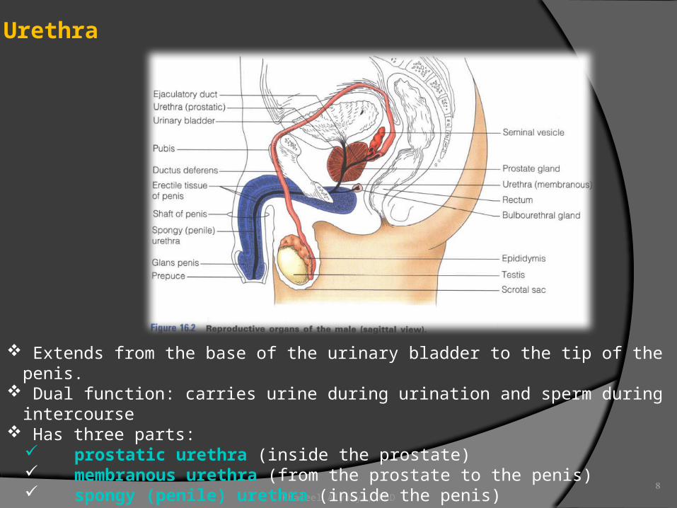
Urethra
Extends from the base of the urinary bladder to the tip of the penis. Dual function: carries urine during urination and sperm during intercourse Has three parts:
prostatic urethra (inside the prostate) membranous urethra (from the prostate to the penis) spongy (penile) urethra (inside the penis)

Khaleel Alyahya, PhD 9
Seminal Vesicles
Located at the base of the bladder. Their ducts join the ductus deferns to make the ejaculatory duct. Produce 60% of the seminal fluid. Their secretion provides nourishment to the sperms and activates them.

Khaleel Alyahya, PhD 10
Prostate
Single gland the size of a chestnut.
Located at the base of the bladder.
Encircles the prostatic urethra.
Its secretion activates the sperm.

Khaleel Alyahya, PhD 11
Bulbourethral Gland
Tiny, pea-sized glands inferior to the prostate
Produce thick mucus
Their secretion cleanses the urethra and serves as lubricant during intercourse

Khaleel Alyahya, PhD 12
Scrotum
A divided sac of skin hanging outside the abdominal cavity.
Keeps the testes at optimal temperature for the spermatogenesis.

Khaleel Alyahya, PhD 13
Penis
Cylindrical organ designed to deliver the sperm inside the female reproductive tract.
Consists of shaft and glans covered with skin.
The foreskin is a fold of skin covering the glans.

FEMALE REPRODUCTIVE SYSTEM

Khaleel Alyahya, PhD 15
Female Reproductive Organs
1. Primary reproductive organs: ovaries.
2. Duct system: uterine tubes, uterus and vagina.
3. External genitalia (vulva): mons pubis, labia, clitoris, urethral and vaginal orifices and greater vestibular glands.

Khaleel Alyahya, PhD 16
Ovaries
Paired organs in the shape and size of almonds Contain follicles Each follicle contains one oocyte surrounded by layers of follicle cells Each mature (Graafian) follicle has an antrum (vesicular stage of maturation) During ovulation, the developing egg is ejected from the ovary After ovulation, the ruptured follicle becomes a corpus luteum Ovulation occurs every 28 days

Khaleel Alyahya, PhD 17
Ovulation

Khaleel Alyahya, PhD 18
Ovaries
The ovaries are secured to the lateral walls of the pelvis by the suspensory
ligaments
The ovarian ligaments attach the ovaries to the uterus.
The broad ligaments are folds of peritoneum covering the ovaries

Khaleel Alyahya, PhD 19
Uterine Tube
The uterine (fallopian) tubes receive the ovulated oocyte and provide a site for fertilization
Each tube is 10 cm long and extends from the ovary to the uterus They are enclosed in the broad ligament Their distal end expands in the funnel-shaped infundibulum which has finger-like
projections (fimbriae)

Khaleel Alyahya, PhD 20
Uterus
Size and shape of a pear.
Located in the pelvis between the urinary bladder and the rectum
Receives, retains and nourishes the fertilized egg, accommodates the growing fetus
Attached to the pelvis by the broad, round and uterosacral ligaments

Khaleel Alyahya, PhD 21
Uterus
It has body, fundus and cervix Its wall has three layers; the inner layer (endometrium) serves as place of
implantation of the fertilized egg The myometrium is the middle layer of the wall It is made from smooth muscle and contracts during delivery The outermost layer of the uterus (perimetrium) is serous and is made from the
visceral peritoneum

Khaleel Alyahya, PhD 22
Vagina
Thin-walled tube, 8-10 cm long Extends from the cervix of the uterus to the exterior Located between the bladder and the rectum Receives the penis and the semen during intercourse It is a birth canal and provides an outflow for the menses Its distal end isclosed by a thin fold of mucosa (hymen) which is ruptured during the
first intercourse

Khaleel Alyahya, PhD 23
External Genitalia (Vulva)
Mons pubis is a fatty, rounded area covering the symphysis, covered with pubic hair
Labia majora are two elongated hair-covered folds
Labia minora a two delicate hair-free folds The vestibule is an area including the
external opening of the urethra and the vagina
The greater vestibular glands are two mucus-producing glands on each side of the vagina; they provide lubrication during intercourse
The clitoris is a small protruding structure, similar to the penis; it is excited during intercourse
The perineum is a diamond-shaped region between the mons pubis and the anus.
Khaleel Alyahya, PhD 24
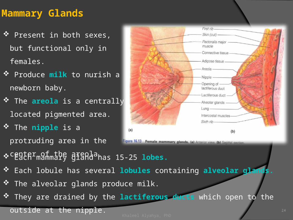
Mammary Glands
Present in both sexes, but
functional only in females.
Produce milk to nurish a newborn
baby.
The areola is a centrally located
pigmented area.
The nipple is a protruding area in
the center of the areola.
Each mammary gland has 15-25 lobes.
Each lobule has several lobules containing alveolar glands.
The alveolar glands produce milk.
They are drained by the lactiferous ducts which open to the outside at the nipple.

THAT’S ALL FOLKS!
Feedback?
Recommended