InternatIonal Journal of
Psychology & PsychologIcal
theraPy
edItorFrancisco Javier Molina CobosUniversidad de Almería, España
revIewIng edItors Mónica Hernández López Francisco Ruiz Jiménez Universidad de Jaén Fundación Universitaria Konrad Lorenz España Colombia
assocIate edItors Dermot Barnes-Holmes J. Francisco Morales Mauricio Papini Universiteit Gent UNED-Madrid Christian Texas University Belgium España USA Miguel Ángel Vallejo Pareja Kelly Wilson UNED-Madrid University of Mississipi España USA
assIstant edItorsAdolfo J. Cangas Díaz Universidad de Almería, EspañaEmilio Moreno San Pedro Universidad de Huelva, España
ManagIng edItorAdrián Barbero Rubio
Universidad de Almería & MICPSY, España
edItorIal offIce/secretaría de edIcIón MICPSY
Madrid, España
http://www.ijpsy.com
Volume 18, number 3 October 1, 2018Volumen 18, número 3 1 Octubre, 2018 ISSN: 1577-7057
Int
er
na
tIo
na
l J
ou
rn
al
of P
sy
ch
ol
og
y &
Ps
yc
ho
lo
gIc
al
th
er
aP
y20
18,
18,
3
Volume 18, number 3, 2018 http://www.ijpsy.com Volumen 18, número 3, 2018
Research Articles // Artículos de investigación Juan Carmelo Visdómine Lozano 257-271 Brain Activation for Effort in Human Learning: A Critical and Systematic Review of fMRI Studies.
Daniela M Salazar 273-287 Psychometric Properties of the Generalized Pliance Francisco J Ruiz Questionnaire -Children. Cindy L Flórez Juan C Suárez Falcón Ciara Dunne 289-300 Faking a Race IRAP Effect in the Context of Ciara McEnteggart Single versus Multiple Label Stimuli. Colin Harte Dermot Barnes-Holmes Yvonne Barnes-Holmes Hortensia Hickman Rodríguez 301-313 Tipos instruccionales y regulación verbal. M Luisa Cepeda Islas Comparación entre niños y adultos. [Types of Diana Moreno Rodríguez instructions and verbal regulation. Comparative Sergio M Méndez study between children and adults.] Rosalinda Arroyo Hernández Valeria E Morán 315-330 Emotional-Evolutional Model of Social Anxiety Fabián O Olaz in University Students. Edgardo R Pérez Zilda AP Del Prette Louis De Page 331-343 Differentiation between Defensive Personality Paul T van der Heijden Functioning and Psychopathology as Measured Mercedes De Weerdt by the DSQ-42 and MMPI-2-RF. Jos IM Egger Gina Rossi Julieta Azevedo 345-356 Early Emotional Memories and Borderline Symptoms: Paula Castilho The Mediating Role of Decentering. Lara Palmeira Angel Javier Tabullo 357-370 Associations between Fiction Reading, Trait Violeta Araceli Navas Jiménez Empathy and Theory of Mind Ability. Claudia Silvana García Lorraine T Benuto 371-384 The Evolving Definition of Cultural Competency: Jonathan Singer A Mixed Methods Study. Jena Casas Frances González Allison Ruork
Notes and Editorial Information // Avisos e información editorial
EditorialOffice 387-388 Normas de publicación-Instructions to authors. EditorialOffice 389 Cobertura e indexación de IJP&PT. [IJP&PT Abstracting and Indexing.]
ISSN 1577-7057 © 2018 Asociación de Análisis del Comportamiento, Madrid, España
IJP&Pt
International Journal of Psychology & Psychological Therapy is a four-monthly interdisciplinary publication open to publish original empirical articles, substantive reviews of one or more area(s), theoretical reviews, or reviews or methodological issues, and series of interest to some of the Psychology areas. The journal is published for the Asociación de Análisis del Comportamiento (AAC), indexed and/or abstracted in SCOPUS, Google Scholar Metrics, ISOC (CINDOC, CSIC), PSICODOC, Catálogo Latindex, IN-RECS (Index of Impact of the Social Sciences Spanish Journals), PsycINFO, Psychological Abstracts, ClinPSYC (American Psychological Association), ProQuest, PRISMA, EBSCO Publishing Inc., DIALNET, and RedALyC.
International Journal of Psychology & Psychological Therapy es una publicación interdisciplinar cuatrimestral, publicada por la Asociación de Análisis del Comportamiento (AAC), abierta a colaboraciones de carácter empírico y teórico, revisiones, artículos metodológicos y series temáticas de interés en cualquiera de los campos de la Psicología. Es publicada por la Asociación de Análisis del Comportamiento (AAC) y está incluida en las bases y plataformas bibliográficas: SCOPUS, Google Scholar Metrics, ISOC (CINDOC, CSIC), PSICODOC (Colegio Oficial de Psicólogos) Latindex, IN-RECS (Índice de Impacto de Revistas Españolas de Ciencias Sociales), PsycINFO (American Psychological Association) ClinPSYC, ProQuest, PRISMA, EBSCO Publishing Inc., DIALNET, y RedALyC (Red de Revistas Científicas de América Latina y El Caribe, España y Portugal).
Yolanda Alonso Universidad de Almería, EspañaErik Arntzen University of Oslo, NorwayMª José Báguena Puigcerver Universidad de Valencia, EspañaYvonne Barnes-Holmes National University-Maynooth, IrelandWilliam M. Baum University of New Hampshire, USAGualberto Buela Casal Universidad de Granada, EspañaFrancisco Cabello Luque Universidad de Murcia, EspañaJosé Carlos Caracuel Tubío Universidad de Sevilla, EspañaGonzalo de la Casa Universidad de Sevilla, EspañaCharles Catania University of Maryland Baltimore County, USAJuan Antonio Cruzado Universidad Complutense, EspañaVictoria Diez Chamizo Universidad de Barcelona, EspañaMichael Dougher University of New Mexico, USAMª Paula Fernández García Universidad de Oviedo, EspañaPerry N Fuchs University of Texas at Arlington, USAAndrés García García Universidad de Sevilla, EspañaJosé Jesús Gázquez Linares Universidad de Almería, EspañaInmaculada Gómez Becerra Universidad de Almería, EspañaLuis Gómez Jacinto Universidad de Malaga, EspañaM Victoria Gordillo Álvarez-Valdés Universidad Complutense, EspañaCelso Goyos Universidade de Sao Paulo, Brasil David E. Greenway University of Southwestern Louisiana, USAPatricia Sue Grigson Pennsylvania State College of Medicine, USASteven C. Hayes University of Nevada-Reno, USALinda Hayes University of Nevada-Reno, USAPhillip Hineline Temple University, USAPer Holth University of Oslo, NorwayRobert J. Kohlenberg Univeristy of Washington, Seattle, USAMaría Helena Leite Hunzinger Universidade de Sao Paulo, BrasilJulian C. Leslie University of Ulster at Jordanstown, UKJuan Carlos López García Universidad de Sevilla, EspañaFergus Lowe University of Wales, Bangor, UKArmando Machado Universidade do Miño, PortugalG. Alan Marlatt University of Washington, Seattle, USA
Jose Marques Universidade do Porto, PortugalHelena Matute Universidad de Deusto, EspañaRalph R. Miller State University of New York-Binghamton, USA Fernando Molero UNED, Madrid, EspañaRafael Moreno Universidad de Sevilla, EspañaIgnacio Morgado Bernal Universidad Autónoma Barcelona, EspañaEdward K. Morris University of Kansas-Lawrence, USALourdes Munduate Universidad de Sevilla, EspañaAlba Elisabeth Mustaca Universidad de Buenos Aires, ArgentinaJosé I. Navarro Guzmán Universidad de Cádiz, EspañaJordi Obiols Universidad Autónoma de Barcelona, EspañaSergio M. Pellis University of Lethbridge, CanadaRicardo Pellón UNED, Madrid, EspañaWenceslao Peñate Castro Universidad de La Laguna, EspañaVíctor Peralta Martín Hospital V. del Camino, Pamplona, EspañaM. Carmen Pérez Fuentes Universidad de Almería, EspañaMarino Pérez Álvarez Universidad de Oviedo, EspañaJuan Preciado City University of New York, USAEmilio Ribes Iniesta Universidad Veracruzana, MéxicoJosep Roca i Balasch INEF de Barcelona, EspañaArmando Rodríguez Universidad de La Laguna, EspañaJesús Rosales Ruiz University of North Texas, USAJuan Manuel Rosas Santos Universidad de Jaén, EspañaKurt Saltzinger Hofstra University, USAMark R. Serper Hofstra University, USAArthur W. Staats University of Hawaii, USACarmen Torres Universidad de Jaén, España Peter J. Urcuioli Purdue University, USAGuillermo Vallejo Seco Universidad de Oviedo, EspañaJulio Varela Barraza Universidad de Guadalajara, MéxicoJuan Pedro Vargas Romero Universidad de Sevilla, EspañaGraham F. Wagstaff University of LiverpoolStephen Worchel University of Hawaii, USAEdelgard Wulfert New York State University, Albany, USAThomas R. Zentall University of Kentucky, USA
Consejo Editorial/Editoral Board
InternatIonal Journal of Psychology & PsyhologIcal theraPy
IJP&Pt
Assistant EditorsAdolfo J. Cangas Díaz, Universidad de Almería, EspañaEmilio Moreno San Pedro, Universidad de Huelva, España
Reviewing EditorsMónica Hernández López, Universidad de Jaén, EspañaFrancisco Ruiz Jiménez, Fundación Universitaria Konrad Lorenz, Colombia
Editor: Francisco Javier Molina Cobos, Universidad de Almería, EspañaAssociate Editors
Dermot Barnes-Holmes, Universiteit Gent, Belgique-BelgiëFrancisco Morales, UNED, Madrid, España
Mauricio Papini, Christian Texas University, USAMiguel Ángel Vallejo Pareja, UNED, Madrid, España
Kelly Wilson, University of Mississipi, USA
Managing EditorAdrián Barbero Rubio Universidad de Almería & MICPSY, España
International Journal of Psychology and Psychological Therapy, 2018, 18, 3, 257-271Printed in Spain. All rights reserved. Copyright © 2018 AAC
Brain Activation for Effort in Human Learning: A Critical and Systematic Review of fMRI Studies
Juan Carmelo Visdómine Lozano*Secretaría General de Instituciones Penitenciarias, Madrid, España
* Correspondence concerning this article should be addressed to: Juan Carmelo Visdómine Lozano, Secretaría General de Instituciones Penitenciarias, c/ Alcalá, 40, 28014 Madrid, España. Email: [email protected]
AbstrAct
This paper aims to review studies concerned on registering the activation of brain areas during the performance of tasks based on effort, as well as on determining specifically the role of the amygdala in such situations. The search was carried out in three databases: PubMed database, Neuroscience Information Framework, and PsycARTICLES section of the APA PsycNET database; 48 studies presented a methodological arrangement clearly oriented to analyze the effort during the performance of learning tasks. The studies reviewed employed tasks like memorization, decision-making, calculation, motor sequences, and spatial discrimination. Though some variability is found, the main key areas activated for such tasks were: a) Prefrontal cortex, insula, and anterior cingulate cortex in memorization tasks; (b) Cerebellum, basal ganglia, motor and pre-motor areas in specific motor tasks; (c) Nucleus accumbens and striatum when explicit reinforcing consequences and high effort were involved; (d) Cingulate cortex for effort requirements and persistent behavior; and (e) Hypothalamus, hippocampus, and related regions for the initial consolidation of memory, as well as for spatial discrimination. The amygdala was activated only under very specific conditions: in unpredictable contingencies (i.e., for superstitious behavior), and when the effort was far above the average. Thus, since the amygdala is the main area activated in aversive conditioning, we conclude that the performance of tasks based on effort, in general, cannot be considered equivalent to the aversive conditioning in neurological terms, accordingly to the review performed.Key words: academic learning, amygdala, effort, cingulate cortex, hippocampus, pre-frontal cortex.
How to cite this paper: Visdómine-Lozano, JC (2018). Brain Activation for Effort in Human Learning: A Critical and Systematic Review of fMRI Studies. International Journal of Psychology & Psychological Therapy, 18, 3, 257-271.
Recently, a growing interest on understanding the changes produced by the education in behavior and on the relation of these changes with the underlying modifications produced on the brain has appeared in the field of neuroimaging research (OECD, 2002). “Neuro-education” is the new term that has been created to label the studies derived from this research agenda (Howard-Jones, 2010). However, some transversal
Novelty and SignificanceWhat is already known about the topic?
• Some brain areas like the pre-frontal cortex, the insula, the cingulate cortex, and the hippocampus are activated by different types of learning tasks.
• The activation of the amygdala is found in different experimental situations of aversive conditioning.
What this paper adds?
• This paper provides a systematic review of neuroimaging research concerned on the effort.• The nucleus accumbens and the striatum are activated not only for positive reinforcement, but when such reinforcement is
combined to high effort requirements.• Only two conditions produce activation in the amygdala when the effort is present in a task: when the effort required is far
above the average and under ambiguity conditions.
258
© InternatIonal Journal of Psychology & PsychologIcal theraPy, 2018, 18, 3 http://www. ijpsy. com
Visdómine Lozano
issues of the educative process have not been theoretically integrated in an appropriate way, or they have not been understood correctly (Ansari, De Smedt, & Grabner, 2012; Della Chiesa, 2013). This is what happens, to our view, with the effort. Besides, the attempts of popularizing brain mechanisms in their relation to different human facets, many times serve for confounding the role that the brain plays (Pérez Álvarez, 2011).
In addition, often effort is not explicitly defined even in the studies directly concerned on the matter, despite some degree of effort seems to be necessary for accomplishing whatever learning task. The effort is referred to the adjustment between task features and the behavioral repertoire of an individual, and can be defined from two conceptual perspectives: behavioral-contextual, and cognitive. On the one hand, in behavioral psychology effort has been treated as a form of response cost. A traditional experimental paradigm to study the effort has been the “matching law” (Herrnstein, 1970), which has analyzed the election between two concurrent schedules of reinforcement depending on the rate of responding and the rate of reinforcement programmed for each schedule. On the other hand, in cognitive psychology the effort has been usually analyzed in the study of human (declarative) memory. Craik and Tulving (1975) proposed that the effort was one of the three key variables responsible for long-term retention. The two other variables were the elaboration (i.e. the richness or amplitude of codification in a given dominion); and the distintivity (i.e. the amount of difference between two memory contents). However, these definitions are conceptually recursive, and ask for a principle provided in operational terms. Also from the cognitive viewpoint, Tyler, Hertel, McCallum, and Ellis (1979) defined “effort” as the amount of processing employed by a central processor of limited capability to execute a given task. But this definition is metaphorical, and does not specify who or what such central processor is, and tends to create a conceptual homunculus inside the brain.
Thus, we prefer the contextual definition to avoid explanations based on dualist metaphorical categories such as those of the information processing paradigm, or of the conceptual central nervous system (see Catania, 1998). Hence, this paper will talk of high effort when the response requirements are higher than the mean response rate that an individual is accustomed to perform for achieving a given rate of reinforcement, considering that such response requirements do not exceed the behavioral resources of the individual. But, even so, the pending question is if the effort (or high effort) involves aversive functions per se.
As we are living in a growing welfare state on the western world, the commodity and a non-suffering way of understanding life is invading our habits and values (Roales-Nieto, 2016; Segura & Roales-Nieto, 2016). Regarding the human learning, and specifically the academic learning, several pedagogic proposals (either academic or popular) have incorporated effortless-designed activities as procedures for prompting educative goals, because such theories consider that the effort can easily provoke anxiety, frustration, and other aversive states that are counterproductive for the learning (Alfieri, Brooks, Aldrich, & Tenenbaum, 2011; Donovan, Bransford, & Pellegrino, 1999; Poplin, 1988; Robinson & Aronica, 2015). One of the latest academic reviews asserts that effort “causes an aversive state that corresponds in magnitude to the cost comported” (Kurzban, Duckworth, Kable, & Myers, 2013, p. 669); but is this true neurologically? Kurzban et alii (2013) provide disparate data regarding the relation between the effort and the task persistence, and do not answer such question directly. These authors identify “aversive” and “cost”. However, task costs usually produce fatigue, but fatigue has been defined by Ishii, Tanaka, and Watanabe (2014) as a transitory state that activates brain areas like
http://www. ijpsy. com © InternatIonal Journal of Psychology & PsychologIcal theraPy, 2018, 18, 3
Brain actiVation and effort in Human Learning 259
Broadman areas 39-40 (e.g., angular gyrus) and the right pulvinar, and these areas do not coincide with those of the aversive conditioning (i.e. the amygdala).
A large amount of studies has demonstrated that the amygdala (AMYG) is the key brain area involved in aversive conditioning, due to its function for processing threatening stimuli and their relations to other events (Andreatta et alii, 2012; Bzdok, Laird, Zilles, Fox, & Eickhoff, 2013; Luan, Wager, & Liberzon, 2013; Morris, Buchel, & Dolan, 2001; Phelps, O’Connor, Gatenby, Gore, Grillon, & Davis, 2001; Riedel, Jacob, Müller, Vetter, Smolka, & Marxen, 2016). Indeed, some of the negative emotional states included as examples of such aversive conditioning are the evoked by stimuli like awful sounds, angry faces, electro-shocks, and even instructions about unpleasant upcoming events. This means that the AMYG is not activated only in fear-evoking conditions, but in relation to a wide range of negative emotional conditions. Even, AMYG does not appear involved only in aversive learning. For example, Floresco and Ghods-Sharifi (2007), through a procedure based on the infusion of a drug (e.g. bupivacaine) simulated the inactivation of the AMYG in a group of rats, and concluded that such area had a role on the evaluation of the rewards involved in a T-maze task. Notwithstanding, it is not clear if the effect was related to the costs of the task, instead of to the reward (Baxter & Murray, 2002 also discuss the role of the AMYG in relation to rewards). At any rate, if the effort would involve automatically aversive functions, we would probably find the activation of the AMYG.
Consequently, our aim is to see if the scientific literature finds that the tasks based on effort produces the activation of the AMYG in humans. The present study will examine the activation of brain areas during the performance of tasks that required some degree of effort, and that could be considered, in some way, analogues of academic tasks. The results of the review will be exposed thematically, i.e., by the type of task.
Method
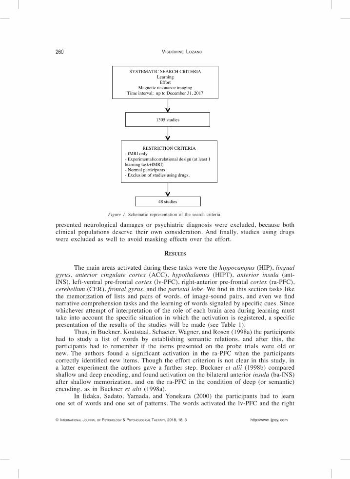
Our method of analysis was descriptive, and was based on a systematic review. The search was carried out in three databases: the PubMed© database, the Neuroscience Information Framework©, and the PsycARTICLES section of the APA PsycNET® database. The search terms were “learning”, “effort”, and “magnetic resonance imaging”, and the time interval was from an open date of beginning up to December 31, 2017. The three terms were introduced in English in a combined frame using “AND” or “&”, and all of them were related to the value “any field”. The search resulted in 1305 registers in total (se Figure 1). Although “magnetic resonance imaging” was one of the terms used for the search, the focus of this study was centered on the brain activation during the performance of different types of tasks, and hence, the studies selected were those using specifically functional magnetic resonance imaging (fMRI), because the goals, procedures, and measures of this technique are more appropriate than simple structural MRI for the study of dynamic brain activity correlated to behavioral phenomena. The use of structural MRI for experimental purposes is widely criticized, and such technique is recommended exclusively for the diagnosis of neurological damages (Illes et alii, 2006). Only 48 studies presented a methodological arrangement clearly oriented to analyze the effort during the performance of learning tasks, and therefore they were the studies finally selected. It was irrelevant for our purposes to make filters by the number of sessions, duration of the study, or number of trials. Studies whose participants
260
© InternatIonal Journal of Psychology & PsychologIcal theraPy, 2018, 18, 3 http://www. ijpsy. com
Visdómine Lozano
presented neurological damages or psychiatric diagnosis were excluded, because both clinical populations deserve their own consideration. And finally, studies using drugs were excluded as well to avoid masking effects over the effort.
results
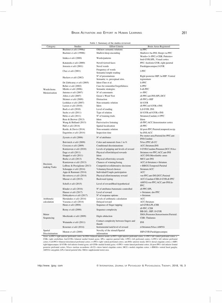
The main areas activated during these tasks were the hippocampus (HIP), lingual gyrus, anterior cingulate cortex (ACC), hypothalamus (HIPT), anterior insula (ant-INS), left-ventral pre-frontal cortex (lv-PFC), right-anterior pre-frontal cortex (ra-PFC), cerebellum (CER), frontal gyrus, and the parietal lobe. We find in this section tasks like the memorization of lists and pairs of words, of image-sound pairs, and even we find narrative comprehension tasks and the learning of words signaled by specific cues. Since whichever attempt of interpretation of the role of each brain area during learning must take into account the specific situation in which the activation is registered, a specific presentation of the results of the studies will be made (see Table 1).
Thus, in Buckner, Koutstaal, Schacter, Wagner, and Rosen (1998a) the participants had to study a list of words by establishing semantic relations, and after this, the participants had to remember if the items presented on the probe trials were old or new. The authors found a significant activation in the ra-PFC when the participants correctly identified new items. Though the effort criterion is not clear in this study, in a latter experiment the authors gave a further step. Buckner et alii (1998b) compared shallow and deep encoding, and found activation on the bilateral anterior insula (ba-INS) after shallow memorization, and on the ra-PFC in the condition of deep (or semantic) encoding, as in Buckner et alii (1998a).
In Iidaka, Sadato, Yamada, and Yonekura (2000) the participants had to learn one set of words and one set of patterns. The words activated the lv-PFC and the right
Figure 1. Schematic representation of the search criteria.
Figure 1. Schematic representation of the search criteria.
SYSTEMATIC SEARCH CRITERIA Learning
Effort Magnetic resonance imaging
Time interval: up to December 31, 2017
RESTRICTION CRITERIA - fMRI only - Experimental/correlational design (at least 1 learning task+fMRI) - Normal participants - Exclusion of studies using drugs.
48 studies
1305 studies
http://www. ijpsy. com © InternatIonal Journal of Psychology & PsychologIcal theraPy, 2018, 18, 3
Brain actiVation and effort in Human Learning 261
Table 1. Summary of the studies reviewed. Category Studies Effort Criteria Brain Areas Registered
Words/items Memorization
Buckner et alii (1998a) Old/new semantic relations ra-PFC Buckner et alii (1998b) Shallow/deep enconding Shallow= ba-INS; Deep= ra-PFC
Iidaka et alii (2000) Words/patterns Words= lv-PFC;r-CER; Patterns= bmf-GYR;SPL; Visual cortex
Katanoda et alii (2000) Novel/viewed faces PFC; fusiform GYR, right parietal Jenssen et alii (2001) Novel words Parahippocampus;f-GYR
Chee et alii (2001) Frequency of words Semantic/simple reading
l-PFC
Heckers et alii (2002) Nº of presentations Semantic vs. perceptual stim.
Right posterior HIP; la-HIP; Ventral tegmentum
De Zubicaray et alii (2005) Idem Chee et al. li-PFC Reber et alii (2002) Cues for remember/forgetfulness li-PFC Miotto et alii (2006) Semantic strategies b-dl-PFC Jansma et alii (2007) Nº of consonants rv-PFC Allen et alii (2007) Green’s Word Test dl-PFC;ant-INS;SPL;DCC Skinner et alii (2009) Distraction dl-PFC;r-HIP Leshikar et alii (2007) Non-semantic relation lif-GYR Larsen et alii (2010) Idem dl-PFC;mf-GYR;r-INS Bach et alii (2010) Level of reading Lif-GYR; INS Sachs et alii (2011) Type of relation lif-GYR;mf-GYR;r-INS Helie et alii (2011) Nº of training trials Striatum;Caudate;vl-PFC Reas & Brewer (2013) Idem Idem Wang & Holland (2013) Pasive/active listening dl-PFC;ACC;Sensorimotor cortex Hall et alii (2014) Spatial localization dl-PFC Rastle, & Davis (2014) Non-semantic relation lif-post-PFC;Parietal,temporal,occip. Engström et alii (2014) Inspection time ba-INS;ACC
Decision-making Tasks
Zyssets et alii (2006) Nº of attributes Pre-motor area;Parietal;lm-PFC;ant-INS;Caudate
Botvinick et alii (2009) Color and numerals from 1 to 9; NAcc;PFC;dl-CC Croxson et alii (2009) Conditional discriminations ACC;Striatum;INS Kurniawan et alii (2010) Levels of gripping and levels of reward f-GYR;Caudate;Putamen;DCC;NAcc Enge et alii (2011) Physical effort/delayed rewards Striatum;vm-PFC;ACC;ant-INS Stoppel et alii (2011) Idem ACC:INS;Mesolimbic areas Burke et alii (2013) Physical effort/risky rewards MCC;ant-INS Kurniawan et alii (2013) Chances of winning/losing ACC;d-Striatum;v-Striatum LeBouc & Pessiglione (2013) Competitive/collaborative decisions v-BBGG;Temporal-Parietal Schouppe et alii (2014) Voluntary/forced choices Striatum;ACC Apps & Ramnani (2014) Individual/Couple participation ACC Skvortsova et alii (2014) Physical effort/monetary reward vm-PFC;ant-INS;DCC;Parietal Massar et alii (2015) Backward typing ACC;Caudate;CER;if-GYR;dl-PFC
Scholl et alii (2015) Level of reward/Real-hypothetical AMYG;vm-PFC;ACC;ant-INS;la-PFC.
Khader et alii (2016) Nº of attributes/Automatic-controlled dl-PFC;SPL Hauser et alii (2017) Level of reward v-Striatum; dm-PFC Dobryakova et alii (2017) Nº of response options v-Striatum
Arithmetic calculation
Hernández et alii (2014) Levels of arithmetic calculation ACC Vassena et alii (2014) Delayed reward ACC;Striatum
Motor Sequencing
Heun et alii (2004) Sequence of finger tapping mf-GYR;LPL;CER
Remy et alii (2008) Sequence complexity dl-PFC; CER BB.GG.; HIP; lf-GYR
Mochizuki et alii (2009) Digits abduction SMA;Premotor;Sensorimotor;Parietal; CER; Thalamus
Watanabe et alii (2011) Contact complexity between fingers and thumb
INS
Kroemer et alii (2014) Instrumental task/level of reward d-Striatum;NAcc;AMYG Spatial Discrimination
Menon et alii (2000) Novelty of the stimuli/Spatial information
HIP;GYR;Parahippocampus
Notes: ra-PFC= right anterior prefrontal cortex; ba-INS= bilateral anterior insula; ra-PFC= right anterior prefrontal cortex; lv-PFC= left ventral prefrontal cortex; r-CER= right cerebellum; bmf-GYR= bilateral medial frontal gyrus; SPL= superior parietal lobe; l-PFC= left prefrontal contex; li-PFC= left inferior prefrontal cortex; b-dl-PFC= bilateral dorsolateral prefrontal cortex; rv-PFC= right ventral prefrontal cortex; ant-INS= anterior insula; DCC= dorsal cingulate cortex; r-HIP= right hippocampus; lif-GYR= left inferior frontal gyrus; mf-GYR= medial frontal gyrus; vl-PFC= ventro lateral prefrontal cortex; lif-post-PFC= left inferior frontal posterior prefrontal cortex; NAcc= nucleus accumbens; dl-CC= dorso lateral cingulate cortex; MCC= medial cingulate cortex; v-BBGG= ventral basal ganglia; AMYG= amygdala; LPL= lateral parietal lobe; SMA= supplemental motor area.
262
© InternatIonal Journal of Psychology & PsychologIcal theraPy, 2018, 18, 3 http://www. ijpsy. com
Visdómine Lozano
cerebellum (r-CER); and the patterns, the bilateral middle frontal gyrus (bmf-GYR), the superior parietal lobe (SPL), and the occipital visual cortex. We could presuppose that the remembering of patterns is more difficult than the remembering of words, however, the findings only appear to indicate to a particular specialization of the brain. In the same line, Heckers, Weiss, Alpert, and Schacter (2002) combined repetition times and the type of stimuli (semantic or perceptual), and found that the combination between words and repetitions activated la-HIP. And regarded with this matter, Katanoda, Yoshikawa , and Sugishita (2000) found that when the participants had to recognize previously viewed faces (low effort), the bilateral fusiform gyrus was activated, whereas when they were presented both novel and viewed faces (high effort), the right parietal and the PFC were activated, clarifying in some extent the question.
Jessen et alii (2001) designed a continuous verbal recognition task in which the items were repeated twice. This time a measure of effort was explicitly provided inasmuch as the authors tested for the amount of memorization, and they found that the memorization of novel words activated the parahippocampal and the frontal GYR during training and the inferior parietal lobe (IPL) during testing, showing apparently that responding in absence of continuous consequences (the testing phase), needs of the participation of areas where the learning has been consolidated.
Chee, Hon, Caplan, Ling Lee, and Goh (2001) chose to combine two conditions of effort: the frequency of word triplets, and the strategy of memorization (semantic or simple reading), and found again activation in the left PFC for the highest effort condition (low frequency and semantic memorization). De Zubicaray, McMahon, Eastburn, Finnigan, and Humphreys (2005) also found activation of the left inferior PFC (li-PFC), confirming the results of earlier studies.
When the effort was manipulated by indicating to the participants with two cues if they must remember or forget a word (i.e., through a discriminative stimulus), the words cued that were remembered activated the li-PFC during the training and the left medial temporal lobe (MTL) during the testing (Reber, Siwiec, Gitleman, Parrish, Mesulam, & Paller, 2002). Remember that Jensen et alii (2001) found activation in the parietal lobe during testing conditions, although they did not presented cues as discriminative stimuli. Similar results reported Miotto et alii (2006), who found that effort-based memorization using semantic organizational strategies, activated the bilateral dorsolateral PFC.
Jansma, Ramsey, de Zwart, van Gelderen, and Duyn (2007) instructed their participants to memorize a set of 1, 3 or 5 consonants, and found that the activation of the right ventral PFC (rv-PFC) changed as a function of effort, and conversely, the ACC, and the HIPT reduced their activity. Allen, Bigler, Larsen, Goodrich-Hunsaker, and Hopkins (2007) administered the Green’s Word Memory Test as a probe of increasing effort, and found activation in the dorso-lateral PFC (dl-PFC), the ant-INS, the SPL, and the dorsal cingulate cortex (DCC). Likewise, Skinner, Fernandes, and Grady (2009) used a recognition task without distraction or interfered with a word, and the memory success in both conditions was correlated with the activation of the dl-PFC and the right HIP.
In Leshikar, Gutchess, Hebrank, Sutton, and Park (2007) the participants had to remember pairs of objects semantically related (low effort condition), or unrelated (high effort), and they found activation in the left inferior frontral GYR (lif-GYR) for the first condition, and in the left HIP for the second condition. Larsen, Allen, Bigler, Goodrich-Hunsaker, and Hopkins (2010) employed the same task and found activation also in the dl-PFC, the SPL, the ACC, the ant-INS, and the bilateral lingual cortices for the full effort trials, but not directly on the lif-GYR or the left HIP.
http://www. ijpsy. com © InternatIonal Journal of Psychology & PsychologIcal theraPy, 2018, 18, 3
Brain actiVation and effort in Human Learning 263
Bach, Bandeis, Hofstetter, Martin, Richardson, and Brem (2010) found that increased effort in poor readers in a task in which they had to substitute letters in words and non-words, activated bilateral GYR and INS, instead of lif-GYR, activated for good readers, which coincides with the results of Leshikar et alii (2007).
Similarly, in Sachs et alii (2011) the participants had to pair associated words, words related categorically, words without relation, or non-words, and found a diversity of areas activated (lif-GYR, medial frontal GYR, and right INS).
In Helie, Roeder, and Ashby (2011) the participants had to complete a categorization task composed of more than 10000 trials, and found increased subcortical activation with practice around the striatum and the caudate, and a cortical activation throughout the training phase (mainly in the ventro-lateral PFC), that became more caudal and dorsal along the training. Reas and Brewer (2013) found the same result, and added that failures at remembering were correlated with reduced activity in the HIP.
The effort on verbal comprehension has also been studied. Wang and Holland (2013) studied passive listening vs. active listening in a narrative comprehension task, and found activation in the left dl-PFC, the ACC, and in the sensorimotor networks in the active way of responding. When memorizing images-sounds pairs was combined with localizing spatially sounds presented without images, was found greater activity in the dl-PFC (Hall et alii, 2014).
Taylor, Rastle, and Davis (2014) conceived that the learning of non-words was more difficult than that of words, and found greater activation for words in the left angular GYR, as well as in the left posterior inferior frontal, parietal, and occipital-temporal cortices.
Finally, the effort in Engström, Karlsson, Landblom, and Craig (2014) was implemented through both a reading task and an inspection time task, and found that effort-related tasks elicited strong activation in the ba-INS and the ACC.
The areas activated by these tasks were the HIPT, HIP, basal ganglia (BB.GG.), the striatum, the nucleus accumbens (Nacc), and different regions of the cortex. Regarding specific experimental situations that lead to the activation of such areas, in Zyssets et alii (2006) the participants had to decide between two alternatives that had five attributes, and found activation in pre-motor areas, the parietal lobe, the lm-PFC, the ant-INS, and the caudate (a sub-area of the BB.GG. related to the movement and motor coordination).
Botvinick, Huffstetler, and McGuire (2009) investigated the relation between two effort levels (the correct response depended on the color in which a range of numerals was presented) and reward, and observed activation in the NAcc, orbito-frontal cortex, and a preceding activation in the dorsal-lateral cingulate cortex. Croxson, Walton, O’Reilly, Behrens, and Rushworth (2009) employed similar tasks that were oriented to attain secondary reinforcers and also found activation in the ACC, the ventral striatum, and INS.
Kurniawan, Seymour, Talmi, Yoshida, Chater, & Dolan (2010) mixed two levels of gripping (high and low) and two levels of monetary reward (high and low), and found activation in the frontal GYR, the caudate, and the putamen in relation to the level of effort, as well as in the DCC in those most persistent participants. The NAcc was activated for rewards only in trials in which the participants opted to a high effort option (i.e., as if the things that cost were the really valued).
Enge, Fleischhauer, Lesch, Reif, and Strobel (2011) designed a decision-making task based on different levels of physical effort and delayed rewards (erotic stimuli).
264
© InternatIonal Journal of Psychology & PsychologIcal theraPy, 2018, 18, 3 http://www. ijpsy. com
Visdómine Lozano
The authors found activation in the striatum and the vm-PFC for the increasing value of delayed rewards, and in the ACC and the ant-INS for the expected expense of energy in high effort trials. Stoppel, Boehler, Strumpf, Heinze, Hopf, & Schoenfeld (2011) reproduced this experimental procedure and found activity in the ACC, the INS, and mesolimbic regions.
Burke, Brünger, Kahnt, Park, and Tobler (2013) attempted to differentiate between physical effort costs and costs associated with risky rewards, and they found that the first condition produced activation in the medial cingulate cortex (MCC), and the second condition did it in the ant-INS. Likewise, Kurniawan, Guitart-Masip, Dayan, and Dolan (2013) combined two levels of effort and chances of winning or avoiding the loss of money, and they found activation in the ACC and the dorsal striatum for the anticipation of effort, and in the ventral striatum for outcomes better than expected.
When were explored the differences on brain activation during competitive vs. collaborative decision-making in the context of strategic games the activation was produced in the ventral basal ganglia (v-BBGG) for the condition of personal utility (competitive strategy), and in the temporal-parietal junction for the collaborative strategy (LeBouc & Pessiglione, 2013). Complementarily, Apps and Ramnani (2014) examined the reward magnitude and the level of effort when the participants had to accomplish alone the experimental tasks or when they had to do it accompanied by a social confederate, and the authors found activation in the sulcus of the ACC for response costs, and in the gyral of the ACC for the net value of rewards gained by others. Schouppe, Demanet, Boehler, Ridderinkhof, and Notebaert (2014) found that the striatum and the ACC activations were higher when participants chose voluntarily in the most effort option than when they responded on force-choice trials.
Skvortsova, Palminteri, and Pessiglione (2014) managed the amount of physical effort and monetary outcome, and they found that the vm-PFC was activated with expected and actual rewards, and the ant-INS, the DCC, and the parietal cortex with expected and actual efforts. In constrast, Massar, Libedinsky, Weiyan, Huettel, and Chee (2015) described that the value of the chosen options activated the ACC, the caudate, and the CER, and that cognitive efforts (to type backwards a specified number of words) activated the inferior frontal GYR and the dl-PFC.
More interestingly was the design employed by Scholl, Kolling, Nelissen, Wittmann, Harmer, & Rushworth (2015). They arranged a procedure that allowed them test for the effects of varying levels of reward and effort, as well as the real or hypothetical (but unknown) reward delivery. This procedure led to observe neurological patterns associated to a superstitious-like behavior. The authors found a pattern of behavior that they called “irrational chose bias”, and found activation in the AMYG and the vm-PFC in this condition; however, they found activation in the dorsal ACC, the ant-INS, and the la-PFC when the participants chose options in a defined way in the condition of being guided by a relation more predictable between their behavior and its consequences. In Khader, Pachur, Weber, and Jost (2016) the elections were associated with one, two or three attributes activated automatically, or controlled, and found that increasing efforts activated the dl-PFC, as well as the SPL only when remembering was controlled.
Finally, in Dobryakova, Jessup, and Tricomi (2017) the low effort condition was comprised of a single image that was presented with four response options, and the high effort condition was comprised of two images that were presented with two response options; correct feedback was presented only when the participants responded correctly to both of the images. The high effort condition correlated with activation in the ventral
http://www. ijpsy. com © InternatIonal Journal of Psychology & PsychologIcal theraPy, 2018, 18, 3
Brain actiVation and effort in Human Learning 265
striatum. And Hauser, Eldar, and Dolan (2017) employed a similar decision-making task, and found that the amount of reward activated ventral striatum, and effort the dm-PFC. Obviously, these tasks do not need clarification about their parallelism with academic activities. Nonetheless, paradoxically these tasks do not appear to have been studied as much as others. Hernandez, Kuss, Trautner, Weber, Falk, Fliessbach (2014) designed an arithmetic calculation task with three levels of difficulty that, in addition, were differently rewarded. They found activation in the subgenual ACC only for the high effort condition. Vassena, Silvetti, Boehler, Achten, Fias, & Verguts (2014) added a delay in the reward delivery, and they found again that upcoming difficult tasks elicited activation in the ACC and in the striatum.
The main areas activated for these tasks were the GYR, supplemental motor area, LPL, CER, dl-PFC, occipital cortices, and BBGG. Specifically, Heun et alii (2004) used a finger tapping sequence as task, and they found strong bilateral activation in the mid-frontal GYR, the supplementary motor area, the LPL, and the CER, which is congruent with the employment of motor tasks.
Remy, Wenderoth, Lipkens, and Swinnen (2008) examined the acquisition of a complex bimanual coordination pattern, and found activation decreases along training in the dl-PFC, right middle temporal and occipital cortices, and in the posterior CER; and found increases in the BBGG, HIP, and frontal GYR.
Mochizuki et alii (2009) used as task the abduction of all digits (easy condition), and finger abduction with digits 2 and 3 abducted together, concurrently with digits 4 and 5 (hard condition). The authors found that the hard condition produced increased activation in the SMA, the pre-motor, sensorimotor, and parietal cortices, the CER, and the thalamus.
Watanabe, Watanabe, Kuruma, Murakami, Seno, and Matsuda (2011) presented four sequences of contact between different fingers and the thumb. The participants had to imitate them from one out of two perspectives. The authors found that the easiest sequence and perspective activated rp-INS.
Finally, Kroemer, Guevara, Ciocanea Teodorescu, Wuttig, Kobiella, and Smolka (2014) found that an average effort activated the dorsal striatum, that higher effort in an instrumental task was predicted by a higher activation in the NAcc, and that the AMYG was activated only when effort was far above the average.
At last, we can say that spatial discriminations are always present in whatever learning. For example, when somebody memorizes a schema, the spatial disposition of the verbal stimuli in such schema is a key element for remembering. However, there are not many works specifically centered on manipulating spatial difficulty. Menon, Rivera, White, Eliez, Glover, & Reiss (2000) combined the novelty of stimuli with the richness of their spatial information, and found greater activation in the HIP, the lingual GYR, and the parahippocampal GYR in accordance to the spatial complexity. The novelty only was correlated with activation in the lingual GYR.
discussion
First of all, through the present review we can see the disparity of studies concerned on the matter, and we discover that there is not an organized agenda of research. The different learning tasks that can be employed to study the effort have not been examined in the same degree, which limits the conclusions that we can extract. In second place, the definition of effort is made a priori in the majority of the studies, or is made under
266
© InternatIonal Journal of Psychology & PsychologIcal theraPy, 2018, 18, 3 http://www. ijpsy. com
Visdómine Lozano
the perspective of the experimenters. It would be advisable to define the effort according to the perspectives of the participants for assuring that effort is really implicated in a task. In fact, “semantic encoding” is sometimes understood as a strategy that requires high effort and other times low effort. And in third place, the results presented point out to certain variability in the areas involved, and even in the participation of different regions belonging to a same area along a given class of task, and the participation of a same area in different types of tasks. Moreover, some of the areas mentioned also participate in other matters. For example, the caudate and ventral tegmentum have been activated in relation to love-evoking stimuli, the INS when distributing reinforcers in accordance with criteria of “justice”; and the PFC in moral reasoning and during the practice of religious exercises (Pérez Álvarez, 2011). Hence, such crossed or multiple specializations of some brain areas can explicate the variability found. Other variables that could explicate the variability are certain methodological issues involved in fMRI, such as the specific timing of the registry, inappropriate parametrical statistical analysis, invalid cluster inferences, the methodological isolation of the relation between the oxigened blood levels that we call “activation” and the tasks employed, etc.; as pointed out by Eklund, Nichols, and Knutsson (2016).
At any rate, we can conclude that the most relevant areas related to the training of tasks based on effort were: (a) PFC, INS, and ACC in memorization tasks; (b) CER, BBGG, motor and pre-motor areas in specific motor tasks; (c) NAcc and striatum when explicit reinforcing consequences and high effort were involved; (d) Cingulate cortex for effort requirements and persistent behavior; (e) AMYG with unpredictable contingencies or superstitious behavior, and when the effort was far above the average; and (f) HIPT, HIP, and related regions for the initial consolidation of memory.
And another important conclusion that we can extract from this review, is that tasks that are based on effort do not activate automatically the fundamental area that is involved in aversive learning (i.e., AMYG). This area is only activated when the effort required is considerably higher than the average that an individual is accustomed to perform, as well as when the tasks consist of uncertainty conditions. The latter is congruent with other findings that connect the AMYG to behaving under conditions of ambiguity (DiChiara & Imperato, 1988; Whalen, 1998). Both the former and the latter have the same behavioral function, that is, both involve the withdrawal of positive reinforcement. In the former, the effort required exceeds an individual’s resources to obtain such reinforcer, and in the latter there are not cues (discriminative stimuli) signaling criteria for attaining the reinforcement. Thus, the reinforcement “moves away” in both conditions.
Nonetheless, in accordance to the findings reported by others studies included in this review, a possible initial activation of the AMYG when the effort required was quite higher than the mean effort that an individual is accustomed to perform, could be moderated with a progressive adaptation to such level of effort. This process would finally lead to the activation of the striatum and NAcc (see Kroemer et alii, 2014; Kurniawan et alii, 2010), which would indicate that such activity passes by from an aversive function to a function of reinforcement (DiChiara & Imperato, 1988; Peciña & Berridge, 2005). This can be useful for programming more effective instructional procedures than the designed up to now, and for not forgetting the importance of effort in the process of the academic human learning, as has been remembered by some authors (Dweck, 2016; Enkvist, 2011). Furthermore, considering the results obtained by Segura and Roales Nieto (2016), that paradoxically show that a consolidated wellbeing
http://www. ijpsy. com © InternatIonal Journal of Psychology & PsychologIcal theraPy, 2018, 18, 3
Brain actiVation and effort in Human Learning 267
when some social and intergenerational difficulties have been surmounted is related to a higher unhappiness, perhaps the best thing is not to dispense with the effort in the academic learning.
One method for the improvement of the instructional procedures would be programming tasks with successive levels of difficulty and effort, which has been successfully put into practice by Behavior Analysis since ever (Fredrick, Deitz, Bryceland, & Hummel, 2002; Luciano, 1995; Sulzer-Azaroff & Mayer, 1986). The trouble is that not all students are able to deploy initially the same level of effort. Consequently, such programming should be individualized in some extent, and the different social agencies involved in the development of an individual should be concerned on helping to achieve such growing level of effort tolerance (parents included). What is clear is that effort per se is not equivalent to aversive conditioning in neurological terms, and is restricted to very specific conditions, which contradicts proposals like that by Kurzban et alii (2013). Even more, it seems that, under some conditions, only when the effort is explicitly required to achieve a given amount of rewards, are activated the brain areas involved in positive reinforcement (see Kurniawan et alii, 2010). As Plato (c. 390 BC) wrote in Hippias Major (p. 304e), “beautiful things are difficult”.
references
Alfieri L, Brooks PJ, Aldrich NJ, & Tenenbaum HR (2011). Does discovery-based instruction enhance learning? Journal of Educative Psychology, 103, 1-18. Doi: 10.1037/a0021017
Allen MD, Bigler ED, Larsen J, Goodrich-Hunsaker NJ, & Hopkins RO (2007). Functional neuroimaging evidence for high cognitive effort on the Word Memory Test in the absence of external incentives. Brain Injury, 21, 1425-1428. Doi: 10.1080/02699050701769819
Andreatta M, Fendt M, Mühlberger A, Wieser MJ, Imobersteg S, Yarali A, Gerber B, & Pauli P (2012). Onset and offset of aversive events establish distinct memories requiring fear and reward networks. Learning and Memory, 19, 518-526. Doi: 10.1101/lm.026864.112
Ansari D, De Smedt B, & Grabner RH (2012). Neuroeducation -A critical overview of an emerging field. Neuroethic, 5, 105. Doi: 10.1007/s12152-011-9119-3
Apps MAJ & Ramnani N (2014). The anterior cingulate gyrus signals the net value of others’ rewards. Journal of Neuroscience, 34, 6190–6200. Doi: 10.1523/JNEUROSCI.2701-13.2014
Bach S, Brandeis D, Hofstetter C, Martin E, Richardson U, & Brem S (2010). Early emergence of deviant frontal fMRI activity for phonological processes in poor beginning readers. Neuroimage, 53, 682-693. Doi: 10.1016/j.neuroimage.2010.06.039
Barkovich AJ (2010). Morphologic characteristics of subcortical heterotopia: MR imaging study. American Journal of Neuroradiology, 21, 290-295
Baxter MG & Murray AE (2002). The amygdala and reward. Nature Reviews Neuroscience, 3, 563-573. Doi: 10.1038/nrn875
Botvinick MM, Huffstetler S, & McGuire JT (2009). Effort discounting in human nucleus accumbens. Cognitive and Affective Behavioral Neuroscience, 9, 16-27. Doi: 10.3758/CABN.9.1.16
Buckner RL, Koutstaal W, Schacter DL, Wagner AD, & Rosen BR (1998a). Functional-anatomic study of episodic retrieval using fMRI: I. Retrieval effort versus retrieval success. Neuroimage, 7, 151-162. Doi: 10.1006/nimg.1998.0327
Buckner RL, Koutstaal W, Schacter DL, Wagner AD, & Rosen BR (1998b). Functional-anatomic study of episodic retrieval II. Selective averaging of event-related fMRI trials to test the retrieval success hypothesis. Neu-roimage, 7, 163-175. Doi: 10.1006/nimg.1998.0328
Burke CJ, Brünger C, Kahnt T, Park SQ, & Tobler PN (2013). Neural integration of risk and effort costs by the frontal pole: Only upon request. Journal of Neuroscience, 33, 1706-1713a. Doi: 10.1523/JNEUROSCI.3662-12.2013
268
© InternatIonal Journal of Psychology & PsychologIcal theraPy, 2018, 18, 3 http://www. ijpsy. com
Visdómine Lozano
Bzdok D, Laird AR, Zilles K, Fox PT, & Eickhoff, SB (2013). An investigation of the structural, connectional, and functional subspecialization in the human amygdala. Human Brain Mapping, 34, 3247-3266. Doi: 10.1002/hbm.22138
Catania, AC (1998). Learning. New Jersey: Prentice Hall. Chee M, Hon N, Caplan D, Ling Lee H, & Goh J (2002). Frequency of concrete words modulates prefrontal activation
during semantic judgments. Neuroimage, 16, 259-268. Doi: 10.1006/nimg.2002.1061Craik FIM & Tulving E (1975). Depth of processing and the retention of words in episodic memory. Journal of
Experimental Psychology: General, 104, 268-294. Doi: 10.1037/0096-3445.104.3.268Croxson P L, Walton ME, O’Reilly JX, Behrens TE, & Rushworth MF (2009). Effort-based cost-benefit valuation
and the human brain. Journal of Neuroscience, 29, 4531-4541. Doi: 10.1523/JNEUROSCI.4515-08.2009De Zubicaray GI, McMahon KL, Eastburn MM, Finnigan S, & Humphreys MS (2005). fMRI evidence of word
frequency and strength effects during episodic memory encoding. Cognitive Brain Research, 22, 439-450. Doi: 10.1016/j.cogbrainres.2004.10.002
Della Chiesa B (2013). Our learning/teaching brains: What can be expected from neuroscience, and how? What should not be expected from neuroscience, and why? Proceedings of the 2013 Research Conference. How the Brain Learns: What lessons are there for teaching? (pp. 3-6). Melbourne: Australian Council for Educational Research.
Di Chiara G & Imperato A (1988). Drugs abused by humans preferentially increase synaptic dopamine concentrations in the mesolimbic system of freely moving rats. PNAS, 85, 5274-5278. Doi: 10.1073/pnas.85.14.5274
Dobryakova E, Jessup RK, & Tricomi, E (2017). Modulation of ventral striatal activity by cognitive effort. Neuroimage, 147, 330-338. Doi: 10.1016/j.neuroimage.2016.12.029
Donovan S, Bransford J, & Pellegrino J (1999). How people learn: Bridging research and practice. Washington DC: National Academy of Sciences.
Dweck C (2017). The journey to children’s mindsets -and beyond. Child Developmental Perspectives, 11, 139-144. Doi: 10.1111/cdep.12225
Enge S, Fleischhauer M, Lesch K, Reif A, & Strobel A (2011). Serotonergic modulation in executive functioning: Linking genetic variations to working memory performance. Neuropsychologia, 49, 3776-3785. Doi: 10.1016/j.neuropsychologia.2011.09.038
Engström M, Karlsson T, Landtblom AM, & Craig AD (2014). Evidence of conjoint activation of the anterior insular and cingulate cortices during effortful tasks. Frontier Human Neuroscience, 8, 1071. Doi: 10.3389/fnhum.2014.01071. Retrieved from https://www.frontiersin.org/articles/10.3389/fnhum.2014.01071/full
Eklund A, Nichols TE, & Knutsson H (2016). Cluster failure: Why fMRI inferences for spatial extent have inflated false-positive rates. PNAS, 113, 7900-7905. Doi: 10.1073/pnas.1602413113
Enkvist I (2011). La buena y la mala educación. Ejemplos internacionales. Barcelona: Encuentro.Floresco SB & Ghods-Sharifi S (2007). Amygdala-prefrontal cortical circuitry regulates effort-based decision making.
Cerebral Cortex, 17, 251-260. Doi: 10.1093/cercor/bhj143Fredrick L, Deitz, SM, Bryceland JA, & Hummel JH (2002). Behavior analysis, education and effective schooling.
Reno, NV: Context Press.Hall SA, Rubin DC, Miles A, Davis SW, Wing EA, Cabeza R, & Berntsen D (2014). The neural basis of involuntary
episodic memories. Journal of Cognitive Neuroscience, 26, 2385-2399. Doi: 10.1162/jocn_a_00633Hauser T, Eldar E, & Dolan R (2017). Separate mesocortical and mesolimbic pathways encode effort and reward
learning signals. PNAS, 114, E7395-E7404. Doi: 10.1073/pnas.1705643114Heckers S, Weiss AP, Alpert NM, & Schacter DL (2002). Hippocampal and brain stem activation during word retrieval
after repeated and semantic encoding. Cerebral Cortex, 12, 900-907. Doi: 10.1093/cercor/12.9.900Helie S, Roeder JL, & Ashby FG (2010). Evidence for cortical automaticity in rule-based categorization. Journal of
Neuroscience, 30, 14225-14234. Doi: 10.1523/JNEUROSCI.2393-10.2010.Hernandez J, Kuss K, Trautner P, Weber B, Falk A, & Fliessbach K (2014). Effort increases sensitivity to reward
and loss magnitude in the human brain. Social Cognition and Affective Neuroscience, 9, 342-349. Doi: 10.1093/scan/nss147
Herrnstein RJ (1970). On the law of effect. Journal of the Experimental Analysis of Behavior, 13, 243-266. Doi: 10.1901/jeab.1970.13-243
Heun R, Freymann N, Granath DO, Stracke CP, Jessen F, Barkow K, & Reul J (2014). Differences of cerebral activation between superior and inferior learners during motor sequence encoding and retrieval. Psychiatry
http://www. ijpsy. com © InternatIonal Journal of Psychology & PsychologIcal theraPy, 2018, 18, 3
Brain actiVation and effort in Human Learning 269
Research, 132, 19-32. Doi: 10.1016/j.pscychresns.2004.01.007.Howard-Jones P (2010). Introducing neuroeducational research, neuroscience, education and the brain from contexts
to practice. London: Routledge.Iidaka T, Sadato N, Yamada H, & Yonekura Y (2000). Functional asymmetry of human prefrontal cortex in verbal
and non-verbal episodic memory as revealed by fMRI. Cognitive Brain Research, 9, 73-83. Doi: 10.1016/S0926-6410(99)00047-6
Illes J, Kirschen MP, Edwards E, Stanford LR, Bandettini P, Cho M, Ford P, Glover G, Kulynych J, Macklin R, Michael D, & Wolf M (2006). Incidental findings in brain imaging research: What should happen when a researcher sees a potential health problem in a brain scan from a research subject? Science, 311, 783-784. Doi: 10.1126/science.1124665
Ishii A, Tanaka M, & Watanabe Y (2014). Neural mechanisms of mental fatigue. Review of Neuroscience, 25, 469-479. Doi: 10.1515/revneuro-2014-0028
Jansma JM, Ramsey NF, de Zwart JA, van Gelderen P, & Duyn JH (2007). fMRI study of effort and information processing in a working memory task. Human Brain Mapping, 28, 431-440. Doi: 10.1002/hbm.20297
Jessen J, Flacke S, Granath DO, Manka C, Scheef L, Papassotiropoulos A, Schild HH, & Heun R (2001). Encoding and retrieval related cerebral activation in continuous verbal recognition. Cognitive Brain Research, 21, 199-206. Doi: 10.1016/S0926-6410(01)00046-5
Katanoda K, Yoshikawa K, & Sugishita M (2000). Neural substrates for the recognition of newly learned faces: A functional MRI study. Neuropsychologia, 38, 1616-1625. Doi: 10.1016/S0028-3932(00)00069-5
Khader PH, Pachur T, Weber LAE, & Jost K (2016). Neural signatures of controlled and automatic retrieval processes in memory-based decision-making. Journal of Cognitive Neuroscience, 28, 69-83. Doi: 10.1162/jocn_a_00882
Kroemer N, Guevara A, Ciocanea Teodorescu I, Wuttig F, Kobiella A, & Smolka M (2014). Balancing reward and work: Anticipatory brain activation in NAcc and VTA predict effort differentially. Neuroimage, 102, 510-519. Doi: 10.1016/j.neuroimage.2014.07.060
Kurniawan IT, Seymour B, Talmi D, Yoshida W, Chater N, & Dolan RJ (2010). Choosing to make an effort: The role of striatum in signaling physical effort of a chosen action. Journal of Neurophysiology, 104, 313-321. Doi: 10.1152/jn.00027.2010
Kurniawan IT, Guitart-Masip M, Dayan P, & Dolan RJ (2013). Effort and valuation in the brain: The effects of anticipation and execution. Journal of Neuroscience, 33, 6160-6169. Doi: 10.1523/JNEUROSCI.4777-12.2013
Kurzban R, Duckworth A, Kable JW, & Myers J (2013). An opportunity cost model of subjective effort and task performance. Behavioral and Brain Sciences, 36, 661-679. Doi: 10.1017/S0140525X12003196
Larsen JD, Allen MD, Bigler ED, Goodrich-Hunsaker NJ, & Hopkins RO (2010). Different patterns of cerebral activation in genuine and malingered cognitive effort during performance on the Word Memory Test. Brain Injury, 24, 89-99. Doi: 10.3109/02699050903508218
LeBouc R & Pessiglione M (2013). Imaging social motivation: Distinct brain mechanisms drive effort production during collaboration versus competition. Journal of Neuroscience, 33, 15894-15902. Doi: 10.1523/JNEU-ROSCI.0143-13.2013
Leshikar ED, Gutchess AH, Hebrank AC, Sutton BP, & Park DC (2010). The impact of increased relational enco-ding demands on frontal and hippocampal function in older adults. Cortex, 46, 507-521. Doi:10.1016/j.cortex.2009.07.011
Luan KP, Wager T, & Liberzon I (2002). Functional neuroanatomy of emotion: a meta-analysis of emotion activation studies in PET and fMRI. Neuroimage, 16, 331-348. Doi: 10.1006/nimg.2002.1087
Luciano, MC (1995). Aportaciones funcionales en educación. Granada: Némesis.Massar SA, Libedinsky C, Weiyan C, Huettel SA, & Chee M (2015) Separate and overlapping brain areas encode
subjective value during delay and effort discounting. Neuroimage, 15, 104-113. Doi: 10.1016/j.neuroima-ge.2015.06.080
Menon V, Rivera SM, White CD, Eliez S, Glover GH, & Reiss A (2000). Functional optimization of arithmetic processing in perfect performers. Cognitive and Brain Research, 9, 343-345. Doi: 10.1016/S0926-6410(00)00010-0
Miotto EC, Savage CR, Evans JJ, Wilson BA, Martins MGM, Iaki S, & Amaro E (2006). Bilateral activation of the prefrontal cortex after strategic semantic cognitive training. Human Brain Mapping, 27, 288-295. Doi: 10.1002/hbm.20184
Mochizuki G, Hoque T, Mraz R, MacIntosh BJ, Graham SJ, Black SE, & McIlroy WE (2009). Challenging the bra-
270
© InternatIonal Journal of Psychology & PsychologIcal theraPy, 2018, 18, 3 http://www. ijpsy. com
Visdómine Lozano
in: Exploring the link between effort and cortical activation. Brain Research, 1301, 9-19. Doi: 10.1016/j.brainres.2009.09.005
Morris S, Buchel C, & Dolan R (2001). Parallel neural responses in amygdala subregions and sensory cortex during implicit fear conditioning. Neuroimage, 13, 1044-1052. Doi: 10.1006/nimg.2000.0721
OECD (2002). Understanding the brain: Towards a new learning science. Paris: OECD Publishing.Peciña S & Berridge K (2005). Hedonic hot spot in nucleus accumbens shell: Where do mu-opioids cause increa-
sed hedonic impact of sweetness? Journal of Neuroscience, 25, 11777-11786. Doi: 10.1523/JNEUROS-CI.2329-05.2005
Pérez Alvarez, M (2011). El mito del cerebro creador. Cuerpo, conducta y cultura. Madrid: Alianza Editorial. Phelps EA, O’Connor KJ, Gatenby JC, Gore JC, Grillon C, & Davis M (2001). Activation of the left amygdala to a
cognitive representation of fear. Nature Neuroscience, 4, 437-441. Doi: 10.1038/86110Plato (c. 390 BC/1925). Plato in Twelve Volumes (Vol. 9 translated by WRM Lamb). Cambridge, MA: Harvard
University Press.Poplin M (1988). Holistic/constructivists principles of teaching/learning process: Implications for the field of learning
disabilities. Journal of Learning Disabilities, 7, 401-416.Reas ET & Brewer JB (2013). Effortful retrieval reduces hippocampal activity and impairs incidental encoding.
Hippocampus, I, 367-379. Doi: 10.1002/hipo.22096Reber P, Siwiec R, Gitleman D, Parrish T, Mesulam M, & Paller K (2002). Neural correlates of successful encoding
identified using functional magnetic resonance imaging. Journal of Neuroscience, 22, 9541-9548. Doi: 10.1523/JNEUROSCI.22-21-09541.2002
Remy F, Wenderoth N, Lipkens K, & Swinnen SP (2008). Acquisition of a new bimanual coordination pattern modulates the cerebral activations elicited by an intrinsic pattern: An fMRI study. Cortex, 44, 482-493. Doi: 10.1016/j.cortex.2007.07.004
Riedel P, Jacob MJ, Müller DK, Vetter NC, Smolka MN, & Marxen M (2016). Amygdala fMRI signal as a predictor of reaction time. Frontiers in Human Neuroscience, 10, 51. Doi: 10.3389/fnhum.2016.00516.
Roales-Nieto JG (2016). Psicopatología de la identidad. Boletín de Estudios de Filosofía y Cultura, 11, 57-80. Robinson K & Aronica L (2015). Creative schools: Revolutionizing education from the ground up. Allen lane:
Penguin Books.Sachs O, Weis S, Zellagui N, Sass K, Huber W, Zvyagintsev M, Mathiak K, & Kircher T (2011). How different types
of conceptual relations modulate brain activation during semantic priming. Journal of Cognitive Neuroscience, 23, 1263-1273. Doi: 10.1162/jocn.2010.21483
Saleem NM, Surmeier DJ, & Malenka RC (2000). Dopaminergic modulation of neuronal excitability in the striatum and nucleus accumbens. Annual Review of Neurosciences, 23, 185-215. Doi: 10.1146/annurev.neuro.23.1.18
Scholl J, Kolling N, Nelissen N, Wittmann MK, Harmer CJ, & Rushworth MFS (2015). The good, the bad, and the irrelevant: Neural mechanisms of learning real and hypothetical rewards and effort. Journal of Neurosciences, 35, 11233-11251. Doi: 10.1523/JNEUROSCI.0396-15.2015
Schouppe N, Demanet J, Boehler CN, Ridderinkhof KR, & Notebaert W (2014). The role of the striatum in effort-based decision-making in the absence of reward. Journal of Neuroscience, 34, 2148-2154. Doi: 10.1523/JNEUROSCI.1214-13.2014
Segura A & Roales-Nieto JG (2016). Diferencias intergeneracionales en satisfacción y felicidad percibidas, relacio-nadas con la prosperidad material [Intergenerational differencies in satisfaction and hapiness regarded with material prosperity], Equidad y Desarrollo, 25, 11-28. Doi: 10.19052/ed.3724.
Seymour B & Dolan R (2008). Emotion, decision making, and the amygdala. Neuron, 58, 662-667. Doi: 10.1016/j.neuron.2008.05.020
Skinner EI, Fernandes MA, & Grady CL (2009). Memory networks supporting retrieval effort and retrieval success under conditions of full and divided attention. Experimental Psychology, 56, 386-396. Doi: 10.1027/1618-3169.56.6.386
Skvortsova V, Palminteri S, & Pessiglione M (2014). Learning to minimize efforts versus maximizing rewards: Computational principles and neural correlates. Journal of Neuroscience, 34, 15621-15630. Doi: 10.1523/JNEUROSCI.1350-14.2014
Stoppel CM, Boehler CN, Strumpf H, Heinze HJ, Hopf JM, & Schoenfeld MA (2011). Neural processing of reward magnitude under varying attentional demands. Brain Research, 1383, 218-229. Doi: 10.1016/j.brainres.2011.01.095
http://www. ijpsy. com © InternatIonal Journal of Psychology & PsychologIcal theraPy, 2018, 18, 3
Brain actiVation and effort in Human Learning 271
Sulzer-Azaroff B & Mayer GR (1986). Achieving educational excellence. Using behavioral strategies. New York: Holt, Rinehart & Winston, Inc.
Taylor JSH, Rastle K, & Davis MH (2014). Interpreting response time effects in functional imaging studies. Neuroimage, 99, 419-433. Doi: 10.1016/j.neuroimage.2014.05.073
Tyler SW, Hertel PT, McCallum MC, & Ellis HC (1979). Cognitive effort and memory. Journal of Experimental Psychology: Human Learning and Memory, 5, 607-617. Doi: 10.1037/0278-7393.5.6.607
Vassena E, Silvetti M, Boehler CN, Achten E, Fias W, & Verguts T (2014). Overlapping neural systems represent cognitive effort and reward anticipation. PLoS ONE, 9, e91008. Doi: 10.1371/journal.pone.0091008
Wang Y & Holland SK (2014). Comparison of functional network connectivity for passive-listening and active-response narrative comprehension in adolescents. Brain Connections, 4, 273-285. Doi: 10.1089/brain.2013.0190
Watanabe R, Watanabe S, Kuruma H, Murakami Y, Seno A, & Matsuda T (2011). Neural activation during imitation of movements presented from four different perspectives: A functional magnetic resonance imaging study. Neuroscience Letters, 503, 100-104. Doi: 10.1016/j.neulet.2011.08.016
Whalen PJ (1998). Fear, vigilance and ambiguity: Initial neuroimaging studies of the human amygdala. Current Directions on Psychological Science, 7, 177-188. Doi: 10.1111/1467-8721.ep10836912
Zysset S, Wendt C, Volz K, Neumann J, Huber O, & von Cramon Y (2006). The neural implementation of multi-attribute decision making: A parametric fMRI study with human subjects. Neuroimage, 31, 1380-1388. Doi: 10.1016/j.neuroimage.2006.01.017
Received, June 11, 2018Final Acceptance, July 21, 2018
Recommended