Interpreting the findings of the Neurological Examination
Vitalie Lupu, MD, FAAN
Providence Brain and Spine InstituteNovember 17, 2016

Disclosures
Nothing to disclose

Objectives
1. Review the standard elements of the neuro exam;
2. Review key neuroanatomy principles;
3. Make sense of exam findings to help with lesion localizationand differential diagnosis;
4. Discuss the screening neurological examination.

Neurological complaints are very common
Does the patient who just collapsed on the street have an MI or an ICH?
Is the patient with leg weakness and numbness suffering from lumbar DJD or from impending spinal cord compression?
Does the patient with nausea and vomiting need a GI consult, a head CT, or emergency interventions to lower high ICP?
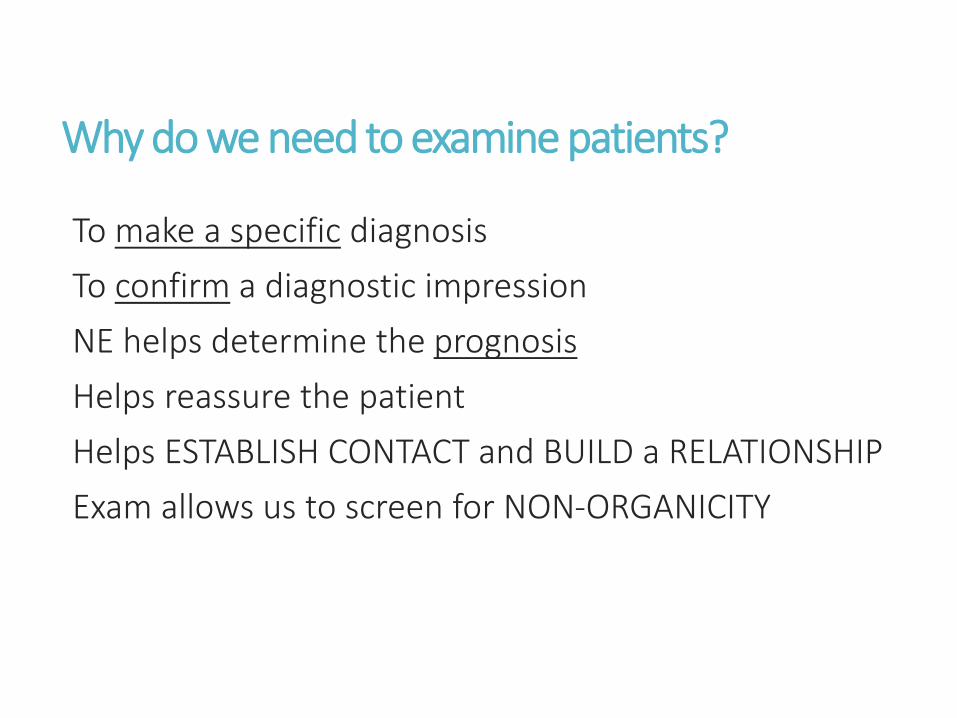
Why do we need to examine patients?
To make a specific diagnosis
To confirm a diagnostic impression
NE helps determine the prognosis
Helps reassure the patient
Helps ESTABLISH CONTACT and BUILD a RELATIONSHIP
Exam allows us to screen for NON-ORGANICITY

Exam gives us something else!
TIME TO THINK!
Gives us time to feel what type of person we have in front of us and her/his family and friends
What else can it be? What have I missed?
Examine patients to avoid unnecessary tests
Over-testing can lead REAL HARM to our patients (not just in direct costs)
• DJD of the spine – may lead to unnecessary surgeries
• NSWMC (leukoarayosis) – epidemic of pseudo-MS

Evidence based value of the neuro exam
Does it really improve our care for the patient?
If you think of the Neuro Exam as a dx test it should:Be reliable, be accurate
Have a measurable dx value (PPV, NPV, Specificity, Sensitivity)
Positive findings should be used in combinationIf only one sign is abnormal, treat it with caution, and correlate with Hx
Patients do not have a constant exam overtime

What is being tested by each part of the NE?
Performing NE well is linked to understanding:NEUROANATOMY
Cerebral and spinal cord BLOOD SUPPLY
Musculoskeletal system
Psychiatric and psychological problems
Other medical systems (respiratory, cardiovascular, etc)

Making Sense of Neurological Findings
Is there evidence of motor dysfunction?Weakness, Spasticity, Tremor
Does the pattern follow an UMN or LMN pattern? UMN process (weakness + spasticity). What is the level?
LMN process (weakness + flaccidity). What is the distribution?
spinal nerve root
peripheral nerve
Bilateral? Distal?
Do reflexes support a UMN or LMN process?hyper-reflexic in UMN disorders VS hypo-reflexic in LMN disorders
Does the plantar reflex testing suggest an UMN lesion?

Making Sense of Neurological Findings
Is there impaired sensation? some disorders affect only the motor pathways, sparing sensation
Which aspects of sensation are impaired? spinothalamic tractdorsal columns
Does the sensation loss suggest dysfunction at a specific level? spinal nerve root? peripheral nerve problem?
Does the sensory deficit correlate with the "correct" motor deficit?
radial nerve lesion leads to characteristic motor and sensory findings

General rules for the neuro examLearn standard exam techniques
http://www.clinicalneurologyvideos.com
http://www.neuroexam.com
Follow some kind of ORDERAnatomic
Instrument
Functional
QUANTIFY findings
Measure - Pupils, Limb circumference, Duration of vibration sensation
Grade - Reflexes 0/4 to +4/4; Strength 0/5 to 5/5
COMPARE with contralateral side
Assume that everything is ABNORMAL until proven otherwise

General rules for the neuro exam
Analyze the NE
Record all findings OBJECTIVELY
Consider the underlying neurophysiological process
Convert findings to technical terminology unilateral arm and leg weakness + increased tone = hemiplegia
Place the lesion in the CNS or PNS pyramidal tract in CNS
Think CIRCUITS and NEUROANATOMY
Localize the LEVEL of the lesion within the
Motor, sensory, autonomic, mental circuits

Clinical diagnosis in Neurology
Is there a lesion?Where is the lesion?What is the lesion or disease?
What do I do to confirm Dx?What is the management?
Anatomic localization
Pathophysiologic considerations
Probability of a Dx
ETIOLOGICAL Dx

NEUROLOGICAL EXAMMental status
Cranial nerves
Motor exam
Reflexes
Coordination and gait
Sensory exam

NEUROLOGICAL EXAMMental status
Cranial nerves
Motor exam
Reflexes
Coordination and gait
Sensory exam

Primary and association cortex

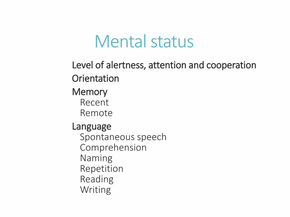
Mental statusLevel of alertness, attention and cooperation
Orientation
Memory Recent Remote
LanguageSpontaneous speechComprehensionNamingRepetitionReadingWriting

Limbic system

Basal ganglia
Mental statusLevel of alertness, attention and cooperation
Orientation
Memory Recent Remote
LanguageSpontaneous speechComprehensionNamingRepetitionReadingWriting

Mental status
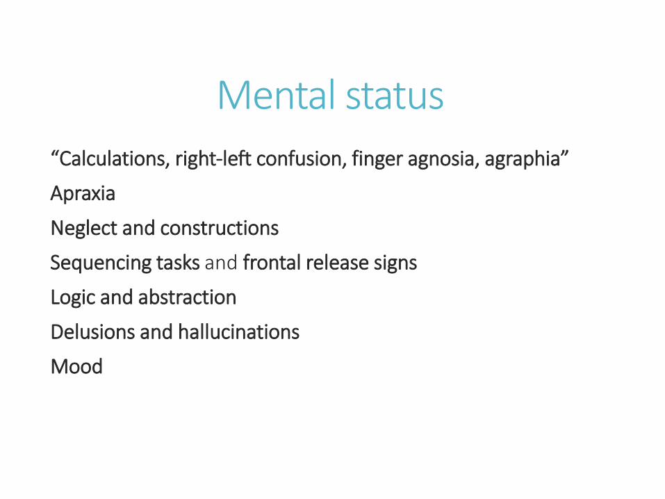
“Calculations, right-left confusion, finger agnosia, agraphia”
Apraxia
Neglect and constructions
Sequencing tasks and frontal release signs
Logic and abstraction
Delusions and hallucinations
Mood

Overlap of the brain lesions for (A) all 80 patients, (B) a subgroup of 16 patients showing
consistent neglect, and (C) 25 patients showing no neglect
Vincent Verdon et al. Brain 2010;133:880-894
Mental status
“Calculations, right-left confusion, finger agnosia, agraphia”
Apraxia
Neglect and constructions
Sequencing tasks and frontal release signs
Logic and abstraction
Delusions and hallucinations
Mood

NEUROLOGICAL EXAMMental status
Cranial nerves
Motor exam
Reflexes
Coordination and gait
Sensory exam

Cranial nerves
Olfaction (CN I)
Ophthalmoscopic exam (CN II)
Vision (CN II)
Pupillary responses (CN II, III)
Extraocular movements (CN HI, IV, VI)
Facial sensation and mastication
(CN V)


Cranial nerves
Olfaction (CN I)
Ophthalmoscopic exam (CN II)
Vision (CN II)
Pupillary responses (CN II, III)
Extraocular movements (CN HI, IV, VI)
Facial sensation and mastication
(CN V)

Cranial nerves
Facial expression and taste (CN VII)
Hearing and vestibular sense (CN VIII)
Palate elevation and gag reflex (CN IX, X)
Muscles of articulation (CN V, VII, IX, X, XII)
Sternocleidomastoid and trapezius(CN XI)
Tongue (CN XII)


Cranial nerves
Facial expression and taste (CN VII)
Hearing and vestibular sense (CN VIII)
Palate elevation and gag reflex (CN IX, X)
Muscles of articulation (CN V, VII, IX, X, XII)
Sternocleidomastoid and trapezius(CN XI)
Tongue (CN XII)

NEUROLOGICAL EXAMMental status
Cranial nerves
Motor exam
Reflexes
Coordination and gait
Sensory exam

Corticospinal Tract

Motor examObservation
Involuntary movements – BG, cerebellumTremor - BG, cerebellum, with peripheral nerve lesionsHypokinesia
InspectionMuscle wasting - MNDFasciculations - MND
PalpationTenderness – myositis, fibromyalgiaFasciculations
Muscle tone – corticospinal tract, BG
Functional testingDriftFine finger movements Rapid toe tapping
Strength of individual muscle groups

Muscle strength scale
0/5 NO contraction
1/5 Flicker, but NO movement
2/5 Movement possible, but not against gravity
3/5 Movement possible against gravity
4/5 Movement possible against resistance Optional grades: -4/5 and +4/5
5/5: Normal strength

Upper vs Lower Motor Neuron Lesions
Clinical SIGN UMN LESIONS LMN LESIONS
Weakness Yes Yes
Atrophy No Yes
Fasciculations No Yes
Reflexes Increased Decreased
Tone Increased Decreased
_________________________________________________Mild atrophy may develop as a result of disuseWith acute UMN lesions, reflexes and tone may be decreased

Upper Extremity Strength Testing
ACTION MUSCLES NERVES ROOTS
Finger extension at MCP joints
Thumb abduction in plane of palm
Ext digitorum, ext indicis, ext digiti min
Abductor pollicis longus
Radial nerve (post
interosseous nerve)
C7,C8
C7,C8
Finger abduction Dorsal interossei, abd digiti minimi Ulnar nerve C8,T1
Thumb opposition Opponens pollicis Median nerve C8,T1
Thumb abduction perpend plane of palm Abductor pollicis brevis Median nerve C8,T1
Flexion at DIP joints, dig 2,3 Flexor digitorum prof to digits 2,3 Median nerve C7, C8
Flexion at DIP joints, dig 4,5 Flexor digitorum prof to digits 4,5 Ulnar nerve C7,C8
Wrist flexion and hand abduction Flexor carpi radialis Median nerve C6, C7
Wrist flexion and hand adduction Flexor carpi ulnaris Ulnar nerve C7, C8, T1
Wrist extension and hand abduction Extensor carpi radialis Radial nerve C5, C6
Elbow flexion (with forearm supinated) Biceps, brachialis Musculocutaneous n C5,C6
Elbow extension Triceps Radial nerve C6, C7, C8
Arm abduction at shoulder Deltoid Axillary nerve C5

Lower Extremity Strength TestingACTION MUSCLES NERVES ROOTS
Hip flexion IliopsoasFemoral n and L1-L3 n roots
L1, L2, L3, L4
Knee extension Quadriceps Femoral nerve L2, L3, L4
Knee flexion Hamstrings Sciatic nerve L5, S1, S2
Leg abduction Glut med, glut min, tensor fasciae latae Superior gluteal nerve L4, L5, SI
Leg adductionObturator extemus, adductor longus, magnus, and brevis; gracilis
Obturator nerve L2, L3, L4
Toe dorsiflexionExtensor hallucis longus, extensor digitorum longus
Deep fibular nerve L5, SI
Foot dorsiflexion Tibialis anterior Deep fibular nerve L4,L5
Foot plantar flexion Triceps surae (gastrocnemius, soleus) Tibial nerve SI, S2
Foot eversion Peroneus longus, peroneus brevis Superficial fibular nerve L5, SI
Foot inversion Tibialis posterior Tibial nerve L4,L5

DETECTING SUBTLE HEMIPARESIS
PRONATOR DRIFT - Slight inward drifting rotation (pronation) of one forearm, or even a slight curling of the fingertips on one side, is abnormal
FINGER EXTENSORS
FINE MOVEMENTS - Patient rapidly taps index finger and thumb together; taps each finger to the thumb in sequence;
ISOLATED FINGER MOVEMENTS - Patient holds fingers abducted and extended and then moves one finger at a time.
SPASTIC CATCH - Feel for a subtle "catch" on one side compared to the other when holding the patient's hand and then rapidly supinating the patient's forearm
SUBTLE DECREASED NASOLABIAL FOLD
FORCED GAIT - Patient walks on the outsides of the feet. Observe the hands for subtle dystonic posturing on one side
CAREFUL GAIT TESTING - Look for slight circumduction of one leg (the leg swings out in a circular arc with each step) or decreased arm swing. Also have the patient hop on each foot and walk on the toes
SILENT PLANTAR - If a normal flexor plantar response is present on one side, then a silent plantar response on the other side may represent a subtle Babinski

NEUROLOGICAL EXAMMental status
Cranial nerves
Motor exam
Reflexes
Coordination and gait
Sensory exam
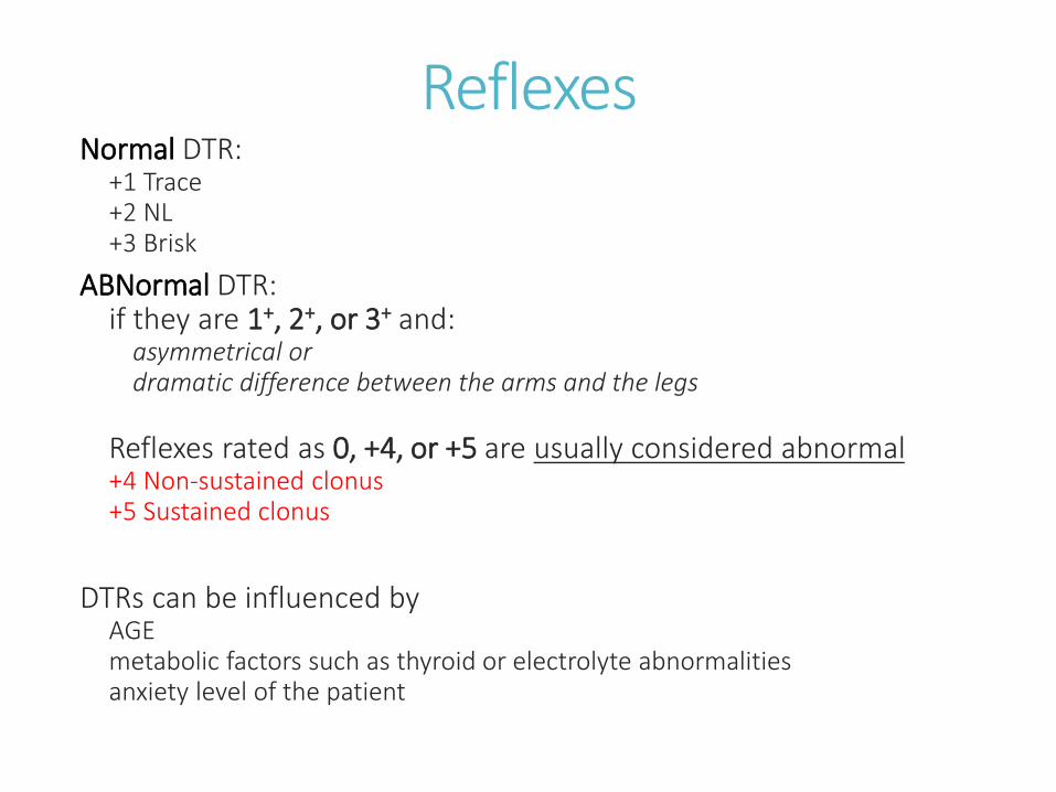
ReflexesNormal DTR:
+1 Trace+2 NL+3 Brisk
ABNormal DTR:if they are 1+, 2+, or 3+ and:
asymmetrical or dramatic difference between the arms and the legs
Reflexes rated as 0, +4, or +5 are usually considered abnormal+4 Non-sustained clonus +5 Sustained clonus
DTRs can be influenced by AGEmetabolic factors such as thyroid or electrolyte abnormalitiesanxiety level of the patient
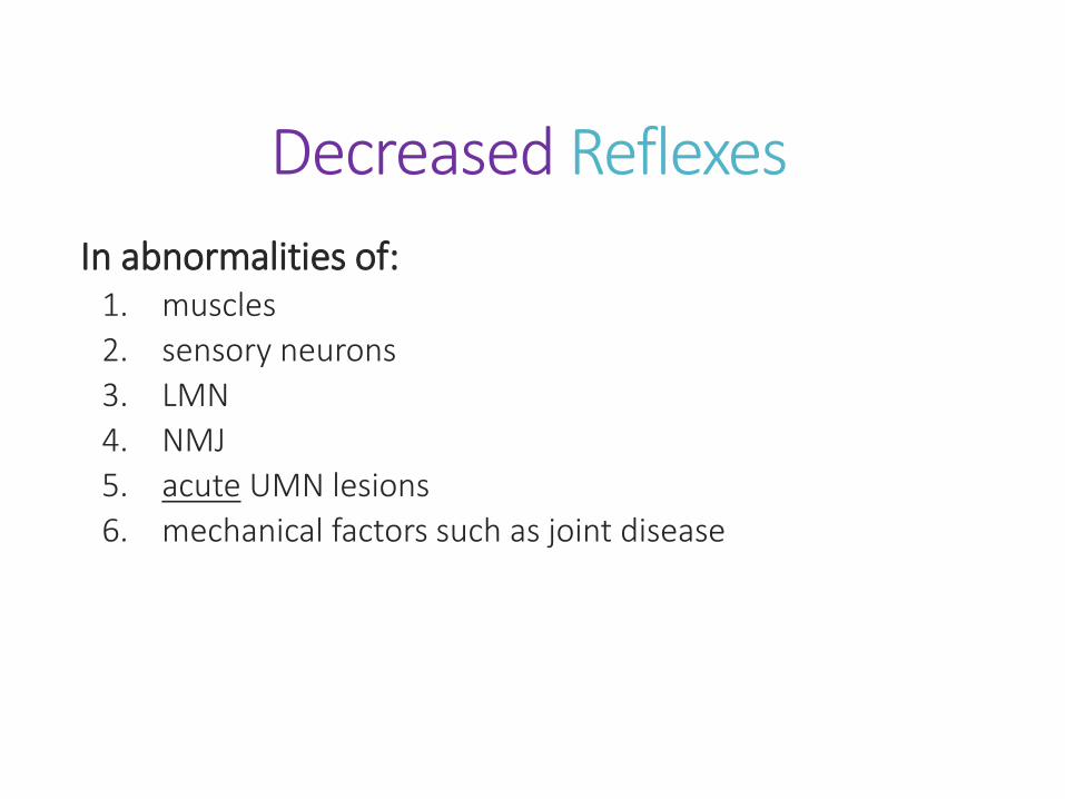
Decreased Reflexes
In abnormalities of:1. muscles
2. sensory neurons
3. LMN
4. NMJ
5. acute UMN lesions
6. mechanical factors such as joint disease

Increased Reflexes
UMN lesions
Other signs of hyperreflexiaspreading of reflexes to other muscles
crossed adduction of the opposite leg
Hoffmann's sign
Plantar response - Babinski's sign is associated with UMN lesions anywhere along the corticospinal tract

Reflexes tested in special situations
Coma
Spinal cord injury absence of certain reflexes can help localize the level and severity
abdominal cutaneous reflexes
cremasteric reflex in males
bulbocavernosus reflex
anal wink
Frontal lobe dysfunction
Neurodegenerative disorders

NEUROLOGICAL EXAMMental status
Cranial nerves
Motor exam
Reflexes
Coordination and gait
Sensory exam

Coordination and gaitAppendicular coordination
Rapid alternating movements Finger-nose-finger test Heel-shin test Overshoot
Axial coordinationRomberg testGaitOrdinary gait Tandem gait Forced gait
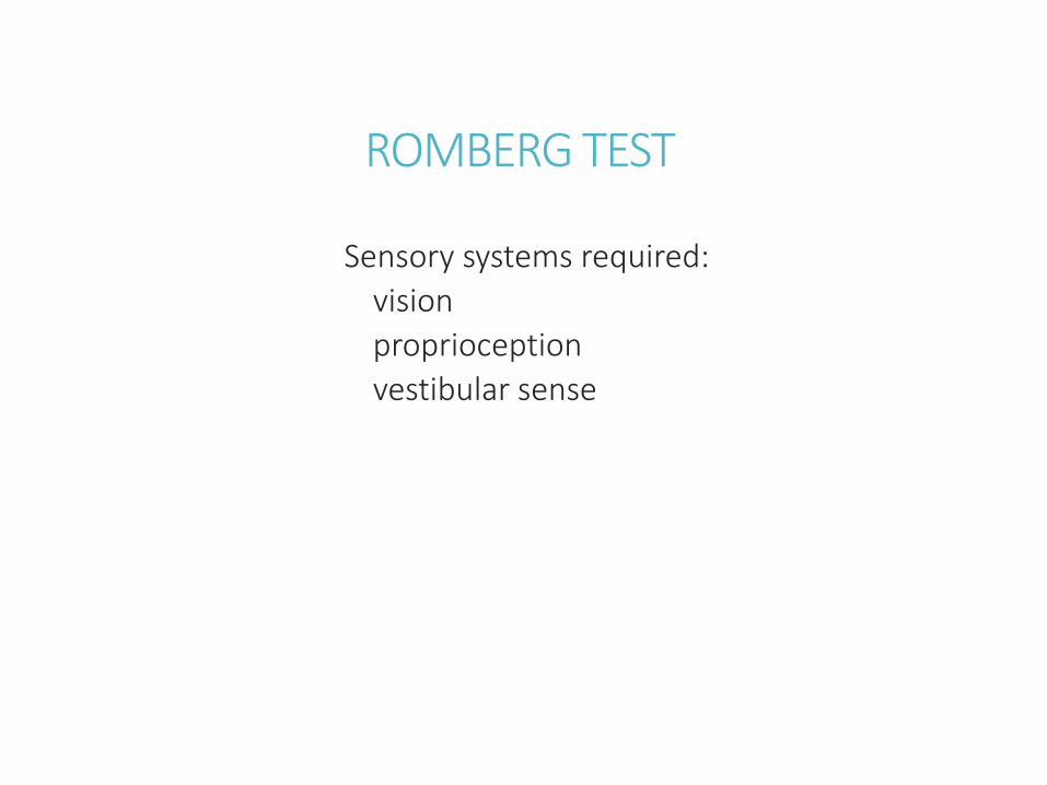
ROMBERG TEST
Sensory systems required:
vision
proprioception
vestibular sense

Gait involves multiple sensory and motor systems
vision
proprioception
vestibular sense
LMN
UMN
basal ganglia
cerebellum
higher-order motor planning systems in the association cortex

Gate exam
stance (how far apart the feet are)
posture
stability
how high the feet are raised off the floor
trajectory of leg swing
leg stiffness and degree of knee bending
arm swing
tendency to fall or swerve
rate and speed
difficulty initiating and stopping gait
involuntary movements
turning
patient's ability to rise from a chair

Localization of Common Gait Disorders 1
NAME LOCALIZATION DESCRIPTION COMMON CAUSES
SPASTICUnilateral or bilateral Corticospinal tracts
Unilateral or bilateral. Stiff-legged, circumduction, sometimes with scissoring of the legs and toe-walking (from increased calf tone), decreased arm swing, unsteady, falling toward side of greater spasticity
Cortical, subcortical, or brainstem infarcts affecting upper motor neuron pathways; cerebral palsy; degenerative conditions; multiple sclerosis; spinal cord lesions
ATAXIC
Cerebellar vermis or other midlinecerebellar structures
Wide based, unsteady, staggering side to side, and falling toward side of worse pathology. Subtle deficit can be detected with tandem (heel-to-toe, or "drunk walk") testing
Toxins such as alcohol; meds; tumors of cerebellar vermis; infarcts or ischemia of cerebellar pathways; cerebellar degener
VERTIGINOUSVestibular nuclei, vestibular nerve, or semicircular canals
Looks similar to ataxic gait, wide based and unsteady. Patients sway and fall when attempting to stand with feet together and eyes closed (Romberg sign)
Toxins such as alcohol; infarcts or ischemia of vestibular nuclei; benign positional vertigo; Me-niere's disease
FRONTALFrontal lobes or frontal subcortical white matter
Slow, shuffling, narrow or wide based, "magnetic" (barely raising feet off floor), unsteady. Sometimes resembles Parkinsonian gait. Some can perform cycling movements on their back much better than they can walk ("gait apraxia“)
Hydrocephalus; frontal tumors such as glioblastoma or meningioma; bilateral anterior cerebral artery infarcts; diffuse subcortical white matter disease
PARKINSONIANSubstantia nigra or other regions of basal ganglia
Slow, shuffling, narrow based. Difficulty initiating walking. Often stooped forward, with decreased arm swing, and "en bloc turning." Unsteady, with "retropulsion”
Parkinson's disease; other parkinsonian syndromes, such as progressive supranuclear palsy, or use of neuroleptic drugs

Localization of Common Gait Disorders 2
NAME LOCALIZATION DESCRIPTION COMMON CAUSES
DYSKINETIC
Subthalamicnucleus, or other regions of basal ganglia
Unilateral or bilateral dancelike (choreic), flinging (ballistic), or writhing (athetoid) movements occur during walking and may be accompanied by some unsteadiness.
Huntington's disease; infarct of subthalamic nucleus or striatum; side effect of levodopa; other familial or drug-induced dyskinesias
TABETICPosterior columns or sensory nerve fibers
High-stepping, foot-flapping gait, with particular difficulty walking in the dark or on uneven surfaces. Patients sway and fall in attempts to stand with feet together and eyes closed (Romberg sign)
Posterior cord syndrome; severe sensory neuropathy
PARETIC
Nerve roots, peripheral nerves, neuromuscular junction, or muscles
Exact appearance depends on location of lesion. With proximal hip weakness there may be a waddling, Trendelenburg gait. Severe thigh weakness may cause sudden knee buckling.Foot drop can cause a high-stepping, slapping gait, with frequent tripping.
Numerous peripheral nerve and muscle disorders
PAINFUL (ANTALGIC)
Peripheral nerve or orthopedic injury
Pain may be obvious based on patient's report or facial expression. Patients tend to avoid putting pressure on affected limb.
Herniated disc; peripheral neuropathy; muscle strain; contusions; fractures
ORTHOPEDICBones, joints, tendons, ligaments, and muscles
Depends on nature and location of the disorder. Peripheral nerve injury or spinal cord-related deficits may be present as well
Arthritis; fractures; dislocations; contractures; soft tissue injuries
FUNCTIONALPsychologically based
Can be hard to diagnose. Sometimes patients say they have poor balance, yet spontaneously perform highly destabilizing swaying movements while walking, without ever falling.
Conversion disorder; factitious disorder

NEUROLOGICAL EXAMMental status
Cranial nerves
Motor exam
Reflexes
Coordination and gait
Sensory exam

Sensory examPrimary sensation — asymmetry, sensory level
Light touch, two-point discriminationPain (sharp vs. dull)Temperature (cold vs. warm)Vibration and joint position sense
Cortical sensationGraphesthesiaStereognosisExtinction

Localization of sensory deficits
peripheral nerves
spinal nerve roots
spinal cord or brainstemposterior columns
anterolateral sensory systems
thalamus
sensory cortex

Pain and TemperatureSpinothalamic (anterolateral) pathway

Vibration & Joint PositionPosterior Column Pathway

Spinal nerve root pattern (Dermatomes)

Peripheral nerve patterns ARM

Peripheral nerve patterns LEG

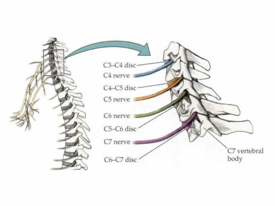
3 Roots to Remember in the ARM
NERVE
ROOT
MAIN
WEAKNESS
REFLEX
DECREASED
REGION OF
SENSORY
ABNORMALITY
USUAL DISC
INVOLVED
APPROXIMATE
PERCENTAGE
OF CERVICAL
RADICULOPATHIES
C5
Deltoid,
infraspinatus,
biceps
Biceps,
pectoralis
Shoulder, upper
lateral armC4-C5 7%
C6Wrist extensors,
biceps
Biceps,
brachioradialis
First and second
fingers, lateral
forearm
C5-C6 18%
C7 Triceps Triceps Third finger C6-C7 46%

3 Roots to Remember in the LEG


SCREENING NEURO EXAM
First, learn the complete neurological exam
Then learn when and how to do a focused exam
Examine patients to get a feel of NORMAL
Are the findings of the neuro exam anatomically CORRECT?
What should a focused exam involve?

SCREENING NEURO EXAM
Mental statusLevel of alertness and orientation. Months forward/backward. Immediate registration and delayed recall of 3 objects for 4 minutes (timed). Naming of watch parts. Note behavior, language, affect.
CN
Pupil light reflexes. Ophthalmoscopy. Visual fields, including extinction testing. Horizontal and vertical smooth pursuit eye movements. Facial sensation to light touch including extinction testing. Facial symmetry during emotional smile. Hearing of finger rub. Palate elevation. Note quality of voice during exam. Head turning and shoulder shrug against resistance. Tongue protrusion.
Motor
Drift. Rapid hand and foot tapping. Upper and lower extremity tone. Strength in several proximal and distal muscles in the upper and lower extremities bilaterally (e.g., finger extensors, finger abductors, wrist extensors, biceps, triceps, deltoids, iliopsoas, quadriceps, foot and toe dorsiflexors, and knee flexors).
Reflexes Biceps, brachioradialis, patellar, Achilles tendon, plantars.
Coordination and gait Finger-nose-finger and heel-shin tests. Gait and tandem gait.
SensoryLight touch in hands and feet, including extinction testing. Pin prick and temp testing in feet. Vibration and joint position sense in feet.

Patient presentation is determined by
Time courseAcute
(i.e. stroke) generally causes obvious symptoms as the loss of function is abruptno time to develop compensatory mechanisms
Sub-acute or chronicDisorders which occur more slowly tend to cause relatively subtle symptoms
Size and location of the lesionmore overt problems
Larger lesions Lesions affecting critical areas of function
Pre-existing medical or neurological dysfunction

Exam Limitations and Workaround StrategiesPATIENTCE IS REQUIRED!
In MILD to MODERATE impairment of alertness or attentionmost aspects of the exam can be done with repeated stimulation
In SEVERE impairment of alertness or attentionfollow comatose patient exam procedures
UNCOOPERATIVE patient, strategy depends on the situation:careful observation of spontaneous speech and movementssome techniques from the coma exam
For poor language COMPREHENSIONsimple questions or commands gestures or demonstrations of the action desired.
For impaired EXPRESSIONask yes-no or multiple-choice questionsfor testing memory, several objects can be hidden around the room and the patient can be asked to find them after a delay

ConclusionsThe Neuro Exam can be adapted to test patients who are
awake and cooperativesuffering from psychiatric disordersmalingeringcomatoseor any combination of other impairments
Explore the neuroanatomical systems being tested and the effects of disease on function
Once the clinical suspicion of a lesion has been raised, several important decisions need to be made
Depending on the TYPE and LOCATION of the lesion consider: Emergency surgical or medical therapyLess urgent therapyFurther investigations
ReferencesAids to the Examination of the Peripheral Nervous System.
Bickley LS (ed.). 2008. Bates' Guide to Physical Examination and History Taking. 10th Ed. Lippincott-Raven, Philadelphia.
Blumenfeld H. 2001. The NeuroExam Video. Sinauer, Sunderland, MA.
Blumenfeld H. 2009. The neurological examination of consciousness. In The Neurology of Consciousness, S Laureys and G Tononi (eds.), Chapters 15-30. Academic Press, New York.
Lectures by Dr Michael Aminoff

PROVIDENCEBrain and Spine Institute
Recommended