IMMUNE NETWORK http://www.ksimm.or.kr Volume 11 Number 6 December 2011
http://dx.doi.org/10.4110/in.2011.11.6.358
pISSN 1598-2629 eISSN 2092-6685ORIGINAL ARTICLE
358
Received on October 4, 2011. Revised on October 19, 2011. Accepted on October 25, 2011.CC This is an open access article distributed under the terms of the Creative Commons Attribution Non-Commercial
License (http://creativecommons.org/licenses/by-nc/3.0) which permits unrestricted non-commercial use, distribu-tion, and reproduction in any medium, provided the original work is properly cited.
*Corresponding Author. Tel: 82-2-740-8308; Fax: 82-2-743-0881; E-mail: [email protected]
Keywords: Interferon-gamma, Islet transplantation, Immune tolerance
Influence of Interferon-γ Deficiency in Immune Tolerance Induced by Male Islet TransplantationYong-Hee Kim1,2,3,4, Youngkyoung Lim1 and Chung-Gyu Park1,2,3,4*1Department of Microbiology and Immunology, 2Tumor Immunity Medical Research Center, 3Cancer Research Institute, 4Xenotransplantation Research Center, Seoul National University College of Medicine, Seoul 110-799, Korea
Background: Traditionally, interferon-γ (IFN-γ) was regar-ded as a pro-inflammatory cytokine, however, recent reports suggested role of IFN-γ in immune tolerance. In our pre-vious report, we could induce tolerance to male antigen (HY) just by male islet transplantation in wild type C57BL/6 mice without any immunological intervention. We tried to inves-tigate the influence of IFN-γ deficiency on tolerance induc-tion by male islet transplantation. Methods: To examine the immunogenicity of male tissue in the absence of IFN-γ, we transplanted male IFN-γ knock-out (KO) skin to female IFN-γ KO mice. Next, we analyzed male IFN-γ KO islet to streptozotocin-induced diabetic female IFN-γ KO mice. And, we checked the functionality of grafted islet by graft re-moval and insulin staining. Results: As our previous results in wild type C57BL/6 mice, female IFN-γ KO mice rejected male IFN-γ KO skin within 29 days, and did not reject male IFN-γ KO islet. The maintenance of normal blood glucose level was dependent on the presence of grafted male islet. And the male islet recipient did not reject 2nd challenge of male islet graft also. Conclusion: Deficiency of IFN-γ does not have influence on the result of male skin graft and male islet transplantation. Conclusively, male islet transplantation induced T cell tolerance is not dependent on the presence of IFN-γ.[Immune Network 2011;11(6):358-363]
INTRODUCTION
It has been known that the skin graft across minor histo-
compatibility of male antigen (HY) mismatch is normally re-
jected (1,2). However, in our previous report, islet trans-
plantation across HY barrier without any immunological ma-
nipulation induced HY antigen specific immunological toler-
ance which prevented the rejection of subsequent skin graft
in C57BL/6 mouse model (3). In that tolerance, increased
Foxp3+ regulatory T cells (Treg) mediated the tolerance
induction. To explore the factor that made Treg increase and
tolerance induction caused by islet transplantation, we started
subtraction study using knock-out (KO) mouse. Our first
choice was interferon-γ (IFN-γ) KO mouse.
IFN-γ was regarded traditionally as a pro-inflammatory
cytokine. However its protective role in Th-1 cell-associated
autoimmune diseases has recently been reported (4). For in-
stance, ablation of IFN-γ resulted in aggravation of murine
autoimmune disease, collagen-induced arthritis (CIA), indicat-
ing that endogenous IFN-γ inhibits important inflammatory
pathway (5,6). The protective effects of endogenous IFN-γ
also have been reported in other autoimmune models such
as experimental autoimmune encephalomyelitis (EAE), ex-
perimental autoimmune uveitis (EAU), autoimmune nephritis,
and myocarditis (7-13).
In Treg development, activation of signal transducer and
activator of transcription-1 (STAT-1) seems to have a role
(14). This suggests that cytokine, such as IFN-γ, that activate
STAT-1 have a role in Treg development (4). Wang et al.
(15) have reported that in vitro treatment of CD4+
CD25−
T
cells with IFN-γ leads to their conversion to Foxp3+ Treg
cells. And, copolymer-I induced in vitro Treg conversion was
dependent on IFN-γ (16). In addition, Feng et al.(17) re-

Male Islet Transplantation in IFN-γ KO MiceYong-Hee Kim, et al.
359IMMUNE NETWORK http://www.ksimm.or.kr Volume 11 Number 6 December 2011
ported that ex vivo IFN-γ conditioning of CD4+ T cells with
immature dendritic cells resulted in the Treg conversion, and
these induced Treg cells could prevent allograft rejection.
Another study in mice showed that IFN-γ expression in Treg
cells is transiently up-regulated after alloantigen exposure in
vivo and is important for their regulatory function (18). In
another study, in vitro activated Treg cells have shown to pro-
duce IFN-γ and to also stimulate dendritic cells to produce
indoleamine 2,3-dioxygenase (IDO) (19). This enzyme con-
verts the essential amino acid tryptophan into N-formylkynur-
enine (20). By depleting tryptophan, IDO exerts the capacity
to inhibit T-cell response. Recent evidence suggests an im-
portant role of IDO in the induction of tolerance (21).
Considering roles of IFN-γ in self-tolerance and Treg de-
velopment, we speculated that deficiency of IFN-γ might re-
sult in the failure of tolerance induction in our male islet
transplantation model. We examined immunogenicity of male
skin in IFN-γ KO mice, and transplanted male islets to IFN-γ
KO mice. By these experiments, we confirmed the role of
IFN-γ in tolerance induction by male islet transplantation.
MATERIALS AND METHODS
MiceC57BL/6-Ifngtm1Ts(IFN-γ knock-out) mice were purchased from
The Jackson Laboratory (Bar Harbor, ME, USA). All IFN-γ KO
mice used in this study were genotyped for IFN-γ. PCR was
done with primers and cycling conditions recommended by
The Jackson Laboratory. The normal endogenous gene was
detected with forward primer IMR126 (5’-AGA AG T AAG
TGG AAG GGC CCA GAA G-3’) and reverse primer IMR127
(5’-AGG GAA ACT GGG AGA GGA GAA ATA T-3’). The mu-
tant (knock-out) IFN-γ was detected with forward primer
IMR128 (5’-TCA GCG CAG GGG CGC CCG GTT CTT T-3’)
and reverse primer IMR129 (5’-ATC GAC AAG ACC GGC TTC
CAT CCG A-3’). Endogenous IFN-γ resulted in a 220-bp PCR
product and mutant IFN-γ resulted in a 375-bp PCR product.
Mice were bred and housed in a specific pathogen-free
facility. Animal studies were conducted under protocols ap-
proved by Seoul National University Institutional Animal Care
and Use Committee.
Induction of diabetes mellitus and blood glucose monitoringTo induce diabetes mellitus, mice were injected intra-
peritoneally with 125 mg/kg streptozotocin (Sigma, St. Louis,
MO, USA) in two consecutive days. After the injections,
non-fasting blood glucose levels were monitored using a gluc-
ometer, OneTouch Ultra (LifeScan Inc., Milpitas, CA, USA)
from blood obtained by tail snipping. Mice with two consec-
utive non-fasting blood glucose levels higher than 250 mg/dl
were considered as hyperglycemic mice. Hyperglycemic mice
were selected for the islet transplantation.
Isolation and transplantation of pancreatic isletsSacrificed donor mice’s pancreases were injected with Hanks
balanced salt solution containing 0.5 mg/ml of Collagenage
P (Roche, Mannheim, Germany) via pancreatic duct. Inflated
pancreases were excised and incubated in a 37oC water bath
for 20 minutes. Digested pancreases were filtered through a
sieve and washed. Then, islets were purified from digests us-
ing Euro-Ficoll gradients. Obtained islets were re-suspended
with RPMI 1640 medium (Invitrogen, Carlsbad, CA, USA) sup-
plemented with 10% fetal bovine serum, 2 mM L-glutamine,
50μg/ml gentamycin, 100μM non-essential amino acids (all
purchased from Invitrogen). After overnight culture, islets
were used for the transplantation. Recipient mice were anes-
thetized with Isoflurane. Then, the left kidney was exposed
and 250∼300 islet equivalent number (IEQ) islets were deliv-
ered beneath the renal capsule.
Skin graftingFull-thickness tail skins obtained from donors were trans-
planted to the anesthetized recipient mice’s graft beds on the
left flank and covered with Vaseline gauze and Band-Aid
(Johnson&Johnson, New Brunswick, NJ, USA). Bandages we-
re removed after 7 days, and grafts were observed every 2
to 3 days for 3 week, and weekly thereafter. The graft was
scored as rejected when less than 10% of viable tissue re-
mained and visible inflammation ended.
Nephrectomy & histological analysisIslet recipient’s left kidney was removed under the anesthesia
to confirm the graft function and to analyze graft histology.
Removed kidney was frozen embedded in optimal cutting
temperature (OCT) compound (Sakura Finetek, Torrance, CA,
USA). 5μm frozen section was done using Leica CM1850 cry-
ocut microtome (Leica, Wetzlar, Germany). Acetone-fixated
sections were stained with the hematoxylin and eosin. For
insulin staining, sections were treated with guinea pig anti-in-
sulin antibody (Dako Cytomation, Glostrup, Denmark) for 1
hr at room temperature. After wash with PBS, sections were
Male Islet Transplantation in IFN-γ KO MiceYong-Hee Kim, et al.
360 IMMUNE NETWORK http://www.ksimm.or.kr Volume 11 Number 6 December 2011
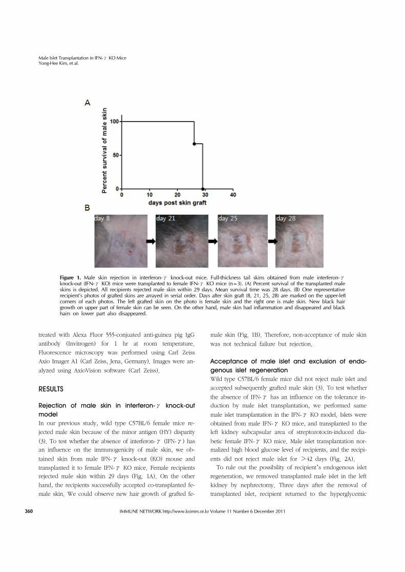
Figure 1. Male skin rejection in interferon-γ knock-out mice. Full-thickness tail skins obtained from male interferon-γknock-out (IFN-γ KO) mice were transplanted to female IFN-γ KO mice (n=3). (A) Percent survival of the transplanted male skins is depicted. All recipients rejected male skin within 29 days. Mean survival time was 28 days. (B) One representative recipient’s photos of grafted skins are arrayed in serial order. Days after skin graft (8, 21, 25, 28) are marked on the upper-leftcorners of each photos. The left grafted skin on the photo is female skin and the right one is male skin. New black hair growth on upper part of female skin can be seen. On the other hand, male skin had inflammation and disappeared and black hairs on lower part also disappeared.
treated with Alexa Fluor 555-conjuated anti-guinea pig IgG
antibody (Invitrogen) for 1 hr at room temperature.
Fluorescence microscopy was performed using Carl Zeiss
Axio Imager A1 (Carl Zeiss, Jena, Germany). Images were an-
alyzed using AxioVision software (Carl Zeiss).
RESULTS
Rejection of male skin in interferon-γ knock-out modelIn our previous study, wild type C57BL/6 female mice re-
jected male skin because of the minor antigen (HY) disparity
(3). To test whether the absence of interferon-γ (IFN-γ) has
an influence on the immunogenicity of male skin, we ob-
tained skin from male IFN-γ knock-out (KO) mouse and
transplanted it to female IFN-γ KO mice. Female recipients
rejected male skin within 29 days (Fig. 1A). On the other
hand, the recipients successfully accepted co-transplanted fe-
male skin. We could observe new hair growth of grafted fe-
male skin (Fig. 1B). Therefore, non-acceptance of male skin
was not technical failure but rejection.
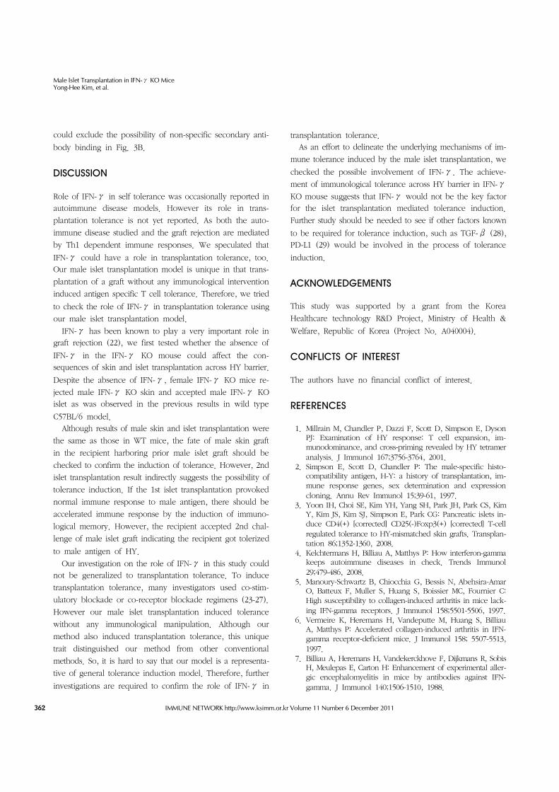
Acceptance of male islet and exclusion of endo-genous islet regenerationWild type C57BL/6 female mice did not reject male islet and
accepted subsequently grafted male skin (3). To test whether
the absence of IFN-γ has an influence on the tolerance in-
duction by male islet transplantation, we performed same
male islet transplantation in the IFN-γ KO model. Islets were
obtained from male IFN-γ KO mice, and transplanted to the
left kidney subcapsular area of streptozotocin-induced dia-
betic female IFN-γ KO mice. Male islet transplantation nor-
malized high blood glucose level of recipients, and the recipi-
ents did not reject male islet for >42 days (Fig. 2A).
To rule out the possibility of recipient’s endogenous islet
regeneration, we removed transplanted male islet in the left
kidney by nephrectomy. Three days after the removal of
transplanted islet, recipient returned to the hyperglycemic

Male Islet Transplantation in IFN-γ KO MiceYong-Hee Kim, et al.
361IMMUNE NETWORK http://www.ksimm.or.kr Volume 11 Number 6 December 2011
Figure 2. Male islet transplantation in IFN-γ knock-out mice. Female IFN-γ KO mice were rendered diabetic by 2 times of 125 mg/kg streptozotocin intraperitoneal injection. Non-fasting blood glucose levels were monitored. Male IFN-γ KO islet transplantations were operated to the diabetic recipients. Blood glucose levels (mg/dl) during the transplantation period are depicted. Each line and points indicates individual recipients. (A) Male islet transplantation norma-lized hyperglycemia of recipients. Recipients maintained normal blood glucose levels for >42 days (n=3). Arrow indicates the time point of male islet transplantation. (B) After the male islet trans-plantation to recipient’s left kidney, grafted islet was removed by nephrectomy. After the re-surge of blood glucose level, 2nd male islet transplantation was done and normalized the hyperglycemia again. Arrows indicates the time points of 1st male islet transplantation, nephrectomy and re-transplantation, respectively.
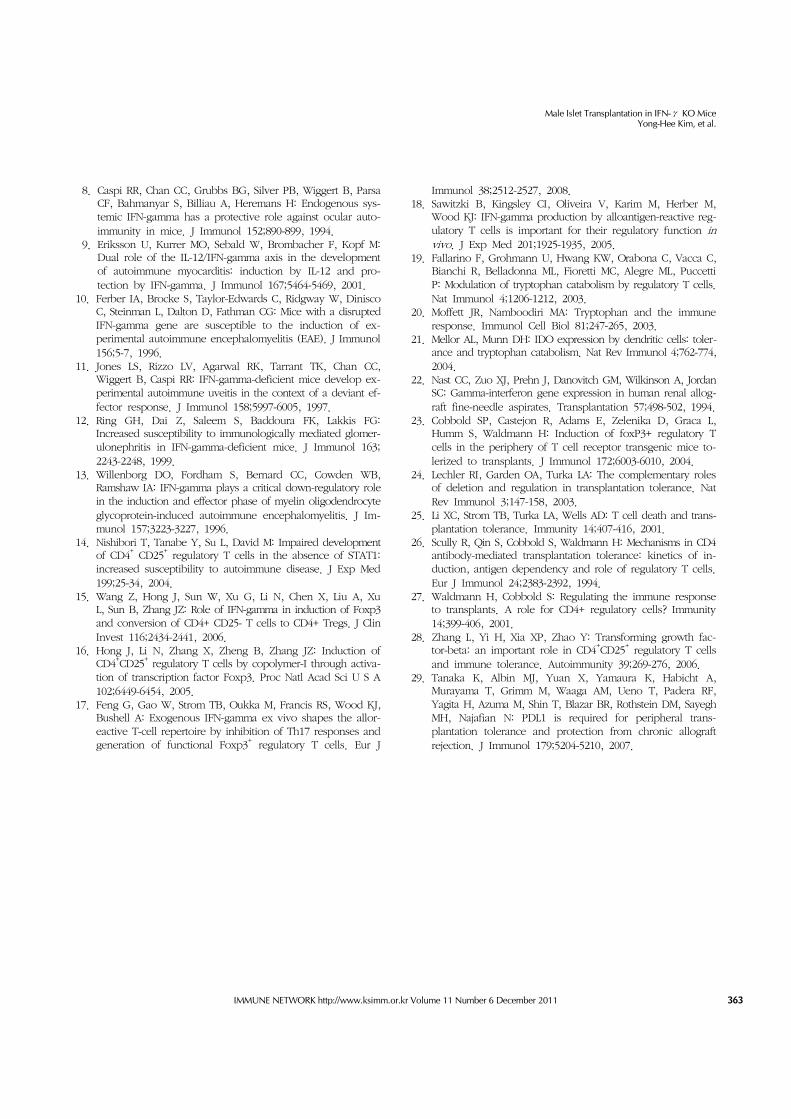
Figure 3. Histological analysis of islet graft. Removed kidney containing grafted islet of recipient in Fig. 3B was frozen-sectioned as 5μm thickness and acetone-fixated. (A) H&E staining was done to the section. Rectangular area indicates islet region. (B) Insulin staining was done with primary guinea pig anti-insulin antibody and secondary Alexa Fluor 555-conjugated anti-guinea pig IgG antibody, subsequently. Red spotsindicate plenty of stained insulin in islet region. Blue spots are nuclei of cells resulted by DAPI staining. (C) As a control of ‘(B)’, secondary antibody staining without primary antibody did not result in non-specific red spots. Magnifications are ×100.
state (Fig. 2B). Therefore, the maintenance of normal blood
glucose level was not because of the endogenous islet re-
generation, but was dependent on the presence of trans-
planted male islet. To re-normalize the blood glucose level,
and to check the recipient’s response to 2nd antigen chal-
lenge, we again transplanted male IFN-γ KO islets to the
recipient. 2nd islet transplantation promptly re-normalized the
hyperglycemia, and the recipient maintained normal blood
glucose level for >32 days (Fig. 2B). Because recipient did
not reject 2nd male islet, we could conclude that non-re-
jection of 1st male islet was not just adaptation, and 1st male
islet transplantation did not provoked immunological memory
to male antigen.
Presence of insulin in grafted isletTo confirm whether the grafted islets were functioning, we
analyzed the insulin secretion by grafted islet using the insulin
staining of the graft site. Removed kidney containing grafted
islet of recipient in Fig. 2B was frozen-sectioned and stained
with hematoxylin & eosin to identify the position of graft site
(Fig. 3A). Insulin staining was also conducted on the serial
section. We could detect plenty of insulin-specific spots in
islet region (Fig. 3B). Indicating that grafted islets were ac-
tively secreting insulin. As a control staining, only secondary
Alexa Fluor 555-conjugated anti-guinea pig IgG antibody
staining without primary anti-insulin antibody staining, did
not produced any positive signal (Fig. 3C). Therefore, we
Male Islet Transplantation in IFN-γ KO MiceYong-Hee Kim, et al.
362 IMMUNE NETWORK http://www.ksimm.or.kr Volume 11 Number 6 December 2011
could exclude the possibility of non-specific secondary anti-
body binding in Fig. 3B.
DISCUSSION
Role of IFN-γ in self tolerance was occasionally reported in
autoimmune disease models. However its role in trans-
plantation tolerance is not yet reported. As both the auto-
immune disease studied and the graft rejection are mediated
by Th1 dependent immune responses. We speculated that
IFN-γ could have a role in transplantation tolerance, too.
Our male islet transplantation model is unique in that trans-
plantation of a graft without any immunological intervention
induced antigen specific T cell tolerance. Therefore, we tried
to check the role of IFN-γ in transplantation tolerance using
our male islet transplantation model.
IFN-γ has been known to play a very important role in
graft rejection (22), we first tested whether the absence of
IFN-γ in the IFN-γ KO mouse could affect the con-
sequences of skin and islet transplantation across HY barrier.
Despite the absence of IFN-γ, female IFN-γ KO mice re-
jected male IFN-γ KO skin and accepted male IFN-γ KO
islet as was observed in the previous results in wild type
C57BL/6 model.
Although results of male skin and islet transplantation were
the same as those in WT mice, the fate of male skin graft
in the recipient harboring prior male islet graft should be
checked to confirm the induction of tolerance. However, 2nd
islet transplantation result indirectly suggests the possibility of
tolerance induction. If the 1st islet transplantation provoked
normal immune response to male antigen, there should be
accelerated immune response by the induction of immuno-
logical memory. However, the recipient accepted 2nd chal-
lenge of male islet graft indicating the recipient got tolerized
to male antigen of HY.
Our investigation on the role of IFN-γ in this study could
not be generalized to transplantation tolerance. To induce
transplantation tolerance, many investigators used co-stim-
ulatory blockade or co-receptor blockade regimens (23-27).
However our male islet transplantation induced tolerance
without any immunological manipulation. Although our
method also induced transplantation tolerance, this unique
trait distinguished our method from other conventional
methods. So, it is hard to say that our model is a representa-
tive of general tolerance induction model. Therefore, further
investigations are required to confirm the role of IFN-γ in
transplantation tolerance.
As an effort to delineate the underlying mechanisms of im-
mune tolerance induced by the male islet transplantation, we
checked the possible involvement of IFN-γ. The achieve-
ment of immunological tolerance across HY barrier in IFN-γ
KO mouse suggests that IFN-γ would not be the key factor
for the islet transplantation mediated tolerance induction.
Further study should be needed to see if other factors known
to be required for tolerance induction, such as TGF-β (28),
PD-L1 (29) would be involved in the process of tolerance
induction.
ACKNOWLEDGEMENTS
This study was supported by a grant from the Korea
Healthcare technology R&D Project, Ministry of Health &
Welfare, Republic of Korea (Project No. A040004).
CONFLICTS OF INTEREST
The authors have no financial conflict of interest.
REFERENCES
1. Millrain M, Chandler P, Dazzi F, Scott D, Simpson E, Dyson PJ: Examination of HY response: T cell expansion, im-munodominance, and cross-priming revealed by HY tetramer analysis. J Immunol 167;3756-3764, 2001.
2. Simpson E, Scott D, Chandler P: The male-specific histo-compatibility antigen, H-Y: a history of transplantation, im-mune response genes, sex determination and expression cloning. Annu Rev Immunol 15;39-61, 1997.
3. Yoon IH, Choi SE, Kim YH, Yang SH, Park JH, Park CS, Kim Y, Kim JS, Kim SJ, Simpson E, Park CG: Pancreatic islets in-duce CD4(+) [corrected] CD25(-)Foxp3(+) [corrected] T-cell regulated tolerance to HY-mismatched skin grafts. Transplan-tation 86;1352-1360, 2008.
4. Kelchtermans H, Billiau A, Matthys P: How interferon-gamma keeps autoimmune diseases in check. Trends Immunol 29;479-486, 2008.
5. Manoury-Schwartz B, Chiocchia G, Bessis N, Abehsira-Amar O, Batteux F, Muller S, Huang S, Boissier MC, Fournier C: High susceptibility to collagen-induced arthritis in mice lack-ing IFN-gamma receptors. J Immunol 158;5501-5506, 1997.
6. Vermeire K, Heremans H, Vandeputte M, Huang S, Billiau A, Matthys P: Accelerated collagen-induced arthritis in IFN- gamma receptor-deficient mice. J Immunol 158; 5507-5513, 1997.
7. Billiau A, Heremans H, Vandekerckhove F, Dijkmans R, Sobis H, Meulepas E, Carton H: Enhancement of experimental aller-gic encephalomyelitis in mice by antibodies against IFN- gamma. J Immunol 140;1506-1510, 1988.
Male Islet Transplantation in IFN-γ KO MiceYong-Hee Kim, et al.
363IMMUNE NETWORK http://www.ksimm.or.kr Volume 11 Number 6 December 2011
8. Caspi RR, Chan CC, Grubbs BG, Silver PB, Wiggert B, Parsa CF, Bahmanyar S, Billiau A, Heremans H: Endogenous sys-temic IFN-gamma has a protective role against ocular auto-immunity in mice. J Immunol 152;890-899, 1994.
9. Eriksson U, Kurrer MO, Sebald W, Brombacher F, Kopf M: Dual role of the IL-12/IFN-gamma axis in the development of autoimmune myocarditis: induction by IL-12 and pro-tection by IFN-gamma. J Immunol 167;5464-5469, 2001.
10. Ferber IA, Brocke S, Taylor-Edwards C, Ridgway W, Dinisco C, Steinman L, Dalton D, Fathman CG: Mice with a disrupted IFN-gamma gene are susceptible to the induction of ex-perimental autoimmune encephalomyelitis (EAE). J Immunol 156;5-7, 1996.
11. Jones LS, Rizzo LV, Agarwal RK, Tarrant TK, Chan CC, Wiggert B, Caspi RR: IFN-gamma-deficient mice develop ex-perimental autoimmune uveitis in the context of a deviant ef-fector response. J Immunol 158;5997-6005, 1997.
12. Ring GH, Dai Z, Saleem S, Baddoura FK, Lakkis FG: Increased susceptibility to immunologically mediated glomer-ulonephritis in IFN-gamma-deficient mice. J Immunol 163; 2243-2248, 1999.
13. Willenborg DO, Fordham S, Bernard CC, Cowden WB, Ramshaw IA: IFN-gamma plays a critical down-regulatory role in the induction and effector phase of myelin oligodendrocyte glycoprotein-induced autoimmune encephalomyelitis. J Im-munol 157;3223-3227, 1996.
14. Nishibori T, Tanabe Y, Su L, David M: Impaired development of CD4+ CD25+ regulatory T cells in the absence of STAT1: increased susceptibility to autoimmune disease. J Exp Med 199;25-34, 2004.
15. Wang Z, Hong J, Sun W, Xu G, Li N, Chen X, Liu A, Xu L, Sun B, Zhang JZ: Role of IFN-gamma in induction of Foxp3 and conversion of CD4+ CD25- T cells to CD4+ Tregs. J Clin Invest 116;2434-2441, 2006.
16. Hong J, Li N, Zhang X, Zheng B, Zhang JZ: Induction of CD4+CD25+ regulatory T cells by copolymer-I through activa-tion of transcription factor Foxp3. Proc Natl Acad Sci U S A 102;6449-6454, 2005.
17. Feng G, Gao W, Strom TB, Oukka M, Francis RS, Wood KJ, Bushell A: Exogenous IFN-gamma ex vivo shapes the allor-eactive T-cell repertoire by inhibition of Th17 responses and generation of functional Foxp3+ regulatory T cells. Eur J
Immunol 38;2512-2527, 2008.18. Sawitzki B, Kingsley CI, Oliveira V, Karim M, Herber M,
Wood KJ: IFN-gamma production by alloantigen-reactive reg-ulatory T cells is important for their regulatory function in vivo. J Exp Med 201;1925-1935, 2005.
19. Fallarino F, Grohmann U, Hwang KW, Orabona C, Vacca C, Bianchi R, Belladonna ML, Fioretti MC, Alegre ML, Puccetti P: Modulation of tryptophan catabolism by regulatory T cells. Nat Immunol 4;1206-1212, 2003.
20. Moffett JR, Namboodiri MA: Tryptophan and the immune response. Immunol Cell Biol 81;247-265, 2003.
21. Mellor AL, Munn DH: IDO expression by dendritic cells: toler-ance and tryptophan catabolism. Nat Rev Immunol 4;762-774, 2004.
22. Nast CC, Zuo XJ, Prehn J, Danovitch GM, Wilkinson A, Jordan SC: Gamma-interferon gene expression in human renal allog-raft fine-needle aspirates. Transplantation 57;498-502, 1994.
23. Cobbold SP, Castejon R, Adams E, Zelenika D, Graca L, Humm S, Waldmann H: Induction of foxP3+ regulatory T cells in the periphery of T cell receptor transgenic mice to-lerized to transplants. J Immunol 172;6003-6010, 2004.
24. Lechler RI, Garden OA, Turka LA: The complementary roles of deletion and regulation in transplantation tolerance. Nat Rev Immunol 3;147-158, 2003.
25. Li XC, Strom TB, Turka LA, Wells AD: T cell death and trans-plantation tolerance. Immunity 14;407-416, 2001.
26. Scully R, Qin S, Cobbold S, Waldmann H: Mechanisms in CD4 antibody-mediated transplantation tolerance: kinetics of in-duction, antigen dependency and role of regulatory T cells. Eur J Immunol 24;2383-2392, 1994.
27. Waldmann H, Cobbold S: Regulating the immune response to transplants. A role for CD4+ regulatory cells? Immunity 14;399-406, 2001.
28. Zhang L, Yi H, Xia XP, Zhao Y: Transforming growth fac-tor-beta: an important role in CD4+CD25+ regulatory T cells and immune tolerance. Autoimmunity 39;269-276, 2006.
29. Tanaka K, Albin MJ, Yuan X, Yamaura K, Habicht A, Murayama T, Grimm M, Waaga AM, Ueno T, Padera RF, Yagita H, Azuma M, Shin T, Blazar BR, Rothstein DM, Sayegh MH, Najafian N: PDL1 is required for peripheral trans-plantation tolerance and protection from chronic allograft rejection. J Immunol 179;5204-5210, 2007.
Recommended