Embed Size (px)
Citation preview
18-1152-69
Edition AB
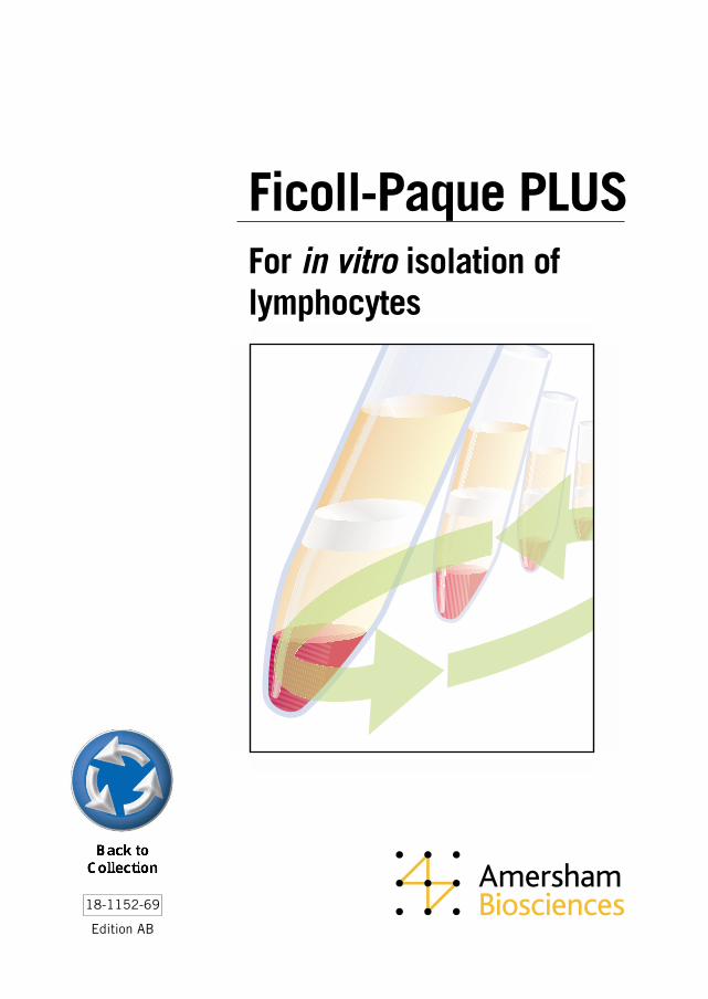
For in vitro isolation oflymphocytes
Ficoll-Paque PLUS
Antibody PurificationHandbook18-1037-46
The Recombinant Protein HandbookProtein Amplification and Simple Purification18-1142-75
Protein PurificationHandbook18-1132-29
Ion Exchange ChromatographyPrinciples and Methods18-1114-21
Affinity ChromatographyPrinciples and Methods18-1022-29
Hydrophobic Interaction ChromatographyPrinciples and Methods18-1020-90
Gel FiltrationPrinciples and Methods18-1022-18
Handbooksfrom Amersham Biosciences
Reversed Phase ChromatographyPrinciples and Methods18-1134-16
Expanded Bed AdsorptionPrinciples and Methods18-1124-26
Chromatofocusingwith Polybuffer and PBE50-01-022PB
Microcarrier cell culturePrinciples and Methods18-1140-62
Ficoll-Paque PLUSFor in vitro isolation of lymphocytes
4
Content
Introduction ................................................................................................. 5
Ficoll-Paque PLUS ....................................................................................... 6The separation principle ............................................................................................... 6
A recommended standard method ................................................................................. 7Equipment and solutions required .................................................................................................... 8
Preparation of the sample ............................................................................................................... 8
Procedure for isolation of lymphocytes .............................................................................................. 8
Washing lymphocytes free of platelets ............................................................................................. 10
Typical results from our laboratories ............................................................................................... 10
Notes .......................................................................................................................................... 10
Troubleshooting inadequate performance ........................................................................................ 12
Properties of lymphocytes isolated by the Ficoll-Paque PLUS method ............................. 13
Further applications of Ficoll-Paque PLUS ................................................................... 13Availability and storage ................................................................................................................. 14
Precautionary note ........................................................................................................................ 14
References ................................................................................................. 15
Ordering Information ................................................................................... 18Selected products for cell science from Amersham Biosciences ......................................................... 18
5
IntroductionIsolation of lymphocytes from whole human blood is often required in such clinical investigationsas histocompatibility testing and the assay of cell-mediated immune responses, as well as in manyareas of immunological research.
Ficoll-Paque™ PLUS is a sterile, ready to use density gradient medium for purifying lymphocytes inhigh yield and purity from small or large volumes of human peripheral blood, using a simple andrapid centrifugation procedure based on the method developed by Bøyum (1). Ficoll-Paque PLUScan also be used to prepare purified lymphocytes from sources other than human peripheral blood.
Ficoll-Paque PLUS from Amersham Biosciences is:
• A sterile endotoxin tested (<0.12 EU/ml) solution of Ficoll™ 400 and sodium diatrizoate with adensity of 1.077 + 0.001 g/ml.
• Recommended for small- or large-scale isolation of viable lymphocytes in high yield fromwhole human peripheral blood.
• Subjected to rigorous quality control function testing, which guarantees reproducibleperformance from batch to batch.
• Supplied in bottles sealed with a rubber septum closure, which facilitates aseptic withdrawalof solution.
• Available in convenient pack sizes: 6 x 100 ml for research requirements and 6 x 500 ml fordaily routine lymphocyte isolation. Detailed instructions for use are included with each pack.
• Stable for at least 3 yr when stored at 4–25 °C and protected from light.
Separation of normal whole human peripheral blood by the procedure recommended in this booklettypically yields a lymphocyte preparation with:
• 60 + 20% recovery of the lymphocytes present in the original blood sample,
• 95 + 5% monocular cells,
• >90% viability of the separated cells,
• 3 + 2% granulocytes,
• 5 + 2% erythrocytes and
• <0.5% of the total platelet content of the original blood sample.
6
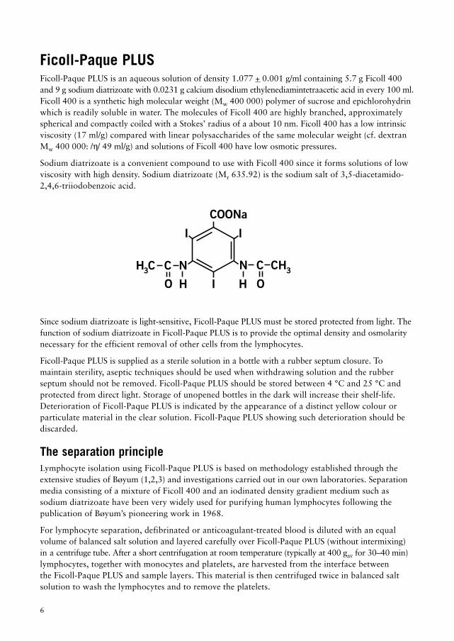
Ficoll-Paque PLUSFicoll-Paque PLUS is an aqueous solution of density 1.077 + 0.001 g/ml containing 5.7 g Ficoll 400and 9 g sodium diatrizoate with 0.0231 g calcium disodium ethylenediamintetraacetic acid in every 100 ml.Ficoll 400 is a synthetic high molecular weight (Mw 400 000) polymer of sucrose and epichlorohydrinwhich is readily soluble in water. The molecules of Ficoll 400 are highly branched, approximatelyspherical and compactly coiled with a Stokes’ radius of a about 10 nm. Ficoll 400 has a low intrinsicviscosity (17 ml/g) compared with linear polysaccharides of the same molecular weight (cf. dextranMw 400 000: /h/ 49 ml/g) and solutions of Ficoll 400 have low osmotic pressures.
Sodium diatrizoate is a convenient compound to use with Ficoll 400 since it forms solutions of lowviscosity with high density. Sodium diatrizoate (Mr 635.92) is the sodium salt of 3,5-diacetamido-2,4,6-triiodobenzoic acid.
Since sodium diatrizoate is light-sensitive, Ficoll-Paque PLUS must be stored protected from light. Thefunction of sodium diatrizoate in Ficoll-Paque PLUS is to provide the optimal density and osmolaritynecessary for the efficient removal of other cells from the lymphocytes.
Ficoll-Paque PLUS is supplied as a sterile solution in a bottle with a rubber septum closure. Tomaintain sterility, aseptic techniques should be used when withdrawing solution and the rubberseptum should not be removed. Ficoll-Paque PLUS should be stored between 4 °C and 25 °C andprotected from direct light. Storage of unopened bottles in the dark will increase their shelf-life.Deterioration of Ficoll-Paque PLUS is indicated by the appearance of a distinct yellow colour orparticulate material in the clear solution. Ficoll-Paque PLUS showing such deterioration should bediscarded.
The separation principleLymphocyte isolation using Ficoll-Paque PLUS is based on methodology established through theextensive studies of Bøyum (1,2,3) and investigations carried out in our own laboratories. Separationmedia consisting of a mixture of Ficoll 400 and an iodinated density gradient medium such assodium diatrizoate have been very widely used for purifying human lymphocytes following thepublication of Bøyum’s pioneering work in 1968.
For lymphocyte separation, defibrinated or anticoagulant-treated blood is diluted with an equalvolume of balanced salt solution and layered carefully over Ficoll-Paque PLUS (without intermixing)in a centrifuge tube. After a short centrifugation at room temperature (typically at 400 gav for 30–40 min)lymphocytes, together with monocytes and platelets, are harvested from the interface betweenthe Ficoll-Paque PLUS and sample layers. This material is then centrifuged twice in balanced saltsolution to wash the lymphocytes and to remove the platelets.
COONa
3CHN CNC
II
I
H C3
OO H H
7
Several factors contribute to the success of this separation. On centrifugation, cells in the bloodsample sediment towards the blood/Ficoll-Paque PLUS interface, where they come in contact withthe Ficoll 400 present in Ficoll-Paque PLUS. Red blood cells are efficiently aggregated by this agentat room temperature. Aggregation increases the rate of sedimentation of the red cells, which rapidlycollect as a pellet at the bottom of the tube, where they are well separated from lymphocytes.Granulocytes also sediment to the bottom of the Ficoll-Paque PLUS layer. This process is facilitated byan increase in their densities caused by contact with the slightly hypertonic Ficoll-Paque PLUSmedium. Thus, on completion of centrifugation, both granulocytes and red blood cells are found atthe bottom of the tube, beneath the Ficoll-Paque PLUS.
Lymphocytes, monocytes, and platelets are not dense enough to penetrate into the Ficoll-Paque PLUSlayer. These cells therefore collect as a concentrated band at the interface between the original bloodsample and the Ficoll-Paque PLUS. This banding enables the lymphocytes to be recovered with highyield in a small volume with little mixing with the Ficoll-Paque PLUS medium. Washing and centrifu-gation the harvested cells subsequently removes platelets, any contaminating Ficoll-Paque PLUS andplasma. The resulting cell suspension then contains highly purified, viable lymphocytes andmonocytes and is suitable for further studies.
A recommended standard methodLymphocyte purification using Ficoll-Paque PLUS can be carried out over a wide range of bloodsample volumes. With its high yield, this method can be adapted to the processing of very smallamounts of blood, such as may be obtained from children. Because of its rapidity and simplicity it isalso the method of choice for emergency tissue typing procedures (4). For maximum reproducibility ofseparation it is recommended that a standardized procedure be used. The following procedure hasbeen evaluated in our laboratories and is recommended for separation of normal blood samples onFicoll-Paque PLUS. Simple changes can easily be made to suit a particular centrifugation system.
To standardize the technique, blood volume and diameter of the centrifuge tube should be chosen first.These factors determine the height of the blood sample in the tube and consequently the centrifugationtime. Increasing the height of the blood sample in the tube increases red cell contamination. Theseparation is, however, not appreciably affected by changing the diameter of the tube. Hence a largervolume can be separated with the same degree of purification in a tube of larger diameter if the heightof the blood sample in the tube and the separation time are kept constant.
The yield and degree of purity of the lymphocytes depend to a considerable extent on the efficiencyof red cell removal.
When erythrocytes in whole blood are aggregated, some lymphocytes are trapped in the clumps andtherefore sediment with the erythrocytes. This tendency to trap lymphocytes is reduced by dilutingthe blood. Dilution gives a better lymphocyte yield and reduces the size of the red cell clumps.Aggregation of erythrocytes is enhanced at higher temperatures (37 °C), which consequentlydecreases the yield of lymphocytes. At lower temperatures (4 °C), however, the rate of aggregation isdecreased but the time of separation is increased, which also decreases the yield of lymphocytes.A compromise temperature of 18–20 °C gives optimal results.
8
Equipment and solutions required1. Two 10 ml glass test-tubes for each blood sample to be processed. The test-tubes should be
siliconized (see “Notes”, page 10).
2. Balanced salt solution. At least 20 ml for each sample to be processed. The balanced salt solutionmay be prepared from two stock solutions, A and B.
Solution A
Conc. g/l
Anhydrous D-glucose 5.5 x 10⁻³ M (0.1%) 1.0CaCl2
.2H2O 5.0 x 10⁻³ M 0.0074MgCl2
.6H20 9.8 x 10⁻⁴ M 0.1992KCl 5.4 x 10⁻³ M 0.4026TRIS 0.145 M 17.565
Dissolve in approximately 950 ml distilled water and add conc. HCl until pH is 7.6 before adjustingthe volume to 1 l.
Solution B
Conc. g/l
NaCl 0.14 M 8.19
To prepare the balanced salt solution, mix 1 volume of solution A with 9 volumes of solution B.Prepare the solution freshly each week. Other standard salt solutions may be used.
3. Pasteur pipettes (3 ml). One for each sample to be processed. These pipettes should be siliconized(see “Notes”, page 10).
4. A low speed centrifuge.
5. Glass centrifuge tubes. Two centrifuge tubes for each blood sample to be processed. Internaldiameter approximately 1.3 cm, volume 15 ml. The centrifuge tubes should be siliconizied (see“Notes”, page 10). For larger or smaller samples see “Notes”, page 10.
6. Ficoll-Paque PLUS. 3 ml for each sample being processed. For larger or smaller samples see “Notes”, p 10.
7. Syringe with needle. Needed for withdrawing Ficoll-Paque PLUS from the bottle under aseptic conditions.
Preparation of the sampleFresh blood should be used to ensure high viability of isolated lymphocytes. Prepare the sample at+18 to +20 °C.
1. To a 10 ml test-tube add 2 ml of defibrinated- or anticoagulant-treated blood and an equalvolume of balanced salt solution (final volume 4 ml).
2. Mix by drawing the blood and the buffer in and out of a Pasteur pipette.
Procedure for isolation of lymphocytes1. Remove the blue cap on the bottle of Ficoll Paque PLUS (Fig. 1).
2. Invert the bottle of Ficoll-Paque PLUS several times to ensure mixing. Using the syringe withneedle attached, pierce the septum and withdraw the required volume of Ficoll-Paque PLUS (3 mlfor each centrifuge tube) from the inverted bottle (Fig 2). If this method is employed, each bottle

9
Fig 3.
Blood sample
Ficoll-Paque PLUS
Fig 4.
Plasma Platelets
Lymphocytes
Ficoll-Paque PLUS
GranulocytesErythrocytes
Fig 5.
Upper layerremoved
Lymphocytes
will deliver at least 100 ml Ficoll-Paque PLUS.
3. Add Ficoll-Paque PLUS (3 ml) to the centrifuge tube.
4. Carefully layer the diluted blood sample (4 ml) onto the Ficoll-Paque PLUS (Fig 3).Important. When layering the sample do not mix the Ficoll-Paque PLUS and the diluted blood sample.
5. Centrifuge at 400gav for 30–40 min at 18–20 °C.
6. Draw off the upper layer using a clean Pasteur pipette, leaving the lymphocyte layer undisturbedat the interface (Fig 4 and Fig 5). Care should be taken not to disturb the lymphocyte layer.The upper layer, which contains the plasma, may be saved for later use.
Fig 1.
Fig 2.
Ficoll-Paque PLUS
GranulocytesErythrocytes
10
Washing lymphocytes free of platelets1. Using a clean Pasteur pipette transfer the lymphocyte layer to a clean centrifuge tube. It is critical to
remove all the material at the interface but in a minimum volume. Removing excess Ficoll-Paque PLUScauses granulocyte contamination; removing excess supernatant results in platelet contamination.
2. Add at least 3 volumes (6 ml) of balanced salt solution to the lymphocytes in the test-tube.
3. Suspended the cells by gently drawing them in and out of a Pasteur pipette.
4. Centrifuge at 60–100 gav for 10 min at 18–20 °C.
5. Discard the supernatant.
6. Suspend the lymphocytes in 6–8 ml balanced salt solution by gently drawing them in and out ofa Pasteur pipette.
7. Centrifuge at 60–100 gav for 10 min at 18–20 °C.
8. Discard the supernatant. The lymphocytes should now be suspended in the medium appropriateto the application.
Typical results from our laboratoriesLymphocytes: 60 + 20% recovery of lymphocytes from the original blood sample
95 + 5% of cells present in the lymphocyte fraction are mononuclear leukocytes>90% viability (measured by trypan blue exclusion)
Other cells: 3 + 2% granulocytes5 + 2% erythrocytes
<0.5% of the total platelet content of the original blood sample
NotesPreparation of glassware. All glassware that comes in contact with the sample should be siliconizedbefore use. Glassware should be immersed in a 1% silicone solution for 10 seconds (where nospecific coating procedure is recommended by the manufacturer), washed thoroughly with distilledwater and dried in an oven. The best siliconizing fluids are those based on dimethyldichlorosilanedissolved in an organic solvent.
Examples of suitable fluids are:
Sigmacote Sigma Chemicals Co., Cat. No. SL-2.Repelcote Hopkins and Williams, Cat. No. 9962-70Dimethyldichlorosilane BDH, Cat. No. 33164Prosil-28 PCR Research Chemicals.Silicone Oil Midland Silicones Ltd., Cat. No. MS 1107, use as 2—5% (v/v) solution
in ethyl acetate.Siliclad Clay-Adams, Cat. No. 1950.
Alternatively, tissue culture plasticware may be used.
Anticoagulants, Heparin, EDTA, citrate, acid citrate dextrose (ACD), and citrate phosphate dextrose(CPD) may be used as anticoagulants for the blood sample. Defibrinated blood requires no anticoagulant.Defibrination, however, results in a lower lymphocyte yield and may cause increased contaminationby red cells (3). It also causes selective loss of monocytes. Bøyum has found that a slightly purer
11
lymphocyte preparation is obtained using EDTA instead of heparin as anticoagulant (3). It has alsobeen noted in the purification of lymphocytes from sources other than peripheral blood thataddition of heparin may cause gelling of cell suspensions (5).
Larger blood samples. Larger volumes of blood may be processed with the same efficiency ofseparation by using centrifuge tubes of increased diameter while maintaining approximately thesame heights of Ficoll-Paque PLUS (2.4 cm) and blood sample (3.0 cm) as in standard methoddescribed above. Increasing the tube diameter does not affect the separation time required.
Smaller blood samples. Smaller volumes of blood can be processed rapidly by a modification of themethod of Bøyum (6). A micromethod suitable for use in tissue typing of blood cells from cadavershas also been described (4).
Sample storage. Blood samples should be processed as soon as possible after collection to ensureoptimal results. Storage for 24 h at room temperature has been reported to result in reducedlymphocyte yield, altered expression of surface markers and reduced response to mitogenicstimulation (7).
Pathological blood samples. The standard method described above has been developed for thepurification of lymphocytes from peripheral blood of normal, healthy, human donors. Differentresults may be obtained with samples taken from donors with infections or other pathologicalconditions, e.g. cancer (see “Further Applications”, page 13).
Platelet removal. The washing procedure described in this standard method will give efficientremoval of platelets from the lymphocytes in the majority of cases. If difficulty is experienced,centrifugation through a 4–20% sucrose gradient layered over Ficoll-Paque PLUS may be used to removeplatelets (8). Alternatively, the platelets may be removed by aggregation with adenosine-5-diphoshate(ADP) before separating the lymphocytes (9).
12
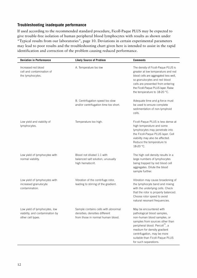
Troubleshooting inadequate performanceIf used according to the recommended standard procedure, Ficoll-Paque PLUS may be expected togive trouble-free isolation of human peripheral blood lymphocytes with results as shown under“Typical results from our laboratories”, page 10. Deviations in certain experimental parametersmay lead to poor results and the troubleshooting chart given here is intended to assist in the rapididentification and correction of the problem causing reduced performance.
Deviation in Performance Likely Source of Problem Comments
Increased red blood A. Temperature too low The density of Ficoll-Paque PLUS iscell and contamination of greater at low temperature and redthe lymphocytes. blood cells are aggregated less well,
so granulocytes and red bloodcells are prevented from enteringthe Ficoll-Paque PLUS layer. Raisethe temperature to 18–20 °C.
B. Centrifugation speed too slow Adequate time and g-force mustand/or centrifugation time too short. be used to ensure complete
sedimentation of non-lymphoidcells.
Low yield and viability of Temperature too high. Ficoll-Paque PLUS is less dense atlymphocytes. high temperature and some
lymphocytes may penetrate intothe Ficoll-Paque PLUS layer. Cellviability may also be affected.Reduce the temperature to18–20 °C.
Low yield of lymphocytes with Blood not diluted 1:1 with The high cell density results in anormal viability. balanced salt solution; unusually large numbers of lymphocytes
high hematocrit. being trapped by red blood cellaggregates. Dilute the bloodsample further.
Low yield of lymphocytes with Vibration of the centrifuge rotor, Vibration may cause broadening ofincreased granulocyte leading to stirring of the gradient. the lymphocyte band and mixingcontamination. with the underlying cells. Check
that the rotor is properly balanced.Choose rotor speed to avoidnatural resonant frequencies.
Low yield of lymphocytes, low Sample contains cells with abnormal May be encountered withviability, and contamination by densities; densities different pathological blood samples,other cell types. from those in normal human blood. non-human blood samples, or
samples from sources other thanperipheral blood. Percoll™, amedium for density gradientcentrifugation, may be moresuitable than Ficoll-Paque PLUSfor such separations.
13
Properties of lymphocytes isolated by the Ficoll-Paque PLUS methodSince its introduction in 1968, the lymphocyte separation method of Bøyum (1,2) has been used innumerous immunological investigations as well as in routine diagnostic studies. This widespreadadoption indicates the superior results obtained with this technique and its freedom from impairmentof lymphocyte function. Nevertheless, certain effects of the separation procedure have been seen andthese are noted below, since research situations may arise in which they are of significance.
Separation with Ficoll-Paque PLUS has been reported to lead to adsorption of cytophilic IgG to themononuclear leukocytes (10), resulting in erroneously high estimates of the number of Ig-bearinglymphocytes and too low estimates of the number of cells bearing Fc receptors. This interference canbe avoided by washing the blood cells with balanced salt solution before isolation, thus removingthe IgG present in the plasma that gives rise to these artifacts.
Selective loss of a population of lymphocytes that form rosettes with autologous red blood cells hasbeen reported to occur using the standard procedure (11,12) and evidence was found that this is theresult of a specific lymphocyte-red blood cell interaction, not a non-specific trapping (12).This population was found to account for ca. 6% of the lymphocytes initially present in the bloodsample and could be recovered almost quantitatively by resuspending the red cell pellet in mediumand recentrifuging over a gradient of slightly higher density than normal, i.e. 1.083 g/ml (12).
Lymphocytes separated by the Bøyum procedure have been reported (13) to show enhanced stimulationin mixed lymphocyte cultures as compared with lymphocytes in “leukocyte-rich plasma”(not exposed to Ficoll-Paque PLUS). This enhanced reaction was postulated to depend at least par-tially on the removal in the Ficoll-Paque PLUS method of neutrophils that appear otherwise to havea suppressive effect on the mixed lymphocyte reaction (13). Ficoll-Paque PLUS-separatedlymphocytes have also been reported to show increased levels of “spontaneous” blastogenesis, asmeasured by 3H-thymidine incorporation in cultures not stimulated by mitogens, but the cause of thisenhancement was not established (14). Diminished response of Ficoll-Paque PLUS-separatedlymphocytes to mitogenic stimulation by phytohaemagglutinin (PHA) as compared to lymphocytesprepared by centrifugal elutriation has been reported in one instance (15).
Further applications of Ficoll-Paque PLUSA great many modifications and extensions of the method have come into use follwing the introductionof the technique by Bøyum in 1968 and its subsequent widespread adoption. For example, monocytes(which are recovered in the lymphocyte fraction, using the standard procedure decribed in thisbooklet) can be removed, if desired, by incubating the blood sample with iron (or iron carbonyl)before separation on Ficoll-Paque PLUS. The monocytes phagocytose the iron particles and becomedenser, with the result that they sediment through the Ficoll-Paque PLUS layer on centrifugation andcollect in the red blood cell pellet at the bottom of the tube (3).
An important and widely used extension of the original technique is its application, in combinationwith selective “rosetting” (clustering), to the isolation of lymphocyte subclasses. In the most oftenused case, the purified lymphocytes obtained by the standard procedure (with or without monocyteremoval) are incubated with an excess of sheep red blood cells (ratio of red blood cells tolymphocytes at least 50:1), whereupon the T lymphocytes spontaneously form “rosettes” (clusters)with the sheep red blood cells. On centrifugation for a second time over Ficoll-Paque PLUS, the Tlymphocyte rosettes sediment to the bottom of the tube together with the excess red blood cells,leaving the other (non-rosetting) lymphocytes at the interface (3).
14
Such techniques for the separation of lymphocyte subclasses, as well as the standard method forisolating the entire lymphocyte population, have been widely applied to studies of lymphocytefunctions and surface markers in disease states as compared to normal controls. For such comparativestudies it is important that the lymphocyte purification method should not lead to preferentialenrichment or loss of any particular lymphocyte subclass. Evidence that the standard Ficoll-Paque PLUSprocedure is free from this kind of distortion has been presented by Häyry et al. (16), although theexistence of the minor subset of autologous rosette-forming lymphocytes described by Hokland andHeron (11) was not recognized in the earlier work. Caution is, however, necessary in applying theFicoll-Paque PLUS technique to pathological blood specimens, since it has been found that theresulting lymphocyte layer may be contaminated with immature granulocytes in patients with certaininfections (17), and particularly cancer (18,19). In the latter case, elevated numbers of monocytesmay also be present (20). However, in a study of immunocompromised patients with aplastic anemiaor acute leukemia, lymphocyte isolation proceeded normally using Ficoll-Paque PLUS and resulted, incombination with purification of granulocytes from the red blood cell pellet by dextran sedimentation,in substantially increased rates of virus recovery from the blood samples (21).
Ficoll-Paque PLUS has been used with success to separate cells from a variety of sources other thanperipheral blood, even though its properties have been optimized specifically for blood lymphocyteisolation. Thus, separation over Ficoll-Paque PLUS facilitated detection and identification of malig-nant cells in abdominal and pleural fluids (22) and similar conclusions have been drawn using Ficoll-Paque PLUS mixtures of densities other than 1.077 g/ml (23,24). Separation on Ficoll-Paque PLUS hasalso been reported to assist in establishing cultures of amniotic fluid cells and to facilitate theirsubsequent cytogenic analysis (25).
Ficoll-Paque PLUS can also be used to isolate lymphocytes from species other than man. In somecases, e.g. cow, goat, and rabbit, it may be necessary to alter the standard procedure to achieve goodresults (3) and it should be remembered that the density of Ficoll-Paque PLUS (1.077 g/ml), al-though optimized for the isolation of human lymphocytes, may not give optimal yield and purity oflymphocytes from other species. However, isolation methods using Ficoll-Paque PLUS of standarddensity have been described for mouse (26), dog (27), monkey (28), cow (29,30), rabbit (31), horse(32), pig (32,33), and even fish (34) lymphocytes. Where it is desired to work with solutions ofdensities other than 1.077 g/ml it may be convenient to use the alternative centrifugation mediumPercoll™ (a descriptive handbook is available free on request), since iso-osmotic solutions of differentdensities are very easily prepared with this medium, facilitating the optimization of a particularseparation. Separation with Percoll has also been reported to give improved lymphocyte yields andpurities in some cases (35–37).
Availability and storageFicoll-Paque PLUS is available in packs of 6 x 100 ml (Code No. 17-1440-02) and 6 x 500 ml(Code No. 17-1440-03) as a sterile, ready to use liquid in glass bottles with rubber septum caps. Fullinstructions for use are included with each pack. Ficoll-Paque PLUS should be stored between 4 °Cand 25 °C protected from light, under which conditions it will maintain sterility and stability for 3 yr.Storage in the cold will prolong shelf-life. Freezing of this product is not recommended, but if frozenaccidentally, the bottle should be inverted several times after thawing to ensure a homogeneous solution.
Precautionary noteThis material is intended for in vitro diagnostic use for the isolation of lymphocytes and otherresearch applications.

15
References1. Isolation of mononuclear cells and granulocytes from human blood. (Paper IV). Bøyum, A.
Scand J Clin Lab Invest 21 Suppl, 97, 77–89 (1968).
2. Isolation of leucocytes from human blood - further observations. (Paper II). Bøyum, A.Scand J Clin Lab Invest 21 Suppl, 97, 31–50 (1968).
3. Isolation of lymphocytes, granulocytes and macrophages. Bøyum, A. Scand J Immunol 5 Suppl, 5,9–15 (1976).
4. Micromethod for rapid separation of lymhocytes from peripheral blood. Fotino, M., Merson, E.J.,Allen, F.H. Ann Clin Lab Sci, 1, 131–133 (1971).
5. Gel formation with leucocytes and heparin. Almeida, A.P., Beaven, M.A. Life Sci, 26,549–555 (1980).
6. Tissue typing using a routine one-step lymphocyte separation procedure. Harris, R.,Ukaejiofo, E.O. Brit J Haematol, 18, 229–235 (1970).
7. Altered lymphocyte markers and blastogenic responses associated with 24 hour delay inprocessing of blood samples. Kaplan, J., Nolan, D., Ree, A. J Immunol Methods, 50,187–191 (1982).
8. Purification of lymphocytes and platelets by gradient centrifugation. Perper, R.J., Zee, T.W.,Mickelson, M.M. J Lab Clin Med, 72, 842–848 (1968).
9. Platelet aggregation technique used in the preparation of lymphocyte suspensions. Vives, J.,Parra, M., Castillo, R. Tissue Antigens 1, 276–278 (1971).
10. Quantitation of Fc receptors and surface immunoglobulin is affected by cell isolationprocedures using Plasmagel and Ficoll-Hypaque. Alexander, E.L., Titus, J.A., Sega, D.M.J Immunol Methods, 22, 263–272 (1978).
11. Analysis of the lymphocyte distribution during Isopaque-Ficoll isolation of mononuclear cellsfrom human peripheral blood. Hokland, P., Heron, I. J Immunol Methods, 32, 31–39 (1980).
12. The Isopaque-Ficoll method re-evaluated: Selective loss of autologous rosette-forminglymphocytes during isolation of mononuclear cells from human peripheral blood.Hokland, P., Heron, I. Scand J Immunol, 11, 353–356 (1980).
13. Reactivity in mixed cultures of mononuclear leucocytes separated on Ficoll-Hypaque.Bain, B., Pshyk, K. Proceedings 7th Leucocyte Culture Conference, (Ed. Daguillard, F.)Academic Press, New York, 29–37 (1973).
14. Effect of Ficoll-Hypaque separation on activation and DNA synthesis of human bloodlymphocytes. Farnes, P., Barker, B.E. In: Regulatory Mechanisms in Lymphocyte Activation,( Ed. Lucas, D.O.) Academic Press, New York, 368–370 (1977).
15. Comparison of lymphocyte function after isolation by Ficoll-Hypaque flotation or elutriaton.Berger, C.L.,Edelson, R.L. J Invest Dermatol, 73, 231–235 (1979).
16. Lack of subclass selection during density purification of human lymphocytes.Häyry, P., Tötterman, T.H., Ranki, A. Clin Exp Immunol, 28, 341–346 (1977).
16
17. Special requirements for isolation of purified lymphocyte populations in infected patients.Dougherty, P.A., Balch, C.M. Fed Proc, 40, 1120 (1981).
18. Changes in Ficoll-Hypaque gradients with advancing stage of lung cancer.Check, I.J., Hunter, R.L. Fed Proc, 38, 1224 (1979).
19. Contamination of mononuclear cell suspensions obtained from cancer patients by the Bøyummethod. Currie, G.A., Hedley, D.W., Nyholm, R.E., et al. Brit J Cancer, 38, 555–556 (1978).
20. Non-lymphoid cells obtained by the Bøyum technique and their significance in cancer patients.Kluin-Nelemans, J.C., van Helden, H.P.T. J Clin Lab Immunol, 4, 99–102 (1980).
21. Comparison of rates of virus isolation from leukocyte populations separated from blood byconventional and Ficoll-Paque/Macrodex methods. Howell, C.L., Miller, M.J., Martin, W.J.J Clin Microbiol, 10, 533–537 (1979).
22. Gradient separation of normal and malignant cells. II. Application to in vivo tumour diagnosis.Minami, R., Yokota, S., Teplitz, R.L. Acta Cytol, 22, 584–588 (1978).
23. A quick method for concentration and processing cancer cells from serous fluids and fine-needle nodule aspirates. Elequin, F.T., Muggia, F.M., Ghossein, N.A., et al. Acta Cytol, 21,596–599 (1977).
24. The comparative diagnostic accuracy of cancer-cell detection obtained with Ficoll-Hypaquegradient separation and standard centrifugation technics on body-cavity fluids.Katz, R.L., Lukeman, J.M. Amer J Clin Pathol, 74, 18–24 (1980).
25. Enhancement of human amniotic cell growth by Ficoll-Paque gradient fractionation.Chang, H-C., Jones, O.W., Bradshaw, C., et al. In Vitro. 17, 81–90 (1981).
26. A simple method for the isolation of murine peripheral blood lymphocytes.Chi, D.S., Harris, N.S. J Immunol Methods, 19, 169–172 (1978).
27. Isolation of various canine leucocytes and their characterization by surface marker analysis.Ho, C.K., Babiuk, L.A. Immunol, 35, 733–740 (1978).
28. Lymphocyte isolation, rosette formation, and mitogen stimulation in rhesus monkeys.Taylor, D.W., Marchette, N.J., Siddiqui, W.A. Develop Comp Immunol, 2, 539–546 (1978).
29. The bovine lymphoid system: Binding and stimulation of peripheral blood lymphocytes bylectins. Pearson, T.W., Roelants, G.E., Lundin, L.B., et al. J Immunol Methods, 26,271–282 (1979).
30. Acid a-naphthyl acetate asterase: presence of activity in bovine and human T and B lymphocytes.Yang, T.J., Jantzen, P.A., Williams, L.F. Immunol, 38, 85–93 (1979).
31. Humoral and formed elements of blood modulate the response of peripheral blood monocytes.I. Plasma and serum inhibit and platelets enhance monocyte adherence. Musson, R.A.,Henson, P.M. J Immunol, 122, 2026–2031 (1979).
32. Comparative study of six methods for lymphocyte isolation from several mammalian sourcesand determination of their carbohydrate composition. Hueso, P., Rocha, M. (Article in Spanish)Rev Esp Fisiol, 34, 339–344 (1978).
33. Separation of porcine blood cells by means of Ficoll-Paque. Wittman, G. (Article in German)Zbl Vet Med B, 27, 253–256 (1980).
17
34. A comparison of the methods used for the separation of fish lymphocytes.Blaxhall, P.C. J Fish Biol, 18, 177–181 (1981).
35. Separation of human peripheral blood monocytes on continuous density gradients ofPolyvinylpyrrolidone-coated silica gel (Percoll). Brandslund, I., Møller-Rasmussen, J.,Fisker, D., et al. J Immunol Methods, 48, 199–211 (1982).
36. Efficient separation of human T lymphocytes from venous blood using PVP-coated colloidalsilica particles (Percoll). Feucht, H.E., Hadam, M.R., Frank, F., et al. J Immunol Methods,38, 43–51 (1980).
37. An improved technique for the isolation of lymphocytes from small volumes of peripheralmouse blood. Mizobe, F., Martial, E., Colby-Germinario, S., et al. J Immunol Methods, 48,269–279 (1982).
18
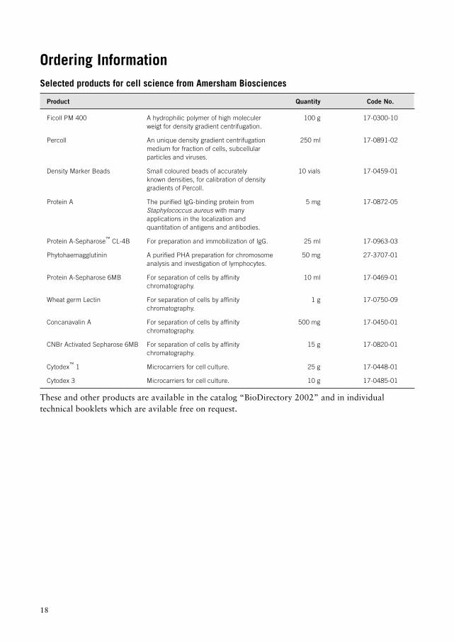
Ordering InformationSelected products for cell science from Amersham Biosciences
Product Quantity Code No.
Ficoll PM 400 A hydrophilic polymer of high moleculer 100 g 17-0300-10weigt for density gradient centrifugation.
Percoll An unique density gradient centrifugation 250 ml 17-0891-02medium for fraction of cells, subcellularparticles and viruses.
Density Marker Beads Small coloured beads of accurately 10 vials 17-0459-01known densities, for calibration of densitygradients of Percoll.
Protein A The purified IgG-binding protein from 5 mg 17-0872-05Staphylococcus aureus with manyapplications in the localization andquantitation of antigens and antibodies.
Protein A-Sepharose™ CL-4B For preparation and immobilization of IgG. 25 ml 17-0963-03
Phytohaemagglutinin A purified PHA preparation for chromosome 50 mg 27-3707-01analysis and investigation of lymphocytes.
Protein A-Sepharose 6MB For separation of cells by affinity 10 ml 17-0469-01chromatography.
Wheat germ Lectin For separation of cells by affinity 1 g 17-0750-09chromatography.
Concanavalin A For separation of cells by affinity 500 mg 17-0450-01chromatography.
CNBr Activated Sepharose 6MB For separation of cells by affinity 15 g 17-0820-01chromatography.
Cytodex™ 1 Microcarriers for cell culture. 25 g 17-0448-01
Cytodex 3 Microcarriers for cell culture. 10 g 17-0485-01
These and other products are available in the catalog “BioDirectory 2002” and in individualtechnical booklets which are avilable free on request.
Percoll, Ficoll, Ficoll-Paque, Sepharose and Cytodex are trademarks of Amersham Biosciences Limited.
Amersham is a trademark of Amersham plc.
Pharmacia and Drop Design are trademarks of Pharmacia Corporation.
The data presented herein have been carefully compiled from our records, which we believe to be accurate and reliable.We make, however, no warranties or representations with respect hereto, nor is freedom from any patent to be inferred.Before any part of this manual is reproduced, please request permission from Amersham Biosciences.
The products described in this literature are intended for in vitro use only. Nothing in this literature should be construed aseither a recommendation or an authorization to use these products for in vivo applications.
All goods and services are sold subject to the terms and conditions of sale of the company withinthe Amersham Biosciences group that supplies them.
A copy of these terms and conditions is available on request.
© Amersham Biosciences AB 2001 – All rights reserved.
Amersham Biosciences AB Björkgatan 30, SE-751 84 Uppsala, Sweden
Amersham Biosciences Amersham Place, Little Chalfont, Buckinghamshire HP7 9NA, England
Amersham Biosciences Inc 800 Centennial Avenue, PO Box 1327, Piscataway, NJ 08855 USA
Amersham Biosciences Europe GmbH Munzinger Strasse 9, D-79111 Freiburg, GermanyAmersham Biosciences KK, Sanken Bldg. 3-25-1, Hyakunincho Shinjuku-ku, Tokyo 169-0073 Japan

www.amershambiosciences.com