4/26/2013
1
LEADLeadership, Education, Accountability, Development
Implanted Vascular Access Ports: Complication Management
Written and presented by Elena Nelson Squires, BSN, RN, OCN, VA-BC
BAS©
LEADLeadership, Education, Accountability, Development
Disclosures
• No financial disclosures.
• Employee of Banner Health at North Colorado Medical Center.
• All images public domain clip art, created by author, duplicated with permissions, or duly cited.– Several images duplicated with permission from Bard Access
Systems (notated with BAS©).
• A special thanks to all the patients who share their dreams and battle scars with the rest of us via the world wide web.
LEADLeadership, Education, Accountability, Development
Objectives
• Participants will be able to discuss patient assessment for the management of implanted vascular access devices
• Participants will be able to discuss identification, intervention, and management of adverse events associated with the implanted vascular access device

4/26/2013
2
LEADLeadership, Education, Accountability, Development
Definition
• An implanted port is a medical device consisting of a housed reservoir which is accessed through a septum that is connected to a catheter
• The housed reservoir is located under the skin and the catheter is surgically placed into a vessel, body cavity, or organ for the purpose of infusate and/or transfusate delivery
LEADLeadership, Education, Accountability, Development
Port Terminology
• Brands:– Infusaport– Port-a-cath– Medi-port– Power Port
• General: Implanted
Venous Access Port
Port TIVAS
BAS©
LEADLeadership, Education, Accountability, Development
Port Configurations
• Some port reservoirs are implanted prior to attaching the catheter; others are all-in-one from the manufacturer.
4/26/2013
3
LEADLeadership, Education, Accountability, Development
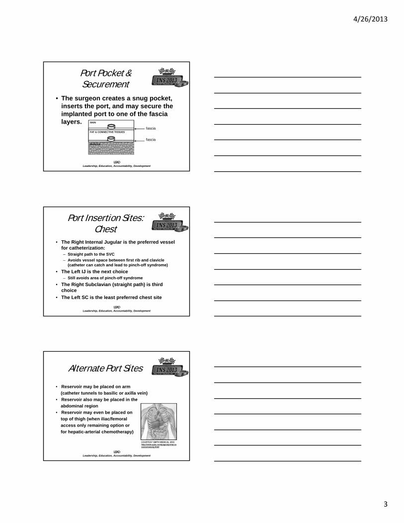
Port Pocket & Securement
• The surgeon creates a snug pocket, inserts the port, and may secure the implanted port to one of the fascia layers.
fascia
fascia
SKIN
FAT & CONNECTIVE TISSUES
MUSCLE
LEADLeadership, Education, Accountability, Development
Port Insertion Sites: Chest
• The Right Internal Jugular is the preferred vessel for catheterization:– Straight path to the SVC
– Avoids vessel space between first rib and clavicle (catheter can catch and lead to pinch-off syndrome)
• The Left IJ is the next choice– Still avoids area of pinch-off syndrome
• The Right Subclavian (straight path) is third choice
• The Left SC is the least preferred chest site
LEADLeadership, Education, Accountability, Development
Alternate Port Sites
• Reservoir may be placed on arm
(catheter tunnels to basilic or axilla vein)
• Reservoir also may be placed in the
abdominal region
• Reservoir may even be placed on
top of thigh (when iliac/femoral
access only remaining option or
for hepatic-arterial chemotherapy)
COURTESY SMITH MEDICAL 2003http://www.quia.com/pages/portaccesscoursesurg.html

4/26/2013
4
LEADLeadership, Education, Accountability, Development
Alternate Port Purposes
• Dialysis access
• Peritoneal access
• Hepatic-Arterial access
COURTESY CIRCUPORT, INChttp://www.innfusionstudios.com/circuport/contact.htmlCOURTESY MEDTRONICS
http://www.medtronic.com/SE/physician/p_pump_index.html
COURTESY CIRCUPORT, INChttp://www.kidney.org.uk/conf02/steele-detail.html
LEADLeadership, Education, Accountability, Development
Other Implanted Devices
• Pain management devices
– Intrathecal
– Neurostimulator/modulation
COURTESY MEDTRONICShttp://professional.medtronic.com/pt/neuro/idd/prod/index.htm
COURTESY MEDTRONICShttp://professional.medtronic.com/pt/neuro/scs/prod/index.htm
LEADLeadership, Education, Accountability, Development
Assessment: Radiograph
• A radiograph of tip location
(or L.I.P. reading thereof)
must be reviewed prior to
accessing or using any C.V.C.,
including an implanted port
– If not available, please
obtain a Chest X-rayCOURTESY http://www.monil.dk/Engelsk/Eng-Illness.htm

4/26/2013
5
LEADLeadership, Education, Accountability, Development
Assessment: Pressure Inject-Ability
• Must be identifiable:– A radiograph may reveal that the implanted port may be used
for pressure (power) injection
– Patient presents information card/booklet provided by surgical staff at time of port placement
– Port has palpable bumps on septum
• If unable to verify as pressure injectable, must not be used for such; catheter fracture or embolus may result
COURTESY http://jmmultiplemyeloma.blogspot.com/2011/07/bard-power-port-day-39-july-22-2011.html
BAS©
LEADLeadership, Education, Accountability, Development
Port Site Assessment
• Port site assessment is both visual and palpable
COURTESY OF JASONhttp://www.cancerguy.com
LEADLeadership, Education, Accountability, Development
Assessment continued
COURTESY OF BASIL WILLIS IIIhttp://basilwillis.com/?p=73
COURTESY OF / RETRIEVED FROMhttp://ulebaququn.500mb.net/
COURTESY OF SCHULICH SCHOOL OF MEDICINE AND DENTISTRYhttp://web.schulich.uwo.ca/Students/medicine/year3/radiology/tubeslinesdrains/portacaths.html
4/26/2013
6
LEADLeadership, Education, Accountability, Development
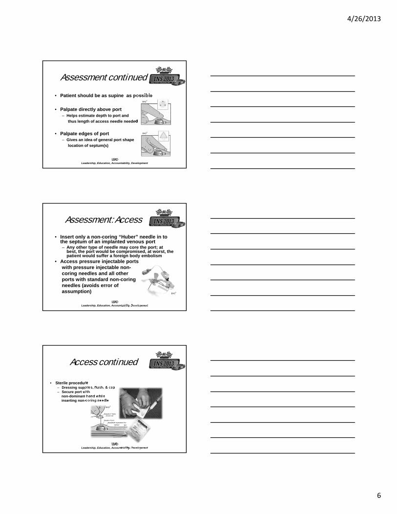
Assessment continued
• Patient should be as supine as possible
• Palpate directly above port– Helps estimate depth to port and
thus length of access needle needed
• Palpate edges of port– Gives an idea of general port shape
location of septum(s)
BAS©
BAS©
LEADLeadership, Education, Accountability, Development
BAS©
Assessment: Access
• Insert only a non-coring “Huber” needle in to the septum of an implanted venous port – Any other type of needle may core the port; at
best, the port would be compromised, at worst, the patient would suffer a foreign body embolism
• Access pressure injectable portswith pressure injectable non-coring needles and all other ports with standard non-coringneedles (avoids error of assumption)
LEADLeadership, Education, Accountability, Development
Access continued
• Sterile procedure– Dressing supplies, flush, & cap– Secure port with
non-dominant hand while inserting non-coring needle
BAS©

4/26/2013
7
LEADLeadership, Education, Accountability, Development
Access continued
• Aspirate for blood return and discard, rather than re-inject blood, then flush with
20 mL normal saline.
RETREIVED 12-19-2012 FROMhttp://www.fda.gov/MedicalDevices/Safety/AlertsandNotices/ucm198766.htm
BAS©
LEADLeadership, Education, Accountability, Development
Access RED flags
• Can not insert non-coring needle– Reassess port position, secure with non-dominant hand and try again.
• Can not aspirate– Do not flush, remove needle, reassess and re-access– Obtain CXR and anticipate Alteplase administration– Consider flushing if known port condition
• Can not flush or resistance with flushing – Obtain CXR and anticipate Alteplase administration
• Patient “hears” flush – Obtain CXR – Patient feels swelling near port with flush or flush tracks back up needle– Consider needle malposition, remove and re-access– Obtain CXR and notify physician if reoccurs
LEADLeadership, Education, Accountability, Development
Maintenance points
• Change dressing and non-coring needle at least every 7 days, and as needed
• Assess patency (blood return and flushing) and site appearance with each patient assessment and as needed (or as per institutional/employer policy)
• Pay attention to patient tolerance of infusion
• Potentially adverse events may happen at any point; recognition and intervention are key to patient well being

4/26/2013
8
LEADLeadership, Education, Accountability, Development
Adverse Events
• Whew! Might make you wonder why anyone would want an implanted venous port
RETRIEVED 12-19-2012 FROMhttp://www.bardaccess.com/powerPort/assets/pdfs/MC-0475-01_PowerPort_Nursing_Guide_web.pdf
BAS©
LEADLeadership, Education, Accountability, Development
Adverse Events:Primarily related to insertion
• Pneumothorax, Hemothorax, or Hydrothorax– Diagnosis: Respiratory signs and symptoms
• Shortness of breath
• Decreased pulse ox or spO2
• Absent or muffled breath sounds
– Treatment
• Mild: may require increased monitoring only
• Moderate-Severe or persistent: chest tube insertion
LEADLeadership, Education, Accountability, Development
Adverse Events: Primarily related to insertion
• Thoracic duct injury: Main lymphatic duct
– Diagnosis: Chylothorax (lymph in the pleural cavity)
• Shortness of breath• Decreased pulse ox or spO2
• Absent or muffled breath sounds
– Treatment• Intra-thoracic surgical repair of main duct• Also, measures to re-inflate the lung as necessary
GRAY, HENRY. 1918. Anatomy of the Human Body

4/26/2013
9
LEADLeadership, Education, Accountability, Development
Adverse Events: Primarily related to insertion
• Laceration or perforation of blood vessel
– Diagnosis: if missed during insertion• May include signs/symptoms of hemothorax
• Hematoma
• Hypotension / hypovolemia
– Treatment• Surgical intervention if patient symptomatic or vessel
injury does not self resolve
• Hemodynamic stability
Image COURTESY OF http://wellnessadvocate.com/Anatomy/img/500px-Circulatory_System_no_tags.svg.png
LEADLeadership, Education, Accountability, Development
Adverse Events: Primarily related to insertion
• Laceration or perforation of viscus– Probable organ involved lung or heart
• Lung insult = pneumothorax
– Cardiocentesis Diagnosis: if persists• Cardiac Tamponade
– Retrosternal pain– Tachycardia– Muffled heart tones– Jugular Vein Distension– Hypotension / Paradoxical pulse
– Treatment• Placement of septal occlusion device• Pericardiocentesis• Supportive care
IMAGE RETRIEVED FROM http://www.pediatricheartspecialists.com/articles/detail/atrial_septal_defect
LEADLeadership, Education, Accountability, Development
Adverse Events: Primarily related to insertion
• Brachial Plexus Injury– Diagnosis
• Mild: numbness, tingling, and / or weakness in arm• Moderate: shooting or shocking and burning arm pain• Severe:
– Loss of finger, elbow, and / or shoulder motion– Severe pain
– Treatment• Time and pain management• Surgical intervention for scar tissue, cut, or torn nerves
– Nerve graft or nerve transfer– Muscle graft
• Anesthetic related complications
4/26/2013
10
LEADLeadership, Education, Accountability, Development
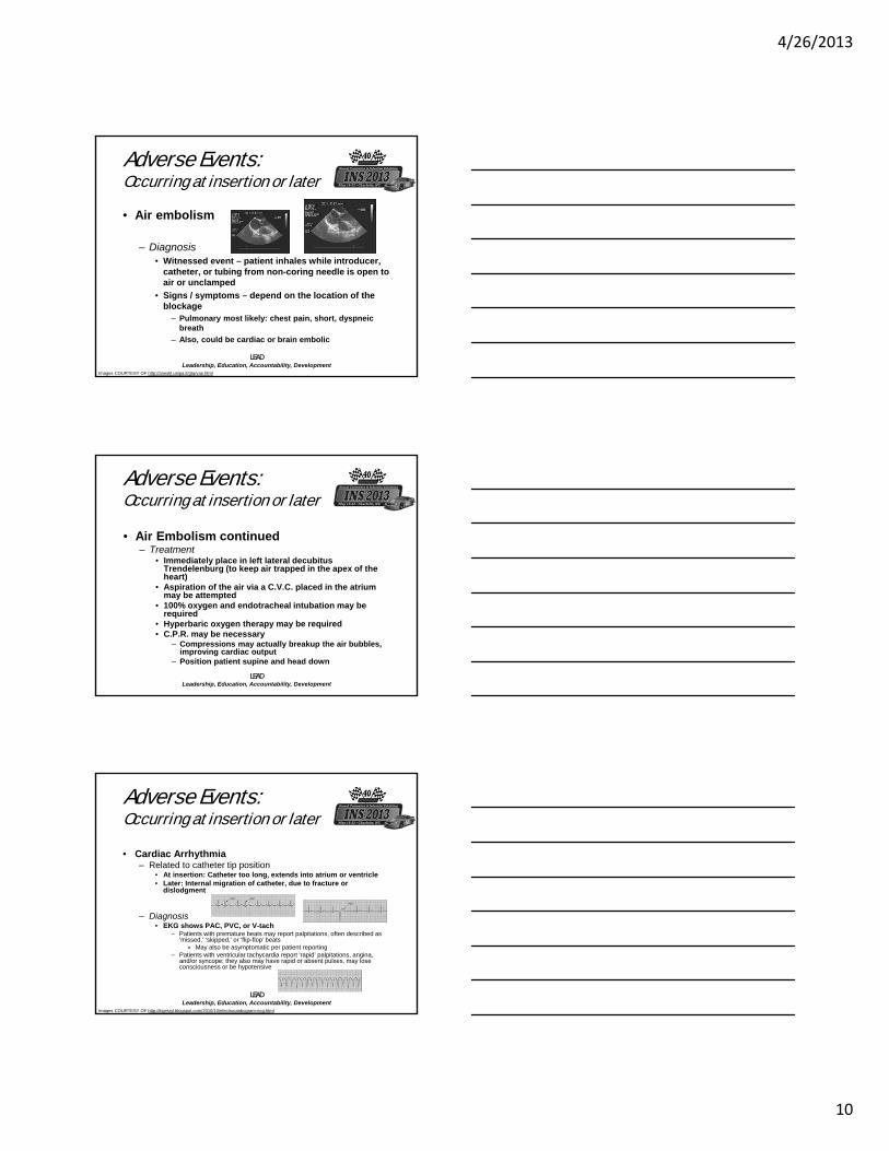
Adverse Events: Occurring at insertion or later
• Air embolism
– Diagnosis• Witnessed event – patient inhales while introducer,
catheter, or tubing from non-coring needle is open to air or unclamped
• Signs / symptoms – depend on the location of the blockage
– Pulmonary most likely: chest pain, short, dyspneic breath
– Also, could be cardiac or brain embolic
Images COURTESY OF http://anestit.unipa.it/gta/vae.html
LEADLeadership, Education, Accountability, Development
Adverse Events: Occurring at insertion or later
• Air Embolism continued– Treatment
• Immediately place in left lateral decubitus Trendelenburg (to keep air trapped in the apex of the heart)
• Aspiration of the air via a C.V.C. placed in the atrium may be attempted
• 100% oxygen and endotracheal intubation may be required
• Hyperbaric oxygen therapy may be required• C.P.R. may be necessary
– Compressions may actually breakup the air bubbles, improving cardiac output
– Position patient supine and head down
LEADLeadership, Education, Accountability, Development
Adverse Events: Occurring at insertion or later
• Cardiac Arrhythmia– Related to catheter tip position
• At insertion: Catheter too long, extends into atrium or ventricle• Later: Internal migration of catheter, due to fracture or
dislodgment
– Diagnosis• EKG shows PAC, PVC, or V-tach
– Patients with premature beats may report palpitations, often described as ‘missed,’ ‘skipped,’ or ‘flip-flop’ beats
» May also be asymptomatic per patient reporting– Patients with ventricular tachycardia report ‘rapid’ palpitations, angina,
and/or syncope; they also may have rapid or absent pulses, may lose consciousness or be hypotensive
Images COURTESY OF http://itsjesryl.blogspot.com/2010/10/electrocardiogram-ecg.html

4/26/2013
11
LEADLeadership, Education, Accountability, Development
Adverse Events: Occurring at insertion or later
• Arrhythmia continued
– Treatment• Requires repositioning of catheter tip (surgical
or radiological intervention)
• Urgent - in cases of ventricular tachycardia where patient is asymptomatic
• Emergent - in cases of symptomatic v-tach or as a result of catheter dislodgment
LEADLeadership, Education, Accountability, Development
Adverse Events: Occurring at insertion or later
• Hematoma and Bleeding– Diagnosis
• Superficial: visible and or palpable• Internal: hypovolemia or anemia
– Treatment• Superficial: direct pressure to slow or stop bleeding• Internal: supportive care while self resolves or
surgical intervention• Application of recumbinant thrombin may aid in
sealing capillary or small venule leaks• Correction of underlying coagulopathies • Rarely requires evacuation of hematoma
LEADLeadership, Education, Accountability, Development
Adverse Events: Occurring at insertion or later
• Bacteremia – invasion of the blood by pathogenic microorganisms
• Sepsis – suspected infection with 2 or more: T>38.3°C, HR>90, RR>20, BG>140 in absence of diabetes, WBC>12k or >10% immature forms, and acutely altered mental status
• Severe sepsis – Acute organ dysfunction, hypoperfusion, or hypotension prior to fluid challenge – Differential Diagnosis
• Signs and symptoms of infection without any other identified source
– Positive blood cultures
• May be insertion, care, or contamination related• May also be result of colonized port catheter / reservoir
– Patient exhibits fever and chills synchronized with port irrigation

4/26/2013
12
LEADLeadership, Education, Accountability, Development
Adverse Events: Occurring at insertion or later
• Treatment– IV anti-microbial ASAP– Maintain or regain hemodynamic stability
» Fluid therapy» Vasopressors» Inotropic therapy» Steroids, Activated Protein C, Blood products, Glucose
control, and lactate clearance (severe sepsis/septic shock)– Device removal
» Pocket inspection and swab culture » Reservoir culture
– Device preservation» In cases of coagulase negative staphylococcal infection
(most common), may try» Antibiotic therapy through catheter» Antibiotic lock
LEADLeadership, Education, Accountability, Development
Adverse Events: Occurring at insertion or later
• Other infection– Reservoir Pocket Infection
• Diagnosis– Redness, swelling, sero-purulent drainage around port
site– Positive cultures of pocket surrounding the port– Often progressed to bacteremia or port colonization– Definitive diagnosis by culture of pocket surrounding port
during device removal• Treatment
– Anti-microbial specific to causative organism– Device removal– May require additional wound care if severe abscess or
port extrusion
LEADLeadership, Education, Accountability, Development
Adverse Events: Occurring at insertion or later
– Endocarditis• Diagnosis
– Signs and symptoms of
infection
– Positive blood cultures
– Heart murmur
– Echocardiogram: detects vegetation on heart valves
• Treatment– Device removal
– IV anti-microbial for at least 6 weeks
– Heart surgery for valve repair or replacement if necessary
Image COURTESY OF The Full Wiki: http://www.thefullwiki.org/Infective_endocarditis

4/26/2013
13
LEADLeadership, Education, Accountability, Development
Adverse Events: Primarily apparent some time after insertion
• Skin erosion / reservoir extrusion– Diagnosis
• Visually apparent
• Rule out local infection – often associated with pocket infection
– Treatment• Device removal, wound care, possible skin
grafting
• Device preservation and wound care
LEADLeadership, Education, Accountability, Development
Adverse Events: Primarily apparent some time after insertion
• Fibrin Sheath– Diagnosis
• Chest radiograph shows catheter in SVC• Partial occlusion
– Unable to obtain blood return– Some resistance “sluggish” to flush
• Complete occlusion– Unable to withdraw or flush
– Treatment• Alteplase 2 mg in 2.2 mL preservative free
sterile water• May repeat dose, may require overnight dwell
Images COURTESY OF http://www.cathflo.com/catheter/occlusions.jsp
LEADLeadership, Education, Accountability, Development
Adverse Events: Primarily apparent some time after insertion
• Occlusions unrelated to clotting– Treatment
• Lipids 70% Ethanol
• Mineral precipitates 0.1-N hydrochloric acid
• Acidic infusates 0.1-N hydrochloric acid
• Basic infusates Sodium bicarbonate or 0.1-N sodium hydroxide (NaOH)
• Contrast media Sodium bicarbonate
• Unknown/other consider one dose of Alteplase

4/26/2013
14
LEADLeadership, Education, Accountability, Development
Adverse Events: Primarily apparent some time after insertion
• Lack of recoverable blood return– Tissue around catheter inside the vein
• Days 1-7 some fibrin• Days 7-14 endothelial and smooth muscle cells• Over 21 days collagen • Tissue may encase catheter in such a way as to allow infusion, but
eliminate blood return• Also, may be due to SVC stenosis or thrombosis
– Diagnosis• Catheter dye study
– Treatment• Continue catheter use and obtain blood peripherally • Remove and replace catheter
– Prevention• Place tip at cavoatrial junction upon insertion of port access
LEADLeadership, Education, Accountability, Development
Adverse Events: Primarily apparent some time after insertion
• Thromboembolism– Diagnosis
• Subclavian or SVC thrombus – SVC syndrome– Periorbital, facial, arm, and chest wall edema– Jugular vein distention – Hemoptysis– Headache and chest pain– Bluish upper body and face
• Embolism– Chest pain– Cough, wheeze, blood-streaked sputum– Increased, irregular pulse– Syncope
• Imaging studies– Venography– Magnetic resonance venography
LEADLeadership, Education, Accountability, Development
Adverse Events: Primarily apparent some time after insertion
• Thromboembolism continued– Treatment
• Fibrinolytics– May be through catheter – Example: 3 mg Alteplase per hour and 1000u heparin
• Interventional radiology to snare and remove clot• Surgery to remove clot• Stent vessel open• Respiratory support
– Maintain airway– Oxygen therapy

4/26/2013
15
LEADLeadership, Education, Accountability, Development
Adverse Events: Primarily apparent some time after insertion
• Scarring and SVC stenosis– Diagnosis
• Catheter malfunction
• SVC syndrome symptoms
– Treatment• Elimination of any associated thrombus
• Surgical or IR – Retraction of catheter (to pull back out of the way)
– Stent placement in affected area of SVC
– Return of catheter to position in SVC
Image COURTESY of http://cyt-ar.com.ar/cyt-ar/index.php/Archivo:Stent_Palmaz.jpg
LEADLeadership, Education, Accountability, Development
Adverse Events: Primarily apparent some time after insertion
• Catheter fracture, separation, or embolism
– Causation• Port design: reservoir and catheter were not
manufactured as one piece
• Pinch-off syndrome: when catheter is compressed between the clavicle and first rib
• Pressure injection: through
catheter not designed for such
LEADLeadership, Education, Accountability, Development
Adverse Events: Primarily apparent some time after insertion
• Catheter fracture / embolism continued– Diagnosis
• Rapid swelling around reservoir pocket due to infusate infiltration – may be only symptom
• Signs of pulmonary embolism: Dyspnea, Chest pain, Cough, Wheeze, Rapid and irregular pulse, Syncope
– Treatment: Urgent if not Emergent• LIFE SUPPORT MEASURES• May require positioning to keep catheter in heart• Immediate fluoroscopically guided catheter retrieval• Replacement of implanted port

4/26/2013
16
LEADLeadership, Education, Accountability, Development
Adverse Events: Primarily apparent some time after insertion
• Catheter fracture, separation, or embolism continued– Prevention
• At insertion– Place implanted port manufactured as one piece or
place suture at reservoir / catheter junction
– Insert catheter into venous anatomy from an internal jugular approach
• During use– Place only pressure injectable implanted ports
– If unable to verify pressure tolerance, do not pressure inject through an implanted port
LEADLeadership, Education, Accountability, Development
Adverse Events: Primarily apparent some time after insertion
• Pinch off syndrome – a few more points
– Diagnosis• Only occurs when port implanted from the
Subclavian approach
• Suspect when catheter intermittently occludes completely and clearance is not related to Alteplase administration
– Treatment• Catheter removal and replacement, preferably from
an internal jugular approach
LEADLeadership, Education, Accountability, Development
Adverse Events: Primarily apparent some time after insertion
• Vessel erosion – uncommon– Diagnosis
• Presents with chest pain, dyspnea• Widened mediastinum and pleural effusion per
radiograph• Contrast study shows extravasation into mediastinum
rather than SVC– Treatment
• Catheter removal• Thoracentesis, thoracostomy, pericardocentesis
– Preventive measures• Place catheter well into the SVC at the cavoatrial
junction to decrease mechanical erosion of vessel due to tip abutment

4/26/2013
17
LEADLeadership, Education, Accountability, Development
Adverse Events: Primarily apparent some time after insertion
• Reservoir rotation or inversion
– Diagnosis• Visualization or Palpation: may not look or feel as it
should• Inability to access: Unable to pierce septum with non-
coring needle or must approach at awkward angle or patient position to access
• May also be detectable per two view chest radiographs– Treatment
• Correct or replace surgically • May continue to use if possible and short term• Remove
LEADLeadership, Education, Accountability, Development
Adverse Events: Primarily apparent some time after insertion
• Device intolerance RARE– Sensitivity to poly-urethane or silicone
• Diagnosis– Inflammatory response, likely localized, without
presence of micro-organisms
– Usually diagnosed by ruling out more likely scenarios
• Treatment– Device removal
LEADLeadership, Education, Accountability, Development
Adverse Events: Primarily apparent some time after insertion
• Non-coring needle dislodgment
– Diagnosis• Swelling around port reservoir that appears during infusion• Lack of blood return with aspiration• Removal and replacement of non-coring needle regains blood return
– Treatment• Benign infusate: removal and replacement of access• Vesicant therapy: consult with pharmacist and administer antidote if
available– May also aspirate through access prior to removal– May require wound care and grafting
– Prevention• Access reservoir with non-coring needle of appropriate size• Secure port with non-dominant hand during access
4/26/2013
18
LEADLeadership, Education, Accountability, Development
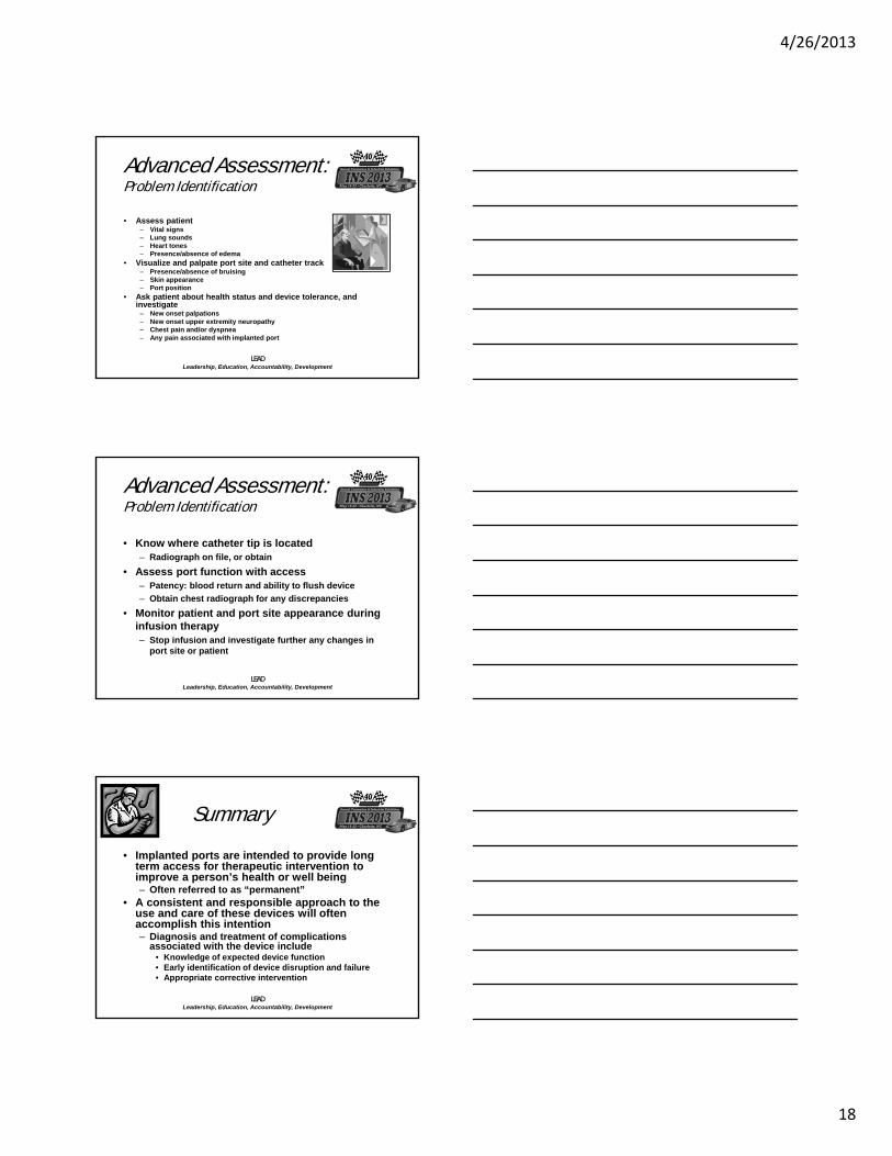
Advanced Assessment: Problem Identification
• Assess patient– Vital signs – Lung sounds– Heart tones– Presence/absence of edema
• Visualize and palpate port site and catheter track– Presence/absence of bruising– Skin appearance – Port position
• Ask patient about health status and device tolerance, and investigate
– New onset palpations– New onset upper extremity neuropathy– Chest pain and/or dyspnea– Any pain associated with implanted port
LEADLeadership, Education, Accountability, Development
Advanced Assessment: Problem Identification
• Know where catheter tip is located– Radiograph on file, or obtain
• Assess port function with access– Patency: blood return and ability to flush device
– Obtain chest radiograph for any discrepancies
• Monitor patient and port site appearance during infusion therapy– Stop infusion and investigate further any changes in
port site or patient
LEADLeadership, Education, Accountability, Development
Summary
• Implanted ports are intended to provide long term access for therapeutic intervention to improve a person’s health or well being– Often referred to as “permanent”
• A consistent and responsible approach to the use and care of these devices will often accomplish this intention– Diagnosis and treatment of complications
associated with the device include • Knowledge of expected device function• Early identification of device disruption and failure• Appropriate corrective intervention

4/26/2013
19
LEADLeadership, Education, Accountability, Development
References• Bard Access System. (2010). Power Port Implantable Port: Nursing Guide. Retrieved from
http://www.bardaccess.com/port-powerport.php?section=Resources
• Brouns, F., Schuermans, A., Verhaegen, J., De Wever, I., Stas, M. (2006). Infection Assessment of Totally Implanted Venous Access Devices. Journal of Vascular Access, 7, 24-28.
• Cerfolio, R.J. MD. (2003). Ligation of the Thoracic Duct for Chylothorax. CTSNet: The Cardiothoracic Surgery Network. Retrieved from http://www.ctsnet.org/sections/clinicalresources/thoracic/expert_tech-19.html
• Coletti, C. MD., and Powell, J. MD. (2009). Sepsis Card. Emergency Residents Medicine Association.
• Cope, D.G., Ezzone, S.A., Hagle, M.E., McCorkindale, D.J., Moran, A.B., Sanoshy, J.K., et al. (2004). Access Device Guidelines: Recommendations for Nursing Practice and Education. Oncology Nursing Society. Pittsburgh, PA.
• Infusion Nurses Society. Infusion Nursing Standards of Practice. Journal of Infusion Nursing, 34(1S).
• Kosvic, S., Kovcin, V., Granic, M., Jevdic, D., Stanisavljevic, N. (2010). Disconnection of Chamber and Catheter as a Complication of Central Venous Catheter Type: Port-A-Cath. Medical Oncology, 28, 1176-1179.
• Kulvatunyou, N., Rucinski, J., Schein, M. (1997). Delayed Complication of Port-A-Cath: Perforation of the Superior Vena Cava. Journal of Clinical Oncology, 15(2), 865.
• Lam, A., Chen, Y., Yang, K., Tsal, C., Perng, R. (1999). Disconnection of a Venous Port-A-Cath Followed By Embolization After Saline Flush: Rare Case Report. Journal of Clinical Oncology, 29(12), 643-645.
LEADLeadership, Education, Accountability, Development
References• Lin, C.H., Wu, H.S., Chan, D.C., Hsieh, C.B. (2009). The Mechanisms of Failure of Totally Implantable Central Venous Access
System: Analysis of 73 Cases with Fracture of Catheter. The Journal of Cancer Surgery, 36, 100-103.
• Mayo Clinic Staff. (2011). Brachial Plexus Injury. Mayo Clinic. Retrieved from http://www.mayoclinic.com/health/brachial-plexus-injury/DS00897/DSECTION=treatments-and-drugs
• Mayo Clinic Staff. (2011). Endocarditis. Mayo Clinic. Retrieved from http://www.mayoclinic.com/health/endocarditis/DS00409/DSECTION=tests-and-diagnosis
• Natal, B.L. MD, and Doty, C.I. MD. (2012). Venous Air Embolism Treatment and Management. Medscape Reference: Drugs, Diseases, and Procedures. Retrieved from http://emedicine.medscape.com/article/761367-treatment#a1126
• Reed, E.D. MD, and Dmitry, J.R. MD, PhD. (2013). Malignancy-Related Superior Vena Cava Syndrome. UpToDate. Retrieved from http://www.uptodate.com/contents/malignancy-related-superior-vena-cava-syndrome
• Stockx, L., Raat, H., Donck, J., Wilms, G., Marchal, G. (1999). Repositioning and Leaving In Situ the Central Venous Catheter During Percuntaneous Treatment of Associated Superior Vena Cava Syndrome: A Report of 8 Cases. Cardiovascular Interventional Radiology, 22, 224-226.
• Subramaniam, A., Kim, K.H., Bryant, S.A., Kimball, K.J., Huh, W.K., Straughn, J.M., et al. (2011). Incidence of Malfunction in Low Profile Subcutaneous Implantable Vascular Access Devices in Patient Receiving Chemotherapy for Gynecological Malignancies. Gynecological Oncology, 123. 54-57.
• Tonkin, J.L., Campbell, G., Golding, L., Hamblin, M., Hunter, S., Jaggia, A. (2008). Atrial Thrombosis: A Near Fatal Complication of a Portacath. Journal of Vascular Access, 9, 148-151.
• Wani, S. MD, Dick, F. MD, Meier, B. MD. (2007). Port-A-Cath Perforation of the Right Atrium Closed with an Amplatzer ASD Occluder. Catheterization and Cardiovascular Interventions, 70, 57-59.
Recommended