ORIGINAL RESEARCH
Glycerol-plasticized bacterial nanocellulose-basedcomposites with enhanced flexibility and liquid sorptioncapacity
Izabela Cielecka . Marcin Szustak . Halina Kalinowska . Edyta Gendaszewska-Darmach .
Małgorzata Ryngajłło . Waldemar Maniukiewicz . Stanisław Bielecki
Received: 9 January 2019 / Accepted: 9 May 2019 / Published online: 16 May 2019
� The Author(s) 2019
Abstract Bacterial nanocellulose (BNC) and two
BNC-based composites with carboxymethyl cellulose
or hydroxyethyl cellulose (BNC-CMC or BNC-HEC,
respectively), were produced in situ by Koma-
gataeibacter xylinus E25 under stationary conditions
and plasticized with glycerol (ex situ modification).
The BNC-CMC composite had the loosest structure
(visible in SEM images) and was less crystalline (CI of
88.6%) than BNC (CI of 92.9%) and BNC-HEC (CI of
90.4%). Cellulose fibers synthesized by K. xylinus E25
in the presence of HEC were thinner in comparison to
the fibers of control BNC while there was no
difference in the fibers width between the BNC-
CMC and control BNC. The glycerol-plasticized
BNC, BNC-CMC and BNC-HEC membranes were
flexible after drying, and absorbed high amounts of
artificial exudate and water after rehydration. BNC-
CMC treated with 2.5% v/v aqueous glycerol was
characterized by the greatest free swell absorptive
capacity (up to 19 g artificial exudate/g dry weight in
24 h) while the highest rehydration capacity (around
96% of the initial water content) was observed in case
of BNC-CMC plasticized with 10% v/v glycerol and
dehydrated. The in situ and ex situ modifications of
BNC affected also the tensile strength. The highest
values of tensile strength at break (around 152.2 N)
and Young’s modulus (around 290.3 MPa) were
observed in case of the BNC-CMC composite plasti-
cized with 2.5% v/v glycerol. The impact of plasti-
cized BNC, BNC-CMC and BNC-HEC on the
viability of HaCaT keratinocytes was also studied
and found to be positive at glycerol concentrations up
to 2.5% (v/v) that suggests their potential utility as
wound dressings.
Keywords Bacterial nanocellulose � Composite �Wound dressing � Liquid sorption capacity
Introduction
Bacterial nanocellulose (BNC) is a natural, nontoxic,
biocompatible and mechanically durable gelatinous
polymer with high water-holding capacity, which has
proven to be an excellent wound dressing and found
numerous potential applications not only in medicine
(Campano et al. 2016; Ullah et al. 2016). Its most
efficient producers are non-pathogenic, Gram-nega-
tive, rod shaped and strictly aerobic bacteria of the
Komagataeibacter genus that assimilate various sug-
ars and other compounds as carbon source. Cells of
Komagataeibacter species extrude cellulose chains
I. Cielecka (&) � M. Szustak � H. Kalinowska �E. Gendaszewska-Darmach � M. Ryngajłło � S. Bielecki
Institute of Technical Biochemistry, Lodz University of
Technology, Lodz, Poland
e-mail: [email protected]
W. Maniukiewicz
Institute of General and Ecological Chemistry, Lodz
University of Technology, Lodz, Poland
123
Cellulose (2019) 26:5409–5426
https://doi.org/10.1007/s10570-019-02501-1(0123456789().,-volV)( 0123456789().,-volV)
through multiple pores (around 50 per cell) that are
located in the cytoplasmic membrane in a row along
the longitudinal axis, and produce 70–150 nm wide
ribbons composed of 10–100 hydrogen-bonded cellu-
lose microfibrils (Bielecki et al. 2002). The polymer-
ization of uridine diphosphate glucose (UDP-glucose),
mediated by cellulose synthase, is coupled with
formation of the highly crystalline (above 60%)
nanocellulose structure, maintained by the three-
dimensional network of multiple intra- and inter-
molecular hydrogen bonds (Bielecki et al. 2002).
Examples of media that are used for cultivation of
BNC producers include the Schramm-Hestrin (SH)
medium, natural media like coconut water (Budhiono
et al. 1999) or coconut milk (Nakagaito et al. 2005)
and media based on various low-cost feedstocks,
including by-products from food processing
(Machado et al. 2018; Velasquez-Riano and Bojaca
2017).
One of attractive BNC properties is the chemical
purity, what makes it suitable for various modifica-
tions. The polymer may be modified either in situ
during biosynthesis by bacteria or ex situ after BNC
harvesting. Diverse cellulose-based nanocomposites
obtained by either in situ and/or ex situ methods could
found numerous applications in medicine (Taokaew
et al. 2015; Ullah et al. 2016) and other fields
(Campano et al. 2016). In case of the in situ approach,
changes in the structure of BNC may be achieved,
among others, by supplementation of culture media
with various polysaccharides and other polymers. The
first polysaccharide studied was carboxymethylcellu-
lose (CMC), which was found to delay the aggregation
of cellulose molecules (Ben-Hayyim and Ohad 1965).
Other examples of polymers which can form hydrogen
bonds with cellulose chains and this way reduce the
strength of noncovalent interactions between cellulose
microfibrils and alter the assembly pattern of BNC
fibres are polysaccharides like pectin, sodium alginate,
chitin, agar (Dayal and Catchmark 2016), r-car-
rageenan (Cielecka et al. 2018), hyaluronic acid (Li
et al. 2015), hydroxyethylcellulose (Zhou et al. 2009),
exopolysaccharides from Escherichia coli (Liu and
Catchmark 2018), water soluble exopolysaccharides
fromGluconacetobacter xylinus (Fang and Catchmark
2014), and hemicelluloses (Uhlin et al.1995). Also
Aloe vera gel pulp, gel extract and polysaccharide
fraction (Godinho et al. 2016), poly(ethylene glycol)
(Hessler and Klemm 2009) and gelatin (Chen et al.
2014) have the similar impact. The noncovalent
binding of aforementioned polymers to cellulose
affects the crystal size, crystallinity, porosity and
water holding capacity of the biosynthesized product.
Apart from the in situ modifications, also a variety of
ex situ methods are used to obtain novel BNC-based
materials for medicine and cell cultures, e.g. modifi-
cation with dextran (Lin et al. 2017), organosilanes
(Taokaew et al. 2015) or plasticizers like poly(ethy-
lene glycol) (Cai and Kim 2010).
In this study, BNC-based nanocomposites obtained
by combination of the in situ and ex situ modifications
were characterized and their effect on the viability of
keratinocytes was studied. BNC was modified in situ
using either CMC or HEC. Then, the native BNC (the
control), BNC-CMC and BNC-HEC membranes,
produced by K. xylinus E25 under stationary condi-
tions, were plasticized with 0.5–10% v/v aqueous
glycerol solutions. This polyol is one of popular,
natural, renewable and biodegradable plasticizers
(Vieira et al. 2011). Glycerol used as a plasticizer of
bacterial cellulose, causes a decrease in the strength of
intermolecular hydrogen bonds between adjacent
cellulose chains. That results in improvement of the
flexibility of pellicles and prevention of formation of
rigid and brittle BNC sheets after dehydration (Sun
et al. 2018). Furthermore, glycerol-plasticized BNC
becomes more transparent, which is also advanta-
geous. The transparency of BNC-based wound dress-
ings facilitates monitoring of wound healing process
while the increased hydration and flexibility enable
easy and painless application and removal of wound
coverings (Bielecki et al. 2013). Glycerol is not only a
widely applied plasticizer but also a component of
many moisturizing cosmetics and therefore, its contact
with skin is permitted. The outermost layer of the skin,
exposed to the contact with wound dressings is the
epidermis, and the predominant cell type in the
epidermis is keratinocyte (MacNeil 2007). Therefore,
to assess potential effect of the long-term contact of
glycerol-plasticized BNC-based wound dressings we
decided to determine the impact of glycerol concen-
tration in the wound dressing on the viability of
keratinocytes. Bacterial cellulose films are known to
support the growth, spreading and migration of human
keratinocytes (Sanchavanakit et al. 2006). However,
the occurrance of CMC or HEC in the BNC-based
nanocomposites may either positively or negatively
affect these cells. Therefore, the final objective of this
123
5410 Cellulose (2019) 26:5409–5426
study was to determine the impact of BNC modifica-
tion with water soluble polysaccharides and glycerol
on the viability of keratinocytes, using the immortal
human keratinocyte HaCaT cell line as a model.
Materials and methods
Microbial strain
Komagataeibacter xylinus E25 strain used in this
study was obtained from the culture collection of the
Institute of Technical Biochemistry (Lodz University
of Technology). The strain belongs to BOWIL Biotech
Ltd., Poland.
Culture media
K. xylinus E25 strain was cultivated under stationary
conditions in the Schramm-Hestrin (SH) medium
containing glucose (20 g/L), yeast extract (5.0 g/L),
peptone (5.0 g/L), MgSO4 9 7 H2O (0.5 g/L), anhy-
drous Na2HPO4 (2.7 g/L) and citric acid (1.15 g/L),
dissolved in distilled water. The pH of the culture
medium was adjusted to 5.7 using 0.1 M acetic acid.
To produce the BNC-CMC and BNC-HEC compos-
ites, the SH medium was supplemented with either
0.5% carboxymethyl cellulose (CMC) or 0.5%
hydroxyethyl cellulose (HEC), respectively. Both
CMC (Mw around 90,000, degree of substitution of
0.7) and HEC (Mw around 90,000, degree of substi-
tution of 0.6) were purchased from Sigma-Aldrich.
Biosynthesis and purification of BNC, BNC-CMC
and BNC-HEC
K. xylinus E25 was activated by cultivation under
stationary conditions in the Schramm-Hestrin (SH)
medium (supplemented with 1% v/v ethanol before
inoculation) at 30 �C for 2 days and used as an
inoculum. The SH medium, either without additives or
supplemented with either 0.5% carboxymethyl cellu-
lose (SH-CMC) or 0.5% hydroxyethyl cellulose (SH-
HEC), was supplemented with ethanol (1% v/v) before
the inoculation with bacterial pre-culture (5% v/v).
BNC, BNC-CMC and BNC-HEC membranes were
obtained after 7-day culture in stationary conditions at
30 �C.
Purification of all the membranes included rinsing
with tap water until flushing out of the remaining
medium, 24 h washing with 1% w/v sodium hydrox-
ide for bacterial cells removal, and subsequent
immersing in 1% v/v acetic acid and then distilled
water for pH neutralization. Purified membranes were
packed, sterilized by autoclaving at 121 �C and stored
at 4 �C before further analytical steps.
Preparation of composites with glycerol
Never-dried BNC, BNC-CMC and BNC-HEC mem-
branes with a diameter of 38 mm were immersed in
300 mL aliquots of aqueous glycerol solutions with
concentrations ranging from 0.1 to 10%. Glycerol (for
molecular biology) was purchased from Sigma-
Aldrich. The incubation was conducted for 24 h at
80 rpm and 20–22 �C. Then the plasticized mem-
branes were dried at 80 �C for 24 h until constant mass
was achieved. An excess of aqueous glycerol solution
was used to minimize the dilution caused by the
occurrence of water inside the never-dried mem-
branes. The calculations showed that in the experi-
mental conditions the decrease in glycerol
concentration was relatively small, e.g. from 10 to
9.8% v/v. Next, the samples were sterilized and stored
at 4 �C before use.
Characterization of BNC, BNC-CMC and BNC-
HEC
Scanning electron microscopy
Scanning electron microscopy was applied for visu-
alization of BNC, BNC-CMC and BNC-HEC fibres in
nanocomposites. Samples of the membranes were
freeze-dried and afterwards, sputter coated with gold
layer and observed using a FEI QUANTA 250 FEG
microscope (Thermo Fischer Scientific, MA, USA)
(HV 2 kV, magnification 400009). Fibres thickness
was measured using the Makroaufmassprogramm
software on the basis on SEM images. The probability
density functions were drown using the R (v. 3.4.1)
software.
123
Cellulose (2019) 26:5409–5426 5411

Fourier transform infrared spectrometry
in an attenuated total reflectance mode analysis
The incorporation of either CMC or HEC into BNC
membranes was evaluated with the FTIR-ATR using a
Nicolet 6700 FT-IR (Thermo Scientific, MA, USA).
The spectra of BNC, BNC-CMC, BNC-HEC, CMC
and HEC were recorded over the range from 4000 to
650 cm-1 and 200 scans were accumulated, with a
spectral resolution of 4 cm-1.
X-ray diffractometry
Room temperature powder X-ray diffraction patterns
were collected using a PANalytical X’Pert Pro MPD
diffractometer (Malvern Panalytical Ltd, UK) with the
Bragg–Brentano reflection geometry and the graphite
monochromated Cu-Ka radiation. It was equipped
with a PANalytical X’Celerator detector. Samples
were scanned in the 2h range between 5� and 60�. The
scan speed was 30 s per step of 0.0167�. The samples
were spun during data collection to minimize pre-
ferred orientation effects. X-ray diffraction patterns
were resolved into a broad amorphous halo and five
crystalline peaks using WAXFIT program (Rabiej
2014). The crystallinity index was calculated as the
ratio of the integral intensity under all crystalline
peaks to the sum of integral intensity under the
crystalline peaks and amorphous halo (Park et al.
2010).
Characterization of BNC, BNC-CMC and BNC-
HEC composites with glycerol
Free swell absorptive capacity
The dependence of liquid absorption properties of
BNC, BNC-CMC and BNC-HEC membranes on
glycerol concentration was determined according to
the modified ISO 13726 standard. The plasticized
membranes with diameter of 38 mm (dried at 80 �Cfor 24 h until constant weight) were immersed in
artificial exudates (solution of 8.298 g of sodium
chloride and 0.368 g of anhydrous calcium chloride in
1 L of deionised water) at 37 �C for 30 min and 24 h.
Then the excess of the artificial exudates was removed
with filter paper, and the swollen membranes were
weighted (WW). Free swell absorptive capacity values
were calculated from the Eq. 1, and expressed as
grams of artificial exudates absorbed by 1 g of
composite dry weight (WD).
Absorptive capacity ¼ WW
WD
ð1Þ
The experiment was carried out in triplicate for each
BNC-based composite.
Rehydration capacity of dehydrated plasticized
membranes
After plasticization with the aqueous glycerol solu-
tions (at glycerol concentrations ranging between 0.1
and 10% v/v, as described in the section Preparation
of composites with glycerol, the membranes (diameter
of 38 mm) were weighted (WW1) and next, dehydrated
at 80 �C until constant weight (WD). To determine the
rehydration capacity, the membranes were immersed
in distilled water for 24 h to achieve the swollen state.
Then the water excess on the surface was removed
with filter paper, and the wet membranes were
weighted (WW2). The rehydration capacity (RC) was
calculated from the Eq. (2) after 30 min and 24 h.
RC ¼ WW2 �WD
WW1 �WD
� 100% ð2Þ
All the measurements were performed in triplicate.
Tensile test
Before characterization of tensile strength, BNC,
BNC-CMC and BNC-HEC membranes
(9 cm 9 15 cm) were gently wiped with filter paper
and immersed for 24 h at 80 rpm and 20–22 �C in
300 mL of either 2.5% or 10% (v/v) aqueous glycerol
solution. The concentration of 2.5% v/v was selected
because it corresponded to the highest free swell
absorptive capacity of all the nanocomposites while
the concentration of 10.0% v/v was the highest
glycerol concentration studied in this work. Besides,
the results of this study showed that the latter glycerol
concentration corresponded to the highest rehydration
capacity and high free swell absorption capacity,
which are attractive in case of wound dressings. This
procedure was repeated 3 times to minimize the
dilution of glycerol solution caused by the presence of
residual water in the never-dried membranes.
The BNC, BNC-CMC and BNC-HEC membranes,
either treated with 2.5 and 10% v/v glycerol or not,
123
5412 Cellulose (2019) 26:5409–5426
were examined for tensile strength using a universal
testing machine (Zwick/Roell Z1.0, Germany) accord-
ing to (Cielecka et al. 2018). Before the tensile tests, an
excess of water attached to BNC and BNC-based
nanocomposites was removed by pressing until 1 mm
thickness was achieved. The composites plasticized
with glycerol were dehydrated at 80 �C until constant
weight was achieved.
The maximum stress and elongation at break were
estimated using the TestXpert@II software. Stress (r)
was calculated as F/A, where F is the loading force,
expressed in Newtons (N), and A is the cross-section
area measured as the width 9 thickness of a sample.
Strain (e) was calculated as DL/L0 9 100%, where L0
is the initial length and DL is the exerted extension
from starting point. Values of the Young’s modulus
under tension were calculated from the stress/strain
relationship in the first linear region of the graph. The
measurements were performed in 5 replicates.
Keratinocytes viability study
Human keratinocyte cell line HaCaT
The immortal human keratinocyte HaCaT cell line
was purchased from Leibniz Institute DSMZ - German
Collection of Microorganisms and Cell Cultures
(Braunschweig, Germany). The cells were cultured
in Dulbecco’s modified Eagle’s medium (DMEM)
supplemented with 10% fetal bovine serum (FBS)
containing 100 IU/mL penicillin, 100 lg/mL neomy-
cin and 2.5 lg/mL amphotericin B. All reagents for
cell culture were obtained from the Life Technologies
(Carlsbad, CA, USA). The cells were incubated at
37 �C in a humidified atmosphere supplemented with
5% CO2.
Scaffold preparation
Wet BNC, BNC-CMC and BNC-HEC scaffolds
(16 mm in diameter) were plasticized with either
2.5% v/v or 10% v/v glycerol for 24 h, and then
dehydrated. The concentration of 2.5% v/v was
selected because it corresponded to the highest free
swell absorptive capacity of all the nanocomposites
while the concentration of 10.0% v/v, being the
highest glycerol concentration studied in this work,
corresponded to the highest rehydration capacity.
After dehydration, the scaffolds were transferred to a
24 well plate, and 500 lL aliquots of the growth
medium (DMEM/FBS) were added to each well
before the seeding of the HaCaT cells. The soaked
scaffolds were used to determine the surface growth
viability or viability of cells growing under scaffolds
as described below. The control scaffolds without
glycerol were prepared analogously.
Extract exposure method
Extracts were obtained by immersing BNC, BNC-
CMC, BNC-HEC membranes plasticized with 10%
glycerol (1.9 cm2/0.5 mL) in the growth medium
(DMEM/FBS) at 37 �C for 24 h. Aliquots of each
extract were collected and used to prepare 2 mixtures
with the DMEM culture medium, containing either
50% (50 lL of extract were mixed with 50 lL of
DMEM culture medium) or 25% (25 lL of extract
were mixed with 75 lL of DMEM culture medium) of
the extract. The third variant of extract medium
contained 100% extract. Before the exposure to the
extract and its mixtures with the DMEM culture
medium, HaCaT cells were plated in 96-well plates
and grown to subconfluency. Then, the culture
medium was removed and replaced with the extract
media, and the cells were incubated for 24 h at 37 �Cin a humidified atmosphere. At the end of incubation,
8 ll aliquots of PrestoBlue cell viability reagent (Life
Technologies, Van Allen Way, CA, USA), being a
resazurin-based solution, were added into each well
and the mixtures were further incubated for 60 min at
37 �C and 5% CO2. Cell viability was determined by
measuring the fluorescent signal F530/590 on a
Synergy 2 Microplate Reader (Bio-Rad, CA, USA).
The obtained fluorescence magnitudes were used to
calculate cell viability expressed as a percent of the
viability of the untreated control cells.
Glycerol cytotoxicity
The HaCaT cells were seeded into 96-well plates (104
cells per well, suspended in 100 lL of the growth
medium). After 24 h incubation, the growth medium
was replaced with 100 lL medium supplemented with
0.5, 1.0, 2.5, 5.0 and 10.0% v/v glycerol. Afterwards, 8
lL aliquots of PrestoBlue cell viability reagent (Life
Technologies, Van Allen Way, CA, USA), being a
resazurin-based solution, were added into each well
and the mixtures were further incubated for 60 min at
123
Cellulose (2019) 26:5409–5426 5413

37 �C and 5% CO2. Cell viability was determined by
measuring the fluorescent signal F530/590 on a
Synergy 2 Microplate Reader (Bio-Rad, CA, USA).
The obtained fluorescence magnitudes were used to
calculate the cell viability, which was expressed as a
percent of the viability of the untreated control cells.
Surface growth viability of HaCaT cells
For the direct assay, the soaked scaffolds, either with
or without glycerol were placed on a 24-well plate.
Then 2 9 105 HaCaT cells (suspended in 500 lL
growth medium) were seeded on the surface of the
scaffolds. The cells were incubated at 37 �C in a
humidified atmosphere for 24 h. Then, 40 lL aliquots
of PrestoBlue cell viability reagent were added to each
well and the mixtures were further incubated for
60 min at 37 �C and 5% CO2. Next, 100 lL aliquots of
these mixtures were transferred to a 96-well plate to
measure the fluorescent signal F530/590 of samples.
The obtained fluorescence magnitudes were used to
calculate the cell viability, which was expressed as a
percent of the viability of the cells growing on the
control commercial plastic scaffold (Eq. 3).
viability %½ � ¼ RFUsample � RFUmedium
RFUcontrol � RFUmedium
� 100% ð3Þ
Viability of HaCaT cells growing under scaffolds
HaCaT cells were seeded into 24-well plates in the
number of 2 9 104 per well (suspended in 500 lL
growth medium). After 24 h incubation, the medium
was removed and the cells were covered by the soaked
scaffold and incubated under the scaffold for the next
24 h. Then, the scaffolds were removed and 40 ll
aliquots of PrestoBlue� Cell Viability Reagent were
added into each well and the mixtures were further
incubated for 60 min at 37 �C and 5% CO2. Then 100
lL aliquots of the mixtures were transferred to a
96-well plate to measure the fluorescent signal F530/
590. The obtained fluorescence magnitudes were used
to calculate cell viability expressed as a percent of the
viability of the cells growing on the plastic surface
(Eq. 3).
Statistical analysis
The data are presented as mean ± SD from at least
triplicate analyses. Statistical significance of data was
evaluated by analysis of variance (ANOVA; p\ 0.05)
using the GraphPad Prism 6 software. For significant
effects in case of viability study the Bonferroni test
was performed.
Results and discussion
Characterization of BNC, BNC-CMC and BNC-
HEC
BNC-CMC and BNC-HEC biosynthesis yields
The results of several independent cultures of K.
xylinus E25 in the SH medium, either without
additives or supplemented with either 0.5% car-
boxymethyl cellulose (CMC) or 0.5% hydroxyethyl
cellulose (HEC), presented in Fig. 1, demonstrate that
the yields of biosynthesis of BNC (3.93 ± 0.12 g
d.w./L) and BNC-HEC (3.91 ± 0.01 g d.w./L) mem-
branes were slightly greater compared to the BNC-
CMC (3.71 ± 0.04 g d.w./L) ones. The lower yield of
BNC-CMC biosynthesis (by around 5.6% compared to
BNC) is contradictory to findings of other authors who
reported the enhanced in situ production of many
BNC-based composites, including BNC-CMC, com-
pared to BNC (Cheng et al. 2009). According to the
latter authors, supplementation of the culture medium
with either 0.2% or 0.5% CMC increased the yield of
BNC from 1.34 to around 4.9 and 7.2 g/L, respec-
tively. According to Zhou et al. (2009) the culture
BNC BNC-CMC BNC-HEC
4
0
1
2
3
Yiel
d [g
/L]
Fig. 1 The yields of BNC, BNC-CMC and BNC-HEC
biosynthesis
123
5414 Cellulose (2019) 26:5409–5426

medium supplementation with HEC increased the
yield of bacterial cellulose synthesis (e.g. to 128% and
190% for 0.5% and 4% w/v HEC, respectively).
However, certain authors who studied the effect of
various additives on BNC production have not men-
tioned their impact on BNC yield (Dayal and Catch-
mark 2016). The K. xylinus E25 strain used in this
study produced nearly the same amounts of extracel-
lular polymer in the presence and absence of the two
soluble cellulose derivatives. Different yields of BNC-
based composites produced in situ in culture media
supplemented with CMC and HEC may be ascribed to
differences in dynamics of BNC biosynthesis by
various K. xylinus strains and diverse cultivation
conditions.
Despite the weak effect of both CMC and HEC on
the yield of the polymer produced by the K. xylinus
E25 strain, further analyses showed that both BNC-
CMC and BNC-HEC composites differed from the
control BNC in the structure, crystallinity and tensile
strength.
Morphology of BNC, BNC-CMC and BNC-HEC
The SEM images, presented in Fig. 2a–c, demonstrate
changes in the porous morphology of cellulose caused
by incorporation of HEC and CMC. The BNC-CMC
composite had the loosest structure.
Further analyses showed that cellulose fibers syn-
thesized by K. xylinus E25 in the presence of HEC
were thinner compared to the fibers of control BNC
membranes (Fig. 2) while there was no difference in
the fibers width between the BNC-CMC and control
BNC. The difference in the size of fibers suggests that
coating of cellulose microfibrils by HEC whose
substituent group is larger than –CH2OH, disturbed
their co-crystallization, which is a prerequisite of
typical BNC ribbons formation. The substituent group
of HEC may shield the cellulose backbone which
negatively affects the assembly of cellulose ribbons.
The similar phenomenon was reported by (Zhou et al.
2009) who observed that cellulose fibrils were coated
by HEC (in concentrations ranging from 0.5 to 4.0%
w/v) and had a smaller lateral dimension than pure
BNC fibrils. Although the concentration of CMC and
HEC in the culture medium was the same, crystalliza-
tion of cellulose synthesized by K. xylinus E25 in the
presence of 0.5% w/v CMC was apparently unaffected
and therefore the fibers produced in the presence and
absence of this polymer had nearly the same width.
According to Haigler et al. (1982), substitution of 4, 7,
or 12 carboxyls per 10 glucose molecules does not
dens
ity
0.050.040.030.020.010.00
25 50 75 100width of fibers [nm]
(a) (b) (c)
(d) (e) (f)
dens
ity
0.050.040.030.020.010.00
25 50 75 100width of fibers [nm]
dens
ity
0.050.040.030.020.010.00
25 50 75 100width of fibers [nm]
5 μm5 μm 5 μm
Fig. 2 Scanning electron microscope images of cellulose fibers
synthesized by K. xylinus E25: a native BNC, b BNC-CMC and
c BNC-HEC, and the probability density function of fibers width
in d BNC, e BNC-CMC and f BNC-HEC; the images a–c were
recorded at the magnification of 40,0009 (bar–5 lm), and the
plots of the probability density function of fibers width were
prepared on the basis of at least 300 measurements for each
sample
123
Cellulose (2019) 26:5409–5426 5415
shield the glucan backbone of CMC and this polymer
may associate closely with the bacterial cellulose.
However, CMC strongly affects the assembly of
ribbons and therefore, the structure of the BNC-
CMC composite was the loosest.
This result cannot be compared with findings of
other authors because of the lack of detailed literature
data showing the distribution of fibers width. Cheng
et al. (2009) observed that as CMC concentration in
the culture medium increased from 0.2 to 0.5% w/v,
the width of cellulose fibers, which was around
15–30 nm, decreased slightly. The other authors
observed incorporation of CMC (1–5% w/v) into the
BNC matrix (Dayal and Catchmark 2016) or changes
in the microstructure, e.g. parallel oriented fibrils in
the BNC-CMC composites (Ben-Hayyim and Ohad
1965) but none of them plotted the probability density
function of fibers width.
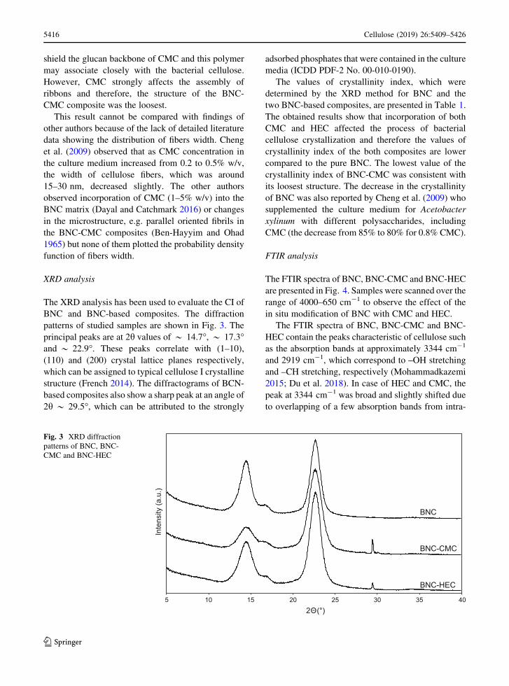
XRD analysis
The XRD analysis has been used to evaluate the CI of
BNC and BNC-based composites. The diffraction
patterns of studied samples are shown in Fig. 3. The
principal peaks are at 2h values of * 14.7�, * 17.3�and * 22.9�. These peaks correlate with (1–10),
(110) and (200) crystal lattice planes respectively,
which can be assigned to typical cellulose I crystalline
structure (French 2014). The diffractograms of BCN-
based composites also show a sharp peak at an angle of
2h * 29.5�, which can be attributed to the strongly
adsorbed phosphates that were contained in the culture
media (ICDD PDF-2 No. 00-010-0190).
The values of crystallinity index, which were
determined by the XRD method for BNC and the
two BNC-based composites, are presented in Table 1.
The obtained results show that incorporation of both
CMC and HEC affected the process of bacterial
cellulose crystallization and therefore the values of
crystallinity index of the both composites are lower
compared to the pure BNC. The lowest value of the
crystallinity index of BNC-CMC was consistent with
its loosest structure. The decrease in the crystallinity
of BNC was also reported by Cheng et al. (2009) who
supplemented the culture medium for Acetobacter
xylinum with different polysaccharides, including
CMC (the decrease from 85% to 80% for 0.8% CMC).
FTIR analysis
The FTIR spectra of BNC, BNC-CMC and BNC-HEC
are presented in Fig. 4. Samples were scanned over the
range of 4000–650 cm-1 to observe the effect of the
in situ modification of BNC with CMC and HEC.
The FTIR spectra of BNC, BNC-CMC and BNC-
HEC contain the peaks characteristic of cellulose such
as the absorption bands at approximately 3344 cm-1
and 2919 cm-1, which correspond to –OH stretching
and –CH stretching, respectively (Mohammadkazemi
2015; Du et al. 2018). In case of HEC and CMC, the
peak at 3344 cm-1 was broad and slightly shifted due
to overlapping of a few absorption bands from intra-
Inte
nsity
(a.u
.)
2Θ(°)
BNC
BNC-HEC
BNC-CMC
5 15 25 3510 20 30 40
Fig. 3 XRD diffraction
patterns of BNC, BNC-
CMC and BNC-HEC
123
5416 Cellulose (2019) 26:5409–5426

and intermolecular hydrogen bonds, such as
0(2)H…O(6), 0(3)H…O(5) and 0(6)H…0(3) (Gor-
gieva and Kokol 2011). The peaks at around 1050 and
1317 cm-1 were assigned to the C–O symmetric
stretching and CH2 wagging, respectively (Moham-
madkazemi 2015), while absorption band at
1428 cm-1 corresponded to symmetrical bending of
CH2 group (Jia et al. 2017). The appearance of the
peak at 1647 cm-1 corresponded to the bending
vibration of hydroxyl groups for BNC and HEC
(Gorgieva and Kokol 2011), while the band at
1590 cm-1 was associated with COO- in case of
CMC (Hessler and Klemm 2009). The spectrum of
BNC-CMC contained a broad peak at that region, due
to overlapping of the band from both BNC and CMC.
The occurrence of the hydroxyethyl group in the
cellulose derivative, such as HEC, can be seen as a
shift of C–O–C stretching vibration peak from 900 to
887 cm-1 (Gorgieva and Kokol 2011). In BNC-HEC
that band is visible at both these wavelengths. The
wide peak at 1352 cm-1 is assigned to C–OH in plane
stretching of HEC and therefore in the BNC-HEC
composite we can observe the slight deformation
between 1200 and 1400 cm-1.
3344
2919
1647
1317
1428
1554
900
1647
1352
887
1590
4000 3000 2000 1000
BNC
BNC-CMC
CMC
BNC-HEC
HEC
Wavenumber (cm-1)
Abs
orba
nce
1050
Fig. 4 FTIR spectra of BNC and BNC-based composites
Table 1 Crystallinity index for BNC, BNC-CMC and BNC-
HEC
Sample CI (%)
BNC 92.9
BNC-HEC 90.4
BNC-CMC 88.6
123
Cellulose (2019) 26:5409–5426 5417
Characterization of BNC, BNC-CMC and BNC-
HEC composites with glycerol
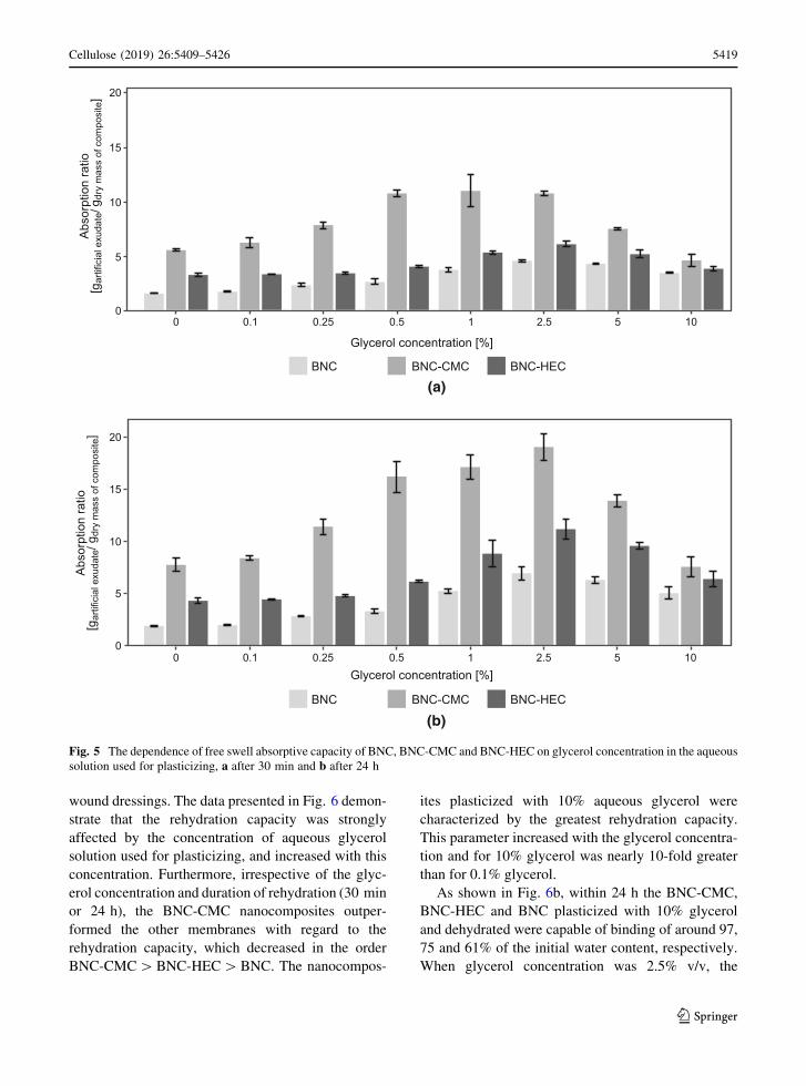
The dependence of free swell absorptive capacity
on glycerol concentration
Cellulose membranes with an enhanced liquid absorp-
tion capacity may be used for healing of wounds
characterized by the massive secretion of exudates
because they will be able to absorb an excess of fluids,
and isolate the wounds from external conditions. The
parameter which specifies the highest amount of
liquid, which may be absorbed by the wound dressing,
is the free swell absorptive capacity (ISO 13726
standard). The dependence of this parameter on
glycerol concentration, which was used for plasticiz-
ing of the BNC, BNC-CMC and BNC-HEC mem-
branes, and varied from 0.1 to 10% v/v, was
determined according to the ISO 13726 standard.
The amounts of artificial exudates absorbed by the
membranes were measured after 30 min (Fig. 5a) and
24 h (Fig. 5b).
The results presented in Fig. 5 demonstrate that
both the glycerol-plasticized and glycerol-free BNC-
CMC absorbed much more artificial exudate than the
BNC-HEC and BNC. This difference may be ascribed
to the fact that the BNC-CMC had the looser structure
than BNC and BNC-HEC. The BNC-HEC, which was
characterized by the thinnest fibers (the greater
specific surface area comparing to BNC and BNC-
CMC), absorbed significantly more artificial exudate
than BNC, but significantly less than BNC-CMC. The
amounts of artificial exudate absorbed within 30 min
were the greatest (above 10 g per 1 g dry weight) for
the glycerol concentrations ranging from 0.5 to 2.5%
v/v. After 24 h, the maximum absorption of artificial
exudate (of 19 g per 1 g dry weight) was observed at
the glycerol concentration of 2.5% v/v. Also at the
glycerol concentrations of 0.5 and 1.0% (v/v) the
uptake of the exudate was relatively high (of 16 and
17 g per 1 g dry weight). At the higher (5 and 10%
v/v) and lower (0.05–0.25% v/v) glycerol concentra-
tions, the liquid uptake was below 14 g/g dry weight.
The data presented in the Fig. 5 demonstrate that the
absorption of the artificial exudate was slower at the
glycerol concentrations ranging from 1 to 5%, com-
paring to the concentrations below 1% v/v. For
instance, within 30 min BNC-CMC and BNC-HEC
plasticized with 2.5% glycerol adsorbed only 54% and
56% of the artificial exudate adsorbed within 24 h,
respectively. At the glycerol concentrations below
1%, the liquid adsorbed within 30 min accounted for
60–85% of the amount adsorbed within 24 h. These
data demonstrate that plasticization of BNC, BNC-
CMC and BNC-HEC with glycerol extends the time of
exudates absorption by the dressing.
The dependence of rehydration capacity
on the glycerol concentration used to plasticize
the nanocomposites
Bacterial cellulose is an excellent dressing of various
wounds, including burns. One of most important
parameters, deciding of the utility of BNC is its high
water holding capacity, reaching up to 99% wet
pellicle mass (Ul-Islam et al. 2012). In the contact
with skin, BNC creates an ideal environment ensuring
regeneration of cells of soft tissues (Czaja et al. 2007).
The proposed modification of BNC and its composites
with HEC and CMC is based on plasticization with
glycerol, which leads to the improvement of certain
properties comparing to the native BNC. First of all,
glycerol ensures the rehydration of dehydrated com-
posites after the long-term storage. The storage of
dehydrated bacterial cellulose membranes is more
convenient than the storage of never-dried membranes.
Furthermore, glycerol is one of antimicrobial agents
(Sun et al. 2018), which additionally protect the wound
from contamination. The effect of glycerol concentra-
tion in the aqueous solutions used to plasticize the
BNC, BNC-CMC and BNC-HEC nanocomposites on
the rehydration capacity after dehydration of the
plasticized membranes is shown in Fig. 6. It is well
known that the unique hydrogel microstructure of
native BNC cannot be recovered after dehydration, and
also our investigations confirmed this conclusion.
Therefore, determination of the parameter such as
rehydration capacity, which is the ratio of water
absorbed after dehydration to the water content in the
never-dried membrane, is very important for the
characterization of wound dressings. The values of
rehydration capacity of the control BNC equaled 2.1%
and 2.4% after 30 min and 24 h, respectively. The
BNC-HEC and BNC-CMC composites were more
prone to rehydration as they bound 4.6 and 9.1% water
after 24 h. Thus, the dehydrated BNC, BNC-CMC and
BNC-HEC will not be able to ensure the stable, moist
environment which is required in case of the modern
123
5418 Cellulose (2019) 26:5409–5426
wound dressings. The data presented in Fig. 6 demon-
strate that the rehydration capacity was strongly
affected by the concentration of aqueous glycerol
solution used for plasticizing, and increased with this
concentration. Furthermore, irrespective of the glyc-
erol concentration and duration of rehydration (30 min
or 24 h), the BNC-CMC nanocomposites outper-
formed the other membranes with regard to the
rehydration capacity, which decreased in the order
BNC-CMC[BNC-HEC[BNC. The nanocompos-
ites plasticized with 10% aqueous glycerol were
characterized by the greatest rehydration capacity.
This parameter increased with the glycerol concentra-
tion and for 10% glycerol was nearly 10-fold greater
than for 0.1% glycerol.
As shown in Fig. 6b, within 24 h the BNC-CMC,
BNC-HEC and BNC plasticized with 10% glycerol
and dehydrated were capable of binding of around 97,
75 and 61% of the initial water content, respectively.
When glycerol concentration was 2.5% v/v, the
1052.510.50.250.10
Glycerol concentration [%]
Glycerol concentration [%]1052.510.50.250.10
5
10
15
20
0
5
10
15
20A
bsor
ptio
n ra
tio
[gar
tific
ial e
xuda
te/g
dry
mas
s of
com
posi
te]
BNC BNC-CMC BNC-HEC
0
Abs
orpt
ion
ratio
[g
artif
icia
l exu
date
/ gdr
y m
ass
of c
ompo
site
]
BNC BNC-CMC BNC-HEC
(a)
(b)
Fig. 5 The dependence of free swell absorptive capacity of BNC, BNC-CMC and BNC-HEC on glycerol concentration in the aqueous
solution used for plasticizing, a after 30 min and b after 24 h
123
Cellulose (2019) 26:5409–5426 5419
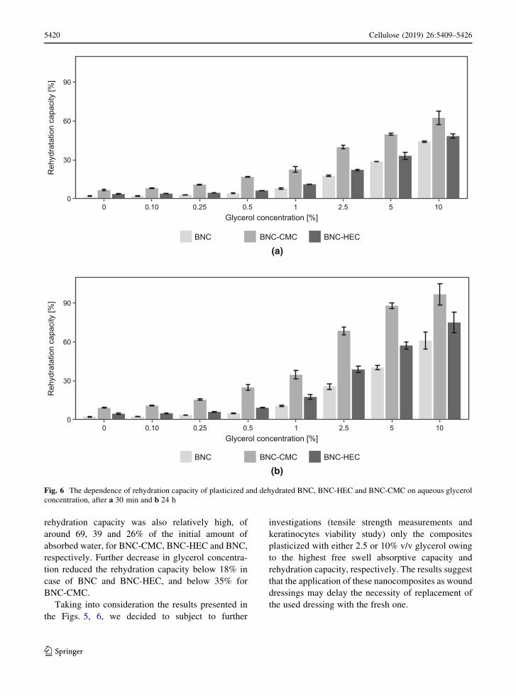
rehydration capacity was also relatively high, of
around 69, 39 and 26% of the initial amount of
absorbed water, for BNC-CMC, BNC-HEC and BNC,
respectively. Further decrease in glycerol concentra-
tion reduced the rehydration capacity below 18% in
case of BNC and BNC-HEC, and below 35% for
BNC-CMC.
Taking into consideration the results presented in
the Figs. 5, 6, we decided to subject to further
investigations (tensile strength measurements and
keratinocytes viability study) only the composites
plasticized with either 2.5 or 10% v/v glycerol owing
to the highest free swell absorptive capacity and
rehydration capacity, respectively. The results suggest
that the application of these nanocomposites as wound
dressings may delay the necessity of replacement of
the used dressing with the fresh one.
Reh
ydra
tatio
n ca
paci
ty [%
]R
ehyd
rata
tion
capa
city
[%]
Glycerol concentration [%]1052.510.50.250 0.10
60
30
0
90
Glycerol concentration [%]1052.510.50.250.100
60
30
0
90
BNC BNC-CMC BNC-HEC
BNC BNC-CMC BNC-HEC
(a)
(b)
Fig. 6 The dependence of rehydration capacity of plasticized and dehydrated BNC, BNC-HEC and BNC-CMC on aqueous glycerol
concentration, after a 30 min and b 24 h
123
5420 Cellulose (2019) 26:5409–5426
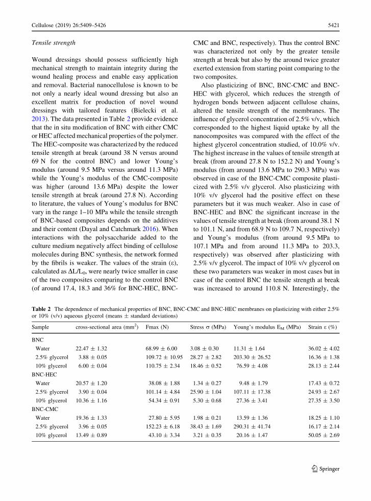
Tensile strength
Wound dressings should possess sufficiently high
mechanical strength to maintain integrity during the
wound healing process and enable easy application
and removal. Bacterial nanocellulose is known to be
not only a nearly ideal wound dressing but also an
excellent matrix for production of novel wound
dressings with tailored features (Bielecki et al.
2013). The data presented in Table 2 provide evidence
that the in situ modification of BNC with either CMC
or HEC affected mechanical properties of the polymer.
The HEC-composite was characterized by the reduced
tensile strength at break (around 38 N versus around
69 N for the control BNC) and lower Young’s
modulus (around 9.5 MPa versus around 11.3 MPa)
while the Young’s modulus of the CMC-composite
was higher (around 13.6 MPa) despite the lower
tensile strength at break (around 27.8 N). According
to literature, the values of Young’s modulus for BNC
vary in the range 1–10 MPa while the tensile strength
of BNC-based composites depends on the additives
and their content (Dayal and Catchmark 2016). When
interactions with the polysaccharide added to the
culture medium negatively affect binding of cellulose
molecules during BNC synthesis, the network formed
by the fibrils is weaker. The values of the strain (e),
calculated as DL/L0, were nearly twice smaller in case
of the two composites comparing to the control BNC
(of around 17.4, 18.3 and 36% for BNC-HEC, BNC-
CMC and BNC, respectively). Thus the control BNC
was characterized not only by the greater tensile
strength at break but also by the around twice greater
exerted extension from starting point comparing to the
two composites.
Also plasticizing of BNC, BNC-CMC and BNC-
HEC with glycerol, which reduces the strength of
hydrogen bonds between adjacent cellulose chains,
altered the tensile strength of the membranes. The
influence of glycerol concentration of 2.5% v/v, which
corresponded to the highest liquid uptake by all the
nanocomposites was compared with the effect of the
highest glycerol concentration studied, of 10.0% v/v.
The highest increase in the values of tensile strength at
break (from around 27.8 N to 152.2 N) and Young’s
modulus (from around 13.6 MPa to 290.3 MPa) was
observed in case of the BNC-CMC composite plasti-
cized with 2.5% v/v glycerol. Also plasticizing with
10% v/v glycerol had the positive effect on these
parameters but it was much weaker. Also in case of
BNC-HEC and BNC the significant increase in the
values of tensile strength at break (from around 38.1 N
to 101.1 N, and from 68.9 N to 109.7 N, respectively)
and Young’s modulus (from around 9.5 MPa to
107.1 MPa and from around 11.3 MPa to 203.3,
respectively) was observed after plasticizing with
2.5% v/v glycerol. The impact of 10% v/v glycerol on
these two parameters was weaker in most cases but in
case of the control BNC the tensile strength at break
was increased to around 110.8 N. Interestingly, the
Table 2 The dependence of mechanical properties of BNC, BNC-CMC and BNC-HEC membranes on plasticizing with either 2.5%
or 10% (v/v) aqueous glycerol (means ± standard deviations)
Sample cross-sectional area (mm2) Fmax (N) Stress r (MPa) Young’s modulus EM (MPa) Strain e (%)
BNC
Water 22.47 ± 1.32 68.99 ± 6.00 3.08 ± 0.30 11.31 ± 1.64 36.02 ± 4.02
2.5% glycerol 3.88 ± 0.05 109.72 ± 10.95 28.27 ± 2.82 203.30 ± 26.52 16.36 ± 1.38
10% glycerol 6.00 ± 0.04 110.75 ± 2.34 18.46 ± 0.52 76.59 ± 4.08 28.13 ± 2.44
BNC-HEC
Water 20.57 ± 1.20 38.08 ± 1.88 1.34 ± 0.27 9.48 ± 1.79 17.43 ± 0.72
2.5% glycerol 3.90 ± 0.04 101.14 ± 4.84 25.90 ± 1.04 107.11 ± 17.38 24.93 ± 2.67
10% glycerol 10.36 ± 1.16 54.34 ± 0.91 5.30 ± 0.68 27.36 ± 3.41 27.35 ± 3.50
BNC-CMC
Water 19.36 ± 1.33 27.80 ± 5.95 1.98 ± 0.21 13.59 ± 1.36 18.25 ± 1.10
2.5% glycerol 3.96 ± 0.05 152.23 ± 6.18 38.43 ± 1.69 290.31 ± 41.74 16.17 ± 2.14
10% glycerol 13.49 ± 0.89 43.10 ± 3.34 3.21 ± 0.35 20.16 ± 1.47 50.05 ± 2.69
123
Cellulose (2019) 26:5409–5426 5421

values of strain (e) were significantly greater after
plasticizing with 10% v/v glycerol comparing to 2.5%
v/v glycerol. Thus the exerted extension from starting
point was significantly greater in the first case.
Noteworthy, plasticizing with both 2.5% and 10%
v/v glycerol significantly decreased the cross-sectional
area (A) of BNC, BNC-CMC and BNC-HEC that
along with the significantly greater values of the
tensile strength at break (F) caused that the values of
stress (d), calculated as F/A were also significantly
greater.
As it was mentioned above also other authors
reported the impact of BNC modifications on the
tensile strength of BNC-based composites. Depen-
dently on the bacterial producer and concentration of
the modifying agents, either an increase or decrease in
the tensile modulus values has been reported (Cai and
Kim 2010; Cheng et al. 2009; Dayal and Catchmark
2016; Godinho et al. 2016; Zhou et al. 2009).
Reassuming, plasticizing of BNC, BNC-CMC and
BNC-HEC with 2.5% v/v glycerol enabled to produce
mechanically durable nanocomposites that are char-
acterized by the high free swell absorptive capacity
and rehydration capacity. Also the enhanced flexibility
of these membranes make them potential wound
dressing materials.
The effect of plasticized nanocomposites
on keratinocytes viability
Cytotoxicity testing is a primary and crucial step that
can decide of the fate of a health-related product for
clinical application. To estimate the influence of the
long-term contact of glycerol-plasticized BNC-based
wound dressings on the viability of keratinocytes, we
decided to determine the dependence of the latter
parameter not only on glycerol concentration in the
wound dressing but also on the polyol level in the
culture medium used for cultivation of these cells.
According to literature, different cells showed various
sensitivity towards glycerol. For instance, in case of
CHO (Chinese hamster ovary) cells line even 0.1% (v/
v) glycerol negatively affected cell viability (Rodas
et al. 2008) while in case of spleen cells isolated from
BALB/c mouse, 10% glycerol reduced cell viability
by only 10%, comparing to the control (Vaitkuviene
et al. 2009). The impact of the glycerol content in the
culture medium used for 24 h cultivation of ker-
atinocytes on the viability of these cells is presented in
Fig. 7a. The concentrations ranging from 0.5 to 2.5%
v/v ensured the relatively high viability, of at least
80%, comparing to the glycerol-free control. Further
increase in the glycerol concentration, to 5 and 10%
v/v, significantly reduced the viability, to around 50%
and 10%, respectively. These results suggest that
contact of keratinocytes with the BNC-based wound
dressings plasticized with 0.5–2.5% glycerol may not
negatively affect these cells. Thus, the BNC-CMC
nanocomposites plasticized with 2.5% v/v glycerol,
which are characterized by very attractive liquid
sorption and tensile properties, may be regarded
potential wound dressings.
To verify the latter conclusion, we applied both the
direct contact and the extract dilution methods. HaCaT
cells were seeded on the surface of the BNC, BNC-
CMC and BNC-HEC scaffolds plasticized with either
2.5% or 10% v/v glycerol (or glycerol-free), and
incubated at 37 �C in a humidified atmosphere for
24 h. Like in the former experiment, the viability of
cells was determined using the PrestoBlue� Cell
Viability Reagent. It was expressed as a percent of the
viability of the cells growing on the control commer-
cial plastic scaffold. The comparison of the growth of
the HaCaT cells on the latter scaffold and the glycerol-
free BNC, BNC-CMC and BNC-HEC scaffolds
showed that these three BNC-based scaffolds may be
used for cultivation of keratinocytes (Fig. 7d). Fur-
thermore, this experiment showed that the 24 h
contact with 2.5% v/v aqueous glycerol solution was
completely safe for the keratinocytes while 10% v/v
glycerol significantly reduced their viability. The
negative impact of 10% v/v glycerol was the weakest
in case of the BCN-HEC nanocomposite.
To determine the effect of glycerol-plasticized
wound coverings on the viability of keratinocytes, the
HaCaT cells were cultivated on plastic surface for
24 h in the culture medium, which was then removed,
and the cells were covered by the scaffolds plasticized
with either 2.5% or 10% v/v glycerol (or glycerol-free
controls). After 24 h incubation of the cells under the
scaffolds, the latter were removed and the viability of
cells was determined using the PrestoBlue� Cell
Viability Reagent. It was expressed as a percent of the
viability of the cells incubated on the plastic surface in
DMEM medium. The results presented in Fig. 7c
provide evidence that also in this case, the scaffolds
soaked in 2.5% v/v aqueous glycerol solution had no
negative impact on the viability of keratinocytes while
123
5422 Cellulose (2019) 26:5409–5426
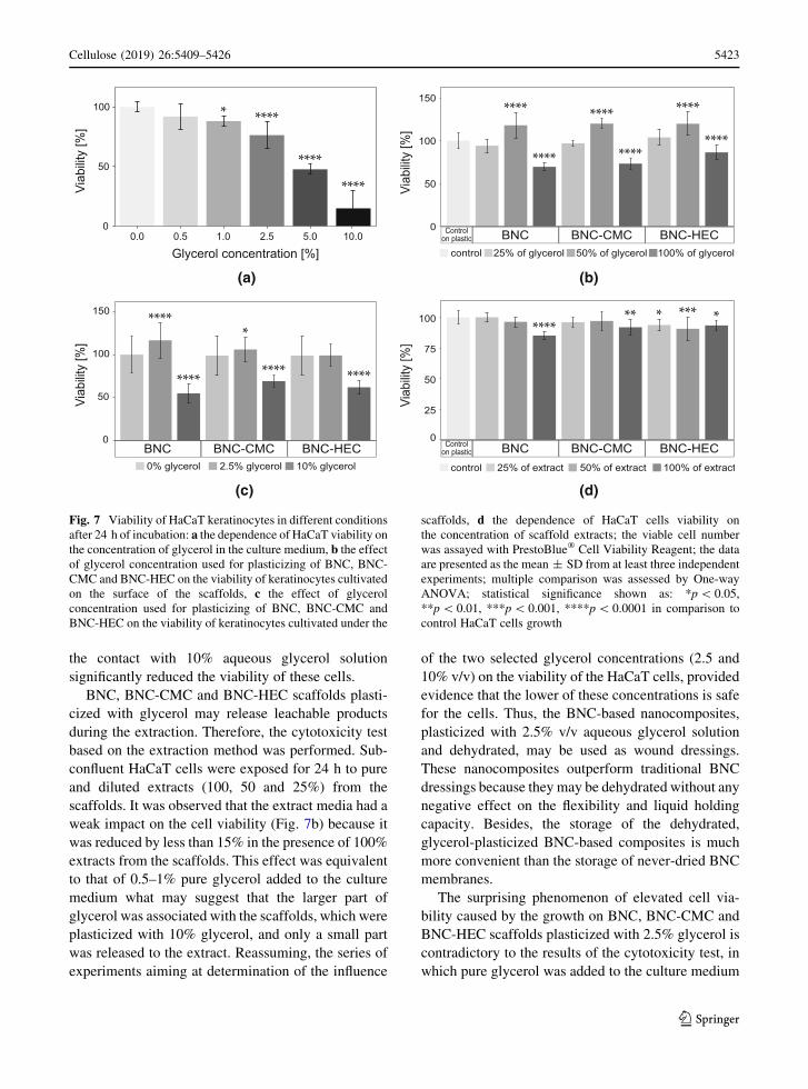
the contact with 10% aqueous glycerol solution
significantly reduced the viability of these cells.
BNC, BNC-CMC and BNC-HEC scaffolds plasti-
cized with glycerol may release leachable products
during the extraction. Therefore, the cytotoxicity test
based on the extraction method was performed. Sub-
confluent HaCaT cells were exposed for 24 h to pure
and diluted extracts (100, 50 and 25%) from the
scaffolds. It was observed that the extract media had a
weak impact on the cell viability (Fig. 7b) because it
was reduced by less than 15% in the presence of 100%
extracts from the scaffolds. This effect was equivalent
to that of 0.5–1% pure glycerol added to the culture
medium what may suggest that the larger part of
glycerol was associated with the scaffolds, which were
plasticized with 10% glycerol, and only a small part
was released to the extract. Reassuming, the series of
experiments aiming at determination of the influence
of the two selected glycerol concentrations (2.5 and
10% v/v) on the viability of the HaCaT cells, provided
evidence that the lower of these concentrations is safe
for the cells. Thus, the BNC-based nanocomposites,
plasticized with 2.5% v/v aqueous glycerol solution
and dehydrated, may be used as wound dressings.
These nanocomposites outperform traditional BNC
dressings because they may be dehydrated without any
negative effect on the flexibility and liquid holding
capacity. Besides, the storage of the dehydrated,
glycerol-plasticized BNC-based composites is much
more convenient than the storage of never-dried BNC
membranes.
The surprising phenomenon of elevated cell via-
bility caused by the growth on BNC, BNC-CMC and
BNC-HEC scaffolds plasticized with 2.5% glycerol is
contradictory to the results of the cytotoxicity test, in
which pure glycerol was added to the culture medium
Glycerol concentration [%]
Viab
ility
[%]
0.0 0.5 2.5 5.01.0 10.0
100
50
0
Viab
ility
[%]
150
100
0
50
BNC BNC-CMC BNC-HEC
(a) (b)
0% glycerol 2.5% glycerol 10% glycerol
(c) (d)
****
****
********
*
* ****
****
****
100
0
75
50
25Vi
abilit
y [%
]
control 25% of extract 100% of extract50% of extract
**** *******
Controlon plastic BNC BNC-CMC BNC-HEC
Controlon plastic BNC BNC-CMC BNC-HEC
Viab
ility
[%]
150
100
0
50
****
****
****
****
****
****
control 25% of glycerol 100% of glycerol50% of glycerol
Fig. 7 Viability of HaCaT keratinocytes in different conditions
after 24 h of incubation: a the dependence of HaCaT viability on
the concentration of glycerol in the culture medium, b the effect
of glycerol concentration used for plasticizing of BNC, BNC-
CMC and BNC-HEC on the viability of keratinocytes cultivated
on the surface of the scaffolds, c the effect of glycerol
concentration used for plasticizing of BNC, BNC-CMC and
BNC-HEC on the viability of keratinocytes cultivated under the
scaffolds, d the dependence of HaCaT cells viability on
the concentration of scaffold extracts; the viable cell number
was assayed with PrestoBlue� Cell Viability Reagent; the data
are presented as the mean ± SD from at least three independent
experiments; multiple comparison was assessed by One-way
ANOVA; statistical significance shown as: *p\ 0.05,
**p\ 0.01, ***p\ 0.001, ****p\ 0.0001 in comparison to
control HaCaT cells growth
123
Cellulose (2019) 26:5409–5426 5423
because even the lowest glycerol concentration, of
0.5%, caused a decrease in the cell viability. However,
the cumulative effect of 2.5% glycerol and neutral
BNC, BNC-CMC and BNC-HEC matrices on ker-
atinocytes was positive. The similar phenomenon was
reported in literature. For instance, the cytotoxic
compound like ZnCl2 used along with the neutral
3-aminopropyl triethoxysilane enhanced cell viability
(Chun et al. 2017). This is difficult to compare the
results of our study with findings of other authors
because the glycerol-induced toxicity has not been
reported so far for mammalian cells exposed to BNC
scaffolds. However, cytoprotective properties of car-
boxymethylcellulose against polyhexamethylene
biguanides added to bacteria were reported (Vehige
et al. 2003). Therefore, we may only assume that the
BNC composites studied have the similar cytoprotec-
tive properties, and further investigations in this field
are necessary.
Conclusions
The presented combination of in situ and ex situ
modifications of bacterial cellulose synthesized by K.
xylinus E25 under stationary conditions, enabled to
produce nanocomposites that may replace traditional
BNC dressings. The glycerol-plasticized BNC, BNC-
CMC and BNC-HEC membranes were flexible after
drying, and absorbed large amounts of artificial
exudate and water after rehydration. The impact of
plasticized BNC, BNC-CMC and BNC-HEC on the
viability of HaCaT keratinocytes was positive at
glycerol concentrations up to 2.5% (v/v). BNC-CMC
treated with 2.5% v/v aqueous glycerol was charac-
terized by the greatest free swell absorptive capacity
(up to 19 g artificial exudate/g dry weight in 24 h), and
the highest values of tensile strength at break (around
152.2 N) and Young’s modulus (around 290.3 MPa).
These properties suggest that the latter nanocomposite
may be applied as a convenient, mechanically durable
and safe wound dressing.
Acknowledgments The authors would like to thank Dr.
Przemysław Rytczak for comments related to
conceptualization of this study and for material support of the
fibroblast viability evaluation. We are indebted to Jolanta
Płoszynska for technical support and to Marcin Rosowski for his
contribution to recording the SEM images.
Compliance with ethical standards
Conflicts of interest The authors declare no conflict of
interest.
Open Access This article is distributed under the terms of the
Creative Commons Attribution 4.0 International License (http://
creativecommons.org/licenses/by/4.0/), which permits unre-
stricted use, distribution, and reproduction in any medium,
provided you give appropriate credit to the original
author(s) and the source, provide a link to the Creative Com-
mons license, and indicate if changes were made.
References
Ben-Hayyim G, Ohad I (1965) Synthesis of cellulose by Ace-
tobacter xylinum: VIII. On the formation and orientation of
bacterial cellulose fibrils in the presence of acidic
polysaccharides. J Cell Biol 25(2):191–207. https://doi.
org/10.1083/jcb.25.2.191
Bielecki S, Krystynowicz A, Turkiewicz M, Kalinowska H
(2002) Bacterial cellulose. In: Steinbuchel A (ed)
Biopolymers, vol 5. Wiley-VCH, Weinheim, pp 37–90.
ISBN 978-3-527-30226-0
Bielecki S, Kalinowska H, Krystynowicz A, Kubiak K,
Kołodziejczyk M, de Groeve M (2013) Wound dressings
and cosmetic materials from bacterial nanocellulose. In:
Gama M, Gatenholm P, Klemm D (eds) Bacterial
Nanocellulose. A sophisticated multifunctional material,
1st edn. CRC Press, Taylor & Francis Group, Boca Raton,
Florida, pp 157–174, ISBN 9781138073166
Budhiono A, Rosidi B, Taher H, Iguchi M (1999) Kinetic
aspects of bacterial cellulose formation in nata-de-coco
culture system. Carbohydr Polym 40:137–143. https://doi.
org/10.1016/S0144-8617(99)00050-8
Cai Z, Kim J (2010) Bacterial cellulose/poly(ethylene glycol)
composite: characterization and first evaluation of bio-
compatibility. Cellulose 17(1):83–91. https://doi.org/10.
1007/s10570-009-9362-5
Campano C, Balea A, Blanco A, Negro C (2016) Enhancement
of the fermentation process and properties of bacterial
cellulose: a review. Cellulose 23:57–91. https://doi.org/10.
1007/s10570-015-0802-0
Chen Y, Zhou X, Lin Q, Jiang D (2014) Bacterial cellulose/
gelatin composites: in situ preparation and glutaraldehyde
treatment. Cellulose 21:2679–2693. https://doi.org/10.
1007/s10570-014-0272-9
Cheng KC, Catchmark JM, Demirci A (2009) Effect of different
additives on bacterial cellulose production by Acetobacter
xylinum and analysis of material property. Cellulose
16(6):1033–1045. https://doi.org/10.1007/s10570-009-
9346-5
Chun H-S, Park D, Lim SE, Jeong K-H, Park J-S, Park H-J,
Kang S, Kang KS, Park H-G, An H-R, Huh YS, Lee Y-C
(2017) Two zinc-aminoclays’ in vitro cytotoxicity assess-
ment in HeLa cells and in vivo embryotoxicity assay in
zebrafish. Ecotoxicol Environ Saf 137:103–112. https://
doi.org/10.1016/j.ecoenv.2016.11.022
123
5424 Cellulose (2019) 26:5409–5426

Cielecka I, Szustak M, Gendaszewska-Darmach E, Kalinowska
H, Ryngajłło M, Maniukiewicz W, Bielecki S (2018)
Novel bionanocellulose/r-carrageenan composites for tis-
sue engineering. Appl Sci 8(8):1352. https://doi.org/10.
3390/app8081352
Czaja W, Young DJ, Kawecki M, Brown RM (2007) The future
prospects of microbial cellulose in biomedical applica-
tions. Biomacromol 8(1):1–12. https://doi.org/10.1021/
bm060620d
Dayal MS, Catchmark JM (2016) Mechanical and structural
property analysis of bacterial cellulose composites. Car-
bohydr Polym 144:447–453. https://doi.org/10.1016/j.
carbpol.2016.02.055
Du R, Zhao F, Peng Q, Zhou Z, Han Y (2018) Production and
characterization of bacterial cellulose produced by Glu-
conacetobacter xylinus isolated from Chinese persimmon
vinegar. Carbohydr Polym 194:200–207. https://doi.org/
10.1016/j.carbpol.2018.04.041
Fang L, Catchmark JM (2014) Characterization of water-soluble
exopolysaccharides from Gluconacetobacter xylinus and
their impacts on bacterial cellulose crystallization and
ribbon assembly. Cellulose 21:3965–3978. https://doi.org/
10.1007/s10570-014-0443-8
French AD (2014) Idealized powder diffraction patterns for
cellulose polymorphs. Cellulose 21:885–896. https://doi.
org/10.1007/s10570-013-0030-4
Godinho JF, Berti FV, Muller D, Rambo CR, Porto LM (2016)
Incorporation of Aloe vera extracts into nanocellulose
during biosynthesis. Cellulose 23:545–555. https://doi.org/
10.1007/s10570-015-0844-3
Gorgieva S, Kokol V (2011) Synthesis and application of new
temperature-responsive hydrogels based on carboxymethyl
and hydroxyethyl cellulose derivatives for the functional
finishing of cotton knitwear. Carbohydr Polym
85:664–673. https://doi.org/10.1016/j.carbpol.2011.03.
037
Haigler CH, White AR, Brown RM Jr, Cooper KM (1982)
Alteration of in vivo cellulose ribbon assembly by car-
boxymethylcellulose and other cellulose derivatives. J Cell
Biol 94:64–69. https://doi.org/10.1083/jcb.94.1.64
Hessler N, Klemm D (2009) Alteration of bacterial nanocellu-
lose structure by in situ modification using polyethylene
glycol and carbohydrate additives. Cellulose 16:899–910.
https://doi.org/10.1007/s10570-009-9301-5
Jia YY, Wang XH, Huo MM, Zhai XL, Li F, Zhong C (2017)
Preparation and characterization of a novel bacterial cel-
lulose/chitosan bio-hydrogel. Nanomater Nanotechnol
7:1–8. https://doi.org/10.1177/1847980417707172
Li Y, Jiang H, Zheng W, Gong N, Chen L, Jiang X, Yang G
(2015) Bacterial cellulose-hyaluronan nanocomposite
biomaterials as wound dressings for severe skin injury
repair. J Mater Chem B 3:3498–3507. https://doi.org/10.
1007/s10570-018-1709-3
Lin S-P, Kung H-N, Tsai Y-S, Tseng T-N, Hsu K-D, Cheng K-C
(2017) Novel dextran modified bacterial cellulose hydrogel
accelerating cutaneous wound healing. Cellulose
24:4927–4937. https://doi.org/10.1007/s10570-017-1448-
x
Liu K, Catchmark JM (2018) Effects of exopolysaccharides
from Escherichia coli ATCC 35860 on the mechanical
properties of bacterial cellulose nanocomposites. Cellulose
25:2273–2287. https://doi.org/10.1007/s10570-018-1709-
3
Machado RTA, Meneguin AB, Sabio RM, Franco DF, Antonio
SG, Gutierrez J, Tercjak A, Beretta AA, Ribeiro SJL,
Lazarini SC, Lustri WR, Barud HS (2018) Koma-
gateibacter rhaeticus grown in sugarcane molasses-sup-
plemented culture medium as a strategy for enhancing
bacterial cellulose production. Ind Crops Prod
122:637–646. https://doi.org/10.1016/j.indcrop.2018.06.
048
MacNeil S (2007) Progress and opportunities for tissue-engi-
neered skin. Nature 445:874–880. https://doi.org/10.1038/
nature05664
Mohammadkazemi F (2015) Surface properties of bacterial
nanocellulose using spectroscopic methods and X-ray
diffraction. Am J Appl Indu Chem 1(2):10–13. https://doi.
org/10.11648/j.ajaic.20150102.11
Nakagaito AN, Iwamoto S, Yano H (2005) Bacterial cellulose:
the ultimate nano-scalar cellulose morphology for the
production of high-strength composites. Appl Phys A
80(1):93–97. https://doi.org/10.1007/s00339-004-2932-3
Park S, Baker JO, Himmel ME, Parilla PA, Johnson DK (2010)
Cellulose crystallinity index: measurement techniques and
their impact on interpreting cellulose performance.
Biotechnol Biofuels 3:3–10. https://doi.org/10.1186/1754-
6834-3-10
Rabiej M (2014) A hybrid immune-evolutionary strategy algo-
rithm for the analysis of the wide-angle X-ray diffraction
curves of semicrystalline polymers. J Appl Crystallogr
47:1502–1511. https://doi.org/10.1107/S16005767140147
82
Rodas ACD, Maizato MJS, Leirner AA, Pitombo RNM, Pola-
kiewicz B, Beppu MM, Higa OZ (2008) Cytotoxicity and
genotoxicity of bovine pericardium preserved in glycerol.
Artif Organs 32(4):272–276. https://doi.org/10.1111/j.
1525-1594.2008.00542.x
Sanchavanakit N, Sangrungraungroj W, Kaomongkolgit R,
Banaprasert T, Pavasant P, Phisalaphong M (2006) Growth
of human keratinocytes and fibroblasts on bacterial cellu-
lose film. Biotechnol Prog 22:1194–1199. https://doi.org/
10.1021/bp060035o
Sun Y, Meng C, Zheng Y, Xie Y, He W, Wang Y, Qiao K, Yue L
(2018) The effects of two biocompatibile plasticizers on
the performance of dry bacterial cellulose membrane: a
comparative study. Cellulose 25:5893–5908. https://doi.
org/10.1007/s10570-018-1968-z
Taokaew S, Phisalaphong M, Newby BZ (2015) Modification of
bacterial cellulose with organosilanes to improve attach-
ment and spreading of human fibroblasts. Cellulose
22:2311–2324. https://doi.org/10.1007/s10570-015-0651-
x
Uhlin KI, Atalla RH, Thompson NS (1995) Influence of hemi-
celluloses on the aggregation patterns of bacterial cellu-
lose. Cellulose 2:129–144. https://doi.org/10.1007/
BF00816385
Ul-Islam M, Khan T, Park J (2012) Water holding and release
properties of bacterial cellulose obtained by in situ and ex
situ modification. Carbohydr Polym 88:596–603. https://
doi.org/10.1016/j.carbpol.2012.01.006
Ullah H, Wahid F, Santos HA, Khan T (2016) Advances in
biomedical and pharmaceutical applications of functional
123
Cellulose (2019) 26:5409–5426 5425

bacterial cellulose-based nanocomposites. Carbohydr
Polym 150:330–352. https://doi.org/10.1016/j.carbpol.
2016.05.029
Vaitkuvien _e A, Kas _eta V, Ramanauskait _e G, Biziulevicien _e G
(2009) Cytotoxicity of pharmaceutical and cosmetic gel-
forming polymers, preservatives and glycerol to primary
murine cell cultures. Acta Med Litu 16(3–4):92–97. https://
doi.org/10.2478/v10140-009-0013-9
Vehige JG, Simmons PA, Anger C, Graham R, Tran L, Brady N
(2003) Cytoprotective properties of carboxymethyl cellu-
lose (cmc) when used prior to wearing contact lenses
treated with cationic disinfecting agents. Eye Contact Lens
Sci Clin Pract 29(3):177–180. https://doi.org/10.1097/01.
ICL.0000074106.82322.17
Velasquez-Riano M, Bojaca V (2017) Production of bacterial
cellulose from alternative low-cost substrates. Cellulose
24:2677–2698. https://doi.org/10.1007/s10570-017-1309-7
Vieira MGA, da Silva MA, dos Santos LO (2011) Natural-based
plasticizers and biopolymer films: a review. Eur Polym J
47:254–263. https://doi.org/10.1016/j.europloymj.2010.
12.011
Zhou Q, Malm E, Nilsson H, Larsson PT, Iversen T, Berglund
LA, Bulone V (2009) Nanostructured biocomposites based
on bacterial cellulose nanofibers compartmentalized by a
soft hydroxyethylcellulose matrix coating. Soft Matter
5:4124–4130. https://doi.org/10.1039/B907838J
Publisher’s Note Springer Nature remains neutral with
regard to jurisdictional claims in published maps and
institutional affiliations.
123
5426 Cellulose (2019) 26:5409–5426
Recommended
![Novel cellulose particles by regeneration from homogeneous ... · [amim]Cl: 1-allyl-3-methylimidazoliumchrolide BNC: Bacterial nanocellulose [bmim]Cl: 1-butyl-3-methylimidazoliumchrolide](https://img.pdfslide.us/doc/110x75/60614cf6c7a2c44aea7ff38c/novel-cellulose-particles-by-regeneration-from-homogeneous-amimcl-1-allyl-3-methylimidazoliumchrolide.jpg)