Page 1721
Effect of mutagens on gene expression of amylases producing
Bacilli SPS
Annam Sravan Kumar
M.Sc(Bio-Technology)
Chaitanya Degree And PG College,
Hanamkonda, Warangal, Telangana.
Dr. M.L.N.Reddy
Guide
Ventura Institute of Biosciences
Hyderabad.
Abstract: Amylases are important enzymes employed
in the starch processing industries for the hydrolysis
of polysaccharides such as starch into simple sugar
constituents. Although amylases can be obtained
from several sources, such as plants and animals, the
enzymes from microbial sources generally meet
industrial demand. Microbial amylases have
successfully replaced chemical hydrolysis of starch in
starch processing industries. Besides their use in
starch saccaharification, they also find potential
application in a number of industrial processes such
as in food, baking, brewing, detergent, textile and
paper industries. With the advent of new frontiers in
biotechnology, the spectrum of amylase application
has expanded into many other fields, such as clinical,
medical and analytical chemistry. The objective of
this paper is selection of a suitable strain for the
production of amylase by comparative study of the
strains such as bacilli and cocci Screening of
different agricultural byproducts as substrates for
maximum enzyme production, application of
different combinations of these substrates for enzyme
production, and optimization of cultural conditions
for the production of glucoamylase.
Keywords: Amylase, glycoside hydrolase enzymes ,
Starch, strain, bacilli, cocci, glucoamylase.
Introduction:
Amylase is the name given to glycoside hydrolase
enzymes that break down starch into maltose
molecules. It is mainly found in the saliva, which is the
reason why we can feel the sweetness when eating rice
(contains glucose). However, the glucose is absorbed
into the bloodstream from the small intestines, not in
the mouth. Although the amylases are designated by
different Greek letters, they all act on α-1, 4-glycosidic
bonds.
There are three sub classifications of amylases they are
1. α-Amylase: - The α-amylases are calcium
metalloenzymes, completely unable to function in the
absence of calcium. By acting at random locations
along the starch chain, α-amylase breaks down long-
chain carbohydrates, ultimately yielding maltotriose
and maltose from amylose, or maltose, glucose and
"limit dextrin" from amylopectin. Because it can act
anywhere on the substrate, α-amylase tends to be
faster-acting than β-amylase. In animals, it is a major
digestive enzyme and its optimum pH is 6.7-7.0. In
human physiology, both the salivary and pancreatic
amylases are α-Amylases. Also found in plants
(barley) , fungi (ascomycetes and basidiomycetes) and
bacteria (Bacillus).
Human salivary amylase

Page 1722
Human pancreatic amylase
Calcium ion visible in pale khaki, chloride ion in
green.
2. β-Amylase: - Another form of amylase, β-amylase is
also synthesized by bacteria, fungi, and plants.
Working from the non-reducing end, β-amylase
catalyzes the hydrolysis of the second α-1, 4 glycosidic
bond, cleaving off two glucose units (maltose) at a
time. During the ripening of fruit, β-amylase breaks
starch into sugar, resulting in the sweet flavor of ripe
fruit. Both are present in seeds; β-amylase is present
prior to germination, whereas α-amylase and proteases
appear once germination has begun. Cereal grain
amylase is key to the production of malt. Many
microbes also produce amylase to degrade
extracellular starches. Animal tissues do not contain β-
amylase, although it may be present in microrganisms
contained within the digestive tract.
3. γ-Amylase: - In addition to cleaving the last α(1-4)
glycosidic linkages at the nonreducing end of amylose
and amylopectin, yielding glucose, γ-amylase will
cleave α(1-6) glycosidic linkages. Unlike the other
forms of amylase,γ-amylase is most efficient in acidic
environments and has an optimum pH of 3.
During the last decade, an increased attention was paid
to the use of various agro industrial wastes for value
addition using solid-state fermentation (SSF) by
filamentous fungi. SSF is the most appropriate process
due to the advantages it offers. The hyphal mode of
growth and good tolerance to low water activity and
high osmotic pressure conditions, make fungi most
efficient for bioconversion of solid substrates.
Starch is also present in waste produced from food
processing plants and industries like paper. Starch
waste causes pollution problems. Biotechnological
treatment of waste water by immobilization of amylase
degrade the starch in the waste which can produce
valuable products such as microbial biomass protein
and also purifies the effluent
Microbial amylases are exploited for the following
purposes:
1. High Fructose Corn syrup preparation
2. Additives to detergents for removing stains
3. Saccharification of starch for alcohol production
4. Starch and cellulose in waste and its treatment.
4. Brewing
The enzyme amylase is widely distributed in the
intestinal track of fresh water fishes and plays an
important role in the digestion of starch. Enzyme
found in intestinal lumen of fish could potentially have
come from either pancreas or the secretary cells in the
gut walls of the fish. In addition, enzymes from
intestinal micro flora potentially could have a
significant role in digestion, especially for substrates
such as starch and cellulose. The intestinal track of fish
is generally colonized by great number of
heterotrophic bacteria, including aerobes and
anaerobes.
The other organism enzyme producing. Amylase
production was checked and activity was measured.
Production of amylase was compared with the
reference of crude enzyme kinetics. Amylase was
separated from the crude enzyme by dialysis and ion –
exchange chromatography. The presence of amylase
was conformed by performing SDS-Poly Acrylamide
Gel Electrophoresis. The results were interpreted by
visualizing the protein bands.
Page 1723
MATERIALS AND METHODS
ISOLATION OF MICROORGANISMS FROM
DIFFERENT SOURCES
Organisms ranges from bacteria to nematodes and
includes diverse species of algae and protozoa. soil
microorganisms are responsible for the breakdown of
organic matter, including hydrocarbons, conversion of
inorganic components from one form to another and
the production of humus. Although this group is large
and diverse, soil microorganisms are thought of as
being of three distinct types: fungi actinomycetes, and
bacteria. Cow dung microorganisms form a robust
community capable of surviving and functioning under
extremes of temperature, water availability, pH, energy
resources, nutrient availability and salt concentration.
Isolating individual microorganisms and allowing
them to grow and produce colonies is another method
of enumerating microorganisms in soil. Nitrogen-
fixing bacteria form symbiotic associations with the
roots of legumes Nitrifying bacteria change
ammonium (NH4+) to nitrite (NO2-) then to nitrate
(NO3-) – a preferred form of nitrogen for grasses and
most row crops. Denitrifying bacteria convert nitrate
to nitrogen (N2) or nitrous oxide (N2O) gas.
Denitrifies are anaerobic. Actinomycetes are a large
group of bacteria that grow as hyphae like fungi
(Figure 3). They are responsible for the
characteristically “earthy” smell of freshly turned,
healthy soil. Actinomycetes decompose a wide array
of substrates, but are especially important in degrading
recalcitrant (hard-to-decompose) compounds, such as
chitin and cellulose, and are active at high pH levels.
Fungi are more important in degrading these
compounds at low pH. A number of antibiotics are
produced by actinomycetes such as Streptomycin.
Municipal and rural water supplies can transmit human
diseases such as cholera (Vibrio cholerae), typhoid
fever (Salmonella typhi), shigellosis (Shigella),
salmonellosis (Salmonella), and gastroenteritis
(Campylobacter jejuni, Escherichia coli, Giardia
lamblia). The threat of such disease transmission
becomes more serious as the population density
increases and more sewage pollutes public water
supplies, carrying with it human intestinal pathogens.
Isolation and enumeration of microorganisms can be
done from different sources such as cow dung, soil,
water, and air. Serial dilution agar plating method is
one of most routinely used procedure for the isolation
and enumeration of microorganisms.
SERIAL DILUTION
AIM: To isolate and enumerate microorganisms from
natural cow dung source.
PRINCIPLE: This method is based on the principle
that when material containing bacteria is cultured,
every viable bacterium develops into a visible colony
on a nutrient agar medium .In this procedure a small
measured volume (or weight) is mixed with a large
volume of saline called the diluents or dilution blank.
The number of organisms developed on the plates after
an incubation period of 24-48h per ml is calculated as
follows:
Number of cells/ml= number of colonies/volume
plated*dilution.
PROCEDURE:
1. Label the dilution blanks as 10-1, 10,-210-3, 10-4, 10-
5, 10-6 and 10-7.
2. Prepare the initial dilution by adding 1ml or 1g of
the sample into a 9ml dilution blank labeled 10-1
thus diluting the original sample 10 times.
3. Vortex the tubes to obtain uniform distribution of
organisms.
4. From the first dilution, transfer 1ml of the
suspension to the dilution blank 10-2 with a sterile
1ml pipette.
5. From the 10-2 suspension, transfer 1ml of the
suspension to the 10-3 dilution blank with a sterile
pipette, thus diluting the original sample to 1000
times(1:1000 or 10-3)
6. Repeated this procedure till the original sample has
been diluted 10,000,000(10-7) times using every
time a sterile pipette.

Page 1724
7. From the appropriate dilutions (10-2, 10-4, 10-6, and
10-7) transfer 1ml or 0.1ml of suspension with the
respective pipettes to the sterile petriplates.
8. Add approximately 15ml of the nutrient medium,
melted and cooled to 450c, to each Petri plate
containing the diluted sample. Mix the contents of
the plate by rotating clock wise and antilock wise.
9. Allow the plates to solidify.
10. Incubate the plates in an inverted positon for 24 to
48h at 370c.
OBSERVATION
1. Observe all the plates for the appearance of
bacterial colonies.
2. Count the number of colonies in the plates, that
have colonies in the 30-300 range,by placing each
plate on the colony counter.
RESULTS
Calculate the number of bacteria per ml of the sample
as follows:
Organisms per gram of the sample= number of
colonies/volume plated *dilution
60 colonies were counted on a 1:105 dilution, then
Number of cells=60 colonies/1ml*10-5=6000000
= 6*106 bacteria /ml or gm of sample.
SPREAD PLATE METHOD:
The number of bacteria in solution can be readily
quantified by using the spread plate technique. In this
technique, the sample is appropriately diluted and a
small aliquot transferred to an agar plate. The bacteria
are then distributed evenly over the surface by a
special streaking technique. After colonies are grown,
they are counted and the number of bacteria in the
original sample is calculated.
PRINCIPLE:
The principle behind this technique is that as the Petri
dish spins at some stage, single cells will be deposited
with the bent glass rod on to the agar surface. Some of
these cells will be separated from each other by a
distance sufficient to allow the colonies that develop to
be free from each other.
PROCEDURE:
1. Prepare nutrient agar. Autoclave at 1210c, 15lbs,
for 15min.
2. Pour the agar in the petriplates (15ml).
3. Allow it to solidify
4. 0.1 ml of bacterial suspension is placed in the
center of the plate using a sterile pipet.
5. The glass rod is sterilized by first dipping it into a
70% alcohol solution and then passing it quickly
through the Bunsen burner flame. The burning
alcohol sterilizes the rod at a cooler temperature
than holding the rod in the burner flame thus
reducing the chance of you burning your fingers.
6. When all the alcohol has burned off and the rod
has air-cooled, streak the rod back and forth
across the plate working up and down several
times.
7. Turn the plate 90 degrees and repeat the side to
side, up and down streaking. Turn the plate 45
degrees and streak a third time.
8. Do not sterilize the glass rod between plate
turnings. Cover the plate and wait several minutes
before turning it upside down for incubation. This
will allow the broth to soak into the plate so the
bacteria won't drip onto the plate lid.

Page 1725
RESULT: - . The bacteria are then distributed evenly
over the surface. Colonies grown are counted and the
number of bacteria in the original sample are
calculated.
Organisms per gram of the sample= number of
colonies/volume plated *dilution
60 colonies were counted on a 1:105 dilution, then
Number of cells=60 colonies/1ml*10-5=6000000
= 6*106 bacteria /ml or gm of sample.
THE STREAK PLATE TECHNIQUE
Streak plates allow for the growth of isolated colonies
on the surface of the agar. An isolated colony is a
colony that is not touching any other colonies and is
assumed to be a pure culture. These colonies are easily
accessible for performing staining and Identification
procedures. They also show colonial morphology that
may be useful in identifying the organism.
PRINCIPLE:
An original inoculum containing a mixture of bacteria
is spread into 4 quadrants on solid media.
The goal is to reduce the number of bacteria in each
subsequent quadrant. Colonies are masses of offspring
from an individual cell therefore streaking attempts to
separate individual cells.
Discrete colonies form as the individual cells are
separated and then multiply to form isolated colonies
in the later quadrants.
PROCEDURE:
1. Pick up a loopful of your inoculum from either a
broth or an agar culture. Using a sterile agar medium
plate (lift the lid just enough to insert the loop), streak
a vertical line straight down.
2. When streaking the agar, keep the loop horizontal
and only streak the surface of the agar: DO NOT DIG
INTO THE AGAR.
3. Move the loop in a zig-zag pattern across the agar
until 1/3 of the plate is covered, finishing the first
section.
4. Sterilize the loop in the flame and let it cool before
continuing to spread the bacteria. You can do this by
1) sticking the hot loop in the agar at the edge of the
agar away from the bacteria, or 2) just holding the loop
for a few seconds while it cools.
5. Rotate the plate about 90 degrees and spread the
bacteria from the first streak into a second area using
the same zig-zag spread technique.
6. Sterilize the loop again. Rotate the plate about 90
degrees and spread the bacteria from the second streak
into the 3rd area in the same pattern.
7. Sterilize the loop again. Replace the lid and invert
the plate. Incubate the plates at 370c
RESULT: - Isolated colonies are observed on the
surface of the agar. An isolated colony is a colony that
is not touching any other colonies and is assumed to be
a pure culture. These colonies were accessible for
performing staining and Identification procedures.
They show colonial morphology that may be useful in
identifying the organism.
PURE CULTURE
A pure culture consists of a population of only one
species of microorganisms derived from a single
microorganism. A strain is made up of succession of
one kind of microorganism from a single colony.
Isolation of one kind of microorganism from a mixture
of many different kinds is called pure culture
technique.
AIM: To isolate individual colonies including surface
and subsurface colonies.
PRINCIPLE: - separation of a particular
microorganism from the mixed populations by
inoculating the single and pure colony of
microorganism in the agar slant

Page 1726
PROCEDURE:
1) Prepare the agar slants by adding adequate
amount of agar in the test tubes and made them
solidify by placing in slant position.
2) Take a loop full of pure culture and inoculate in
the slant by streaking on the slant.
3) Close the tube with the cotton plug and place in
the incubator for overnight at 37c.
RESULT: - Pure cultures of microorganisms were
grown in the test-tube which are called pure cultures of
specific microorganism which were further studied by
colony morphology and confirmed by gram staining.
GRAM STAINING
The gram stain a differential stain was developed by
Dr.Hans Christian Gram; Danish physician in 1884.it
is a very useful technique for identifying and
classifying bacteria into two major groups: the gram
positive and gram negative.
PRINCIPLE:
Differential staining requires the use of four reagents
that are applied on the heat fixed smear. The first
reagent is primary stain (crystal violet).its function is
to impart color to all the cells; the second is the
mordant (iodine solution) which serves to intensify the
color of the stain. The CV-I complex binds to
magnesium ribose nucleic acid component of cell wall,
which is more difficult to remove .the third is
decolorizing agent (95% ethyl alcohol) serves as a
lipid solvent and protein dehydrating agent. The last
one is the counter stain(saffranin) has a contrasting
color to that of primary stain .Following
decolourization if the primary stain is not washed out
the counter stain cannot be absorbed or if the primary
stain is removed the decolorized cellular components
will accept and assume the counter stain.
PROCEDURE:
Make thin smears on a grease free glass slide
Let the smears air dry
Heat the slide for few seconds until it becomes
hot to the touch so that bacteria are firmly
mounted to the slide.
Add the primary stain crystal violet and incubate
1 minute. This step colors all cells violet
Add grams iodine for 30 seconds. It is not a stain
it is a mordant it doesn’t give color directly to the
bacteria but it fixes the crystal violet to the
bacterial cell wall. All cells remain violet
Wash with ethanol and acetones.
Wash with ethanol and acetone, They decolorize. If
the bacteia are gram positive it will retain the
primary stain. If it is gram negative it will lose the
primary stain and appear colorless.
Add the secondary stain , safranin and incubate 1
min, then wash with water for maximum of 5
seconds. If the bacteria are gram positive the cell
will retian the primary stain, will not take the
secondary stain., and will appear red-pink.
Blot dry with absorbent paper.
Let the slides air dry
OBSERVATION:
Examine the slides microscopically using oil
immersion objective.
Identify the gram reaction and classify them
Describe the morphology and arrangement of
the cells.
RESULTS:
The bacteria that appear purple are referred as gram
positive. And the bacteria were found to be bacillus
species. I confirmed the organism according to the
BERGEYS MANUAL.
PRECAUTIONS:
Always use fresh and young cultures(less than 24h
old).
Excessive heat should be avoided during heat fixing.
Smear should be thin and uniform.
SCREENING OF MICROOGANISM AIM: - To screen the microorganism by providing the
selective medium.
Principle: - organism’s shows difference in
physiological and biochemical reactions, thus by using
this principle organisms were screened by providing
the selective medium.
PROCEDURE:-
1) Prepare nutrient agar starch medium. Autoclave
at 1210c, 15lbs, for 15min.
2) Pour the agar in the petriplates (15ml).
3) Allow it to solidify.
4) Take a loop full of pure culture and streak as M
shape in the Petri dish
5) Place the plates in the incubator for over night at
37c
Page 1727
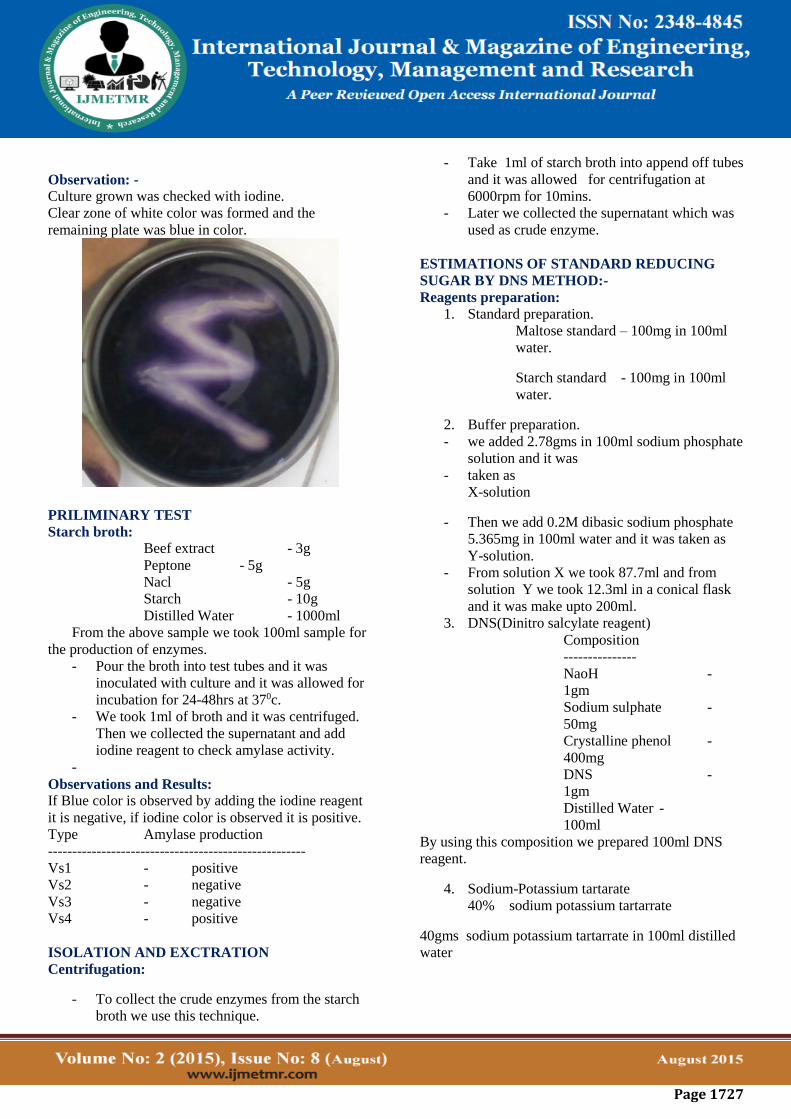
Observation: -
Culture grown was checked with iodine.
Clear zone of white color was formed and the
remaining plate was blue in color.
PRILIMINARY TEST
Starch broth:
Beef extract - 3g
Peptone - 5g
Nacl - 5g
Starch - 10g
Distilled Water - 1000ml
From the above sample we took 100ml sample for
the production of enzymes.
- Pour the broth into test tubes and it was
inoculated with culture and it was allowed for
incubation for 24-48hrs at 370c.
- We took 1ml of broth and it was centrifuged.
Then we collected the supernatant and add
iodine reagent to check amylase activity.
-
Observations and Results:
If Blue color is observed by adding the iodine reagent
it is negative, if iodine color is observed it is positive.
Type Amylase production
-----------------------------------------------------
Vs1 - positive
Vs2 - negative
Vs3 - negative
Vs4 - positive
ISOLATION AND EXCTRATION
Centrifugation:
- To collect the crude enzymes from the starch
broth we use this technique.
- Take 1ml of starch broth into append off tubes
and it was allowed for centrifugation at
6000rpm for 10mins.
- Later we collected the supernatant which was
used as crude enzyme.
ESTIMATIONS OF STANDARD REDUCING
SUGAR BY DNS METHOD:-
Reagents preparation:
1. Standard preparation.
Maltose standard – 100mg in 100ml
water.
Starch standard - 100mg in 100ml
water.
2. Buffer preparation.
- we added 2.78gms in 100ml sodium phosphate
solution and it was
- taken as
X-solution
- Then we add 0.2M dibasic sodium phosphate
5.365mg in 100ml water and it was taken as
Y-solution.
- From solution X we took 87.7ml and from
solution Y we took 12.3ml in a conical flask
and it was make upto 200ml.
3. DNS(Dinitro salcylate reagent)
Composition
---------------
NaoH -
1gm
Sodium sulphate -
50mg
Crystalline phenol -
400mg
DNS -
1gm
Distilled Water -
100ml
By using this composition we prepared 100ml DNS
reagent.
4. Sodium-Potassium tartarate
40% sodium potassium tartarrate
40gms sodium potassium tartarrate in 100ml distilled
water

Page 1728
Preparation of Standard Graph:-
Procedure:
Take a series of test tubes and label them as 1
to 5.
Then add different concentrations of standard
between 0.2 to 1.00 to the test tubes.
Then make up the test tubes to 1ml by using
buffer.
Incubate the test tubes for 10 minutes at room
temperature.
Then add 4ml of DNS reagent to all the test
tubes.
Then keep the test tubes in hot water bath at
100 c for 10 minutes.
Maintain the tube, blank as same as, instead of
standard add distilled water to adjust the
calorimeter to zero.
Then cool the test tubes and add 0.5ml sodium
potassium tartarate to all the test tubes.
Then take O.D values by using calorimeter at
550 nanometers.
Then plot the graph between concentration Vs
O.D values.
Estimation of reducing sugars:-
Reagents preparation:
Standard preparation.
Maltose standard – 100mg in 100ml water.
Starch standard - 100mg in 100ml water.
Buffer preparation.
we added 2.78gms in 100ml sodium phosphate
solution and it was taken as
X-solution
Then we add 0.2M dibasic sodium phosphate
5.365mg in 100ml water and it was taken as
Y-solution.
From solution X we took 87.7ml and from
solution Y we took 12.3ml in a conical flask
and it was make upto 200ml.
DNS (Dinitro salcylate reagent)
Composition
---------------
NaoH - 1gm
Sodium sulphate - 50mg
Crystalline phenol - 400mg
DNS - 1gm
Distilled Water - 100ml
By using this composition we prepared 100ml
DNS reagent.
Sodium-Potassium tartarate
40% sodium potassium tartarrate
40gms sodium potassium tartarrate in 100ml
distilled water.
Estimations:-
Procedure:
Take two tubes and then label them as B and S.
Then add 0.5ml of standard solutions to the
test tubes.
Then add 0.1ml of enzyme to the S test tube.
Incubate the test tubes for10 minutes at 37 c .
Then make up the test tubes to 1 ml by using
buffer solution.
Then add 4 ml of DNS reagent to all the test
tubes.
Maintain the tube blank as same as, instead of
standard add distilled water to adjust the
colorimeter to zero.
Keep the test tubes in hot water bath at 100 c
for 10 minutes.
Then cool the tubes and add 0.5 ml of NaK
tartarate to all the tubes.
Then take the O.D values by using colorimeter
at 550nm.

Page 1729
Then plot the graph between concentration Vs
O.D values.
RESULT:-
500ug/ml of reducing sugars were released from starch
activity of the enzyme
Effect of U.V mutations:-
Preparation of media:-
Starch agar median preparation:-
Peptone -----0.5 gr
Beef extract ---0.3 gr
Nacl ---- 0.5 gr
Starch ---- 1.0 gr
Agar - - -- 2.0 gr
Distill water --100ml
Prepare starch agar medium plates:-
Take 5 ml autoclaved water inoculated with
loop full of culture.
Take 0.1 ml from this and it was spreaded on
starch agar medium.
These plates are exposed to UV light in
different fume intervals like 5, 10 and 15
minutes.
Later these plates were incubated at 37 c for
24 hrs.
Then observe the growth of microbes for
mutational changes.
To identify the type of bacteria we done the
process of gram staining and colony
morphology.
Then after comparing this with wild type of
bacteria by the preparation of starch broth.
Starch broth:-
Peptone 0.5gr
Beef extract 0.3gr
Nacl 0.5gr
Starch 1gr
Distill water---100ml
Prepare starch broth tubes and it was
inoculated with mutated organism. For check
out the amylase production.
Incubate in orbital shaker for 24 hrs at 37 c
Check the amylase production by iodine test
Find out the amylase production by using
iodine test
Collect the crude enzyme from the broth
centrifugation method.
U.V Mutations:-
Procedure:
Take series of test tubes and label them as B, Vs5,
Vs10 & Vs15.
Then add 0.5ml of starch solution [sub] to the four
test tubes.
Then add 0.1ml of enzyme to all the test tubes.
Then add 0.5ml of buffer and make up the tubes to
1ml.
Incubate the tubes for 1mins at 37 c.
Then add 4ml of DNS to all the test tubes.
Maintain the tubes as same as, instead of enzyme
add distill water to adjust the colorimeter to zero.
Keep the tubes in hot water bath with 100 c for 10
mins.
Then cool the tubes and add 0.5ml of Nak tartarate
to all the test tubes
Then take the O.D values by using colorimeter at
550nm
Then plot the graph between concentration Vs O.D
values.
RESULT:-
After mutating the wild organism with uv
forVs5’,Vs10’and Vs15’ 1030ug/ml, 230ug/ml and
480ug/ml of reducing sugars were released from starch
respectively by the activity of the enzyme.
Effect of EtBr mutations
Preparation of starch agar medium:
Peptone
Beef extract
Starch
Nacl
Distill water
In starch agar medium add 0.01% EtBr then
pour this into plate and allow solidifying the
plates.
Then add 5ml of autoclaved water and get
inoculated with loop full of culture.
Take 0.1ml from this solution and this was
spreaded on starch agar medium.
These plates are incubated at37 c for 24hrs.

Page 1730
Then observe the growth of microbes for
mutations.
To identify the type of bacteria we did the
gram staining process.
Then after compare this wild type of bacteria
by the preparation of starch broth.
Starch broth: -
Peptone
Beef extract
Nacl
Starch
Distilled water
Prepare starch broth tubes and these were
inoculated with EtBr mutated microbes for the
amylase production.
Find out amylase production by iodine test
Collect the crude enzyme from the broth by
centrifugation.
Etbr mutation:
Procedure:
Take test tubes and label them as Eb1, Eb2.
Then add 0.5ml of starch solution [sub] to the
test tubes.
Later add 0.1ml of enzyme to it.
Then add 0.5ml of buffer to the tubes.
This was incubated for 10 mins. at 37 c
Later to this add 4ml of DNS reagent and it
was kept in water bath for10mins 100 c.
Later to this add 0.5ml Nak tartarate was
added
Take O.D values in a colorimeter at 550nm
Then plot the graph by taking this O.D values
RESULT:-
After mutating the wild strain with EtBr1, EtBr2,
60ug/ml, 150ug/ml of reducing sugar were released
from starch by the activity of the enzyme.
PURIFICATION
AMMONIUM SULPHATE PRECIPITATION
Ammonium sulphate is a method for protein
purification by altering the solubility of protein
(NH4)2SO4 precipitation is a simple and effective
means of fractionating protein. it is based on the fact
that at high salt concentration the natural tendency of
protein not to aggregate is overcome, since the surface
charges are neutralized Charge neutralized means that
protein will tend to find together, from large
complexes and hence are easy to precipitate out by
mild centrifugation. Since, each protein will start to
aggregate at a characteristic salt concentration, than
approach provides a simple way of enriching for
particular protein in a mixture and is used
SALTING OUT Increase in the salt concentration
implies that there is less and less water available to
solubilize protein finally ,protein starts to precipitate
when there are not sufficient water molecules to
interacts with protein molecule, then phenomenon of
protein precipitation in the presence of excess salt is
know as salting out.
SALTING IN At Low concentration, the presence of
salt stabilizer the various charged groups on a protein
molecule, then attracting protein into the solution and
enhancing the solubility of protein, then phenomenon
commonly know as salting in
FRACTIONATION:
The precipitated proteins is collected and categorized
to the concentration of salt solution at which it is
formed. This partial collection of the separated protein
pellet called Fractionation
For example the attraction of the precipitated protein
collected between 20 and 21% of salt saturation is
commonly referred to as the 20-21% fraction
Salting out is thought to work by dehydrating the
environment
PROCEDURE:
Crude enzyme extracted was taken and amount of
enzyme taken was noted
44grams of ammonium sulphate is taken then
calculated for 4ml of enzyme.
The crude enzyme was taken in a beaker with a
magnetic bead and the beaker was placed on the
magnetic stirrer.

Page 1731
Small amount of ammonium sulphate was added till
it dissolved the same process is continued till the
total salt was dissolved in the enzyme.
Then the enzyme was kept in the stirrer for hours
Then the enzyme was collected and centrifuged at
10000rpm for 6 minutes
The pellet which contains the protein sample was
collected by dissolving it in the buffer of pH-6.
This protein was further purified by dialysis and ion
exchange chromatography.
DIALYSIS In biochemistry, dialysis is the process of separating
molecules in solution by the difference in their rates of
diffusion through a semi permeable membrane, such as
dialysis tubing.
Dialysis is a common laboratory technique, and
operates on the same principle as medical dialysis.
Typically a solution of several types of molecules is
placed into a semi permeable dialysis bag, such as a
cellulose membrane with pores, and the bag is sealed.
The sealed dialysis bag is placed in a container of a
different solution, or pure water. Molecules small
enough to pass through the tubing (often water, salts
and other small molecules) tend to move into or out of
the dialysis bag, in the direction of decreasing
concentration. Larger molecules (often proteins, DNA,
or polysaccharides) that have dimensions significantly
greater than the pore diameter are retained inside the
dialysis bag. One common reason for using this
technique would be to remove the salt from a protein
solution. The technique will not distinguish between
proteins effectively.
Buffer preparation.
- we added 2.78gms in 100ml sodium phosphate
solution and it was taken as
X-solution
- Then we add 0.2M dibasic sodium phosphate
5.365mg in 100ml water and it was taken as
Y-solution.
- From solution X we took 87.7ml and from
solution Y we took 12.3ml in a conical flask
and it was make upto 200ml.
- The pH was adjusted to 6 with HCl and
NaOH.
PREPARATION OF DIALYSIS TUBE
1) Boil the tubing on a sitre plate (preferably in the
hood) in a 4L volume of 2% (w/v) sodium
bicarbonate and 1mM EDTA pH 8.0
2) Rise the tubing in distilled water thoroughly
3) Boil for 10 minutes in 1mM EDTA (pH 8.0)
4) Allow tubing to cool then store it in freezer at 4oC
with tubing submerged.
5) Before use wash out tubing with distilled water
6) Tie one end of the tube with thread and pour the
protein the tube
7) Close the other end also tightly with the thread
8) Submerge the tube in the buffer and place the
magnetic bead and place it on the magnetic stirrer
9) Change the buffer for every 1 hour and allow the
bag in the buffer for 2 ½ hours.
10) Collect the purified protein from the bag after the
dialysis is completed.
11) The purified and collected enzyme is further
purified and screed by ion exchange
chromatography
ION EXCHANGE CHROMATOGRAPHY
Principle:This form of chromatography relies on the
attraction between oppositely charged particles. Many
biological materials, for eg. Amino acids and proteins
have ionizable groups and the fact that they may carry
a net positive or negative charge can be utilized in
separating mixture of such a compounds. The net
charge executed by these compounds is dependant on
the pKa and on the pH of the solution in accordance
with the Henderson Hasselbalch equation.
Ion exchange separations are carried out mainly in
columns packed with an ion exchanger. There are two
types of ion exchanger, namely, Cation and Anion
Exchangers.
Cation exchangers possess negatively charged groups
and these will attract positively charged cations. These
exchangers are also called acidic ion exchange
materials because their negative charges result from
the ionization of acidic groups.
Carboxy methyl cellulose is a cation exchanger, here
CM-cellulose is most applicable for the separation of
proteins which are positively charged at around pH 4-
5.
Anion exchangers have positively charged groups that
will attract negatively charged anions. The term basic

Page 1732
ion exchange material is also used to describe this
exchanger, as positive charges generally results from
the association of the protons with basic groups.
The most frequently used are Diethylaminoethyl
(DEAE)-cellulose which is anion exchanger. The
DEAE group, -CH2H5NH(C2H5)2,is highly positive
charge at pH 6-8, so DEAE- cellulose is most useful
for the chromatography of protein which are
negatively charged in this range. Elution of the
proteins from the columns may be brought about by
changes in either salt concentration of salt (e.g. NaCl)
increases, protein is displaced from DEAE by Cl- ions
and from CMC-cellulose by the cation Na+
The ion exchange mechanism is thought to be
composed of five distinct steps:
Diffusion of the ion to the exchanger surface. This
occurs very quickly in homogenous solution.
Diffusion of the ion to the matrix structure of the
exchanger to the exchange site. This is dependant upon
the degree of cross linkage of the exchanger and the
concentration of the solution. This process is thought
to be the feature that controls the rate of the whole ion
exchange process.
Exchange of ions at the exchange site. This is thought
to occur instantaneously and to be an equilibrium
process.
Buffer preparation.
- we added 2.78gms in 100ml sodium phosphate
solution and it was taken as
X-solution
- Then we add 0.2M dibasic sodium phosphate
5.365mg in 100ml water and it was taken as
Y-solution.
- From solution X we took 87.7ml and from
solution Y we took 12.3ml in a conical flask
and it was make upto 200ml.
ESTIMATION OF TOTAL PROTEIN BY
LOWRY’s METHOD
Preparation of reagents:
1. BSA (Bovine Serum Albumin) -
100mg per 100ml
2. A) 2% Na2Co3 in 0.1N NaoH
B) 0.5% CuSo4.5H2O in 1% NaK
tartarate.
Reagent C:
Take 50ml of reagent A mix with 1ml of reagent B
FC Reagent:
Prepare before use 1:1 dilution.
Estimation of std protein
Procedure:
Take a series of test tubes and label them as 1
to 5.
Then add different concentrations of standard
between 0.2 to 1.00 to the test tubes.
Then make up the test tubes to 1ml by using
buffer.
Then add 4ml of Reagent C to all the test
tubes.
Incubate the test tubes for 10 minutes at room
temperature.
Add 0.5ml FC Reagent to all the tubes.
Then keep the test tubes at room temperature
for 30min
Maintain the tube, blank as same as, instead of
standard add distilled water to adjust the
calorimeter to zero.

Page 1733
Then take O.D values by using calorimeter at
660 nanometers.
Then plot the graph between concentration Vs
O.D values.
Estimation of Protein (Enzyme)
Procedure:
Take a series of test tubes and label them as 1
to 10.
Then add different concentrations of protein is
0.5ml
Then make up the test tubes to 1ml by using
buffer.
Then add 4ml of Reagent C to all the test
tubes.
Incubate the test tubes for 10 minutes at room
temperature.
Add 0.5ml FC Reagent to all the tubes.
Then keep the test tubes at room temperature
for 30min
Maintain the tube, blank as same as, instead of
standard add distilled water to adjust the
calorimeter to zero.
Then take O.D values by using calorimeter at
660 nanometers.
Then plot the graph between concentration Vs
O.D values.
RESULT AND DISCUSSION:-
From standard graph:
It is observed that at every 200ug of maltose serially
increasing the O.D values from 0.20 to 0.40 .
Estimation of reducing sugars (wild type):
It is observed that 0.5ml VS1 culture can release
500ug/ml of reducing sugars.
Estimation of reducing sugars (UV mutated):
Vs(15min) mutated with u.v has concentration of
480ug/ml reducing sugars liberation.
Vs( 10 minutes )mutated with u.v has concentration of
230ug/ml reducing sugars liberation.
Vs(5min) mutated with u.v has concentration of
1030ug//ml reducing sugars liberation.
Estimation of reducing sugars (ETBR mutated):
Vs mutated with ETBR has concentrations of
60ug/ml,150ug/ml reducing sugars liberation.
Estimation of protein:
The amount of protein obtained in 0.15mm
concentration is 150µg/ml.
CONCLUSION
The genomic expression of the bacilli is very sensitive
to the mutagens therefore the selection of effect of UV
is essential for the production of amylase. In this study
the effect of UV decreases the hydrolysis of starch by
producing the sugars. It means that amylase producing
gene is highly affected due to the effect of UV.
Another study of mutagen that is ETBR affected on the
production of amylase by decreasing the hydrolysis of
starch by producing the sugars. It means that amylase
producing gene is highly deactivated by the effect of
ETBR.
REFERENCES
1. Abdul Baki, Anderson (1973). Vigour
determination in soyabean seed by multiple
criteria, Crop Sci. 13: 630-633.
2. Akpan I, Bankole MO, Adesemowo AM, Latunde-
Dada GO (1999b). Production of amylase by A.
niger in a cheap solid medium using rice band and
agricultural materials. Trop. Sci. 39: 77-79.
3. Bergmann FW, Abe J, Hizukuri S (1988).
Selection of micro-organims which produce raw
starch degrading amylases. Appl. Microbiol.
Biotechnol. 27: 443-446.

Page 1734
4. Carrizales V, Jaffe W (1986) Solid State
fermentation an appropriate biotechnology for
developing countries. Interscience, 11: 9-15.
5. Abu EA, Ado SA, James DB (2005). Raw starch
degrading amylase production by mixed culture of
Aspergillus niger and Saccharomyces cerevisae
grown on Sorghum pomace, Afr. J. Biotechnol.
4(8): 785-790.
6. Hayashida S, Teramoto Y, Inove T (1988)
Production and characteristics of raw-potato starch
digesting a-amylase from Bacillus subtilis 65.
Appl. Environ. Microbiol. 54:1516-1522.
7. Lowry OH, Rosebrough NJ, Farr AL, Randall RJ
(1951). Protein measurement with the Folin-
Phenol reagents. J. Biol. Chem. 48: 17-25.
8. Miller GL (1959). Use of dinitro salicylic acid
reagent for determination of reducing sugar. Anal.
Chem. 31:426-429.
9. Mitchell DA, Lonsane BK (1990). In: General
principles of Solid State fermentation, Monogram,
ed by Doelle HW, Rolz C. Publication of Oxford
London.
10. Pandey A, Nigam P, Selvakumar P, Soccol CR
(1999). Solid state fermentation for the production
of industrial enzymes. Curr. Sci.
Recommended